sodium and chloride transport in soft water and hard water acclimated zebrafish ( danio rerio
TRANSCRIPT
www.bba-direct.com
Biochimica et Biophysica Acta 1618 (2003) 207–218
Review
Sodium and chloride transport in soft water and hard water acclimated
zebrafish (Danio rerio)
A.M.Z. Boisena, J. Amstrupa, I. Novaka, M. Grosellb,*
aZoophysiological Laboratory, The August Krogh Institute, University of Copenhagen, Copenhagen, DenmarkbThe Rosenstiel School of Marine and Atmospheric Sciences, University of Miami, 4600 Rickenbacker Causeway, 33149-1098 Miami, FL, USA
Received 10 June 2003; received in revised form 12 August 2003; accepted 13 August 2003
Abstract
While the zebrafish is commonly used for studies of developmental biology and toxicology, very little is known about their
osmoregulatory physiology. The present investigation of Na+ and Cl� transport revealed that the zebrafish is able to tolerate extremely low
ambient ion concentrations and that this is achieved at least in part by a greatly enhanced apparent uptake capacity and affinity for both ions.
Zebrafish maintain plasma and whole body electrolyte concentrations similar to most other freshwater teleosts even in deionized water
containing only 35 AM NaCl, i.e soft water. We recorded an extremely low transport affinity constant (Km) of 8F 1 AM for the active uptake
of Cl� in soft water acclimated fish, while other transport kinetic parameters were in agreement with reports for other freshwater organisms.
While both Na+ and Cl� uptake in soft water clearly depends on apical proton pump activity, changes in abundance and possibly localization
of this protein did not appear to contribute to soft water acclimation. Active Cl� uptake was strongly dependent on branchial carbonic
anhydrase (CA) activity regardless of water type, while the response of Na+ transport to a CA inhibitor was more variable. Differential
response of Na+ uptake to amiloride depending on acclimation medium suggests that different Na+ transport mechanisms are employed by
zebrafish acclimated to soft and hard water.
D 2003 Elsevier B.V. All rights reserved.
Keywords: Osmoregulation; Proton pump; Carbonic anhydrase; Transport kinetics; Rapid regulation of ion transport
+ �
1. IntroductionIn freshwater teleosts active branchial uptake of
especially Na+ and Cl� is necessary to counterbalance
the continuous diffusive ion loss to the hypo-osmotic
environment.
Tolerance to low external ion concentrations varies
greatly among fish species. The concentrations of NaCl in
freshwater are often below 1000 AM and can be subject to
large variations due to the nature of catchment areas and
changes in precipitation [1]. The acclimation of freshwater
teleosts to reduction in ambient NaCl concentrations has
been subject to several investigations, but few studies have
considered potential changes in mechanisms of apical Na+
and Cl� uptake under these conditions. In the present study,
we investigated the potential involvement of the proton
0005-2736/$ - see front matter D 2003 Elsevier B.V. All rights reserved.
doi:10.1016/j.bbamem.2003.08.016
* Corresponding author. Tel.: +1-305-361-4623; fax: +1-305-361-
4001.
E-mail address: [email protected] (M. Grosell).
pump in uptake of both Na and Cl in zebrafish (Danio
rerio) acclimated to different levels of ambient NaCl. The
zebrafish is an increasingly popular model for genetic
studies in part because of the ease with which they are kept
and bred in captivity. Furthermore, the entire zebrafish
genome is in the process of being sequenced, and thus
offers a powerful tool for studies of the molecular basis of
development and physiological adaptation [2]. In the wild,
the zebrafish has a wide geographical distribution in streams
and rivers of the Indian subcontinent and is naturally
exposed to large variations in environmental conditions
including water chemistry [3]. In tributaries of the River
Ganges, the natural habitat of the zebrafish, Na+ and Cl�
concentrations as low as 74 and 32 AM, respectively, have
been recorded [4]. Despite the extensive use of the zebrafish
as an embryonic development model [5], and its more recent
important role as a test organism in standardized toxicity
evaluations [6], hardly anything is known about its osmo-
regulatory physiology. Consequently, one objective of the
present study was to evaluate the tolerance of zebrafish to a
low NaCl concentration.

Table 1
Water composition (in AM) and pH of the two acclimation and test water
types
[Na+] [Cl�] [Ca2 +] pH
Soft water 35F 1 43F 2 4.4F 1.4 6.00
Hard water 1480F 85 1625F 6 3246F 2 8.15
Samples were obtained from the acclimation tanks at random intervals,
n= 5.
A.M.Z. Boisen et al. / Biochimica et Biophysica Acta 1618 (2003) 207–218208
With respect to Na+ uptake, the classical model of Na+/
H+ exchange, first proposed by Krogh [7] and since attrib-
uted to a Na+/H+(NH4+) exchanger, has been challenged in
recent years. While the involvement of this exchange
mechanism in Na+ uptake cannot be dismissed, an alterna-
tive model has gained support at least in certain freshwater
species [8–10]. According to this model, a (V-type) H+
ATPase extrudes protons across the apical membrane which
hyperpolarizes this membrane, and thus favors Na+ entry
into the gill epithelial cell through an apical channel,
presumably the epithelial Na+ channel (ENaC). Immuno-
histochemical observations have revealed that the V-type
proton pump is located in the apical membrane of at least
rainbow trout (Oncorhynchus mykiss) [11–13] and an
ENaC-like protein has been identified to co-localize with
the proton pump in the rainbow trout and tilapia (Oreo-
ochromis mossambicus) [14].
There seems to be a general consensus that Cl� is
transported across the gills by an apical Cl�/HCO3� ex-
changer [14–17]. It is, however, less clear how Cl� is
driven across the apical membrane against a considerable
chemical gradient, which in some cases represents an up to
1000-fold concentration difference. To date, a single study
[8] has investigated the potential involvement of an apical
proton pump in branchial Cl� uptake in freshwater fish.
This study revealed reduced Cl� uptake in the presence of
bafilomycin A1, a proton pump inhibitor, and is in agree-
ment with reports from isolated amphibian skin where
active Cl� uptake is driven by apical proton pump activity
[18–20]. The extrusion of protons by the apical proton
pump presumably results on build-up of cytosolic HCO3�,
which in turn is available for Cl�/HCO3� exchange. Recent-
ly, an additional link between branchial proton pump
activity and Cl� uptake has been suggested. The extrusion
of protons acidifies the boundary layer at the gill surface
and this may effectively titrate HCO3� in this microenviron-
ment [21]. This removal of external HCO3� would aid
HCO3� extrusion via the Cl�/HCO3
� exchanger and thus
facilitate Cl� uptake.
Since zebrafish exhibit high tolerance to extremely low
ambient Na+ and Cl� concentrations, a second goal of
this study was to characterize Na+ and Cl� uptake
kinetics in fish held in soft water (low Na+ and Cl�)
and hard water (relatively high Na+ and Cl�). Acclima-
tion to soft water resulted in substantial increases in
affinity and capacity of both Na+ and Cl� uptake, which
lead us to consider altered ion uptake mechanisms possi-
bly involving alterations in proton pump-mediated trans-
port, carbonic anhydrase (CA)-mediated reactions and
apical Na+ channel entry. Na+ and Cl� uptake was
measured in the presence and absence of bafilomycin
A1, ethoxzolamide and amiloride derivatives in both soft
water and hard water acclimated fish. In addition, the
proton pump protein abundance in crude gill homogenates
was evaluated and subcellular localization of the proton
pump was examined.
2. Materials and methods
2.1. Experimental fish
Zebrafish (D. rerio) were obtained from a local aquarium
store and were kept in two 115-l aerated glass aquariums
(500 fish per tank), with a 12 h:12 h light/dark photoperiod,
at 26–27 jC. The fish were divided into two groups: one of
which was acclimated to Copenhagen City tap water (re-
ferred to as hard water in the following); the other group
was acclimated to soft water generated by reverse osmosis
for a minimum of 40 days prior to experimentation unless
stated otherwise (for water chemistries in the acclimation
tanks, see Table 1). Acclimation to soft water was initiated
by placing fish in a 63-l aquarium (maximum of 200 fish per
tank) with a 40-l content of 50:50 hard and soft water. Fish
were slowly acclimated to low ion levels by exchanging 10
l of the volume of the aquarium water with soft water every
day during a 7-day period after which 20 l of water was
renewed daily. In hard water tanks, 20 l of water was
renewed three times a week. For both water types, aquaria
were fitted with a biological filter and fish were fed
commercial fish food (Tetramin) three times a week.
2.2. Determination of plasma and whole body ion
concentrations
For determination of whole body ion concentrations, fish
from the soft water and hard water groups were anesthetized
in 0.3 g MS222 l� 1, blotted dry and individually wrapped in
tin foil and submerged in liquid nitrogen until frozen.
Subsequently, manually pulverized fish were transferred to
preweighed vials and weighed. One milliliter of 8%
perchloric acid was added to each vial and samples were
allowed to digest overnight. The resulting homogenates
were vortexed briefly and centrifuged prior to analysis of
ionic composition. To determinate the plasma ion concen-
tration, 10 fish from each group were anesthetized as above
and a blood sample was taken by inserting a heparinized
capillary tube into the heart. Plasma was obtained by
centrifugation and diluted to 1.5 ml in nanopure water.
2.3. Determination of Na+ and Cl� influx kinetics
Ten fish (mean weight 0.391F 0.0088 g, range 0.183–
0.666 g) from each water type were placed in individual 10-
A.M.Z. Boisen et al. / Biochimica et Biop
ml flux chambers set up in a temperature-controlled soft
water bath maintained at 27 jC. Each flux chamber was
aerated and supplied with water from the bath by a pump.
The fish were allowed to recover from handling for a
minimum of 45 min prior to experimentation. To ensure
low NaCl concentrations, the water in the bath and flux
chambers was then exchanged twice with fresh soft water.
During measurements on fish acclimated to the hard water,
the Ca2 + concentration of the water was adjusted to 3.2 mM
by addition of Ca-gluconate. This was done to prevent
excessive branchial loss of NaCl due to the low level of
Ca2 + found in soft water [1]. Prior to the flux measure-
ments, water and air flow were terminated and the chamber
volume was adjusted to exactly 10 ml; after which aeration
was reestablished.
To obtain the external target concentrations as dis-
played in Figs. 1 and 3, NaCl from a 100 mM stock
was added to each chamber, as well as 0.1–0.3 ACi of
either 22Na or 36Cl, unless otherwise stated. A 10-min
period was allowed for equilibration before taking the
first water sample of 1 ml while the second sample was
obtained after 1–2.5 h, depending on water chemistry.
After the second water sample was taken, NaCl was
added again to elevate the concentration and the above
procedure was repeated. Pilot studies revealed lower than
expected Na+ uptake rates by soft water fish when
subjected to three or more subsequent flux measure-
ments. For this reason, all presented values are obtained
from a first or second flux period. To test whether this
was the result of a down-regulation of Na+ uptake, in
response to ambient concentrations much above those in
the holding medium (35 AM), the time course of Na+
uptake in soft water zebrafish, exposed to 1200 AMNaCl, was followed for four subsequent 1-h flux periods
as described above.
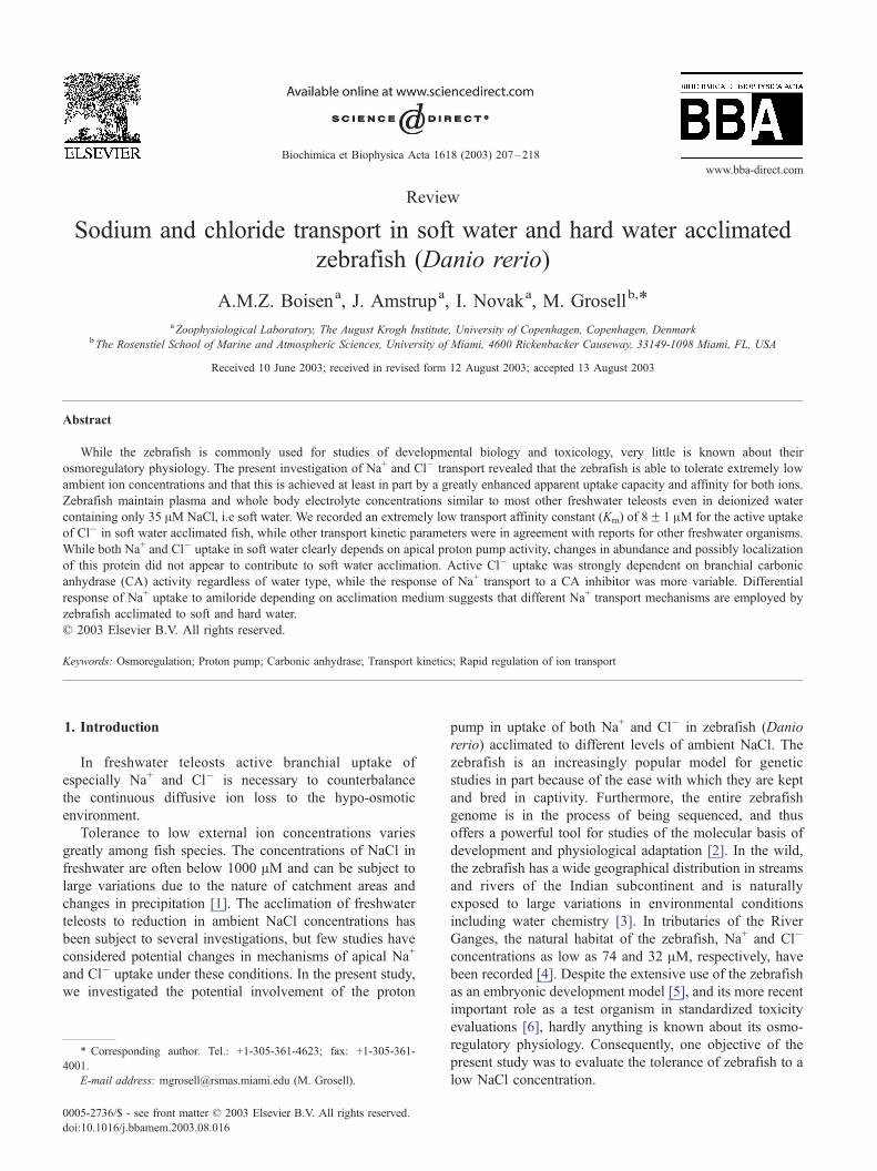
Fig. 1. Na+ influx kinetics as a function of ambient Na+ concentration in
zebrafish acclimated to soft or hard water. The Km and Jmax for soft water
and hard water were calculated from the Michaelis–Menten equation
yielding the fitted curves (SigmaPlot 4.0 for Windows), r2 = 0.870 and
0.866, respectively. Values are meansF S.E. (n= 8–10).
2.4. Measurements in nanopure water
Due to an unexpectedly high branchial affinity for Cl� in
soft water acclimated fish, it was necessary to measure Cl�
influx in nanopure water, which contains an undetectable
level of NaCl. The same procedure as described above was
used for these experiments using fish with a mean weight of
0.369F 0.0147 g (range 0.323–0.460 g). To maintain a low
Cl� concentration, however, only 0.05 ACi 36Cl was added
to each chamber and the experimental setup was rinsed three
times with nanopure water prior to measurements. To mimic
the conditions of acclimation, Ca2 + levels were adjusted to
4.2 AM by adding Ca-gluconate (C12H22CaO14) to the
medium water.
2.5. Inhibition of the proton pump with bafilomycin A1
To test the involvement of a proton ATPase in branchial
Na+ and Cl� uptake, fish were treated with bafilomycin A1,
a specific V-type proton ATPase inhibitor [22]. Due to the
expense of this drug juvenile zebrafish were used for this
experiment. Larvae were hatched in hard water, as breeding
was largely unsuccessful in soft water, and after having
reached the free-swimming stage, half of these were placed
in an aquarium containing soft water. The juvenile fish were
acclimated to the respective water types (Table 1) for 4
weeks, during which time they were fed every day with live
artemia. At the time of experimentation the fish weighed
0.00937F 0.000386 g (range 0.0031–0.0219 g). A total of
eight 10-ml beakers containing 8 ml of aerated water were
set up in a temperature-controlled bath (27 jC), after which10 fish were added to each beaker. Bafilomycin A1 dis-
solved in a final concentration of 0.05% DMSO was added
to four of the beakers to yield a final concentration of 10� 6
M, which has been reported to inhibit ion fluxes in other
freshwater fish [8–10]. To the remaining four control
beakers 0.05% DMSO was added. All beakers were either
spiked with 1 ACi 22Na or 3 ACi 36Cl. After 1 min of
equilibration the first water sample (750 Al) was taken. After12 min, a second water sample was taken and the fish were
rinsed briefly in 200 mM of nonradioactive NaCl to displace
any radioactive surface bound ions, after which they were
then anesthetized as described above, blotted dry and
weighed. The fish containing 22Na were placed directly in
counting vials and assayed for gamma radioactivity while
fish from beakers spiked with 36Cl were placed in 1.5-ml
test tubes containing 1 ml of 8% perchloric acid. Fish were
subsequently homogenized with a small pestle and left
overnight. The homogenates were vortexed briefly and
centrifuged for 10 min at 10,000� g and a sub-sample of
the supernatant (800 Al) was transferred to a vial and
assayed for beta radioactivity after an addition of 5 ml of
scintillation cocktail. Of the water samples taken, 100 Al wasassayed for beta radioactivity (700-Al 8% perchloric acid
and 5-ml scintillation cocktail was added to each vial) while
the remaining sample was analyzed for [Cl�].
hysica Acta 1618 (2003) 207–218 209

A.M.Z. Boisen et al. / Biochimica et Biop210
To test whether longer incubation periods would result in
inhibition of Na+ and Cl� influx, a second set of experi-
ments were performed. These experiments were conducted
as above except eight groups of adult zebrafish (weight
0.238F 0.0266 g; range 0.04–0.809 g) in 80 ml of water in
aerated plastic bags employing a final concentration of
5� 10� 8 M bafilomycin A1 dissolved in 0.025% DMSO
and 2-h flux periods.
2.6. Western blot analysis and immunohistochemistry
For both Western blot analysis and immunofluorescence,
a rabbit polyclonal antibody raised against the catalytic 70-
kDa A-subunit of the bovine V-type H-ATPase was
employed. The antiserum [14] was a kind gift from Jonathan
Wilson. Abundance of V-type H-ATPase (proton pump) in
gill tissue from hard and soft water acclimated zebrafish was
evaluated by Western blot analysis. Eight samples consist-
ing of gills obtained from three individual fish from each
group were homogenized in ice-cold lysis buffer (20 mM
HEPES, 1% Triton X-100, 1 mM EDTA, 1 mM NaF, 1 mM
Na3VO4, and a protease inhibitor tablet; Roche, Germany).
After removal of cellular debris by centrifugation at
10,000� g for 10 min at 4 jC, the protein content in each
sample was measured using Pierce Coomasie protein assay
reagent according to the manufacturer’s instructions (Pierce,
Rockford, IL). Equal amounts of cell lysates were dissolved
in 2� Laemmli buffer and subjected to SDS-PAGE. Sub-
sequently, the proteins were transferred to nitrocellulose
membranes (Scleicher & Schull, Germany). Immunoreac-
tive proteins were made visible using horseradish-peroxi-
dase coupled secondary antibodies and enhanced
chemiluminescence reagents according to the manufactur-
er’s instructions (Amersham Biosciences, Uppsala, Swe-
den), followed by detection and quantification using an
ImageStation 440 CF (Eastman Kodak, USA).
Gill baskets from zebrafish acclimated to both hard water
and soft water were placed in PBS containing 4% formal-
dehyde and fixed for 48 h at 4 jC and dehydrated in ethanol
and xylene prior to imbedding in paraffin. Five-micrometer
sections were made, mounted on glass slides, dried over-
night at 40 jC and kept at room temperature until use.
Deparaffinized sections were blocked in 10% Normal Goat
Serum (NGS) for 15 min followed by incubation overnight
at 4 jC with primary antibody (diluted 1:500) in PBS
containing 0.1% Triton X-100 and 0.25% BSA). After wash
in PBS, sections were covered with secondary goat-anti-
rabbit antibody conjugated to Alexa 568 and incubated for 1
h protected from light. After additional wash in PBS,
sections were mounted and examined using a TCS NT/SP
confocal laser scanning microscope equipped with ArKr
laser and 20� 0.7 NA and 63� 1.2 NA PL APO objectives
(Leica Micro Systems Heidleberg GmbH, Germany).
Images were analyzed using Leica CLSM software or
MetaMorph 5.0 sofware (Universal Imaging Corporation,
West Chester, PA, USA).
2.7. Effects of ethoxzolamide on Na+ and Cl� uptake
Ethoxzolamide (6-ethoxy-2-benzothiazolesulfonamide
97%, Aldrich Chem. Co.), a membrane permeable inhibitor
of CA, used at final concentration of 10� 4 M, was used to
test the importance of H+ and HCO3� availability for apical
exchange with Na+ and Cl�, respectively. A total of eight
beakers, each containing 200 ml of aerated water (four of
each water type), were placed in a temperature-controlled
water bath (27 jC) and used for the Na+ and Cl� influx
experiments. Four groups of zebrafish, two from each water
type were treated with ethoxzolamide dissolved in 0.025%
DMSO while the remaining four groups, which were
exposed to 0.025% DMSO only, were controls (80 fish in
total, mean weight 0.247F 0.0099� g, range 0.036–0.653
g). The fish were allowed a preincubation period of 30 min
under these conditions, after which 0.2 ACi 22Na or 2 ACi36Cl was added. At 10 min and again at 3 h of exposure to
isotope, a water sample of 5 ml was obtained from each
exposure beaker, after which fish were netted out of the
beakers, rinsed, anesthetized and prepared for 22Na or 36Cl
radioactivity, as outlined above.
2.8. Effects of amiloride and EIPA on Na+ influx
In attempt to elucidate the mechanisms responsible for
branchial Na+ uptake in zebrafish, we tested the effect of
Amiloride (N-amidino-3.5-diamino-6-chloropyrazinecarbro-
mide C6H8ClN7*HCl, Sigma) and EIPA (5-(N-ethyl-N-iso-
propyl)-amiloride, C11H18ClN7O, Sigma) on branchial Na+-
uptake was investigated. Amiloride is known to inhibit both
Na+/H+ exchangers and Na+ channels in various epithelia
[23]. Generally, Na+ channels have a higher affinity for
amiloride than the exchanger [23], and we therefore attemp-
ted to distinguish between the Na+ channel and the ex-
changer by using two concentrations of amiloride: 10� 5
and 10� 4 M. Furthermore, EIPA, which more selectively
targets Na+/H+ exchangers, was tested at a final concentra-
tion of 5� 10� 5 M. These Na+-flux experiments were
conducted as the ethoxzolamide experiments described
above.
2.9. Analytical techniques, calculations and statistical
analysis
Water samples from kinetic experiments on Na+ uptake
and samples of whole fish from pharmacological studies of
Na+ influx were assayed for gamma radioactivity on a
Minaxig AutogammaR 5000 Series Gamma Counter. Water
Samples from Cl� uptake kinetic experiments and homo-
genates of whole fish from pharmacological studies were
assayed on a TRI-CARB 2500 TR Liquid Scintillation
Analyzer after an addition of 5-ml scintillation cocktail.
All anion and cation concentrations in water, plasma and
whole fish samples were measured on a Dionex Ion Chro-
matograph DX 120 after appropriate dilutions.
hysica Acta 1618 (2003) 207–218
Table 3
Whole body ion concentration in soft water and hard water acclimated
zebrafish (mmol kg� 1), meanF S.E. (n= 10)
[Na+] [K+] [Ca2 +] [Cl�]
Soft water 44.9F 1.5 81.9F 1.9 33.3F 1.6 23.8F 4.1
Hard water 49.6F 3.1 86.1F 2.4 35.2F 1.1 20.2F 2.0
A.M.Z. Boisen et al. / Biochimica et Biophysica Acta 1618 (2003) 207–218 211
In kinetic studies, Jin of either Na+ and Cl� was
calculated as
Influx; Jin ¼ ððCPMb � CPMaÞ=ðMSAÞÞðV=W*tÞ;
where CPMa and CPMb are measures of radioactivity
(counts min� 1) in the flux chamber at the beginning and
end of the flux period. MSA is the mean specific activity in
the water during the flux period, V is the volume in the flux
chamber (l), W is the mass of the fish (g) and t is time (h).
During pharmacological studies, Na+ and Cl� influx
rates were calculated as follows:
Influx; Jin ¼ ððCPMg�1Þ=MSAÞ=t
Were CPM (counts min� 1) is the final radioactivity in
the fish, MSA and t have the same meaning as above.
Na+ and Cl� influx displayed apparent saturation kinet-
ics. The Michaelis–Menten equation for nonlinear regres-
sion ( Jin[X] = Jmax*[X]/Km+[X]), where X was Na+ or Cl�)
was used to estimate the affinity (Km) and maximum
capacity ( Jmax) of the transport systems (SigmaPlot 4.0
for Windows).
Data are presented as meansF S.E. Statistical compar-
isons were made with a two-tailed t test for independent
observations (P < 0.05).
3. Results
3.1. Plasma and body ions
Both plasma Na+ and Cl� concentrations were not
statistically different in soft water and hard water acclimated
fish; although concentrations in soft water fish appeared
lower (Table 2). Similarly, whole body content of Na+,
Ca2 +, K+ and Cl� did not seem to differ between soft water
and hard water acclimated fish (Table 3).
3.2. Na+ influx kinetics
The influx of Na+ in zebrafish acclimated to soft water
(35 AM Na+) or hard water (1480 AM Na+) exhibited clear
Michaelis–Menten saturation kinetics (Fig. 1). Both appar-
ent Na+ uptake affinity and capacity were significantly
elevated about twofold in response to soft water acclima-
tion. Interestingly, the rate of Na+ uptake in fish at the Na+
concentration to which they had been acclimated was
approximately 500 (nmol g� 1 h� 1) for both soft water
Table 2
Plasma Na+ and Cl� concentration (mM) in soft water and hard water
acclimated zebrafish, meanF S.E. (n= 10)
[Na+] [Cl�]
Soft water 123.3F 13.8 86.2F 6.4
Hard water 154.5F 8.8 105.7F 15.2
and hard water acclimated fish. Soft water Na+ uptake
kinetics was calculated only from values obtained from flux
measurements performed at Na+ concentrations lower than
500 AM (>10 times the level of acclimation for soft water
fish). At concentrations above 500 AM, soft water acclimat-
ed fish displayed large individual differences and at Na+
concentration above 1000 AM, influx values were approx-
imately 50% lower than what would have been expected
according to the Vmax (data not shown).
3.3. Down-regulation of Na+ transport
Soft water acclimated fish exposed to relatively high Na+
concentrations (1200 AM) for more than two subsequent
flux periods displayed a drop in the rate of Na+ influx to less
than half the value measured after one flux period (Fig. 2).
Na+ efflux was down-regulated in a similar manner resulting
in no change in net Na+ flux. In hard water acclimated fish,
the Na+ influx rate did not diminish as a function of time.
3.4. Cl� influx kinetics
As for Na+, Cl� uptake in both soft water and hard water
acclimated zebrafish exhibited saturation kinetics and the
difference between Cl� influx kinetic constants from the
two groups of fish was statistically significant (Fig. 3). The
apparent Km for Cl� influx in soft water acclimated zebra-
Fig. 2. Na+ flux (nmol g� 1 h� 1) measured after 1, 2, 3 and 4 h of exposure
to an ambient Na+ concentration of 1200 AM in zebrafish adapted to soft
water. Open bars indicate unidirectional Na+ flux (positive values indicate
influx while negative values indicate efflux) and hatched bars indicate net
flux. Asterisks signify a statistically significant difference between values
after 1 h and subsequent hours, two-tailed Student’s t test, unpaired
( P< 0.05). Net flux values did not show any statistically significant
difference from zero. All values are meansF S.E. (n= 10).

Fig. 3. Cl� influx kinetics as a function of ambient Cl� concentration in
zebrafish adapted to soft or hard water. The Km and Jmax for soft water and
hard water were calculated from the Michaelis–Menten equation yielding
the fitted curves (SigmaPlot 4.0 for Windows), r2 = 0.982 and 0.711,
respectively. Values are meansF S.E. (n= 3–10).
A.M.Z. Boisen et al. / Biochimica et Biophysica Acta 1618 (2003) 207–218212
fish was remarkably low at only 8F 1 AM and approxi-
mately fivefold lower than that of hard water acclimated
fish. Similarly, the Cl� uptake capacity was elevated ap-
proximately threefold as a result of soft water acclimation.
For adult fish, the Cl� transport capacity was much lower
Fig. 4. Na+ influx (nmol g� 1 h� 1) (upper panel) and Cl� influx (nmol g� 1
h� 1) (lower panel) in juvenile zebrafish treated with 10� 6 M bafilomycin
for 12 min. Asterisks indicate significant difference from the corresponding
control evaluated by Student’s t test, unpaired ( P < 0.05). All values are
meansF S.E. (n= 10).
than that for Na+, regardless of water type. That is, soft and
hard water acclimated fish exhibited Cl� uptake capacity
amounting to 15% and 20%, respectively, of the
corresponding Na+ uptake capacity (Fig. 1 versus Fig. 3).
As for Na+ influx, soft water acclimated fish had Cl� influx
which was also lower than expected, based on the calculated
Vmax, at values greatly exceeding the acclimation Cl�
concentration (Fig. 3).
3.5. Effects of proton pump inhibition on ion uptake
Soft water acclimated zebrafish treated with 1 AMbafilomycin for a short period of time (12 min) displayed
a significant inhibition of Na+ influx, while Na+ uptake in
hard water acclimated fish was unaffected (Fig. 4). In
contrast, during bafilomycin treatment, Cl� influx was not
reduced in fish acclimated to either water type, but was
increased in hard water acclimated fish (Fig. 4). More
prolonged exposure (2 h) to a lower bafilomycin concen-
tration (5� 10� 8 M) resulted in an inhibition of Cl� but
not Na+ influx in the soft water acclimated fish. In hard
water acclimated fish, Cl� uptake was unaffected by this
treatment whereas Na+ influx appeared to be stimulated
(Fig. 5).
Fig. 5. Na+ influx (nmol g� 1 h� 1) (upper panel) and Cl� influx (nmol g� 1
h� 1) (lower panel) in zebrafish treated with 5� 10� 8 M bafilomycin for 2
h. Asterisks indicate significant difference from the corresponding control
evaluated by Student’s t test, unpaired ( P < 0.05). All values are
meansF S.E. (n= 10).
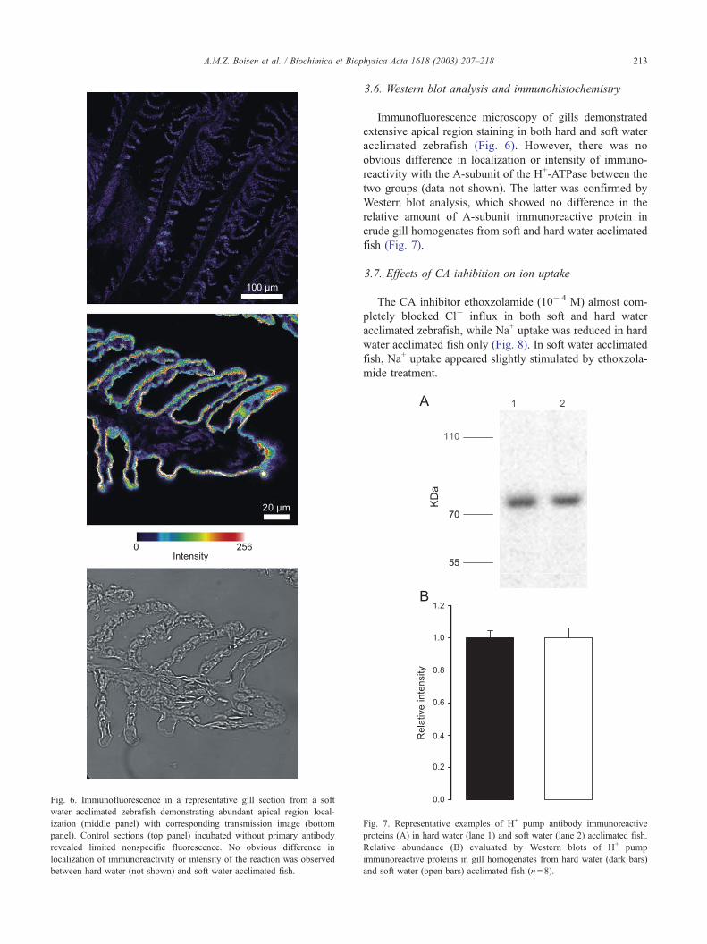
Fig. 6. Immunofluorescence in a representative gill section from a soft
water acclimated zebrafish demonstrating abundant apical region local-
ization (middle panel) with corresponding transmission image (bottom
panel). Control sections (top panel) incubated without primary antibody
revealed limited nonspecific fluorescence. No obvious difference in
localization of immunoreactivity or intensity of the reaction was observed
between hard water (not shown) and soft water acclimated fish.
A.M.Z. Boisen et al. / Biochimica et Biop
3.6. Western blot analysis and immunohistochemistry
Immunofluorescence microscopy of gills demonstrated
extensive apical region staining in both hard and soft water
acclimated zebrafish (Fig. 6). However, there was no
obvious difference in localization or intensity of immuno-
reactivity with the A-subunit of the H+-ATPase between the
two groups (data not shown). The latter was confirmed by
Western blot analysis, which showed no difference in the
relative amount of A-subunit immunoreactive protein in
crude gill homogenates from soft and hard water acclimated
fish (Fig. 7).
3.7. Effects of CA inhibition on ion uptake
The CA inhibitor ethoxzolamide (10� 4 M) almost com-
pletely blocked Cl� influx in both soft and hard water
acclimated zebrafish, while Na+ uptake was reduced in hard
water acclimated fish only (Fig. 8). In soft water acclimated
fish, Na+ uptake appeared slightly stimulated by ethoxzola-
mide treatment.
hysica Acta 1618 (2003) 207–218 213
Fig. 7. Representative examples of H+ pump antibody immunoreactive
proteins (A) in hard water (lane 1) and soft water (lane 2) acclimated fish.
Relative abundance (B) evaluated by Western blots of H+ pump
immunoreactive proteins in gill homogenates from hard water (dark bars)
and soft water (open bars) acclimated fish (n= 8).
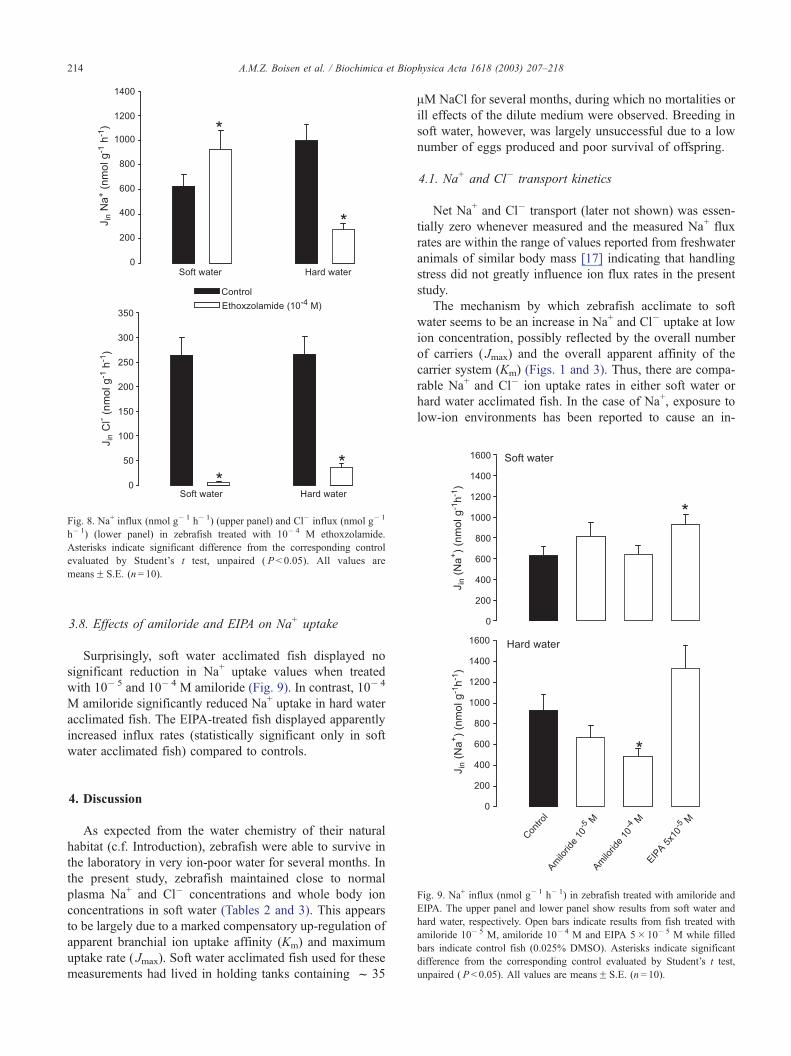
Fig. 8. Na+ influx (nmol g� 1 h� 1) (upper panel) and Cl� influx (nmol g� 1
h� 1) (lower panel) in zebrafish treated with 10� 4 M ethoxzolamide.
Asterisks indicate significant difference from the corresponding control
evaluated by Student’s t test, unpaired ( P < 0.05). All values are
meansF S.E. (n= 10).
A.M.Z. Boisen et al. / Biochimica et Biophysica Acta 1618 (2003) 207–218214
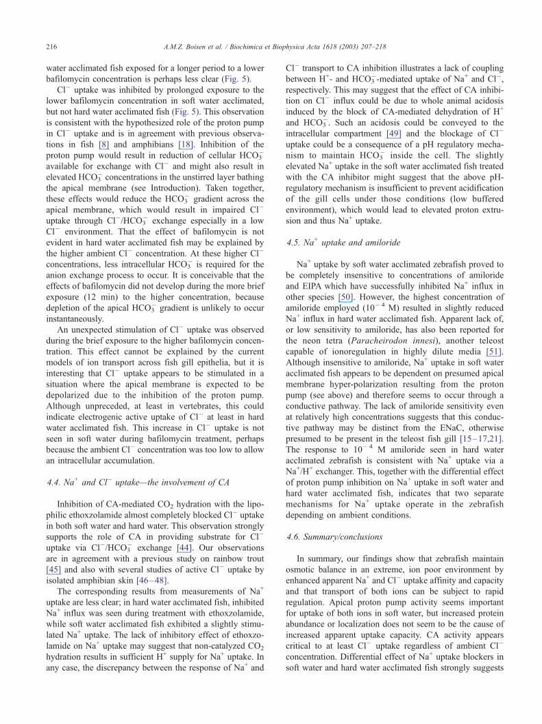
3.8. Effects of amiloride and EIPA on Na+ uptake
Surprisingly, soft water acclimated fish displayed no
significant reduction in Na+ uptake values when treated
with 10� 5 and 10� 4 M amiloride (Fig. 9). In contrast, 10� 4
M amiloride significantly reduced Na+ uptake in hard water
acclimated fish. The EIPA-treated fish displayed apparently
increased influx rates (statistically significant only in soft
water acclimated fish) compared to controls.
Fig. 9. Na+ influx (nmol g� 1 h� 1) in zebrafish treated with amiloride and
EIPA. The upper panel and lower panel show results from soft water and
hard water, respectively. Open bars indicate results from fish treated with
amiloride 10� 5 M, amiloride 10� 4 M and EIPA 5� 10� 5 M while filled
bars indicate control fish (0.025% DMSO). Asterisks indicate significant
difference from the corresponding control evaluated by Student’s t test,
unpaired ( P < 0.05). All values are meansF S.E. (n= 10).
4. Discussion
As expected from the water chemistry of their natural
habitat (c.f. Introduction), zebrafish were able to survive in
the laboratory in very ion-poor water for several months. In
the present study, zebrafish maintained close to normal
plasma Na+ and Cl� concentrations and whole body ion
concentrations in soft water (Tables 2 and 3). This appears
to be largely due to a marked compensatory up-regulation of
apparent branchial ion uptake affinity (Km) and maximum
uptake rate ( Jmax). Soft water acclimated fish used for these
measurements had lived in holding tanks containing f 35
AM NaCl for several months, during which no mortalities or
ill effects of the dilute medium were observed. Breeding in
soft water, however, was largely unsuccessful due to a low
number of eggs produced and poor survival of offspring.
4.1. Na+ and Cl� transport kinetics
Net Na+ and Cl� transport (later not shown) was essen-
tially zero whenever measured and the measured Na+ flux
rates are within the range of values reported from freshwater
animals of similar body mass [17] indicating that handling
stress did not greatly influence ion flux rates in the present
study.
The mechanism by which zebrafish acclimate to soft
water seems to be an increase in Na+ and Cl� uptake at low
ion concentration, possibly reflected by the overall number
of carriers ( Jmax) and the overall apparent affinity of the
carrier system (Km) (Figs. 1 and 3). Thus, there are compa-
rable Na+ and Cl� ion uptake rates in either soft water or
hard water acclimated fish. In the case of Na+, exposure to
low-ion environments has been reported to cause an in-

A.M.Z. Boisen et al. / Biochimica et Biophysica Acta 1618 (2003) 207–218 215
crease in maximum uptake rate and/or affinity in several fish
species [1,24]. The apparent Na+ uptake kinetic constants of
both groups of zebrafish in the present study compare well
with previously reported values from other teleosts [10,24–
28]. The observed increase in at least apparent Na+ uptake
affinity and capacity takes several days to develop (Bury
and Grosell, unpublished observations); indicating that
acclimation to soft water involves genomic regulation.
Saturable and thus carrier-mediated Cl� uptake is not
obligatory for osmoregulation in freshwater teleosts. Fresh-
water acclimated killifish (Fundulus heteroclitus) and mem-
bers of the Anguillidae family do not appear to have the
ability to take up Cl� from freshwater [29–31]. In contrast
to these fishes, zebrafish clearly exhibits carrier-mediated
Cl� uptake in freshwater (Fig. 3) and the greatest relative
response to soft water acclimation was actually the
increases in apparent Cl� uptake affinity and capacity.
Zebrafish generally exhibits a high affinity Cl� uptake
system when compared to other aquatic vertebrates and
invertebrates. Indeed, zebrafish acclimated to soft water had
a remarkably low Km of only 8 AM, to our knowledge, the
lowest ever recorded. Whether, the increase of apparent Cl�
uptake affinity and capacity is the result of genomic
regulation and/or a faster non-genomic regulation waits to
be investigated.
4.2. Rapid regulation of Na+ and Cl� transport
At high ambient Na+ concentrations, there appeared to be
a rapid down-regulation of Na+ uptake, as well as Na+ loss
(Figs. 1 and 2). The mechanism of this rapid apparently non-
genomic regulation, could be a Na+-dependent inhibition of
an epithelial Na+ channel, as previously observed in experi-
ments conducted on isolated frog skin [32,33]. Cl� influx
also seemed to be reduced when soft water acclimated fish
were subjected to Cl� concentrations above 200 AM (Fig.
3). As will be discussed in the following section, apical Cl�
uptake is suspected to be via Cl�/HCO3� exchange, which
expectedly would result in elevated Cl� uptake rates at
higher ambient Cl� concentrations. Although cellular Cl�
uptake via certain conductive pathways appears to be
regulated based on ambient Cl� concentration [34–36],
we are unaware of studies documenting down-regulation
of Cl�/HCO3� exchange in response to increased ambient
Cl� concentrations.
4.3. Na+ and Cl� uptake—the involvement of a proton
pump
The presence of the H+-pump in zebrafish was identified
by use of a polyclonal antibody directed against the catalytic
70-kDa A-subunit. An immunoreactive band of approxi-
mately 75–80 kDa was observed in both soft water and hard
water acclimated fish (Fig. 7A), which is in agreement with
previous results obtained from rainbow trout [14]. Further-
more, immunofluorescence microscopy of gill tissue dem-
onstrated abundant apical region immunoreactivity with no
obvious differences in localization between fish acclimated
to soft or hard water (Fig. 6). Since apical proton pump
activity appears to be involved in both Na+ and Cl� uptake
in soft water and since there is elevated transport capacity of
both ions, one could expect increased proton pump abun-
dance. Current preparation does not allow subcellular reso-
lution, i.e. detecting differences between proton pumps
inserted in the apical membrane and pumps located just
under the apical membrane. It thus appears that the elevated
ion uptake capacity in soft water fish cannot conclusively be
explained by increased proton pump abundance, which is in
agreement with observations of lack of increase in proton-
pump mRNA expression and enzyme activity in response to
soft water acclimation [37,38]. It is, however, possible that
the increase in uptake capacity for both ions is due to an
increase in the kinetic activity of the proton pump without a
change in quantity or distribution [39]. The apical region
localization observed in the present study on zebrafish is as
mentioned in agreement with studies on rainbow trout [14]
but is in contrast to a recent report of basolateral localization
in the gills of freshwater acclimated killifish (F. heteroclitus)
[40]. It should be noted, however, first, that killifish does
not exhibit high affinity Cl� uptake in freshwater [31] and
second, that no studies of the functionality, with respect to
ion uptake, of the basolateral proton pump were reported by
Katoh et al. [40]. The apparent differences between zebra-
fish and killifish with respect to proton pump localization
could well be related to their freshwater stenohalinity and
euryhalinity, respectively.
Overall, experiments with bafilomycin revealed that
proton pump activity may be required for Na+ and Cl�
uptake at least in soft water acclimated fish. Na+ uptake in
soft water but not hard water was inhibited by brief
exposure to 10� 6 M bafilomycin. Effects of bafilomycin
on branchial Na+ uptake in freshwater fish has been reported
previously [8–10], but this is the first study to report
differential effect of proton pump inhibition depending on
acclimation conditions. It has been suggested that one
function of the apical proton pump is to hyperpolarize the
apical membrane to establish a sufficient electrochemical
gradient for Na+ uptake. Considering an expected apical
membrane potential difference of at least � 70mV and
intracellular Na+ concentrations of 20–50 mM [15,41–
43], proton pump activity would only be necessary for
Na+ uptake in situations with low ambient Na+ concentra-
tions (see Ref. [20] in this issue for detailed discussion). In
soft water containing very low Na+ concentrations, the
additional polarization of the apical membrane caused by
the electrogenic proton pump may aid Na+ uptake against
the substantial chemical gradient. Our findings of reduced
Na+ uptake in soft water but absence of effect of bafilomy-
cin in hard water, where the electrochemical Na+ gradient
generated by the Na+/K+-ATPase is sufficient to drive Na+
uptake, is consistent with this proposed function of the
proton pump. The reason for elevated Na+ uptake in hard
A.M.Z. Boisen et al. / Biochimica et Biophysica Acta 1618 (2003) 207–218216
water acclimated fish exposed for a longer period to a lower
bafilomycin concentration is perhaps less clear (Fig. 5).
Cl� uptake was inhibited by prolonged exposure to the
lower bafilomycin concentration in soft water acclimated,
but not hard water acclimated fish (Fig. 5). This observation
is consistent with the hypothesized role of the proton pump
in Cl� uptake and is in agreement with previous observa-
tions in fish [8] and amphibians [18]. Inhibition of the
proton pump would result in reduction of cellular HCO3�
available for exchange with Cl� and might also result in
elevated HCO3� concentrations in the unstirred layer bathing
the apical membrane (see Introduction). Taken together,
these effects would reduce the HCO3� gradient across the
apical membrane, which would result in impaired Cl�
uptake through Cl�/HCO3� exchange especially in a low
Cl� environment. That the effect of bafilomycin is not
evident in hard water acclimated fish may be explained by
the higher ambient Cl� concentration. At these higher Cl�
concentrations, less intracellular HCO3� is required for the
anion exchange process to occur. It is conceivable that the
effects of bafilomycin did not develop during the more brief
exposure (12 min) to the higher concentration, because
depletion of the apical HCO3� gradient is unlikely to occur
instantaneously.
An unexpected stimulation of Cl� uptake was observed
during the brief exposure to the higher bafilomycin concen-
tration. This effect cannot be explained by the current
models of ion transport across fish gill epithelia, but it is
interesting that Cl� uptake appears to be stimulated in a
situation where the apical membrane is expected to be
depolarized due to the inhibition of the proton pump.
Although unpreceded, at least in vertebrates, this could
indicate electrogenic active uptake of Cl� at least in hard
water acclimated fish. This increase in Cl� uptake is not
seen in soft water during bafilomycin treatment, perhaps
because the ambient Cl� concentration was too low to allow
an intracellular accumulation.
4.4. Na+ and Cl� uptake—the involvement of CA
Inhibition of CA-mediated CO2 hydration with the lipo-
philic ethoxzolamide almost completely blocked Cl� uptake
in both soft water and hard water. This observation strongly
supports the role of CA in providing substrate for Cl�
uptake via Cl�/HCO3� exchange [44]. Our observations
are in agreement with a previous study on rainbow trout
[45] and also with several studies of active Cl� uptake by
isolated amphibian skin [46–48].
The corresponding results from measurements of Na+
uptake are less clear; in hard water acclimated fish, inhibited
Na+ influx was seen during treatment with ethoxzolamide,
while soft water acclimated fish exhibited a slightly stimu-
lated Na+ uptake. The lack of inhibitory effect of ethoxzo-
lamide on Na+ uptake may suggest that non-catalyzed CO2
hydration results in sufficient H+ supply for Na+ uptake. In
any case, the discrepancy between the response of Na+ and
Cl� transport to CA inhibition illustrates a lack of coupling
between H+- and HCO3�-mediated uptake of Na+ and Cl�,
respectively. This may suggest that the effect of CA inhibi-
tion on Cl� influx could be due to whole animal acidosis
induced by the block of CA-mediated dehydration of H+
and HCO3�. Such an acidosis could be conveyed to the
intracellular compartment [49] and the blockage of Cl�
uptake could be a consequence of a pH regulatory mecha-
nism to maintain HCO3� inside the cell. The slightly
elevated Na+ uptake in the soft water acclimated fish treated
with the CA inhibitor might suggest that the above pH-
regulatory mechanism is insufficient to prevent acidification
of the gill cells under those conditions (low buffered
environment), which would lead to elevated proton extru-
sion and thus Na+ uptake.
4.5. Na+ uptake and amiloride
Na+ uptake by soft water acclimated zebrafish proved to
be completely insensitive to concentrations of amiloride
and EIPA which have successfully inhibited Na+ influx in
other species [50]. However, the highest concentration of
amiloride employed (10� 4 M) resulted in slightly reduced
Na+ influx in hard water acclimated fish. Apparent lack of,
or low sensitivity to amiloride, has also been reported for
the neon tetra (Paracheirodon innesi), another teleost
capable of ionoregulation in highly dilute media [51].
Although insensitive to amiloride, Na+ uptake in soft water
acclimated fish appears to be dependent on presumed apical
membrane hyper-polarization resulting from the proton
pump (see above) and therefore seems to occur through a
conductive pathway. The lack of amiloride sensitivity even
at relatively high concentrations suggests that this conduc-
tive pathway may be distinct from the ENaC, otherwise
presumed to be present in the teleost fish gill [15–17,21].
The response to 10� 4 M amiloride seen in hard water
acclimated zebrafish is consistent with Na+ uptake via a
Na+/H+ exchanger. This, together with the differential effect
of proton pump inhibition on Na+ uptake in soft water and
hard water acclimated fish, indicates that two separate
mechanisms for Na+ uptake operate in the zebrafish
depending on ambient conditions.
4.6. Summary/conclusions
In summary, our findings show that zebrafish maintain
osmotic balance in an extreme, ion poor environment by
enhanced apparent Na+ and Cl� uptake affinity and capacity
and that transport of both ions can be subject to rapid
regulation. Apical proton pump activity seems important
for uptake of both ions in soft water, but increased protein
abundance or localization does not seem to be the cause of
increased apparent uptake capacity. CA activity appears
critical to at least Cl� uptake regardless of ambient Cl�
concentration. Differential effect of Na+ uptake blockers in
soft water and hard water acclimated fish strongly suggests

A.M.Z. Boisen et al. / Biochimica et Biophysica Acta 1618 (2003) 207–218 217
that fish under these different conditions employ different
mechanisms for Na+ uptake.
Acknowledgements
Excellent animal care and analytical assistance from
Alice Sheuer and Thomas Sørensen is greatly appreciated.
We wish to thank Dr. Jonathan Wilson for the kind gift of
the proton pump antiserum. MG was supported by a Danish
Natural Science Research Council grant (21-01-0255) and
IN is supported by a Danish Natural Science Research
Council grant (21-01-0490).
References
[1] D.G. McDonald, M.S. Rogano, Ion regulation by the rainbow
trout, Salmo gairdneri, in ion-poor water, Physiol. Zool. 59
(1986) 318–331.
[2] J.P. Briggs, The zebrafish: a new model organism for integrative
physiology, Am. J. Phys. 282 (2002) R3–R9.
[3] P.K. Talwar, A.G. Jhingran, Inland Fishes Of India and Adjecent
Countries, vol. 1, A.A. Balkema, Rotterdam, 1991. 541 p.
[4] M.M. Krisnaswami, S. Sarin, Major ion chemistry of the Ganga–
Bramaputra river systems, India’s Nature 312 (1984) 538–545.
[5] J. Sprague, E. Doerry, S. Douglas, M. Westerfield, The Zebrafish
Information Network (ZFIN): a resource for genetic, genomic and
developmental research, Nucleic Acids Res. 29 (1-1-2001) 87–90.
[6] OECD, Guideline 203—for testing of chemicals. OECD 203 (1992).
[7] A. Krogh, The active absorption of ions in some freshwater animals,
Z. Vgl. Physiol. 25 (1938) 335–350.
[8] J.C. Fenwick, W. Bonga, F. Gert, In vivo bafilomycin-sensitive Na+
uptake in young freshwater fish, J. Exp. Biol. 202 (1999) 3659–3666.
[9] N.R. Bury, C.M. Wood, Mechanisms of branchial apical silver uptake
by rainbow trout is via the proton coupled Na+ channel, Am. J. Phys.
277 (1999) R1385–R1391.
[10] M. Grosell, C.M. Wood, Copper uptake across rainbow trout gills:
mechanisms of apical entry, J. Exp. Biol. 205 (2002) 1179–1188.
[11] H. Lin, D.C. Pfeiffer, A.W. Vogl, J. Pan, D.J. Randall, Immunolocal-
ization of H+-ATPase in the gill epithelia of rainbow trout, J. Exp.
Biol. 195 (1994) 169–183.
[12] H. Lin, D.J. Randall, H+-ATPase activity in crude homogenates of
fish gill tissue-inhibitor sensitivity and environmental and hormonal-
regulation, J. Exp. Biol. 180 (1993) 163–174.
[13] H. Lin, D. Randall, Evidence for the presence of an electrogenic
proton pump on the trout gill epithelium, J. Exp. Biol. 161 (1991)
119–134.
[14] J.M. Wilson, P. Laurent, B.L. Tufts, D.J. Benos, M. Donowitz, A.W.
Vogl, D.J. Randall, NaCl uptake by the brachial epithelium in fresh-
water teleost fish: an immunological approach to ion-transport protein
localization, J. Exp. Biol. 203 (2000) 2279–2296.
[15] S.F. Perry, The chloride cell: structure and function in the gills of
freshwater fishes, Ann. Rev. Phys. 59 (1997) 325–347.
[16] C.M. Wood, The toxic response of the gill, in: W.H. Benson, D.W.
Schleuh (Eds.), Target Organ Toxicity in Marine and Freshwater Tele-
osts, Taylor and Francis, Washington, DC, 2001, pp. 1–87.
[17] M. Grosell, C. Nielsen, A. Bianchini, Sodium turnover rate deter-
mines sensitivity to acute copper and silver exposure in freshwater
animals, Comp. Biochem. Physiol. 133C (2002) 287–303.
[18] L.J. Jensen, N.J. Willumsen, E.H. Larsen, Proton pump activity is
requirred for active uptake of chloride in isolated amphibian skin
exposed to freshwater, J. Comp. Physiol. 176B (2002) 503–511.
[19] E.H. Larsen, Chloride transport by high-resistance heterocellular epi-
thelia, Physiol. Rev. 71 (1991) 235–283.
[20] N.J. Willumsen, J. Amstrup, L.J. Jensen, E. Hviid Larsen, Proton
pump driven cutaneous chloride uptake in anuran amphibia, Biochim.
Biophys. Acta, (in press).
[21] W.S. Marshall, Na+, Cl�, Ca2 + and Zn2 + transport by fish gills:
retrospective review and prospective synthesis, J. Exp. Zool. 293
(8-1-2002) 264–283.
[22] E.J. Bowman, A. Siebers, K. Altendorf, Bafilomycins—a class of
inhibitors of membrane ATPases from microorganisms, animal-cells,
and plant-cells, Proc. Natl. Acad. Sci. U. S. A. 85 (1988) 7972–7976.
[23] T.R. Kleyman , E.J. Cragoe Jr., Amiloride and its analogs as tools in
the study of ion transport, J. Membr. Biol. 105 (1998) 1–21.
[24] R.J. Gonzales, R.W. Wilson, C.M. Wood, M.L. Patrick, A.L. Val,
Diverse strategies for ion regulation in fish colected from the ion-
poor, acidic Rio Negro, Physiol. Biochem. Zool. 75 (2001) 37–47.
[25] J. Freda, D.G. McDonald, Physiological correlates of interspecific
variation in acid tolerance in fish, J. Exp. Biol. 136 (1988) 243–258.
[26] G.G. Goss, C.M. Wood, Na+ and Cl� uptake kinetics, diffusive efflu-
xes and acidic equivalent fluxes across the gills of rainbow trout: 1.
Responses to environmental hyperoxia, J. Exp. Biol. 152 (1990)
521–547.
[27] J. Maetz, Branchial sodium exchange and ammonia excretion in the
goldfish Carassius auratus. Effects of ammonia-loading and temper-
ature changes, J. Exp. Biol. 56 (1972) 601–620.
[28] A.M.R. Preest, R.J. Gonzales, R.W. Wilson, A phamacological ex-
amination of Na+ and Cl� transport in two species of freshwater fish,
Physiol. Biochem. Zool., (Submitted).
[29] M. Grosell, C. Hogstrand, C.M. Wood, H.J.M. Hansen, A nose-to-
nose comparison of the physiological effects of exposure to ionic
silver versus silver chloride in the European eel (Anguilla anguilla)
and the rainbow trout (Oncorhynchus mykiss), Aquat. Toxicol. 48
(2000) 327–342.
[30] G.G. Goss, S.F. Perry, Different mechanisms of acid–base regula-
tion in rainbow trout (Oncorhynchus mykiss) and American eel (An-
nguilla rostrata) during NaHCO3 infusion, Physiol. Zool. 67 (1994)
381–406.
[31] M.L. Patrick, P. Part, W.S. Marshall, C.M. Wood, Characterization of
ion and acid–base transport in the fresh water adapted mumichog
(Fundulus heteroclitus), J. Exp. Zool. 279 (1997) 208–219.
[32] W. Fuchs, E. Hviid Larsen, B. Lindemann, Current–voltage curve of
sodium channels and concentration dependence of sodium perme-
ability in frog skin, J. Physiol. 267 (1977) 137–166.
[33] H. Garty, L.G. Palmer, Epithelial sodium channels: function, struc-
ture, and regulation, Phys. Rev. 77 (1997) 359–396.
[34] V. Koefoed-Johnsen, H.H. Ussing, The nature of the frog skin poten-
tial, Acta Physiol. Scand. 42 (1958) 298–308.
[35] L.B. Kirschner, The study of NaCl in aquatic animals, Am. Zool. 10
(1970) 365–376.
[36] P. Kristensen, Chloride transport across frog skin, Acta Physiol.
Scand. 84 (1973) 338–346.
[37] D. Kultz, G.N. Somero, Osmotic and thermal effects on in-situ AT-
Pase activity in permeabilized gill epithelial cells of the fish Gillich-
thys mirabilis, J. Exp. Biol. 198 (1995) 1883–1894.
[38] S.F. Perry, M.L. Beyers, D.A. Johnson, Cloning and molecular char-
acterisation of the trout (Oncorhynchus mykiss) vacuolar H+-ATPase
B subunit, J. Exp. Biol. 203 (2000) 459–470.
[39] S.L. Gluck, R.D. Nelson, B.S. Lee, Z.Q. Wang, X.L. Guo, J.Y. Fu, K.
Zhang, Biochemistry of the renal V-ATPase, J. Exp. Biol. 172 (1992)
219–229.
[40] F. Katoh, S. Hyodo, T. Kaneko, Vacuolar-type proton pump in the
basolateral plasma membrane energizes ion uptake in branchial mi-
tochondria-rich cells of killifish Fundulus heteroclitus, adapted to a
low ion environment, J. Exp. Biol. 206 (2003) 793–803.
[41] C.M. Wood, J. Lemoigne, Intracellular acid–base responses to envi-
ronmental hyperoxia and normoxic recovery in rainbow trout, Respir.
Physiol. 86 (1991) 91–113.
A.M.Z. Boisen et al. / Biochimica et Biophysica Acta 1618 (2003) 207–218218
[42] I.J. Morgan, W.T.W. Potts, K. Oates, Intracellular ion concentrations
in branchial epithelial cells of brown trout (Salmo trutta L) deter-
mined by X-ray-microanalysis, J. Exp. Biol. 194 (1994) 139–151.
[43] I.J. Morgan, W.T.W. Potts, The effects of thiocyanate on the intra-
cellular ion concentrations of branchial epithelial cells of brown trout,
J. Exp. Biol. 198 (1995) 1229–1232.
[44] G.G. Goss, C.M. Wood, 2-substrate kinetic-analysis—a novel ap-
proach linking ion and acid–base transport at the gills of fresh-
water trout, Oncorhynchus mykiss, J. Comp. Physiol. 161B (1991)
635–646.
[45] S.F. Perry, P. Payan, J.P. Girard, Effects of perfusate HCO3� and
PCO2 on chloride uptake in perfused gills of rainbow trout (Salmo
gairdneri), Can. J. Fish. Aquat. Sci. 41 (1984) 1768–1773.
[46] K. Bruus, P. Kristensen, E.H. Larsen, Pathways for chloride and sodium
transport across toad skin, Acta Physiol. Scand. 97 (1976) 31–47.
[47] J. Ehrenfeld, F. Garcia-Romeu, Coupling between chloride absorption
and base excretion in isolated skin of Rana esculenta, Am. J. Physiol.
235 (1978) F33–F39.
[48] F. Garcia-Romeu, J. Ehrenfeld, Chloride transport through the non-
short-circuited isolated skin of Rana esculenta, Am. J. Physiol. 228
(1975) 845–849.
[49] G.G. Goss, S.F. Perry, C.M. Wood, P. Laurent, Mechanisms of ion
and acid–base regulation at the gills of fresh-water fish, J. Exp. Zool.
263 (8-15-1992) 143–159.
[50] R.W. Wilson, P.M. Wright, S. Munger, C.M. Wood, Ammonia excre-
tion in fresh-water rainbow trout (Oncorhynchus mykiss) and the
importance of gill boundary-layer acidification—lack of evidence
for Na+/NH4 + exchange, J. Exp. Biol. 191 (1994) 37–58.
[51] R.J. Gonzales, M.R. Preest, Ionoregulatory specializations for excep-
tional tolerance of ion-poor, acidic waters in the neon tetra (Para-
achereirodon innesi), Physiol. Biochem. Zool. 72 (1998) 156–163.