sloreta current source density analysis of evoked potentials for spatial updating in a virtual...
TRANSCRIPT
ORIGINAL RESEARCH ARTICLEpublished: 04 March 2014
doi: 10.3389/fnbeh.2014.00066
sLORETA current source density analysis of evokedpotentials for spatial updating in a virtual navigation taskHai M. Nguyen , Jumpei Matsumoto , Anh H. Tran , Taketoshi Ono and Hisao Nishijo*
System Emotional Science, Graduate School of Medicine and Pharmaceutical Sciences, University of Toyama, Toyama, Japan
Edited by:
Nuno Sousa, ICVS, University ofMinho, Portugal
Reviewed by:
Lutz Jäncke, University of Zurich,SwitzerlandSebastian Ocklenburg, University ofBergen, NorwayNuno Dias, University of Minho,Portugal
*Correspondence:
Hisao Nishijo, System EmotionalScience, Graduate School ofMedicine and PharmaceuticalSciences, University of Toyama,Sugitani 2630, Toyama 930-0194,Japane-mail: [email protected]
Previous studies have reported that multiple brain regions are activated during spatialnavigation. However, it is unclear whether these activated brain regions are specificallyassociated with spatial updating or whether some regions are recruited for parallelcognitive processes. The present study aimed to localize current sources of event relatedpotentials (ERPs) associated with spatial updating specifically. In the control phase of theexperiment, electroencephalograms (EEGs) were recorded while subjects sequentiallytraced 10 blue checkpoints on the streets of a virtual town, which were sequentiallyconnected by a green line, by manipulating a joystick. In the test phase of the experiment,the checkpoints and green line were not indicated. Instead, a tone was presented whenthe subjects entered the reference points where they were then required to trace the10 invisible spatial reference points corresponding to the checkpoints. The vertex-positiveERPs with latencies of approximately 340 ms from the moment when the subjectsentered the unmarked reference points were significantly larger in the test than in thecontrol phases. Current source density analysis of the ERPs by standardized low-resolutionbrain electromagnetic tomography (sLORETA) indicated activation of brain regions inthe test phase that are associated with place and landmark recognition (entorhinalcortex/hippocampus, parahippocampal and retrosplenial cortices, fusiform, and lingualgyri), detecting self-motion (posterior cingulate and posterior insular cortices), motorplanning (superior frontal gyrus, including the medial frontal cortex), and regions thatprocess spatial attention (inferior parietal lobule). The present results provide the firstidentification of the current sources of ERPs associated with spatial updating, and suggestthat multiple systems are active in parallel during spatial updating.
Keywords: virtual navigation, ERPs, current source density, place recognition, spatial updating
INTRODUCTIONThe ability to navigate one’s environment is a fundamental sur-vival skill, required to locate sources of food (e.g., restaurants)and other important resources, such as shelter, and simply tonavigate between desired locations. Spatial updating enables thenavigator to keep track of the spatial relationship between them-self and their surroundings when moving. According to the typesof information being used in spatial updating, navigations canbe classified as either piloting (landmark-based navigation) orpath integration (dead reckoning or velocity-based navigation)(Gallistel, 1990; Yoder et al., 2011). In piloting, the navigatorupdates his or her current position and orients within the envi-ronment by using external cues, such as significant landmarks(specific buildings, intersections, etc.), in conjunction with a map.In path integration, the navigator integrates self-motion informa-tion (e.g., velocity and acceleration information) to estimate hisor her current position and orientation relative to the startingpoint (Gallistel, 1990; Etienne, 1992). Self-motion (ideothetic)information is derived from the integration of vestibular infor-mation from the otoliths and semicircular canals, proprioceptiveinformation from the muscles, tendons, and joints, motor effer-ent copies, and optical flow. Recent studies suggest that optical
flow provides sufficient information for updating position andorientation (Riecke et al., 2002; Gramann et al., 2005).
Thus, spatial updating allows topographical orientation, whichis generally defined as an individual’s ability to orient and nav-igate from one place to another in the environment (Maguireet al., 1996). Spatial navigation requires many complex cognitiveprocesses, such as attention, perception, memory, and decision-making skills (Redish, 1999; Brunsdon et al., 2007). Visual mentalimagery, in particular, has been suggested to be a cognitive skillcritical for successfully navigating in the environment (Farah,1989; Riddoch and Humphreys, 1989; Davis and Coltheart, 1999;Brunsdon et al., 2007). During actual spatial navigation, indi-viduals usually use mental imagery to internally represent spatialinformation, such as landmarks and routes, and use this infor-mation to navigate the environment (Farah, 1989; Davis andColtheart, 1999; Brunsdon et al., 2007). In this way, individu-als create a mental image of the environment in which they arenavigating and to manipulate and rotate their spatial map toupdate their current position with respect to their target location(Palermo et al., 2008). Furthermore, neuropsychological studiesof patients with brain damage or congenital neurodevelopmen-tal defects suggest that compromised topographical orientation
Frontiers in Behavioral Neuroscience www.frontiersin.org March 2014 | Volume 8 | Article 66 | 1
BEHAVIORAL NEUROSCIENCE
Nguyen et al. ERPs for spatial updating
abilities are associated with disturbances in the capacity to formmental images of pathways and landmarks that would be encoun-tered during navigation (De Renzi, 1982; Aguirre and D’Esposito,1999; Iaria et al., 2005). These findings suggest that internalrepresentations of the environment, and manipulation of theserepresentations, are indispensable cognitive functions requiredfor spatial navigation.
Recent noninvasive studies that simulate spatial navigationusing virtual reality and photos of scenes have identified thebrain regions recruited during spatial navigation: the hippocam-pus, parahippocampal gyrus, posterior cingulate gyrus, temporalcortex, insula, superior and inferior parietal cortex, precuneus,dorsolateral prefrontal cortex, medial prefrontal cortex, premotorarea, and supplemental motor area, etc. (Aguirre and D’Esposito,1997; Aguirre et al., 1998; Maguire et al., 1998; Burgess et al.,2001; Hartley et al., 2003; MacEvoy and Epstein, 2007; Spiersand Maguire, 2007a,b,c; Wolbers et al., 2007; Iseki et al., 2008).Because navigation induces activation of many cortical regionssimultaneously, activity in these areas must be integrated andfunctionally interrelated. Consistent with this idea, parallel coher-ent activation has been reported during virtual navigation (Liet al., 2009; Hori et al., 2013).
However, it is unknown if the above activated brain regionsare associated with spatial updating or with other cognitive pro-cesses; no fMRI studies investigated brain activity at the momentwhen subjects explicitly updated their spatial locations due to lowtemporal resolution. Although three previous electroencephalo-gram (EEG) studies investigated spatial updating (Bellebaum andDaum, 2006; Peterburs et al., 2011, 2013), these studies investi-gated updating of retinal coordinates of images after saccades, butnot updating of own locations. The aim of the present study wasto record EEGs while the subjects explicitly updated their spatiallocations during virtual navigation. To this end, we have set uptwo task conditions; the control phase of the task required nospatial updating since green lines on the floor indicated the path,while the test phase of the task without the green lines requiredexplicit spatial updating based on relationships among multiplelandmarks in the virtual space. In the test phase, beep sounds,which were generated at the moment when they successfullyreached the spatial reference points, indicated that they werelocated at the correct places. In the control phase, the samebeep sounds were generated when the subjects reached the samespatial locations although spatial updating was not required.In this study, event-related potentials (ERPs) in response tothe beep sounds generated at the moment subjects reachedspatial reference points and updated their locations in a virtualenvironment were recorded. The current source density of ERPscomponents was analyzed by the standardized low-resolutionbrain electromagnetic tomography (sLORETA) method(Pascual-Marqui, 2002), and compared between the two taskconditions.
Furthermore, recent studies suggest different theories: (1)Wang and Spelke (2002) suggest that egocentric spatial repre-sentation dominates, wherein the subject is in the center of thereference frame coordinates, whereas (2) Burgess (2006) sug-gests that both egocentric and allocentric (the center of thereference frame is independent of the subject) representations
are processed in parallel during updating and navigation. Thesedifferences in spatial representation might underlie individualdifferences in navigation strategies [e.g., allocentric (bird-view) oregocentric (landmark) strategies] (Jordan et al., 2004). The resultsby sLORETA are discussed in terms of these two forms of spatialrepresentation.
MATERIALS AND METHODSSUBJECTSTwelve healthy right-handed male university subjects (mean age,23.3 ± 0.69 years) participated in the study. They were naïveto the task used in the present study, and none of the sub-jects had a history of neurological problems. All subjects weretreated in strict compliance with the Declaration of Helsinkiand the U.S. Code of Federal Regulations for the protection ofhuman participants. The experiments were conducted with thefull consent of each participant using a protocol approved bythe ethical committee at the University of Toyama. The sub-jects had no previous experience with participation in similarexperiments.
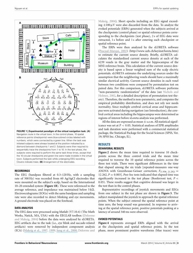
EXPERIMENTAL PARADIGMSThe subjects were seated 1 m from a 20-inch LCD monitor ina chair that was grounded, within a dimly lit, shielded, room.For this task, a large virtual town was created using commer-cial 3D software (EON Studio ver.2.5.2, EON Reality Inc., Irvine,CA, USA). The virtual town consisted of streets and a series ofbuildings (Figure 1A). The subjects were required to manipulatea joystick with their right hand in order to navigate the virtualtown presented on the monitor from a 3D first-person view. Theygrasped the joystick using their thumb, forefinger, and middle fin-ger in a pronated hand position, and could move the joystick in alldirections at a constant speed. The distance travelled by the joy-stick was a maximum of 2.5 cm from the center position in anydirection, which corresponded to rotation of the joystick from aperpendicular line by 30◦. Participants were able to freely navi-gate at constant speed in the forward, backward, right, and leftdirections using the joystick.
After setting up the electrodes, the subjects were given threetrials to learn the navigation route and the layout of the virtualtown. The navigation route contained 10 circular checkpointslabeled with numbers from 1 to 10, which were sequentially con-nected by a green line on the streets (Figure 1A). The subjectswere required to sequentially trace the checkpoints from 1 to10 along the green line by manipulating the joystick (controlphase). When subjects entered each correct checkpoint, a beepsound lasting 0.53 s was generated. When the subjects enteredcheckpoint 10, the task was terminated. After a 1-min inter-trialinterval, the next trial began by displaying a scene near checkpoint1 in the virtual town. After these three learning trials (controlphase), the subjects were required to perform the same task threetimes, except that the 10 circular checkpoints and green line werenot shown in the virtual town (test phase). However, the samebeep sound was generated when they reached each checkpoint.EEG recordings were performed throughout the control and testphases of the experiment. EMG recordings were performed in thetest phases of the experiment.
Frontiers in Behavioral Neuroscience www.frontiersin.org March 2014 | Volume 8 | Article 66 | 2
Nguyen et al. ERPs for spatial updating
FIGURE 1 | Experimental paradigm of the virtual navigation task. (A)
Navigation route in the virtual town. In the control phase, 10 spatialreference points (checkpoints) were blue-colored circles labeled withnumbers, which were connected by a green line. When the task wasinitiated subjects were always located at the position indicated by adiamond between checkpoints 1 and 2. Subjects were then required tosequentially trace the checkpoints from 1 to 10. In the test phase, thesubjects were required to perform the same task three times, except thatthe 10 circular checkpoints and green line were made invisible in the virtualtown. Subjects performed the task while undergoing EEG recording.Clovers indicate trees. (B) Arrangement of the electrodes.
RECORDINGSThe EEG (bandpass filtered at 0.3–120 Hz, with a samplingrate of 500 Hz) was recorded from 60 Ag/AgCl electrodes thatwere mounted on the subject’s scalp, based on the International10–20 extended system (Figure 1B). These were referenced to theaverage reference, and impedance was maintained below 5 k�.Electrooculograms (EOGs) with the same bandpass and samplingrate were also recorded to detect blinking and eye movements.A ground electrode was placed on the forehead.
DATA ANALYSISThe EEG data were processed using Matlab (V7.10.4) (The MathWorks, Natick, MA, USA) with the EEGLAB toolbox (Delormeand Makeig, 2004) before the data were analyzed by sLORETA.EEG artifacts due to the task (i.e., eye blink and saccade-relatedartifacts) were removed by independent component analysis(ICA) (Makeig et al., 1997, 1999; Jung et al., 2000; Delorme and
Makeig, 2004). Short epochs including an EEG signal exceed-ing ±100 µV were also discarded from the data. To analyze theevoked potentials (ERPs) generated when the subjects arrived atthe checkpoints (control phase) or spatial reference points corre-sponding to the checkpoints (test phase), 2 s of EEG data wereextracted, 1 s before and 1 s after entering each checkpoint orspatial reference point.
The ERPs were then analyzed by the sLORETA software(Pascual-Marqui, 2002) (http://www.uzh.ch/keyinst/loreta.htm)to estimate the current source density. Briefly, sLORETA cal-culates the standardized current source density at each of the6239 voxels in the gray matter and the hippocampus of theMNI-reference brain. This calculation of the current source den-sity is based upon a linear weighted sum of the scalp electricpotentials. sLORETA estimates the underlying sources under theassumption that the neighboring voxels should have a maximallysimilar electrical activity. Current source densities in each voxelbetween two conditions were compared by permutation test onpaired data. For this comparison, sLORETA software performs“non-parametric randomization” of the data (see Nichols andHolmes, 2002, for a detailed description of permutation test the-ory). Therefore, the method is non-parametric, and computes theempirical probability distribution, and does not rely nor needsnormality. Since multiple cerebral cortical areas and hippocam-pus were activated during navigation (see Introduction), the cere-bral cortical areas including the hippocampus were determined asregions of interest before sLoreta analysis was performed.
All the data are expressed as mean ± s.e.m. All statistical signif-icance was set at P < 0.05. Statistical analyses of EPR amplitudesand task duration were performed with a commercial statisticalpackage, the Statistical Package for the Social Sciences (SPSS, Ver.19; SPSS Inc. Chicago, IL).
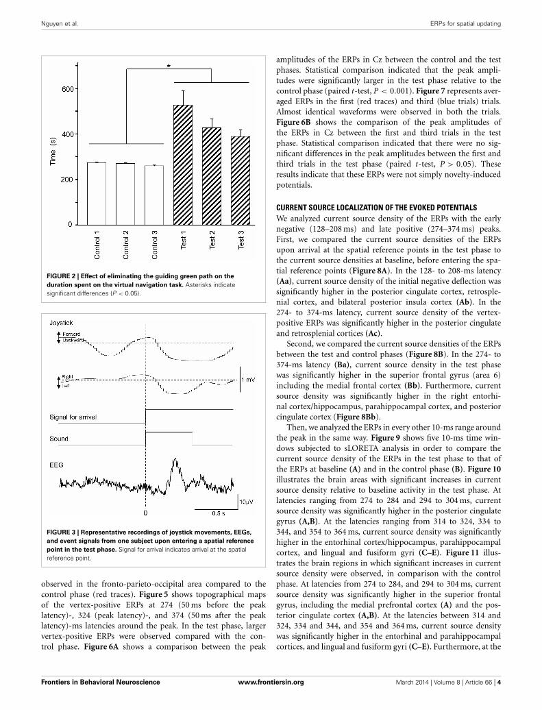
RESULTSBEHAVIORAL RESULTSFigure 2 shows the mean time required to traverse 10 check-points across the three control trials and the mean timerequired to traverse the 10 spatial reference points across thethree test trials. There were significant differences in the timethat elapsed among the six trials [repeated-measures one-wayANOVA with Greenhouse-Geisser correction; F(1.936, 21.292) =12.262, P = 0.003]. Post-hoc tests indicated that elapsed time wassignificantly increased in the test phase (Bonferroni test, P <
0.05). These results suggest that cognitive demand was larger inthe test than in the control phases.
Representative recordings of joystick movements and EEGsfrom one subject in the test phase are shown in Figure 3. Thesubject manipulated the joystick to approach the spatial referencepoints. When the subject entered the spatial reference point attime zero, the beep sound was generated. In response to arriv-ing at the spatial reference point, positive potentials peaking at alatency of around 340 ms were observed.
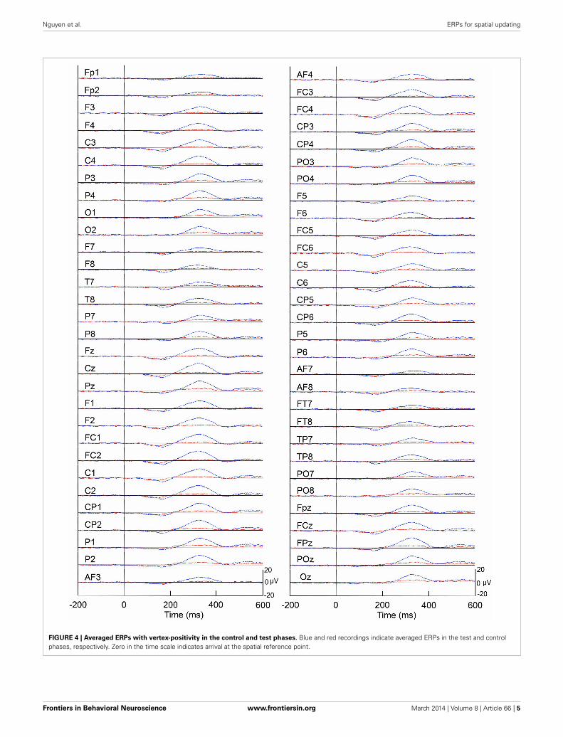
EVOKED POTENTIALSFigure 4 represents averaged ERPs aligned with the arrivalat the checkpoints and spatial reference points. In the testphase, more prominent positive waveforms (blue traces) were
Frontiers in Behavioral Neuroscience www.frontiersin.org March 2014 | Volume 8 | Article 66 | 3
Nguyen et al. ERPs for spatial updating
FIGURE 2 | Effect of eliminating the guiding green path on the
duration spent on the virtual navigation task. Asterisks indicatesignificant differences (P < 0.05).
FIGURE 3 | Representative recordings of joystick movements, EEGs,
and event signals from one subject upon entering a spatial reference
point in the test phase. Signal for arrival indicates arrival at the spatialreference point.
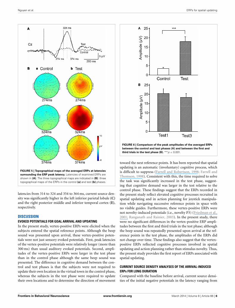
observed in the fronto-parieto-occipital area compared to thecontrol phase (red traces). Figure 5 shows topographical mapsof the vertex-positive ERPs at 274 (50 ms before the peaklatency)-, 324 (peak latency)-, and 374 (50 ms after the peaklatency)-ms latencies around the peak. In the test phase, largervertex-positive ERPs were observed compared with the con-trol phase. Figure 6A shows a comparison between the peak
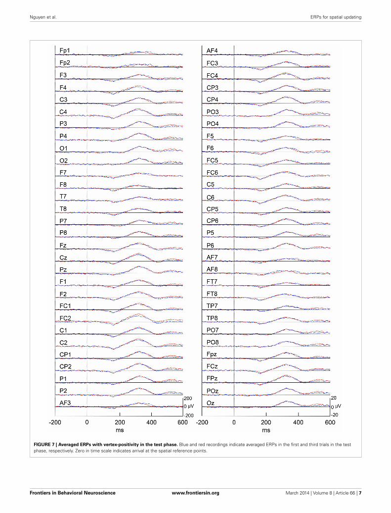
amplitudes of the ERPs in Cz between the control and the testphases. Statistical comparison indicated that the peak ampli-tudes were significantly larger in the test phase relative to thecontrol phase (paired t-test, P < 0.001). Figure 7 represents aver-aged ERPs in the first (red traces) and third (blue trials) trials.Almost identical waveforms were observed in both the trials.Figure 6B shows the comparison of the peak amplitudes ofthe ERPs in Cz between the first and third trials in the testphase. Statistical comparison indicated that there were no sig-nificant differences in the peak amplitudes between the first andthird trials in the test phase (paired t-test, P > 0.05). Theseresults indicate that these ERPs were not simply novelty-inducedpotentials.
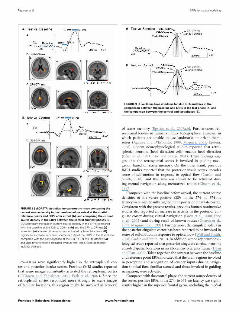
CURRENT SOURCE LOCALIZATION OF THE EVOKED POTENTIALSWe analyzed current source density of the ERPs with the earlynegative (128–208 ms) and late positive (274–374 ms) peaks.First, we compared the current source densities of the ERPsupon arrival at the spatial reference points in the test phase tothe current source densities at baseline, before entering the spa-tial reference points (Figure 8A). In the 128- to 208-ms latency(Aa), current source density of the initial negative deflection wassignificantly higher in the posterior cingulate cortex, retrosple-nial cortex, and bilateral posterior insula cortex (Ab). In the274- to 374-ms latency, current source density of the vertex-positive ERPs was significantly higher in the posterior cingulateand retrosplenial cortices (Ac).
Second, we compared the current source densities of the ERPsbetween the test and control phases (Figure 8B). In the 274- to374-ms latency (Ba), current source density in the test phasewas significantly higher in the superior frontal gyrus (area 6)including the medial frontal cortex (Bb). Furthermore, currentsource density was significantly higher in the right entorhi-nal cortex/hippocampus, parahippocampal cortex, and posteriorcingulate cortex (Figure 8Bb).
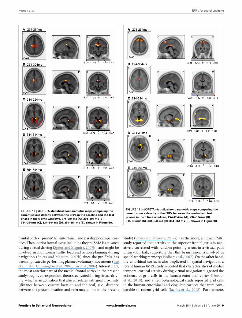
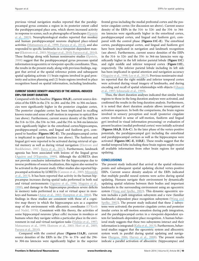
Then, we analyzed the ERPs in every other 10-ms range aroundthe peak in the same way. Figure 9 shows five 10-ms time win-dows subjected to sLORETA analysis in order to compare thecurrent source density of the ERPs in the test phase to that ofthe ERPs at baseline (A) and in the control phase (B). Figure 10illustrates the brain areas with significant increases in currentsource density relative to baseline activity in the test phase. Atlatencies ranging from 274 to 284 and 294 to 304 ms, currentsource density was significantly higher in the posterior cingulategyrus (A,B). At the latencies ranging from 314 to 324, 334 to344, and 354 to 364 ms, current source density was significantlyhigher in the entorhinal cortex/hippocampus, parahippocampalcortex, and lingual and fusiform gyri (C–E). Figure 11 illus-trates the brain regions in which significant increases in currentsource density were observed, in comparison with the controlphase. At latencies from 274 to 284, and 294 to 304 ms, currentsource density was significantly higher in the superior frontalgyrus, including the medial prefrontal cortex (A) and the pos-terior cingulate cortex (A,B). At the latencies between 314 and324, 334 and 344, and 354 and 364 ms, current source densitywas significantly higher in the entorhinal and parahippocampalcortices, and lingual and fusiform gyri (C–E). Furthermore, at the
Frontiers in Behavioral Neuroscience www.frontiersin.org March 2014 | Volume 8 | Article 66 | 4
Nguyen et al. ERPs for spatial updating
FIGURE 4 | Averaged ERPs with vertex-positivity in the control and test phases. Blue and red recordings indicate averaged ERPs in the test and controlphases, respectively. Zero in the time scale indicates arrival at the spatial reference point.
Frontiers in Behavioral Neuroscience www.frontiersin.org March 2014 | Volume 8 | Article 66 | 5
Nguyen et al. ERPs for spatial updating
FIGURE 5 | Topographical maps of the averaged ERPs at latencies
surrounding the ERP peak latency. Latencies of examined ERPs areshown in (A). The three topographical maps are indicated in (B): threetopographical maps of the ERPs in the control (a) and test (b) phases.
latencies from 314 to 324 and 354 to 364 ms, current source den-sity was significantly higher in the left inferior parietal lobule (C)and the right posterior middle and inferior temporal cortex (E),respectively.
DISCUSSIONEVOKED POTENTIALS FOR GOAL ARRIVAL AND UPDATINGIn the present study, vertex-positive ERPs were elicited when thesubjects entered the spatial reference points. Although the beepsound was presented upon arrival, these vertex-positive poten-tials were not just sensory evoked potentials. First, peak latenciesof the vertex-positive potentials were relatively longer (more than300 ms) than usual auditory evoked potentials. Second, ampli-tudes of the vertex-positive ERPs were larger in the test phasethan in the control phase although the same beep sound waspresented. The difference in cognitive demand between the con-trol and test phases is that the subjects were not required toupdate their own location in the virtual town in the control phase,whereas the subjects in the test phase were required to updatetheir own locations and to determine the direction of movement
FIGURE 6 | Comparison of the peak amplitudes of the averaged ERPs
between the control and test phases (A) and between the first and
third trials in the test phase (B). ∗∗∗p < 0.001.
toward the next reference points. It has been reported that spatialupdating is an automatic (involuntary) cognitive process, whichis difficult to suppress (Farrell and Robertson, 1998; Farrell andThomson, 1998). Consistent with this, the time required to solvethe task was significantly increased in the test phase, suggest-ing that cognitive demand was larger in the test relative to thecontrol phase. These findings suggest that the ERPs recorded inthe present study reflect elevated cognitive processes recruited inspatial updating and in action planning for joystick manipula-tion while navigating successive reference points in space withno visible guides. Furthermore, these vertex-positive ERPs werenot novelty-induced potentials (i.e., novelty P3) (Friedman et al.,2001; Ranganath and Rainier, 2003). In the present study, therewere no significant differences in the vertex-positive ERP ampli-tudes between the first and third trials in the test phase; althoughthe beep sound was repeatedly presented upon arrival at the ref-erence points in the test phase, the amplitudes of the ERPs didnot change over time. These findings also suggest that the vertex-positive ERPs reflected cognitive processes involved in spatialupdating and action planning rather than stimulus novelty. Thus,the present study provides the first report of ERPs associated withspatial updating.
CURRENT SOURCE DENSITY ANALYSES OF THE ARRIVAL-INDUCEDERPs FOR LONG DURATIONCompared with the baseline before arrival, current source densi-ties of the initial negative potentials in the latency ranging from
Frontiers in Behavioral Neuroscience www.frontiersin.org March 2014 | Volume 8 | Article 66 | 6
Nguyen et al. ERPs for spatial updating
FIGURE 7 | Averaged ERPs with vertex-positivity in the test phase. Blue and red recordings indicate averaged ERPs in the first and third trials in the testphase, respectively. Zero in time scale indicates arrival at the spatial reference points.
Frontiers in Behavioral Neuroscience www.frontiersin.org March 2014 | Volume 8 | Article 66 | 7
Nguyen et al. ERPs for spatial updating
FIGURE 8 | sLORETA statistical nonparametric maps comparing the
current source density in the baseline before arrival at the spatial
reference points and ERPs after arrival (A), and comparing the current
source density in the ERPs between the control and test phases (B).
(A) Significant increase in current source density in the ERPs comparedwith the baseline at the 128- to 208-ms (b) and the 274- to 374-ms (c)
latencies; (a) analyzed time windows indicated by blue thick lines. (B)
Significant increase in current source density of the ERPs in the test phasecompared with the control phase at the 274- to 374-ms (b) latency; (a)
analyzed time windows indicated by blue thick lines. Calibration barsindicate t-values.
128–208 ms were significantly higher in the retrosplenial cor-tex and posterior insular cortex. Previous fMRI studies reportedthat scene images consistently activated the retrosplenial cortex(O’Craven and Kanwisher, 2000; Park et al., 2007). Since theretrosplenial cortex responded more strongly to scene imagesof familiar locations, this region might be involved in retrieval
FIGURE 9 | Five 10-ms time windows for sLORETA analyses in the
comparison between the baseline and ERPs in the test phase (A) and
the comparison between the control and test phases (B).
of scene memory (Epstein et al., 2007a,b). Furthermore, ret-rosplenial lesions in humans induce topographical amnesia, inwhich patients are unable to use landmarks to orient them-selves (Aguirre and D’Esposito, 1999; Maguire, 2001; Epstein,2008). Rodent neurophysiological studies reported that retro-splenial neurons (head direction cells) encode head direction(Chen et al., 1994; Cho and Sharp, 2001). These findings sug-gest that the retrosplenial cortex is involved in guiding navi-gation based on scene memory. On the other hand, previousfMRI studies reported that the posterior insula cortex encodessense of self-motion in response to optical flow (Cardin andSmith, 2010), and this area was shown to be activated dur-ing mental navigation along memorized routes (Ghaem et al.,1997).
Compared with the baseline before arrival, the current sourcedensities of the vertex-positive ERPs in the 274- to 374-mslatency were significantly higher in the posterior cingulate cortex.Consistent with the present results, previous human noninvasivestudies also reported an increase in activity in the posterior cin-gulate cortex during virtual navigation (Grön et al., 2000; Pineet al., 2002) and during recall of known routes (Ghaem et al.,1997; Maguire et al., 1997). Furthermore, the cingulate sulcus inthe posterior cingulate cortex has been reported to be involved insense of self-motion in response to optical flow (Wall and Smith,2008; Cardin and Smith, 2010). In addition, a monkey neurophys-iological study reported that posterior cingulate cortical neuronsencoded spatial locations in an allocentric reference frame (Deanand Platt, 2006). Taken together, the contrast between the baselineand reference point ERPs indicated that the brain regions involvedin perception and recognition of sensory inputs during naviga-tion (optical flow, familiar scenes) and those involved in guidingnavigation, were activated.
Compared with the control phase, the current source density ofthe vertex-positive ERPs in the 274- to 374-ms latency was signif-icantly higher in the superior frontal gyrus, including the medial
Frontiers in Behavioral Neuroscience www.frontiersin.org March 2014 | Volume 8 | Article 66 | 8
Nguyen et al. ERPs for spatial updating
FIGURE 10 | sLORETA statistical nonparametric maps comparing the
current source density between the ERPs in the baseline and the test
phase in the 5 time windows, 274–284 ms (A), 294–304 ms (B),
314–324 ms (C), 334–344 ms (D), 354–364 ms (E), shown in Figure 9A.
frontal cortex (pre-SMA), entorhinal, and parahippocampal cor-tices. The superior frontal gyrus including the pre-SMA is activatedduring virtual driving (Spiers and Maguire, 2007b), and might beinvolved in monitoring traffic load and action planning duringnavigation (Spiers and Maguire, 2007b) since the pre-SMA hasbeen implicated in performing planned voluntary movements (Leeet al., 1999; Cunnington et al., 2002; Lau et al., 2004). Interestingly,the most anterior part of the medial frontal cortex in the presentstudy roughly corresponds to the area activated during virtual driv-ing, which is an activation that also correlates with goal proximity(distance between current location and the goal) (i.e., distancebetween the present location and reference points in the present
FIGURE 11 | sLORETA statistical nonparametric maps comparing the
current source density of the ERPs between the control and test
phases in the 5 time windows, 274–284 ms (A), 294–304 ms (B),
314–324 ms (C), 334–344 ms (D), 354–364 ms (E), shown in Figure 9B.
study) (Spiers and Maguire, 2007a). Furthermore, a human fMRIstudy reported that activity in the superior frontal gyrus is neg-atively correlated with random pointing errors in a virtual pathintegration task, suggesting that this brain region is involved inspatial working memory (Wolbers et al., 2007). On the other hand,the entorhinal cortex is also implicated in spatial navigation; arecent human fMRI study reported that characteristics of medialtemporal cortical activity during virtual navigation suggested theexistence of grid cells in the human entorhinal cortex (Doelleret al., 2010), and a neurophysiological study reported grid cellsin the human entorhinal and cingulate cortices that were com-parable to rodent grid cells (Jacobs et al., 2013). Furthermore,
Frontiers in Behavioral Neuroscience www.frontiersin.org March 2014 | Volume 8 | Article 66 | 9
Nguyen et al. ERPs for spatial updating
previous virtual navigation studies reported that the parahip-pocampal gyrus contains a region in its posterior extent calledthe parahippocampal place area, which shows increased activityin response to scenes, such as photographs of landscapes (Epsteinet al., 2003). Neurophysiological studies reported that monkeyand human parahippocampal neurons displayed place-relatedactivities (Matsumura et al., 1999; Furuya et al., 2014), and alsoresponded to specific landmarks in a viewpoint-dependent man-ner (Ekstrom et al., 2003; Weniger et al., 2010; Furuya et al., 2014).These findings along with human noninvasive studies (Epstein,2008) suggest that the parahippocampal gyrus processes spatialinformation in egocentric or viewpoint-specific coordinates. Thus,the results in the present study, along with previous findings, sug-gest that arrival at the spatial reference points and subsequentspatial updating activate (1) brain regions involved in goal prox-imity and action planning and (2) brain regions involved in placerecognition based on spatial information, including landmarks.
CURRENT SOURCE DENSITY ANALYSES OF THE ARRIVAL-INDUCEDERPs FOR SHORT DURATIONCompared with the baseline (Figures 10A,B), current source den-sities of the ERPs in the 274- to 284- and the 294- to 304-ms laten-cies were significantly higher in the posterior cingulate cortex.The posterior cingulate cortex has been implicated in recallingknown routes and sense of self-motion in response to optical flow(see above). Furthermore, current source density of the ERPs inthe 314- to 324-, the 334- to 344-, and the 354- to 364-ms latencieswere significantly higher in the entorhinal cortex/hippocampus,parahippocampal cortex, and lingual and fusiform gyri, com-pared to baseline (Figures 10C–E). The parahippocampal cortexis implicated in spatial function (see above). The fusiform andlingual gyri were reported to be activated during retrieval of spa-tial memory as well as during virtual navigation (Ekstrom andBookheimer, 2007; Barra et al., 2012). Furthermore, landmarkagnosia has been associated with lesions of the lingual gyrus(Aguirre and D’Esposito, 1999). Although the sLORETA doesnot provide conclusive information for the hippocampus due toinverse problems of source localization, this region also seemed tobe activated in the present study. Other studies also reported hip-pocampal activation by LORETA (Cannon et al., 2005; Miyanishiet al., 2013). It has been reported that activity in the human hip-pocampus increases during spatial tasks performed in both realand virtual environments (Aguirre et al., 1996; Maguire et al.,1998), and damage to the hippocampus produces severe deficitsin memory tasks performed in a real or virtual space in mon-keys and humans (Astur et al., 2002; Hampton et al., 2004). Thefindings in these studies are consistent with those of a cogni-tive map theory in which the hippocampus acts as a cognitivemap of the environment with allocentric coordinates (O’Keefeand Nadel, 1978). Consistent with this theory, the activities ofsome hippocampal neurons (place cells) increase in monkeys orhumans when they navigate within a particular place in the envi-ronment in real and virtual navigation tasks (Nishijo et al., 1997;Matsumura et al., 1999; Ekstrom et al., 2003; Hori et al., 2005;Furuya et al., 2014).
Compared with the control phase (Figures 11A,B), currentsource densities of the ERPs in the 274- to 374- and the 294-to 304-ms latencies were significantly higher in the superior
frontal gyrus including the medial prefrontal cortex and the pos-terior cingulate cortex (for discussion see above). Current sourcedensity of the ERPs in the 314- to 324- and the 334- to 344-ms latencies were significantly higher in the entorhinal cortex,parahippocampal cortex, and lingual and fusiform gyri, com-pared with the control phase (Figures 11C–E). The entorhinalcortex, parahippocampal cortex, and lingual and fusiform gyrihave been implicated in navigation and landmark recognition(see above). Furthermore, current source densities of the ERPsin the 314- to 324- and the 354- to 364-ms latencies were sig-nificantly higher in the left inferior parietal lobule (Figure 11C)and right middle and inferior temporal cortex (Figure 11E),respectively. The inferior parietal lobule including its left sidehas been implicated in spatial attention and navigation accuracy(Maguire et al., 1998; Lee et al., 2013). Previous noninvasive stud-ies reported that the right middle and inferior temporal cortexwere activated during visual imagery of landmarks, and duringencoding and recall of spatial relationships with objects (Ghaemet al., 1997; Johnsrude et al., 1999).
Thus, the short duration analyses indicated that similar brainregions to those in the long duration analyses were activated, andconfirmed the results in the long duration analysis. Furthermore,it is noted that short duration analysis allows investigation ofactivation sequences. In both the comparisons, the brain regionsinvolved in sensory perception and recall (posterior cingulatecortex involved in sense of self-motion, fusiform and lingualgyri involved in visual information processing) or evaluation ofpresent location (medial prefrontal cortex) were initially activated(Figures 10A,B, 11A–C). In the later phase of the vertex-positivepotentials, the parahippocampal gyri including the entorhinaland parahippocampal cortices as well as the hippocampus wereactivated (Figures 10C–E, 11D,E). These results suggest that themedial temporal lobe including these brain regions might receiveall available information from other brain regions for spatialupdating.
CONCLUSIONSThe present study indicated that arrival at the spatial referencepoints and subsequent spatial updating elicited vertex-positiveERPs. Current source density analysis of the ERPs indicatedthat multiple parallel neural systems were active during spatialupdating. Humans navigate their environment by dynamicallyupdating spatial relations between their bodies and importantlandmarks in the surrounding environment using an egocentricsystem (Wang and Spelke, 2002). This dynamic egocentric sys-tem includes a path integration subsystem and a view (familiarlandmarks)-dependent place recognition subsystem (Wang andSpelke, 2002). The present study indicated that these 2 subsys-tems were activated; the posterior cingulate cortex and posteriorinsular cortex in self-motion sensation during path integration,and the parahippocampal cortex in a viewpoint-dependent sys-tem for landmark-dependent place recognition. A human behav-ioral study suggests that these two subsystems interact and theirinformation is integrated (Kalia et al., 2013). Furthermore, behav-ioral studies suggest that the egocentric system and allocentricsystem work in parallel during spatial updating and naviga-tion (Burgess, 2006; Harvey et al., 2008). The present resultsindicate a parallel activation of allocentric (hippocampus) and
Frontiers in Behavioral Neuroscience www.frontiersin.org March 2014 | Volume 8 | Article 66 | 10
Nguyen et al. ERPs for spatial updating
egocentric (parahippocampal gyrus) systems. Our results provideneurophysiological evidence that humans use multiple spatialrepresentations with different reference frames for spatial updat-ing during navigation.
On the other hand, the inferior medial occipital lobe (lingualand fusiform gyri), right inferior temporal cortex, parahippocam-pal cortex, and hippocampus, which were activated during updat-ing in the present study, are associated with route learning in a realenvironment (Barrash et al., 2000). The medial occipito-temporalcortices (lingual and fusiform gyri) and right inferior temporalcortex might be associated with the ability to quickly and accu-rately perceive and learn multiple topographical scenes, whilethe posterior parahippocampal gyrus and hippocampus might beinvolved in forming an integrated representation of the extendedtopographical environment (i.e., the appearance of places andspatial relationships between specific places), and consolidatingthat representation (Barrash et al., 2000). Compared with the pre-vious studies that investigated remote spatial memory, which isestablished for many years (see a review by Spiers and Maguire,2007c), the present experiments imposed only six trials includ-ing both the control and test trials at one time. These findingssuggest that not only updating processes but also learning andconsolidation processes take place simultaneously.
Finally, it is noted that human subjects display individual dif-ferences in navigation strategies [e.g., allocentric (bird-view) oregocentric (landmark) strategies] (Jordan et al., 2004). In thepresent study, we could not classify the subjects based on theirnavigation strategies since they were required to navigate in thefixed route to receive the same visual stimuli in the virtual space.Further studies are required to investigate brain activation differ-ences based on navigation strategies. The present study at leastindicated common neural networks among the subjects duringspatial updating.
AUTHOR CONTRIBUTIONSHisao Nishijo designed the research; Hai M. Nguyen, JumpeiMatsumoto, and Hisao Nishijo performed research; Hai M.Nguyen, Jumpei Matsumoto, Anh H. Tran, Taketoshi Ono, andHisao Nishijo analyzed data; and Hai M. Nguyen and HisaoNishijo wrote the paper.
ACKNOWLEDGMENTSThis work was supported partly by the Japan Society for thePromotion of Science Asian Core Program and the Ministryof Education, Science, Sports and Culture, Grant-in-Aid forScientific Research (B) (25290005). The funders had no role inthe study design, data collection and analysis, decision to publish,or preparation of the manuscript.
REFERENCESAguirre, G. K., and D’Esposito, M. (1997). Environmental knowledge is subserved
by separable dorsal/ventral neural areas. J. Neurosci. 17, 2512–2518.Aguirre, G. K., and D’Esposito, M. (1999). Topographical disorientation: A synthe-
sis and taxonomy. Brain 122, 1613–1628. doi: 10.1093/brain/122.9.1613Aguirre, G. K., Detre, J. A., Alsop, D. C., and D’Esposito, M. (1996). The parahip-
pocampus subserves topographical learning in man. Cereb. Cortex 6, 823–829.Aguirre, G. K., Zarahn, E., and D’Esposito, M. (1998). An area within human ven-
tral cortex sensitive to “building” stimuli: evidence and implications. Neuron 21,373–383.
Astur, R. S., Taylor, L. B., Mamelak, A. N., Philpott, L., and Sutherland, R. J.(2002). Humans with hippocampus damage display severe spatial memoryimpairments in a virtual Morris water task. Behav. Brain. Res. 132, 77–84. doi:10.1016/S0166-4328(01)00399-0
Barra, J., Laou, L., Poline, J. B., Lebihan, D., and Berthoz, A. (2012). Does anoblique/slanted perspective during virtual navigation engage both egocen-tric and allocentric brain strategies? PLoS ONE 7:e49537. doi: 10.1371/jour-nal.pone.0049537
Barrash, J., Damasio, H., Adolphs, R., and Tranel, D. (2000). The neuroanatomi-cal correlates of route learning impairment. Neuropsychologia 38, 820–836. doi:10.1016/S0028-3932(99)00131-1
Bellebaum C., and Daum, I. (2006). Time course of cross-hemispheric spatialupdating in the human parietal cortex. Behav. Brain Res. 169, 150–161. doi:10.1016/j.bbr.2006.01.001
Brunsdon, R., Nickels, L., and Coltheart, M. (2007). Topographical disorienta-tion: towards an integrated framework for assessment. Neuropsychol. Rehabil.17, 34–52. doi: 10.1080/09602010500505021
Burgess, N. (2006). Spatial memory: how egocentric and allocentric combine.Trends. Cogn. Sci. 10, 551–557. doi: 10.1016/j.tics.2006.10.005
Burgess, N., Maguire, E. A., Spiers, H. J., and O’Keefe, J. (2001). A temporopari-etal and prefrontal network for retrieving the spatial context of lifelike events.Neuroimage 14, 439–453. doi: 10.1006/nimg.2001.0806
Cannon, R., Lubar, J., Thornton, K., Wilson, S., and Congedo, M. (2005). Limbicbeta activation and LORETA: can hippocampal and related limbic activity berecorded and changes visualized using loreta in an affective memory condition?J. Neurotherapy 8, 5–24. doi: 10.1300/J184v08n04_02
Cardin, V., and Smith, A. T. (2010). Sensitivity of human visual and vestibularcortical regions to egomotion- compatible visual stimulation. Cereb. Cortex 20,1964–1973. doi: 10.1093/cercor/bhp268
Chen, L. L., Lin, L. H., Green, E. J., Barnes, C. A., and McNaughton, B. L. (1994).Head-direction cells in the rat posterior cortex. I. Anatomical distribution andbehavioral modulation. Exp. Brain Res. 101, 8–23. doi: 10.1007/BF00243212
Cho, J., and Sharp, P. E. (2001). Head direction, place, and movement corre-lates for cells in the rat retrosplenial cortex. Behav. Neurosci. 115, 3–25. doi:10.1037/0735-7044.115.1.3
Cunnington, R., Windischberger, C., Deecke, L., and Moser, E. (2002). The prepara-tion and execution of self-initiated and externally-triggered movement: a studyof event-related fMRI. Neuroimage 15, 373–385. doi: 10.1006/nimg.2001.0976
Davis, S. J. C., and Coltheart, M. (1999). Rehabilitation of topographical disorien-tation: an experimental single case study. Neuropsychol. Rehabil. 9, 1–30. doi:10.1080/713755586
Dean, H. L., and Platt, M. L. (2006). Allocentric spatial referencing of neuronalactivity in macaque posterior cingulate cortex. J. Neurosci. 26, 1117–1127. doi:10.1523/JNEUROSCI.2497-05.2006
Delorme, A., and Makeig, S. (2004). EEGLAB: an open source toolbox for anal-ysis of single-trial EEG dynamics including independent component analysis.J. Neurosci. Methods 134, 9–21. doi: 10.1016/j.jneumeth.2003.10.009
De Renzi, E. (1982). Disorders of Space Exploration and Cognition. Chichester:Wiley.
Doeller, C. F., Barry, C., and Burgess, N. (2010). Evidence for grid cells in a humanmemory network. Nature 463, 657–661. doi: 10.1038/nature08704
Ekstrom, A. D., and Bookheimer, S. Y. (2007). Spatial and temporal episodic mem-ory retrieval recruit dissociable functional networks in the human brain. Learn.Mem. 14, 645–654. doi: 10.1101/lm.575107
Ekstrom, A. D., Kahana, M. J., Caplan, J. B., Fields, T. A., Isham, E. A., Newman, E.L., et al. (2003). Cellular networks underlying human spatial navigation. Nature425, 184–188. doi: 10.1038/nature01964
Epstein, R. (2008). Parahippocampal and retrosplenial contributionsto human spatial navigation. Trends. Cogn. Sci. 12, 388–396. doi:10.1016/j.tics.2008.07.004
Epstein, R. A., Graham, K. S., and Downing, P. E. (2003). Viewpoint-specific scenerepresentations in human parahippocampal cortex. Neuron 37, 865–876. doi:10.1016/S0896-6273(03)00117-X
Epstein, R. A., Higgins, J. S., Jablonski, K., and Feiler, A. M. (2007a). Visualscene processing in familiar and unfamiliar environments. J. Neurophysiol. 97,3670–3683. doi: 10.1152/jn.00003.2007
Epstein, R. A., Parker, W. E., and Feiler, A. M. (2007b). Where am I now? Distinctroles for parahippocampal and retrosplenial cortices in place recognition. J.Neurosci. 27, 6141–6149. doi: 10.1523/JNEUROSCI.0799-07.2007
Frontiers in Behavioral Neuroscience www.frontiersin.org March 2014 | Volume 8 | Article 66 | 11
Nguyen et al. ERPs for spatial updating
Etienne, A. S. (1992). Navigation of a small mammal by dead reckoning and localcues. Curr. Dir. Psychol. Sci. 23, 1–8.
Farah, M. J. (1989). “Neuropsychology of mental imagery,” in The Handbook ofNeuropsychology: Disorder of Visual Behavior, eds F. Boller and J. Grafman(Amsterdam: Elsevier), 395–413.
Farrell, M. J., and Robertson, I. H. (1998). Mental rotation and automatic updatingof body-centered spatial relationships. J. Exp. Psychol. Learn. Mem. Cogn. 24,227–233. doi: 10.1037/0278-7393.24.1.227
Farrell, M. J., and Thomson, J. A. (1998). Automatic spatial updating dur-ing locomotion without vision. Q. J. Exp. Psychol. A 51, 637–654. doi:10.1080/713755776
Friedman, D., Cycowicz, Y. M., and Gaeta, H. (2001). The novelty P3: An event-related brain potential (ERP) sign of the brain’s evaluation of novelty. Neurosci.Biobehav. Rev. 25, 355–373. doi: 10.1016/S0149-7634(01)00019-7
Furuya, Y., Matsumoto, J., Hori, H., Boas, C. V., Hai Tran, A. H., Shimada, Y.,et al. (2014). Place-related neuronal activity in the monkey parahippocampalgyrus and hippocampal formation during virtual navigation. Hippocampus 24,113–130. doi: 10.1002/hipo.22209
Gallistel, C. R. (1990). The Organization of Learning. Cambridge, MA: MIT Press.Ghaem, O., Mellet, E., Crivello, F., Tzourio, N., Mazoyer, B., Berthoz, A., et al.
(1997). Mental navigation along memorized routes activates the hippocam-pus, precuneus, and insula. Neuroreport 8, 739–744. doi: 10.1097/00001756-199702100-00032
Gramann, K., Müller, H. J., Eick, E. M., and Schönebeck, B. (2005). Evidence of sep-arable spatial representations in a virtual navigation task. J. Exp. Psychol. Hum.Percept. Perform. 31, 1199–1223. doi: 10.1037/0096-1523.31.6.1199
Grön, G., Wunderlich, A. P., Spitzer, M., Tomczak, R., and Riepe, M. W. (2000).Brain activation during human navigation: gender-different neural networks assubstrate of performance. Nat. Neurosci. 3, 404–408. doi: 10.1038/73980
Hampton, R. R., Hampstead, B. M., and Murray, E. A. (2004). Selective hippo-campal damage in rhesus monkeys impairs spatial memory in an open-fieldtest. Hippocampus 14, 808–818. doi: 10.1002/hipo.10217
Hartley, T., Maguire, E. A., Spiers, H. J., and Burgess, N. (2003). The well-worn route and the path less traveled: distinct neural bases of route fol-lowing and wayfinding in humans. Neuron 37, 877–888. doi: 10.1016/S0896-6273(03)00095-3
Harvey, D. R., McGauran, A. M., Murphy, J., Burns, L., McMonagle, E., andCommins, S. (2008). Emergence of an egocentric cue guiding and allocentricinferring strategy that mirrors hippocampal brain-derived neurotrophic fac-tor (BDNF) expression in the Morris water maze. Neurobiol. Learn. Mem. 89,462–479. doi: 10.1016/j.nlm.2007.08.013
Hori, E., Nishio, Y., Kazui, K., Umeno, K., Tabuchi, E., Sasaki, K., et al. (2005).Place-related neural responses in the monkey hippocampal formation in avirtual space. Hippocampus 15, 991–996. doi: 10.1002/hipo.20108
Hori, S., Matsumoto, J., Hori, E., Kuwayama, N., Ono, T., Kuroda, S., et al. (2013).Alpha-and theta-range cortical synchronization and corticomuscular coherenceduring joystick manupulation in a virtual navigation task. Brain Topogr. 26,591–605. doi: 10.1007/s10548-013-0304-z
Iaria, G., Incoccia, C., Piccardi, L., Nico, D., Sabatini, U., and Guariglia, C. (2005).Lack of orientation due to a congenital brain malformation: a case study.Neurocase 11, 463–474. doi: 10.1080/13554790500423602
Iseki, K., Hanakawa, T., Shinozaki, J., Mankaku, M., and Fukuyama, H.(2008). Neural mechanisms involved in mental imagery and observa-tion of gait. Neuroimage 41, 1021–1031. doi: 10.1016/j.neuroimage.2008.03.010
Jacobs, J., Weidemann, C. T., Miller, J. F., Solway, A., Burke, J. F., Wei, X. X.,et al. (2013). Direct recordings of grid-like neuronal activity in human spatialnavigation. Nat. Neurosci. 16, 1188–1190. doi: 10.1038/nn.3466
Johnsrude, I. S., Owen, A. M., Crane, J., Milner, B., and Evans, A. C. (1999). Acognitive activation study of memory for spatial relationships. Neuropsychologia37, 829–841. doi: 10.1016/S0028-3932(98)00136-5
Jordan, K., Schadow, J., Wuestenberg, T., Heinze, H. J., and Jäncke, L.(2004). Different cortical activations for subjects using allocentric or ego-centric strategies in a virtual navigation task. Neuroreport 15, 135–140. doi:10.1097/00001756-200401190-00026
Jung, T. P., Makeig, S., Humphries, C., Lee, T. W., Mckeown, M. J., Iragui, V., et al.(2000). Removing electroencephalographic artifacts by blind source separation.Psychophysiology 37, 163–178. doi: 10.1111/1469-8986.3720163
Kalia, A. A., Schrater, P. R., and Legge, G. E. (2013). Combining path integra-tion and remembered landmarks when navigating without vision. PLoS ONE8:e72170. doi: 10.1371/journal.pone.0072170
Lau, H. C., Rogers, R. D., Ramnani, N., and Passingham, R. E. (2004). Willedaction and attention to the selection of action. Neuroimage 21, 1407–1415. doi:10.1016/j.neuroimage.2003.10.034
Lee, J., Ku, J., Han, K., Park, J., Lee, H., Kim, K., et al. (2013). rTMS over bilateralinferior parietal cortex induces decrement of spatial sustained attention. Front.Hum. Neurosci. 7:26. doi: 10.3389/fnhum.2013.00026
Lee, K. M., Chang, K. H., and Roh, J. K. (1999). Subregions within the supple-mentary motor area activated at different stages of movement preparation andexecution. Neuroimage 9, 117–123. doi: 10.1006/nimg.1998.0393
Li, Y., Umeno, K., Hori, E., Takakura, H., Urakawa, S., Ono, T., et al. (2009).Global synchronization in the theta band during mental imagery of navigationin humans. Neurosci. Res. 65, 44–52. doi: 10.1016/j.neures.2009.05.004
MacEvoy, S. P., and Epstein, R. A. (2007). Position selectivity in scene- andobject-responsive occipitotemporal regions. J. Neurophysiol. 98, 2089–2098. doi:10.1152/jn.00438.2007
Maguire, E. A. (2001). The retrosplenial contribution to human navigation: areview of lesion and neuroimaging findings. Scand. J. Psychol. 42, 225–238. doi:10.1111/1467-9450.00233
Maguire, E. A., Burgess, N., Donnett, J. G., Frackowiak, R. S., Frith, C. D., andO’Keefe, J. (1998). Knowing where and getting there: a human navigationnetwork. Science 280, 921–924.
Maguire, E. A., Burke, T., Phillips, J., and Staunton, H. (1996). Topographicaldisorientation following unilateral temporal lobe lesions in humans.Neuropsychologia 34, 993–1001. doi: 10.1016/0028-3932(96)00022-X
Maguire, E. A., Frackowiak, R. S., and Frith, C. D. (1997). Recalling routes aroundLondon: activation of the right hippocampus in taxi drivers. J. Neurosci. 15,7103–7110.
Makeig, S., Jung, T. P., Bell, A. J., Ghahremani, D., and Sejnowski, T. J.(1997). Blind separation of auditory event-related brain responses into inde-pendent components. Proc. Natl. Acad. Sci. U.S.A. 94, 10979–10984. doi:10.1073/pnas.94.20.10979
Makeig, S., Westerfield, M., Jung, T. P., Covington, J., Townsend, J., Sejnowski, T.J., et al. (1999) Functionally independent components of the late positive event-related potential during visual spatial attention. J. Neurosci. 19, 2665–2680.
Matsumura, N., Nishijo, H., Tamura, R., Eifuku, S., Endo, S., and Ono, T. (1999).Spatial- and task-dependent neuronal responses during real and virtual translo-cation in the monkey hippocampal formation. J. Neurosci. 19, 2381–2393.
Miyanishi, T., Sumiyoshi, T., Higuchi, Y., Seo, T., and Suzuki, M. (2013). LORETAcurrent source density for duration mismatch negativity and neuropsycholog-ical assessment in early schizophrenia. PLoS ONE 8:e61152. doi: 10.1371/jour-nal.pone.0061152
Nichols, T. E., and Holmes, A. P. (2002). Nonparametric permutation tests for func-tional neuroimaging: a primer with examples. Hum. Brain Mapp. 15, 1–25. doi:10.1002/hbm.1058
Nishijo, H., Ono, T., Eifuku, S., and Tamura, R. (1997). The relationshipbetween monkey hippocampus place-related neural activity and action in space.Neurosci. Lett. 226, 57–60. doi: 10.1016/S0304-3940(97)00255-3
O’Craven, K. M., and Kanwisher, N. (2000). Mental imagery of faces and placesactivates corresponding stimulus- specific brain regions. J. Cogn. Neurosci. 12,1013–1023. doi: 10.1162/08989290051137549
O’Keefe, J., and Nadel, L. (1978). The Hippocampus as a Cognitive Map. Oxford:Oxford University Press.
Palermo, L., Iaria, G., and Guariglia, C. (2008). Mental imagery skills and topo-graphical orientation in humans: a correlation study. Behav. Brain Res. 192,248–253. doi: 10.1016/j.bbr.2008.04.014
Park, S., Intraub, H., Yi, D. J., Widders, D., and Chun, M. M. (2007). Beyond theedges of a view: boundary extension in human scene-selective visual cortex.Neuron 54, 335–342. doi: 10.1016/j.neuron.2007.04.006
Pascual-Marqui, R. D. (2002). Standardized low resolution brain electromagnetictomography (sLORETA): technical details. Methods Find. Exp. Clin. Pharmacol.24(Suppl. D), 5–12.
Peterburs, J., Gajda, K., Hoffmann, K. P., Daum, I., and Bellebaum, C.(2011). Electrophysiological correlates of inter-and intrahemispheric saccade-related updating of visual space. Behav. Brain Res. 216, 496–504. doi:10.1016/j.bbr.2010.08.025
Frontiers in Behavioral Neuroscience www.frontiersin.org March 2014 | Volume 8 | Article 66 | 12
Nguyen et al. ERPs for spatial updating
Peterburs, J., Koch, B., Schwarz, M., Hoffmann, K., P., and Aaum, I., Bellebaum,C. (2013). Updating of visual space across horizontal saccades in cerebellar andthalamic lesion patients. Cerebellum 12, 1–15. doi: 10.1007/s12311-012-0386-2
Pine, D. S., Grun, J., Maguire, E. A., Burgess, N., Zarahn, E., Koda, V., et al. (2002).Neurodevelopmental aspects of spatial navigation: a virtual reality fMRI study.Neuroimage 15, 396–406. doi: 10.1006/nimg.2001.0988
Ranganath, C., and Rainier, G. (2003). Neural mechanisms for detecting andremembering novel events. Nat. Rev. Neurosci. 4, 193–202. doi: 10.1038/nrn1052
Redish, A. D. (1999). Beyond the Cognitive Map. From Place Cells to EpisodicMemory. London: MIT Press.
Riddoch, M., and Humphreys, G. (1989). “Finding the way around topographicalimpairments,” Neuropsychology of Visual Perception, ed J. W. Brown (Hillsdale:Lawrence Erlbaum Associates), 79–103.
Riecke, B. E., van Veen, H. A., and Bullthoff, H. H. (2002). Visual hom-ing is possible without landmarks: a path integration study in virtualreality. Presence - Teleoperators Virtual Environments 11, 443–473. doi:10.1162/105474602320935810
Spiers, H. J., and Maguire, E. A. (2007a). A navigational guidance system in thehuman brain. Hippocampus 17, 618–626. doi: 10.1002/hipo.20298
Spiers, H. J., and Maguire, E. A. (2007b). Neural substrates of driving behaviour.Neuroimage 36, 245–255. doi: 10.1016/j.neuroimage.2007.02.032
Spiers, H. J., and Maguire, E. A. (2007c). The neuroscience of remotespatial memory: a tale of two cities. Neuroscience 149, 7–27. doi:10.1016/j.neuroscience.2007.06.056
Wall, M. B., and Smith, A. T. (2008). The representation of egomotion in the humanbrain. Curr. Biol. 18, 191–194. doi: 10.1016/j.cub.2007.12.053
Wang, R., and Spelke, E. (2002). Human spatial representation: insights fromanimals. Trends.Cogn. Sci. 6, 376. doi: 10.1016/S1364-6613(02)01961-7
Weniger, G., Siemerkus, J., Schmidt-Samoa, C., Mehlitz, M., Baudewig, J., Dechent,P., et al. (2010). The human parahippocampal cortex subserves egocentric spa-tial learning during navigation in a virtual maze. Neurobiol. Learn Mem. 93,46–55. doi: 10.1016/j.nlm.2009.08.003
Wolbers, T., Wiener, J. M., Mallot, H. A., and Buchel, C. (2007). Differential recruit-ment of the hippocampus, medial prefrontal cortex, and the human motioncomplex during path integration in humans. J. Neurosci 27, 9408–9416. doi:10.1523/JNEUROSCI.2146-07.2007
Yoder, R. M., Clark, B. J., and Taube, J. S. (2011). Origins of landmark encod-ing in the brain. Trends. Neurosci. 34, 561–571. doi: 10.1016/j.tins.2011.08.004
Conflict of Interest Statement: The authors declare that the research was con-ducted in the absence of any commercial or financial relationships that could beconstrued as a potential conflict of interest.
Received: 28 October 2013; accepted: 16 February 2014; published online: 04 March2014.Citation: Nguyen HM, Matsumoto J, Tran AH, Ono T and Nishijo H (2014)sLORETA current source density analysis of evoked potentials for spatial updating in avirtual navigation task. Front. Behav. Neurosci. 8:66. doi: 10.3389/fnbeh.2014.00066This article was submitted to the journal Frontiers in Behavioral Neuroscience.Copyright © 2014 Nguyen, Matsumoto, Tran, Ono and Nishijo. This is an open-access article distributed under the terms of the Creative Commons Attribution License(CC BY). The use, distribution or reproduction in other forums is permitted, providedthe original author(s) or licensor are credited and that the original publication in thisjournal is cited, in accordance with accepted academic practice. No use, distribution orreproduction is permitted which does not comply with these terms.
Frontiers in Behavioral Neuroscience www.frontiersin.org March 2014 | Volume 8 | Article 66 | 13