sirt1 is a regulator of bone mass and a repressor of sost encoding for sclerostin, a bone formation...
TRANSCRIPT
Sirt1 Is a Regulator of Bone Mass and a Repressor ofSost Encoding for Sclerostin: A Bone FormationInhibitor
Einav Cohen-Kfir, Hanna Artsi, Avi Levin, Eva Abramowitz, Alon Bajayo,Irina Gurt, Lei Zhong, Agustina D’Urso, Debra Toiber, Raul Mostoslavsky,and Rivka Dresner-Pollak
Department of Medicine, Endocrinology and Metabolism Service (E.C.-K., H.A., A.L., E.A., I.G., R.D.-P.),Hadassah-Hebrew University Medical Center, Jerusalem 91120, Israel; Bone Laboratory (A.B.), HebrewUniversity of Jerusalem, Jerusalem 91120, Israel; and the Massachusetts General Hospital Cancer Center(L.Z., A.D., D.T., R.M.), Harvard Medical School, Boston, Massachusetts 02114
Sirt1, the mammalian ortholog of the yeast Sir2 (silent information regulator 2), was shown to playan important role in metabolism and in age-associated diseases, but its role in skeletal homeostasisand osteoporosis has yet not been studied. Using 129/Sv mice with a germline mutation in the Sirt1gene, we demonstrate that Sirt1 haplo-insufficient (Sirt1�/�) female mice exhibit a significantreduction in bone mass characterized by decreased bone formation and increased marrow adi-pogenesis. Importantly, we identify Sost, encoding for sclerostin, a critical inhibitor of bone for-mation, as a novel target of Sirt1. Using chromatin immunoprecipitation analysis, we reveal thatSirt1 directly and negatively regulates Sost gene expression by deacetylating histone 3 at lysine 9at the Sost promoter. Sost down-regulation by small interfering RNA and the administration of asclerostin-neutralizing antibody restore gene expression of osteocalcin and bone sialoprotein aswell as mineralized nodule formation in Sirt1�/� marrow-derived mesenchymal stem cells inducedto osteogenesis. These findings reveal a novel role for Sirt1 in bone as a regulator of bone mass anda repressor of sclerostin, and have potential implications suggesting that Sirt1 is a target forpromoting bone formation as an anabolic approach for treatment of osteoporosis. (Endocrinology152: 0000–0000, 2011)
The sirtuins are a family of evolutionarily highly con-served protein deacetylases that were found to regulate
the life span in lower species and key cellular and met-abolic functions in mammals (1). Sirtuin 1 (Sirt1), anicotinamide adenine dinucleotide-dependent deacety-lase and the mammalian homolog of yeast silent informa-tion regulator 2 (Sir2), was first identified based on its rolein chromatin remodeling associated with gene silencingand prolonged life span (2). It was then shown to mediatethe life-extending effect of calorie restriction, the regimenmost consistently demonstrated to retard aging and ex-tend longevity (3). Initially thought to be a histone
deacetylase only, Sirt1 is now known to be present in boththe cytosol and the nucleus in which it deacetylates a hostof nonhistone-regulatory proteins and transcription fac-tors, such as p53, peroxisomal proliferator-activated re-ceptor-� coactivator-1�, p300, Forkhead box protein O1,nuclear factor-�B, and others (1). Although it is still un-clear whether Sirt1 regulates the life span in mammals,overexpression of Sirt1 in mice confers protection againstage-associated diseases such as obesity, diabetes (4), andAlzheimer’s disease (5).
The role of Sirt1 in bone biology and osteoporosis hasyet not been fully studied. Reduced Sirt1 protein level ac-
ISSN Print 0013-7227 ISSN Online 1945-7170Printed in U.S.A.Copyright © 2011 by The Endocrine Societydoi: 10.1210/en.2011-1128 Received April 20, 2011. Accepted August 23, 2011.
Abbreviations: BM-MSC, Bone marrow stromal mesenchymal stem cell; BV/TV, bone vol-ume fraction; ChIP, chromatin immunoprecipitation; CrossLaps, C terminal telopeptides;�CT, microcomputed tomography; ES, embryonic stem; H3K9, histone 3 at lysine 9; LRP5,low-density lipoprotein receptor-related protein 5; PPAR, peroxisome proliferative-acti-vated receptor; Sir2, silent information regulator 2; siRNA, small interfering RNA; Sirt1,sirtuin 1; WT, wild type.
C A L C I U M - R E G U L A T I N G H O R M O N E S
Endocrinology, November 2011, 152(11):0000–0000 endo.endojournals.org 1
Endocrinology. First published ahead of print September 27, 2011 as doi:10.1210/en.2011-1128
Copyright (C) 2011 by The Endocrine Society

companied by increased marrow adipogenesis was foundin the bone marrow of ovariectomized mice and could bereversed upon estradiol administration (6). Resveratrol, aSirt1 activator, was shown to increase osteoblastogenesis,reduce marrow adipogenesis, and decrease osteoclasto-genesis in vitro (7–10). In addition, resveratrol wasshown to preserve bone mineral density in elderly micein vivo (11). However, resveratrol was also found toactivate estrogen receptor-� and -� as well as MAPK(12, 13). Thus, the reported effects may not reflect Sirt1activity exclusively.
To investigate the role of Sirt1 in bone, we sought tocharacterize the skeletal phenotype in Sirt1-deficientmice. These mice bare a deletion of Sirt1 exon 4, whichencodes for the Sirt1 catalytic domain, Sirt1�ex4/�ex4
(14). General Sirt1 ablation in mice results in a highdegree of postnatal lethality and severe malformations(14, 15). Sirt1 haplo-insufficient Sirt1�/�ex4 (Sirt1�/�)inbred 129/Sv mice are reported to be phenotypicallynormal but have an abnormal adipose tissue with sig-nificantly altered fatty acid release from white adiposetissue upon fasting (16). This effect is believed to bemediated by Sirt1 inhibition of peroxisome prolifera-tive-activated receptor (PPAR)-� expression (16). Be-cause PPAR-� is a major inhibitor of osteoblastogenesis(17), we anticipated significant skeletal changes inSirt1�/� mice and compared them with their Sirt1�/�
wild type (WT) littermates.Our results demonstrate that Sirt1 is a major regulator
of bone mass. Importantly, we uncover Sost as a noveltarget for Sirt1. Sirt1 represses the expression of Sost, en-coding for sclerostin, a critical inhibitor of bone forma-tion, by modifying histone 3 acetylation at the Sost pro-moter. These findings suggest that Sirt1 is a potentialtarget to promote bone formation as an anabolic approachto the treatment of osteoporosis.
Materials and Methods
Animal experimentationSirt1�/� mice in a 129/Sv background and the littermates of
the parental wild-type inbred strain were used (16). To label thebone-forming surfaces, the mice were injected sc with calcein at15 mg/kg, 7 and 2 d before they were killed. At the time the micewere killed, blood was collected and the femurs and L4 wereremoved for primary bone marrow cultures, histomorphometry,and microcomputed tomography (�CT) imaging. Animal stud-ies were approved by the Hebrew University Committee on theUse and Care of Animals. Unless otherwise stated, we performedall experiments in 12-wk-old Sirt1�/� and WT female mice.
�CT and bone histomorphometryWhole femora and the fourth vertebra were examined by a
�CT system (Desktop �CT 42; Scanco, Bruttisellen, Switzer-land). Femoral trabecular bone in the secondary spongiosa in thedistal metaphysis and cortical bone in the middiaphyseal seg-ment were analyzed. For histomorphometric analyses, femurswere embedded undecalcified in polymethylmethacrylate. Themineralizing surface and mineral apposition rate were measured0.75–2.25 mm proximal of the distal growth plate. The analysiswas performed using IMAGE-PRO EXPRESS 4.0 software (Me-dia Cybernetics, Silver Spring, MD). Static parameters of boneformation and resorption were measured in an area between 181and 1080 �m from the growth plate, using an OsteoMeasuremorphometry system (Osteometrics, Atlanta, GA) as previouslydescribed (18).
Biochemical markersSerum CrossLaps (C terminal telopeptides, a bone resorption
marker), procollagen 1 N-terminal peptide (a bone formationmarker), IGF-I, and 25-hydroxyvitamin D3 were measured withIDS kits (Immunodiagnostic Systems Ltd., Boldon Business Park,UK) and 17�-estradiol with an ALPCO Diagnostics kit (ALPCODiagnostics, Salem, NH).
C3H10T1/2 cell lineSirt1 overexpression in the murine mesenchymal embryonic
fibroblast stem cell line C3H10T1/2 was modified through ret-roviral infection with pBABE-Sirt1.
Primary bone marrow cell culturesBone marrow cells were harvested from femurs of 12-wk-old
Sirt1�/� and WT female or male mice. The femurs were removedand cleaned of connective tissue, the ends were cut, and the mar-row was flushed with �-MEM/15% fetal calf serum. Single-cellsuspensions were prepared in �-MEM by drawing the cells sev-eral times through graded needles. Cell density was determinedby counting in a hemocytometer. Nonadherent cells were re-moved after 24 h, and the medium was changed every 3 d. Thecells were plated in a density of 10 � 106 cells in a 60-mm dishfor RNA, protein purification, and alkaline phosphatase activ-ity, and mineralized nodule formation. For the experiment withthe antisclerostin antibody, 6 � 106 cells/well were plated insix-well-plates.
Osteogenic and adipogenic conditionsPrimary bone marrow-derived cells were induced to osteogen-
esisby�-MEM/15%fetalcalf serum/50�g/mlascorbicacid/10mM
�-glycerophosphate.Alkalinephosphataseactivitywasdetermined7 d after induced osteogenesis (Sigma-Aldrich, St. Louis, MO; cat-alog no. N2765 and N1048). Mineralized nodule formation wasassessed on d 14 by Von Kossa staining, and the fraction of min-eralized area was quantified by ImageJ (National Institutes ofHealth, Bethesda, MD). A sclerostin-neutralizing monoclonal an-tibody, kindly provided by Amgen, was added at 10 �g/ml begin-ning on d 0 and on each media change day thereafter. C3H10T1/2cells were induced to osteogenesis with 0.1 nM dexamethasone/50�g/ml ascorbic acid/10 mM �-glycerophosphate.
Adipogenesis was induced by 10 �g/ml insulin/50 �M dexa-methasone/100 �M indomethacin/500 �M 3-isobutyl-1-methylx-anthine and maintained in 10 �g/ml insulin/50 �M dexametha-
2 Cohen-Kfir et al. Sirt1 Regulates Bone Mass and Represses Sclerostin Endocrinology, November 2011, 152(11):0000–0000

sone/5�Mrosiglitazone(19).AdipocytenumberwasdeterminedbyOil-Red-O staining.
Gene expression analysesRNA was extracted, reverse transcribed, and analyzed with
SYBR Green-based RT-PCR. For the gene array assay, RNA wasextracted and reverse transcribed using RT2 first-strand kit
(SABiosciences, Frederick, MD). Analysis of the mouse osteogen-esis signaling pathway was performed with the RT2 Profiler PCRarray (SABiosciences). The antibodies, �-Sir2 (Upstate Biotechnol-ogy, Charlottesville, VA), �-sclerostin (Abcam, Cambridge, UK),�-glyceraldehyde-3-phosphate dehydrogenase (Abcam), and�-HSP90 (BD Transduction Laboratories, Franklin Lakes, NJ)were used for Western blotting.
FIG. 1. Bone mass in Sirt1�/� and WT mice. A–F, �CT images and analyses in distal femurs of 12-wk-old female and male Sirt1�/� and WT mice(n � 9 for female, n � 5 and 7 for male). A, �CT images of distal femoral metaphyseal trabecular bone in 12-wk-old Sirt1�/� and WT femalemice. Scale bar, 0.5 mm. BV/TV, bone volume/total volume (B); Tb.N, Trabecular number (C); Tb.Th, trabecular thickness (D); Conn.D, connectivitydensity (E); Sp, trabecular spacing (F). G and H, Vertebral (L4) bone mineral content and BV/TV in 12-wk-old Sirt1�/� and WT female mice (n � 9).Results are mean � SEM. Statistical analysis was performed in B–F by two-way ANOVA with sex and genotype as the independent variablesfollowed by Student-Newman-Keuls pairwise comparisons. In G and H, the Student’s t test was used. *, P � 0.05; **, P � 0.01.
Endocrinology, November 2011, 152(11):0000–0000 endo.endojournals.org 3

Chromatin immunoprecipitation (ChIP) analysisMouse Sirt1�/� and Sirt1�/� embryonic stem (ES) cells were
cultured on gelatin to 70% confluence. Medium was replacedwith 1% formaldehyde/ PBS for cross-linking and incubated for15 min in room temperature. The cross-linking was stopped with
0.125 M glycine, followed by washes with PBS, and was contin-ued according to the manufacturer’s ChIP assay instructions(Upstate Biotechnology). Immunoprecipitation was performedwith anti-Sir2 antibody (Upstate Biotechnology) and antihistone3 at lysine 9 (H3K9)-Ac antibody (Millipore, Billerica, MA).
FIG. 2. Decreased bone formation in 12-wk-old Sirt1�/� female mice. A–L, Histomorphometric analysis of femurs in 12-wk-old mice (n � 6–10).BV/TV, Bone volume/total volume. Tb.N, Trabecular number; Tb.Th, trabecular thickness; Tb.Sp, trabecular separation; MAR, mineral appositionrate; BFR, bone formation rate; N.Ob/Bpm, osteoblast number per bone perimeter; Ob.S/BS, osteoblast surface per bone surface, N.Oc/Bpm,osteoclast number per bone perimeter; Oc.S/Bs, osteoclast surface per bone surface. E, Fluorescent imaging of calcein incorporated intomineralizing newly formed bone. Bar, 200 �m. F, Tartrate-resistant acid phosphatase staining of osteoclasts in femoral trabecular bone. Bar, 0.5mm. M and N, Serum levels of amino-terminal propeptide of type I procollagen (P1NP) and CrossLaps, markers of bone formation and resorption(n � 10/group). Results are mean � SEM. Statistical analysis was performed by Student’s t test. *, P � 0.05, **, P � 0.01 compared with WT.
4 Cohen-Kfir et al. Sirt1 Regulates Bone Mass and Represses Sclerostin Endocrinology, November 2011, 152(11):0000–0000
ChIP output was analyzed by RT-PCR and normalized by input.Enrichment of Sirt1 on nine Sost promoter segments was calcu-lated by RT-PCR relative to the �53 to –173 primer. Majorsatellite repeats DNA sequence was used as a positive control.Enrichment of H3K9Ac on nine Sost promoter segments inSirt1�/� compared with Sirt1�/� ES cells was calculated by RT-PCR. Studies were performed in triplicates. The experiment wasrepeated three times in three different cell preparations.
Down-regulation of SostBone marrow stromal mesenchymal stem cells (BM-MSC)
derived from 12-wk-old female Sirt1�/� mice were transfectedwith small interfering RNA (siRNA)-Sost (Sigma) or siRNA con-trol, using Lipofectamine RNAiMAX (Invitrogen, Carlsbad,CA). Protein and RNA were collected 6 d after induced osteo-genesis. Data presented are the mean of three experiments.
Statistical analysisData are presented as mean � SEM. Differences of P � 0.05
were considered significant. Statistical analysis was performedusing SigmaPlot 11 analytical software (Systat Software, Inc.,Chicago, IL).
Results
Sirt1�/� female mice exhibit low bone massTwelve-week-old Sirt1�/� female mice exhibited a sub-
stantial reduction in bone mass compared with their WTlittermates. There was a 30% decrease in femoral bone vol-ume fraction (BV/TV) as a result of reduced trabecular num-ber with no significant change in trabecular thickness, lead-ing to decreased connectivity (Fig. 1, A–F). Vertebral (L4)bone mineral content was reduced as well (Fig. 1G). Therewas no statistically significant difference in L4 BV/TV (Fig.1H). There were no significant differences in femoral lengthor body weight between Sirt1�/� and WT mice (15.1 � 0.1vs. 15.5 � 0.19 mm and 20.2 � 0.65 vs. 20.4 � 0.62 g inSirt1�/� and WT mice, respectively), and bone indices weresimilar at age 5 wk (Supplemental Table 1, published on TheEndocrine Society’s Journals Online web site at http://endo.endojournals.org), suggesting no major effects on growth.No differences in femoral cortical bone thickness were de-tected at age 12 wk. At age 7 months, a significant reductionincortical thicknesswasobserved inSirt1�/� comparedwithWT female mice (0.297 � 0.008 vs. 0.327 � 0.010 mm inSirt1�/� and WT mice, respectively, P � 0.046). The de-crease in bone mass detected by �CT imaging was fur-ther confirmed by histology (Fig. 2, A–D). In males, nodifferences in bone indices between Sirt1�/� and WTmice were found at age 5 wk and 3 months (Supple-mental Table 1 and Fig. 1, B–F).
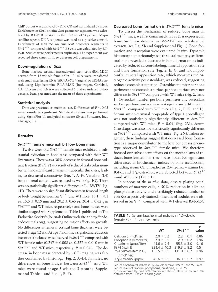
Decreased bone formation in Sirt1�/� female miceTo dissect the mechanism of reduced bone mass in
Sirt1�/� mice, we first confirmed that Sirt1 is expressed inbone. Sirt1 was detected in BM-MSC and whole tibialextracts (see Fig. 5B and Supplemental Fig. 1). Bone for-mation and resorption were evaluated in vivo. Dynamichistomorphometric analysis in the distal metaphyseal fem-oral bone revealed a decrease in bone formation as indi-cated by reduced calcein-labeling, mineral apposition rateand bone formation rate (Fig. 2, E, G, and H). Impor-tantly, mineral apposition rate, which measures the os-teogenic activity per osteoblast, was reduced, suggestingreduced osteoblast function. Osteoblast number per boneperimeter and osteoblast surface per bone surface were notdifferent in Sirt1�/� compared with WT mice (Fig. 2, I andJ). Osteoclast number per bone perimeter and osteoclastsurface per bone surface were not significantly different inSirt1�/� compared with WT mice (Fig. 2, F, K, and L).Serum amino-terminal propeptide of type I procollagenwas not statistically significantly different in Sirt1�/�
compared with WT mice (P � 0.09) (Fig. 2M). SerumCrossLaps was also not statistically significantly differentin Sirt1�/� compared with WT mice (Fig. 2N). Taken to-gether, these findings suggest that decreased bone forma-tion is a major contributor to the low bone mass pheno-type observed in Sirt1�/� female mice. We thereforefocused our subsequent efforts on the mechanisms of re-duced bone formation in this mouse model. No significantdifferences in biochemical indices of bone metabolism,including serum Ca, phosphorus, 25-hydroxyvitamin D3,IGF-I, and 17�-estradiol, were detected between Sirt1�/
�and WT mice (Table 1).In support of the in vivo data, despite plating equal
numbers of marrow cells, a 50% reduction in alkalinephosphatase activity and a strikingly reduced number ofvon Kossa positively stained mineralized nodules were ob-served in Sirt1�/� compared with WT-derived BM-MSC
TABLE 1. Serum biochemical indices in 12-wk-oldfemale Sirt1�/� and WT mice
WT Sirt1�/�P
value
Calcium (mmol/liter) 2.0 � 0.2 2.2 � 0.1 0.86Phosphorus (mmol/liter) 2.9 � 0.5 2.9 � 0.2 0.96Creatinine (�mol/liter) 45.6 � 7.4 55.3 � 3.0 0.16IGF-I (ng/ml) 328.4 � 10.3 319.3 � 8.2 0.525-Hydroxyvitamin D3
(nmol/liter)131.5 � 6.5 131.0 � 6.7 0.96
17�-Estradiol (pg/ml) 41.6 � 8.5 36.3 � 5.7 0.97
Serum biochemical indices in 12-wk-old female Sirt1�/� and WT mice.Serum levels of calcium, phosphorus, creatinine, IGF-I, 25-hydroxyvitamin D3, and 17�-estradiol are shown. Data are mean � SEMobtained from 10 mice in each group.
Endocrinology, November 2011, 152(11):0000–0000 endo.endojournals.org 5

induced to osteogenesis (Fig. 3, A and B). RUNX2 mRNAexpression was not different in Sirt1�/� compared withWT-derived BM-MSC induced to osteogenesis, consistentwith no effect of Sirt1 haplo-insufficiency on osteoblastnumber (Fig. 3C). A significantly reduced mRNA expres-sion of the osteoblastic markers bone sialoprotein, osteo-calcin, and type 1 collagen was found in Sirt1�/� BM-MSC induced to osteogenesis (Fig. 3C), supporting the
notion of reduced osteoblast activity. A reciprocal effectwas observed in C3H10T1/2 cells overexpressing Sirt1 inwhich a 50% increase in alkaline phosphatase activity wasfound (Fig. 3, D and E).
Of note, an increase in adipogenesis was observed inSirt1�/�-derived BM-MSC induced to adipogenesiscompared with WT stromal mesenchymal stem cells asindicated by increased number of Oil-Red-O-stained
FIG. 3. Decreased osteoblast activity in bone marrow-derived mesenchymal stem cells obtained from Sirt1�/� and WT female mice. A, Alkalinephosphatase (Alk. Phos.) activity (n � 3). B, von Kossa positively stained mineralized area in growing (GM) and osteogenic medium (OSM) (�100)(quantified in four wells per condition from three different preparations). C, mRNA expression of runt-related transcription factor (RUNX)-2 48 hafter induced osteogenesis. Results are relative to hypoxanthine-guanine phosphoribosyl transferase (HPRT). Collagen type 1 (Col1a1), osteocalcin,and bone sialoprotein (BSP) mRNA expression 6 d after induced osteogenesis is shown. Results are relative to ubiquitin C (UBC) (n � 3). Sirt1 andglyceraldehyde-3-phosphate dehydrogenase (GAPDH) protein levels (D) and alkaline phosphatase activity (E) (right panel) in Sirt1-overexpressingC3H10T1/2 and control cells. Results are mean � SEM (n � 3). Statistical analysis was performed by Student’s t test. *, P � 0.05 compared withWT or C3H10T1/2.
6 Cohen-Kfir et al. Sirt1 Regulates Bone Mass and Represses Sclerostin Endocrinology, November 2011, 152(11):0000–0000

adipocytes and higher PPAR�2 mRNA expression (Fig.4, A and B).
Sirt1 is a negative regulator of SostTo investigate the mechanism by which Sirt1 affects
bone formation, we looked for an inhibitor of bone for-mation that is up-regulated in the absence of Sirt1. BecauseSirt1 was shown to regulate gene expression, we took anunbiased gene array approach to identify candidate genes.Gene expression in female Sirt1�/�- and WT-derived BM-MSC induced to osteogenesis was compared using an os-teogenesis microarray. Ten genes were differentially ex-pressed (�3-fold change) in Sirt1�/�-derived cells(Supplemental Table 2). The biggest difference was ob-served for Sost (�14-fold expression in Sirt1�/� derivedcells). Based on the known inhibitory effect of sclerostinon bone formation and given that high levels of Sost ex-pression could explain the phenotype observed in ourSirt1�/� mice, we decided to focus our studies on thisparticular gene. Aligned with the gene expression data,sclerostin was increased in whole femoral bone extractsobtained from Sirt1�/� mice and in Sirt1�/�-derived BM-MSC (Fig. 5, A–C). Interestingly, increased sclerostin wasalso found in Sirt1�/� male-derived BM-MSCs (Fig. 5D).However, no difference in alkaline phosphatase activitybetween male Sirt1�/� and WT BM-MSC induced toosteogenesis was found (data not shown). Conversely,a significantly lower sclerostin was observed in Sirt1-overexpressing C3H10T1/2 cells induced to osteogen-esis (Fig. 5E). These results suggest that Sirt1 down-regulates sclerostin.
Previous work has demonstrated that Sirt1 directly reg-ulates gene expression acting as a histone deacetylase (20).To investigate whether this was the case for Sost, a ChIPassay was performed with an anti-Sirt1 antibody in mouse
ES cells. These cells exhibit undetectable sclerostin level,and therefore, we hypothesized that expression of Sost isrepressed in these cells. Sirt1�/ � ES cells were used asnegative controls (14). RT-PCR assays using primers thatcover eight consecutive segments in the 5 regulatory re-gion of the Sost promoter were performed. Strikingly, wefound significant Sirt1 binding to the Sost promoter in aregion encompassing 760-1940 bp upstream of the startcodon (Fig. 5F). Corroborating these data, no binding ofSirt1 to this region was detected in Sirt1�/ � ES cells (Sup-plemental Table 3). These findings suggest that Sirt1 spe-cifically binds to the Sost promoter and likely contributesto its silencing.
We next asked whether Sirt1 functions as a histonedeacetylase at the Sost promoter. H3K9 was previouslyidentified as specific Sirt1 target (21). To test whether thiswas the case for Sost, a ChIP assay was performed withanti-H3K9Ac antibody. Sirt1�/ � ES cells exhibited amarked increase in H3K9 acetylation at the Sost promotercompared with WT cells, peaking at 930-1115 bp up-stream of the start codon, at which maximal Sirt1 bindingoccurred (Fig. 5G). Together these results provide strongsupport for the notion that Sirt1 suppresses Sost expres-sion by modifying H3K9 acetylation at its promoter.
To confirm that increased sclerostin is a major contrib-utor to reduced osteoblast activity, a sclerostin-neutraliz-ing antibody and Sost down-regulation by siRNA wereused in Sirt1�/� marrow-derived osteoblasts. Treatmentwith the sclerostin antibody not only restored von Kossa-positive-stained mineralized nodule formation but over-corrected it, suggesting a higher basal sclerostin level inSirt1�/� mice (Fig. 5H). Sost down-regulation tripledmRNA levels of osteocalcin and bone sialoprotein (Fig.5I). These data strongly suggest that decreased osteoblastactivity in female-derived osteoblasts is due to increasedsclerostin.
Discussion
This study demonstrates for the first time a role for Sirt1in bone and shows that Sirt1 is a regulator of bone mass.Sirt1�/� female mice exhibit a dramatic decrease in bonemass as a result of reduced bone formation. Importantly,we uncovered a link between Sirt1 and Sost, which en-codes for sclerostin, a critical inhibitor of bone formation,and demonstrate that Sost is a novel bone-specific target ofSirt1. Sost gene expression is negatively regulated by Sirt1through deacetylation of H3K9 in the promoter region.
Sclerostin, a secreted cysteine-knot protein with ho-mology to the differential screening-selected gene aberra-tive inneuroblastoma familyofbonemorphogenicprotein
FIG. 4. Increased adipogenesis in bone marrow-derived mesenchymalstem cells obtained from Sirt1�/� and WT 12-wk-old female mice. A, Oil-Red-O positively stained adipocytes in Sirt1�/�- and WT-derived BM-MSCinduced to adipogenesis. Results are mean � SEM of OD (n � 4). B,PPAR�2 mRNA levels in Sirt1�/�-and WT-derived BM-MSC induced toadipogenesis. Results are relative to hypoxanthine-guanine phosphoribosyltransferase (HPRT). Results are mean � SEM (n � 3). Statistical analysis wasperformed by Student’s t test. *, P � 0.05 compared with WT.
Endocrinology, November 2011, 152(11):0000–0000 endo.endojournals.org 7

FIG. 5. Sirt1 is a negative regulator of Sost. A, Increased mRNA expression of Sost in Sirt1�/� bone marrow-derived osteoblasts. Results are relative tohypoxanthine-guanine phosphoribosyl transferase (HPRT) (n � 3). *, P � 0.05. B–D, Increased sclerostin levels in female Sirt1�/� compared to WT bonemarrow-derived osteoblasts and whole femur extracts (B and C) and male Sirt1�/� compared to WT bone marrow-derived osteoblasts (D). E, Sclerostin inSirt1 overexpressing C3H10T1/2 and control cells on d 6 after induced osteogenesis (n � 3 for all Western data). F, ChIP assay using anti-Sirt1 antibody inSirt1�/� ES cells. Real-time PCR analysis of Sirt1 binding to the Sost promoter region. Results are relative enrichment compared with the �51–137 primer.Major satellite repeats DNA sequence (MSR) and a primer at �3600 to –3738 served as positive and negative controls, respectively. G, ChIP assay usingantibody against acetylated histone H3K9 in Sirt1�/� and Sirt1�/ � ES cells. Statistical analysis was performed by Student’s t test (for all ChIP data, n � 3).H, von Kossa positively stained mineralized area in Sirt1�/� and WT BM-MSC induced to osteogenesis (OSM) with and without a sclerostin-neutralizingantibody. Results are mean � SEM (n � 4). Statistical analysis was performed by two-way ANOVA with genotype and treatment as independent variablesfollowed by Student-Newman-Keuls pairwise comparison. Ab, Antibody. *, P � 0.05. I, Sclerostin levels and mRNA levels of bone sialoprotein(BSP) and osteocalcin in Sirt1�/�-derived osteoblasts transfected with Sost or control siRNA for down-regulation of Sost. Results are relative to UBC (n � 3).
8 Cohen-Kfir et al. Sirt1 Regulates Bone Mass and Represses Sclerostin Endocrinology, November 2011, 152(11):0000–0000
antagonists, is a major inhibitor of bone formation (22). Inadultbone, it is constitutively expressedbyosteocytes, a finaldifferentiated cell of the osteoblast lineage (23). Sclerostin isdetected in not only osteocytes but also in marrow stromalcells, osteoclast precursors, and osteoblasts during embry-onic bone development (24–26). Its role in bone biology wasfirst appreciated when excessive bone mass was observed inpatients with Van Buchem’s disease or sclerosteosis inwhom mutations or deletions in the Sost gene were iden-tified (27–30). Its physiological importance was furthersupported by the high bone mass phenotype found insclerostin-null mice (31), and a low bone mass pheno-type in sclerostin overexpressing mice (32).
Sclerostin’s mechanism of action is not fully under-stood. It was shown to disrupt the formation of the Wnt-induced frizzled-low-density lipoprotein receptor-relatedprotein 5 (LRP5) and its corecptor LRP6 complex (33–35). But the mechanism by which its binding to LRP5/6interferes with canonical Wnt signaling and bone forma-tion remains unknown. Data regarding a direct effect ofsclerostin on downstream targets in the canonical Wntpathway such as active �-catenin and its targets T-cellfactor/lymphoid enhancer binding factor 1 have been in-conclusive. Importantly, bone mass and strength in agedovariectomized rats (36) and aged male rats (37) are re-stored by the administration of a sclerostin-neutralizingantibody. Moreover, an impressive increase in spine andhip bone mineral density in postmenopausal womentreated with an antisclerostin monoclonal antibody wasrecently reported (38).
The regulatory control of Sost expression is only par-tially known. Mechanical loading reduces Sost mRNA ex-pression and sclerostin levels in osteocytes, whereas un-loading leads to an increase (23, 39, 40). The anaboliceffect of PTH was shown to be mediated, in part, by sup-pressing Sost expression. Indeed, a blunted anabolic effectof PTH in Sost-deficient mice was observed (41). A role forestrogen in the regulation of sclerostin levels has been im-plicated by the finding of increased serum sclerostin inpostmenopausal compared with premenopausal women(42) and a reversal upon estrogen replacement (43). On-costatin M was reported to inhibit sclerostin production ina stromal cell line and in primary murine osteoblast cul-tures (44). Our study indicates that Sirt1 is a novel negativeregulator of Sost and its product sclerostin by epigeneticsilencing of Sost gene expression. It will be important totest in future studies whether Sirt1�/� female mice aremore susceptible to ovariectomy-induced bone loss.Moreover, whether Sirt1-mediated changes in Sosttranscription are involved in the pathogenesis of osteo-porosis and age-associated bone loss awaits furtherinvestigation.
Our results indicate that the effect of Sirt1 deficiency onfemoral bone mass is more pronounced in females thanmales. Our findings of a sexual dimorphic phenotype areconsistent with other Sirt1-related transgenic mouse mod-els. Thus, female but not male mice lacking Sirt1 in pro-opiomelanocortin neurons display a striking hypersensi-tivity to diet-induced obesity (45). In addition, female butnot male mice lacking nicotinamide phosphoribosyltrans-ferase (Nampt), a key enzyme in the biosynthesis of theoxidation of nicotinamide adenine dinucleotide, an essen-tial cofactor for Sirt1 activity, showed impaired glucosetolerance and reduced insulin secretion (46). A limitationof this work is lack of data on vertebral bone mass inSirt1�/� and WT male mice. Because there was no differ-ence in femoral bone indices between Sirt1�/� and WTmale mice and the differences in vertebral indices in fe-males were small, we anticipated no difference in spinalbone mass in Sirt1�/� and WT male mice.
To better understand the underlying mechanism of thisdimorphic effect on bone mass, we asked whether Sirt1deficiency affects sclerostin in males as it does in females.Interestingly, similar to females, the sclerostin level wasincreased in male Sirt1�/� compared with WT mice (Fig.5D), suggesting that Sirt1 represses sclerostin in both gen-ders. These findings are consistent with the data of Ra-madori et al. (45), who reported that Sirt1 deletion inproopiomelanocortin neurons alters uncoupling pro-tein-1 and tyrosine hydroxylase protein levels in perigo-nodal white adipose tissue in both females and males, yeta body weight phenotype was observed in females but notmales. Thus, we speculate that factors downstream ofsclerostin are affected by sex.
In summary, our study uncovers a role for Sirt1 as aregulator of bone mass and demonstrates a novel Sirt1-Sost connection, revealing that Sirt1 is a transcriptionalrepressor of Sost. The hypothesis that Sirt1-mediated epi-genetic changes in Sost transcription are involved in os-teoporosis awaits further investigation. It is possible thatSirt1 targets, in addition to Sost, also contribute to thephenotype observed in Sirt1�/� mice. Our findings haveimportant clinical implications: they suggest that specificSirt1 activators may stimulate bone formation, and pro-vide a new strategy for generating a much needed anabolictherapy for osteoporosis.
Acknowledgments
We thank Frederick W. Alt for providing the Sirt1�/� mice andP. Oberdoerffer for advice and reagents. We also thank M.Rosenblatt, Gilad Hamdani, Aviel Hankin, Kristen Anderson,Itai Bab, and Zvi Bar-Shavit for assistance. In addition, we thank
Endocrinology, November 2011, 152(11):0000–0000 endo.endojournals.org 9
Amgen for the sclerostin-neutralizing antibody. We are espe-cially grateful to R. Kaplan and the Bnai Brith Leo Baeck LondonLodge for their support of osteoporosis research. E.C.-K. per-formed most of the experiments and participated in writing themanuscript. H.A. performed the histomorphometry and �CTstudies and some of the in vivo and in vitro experiments. A.L.performed some of the in vivo and in vitro experiments. E.A.performed the biochemical analyses and the ChIP experiments.A.B. performed some of the �CT studies. L.Z. designed, per-formed some of the ChIP experiments, and analyzed the ChIPdata. D.T. designed and assisted with the small interference RNAexperiments. I.G. and A.D. assisted with the cell cultures, proteinisolation, and Western blotting. R.M. designed and supervisedsome of the experiments. R.D.-P. conceived and designed theproject, supervised it, and wrote the manuscript.
Address all correspondence and requests for reprints to:Rivka Dresner-Pollak, M.D., Endocrinology and MetabolismService, Hadassah-Hebrew University Medical Center, P.O. Box12000, Jerusalem 91120, Israel. E-mail: [email protected].
This work was supported by the Israel Science FoundationGrant 1230/07 (to R.D.-P.), the Chief Scientist, Ministry ofHealth, Israel Grant 3_3926 (to R.D.-P.), Hadassah ZionistWomen Organization of America Research Fund for Women’sHealth (to R.D.-P.). R.M. is a Kimmel Scholar and is the recipientof a Young Investigator Award from the American Federation ofAging Research.
Disclosure Summary: E.C.-K., H.A., A.L., E.A., A.B., I.G.,L.Z., A.D., D.T., R.M., and R.D.-P. have nothing to declare.
References
1. Finkel T, Deng CX, Mostoslavsky R 2009 Recent progress in thebiology and physiology of sirtuins. Nature 460:587–591
2. Imai S, Armstrong CM, Kaeberlein M, Guarente L 2000 Transcrip-tional silencing and longevity protein Sir2 is an NAD-dependenthistone deacetylase. Nature 403:795–800
3. Lin SJ, Defossez PA, Guarente L 2000 Requirement of NAD andSIR2 for life-span extension by calorie restriction in Saccharomycescerevisiae. Science 289:2126–2128
4. Bordone L, Cohen D, Robinson A, Motta MC, van Veen E, CzopikA, Steele AD, Crowe H, Marmor S, Luo J, Gu W, Guarente L 2007SIRT1 transgenic mice show phenotypes resembling calorie restric-tion. Aging Cell 6:759–767
5. Donmez G, Wang D, Cohen DE, Guarente L 2010 SIRT1 suppresses�-amyloid production by activating the �-secretase gene ADAM10.Cell 142:320–332
6. Elbaz A, Rivas D, Duque G 2009 Effect of estrogens on bone marrowadipogenesis and Sirt1 in aging C57BL/6J mice. Biogerontology 10:747–755
7. Backesjo CM, Li Y, Lindgren U, Haldosen LA 2006 Activation ofSirt1 decreases adipocyte formation during osteoblast differentia-tion of mesenchymal stem cells. J Bone Miner Res 21:993–1002
8. Backesjo CM, Li Y, Lindgren U, Haldosen LA 2009 Activation ofSirt1 decreases adipocyte formation during osteoblast differentia-tion of mesenchymal stem cells. Cells Tissues Organs 189:93–97
9. He X, Andersson G, Lindgren U, Li Y 2010 Resveratrol preventsRANKL-induced osteoclast differentiation of murine osteoclast
progenitor RAW 264.7 cells through inhibition of ROS production.Biochem Biophys Res Commun 401:356–362
10. Shakibaei M, Buhrmann C, Mobasheri A 2011 Resveratrol-medi-ated SIRT-1 interactions with p300 modulate receptor activator ofNF-�B ligand (RANKL) activation of NF-�B signaling and inhibitosteoclastogenesis in bone-derived cells. J Biol Chem 286:11492–11505
11. Pearson KJ, Baur JA, Lewis KN, Peshkin L, Price NL, Labinskyy N,Swindell WR, Kamara D, Minor RK, Perez E, Jamieson HA, ZhangY, Dunn SR, Sharma K, Pleshko N, Woollett LA, Csiszar A, IkenoY, Le Couteur D, Elliott PJ, Becker KG, Navas P, Ingram DK, WolfNS, Ungvari Z, Sinclair DA, de Cabo R 2008 Resveratrol delaysage-related deterioration and mimics transcriptional aspects of di-etary restriction without extending life span. Cell Metab 8:157–168
12. Bowers JL, Tyulmenkov VV, Jernigan SC, Klinge CM 2000 Res-veratrol acts as a mixed agonist/antagonist for estrogen receptors �
and �. Endocrinology 141:3657–366713. Liu M, Wilk SA, Wang A, Zhou L, Wang RH, Ogawa W, Deng C,
Dong LQ, Liu F 2010 Resveratrol inhibits mTOR signaling by pro-moting the interaction between mTOR and DEPTOR. J Biol Chem285:36387–36394
14. Cheng HL, Mostoslavsky R, Saito S, Manis JP, Gu Y, Patel P, Bron-son R, Appella E, Alt FW, Chua KF 2003 Developmental defects andp53 hyperacetylation in Sir2 homolog (SIRT1)-deficient mice. ProcNatl Acad Sci USA 100:10794–10799
15. McBurney MW, Yang X, Jardine K, Hixon M, Boekelheide K,Webb JR, Lansdorp PM, Lemieux M 2003 The mammalian SIR2�
protein has a role in embryogenesis and gametogenesis. Mol CellBiol 23:38–54
16. Picard F, Kurtev M, Chung N, Topark-Ngarm A, Senawong T,Machado De Oliveira R, Leid M, McBurney MW, Guarente L 2004Sirt1 promotes fat mobilization in white adipocytes by repressingPPAR-�. Nature 429:771–776
17. Akune T, Ohba S, Kamekura S, Yamaguchi M, Chung UI, KubotaN, Terauchi Y, Harada Y, Azuma Y, Nakamura K, Kadowaki T,Kawaguchi H 2004 PPAR� insufficiency enhances osteogenesisthrough osteoblast formation from bone marrow progenitors. J ClinInvest 113:846–855
18. Gazzerro E, Pereira RC, Jorgetti V, Olson S, Economides AN, Ca-nalis E 2005 Skeletal overexpression of gremlin impairs bone for-mation and causes osteopenia. Endocrinology 146:655–665
19. Moisan A, Rivera MN, Lotinun S, Akhavanfard S, Coffman EJ,Cook EB, Stoykova S, Mukherjee S, Schoonmaker JA, Burger A,Kim WJ, Kronenberg HM, Baron R, Haber DA, Bardeesy N 2011The WTX tumor suppressor regulates mesenchymal progenitor cellfate specification. Dev Cell 20:583–596
20. Oberdoerffer P, Michan S, McVay M, Mostoslavsky R, Vann J, ParkSK, Hartlerode A, Stegmuller J, Hafner A, Loerch P, Wright SM,Mills KD, Bonni A, Yankner BA, Scully R, Prolla TA, Alt FW,Sinclair DA 2008 SIRT1 redistribution on chromatin promotesgenomic stability but alters gene expression during aging. Cell 135:907–918
21. Wang RH, Zheng Y, Kim HS, Xu X, Cao L, Luhasen T, Lee MH,Xiao C, Vassilopoulos A, Chen W, Gardner K, Man YG, Hung MC,Finkel T, Deng CX 2008 Interplay among BRCA1, SIRT1, andSurvivin during BRCA1-associated tumorigenesis. Mol Cell 32:11–20
22. Poole KE, van Bezooijen RL, Loveridge N, Hamersma H, Papapou-los SE, Lowik CW, Reeve J 2005 Sclerostin is a delayed secretedproduct of osteocytes that inhibits bone formation. FASEB J 19:1842–1844
23. Lin C, Jiang X, Dai Z, Guo X, Weng T, Wang J, Li Y, Feng G, GaoX, He L 2009 Sclerostin mediates bone response to mechanical un-loading through antagonizing Wnt/�-catenin signaling. J BoneMiner Res 24:1651–1661
24. Bellido T, Ali AA, Gubrij I, Plotkin LI, Fu Q, O’Brien CA, Mano-lagas SC, Jilka RL 2005 Chronic elevation of parathyroid hormone
10 Cohen-Kfir et al. Sirt1 Regulates Bone Mass and Represses Sclerostin Endocrinology, November 2011, 152(11):0000–0000

in mice reduces expression of sclerostin by osteocytes: a novel mech-anism for hormonal control of osteoblastogenesis. Endocrinology146:4577–4583
25. Kamiya N, Ye L, Kobayashi T, Mochida Y, Yamauchi M, Kronen-berg HM, Feng JQ, Mishina Y 2008 BMP signaling negatively reg-ulates bone mass through sclerostin by inhibiting the canonical Wntpathway. Development 135:3801–3811
26. Pederson L, Ruan M, Westendorf JJ, Khosla S, Oursler MJ 2008Regulation of bone formation by osteoclasts involves Wnt/BMP sig-naling and the chemokine sphingosine-1-phosphate. Proc Natl AcadSci USA 105:20764–20769
27. Balemans W, Ebeling M, Patel N, Van Hul E, Olson P, Dioszegi M,Lacza C, Wuyts W, Van Den Ende J, Willems P, Paes-Alves AF, HillS, Bueno M, Ramos FJ, Tacconi P, Dikkers FG, Stratakis C, Lind-paintner K, Vickery B, Foernzler D, Van Hul W 2001 Increased bonedensity in sclerosteosis is due to the deficiency of a novel secretedprotein (SOST). Hum Mol Genet 10:537–543
28. Balemans W, Patel N, Ebeling M, Van Hul E, Wuyts W, Lacza C,Dioszegi M, Dikkers FG, Hildering P, Willems PJ, Verheij JB, Lind-paintner K, Vickery B, Foernzler D, Van Hul W 2002 Identificationof a 52 kb deletion downstream of the SOST gene in patients withvan Buchem disease. J Med Genet 39:91–97
29. Brunkow ME, Gardner JC, Van Ness J, Paeper BW, Kovacevich BR,Proll S, Skonier JE, Zhao L, Sabo PJ, Fu Y, Alisch RS, Gillett L,Colbert T, Tacconi P, Galas D, Hamersma H, Beighton P, MulliganJ 2001 Bone dysplasia sclerosteosis results from loss of the SOSTgene product, a novel cystine knot-containing protein. Am J HumGenet 68:577–589
30. Staehling-Hampton K, Proll S, Paeper BW, Zhao L, Charmley P,Brown A, Gardner JC, Galas D, Schatzman RC, Beighton P, Papa-poulos S, Hamersma H, Brunkow ME 2002 A 52-kb deletion in theSOST-MEOX1 intergenic region on 17q12–q21 is associated withvan Buchem disease in the Dutch population. Am J Med Genet110:144–152
31. Li X, Ominsky MS, Niu QT, Sun N, Daugherty B, D’Agostin D,Kurahara C, Gao Y, Cao J, Gong J, Asuncion F, Barrero M, Warm-ington K, Dwyer D, Stolina M, Morony S, Sarosi I, Kostenuik PJ,Lacey DL, Simonet WS, Ke HZ, Paszty C 2008 Targeted deletion ofthe sclerostin gene in mice results in increased bone formation andbone strength. J Bone Miner Res 23:860–869
32. Winkler DG, Sutherland MK, Geoghegan JC, Yu C, Hayes T,Skonier JE, Shpektor D, Jonas M, Kovacevich BR, Staehling-Hamp-ton K, Appleby M, Brunkow ME, Latham JA 2003 Osteocyte con-trol of bone formation via sclerostin, a novel BMP antagonist.EMBO J 22:6267–6276
33. Semenov M, Tamai K, He X 2005 SOST is a ligand for LRP5/LRP6and a Wnt signaling inhibitor. J Biol Chem 280:26770–26775
34. van Bezooijen RL, Roelen BA, Visser A, van der Wee-Pals L, de WiltE, Karperien M, Hamersma H, Papapoulos SE, ten Dijke P, LowikCW 2004 Sclerostin is an osteocyte-expressed negative regulator ofbone formation, but not a classical BMP antagonist. J Exp Med199:805–814
35. van Bezooijen RL, Svensson JP, Eefting D, Visser A, van der Horst
G, Karperien M, Quax PH, Vrieling H, Papapoulos SE, ten Dijke P,Lowik CW 2007 Wnt but not BMP signaling is involved in theinhibitory action of sclerostin on BMP-stimulated bone formation.J Bone Miner Res 22:19–28
36. Li X, Ominsky MS, Warmington KS, Morony S, Gong J, Cao J, GaoY, Shalhoub V, Tipton B, Haldankar R, Chen Q, Winters A, BooneT, Geng Z, Niu QT, Ke HZ, Kostenuik PJ, Simonet WS, Lacey DL,Paszty C 2009 Sclerostin antibody treatment increases bone forma-tion, bone mass, and bone strength in a rat model of postmenopausalosteoporosis. J Bone Miner Res 24:578–588
37. Li X, Warmington KS, Niu QT, Asuncion FJ, Barrero M, GrisantiM, Dwyer D, Stouch B, Thway TM, Stolina M, Ominsky MS, Ko-stenuik PJ, Simonet WS, Paszty C, Ke HZ 2010 Inhibition of scleros-tin by monoclonal antibody increases bone formation, bone mass,and bone strength in aged male rats. J Bone Miner Res 25:2647–2656
38. Padhi D, Jang G, Stouch B, Fang L, Posvar E 2011 Single-dose,placebo-controlled, randomized study of AMG 785, a sclerostinmonoclonal antibody. J Bone Miner Res 26:19–26
39. Robling AG, Niziolek PJ, Baldridge LA, Condon KW, Allen MR,Alam I, Mantila SM, Gluhak-Heinrich J, Bellido TM, Harris SE,Turner CH 2008 Mechanical stimulation of bone in vivo reducesosteocyte expression of Sost/sclerostin. J Biol Chem 283:5866–5875
40. Moustafa A, Sugiyama T, Saxon LK, Zaman G, Sunters A, Arm-strong VJ, Javaheri B, Lanyon LE, Price JS 2009 The mouse fibulaas a suitable bone for the study of functional adaptation to mechan-ical loading. Bone 44:930–935
41. Kramer I, Loots GG, Studer A, Keller H, Kneissel M 2010 Parathy-roid hormone (PTH)-induced bone gain is blunted in SOST over-expressing and deficient mice. J Bone Miner Res 25:178–189
42. Mirza FS, Padhi ID, Raisz LG, Lorenzo JA 2010 Serum sclerostinlevels negatively correlate with parathyroid hormone levels and freeestrogen index in postmenopausal women. J Clin Endocrinol Metab95:1991–1997
43. Modder UI, Clowes JA, Hoey K, Peterson JM, McCready L, OurslerMJ, Riggs BL, Khosla S 2011 Regulation of circulating sclerostinlevels by sex steroids in women and in men. J Bone Miner Res 26:27–34
44. Walker EC, McGregor NE, Poulton IJ, Solano M, Pompolo S, Fer-nandes TJ, Constable MJ, Nicholson GC, Zhang JG, Nicola NA,Gillespie MT, Martin TJ, Sims NA 2010 Oncostatin M promotesbone formation independently of resorption when signaling throughleukemia inhibitory factor receptor in mice. J Clin Invest 120:582–592
45. Ramadori G, Fujikawa T, Fukuda M, Anderson J, Morgan DA,Mostoslavsky R, Stuart RC, Perello M, Vianna CR, Nillni EA, Rah-mouni K, Coppari R 2010 SIRT1 deacetylase in POMC neurons isrequired for homeostatic defenses against diet-induced obesity. CellMetab 12:78–87
46. Revollo JR, Korner A, Mills KF, Satoh A, Wang T, Garten A, Das-gupta B, Sasaki Y, Wolberger C, Townsend RR, Milbrandt J, KiessW, Imai S 2007 Nampt/PBEF/Visfatin regulates insulin secretion in� cells as a systemic NAD biosynthetic enzyme. Cell Metab 6:363–375
Endocrinology, November 2011, 152(11):0000–0000 endo.endojournals.org 11