sir ludwig guttmann lecture functional electrical stimulation
TRANSCRIPT
REVIEW: Sir Ludwig Guttmann Lecture
Functional electrical stimulation after spinal cord injury: currentuse, therapeutic effects and future directions
KT Ragnarsson
Department of Rehabilitation Medicine, Mount Sinai School of Medicine, New York, NY, USA
Repair of the injured spinal cord by regeneration therapy remains an elusive goal. In contrast, progressin medical care and rehabilitation has resulted in improved health and function of persons with spinalcord injury (SCI). In the absence of a cure, raising the level of achievable function in mobility and self-care will first and foremost depend on creative use of the rapidly advancing technology that has been sowidely applied in our society. Building on achievements in microelectronics, microprocessing andneuroscience, rehabilitation medicine scientists have succeeded in developing functional electricalstimulation (FES) systems that enable certain individuals with SCI to use their paralyzed hands, arms,trunk, legs and diaphragm for functional purposes and gain a degree of control over bladder and bowelevacuation. This review presents an overview of the progress made, describes the current challengesand suggests ways to improve further FES systems and make these more widely available.
Spinal Cord (2008) 46, 255–274; doi:10.1038/sj.sc.3102091; published online 11 September 2007
Keywords: spinal cord injury; functional electrical stimulation; rehabilitation; exercise
Introduction
The foundations for modern management of spinal cord
injury (SCI) were created more than half a century ago by the
pioneering work of Dr Donald Munro in the United States
and by Sir Ludwig Guttmann in the United Kingdom. Munro
opened the first SCI unit, but Guttmann created the first
comprehensive SCI center for medical and rehabilitation
care. Under his strong leadership the care of persons with SCI
was completely changed.
I was lucky enough to meet Sir Ludwig once in person. It
was in the late 1970s at a meeting in New York City. He
clearly was a man of great energy and self-confidence, who
addressed my question to him with interest. At that time, I
was involved in research relating to the neuroendocrinology
of persons with SCI, especially production of catecholamines
during episodes of autonomic dysreflexia. Most descriptions
of the clinical symptoms of autonomic dysreflexia at that
time stated that there was increased sweating above the level
of the cord lesion. For example, one textbook contained the
following statement: ‘In complete lesions the lack of
thermoregulatory sweating below the level of injury is often
associated with excessive diaphoresis above the level of
injury’.1 Because sweating is controlled by the sympathetic
nervous system which has all its efferent nerve fibers exiting
from the thoracic cord, I had difficulty understanding which
autonomic nerves control sweating of the head and neck
‘above the level of injury’. Sir Ludwig pointed out to me that
sweating of the head and neck was not a thermal regulatory
response in persons with tetraplegia and was generated by
activity of the sympathetic nerves below the level of the
lesion. In other words, sweating of the head and neck was a
below level phenomenon and it was wrong to state that
excessive sweating in autonomic dysreflexia occurred above
the cord lesion in persons with tetraplegia.
Based on the work of Sir Ludwig Guttmann, specialized
SCI medical care and comprehensive rehabilitation has
focused on securing good health and maximum function
in mobility and self-care compatible with the neurological
condition, as well as to help each person with SCI to return
to the community and to achieve high quality of life.
Much has been accomplished by this approach. Life
expectancy of people with SCI has increased every year
since the 1940s. Morbidity, that is, medical complications,
has decreased and consequently people with SCI have
needed shorter initial hospital length of stay2 and fewer
rehospitalizations.3 Many medical conditions associated
with SCI can now be prevented or managed more efficiently,
for example, spasticity, thromboembolic disease, male sexual
dysfunction and infertility, heterotopic ossifications, pres-
sure ulcers, renal failure and respiratory insufficiency. More
people with SCI are discharged to their homes4 and live in
the community than ever before and it may be argued, but
not proven, that their quality of life has improved over the
years with better health, better equipment, more accessibleReceived 10 April 2007; revised 7 March 2007; accepted 16 May 2007;
published online 11 September 2007
Correspondence: Dr KT Ragnarsson, Department of Rehabilitation Medicine,
Mount Sinai School of Medicine, New York, NY 10029, USA.
E-mail: [email protected]
Spinal Cord (2008) 46, 255–274
& 2008 International Spinal Cord Society All rights reserved 1362-4393/08 $30.00
www.nature.com/sc
urban environment and public recognition of their personal
rights.
In contrast to the advances in the care of SCI, decades of
intense efforts by basic research scientists have unfortunately
achieved little clinically to reverse the neurological loss
associated with SCI by protection or regeneration of axons
within the injured spinal cord.5 The neurological outcome of
SCI is still first and foremost determined by the extent of the
damage that is caused at the moment of injury. No treatment
offered today can verifiably change that outcome. Conse-
quently, persons with SCI still must contend with the
multiple neurological sequelae of SCI, that is, paralysis,
sensory loss, autonomic dysfunction, loss of bladder and
bowel control, and so on.
Although better medical care of persons with SCI has
helped to progressively improve their health and life
expectancy over the last six decades, gains in mobility and
self-care skills have remained relatively stagnant because
further improvements in these areas are dependent on
advancements in basic science that may lead to reversal of
the neurological loss. Prognosis for ambulation and inde-
pendence in the activities of daily living (ADL) continues to
be primarily predicted by the neurological level and the
neurological completeness of the SCI. Only some compen-
satory rehabilitation interventions have helped to further
increase function, for example, by providing state of the art
wheelchairs and other assistive technology, such as commu-
nication devices, by emphasizing education and training for
career opportunities, by facilitating emotional adjustment by
psychological support, by generating an enabling commu-
nity,6 and so on.
It is apparent that some of the most significant scientific
accomplishments of our civilization during recent decades
have been in the field of microelectronics. These have had
global impact and revolutionized human communication
and access to information. Medicine has benefited enor-
mously from this technological development as a vast array
of new medical devices have entered the market after being
shown scientifically to be of clinical value, both for diagnosis
and treatment of human disease. It may be further argued
that creative use of such technology in medicine may result
in quicker development of new effective compensatory
treatments to improve functions after SCI than the search
for a ‘cure’ through basic regeneration research involving
such experimental interventions as grafting and transplanta-
tion of Schwann cells, olfactory ensheathing cells, cloned
undifferentiated cells or varying types of stem cells. Despite
the remarkable knowledge that has been gained in molecular
biology during recent years by animal experiments,
such as the identification of factors which may inhibit or
enhance axonal regeneration, for example trophic factors,
guidance molecules and growth inhibitors (Nogo), the
profound complexity of human SCI is still only partially
understood. Achieving a ‘biological cure’ for SCI will still
require understanding of vast and yet to be discovered
scientific knowledge and overcoming enormous scientific
obstacles. Although basic regeneration research in this
field must be supported as a long-term investment, funding
of technological projects to improve function of persons
with SCI may prove to be a wiser investment for short-term
gains.
The spectacular scientific progress made in electronics and
microprocessor technology during recent decades has been
utilized by biomedical engineers and clinicians to the benefit
of patients suffering from a variety of conditions and
disabilities, including SCI. Building on this new technology
and on the old knowledge that muscle contraction can be
activated by electricity, rehabilitation engineers have de-
signed devices and systems that can apply electrical currents
to neural tissues in a synchronized fashion for the purpose of
restoring a degree of control over abnormal or absent body
functions, a technique referred to as functional electrical
stimulation (FES).
Although FES in a pure sense refers to the achievement of
certain functional motor activities, for example, standing,
transfers, stepping, ambulation, cycling, hand grasp and
release, arm reaching, breathing, coughing, bladder and
bowel evacuation, penile erection and ejaculation, it is often
associated with the considerable therapeutic benefits of
physical exercise. Therapeutically, FES has been used to
increase muscle bulk, improve cardiovascular performance,
prevent and treat pressure ulcers, osteoporosis and joint
contractures, control spasticity, and improve general well-
being. As a means to maintain the person with SCI in
optimal and physical and psychological condition, regular
physical therapy, preferably by FES, has been recommended
by Kakulas,5 who notes that regular sensory stimulation
through the skin has the effect of keeping retained neuro-
logical functions and returning reflexes closer to the normal
physiological state. It has been further speculated that the
normal neural circuitry within the spinal cord necessary for
gait and other motor functions needs continuous main-
tenance by FES and/or body weight supported treadmill
training (BWSTT) ambulation.7 Others have suggested that
such patterned neural activity may be important for both
development and recovery of neurological functions.8
Furthermore, there is substantial scientific evidence that
shows that physical exercise not only stimulates production
of endorphins, but contributes to the upregulation of brain-
derived neurotrophic factor (BDNF), which may promote
synaptic and functional plasticity within the brain and
spinal cord.9 In general, it appears that active exercise for the
paralyzed body parts is of considerable clinical value for
persons with SCI and may be essential for maintenance of
neural circuitry should a ‘cure’ for SCI be found.
Anatomical and physiological principles of FES
To generate muscle contraction by electrical stimulation, the
stimulus is generally applied to a peripheral nerve but not to
the muscle itself. The electrical stimulus may be applied
anywhere along the length of the nerve from its origin to its
motor point where it connects with the muscle. Using
current techniques, the electrical charge needed to activate a
muscle contraction by direct muscle fiber stimulation is too
great to be safely applied multiple times. Compared to nerve
membranes, the different qualities of muscle membranes are
Functional electrical stimulation after spinal cord injuryKT Ragnarsson
256
Spinal Cord
such that it would require more than 100 times stronger
stimulus applied directly to the muscle to generate an action
potential than by stimulating the nerve.10,11 Thus, preserva-
tion of the entire lower motor neuron (LMN), including the
neuromuscular junction, is essential for all forms of FES, and
muscles paralyzed by LMN damage cannot be utilized for FES
by using currently available technology.
The strength of the electrical stimulus in terms of
amplitude and duration determines the number of nerve
fibers activated and the force of the muscle contraction.
Large nerve fibers, for example fibers from a-motor neurons,
are more easily stimulated than small diameter fibers and
because large fibers generally provide innervation for large
motor units, these are activated with less current than small
motor units. Clearly, sensory fibers within the stimulated
peripheral nerve are activated as well as motor fibers, which
may be painful, if sensation is preserved. In addition to
amplitude and duration of the electrical stimulus, the pulse
frequency and waveform are also important. The pulse
frequency needs to be high enough to generate a smooth
muscle contraction. The stimulus itself may be either
monophasic or biphasic, but usually balanced biphasic
stimuli are used clinically, as they provide better control
over muscle contraction force and are less prone to cause
tissue damage.12,13
Muscle fibers are primarily of two different types, that is,
type I (red, slow, aerobic) and type II (white, fast, anaerobic),
and these are histologically and physiologically different.
Inactivity is known to lead to transformation of type I
muscle fibers into type II, but this change is reversible with
appropriate exercise.14–17 In the paralyzed limbs of persons
with SCI, proportionally more muscle fibers are of type II
than type I, but the fibers are also atrophied, leading to a
weak and fatigable muscle upon stimulation. An exercise
regimen of electrical stimulation can increase contraction
time and resistance to fatigue,14 probably as more muscle
fibers become type I.
Components of FES systems
Most FES systems are basically quite similar in design and
purpose, but the electrical stimulation may be delivered
either by surface (transcutaneous), percutaneous or im-
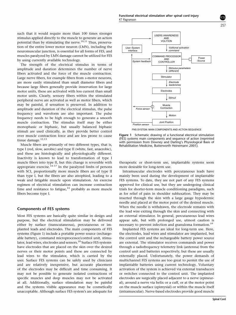
planted leads and electrodes. The main components of FES
systems (Figure 1) include a portable power source (recharge-
able battery), command microprocessor/control unit, stimu-
lator, lead wires, electrodes and sensors.18 Surface FES systems
have electrodes that are placed on the skin over the desired
nerves or their motor points and these are connected by
lead wires to the stimulator, which is carried by the
user. Surface FES systems can be safely used by clinicians
and are relatively inexpensive, but accurate placement
of the electrodes may be difficult and time consuming. It
may not be possible to generate isolated contractions of
specific muscles and deep muscles may not be activated
at all. Additionally, surface stimulation may be painful
and the systems visible appearance may be cosmetically
unacceptable. Although surface FES system’s are adequate for
therapeutic or short-term use, implantable systems seem
more desirable for long-term use.
Intramuscular electrodes with percutaneous leads have
mainly been used during the development of implantable
FES systems. To date, they are not part of any FES systems
approved for clinical use, but they are undergoing clinical
trials for shorter-term muscle conditioning paradigms, such
as for relief of pain in shoulder subluxation. They may be
inserted through the skin with a large gauge hypodermic
needle and placed at the motor point of the desired muscle.
When the needle is withdrawn, the electrode remains with
the lead wire exiting through the skin and connecting with
an external stimulator. In general, percutaneous lead wires
appear safe, but with prolonged use, utmost caution is
necessary to prevent infection and granuloma formation.19
Implanted FES systems are ideal for long-term use. Here,
the electrodes, lead wires and stimulator are implanted, but
the control unit and the rechargeable battery power source
are external. The stimulator receives commands and power
through a radiofrequency telemetry link (antenna) from the
control unit and batteries respectively, but these are usually
externally placed. Unfortunately, the power demands of
multichannel FES systems are too great to permit the use of
implantable batteries using current technology. Voluntary
activation of the system is achieved via external transducers
or switches connected to the control unit. The implanted
electrodes are surgically placed adjacent to a nerve (epineur-
al), around a nerve via helix or a cuff, or at the motor point
on the muscle surface (epimysial) or within the muscle itself
(intramuscular). Nerve-based electrodes provide good muscle
FNS SYSTEM: MAIN COMPONENTS AND ACTION SEQUENCE
USERS AWARENESSAND
VOLITION
COMMAND PROCESSORAND
CONTROLLER
PowerSource
User–Systeminterface
Volitional activationcommand
Stimulus parameters(different)
Electrodeleads/wires
Stimuli
Motion
Force sensor
Position sensor
Stimulator
Electrodes
MuscleContraction
Joint Position
System feedback
Figure 1 Schematic drawing of a functional electrical stimulation(FES) systems main components and sequence of action (reprintedwith permission from Downey and Darling’s Physiological Basis ofRehabilitation Medicine, Butterworth Heinemann 2001).
Functional electrical stimulation after spinal cord injuryKT Ragnarsson
257
Spinal Cord
specificity and allow stimulation of specific muscles and
excellent recruitment with relatively low electrical currents.
Care must be taken during the surgical procedure and during
long-term stimulation at relatively high frequencies to avoid
damage to the nerve. Fortunately, guidelines for safe
stimulation have been developed and these electrodes have
been used successfully in many applications, such as control
of bladder, phrenic nerve for breathing, peripheral nerve
stimulation for pain control, and cranial nerve stimulation
for epilepsy suppression. Epimysial electrodes generate
minimal damage and have proven to be durable for upper
and lower limb applications,20,21 but for activation of
deep and very small muscles, intramuscular electrodes are
preferable.
Normal motor activity depends not only on synchronized
muscle contractions but also on a highly sophisticated
sensory feedback processed through innumerable sensory
end organs, sensory nerves, the spinal cord and the brain.
Unfortunately, modern medical technology does not even
come close to providing such sensory information, which is
a major shortcoming of multichannel FES systems. Relatively
primitive sensory feedback signals from the limbs can be
obtained from externally placed goniometers and potenti-
ometers placed at various joints. These can measure joint
positions and motion, permitting the systems microproces-
sor to calculate the velocity and acceleration of movement
and consequently adjust and control the stimulator.22
The user and the control unit operate the FES system in an
interactive fashion. The control unit, which is located
external to the body in all currently used FES systems,
contains the system’s microprocessor (computer, intelli-
gence) and usually the battery. It has three main functions:13
(a) supplies power from its batteries to the entire system; (b)
extracts information from the user and the sensors; and (c)
transforms the received information into commands that are
transmitted by radiofrequency signals through an externally
located transmitting coil to the implanted stimulator. The
user is able to control the entire system by varieties of
sources, such as a joystick or switches that may use breath
force, myoelectric signals, and voice recognition or motion
sensors. Depending on the feedback and automatic control
provided by the system, the control can be classified either as
open loop or closed loop. In an open loop control,
preprogrammed patterns of electrical stimulation are indivi-
dually created for a specific function without automatic
correction for changes in muscle force or joint motion. In a
closed loop control, the control unit receives information on
muscle force and joint motion from the sensors and
automatically modifies the electrical stimulation.23
Several criteria for effective application of FES have been
identified as follows:13 (a) the strength of the FES-induced
muscle contraction must be forceful, controllable and
repeatable; (b) the electrical stimulus must not be painful;
(c) the LMN must be intact and the neural structures
must not be damaged by the FES; and (d) the method of
FES delivery must be acceptable to the user. These
criteria must be considered when the various methods of
applying electrical stimulation are selected for functional
purposes.
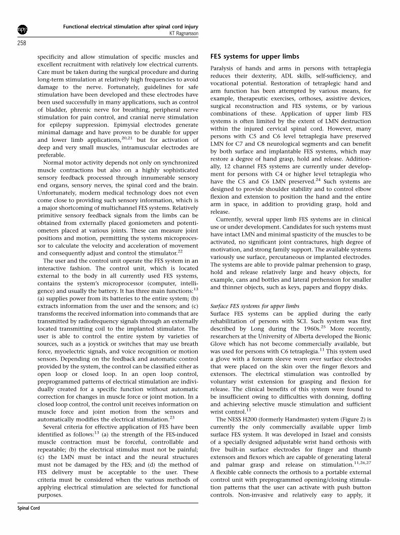
FES systems for upper limbs
Paralysis of hands and arms in persons with tetraplegia
reduces their dexterity, ADL skills, self-sufficiency, and
vocational potential. Restoration of tetraplegic hand and
arm function has been attempted by various means, for
example, therapeutic exercises, orthoses, assistive devices,
surgical reconstruction and FES systems, or by various
combinations of these. Application of upper limb FES
systems is often limited by the extent of LMN destruction
within the injured cervical spinal cord. However, many
persons with C5 and C6 level tetraplegia have preserved
LMN for C7 and C8 neurological segments and can benefit
by both surface and implantable FES systems, which may
restore a degree of hand grasp, hold and release. Addition-
ally, 12 channel FES systems are currently under develop-
ment for persons with C4 or higher level tetraplegia who
have the C5 and C6 LMN preserved.24 Such systems are
designed to provide shoulder stability and to control elbow
flexion and extension to position the hand and the entire
arm in space, in addition to providing grasp, hold and
release.
Currently, several upper limb FES systems are in clinical
use or under development. Candidates for such systems must
have intact LMN and minimal spasticity of the muscles to be
activated, no significant joint contractures, high degree of
motivation, and strong family support. The available systems
variously use surface, percutaneous or implanted electrodes.
The systems are able to provide palmar prehension to grasp,
hold and release relatively large and heavy objects, for
example, cans and bottles and lateral prehension for smaller
and thinner objects, such as keys, papers and floppy disks.
Surface FES systems for upper limbs
Surface FES systems can be applied during the early
rehabilitation of persons with SCI. Such system was first
described by Long during the 1960s.25 More recently,
researchers at the University of Alberta developed the Bionic
Glove which has not become commercially available, but
was used for persons with C6 tetraplegia.11 This system used
a glove with a forearm sleeve worn over surface electrodes
that were placed on the skin over the finger flexors and
extensors. The electrical stimulation was controlled by
voluntary wrist extension for grasping and flexion for
release. The clinical benefits of this system were found to
be insufficient owing to difficulties with donning, doffing
and achieving selective muscle stimulation and sufficient
wrist control.11
The NESS H200 (formerly Handmaster) system (Figure 2) is
currently the only commercially available upper limb
surface FES system. It was developed in Israel and consists
of a specially designed adjustable wrist hand orthosis with
five built-in surface electrodes for finger and thumb
extensors and flexors which are capable of generating lateral
and palmar grasp and release on stimulation.11,26,27
A flexible cable connects the orthosis to a portable external
control unit with preprogrammed opening/closing stimula-
tion patterns that the user can activate with push button
controls. Non-invasive and relatively easy to apply, it
Functional electrical stimulation after spinal cord injuryKT Ragnarsson
258
Spinal Cord
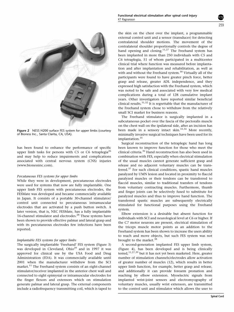
has been found to enhance the performance of specific
upper limb tasks for persons with C5 or C6 tetraplegia26
and may help to reduce impairments and complications
associated with central nervous system (CNS) injuries
(www.bionessinc.com).
Percutaneous FES systems for upper limbs
While they were in development, percutaneous electrodes
were used for systems that now are fully implantable. One
upper limb FES system with percutaneous electrodes, the
FESMate was developed and became commercially available
in Japan. It consists of a portable 30-channel stimulator/
control unit connected to percutaneous intramuscular
electrodes that are activated by a push button switch. A
later version, that is, NEC FESMate, has a fully implantable
16-channel stimulator and electrodes.28 These systems have
been shown to provide effective palmar and lateral grasp and
with its percutaneous electrodes few infections have been
reported.
Implantable FES systems for upper limbs
The surgically implantable ‘Freehand’ FES system (Figure 3)
was developed in Cleveland, Ohio29 and in 1997 it was
approved for clinical use by the USA Food and Drug
Administration (FDA). It was commercially available until
2001 when the manufacturer withdrew from the SCI
market.11 The Freehand system consists of an eight-channel
stimulator/receiver implanted in the anterior chest wall and
connected to eight epimysial or intramuscular electrodes for
the finger flexors and extensors which on stimulation
generate palmar and lateral grasp. The external components
include a radiofrequency transmitting coil, which is taped to
the skin on the chest over the implant, a programmable
external control unit and a sensor (transducer) for detecting
contralateral shoulder motions. The movement of the
contralateral shoulder proportionally controls the degree of
hand opening and closing.11,27 The Freehand system has
been implanted in more than 250 individuals with C5 and
C6 tetraplegia, 51 of whom participated in a multicenter
clinical trial where function was measured before implanta-
tion and after implantation and rehabilitation, as well as
with and without the Freehand system.30 Virtually all of the
participants were found to have greater pinch force, better
grasp and release, greater ADL independence, and they
expressed high satisfaction with the Freehand system, which
was noted to be safe and associated with very few medical
complications during a total of 128 cumulative implant
years. Other investigators have reported similar beneficial
clinical results.31,32 It is regrettable that the manufacturer of
the Freehand system chose to withdraw from the relatively
small SCI market for business reasons.
The Freehand stimulator is surgically implanted in a
subcutaneous pocket over the fascia of the pectoralis muscle
on the chest wall on the ipsilateral side, after an incision has
been made in a sensory intact skin.33,34 More recently,
minimally invasive surgical techniques have been used for its
implantation.35
Surgical reconstruction of the tetraplegic hand has long
been known to improve function for those who meet the
clinical criteria.36 Hand reconstruction has also been used in
combination with FES, especially when electrical stimulation
of the usual muscles cannot generate sufficient grasp and
release and no adjacent voluntary muscles can be trans-
ferred.37 For such clinical conditions, spastic hand muscles
paralyzed by UMN lesion and located in proximity to flaccid
paralyzed muscles or their tendons can be transferred to
flaccid muscles, similar to traditional transfers of tendons
from voluntary contracting muscles. Furthermore, thumb
and finger joints can be selectively fused to substitute for
paralyzed muscles and thus to improve hand function. The
transferred spastic muscles are subsequently electrically
stimulated for functional purposes using the Freehand
system.
Elbow extension is a desirable but absent function for
individuals with SCI and neurological level at C6 or higher. If
the C7 motor neurons are present, electrical stimulation of
the triceps muscle motor points as an addition to the
Freehand system has been shown to increase the users ability
to reach and move objects, but such FES system was not
brought to the market.38
A second-generation implanted FES upper limb system,
(Figure 4), has been developed and is being clinically
tested,11,27,39 but it has not yet been marketed. Here, greater
number of stimulation channels/electrodes allow activation
of greater number of muscles (12), which results in better
upper limb function, for example, better grasp and release,
and additionally it can provide forearm pronation and
reaching by elbow extension. Myoelectric signals from
implanted wrist-joint sensors and electromyography of
voluntary muscles, usually wrist extensors, are transmitted
to the control unit and stimulator which allows the user to
Figure 2 NESS H200 surface FES system for upper limbs (courtesyof Bioness Inc., Santa Clarita, CA, USA).
Functional electrical stimulation after spinal cord injuryKT Ragnarsson
259
Spinal Cord
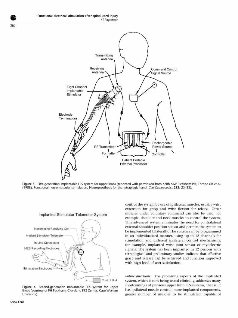
control the system by use of ipsilateral muscles, usually wrist
extension for grasp and wrist flexion for release. Other
muscles under voluntary command can also be used, for
example, shoulder and neck muscles to control the system.
This advanced system eliminates the need for contralateral
external shoulder position sensor and permits the system to
be implemented bilaterally. The system can be programmed
in an individualized manner, using up to 12 channels for
stimulation and different ipsilateral control mechanisms,
for example, implanted wrist joint sensor or myoelectric
signals. The system has been implanted in 12 persons with
tetraplegia27 and preliminary studies indicate that effective
grasp and release can be achieved and function improved
with high level of user satisfaction.
Future directions. The promising aspects of the implanted
system, which is now being tested clinically, addresses many
shortcomings of previous upper limb FES systems, that is, it
has ipsilateral muscle control, more implanted components,
greater number of muscles to be stimulated, capable of
Figure 4 Second-generation implantable FES system for upperlimbs (courtesy of PH Peckham, Cleveland FES Center, Case WesternUniversity).
TransmittingAntenna
ReceivingAntenna
Eight ChannelImplantableStimulator
ElectrodeTerminations
RF Transmitter
Formatter
Patient PortableExternal Processor
Controller
RechargeablePower Source
Command ControlSignal Source
Figure 3 First-generation implantable FES system for upper limbs (reprinted with permission from Keith MW, Peckham PH, Thrope GB et al.(1988). Functional neuromuscular stimulation, Neuroprosthesis for the tetraplegic hand. Clin Orthopaedics 233: 25–33).
Functional electrical stimulation after spinal cord injuryKT Ragnarsson
260
Spinal Cord
providing elbow extension, streamlined programming cap-
abilities and permits implantation in both upper limbs. The
batteries and control unit are still external to the body, but
may be implantable before long using a radiofrequency link
to power and to control the system. Minimally invasive
surgical techniques may permit earlier implantation with
less trauma and earlier restoration of hand function. Further
miniaturizing of the system’s components and total im-
plantation of the entire system will undoubtedly increase its
acceptance by users who have C5–6 tetraplegia. Develop-
ment of implanted upper limb FES systems for persons with
high tetraplegia, that is, C4 or higher, may also be possible
with further advances in technology and surgical techni-
ques,40 that is, by nerve, muscle and/or tendon transfers.41
A clinical trial is soon to begin with a device known as
Micropulse II (NDI Medical, Cleveland, OH, USA), which has
many of the features described above but is also internally
powered and wirelessly controlled allowing elimination of
the external coil and control unit.
FES systems for lower limbs
During the early phases of rehabilitation, most persons with
SCI and their families hope for restoration of lower limb
neurological function sufficient at least to allow standing
and walking. Despite use of lower limb orthoses of various
design, energy efficient ambulation is still not possible for
persons with tetraplegia and thoracic paraplegia. Ever since
1960, when Kantrowitz reported that persons with para-
plegia could stand during electrical stimulation of both
quadriceps muscles simultaneously,42 have investigators in
numerous laboratories worldwide attempted to generate FES
systems to allow persons with SCI to stand and walk, but
they have had limited success. Even with the most advanced
implanted multichannel closed loop control FES systems and
the use of most modern orthoses, standing, transferring
and short distance stepping supported by a walker is the
most significant function that currently can be obtained by
this means.
There are numerous physiological and bioengineering
reasons for the inability to create FES systems for functional
walking, that is, high-energy expenditure, slow speed of gait,
absent lower limb proprioception, poor balance, insufficient
exercise response in persons with high thoracic or cervical
level SCI, lack of large and stable muscle forces for
stimulation, inadequate coordination of muscle activation
for efficient energy consumption, shortcomings in system
design, for example, reliance on visual feedback, user
control, heavy batteries, bulky and unreliable hardware,
and so on.13 Although the functional value of standing,
transferring and stepping by means of FES is clinically
significant, it is not anticipated that FES systems will replace
the wheelchair as the main mobility aid for persons
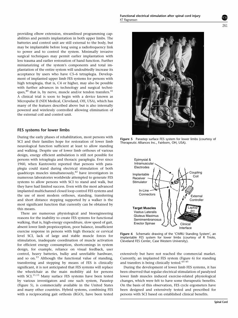
with SCI.11,13 Many surface FES systems have been tested
by various investigators and one such system, Parastep
(Figure 5), is commercially available in the United States
and many other countries. Hybrid systems, combining FES
with a reciprocating gait orthosis (RGO), have been tested
extensively but have not reached the commercial market.
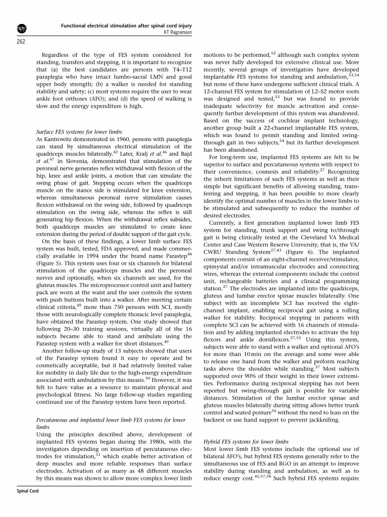
Currently, an implanted FES system (Figure 6) for standing
and transfers is being clinically tested.43–45
During the development of lower limb FES systems, it has
been observed that regular electrical stimulation of paralyzed
lower limb muscles induced exercise-related physiological
changes, which were felt to have some therapeutic benefits.
On the basis of this observation, FES cycle ergometers have
been designed and extensively tested and prescribed for
persons with SCI based on established clinical benefits.
Figure 5 Parastep surface FES system for lower limbs (courtesy ofTherapeutic Alliances Inc., Fairborn, OH, USA).
Epimysial &IntramuscularElectrodes
ImplantableReceiverStimulator
In-LineConnectors
CouplingCoil
ExternalController
ClinicalInterface
LaptopPCVastus Lateralis
Gluteus MaximusSemimembranosusErector Spinae
Target Muscles
Figure 6 Schematic drawing of the ‘CWRU Standing System’, animplantable FES system for lower limbs (courtesy of R Triolo,Cleveland FES Center, Case Western University).
Functional electrical stimulation after spinal cord injuryKT Ragnarsson
261
Spinal Cord
Regardless of the type of FES system considered for
standing, transfers and stepping, it is important to recognize
that (a) the best candidates are persons with T4–T12
paraplegia who have intact lumbo-sacral LMN and good
upper body strength; (b) a walker is needed for standing
stability and safety; (c) most systems require the user to wear
ankle foot orthoses (AFO); and (d) the speed of walking is
slow and the energy expenditure is high.
Surface FES systems for lower limbs
As Kantrowitz demonstrated in 1960, persons with paraplegia
can stand by simultaneous electrical stimulation of the
quadriceps muscles bilaterally.42 Later, Kralj et al.46 and Bajd
et al.47 in Slovenia, demonstrated that stimulation of the
peroneal nerve generates reflex withdrawal with flexion of the
hip, knee and ankle joints, a motion that can simulate the
swing phase of gait. Stepping occurs when the quadriceps
muscle on the stance side is stimulated for knee extension,
whereas simultaneous peroneal nerve stimulation causes
flexion withdrawal on the swing side, followed by quadriceps
stimulation on the swing side, whereas the reflex is still
generating hip flexion. When the withdrawal reflex subsides,
both quadriceps muscles are stimulated to create knee
extension during the period of double support of the gait cycle.
On the basis of these findings, a lower limb surface FES
system was built, tested, FDA approved, and made commer-
cially available in 1994 under the brand name Parastep48
(Figure 5). This system uses four or six channels for bilateral
stimulation of the quadriceps muscles and the peroneal
nerves and optionally, when six channels are used, for the
gluteus muscles. The microprocessor control unit and battery
pack are worn at the waist and the user controls the system
with push buttons built into a walker. After meeting certain
clinical criteria,49 more than 750 persons with SCI, mostly
those with neurologically complete thoracic level paraplegia,
have obtained the Parastep system. One study showed that
following 20–30 training sessions, virtually all of the 16
subjects became able to stand and ambulate using the
Parastep system with a walker for short distances.49
Another follow-up study of 13 subjects showed that users
of the Parastep system found it easy to operate and be
cosmetically acceptable, but it had relatively limited value
for mobility in daily life due to the high-energy expenditure
associated with ambulation by this means.50 However, it was
felt to have value as a resource to maintain physical and
psychological fitness. No large follow-up studies regarding
continued use of the Parastep system have been reported.
Percutaneous and implanted lower limb FES systems for lower
limbs
Using the principles described above, development of
implanted FES systems began during the 1980s, with the
investigators depending on insertion of percutaneous elec-
trodes for stimulation,51 which enable better activation of
deep muscles and more reliable responses than surface
electrodes. Activation of as many as 48 different muscles
by this means was shown to allow more complex lower limb
motions to be performed,52 although such complex system
was never fully developed for extensive clinical use. More
recently, several groups of investigators have developed
implantable FES systems for standing and ambulation,53,54
but none of these have undergone sufficient clinical trials. A
12-channel FES system for stimulation of L2–S2 motor roots
was designed and tested,53 but was found to provide
inadequate selectivity for muscle activation and conse-
quently further development of this system was abandoned.
Based on the success of cochlear implant technology,
another group built a 22-channel implantable FES system,
which was found to permit standing and limited swing-
through gait in two subjects,54 but its further development
has been abandoned.
For long-term use, implanted FES systems are felt to be
superior to surface and percutaneous systems with respect to
their convenience, cosmesis and reliability.27 Recognizing
the inherit limitations of such FES systems as well as their
simple but significant benefits of allowing standing, trans-
ferring and stepping, it has been possible to more clearly
identify the optimal number of muscles in the lower limbs to
be stimulated and subsequently to reduce the number of
desired electrodes.
Currently, a first generation implanted lower limb FES
system for standing, trunk support and swing to/through
gait is being clinically tested at the Cleveland VA Medical
Center and Case Western Reserve University, that is, the VA/
CWRU Standing System27,43 (Figure 6). The implanted
components consist of an eight-channel receiver/stimulator,
epimysial and/or intramuscular electrodes and connecting
wires, whereas the external components include the control
unit, rechargeable batteries and a clinical programming
station.27 The electrodes are implanted into the quadriceps,
gluteus and lumbar erector spinae muscles bilaterally. One
subject with an incomplete SCI has received the eight-
channel implant, enabling reciprocal gait using a rolling
walker for stability. Reciprocal stepping in patients with
complete SCI can be achieved with 16 channels of stimula-
tion and by adding implanted electrodes to activate the hip
flexors and ankle dorsiflexors.27,55 Using this system,
subjects were able to stand with a walker and optional AFO’s
for more than 10min on the average and some were able
to release one hand from the walker and perform reaching
tasks above the shoulder while standing.27 Most subjects
supported over 90% of their weight in their lower extremi-
ties. Performance during reciprocal stepping has not been
reported but swing-through gait is possible for variable
distances. Stimulation of the lumbar erector spinae and
gluteus muscles bilaterally during sitting allows better trunk
control and seated posture56 without the need to lean on the
backrest or use hand support to prevent jackknifing.
Hybrid FES systems for lower limbs
Most lower limb FES systems include the optional use of
bilateral AFO’s, but hybrid FES systems generally refer to the
simultaneous use of FES and RGO in an attempt to improve
stability during standing and ambulation, as well as to
reduce energy cost.45,57,58 Such hybrid FES systems require
Functional electrical stimulation after spinal cord injuryKT Ragnarsson
262
Spinal Cord
the user to wear the RGO,59 which is a knee-ankle-foot
orthosis connected to a thoracolumbosacral orthosis by hip
joints and a cable coupling mechanism, whereas four-
channel surface electrical stimulation is delivered to the
rectus femoris muscle for hip flexion and to the hamstring
muscles for hip extension. Studies have shown that hybrid
FES systems moderately decrease energy expenditure com-
pared to ambulation with either FES or RGO alone,45,57,58 but
short stride length and mechanical difficulties with the RGO
present significant clinical drawbacks,27 which have pre-
vented widespread use.
Future directions
The energy cost of paraplegic ambulation at functional speeds
with orthoses, FES systems and combinations thereof is such
that these devices cannot be used practically for long distance
locomotion and therefore are not expected to replace the
wheelchair as a mobility device.60–62 However, lower limb FES
systems can provide relatively effortless standing, permit
overhead reaching with one arm, allow trained individuals
short distance stepping which is useful for transfers and for
locomotion within small spaces and additionally they can
improve sitting balance and posture. Exact identification of
the minimum number of muscles needed for such relatively
simple tasks and the development of a small, totally
implantable FES system (similar to cardiac pacemakers)
capable of generating the desired functions would provide
significant benefits for many persons with SCI.
For persons with neurologically incomplete SCI, combined
use of FES and BWSTT may prove to be more effective in
improving their ambulation skills than other clinical
approaches.63–65 In contrast to ambulation with orthoses,
which generate exclusively upper body exercise, ambulation
with lower limb FES systems generates active contraction of
lower limb muscles which may have significant health
benefits as is discussed below.
FES systems for respiratory muscles
A degree of respiratory insufficiency is present in all persons
with neurologically complete tetraplegia due to the loss of
intercostal muscle innervation, but the diaphragm, which is
innervated by the phrenic nerve, permits ventilatory free
breathing for most. The phrenic nerve consists of nerve
fibers that derive primarily from C3, C4, C5 and even C6
LMN and nerve roots, but most of its nerve supply comes
from the C4 neurological segment. Therefore, virtually all
persons with neurological level at or below C4 will be able to
breathe without the assistance of mechanical ventilation. An
injury to the spinal cord above C4 level will result in
respiratory failure requiring mechanical ventilatory assis-
tance, often permanently. A severe injury exactly at the C4
cord level may destroy the LMN’s for C4 and even for C3 and
C5 LMN as well, but a complete transverse lesion at C1 and
C2 levels may leave those neurons and hence the phrenic
nerves relatively or even completely intact, albeit without
supraspinal control. A clinical condition that spares the
C3, -4 and -5 LMN’s may allow electrical stimulation of the
phrenic nerves for ventilator-free breathing, a technique
generally referred to as electrophrenic respiration (EPR) or
sometimes as phrenic nerve pacing.
Clinical guidelines for choosing candidates for EPR have
been described by Carter et al.,66 but the most important
criteria are to have proven viability of the phrenic nerve,
healthy lungs and airways, good sitting tolerance, strong
desire and motivation, family support and professional
technical knowledge. Because spontaneous recovery of
diaphragmatic function occurs in many persons with high
SCI during the first year after injury, it is generally felt to be
appropriate to wait at least 4–6 months after the SCI before
EPR is considered.
Evaluation of diaphragmatic function usually begins by
establishing the absence of voluntary diaphragmatic motion
during fluoroscopic examination of the chest. This is
followed by electrical stimulation of the cervical portion of
the phrenic nerve, whereas simultaneously monitoring
diaphragmatic responses by electromyography using surface
recording electrodes located between the seventh and ninth
intercostal spaces66 during stimulation. The latency and
conduction time of the phrenic nerve should be recorded to
assess its integrity and a fluoroscopic examination of the
chest should be done to measure the diaphragmatic excur-
sions, which should be 4.5–6 cm bilaterally.66
EPR was first developed for central hypoventilation
syndrome, often referred to as Ondine’s Curse, but through
the pioneering efforts of Glenn et al. it has been used since
the 1970s for persons with SCI and neurological levels of C3
or higher. Currently, at least three EPR systems are commer-
cially available but these are fundamentally similar in
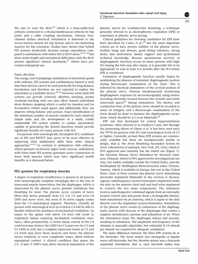
design, that is, the Avery Breathing Pacemaker System by
Avery Laboratories (Commack, New York, NY, USA), which is
FDA approved and currently has the widest use (Figure 7),
the Atrostim Phrenic Nerve Stimulator by Atrotech (Tam-
pere, Finland), which is FDA approved for investigational use
only, but widely available outside the United States, and the
MedImplant by MedImplant Biotechnisches Labor (Vienna,
Austria), which is available in Europe, but not in the United
States. Each of these systems has phrenic nerve stimulating
electrodes implanted bilaterally in the cervical or thoracic
regions, radiofrequency receiver/stimulator implanted under
the skin on the anterior chest wall and lead wires implanted
to connect the two main components. The stimulator
receives radiofrequency command signals from an externally
located control unit and power from a rechargeable battery,
both transmitted via an antenna, which is taped to the skin
directly over the implanted receiver/stimulator. Stimulation
of the phrenic nerve results in contraction of the diaphrag-
matic muscle with descent of the diaphragm that creates a
negative intrathoracic pressure and inhalation of air. When
the stimulation stops, the diaphragm relaxes and ascends,
resulting in exhalation. The amplitude and frequency of the
stimulus is manually adjustable, but ordinarily 8–14 stimuli
per minute are required for adequate ventilation.
The main differences between the three EPR systems lie in
the electrodes. The Avery system uses monopolar or bipolar
nerve cuff electrodes, but the Atrostim system uses a four-pole
sequential stimulation, that is, each electrode makes four
Functional electrical stimulation after spinal cord injuryKT Ragnarsson
263
Spinal Cord
evenly spaced contacts with the phrenic nerve. These contacts
are stimulated sequentially, apparently activating different
parts of the nerve (axons) and diaphragmatic muscle, which
may delay diaphragmatic muscle fatigue.67,68 The MedImplant
system uses a so-called ‘Carousel’ stimulation where four
electrodes are sutured to the epineurium of each phrenic nerve
and connected to a single eight-channel receiver/stimulator.
One of the four electrodes is used to stimulate each nerve
sequentially, also aiming to reduce muscle fatigue.11,68
Surgical implantation of EPR systems may be done by a
cervical or preferably by thoracic approaches.68,69 Although
the cervical approach is less invasive as it avoids the need for
thoracotomy, an electrical stimulus of the phrenic nerve in
the neck may result in submaximal response or cause pain by
stimulation of other nerves in the neck. The phrenic nerve in
the thorax is usually accessed by incisions through the
second or third intercostal spaces and the electrodes are
secured behind or over the nerve. The receiver/stimulator is
implanted subcutaneously through a second incision, which
is usually made in the lower anterolateral chest wall and
connected to the electrodes by wires placed subcutaneously.
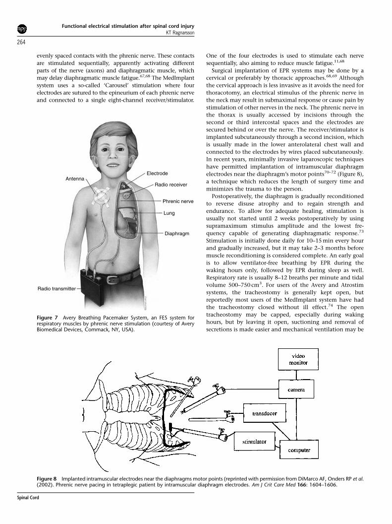
In recent years, minimally invasive laparoscopic techniques
have permitted implantation of intramuscular diaphragm
electrodes near the diaphragm’s motor points70–72 (Figure 8),
a technique which reduces the length of surgery time and
minimizes the trauma to the person.
Postoperatively, the diaphragm is gradually reconditioned
to reverse disuse atrophy and to regain strength and
endurance. To allow for adequate healing, stimulation is
usually not started until 2 weeks postoperatively by using
supramaximum stimulus amplitude and the lowest fre-
quency capable of generating diaphragmatic response.73
Stimulation is initially done daily for 10–15min every hour
and gradually increased, but it may take 2–3 months before
muscle reconditioning is considered complete. An early goal
is to allow ventilator-free breathing by EPR during the
waking hours only, followed by EPR during sleep as well.
Respiratory rate is usually 8–12 breaths per minute and tidal
volume 500–750 cm3. For users of the Avery and Atrostim
systems, the tracheostomy is generally kept open, but
reportedly most users of the MedImplant system have had
the tracheostomy closed without ill effect.74 The open
tracheostomy may be capped, especially during waking
hours, but by leaving it open, suctioning and removal of
secretions is made easier and mechanical ventilation may be
Figure 8 Implanted intramuscular electrodes near the diaphragms motor points (reprinted with permission from DiMarco AF, Onders RP et al.(2002). Phrenic nerve pacing in tetraplegic patient by intramuscular diaphragm electrodes. Am J Crit Care Med 166: 1604–1606.
AntennaElectrode
Radio receiver
Phrenic nerve
Lung
Diaphragm
Radio transmitter
Figure 7 Avery Breathing Pacemaker System, an FES system forrespiratory muscles by phrenic nerve stimulation (courtesy of AveryBiomedical Devices, Commack, NY, USA).
Functional electrical stimulation after spinal cord injuryKT Ragnarsson
264
Spinal Cord
restarted when medically necessary. Throughout the con-
ditioning phase and whenever the user experiences difficulty
breathing as well as periodically during follow-up, the EPR
function should be monitored by assessing tidal volumes
and blood gasses, as well as diaphragmatic excursions.75,76
EPR is generally felt to be safe and effective and its users
describe multiple advantages over mechanical ventilation,
for example, greater comfort and sense of well-being,
improved breathing and speech and better cosmesis.77,78 In
general, the incidence of complications and EPR system
failures is low but all users should have immediate access to
mechanical ventilation or other means of ventilatory
support when respiratory emergencies arise. No recent
longitudinal follow-up studies are available to describe the
successes and failures of EPR, but anecdotal reports describe
damage to the phrenic nerve due to direct mechanical
trauma, compromise of nerve blood supply and fibrosis
around the electrode.73,77,79,80 The incidence of nerve injury
may be reduced by use of monopolar electrodes or perhaps
by sequential stimulation provided by use of the Atrotech
and MedImplants systems. Failure of EPR systems may be
related to dysfunction of batteries, antennas, receiver/
stimulator, wires or electrodes, all of which need to be
evaluated systematically when failures occur. As with all
implanted electronic systems, magnetic resonance imaging
(MRI), shock wave lithotripsy and various types of diather-
my, which generate strong radiofrequency fields, are contra-
indicated. Respiratory complications do not appear to be any
more common than among persons requiring mechanical
ventilation, provided that patients are properly selected,
stimulation parameters are carefully determined and con-
tinuous monitoring is available. Upper airway obstruction,
usually by accumulation of airway secretions, requires
prompt suctioning, optimally via open tracheostomy.
Furthermore, tendency for negative inspiratory pressure
and upper airway collapse during sleep is alleviated by
maintaining an open tracheostomy, although during waking
hours, capping of the tracheostomy is appropriate and safe.
Future directions
Compared to mechanical ventilation, EPR offers an attractive
alternative that may increase the physical and emotional
comfort, mobility, communication, cosmesis and social
integration of persons with tetraplegia and respiratory
insufficiency. Unfortunately, relatively few individuals with
tetraplegia use this method of ventilation, as mechanical
ventilation continues to be the standard of care. A possible
underutilization of EPR may be related to the high cost of
surgical implantation and lengthy reconditioning phase, as
well as medical factors such as damage of the phrenic nerve.
Potential candidates for EPR may be ignorant of its benefits
or reluctant to have surgical implantation done many
months or even years after completing their inpatient
rehabilitation. Additionally, EPR systems have certain short-
comings. As noted above, stimulation of the phrenic nerve
causes a contraction of the diaphragmatic muscle, resulting
in inhalation only, but exhalation is passive. Therefore, EPR
does not produce an effective cough which would be
generated by a combined and synchronized contraction of
the abdominal and intercostal muscles, a function that none
of the available EPR system provides. Furthermore, contrac-
tion of the diaphragm by EPR is independent of upper airway
muscle activation, which places the user at a risk of upper
airway obstruction during sleep. Owing to these two factors,
EPR users need to have an open tracheostomy to secure safe
breathing during sleep and to permit suction of secretions.
It may be argued that more persons with tetraplegia who
are ventilatory dependent could become users of EPR, if
damaged phrenic nerves could be repaired by nerve grafting
techniques,81 if cost of implantation could be reduced by
placing electrodes less invasively and less costly through
laparoscopic surgery,72 and if implantations of electrodes
were performed laparoscopically earlier after SCI than is
currently done. Early implantation of electrodes and regular
stimulation may reduce the need for lengthy reconditioning
of the diaphragmatic muscle which might facilitate weaning
off the ventilator, if phrenic nerve function returns, a
development which would permit removal of the electrodes
and the entire EPR system.
In recent years, it has been shown that accurate mapping of
the phrenic nerve motor points permits successful laparo-
scopic implantation of intramuscular electrodes within the
costal portion of each hemi-diaphragm.51,73,71,72 Such proce-
dure involves placement of four laparoscopic ports to the
abdominal cavity for creation of a pneumoperitoeum, visua-
lization of the diaphragm, diaphragmatic mapping by elec-
trical stimulation and implantation of two electrodes at the
motor points in each hemi-diaphragm.73 The electrode wires
are tunneled subcutaneously to the chest wall where they exit
and connect to an external stimulator. Such system is felt to be
low risk, cost-effective and implantable during and outpatient
visit.71 Early implantation, even within weeks of acute SCI, of
all system components is ultimately envisioned.71
DiMarco et al.73 have recently described in detail short-
comings of currently used EPR systems and outlined needs
for their further development. In most cases, viability of only
one phrenic nerve is not sufficient to allow breathing
exclusively by EPR only. A development of an intercostal
muscle FES system, which could be used simultaneously with
a unilateral EPR, may generate effective ventilation and
eliminate the need for mechanical ventilation. There also is
need to develop an FES system for abdominal muscles that
upon stimulation would generate a more forceful active
expiration and cough.82–85 There is need to synchronize the
upper airway muscle activation with diaphragmatic activa-
tion to reduce the risk of upper airway obstruction during
sleep. This might reduce the need for maintaining an open
tracheostomy. There is need to develop a totally implantable
EPR system, similar to cardiac pacemakers, with elimination
of all the external hardware, for example, stimulator/
receiver, antenna and battery power source. This would
make EPR systems more attractive to potential users. Muscle
fatigue continues to be prevalent among some users of EPR.
It is possible that sequential stimulation, such as is currently
provided by Atrostim and MedImplant Systems will reduce
development of such fatigue.
Functional electrical stimulation after spinal cord injuryKT Ragnarsson
265
Spinal Cord
FES systems for bladder, bowel and sexual function
Contraction of the bladder by electrical stimulation of the
pelvic nerves was reported as early as the mid-nineteenth
century.86 This observation led to attempts to improve
micturition in persons with SCI by stimulation of the conus
medullaris, sacral nerve roots, pelvic nerves or even the
bladder itself, usually with unreliable results. Anatomical
studies revealed that the sacral nerve roots originating in the
conus medullaris provide somatic motor axons to the
external urethral and anal sphincters through the prudendal
nerves and also provide parasympathetic pre-ganglionic
efferent axons to the smooth muscles of the bladder, rectum,
sphincters and erectile tissue through the pelvic splanchnic
nerves. Sensory fibers from these organs also enter the conus
through the sacral nerve roots. During the 1970s, Brindley
reported the implantation of the first sacral anterior root
stimulator (SARS) for bladder control, which were initially
done in animals, but subsequently also in humans.87,88
Refinement of this approach led to the development of the
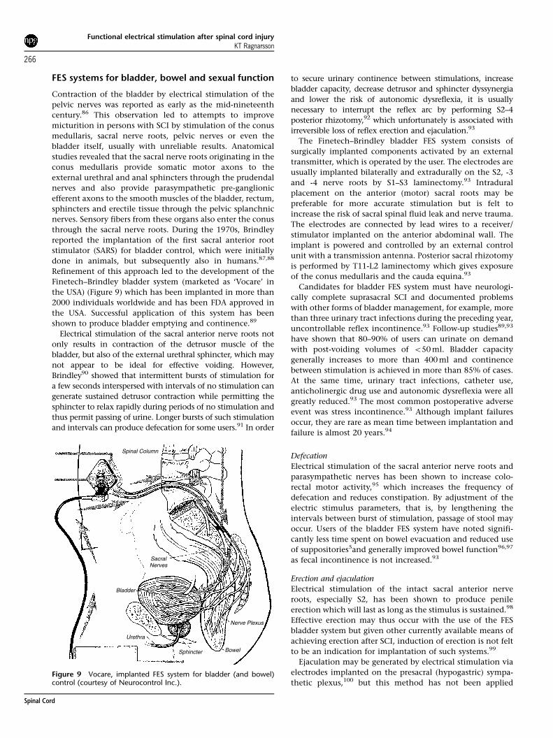
Finetech–Brindley bladder system (marketed as ‘Vocare’ in
the USA) (Figure 9) which has been implanted in more than
2000 individuals worldwide and has been FDA approved in
the USA. Successful application of this system has been
shown to produce bladder emptying and continence.89
Electrical stimulation of the sacral anterior nerve roots not
only results in contraction of the detrusor muscle of the
bladder, but also of the external urethral sphincter, which may
not appear to be ideal for effective voiding. However,
Brindley90 showed that intermittent bursts of stimulation for
a few seconds interspersed with intervals of no stimulation can
generate sustained detrusor contraction while permitting the
sphincter to relax rapidly during periods of no stimulation and
thus permit passing of urine. Longer bursts of such stimulation
and intervals can produce defecation for some users.91 In order
to secure urinary continence between stimulations, increase
bladder capacity, decrease detrusor and sphincter dyssynergia
and lower the risk of autonomic dysreflexia, it is usually
necessary to interrupt the reflex arc by performing S2–4
posterior rhizotomy,92 which unfortunately is associated with
irreversible loss of reflex erection and ejaculation.93
The Finetech–Brindley bladder FES system consists of
surgically implanted components activated by an external
transmitter, which is operated by the user. The electrodes are
usually implanted bilaterally and extradurally on the S2, -3
and -4 nerve roots by S1–S3 laminectomy.93 Intradural
placement on the anterior (motor) sacral roots may be
preferable for more accurate stimulation but is felt to
increase the risk of sacral spinal fluid leak and nerve trauma.
The electrodes are connected by lead wires to a receiver/
stimulator implanted on the anterior abdominal wall. The
implant is powered and controlled by an external control
unit with a transmission antenna. Posterior sacral rhizotomy
is performed by T11-L2 laminectomy which gives exposure
of the conus medullaris and the cauda equina.93
Candidates for bladder FES system must have neurologi-
cally complete suprasacral SCI and documented problems
with other forms of bladder management, for example, more
than three urinary tract infections during the preceding year,
uncontrollable reflex incontinence.93 Follow-up studies89,93
have shown that 80–90% of users can urinate on demand
with post-voiding volumes of o50ml. Bladder capacity
generally increases to more than 400ml and continence
between stimulation is achieved in more than 85% of cases.
At the same time, urinary tract infections, catheter use,
anticholinergic drug use and autonomic dysreflexia were all
greatly reduced.93 The most common postoperative adverse
event was stress incontinence.93 Although implant failures
occur, they are rare as mean time between implantation and
failure is almost 20 years.94
Defecation
Electrical stimulation of the sacral anterior nerve roots and
parasympathetic nerves has been shown to increase colo-
rectal motor activity,95 which increases the frequency of
defecation and reduces constipation. By adjustment of the
electric stimulus parameters, that is, by lengthening the
intervals between burst of stimulation, passage of stool may
occur. Users of the bladder FES system have noted signifi-
cantly less time spent on bowel evacuation and reduced use
of suppositories5and generally improved bowel function96,97
as fecal incontinence is not increased.93
Erection and ejaculation
Electrical stimulation of the intact sacral anterior nerve
roots, especially S2, has been shown to produce penile
erection which will last as long as the stimulus is sustained.98
Effective erection may thus occur with the use of the FES
bladder system but given other currently available means of
achieving erection after SCI, induction of erection is not felt
to be an indication for implantation of such systems.99
Ejaculation may be generated by electrical stimulation via
electrodes implanted on the presacral (hypogastric) sympa-
thetic plexus,100 but this method has not been applied
Spinal Column
SacralNerves
Bladder
Urethra
Sphincter Bowel
Nerve Plexus
Figure 9 Vocare, implanted FES system for bladder (and bowel)control (courtesy of Neurocontrol Inc.).
Functional electrical stimulation after spinal cord injuryKT Ragnarsson
266
Spinal Cord
clinically, as semen can usually be obtained more easily by
electro-ejaculation using a temporarily inserted rectal probe.99
Future directions
The need for posterior sacral rhizotomy resulting in loss of
reflex erection has been the greatest clinical drawback of the
bladder FES systems. It has been demonstrated that stimula-
tion of afferent nerve fibers within the pudendal nerve or
sacral nerve roots101,102 can result in relaxation of the
detrusor muscle, a phenomenon referred to as neuromodu-
lation, that is, the influence of activity in one neural
pathway affects the pre-existing activity in another by
synaptic interaction.102 It has been shown that neuromodu-
lation can be used in combination with bladder FES systems.
Here, simultaneous stimulation of the sacral posterior and
anterior nerve roots can suppress detrusor hyperreflexia and
increase bladder capacity.103 Although there was incomplete
bladder emptying in the five subjects in the study, such
techniques utilizing natural inhibitory reflexes may ulti-
mately eliminate the need for posterior rhizotomy. It has also
been suggested that a fast acting and quickly reversible
neural block can be created by electrical techniques that
provide unidirectional propagation of electrical signals or by
use of high-frequency alternating currents.13,104
Therapeutic effects of FES
SCI adversely affects the physiological functions of most
organ systems.18 Most of these changes are directly related to
the loss of supraspinal control over voluntary and auto-
nomic functions, but they are compounded by the sedentary
lifestyle enforced by the disability and the lack of appropriate
exercise programs for persons with SCI. Table 1 shows some
of the clinical conditions associated with SCI sedentary
lifestyle and lack of physical exercise, conditions which may
be partly reversible by FES and other forms of exercise, for
example, BWSTT and wheelchair sports.
These conditions will be briefly discussed with respect to
their occurrence in persons with SCI and observed ther-
apeutic effects of FES-induced exercise. In general, the effects
of physical exercise on these conditions is directly related to
the size of the muscle mass involved and the duration of the
activity, that is, lower limb exercise is more effective than
upper limb exercise, and sustained activity is more effective
than brief spurts. Although ambulation by FES would likely
have a therapeutic effect, it is not easily achieved or available
to most persons with SCI and has not been studied much in
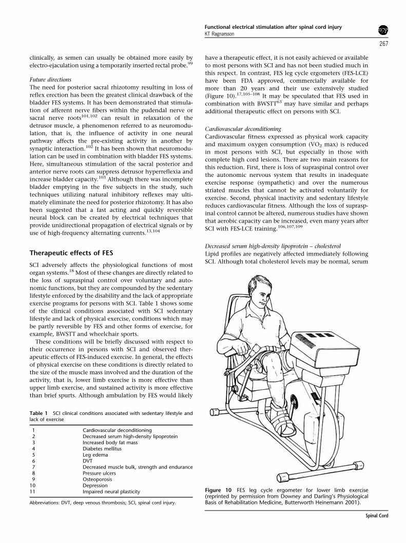
this respect. In contrast, FES leg cycle ergometers (FES-LCE)
have been FDA approved, commercially available for
more than 20 years and their use extensively studied
(Figure 10).17,105–108 It may be speculated that FES used in
combination with BWSTT63 may have similar and perhaps
additional therapeutic effect on persons with SCI.
Cardiovascular deconditioning
Cardiovascular fitness expressed as physical work capacity
and maximum oxygen consumption (VO2 max) is reduced
in most persons with SCI, but especially in those with
complete high cord lesions. There are two main reasons for
this reduction. First, there is loss of supraspinal control over
the autonomic nervous system that results in inadequate
exercise response (sympathetic) and over the numerous
striated muscles that cannot be activated voluntarily for
exercise. Second, physical inactivity and sedentary lifestyle
reduces cardiovascular fitness. Although the loss of suprasp-
inal control cannot be altered, numerous studies have shown
that aerobic capacity can be increased, even many years after
SCI with FES-LCE training.106,107,109
Decreased serum high-density lipoprotein – cholesterol
Lipid profiles are negatively affected immediately following
SCI. Although total cholesterol levels may be normal, serum
Table 1 SCI clinical conditions associated with sedentary lifestyle andlack of exercise
1 Cardiovascular deconditioning2 Decreased serum high-density lipoprotein3 Increased body fat mass4 Diabetes mellitus5 Leg edema6 DVT7 Decreased muscle bulk, strength and endurance8 Pressure ulcers9 Osteoporosis
10 Depression11 Impaired neural plasticity
Abbreviations: DVT, deep venous thrombosis; SCI, spinal cord injury.
Figure 10 FES leg cycle ergometer for lower limb exercise(reprinted by permission from Downey and Darling’s PhysiologicalBasis of Rehabilitation Medicine, Butterworth Heinemann 2001).
Functional electrical stimulation after spinal cord injuryKT Ragnarsson
267
Spinal Cord
high-density lipoprotein cholesterol (HDL-C) is significantly
lower in persons with SCI than in the able-bodied popula-
tion,110–113 a finding that has been felt to be related to low
levels of physical activity. Among persons with SCI, serum
HDL-C has been found to be higher in women than in men,
and in wheelchair athletes than in persons with SCI who are
physically inactive.110 Serum HDL-C has been noted to rise
modestly during the first 2 years after SCI113 and in general,
serum HDL-C levels have been shown to rise in persons with
or without SCI with increased physical activity, increased
aerobic capacity and by participation in sports activ-
ities.110,111,114 Persons with motor complete SCI tend to
have lower HDL-C than those with incomplete SCI.115
Currently, it is not known if the physical exercise associated
with FES-LCE training is sufficient to raise serum HDL-C in
persons with SCI but it has been shown that both upper limb
exercise training for endurance and strength as well as
dietary intervention affects lipid profiles favorably.116,117
Increased body fat mass
Being overweight or obese is common among persons with
SCI,118 even more so than in the general population. Even
when body mass index, that is, body weight as a function of
height, is near normal, body fat mass is increased in persons
with SCI,118–120 that is, persons with SCI who do not appear
to be obese, carry relatively large amounts of fat tissue.119
Although obesity is associated with increased risk of sleep
apnea and lower functional skills, both obesity and increased
body fat mass are associated with increased risk of diabetes
mellitus, hypertension, cardiovascular disease, and so on.
Increased body fat mass and obesity are related to physical
inactivity and diet. It is not known if physical exercise can
alter the body weight of persons with SCI, but it has
been reported that FES-LCE training results in less fat
infiltration between exercised muscle fibers as evident by
imaging studies.121
Diabetes mellitus
Physical inactivity and obesity are recognized as major risk
factors for diabetes mellitus,122 which in turn is a risk factor
for cardiovascular disease. Increased prevalence of glucose
intolerance and diabetes among persons with SCI has been
well documented,123–125 which is not surprising given their
sedentary lifestyle and increased body fat mass. It has been
shown that peak serum glucose in persons with SCI is
associated with increased total body percentage fat, com-
plete tetraplegia, older age and male gender.125 Physical
activity, especially regular aerobic endurance exercise, has
been shown to reduce the incidence and prevalence of
diabetes in the general population122,126 and among persons
with SCI, it has been shown that following 8 weeks of FES-
LCE training, blood glucose levels were significantly reduced
and glucose utilization improved.127
Leg edema
Peripheral edema due to leg dependency, lack of muscle
activity and poor venous return is clinically apparent in
many persons with SCI. Such edema is generally felt to be
harmless, although usually of considerable concern to
the person affected. Leg edema as well as acrocyanosis of
the feet have been reported to improve during FES-LCE
training.128,129
Deep vein thrombosis
Venus thromboembolic disease is common and a leading
cause of mortality following acute SCI. Predisposing factors
include, venous stasis due to paralysis of leg muscles
associated with failure of the venous muscle pump as well
as changes in the clotting mechanism.130 Simple electrical
stimulation of leg muscles in combination with low dose
heparin therapy has been shown to be superior to using only
heparin to prevent deep vein thrombosis (DVT) in acute
SCI.131 Although DVT is relatively rare during the chronic
stage of SCI, it may be speculated that regular leg exercise by
FES-LCE might be helpful in preventing DVT, but no studies
have been published that have addressed this issue.
Decreased muscle bulk, strength and endurance
Following SCI, the paralyzed muscles develop atrophy, even
when spastic, and lose strength in response to electrical
stimulation as well as ability to forcefully contract repeti-
tively. In response to FES-LCE training, subjects with SCI
have been shown to increase their thigh circumference as
measured clinically by tape17 or as visualized on CT or MRI
imaging studies,17,121,132 increase the resistance while pedal-
ing the FES-LCE17,105,106,133 and all gained endurance, that
is, they could cycle for longer periods of time.17,105,133
Muscle biopsy studies have shown a switch from myosin
heavy chain (MHC) isoform II B muscle fibers (fast twitch,
fast fatiguable and glycolytic fibers) to MHC isoform II fibers
(fast twitch and fatigue resistant), with such exercise, which
is accompanied by doubling of the mitochondrial oxidative
capacity.17 These studies have confirmed the reversibility of
disuse changes in muscles paralyzed by spinal cord UMN
lesions.
Pressure ulcers
It is well known that persons with SCI are at a high risk and
frequently develop pressure ulcers at any time during their
lives. Pulsed electrical stimulation has been shown to
facilitate healing of stage II, III and IV pressure ulcers134,135
and is recommended for that purpose by the Agency for
Healthcare Research and Quality.136 Electric muscle stimula-
tion for prevention of pressure ulcers has been suggested
based on research which showed that such stimulation of the
gluteus maximus muscle produces increased muscle blood
flow and undulation of the buttock tissues, that is muscle
contraction and relaxation, with shape refiguration,137,138
effects which are in addition to the well-documented and
potentially protective increase in muscles bulk with such
intervention. It is not known how effective FES-LCE is in
prevention of pressure ulcers but none of the published
reports on this intervention has reported development of
pressure ulcers during the training period. An anecdotal
report describes the healing of pressure ulcer during FES-LCE
when traditional wound care seemed ineffective.139
Functional electrical stimulation after spinal cord injuryKT Ragnarsson
268
Spinal Cord
Osteoporosis
Immediately following SCI, bone resorption occurs at a faster
rate than bone formation, a process that results in clinical
osteoporosis within a few weeks of injury.140,141 Normal
bone remodeling rate, that is, resorption equals formation, is
re-established in 1–2 years after SCI, but by then loss of
approximately one-third of the original bone mass has
occurred.140 This bone loss remains constant throughout
the life of the individual and is associated with increased risk
of fractures. No effective intervention to prevent or to restore
bone loss after SCI is currently known. However, as the cause
of such bone loss after SCI is felt to be related to the paralysis
and disuse, it has been speculated that reversal of bone
loss may occur with weight-bearing activities142,143 and with
FES-LCE training. Most studies have failed to show any
significant increase in bone mineral density with FES-LCE
training144,145 or by lower limb FES ambulation (Parastep),146
whereas other investigators have shown that FES-LCE can
restore tibial bone mass by 10–30%.147,148
Depression
Mood disorders, such as depression and anxiety, are
common after SCI and are present in as many as 30% for 2
or more years after injury.149 Risk factors are felt to be similar
to those in able-bodied persons, as well as the extent of the
neurological impairment and the presence of medical
comorbidities.150 It has been well documented that there
may be an inverse association between physical inactivity
and mental health in both men and women151 and that
physical exercise has significant antidepressive effect152–155
and may even improve cognitive function. It is known that
SCI disturbs the hypothalamic–pituitary–adrenal axis,156,157
which affects production of various hormones, including
neuroendocrine stress hormones, for example, cortisol,
growth hormone, endorphins and norepinephrine. After
SCI, production of ACTH and cortisol does not follow
normal circadian rhythm156 and endorphin levels have been
shown to be low,158 both findings that may be associated
with depression. One study on persons with SCI showed that
with regular FES-LCE endorphin production increased, base-
line cortisol levels became better regulated and closer to
normal circadian rhythmicity and depression scores im-
proved,158 whereas another study showed that majority of
participants reported improvement in self image and
perceived appearance.128
Impaired neural plasticity
CNS plasticity refers to the CNS ability to change its structure
and function in response to environmental stimuli, such as
physical exercise. Undamaged spinal cord circuits have been
shown to be capable of significant reorganization which may
be induced by both activity-dependent and injury-induced
plasticity.159 Both clinical and animal studies have shown
that physical exercise is beneficial for neuronal func-
tion160,161 as clinically evident by better cognitive func-
tion.160,162 Animal studies have found that exercise elevates
the levels of neurotrophic factors in select regions of the
adult brain and spinal cord,9 including levels of insulin-like
growth factor,163 fibroblast growth factor II164 and BDNF.165
Several clinical investigators have shown that in persons
with incomplete SCI walking ability may be improved with
FES-LCE training,166–168 BWSTT169–171 or combinations
thereof63 or overground mobility therapy.172 Other investi-
gators have suggested that patterned neuronal activity with
FES and/or BWSTT is important for development and even
recovery of neurological functions8 or by using both BWSTT
and epidural spinal cord stimulation.7,173 In general, it is
now felt that the functional motor benefits of such exercise
training does more than improve muscle strength and
cardiovascular fitness in persons with SCI as animal studies
have shown that it may also have an influence on synapses
and motor neurons.174 If and when a treatment will be found
to reverse the neurological loss after SCI, it is unlikely that
such treatment will result in complete reestablishment of all
the descending and ascending projections within the spinal
cord. Therefore, it will probably be necessary to activate the
CNS plasticity, including neural circuitry within the spinal
cord to maximize locomotor recovery through exercise
training, such as FES and BWSTT.
The future for FES
The earliest observations of the use of FES to overcome
paralysis were published almost 50 years ago.25,42,175 Since
that time, the spectacular technological advances in electro-
nics and microprocessing have enabled scientists to develop
a variety of FES systems that have been shown to have
significant clinical benefits for their users. However, com-
mercial success has been limited for several reasons, includ-
ing the relatively small market, high cost of FES systems and
their implantation, reluctant third-party payors, technologi-
cal shortcomings, lack of totally implantable FES systems,
insufficient clinical trials to scientifically document benefits,
including reduced cost of care.
Several of these concerns are currently being addressed by
scientists in the field of FES11 who plan to use state-of-the-art
electronics to miniaturize all components of FES systems and
to create long-lasting implanted miniaturized batteries that
can be recharged transcutaneously by radiofrequency trans-
mission. Investigators are addressing the imminent need to
develop a totally implantable, easily manufactured, modular
FES system that can be used for all purposes, for example, for
upper and lower limbs, trunk, bladder, bowel and diaphrag-
matic functions. Such system will be upgradeable to provide
additional and advanced functions by adding different
components, for example, stimulator and sensor modules.
Implantation early after SCI using less-invasive surgical
techniques would make such FES systems a clinical option
during acute rehabilitation. Reconstructive hand surgery in
combination with implantation of FES system may enable
more persons with tetraplegia to achieve greater hand
function. Judicious application of orthoses similarly may
enhance functional performance of many users of FES
systems.
More futuristic scientific developments include the poten-
tial use of high-frequency alternating currents to create a
Functional electrical stimulation after spinal cord injuryKT Ragnarsson
269
Spinal Cord
quickly reversible nerve conduction block,104 which may
eliminate the need for posterior rhizotomy, which is often
needed for bladder FES systems, as well as to suppress
contractions of a hyperactive bladder, control pain, and so
on. Such conduction block appears to be more achievable in
nerve fibers of large diameter, for example motor fibers,
than those of small diameter, for example sensory fibers. If
electrical stimulation could be used to both activate and
inhibit nerve and muscle activity, the clinical benefits of FES
systems could be significantly increased.11
Advanced methods to control FES systems have been
proposed,11 and in some cases implemented, including the
use of myoelectric signals from voluntary muscle contrac-
tions and sophisticated closed loop systems that automati-
cally adjust stimulation patterns based on feedback from
peripherally placed sensors.176 It has been shown that
epidural spinal cord stimulation in conjunction with BWSTT
can facilitate functional walking in persons with chronic SCI
ASIA C, perhaps by eliciting greater activation of the
oxidative motor unit pool.7,173 Other investigators have
shown in animal experiments that stimulation of motor
neuron pools within the gray matter of the spinal cord by
implanted microelectrode arrays can activate large number
of individual muscle fibers in a relatively coordinated
pattern.177,178 These observations suggest that it may be
possible to activate by electrical stimulation the natural
neural circuitry in the spinal cord to generate functional
muscle activity.11
Perhaps the greatest attention in recent years has been
paid to research which aims to enable persons with
tetraplegia to control their environment by thought.179–181
Recently, investigators have shown that a persons with C4
ASIA A tetraplegia and a 96 microelectrode array surgically
implanted on the surface of the primary motor center area of
the brain was able to modulate cortical spiking patterns by
intended hand motion and use such neural control to open
simulated e-mail, operate devices such as television, open
and close a prosthetic hand, and perform rudimentary
actions with multi-jointed robotic arm.182 Such thought
created brain signals may ultimately allow persons with SCI
to create reliable commands and control FES systems by
thought, rather than by the current methods which rely on
external joint angle sensors, accelerometers and switches
operated by voluntary muscle power.11
Conclusions
At present, creative transfer of the vast available technology
in microelectronics to benefit persons with SCI appears to be
a swifter way to enhance their function, quality of life, and
community reintegration, than by waiting for science to find
methods to regenerate axons within the injured spinal cord.
With relatively limited research funding, development of FES
systems has rapidly advanced by using recent technological
developments, particularly those of advanced materials,
implantable rechargeable batteries, wireless communication
and miniaturization of electronics. These systems have been
shown to be of great clinical value to several select groups of
persons with SCI. People with C5–6 tetraplegia have become
able to open their hands, grasp, hold and release objects.
Those with T1–T12 paraplegia have become able to stand,
step and adjust their body positions. Ventilatory dependent
persons with tetraplegia at C3 and above have achieved
ventilatory free breathing and persons with neurogenic
bladder and intact S2–S4 LMN have been able to maintain
urinary continence and effectively evacuate the bladder and
even the bowel. Regular active physical exercise of the
paralyzed lower limbs by FES-LCE has been shown to have
numerous health-related benefits. Development of minia-
ture modular FES systems, totally implantable by minimally
invasive surgical techniques, are likely to simplify the
application of this clinical process and may ultimately make
FES technology a standard of care for many persons with SCI,
reduce their total cost of care and enhance their quality of
life. Enlarging the market for FES systems beyond SCI is
likely to drive down prices that in combination with reduced
cost of care may encourage third-party payors to more
readily reimburse its cost.
Acknowledgements
The preparation of this paper was supported in part by the
National Institute on Disability and Rehabilitation Research
US Department of Education (grant #133N060027). The
author is indebted to P Hunter Peckham for review of this
manuscript. The Sir Ludwig Guttmann Lecture at the 46th
Annual Scientific Meeting of the International Spinal Cord
Society and the 10th Annual Scientific Meeting of the Nordic
Spinal Cord Society Reykjavik, Iceland June 27–July 1, 2007.
References
1 Thomas EL. Nursing care of the patient with spinal cord injury.In: Pierce DS, Nickel VH (eds). The Total Care of SpinalCord Injuries. Little Brown and Company: Boston, MA, 1977,pp 249–297.
2 Spinal Cord Injury. Facts and Factors at a Glance, Birmingham,Alabama. National Spinal Cord Injury Statistical Center, Bir-mingham, Alabama, 2006.
3 Fiedler IG, Laud PW, Maiman DJ, Apple DF. Economics ofManaged Care in Spinal Cord Injury. Arch Phys Med Rehabil 1999;80: 1441–1449.
4 DeVivo MJ. Epidemiology of Traumatic Spinal Cord Injury. In:Kirshblum S, Campagnolo DI, DeLisa JA (eds). Spinal CordMedicine. Lippincott, Williams and Wilkins: Philadelphia, 2002,pp. 69–81.
5 Kakulas BA. Neuropathology: the foundation for new treatmentsin spinal cord injury. Spinal Cord 2004; 42: 549–563.
6 Brandt EN, Pope AM (eds). Assessing the role of rehabilitationscience and engineering. Enabling America. National AcademyPress: Washington, DC, 1997.
7 Herman R, He J, D’Luzansky S, Willis W, Dilli S. Spinal cordstimulation facilitates functional walking in a chronic incom-plete spinal cord injured. Spinal Cord 2002; 40: 65–68.
8 McDonald JW, Becker D, Sadowsky CL, Jane JA, Conturo TE,Schultz LM. Late recovery following spinal cord injury: casereport and review of the literature. J Neurosurg (Spine 2) 2002; 97:252–265.
9 Vaynman S, Gomez-Pinella F. License to run: exercise impactsfunctional plasticity in the intact and injured central nervoussystem by using neurotrophins. Neurorehab Neural Repair 2005;19: 283–295.
Functional electrical stimulation after spinal cord injuryKT Ragnarsson
270
Spinal Cord
10 Peckham PH. Principles of electrical stimulation. Top Spinal CordInj Rehabil 1999; 5: 1–5.
11 Peckham PH, Knutson JS. Functional electrical stimulationfor neuromuscular applications. Ann Rev Med Eng 2005; 7:4.1–4.34.
12 Gorman PH, Mortimer JT. The effect of stimulus parameters onthe recruitment characteristics of direct nerve stimulation. IEEETrans Biomed Eng 1983; 30: 407–414.
13 Peckham PH, Gorman PH. Functional electrical stimulation inthe 21st century. Topics Spinal Cord Inj Rehabil 2004; 10: 126–150.
14 Peckham PH, Mortimer JT, Marsolais EB. Alterations in the forceand fatigability of skeletal muscle in quadriplegic humansfollowing exercise induced by chronic electrical stimulation.Clin Orthop 1976; 114: 326–334.
15 Lieber RL. Comparison between animal and human studies ofskeletal muscle adaptation to chronic stimulation. Clin Orthop1988; 233: 19–24.
16 Salmons S, Henriksson J. The adaptive response of skeletalmuscle to increased use. Muscle Nerve 1981; 4: 94–105.
17 Mohr T, Andersen JL, Biering-Sorensen F, Galbo H, Bangsbo J,Wagner A et al. Long term adaptation to electrically inducedcycle training in severe spinal cord injured individuals. SpinalCord 1997; 35: 1–16.
18 Ragnarsson KT, Baker LL. Functional Electrical Stimulation inpersons with spinal cord injury. In: Gonzalez EG, Myers SJ,Edelstein AE, Lieberman JS, Downey JA (eds). Physiological Basisof Rehabilitation Medicine. Butterworth-Heinemann: Boston,2001, pp. 723–745.
19 Knutson JS, Naples GG, Peckham PF, Keith MW. Electrodefracture rates and occurrences of infection and granulomaassociated with percutaneous intramuscular electrodes in upperlimb functional electrical stimulation applications. J Rehabil ResDevel 2002; 39: 671–684.
20 Kilgore KL, Peckham PH, Keith MW, Montague FW, Hart RL,Gazdik MM et al. Durability of implanted electrodes and leadsin upper limb neuroprosthesis. J Rehabil Res Devel 2003; 40:457–468.
21 Uhlir JP, Triolo RJ, Davis Jr JA, Bieri C. Performance of epimysialstimulating electrodes in the lower extremities of individualswith spinal cord injury. IEEE Trans Syst Rehabil Eng 2004; 12:279–287.
22 Loeb GE, Walmsley B, Duysens J. Obtaining proprioceptiveinformation from natural limbs: implantable transducers vs.somatosensory neuron recordings. In: Newman MR, FlemingDG, Cheung PW, Ko WH (eds). Solid State Physical Sensors forBiomedical Applications. Boca Raton, Florida, CRC, 1980.
23 Petrofsky JS, Phillips CA, Stafford DE. Closed Loop Control forrestoration of movement in paralyzed muscles. Orthopaedics1984; 7: 1289–1302.
24 Bryden AM, Kilgore KL, Kirsch RF, Memberg WD, Peckham PH,Keith MW. An implanted neuroprosthesis for high tetraplegia.Top Spinal Cord Inj Rehabil 2005; 10: 38–52.
25 Long C, Masciarelli VD. An Electophysiologic Splint for theHand. Archives Phys Med Rehabil 1963; 44: 499–503.
26 Alon G, McBride K. Persons with C5 or C6 tetraplegia achieveselected functional gains using a neuroprosthesis. Arch Phys MedRehabil 2003; 84: 119–124.
27 Knutson J, Audu M, Triolo R. Interventions for mobility andmanipulation after spinal cord injury: a review of orthoticand neuroprosthetic options. Top Spinal Cord Inj Rehabil 2006;11: 61–81.
28 Popovic MR, Popovic DB, Keller T. Neuroprosthesis for grasping.Neurol Res 2002; 24: 443–452.
29 Keith MS, Peckham PH, Thrope GB, Stroh KC, Smith B, BurkettJR et al. Implantable functional neuromuscular stimulation inthe tetraplegic hand. J Hand Surg 1989; 14A: 524–530.
30 Peckham PH, Keith MW, Kilgore KL, Grill JH, Wuolle KS, ThropeGB et al. Efficacy of an implanted neuroprosthesis for restoringhand grasp in tetraplegia: a multi-center study. Arch Phys MedRehabil 2001; 82: 1380–1388.
31 Taylor P, Esnouf J, Hobby J. The functional impact of theFreehand system on tetraplegic hand function. Clinical results.Spinal Cord 2002; 40: 560–566.
32 Peckham PH, Kilgore K, Keith MW, Bryden AM, Bradra N,Montague FW. An advanced neuroprosthesis for restoration ofhand and upper arm control using an implantable controller.J Hand Surg 2002; 27A: 265–276.
33 Peckham PH, Keith MW, Freehafer AA. Restoration of functionalcontrol by electrical stimulation in the upper extremity of thequadriplegic patient. J Bone Joint Surg 70A; 144–148: 1988.
34 Keith MW, Peckham PH, Thrope GB, Stroh KC, Smith B, BuckettJR et al. Functional neuromuscular stimulation neuroprosthesisfor the tetraplegic hand. Clin Orthop 1988; 233: 25–33.
35 Mulcahey MJ, Betz RR, Kozin SH, Smith BT, Hutchinson D, LutzC. Implantation of the Freehand system during initial rehabi-litation using minimally invasive techniques. Spinal Cord 2004;42: 146–155.
36 Waters RL, Muccitelli LM. Tendon transfers to improvefunction of patients with tetraplegia. In: Kirshblum S, Campag-nolo DI, DeLisa JA (eds). Spinal Cord Medicine, Chapter28, Lippincott, Williams and Wilkins: Philadelphia, 2002,pp 424–437.
37 Keith MW, Kilgore KL, Peckham PH, Wuolle KS, Creasey G,Lemay M. Tendon transfers and functional electrical stimulationfor restoration of hand function in spinal cord injury. J HandSurg 1996; 21A: 89–99.
38 Memberg WD, Crago PE, Keith MW. Restoration of elbowextension via functional electrical stimulation in individualswith tetraplegia. J Rehabil Res Develop 2003; 40: 477–486.
39 Peckham PH, Kilgore KL, Keith MW, Bryden AM, Bhadra N,Montague FW. An advanced neuroprosthesis for restoration ofhand and upper arm control using an implantable controller.J Hand Surg 2002; 27A: 265–276.
40 Bryden AM, Kilgore KL, Kirsch RF, Memberg WD, Peckham PH,Keith MW. An implanted neuroprosthesis for high tetraplegia.Topics Spinal Cord Inj Rehabil 2005; 10: 38–52.
41 Keith MW, Hoyen H. Indications and future directions for upperlimb neuroprosthesis in tetraplegic patients: a review. Hand Clin2002; 18: 519–528.
42 Kantrowitz A. Electronic Physiologic Aids. Maimonides Hospital:Brooklyn, New York, 1960, pp 4–5.
43 Davis JA, Triolo RJ, Uhlir JP, Bieri C, Rohde L, Lissy D et al.Preliminary performance of a surgically implanted neuroprosth-esis for standing and transfers. J Rehabil Res Devel 2001; 38:609–617.
44 Solomonow M. Biomechanics and physiology of practicalfunction on neuromuscular stimulation powered walkingorthosis for paraplegics. In: Stein RB, Peckham PH, Popovic DP(eds). Neural Prostheses: Replacing Motor Function after Disease orDisability. Oxford University Press: New York, 1992, pp 202–232.
45 Marsolais EB, Kobetic R, Polando G, Ferguson K, Tashman S,Gaudio R et al. The Case Western Reserve University Hybrid GaitOrthosis. J Spinal Cord Med 2000; 23: 100–108.
46 Kralj A, Bajd T, Turk R. Electrical stimulation providingfunctional use of paraplegia patient muscles. Med Prog Technol1980; 7: 3–9.
47 Bajd T, Kralj A, Turk R, Benko H, Sega J. The use of a four-channel electrical stimulator as an ambulatory aid for paraplegicpatients. Phys Ther 1983; 63: 1116–1120.
48 Graupe D, Kohn KH. Functional Electrical Stimulation for Ambula-tion by Paraplegics: 12 Years of Clinical Observations and SystemStudies. Kreger Publishing Company: Malibar, Florida, 1994.
49 Klose KJ, Jacobs PL, Broton JG, Guest RS, Needham-ShropshireRM, Lebwohl N et al. Evaluation of a training program forpersons with SCI paraplegia using the Parastep I ambulationsystem: Part 1. Ambulation performance and anthropometricmeasures. Arch Phys Med Rehabil 1997; 78: 789–793.
50 Brissot R, Gallien P, LeBot MP, Beaubras A, Laisene D, Beillot Jet al. Clinical experience with functional electrical stimulationassisted gait with parastep in spinal cord injured patients. Spine2000; 25: 501–508.
51 Marsolais EB, Kobetic R. Functional electrical stimulation forwalking in paraplegia. J Bone Joint Surg 1987; 69A: 728–733.
52 Kobetic R, Marsolais EB. Synthesis of Paraplegic Gait with multi-channel functional neuromuscular stimulation. IEEE TransRehabil Engin 1994; 2: 66–79.
Functional electrical stimulation after spinal cord injuryKT Ragnarsson
271
Spinal Cord
53 Rushton DN, Donaldson ND, Barr FM, Harper BJ, Perkins TA,Taylor PN et al. Lumbar root stimulation for restoring legfunction: results in paraplegia. Artific Org 1997; 21: 180–182.
54 Davis R, Patrick J, Barriskill A. Development of functionalelectrical stimulators using cochlear implant technology. MedEng Phys 2001; 23: 61–68.
55 Kobetic R, Triolo RJ, Uhlir JP, Bieri C, Wibowo M, Polando Get al. Implanted functional electrical stimulation system formobility in paraplegia: a follow-up case report, IEEE Trans.Rehabil Eng 1999; 7: 390–398.
56 Wilkenfeld AJ, Audu ML, Triolo RJ. Feasibility of functionalelectrical stimulation for control of seated posture afterspinal cord injury: a simulation study. J Rehabil Res Devel 2006;43: 139–152.
57 Solomonow M, Aguilar E, Reisin E, Baratta RV, Best R,Coetzee T et al. Reciprocating gait orthosis powered withelectrical muscle stimulation (RGO II). Part 1: performanceevaluation of 70 paraplegic patients. Orthopaedics 1997; 20:315–324.
58 Solomonow M, Reisin E, Aguilar E, Baratta RV, Best R, Coetzee Tet al. Reciprocal gait orthosis powered with electrical musclestimulation (RGO II). Part 2: medical evaluation of 70 paraplegicpatients. Orthopaedics 1997; 20: 411–418.
59 Douglas R, Larsen PF, D’Ambrosia R, McCall RE. The LSUreciprocating gait orthosis. Orthopaedics 1983; 6: 834–839.
60 Marsolais EB, Edwards BG. Energy costs of walking and standingwith functional neuromuscular stimulation and long leg braces.Arch Phys Med Rehabil 1988; 69: 243–249.
61 Petrofsky JS, Smith JB. Physiologic cost of computer controlledwalking in persons with paraplegia using a reciprocating gaitorthosis. Arch Phys Med Rehabil 1991; 72: 890–896.
62 Nene AV, Patrick JH. Energy cost of paraplegic locomotion usingthe parawalker-electrical stimulation ‘hybrid’ orthosis. Arch PhysMed Rehabil 1990; 71: 116–120.
63 Field-Fote EC. Combined use of body weight support, functionalelectric stimulation and treadmill training to improve walkingability in individuals with chronic incomplete spinal cordinjury. Arch Phys Med Rehabil 2001; 82: 818–824.
64 Hesse S, Werner C, Bardeleben A. Electromechanical gaittraining with functional electrical stimulation: case studies inspinal cord injury. Spinal Cord 2004; 42: 346–352.
65 Hicks AL, Adams MM, Ginis KM, Giangregorio L, Latimer A,Phillips SM et al. Long-term-body-weight-supported treadmilltraining and subsequent follow-up in persons with chronic SCI:effects on functional walking ability and measures of subjectivewell-being. Spinal Cord 2005; 43: 291–298.
66 Carter RE, Menter R, Wood M, Wilmot C, Hall K. Availablerespiratory options. In: Whiteneck G, Adler C, Carter RE et al.(eds). The Management of High Quadriplegia, Chapter 12. DemosPublications: New York, NY, 1989, pp. 166–168.
67 Baer GA, Talonen PP, Hakkinen V, Exner G, Yrjola H. Phrenicnerve stimulation in tetraplegia. A new regiment to conditionthe diaphragm for full time respiration. Scand J Rehabil Med1990; 22: 107–111.
68 Creasey G, Elefteriades J, DiMarco A, Tallinan P, Bijak M, GirschW et al. Electrical stimulation to restore respiration. J Rehabil Resand Devel 1996; 33: 123–132.
69 Chervin RD, Guilleminault C. Diphragm pacing: review andreassessment. Sleep 1994; 17: 176–187.
70 DiMarco AF, Onders RP, Kowalsky KE, Miller ME, Ferek S,Mortimer JT. Phrenic nerve pacing in a tetraplegic patient viaintramuscular diaphragm electrodes. Am J Respira Crit Care Med2002; 166: 1604–1606.
71 Onders RP, DiMarco AF, Ignani AR, Aiyar H, Mortimer JT.Mapping of the phrenic nerve motor point: the key to asuccessful laparoscopic diaphragm pacing system in the firsthuman series. Surgery 2004; 136: 819–826.
72 DiMarco AF, Onders RP, Ignani AR, Kowalsky KE, Mortimer JT.Phrenic nerve pacing via intramuscular diaphragm electrodes intetraplegic subjects. Chest 2005; 127: 671–678.
73 DiMarco AF, Onders RP, Ignani A, Kowalski KE. Inpiratorymuscle pacing in spinal cord injury: case report and clinicalcommentary. J Spinal Cord Med 2006; 29: 95–108.
74 Mayr W, Bijak M, Girsch W, Holle J, Lanmuller H, Thoma H et al.Multichannel stimulation of phrenic nerves by epineuralelectrodes: clinical experience and future developments. TransAm Soc Artific Int Org J 1993; 39: M729–M735.
75 GlennWW, HolcombWG, McLaughlin AJ, O’Hare JM, Hogan JF,Yasuda R. Total ventilatory support in a quadriplegic patientwith radio-frequency electrophrenic respiration. New Engl J Med1972; 286: 513–516.
76 Glenn WW, Holcomb WG, Shaw RK, Hogan JF, Holschuh KR.Long term ventilatory support by diaphragm pacing in quad-riplegia. Ann Surg 1976; 183: 566–577.
77 Glenn WW, Phelps ML, Elefteriades JA, Dentz B, Hogan JF.Twenty years of experience in phrenic nerve stimulation to pacethe diaphragm. Pacing Clin Electrophysiol 1986; 9: 780–784.
78 Dobelle WH, D’Angelo MS, Goetz BF, Kieter DG, Lallier TJ, LambJI et al. Two hundred cases with a new breathing pacemakerdispels myths about diaphragm pacing. Trans Am Soc Artific IntOrg 1994; 40: 244–252.
79 Glenn WW, Brouillette RT, Dentz B, Fodstad H, Hunt CE, KeensTG et al. Fundamental considerations in pacing of thediaphragm for chronic ventilatory insufficiency: a multi-centerstudy. Pacing Clinical Electrophysiology 1988; 11: 2121–2127.
80 Weese-Mayer EE, Silvestri JM, Kenny AS. Diaphragm pacing withquadripolar phrenic nerve electrode: an international study.Pacing Clin Electrophysiol 1996; 19: 1311–1319.
81 Krieger AJ, Gropper MR, Adler RJ. Electrophrenic respirationafter intercostal phrenic nerve anastomosis in a patientwith anterior spinal artery syndrome: technical case report.Neurosurgery 1994; 35: 760–763.
82 Linder SH. Functional electrical stimulation to enhance coughin quadriplegia. Chest 1993; 103: 166–169.
83 Jaeger RJ, Turba RM, Yarkony GM, Roth EJ. Cough in spinal cordinjured patients: comparison of three methods to producecough. Arch Phys Med Rehabil 1993; 74: 1358–1361.
84 DiMarco FR, Romaniuk JR, Supinski GS. Electrical activation ofthe expiratory muscles to restore cough. Am J Respir Crit CareMed 1995; 151: 1466–1471.
85 Lin VWH, Singh H, Chitkara RK, Perkash I. Functional magneticstimulation for restoring cough in patients with tetraplegia. ArchPhys Med Rehabil 1998; 79: 517–522.
86 Budge J. ber Der Einfluss Des Nervensystems Auf Die BewegungDer Blase. Zep Rationelle Medicine 21; 1: 1854.
87 Brindley GS. Electrode-arrays for making long-lasting electricalconnection to spinal roots. J Physiol 1972; 222: 135–136.
88 Brindley GS, Polkey CE, Rushton DN, Cardozo L. Sacral anteriorroot stimulators for bladder control in paraplegia: the first 50cases. J Neurol, Neurosurg Psychiatr 1986; 49: 1104–1114.
89 Brindley GS. The first 500 patients with sacral anteriorstimulator implants: general description. Paraplegia 1994; 32:795–805.
90 Brindley GS, Polkey CE, Rushton DN. Sacral anterior rootsimulators for bladder control in paraplegia. Paraplegia 1982;20: 365–381.
91 Creasey GH, Ho CH, Triolo RJ, Gater DR, DiMarco AF, Bogie KMet al. Clinical applications of electrical stimulation after spinalcord injury. J Spinal Cord Med 2004; 27: 365–375.
92 Madersbacher H, Fischer J. Anterior sacral root stimulation andposterior sacral root rhizotomy. Akta Urologica 1993; 24 (Suppl):32–35.
93 Creasey GH, Grill JH, Korsten M, Hoi Sang U, Betz R, Anderson Ret al. An implantable neuroprosthesis for restoring bladder andbowel control in patients with spinal cord injuries: a multi-center trial. Arch Phys Med Rehabil 2001; 82: 1512–1519.
94 Brindley GS. The first 500 sacral anterior root stimulators:implant failures and their repair. Paraplegia 1995; 33: 5–9.
95 Varma JS, Binnie N, Smith AN, Creasey GH, Edmond P.Differential effects of sacral anterior root stimulation on analsphincter and colorectal mortility in spinally injured man. Br JSurg 1986; 73: 478–482.
96 MacDonagh RP, Sun WM, Smallwood R, Forster D, Read NW.Control of defecation in patients with spinal injuries bystimulation of sacral anterior nerve roots. B MJ 1990; 300:1494–1497.
Functional electrical stimulation after spinal cord injuryKT Ragnarsson
272
Spinal Cord
97 Binnie N, Smith A, Creasey GH, Edmond P. The effects ofelectrical anterior sacral nerve root stimulation on pelvicfloor function in paraplegic subjects. J Gastrointest Motility1991; 3: 39–45.
98 Brindley GS. Neuroprosthesis used to restore male sexual orreproductive function. Baillieres Clin Neurol 1995; 4: 15–20.
99 Biering-Sorensen F, Sonksen J. Sexual function in spinal cordlesion men. Spinal Cord 2001; 39: 456–470.
100 Brindley GS, Sauerwein D, Hendry WF. Hypogastric plexusstimulators for obtaining semen from paraplegic men. Br J Urol1989; 64: 72–77.
101 Vodusek DB, Light KJ, Libby JM. Detrusor inhibition induced bystimulation of pudendal nerve afferents. Neurourol Urodyn 1986;5: 381–389.
102 Craggs MD, McFarlane JP. Neuromodulation of the lowerurinary tract. Exper Physiol 1999; 84: 149–160.
103 Kirkham APS, Knight SL, Craggs MD, Casey ATM, Shah PJR.Neuromodulation through sacral nerve roots 2 to 4 with aFinetech–Brindley sacral posterior and anterior root simulator.Spinal Cord 2002; 40: 272–281.
104 Kilgore KL, Bhadra N. Nerve conduction block utilizing high-frequency alternating current. Med Biol Eng Comput 2004; 42:394–406.
105 Ragnarsson KT, Pollack S, O’Daniel W, Edgar R, Petrofsky J, NashMS. Clinical evaluation of computerized functional electricalstimulation after spinal cord injury: A multi-center pilot study.Arch Phys Med Rehabil 1988; 69: 672–677.
106 Pollack SF, Axen K, Spielholz N, Levin N, Haas F, Ragnarsson KT.Aerobic training effects of electrically induced lower extremityexercises in spinal cord injured people. Arch Phys Med Rehabil1989; 70: 214–219.
107 Hooker SP, Figoni SF, Glaser RM, Rodgers MM, Ezenwa BN,Faghri PD. Physiological responses to prolonged electricallystimulated leg cycle exercise in the spinal cord injured. Arch PhysMed Rehabil 1990; 71: 863–869.
108 Hooker SP, Figoni SF, Rogers MM, Glaser RM, Mathews T,Suryaprasad AG et al. Physiologic effects of electrical stimulationleg cycle exercise training in spinal cord injured persons. ArchPhys Med Rehabil 1992; 73: 470–476.
109 Hooker SP, Scremin E, Mutton DL, Kunkel CF, Cagle G. Peak andsubmaximal physiologic responses following electrical stimula-tion leg cycle ergometer training. J Rehabil Res Devel 1994; 32:361–366.
110 Brenes G, Dearwater S, Shapera R, LaPorta RE, Collins E. Highdensity lipoprotein cholesterol concentration in physicallyactive and sedentary spinal cord patients. Arch Phys Med Rehabil1986; 67: 445–450.
111 Bauman WA, Spungen AM, Zhong YG, Rothstein JL, Petry C,Gordon SK. Depressed serum high density lipoprotein choles-terol levels in veterans with spinal cord injury. Paraplegia 1992;30: 697–703.
112 Bauman WA, Spungen AM. Disorders of carbohydrate and lipidmetabolisms in veterans with paraplegia or quadriplegia: amodel of premature aging. Metabolism 1994; 43: 749–756.
113 Dallmeijer AJ, van der Woude LHV, vanKamp GJ, Hollander AP.Changes in lipid lipoprotein and appolipoprotein profiles inpersons with spinal cord injuries during the first two years postinjury. Spinal Cord 1999; 37: 96–102.
114 Hartung GH. Physical activity and high density lipoproteincholesterol. J Sports Med Phys Fitness 1995; 35: 1–5.
115 Bauman WA, Adkins RH, Spungen AM, Kemp BJ, Waters Rl. Theeffect of residual neurological deficit on serum lipoproteins inindividuals with chronic spinal cord injury. Spinal Cord 1998;36: 13–17.
116 Nash MS, Jacobs PL, Mendez AJ, Goldberg RC. Currentresistance training improves the atherogenic lipid profilesof persons with chronic paraplegia. J Spinal Cord Med 2001; 24:2–9.
117 Szlachcic Y, Adkins RH, Adal T, Yee F, Bauman W, Waters RL. Theeffect of dietary intervention on lipid profiles in individualswith spinal cord injury. J Spinal Cord Med 2001; 24: 26–29.
118 Gupta N, Whyte KT, Sanford PR. Body mass index in spinal cordinjury – a retrospective study. Spinal Cord 2006; 44: 92–94.
119 Jones LM, Legge M, Goulding A. Healthy body mass indexvalues often underestimate body fat in men with spinal cordinjury. Arch Phys Med Rehabil 2003; 84: 1068–1071.
120 Rasmann-Nuhlicek DN, Spurr GB, Barboriak JJ, Rooney CB,Ghatit AZE, Bongard RD. Body composition of patients withspinal cord injury. Eur J Clin Nutr 1988; 42: 765–773.
121 Ragnarsson KT. Physiologic effects of functional electricalstimulation induced exercises in spinal cord injured individuals.Clin Orthopaedics 1988; 233: 53–63.
122 Helmrich SP, Ragland DR, Leung RW, Raffenbarger RS. Physicalactivity and reduced occurrence of non-insulin dependantdiabetes mellitus. N Engl J Med 1991; 325: 147–152.
123 Duckworth WC, Solomon SS, Jallepalli P, Heckemeyer C,Finnern J, Powers A. Glucose intolerance due to insulinresistance in patients with spinal cord injuries. Diabetes 1989;29: 906–910.
124 Bauman WA, Spungen AM. Disorders of carbohydrate and lipidmetabolism in veterans with paraplegia or quadriplegia: a modelof premature aging. Metabolism 1994; 43: 749–756.
125 Bauman WA, Adkins RH, Spungen AM, Waters RL. The effect ofresidual neurological deficit on oral glucose tolerance inpersons with chronic spinal cord injury. Spinal Cord 1999; 37:765–771.
126 Manson JE, Nathan DM, Krolewski AS, Stampfer MJ, Willett WC,Hennekens CT. A prospective study of exercise and incidence ofdiabetes among US male physicians. JAMA 1992; 268: 63–67.
127 Jeon JY, Weiss CB, Steadward RD, Ryan E, Burnham RS, Bell Get al. Improved glucose tolerance and insulin sensitivity afterelectrical stimulation-assisted cycling in people with spinal cordinjury. Spinal Cord 2002; 40: 110–117.
128 Sipski ML, DeLisa JA, Schweer S. Functional Electrical stimula-tion bicycle ergometry: patient perceptions. Am J Phys MedRehabil 1989; 68: 147–149.
129 Twist DJ. Acrocyanosis in a spinal cord injured patient. Theeffects of computer controlled neuromuscular electrical stimu-lation: a Case report. Phys Ther 1990; 70: 45–49.
130 Rossi E, Green D, Rosen J. Sequential changes in factor VIII andplatelets proceeding deep vein thrombosis in patients withspinal cord injury. Br J Hematol 1980; 45: 143–151.
131 Merli GJ, Herbison JG, Ditunno JF, Weitz HH, Henzes JH, ParkCH et al. Deep vein thrombosis: prophylaxis in acute spinal cordinjured patients. Arch Phys Med Rehabil 1988; 69: 661–664.
132 Pacy PJ, Hesp R, Halliday DA, Katz D, Cameron G, Reeve J.Muscle and bone in paraplegic patients and the effect offunctional electrical stimulation. Clin Science 1988; 75: 481–487.
133 Sloan KE, Bremner LA, Byrne J, Day RE, Scull ER. Musculoske-letal effects of electrical stimulation induced cycling programmein the spinal injured. Paraplegia 1994; 32: 407–415.
134 Griffin JW, Tooms RE, Mendius RA, Clift JK, VanderZwaag R, el-Zeky F. Efficacy of high voltage pulsed current for healing ofpressure ulcers in patients with spinal cord injury. Phys Ther1991; 71: 433–443.
135 Feedar JA, Kloth LC, Gentzkow GD. Chronic dermal ulcerhealing enhanced with monophasic pulsed electrical stimula-tion. Phys Ther 1991; 71: 639–649.
136 Agency for Healthcare Policy and Research Treatment ofPressure Ulcers. Washington, DC, US Department of Healthand Human Services, 1994.
137 Levine SP, Kett RL, Cederna PS, Brooks SV. Electric musclestimulation for pressure sore prevention: tissue shape variation.Arch Phys Med Rehabil 1990; 71: 210–215.
138 Levine SP, Kett RL, Gross MD, Wilson BA, Cederna PS, Juni JE.Blood flow in the gluteus maximus of seated individuals duringelectrical muscle stimulation. Arch Phys Med Rehabil 1990; 71:682–686.
139 Pollack SF, Ragnarsson KT, Dijkers M. The effect of electricallyinduced lower extremity ergometry on an ischial pressure ulcer:a case study. J Spinal Cord Med 2004; 27: 143–147.
140 Garland DE, Stewart CA, Adkins RH, Hu SS, Rosen C, Liotta FJet al. Osteoporosis after spinal cord injury. J Orthop Res 1992; 10:371–378.
141 Chen B, Stein A. Osteoporosis in acute spinal cord injury. TopSpinal Cord Inj Rehabil 2003; 9: 26–35.
Functional electrical stimulation after spinal cord injuryKT Ragnarsson
273
Spinal Cord
142 Kaplan PE, Gandhavadi B, Richards L, Goldschmidt J. Calciumbalance in paraplegic patients: Influence of injury duration andambulation. Arch Phys Med Rehabil 1978; 59: 447–450.
143 Kaplan PE, Roden W, Gilbert E, Richards L, Goldschmidt JW.Reduction of hypercalcuria in tetraplegia after weight bearingand strengthening exercises. Paraplegia 1981; 19: 289–293.
144 Leeds EM, Klose KJ, Ganz W, Serafini A, Green BA. Bone mineraldensity after bicycle ergometry training. Arch Phys Med Rehabil1990; 71: 207–209.
145 Bedell KK, Scremin AN, Perell KL, Kunkel CF. Effects offunctional electrical stimulation induced lower extremitycycling on bone density of spinal cord injured patients. Am JPhys Med Rehabil 1996; 75: 29–34.
146 Needham-Shropshire BM, Broton JG, Klose KJ, Lebwohl N,Guest RS, Jacobs PL. Evaluation of a training program forpersons with SCI paraplegia using the Parastep I ambulationsystem: Part III. Lack of effect on bone mineral density. Arch PhysMed Rehabil 1997; 78: 799–803.
147 Mohr T, Podenphant J, Biering-Sorensen F, Galbo H, ThamsborgG, Kjaer M. Increased bone mineral density after prolongedelectrical induced cycle training of paralyzed limbs in spinalcord injured man. Calcific Tissue Int 1997; 61: 22–25.
148 Belanger M, Stein RB, Wheeler GD, Gordon T, Leduc B. Electricalstimulation: can it increase muscle strength and reverseosteopenia in spinal cord injured individuals. Arch Phys MedRehabil 2000; 81: 1090–1098.
149 Craig AR, Hancock KM, Dixon HG. A longitudinal investigationinto anxiety and depression in the first two years following aspinal cord injury. Paraplegia 1994; 32: 675–679.
150 Consortium for Spinal Cord Medicine. Depression following spinalcord injury. A Clinical Practice Guideline for Primary CarePhysicians. Paralyzed Veterans of America: Washington, DC,1998.
151 Galper DI, Trivedi MH, Barlow CE, Dunn AL, Kampert JB.Inverse association between physical inactivity and mentalhealth in both men and women. Med Sci Sports Exerc 2006; 38:173–178.
152 Trivedi MH, Greer TL, Grannemann BD, Church TS, Galper DI,Sunderajan P et al. Exercise as an augmentation strategyfor treatment of major depression. J Psychiatr Pract 2006; 12:2005–2013.
153 Sjosten N, Kivela SL. The effects of physical exercise ondepressive symptoms among the aged: a systematic review. IntJ Geriatr Psychiatry 2006; 21: 410–418.
154 Ernst C, Olson AK, Pinel JP, Pinel JP, Lam RW, Christie BR.Antidepressant effect of exercise: evidence for an adult-neuro-genesis hypothesis? J Psychiatry Neurosci 2006; 31: 84–92.
155 Shirayama Y, Chen ACH, Nakagawa S, Russell Ds, Duman RS.Brain derived neurotrophic factor produces antidepressanteffects in behavioral models of depression. J Neurosci 2002; 22:3251–3261.
156 Claus-Walker J, Halstead LS. Metabolic and endocrine changesin spinal cord injury. III, less quanta of sensory input plusbedrest and illness. Arch Phys Med Rehabil 982; 63: 628–631.
157 McDaniel JW, Sexton AW. Psychoendocrine functions in rela-tion to level of spinal cord transsection. Hormonal Behav 1971; 2:56–96.
158 Twist DJ, Culpepper-Morgan JA, Ragnarsson KT, Petrillo CR,Creek MJ. Neuroendocrine changes during functional electricalstimulation. Am J Phys Med Rehabil 1992; 71: 156–163.
159 Curt A, Schwab ME, Deitz V. Providing the clinical basis for newinterventional therapies: refined diagnosis and assessment ofrecovery after spinal cord injury. Spinal Cord 2004; 42: 1–6.
160 Kramer AF, Hahn S, Cohen MJ, Banich MT, McAuley E, HarrisonCR et al. Aging, fitness and neurocognitive function. Nature1999; 400: 418–419.
161 Samorajski T, Delaney C, Durham L, Ordy JM, Johnson JA,Dunlap WP. Effect of exercise on longevity, body weight,locomotive performance and passive-avoidance memory ofC57 BL/6J mice. Neurobiol Aging 1985; 6: 17–24.
162 Laurin D, Verreoult R, Lindsay J, MacPherson K, Rockwood K.Physical activity and risk of cognitive impairment and dementiain elderly persons. Arch Neurol 2001; 58: 498–504.
163 Carro E, Trejo JL, Busiguina S, Torres-Aleman I. Circulatinginsulin like growth factor I mediates the protective effects ofphysical exercise against brain insults of different etiology andanatomy. J Neurosc 2001; 21: 5678–5684.
164 Gomez-Pinilla F, Dao L, So V. Physical exercise induces FGF-2and its mRNA in the Hippocampus. Brain Res 1997; 764: 1–8.
165 Neeper SA, Gomez-Pinilla F, Choi J, Cotman C. Exercise andbrain neurotrophins. Nature 1995; 373: 109.
166 Donaldson N, Perkins TA, Fitzwater R, Wood DE, Middleton F.FES cycling may promote recovery of leg function afterincomplete spinal cord injury. Spinal Cord 2000; 38: 680–682.
167 Johnston TE, Finson RL, Smith BT, Bonaroti DM, Betz RR,Mulcahey MJ. Functional electrical stimulation for augmentedwalking in adolescents with incomplete spinal cord injury.J Spinal Cord Med 2003; 26: 390–400.
168 Thrasher TA, Flett HM, Popovic MR. Gait training regimen forincomplete spinal cord injury using functional electricalstimulation. Spinal Cord 2006; 44: 357–361.
169 Dobkin BH, Harkema S, Requejo P, Edgerton VR. Modulationof locomotor like EMG activity in subjects with completeand incomplete spinal cord injury. J Neurol Rehabil 1995; 9:183–190.
170 Wernig A, Nanassy A, Muller S. Laufband (treadmill) therapy inincomplete paraplegia and tetraplegia. J Neurotrauma 1999; 16:719–726.
171 Postans HG, Hasler JP, Granat MH, Maxwell DJ. Functionalelectrical stimulation to augment partial weight-bearing sup-ported treadmill training for patients with acute incompletespinal cord injury: A pilot study. Arch Phys Med Rehabil 2004; 85:604–610.
172 Wernig A, Dobkin BH. Weight-supported treadmill vs. over-ground training for walking after acute incomplete spinal cordinjury. Neurol 2006; 67: 1900.
173 Ganley KJ, Willis WT, Carhart MR, He J, Herman RM. Epiduralspinal cord stimulation improves locomotor performance in lowASIA C, Wheelchair dependant, spinal cord injured individuals:insights from metabolic response. Top Spinal Cord Inj Rehabil2005; 11: 50–63.
174 Mendell LM, Arvanian V, Petruska J. Treadmill step training afterspinal cord injury influences synapses and motor neurons.2006. Unpublished information from the InternationalResearch Consortium on Spinal Cord Injury.
175 Liberson WT, Holmquest HJ, Scot D, Dow A. Functionalelectrotherapy: stimulation of the perineal nerve synchronizedwith the swing phase of gait of hemiplegic patients. Arch PhysMed Rehabil 1961; 42: 101–195.
176 Sinkjaer T, Haugland M, Inmann A, Hansen M, Nielsen KD.Biopotentials as command and feedback signals in functionalelectrical stimulation systems. Med Eng Physiol 2003; 25: 29–40.
177 Prochaska A, Mushahvar V, Yakovenko S. Activation andcoordination of spinal motor neuron pools after spinal cordinjury. Prog Brain Res 2002; 137: 109–134.
178 McCreery D, Pikov V, Lossinsky A. Arrays for chronic functionalmicrostimulation of the lumbo-sacral spinal cord. IEEE TransNeural Syst Rehabil Eng 2004; 12: 195–207.
179 Donoghue JP. Connecting cortex to machines: recent advancesin brain interfaces. Nature Neurosci Suppl 2002; 5: 1085–1088.
180 Nicolelis MAL. Brain machine interfaces to restore motorfunction and probe neural circuits. Nature Rev Neurosci 2003;4: 417–422.
181 Schwartz AB. Cortical neural prosthetics. Ann Rev Neurosci 2004;27: 487–507.
182 Hochberg LR, Serruya MD, Friehs GM, Mukand JA, Saleh M,Caplan AH et al. Neuronal ensemble control of prostheticdevices by a human with tetraplegia. Nature 2006; 442: 164–171.
183 Creasey GH. Restoration of bladder, bowel and sexual function.Top Spinal Cord Inj Rehabil 1994; 5: 21–32.
Functional electrical stimulation after spinal cord injuryKT Ragnarsson
274
Spinal Cord