single‐fiber f waves compared with conventional surface f
TRANSCRIPT

Title Page
Title:
Single fiber F-waves compared with conventional surface F-waves, and their utility in detecting early
diabetic neuropathy:
Authors:
Jordan Kamel (MBBS), Rory Knight-Sadler (BSc(Hon)), Mark Cook (MD), Leslie Roberts
(MBBS)
Affiliations:
The Department of Neurology & Neurological Research, The Department of Medicine, St. Vincent’s
Hospital Melbourne, PO Box 2900 Fitzroy 3065, Melbourne, Australia.
Corresponding author:
Jordan T. Kamel
E-mail: [email protected]
Fax: +613 9288 3350
This article is protected by copyright. All rights reserved.
This is the author manuscript accepted for publication and has undergone full peer review buthas not been through the copyediting, typesetting, pagination and proofreading process, whichmay lead to differences between this version and the Version of Record. Please cite this articleas doi: 10.1002/mus.26290

Key words: diabetic neuropathy; single fiber F-waves; surface F-waves; motor nerve impairment;
concentric needle; minimum F-wave latency; maximum F-wave latency; chronodispersion; F-wave
persistence
Number of tables: 2
Number of figures: 2
Number of words in abstract: 148
Number of words in body: 2913
Acknowledgements:
This study was approved by our local Human Research Ethics Committee, and is in accordance with
the Helsinki Declaration of 1975.
The authors have read the journal’s position on issues involved in ethical publication.
We confirm that we have read the Journal’s position on issues involved in ethical publication and
affirm that this report is consistent with those guidelines
This article is protected by copyright. All rights reserved.

None of the authors has any conflict of interest to disclose.
Dr J Kamel has full access to all the data in the study and takes responsibility for its accuracy.
This article is protected by copyright. All rights reserved.
Single fiber F-waves compared with conventional surface F-waves, and their utility in detecting
early diabetic neuropathy
Jordan Kamel, Rory Knight-Sadler, Mark Cook, Leslie Roberts
Abstract:
Introduction: Single fiber F-waves (SFF-waves) is a technique that assesses the entire length of single
motor fibers using a concentric needle. We investigate its utility in detecting early diabetes-related
neuropathy, and compare this to conventional surface F-waves (CF-waves).
Methods: 16 patients with diabetes and either no neuropathy or mild neuropathy were assessed and
compared to 16 age and height matched control participants.
Results: Both CF-waves and SFF-waves were abnormal in all five patients who had mild neuropathy.
However, SFF-waves demonstrated subclinical abnormalities in seven of 11 patients (64%) with no
neuropathy, whilst only two of these patients (18%) had prolonged CF-waves. Minimum F-wave
latency was comparable between techniques, but maximum SFF-wave latency was more frequently
prolonged, as these delayed motor units were better isolated, rather than buried amongst
summated CF-wave responses.
Discussion: SFF-waves highlight the segmental involvement in diabetic neuropathy, and this
technique detects more abnormalities than CF-waves.
Key words: diabetic neuropathy; single fiber F-waves; surface F-waves; motor nerve impairment;
concentric needle; minimum F-wave latency; maximum F-wave latency; chronodispersion; F-wave
persistence
This article is protected by copyright. All rights reserved.
Introduction:
Routine nerve conduction studies (NCS) can often be normal in early diabetes-related neuropathy, as
sensory and motor nerve fibers tend to be segmentally affected. Thus, sparing of a sufficient
proportion of larger diameter fibers can still result in normal amplitudes, latencies and conduction
velocities recorded by surface electrodes. A novel neurophysiological technique known as the single
fiber conduction velocity (SF-CV) study,1 has been shown to assess the conduction of individual alpha
motor fibers by recording a very small volume of muscle with a single fiber needle. This technique
has demonstrated an increased sensitivity over routine NCS for a range of neuropathy etiologies,1 as
well as both subclinical and clinical stages of diabetes-related neuropathy.2,3
Regarding routine NCS, prolonged F-wave latencies have been demonstrated to be the earliest and
most sensitive measure to detect both subclinical and overt large fiber neuropathy in diabetes.4,5
We explore another electrophysiological technique that in essence combines SF-CV with F-waves:
single fiber EMG recordings of F-waves, or SFF-waves. In our laboratory, we typically perform
apparent single fiber EMG (SFEMG) with a concentric needle (with a recording area of 0.019 mm2)
for the diagnosis of neuromuscular junction disorders, as this is a valid alternative to using a
dedicated SFEMG needle,6 with substantially lower cost of consumables. The authors of this paper
have unsuccessfully attempted the technique of SF-CV with a concentric needle, as supramaximal
nerve stimulation resulted in several simultaneous motor units being recorded given the larger
recording area (compared to the recording area of 0.0005 mm2 for a SFEMG needle).7
Needle, rather than surface, F-wave recordings are described in the literature in 1965, where a
bipolar concentric needle electrode was used to record M and F responses in the first dorsal
This article is protected by copyright. All rights reserved.
interosseous muscle with stimulation of the ulnar nerve at the wrist.8 Threshold stimulations were
able to identify single motor units in the direct M response with intermittent appearance of an F
response (SFF-wave) of the same morphology, but this was not always possible, likely because the
lower stimulus intensity cannot always activate a sufficient proportion of motor neurons. Increasing
the stimulus intensity produced different F responses comprised of different motor units. SFF-waves
can also be recorded with a SFEMG needle, but with very low persistence (<2%), likely due to the
small recording area.9,10
F-waves have been recorded with a needle in the extensor digitorum brevis (EDB) muscle with
stimulation of the fibular nerve, in patients with known diabetic polyneuropathy,11 but this was
without attempt to isolate single motor units (and thus SFF-waves); rather the shortest recorded
latency was used in each patient, in a similar manner to that which is conventionally done with
routine surface F-wave recordings. It is, however, conceivable that SFF-waves of the fibular nerve
could be detected in this manner, similar to techniques that have been described in the upper limbs
above. This would be more relevant in the assessment of early diabetes-related neuropathy, due to
its typical length dependent pathophysiology. Furthermore, detailed analysis of other SFF-wave
parameters such as maximum F-wave latency and chronodispersion may provide insight into the
segmental involvement of nerve fibers, similar to that seen with SF-CV studies.
This study aims to: i. Serve as a pilot study and provide proof of concept that the entire length of
individual motor fibers of the fibular nerve can be measured with SFF-waves, using standard EMG
equipment with a concentric needle, and ii. Assess this technique in a population of patients with
This article is protected by copyright. All rights reserved.
diabetes, and as to whether this offers additional information when assessing subclinical or early
diabetic polyneuropathy (DPN).
Methods:
Study population:
16 consecutive eligible patients with diabetes were evaluated at our neurophysiology laboratory in a
tertiary referral center. Inclusion criteria included any patient with either type 1 or type 2 diabetes
mellitus that had either no neuropathy (stage 0) or mild neuropathy (stage 1) as graded by the
Michigan Diabetic Neuropathy Score (MDNS).12 This was used to identify and exclude patients that
had significant and established large fiber neuropathy (i.e. more than mild). This was because by this
stage, an additional technique such as SFF-waves would be of limited utility in addition to routine
nerve conduction studies (NCS). It should be noted, however, that surface F-wave recordings are not
part of the MDNS.
Only patients with disease duration >12 months were included, as an assumption was made that all
patients with diabetes as a chronic illness would have some degree of subclinical neuropathy, albeit
possibly very minor. Patients that had any disorder other than diabetes that could cause symptoms
or signs of peripheral neuropathy were excluded. Participants were assessed for vitamin B12 or
folate deficiency, thyroid or other autoimmune diseases, malignancy, use of neurotoxic drugs
(including alcohol and chemotherapeutic agents), as well as a family history of inherited neuropathy.
Normative values were obtained from 16 age, gender and height-matched healthy control
This article is protected by copyright. All rights reserved.
participants for comparison. Participants were all under the age of 75. Informed consent was
obtained from all research participants. This study was approved by our local Human Research Ethics
Committee.
Initial Evaluation:
The MDNS was performed as per Feldman et. al.12 Routine NCS were performed using an
electromyographic (EMG) device (Dantec Keypoint G4 Workstation), examining the right upper and
lower extremities, and included median and fibular motor studies, and sural, median and ulnar
sensory studies. Abnormal parameters were defined as amplitudes, conduction velocities or
latencies that exceeded two standard deviations from the normal values of our laboratory. Testing
was performed with a target temperature of 32 degrees Celsius in the upper extremity and 30
degrees Celsius in the lower extremity. When required, limbs were warmed to achieve target
temperatures.
Measurement of conventional fibular F-waves (CF-waves) was then performed with 20
supramaximal percutaneous stimulations of the fibular nerve administered at the ankle, whilst
recording responses with the active surface electrode over the extensor digitorum brevis (EDB)
muscle, and reference surface electrode over the distal 5th metatarsal. Minimum F-wave latency
(FMIN), Maximum F-wave latency (FMAX), F-wave dispersion (FDISP) which is calculated as FMAX -
FMIN, and F-wave persistence (FP) were all recorded.
This article is protected by copyright. All rights reserved.
SFF-Waves:
A 30 Gauge concentric recording needle electrode was inserted into the right EDB muscle, and the
cable was taped over the dorsum of the foot for stability. We administered the same intensity and
duration supramaximal percutaneous electrical impulse of the fibular nerve at the ankle that was
required for the surface recordings. If this demonstrated a clear and sharp M-response, we
proceeded with a further 19 stimulations to comprise site 1. If a clear M-response was not obtained,
we repositioned the recording electrode to ensure it was placed within the EDB muscle. After each
site was complete, we repositioned the recording electrode to another site within the EDB muscle
such that we completed a total of 10 sites of 20 stimulations per site (i.e. total of 200 recordings).
SFF-waves were recorded with filter settings of 1 kHz – 10 kHz, with sweep speed of 10ms/division
and gain of 0.2mV/division. In order to accept an F-wave for analysis, the response had to contain at
least one apparent single fiber with a sharp rising slope and well-defined peak, and minimum
amplitude of 0.1 mV. This was to help confirm that the concentric needle was within the motor unit
rather than recording fibers of a distant unit. Reproducible repeater single motor units were
preferable, as they could help confirm that a single motor unit was being recorded, rather than the
compound response of >1 unit (see Figure 1). However, due to the relatively low overall persistence
of SFF-Waves, when compared with surface F-wave recordings, the presence of repeater waves was
not a requirement for inclusion. Late potentials falling short of expected minimum F-latency (as
guided by surface F-waves) were rejected due to the likelihood that these were A-waves. As with CF-
waves, FMIN, FMAX, FDISP, and FP were all recorded.
This article is protected by copyright. All rights reserved.
Statistical analysis:
Statistical analyses were performed using STATA 10.1 (Stata Corporation, College Station, TX, USA).
Data is expressed as mean +/- standard error of the mean, unless otherwise stated.
Skewness/Kurtosis tests for normality were used, and where appropriate, comparison between
groups was performed with the unpaired Student’s t-test. The statistical significance limit was
accepted at p < 0.05.
Results:
Of the 16 patients who met the inclusion criteria, seven had type 1 diabetes mellitus (T1DM) and
nine had T2DM. Patient age and height were comparable between groups (see Table 1). Five
patients had a mild neuropathy detectable with the MDNS (Score of 1), whilst the other 11 patients
had no neuropathy (Score of 0).
Conventional fibular F-waves:
There was a statistically significant prolongation of both FMIN and FMAX when comparing patients
to the control group. FDISP or Fp showed no significant overall difference (see Table 2). Using an
FMIN cut-off of 57.3 ms and FMAX cut-off of 64.7 ms, as determined by the control group, these two
parameters were more sensitive than FDISP and Fp, neither of which offeredadditional diagnostic
This article is protected by copyright. All rights reserved.
information. All five patients with mild neuropathy on routine NCS showed abnormalities in CF-
waves, with an additional two of the 11 patients (18%) without neuropathy on NCS identified.
SFF-waves
In addition to the significant prolongation of FMIN and FMAX, FDISP was also significantly prolonged
in the patient group, whilst Fp showed no difference as with CF-waves (see Table 2). The patient and
control FMIN were virtually identical when recorded with surface electrodes or concentric needle,
and the sensitivities of the two techniques were comparable. The patient FMAX was significantly
more prolonged when recording SFF-waves than with CF-waves. The reason for this is best
highlighted by the example in Figure 2, where CF-wave studies with high persistence can often
contain multiple F-waves of different latencies, the slowest of which are not recorded. SFF-wave
studies in the same patient can better isolate and count these single late motor unit responses.
Similarly, this is thought to be the reason behind the wider range of FDISP values of SFF-waves.
With an FMAX cut-off of 64.0 ms, this was the most sensitive SFF-wave parameter. Using an FDISP
cut-off of 11.1 ms, this was significantly different in patients when compared with the control group,
and more sensitive than that seen with CF-waves.
Fp was reduced in both groups when compared to CF-waves, which is to be expected due to the
significantly smaller recording area. The normal Fp lower limit was 2% (at least four separate SFF-
waves could be identified in all control participants), and although the sensitivity of this parameter
was low, it identified one additional patient as abnormal, as they had no recordable SFF-waves. This
This article is protected by copyright. All rights reserved.
patient showed a single delayed CF-wave, which demonstrates the complimentary information of
these two techniques.
The overall sensitivity of SFF-waves was 75% (12/16 patients), which was higher than that of CF-
waves 44% (7/16 patients). All five patients with mild neuropathy on routine NCS were identified
with SFF-wave recordings, with 64% (7/11) of patients without neuropathy identified.
Discussion:
In patients with subclinical or early diabetic neuropathy, SFF-wave studies detected more
abnormalities than CF-wave recordings. FMIN were comparable, which indicates that needle
sampling of the EDB is likely extensive enough to locate motor units supplied by the fastest
conducting fibers. The main additional benefit seen with SFF-wave analysis is that slowest motor
fibers can be individually identified, and thus the FMAX (as well as FDISP to an extent) can
demonstrate the segmental involvement in subclinical or early diabetes-related neuropathy. Surface
F-waves can be composed of multiple antidromically activated motor neurons, as demonstrated in a
study that performed simultaneous recordings with a SFEMG needle and surface electrodes.13 This
supports our findings, in that surface recordings cannot always identify the true FMAX, as this can be
preceded by F-waves from faster conducting fibers in the same trace (see Figure 2). Attempting to
include more than one response from the same trace in F-wave analysis can be misleading,
especially when they merge with each other and the onset of each response is not clear. We did not
formally perform simultaneous needle/surface recordings in this study, but this could be considered
This article is protected by copyright. All rights reserved.
in the future to attempt to directly correlate surface recordings with apparent single motor units
obtained with CN. It would also be a method to minimize the number of supramaximal stimulations
required for each participant.
FDISP was overall more useful in SFF-wave recordings. However, due to the overall lower Fp,
dispersion values can appear low. If there is only a single delayed SFF-wave, as was the case with one
patient, then dispersion cannot be recorded at all. If there are only 2-3 different SFF-waves
identified, which are similarly prolonged, then dispersion can still appear ‘normal’. This phenomenon
occurs less with surface recordings due to the significantly higher persistence.
Regarding nomenclature, the terms single nerve fiber conduction velocities and single nerve fiber F-
waves would technically be correct, to distinguish this from implying the measurement of
neuromuscular junctions of single muscle fibers (given this well-established use in SFEMG literature).
Rather, we are actually measuring conduction of single motor axons. However, for convenience, and
to remain in keeping with previous established terminology, we use the term single fiber F-waves
(SFF-waves). Also, just as CN measure apparent single muscle fibers in SFEMG, we are measuring
apparent single motor units. Although a dedicated SFEMG needle would ensure that a single motor
unit is being measure each time, the persistence would be significantly lower due to the smaller
recording surface area,9,10 and an adequate number of SFF-waves would unlikely be obtained for
analysis after even 200 stimulations.
Our protocol compares 20 CF-waves with 200 SFF-waves. Although an equal number of surface
recordings (i.e. 200) may have been statistically ideal for comparison, this was not done for two
This article is protected by copyright. All rights reserved.
reasons. Firstly, given the relatively high persistence of CF-waves (around 60% in both patient and
control group), with several traces composed of multiple motor units, further recordings were
unlikely to alter FMIN, FMAX or any other parameter. Secondly, although the protocol was generally
well tolerated, an additional 180 stimulations was considered excessive. Regarding feasibility in daily
clinical practice, the protocol takes only approximately 5 minutes (1Hz stimulations with a 10 second
pause whilst repositioning the EMG needle). However, there may be some patients that do not
tolerate the number of supramaximal stimulations. We do not suggest that SFF-waves are assessed
in every patient being tested for DPN, but rather highlight its potential role in instances when
neuropathy is suspected but routine NCS are normal (for example, if sural SNAP and fibular CMAP
are at the lower limit of normal, and fibular minimum CF-wave latency is at the upper limit of
normal).
Previous studies that obtained SFF-waves have used threshold stimulations, in order to confirm that
the single unit obtained in the M response is the same as the F response.8,14 The first main drawback
of this technique is that persistence will be significantly lower, even less than 1%, as only one, rather
than every alpha motor fiber will antidromically travel towards their respective anterior horn cell,
which may rarely (or never) discharge in response. Stimulating all motor fibers, as we have done
with supramaximal impulses, significantly increases the chances of receiving a late response, and
thus obtaining a sufficient population of motor units to analyse. Secondly, threshold stimulations
may demonstrate a skewed deviation towards detecting shorter (i.e. faster) SFF-waves, with a
tendency for longer SFF-waves to only appear with higher stimulus intensities.14 It is these slower
waves that we are more interested in detecting, and were able to do so given supramaximal
stimulations produced SFF-wave latencies that were normally distributed. Although we could not
This article is protected by copyright. All rights reserved.
always definitely determine that every SFF-wave response was solely composed of one motor unit,
the presence of repeater F-waves often provided this confirmation (see Figure 1). Given that overall
persistence was around 10%, in the likely occasional instances that the concentric needle (with a
high-pass filter setting of 1000 Hz) was picking up apparent single fibers from more than one motor
unit, we would then still be erring on the conservative side of measuring only the fastest conducting
fiber.
We did not adjust for patient height in our study, or calculate equations to explore the impact of
this, mainly due to the relatively small sample sizes of each group, although it is known that height is
an important variable in CF-waves,15 and thus likely SFF-waves. However, there was no overall
difference in height between the control and patient group, and the normal cut-off values obtained
from our control group were calculated by including data from taller participants (up to 185 cm).
Thus, these values, when applied to the patient group, would at least be erring on the side of
caution, especially when applied to shorter patients. Establishing a larger reference value database
of SFF-wave parameters could further increase the diagnostic utility of this technique. We also
recognize that our control group is relatively small, and although it serves the purpose here as a pilot
study, again a larger study would be of benefit in the future.
Conclusion:
Just as SF-CV studies have been demonstrated to be more sensitive than routine motor NCS,2,3 we
have demonstrated that SFF-wave studies are more sensitive than conventional F-wave recordings in
the detection of early DPN. Our technique has the advantages of assessing the entire length of
This article is protected by copyright. All rights reserved.
motor nerves, being performed with a standard concentric needle, and detecting the segmental
involvement that occurs in early DPN. Its potential uses may include: i. confirming the presence of
clinically suspected DPN when routine NCS are normal, and ii. assessing research subjects, in whom
an earlier marker of large fiber neuropathy may be beneficial.
Abbreviations:
SFF-waves, single fiber F-waves; CF-waves, conventional surface F-waves; SF-CV, single fiber
conduction velocity; SFEMG, single fiber EMG; EDB, extensor digitorum brevis, CN, concentric
needle; DPN, diabetic polyneuropathy, MDNS, Michigan Diabetic Neuropathy Score; EMG,
electromyography; FMIN, minimum F-wave latency; FMAX, maximum F-wave latency; FDISP, F-wave
dispersion; FP, F-wave persistence; T1DM, type 1 diabetes mellitus; T2DM, type 2 diabetes mellitus;
SNAP, sensory nerve action potential; CMAP, compound muscle action potential.
References:
1. Padua L, Caliandro P, Stålberg E. A novel approach to the measurement of motor conduction velocity using a single fibre EMG electrode. Clin Neurophysiol. 2007;118(9):1985-1990. doi:10.1016/j.clinph.2007.05.010.
2. Padua L, Stålberg E, Caliandro P, et al. Single-fiber conduction velocity test allows earlier detection of abnormalities in diabetes. Muscle Nerve. 2011;43(5):652-656. doi:10.1002/mus.21935.
3. Sunter G, Uluc K, Salcini C, et al. Motor nerve impairment in diabetic patients with symmetrical distal sensory polyneuropathy: a single nerve fiber conduction velocity study. Muscle Nerve. 2014;49(1):84-89. doi:10.1002/mus.23876.
This article is protected by copyright. All rights reserved.

4. Andersen H, Stålberg E, Falck B. F-wave latency, the most sensitive nerve conduction parameter in patients with diabetes mellitus. Muscle Nerve. 1997;20(10):1296-1302.
5. Pan H, Jian F, Lin J, et al. F-wave latencies in patients with diabetes mellitus. Muscle Nerve. 2014;49(6):804-808. doi:10.1002/mus.24127.
6. Stålberg EV, Sanders DB. Jitter recordings with concentric needle electrodes. Muscle Nerve. 2009;40(3):331-339. doi:10.1002/mus.21424.
7. Stålberg E, Trontelj J. Single Fiber Electromyography. Raven Press (ID); 1994.
8. Thorne J. Central responses to electrical activation of the peripheral nerves supplying the intrinsic hand muscles. J Neurol Neurosurg Psychiatr. 1965;28(6):482-495.
9. Jabre JF, Stålberg EV. Single-fiber EMG study of the flexor carpi radialis H reflex. Muscle Nerve. 1989;12(7):523-527. doi:10.1002/mus.880120702.
10. Schiller HH, Stålberg E. F responses studied with single fibre EMG in normal subjects and spastic patients. J Neurol Neurosurg Psychiatr. 1978;41(1):45-53.
11. Driessens M, Saldien V, Dijs H, et al. F-wave latencies of the deep peroneal nerve in diabetic polyneuropathy. Electromyogr Clin Neurophysiol. 1989;29(6):339-344.
12. Feldman EL, Stevens MJ, Thomas PK, Brown MB, Canal N, Greene DA. A practical two-step quantitative clinical and electrophysiological assessment for the diagnosis and staging of diabetic neuropathy. Diabetes Care. 1994;17(11):1281-1289.
13. Pradhan S. Time dependent selective recurrent discharge of motor units in F-response. Electroencephalogr Clin Neurophysiol. 1998;109(4):341-349.
14. Guiloff RJ, Modarres-Sadeghi H. Preferential generation of recurrent responses by groups of motor neurons in man. Conventional and single unit F wave studies. Brain. 1991;114 ( Pt 4):1771-1801.
15. Puksa L, Stålberg E, Falck B. Reference values of F wave parameters in healthy subjects. Clinical Neurophysiology. 2003;114(6):1079-1090.
Figure 1. SFF-waves. In this example, two separate repeater F-wave responses are seen, each of an
apparent single motor unit. In the top trace both responses are superimposed due to
simultaneous discharge of the two motor neurons. Thus the latency can more accurately be
This article is protected by copyright. All rights reserved.
determined by the onset of the motor unit from the 4th and 18th trace. The response obtained
from the 11th and 20th, although a repeater F-wave, is not accepted as a SFF-wave as it is too
distant from the recording needle, lacking a sharp rise time and amplitude > 100µV.
Sweep speed 10ms/div. Gain 0.2mV/div.
Figure 2. Advantage of SFF-wave technique in detecting prolonged (slower) responses. The surface
F-wave recordings (top set of traces) demonstrate several late responses that appear to be
chronodispersed. However, when multiple motor units are present, only the earliest latent
response after each stimulus is counted. The 2nd line shows two separate surface F-wave
responses, but the later response (marked with *) would not be included in a typical F-wave
analysis. By selectively identifying apparent single motor units, the SFF-wave (bottom set of
traces) is able to isolate these slower fibers, and thus provide more accurate calculations of
FMAX and chronodispersion.
Sweep speed 10ms/div. Gain 0.5mV/div (surface F-waves), 0.2mV/div (SFF-waves)
This article is protected by copyright. All rights reserved.
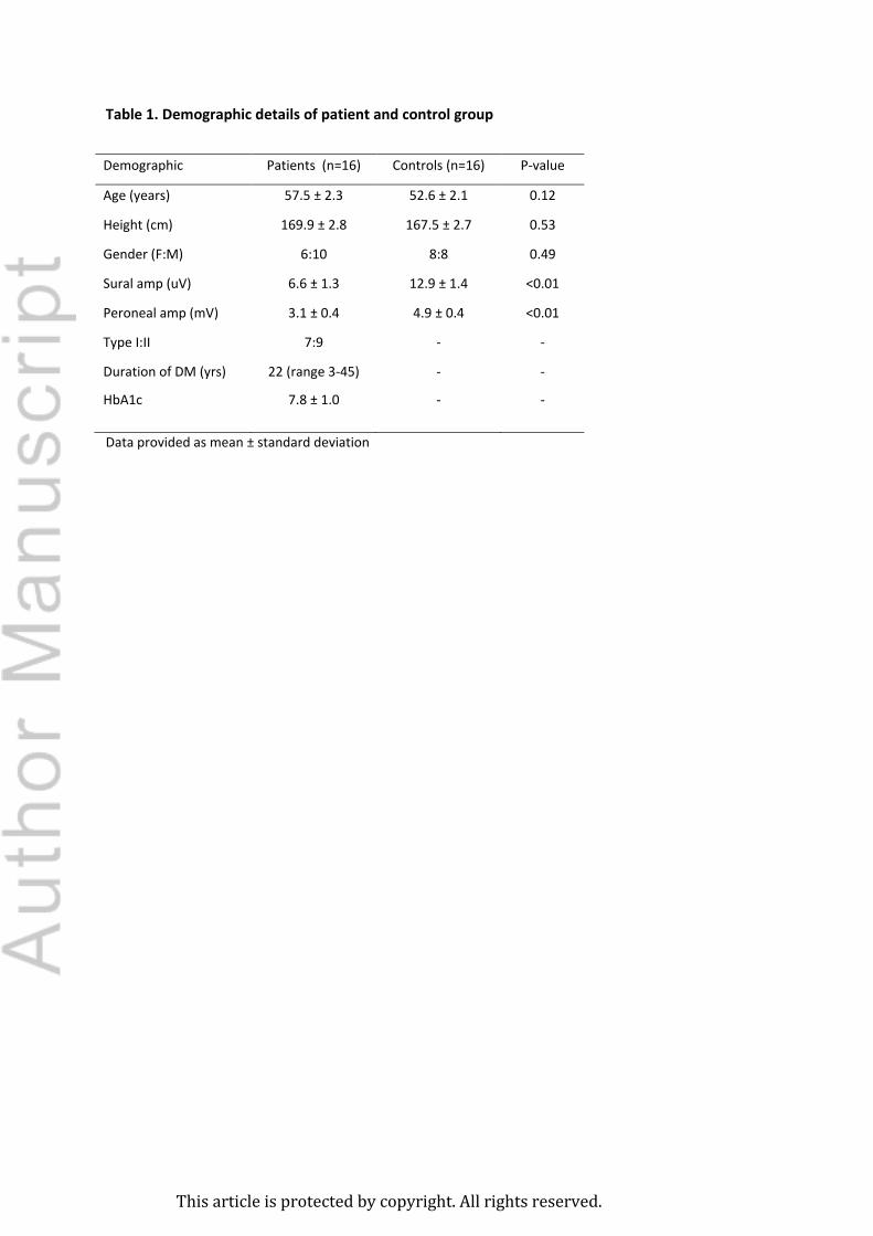
Table 1. Demographic details of patient and control group
Demographic Patients (n=16) Controls (n=16) P-value
Age (years) 57.5 ± 2.3 52.6 ± 2.1 0.12
Height (cm) 169.9 ± 2.8 167.5 ± 2.7 0.53
Gender (F:M) 6:10 8:8 0.49
Sural amp (uV) 6.6 ± 1.3 12.9 ± 1.4 <0.01
Peroneal amp (mV) 3.1 ± 0.4 4.9 ± 0.4 <0.01
Type I:II 7:9 - -
Duration of DM (yrs) 22 (range 3-45) - -
HbA1c 7.8 ± 1.0 - -
Data provided as mean ± standard deviation
This article is protected by copyright. All rights reserved.

Table 2. Conventional and single fiber fibular F-waves
CF-waves Patients (n=16) Controls (n=16) P-value Normal/cut-off Sensitivity*
FMIN (ms) 56.7 ± 1.8 49.9 ± 1.0 <0.01 ≤57.3 6/16
FMAX (ms) 62.3 ± 2.4 54.9 ± 1.4 <0.05 ≤64.7 6/16
FDISP (ms) 6.1 ± 0.9 5.0 ± 0.6 0.31 ≤9.2 3/16
Fp (%) 59.7 ± 6.8 63.7 ± 6.8 0.69 ≥13 2/16
Overall 7/16
SFF-waves
FMIN (ms) 56.1 ± 2.1 49.7 ± 1.1 <0.01 ≤58.1 7/16
FMAX (ms) 67.4 ± 3.0 54.6 ± 1.2 <0.001 ≤64.0 11/16
FDISP (ms) 12.2 ± 2.3 5.0 ± 0.8 <0.01 ≤11.1 7/16
Fp (%) 8.3 ± 2.2 10.2 ± 2.3 0.41 ≥2 3/16
Overall 12/16
*Detection of subclinical or mild diabetic neuropathy
This article is protected by copyright. All rights reserved.
Minerva Access is the Institutional Repository of The University of Melbourne
Author/s:
Kamel, J; Knight-Sadler, R; Cook, M; Roberts, L
Title:
Single-fiber F waves compared with conventional surface F waves, and their utility in
detecting early diabetic neuropathy
Date:
2018-11-01
Citation:
Kamel, J., Knight-Sadler, R., Cook, M. & Roberts, L. (2018). Single-fiber F waves compared
with conventional surface F waves, and their utility in detecting early diabetic neuropathy.
MUSCLE & NERVE, 58 (5), pp.665-670. https://doi.org/10.1002/mus.26290.
Persistent Link:
http://hdl.handle.net/11343/284242
File Description:
Accepted version