should we isolate human preantral follicles before or after cryopreservation of ovarian tissue?
TRANSCRIPT

ORIGINAL ARTICLE: REPRODUCTIVE BIOLOGY
Should we isolate human preantralfollicles before or aftercryopreservation of ovarian tissue?
Julie Vanacker, M.Bio.Sc., Val�erie Luyckx, M.D., Christiani Amorim, V.M.D., Ph.D.,Marie-Madeleine Dolmans, M.D., Ph.D., Anne Van Langendonckt, Ph.D., Jacques Donnez, M.D., Ph.D.,and Alessandra Camboni, M.D., Ph.D.Pole de Recherche en Gyn�ecologie, Institut de Recherche Exp�erimentale et Clinique, Universit�e Catholique de Louvain,Brussels, Belgium
Objective: To evaluate the survival and growth potential of human preantral follicles isolated before and after cryopreservation.Design: Pilot study.Setting: Gynecology research unit in a university hospital.Patient(s): Six women aged 27 to 32 years.Intervention(s): Six ovarian biopsy samples were cut into two equal parts, half subjected to slow-freezing followed by follicle isolation(cryo-iso group) and alginate-matrigel embedding, and half immediately processed for follicle isolation and alginate-matrigelembedding followed by slow-freezing (iso-cryo group) or used as fresh controls (fresh group).Main Outcome Measure(s): Follicle number, viability, diameter, and morphology.Result(s): After 1,134 preantral follicles had been isolated from fresh biopsy samples and 1,132 from frozen specimens, the threegroups were compared before and after 7 days of in vitro culture (IVC) in alginate-matrigel beads. No statistically significantdifferences in viability were found between the three groups before or after IVC, but follicle diameter increased in all three groupsafter IVC. Morphology analysis revealed well-preserved follicles in both the iso-cryo and cryo-iso groups after IVC.Conclusion(s): Human preantral follicles can be successfully cryopreserved before or after isolation without impairing their ability to
Use your smartphone
survive and grow in vitro. This could lead to development of new protocols for follicle cryopres-ervation, IVC, and grafting in clinical and research settings for fertility preservation. (Fertil Ster-il� 2013;-:-–-. �2013 by American Society for Reproductive Medicine.)Key Words: Alginate, cryopreservation, follicle culture, follicle isolation, slow-freezingDiscuss: You can discuss this article with its authors and with other ASRM members at http://fertstertforum.com/vanackerj-follicle-culture-isolation-cryopreservation/
to scan this QR codeand connect to thediscussion forum forthis article now.*
* Download a free QR code scanner by searching for “QRscanner” in your smartphone’s app store or app marketplace.
dvances in the diagnosis and mature ovarian failure caused by their partner, or those unable to undergo
A treatment of cancer have greatlyincreased the life expectancy ofpremenopausal women, but this hasalso resulted in a growing populationof women who, having survived child-hood malignancy, find themselves athigh risk of infertility secondary to pre-
Received October 17, 2012; revised December 3, 201J.V. has nothing to disclose. V.L. has nothing to dis
nothing to disclose. A.V. has nothing to disclohas also received payments for lectures from Seto this work). A.C. has nothing to disclose.
Supported by grants from the R�egion Wallonne (graRecherche Scientifique de Belgique (grant T�el�eFonds Sp�eciaux de Recherche, Fondation St. Lucfrom Mr. Pietro Ferrero, Baron Albert Fr�ere, an
Reprint requests: Marie-Madeleine Dolmans, M.D.,Catholique de Louvain, Cliniques Universitaire1200 Brussels, Belgium (E-mail: marie-madelein
Fertility and Sterility® Vol. -, No. -, - 2013 0015-Copyright ©2013 American Society for Reproductivehttp://dx.doi.org/10.1016/j.fertnstert.2012.12.016
VOL. - NO. - / - 2013
cancer treatment (1). Methods such asembryo or oocyte cryopreservation arecurrently available to preserve fertilitybefore cancer therapy (1, 2).Unfortunately, these two optionscannot be applied to patients ofprepubertal age, women with no
2; accepted December 14, 2012.close. C.A. has nothing to disclose. M.-M.D. hasse. J.D. is a board member of PregLem SA andrrono, MSD, Organon, and Ferring (all unrelated
nt WBI 2009–2010 to A.C.), Fonds National de lavie No. 7.4562.08, grant 3.4.590.08 to M.-M.D.),, and Foundation Against Cancer, and donationsd Viscount Philippe de Spoelberch.Ph.D., Department of Gynecology, Universit�es St. Luc, Avenue Hippocrate 10, bte B2.9502,[email protected]).
0282/$36.00Medicine, Published by Elsevier Inc.
a cycle of ovarian stimulation (3–5).For these patients, cryopreservationof ovarian cortex followed bytransplantation can be performed. Thistechnique, which is the only option todate to have restored both endocrinefunction and fertility, has led to 20 livebirths so far (5–10). Despite thepromising results obtained with thistechnique, there is a risk ofreintroducing malignant cells in case ofcertain types of cancer (11, 12). A saferalternative could be grafting ofa specific number of isolated preantralfollicles.
The follicle isolation procedure isusually performed after thawing of fro-zen ovarian cortical strips (11, 13–18).However, cryopreserving isolated
1
ORIGINAL ARTICLE: REPRODUCTIVE BIOLOGY
preantral follicles could have some advantages over tissuefreezing. It could prevent technical problems related tocryopreservation of heterogeneous cell types such as thosefound in ovarian cortex (19) and improve the cryoprotectiveagent effect by smaller diffusion lengths. Moreover, theliterature shows high follicular loss after ovarian tissuetransplantation caused by the ischemic process (20), anddifficulty in quantitatively and qualitatively evaluating thefollicular population in ovarian fragments (21, 22) becausedistribution of preantral follicles in human ovarian cortex isuneven (23, 24).
Results already obtained from isolated follicle studies havebeen encouraging. Indeed, several investigations into cryopres-ervation of isolated follicles have been performed in mice (17),rats (25), sheep (22, 26–29), goats (30–33), dasyurid marsupials(34), cats (35), and monkeys (36). Live birth of mouse pups wasalso achieved by Wang et al. (18), who in vitro culturedsecondary follicles isolated from vitrified murine ovariantissue. In our team, Dolmans et al. (37) isolated small humanpreantral follicles from fresh ovarian tissue and grafted themto SCID mice for a period of 5 months, at the end of whichantral-stage human follicles were obtained. All this demon-strates the ability of both fresh and frozen isolated follicles tosurvive and develop in vitro and in vivo.
In this study, we isolated human preantral follicles usingour recently developed procedure with Liberase DH (DispaseHigh) (38). To facilitate handling and maintain their three-dimensional structure during cryopreservation and in vitro cul-ture (IVC) procedures, we encapsulated the follicles in analginate-matrigelmatrix (39).Wedeterminedwhether isolatinghuman preantral follicles before freezing could improve folliclequality compared with isolation after tissue cryopreservation.For this purpose, we compared the follicles' viability, growthability, and morphology before and after the cryopreservationprocedure. Follicle analyses were performed before and after7 days of IVC to show any cryodamage more accurately.
MATERIALS AND METHODSCollection of Ovarian Tissue
Use of human tissue for this study was approved by the insti-tutional review board of the Universit�e Catholique de Lou-vain. Ovarian tissue biopsy samples were collected from sixpatients (between 27 and 32 years of age) after obtainingwrit-ten informed consent. The patients were all undergoing lapa-roscopic surgery for benign gynecologic disease.
Tissue was transported within 5 minutes of surgery fromthe operating theater to the research laboratory in 4-(2-hydroxyethyl)-1-piperazineethanesulfonic acid-bufferedmodified Eagle's medium (HEPES-MEM, ref 42360–024;GIBCO) on ice. The medullar part was removed from the sam-ples using surgical scissors. The cortical biopsy samplesranged between 85 mm3 and 351 mm3 in size.
Tissue Treatment, Follicle Isolation, andCryopreservation
Experimental design. Six series of experiments were per-formed with ovarian biopsy samples from six patients. As
2
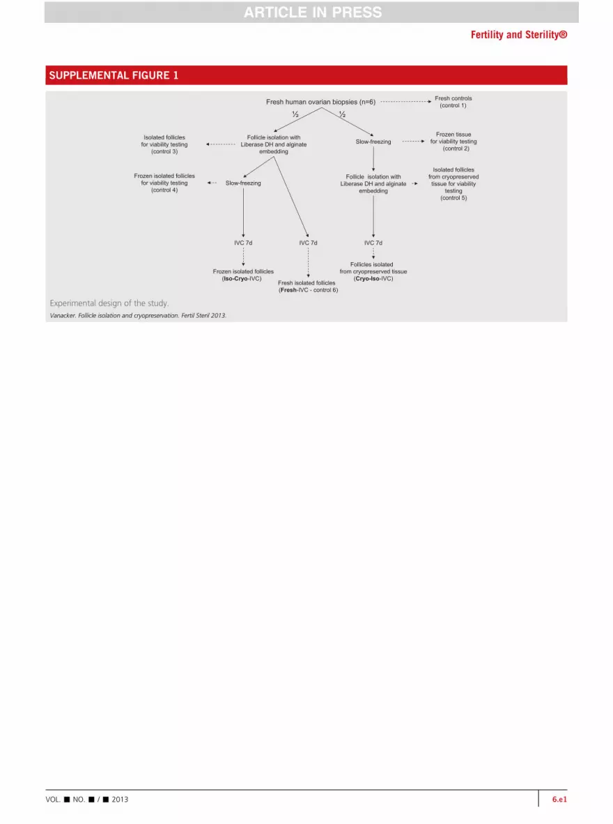
detailed in Supplemental Figure 1 (available online), a smallfragment from each sample was fixed in formol to serve asa fresh control (control). The remaining tissue was cut intotwo equal parts: one-half for preantral follicle isolation andalginate-matrigel encapsulation followed or not by cryopres-ervation (iso-cryo or fresh groups) and the other half for tissuecryopreservation followed by follicle isolation and alginate-matrigel embedding (cryo-iso group). Encapsulated folliclesfrom all three groups were in vitro cultured for 7 days(fresh-IVC, iso-cryo-IVC, and cryo-iso-IVC). At each stageof the procedure, follicles were analyzed for viability, andtheir diameter was recorded. A sample was also taken for mor-phological analysis of the follicles (see Supplemental Fig. 1).
Enzymatic Digestion of Ovarian Tissue
The enzymatic protocol used to isolate preantral follicles waspreviously describedbyVanacker et al. (38). Briefly, small piecesof fresh tissue or the frozen-thawed half of each cortical biopsywere incubated in 10 mL of Dulbecco's phosphate-buffered sa-line (DPBS) medium with calcium and magnesium (ref BE17–513F; Lonza, BioWhittaker) supplemented with 0.28 W€unschunit/mL of Liberase DH (Dispase High) (ref 05401054001;Roche) at 37�C with gentle agitation for 75 minutes.
Recovery of Isolated Follicles
After enzymatic digestion, the resulting suspension wascentrifuged at 50 � g for 10 minutes at 4�C. The supernatantwas then discarded, and the pellet was transferred to plasticPetri dishes and investigated for preantral follicles under a ste-reomicroscope (Leica, Van Hopplynus Instruments). The folli-cles were picked up using a 135 mm-diameter stripper tip (refMXL3–135 mm; Mid-Atlantic Diagnostics) linked to a tubingset with a sterile filter (ref H-903513; Swemed Laboratories).Care was taken to avoid picking up stromal cells.
Isolated Follicle Embedding
The protocol used was a modification of that described byAmorim et al. (16). A 1% (w/v) solution of sodium alginate (al-ginic acid, ref 72138; Sigma) was prepared and autoclaved.Isolated follicles were transferred to droplets (20 mL) of algi-nate solution containing 10% growth factor-reduced extra-cellular matrix (GFR Matrigel, ref 356230; BD Biosciences)(39). To form the beads, the droplets were slowly releasedinto a small Petri dish containing a solution of CaCl2 (0.1M). Beads containing follicles (approximately 15/bead) wereremoved from the dish and then washed in DPBS without cal-cium and magnesium supplemented with 10% fetal bovineserum (FBS, ref 10437028; GIBCO).
Freezing and Thawing of Ovarian Tissue orEmbedded Isolated Follicles
For each patient, one fresh half of each cortical biopsy sampleand a number of alginate-matrigel beads (2 to 17 dependingon the patient) containing isolated follicles were frozen ac-cording to the method described by Van Eyck et al. (20).The tissue was cut into cortical strips of 2 � 5 mm. The strips
VOL. - NO. - / - 2013

TABLE 1
Follicle count in both halves of each biopsy sample.
ExperimentPatientage (y)
Biopsysize (mm3)
No. isolated follicles
Fresh half Frozen half
1 29 135 180 1202 28 285 97 1353 30 130 180 1354 29 85 70 605 28 209 322 2976 25 351 285 385Total 1,134 1,132Note: Fresh ¼ control.
Vanacker. Follicle isolation and cryopreservation. Fertil Steril 2013.
Fertility and Sterility®
or beads were placed in a Petri dish containing HEPES-MEMsupplemented with 10% dimethylsulfoxide (ref. D2650;Sigma) and 2% human serum albumin (ref RVG 16910; San-quin). They were then transferred to cryovials containing 800mL of this freezing medium. The cryovials were cooled ina programmable freezer (CL-8800i, CryoLogic) using the fol-lowing program: [1] cooled from 0�C to �8�C at �2�C/min;[2] seeded manually by touching the cryovials with forcepsprechilled in liquid nitrogen; [3] cooled to �40�C at�0.3�C/min; and [4] cooled to �150�C at �30�C/min andtransferred to liquid nitrogen (�196�C) for storage.
For thawing and cryoprotective agent removal, the cryo-vials were exposed to room temperature for 2minutes and im-mersed in a water bath at 37�C for 2 minutes. To remove thecryoprotective agent, the ovarian tissue or beads with follicleswere transferred from the cryovials to Petri dishes containingHEPES-MEM, where they were washed three times (5 minutesper bath).
In Vitro Culture of Ovarian Follicles
For each patient, alginate-matrigel beads containing isolatedfollicles were in vitro cultured, as described by Amorim et al.(16). The beads were placed in a 4-well or 24-well culture dish(ref 176740 or 150628; Nunc) fitted with Millicell-CM inserts(12 mm in diameter, 0.4 mm pore size, ref PICM01250; Milli-pore). One bead was cultured per well. Culture medium wasadded to each well: 250 mL was pipetted into the inserts and400 mL into the well outside the inserts. The isolated folliclesin the alginate-matrigel beads were cultured for 7 days at37�C in a 95% air and 5% CO2 humidified environment. Everysecond day, 150 mL of culture medium was removed and re-placed with fresh medium in the inserts.
Follicle Evaluation before and after IVC
Number of follicles recovered from fresh and frozen ovarian
tissue. The number of follicles isolated from fresh tissue andthe frozen-thawed parts of each biopsy sample was countedimmediately after enzymatic isolation to ensure that thetwo groups were comparable.
Viability testing. Follicle viability was assessed as describedby Dolmans et al. (40). One or two alginate-matrigel beadscontaining isolated follicles were transferred to 20 mL ofDPBS containing 2 mmol/L calcein-AM and 5 mmol/L ethid-ium homodimer-I (ref L-3224; Molecular Probes). The follicleswere incubated with the fluorescent dyes for 30 minutes at37�C in the dark (41). They were then classified into four cat-egories depending on the percentage of dead granulosa cells(GCs): V1, live follicles ¼ follicles with the oocyte and allGCs viable; V2, minimally damaged follicles ¼ follicles with<10% of dead GCs; V3, moderately damaged follicles ¼ fol-licles with 10% to 50% of dead GCs; V4, dead follicles ¼ fol-licles with both the oocyte or >50% GCs dead.
Follicle diameter evaluation. Before fixation, each follicletested for viability was photographed with a Leica DFC320camera at�40 magnification. The follicle diameter was mea-sured from the basement membrane using ImageJ, an imageprocessing and analysis program developed at the U.S. Na-tional Institutes of Health.
VOL. - NO. - / - 2013
Morphological analysis. Isolated follicles embedded inalginate-matrigel beads were fixed for light microscopy andprocessed according to the procedure reported by Dolmanset al. (40). They were first fixed in 1.5% glutaraldehyde inDPBS, then postfixed with 1% osmium tetroxide (Agar Scien-tific) in DPBS and embedded in small blocks (width: 5 mm;height: 1 mm) of 1% agar (UltraPure agarose; GIBCO). Theywere then dehydrated through an ascending series of ethanol,immersed in propylene oxide (solvent substitution), embed-ded in Epon 812 (Agar Scientific), and sectioned using a LeicaEM UC7 ultramicrotome. Semithin sections (1 mm thick) werestained with toluidine blue and examined by light microscopy(Zeiss Axioskop).
Statistical Analysis
The number of follicles recovered from fresh and frozen-thawed tissue were compared using Student's t test. Themean diameter of isolated follicles was compared betweenthe three groups (fresh, iso-cryo, and cryo-iso) before and af-ter 7 days of culture using the Mann-Whitney test. Compari-sons between percentages of viable follicles found in controlsamples (controls 3 to 5) and after IVC were analyzed by thechi-square test. P%.05 was considered statisticallysignificant.
RESULTSFollicle Count after Isolation from Fresh andFrozen-Thawed Biopsy Samples
As shown in Table 1, the size of the biopsy samples ranged from85 mm3 to 351 mm3. A total of 1,134 preantral follicles wereisolated from fresh biopsy halves and 1,132 from frozen halves.No statistically significant differences were observed betweenthe number of follicles obtained from fresh and frozen-thawed tissue (P>.05), which means results in fresh, cryo-iso,and iso-cryo groups could be compared. It also indicates thatfreezing and thawing of ovarian tissue do not appear to alterthe yield of follicles recovered after enzymatic isolation.
Follicular Viability
Altogether in the three groups, a total of 1,588 follicles wereanalyzed for viability before and after IVC using calcein-AM and ethidium homodimer-1. As in our previous study
3

TABLE 3
Viability after 7 days of in vitro culture.
Fresh IVC Iso-cryo IVC Cryo-iso IVC
V1 82.4% (154/187) 72.5% (272/375) 64.65% (397/614)V2 17.1% (32/187) 25.1% (94/375) 33.9% (208/614)V3 0.5% (1/187) 1.3% (5/375) 0.65% (4/614)V4 0 1.1% (4/375) 0.8% (5/614)Note: Cryo-iso ¼ slow-freezing followed by follicle isolation and alginate-matrigel embed-ding; fresh ¼ control; iso-cryo ¼ follicle isolation and alginate-matrigel embedding followedby slow-freezing; IVC ¼ in vitro culture.
Vanacker. Follicle isolation and cryopreservation. Fertil Steril 2013.
ORIGINAL ARTICLE: REPRODUCTIVE BIOLOGY
(40), we grouped V1 and V2 together because they representedfollicles with high viability. Before IVC (Table 2), no statisti-cally significant difference was found between the freshlyisolated follicles (fresh group), cryopreserved isolated follicles(iso-cryo group), and follicles isolated from frozen-thawedtissue (cryo-iso group) within the highly (V1 and V2) andpoorly (V3 and V4) viable follicle categories (P>.05).
We performed IVC to reveal any possible serious cryoin-jury that could be underestimated if viability was only ana-lyzed soon after the isolation procedure. The IVC stepconfirmed the results obtained after enzymatic isolation: nostatistically significant difference was observed between thethree groups in the proportions of highly (V1 and V2) andpoorly (V3 and V4) viable follicles (Table 3).
Follicle Diameter
As detailed in Table 4, the initial follicle diameters were sim-ilar in freshly isolated follicles (control 3) (40.3 � 7.8 mm),frozen-thawed isolated follicles (iso-cryo group) (43.6 �12.1 mm), and follicles isolated from frozen-thawed tissue(cryo-iso group) (43.15 � 11.0 mm).
After 7 days of IVC, follicles were found to have increasedin size in all three groups. However, we also noted that of the15 follicles encapsulated in each of the alginate beads, 2.4% to3.6% had a similar diameter on day 0 and day 7. Follicle di-ameter statistically significantly increased to 50.7 � 14.4mm, 52.5 � 18.5 mm, and 55.8 � 18.8 mm for freshly isolatedfollicles, frozen-thawed isolated follicles, and follicles iso-lated from frozen-thawed tissue, respectively (P< .001). How-ever, follicles isolated from cryopreserved tissue and in vitrocultured for 7 days (cryo-iso-IVC group) showed a statisticallysignificantly greater diameter than the freshly isolated andfrozen-thawed isolated follicles (control 6 and iso-cryo-IVCgroups, respectively) (P< .05). By contrast, no statistically sig-nificant difference was observed between freshly isolated andfrozen-thawed isolated follicle diameters after culture (con-trol 6 and iso-cryo-IVC groups, respectively).
Follicle Morphology
As shown by light microscopy pictures in SupplementalFigure 2 (available online), both the general morphologyand three-dimensional structure of freshly isolated follicles,cryopreserved isolated follicles, and follicles isolated fromfrozen-thawed ovarian tissue were maintained before and af-ter IVC (see Supplemental Fig. 2A–2D).
TABLE 2
Viability before 7 days of in vitro culture.
Fresh Iso-cryo Cryo-iso
V1 96.4% (106/110) 90.6% (116/128) 85% (148/174)V2 2.7% (3/110) 9.4% (12/128) 14.4% (25/174)V3 0 0 0V4 0.9% (1/110) 0 0.6% (1/174)Note: Cryo-iso ¼ slow-freezing followed by follicle isolation and alginate-matrigel embed-ding; fresh¼ control; iso-cryo ¼ follicle isolation and alginate-matrigel embedding followedby slow-freezing.
Vanacker. Follicle isolation and cryopreservation. Fertil Steril 2013.
4
A total of 16 isolated follicles were analyzed by light mi-croscopy on semithin sections from the three groups beforeand after 7 days of IVC. Before IVC, freshly isolated follicles(fresh group) and follicles isolated from cryopreserved tissue(cryo-iso group) were found to be round and well preservedwhen observed at�1,000magnification. The GCs were cuboi-dal with a large nucleus, and very few vacuoles wereencountered.
After IVC, isolated follicles were retracted, even in thefresh group, and vacuoles were also observed. Nevertheless,isolated follicles from the iso-cryo and cryo-iso groups werefound to be well preserved (see Supplemental Fig. 2E and2F), showing normal GCs and oocytes and no sign of swelling.Only a few vacuoles were visible. Preantral follicles with morethan one layer of cuboidal GCs (secondary follicles) were ob-served in both groups.
DISCUSSIONFor cancer patients who cannot benefit from transplantationof cryopreserved ovarian tissue due to the risk of transmissionof malignant cells, an alternative is isolation of preantral fol-licles for further in vitro culture or transplantation. In vitroprocedures need to be developed to allow complete folliculo-genesis from isolated primordial follicles in humans. Promis-ing results have already been achieved in this field. Outcomesafter culture of single-layered follicles embedded in alginate(16) are encouraging, and multilayered follicles maturedin vitro within cortical strips (42) or isolated from cryobankedtissue and embedded in alginate (17) were shown to reach theantral stage. In vivo, isolated human primordial and primaryfollicles were found to reach the antral stage after
TABLE 4
Follicle diameter in all three groups before and after 7 days of in vitroculture.
Size(mm)
Freshly isolated(n [ 227)
Iso-cryo(n [ 421)
Cryo-iso(n [ 538)
Initial 40.3 � 7.8 (control 3)a 43.6 � 12.1a 43.15 � 11.0a
Final 50.7 � 14.4 (control 6)b,c 52.5 � 18.5b,c 55.8 � 18.8b,d
Note: Cryo-iso ¼ slow-freezing followed by follicle isolation and alginate-matrigelembedding; fresh ¼ control; iso-cryo ¼ follicle isolation and alginate-matrigel embeddingfollowed by slow-freezing.a,b Values with different superscripts differ significantly between rows (P< .05).c,d Values with different superscripts differ significantly between columns (P< .05).
Vanacker. Follicle isolation and cryopreservation. Fertil Steril 2013.
VOL. - NO. - / - 2013
Fertility and Sterility®
xenotransplantation to immunodeficient mice (37) Alginate iscurrently being tested as a biodegradable scaffold to trans-plant isolated preantral follicles and ovarian cells (39) withthe ultimate goal of recreating an artificial ovary.
Regardless of the approach (in vitro or in vivo develop-ment), isolated preantral follicles need to be cryopreserved,so our study investigated when follicles should be isolated.In this study, we compared freshly isolated follicles, cryopre-served isolated follicles, and follicles isolated from frozen-thawed ovarian tissue in terms of viability, growth capacity,and morphology before and after 7 days of IVC to determinewhether isolating human preantral follicles before freeze-thawing could improve follicle quality compared with isola-tion after tissue cryopreservation. Our results showed thatfollicle viability was comparable before and after IVC be-tween freshly isolated follicles, cryopreserved isolated folli-cles, and follicles isolated from frozen-thawed ovariantissue, showing that cryopreservation before or after isolationdoes not appear to affect follicle viability.
We observed an increase in size in all three groups after 7days of IVC. Surprisingly, we noted a greater increase indiameter in in vitro cultured follicles isolated from frozen-thawed ovarian tissue than in freshly isolated or frozen-thawed isolated follicles. Such an increase in follicle diameterafter IVC may be due to typical development of follicles dur-ing culture or a consequence of swelling of GCs caused by os-motic influx after thawing. Follicular morphology analysis bylight microscopy showed that this increase in size was not dueto swelling but rather to real growth, as some follicles werefound to be at the secondary stage and no cell swelling wasobserved. Based on the appearance of well-preserved isolatedfollicles after IVC, we can assume that human preantral folli-cles can be successfully cryopreserved before or after isolationwithout impairing their ability to survive and grow in vitro.
It is interesting that we were able to isolate a similar num-ber of follicles from the fresh and frozen halves of each bi-opsy. This was quite unexpected as, in our experience,isolating follicles from cryopreserved ovarian tissue is morechallenging than from fresh tissue, and ovarian stromal cellsare known to be damaged by current slow-freezing protocols.Gosden et al. (43) suggested that stromal cells are more vul-nerable to cryoinjury than follicular cells because of consider-able water diffusion distance within tissue due to the densityof ovarian stroma. Thus, leaking intracellular content of theseinjured stromal cells could be toxic to surrounding follicles.However, in our study, follicles were successfully isolatedfrom cryopreserved tissue, were viable right after the isolationprocedure, and were able to survive 7 days of IVC. We believethat these successful results were due to the isolation protocolwe developed with Liberase DH (38). This enzyme blend con-tains dispase, which does not cleave laminin (44), the mainconstituent of the basal membrane of primordial and primaryfollicles (45). Furthermore, Liberase DH contains only negligi-ble levels of endotoxins, avoiding any lot-to-lot variationsand thus yielding very reproducible results.
Our study demonstrated that it is now conceivable to iso-late follicles from frozen-thawed tissue when tissue trans-plantation is excluded, as in case of leukemia patients (11).Indeed, female fertility preservation banks worldwide are
VOL. - NO. - / - 2013
currently made up of cryopreserved ovarian tissue fragments,so for these patients development of new clinical protocols forin vitro growth and grafting of human preantral follicles is vi-tal. On the other hand, the fact that human preantral folliclescan also be successfully cryopreserved after isolation is ofparticular importance for experimental research. Indeed,cryopreservation of isolated follicles allows qualitative eval-uation of the follicle population and freezing of a knownnumber of follicles per bead. Moreover, freezing of isolatedfollicles within alginate beads would facilitate follicle han-dling and transportation to centers not performing follicleisolation, thereby increasing material exchange among labo-ratories all over the world.
In conclusion, this study demonstrates that human prean-tral follicles can be successfully cryopreserved before or afterisolation without impairing their ability to survive and growin vitro, although follicles from frozen-thawed ovarian tissueappear to grow faster than cryopreserved isolated follicles.Further experimental studies are therefore warranted to assessthe functionality of cryopreserved isolated follicles after xen-otransplantation. This could lead to the development of newprotocols in clinical and experimental research settings torestore endocrine activity and fertility in cancer patients.Keeping follicles in the cortex is clearly the best clinicalapproach, if appropriate; this will enable application of newtechnologies that will emerge in the future. However, for pa-tients at high risk of ovarian metastasis, freezing preantralfollicles both ways (enclosed in ovarian tissue and after isola-tion) may be advised to maximize their options.
Acknowledgments: The authors thank Mira Hryniuk, B.A.,for reviewing the manuscript, and the Department of Anato-mopathology of Saint-Luc's Hospital for biopsy embeddingand hematoxylin–eosin staining.
REFERENCES1. Donnez J, Martinez-Madrid B, Jadoul P, Van Langendonckt A, Demylle D,
DolmansMM.Ovarian tissue cryopreservation and transplantation: a review.Hum Reprod Update 2006;12:519–35.
2. Wallace WH. Oncofertility and preservation of reproductive capacity in chil-dren and young adults. Cancer 2011;117(Suppl):2301–10.
3. Jadoul P, Dolmans MM, Donnez J. Fertility preservation in girls during child-hood: is it feasible, efficient and safe and to whom should it be proposed?Hum Reprod Update 2010;16:617–30.
4. Donnez J, Jadoul P, Squifflet J, Van Langendonckt A, Donnez O, VanEyck AS, et al. Ovarian tissue cryopreservation and transplantation in cancerpatients. Best Pract Res Clin Obstet Gynaecol 2010;24:87–100.
5. Roux C, Amiot C, Agnani G, Aubard Y, Rohrlich PS, Piver P. Live birth afterovarian tissue autograft in a patient with sickle cell disease treated by allo-geneic bone marrow transplantation. Fertil Steril 2010;93:2413.e15–9.
6. Revel A, Laufer N, Ben Meir A, Lebovich M, Mitrani E. Micro-organ ovariantransplantation enables pregnancy: a case report. Hum Reprod 2011;26:1097–103.
7. Donnez J, Silber S, Andersen CY, Demeestere I, Piver P, MeirowD, et al. Chil-dren born after autotransplantation of cryopreserved ovarian tissue. a reviewof 13 live births. Ann Med 2011;43:437–50.
8. Donnez J, Jadoul P, Pirard C, Hutchings G, Demylle D, Squifflet J, et al. Livebirth after transplantation of frozen-thawed ovarian tissue after bilateral oo-phorectomy for benign disease. Fertil Steril 2012;98:720–5.
9. Dittrich R, Lotz L, Keck G, Hoffmann I, Mueller A, Beckmann MW, et al. Livebirth after ovarian tissue autotransplantation following overnight transpor-tation before cryopreservation. Fertil Steril 2012;97:387–90.
5

ORIGINAL ARTICLE: REPRODUCTIVE BIOLOGY
10. Revelli A, Molinari E, Salvagno F, Delle Piane L, Dolfin E, Ochetti S. Oocytecryostorage to preserve fertility in oncological patients. Obstet Gynecol Int2012;2012:525896.
11. Dolmans MM, Marinescu C, Saussoy P, Van Langendonckt A, Amorim C,Donnez J. Reimplantation of cryopreserved ovarian tissue from patientswith acute lymphoblastic leukemia is potentially unsafe. Blood 2010;116:2908–14.
12. Rosendahl M, Andersen MT, Ralfkiær E, Kjeldsen L, Andersen MK,Andersen CY. Evidence of residual disease in cryopreserved ovarian cortexfrom female patients with leukemia. Fertil Steril 2010;94:2186–90.
13. Abir R, Roizman P, Fisch B, Nitke S, Okon E, Orvieto R, et al. Pilot study ofisolated early human follicles cultured in collagen gels for 24 hours. Hum Re-prod 1999;14:1299–301.
14. Abir R, Fisch B, Nitke S, Okon E, Raz A, Ben Rafael Z. Morphological studyof fully and partially isolated early human follicles. Fertil Steril 2001;75:141–6.
15. Hreinsson J, Zhang P, Swahn ML, Hultenby K, Hovatta O. Cryopreservationof follicles in human ovarian cortical tissue: comparison of serum and hu-man serum albumin in the cryoprotectant solutions. Hum Reprod 2003;18:2420–8.
16. Amorim CA, Van Langendonckt A, David A, Dolmans MM, Donnez J. Sur-vival of human pre-antral follicles after cryopreservation of ovarian tissue,follicular isolation and in vitro culture in a calcium alginate matrix. Hum Re-prod 2009;24:92–9.
17. Xu M, Banc A, Woodruff TK, Shea LD. Secondary follicle growth and oocytematuration by culture in alginate hydrogel following cryopreservation of theovary or individual follicles. Biotechnol Bioeng 2009;103:378–86.
18. Wang X, Catt S, Pangestu M, Temple-Smith P. Successful in vitro culture ofpre-antral follicles derived from vitrified murine ovarian tissue: oocyte mat-uration, fertilization, and live births. Reproduction 2011;141:183–91.
19. Camboni A, Martinez-Madrid B, Dolmans MM, Amorim CA, Nottola SA,Donnez J, et al. Preservation of fertility in young cancer patients: contribu-tion of transmission electron microscopy. Reprod Biomed Online 2008;17:136–50.
20. Van Eyck AS, Bouzin C, Feron O, Romeu L, Van Langendonckt A, Donnez J,et al. Both host and graft vessels contribute to revascularization of xeno-grafted human ovarian tissue in a murine model. Fertil Steril 2010;93:1676–85.
21. Shaw JM, Cox SL, Trounson AO, Jenkin G. Evaluation of the long-term func-tion of cryopreserved ovarian grafts in the mouse, implications for humanapplications. Mol Cell Endocrinol 2000;161:103–10.
22. Amorim CA, Goncalves PB, Figueiredo JR. Cryopreservation of oocytes frompre-antral follicles. Hum Reprod Update 2003;9:119–29.
23. Hovatta O, Silye R, Krausz T, Abir R, Margara R, Trew G, et al. Cryopreserva-tion of human ovarian tissue using dimethylsulphoxide and propanediol-sucrose as cryoprotectants. Hum Reprod 1996;11:1268–72.
24. Qu J, Godin PA, Nisolle M, Donnez J. Distribution and epidermal growth fac-tor receptor expression of primordial follicles in human ovarian tissue beforeand after cryopreservation. Hum Reprod 2000;15:302–10.
25. XingW, Zhou C, Bian J, MontagM, Xu Y, Li Y, et al. Solid-surface vitrificationis an appropriate and convenient method for cryopreservation of isolated ratfollicles. Reprod Biol Endocrinol 2010;8:42.
26. Amorim CA, Rondina D, Rodrigues AP, Costa SH, Goncalves PB, deFigueiredo JR, et al. Isolated ovine primordial follicles cryopreserved in differ-ent concentrations of ethylene glycol. Theriogenology 2003;60:735–42.
27. Amorim CA, Rondina D, Rodrigues AP, Goncalves PB, de Figueiredo JR,Giorgetti A. Cryopreservation of isolated ovine primordial follicles with pro-pylene glycol and glycerol. Fertil Steril 2004;81:735–40.
6
28. Amorim CA, Rondina D, Lucci CM, Giorgetti A, de Figueiredo JR,Goncalves PB. Cryopreservation of sheep primordial follicles. ReprodDomest Anim 2007;42:53–7.
29. Santos RR, Van den Hurk R, Rodrigues AP, Costa SH, Martins FS, Matos MH,et al. Effect of cryopreservation on viability, activation and growth of in situand isolated ovine early-stage follicles. Anim Reprod Sci 2007;99:53–64.
30. Rodrigues AP, Amorim CA, Costa SH, Matos MH, Santos RR, Lucci CM, et al.Cryopreservation of caprine ovarian tissue using dimethylsulphoxide andpropanediol. Anim Reprod Sci 2004a;84:211–27.
31. Rodrigues AP, Amorim CA, Costa SH, Matos MH, Santos RR, Lucci CM, et al.Cryopreservation of caprine ovarian tissue using glycerol and ethylene gly-col. Theriogenology 2004;61:1009–24.
32. Rodrigues APR, AmorimCA, Costa SHF, Santos RR, Lucci CM, Nunes JF, et al.Cryopreservation and short-term culture of isolated caprine primordial folli-cles. Small Ruminant Res 2005;56:103–11.
33. Santos RR, van Haeften T, Roelen BA, Knijn HM, Colenbrander B,Gadella BM, et al. Osmotic tolerance and freezability of isolated caprineearly-staged follicles. Cell Tissue Res 2008;333:323–31.
34. Czarny NA, Harris MS, Rodger JC. Dissociation and preservation of preantralfollicles and immature oocytes from female dasyurid marsupials. Reprod Fer-til Dev 2009;21:640–8.
35. Jewgenow K, Penfold LM, Meyer HHD,Wildt DE. Viability of small pre-antralovarian follicles from domestic cats after cryoprotectant exposure and cryo-preservation. J Reprod Fertil 1998;112:39–47.
36. Barrett SL, Shea LD, Woodruff TK. Noninvasive index of cryorecovery andgrowth potential for human follicles in vitro. Biol Reprod 2010;82:1180–9.
37. DolmansMM,YuanWY,CamboniA, TorreA, Van LangendoncktA,Martinez-Madrid B, et al. Development of antral follicles after xenografting of isolatedsmall human preantral follicles. Reprod Biomed Online 2008;16:705–11.
38. Vanacker J, Camboni A, Dath C, Van Langendonckt A, Dolmans MM,Donnez J, et al. Enzymatic isolation of human primordial and primary ovarianfollicles with Liberase DH: protocol for application in a clinical setting. FertilSteril 2011;96:379–83.
39. Vanacker J, Luyckx V, Dolmans MM, Des Rieux A, Jaeger J, VanLangendonckt A, et al. Transplantation of an alginate-matrigel matrix con-taining isolated ovarian cells: First step in developing a biodegradable scaf-fold to transplant isolated preantral follicles and ovarian cells. Biomaterials2012;33:6079–85.
40. Dolmans MM, Michaux N, Camboni A, Martinez-Madrid B, VanLangendonckt A, Nottola SA, et al. Evaluation of Liberase, a purified enzymeblend, for the isolation of human primordial and primary ovarian follicles.Hum Reprod 2006;21:413–20.
41. Cortvrindt RG, Smitz JE. Fluorescent probes allow rapid and precise record-ing of follicle density and staging in human ovarian cortical biopsy samples.Fertil Steril 2001;75:588–93.
42. Telfer EE, McLaughlin M, Ding C, Thong KJ. A two-step serum-free culturesystem supports development of human oocytes from primordial follicles inthe presence of activin. Hum Reprod 2008;23:1151–8.
43. Gosden RG, Yin H, Bodine RJ, Morris GJ. Character, distribution and biolog-ical implications of ice crystallization in cryopreserved rabbit ovarian tissuerevealed by cryo-scanning electron microscopy. Hum Reprod 2010;25:470–8.
44. Stenn KS, Link R, Moellmann G, Madri J, Kuklinska E. Dispase, a neutral pro-tease from Bacillus polymyxa, is a powerful fibronectinase and type IV colla-genase. J Invest Dermatol 1989;93:287–90.
45. Berkholtz CB, Lai BE, Woodruff TK, Shea LD. Distribution of extracellular ma-trix proteins type I collagen, type IV collagen, fibronectin, and laminin inmouse folliculogenesis. Histochem Cell Biol 2006;126:583–92.
VOL. - NO. - / - 2013
SUPPLEMENTAL FIGURE 1
Fresh controls(control 1)
Isolated follicles for viability testing
(control 3)
Frozen isolated follicles for viability testing
(control 4)
Isolated follicles from cryopreservedtissue for viability
testing(control 5)
Frozen tissue for viability testing
(control 2)
Fresh human ovarian biopsies (n=6)
Follicle isolation with Liberase DH and alginate
embeddingSlow-freezing
Follicle isolation with Liberase DH and alginate
embedding
IVC 7dIVC 7d
Slow-freezing
IVC 7d
½ ½
Frozen isolated follicles(Iso-Cryo-IVC)
Fresh isolated follicles (Fresh-IVC - control 6)
Follicles isolatedfrom cryopreserved tissue
(Cryo-Iso-IVC)
Experimental design of the study.Vanacker. Follicle isolation and cryopreservation. Fertil Steril 2013.
Fertility and Sterility®
VOL. - NO. - / - 2013 6.e1

SUPPLEMENTAL FIGURE 2
Light microscopy pictures of (A) a freshly isolated follicle before IVC, (B) a freshly isolated follicle after IVC, (C) a cryopreserved isolated follicle afterIVC, (D) and a follicle isolated from frozen-thawed ovarian tissue after IVC encapsulated in an alginate-matrigel bead at �40 magnification. Semi-thin sections of (E) a cryopreserved isolated follicle and (F) a follicle isolated from frozen-thawed ovarian tissue encapsulated in an alginate-matrigelbead after IVC at �1,000 and �400 magnification, respectively.Vanacker. Follicle isolation and cryopreservation. Fertil Steril 2013.
ORIGINAL ARTICLE: REPRODUCTIVE BIOLOGY
6.e2 VOL. - NO. - / - 2013