serotonergic modulation of behaviour: a phylogenetic overview
TRANSCRIPT
Biol. Rev. (), , pp. –
Printed in United Kingdom
SEROTONERGIC MODULATION OF BEHAVIOUR: A
PHYLOGENETIC OVERVIEW
B WENDY A. WEIGER
Department of Neurobiology, Harvard Medical School, Boston, MA, U.S.A.
(Received June ; revised April ; accepted April )
ABSTRACT
Serotonergic neurons are present in all phyla that possess nervous systems. In most of these
phyla, serotonin modulates important behaviours, including feeding, sexual and aggressive
behaviour. Serotonin exerts its effects by acting in three basic modes: as a classical
neurotransmitter, as a neuromodulator, or as a neurohormone. In a number of invertebrate
species, the neural circuitry underlying the effects of serotonin has been well characterized,
whereas in vertebrates, the mechanisms by which serotonin affects behaviour are currently less
fully understood. The following review examines the role played by serotonin in the
generation and modulation of behaviour in successively more complex species, ranging from
coelenterates to humans.
Key words : Serotonin, behaviour, neurotransmitter, neuromodulator, neurohormone,
biogenic amine.
CONTENTS
I. Introduction . . . . . . . . . . . . . . .
II. Serotonergic modulation of behaviour in animal phyla . . . . . . .
() Coelenterates . . . . . . . . . . . . . .
() Platyhelminths (flatworms) . . . . . . . . . . .
() Nematodes . . . . . . . . . . . . . .
() Molluscs . . . . . . . . . . . . . . .
() Annelids . . . . . . . . . . . . . . .
() Arthropods: crustaceans . . . . . . . . . . . .
() Echinoderms . . . . . . . . . . . . . .
() Chordates . . . . . . . . . . . . . . .
III. Conclusions . . . . . . . . . . . . . . .
IV. Acknowledgements . . . . . . . . . . . . .
V. References . . . . . . . . . . . . . . .
I. INTRODUCTION
The orchestration of complex behaviour by the nervous system relies on a
surprisingly small number of neurotransmitters. Each transmitter, however, may act in
multiple ways: it may bind to a variety of receptors to open or close ion channels or to
activate any of several different second messenger systems. Furthermore, a single
transmitter may, in different systems, function in any of three basic modes: as a classical
neurotransmitter, as a neuromodulator, or as a neurohormone. A classical neuro-
transmitter exerts its effects directly on a postsynaptic cell. A neuromodulator does not
directly alter the activity of a postsynaptic cell, but rather alters either presynaptic
W. A. W
release of, or postsynaptic response to, another compound which acts as the primary
neurotransmitter at a synapse. Neuromodulators may be released in a diffuse manner
into a region of the nervous system rather than onto discrete postsynaptic sites. A
neurohormone is released into the general circulation rather than onto a specific
postsynaptic cell and can therefore exert modulatory effects throughout an entire
organism.
Serotonin (-hydroxytryptamine, -HT) functions as a neurotransmitter in even the
most primitive nervous systems. The following review assesses the role that serotonin
plays as a classical neurotransmitter, as a neuromodulator, and as a neurohormone in the
various phyla of the animal kingdom. By examining the role of serotonin in successively
more complex nervous systems, an understanding will emerge of the ways in which a
single neurotransmitter can contribute to the generation and modulation of complex
behaviours, including feeding, sexual and aggressive behaviour.
II. SEROTONERGIC MODULATION OF BEHAVIOUR IN ANIMAL PHYLA
() Coelenterates
Coelenterates are the most primitive organisms that possess a nervous system.
Serotonin appears to be present in the neurons of some members of this phylum (Wood
& Lentz, ; Umbriaco, Anctil & Descarries, ). In the sea pansy (Renilla
koellikeri), the morphology of serotonin-immunoreactive neurons suggests that they are
sensory (Anctil, ; Umbriaco et al., ). In this animal, serotonin enhances the
amplitude of endogenous rhythmic contractions, and it may be the transmitter that
mediates an increase in the amplitude of such contractions in response to slow water
movement. Water flow may activate mechanosensory serotonergic neurons directly,
causing a release of serotonin that modifies motor activity in the animal in accordance
with changing environmental conditions (Anctil, ).
Serotonin is also found in the stinging nematocysts of some coelenterates (Wood &
Lentz, ; Castano & Rossi, ; Umbriaco et al., ). As a component of the
toxin released by these organelles, serotonin may exert effects on the neuromuscular
system of predators or prey (Welsh, ).
() Platyhelminths (flatworms)
Welsh () reported that planarians have serotonergic neurons in the brain, ventral
nerve cord and peripheral nervous system. Serotonergic sensory neurons also appear to
be present.
Serotonin-immunoreactive neurons are observed in the central and peripheral
nervous systems of the monogenean parasite, Diclidophora merlangi, and possible
serotonergic sensory fibres are also found (Halton et al., ). Exogenous serotonin
enhances spontaneous muscular contractions (Maule et al., ). Serotonergic
innervation of the reproductive system is found in male D. merlangi only, suggesting a
role for serotonin in the control of gender-specific reproductive behaviour (Maule et al.,
).
() Nematodes
Horvitz et al. () identified two serotonergic neurons in Caenorhabditis elegans.
These are known as the pharyngeal neurosecretory motor neurons; they innervate
Serotonergic modulation of behaviour : a phylogenetic overview
pharyngeal muscles and appear to have neurohumoral outputs to the pseudocoelom.
Exposure of intact animals to exogenous serotonin increases the rate of pharyngeal
pumping, stimulates egg release and depresses locomotion. These results suggest that
serotonin plays the role of a classical neurotransmitter or neuromodulator in feeding
and a neurohormonal role in reproduction and locomotion.
Octopamine application decreases both the rate of pharyngeal pumping and that of
egg laying. Although individual octopaminergic neurons were not identified in this
study, octopamine was found in crude extracts of C. elegans. Phentolamine, an
octopamine antagonist, stimulates egg laying, which suggests that octopamine tonically
inhibits egg laying in vivo. Octopamine suppresses the serotonergic stimulation of egg
laying (although it does not suppress serotonergic stimulation of the pharynx),
indicating that serotonin and octopamine may act antagonistically in vivo to control the
rate of egg laying in the animal. Opposition between the effects of serotonin and
octopamine (or norepinephrine, the vertebrate analogue of octopamine) is also found
in the mollusc Tritonia diomedea, in crustaceans and in gymnotid fish, and will be
described below.
() Molluscs
The best-understood molluscan nervous system is that of Aplysia californica. A
number of studies have implicated a pair of large serotonergic neurons in the cerebral
ganglion in the modulation of feeding behaviour in this organism. Axonal branches of
these metacerebral cells (MCCs) project to the buccal ganglion, to the muscles of the
buccal mass (which mediate biting and swallowing) and to the lips (Kupfermann &
Weiss, ).
In the buccal ganglion, the MCC terminals play a neuromodulatory role. Firing of
the MCCs may produce excitatory potentials in buccal motor neurons, although these
inputs are usually insufficient to elicit spikes. However, if these neurons have been
brought close to firing threshold by some other excitatory input, then MCC activity
may facilitate the primary excitation and result in buccal motor neuron activity. In a
preparation in which the buccal neurons are firing rhythmically, MCC activity can
increase the bursting frequency; this effect may result from input to premotor pattern-
generating interneurons (Kupfermann & Weiss, ).
Serotonin from the MCCs also plays a modulatory role at the buccal motor neuron-
buccal muscle junction. First, serotonin enhances excitatory junctional potentials
(EJPs). This effect may be pre- or postsynaptic. Secondly, serotonin enhances muscle
contraction even when EJP size is not increased. This postsynaptic effect on the muscle
fibre is mediated through the stimulation of muscle adenylate cyclase and the
subsequent phosphorylation of five muscle proteins, possibly including the contractile
protein paramyosin (Kupfermann & Weiss, ).
The MCC cells thus potentiate biting at two levels : the buccal ganglion and the
buccal musculature. Kupfermann & Weiss (, ) demonstrated the behavioural
relevance of this potentiation to the state they termed ‘food-induced arousal ’. Food
arousal is initiated by exposure of the animal to food and results in an increased
efficiency of feeding. Strength and frequency of biting increases, as does bite speed
(defined as the inverse of the length of time from presentation of food on the lip to
maximal radula protraction). Food arousal is decreased by satiation or by noxious
W. A. W
stimuli. Extracellular recordings from freely moving animals showed that food stimuli
can activate the MCCs, and that at all levels of arousal the corresponding bite speed
correlates directly with the firing rate of the MCCs.
Experiments in which the MCCs were inhibited or lesioned support their role in the
potentiation of biting. When a food stimulus was presented to a single semi-intact
preparation during depolarization and hyperpolarization of both MCCs, the movements
of the buccal muscles were slower and weaker when the MCCs were hyperpolarized
(Weiss et al., a). Furthermore, Rosen et al. (, ) found that selective
lesioning of the MCC cells in otherwise intact animals produces abnormal biting
behaviour, characterized by reduction of bite speed, prolongation of mouth opening, an
increase in interbite interval, and an exaggerated reduction in bite magnitude over the
course of a meal. However, several other measures of feeding behaviour are unaffected
by MCC lesions. These include the time interval between exposure to food and
assumption of the feeding posture, rate of swallowing and meal size. The MCCs are not
necessary for the manifestation of any component of feeding behaviour, and their
modulatory effects appear to be restricted to biting.
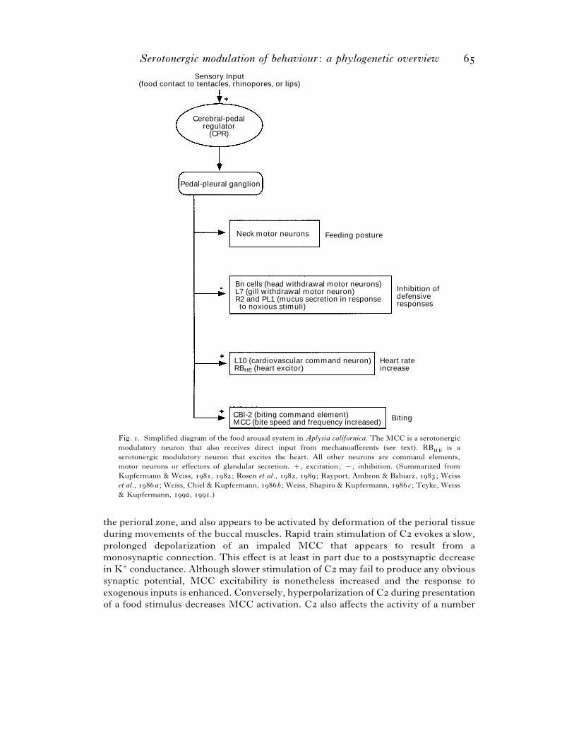
Teyke, Weiss & Kupfermann () clarified the position of the MCCs in the neural
hierarchy controlling feeding behaviour. Sensory input (a food stimulus to the
tentacles, rhinopores, or lips) activates a pair of high-level neurons known as the
cerebral-pedal regulators (CPRs). These neurons act through connections in the pedal-
pleural ganglia to coordinate all aspects of feeding behaviour (Fig. ). Neck motor
neurons are activated to produce the characteristic feeding posture; head and gill
withdrawal neurons and defensive secretion neurons are inhibited; and heart rate is
increased through activation of neurons that control the cardiovascular system.
Finally, the paired CBI- neurons, putative biting command elements, are activated;
the MCCs are also excited and enhance the speed and frequency of biting. CPR
activation of the MCCs may be mediated through the CBI- cells ; intracellular
stimulation of CBI- can excite the ipsilateral MCC (Rosen et al., ). Extracellular
recordings from free-moving animals suggest that the CPRs are activated by
presentation of a food stimulus, and that increased CPR firing correlates with lifting of
the head to assume the feeding posture. Secondary MCC activation should accompany
this enhanced CPR activity (Teyke, Weiss & Kupfermann, ).
The MCCs receive direct sensory inputs in addition to those mediated through the
CPRs. One such input arises from the interganglionic cerebral-buccal mechanoafferents
(ICBMs). These mechanoafferent cells in the cerebral ganglion respond to tactile
stimulation of specific receptive fields in the perioral zone or on the inner wall of the
buccal mass. Intracellular stimulation of an ICBM can evoke a compound excitatory
postsynaptic potential (EPSP) in the ipsilateral MCC. The first component of this
EPSP appears to be monosynaptic, while the later components appear polysynaptic.
The ICBMs also monosynaptically excite neurons in the buccal ganglion and can evoke
coordinated output from this ganglion (Rosen et al., ). Both the CPRs and the
ICBMs, therefore, respond to sensory stimuli relevant to feeding by co-activating the
modulatory MCCs along with other neurons that participate in the generation of
feeding behaviour.
Another direct sensory input to the MCCs comes from C, a paired histaminergic
neuron that innervates the perioral region. C is excited by mechanical stimulation of
Serotonergic modulation of behaviour : a phylogenetic overview
Sensory Input(food contact to tentacles, rhinopores, or lips)
Cerebral-pedalregulator
(CPR)
Pedal-pleural ganglion
Neck motor neurons Feeding posture
Bn cells (head withdrawal motor neurons)L7 (gill withdrawal motor neuron)R2 and PL1 (mucus secretion in response to noxious stimuli)
Inhibition ofdefensiveresponses
Heart rateincrease
BitingCBl-2 (biting command element)MCC (bite speed and frequency increased)
L10 (cardiovascular command neuron)RBHE (heart excitor)
Fig. . Simplified diagram of the food arousal system in Aplysia californica. The MCC is a serotonergic
modulatory neuron that also receives direct input from mechanoafferents (see text). RBHE
is a
serotonergic modulatory neuron that excites the heart. All other neurons are command elements,
motor neurons or effectors of glandular secretion. , excitation; ®, inhibition. (Summarized from
Kupfermann & Weiss, , ; Rosen et al., , ; Rayport, Ambron & Babiarz, ; Weiss
et al., a ; Weiss, Chiel & Kupfermann, b ; Weiss, Shapiro & Kupfermann, c ; Teyke, Weiss
& Kupfermann, , .)
the perioral zone, and also appears to be activated by deformation of the perioral tissue
during movements of the buccal muscles. Rapid train stimulation of C evokes a slow,
prolonged depolarization of an impaled MCC that appears to result from a
monosynaptic connection. This effect is at least in part due to a postsynaptic decrease
in K+ conductance. Although slower stimulation of C may fail to produce any obvious
synaptic potential, MCC excitability is nonetheless increased and the response to
exogenous inputs is enhanced. Conversely, hyperpolarization of C during presentation
of a food stimulus decreases MCC activation. C also affects the activity of a number
W. A. W
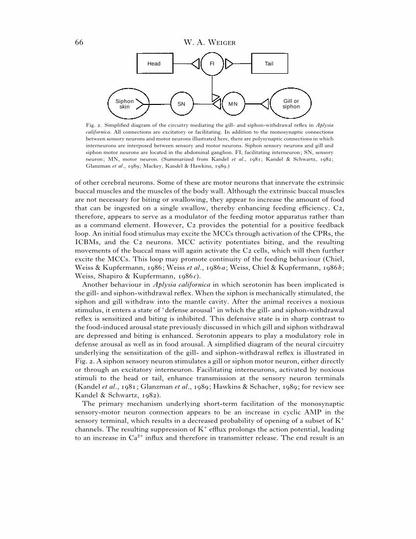
Head FI Tail
MNSNGill orsiphon
Siphonskin
Fig. . Simplified diagram of the circuitry mediating the gill- and siphon-withdrawal reflex in Aplysia
californica. All connections are excitatory or facilitating. In addition to the monosynaptic connections
between sensory neurons and motor neurons illustrated here, there are polysynaptic connections in which
interneurons are interposed between sensory and motor neurons. Siphon sensory neurons and gill and
siphon motor neurons are located in the abdominal ganglion. FI, facilitating interneuron; SN, sensory
neuron; MN, motor neuron. (Summarized from Kandel et al., ; Kandel & Schwartz, ;
Glanzman et al., ; Mackey, Kandel & Hawkins, .)
of other cerebral neurons. Some of these are motor neurons that innervate the extrinsic
buccal muscles and the muscles of the body wall. Although the extrinsic buccal muscles
are not necessary for biting or swallowing, they appear to increase the amount of food
that can be ingested on a single swallow, thereby enhancing feeding efficiency. C,
therefore, appears to serve as a modulator of the feeding motor apparatus rather than
as a command element. However, C provides the potential for a positive feedback
loop. An initial food stimulus may excite the MCCs through activation of the CPRs, the
ICBMs, and the C neurons. MCC activity potentiates biting, and the resulting
movements of the buccal mass will again activate the C cells, which will then further
excite the MCCs. This loop may promote continuity of the feeding behaviour (Chiel,
Weiss & Kupfermann, ; Weiss et al., a ; Weiss, Chiel & Kupfermann, b ;
Weiss, Shapiro & Kupfermann, c).
Another behaviour in Aplysia californica in which serotonin has been implicated is
the gill- and siphon-withdrawal reflex. When the siphon is mechanically stimulated, the
siphon and gill withdraw into the mantle cavity. After the animal receives a noxious
stimulus, it enters a state of ‘defense arousal ’ in which the gill- and siphon-withdrawal
reflex is sensitized and biting is inhibited. This defensive state is in sharp contrast to
the food-induced arousal state previously discussed in which gill and siphon withdrawal
are depressed and biting is enhanced. Serotonin appears to play a modulatory role in
defense arousal as well as in food arousal. A simplified diagram of the neural circuitry
underlying the sensitization of the gill- and siphon-withdrawal reflex is illustrated in
Fig. . A siphon sensory neuron stimulates a gill or siphon motor neuron, either directly
or through an excitatory interneuron. Facilitating interneurons, activated by noxious
stimuli to the head or tail, enhance transmission at the sensory neuron terminals
(Kandel et al., ; Glanzman et al., ; Hawkins & Schacher, ; for review see
Kandel & Schwartz, ).
The primary mechanism underlying short-term facilitation of the monosynaptic
sensory-motor neuron connection appears to be an increase in cyclic AMP in the
sensory terminal, which results in a decreased probability of opening of a subset of K+
channels. The resulting suppression of K+ efflux prolongs the action potential, leading
to an increase in Ca#+ influx and therefore in transmitter release. The end result is an
Serotonergic modulation of behaviour : a phylogenetic overview
enhanced EPSP in the postsynaptic motor neuron which contributes to sensitization of
the gill- and siphon-withdrawal reflex. Exogenous application of serotonin mimics
these effects of stimulation of the facilitating interneurons in dissected preparations and
induces facilitation of sensory-motor synapses in culture (Kandel et al., ; Montarolo
et al., ; Glanzman et al., ; Mackey, Kandel & Hawkins, ; for review of
early work see Kandel & Schwartz, ). At sensory-motor synapses that have become
depressed as a result of repeated sensory neuron stimulation, there appears to be a
second mechanism that contributes to presynaptic facilitation (Hochner et al., ).
Studies of depressed synapses in culture suggest that exogenous serotonin can also
activate this second mechanism, which may be mediated by protein kinase C (Ghirardi
et al., ).
A single noxious stimulus produces short-term sensitization of the gill- and siphon-
withdrawal reflex; in sensory-motor co-culture, a single pulse of serotonin produces
short-term facilitation of the sensory-motor synapse. Repeated noxious stimuli or
multiple pulses of higher concentrations of serotonin result in long-term enhancement
lasting h or more (Castellucci et al., ; Ghirardi, Montarolo & Kandel, ; for
review of early work see Kandel & Schwartz, ). The transition from short- to long-
term facilitation requires both protein and RNA synthesis. Inhibition of protein
synthesis blocks long-term sensitization in semi-intact preparations subjected to tail
shocks, and inhibitors of protein or RNA synthesis block long-term facilitation in
sensory-motor co-cultures treated with serotonin (Montarolo et al., ; Castellucci
et al., ).
Potential mechanisms for induction of transcription by serotonin have been explored
in sensory neurons from pleural ganglia. Application of serotonin increases the
cytosolic concentration of cyclic AMP. Although this effect is most pronounced in
distal processes, smaller increases are noted in the perinuclear cytoplasm. Prolonged
application or multiple pulses of serotonin can lead to translocation of the free catalytic
subunit of the cyclic-AMP-dependent protein kinase into the nucleus (Bacskai et al.,
). The next step is phosphorylation of cyclic AMP response element binding
proteins (CREBs). These proteins then induce the transcription of genes regulated by
the cyclic AMP response element (CRE) (Kaang, Kandel & Grant, ).
Long-term sensitization appears to rely in part on the same mechanisms that
contribute to the short-term process. In animals showing long-term behavioural
sensitization, there is a drop in the ratio of the regulatory to catalytic subunit of the
Aplysia cyclic-AMP-dependent protein kinase in tissue containing the siphon sensory
neurons. This reduction in the regulatory subunit would make the catalytic subunit of
this kinase less dependent on cyclic AMP, so that the same substrate proteins
phosphorylated during short-term sensitization would remain phosphorylated in the
long term when cyclic AMP is no longer elevated (Greenberg et al., ). In vitro,
exogenous serotonin lowers the ratio of regulatory to catalytic subunits in Aplysia
californica pleural sensory neurons, suggesting that similar changes in siphon sensory
neurons might be mediated by serotonin in vivo (Bergold et al., ).
Another important change that accompanies long-term sensitization is an increase in
the number of presynaptic varicosities of abdominal sensory neurons. This growth is
observed in long-term behaviourally sensitized animals as well as in sensory-motor co-
cultures treated with serotonin (Bailey & Chen, ; Glanzman, Kandel & Schacher,
W. A. W
; for review see Schacher et al., ). One factor that may contribute to this
growth is a decrease in cell surface proteins that mediate adhesion. These proteins,
known as Aplysia cell adhesion molecules (apCAMs), appear to belong to the
immunoglobulin class of cell adhesion molecules and are similar to another member of
this class, the neural cell adhesion molecule (NCAM), which is found in vertebrates.
Exposure to serotonin alters both synthesis and distribution of apCAM in Aplysia
californica sensory neurons. It decreases synthesis of new apCAM and increases
endocytosis of apCAM that is already present at the cell surface (Bailey et al., ;
Mayford et al., ).
An increase in the number of sensory presynaptic varicosities may be accompanied
by corresponding changes in postsynaptic motor neurons. In the intact abdominal
ganglion, a prolonged application of serotonin that produces long-term sensory-motor
facilitation also results in long-term enhancement of the response of an identified gill
motor neuron to an applied agonist (Trudeau & Castellucci, ).
The evidence presented above indicates that exogenous serotonin can produce both
short- and long-term changes that mimic the effects of sensitizing sensory stimuli.
However, identified facilitating interneurons in the abdominal ganglion are not
serotonergic (Ono & McCaman, ; Kistler et al., ; Longley & Longley, ;
Hawkins, ). A pair of serotonergic facilitating interneurons (CBs) has been found
in the cerebral ganglion. These neurons, LCB and RCB, show a prolonged activation
following tail shock (application of an electrical current to the tail, a stimulus known to
produce sensitization). CB stimulation enhances the monosynaptic EPSP produced in
gill or siphon motor neurons by stimulation of siphon sensory neurons through a
mechanism that appears to be at least partly presynaptic (Mackey et al., ).
In addition to its well-characterized effects on sensory-motor synapses, serotonin
may act at other sites in the circuitry mediating the gill- and siphon-withdrawal reflex.
Both connective stimulation (which can be used to simulate head or tail shock) and
exogenous serotonin enhance the central excitability of sensory neurons; this effect is
demonstrated by an increase in the number of spikes elicited in abdominal sensory
neurons by current injection into the cell body. In addition, both connective stimulation
and application of serotonin to the siphon can produce a small enhancement of the
response of siphon sensory neurons to tactile stimulation of the siphon (Klein, Hochner
& Kandel, ). At the level of the motor neurons, both tail shock and exogenous
serotonin cause an increase in the tonic firing rate of a subset of siphon motor neurons.
This tonic increase in activity can amplify phasic siphon contractions produced by brief
high-frequency activation of these neurons (Frost, Clark & Kandel, ).
As noted above, serotonin is not the only transmitter that modulates the gill- and
siphon-withdrawal reflex. However, serotonin depletion by injection of ,-dihydroxy-
tryptamine (,-DHT) significantly reduces the enhancement of gill withdrawal by tail
shock. Serotonin thus appears to be an important mediator of the sensitization of the
withdrawal reflex in Aplysia californica (Glanzman et al., ).
In another mollusc, the sea slug Tritonia diomedea, serotonin appears to act both as
a classical neurotransmitter and as a neuromodulator in the central pattern generator
(CPG) network that controls escape swimming. In Tritonia diomedea, contact with the
tube feet of predatory starfish causes a series of – alternating ventral and dorsal
flexions that move the animal away from the predator (McClellan, Brown & Getting,
Serotonergic modulation of behaviour : a phylogenetic overview
). Numerous observations support the importance of serotonin in the generation
of escape swimming behaviour. In whole animals, injection of serotonin can initiate
swimming in the absence of epithelial stimulation, whereas methysergide, a serotonin
antagonist, inhibits swimming in response to stimuli. In isolated brain preparations,
peripheral nerve stimulation elicits the swim motor programme; shortly after bath
application of serotonin, a similar pattern of neural activity is observed. Methysergide
inhibits production of the swim motor programme both by nerve stimulation and by
bath-applied serotonin (McClellan et al., ). A number of other compounds,
including octopamine, inhibit the production of swimming activity by nerve
stimulation; some of these compounds may oppose the effects of serotonin in vivo.
The neurons that form the CPG circuit for escape swimming in Tritonia diomedea
have been well characterized (Katz, Getting & Frost, ; McClellan et al., ). On
each side of the brain, there are one cerebral cell interneuron (C), three dorsal swim
interneurons (DSIs), and two ventral swim interneurons (VSIs). The CPG neurons on
a given side of the brain form a complex set of monosynaptic interconnections. They
also project to two classes of flexion neurons: the ventral flexion neurons (VFNs) and
the dorsal flexion neurons (DFNs), which are further subdivided into two groups
(DFN-A and DFN-B).
The DSIs are serotonin-immunoreactive (Katz et al., ; McClellan et al., ).
They make monosynaptic excitatory connections with C and DFN-A, and
monosynaptic inhibitory connections with the VSIs, DFN-B and VFN. However, the
DSIs play an additional neuromodulatory role. DSI stimulation enhances the synaptic
potentials produced by C in the VSIs, DFN-A, DFN-B and VFN, as well as the
potentials evoked by C in other DSIs (Katz et al., ; Katz & Frost, a). DSI
activation increases the amplitude of both excitatory and inhibitory potentials evoked
by C. This enhancement of the effects of C occurs regardless of whether the direct
synaptic action of DSI on a given neuron is excitatory or inhibitory. The mechanism
underlying the enhancement appears to be an increase in presynaptic release of
transmitter from C terminals.
The effects of DSI activation on DFN-A show an additional level of complexity
(Katz & Frost, a). As noted above, stimulation of a DSI monosynaptically excites
DFN-A; it also increases the amplitude of the EPSP produced in DFN-A by C.
However, there is at least one other effect: stimulation of one DSI decreases the
amplitude of EPSPs evoked by subsequent stimulation of other DSIs. Future studies
may reveal further complexity in the modulation of the Tritonia diomedea swim
network.
As already stated, the DSIs are serotonin-immunoreactive. Specific evidence
indicates that both the synaptic and modulatory effects of these neurons are mediated
by serotonin (Katz et al., ; Katz & Frost, b). Exogenous puffs of serotonin
mimic both the fast and slow monosynaptic EPSPs evoked in DFN-A by DSI
stimulation, and bath application of high concentrations of serotonin during DSI
activation occludes both EPSPs. The fast EPSP is blocked by the serotonergic
antagonist gramine, whereas the slow EPSP is reduced by methysergide. The
modulatory effects of DSI stimulation also appear to be mediated by serotonin. Bath-
applied serotonin increases the amplitude of the EPSP evoked in DFN-A by C
stimulation, and no further enhancement follows DSI activation. Methysergide blocks
W. A. W
both the modulatory effects of DSI activation and those of exogenous serotonin.
Imipramine, a serotonin reuptake inhibitor, enhances both the synaptic effects evoked
by DSIs in DFN-A and the modulatory effects of DSI activation on the C}DFN-A
synapse.
In the Tritonia diomedea swim network, the serotonergic DSIs synaptically excite or
inhibit CPG and efferent neurons and enhance transmission between C and its targets
within the network. Afferent pathways that initiate swimming strongly activate the
DSIs. These neurons may play an important role in the reconfiguration of the swim
network from the resting state to the oscillatory state that produces the rhythmic swim
motor output (Katz et al., ; McClellan et al., ; Katz & Frost, a).
The DSIs may also play a role in sensitization of the escape swimming response.
Following exposure to a stimulus that causes an animal to swim, the animal displays a
decreased onset latency for swimming in response to further stimulation (Abraham &
Willows, ). After a swim episode ends, the firing frequency of the DSIs remains
elevated for several minutes. This increase in spontaneous DSI activity may continue
to enhance transmission between C and its targets and predispose the animal to
initiate swimming in response to sensory stimuli (Katz et al., ; McClellan et al.,
). In this regard, the DSIs in Tritonia diomedea may play a role analogous to that
served by Aplysia californica serotonergic neurons in sensitization of the gill- and
siphon-withdrawal reflex.
() Annelids
The most thoroughly analysed annelid nervous system is that of the leech (Hirudo
medicinalis, Macrobdella decora, Haementeria ghilianii). The leech ventral nervous
system consists of ganglia: the rostral four are fused into the compound
suboesophageal ganglion, lie in individual segments, and the caudal seven are fused
into another compound ganglion. – serotonin-containing neurons are found in every
ganglion. Each ganglion contains a pair of large Retzius (RZ) neurons, which project
peripherally and appear to play a neurohormonal role. The first ganglion has an
additional pair of peripherally projecting cells, the large lateral (LL) neurons. Each
ganglion also contains serotonergic interneurons which project only within the central
nervous system (CNS) (Willard, ; Glover & Kramer, ; Lent & Dickinson,
; Nusbaum & Kristan, ; for review see Lent, ). All serotonergic neurons
within a ganglion are electrically coupled to varying degrees; some interganglionic
coupling also exists (Nusbaum & Kristan, ).
A number of studies indicate that the central serotonergic neurons play an important
role in initiating and modulating leech feeding behaviour. Fig. presents a summary
of the known serotonergic effects on feeding which will be discussed below.
The first component of leech feeding behaviour requires swimming towards the
source of a vibrational stimulus. Willard () found that bath-applied serotonin can
initiate swim motor output in isolated nerve cords. Additional findings confirmed that
serotonin normally plays a role in the promotion of swimming in the intact leech. In
whole leeches, bath-applied serotonin decreases the latency to initiate swimming
towards a vibrating point source (Lent & Dickinson, ). In adult leeches, treatment
with ,-DHT reduces serotonin levels in RZ and LL cells, while serotonergic
interneurons remain largely unaffected. Such treatment increases swim onset latency
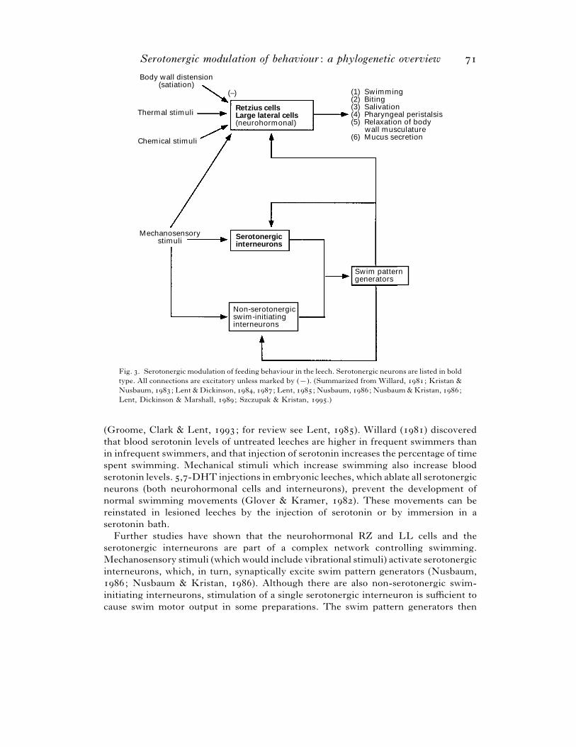
Serotonergic modulation of behaviour : a phylogenetic overview
Body wall distension(satiation)
Thermal stimuli
Chemical stimuli
(–)
Retzius cellsLarge lateral cells(neurohormonal)
Serotonergicinterneurons
Non-serotonergicswim-initiatinginterneurons
Swim patterngenerators
Mechanosensorystimuli
(1) Swimming(2) Biting(3) Salivation(4) Pharyngeal peristalsis(5) Relaxation of body wall musculature(6) Mucus secretion
Fig. . Serotonergic modulation of feeding behaviour in the leech. Serotonergic neurons are listed in bold
type. All connections are excitatory unless marked by (®). (Summarized from Willard, ; Kristan &
Nusbaum, ; Lent & Dickinson, , ; Lent, ; Nusbaum, ; Nusbaum & Kristan, ;
Lent, Dickinson & Marshall, ; Szczupak & Kristan, .)
(Groome, Clark & Lent, ; for review see Lent, ). Willard () discovered
that blood serotonin levels of untreated leeches are higher in frequent swimmers than
in infrequent swimmers, and that injection of serotonin increases the percentage of time
spent swimming. Mechanical stimuli which increase swimming also increase blood
serotonin levels. ,-DHT injections in embryonic leeches, which ablate all serotonergic
neurons (both neurohormonal cells and interneurons), prevent the development of
normal swimming movements (Glover & Kramer, ). These movements can be
reinstated in lesioned leeches by the injection of serotonin or by immersion in a
serotonin bath.
Further studies have shown that the neurohormonal RZ and LL cells and the
serotonergic interneurons are part of a complex network controlling swimming.
Mechanosensory stimuli (which would include vibrational stimuli) activate serotonergic
interneurons, which, in turn, synaptically excite swim pattern generators (Nusbaum,
; Nusbaum & Kristan, ). Although there are also non-serotonergic swim-
initiating interneurons, stimulation of a single serotonergic interneuron is sufficient to
cause swim motor output in some preparations. The swim pattern generators then
W. A. W
exert positive feedback on the serotonergic interneurons. RZ neurons are also activated
by mechanosensory stimuli and by swim pattern generators (Lent & Dickinson, ;
Nusbaum & Kristan, ; Szczupak & Kristan, ; for review see Kristan &
Nusbaum, ). Direct stimulation of RZ neurons, which releases serotonin into
extraganglionic fluid, initiates swim motor output in isolated nerve cords when the
volume of saline in the surrounding bath is similar to the blood volume in the intact
animal (Willard, ). The RZ neurons do not appear to make synaptic connections
with any swim-related neurons (Willard, ; for review see Kristan & Nusbaum,
). RZ cells, therefore, appear to promote swimming through neurohormonal
release of serotonin rather than through synaptic contacts with the swim circuitry.
Serotonin superfusion affects various central neurons involved in swimming.
Serotonin decreases the threshold current injection required for an identified non-
serotonergic swim-initiating interneuron, cell , to elicit swim episodes in isolated
nerve cords (Angstadt & Friesen, ). Serotonin also decreases inhibitory synaptic
interactions between swim motor neurons (Mangan, Cometa & Friesen, ).
However, the concentrations of serotonin used in these studies ( µ) were higher
than those necessary to initiate swim motor output in isolated nerve cords (Willard,
).
The swimming phase of feeding behaviour is followed by the consummatory phase,
which begins once a vibrational target has been located and recognized as suitable by
virtue of its warm temperature. Stimulation of RZ and LL neurons induces five
components of consummatory feeding behaviour: bite-like jaw movements, salivary
secretion, pharyngeal peristalsis, mucus secretion and a relaxation of the body wall in
preparation for meal ingestion (Lent & Dickinson, ; for reviews see Lent, ;
Lent, Dickinson & Marshall, ). Exogenous application of serotonin produces the
same effects. Jaw movement, salivation and peristalsis are evoked by serotonin
superfusion even when the effector organs are isolated from the leech, indicating that
serotonin acts directly on peripheral targets to produce these behaviours. Depletion of
serotonin in the RZ and LL cells of starved leeches causes a failure of biting, while
bathing lesioned leeches in serotonin reinstates biting.
The activity of RZ and LL cells is affected by sensory input relevant to biting
behaviour. Thermal stimulation of the lip, a major sensory determinant for bite
initiation, synaptically excites RZ and LL cells ; chemical stimulation of the lip can
excite RZ neurons (Lent & Dickinson, ; Groome & Lent, ; for review see
Lent, ). Body wall distension, which suppresses biting, hyperpolarizes RZ and LL
cells. Bathing leeches in serotonin before a meal increases the volume of blood they
ingest ; bathing well-fed, distended leeches in serotonin restores biting. These
observations suggest that the effects of distension on behaviour may be primarily
mediated through this inhibition of serotonergic neurons (Lent & Dickinson, ,
; for review see Lent, ).
After feeding, leeches have lower ganglionic and intracellular levels of serotonin than
are found in hungry leeches (Lent et al., ; for review see Lent, ). This
reduction of serotonin may account for the primary behavioural alterations that follow
feeding: increased latency to initiate swimming when exposed to a vibrational stimulus
and decreased frequency of biting (Groome et al., ).
In summary, the data indicate that the serotonergic interneurons, acting through
Serotonergic modulation of behaviour : a phylogenetic overview
classical synaptic connections, and the RZ and LL neurons, acting as neurohormonal
cells, promote swimming, while the RZ and LL cells also control the consummatory
phase of feeding behaviour. The serotonergic neurons are activated and inhibited by
appropriate environmental and internal stimuli.
() Arthropods: crustaceans
Crustaceans comprise an arthropod class in which serotonergic modulation of
behaviour has been extensively studied. In the east coast lobster Homarus americanus,
serotonin may play a role in aggressive behaviour. When two lobsters are placed
together, they engage in agonistic encounters; eventually, one emerges as dominant and
the other as subordinate (Scrivener, ; Huber & Kravitz, ; for review see
Kravitz, ). In larger groups, aggressive interactions still occur, although the
resulting hierarchies may take a variety of forms and dominance status may change over
time. Dominant animals, when approaching subordinates, assume a characteristic
posture in which the animal stands high on its walking legs with its tail flexed beneath
it and its claws raised. This posture is characterized by contraction of the postural flexor
muscles. Subordinate animals assume a posture characterized by contraction of
postural extensors, in which the animal crouches low to the substrate, with its tail
extended and its claws stretched out in front at substrate level. Livingstone, Harris-
Warrick & Kravitz () discovered that a dominant-type posture can be produced by
the injection of serotonin into the haemolymph, while a subordinate-type posture
results from octopamine injection.
Among the first steps in understanding the mechanism of this monoaminergic
modulation of posture were attempts to identify the serotonergic and octopaminergic
neurons. Beltz & Kravitz () used immunocytochemical techniques to identify
approximately serotonergic neurons in the lobster ventral nerve cord. The nerve
cord consists of a chain of ganglia: the supra-, circum-, and suboesophageal ganglia,
five thoracic and six abdominal ganglia. Each ganglion contains at least one serotonin-
immunoreactive neuronal cell body. Among the largest serotonergic neurons are two
bilaterally symmetrical neurosecretory pairs, one each in the fifth thoracic (T) and first
abdominal (A) ganglia. Each of these neurons has extensive central projections within
the neuropil of the ganglion of origin. These cells also send axonal projections out via
the thoracic second roots of at least four anterior ganglia; terminals are found in
neurosecretory regions near the base of each root and in the pericardial organs at the
distal ends of the roots (Beltz & Kravitz, ). These neurons also contain the peptide
proctolin (Siwicki, Beltz & Kravitz, ).
Schneider et al. () identified approximately octopamine-immunoreactive
neurons in the lobster ventral nerve cord. Twenty-eight neurons (two pairs in each
thoracic ganglion and four pairs in the suboesophageal ganglion) appear to be
neurosecretory. These neurons have extensive projections within the neuropil and
project out of the CNS via suboesophageal and second thoracic roots, where they form
varicosities in the neurosecretory regions near the base of each root.
The proximal neurosecretory plexuses of the thoracic ganglia were studied in detail
by Evans, Kravitz & Talamo () and by Livingstone, Schaeffer & Kravitz ().
The serotonergic and octopaminergic terminals are distinct. The terminals take up
radioactive precursors of these transmitters, synthesize the transmitters and release
W. A. W
them in a Ca#+-dependent manner when depolarized. These terminals are therefore a
site of release of serotonin and octopamine into the haemolymph. Actual concentrations
of serotonin and octopamine in the haemolymph typically fall between −* and −)
(Livingstone et al., ).
Monoaminergic modulation of posture is mediated at both the central and peripheral
levels. In the periphery, both serotonin and octopamine facilitate transmission at the
neuromuscular junction (Battelle & Kravitz, ; Florey & Rathmayer, ; for
review see Kravitz, ). Serotonin acts at the presynaptic excitatory terminal to
enhance transmitter release. This effect appears to be mediated both by the
phosphatidylinositol system (inositol triphosphate and protein kinase C) and by cyclic
AMP (Glusman & Kravitz, ; Dixon & Atwood, a, b ; Goy & Kravitz, ;
for review see Kravitz et al., ). Serotonin also increases transmitter release from
inhibitory nerve terminals (for reviews see Kravitz et al., ; Kravitz, ).
Octopamine enhances the release of excitatory transmitter, but its effects are less than
those of serotonin (Breen & Atwood, ; for review see Kravitz, ). Serotonin and
octopamine also facilitate muscle contraction at the postsynaptic level. There are four
effects produced by both amines in exoskeletal muscles: an increase in resting tension,
a small (– mV) depolarization, a small (–%) increase in input resistance, and
a decreased threshold for the generation of Ca#+ spikes (Battelle & Kravitz, ; Florey
& Rathmayer, ; for reviews see Kravitz et al., , ). Although these amines
increase cyclic AMP levels within the muscle, this increase is not the primary mediator
of aminergic effects ; agents which only raise cyclic AMP levels do not fully mimic the
effects of the amines (Battelle & Kravitz, ; Goy & Kravitz, ).
Thus, the peripheral effects of both serotonin and octopamine are to prime all
muscles, whether extensors or flexors, to respond to excitatory input. The antagonism
between the effects of the two amines is generated in the central nervous system. The
motor output of the dissected ventral nerve cord can be monitored in the presence of
serotonin and octopamine (Livingstone et al., ; Harris-Warrick & Kravitz, ).
Octopamine increases the firing rate of extensor excitor motor neurons and flexor
inhibitor motor neurons; it decreases the firing rate of extensor inhibitors and flexor
excitors. Overall, these effects combine to enhance postural extension. Conversely,
serotonin alters central motor output so as to enhance postural flexion. It increases the
firing rate of flexor excitors and extensor inhibitors, and decreases the firing rate of
flexor inhibitors and extensor excitors.
Harris-Warrick & Kravitz () investigated the cellular basis of the antagonistic
effects of serotonin and octopamine on central motor neurons by recording from M,
an identified extensor excitor, and F, an identified flexor inhibitor. They found that
octopamine reduces the necessary depolarization for spike initiation while serotonin
increases it ; these effects are dependent upon synaptic inputs and are blocked by low
Ca#+ levels. Octopamine increases the frequency of EPSPs in these cells and causes the
appearance of a new, larger size-class of EPSPs in F ; serotonin decreases the EPSP
frequency. Harris-Warrick () also looked at the effects of serotonin and octopamine
on extension-command-evoked excitation of M and F. Octopamine enhances
extension-command excitation of M and F while serotonin decreases F excitation.
Octopamine enhances the amplitude of the command-evoked EPSPs in both neurons
while serotonin reduces EPSP amplitude in F. In cases where the extension command
Serotonergic modulation of behaviour : a phylogenetic overview
Flexioncommand
Extensioncommand
A1
FlexMN
ExtMN
IN
Toposturalmuscles
A1 Ganglion
To morecaudal ganglia
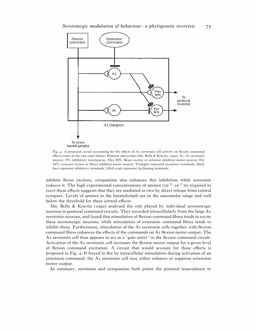
Fig. . A proposed circuit accounting for the effects of A serotonin cell activity on flexion command
effectiveness in the east coast lobster Homarus americanus (Ma, Beltz & Kravitz, ). A, A serotonin
neuron; IN, inhibitory interneuron; Flex MN, flexor excitor or extensor inhibitor motor neuron; Ext
MN, extensor excitor or flexor inhibitor motor neuron. Triangles represent excitatory terminals ; black
bars represent inhibitory terminals ; filled ovals represent facilitating terminals.
inhibits flexor excitors, octopamine also enhances this inhibition while serotonin
reduces it. The high experimental concentrations of amines (−'–−& ) required to
exert these effects suggests that they are mediated in vivo by direct release from central
synapses. Levels of amines in the haemolymph are in the nanomolar range and well
below the threshold for these central effects.
Ma, Beltz & Kravitz () analysed the role played by individual serotonergic
neurons in postural command circuits. They recorded intracellularly from the large A
serotonin neurons, and found that stimulation of flexion command fibres tends to excite
these serotonergic neurons, while stimulation of extension command fibres tends to
inhibit them. Furthermore, stimulation of the A serotonin cells together with flexion
command fibres enhances the effects of the commands on A flexion motor output. The
A serotonin cell thus appears to act as a ‘gain setter ’ in the flexion command circuit.
Activation of the A serotonin cell increases the flexion motor output for a given level
of flexion command excitation. A circuit that would account for these effects is
proposed in Fig. . If forced to fire by intracellular stimulation during activation of an
extension command, the A serotonin cell may either enhance or suppress extension
motor output.
In summary, serotonin and octopamine both prime the postural musculature to
W. A. W
respond to excitatory impulses from the CNS. In the CNS, however, the two amines act
antagonistically: serotonin biases the CNS towards the readout of the flexion motor
programme, while octopamine biases the CNS towards extension motor output.
Serotonin has a number of effects in the lobster other than those exerted on the
postural musculature and postural command circuitry. Aminergic modulation of
lobster sensory input has been observed (Pasztor & Bush, ). The lobster oval organ
contains three mechanoreceptors which project to the CNS; in the intact animal, a
sinusoidally oscillating force is applied to these receptors as the ventilatory appendage
moves water over the gills. Rhythmic stimulation produces a stable pattern of
depolarizing receptor potentials, a certain percentage of which give rise to spikes.
Application of nanomolar concentrations of serotonin or octopamine decreases the
percentage of receptor potentials which give rise to spikes. In the case of a single rather
than a sinusoidal stimulus, both amines decrease the amplitude of the resulting receptor
potentials, although the effects of octopamine are somewhat inconsistent. These effects
have concentration thresholds that can be surpassed by neurohormonal release into the
haemolymph, suggesting that serotonin and octopamine modulate sensory input in
vivo.
Serotonin also affects the circulatory system and digestive tract of the lobster.
Serotonin and octopamine both increase the intensity and frequency of the heartbeat
(Battelle & Kravitz, ; Florey & Rathmayer, ). Serotonergic fibres are observed
in the cardiac ganglion, although no serotonergic cell bodies are present (Beltz &
Kravitz, ). Serotonin may modulate the endogenous rhythm of the cardiac
ganglion through synaptic release (Hartline, ). In the gut, serotonin alters the
pyloric motor output of the lobster stomatogastric ganglion by affecting the activity of
a number of neurons. As in the heart, no serotonin-immunoreactive cell bodies are seen
in the stomatogastric ganglion; however, fibres in the stomatogastric nerve and
ganglion and the ganglionic neuropil stain for serotonin (Beltz et al., ).
Serotonergic innervation of the crustacean stomatogastric ganglion has been more
extensively studied in crabs (Cancer borealis and Cancer irroratus) than in Homarus
americanus. The sole serotonergic innervation of the ganglion is provided by two
bilateral pairs of muscle receptor cells known as the gastropyloric receptor (GPR) cells
(Katz, Eigg & Harris-Warrick, ). These cells employ both serotonin and
acetylcholine as transmitters. The dendritic processes of these cells cover muscles of the
gastric mill (which macerates food before it travels to the pylorus) as well as the pylorus
(Katz et al., ; Katz & Harris-Warrick, a). The two receptor cells on each side
of the foregut are designated as GPR and GPR, and each innervates different
muscles. GPR is excited when the muscles it innervates are stretched by movements
of the gastric mill ; it can also produce endogenous rhythmic activity in the absence of
gastric mill movement.
In the stomatogastric ganglion, there are two semi-autonomous central pattern
generators that control foregut movement: the gastric mill CPG and the pyloric CPG.
GPR activity affects neurons in both of these CPGs (Katz & Harris-Warrick, ;
Katz & Harris-Warrick, b). With regard to the gastric mill CPG, the interactions
of GPR with the dorsal gastric (DG) motor neuron are of particular interest. First,
GPR activation evokes a nicotinic cholinergic EPSP in the DG. Secondly, train
stimulation of a GPR often causes changes in the DG that allow a plateau potential to
Serotonergic modulation of behaviour : a phylogenetic overview
be elicited by brief depolarizing input. A plateau potential is a prolonged depolarization
that leads to high-frequency firing; it is followed by an after-hyperpolarization. The
effects of GPR stimulation on plateau induction in the DG are not blocked by
cholinergic antagonists and are mimicked by serotonin (Katz & Harris-Warrick, ;
Kiehn & Harris-Warrick, a). Train stimulation of a GPR can produce a series of
summating EPSPs that provide adequate depolarization to elicit a plateau potential in
the absence of further excitatory input. A plateau potential in DG causes contraction of
gastric mill muscles that stretch a muscle innervated by GPR. Therefore, a positive
feedback loop may exist : GPR excites DG, which in turn leads to further activation
of GPR. GPR exhibits endogenous activity and could initiate this cycle; alternatively,
DG could begin firing as a result of gastric mill CPG activity and cause secondary
excitation of GPR (Katz & Harris-Warrick, ).
The ionic mechanisms underlying the production of plateau potentials by DG
neurons have been explored. Serotonin modulates four different ionic currents in these
cells. First, serotonin enhances a hyperpolarization-activated net inward current
carried by Na+ and K+ ions. Secondly, serotonin reduces a Ca#+-dependent outward
current carried by K+ ions (Kiehn & Harris-Warrick, b). Thirdly, serotonin
enhances a voltage-dependent Ca#+ current that is activated by depolarization (Zhang
& Harris-Warrick, ). The resulting increase in Ca#+ influx secondarily enhances a
fourth current, a Ca#+-activated slow inward cation current (Zhang, Wootton & Harris-
Warrick, ). This serotonergic enhancement of inward currents and suppression of
outward current promotes the generation and maintenance of plateau potentials in DG
neurons.
As noted above, the GPR neurons also modulate activity in the pyloric CPG. All
neurons in this CPG appear to receive GPR input. The effects of GPR stimulation
include rapid nicotinic synaptic potentials and slower neuromodulatory input that
appears to be mediated by serotonin. GPR stimulation can initiate rhythmic activity in
the pyloric CPG or increase pyloric cycle frequency in an active preparation. This effect
largely results from a direct enhancement of bursting in the pyloric dilator}anterior
burster (PD}AB) pacemaker cell group that is mediated by serotonin (Katz & Harris-
Warrick, , b ; Zhang & Harris-Warrick, ). GPR stimulation can also
recruit gastric mill CPG neurons to fire in phase with the pyloric motor pattern. This
effect may also be mediated by serotonin (Katz & Harris-Warrick, ).
Although the GPR cells have been most extensively studied in the crab, homologous
cells are found in the lobster Homarus americanus. In the lobster, these neurons are
immunoreactive for the peptides cholecystokinin (CCK) and FMRFamide as well as
serotonin (Katz & Harris-Warrick, a, b). Future work may identify similarities
and differences between the lobster cells and their counterparts in the crab.
Another crustacean whose nervous system has been extensively studied is the crayfish
Procambarus clarkii. Injection of serotonin or octopamine into the haemolymph
produces flexed or extended postures similar to those seen in the lobster (Livingstone
et al., ). Presumably, then, serotonin and octopamine have similar effects on the
tonic postural motor system in the crayfish and lobster.
The most carefully analysed behaviour in the crayfish Procambarus clarkii is the
lateral giant escape reaction, which is a phasic rather than a tonic motor response. The
lateral giant fibres (LGs) consist of a series of large-diameter, bilaterally paired
W. A. W
segmental interneurons connected through electrical junctions (Remler, Selverston &
Kennedy, ). Mechanosensory stimuli to the tail activate the LGs, resulting in a
rapid upward movement that propels the animal away from the source of the stimulus
(Wine, ). In the intact animal, this reflex is facilitated by painful stimuli, and
inhibited by restraint or by feeding (Glanzman & Krasne, ). Electrical stimulation
of sensory roots produces a compound biphasic EPSP. The first peak of the EPSP
corresponds to monosynaptic excitation of the LG by sensory axons through nicotinic
cholinergic and rectifying electrical synapses. The second peak corresponds to
excitation of the LG through a disynaptic pathway; a sensory axon excites an
interneuron that forms a rectifying electrical synapse on the LG (Edwards et al., ;
Yeh, Opydyke & Edwards, ).
Initial experiments on adult crayfish indicated that serotonin and octopamine
produce opposing effects on the amplitude of this biphasic EPSP (Glanzman & Krasne,
). In semi-intact preparations, octopamine increases the amplitude of both
components of the EPSP, while serotonin decreases it. The second component of the
EPSP shows greater change in amplitude than the first. The activation of the LG by
sensory root shocks is correspondingly affected.
Further studies explored the mechanisms underlying these effects. Octopamine was
shown to cause the appearance of a new class of EPSP in an identified sensory
interneuron. Octopamine lowers the minimum stimulus required for interneuronal
activation, probably as a result of this modulation of exogenous inputs (Glanzman &
Krasne, ; Bustamante & Krasne, ). The net result would be the observed
enhancement of the second, or disynaptic, peak of the LG EPSP following sensory root
stimulation. Serotonin has no consistent effect on the activation threshold of the
interneuron affected by octopamine, suggesting that serotonergic inhibition of LG
activation is mediated at some other point in the circuit (Glanzman & Krasne, ).
Serotonin appears to induce a conductance increase in distal dendrites of the LG. This
increase in conductance, as well as serotonergic reduction of the first, or monosynaptic,
component of the biphasic EPSP persist in the absence of chemical synaptic
transmission, indicating that serotonin acts directly on the LG itself (Vu & Krasne,
).
Initial experiments suggested that serotonin might mediate the restraint-induced
inhibition of the escape response. A descending tonic inhibition of the LG neuron in
the crayfish suppresses LG activation during restraint. Like serotonin, this tonic
inhibition decreases the amplitude of both peaks of the biphasic LG EPSP.
Furthermore, animals whose serotonergic neurons are depleted of transmitter (by
injection of ,-DHT) fail to show normal suppression of the LG escape response when
restrained (Glanzman & Krasne, ). However, the tonic inhibition is blocked by
picrotoxin application, suggesting that it is GABAergic rather than serotonergic in
nature. Serotonin continues to reduce EPSP amplitude in the presence of picrotoxin
and therefore cannot mediate the effects of tonic inhibition on the LG (Vu & Krasne,
). It is possible, however, that serotonin plays a facilitatory role in the rostral
centres that generate tonic inhibition in vivo, and that the absence of this facilitation
blocks tonic inhibition in animals treated with ,-DHT.
Recent studies indicate that the effects of serotonin on sensory activation of the LG
may be more complex than previously suspected. Crayfish living in pairs in which one
Serotonergic modulation of behaviour : a phylogenetic overview
animal is dominant and the other subordinate show differing effects of serotonin on LG
inputs. In dominant animals, serotonin enhances both components of the biphasic
EPSP and reduces the stimulus threshold for an LG spike. In subordinate animals,
serotonin decreases the amplitude of both EPSP components and increases the stimulus
threshold. A vertebrate -HT"
receptor agonist reduces the LG EPSP and raises the
stimulus threshold regardless of dominance status, whereas a vertebrate -HT#agonist
enhances the EPSP and lowers the stimulus threshold in all animals. The ratio of -
HT"-like to -HT
#-like receptors may be lower in dominant animals and higher in
subordinates (Yeh, Fricke & Edwards, ).
Why serotonin enhances escape responses in dominant animals is not clear;
subordinates might be expected to employ escape behaviour more often. However, in
juvenile Homarus americanus, the LG tail flip may be used in an offensive manner;
animals grip the appendages of opponents with their claws and use tail flips to attempt
to tear the appendages off (Huber & Kravitz, ). Future work should elucidate the
mechanisms underlying these status-related changes in the response to serotonin. Of
particular interest is the identity of the compound or compounds that communicate the
dominance status of the animal to its nervous system. One of these compounds may be
serotonin itself.
A third crustacean in which serotonin appears to play a role in the modulation of
posture and the behavioural response to sensory stimuli is Gammarus lacustris (Helluy
& Holmes, ). In normal gammarids, a mechanical disturbance results in movement
away from a light source and burrowing behaviour. However, in gammarids infected
with the parasite Polymorphus paradoxus, a marked alteration in escape behaviour is
seen: the gammarid moves towards a light source and clings to the first object it
encounters in a flexed posture reminiscent of the serotonin-induced posture in lobsters
and crayfish. This altered behaviour makes the infected gammarids more susceptible to
predation by the definitive host of the parasite. Helluy & Holmes () found that
serotonin injected into normal gammarids causes them to adopt the photopositivity and
clinging behaviour exhibited by infected gammarids. Octopamine injected into infected
gammarids suppresses the clinging behaviour but not the photopositivity. The high
(millimolar) experimental concentrations required for serotonergic effects suggest that
serotonin acts in the CNS, where high concentrations of transmitter may routinely be
found in synaptic clefts. It appears that an endogenous postural control system in which
serotonin and octopamine act antagonistically may be manipulated by the parasite.
Serotonin is implicated not only in the control of posture, but in the modulation of the
motor response to light stimuli. Gammarus lacustris illustrates the dangerous effects of
pathological changes in behavioural modulatory systems.
() Echinoderms
In sea urchins, serotonin appears to serve as a neurotransmitter. Serotonin-
immunoreactive neurons have been found in larval Strongylocentrotus purpuratus
(Bisgrove & Burke, ). In larvae of other species, endogenous serotonin as well as
receptors that bind [$H]serotonin are present (Toneby, ; Brown & Shaver, ).
In larval Psammechinus miliaris, serotonin and its precursors stimulate muscular
activity (Gustafson, Lundgren & Treufeldt, ).
Serotonin has also been found in larvae of the starfish Pisaster ochraceus (Toneby,
W. A. W
). The nervous system of the starfish Pycnopodia helianthoides has little serotonin
but shows high levels of tryptamine, which is closely related to serotonin (-
hydroxytryptamine). In this organism, tryptamine may perform the functions served
by serotonin in other animals (Robertson & Juorio, ).
() Chordates
Serotonin is present in several cell types in vertebrates, including mast cells and
platelets (Cooper, Bloom & Roth, ). Serotonin derives its name from the fact that
it exerts potent vasoconstrictive effects in mammals (Rapport, Green & Page, ).
Serotonin is also found in the chromaffin cells of the intestinal mucosa and causes
constriction of the smooth muscle of the gut (Cooper et al., ). Wood & Mayer
() found evidence that, in the guinea pig gut, stimulation of interganglionic fibre
tracts causes the release of serotonin, which produces EPSPs in myenteric neurons
through the closing of hyperpolarizing ion channels.
All vertebrates have central serotonergic neurons. The anatomical organization is
similar in all vertebrate classes: most cell bodies cluster in the brainstem raphe, and
their axons project widely throughout the forebrain. In at least some species, including
primates, some serotonergic cell bodies are found elsewhere in the brainstem, in regions
including the reticular formation and locus coeruleus (Thor & Helke, ; for reviews
see Parent, ; To$ rk & Hornung, ). In mammals, the serotonergic raphe
neurons extend dendrites to ventricular tanycytes, the midline blood vessels, and the
dendrites of other raphe neurons, suggesting that they receive information from the
blood and cerebrospinal fluid (CSF) as well as from neighbouring neurons. Axons from
these neurons descend to the spinal cord and ascend to the hypothalamus, basal ganglia,
olfactory bulb, hippocampus and neocortex; serotonergic axons also project to the walls
of the ventricles. A single raphe neuron may send axon collaterals to more than one
forebrain region (for review see Parent, ). In the rat brainstem, serotonin-
immunoreactive neurons project to the nucleus tractus solitarii, where serotonin release
may regulate autonomic function (Thor & Helke, ).
According to early reports, the axonal varicosities of the serotonergic neurons seldom
displayed the membrane differentiation typically seen at synaptic terminals, suggesting
that these terminals released serotonin in a diffuse manner rather than onto discrete
postsynaptic neurons (for reviews see Parent, ; Papadopoulos & Parnavelas,
). More recent evidence indicates that a larger number of serotonergic terminals
than were previously suspected form conventional synaptic junctions (for review see
Papadopoulos & Parnavelas, ).
Although serotonergic neurons project widely throughout the cerebral cortex, their
projections show some specificity. In a given region, the density of serotonergic
innervation may vary from layer to layer. Furthermore, the density of serotonergic
innervation appears to be greater in primary sensory cortex than in secondary sensory
or association cortex (for review see Papadopoulos & Parnavelas, ).
Stimulation of raphe neurons in the rat or iontophoresis of serotonin onto different
areas of the brain causes a variety of effects, some excitatory, some inhibitory
(Aghajanian, ). Serotonin may facilitate or inhibit the response in a certain region
to a particular neurotransmitter, which may itself be either excitatory or inhibitory.
One especially well-characterized example is the facial nucleus of the rat (McCall &
Serotonergic modulation of behaviour : a phylogenetic overview
Aghajanian, ). Iontophoresis of serotonin onto the facial motor neurons does not
excite them; however, it facilitates the excitatory response produced by glutamate.
Serotonin application reduces the amount of glutamate required to produce a given
level of depolarization and also facilitates the excitation of facial motor neurons
produced by stimulation of the red nucleus or motor cortex. The relevance of this effect
in vivo is supported by the fact that p-chloroamphetamine (PCA), which causes the
release of endogenous serotonin from terminals, also produces this facilitation. The
effect of PCA is blocked by pre-treatment with p-chlorophenylalanine (PCPA), which
blocks serotonin synthesis by inhibiting the enzyme tryptophan hydroxylase.
Another example of serotonergic modulation of synaptic transmission is provided by
the glycinergic inhibitory inputs to the Mauthner neurons in the teleost fish Carassius
auratus (Mintz et al., ; Mintz & Korn, ). Serotonergic terminals are present
at the inhibitory synapses and serve to enhance presynaptic release of the inhibitory
neurotransmitter. Serotonin may cause a decrease in presynaptic K+ conductance as it
does in sensory terminals in Aplysia californica (for review see Kandel & Schwartz,
). Serotonin also acts postsynaptically to induce a transient inward-rectifying K+
current in the Mauthner cell (Mintz et al., ). Since the Mauthner neuron
coordinates the motor escape response to visual or auditory stimuli, serotonergic
potentiation of its inhibition will have significant behavioural effects (Eaton &
Bombardieri, ; Rock, Hackett & Brown, ; Hackett & Faber, ).
In vertebrates, serotonin may exert widespread effects on the state of the central
nervous system that secondarily alter all behavioural interactions between the animal
and its environment. An example is provided by the proposed role of serotonin in
arousal in mammals. Slow-wave sleep, in which an animal is relatively unresponsive to
external sensory input, is characterized by slow, synchronous, rhythmic activity of
thalamocortical circuits and synchronization of the electroencephalogram (EEG).
Increased release of serotonin and other modulatory transmitters (including nor-
epinephrine, acetylcholine and histamine) may diminish the ability of thalamocortical
circuits to generate these synchronous oscillations and promote desynchronous neural
activity and arousal, with a concomitant increase in responsiveness to environmental
stimuli (for reviews see McCormick, a, b).
There are a large number of studies in vertebrates that suggest that serotonin also
modulates specific behaviours, including feeding, sexual and aggressive behaviour.
Injection of PCPA into the ventricles of rats causes an increase in food intake and a
corresponding increase in body mass relative to saline-injected controls (Breisch,
Zemlan & Hoebel, ). In juvenile rats, intraventricular injection of ,-DHT, which
depletes serotonin, results in increased food intake and growth (Saller & Stricker,
). These results suggest that the net effect of serotonin is a tonic inhibition of
feeding behaviour in the normal rat. This hypothesis is further supported by the fact
that drugs that enhance serotonin release and block serotonin reuptake (such as
fenfluramine) or simply block reuptake (such as sertraline) cause anorexia in rats. Some
direct serotonergic agonists are also anorectics. However, -OH-DPAT [-hydroxy--
(di-n-propylamino)tetralin], a selective -HT"A
agonist, can actually increase food
intake in free-feeding rats (for review see Garattini, Mennini & Samanin, ). This
effect is difficult to interpret because -HT"A
receptors mediate autoinhibition of
serotonergic raphe neurons (for review see Hen, ). Therefore, a -HT"A
agonist
W. A. W
may simultaneously reduce the release of endogenous serotonin and stimulate post-
synaptic serotonin receptors.
The role of serotonin in the modulation of sexual behaviour has been examined in
rats of both sexes. In male rats, the post-ejaculatory refractory period is defined as the
time from ejaculation to the next mount or intromission. McIntosh & Barfield ()
depleted serotonin in male rats using three different methods: systemic administration
of PCPA, intraventricular or intraraphe injection of ,-DHT, and electrolytic lesion
of the dorsal raphe. Each of these interventions produces a significant decrease in
refractory period, implicating serotonin as a tonic enhancer of the refractory period in
vivo. Serotonin also appears to play a role in inhibition of female mating behaviour. In
female rats, removal of the ovaries decreases the frequency of the normal lordotic
response to male mounting. In ovariectomized females, several methods have been used
to reduce the effects of endogenous serotonin: application of systemic PCPA, systemic
methysergide, intrahypothalamic methysergide or cinanserin (serotonin receptor
blockers) and intrahypothalamic ,-DHT. All of these treatments increase the
frequency of the lordotic response (Zemlan et al., ; Luine et al. ). Serotonin,
therefore, appears to inhibit certain aspects of sexual behaviour in both males and
females: it prolongs the refractory period in males and inhibits the lordotic response in
females.
Finally, several studies have implicated serotonin in the control of aggressive
behaviour in a number of vertebrates. Male gymnotid fish (Apteronotus leptorhynchus)
use transient increases in electric organ discharge (EOD) frequency, known as chirps,
as aggressive signals to other males. Maler & Ellis () elicited such chirps by
stimulating fish with a simulated EOD. They found that intraventricular injection of
serotonin results in a decrease in the chirping response. Application of norepinephrine
enhances chirping, while dopamine has inconsistent effects. These opposing effects of
serotonin and norepinephrine (the vertebrate analogue of octopamine) on aggressive
displays in fish are reminiscent of the opposing actions of serotonin and octopamine on
dominance-related postures in the lobster Homarus americanus.
Golebiewski & Romaniuk () found that carbachol (a cholinergic agonist) elicits
growling when injected into the anteromedial hypothalamus of the cat. Injection of
serotonin or methysergide alone has no effect; however, serotonin inhibits and
methysergide potentiates carbachol-evoked growling. ,-DHT injection also potenti-
ates carbachol induction of growling. In this case, serotonin appears to act as an
inhibitory neuromodulator of cholinergic function.
Other studies have examined the role played by serotonin in conspecific and
interspecific aggression in rats. With regard to conspecific aggression, Vergnes et al.
() reported that injection of ,-DHT into the lateral hypothalamus of rats results
in an increased frequency and duration of offensive behaviours when an intruder rat is
placed in the cage. Effects of more global lesions vary. Vergnes, Depaulis & Boehrer
() found that a systemic PCPA injection enhances offensive behaviour when a rat
is confronted with an intruder. Sijbesma et al. () reported that ,-DHT lesions
of the dorsal and median raphe cause a modest reduction of offensive behaviour against
an intruder. File, Hyde & MacLeod () found that lesion of the median raphe alone
causes an increase in jumping and standing on an intruder, whereas lesion of the dorsal
raphe alone causes a non-specific decrease in social interaction with an intruder.
Serotonergic modulation of behaviour : a phylogenetic overview
Some of the variation in experimental results may be due to differences in the strain
of rat used, the rats’ environment, or the parameters used to quantify aggressive
behaviour. However, it appears that serotonin exerts a mixed effect on conspecific
aggressive behaviour. Certain populations of serotonergic neurons and serotonergic
projections to certain areas apparently facilitate such behaviour, while other neurons
and their projections exert an inhibitory effect (Sijbesma et al., ).
Differing effects of serotonin on conspecific aggression may be mediated through
different receptor subtypes. Genetic knockout mice lacking the -HT"B
receptor attack
intruder mice more quickly and more intensely than wild-type controls (Saudou et al.,
). Further evidence for the role of the -HT"B
receptor in the control of aggressive
behaviour is provided by serotonergic agonists that are specific for certain receptor
subtypes. Eltoprazine, widely used as a -HT"A/"B
agonist, reduces conspecific
aggressive behaviour in rats and mice in a variety of behavioural tests (for review see
Miczek et al., ). Both -HT"A
and -HT"B
receptors mediate autoinhibition of
serotonergic raphe neurons (for review see Hen, ). However, eltoprazine exerts
anti-aggressive effects in rats even after lesioning of the raphe nuclei with ,-DHT,
thereby ruling out an autoinhibitory mechanism of action (Sijbesma et al., ).
Injection of eltoprazine into the lateral ventricles of rats suppresses aggression, whereas
injection of -OH-DPAT, a specific -HT"A
agonist, does not (Mos et al., ). These
results suggest that the anti-aggressive effects of eltoprazine may be mediated primarily
through -HT"B
receptors on non-serotonergic neurons. -HT"B
receptors appear to be
present on the terminals of various types of neurons; in the hippocampus, -HT"B
receptors present on cholinergic terminals inhibit acetylcholine release (for review see
Hen, ).
The evidence to date is consistent with the following hypothesis : serotonin, acting
through -HT"B
receptors to modulate the release of other transmitters, suppresses at
least some types of conspecific aggressive behaviour in rats and mice. The absence of
these receptors in the genetic knockout mice may prevent this inhibition and increase
aggression. However, more data must be gathered on the effects of eltoprazine and the
-HT"B
receptors on both aggressive and non-aggressive behaviours and on various
transmitter systems before firm conclusions can be reached. It should be noted that
eltoprazine has some affinity for -HT"C
receptors. Furthermore, it acts only as a weak
agonist at -HT"A
receptors on rat hippocampal pyramidal neurons and can actually
reduce the effects of applied serotonin (Joe$ ls et al., ). Therefore, caution must be
used in interpreting studies in which eltoprazine is assumed to act as a -HT"A/"B
agonist.
Molecular biological techniques may further clarify the role of serotonin in
conspecific aggression. Chen et al. () found that intruder mice missing one copy of
the gene encoding α-calcium-calmodulin-dependent kinase II show increased ag-
gression in response to attacks by resident mice. This enhancement of defensive
aggression may result from decreased fear. Serotonin release from dorsal raphe neurons
appears to be reduced in these mutants, and altered serotonergic function may
contribute to the observed behavioural effects of the mutation. However, many non-
serotonergic neurons are undoubtedly affected by this mutation, and transmitters other
than serotonin may play a role in the noted behavioural changes.
With regard to interspecific, or predatory, aggression, Vergnes & Kempf ()
W. A. W
found that when naive rats receive ,-DHT injections into the lateral hypothalamus,
a significantly higher percentage will kill mice on their first exposure to them. Injection
of ,-DHT into the medial hypothalamus, septum, hippocampus, amygdala, or
cingulate cortex fails to affect muricidal behaviour. Molina et al. () found that
–% of rats who do not spontaneously kill mice develop killing behaviour following
systemic PCPA injection or electrolytic raphe lesions. Both of these lesioned groups of
rats, as well as rats who spontaneously kill mice, show a reversible and dose-dependent
inhibition of muricidal behaviour when systemically treated with serotonergic agonists
(-methoxy-N,N-dimethyltryptamine or -OH-DPAT) or serotonin reuptake blockers
(fluoxetine, also known as Prozac, or citalopram). These studies suggest that serotonin
is an inhibitory modulator of predatory aggression in the rat, and that its locus of action
may be the lateral hypothalamus.
Earlier studies supported the importance of the lateral hypothalamus in predatory
aggression. Carbachol, acetylcholine plus physostigmine or neostigmine (cholinesterase
inhibitors), and neostigmine alone all facilitate muricidal behaviour when injected into
the lateral hypothalamus of the rat, while lateral hypothalamic injection of atropine (a
muscarinic agonist) inhibits such behaviour (Smith, King & Hoebel, ; Bandler,
). In rats who do not spontaneously kill mice, lateral hypothalamic injection of
carbachol or neostigmine may induce such behaviour (Smith et al., ). Thus, it
appears that lateral hypothalamic acetylcholine is a trigger of interspecific aggression in
rats. The control of offensive or killing behaviour by the lateral hypothalamus may be
similar to the control of growling by the anteromedial hypothalamus in the cat.
Acetylcholine may elicit aggressive behaviour, while serotonin inhibits it.
Serotonin also appears to modulate complex social behaviour in primates. Raleigh et
al. () explored the role of serotonin on dominance status in vervet monkeys
(Cercopithecus aethiops sabaeus). In these animals, male dominance appears to depend
more on the formation of alliances with female group members than on individual
fighting ability. At the beginning of the study, stable mixed-sex groups were
established; each group contained one dominant and two subordinate males. The
dominant male was removed and one of the two remaining males received a drug that
affected the serotonergic system. Fenfluramine, which acts as an indirect agonist in the
short term but causes serotonin depletion in the long term, or cyproheptadine, a
serotonergic receptor antagonist, were used to suppress serotonergic function.
Tryptophan, a serotonin precursor, or fluoxetine, a reuptake inhibitor, were used to
enhance serotonergic function. Fenfluramine and cyproheptadine decreased affiliative
behaviours (approaching, grooming and proximity), while aggressive behaviour
increased; animals receiving these treatments invariably remained subordinate.
Tryptophan and fluoxetine increased affiliative behaviours and decreased aggression;
animals receiving these treatments invariably became dominant.
In this example, it appears that serotonergic suppression of aggressive behaviour
promotes dominance. However, the long-term effects of drugs intended to diminish or
enhance serotonergic function are not fully understood. For example, fluoxetine is a
serotonin reuptake blocker, and in the short term it should increase the amount of
serotonin in synaptic clefts or extracellular fluid. However, this increase in serotonin
will enhance activation of autoinhibitory receptors and may cause down regulation of
postsynaptic receptors. In the long term, it is difficult to determine whether
Serotonergic modulation of behaviour : a phylogenetic overview
serotonergic function will remain elevated. Similar arguments could be applied to the
other drugs used in this study. Raleigh & McGuire () noted that the effects of
tryptophan on aggressive behaviour vary for animals in different social circumstances.
Dominant animals in a social group, subordinate animals in a group, and animals living
in isolation differ in the degree and even the direction of their response to tryptophan.
Nonetheless, it is clear that drugs that affect serotonergic function also affect affiliative
and aggressive behaviour and can lead to changes in dominance status.
Yodyingyuad et al. () examined the relationship of serotonergic function to
dominance status in talapoin monkeys (Miopithecus talapoin). They measured levels of
-HIAA (-hydroxyindoleacetic acid, a serotonin metabolite) in the CSF of monkeys
before and after the formation of social dominance hierarchies as well as in established
hierarchies. They found that when a group of monkeys is placed together, -HIAA
levels fall as a monkey becomes dominant and rise as a monkey becomes subordinate.
Monkeys who assume intermediate positions show no consistent changes in CSF -
HIAA. In established groups, the top-ranking monkeys have significantly lower -
HIAA levels than intermediate or low-ranking monkeys, who do not differ significantly
from each other. No correlation was found between levels of homovanillic acid (a
dopamine metabolite) and position in the dominance hierarchy.
As with the work on vervet monkeys, this study clearly supports a link between the
central serotonergic system and dominance status. At first glance, reduced CSF -
HIAA levels might suggest lowered central serotonin levels and diminished
serotonergic function. However, many factors could potentially affect -HIAA levels as
well as the ratio of serotonin to -HIAA in the brain. Decreased release of serotonin,
which would diminish serotonergic function, would lower -HIAA; reduced serotonin
degradation, which would enhance serotonergic function, would also lower -HIAA.
For example, juvenile mice lacking functional monoamine oxidase type A (MAOA),
which degrades serotonin as well as other amines, have increased levels of brain
serotonin and reduced levels of -HIAA (Cases et al., ). Agonistic encounters can
raise the ratio of -HIAA to serotonin in the amygdalas of inexperienced mice; this
observation suggests that situations that result in serotonin release may alter this ratio
in the short term (for review see Miczek et al., ). Chronically raised or lowered
levels of -HIAA indicate alterations in the serotonergic system, but it is difficult to say
whether overall serotonergic function is enhanced or diminished.
Serotonin clearly serves as a behavioural modulator in humans. Several drugs that
specifically affect serotonergic transmission are used to treat psychiatric illnesses. The
serotonin reuptake blockers fluoxetine and fluvoxamine effectively treat depression,
bulimia, panic attacks and obsessive-compulsive disorder (Schatzberg & Cole, ).
The -HT"A/"B
agonist eltoprazine may suppress pathological aggression and self-
injurious behaviour in some patients (Verhoeven et al., ; Kohen, ; Tiihonen
et al., ). Many other drugs that affect the serotonergic system along with
noradrenergic or dopaminergic function are used to treat a wide variety of mental
illnesses (Schatzberg & Cole, ).
Although the behavioural effects of these drugs are obvious, the mechanisms
underlying their effects are difficult to unravel. As noted above with regard to non-
human primate studies, long-term administration of a drug may cause changes both up-
and downstream of its initial site of action. Some of these poorly understood long-term
W. A. W
changes are clearly important for clinical efficacy. For example, fluoxetine may take
weeks to relieve depression even though maximum blood levels of the drug and effective
inhibition of serotonin reuptake are achieved much sooner.
A number of studies have sought a relationship between indices of serotonergic
function and self-directed aggression (suicide or suicide attempts) or outwardly directed
aggressive behaviour. A few studies have reported decreased levels of serotonin in the
brainstems of suicide victims, while other studies found no change. Some studies of
suicide victims found decreased levels of -HIAA in various regions of the brain,
although other studies did not confirm these findings (for review see Mann et al., ).
Several studies have found lowered levels of CSF -HIAA in subjects who have
attempted suicide as compared to various controls (for reviews see van Praag, Plutchik
& Conte, ; Coccaro, ). A number of studies indicate that lower levels of CSF
-HIAA correlate with aggressive, violent or criminal behaviour and with ratings of
hostility. In particular, some studies suggest that lower CSF -HIAA levels correlate
with impulsive aggression (for reviews see van Praag et al., ; Coccaro, ;
Golden et al., ). The potential pitfalls in using -HIAA as a measure of
serotonergic function have already been discussed. Furthermore, many of the subjects
in these studies took drugs that may have altered -HIAA levels (for reviews see Mann
et al., ; Golden et al., ). Other available indices of serotonergic function (post-
mortem analyses of agonist binding to serotonergic receptors or, in living patients,
hormonal responses to serotonin mimetics) are similarly difficult to interpret (for
reviews see Coccaro, ; Mann et al., ; Golden et al., ).
Molecular techniques may uncover a genetic basis for some of the differences noted
between suicidal or aggressive patients and controls. One recent study revealed a
correlation between a particular allele of tryptophan hydroxylase and a history of
suicide attempts in a group of alcoholic violent offenders (Nielsen et al., ).
Another study focused on a family that displayed an X-chromosome-linked
behavioural disturbance characterized by a tendency towards impulsivity and
aggression (Brunner et al., a, b). Affected males were found to have a mutation in
the gene for MAOA. An inappropriate termination codon resulted in a complete loss
of activity of this enzyme. Altered serotonergic function was indicated by increased
urinary levels of serotonin and decreased levels of -HIAA. However, excretion of the
metabolites of other amines, including norepinephrine and dopamine, was also
lowered; alterations in these other aminergic systems may contribute to the behavioural
effects of this mutation in humans.
A subsequent study has shown that transgenic adult male mice lacking functional
MAOA also show increased aggression (Cases et al., ). These mice attack intruders
more quickly than non-transgenic controls. Transgenic juvenile mice lacking MAOA
have increased brain levels of serotonin and norepinephrine and decreased brain levels
of -HIAA. As these mice age, however, their levels of serotonin, norepinephrine and
-HIAA eventually return to normal, perhaps as a result of an increase in activity of
monoamine oxidase type B (MAOB). There are two possible explanations for persistent
behavioural alterations. First, despite the evident compensation for the missing
enzyme, it is possible that short-term imbalances in amine metabolism could still occur.
For instance, transient increases in transmitter release triggered by environmental
stimuli might temporarily overload compensatory mechanisms. Secondly, exposure to
Serotonergic modulation of behaviour : a phylogenetic overview
high levels of amines during development could result in permanent structural
abnormalities in the brain. Cytoarchitectural changes have been discovered in the
somatosensory cortex of the transgenic mice and may be present in other brain regions
as well. Further study of these mice should yield more detailed information about the
mechanisms underlying the effects of a mutation that appears to alter human behaviour.
III. CONCLUSIONS
Serotonergic neurons are present in all phyla that possess nervous systems, and in
most of these phyla, serotonin modulates important behaviours, including feeding,
sexual and aggressive behaviour. In invertebrates, the mechanisms underlying these
effects have been well characterized. In the nematode Caenorhabditis elegans, serotonin
enhances feeding and egg release. In the mollusc Aplysia californica, serotonergic
neurons potentiate biting and enhance a defensive reflex. In the mollusc Tritonia
diomedea, serotonin acts as both a classical neurotransmitter and as a neuromodulator
in a neural network that generates escape swimming. In the leech (Hirudo medicinalis,
Macrobdella decora, Haementeria ghilianii), serotonin plays an important role in the
initiation and modulation of both appetitive and consummatory components of feeding
behaviour. In the lobster Homarus americanus, serotonin promotes the adoption of a
flexed or dominant-type posture. In the crayfish Procambarus clarkii, serotonin affects
sensory activation of the circuitry controlling tail flips, which may be used defensively
or offensively in aggressive encounters; the direction of the effect depends on the
dominance status of the animal.
Three basic mechanisms underlie the effects of serotonin on invertebrate nervous
systems. In the central nervous system, serotonin may act as a classical neurotransmitter
to initiate a behaviour; an example is provided by the serotonergic swim-initiating
interneurons in the leech (Hirudo medicinalis, Macrobdella decora). More commonly,
serotonin acts as a neuromodulator to enhance transmission at a synapse which
employs some other compound as the primary transmitter; an example is serotonergic
facilitation of presynaptic release from sensory terminals in Aplysia californica. In
Tritonia diomedea, these two roles of serotonin are combined: serotonin released by the
dorsal swim interneurons acts both as a classical neurotransmitter and as a
neuromodulator. A third role for serotonin is that of a neurohormone: through release
into the circulation, it can affect sensory neurons or alter transmission at neuromuscular
junctions. Such effects are observed in the lobster Homarus americanus as well as in
other invertebrates.
Serotonin also modulates behaviour in vertebrates. It appears to inhibit feeding and
sexual behaviour in rats and many studies support an important role for serotonin in the
control of aggression. Serotonin inhibits aggressive vocalizations in gymnotid fish
(Apteronotus leptorhynchus) and cats and may inhibit predatory aggression in rats. With
regard to rat conspecific aggression, serotonin may act on different receptor subtypes or
in different brain regions to facilitate or inhibit such behaviour. In non-human
primates, alterations in serotonergic function clearly correlate with changes in
aggressive behaviour and dominance status. In humans, drugs that affect serotonergic
function produce a variety of behavioural effects ; in particular, studies of -HIAA
levels and of genes for enzymes that synthesize or degrade serotonin indicate that
serotonin modulates aggressive behaviour.

W. A. W
The mechanisms by which serotonin affects behaviour are not as well understood in
vertebrates as in invertebrates. In invertebrates, only a few serotonergic neurons are
present in the nervous system. These neurons can be reliably identified in each animal
and their interactions with other identified neurons in well-defined circuits can be
determined. In vertebrates, no individual neurons can be identified from preparation to
preparation. It is difficult to predict the long-term effects of drugs that alter
serotonergic function and is often impossible even to tell whether overall serotonergic
function is enhanced or diminished. While it is possible to examine various indices of
ongoing serotonergic activity, such as the levels of serotonin metabolites in the nervous
system or urine, these results are likewise difficult to interpret. More thorough analysis
of simpler organisms may suggest future directions for the study of serotonergic actions
in more complex systems.
IV. ACKNOWLEDGEMENTS
The author would like to thank Dr Edward A. Kravitz for his critical reading of the original version of this
manuscript and his many helpful suggestions, and would also like to thank Dr Doreen Valentine for her valuable
recommendations. This work was supported by Public Health Service National Research Service Award T
GMO, a Ryan Fellowship, and a Sackler Scholarship in Psychobiology to W. A. Weiger, and National
Institute of Neurological Disorders and Stroke Grant NS- to E. A. Kravitz.
V. REFERENCES
A, F. D. & W, A. O. D. (). Plasticity of a fixed action pattern in the sea slug Tritonia diomedia.
Communications in Behavioral Biology , –.
A, G. K. (). The modulatory role of serotonin at multiple receptors in brain. In Serotonin
Neurotransmission and Behavior (ed. B. L. Jacobs and A. Gelperin), pp. –. MIT Press, Cambridge.
A, M. (). Modulation of a rhythmic activity by serotonin via cyclic AMP in the coelenterate Renilla
koX llikeri. Journal of Comparative Physiology B , –.
A, J. D. & F, W. O. (). Modulation of swimming behavior in the medicinal leech. I. Effects of
serotonin on the electrical properties of swim-gating cell . Journal of Comparative Physiology A , –.
B, B. J., H, B., M-S, M., A, S. R., K, B.-K., K, E. R. & T, R. Y.
(). Spatially resolved dynamics of cAMP and protein kinase A subunits in Aplysia sensory neurons. Science
, –.
B, C. H. & C, M. (). Time course of structural changes at identified sensory neuron synapses during
long-term sensitization in Aplysia. Journal of Neuroscience , –.
B, C. H., C, M., K, F. & K, E. R. (). Serotonin-mediated endocytosis of apCAM: an
early step of learning-related synaptic growth in Aplysia. Science , –.
B, R. J. (). Cholinergic synapses in the lateral hypothalamus for the control of predatory aggression in
the rat. Brain Research , –.
B, B.-A. & K, E. A. (). Targets of octopamine action in the lobster: cyclic nucleotide changes
and physiological effects in hemolymph, heart and exoskeletal muscle. Journal of Pharmacology and Experimental
Therapeutics , –.
B, B. S., E, J. S., F, F., H-W, R. M., H, S. L. & M, E. (). Serotonergic
innervation and modulation of the stomatogastric ganglion of three decapod crustaceans (Panuliris interruptus,
Homarus americanus and Cancer irroratus). Journal of Experimental Biology , –.
B, B. S. & K, E. A. (). Mapping of serotonin-like immunoreactivity in the lobster nervous system.
Journal of Neuroscience , –.
B, B. S. & K, E. A. (). Physiological identification, morphological analysis, and development of
identified serotonin-proctolin containing neurons in the lobster nerve cord. Journal of Neuroscience , –.
B, P. J., S, J. D., W, I., W, K. R., K, E. R. & S, J. H. (). Protein
synthesis during acquisition of long-term facilitation is needed for the persistent loss of regulatory subunits of
the Aplysia cAMP-dependent protein kinase. Proceedings of the National Academy of Sciences , –.
B, B. W. & B, R. D. (). Development of serotonergic neurons in embryos of the sea urchin,
Strongylocentrotus purpuratus. Development, Growth and Differentiation , –.
Serotonergic modulation of behaviour : a phylogenetic overview
B, C. A. & A, H. L. (). Octopamine – a neurohormone with presynaptic activity-dependent effects
at crayfish neuromuscular junctions. Nature , –.
B, S. T., Z, F. P. & H, B. G. (). Hyperphagia and obesity following serotonin depletion by
intraventricular p-chlorophenylalanine. Science , –.
B, K. M. & S, J. R. (). [$H]serotonin binding to blastula, gastrula, prism, and pluteus sea urchin
embryo cells. Comparative Biochemistry and Physiology C, –.
B, H. G., N, M., B, X. O., R, H. H. & O, B. A. (a). Abnormal behavior
associated with a point mutation in the structural gene for monoamine oxidase A. Science , –.
B, H. G., N, M. R., Z, P., A, N. G. G. M., G, A. H., W,
E. C., K, M. A., R, H. H. & O, B. A. (b). X-linked borderline mental retardation with
prominent behavioral disturbance: phenotype, genetic localization, and evidence for disturbed monoamine
metabolism. American Journal of Human Genetics , –.
B, J. & K, F. (). Effects of octopamine on unitary EPSPs in a first order interneuron of
crayfish lateral giant escape circuit. Society for Neuroscience Abstracts , .
C, O., S, I., G, J., G, P., C, K., P, S., M$ , U., A, M., B, C., C
S, J. & D M, E. (). Aggressive behavior and altered amounts of brain serotonin and
norepinephrine in mice lacking MAOA. Science , –.
C, P. & R, S. (). Cytochemical ultrastructural and fluorescence study of the nervous net of Hydra
sp. Journal of Submicroscopic Cytology , –.
C, V. F., B, H., G, P. & K, E. R. (). Inhibitor of protein synthesis blocks
long-term behavioral sensitization in the isolated gill-withdrawal reflex of Aplysia. Journal of Neurobiology ,
–.
C, C., R, D. G., G, R. W. & T, S. (). Abnormal fear response and aggressive
behavior in mutant mice deficient for α-calcium-calmodulin kinase II. Science , –.
C, H. J., W, K. R. & K, I. (). An identified histaminergic neuron modulates feeding motor
circuitry in Aplysia. Journal of Neuroscience , –.
C, E. F. (). Central serotonin and impulsive aggression. British Journal of Psychiatry (suppl. ),
–.
C, J. R., B, F. E. & R, R. H. (). The Biochemical Basis of Neuropharmacology. Oxford
University Press, New York.
D, D. & A, H. L. (a). Phosphatidylinositol system’s role in serotonin-induced facilitation at the
crayfish neuromuscular junction. Journal of Neurophysiology , –.
D, D. & A, H. L. (b). Conjoint action of phosphatidylinositol and adenylate cyclase systems in
serotonin-induced facilitation at the crayfish neuromuscular junction. Journal of Neurophysiology , –.
E, R. C. & B, R. A. (). Behavioral functions of the Mauthner neuron. In Neurobiology of the
Mauthner Cell (ed. D. S. Faber and H. Korn), pp. –. Raven Press, New York.
E, D. H., H, W. J., L, E. M. & F, R. A. (). Postsynaptic modulation of rectifying
electrical sysnaptic inputs to the LG escape command neuron in crayfish. Journal of Neuroscience , –.
E, P. D., K, E. A. & T, B. R. (). Octopamine release at two points along lobster nerve
trunks. Journal of Physiology (Cambridge) , –.
F, S. E., H, J. R. G. & ML, N. K. (). ,-dihydroxytryptamine lesions of dorsal and median
raphe! nuclei and performance in the social interaction test of anxiety and in a home-cage aggression test. Journal
of Affective Disorders , –.
F, E. & R, M. (). The effects of octopamine and other amines on the heart and on
neuromuscular transmission in decapod crustaceans: further evidence for a role as neurohormone. Comparative
Biochemistry and Physiology C, –.
F, W. N., C, G. A. & K, E. R. (). Parallel processing of short-term memory for sensitization
in Aplysia. Journal of Neurobiology , –.
G, S., M, T. & S, R. (). Reduction of food intake by manipulation of central serotonin:
current experimental results. British Journal of Psychiatry (suppl. ), –.
G, M., B, O., H, B., M, P. G., K, E. R. & D, N. (). Roles of PKA
and PKC in facilitation of evoked and spontaneous transmitter release at depressed and nondepressed synapses
in Aplysia sensory neurons. Neuron , –.
G, M., M P. G. & K, E. R. (). A novel intermediate stage in the transition between
short- and long-term facilitation in the sensory to motor neuron synapse of Aplysia. Neuron , –.
G, D. L., K, E. R. & S, S. (). Target-dependent structural changes accompanying
long-term synaptic facilitation in Aplysia neurons. Science , –.
W. A. W
G, D. L. & K, F. B. (). Serotonin and octopamine have opposite modulatory effects on the
crayfish’s lateral giant escape reaction. Journal of Neuroscience , –.
G, D. L. & K, F. B. (). ,-dihydroxytryptamine lesions of crayfish serotonin-containing
neurons: effect on the lateral giant escape reaction. Journal of Neuroscience , –.
G, D. L., M, S. L., H, R. D., D, A. M., L, P. E. & K, E. R. ().
Depletion of serotonin in the nervous system of Aplysia reduces the behavioral enhancement of gill withdrawal
as well as the heterosynaptic facilitation produced by tail shock. Journal of Neuroscience , –.
G, J. G. & K, A. P. (). Serotonin analog selectively ablates identified neurons in the leech embryo.
Science , –.
G, S. & K, E. A. (). The action of serotonin on excitatory nerve terminals in lobster nerve-
muscle preparations. Journal of Physiology (Cambridge) , –.
G, R. N., G, J. H., C, M. H. N., E, R. D., K, B. T. & G, J. C. ().
Serotonin, suicide, and aggression: clinical studies. Journal of Clinical Psychiatry (suppl.), –.
G, H. & R, A. (). The participation of serotonergic system in the regulation of emotional-
defensive behavior evoked by intrahypothalamic carbachol injections in the cat. Acta Neurobiologiae
Experimentalis , –.
G, M. F. & K, E. A. (). Cyclic AMP only partially mediates the actions of serotonin at lobster
neuromuscular junctions. Journal of Neuroscience , –.
G, S. M., C, V. F., B, H. & S, J. H. (). A molecular mechanism for long-
term sensitization in Aplysia. Nature , –.
G, J. R., C, M. & L, C. M. (). The behavioural state of satiation in the leech is regulated by
body distension and mimicked by serotonin depletion. Journal of Experimental Biology , –.
G, J. R. & L, C. M. (). Thermal and chemical inputs differentially affect serotonin neurons that
express feeding by the medicinal leech. Society for Neuroscience Abstracts , .
G, T., L, B. & T, R. (). Serotonin and contractile activity in the echinopluteus.
A study of the cellular basis of larval behavior. Experimental Cell Research , –.
H, J. T. & F, D. S. (). Mauthner axon networks mediating supraspinal components of the startle
response in goldfish. Neuroscience , –.
H, D. W., M, A. G., J, C. F. & F, I. (). Occurrence of -hydroxytryptamine
(serotonin) in the nervous system of a monogenean, Diclidophora merlangi. Parasitology Research , –.
H-W, R. M. (). Amine modulation of extension command element-evoked motor activity in the
lobster abdomen. Journal of Comparative Physiology A , –.
H-W, R. M. & K, E. A. (). Cellular mechanisms for modulation of posture by octopamine
and serotonin in the lobster. Journal of Neuroscience , –.
H, D. K. (). Integrative neurophysiology of the lobster cardiac ganglion. American Zoologist , –
H, R. D. (). Localization of potential serotonergic facilitator neurons in Aplysia by glyoxylic acid
histofluorescence combined with retrograde fluorescent labeling. Journal of Neuroscience , –.
H, R. D. & S, S. (). Identified facilitator neurons L and L are excited by cutaneous
stimuli used in dishabituation, sensitization, and classical conditioning of Aplysia. Journal of Neuroscience ,
–.
H, S. & H, J. C. (). Serotonin, octopamine and the clinging behavior induced by the parasite
Polymorphus paradoxus (Acanthocephala) in Gammarus lacustris (Crustacea). Canadian Journal of Zoology ,
–.
H, R. (). Of mice and flies : commonalities among -HT receptors. Trends in Pharmacological Sciences ,
–.
H, B., K, M., S, S. & K, E. R. (). Additional component in the cellular mechanism
of presynaptic facilitation contributes to behavioral dishabituation in Aplysia. Proceedings of the National
Academy of Sciences , –.
H, H. R., C, M., T, C., S, J. E. & E, P. D. (). Serotonin and octopamine in the
nematode Caenorhabditis elegans. Science , –.
H, R. & K, E. A. (). A quantitative analysis of agonistic behavior in juvenile American lobsters
(Homarus americanus L.). Brain, Behavior and Evolution , –.
J$ , M., P, C. M. A., S, H. & S, J. (). Eltoprazine suppresses hyperpolarizing
responses to serotonin in rat hippocampus. Journal of Pharmacology and Experimental Therapeutics ,
–.
K, B.-K., K, E. R. & G, S. G. N. (). Activation of cAMP-responsive genes by stimuli that
produce long-term facilitation in Aplysia sensory neurons. Neuron , –.
Serotonergic modulation of behaviour : a phylogenetic overview
K, E. R., K, M., B, C. H., H, R. D., C, V. F., L, B. W. & S,
J. H. (). Serotonin, cyclic AMP, and the modulation of the calcium current during behavioral arousal. In
Serotonin Neurotransmission and Behavior (ed. B. L. Jacobs and A. Gelperin), pp. –. MIT Press,
Cambridge.
K, E. R. & S, J. H. (). Molecular biology of learning: modulation of transmitter release.
Science , –.
K, P. S., E, M. H. & H-W, R. M. (). Serotonergic}cholinergic muscle receptor cells in the
crab stomatogastric nervous system: I. Identification and characterization of the gastropyloric receptor cells.
Journal of Neurophysiology , –.
K, P. S. & F, W. N. (a). Intrinsic neuromodulation in the Tritonia swim CPG: the serotonergic
dorsal swim interneurons act presynaptically to enhance transmitter release from interneuron C. Journal of
Neuroscience , –.
K, P. S. & F, W. N. (b). Intrinsic neuromodulation in the Tritonia swim CPG: serotonin mediates
both neuromodulation and neurotransmission by the dorsal swim interneurons. Journal of Neurophysiology ,
–.
K, P. S., G, P. A. & F, W. N. (). Dynamic neuromodulation of synaptic strength intrinsic to
a central pattern generator circuit. Nature , –.
K, P. S. & H-W, R. M. (). Serotonergic}cholinergic muscle receptor cells in the crab
stomatogastric nervous system: II. Rapid nicotinic and prolonged modulatory effects on neurons in the
stomatogastric ganglion. Journal of Neurophysiology , –.
K, P. S. & H-W, R. M. (a). Actions of identified neuromodulatory neurons in a simple motor
system. Trends in Neurosciences , –.
K, P. S. & H-W, R. M. (b). Neuromodulation of the crab pyloric central pattern generator by
serotonergic}cholinergic proprioceptive afferents. Journal of Neuroscience , –.
K, P. S. & H-W, R. M. (). Recruitment of crab gastric mill neurons into the pyloric motor
pattern by mechanosensory afferent stimulation. Journal of Neurophysiology , –.
K, O. & H-W, R. M. (a). Serotonergic stretch receptors induce plateau properties in a
crustacean motor neuron by a dual-conductance mechanism. Journal of Neurophysiology , –.
K,O.&H-W, R. M. (b). -HTmodulation of hyperpolarization-activated inward current and
calcium-dependent outward current in a crustacean motor neuron. Journal of Neurophysiology , –.
K, J., H. B., H, R. D., K, J., S, H. W. M., K, E. R. & S, J. H.
(). Distribution of serotonin-immunoreactive cell bodies and processes in the abdominal ganglion of mature
Aplysia. Journal of Neuroscience , –.
K, M., H, B. & K, E. R. (). Facilitatory transmitters and cAMP can modulate
accommodation as well as transmitter release in Aplysia sensory neurons: evidence for parallel processing in a
single cell. Proceedings of the National Academy of Sciences , –.
K, D. (). Eltoprazine for aggression in mental handicap (letter). Lancet , –.
K, E. A. (). Hormonal control of behavior: amines and the biasing of behavioral output in lobsters.
Science , –.
K, E. A., G, S., H-W, R. M., L, M. S., S, T. & G, M. F. ().
Amines and a peptide as neurohormones in lobsters : actions on neuromuscular preparations and preliminary
behavioural studies. Journal of Experimental Biology , –.
K, E. A., G, S., L, M. S. & H-W, R. M. (). Serotonin and octopamine
in the lobster nervous system: mechanism of action at neuromuscular junctions and preliminary behavioral
studies. In Serotonin Neurotransmission and Behavior (ed. B. L. Jacobs and A. Gelperin), pp. –. MIT
Press, Cambridge.
K, W. B. & N, M. P. (). The dual role of serotonin in leech swimming. Journal of Physiology
(Paris) , –.
K, I. & W, K. R. (). The role of serotonin in arousal of feeding behavior of Aplysia. In
Serotonin Neurotransmission and Behavior (ed. B. L. Jacobs and A. Gelperin), pp. –. MIT Press,
Cambridge.
K, I. & W, K. R. (). Activity of an identified serotonergic neuron in free moving Aplysia
correlates with behavioral arousal. Brain Research , –.
L, C. M. (). Serotonergic modulation of the feeding behavior of the medicinal leech. Brain Research
Bulletin , –.
L, C. M. & D, M. H. (). Serotonin integrates the feeding behavior of the medicinal leech. Journal
of Comparative Physiology A , –.
W. A. W
L, C. M. & D, M. H. (). On the termination of ingestive behaviour by the medicinal leech.
Journal of Experimental Biology , –.
L, C. M., D, M. H. & M, C. G. (). Serotonin and leech feeding behavior: obligatory
neuromodulation. American Zoologist , –.
L, C. M., Z, D., F, E. & G, J. R. (). Serotonin in the leech central nervous system:
anatomical correlates and behavioral effects. Journal of Comparative Physiology A , –.
L, M. S., H-W, R. M. & K, E. A. (). Serotonin and octopamine produce
opposite postures in lobsters. Science , –.
L, M. S., S, S. F. & K, E. A. (). Biochemistry and ultrastructure of serotonergic
nerve endings in the lobster: serotonin and octopamine are contained in different nerve endings. Journal of
Neurobiology , –.
L, R. D. & L, A. J. (). Serotonin immunoreactivity of neurons in the gastropod Aplysia
californica. Journal of Neurobiology , –.
L, V. N., F, M., R, T. C., B, A. & A, E. (). Intrahypothalamic ,-
dihydroxytryptamine facilitates feminine sexual behavior and decreases [$H]imipramine binding and -HT
uptake. Brain Research , –.
M, P. M., B, B. S. & K, E. A. (). Serotonin-containing neurons in lobsters : their role as ‘‘gain-
setters ’’ in postural control mechanisms. Journal of Neurophysiology , –.
M, S. L., K, E. R. & H, R. D. (). Identified serotonergic neurons LCB and RCB in the
cerebral ganglia of Aplysia produce presynaptic facilitation of siphon sensory neurons. Journal of Neuroscience
, –.
M, L. & E, W. G. (). Inter-male aggressive signals in weakly aggressive fish are modulated by
monoamines. Behavioural Brain Research , –.
M, P. S., C, A. K. & F, W. O. (). Modulation of swimming behavior in the medicinal
leech. IV. Serotonin-induced alteration of synaptic interactions between neurons of the swim circuit. Journal of
Comparative Physiology A , –.
M, J. J., A, V., M, P. M., T, S. & R, D. J. (). Evidence for the -HT hypothesis
of suicide: a review of post-mortem studies. British Journal of Psychiatry (suppl. ), –.
M, A. G., H, D. W., A, J. M. & F, I. (). Studies on motility in vitro of an
ectoparasitic monogenean, Diclidophora merlangi. Parasitology , –.
M, A. G., H, D. W., J, C. F., S, C. & F, I. (). A cytochemical study of
the serotoninergic, cholinergic and peptidergic components of the reproductive system in the monogenean
parasite, Diclidophora merlangi. Parasitology Research , –.
M, M., B, A., K, F., S, S. & K, E. R. (). Modulation of an NCAM-related
adhesion molecule with long-term synaptic plasticity in Aplysia. Science , –.
MC, R. B. & A, G. K. (). Serotonergic facilitation of facial motoneuron excitation. Brain
Research , –.
MC, A. D., B, G. D. & G, P. A. (). Modulation of swimming in Tritonia : excitatory and
inhibitory effects of serotonin. Journal of Comparative Physiology A , –.
MC, D. A. (a). Neurotransmitter actions in the thalamus and cerebral cortex. Journal of Clinical
Neurophysiology , –.
MC, D. A. (b). Neurotransmitter actions in the thalamus and cerebral cortex and their role in
neuromodulation of thalamocortical activity. Progress in Neurobiology , –.
MI, T. K. & B, R. J. (). Brain monoaminergic control of male reproductive behavior. I.
Serotonin and the post-ejaculatory refractory period. Behavioral Brain Research , –.
M, K. A., W, E., H, M. & T, J. (). Neurobiological mechanisms controlling aggression:
preclinical developments for pharmacotherapeutic interventions. Neuroscience and Biobehavioral Reviews ,
–.
M, I., G, T., T, A. & K, H. (). Effect of serotonergic afferents on quantal release at
central inhibitory synapses. Science , –.
M, I. & K, H. (). Serotonergic facilitation of quantal release at central inhibitory synapses. Journal
of Neuroscience , –.
M, V., C, L., G, S., I, F. & M, P. (). Inhibition of mouse killing behavior by
serotonin-mimetic drugs: effects of partial alterations of serotonin neurotransmission. Pharmacology Bio-
chemistry and Behavior , –.
M, P. G., G, P., C, V. F., M, J., K, E. R. & S, S. (). A critical
period for macromolecular synthesis in long-term heterosynaptic facilitation in Aplysia. Science , –.
Serotonergic modulation of behaviour : a phylogenetic overview
M, J., O, B., P, M. & A, H. (). The effects of intraventricular administration of
eltoprazine, -(-trifluoromethylphenyl)piperazine hydrochloride and -hydroxy--(di-n-propylamino)tetralin
on resident intruder aggression in the rat. European Journal of Pharmacology , –.
N, D. A., G, D., V, M., T, R., R, R. & L, M. (). Suicidality
and -hydroxyindoleacetic acid concentration associated with a tryptophan hydroxylase polymorphism. Archives
of General Psychiatry , –.
N, M. P. (). Synaptic basis of swim initiation in the leech. III. Synaptic effects of serotonin-
containing interneurones (cells and ) on swim CPG neurones (cells and ). Journal of Experimental
Biology , –.
N, M. P. & K, W. B. (). Swim initiation in the leech by serotonin-containing interneurones,
cells and . Journal of Experimental Biology , –.
O, J. K. & MC, R. E. (). Immunocytochemical localization and direct assays of serotonin-containing
neurons in Aplysia. Neuroscience , –.
P, G. C. & P, J. G. (). Monoamine systems in the cerebral cortex: evidence for
anatomical specificity. Progress in Neurobiology , –.
P, A. (). The anatomy serotonin-containing neurons across phylogeny. In Serotonin Neurotransmission
and Behavior (ed. B. L. Jacobs and A. Gelperin), pp. –. MIT Press, Cambridge.
P, V. M. & B, B. M. H. (). Peripheral modulation of mechanosensitivity in primary afferent
neurons. Nature , –.
R, M. J. & MG, M. T. (). Bidirectional relationships between tryptophan and social behavior in
vervet monkeys. In Kynurenine and Serotonin Pathways (ed. R. Schwarcz, S. N. Young and R. R. Brown), pp.
–. Plenum Press, New York.
R, M. J., MG, M. T., B, G. L., P, D. B. & Y, A. (). Serotonergic
mechanisms promote dominance acquisition in adult male vervet monkeys. Brain Research , –.
R, M. M., G, A. A. & P, I. H. (). Serum vasoconstrictor (serotonin). IV. Isolation and
characterization. Journal of Biological Chemistry , –.
R, S. G., A, R. T. & B, J. (). Identified cholinergic neurons R and LPl"control mucus
release in Aplysia. Journal of Neurophysiology , –.
R, M., S, A. & K, D. (). Lateral giant fibers of crayfish: location of somata by dye
injection. Science , –.
R, H. A. & J, A. V. (). Octopamine and some related noncatecholic amines in invertebrate
nervous systems. International Review of Neurobiology , –.
R, M. K., H, J. T. & B, D. L. (). Does the Mauthner cell conform to the criteria of the
command neuron concept? Brain Research , –.
R, S. C., K, I., G, R. S. & W, K. (). Lesion of a serotonergic modulatory neuron
in Aplysia produces a specific defect in feeding behavior. Brain Research , –.
R, S. C., T, T., M, M. W., W, K. R. & K, I. (). Identification and
characterization of cerebral-to-buccal interneurons implicated in the control of motor programs associated with
feeding in Aplysia. Journal of Neuroscience , –.
R, S. C., W, K. R., C, J. L. & K, I. (). Interganglionic cerebral-buccal
mechanoafferents of Aplysia : receptive fields and synaptic connections to different classes of neurons involved
in feeding behavior. Journal of Neurophysiology , –.
R, S. C., W, K. R., G, R. S. & K, I. (). The role of a modulatory neuron in
feeding and satiation in Aplysia : effects of lesioning of the serotonergic metacerebral cells. Journal of
Neuroscience , –.
S, C. F. & S, E. M. (). Hyperphagia and increased growth in rats after intraventricular
injection of ,-dihydroxytryptamine. Science , –.
S, F., A, D. A., D, A., LM, M., R, S., S, L., B, M. C. & H, R. ().
Enhanced aggressive behavior in mice lacking -HT"B
receptor. Science , –.
S, S., G, D., B, A., D, P., G, S. G. N., K, F., M, M. & K,
E. R. (). Long-term facilitation in Aplysia : persistent phosphorylation and structural changes. Cold Spring
Harbor Symposia on Quantitative Biology , –.
S, A. F. & C, J. O. (). Manual of Clinical Psychopharmacology. American Psychiatric Press,
Washington D.C.
S, H., T, B. A., R, J., E, M., V, D. E. & K, E. A. (). Mapping of
octopamine-immunoreactive neurons in the central nervous system of the lobster. Journal of Comparative
Neurology , –.
W. A. W
S, J. C. E. (). Agonistic Behaviour of the American Lobster Homarus americanus (Milne-Edwards).
Fisheries Research Board of Canada, Technical Report Number , Nanaimo, British Columbia.
S, H., S, J., K, E. R., M, J., A, H. & O, B. (). Postsynaptic -HT"
receptors and offensive aggression in rats : a combined behavioural and autoradiographic study with eltoprazine.
Pharmacology Biochemistry and Behavior , –.
S, K. K., B, B. S. & K, E. A. (). Proctolin in identified serotonergic, dopaminergic, and
cholinergic neurons in the lobster, Homarus americanus. Journal of Neuroscience , –.
S, D. E., K, M. D. & H, B. G. (). Lateral hypothalamic control of killing: evidence for a
cholinoceptive mechanism. Science , –.
S, L. & K, W. B. (). Widespread mechanosensory activation of the serotonergic system of the
medicinal leech. Journal of Neurophysiology , –.
T, T., W, K. R. & K, I. (). An identified neuron (CPR) evokes neuronal responses
reflecting food arousal in Aplysia. Science , –.
T, T., W, K. R. & K, I. (). Activity of identified cerebral neuron correlates with food-
induced withdrawal in Aplysia. Neuroscience Letters , –.
T, K. B. & H, C. J. (). Serotonin- and substance P-containing projections to the nucleus tractus
solitarii of the rat. Journal of Compararative Neurology , –.
T, J., H, P., P, J. & T, M. (). Eltoprazine for aggression in schizophrenia and
mental retardation (letter). Lancet , .
T, M. (). Determination of -hydroxytryptamine in early Echinoderm embryos. Comparative
Biochemistry and Physiology C, –.
T$ , I. & H, J.-P. (). Raphe nuclei and the serotonergic system. In The Human Nervous System (ed.
G.Paxinos), pp. –. Academic Press, San Diego.
T, L.-E. & C, V. F. (). Postsynaptic modifications in long-term facilitation in Aplysia :
upregulation of excitatory amino acid receptors. Journal of Neuroscience , –.
U, D., A, M. & D, L. (). Serotonin-immunoreactive neurons in the Cnidarian
Renilla koellikeri. Journal of Comparative Neurology , –.
P, H. M., P, R. & C, H. (). The serotonin hypothesis of (auto)aggression: critical
appraisal of the evidence. Annals of the New York Academy of Sciences , –.
V, M., D, A. & B, A. (). Parachlorophenylalanine-induced serotonin depletion increases
offensive but not defensive aggression in male rats. Physiology and Behavior , –.
V, M., D, A., B, A. & K, E. (). Selective increase of offensive behavior in the rat
following intrahypothalamic ,-DHT-induced serotonin depletion. Behavioural Brain Research , –.
V, M. & K, E. (). Effect of hypothalamic injections of ,-dihydroxytryptamine on elicitation of
mouse-killing in rats. Behavioural Brain Research , –.
V, W. M. A., T, S., S, N. A. S., B, Y. W. H. M., W- S,
E. P. P. M., P, L. & N, O. (). Eltoprazine in mentally retarded self-
injuring patients (letter). Lancet , –.
V, E. T. & K, F. B. (). Crayfish tonic inhibition: prolonged modulation of behavioral excitability by
classical GABAergic inhibition. Journal of Neuroscience , –.
W, K. R., C, H. J., K, U. & K, I. (a). Activity of an identified histaminergic neuron,
and its possible role in arousal of feeding behavior in semi-intact Aplysia. Journal of Neuroscience , –.
W, K. R., C, H. J. & K, I. (b). Sensory function and gating of histaminergic neuron C
in Aplysia. Journal of Neuroscience , –.
W, K. R., S, E. & K, I. (c). Modulatory synaptic actions of an identified histaminergic
neuron on the serotonergic metacerebral cell of Aplysia. Journal of Neuroscience , –.
W, J. H. (). Serotonin as a possible neurohumoral agent: evidence obtained in lower animals. Annals of
the New York Academy of Sciences , –.
W, J. H. (). Phylogenetic aspects of the distribution of biogenic amines. In Biogenic Amines as
Physiological Regulators (ed. J. J. Blum), pp. –. Prentice-Hall, Englewood Cliffs, New Jersey.
W, A. L. (). Effects of serotonin on the generation of the motor program for swimming by the medicinal
leech. Journal of Neuroscience , –.
W, J. J. (). The structural basis of an innate behavioural pattern. Journal of Experimental Biology ,
–.
W, J. G. & L, T. L. (). Histochemical localization of amines in Hydra and in the sea anemone. Nature
, –.
W, J. D. & M, C. J. (). Serotonergic activation of tonic-type enteric neurons in guinea pig small
bowel. Journal of Neurophysiology , –.
Serotonergic modulation of behaviour : a phylogenetic overview
Y, S.-R., F, R. A. & E, D. H. (). The effect of social experience on serotonergic modulation
of the escape circuit of crayfish. Science , –.
Y, S.-R., O, C. A. & E, D. H. (). Mixed chemical and electrical synaptic inputs to the lateral
giant interneuron of crayfish. Society for Neuroscience Abstracts , .
Y, U., R, C., A, D. H., H, J. & K, E. B. (). Relationship between
dominance hierarchy, cerebrospinal fluid levels of amine transmitter metabolites (-hydroxyindoleacetic acid
and homovanillic acid) and plasma cortisol in monkeys. Neuroscience , –.
Z, F. P., W, I. L., C, W. R. & M, D. L. (). Activation of lordotic responding in
female rats by suppression of serotonergic activity. Science , –.
Z, B. & H-W, R. M. (). Multiple receptor mediate the modulatory effects of serotonergic
neurons in a small neural network. Journal of Experimental Biology , –.
Z, B. & H-W, R. M. (). Calcium-dependent plateau potentials in a crab stomatogastric
ganglion motor neuron. I. Calcium current and its modulation by serotonin. Journal of Neurophysiology ,
–.
Z, B., W, J. F. & H-W, R. M. (). Calcium-dependent plateau potentials in a crab
stomatogastric ganglion motor neuron. II. Calcium-activated slow inward current. Journal of Neurophysiology
, –.