degeneration of serotonergic neurons in amyotrophic lateral sclerosis: a link to spasticity
TRANSCRIPT
BRAINA JOURNAL OF NEUROLOGY
Degeneration of serotonergic neurons inamyotrophic lateral sclerosis: a link to spasticityChristel Dentel,1,2,3 Lavinia Palamiuc,1,2 Alexandre Henriques,1,2 Beatrice Lannes,2,4
Odile Spreux-Varoquaux,5,6,7 Lise Gutknecht,8,9 Frederique Rene,1,2 Andoni Echaniz-Laguna,1,2,3
Jose-Luis Gonzalez de Aguilar,1,2 Klaus Peter Lesch,8,10 Vincent Meininger,11,12
Jean-Philippe Loeffler1,2 and Luc Dupuis1,2,12,13
1 U692, INSERM, 67085 Strasbourg, France
2 Faculte de Medecine, Universite de Strasbourg, 67000 Strasbourg, France
3 Departement de Neurologie, Hopitaux Universitaires de Strasbourg, 67000 Strasbourg, France
4 Departement d’Anatomopathologie, Hopitaux Universitaires de Strasbourg, 67000 Strasbourg, France
5 Departement de pharmacologie, Faculte de Medecine Paris-Ile de France-Ouest, 78180 Paris, France
6 Universite de Versailles Saint-Quentin-en-Yvelines, 78000, Versailles, France
7 Centre Hospitalier Versailles, 78150, Le Chesnay, France
8 Unit for Molecular Psychiatry, Department of Psychiatry, Psychosomatics and Psychotherapy, University of Wurzburg, 97080, Wurzburg, Germany
9 Department of Neurobiology, Functional Genomic Institute, CNRS /INSERM UMR 5203, University of Montpellier, 35000 Montpellier, France
10 Department of Neuroscience, School for Mental Health and Neuroscience, Maastricht University, 6229, Maastricht, The Netherlands
11 Departement des maladies du systeme nerveux, Universite Pierre et Marie Curie, 75000 Paris, France
12 Departement des Maladies du Systeme Nerveux, Centre Referent Maladie Rare SLA Hopital de la Pitie-Salpetriere (AP-HP), 75000, Paris, France
13 Department of Neurology, Ulm University, 89081, Ulm, Germany
Correspondence to: Luc Dupuis,
INSERM U692, Faculte de medecine,
11 rue Humann,
67085 Strasbourg, France
E-mail: [email protected]
Spasticity is a common and disabling symptom observed in patients with central nervous system diseases, including amyotrophic
lateral sclerosis, a disease affecting both upper and lower motor neurons. In amyotrophic lateral sclerosis, spasticity is tradition-
ally thought to be the result of degeneration of the upper motor neurons in the cerebral cortex, although degeneration of other
neuronal types, in particular serotonergic neurons, might also represent a cause of spasticity. We performed a pathology study in
seven patients with amyotrophic lateral sclerosis and six control subjects and observed that central serotonergic neurons suffer
from a degenerative process with prominent neuritic degeneration, and sometimes loss of cell bodies in patients with amyo-
trophic lateral sclerosis. Moreover, distal serotonergic projections to spinal cord motor neurons and hippocampus systematically
degenerated in patients with amyotrophic lateral sclerosis. In SOD1 (G86R) mice, a transgenic model of amyotrophic lateral
sclerosis, serotonin levels were decreased in brainstem and spinal cord before onset of motor symptoms. Furthermore, there was
noticeable atrophy of serotonin neuronal cell bodies along with neuritic degeneration at disease onset. We hypothesized that
degeneration of serotonergic neurons could underlie spasticity in amyotrophic lateral sclerosis and investigated this hypothesis
in vivo using tail muscle spastic-like contractions in response to mechanical stimulation as a measure of spasticity. In SOD1
(G86R) mice, tail muscle spastic-like contractions were observed at end-stage. Importantly, they were abolished by
5-hydroxytryptamine-2b/c receptors inverse agonists. In line with this, 5-hydroxytryptamine-2b receptor expression was strongly
increased at disease onset. In all, we show that serotonergic neurons degenerate during amyotrophic lateral sclerosis, and that
this might underlie spasticity in mice. Further research is needed to determine whether inverse agonists of
5-hydroxytryptamine-2b/c receptors could be of interest in treating spasticity in patients with amyotrophic lateral sclerosis.
doi:10.1093/brain/aws274 Brain 2012: Page 1 of 11 | 1
Received April 25, 2012. Revised July 24, 2012. Accepted August 16, 2012.
� The Author (2012). Published by Oxford University Press on behalf of the Guarantors of Brain. All rights reserved.
For Permissions, please email: [email protected]
Brain Advance Access published October 31, 2012 at U
niversité
de Strasbourg, Service Com
mun de la D
ocumentation on N
ovember 2, 2012
http://brain.oxfordjournals.org/D
ownloaded from

Keywords: ALS; animal models; motor neuron; serotonin; spasticity
Abbreviations: ALS = amyotrophic lateral sclerosis; 5-HIAA = 5-hydroxyindoleacetic acid; 5-Ht = 5-hydroxytryptamine;Tph2 = tryptophan hydroxylase 2
IntroductionSpasticity is a symptom of many motor diseases that consists of
velocity-dependent increase in muscle tone and exaggerated
muscle responses to stretching. Spasticity develops either after
trauma, in particular spinal cord injury, or in the course of degen-
erative diseases such as amyotrophic lateral sclerosis (ALS), a fatal
neurodegenerative disorder affecting upper and lower motor neu-
rons (Kiernan et al., 2011). Spasticity represents the major pheno-
type of the upper motor neuron predominant subtype of ALS
called primary lateral sclerosis and might be under recognized in
other patients with ALS, as the physiological basis for detecting
spasticity is disrupted by the degenerative process involving motor
neurons of all classes (Swash, 2012). Spasticity is a painful and
disabling symptom, and treatment options remain limited, espe-
cially in patients with ALS and those with primary lateral sclerosis
(Ashworth et al., 2012).
Mechanisms of spasticity have been mostly studied after spinal
cord injury. In the current view, spinal cord injury-associated spas-
ticity arises from several mechanisms, a major one being injury to
serotonergic axons. Indeed, serotonergic axons, descending from
several brainstem serotonergic nuclei, densely innervate lower
motor neurons and maintain motor neuron excitability through
increased persistent calcium current (Heckman et al., 2009).
After spinal cord injury, the transection of serotonergic axons
leads to transient hypoexcitability of lower motor neurons. After
a few weeks, lower motor neurons compensate for loss of sero-
tonin input through the production of constitutively active
5-hydroxytryptamine (5-Ht)-2b and 5-Ht2c receptors, leading to
an intrinsic hyperexcitability and subsequent spasticity (Murray
et al., 2010, 2011).
In ALS, degeneration of upper motor neurons, whose axons
form the corticospinal tract, is traditionally thought to cause spas-
ticity as part of the ‘upper motor neuron syndrome’ (Ivanhoe and
Reistetter, 2004), but direct evidence linking upper motor neurons
and spasticity in ALS is lacking. Other hypotheses, in particular the
implication of serotonergic neurons, have not been explored so
far. Indeed, studies on serotonergic involvement in ALS are
scarce and limited. Early studies focusing on the quantification of
serotonin and its metabolites yielded inconsistent results, most
likely owing to the very limited numbers of post-mortem brain
tissues included (Bertel et al., 1991; Sofic et al., 1991; Forrest
et al., 1996). More recent imaging studies have shown decreased
binding of serotonin 1A (5-HT1A) ligands in ALS raphe and cortex
(Turner et al., 2005, 2007). To address the potential involvement
of serotonin in ALS, we recently measured levels of platelet sero-
tonin in a cohort of 85 patients with ALS and a control group of
29 healthy subjects. We found that platelet serotonin levels were
significantly decreased in patients with ALS, and that higher plate-
let serotonin levels were positively correlated with increased sur-
vival of the patients (Dupuis et al., 2010), suggesting that
serotonin might influence the course of ALS disease. However,
investigation of a direct involvement of central serotonin in
ALS has not been performed until now. Here, we show that cen-
tral serotonergic neurons degenerate during ALS. From a func-
tional point of view, our animal studies also suggest that
spasticity might arise from serotonergic loss, at least in animal
models.
Materials and methods
Patient tissuesAutopsy samples from hippocampus, brainstem and spinal cord were
obtained from seven patients with ALS and six control subjects. Patient
2 had familial history of ALS, but gene analysis demonstrated no
pathogenic variations in the SOD1 gene. Hippocampus and brainstem
samples were available for all patients. Spinal cord specimens were
available for all patients with ALS and control subjects. Patients and/
or families had provided written informed consent. Clinical details are
presented in Supplementary Tables 1 and 2. ALS diagnosis was ob-
tained using El Escorial criteria (Brooks et al., 2000) and was confirmed
after autopsy. During autopsy, tissues were fixed in 4% formaldehyde
and embedded in paraffin using standard protocols. Use of these tis-
sues for research was declared at the French ministry for research and
higher education (DC-2011-1433).
Transgenic miceTransgenic mice carrying the SOD1 (G86R) mutation (Ripps et al.,
1995; Dupuis et al., 2000) and their non-transgenic littermates on a
FVB/N background were housed in our animal facility with unre-
stricted access to food and water. Mice were sacrificed at different
stages of the disease to perform the studies using the following clinical
scale: asymptomatic mice show normal gait and no paralysis and were
scored 4. EMG is typically normal in these mice. Animals with a score
of 3 showed a mildly abnormal gait or one hindlimb with paralysis.
Score 3 typically occurs between 90 and 100 days of age, and is
associated with already detectable EMG abnormalities, i.e. spontan-
eous muscle electrical activity, but no loss of motor neuron cell
bodies (Halter et al., 2010). Frank paralysis of one limb is scored 2
and of both hindlimbs is scored 1. Profound weight loss and kyphosis
are typical of score 0, and mice are euthanized at this stage. In this
study, asymptomatic mice used were all scored 4, and were 75 days
old. Mice at disease onset were mice with a score of 3. These mice
were followed daily and were sacrificed the second day on which they
showed a score of 3. End stage mice used in the EMG studies were
scored 1 and thus showed frank paralysis of both hindlimbs. For ethical
reasons, we did not use mice scored 0 in experiments but proceeded
to their euthanasia.
For histology, brains were fixed by immersion in 4% formaldehyde
in phosphate buffer 0.1 M pH 7.4, and tissues were post-fixed 24 h
before paraffin embedding. For molecular biology, brainstem and
lumbar spinal cord tissues were snap frozen in liquid nitrogen.
Animal experiments were performed under the supervision of
2 | Brain 2012: Page 2 of 11 C. Dentel et al.
at UniversitÃ
© de Strasbourg, Service C
omm
un de la Docum
entation on Novem
ber 2, 2012http://brain.oxfordjournals.org/
Dow
nloaded from

authorized investigators (L.D. and F.R.), and approved by the local
ethical committee for animal experiments (CREMEAS, agreement N�
AL/01/02/02/12).
HistologyParaffin embedded tissues were cut in 4 mm sections using a HM 340E
Microtome (Microm). Luxol Fast blue/Cresyl violet stain was performed
using a standard histological technique. Immunohistochemistry was per-
formed in a Benchmark XT automate slide system using the Ventana
NexES� software and EZ Prep Ventana Roche� reagent. Sections were
heated, and endogenous peroxidases were inactivated using H2O2
(Ventana Roche�). Primary and secondary antibodies were incubated
for 2 h at 37�C. Staining was performed using ultraview DAB (Ventana
Roche�). Human sections were counterstained with haematoxylin
(Ventana Roche�). Primary antibodies were as follows: rabbit polyclonal
anti-ubiquitin (Dakocytomation 1/200), rabbit polyclonal TDP-43
(Proteintech LTD, 1/800) and rabbit polyclonal tryptophan hydroxylase
2 (Tph2) [described in Gutknecht et al. (2009), 1/1000].
Quantification of tryptophanhydroxylase 2 positive neurons inhuman samplesThe number of Tph2-positive cell bodies in various regions of interest
was evaluated semi-quantitatively in at least two sections of the con-
sidered nuclei identified as shown in Supplementary Fig. 1. Number of
neurons per section: negative = 0–10, + = 11–20, + + = 21–30,
+ + + = 430. We systematically compared sections stained in parallel
in matched regions. Regions of interest were identified in adjacent
sections using Luxol Fast blue/Cresyl violet staining, and counting of
neurons were performed at �20 magnification in a blinded manner,
on two sections of each region of interest.
Measurement of perikaryon size oftryptophan hydroxylase 2 positiveneuronsSagittal brain sections (4 mm) were cut in series starting from the mid-
line. In each animal, one of every five serial sections was sampled for
Tph2 immunostaining. Using the second edition of the mouse brain in
stereotaxic co-ordinates atlas (Franklin and Paxinos, 1997), position
of the dorsalis raphe nucleus was determined on each section
(medio-lateral: 0 to + 0.48 mm; antero-posterior: �4 to �5.3 mm
from Bregma; dorso-ventral: + 2.75 to + 4 mm). Images of the dorsalis
raphe nucleus were captured using a Nikon digital camera DXM1200
connected to a Nikon eclipse E800 microscope. Tph2-positive neurons
were analysed in seven to nine sections per animal in each group. The
cell body area of all Tph2-positive neurons with a visible nucleus in the
dorsalis raphe nucleus was measured using the NIH Image analysis
software (ImageJ, version 1.45r), and 200–800 neurons were mea-
sured per animal.
Real-time quantitative polymerasechain reactionTotal RNA was extracted using TRIzol� (Invitrogen) and standard pro-
cedures. Real-time quantitative PCR was performed as previously
described (Braunstein et al., 2010) using BIO-RAD iScriptTM cDNA
Synthesis Kit, iQTM qPCR mix and a CFX95 thermocycler (BioRad).
Data were normalized with the GeNorm software (Vandesompele
et al., 2002) using geometric averaging of three internal standards
(18S ribosomal RNA, Tata-box binding protein and RNA polymerase
II subunit).
5-Hydroxytryptamine-2c receptormRNA editingWe used the quantitative PCR method developed by Lanfranco et al.
(2009, 2010) to measure 5-Ht2c messenger RNA editing. This method
is based on the use of TaqMan� probes selective for the various edited
isoforms. We used the DNA templates provided by Lanfranco et al.
(2009, 2010) to check for selectivity and specificity of the measure-
ments, and obtained quantitative PCR cycling conditions that discrim-
inate fully between the different templates using the published
TaqMan� probes.
High-performance liquid chromatographySerotonin and 5-hydroxyindoleacetic acid (5-HIAA) were measured on
tissue extracts using high-performance liquid chromatography with
coulometric detection using a technique similar to Alvarez et al.
(1999). Results were standardized to initial wet weight of tissue.
Electromyographical evaluationof spasticitySpasticity in tail muscles was measured with percutaneous EMG wires
inserted in segmental tail muscles at the midpoint of the tail, as
described by Bennett et al. (2004) and adapted to mouse. During
EMG recording, muscle spasms were evoked with mechanical stimu-
lation of the tail skin, and the tail was free to move. EMG was sampled
at 5 kHz, rectified and averaged for a 4-s interval starting 1 s after
stimulation. EMG over 1 s before stimulation was averaged for meas-
ure of background signal.
Statistical analysisStatistical analysis was performed using GraphPad Prism software. For
comparison between two groups, Student’s t-test was used. For com-
parison between three or more groups, ANOVA followed by
Newman–Keuls post hoc test was applied. Significance level was set
at P5 0.05.
Results
Degeneration of serotonergic neurons inamyotrophic lateral sclerosisWe analysed autoptic brains from seven patients with ALS and six
control subjects (Supplementary Tables 1 and 2). Three patients
with ALS had a bulbar onset of symptoms, and four had spinal
onset. We focused our studies on major serotonergic nuclei of the
brainstem presented in Supplementary Fig. 1. Ubiquitin and
TDP43 cytoplasmic aggregates, two pathological hallmarks of
ALS (Neumann et al., 2006; Kiernan et al., 2011), were observed
almost systematically in the raphe magnus and gigantocellular
nuclei but more rarely in other nuclei studied (Supplementary
Figs 2 and 3). One patient (Patient 5) showed extensive ubiquitin
Degeneration of serotonergic neurons in ALS Brain 2012: Page 3 of 11 | 3
at UniversitÃ
© de Strasbourg, Service C
omm
un de la Docum
entation on Novem
ber 2, 2012http://brain.oxfordjournals.org/
Dow
nloaded from

and TDP43 pathology in all serotonergic nuclei studied.
Serotonergic neurons were easily detected in the pons and rostral
medulla nuclei of control patients using an antibody directed
against TPH2, the rate limiting enzyme in central serotonin syn-
thesis. Patients with ALS showed loss of TPH2-positive cell bodies
in serotonergic nuclei (Fig. 1A), although these nuclei were not
uniformly affected in patients with ALS. In many cases, cell bodies
were still present, but loss of TPH2-positive neurites was obvious
(Fig. 1B). Semi-quantitative analysis of TPH2-positive cell bodies
showed a heterogenous decrease in cell density in the studied
serotonergic nuclei, irrespective of the site of onset of disease,
gender or age (Table 1). Patients 3 and 6 showed widespread
serotonergic degeneration, whereas degeneration of serotonin
cell bodies was more localized in Patients 1, 2 and 4. Patient 5,
although displaying prominent ubiquitin and TDP43 pathology in
these nuclei, and Patient 7 appeared to show preserved neuronal
counts. Analysis of serial sections revealed that the cells displaying
TDP-43 or ubiquitin-positive inclusions were not serotonergic neu-
rons (not shown). Thus, serotonergic neurons suffer from a
degenerative process with prominent neuritic degeneration, and
sometimes cell body loss in patients with ALS, but do not show
typical ALS pathology.
Figure 1 Serotonergic neurons degenerate in patients with ALS. (A) Representative TPH2 immunohistochemistry in the raphe superior
central nucleus of one control subject (control subject 3, left panels) and one ALS (Patient 3, right panel). Upper pictures show low
magnification. Lower pictures show a magnification of the rectangle in upper picture. This ALS case exhibits extensive degeneration of
TPH2-specific cell bodies in this serotonergic nucleus. (B) Representative TPH2 immunohistochemistry in the raphe magnus nucleus
(rostral medulla) of one control subject (control subject 6, left panels) and one ALS (Patient 7, right panels). Upper pictures show low
magnification. Lower pictures show a magnification of the rectangle in upper picture. Neuronal density of TPH2-positive cell bodies are
similar in ALS and control subject, but that intense degeneration of TPH2-positive neurites is visible in the ALS patient.
Table 1 Semi-quantitative analysis of Tph2-positive cell bodies in regions of interest in patients with ALS
Rostral pons Caudal pons Rostral medulla Medulla(Inf. olive)
Case Site of onset RPF RSCN GCN RM RLN RO
ALS 1 Bulbar + + + + + + Neg + + + +2 Spinal + + + + + + Neg + + + + +
3 Spinal + + + Neg + Neg +
4 Spinal + + + + + + + + + Neg Neg
5 Bulbar + + + + + + + + + + + + +
6 Bulbar + + + + + + + Neg
7 Spinal + + + + + + + + + + + +
Control 1 +2 + + + + + + + + + + + +
3 + + + + + + + + + +
4 + +
5 + + + + +
6 + + + + + +
Number of neurons per section: Negative (Neg) = 0–10, + = 11–20, + + = 21–30, + + + = 430.
Empty cells = tissue not available; Inf = inferior; RPF = reticular pontine formation; RSCN = raphe superior central nucleus; GCN = gigantocellularnucleus; RM = raphe magnus; RLN = reticular lateral nucleus. Inf. olive: inferior olive.
4 | Brain 2012: Page 4 of 11 C. Dentel et al.
at UniversitÃ
© de Strasbourg, Service C
omm
un de la Docum
entation on Novem
ber 2, 2012http://brain.oxfordjournals.org/
Dow
nloaded from
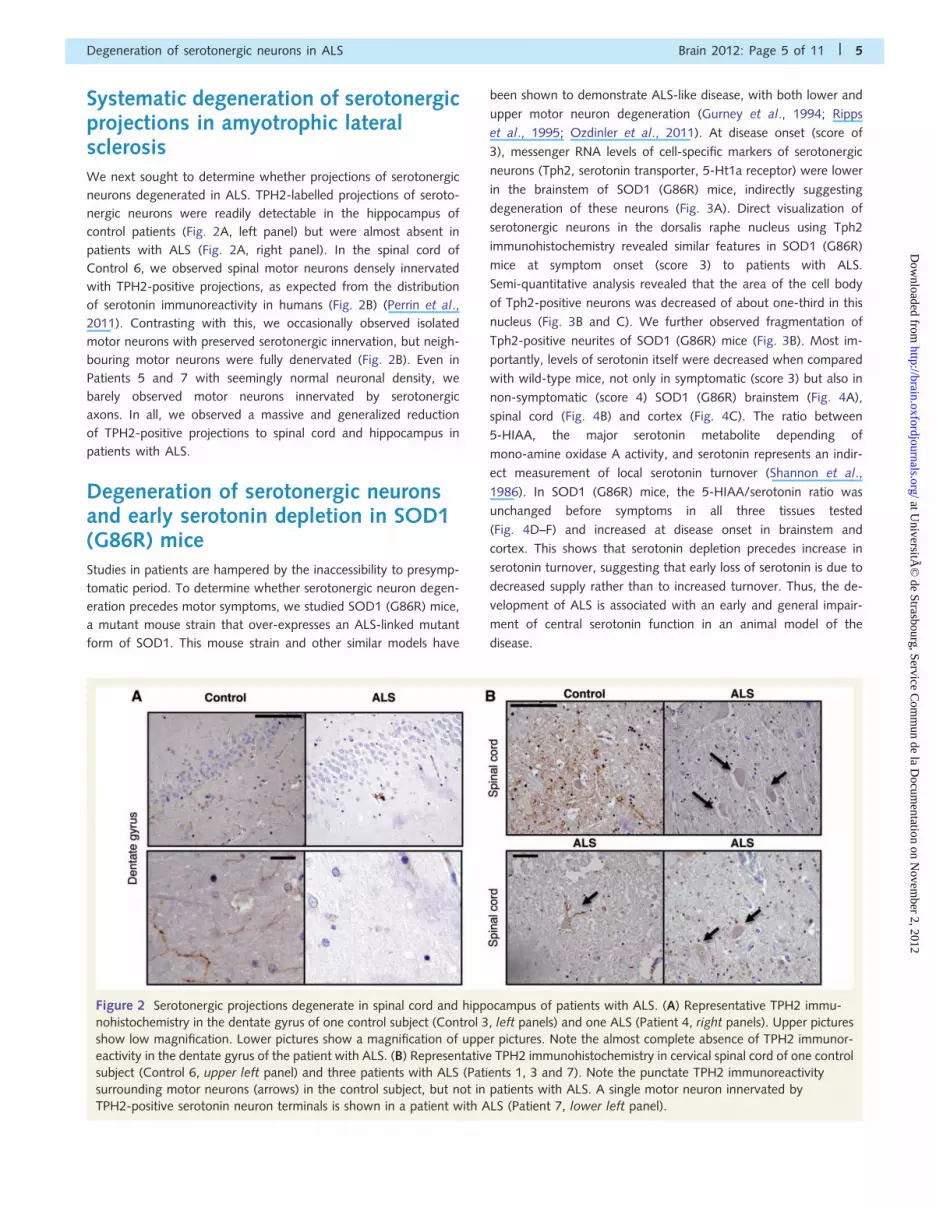
Systematic degeneration of serotonergicprojections in amyotrophic lateralsclerosisWe next sought to determine whether projections of serotonergic
neurons degenerated in ALS. TPH2-labelled projections of seroto-
nergic neurons were readily detectable in the hippocampus of
control patients (Fig. 2A, left panel) but were almost absent in
patients with ALS (Fig. 2A, right panel). In the spinal cord of
Control 6, we observed spinal motor neurons densely innervated
with TPH2-positive projections, as expected from the distribution
of serotonin immunoreactivity in humans (Fig. 2B) (Perrin et al.,
2011). Contrasting with this, we occasionally observed isolated
motor neurons with preserved serotonergic innervation, but neigh-
bouring motor neurons were fully denervated (Fig. 2B). Even in
Patients 5 and 7 with seemingly normal neuronal density, we
barely observed motor neurons innervated by serotonergic
axons. In all, we observed a massive and generalized reduction
of TPH2-positive projections to spinal cord and hippocampus in
patients with ALS.
Degeneration of serotonergic neuronsand early serotonin depletion in SOD1(G86R) miceStudies in patients are hampered by the inaccessibility to presymp-
tomatic period. To determine whether serotonergic neuron degen-
eration precedes motor symptoms, we studied SOD1 (G86R) mice,
a mutant mouse strain that over-expresses an ALS-linked mutant
form of SOD1. This mouse strain and other similar models have
been shown to demonstrate ALS-like disease, with both lower and
upper motor neuron degeneration (Gurney et al., 1994; Ripps
et al., 1995; Ozdinler et al., 2011). At disease onset (score of
3), messenger RNA levels of cell-specific markers of serotonergic
neurons (Tph2, serotonin transporter, 5-Ht1a receptor) were lower
in the brainstem of SOD1 (G86R) mice, indirectly suggesting
degeneration of these neurons (Fig. 3A). Direct visualization of
serotonergic neurons in the dorsalis raphe nucleus using Tph2
immunohistochemistry revealed similar features in SOD1 (G86R)
mice at symptom onset (score 3) to patients with ALS.
Semi-quantitative analysis revealed that the area of the cell body
of Tph2-positive neurons was decreased of about one-third in this
nucleus (Fig. 3B and C). We further observed fragmentation of
Tph2-positive neurites of SOD1 (G86R) mice (Fig. 3B). Most im-
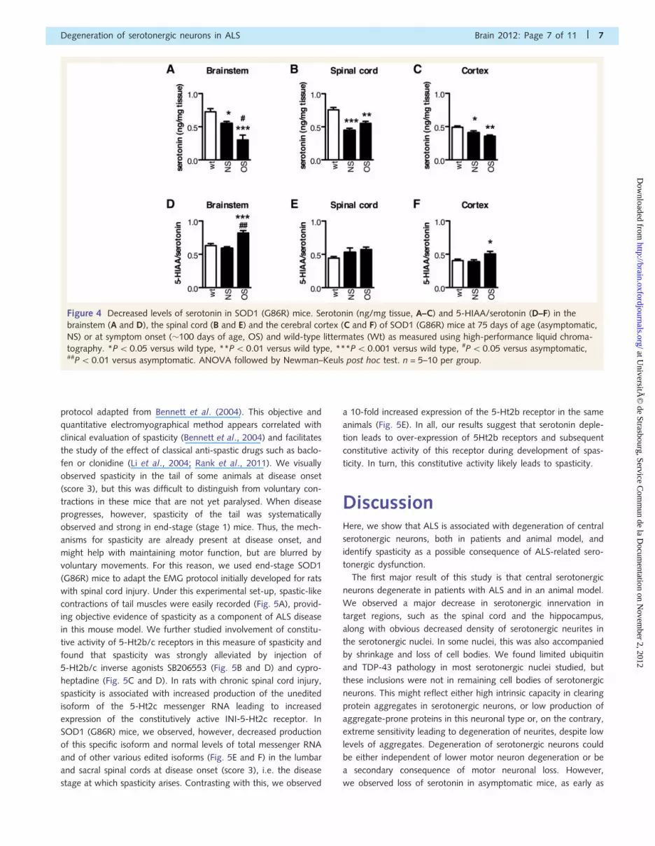
portantly, levels of serotonin itself were decreased when compared
with wild-type mice, not only in symptomatic (score 3) but also in
non-symptomatic (score 4) SOD1 (G86R) brainstem (Fig. 4A),
spinal cord (Fig. 4B) and cortex (Fig. 4C). The ratio between
5-HIAA, the major serotonin metabolite depending of
mono-amine oxidase A activity, and serotonin represents an indir-
ect measurement of local serotonin turnover (Shannon et al.,
1986). In SOD1 (G86R) mice, the 5-HIAA/serotonin ratio was
unchanged before symptoms in all three tissues tested
(Fig. 4D–F) and increased at disease onset in brainstem and
cortex. This shows that serotonin depletion precedes increase in
serotonin turnover, suggesting that early loss of serotonin is due to
decreased supply rather than to increased turnover. Thus, the de-
velopment of ALS is associated with an early and general impair-
ment of central serotonin function in an animal model of the
disease.
Figure 2 Serotonergic projections degenerate in spinal cord and hippocampus of patients with ALS. (A) Representative TPH2 immu-
nohistochemistry in the dentate gyrus of one control subject (Control 3, left panels) and one ALS (Patient 4, right panels). Upper pictures
show low magnification. Lower pictures show a magnification of upper pictures. Note the almost complete absence of TPH2 immunor-
eactivity in the dentate gyrus of the patient with ALS. (B) Representative TPH2 immunohistochemistry in cervical spinal cord of one control
subject (Control 6, upper left panel) and three patients with ALS (Patients 1, 3 and 7). Note the punctate TPH2 immunoreactivity
surrounding motor neurons (arrows) in the control subject, but not in patients with ALS. A single motor neuron innervated by
TPH2-positive serotonin neuron terminals is shown in a patient with ALS (Patient 7, lower left panel).
Degeneration of serotonergic neurons in ALS Brain 2012: Page 5 of 11 | 5
at UniversitÃ
© de Strasbourg, Service C
omm
un de la Docum
entation on Novem
ber 2, 2012http://brain.oxfordjournals.org/
Dow
nloaded from

Spasticity develops in SOD1(G86R) mice, and is alleviated by5-hydroxytryptamine 2 b/c receptorsinverse agonistsWe sought then to characterize whether serotonin depletion
occurring early in SOD1 (G86R) had pathogenic consequences
on motor neurons. Serotonin modulates excitability of motor neu-
rons by allowing sustained entry of calcium (Heckman et al.,
2009). In animal models of spinal cord injury, it was recently
shown that serotonin depletion due to transection of serotonergic
axons was over-compensated by motor neurons. More specifically,
motor neurons produce constitutively active 5-Ht2b and 5-Ht2c
receptors through still poorly defined mechanisms, decreased edit-
ing of the 5Ht2c messenger RNA being one of these (Murray
et al., 2010). This constitutive activity of 5-Ht2b/c receptors is
responsible for the occurrence of spasticity on spinal cord injury
(Murray et al., 2010). Other serotonin receptors, including
5-HT1A, 2A, 3, 4, 5, 6 and 7 appear to not be involved in this
event (Murray et al., 2011). By analogy, we reasoned that the
chronic loss of serotonergic innervation of lower motor neurons in
patients with ALS and SOD1 (G86R) mice could lead to spasticity.
To explore this hypothesis, we used an electromyographical
Figure 3 Serotonergic neurons degenerate in SOD1 (G86R) mice. (A) Messenger RNA levels of 5-Ht1a receptor (5-Ht1a), serotonin
transporter (Sert) and Tph2 in SOD1 (G86R) mice at 75 days of age (asymptomatic, NS) or at symptom onset (�100 days of age, OS) and
wild-type litter mates (Wt). Note that serotonin transporter and Tph2 gene expression levels are downregulated at symptom onset.
n = 7–10 per group. *P5 0.05 versus wild type, ANOVA followed by Newman–Keuls post hoc test. (B) Representative Tph2 immuno-
histochemistry in SOD1 (G86R) mice at disease onset (SOD1; OS) and wild-type littermates. Note the shrunken Tph2-positive cell bodies
in SOD1 (G86R) mice (upper right) and neuritic degeneration (lower right). n = 3 per group. (C) Mean area of Tph2-positive neurons in
SOD1 (G86R) mice at disease onset (OS) and wild-type (Wt) littermates. The area of 200–800 neurons was measured per animal, with
n = 3 per group. ***P50.0005 versus wild type, Student’s t-test.
6 | Brain 2012: Page 6 of 11 C. Dentel et al.
at UniversitÃ
© de Strasbourg, Service C
omm
un de la Docum
entation on Novem
ber 2, 2012http://brain.oxfordjournals.org/
Dow
nloaded from
protocol adapted from Bennett et al. (2004). This objective and
quantitative electromyographical method appears correlated with
clinical evaluation of spasticity (Bennett et al., 2004) and facilitates
the study of the effect of classical anti-spastic drugs such as baclo-
fen or clonidine (Li et al., 2004; Rank et al., 2011). We visually
observed spasticity in the tail of some animals at disease onset
(score 3), but this was difficult to distinguish from voluntary con-
tractions in these mice that are not yet paralysed. When disease
progresses, however, spasticity of the tail was systematically
observed and strong in end-stage (stage 1) mice. Thus, the mech-
anisms for spasticity are already present at disease onset, and
might help with maintaining motor function, but are blurred by
voluntary movements. For this reason, we used end-stage SOD1
(G86R) mice to adapt the EMG protocol initially developed for rats
with spinal cord injury. Under this experimental set-up, spastic-like
contractions of tail muscles were easily recorded (Fig. 5A), provid-
ing objective evidence of spasticity as a component of ALS disease
in this mouse model. We further studied involvement of constitu-
tive activity of 5-Ht2b/c receptors in this measure of spasticity and
found that spasticity was strongly alleviated by injection of
5-Ht2b/c inverse agonists SB206553 (Fig. 5B and D) and cypro-
heptadine (Fig. 5C and D). In rats with chronic spinal cord injury,
spasticity is associated with increased production of the unedited
isoform of the 5-Ht2c messenger RNA leading to increased
expression of the constitutively active INI-5-Ht2c receptor. In
SOD1 (G86R) mice, we observed, however, decreased production
of this specific isoform and normal levels of total messenger RNA
and of other various edited isoforms (Fig. 5E and F) in the lumbar
and sacral spinal cords at disease onset (score 3), i.e. the disease
stage at which spasticity arises. Contrasting with this, we observed
a 10-fold increased expression of the 5-Ht2b receptor in the same
animals (Fig. 5E). In all, our results suggest that serotonin deple-
tion leads to over-expression of 5Ht2b receptors and subsequent
constitutive activity of this receptor during development of spas-
ticity. In turn, this constitutive activity likely leads to spasticity.
DiscussionHere, we show that ALS is associated with degeneration of central
serotonergic neurons, both in patients and animal model, and
identify spasticity as a possible consequence of ALS-related sero-
tonergic dysfunction.
The first major result of this study is that central serotonergic
neurons degenerate in patients with ALS and in an animal model.
We observed a major decrease in serotonergic innervation in
target regions, such as the spinal cord and the hippocampus,
along with obvious decreased density of serotonergic neurites in
the serotonergic nuclei. In some nuclei, this was also accompanied
by shrinkage and loss of cell bodies. We found limited ubiquitin
and TDP-43 pathology in most serotonergic nuclei studied, but
these inclusions were not in remaining cell bodies of serotonergic
neurons. This might reflect either high intrinsic capacity in clearing
protein aggregates in serotonergic neurons, or low production of
aggregate-prone proteins in this neuronal type or, on the contrary,
extreme sensitivity leading to degeneration of neurites, despite low
levels of aggregates. Degeneration of serotonergic neurons could
be either independent of lower motor neuron degeneration or be
a secondary consequence of motor neuronal loss. However,
we observed loss of serotonin in asymptomatic mice, as early as
Figure 4 Decreased levels of serotonin in SOD1 (G86R) mice. Serotonin (ng/mg tissue, A–C) and 5-HIAA/serotonin (D–F) in the
brainstem (A and D), the spinal cord (B and E) and the cerebral cortex (C and F) of SOD1 (G86R) mice at 75 days of age (asymptomatic,
NS) or at symptom onset (�100 days of age, OS) and wild-type littermates (Wt) as measured using high-performance liquid chroma-
tography. *P50.05 versus wild type, **P50.01 versus wild type, ***P5 0.001 versus wild type, #P50.05 versus asymptomatic,##P5 0.01 versus asymptomatic. ANOVA followed by Newman–Keuls post hoc test. n = 5–10 per group.
Degeneration of serotonergic neurons in ALS Brain 2012: Page 7 of 11 | 7
at UniversitÃ
© de Strasbourg, Service C
omm
un de la Docum
entation on Novem
ber 2, 2012http://brain.oxfordjournals.org/
Dow
nloaded from

75 days of age, an age that precedes from several weeks the
onset of motor neuron degeneration. This suggests that degener-
ation of serotonergic neurons occurs independently of motor
neuron death, at least in animal models.
The loss of serotonergic neurons causes loss of serotonin itself in
regions of projections. In our animal model, serotonin levels are
decreased in the brainstem and the spinal cord long before motor
symptoms arise. Previous studies on serotonin and 5-HIAA in
patients with ALS yielded conflicting results. Bertel et al. (1991)
observed normal levels of serotonin and decreased levels of
5-HIAA in autopsy samples, whereas Forrest et al. (1996)
observed normal serotonin levels but increased 5-HIAA levels.
However, serotonin is a labile molecule that might be significantly
altered in post-mortem human samples with hours of delay before
autopsy (Yoshimoto et al., 1993). Our study overcomes this prob-
lem by studying serotonergic neurons using TPH2 immunostaining
in fixed tissues. In asymptomatic animals, the loss of serotonin
was associated with normal serotonin turnover (unchanged
5-HIAA/serotonin ratio), strengthening the idea that decreased
serotonin was owing to decreased supply in serotonin rather
than to increased degradation. Contrastingly, in end-stage mice,
we observed an increased serotonin turnover (increased 5-HIAA/
Figure 5 Spasticity in SOD1 (G86R) mice is alleviated by 5-Ht2b/c inverse agonists. (A–C) Representative recording of long-lasting
reflexes using tail EMG in one diseased SOD1 (G86R) mouse before (left) and after injections of vehicle (Vh, A), SB206553 (SB206, B) or
cyproheptadine (Cypro, C). Spasticity was considered to be electrical activity above the baseline recorded 1 s after mechanical stimulation
(arrowhead). (D) Quantitative analysis. n = 5–6 mice per group and two to three EMGs were obtained before and after injection. We
calculated a ratio between spasticity before and after injection and present the result as a percentage. *P5 0.05 versus before injection,
ANOVA followed by Newman–Keuls post hoc test. (E) Messenger RNA levels of 5-Ht2b (5-Ht2b), and 5-Ht2c (5-Ht2c) receptors in SOD1
(G86R) mice at 75 days of age (asymptomatic, NS) or at symptom onset (�100 days of age, OS) and wild-type littermates (Wt). 5-Ht2b
gene expression levels are heavily upregulated at symptom onset. n = 12 per group. ***P50.001 versus wild type, ANOVA followed by
Newman–Keuls post hoc test. (F) Levels of editing variants of the 5-Ht2c messenger RNA in the spinal cord of SOD1 (G86R) mice at onset
(OS) and wild-type littermates (Wt). ABECD (messenger RNA variant with full editing of A, B, E, C and D sites), ABD (messenger RNA
variant edited at A, B and D sites) and the non-edited messenger RNA, which leads to the production of the constitutively active INI
receptor, were measured by TaqMan� assays. Levels of the non-edited messenger RNA are decreased. n = 12 per group. *P50.05 versus
wild type, Student’s t-test.
8 | Brain 2012: Page 8 of 11 C. Dentel et al.
at UniversitÃ
© de Strasbourg, Service C
omm
un de la Docum
entation on Novem
ber 2, 2012http://brain.oxfordjournals.org/
Dow
nloaded from
serotonin ratio). This late increased serotonin turnover is likely to
be due to increased release of serotonin by remaining nerve ter-
minals. Indeed, this increased mobilization of residual serotonin in
end-stage mice coincides with the loss of serotonin transporter
expression, an event expected to limit serotonin reuptake and in-
crease its turnover. Our studies are consistent with imaging studies
using the PET scan ligand WAY100635 (Turner et al., 2005,
2007). This compound binds to the 5-HT1A receptor, which is
broadly expressed in serotonergic neurons, notably in brainstem
and acts as an inhibitory somatodendritic autoreceptor in these
neurons. The decreased binding potential of WAY100635 in the
raphe of patients with ALS was hypothesized to be either owing to
decreased sensitivity of 5-HT1A receptors to WAY100635 or to
loss of neurons that express this receptor. As 5-HT1A receptor is
strongly expressed in serotonergic neurons in the raphe, our cur-
rent results argue for the latter view.
We next sought to delineate whether serotonin depletion had
pathogenic consequences and focused on one of the potential
consequences, the occurrence of spasticity. Spasticity had been
previously shown to occur in spinal cord injury as a late conse-
quence of transection of serotonergic axons (Murray et al., 2010,
2011). Spasticity represents a symptom that is difficult to object-
ively measure in animals. We adapted an EMG technique measur-
ing spastic-like contractions of tail muscles in response to a
mechanical stimulation. Others had previously shown that this
EMG method is clinically correlated with onset of spasticity in
rats with spinal cord injury and sensitive to classical anti-spastic
drugs (Bennett et al., 2004; Li et al., 2004; Rank et al., 2011).
This method thus represents a quantitative, observer-independent
measurement of spasticity. In our model, we were able to almost
abolish spasticity by the use of cyproheptadine, a broad 5-HT2
inverse agonist, and SB206553, a much more selective compound
known to target 5-HT2B and C (Kennett et al., 1996), arguing for
the involvement of one of these two receptors in ALS spasticity.
Murray et al. (2010) observed increased production of the un-
edited 5-Ht2c messenger RNA in rats with chronic spinal cord
injury, whereas our result in mSOD1 mice was opposite. Recent
work in another paradigm of spinal cord injury found unchanged
levels of editing of the 5-ht2c messenger RNA in rats (Navarrett
et al., 2012). This discrepancy might be due to species differences
(rats versus mice), to the different kinetics of serotonin loss [abrupt
in spinal cord injury but much slower in SOD1 (G86R) mice]. We
found a strong increase in spinal 5-Ht2b receptor expression when
spasticity was obvious, which could underlie the constitutive
activity observed. Indeed, 5-Ht2b receptor has intrinsic constitutive
activity, and the increase of concentration of a G-protein coupled
receptor is on its own sufficient to further increase any constitutive
activity (Seifert and Wenzel-Seifert, 2002). For instance, a 7-fold
over-expression of the 5-Ht2b receptor in cardiomyocytes leads to
a dramatic cardiac phenotype, suggesting that the over-expression
of this receptor in the range we observed in SOD1 (G86R) mice is
sufficient to induce strong constitutive activity (Nebigil et al.,
2003). Our mouse model of ALS is based on transgenic over-
expression of mutant SOD1. Although these mouse models rep-
resent the only currently available model that display selective loss
of both lower and upper motor neurons (Halter et al., 2010;
Ozdinler et al., 2011), they only mimic the 20% of familial
cases that display mutations in the SOD1 gene. Whether spasticity
might also be alleviated by 5-HT2 inverse agonists in other,
non-SOD1 ALS cases represents an open question. In all, our
animal study suggests that spasticity, at least in SOD1-linked
ALS, is due to constitutive activity of the 5-Ht2b receptor rather
than 5-Ht2c.
How far can these mechanistic results be compared with our
pathology study in patients with ALS? Among the patients
included in our study, only Patient 5 showed the complete picture
of upper motor neuron signs, in particular spasticity, whereas the
other patients exhibited either increased reflexes and/or Babinski
signs, but not obvious spasticity (Supplementary Table 1). Patient
5, who displayed spasticity, showed strong loss of serotonergic
terminals on motor neurons and thus appeared indistinguishable
from the other patients with ALS in terms of loss of TPH2 projec-
tions. Interestingly, however, Patient 5 was the single case with
ALS with widespread TDP43 pathology in serotonergic nuclei.
Further work comparing autoptic material from patients with or
without spasticity should be done to highlight potential correl-
ations between serotonin loss and spasticity. Importantly, such a
study could also investigate other phenotypes potentially related
with serotonin such as weight loss, depression or dementia.
Our work has potential clinical implications for the management
of spasticity in those patients presenting such a phenotype. This is
especially true for patients with primary lateral sclerosis, a subtype
of ALS with primary upper motor neuron involvement (Singer
et al., 2007; Gordon et al., 2009). These patients develop prom-
inent spasticity (Kuipers-Upmeijer et al., 2001; Le Forestier et al.,
2001a, b) that is likely to be due to motor neuron hyperexcitability
(Floeter et al., 2005). Spasticity is also sometimes associated with
ALS but difficult to detect clinically, as the tests used to assess
spasticity rely on the integrity of alpha and gamma motor neurons,
both degenerating during ALS (Swash, 2012). Spasticity in ALS
and primary lateral sclerosis has been poorly studied, and few
clinical trials have been performed to treat this symptom
(Ashworth et al., 2012). Only physical therapy was proven to
be effective in a small trial (Drory et al., 2001), and current guide-
lines of the European Federation of Neurological Societies state
that other anti-spastic medications display class IV level of evi-
dence of efficacy and ‘may be tried’ (Ashworth et al., 2006).
A rigorous clinical trial assessing cyproheptadine in ALS spasticity
is thus needed, although treatment of spasticity might also lead to
worsening of motor function as observed in spinal cord injury.
In conclusion, loss of serotonergic neurons is part of the degen-
erative process in ALS and may be involved in spasticity. Further
research is needed to determine whether serotonergic degener-
ation has broader consequences on ALS pathophysiology.
AcknowledgementsAnnie PICCHINENNA (INSERM U692), Marie-Jo RUIVO (INSERM
U692), Isabelle DROUET (Centre hospitalier de Versailles), Eliane
SUPPER (Hopitaux Universitaires de Strasbourg) and Martine
MUCKENSTURM (Hopitaux Universitaires de Strasbourg) provided
technical support for this study.
Degeneration of serotonergic neurons in ALS Brain 2012: Page 9 of 11 | 9
at UniversitÃ
© de Strasbourg, Service C
omm
un de la Docum
entation on Novem
ber 2, 2012http://brain.oxfordjournals.org/
Dow
nloaded from
FundingThis work was funded in part by the Agence Nationale de la
Recherche (ANR Jeune Chercheur Dynemit, to L.D.), Association
pour la recherche sur la SLA et les autres maladies du motoneuron
(ARSla, to F.R. and J.P.L.), Thierry Latran Foundation (L.D., J.P.L.),
Association pour la recherche et le developpement de moyens de
lutte contre les maladies neurodegeneratives (AREMANE),
the European Community’s Health Seventh Framework
Programme (FP7/2007-2013) under grant agreement No
259867 (J.P.L.), and Association francaise de lutte contre les myo-
pathies (AFM, to J.P.L.). The generation of the Tph2 antibody was
supported by the German Research Foundation (DFG) (SFB 581
and SFB TRR 58, to K.P.L.) and the European Community
(NEWMOOD LSHM-CT-2003-503474, to K.P.L.). Collaboration
between L.D. and V.M. is supported by a contrat d’interface
INSERM/AP-HP. L.D. is supported by a Mercator Professorship
(DFG, 2011-2012). A.H. is a research fellow receiving funding
from FP7/2007-2013. J.L.G.D.A. is recipient of a Chaire d’excel-
lence INSERM/Universite de Strasbourg.
Supplementary materialSupplementary material is available at Brain online.
ReferencesAlvarez JC, Bothua D, Collignon I, Advenier C, Spreux-Varoquaux O.
Simultaneous measurement of dopamine, serotonin, their metabolites
and tryptophan in mouse brain homogenates by high-performance
liquid chromatography with dual coulometric detection. BiomedChromatogr 1999; 13: 293–8.
Ashworth NL, Satkunam LE, Deforge D. Treatment for spasticity in
amyotrophic lateral sclerosis/motor neuron disease. Cochrane
Database Syst Rev 2006; 1: CD004156.Ashworth NL, Satkunam LE, Deforge D. Treatment for spasticity in
amyotrophic lateral sclerosis/motor neuron disease. Cochrane
Database Syst Rev 2012; 2: CD004156.
Bennett DJ, Sanelli L, Cooke CL, Harvey PJ, Gorassini MA. Spastic
long-lasting reflexes in the awake rat after sacral spinal cord injury.J Neurophysiol 2004; 91: 2247–58.
Bertel O, Malessa S, Sluga E, Hornykiewicz O. Amyotrophic lateral scler-
osis: changes of noradrenergic and serotonergic transmitter systems in
the spinal cord. Brain Res 1991; 566: 54–60.Braunstein KE, Eschbach J, Rona-Voros K, Soylu R, Mikrouli E, Larmet Y,
et al. A point mutation in the dynein heavy chain gene leads to striatal
atrophy and compromises neurite outgrowth of striatal neurons. Hum
Mol Genet 2010; 19: 4385–98.Brooks BR, Miller RG, Swash M, Munsat TL. El Escorial revisited: revised
criteria for the diagnosis of amyotrophic lateral sclerosis. Amyotroph
Lateral Scler Other Motor Neuron Disord 2000; 1: 293–9.
Drory VE, Goltsman E, Reznik JG, Mosek A, Korczyn AD. The value of
muscle exercise in patients with amyotrophic lateral sclerosis. J NeurolSci 2001; 191: 133–7.
Dupuis L, de Tapia M, Rene F, Lutz-Bucher B, Gordon JW, Mercken L,
et al. Differential screening of mutated SOD1 transgenic mice reveals
early up-regulation of a fast axonal transport component in spinal cordmotor neurons. Neurobiol Dis 2000; 7: 274–85.
Dupuis L, Spreux-Varoquaux O, Bensimon G, Jullien P, Lacomblez L,
Salachas F, et al. Platelet serotonin level predicts survival in amyo-
trophic lateral sclerosis. PLoS One 2010; 5: e13346.
Floeter MK, Zhai P, Saigal R, Kim Y, Statland J. Motor neuron firing
dysfunction in spastic patients with primary lateral sclerosis.
J Neurophysiol 2005; 94: 919–27.
Forrest V, Ince P, Leitch M, Marshall EF, Shaw PJ. Serotonergic neuro-
transmission in the spinal cord and motor cortex of patients with
motor neuron disease and controls: quantitative autoradiography for
5-HT1a and 5-HT2 receptors. J Neurol Sci 1996; 139 (Suppl): 83–90.Franklin K, Paxinos G. The mouse brain in stereotaxic coordinates. San
Diego: Academic Press; 1997.Gordon PH, Cheng B, Katz IB, Mitsumoto H, Rowland LP. Clinical fea-
tures that distinguish PLS, upper motor neuron-dominant ALS, and
typical ALS. Neurology 2009; 72: 1948–52.Gurney ME, Pu H, Chiu AY, Dal Canto MC, Polchow CY, Alexander DD,
et al. Motor neuron degeneration in mice that express a human Cu,Zn
superoxide dismutase mutation. Science 1994; 264: 1772–5.
Gutknecht L, Kriegebaum C, Waider J, Schmitt A, Lesch KP.
Spatio-temporal expression of tryptophan hydroxylase isoforms in
murine and human brain: convergent data from Tph2 knockout
mice. Eur Neuropsychopharmacol 2009; 19: 266–82.
Halter B, Gonzalez de Aguilar JL, Rene F, Petri S, Fricker B, Echaniz-
Laguna A, et al. Oxidative stress in skeletal muscle stimulates early
expression of Rad in a mouse model of amyotrophic lateral sclerosis.
Free Radic Biol Med 2010; 48: 915–23.Heckman CJ, Mottram C, Quinlan K, Theiss R, Schuster J. Motoneuron
excitability: the importance of neuromodulatory inputs. Clin
Neurophysiol 2009; 120: 2040–54.
Ivanhoe CB, Reistetter TA. Spasticity: the misunderstood part of the
upper motor neuron syndrome. Am J Phys Med Rehabil 2004; 83
(10 Suppl): S3–9.
Kennett GA, Wood MD, Bright F, Cilia J, Piper DC, Gager T, et al.
In vitro and in vivo profile of SB 206553, a potent 5-HT2C/5-HT2B
receptor antagonist with anxiolytic-like properties. Br J Pharmacol
1996; 117: 427–34.
Kiernan MC, Vucic S, Cheah BC, Turner MR, Eisen A, Hardiman O, et al.
Amyotrophic lateral sclerosis. Lancet 2011; 377: 942–55.
Kuipers-Upmeijer J, de Jager AE, Hew JM, Snoek JW, van Weerden TW.
Primary lateral sclerosis: clinical, neurophysiological, and magnetic
resonance findings. J Neurol Neurosurg Psychiatry 2001; 71: 615–20.Lanfranco MF, Anastasio NC, Seitz PK, Cunningham KA. Quantification
of RNA editing of the serotonin 2C receptor (5-HT(C)R) ex vivo.
Methods Enzymol 2010; 485: 311–28.
Lanfranco MF, Seitz PK, Morabito MV, Emeson RB, Sanders-Bush E,
Cunningham KA. An innovative real-time PCR method to measure
changes in RNA editing of the serotonin 2C receptor (5-HT(2C)R) in
brain. J Neurosci Methods 2009; 179: 247–57.
Le Forestier N, Maisonobe T, Spelle L, Lesort A, Salachas F, Lacomblez L,
et al. Primary lateral sclerosis: further clarification. J Neurol Sci 2001a;
185: 95–100.
Le Forestier N, Maisonobe T, Piquard A, Rivaud S, Crevier-Buchman L,
Salachas F, et al. Does primary lateral sclerosis exist? A study of 20
patients and a review of the literature. Brain 2001b; 124 (Pt 10):
1989–99.
Li Y, Li X, Harvey PJ, Bennett DJ. Effects of baclofen on spinal reflexes
and persistent inward currents in motoneurons of chronic spinal rats
with spasticity. J Neurophysiol 2004; 92: 2694–703.
Murray KC, Stephens MJ, Ballou EW, Heckman CJ, Bennett DJ.
Motoneuron excitability and muscle spasms are regulated by
5-HT2B and 5-HT2C receptor activity. J Neurophysiol 2011; 105:
731–48.
Murray KC, Nakae A, Stephens MJ, Rank M, D’Amico J, Harvey PJ, et al.
Recovery of motoneuron and locomotor function after spinal cord
injury depends on constitutive activity in 5-HT2C receptors. Nat
Med 2010; 16: 694–700.Navarrett S, Collier L, Cardozo C, Dracheva S. Alterations of serotonin
2C and 2A receptors in response to T10 spinal cord transection in rats.
Neurosci Lett 2012; 506: 74–8.
Nebigil CG, Jaffre F, Messaddeq N, Hickel P, Monassier L, Launay JM,
et al. Overexpression of the serotonin 5-HT2B receptor in heart leads
10 | Brain 2012: Page 10 of 11 C. Dentel et al.
at UniversitÃ
© de Strasbourg, Service C
omm
un de la Docum
entation on Novem
ber 2, 2012http://brain.oxfordjournals.org/
Dow
nloaded from

to abnormal mitochondrial function and cardiac hypertrophy.Circulation 2003; 107: 3223–9.
Neumann M, Sampathu DM, Kwong LK, Truax AC, Micsenyi MC,
Chou TT, et al. Ubiquitinated TDP-43 in frontotemporal lobar degen-
eration and amyotrophic lateral sclerosis. Science 2006; 314: 130–3.Ozdinler PH, Benn S, Yamamoto TH, Guzel M, Brown RH Jr, Macklis JD.
Corticospinal motor neurons and related subcerebral projection neu-
rons undergo early and specific neurodegeneration in hSOD1G(9)(3)A
transgenic ALS mice. J Neurosci 2011; 31: 4166–77.Perrin FE, Gerber YN, Teigell M, Lonjon N, Boniface G, Bauchet L, et al.
Anatomical study of serotonergic innervation and 5-HT(1A) receptor in
the human spinal cord. Cell Death Dis 2011; 2: e218.Rank MM, Murray KC, Stephens MJ, D’Amico J, Gorassini MA,
Bennett DJ. Adrenergic receptors modulate motoneuron excitability,
sensory synaptic transmission and muscle spasms after chronic spinal
cord injury. J Neurophysiol 2011; 105: 410–22.Ripps ME, Huntley GW, Hof PR, Morrison JH, Gordon JW. Transgenic
mice expressing an altered murine superoxide dismutase gene provide
an animal model of amyotrophic lateral sclerosis. Proc Natl Acad Sci
USA 1995; 92: 689–93.Seifert R, Wenzel-Seifert K. Constitutive activity of G-protein-coupled
receptors: cause of disease and common property of wild-type recep-
tors. Naunyn Schmiedebergs Arch Pharmacol 2002; 366: 381–416.
Shannon NJ, Gunnet JW, Moore KE. A comparison of biochemical indicesof 5-hydroxytryptaminergic neuronal activity following electrical
stimulation of the dorsal raphe nucleus. J Neurochem 1986; 47:958–65.
Singer MA, Statland JM, Wolfe GI, Barohn RJ. Primary lateral sclerosis.
Muscle Nerve 2007; 35: 291–302.
Sofic E, Riederer P, Gsell W, Gavranovic M, Schmidtke A, Jellinger K.Biogenic amines and metabolites in spinal cord of patients with
Parkinson’s disease and amyotrophic lateral sclerosis. J Neural
Transm Park Dis Dement Sect 1991; 3: 133–42.
Swash M. Why are upper motor neuron signs difficult to elicit in amyo-trophic lateral sclerosis? J Neurol Neurosurg Psychiatry 2012; 83:
659–62.
Turner MR, Rabiner EA, Al-Chalabi A, Shaw CE, Brooks DJ, Leigh PN,et al. Cortical 5-HT1A receptor binding in patients with homozygous
D90A SOD1 vs sporadic ALS. Neurology 2007; 68: 1233–5.
Turner MR, Rabiner EA, Hammers A, Al-Chalabi A, Grasby PM,
Shaw CE, et al. [11C]-WAY100635 PET demonstrates marked5-HT1A receptor changes in sporadic ALS. Brain 2005; 128 (Pt 4):
896–905.
Vandesompele J, De Preter K, Pattyn F, Poppe B, Van Roy N, De
Paepe A, et al. Accurate normalization of real-time quantitativeRT-PCR data by geometric averaging of multiple internal control
genes. Genome Biology 2002; 3.
Yoshimoto K, Irizawa Y, Komura S. Rapid postmortem changes of rat
striatum dopamine, serotonin, and their metabolites as monitored bybrain microdialysis. Forensic Sci Int 1993; 60: 183–8.
Degeneration of serotonergic neurons in ALS Brain 2012: Page 11 of 11 | 11
at UniversitÃ
© de Strasbourg, Service C
omm
un de la Docum
entation on Novem
ber 2, 2012http://brain.oxfordjournals.org/
Dow
nloaded from