selective in vitro effects of the farnesyl pyrophosphate synthase inhibitor risedronate on...
TRANSCRIPT

International Journal of Antimicrobial Agents 23 (2004) 273–285
Selective in vitro effects of the farnesyl pyrophosphate synthaseinhibitor risedronate onTrypanosoma cruzi
Luciana R. Garzonia, Aura Calderab, Maria de Nazareth L. Meirellesa, Solange L. de Castroa,Roberto Docampoc, Gary A. Meintsd, Eric Oldfieldd, Julio A. Urbinab,∗
a Departmento de Ultra-estrutura e Biologia Celular, Instituto Oswaldo Cruz, Av. Brasil, 4365, Manguinhos,21045-900 Rio de Janeiro, RJ, Brazil
b Laboratorio de Qu´ımica Biológica, Centro de Biof´ısica y Bioqu´ımica, Instituto Venezolano de Investigaciones Cientificas,Apartado Postal 21.827, Caracas 1020A, Venezuela
c Laboratory of Molecular Parasitology, Department of Pathobiology and Center for Zoonoses Research,University of Illinois at Urbana-Champaign, Urbana, IL 61802, USA
d Department of Chemistry and Department of Biophysics, University of Illinois at Urbana-Champaign, Urbana, IL 61801, USA
Received 21 March 2003; accepted 3 July 2003
Abstract
We present the results of the first detailed study of the molecular and cellular basis of the antiproliferative effects of the bisphosphonaterisedronate (Ris) onTrypanosoma cruzi, the causative agent of Chagas’ disease. Ris and related compounds, which block poly-isoprenoidbiosynthesis at the level of farnesyl pyrophosphate synthase, are currently used for the treatment of bone resorption disorders, but also displayselective activity against trypanosomatid and apicomplexan parasites. Ris induced a dose-dependent effect on growth of the extracellularepimastigote form ofT. cruzi; complete growth arrest and cell lysis ensued at 150�M. Growth inhibition was associated with depletion ofthe parasite’s endogenous sterols, but complete growth arrest and loss of cell viability took place before full depletion of these compounds,suggesting that disappearance of other essential poly-isoprenoids is involved in its anti-parasitic action. Ris had a variety of effects on cellularultrastructure, including mitochondrial swelling, disorganisation of other organelles, such as reservosomes and the kinetoplast, together withthe appearance of autophagic vesicles and progressive vacuolization of the cytoplasm. Ris had selective antiproliferative effects against theclinically relevant amastigote form ofT. cruzi, and at 100�M, was able to prevent completely the development ofT. cruziinfection of murinemuscle heart or Vero cells, and to cure cultures which were already infected. Ris induced drastic ultrastructural alterations in the intracellularparasites and blocked amastigote to trypomastigote differentiation, with no biochemical or ultrastructural effects on the host cells, which fullyrecovered their normal structure and activity after treatment. Ris is, therefore, a promising lead compound for the development of new drugsagainstT. cruzi.© 2003 Elsevier B.V. and the International Society of Chemotherapy. All rights reserved.
Keywords:Risedronate;T. cruzi; Chagas’ disease; Antiproliferative effect; Amastigote
1. Introduction
Chagas disease, caused byTrypanosoma cruzi, is endemicin Latin America, affecting 16–18 million people[1]. Trans-mission occurs mainly by insect vectors (Reduviidae), butalso by blood transfusion and congenital routes[2]. Treat-ment still relies on the use of nifurtimox and benznidazole,drugs that were introduced empirically in the 1970s. How-
∗ Corresponding author. Tel.:+58-212-5041479;fax: +58-212-5041093.
E-mail address:[email protected] (J.A. Urbina).
ever, both these nitroheterocycles frequently produce toxicside effects, have considerable variability in cure rates (de-pending of the geographic region), and very limited efficacyfor treatment of chronic patients. Moreover, the commercialproduction of nifurtimox has been discontinued since the1980s in Brazil and more recently in Argentina, Chile andUruguay. There is, therefore, clearly an unmet need for new,specific chemotherapeutic approaches for the treatment ofthis serious condition[3,4].
Recent developments in understanding the biochemistryof T. cruzihave permitted the identification of several newdrug targets in this parasite. Among the most promising
0924-8579/$ – see front matter © 2003 Elsevier B.V. and the International Society of Chemotherapy. All rights reserved.doi:10.1016/j.ijantimicag.2003.07.020

274 L.R. Garzoni et al. / International Journal of Antimicrobial Agents 23 (2004) 273–285
are the de novo sterol and phosphatidylcholine biosynthesispathways, the essential cathepsin L-like protease cruzipainand the unique Kinetoplastida enzymes trypanothione re-ductase and trypanothione synthase[5,6]. Another promis-ing approach developed recently is interference with themetabolism of inorganic pyrophosphate and various iso-prenoid pyrophosphates, which are precursors of severalessential cellular components. Both theoretical and experi-mental work has shown that the antiproliferative effects ofnitrogen containing bisphosphonates (NBPs) in vertebrateand plant cells and in the slime mouldDictyostelium dis-coideum, result from a blockade of isoprenoid synthesis atthe level of farnesyl pyrophosphate synthase (FFPS, refs.[7–12]). A gene encoding the farnesyl pyrophosphate syn-thase ofT. cruzi has been cloned and sequenced and itsexpression inEscherichia coliproduced an enzyme whichwas potently inhibited by NBPs, risedronate (Ris) beingamong the most active compounds investigated[13].
The selective action of NBPs against trypanosomatidand apicomplexan parasites could result from preferen-tial drug accumulation in the parasite’s acidocalcisomes,acidic organelles which contain most of the cell’s polyphos-phates, complexed with divalent cations such as Ca2+and Mg2+ [14,15]. The presence of polyphosphate-richacidocalcisomes, as well as the recent discovery of sev-eral pyrophosphate-utilizing enzymes in trypanosomatids,therefore opens up a potential new route for the treatmentof numerous parasitic diseases including Chagas’ disease[15–20]. Moreover, since Ris is currently used for the pre-vention and treatment of postmenopausal and corticosteroid-induced osteoporosis[21], there is extensive data availableon its pharmacology, toxicology and tolerance in humans,reducing the costs of development of this drug againstChagas’ and other parasitic diseases.
Although the selective antiparasitic activity of bisphos-phonates has been demonstrated previously, little informa-tion exists on their cellular and molecular basis of action.Here, we present the first detailed study of the effects ofRis on the viability and proliferation of epimastigotes andintracellular amastigotes ofT. cruzi. Biochemical and ultra-structural alterations induced by the bisphosphonate in bothforms of the parasite, as well as in primary cultures of mouseheart muscle and cultured Vero cells, have been investigatedusing gas–liquid chromatography coupled to mass spectrom-etry, together with electron and fluorescence microscopy, toobtain a detailed picture of the drug action.
2. Materials and methods
2.1. Parasites
Both the Y[22] and EP[23] strains ofT. cruziwere usedin this work, with essentially identical results. Epimastigoteswere grown in liver-infusion (LIT) medium[23] at 28◦C,with strong aeration. Trypomastigote forms of the Y strain
were obtained from the supernatant of infected heart musclecells (10:1 parasites:host cells) grown in complete Eaglemedium supplemented with 5% foetal calf serum (FCS),1 mM CaCl2, 15% horse serum, 1 mMl-glutamine, 2%chick embryo extract, 1000 U/ml penicillin and 50�g/mlstreptomycin (DMEM). After 96 h of infection the para-sites were collected by centrifugation then resuspended inDMEM. Handling of liveT. cruziwas performed accordingto established guidelines[24].
2.2. Mammalian cell cultures
Hearts of 18-day-old Swiss mouse embryos were submit-ted to mechanical and enzymatic dissociation using 0.05%trypsin plus 0.01% collagenase in PBS at 37◦C, followingthe method described previously[25]. The ventricular heartmuscle cells (HMCs) were plated on 0.02% gelatin-coatedplastic bottles, on glass coverslips in 24-well plates or inPetri dishes. Cells were maintained at 37◦C in a 5% CO2 at-mosphere in DMEM. After 72 h of plating, the cultures wereused for the experiments with Ris. Vero cells were main-tained in minimal essential medium supplemented with 2%FCS, as described previously[26].
2.3. Effect of risedronate on epimastigotes
Y strain epimastigotes were resuspended in LIT mediumat a density of 1× 10⁷ cells/ml in 24-well plates[27] and500�l of the suspension was added to the same volume ofRis solution, previously prepared in LIT at twice the desiredfinal concentration. After 1–8 days of incubation at 28◦C,parasites were counted in a haemocytometer. The motilityand morphology of the parasites was monitored by opticalmicroscopy (Axioplan, Zeiss, Oberköchen, Germany). Forthe EP strain, Ris was added to cultures in conical flasks ata cell density of 0.5–1×10⁷ epimastigotes/ml and followedfor 7–10 days. Cell densities were measured by turbidime-try as well as by direct counting in a haemocytometer. Cellviability was followed by trypan blue exclusion, using lightmicroscopy. After the incubation period, cells were har-vested by centrifugation and processed for lipid analysis, asdescribed below.
2.4. Effect of risedronate on lipid composition ofepimastigotes and Vero cells
For the analysis of the effects of Ris on sterol composi-tion of epimastigotes or Vero cells, total lipids from controland drug-treated cells were extracted, then fractionated intoneutral and polar lipid fractions, by using silicic acid columnchromatography and gas–liquid chromatography[28–31].The neutral lipid fraction was first analysed by thin layerchromatography (Merck 5721 silica gel plates; heptane-isopropyl ether–glacial acetic acid [60:40:4] as developingsolvent) and by gas–liquid chromatography (isothermalseparation on a 4 m glass column packed with 3% OV-1 on
L.R. Garzoni et al. / International Journal of Antimicrobial Agents 23 (2004) 273–285 275
Chromosorb, 100/200 mesh, with nitrogen (24 ml/min) ascarrier gas and using flame ionisation detection, on a Varian3700 gas chromatograph). For structural assignments andquantitative analysis, the neutral lipids were separated byusing a high resolution capillary column (25 m× 0.20 mmi.d., Ultra-2 column, 5% phenyl-methyl-siloxane, 0.33�mfilm thickness) on a Hewlett-Packard 6890 Plus gas chro-matograph equipped with an HP5973A mass sensitivedetector. Lipids were injected as chloroform solutions andthe column kept at 50◦C for 1 min, then the temperaturewas increased to 270◦C at a rate of 25◦C/min, and finallyto 300◦C, at a rate of 1◦C/min. The carrier gas (He) flowwas kept constant at 0.5 ml/min. Injector temperature was250◦C and the detector was kept at 280◦C. The polar lipidfraction (containing mostly phospholipids) was analysed asdescribed previously[32]. Briefly, the lipid fractions elut-ing from the silicic acid column with chloroform:methanol(1:1, v/v) were pooled, then further fractionated by thin-layer chromatography on Merck 5721 silica gel plates,using chloroform:methanol:32.5% ammonia w/v (17:7:1;v/v/v) as developing solvent[33]. The phospholipid spotswere visualised by using iodine, scraped off, then the totalorganic phosphorus was measured by using the method ofAmes and Dubin[34].
2.5. Effects of risedronate on the proliferation anddifferentiation of T. cruzi amastigotes in heart muscleand Vero cells
HMCs (1.5 × 10⁵ cells/well in 24-well plates) wereinfected with culture-derived trypomastigotes (10:1 par-asites:host cells), in a final volume of 300�l DMEM.After 2 h, the cultures were washed with PBS to removenon-adherent parasites and maintained in DMEM. Treat-ment was performed by following two protocols: (i) addi-tion of 50–200�M Ris in DMEM immediately after theinteraction step; (ii) addition of 100�M Ris after 48 h ofinfection. The total volume in each well was 500�l. Atspecific times, coverslips (in triplicate) were collected, fixedwith Bouin and stained in Giemsa solution. The percent-age of infection was quantified by counting randomly atleast 300 cells. In addition, the supernatant was collectedand counted in a haemocytometer, to obtain the numberof released trypomastigotes from the HMCs. The cover-slips were used for the fluorescence assays. For electronmicroscopic analysis, experiments were performed in Petridishes, maintaining both the number of HMCs/area andproportion of parasites/HMC, then using the two treatmentprotocols described above.
Vero cells were infected with culture-derived trypomastig-otes (10:1 parasites:host cell) for 2 h, then washed threetimes with PBS to remove non-adherent parasites. Freshmedium, with or without Ris, was then added and the cellsincubated for 96 h, with a medium change at 48 h. Quanti-tation of the number of infected cells was then carried outas described previously[30,31].
2.6. Ultrastructural studies
At chosen times, epimastigotes or infected HMC cellswere fixed (60 min/4◦C) with 2.5% glutaraldehyde, 2.5 mMCaCl2 and 0.1 M Na-cacodylate buffer (pH 7.2). Afterpost-fixation for 1 h in cacodylate buffer solution contain-ing 1% OsO4, 0.8% potassium ferricyanide and 2.5 mMCaCl2, samples were dehydrated in acetone then em-bedded in Polybed 812 resin. Thin sections (Leica Ul-tracuts, UCT, Vienna, Austria) were stained with uranylacetate and lead citrate, and were examined by transmis-sion electron microscopy using an Zeiss EM10C micro-scope.
2.7. Effect of risedronate on HMC cytoskeletal actin
Using protocol (i), after 144 h of treatment with 100�MRis, coverslips containing treated cells were fixed in 4%paraformaldehyde in PBS and stained for 30 min at 37◦Cwith 4�g/ml rhodamine-labelled phalloidin in PBS, to stainactin filaments. During the last 5 min, DAPI was added tostain DNA, as described previously[35]. The cultures wereobserved by using a fluorescence microscope (Axioplan,Zeiss).
2.8. Flow cytometry
Epimastigotes were treated with 50–200�M Ris in LITmedium at 28◦C. After 4 and 8 days, 500�l of the cul-tures were taken and added to the same volume of a solutioncontaining 20�g/ml propidium iodide, for 30 min. Sampleswere kept on ice until analysis, which was carried out us-ing a flow cytometer (FACSCalibur, BectonDickinson, SanJose, USA) equipped with Cell Quest and WinMDI2.8 soft-ware (Joseph Trotter, Scripps Research Institute, San Diego,USA).
2.9. Statistical analysis
Mean value comparisons were performed by using theStudentt-test or Anova and Krunskal–Wallis test.P-valuesbelow 0.05 were considered significant. IC50 values werecalculated from dose-response curves by using a non-linearregression analysis with the GraFit software package.
2.10. Materials
Risedronate (2-(3-pyridyl)-1-hydroxy-ethane-1,1-bispho-sphonate, monosodium salt) was synthesized basically as de-scribed previously[12]. Elemental analysis and1H, 13C and31P NMR spectra indicated that the compound was 98.8%pure. Stock solutions were prepared in phosphate bufferedsaline (PBS) (pH adjusted to 7.4) and sterilized by usinga 0.2�M filter (Millipore). Trypsin and rhodamine-labelledphalloidin were purchased from Sigma Chemical Co. (St.Louis, MO, USA), collagenase was from Aldrich–Sigma
276 L.R. Garzoni et al. / International Journal of Antimicrobial Agents 23 (2004) 273–285
(St. Louis, MO, USA). All other reagents were of analyticalgrade.
3. Results
3.1. Effects of risedronate on T. cruzi epimastigotes
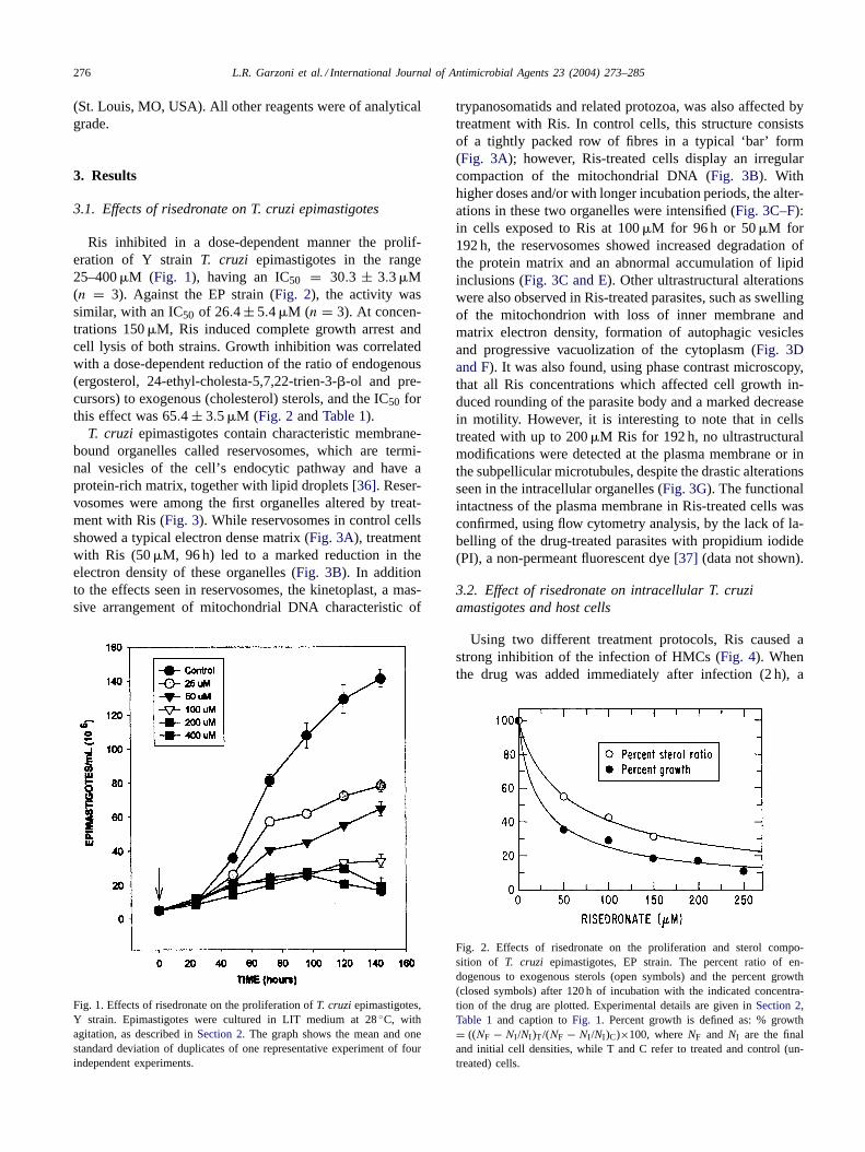
Ris inhibited in a dose-dependent manner the prolif-eration of Y strainT. cruzi epimastigotes in the range25–400�M (Fig. 1), having an IC50 = 30.3 ± 3.3�M(n = 3). Against the EP strain (Fig. 2), the activity wassimilar, with an IC50 of 26.4± 5.4�M (n = 3). At concen-trations 150�M, Ris induced complete growth arrest andcell lysis of both strains. Growth inhibition was correlatedwith a dose-dependent reduction of the ratio of endogenous(ergosterol, 24-ethyl-cholesta-5,7,22-trien-3-�-ol and pre-cursors) to exogenous (cholesterol) sterols, and the IC50 forthis effect was 65.4 ± 3.5�M (Fig. 2 andTable 1).
T. cruzi epimastigotes contain characteristic membrane-bound organelles called reservosomes, which are termi-nal vesicles of the cell’s endocytic pathway and have aprotein-rich matrix, together with lipid droplets[36]. Reser-vosomes were among the first organelles altered by treat-ment with Ris (Fig. 3). While reservosomes in control cellsshowed a typical electron dense matrix (Fig. 3A), treatmentwith Ris (50�M, 96 h) led to a marked reduction in theelectron density of these organelles (Fig. 3B). In additionto the effects seen in reservosomes, the kinetoplast, a mas-sive arrangement of mitochondrial DNA characteristic of
Fig. 1. Effects of risedronate on the proliferation ofT. cruziepimastigotes,Y strain. Epimastigotes were cultured in LIT medium at 28◦C, withagitation, as described inSection 2. The graph shows the mean and onestandard deviation of duplicates of one representative experiment of fourindependent experiments.
trypanosomatids and related protozoa, was also affected bytreatment with Ris. In control cells, this structure consistsof a tightly packed row of fibres in a typical ‘bar’ form(Fig. 3A); however, Ris-treated cells display an irregularcompaction of the mitochondrial DNA (Fig. 3B). Withhigher doses and/or with longer incubation periods, the alter-ations in these two organelles were intensified (Fig. 3C–F):in cells exposed to Ris at 100�M for 96 h or 50�M for192 h, the reservosomes showed increased degradation ofthe protein matrix and an abnormal accumulation of lipidinclusions (Fig. 3C and E). Other ultrastructural alterationswere also observed in Ris-treated parasites, such as swellingof the mitochondrion with loss of inner membrane andmatrix electron density, formation of autophagic vesiclesand progressive vacuolization of the cytoplasm (Fig. 3Dand F). It was also found, using phase contrast microscopy,that all Ris concentrations which affected cell growth in-duced rounding of the parasite body and a marked decreasein motility. However, it is interesting to note that in cellstreated with up to 200�M Ris for 192 h, no ultrastructuralmodifications were detected at the plasma membrane or inthe subpellicular microtubules, despite the drastic alterationsseen in the intracellular organelles (Fig. 3G). The functionalintactness of the plasma membrane in Ris-treated cells wasconfirmed, using flow cytometry analysis, by the lack of la-belling of the drug-treated parasites with propidium iodide(PI), a non-permeant fluorescent dye[37] (data not shown).
3.2. Effect of risedronate on intracellular T. cruziamastigotes and host cells
Using two different treatment protocols, Ris caused astrong inhibition of the infection of HMCs (Fig. 4). Whenthe drug was added immediately after infection (2 h), a
Fig. 2. Effects of risedronate on the proliferation and sterol compo-sition of T. cruzi epimastigotes, EP strain. The percent ratio of en-dogenous to exogenous sterols (open symbols) and the percent growth(closed symbols) after 120 h of incubation with the indicated concentra-tion of the drug are plotted. Experimental details are given inSection 2,Table 1 and caption toFig. 1. Percent growth is defined as: % growth= ((NF − NI /NI )T/(NF − NI /NI )C)×100, whereNF and NI are the finaland initial cell densities, while T and C refer to treated and control (un-treated) cells.

L.R. Garzoni et al. / International Journal of Antimicrobial Agents 23 (2004) 273–285 277
Table 1Effects of risedronate on the free sterol composition ofTrypanosoma cruziepimastigotes (EP stock)a
Name Structure Ris0�M Ris50�M Ris100�M Ris 150�M
Exogenous
Cholesterol 33.9 48.2 54.8 62.1
Endogenous
24-Methyl-5,7,22-cholesta-trien-3�-ol (ergosterol) 22.5 20.8 20.2 17.3
24-Ethyl-5,7,22-cholesta-trien-3�-ol 16.0 22.8 25.0 20.6
Ergosta-5,7-dien-3�-ol 9.6 3.2 n.d. n.d.
Ergosta-5,7,24(241)-dien-3�-ol 13.9 5.0 n.d. n.d.
Ergosta-7,24(241)-dien-3�-ol 4.1 n.d. n.d. n.d.
Endogenous/exogenous sterols 1.95 1.07 0.82 0.61
a Sterols were extracted fromT. cruzi epimastigotes cultured in LIT medium and drugs added at a cell density of 5× 10⁶ epimastigotes/ml; theywere separated from polar lipids by silicic acid column chromatography and analyzed by quantitative capillary gas–liquid chromatography and massspectrometry. Results are expressed as mass percent.
dose-dependent reduction in the percentage of infected cellswas seen as a function of time. The inhibition was statisti-cally significant for treatment with 200�M after 72 h, with100 and 150�M Ris after 96 h and with 50�M Ris, after120 h (Fig. 4A). No infected cells were observed with Ris at200, 150 and 100�M after 96, 120 and 144 h, respectively.Under our experimental conditions, Ris did not interfere withthe contractility of either infected or uninfected HMCs.
Addition of 100�M Ris 48 h after infection also led toa highly significant reduction in the percentage of infected
cells, 72 to 144 h after treatment. With the longer exposure,the infection disappeared completely (Fig. 4B). We also in-vestigated the effects of Ris on the release of trypomastigotesfrom infected cells in this model, after completion of the in-tracellular cycle of the parasite. In control HMC cultures, thefirst two peaks of trypomastigote release to the supernatantoccurred at 96 h and 192 h post-infection (Fig. 4C). Treat-ment with 100�M Ris, starting 48 h post-infection, signifi-cantly inhibited (61.1%) such release after 48 h of treatment(96 h post-infection), while after 96 and 144 h of treat-

278 L.R. Garzoni et al. / International Journal of Antimicrobial Agents 23 (2004) 273–285
Fig. 3. Effects of risedronate on the ultrastructure ofT. cruzi epimastigotes (Y strain). (A) Control parasite showing organelles with their characteristicaspect, such as kinetoplast (k), reservosomes (R) and nucleus (N). (B) 50�M Ris for 96 h led to alterations on the kinetoplast and reservosomes (arrowheads). (C, D) 100�M Ris for 96 h caused (C) marked alterations of reservosomes (arrow heads) and (D) alterations in the nucleus (dark star), strongcytoplasmic vacuolisation, together with mitochondrial swelling (white star); (E) 50�M Ris for 192 h led to the appearance of autophagic vesicles in theparasite, besides intense vacuolisation (asterisks), and loss of proteic matrix in reservosomes; (F) 100�M Ris for 192 h led to mitochondrial swelling(white star) and alterations on the kinetoplast (arrow head); (G) 200�M Ris for 192 h caused breakdown of internal organelles, but plasma membrane andsubpellicular microtubules were preserved (arrows). Magnifications: (A) 15,300×; (B) 11,500×; (C) 18,000×; (D) 14,600×; (E) 23,500×; (F) 6300×.
ment, no trypomastigotes were detected in the supernatant(Fig. 4C).
A quantitative analysis of the effects of Ris (added im-mediately after infection) on the percentage of infected hostcells, intracellular parasite load and host cell viability, wascarried out after 96 h of incubation (Fig. 5). The IC50 was66.0±0.8�M for Y strain in HMC cells and 30.1±0.6�Mfor the EP strain, in Vero cells; the minimal concentration
required to completely eradicate the infection in these ex-perimental models, were 150 and 100�M Ris, respectively.At higher concentrations, there was a slight, non-statisticallysignificant reduction in the total number of host cells,but no alteration of their gross morphology. No effectson the sterol (cholesterol) or phospholipid composition ofuninfected Vero cells were detected after incubation with100�M Ris for 96 h (data not shown). Infected Vero cells

L.R. Garzoni et al. / International Journal of Antimicrobial Agents 23 (2004) 273–285 279
Fig. 4. Time and concentration dependence of the effects of risedronateon the infection of HMC byT. cruzi. (A) Ris (50–200�M) added tothe cultures after 2 h of infection and the percentage of infected cellsfollowed as a function of time. (B, C) Ris (100�M) added to the culturesafter 48 h of infection induced (B) a potent inhibitory effect on infectionof HMC and (C) on trypomastigote release. Asterisks indicate statisticaldifferences in relation to control cultures. The graphs show the mean andstandard deviation of triplicates of one representative experiment of fourindependent experiments.
treated with Ris at concentrations that eradicated the infec-tion had no relapse of the infection after further incubationin a drug-free medium, indicating a trypanocidal effect.
Using optical microscopy, we followed the morphologi-cal alterations induced by Ris on intracellular amastigotes(Fig. 6). After 72 h of treatment with 100�M Ris, theparasites began to exhibit morphological alterations withprogressive damage. Treatment with Ris inhibited the dif-ferentiation to trypomastigotes (see alsoFig. 4C) and, after120–144 h of treatment, essentially no intact intracellularparasites were observed. Changes induced by drug treat-ment were also investigated at the ultrastructural level inboth parasites and host cells, using electron microscopy.It was found that in untreated HMC cells after 144 h ofinfection, intracellular parasites (mainly trypomastigotes)
exhibited normal morphology, with characteristic emptyspaces, devoid of electron density, in cytoplasmic regionsclose to parasite cells (Fig. 7B). Treatment with 100�M Risfor 96 h (144 h of infection) caused marked alteration of theoverall shape of the parasites, together with swelling of thesarcoplasmic reticulum of the HMCs (Fig. 7C). The potenteffect of Ris on intracellular amastigotes can be seen inFig. 7D. These parasites exhibited mitochondrial swelling,the presence of autophagic vesicles and intense vacuolisa-tion, as seen with the extracellular epimastigotes. No intactparasites could be seen with 100�M Ris after 144 h of treat-ment (192 h of infection). At this point, HMCs exhibitedmyofibrils with Z lines as well as mitochondrial, sarcoplas-mic reticulum and cytoplasmic structures very similar tothose seen in the control cultures (compareFig. 7A and E).All of these findings clearly indicate that Ris induces thedestruction of parasite cells, but permits the recovery of nor-mal structure and metabolism of host cells, together with denovo protein biosynthesis. This notion is reinforced by thepresence of a large number of ribosomes in the sarcoplasmicreticulum (Fig. 7E). To explore further these observations,we investigated the distribution of actin filaments in HMCs,labelling the cells with rhodamine-phalloidin and DAPI.Control HMCs showed highly developed actin-containingmyofibrils (Fig. 8A). Infection with T. cruzi for 144 h ledto a breakdown of actin filaments close to the intracellularparasites (Fig. 8C). However, after treatment of infectedHMCs with 100�m Ris for 142 h, the host cells presentedboth polygonal and filamentous configurations of actin(Fig. 8E), and no parasites (small blue dots) were detectedusing DAPI (Fig. 8D and F).
4. Discussion
Ris induced a dose-dependent effect on the ratio of en-dogenous/exogenous epimastigote sterols, consistent with ablockade of the synthesis of these essential components ata pre-squalene level (Fig. 2andTable 1), in agreement withthe accepted primary target of NBPs in eukaryotic cells,FPPS[7–12]. However, although this effect was associatedwith a reduction in parasite growth rate, the antiprolifer-ative effects were observed at drug concentrations signifi-cantly lower than those required to produce equivalent ef-fects on the sterol composition of the cells (Figs. 1 and 2andTable 1). This may be explained by the fact that block-ade at the level of FPPS depletes the cells of other essentialpoly-isoprenoids. Similar conclusions have been reached ina previous study, which investigated the combined effectsof risedronate and ketoconazole at fixed concentrations onepimastigotes[16].
One of the characteristic ultrastructural effects of sterolbiosynthesis inhibitors on trypanosomatid parasites is amarked swelling of their single, giant mitochondrion, whichcorrelates with depletion of the endogenous parasite sterolsand leads to cell lysis[38–43]. Previous work from our

280 L.R. Garzoni et al. / International Journal of Antimicrobial Agents 23 (2004) 273–285
Fig. 5. Concentration dependence of the effects of risedronate on the proliferation ofT. cruzi amastigotes in mammalian cells. Shown are the percentageof infected cells (�), the number of amastigotes per cell (�) and the number of host cells per field (�) after 96 h of treatment, as a function of thedrug concentration for (A) HMC and (B) Vero cells. Cultured mammalian cells were infected withT. cruzi and incubated at 37◦C with Ris at indicatedconcentrations for 96 h. Experiments were carried out in quadruplicate and each bar represents one standard deviation.
groups has shown thatT. cruzimitochondrial membranes, incontrast to those of vertebrate cells, are rich in endogenousparasite sterols, which are thought to be required for theirenergy transducing activities[44]. Mitochondrial swellingwas among the most prominent ultrastructural alterationsseen in both epimastigotes and amastigotes treated with Ris(Fig. 3), consistent with the hypothesis that sterol depletionis one of the primary effects of this drug on the parasite.However, other ultrastructural effects were also observed forthis drug, and could also be important in growth inhibition.For example, the early effects of Ris on the reservosomes,which are involved in endocytosis, storage and breakdownof nutrients[36,45], could hamper the proliferation processin epimastigotes. Although the precise molecular mecha-nisms of these effects are doubtless complex and at this time
remain obscure, they could be related to altered protein syn-thesis and sorting associated with abnormal cell signalling,due to a depletion of essential poly-isoprenoids. Since Risinhibits sterol biosynthesis (see above and ref.[16]), theaccumulation of lipid droplets in the reservosomes could bea response of the parasite to the drug, as a consequence ofthe accumulation of abnormal lipids and/or the uptake oflipids from the culture medium. Other abnormalities such ascytoplasmic lipid inclusions and autophagic vacuoles, havealso been noted in this and other trypanosomatid parasitestreated with sterol biosynthesis inhibitors and are likely dueto the accumulation of abnormal lipids and lipid precursorsin these cells[38–43].
Although the primary target of Ris and other NBPs,FPPS, is an essential enzyme in all eukaryotic cells, the

L.R. Garzoni et al. / International Journal of Antimicrobial Agents 23 (2004) 273–285 281
Fig. 6. Effects of risedronate on the morphology of HMC and intracellularT. cruzi (Y strain) stages. (A, C, E, G, I) untreated infected cultures. After48 h of infection, large numbers of proliferating amastigotes were seen (arrow) (A). After 72 h of infection many oval-shaped parasites were seen andthe amastigote-trypomastigote differentiation process began (C). After 96 h of infection the first parasite cycle inside the host cell was completedandthe high number of intracellular trypomastigotes (arrow head) led to disruption of HMC (E), and infection of other host cells (arrows in G, I; 120 and144 h after infection). (B, D, F, H, J) Infected cultures treated with 100�M Ris immediately after the initial 2 h infection step. No apparent effects onamastigotes were seen after 48 h of treatment (arrow) (B). After 72 h (D) and 96 h (E) of infection, a progressive alteration of the parasites’ shape wasobserved as well as a marked inhibition of the differentiation to trypomastigotes (F). Intracellular parasites were rarely detected after 120 or 144h oftreatment, while the host cells recover their normal morphology (H, J). Magnification: 1200×.

282 L.R. Garzoni et al. / International Journal of Antimicrobial Agents 23 (2004) 273–285
Fig. 7. Effects of risedronate on the ultrastructure of HMC and intracellularT. cruzi (Y strain) stages (A) non-infected and untreated HMC presentingmyofibrils (MF) with normal Z line (Z), mitochondria (M), sarcoplasmic reticulum profiles (SR) and membrane invaginations. (B) Untreated HMC after144 h of infection withT. cruzi, presenting several empty spaces (arrows) in the sarcoplasma close to internal parasites (P). (C) Infected HMC (144 h)treated with 100�M Ris for 96 h, showed swelling of sarcoplasmic reticulum (large arrows) and bearing some damaged amastigotes (arrow heads). (D)Infected HMC (144 h) treated with 100�M Ris for 96 h showing a parasite in detail with several alterations, such as mitochondrial swelling (whiteasterisk), altered flagellar pocket (small arrow), strong vacuolisation (asterisks). (E, F) Infected HMC (192 h) treated with 100�M Ris for 144 h: HMCshowed normal characteristics of the myofibrils, mitochondria, sarcoplasmic reticulum and ribosomes (white arrows); some regions of the sarcoplasmashowed some electrondensity, suggesting recovery of the host cell biosynthetic machinery (arrows). Magnifications: (A) 8000×; (B) 10,000×; (C) 8000×;(D) 27,500×; (E) 11,429×; (F) 26,000×.

L.R. Garzoni et al. / International Journal of Antimicrobial Agents 23 (2004) 273–285 283
Fig. 8. Effect of risedronate on organization of actin cytoskeletal inT. cruzi-infected HMC. Infected HMC untreated and treated with Ris were doublelabelled with rhodamine–phalloidin and DAPI. Uninfected cells displayed well-developed myofibrils (arrow) (A) and nuclei stained with DAPI (B). HMCinfected with trypomastigotes after 144 h showed broken myofibrils localized close to the parasites (small arrow) (C) and DAPI stained the host cellsnucleus and the nucleus and kinetoplast of the parasites (D). Infected HMC treated with 100�M Ris for 142 h (144 h of infection) presented polygonal(arrow head) as well as filamentous configuration of actin filaments (arrow) (E) and DAPI stained only the host cells nucleus, with no detectableintracellular parasites (D). Magnification: 1400×.

284 L.R. Garzoni et al. / International Journal of Antimicrobial Agents 23 (2004) 273–285
selective activity of bisphosphonates as anti-resorptiveagents in mammals is thought to be due to their differentialaccumulation in the osteoclasts of bone[46,47]. Likewise, ithas been argued that the selective anti-trypanosomatid andanti-apicomplexan activities of these compounds could beexplained, at least in part, by their selective accumulationin the parasite due in part to the presence of acidocalci-somes, which are very rich in short-chain polyphosphates[14,15,48]. This hypothesis has recently received supportfrom studies which demonstrate the accumulation of [3H]-labelled pamidronate in intact acidocalcisomes ofT. cruzi(Ruiz FA and Docampo R, unpublished results). In thiswork, we have investigated the molecular and cellular ba-sis of the selective activity of Ris against the intracellularamastigote stage of the parasite by using both biochemicaland ultrastructural techniques (Figs. 6–8). Our results showthat, in contrast to the effects seen on parasite sterol content(Table 1), no effects of Ris were observed on the lipid com-position of the host cells. At the ultrastructural level, ourresults show that in infected HMC cells treated with Ris,drug-induced damage is confined to the intracellular para-sites, with the host cells being able to fully recover normalmorphology, intracellular organization and contractility, attherapeutic doses (Figs. 7 and 8). It was previously shownby one of our groups (M.N.M.) that infection byT. cruziin-duces a cytoskeletal disruption in HMCs, due to myofibrillarbreakdown[35]. The present results confirm these effectsof infection on actin filaments (Fig. 8C), and show that ontreatment with Ris, reorganization of the actin-containingmyofibrils to their normal state occurs via a characteristicpolygonal arrangement (Fig. 8E). Lin et al. [49] have alsorecently described this polygonal configuration in dissoci-ated cardiac myocytes as an indicator of myofibrillar re-assembly. The polygonal configuration of actin-containingmyofibrils was not observed in non-infected HMC treatedwith Ris (data not shown), indicating that this state is notdirectly due to drug treatment.
In conclusion, the results of the present study demonstraterapid and drastic biochemical and ultrastructural effects ofRis on T. cruzi epimastigotes and amastigotes, effects notobserved in the host cells. The action of the drug on both theextracellular and intracellular parasites leads to cell lysis 96–120 h after treatment (Figs. 1 and 3), an observation whichsuggests the potential for trypanocidal activity in vivo. Thishas been confirmed by the results of studies of a murinemodel of acute Chagas’ disease ([50] accompanying article).Risedronate and related NBPs, are therefore interesting leadanti-T. cruzicompounds which could be further developed asnovel chemotherapeutic agents for the treatment of Chagas’disease.
Acknowledgements
We are grateful to Dr. Maurı́lio, J. Soares for the criticalreading of this manuscript, Dr. Andrea Henriques-Pons and
Ricardo M. Santa-Rita for the support on the flow cytome-try analysis, to Bernardo Visentin for his excellent techni-cal assistance. This work was supported with grants fromFIOCRUZ, CNPq and FAPERJ, and the Howard HughesMedical Institute (Grant 55000620 to J.U.) and in part bythe US Public Health Service (NIH grant GM 65307 to E.O.and R.D.; NIH NRSA grant GM 65782 to G.A.M.) and bythe American Heart Association, Midwest Affiliate (E.O.)and National Center (R.D.).
References
[1] World Health Organisation. Control of Chagas disease. Tech RepSer 905;2002:1–109.
[2] Dias JCP. Epidemiologia. In: Brener Z, Andrade Z, Barral-Netto C,editors. Trypanosoma cruzie doença de Chagas. Rio de Janeiro:Guanabara Koogan; 1999. p. 48–74.
[3] Urbina JA. Chemotherapy of Chagas disease. Curr Pharma Des2002;8:287–95.
[4] Coura JR, De Castro SL. A critical review on Chagas diseasechemotherapy. Memórias do Instituto Oswaldo Cruz 2002;97:3–24.
[5] Docampo R. Recent developments in the chemotherapy of Chagasdisease. Curr Pharma Des 2001;7:1157–64.
[6] Rodriguez JB. Specific molecular targets to control tropical diseases.Curr Pharma Des 2001;7:1105–16.
[7] Martin MB, Arnold W, Heath III HT, Urbina JA, Oldfield E.Nitrogen-containing bisphosphonates as carbocation transition stateanalogs for isoprenoid biosynthesis. Biochem Biophys Res Commun1999;263:754–8.
[8] Bergstrom J, Bostedor DRG, Massarachia PJ, Reszka AA, Rodan GA.Alendronate is a specific, nanomolar inhibitor of farnesyl diphosphatesynthase. Arch Biochem Biophys 2000;373:231–41.
[9] Cromartie TH, Fisher KJ, Grossman JN. The discovery of a novelsite of action for herbicidal bisphosphonates. Pest Biochem Physiol1999;63:114–26.
[10] Grove JE, Brown RJ, Watts DJ. The intracellular target of the an-tiresorptive aminobisphosphonate drugs inDictyostelium discoideumis the enzyme farnesyl diphosphate synthase. J Bone Mineral Res2000;15:971–81.
[11] van Beek E, Pieterman E, Cohen L, Löwik C, Papapoulos S.Nitrogen-containing bisphosphonates inhibit isopentenyl pyrophos-phate isomerase/farnesyl pyrophosphate synthase activity with rela-tive potencies corresponding to their anti-resorptive potencies in vitroand in vivo. Biochem Biophys Res Commun 1999a;251:491–4.
[12] van Beek E, Pieterman E, Cohen L, Löwik C, Papapoulos S. Farnesylpyrophosphate synthase is the molecular target of nitrogen-containingbisphosphonates. Biochem Biophys Res Commun 1999;264:108–11.
[13] Montalvetti A, Bailey BN, Martin MB, Severin GW, Oldfield E,Docampo R. Bisphosphonates are potent inhibitors ofTrypanosomacruzi farnesyl pyrophosphate synthase. J Biol Chem 2001;276:33930–7.
[14] Docampo R, Moreno SNJ. The acidocalcisome. Mol Biochem Par-asitol 2001;33:151–9.
[15] Docampo R, Moreno SNJ. Bisphosphonates as chemotherapeuticagents against trypanosomatids and Apicomplexan parasites. CurrDrug Targets-Infect Dis 2001;1:51–61.
[16] Martin MB, Grimley JS, Lewis JC, Heath III HT, Bailey BN,Kendrick H, et al. Bisphosphonates inhibit the growth ofTry-panosoma brucei, Trypanosoma cruzi, Leishmania donovani, Tox-oplasma gondiiand Plasmodium falciparum: a potential route tochemotherapy. J Med Chem 2001;44:909–16.
[17] Martin MB, Sanders JM, Kendrick H, de Luca-Fradley K, Lewis JC,Grimley JS, et al. Activity of bisphosphonates againstTrypanosomabrucei rhodesiense. J Med Chem 2002;45:2904–14.

L.R. Garzoni et al. / International Journal of Antimicrobial Agents 23 (2004) 273–285 285
[18] Rodriguez N, Bailey BN, Martin MB, Oldfield E, Urbina JA, Do-campo R. Radical cure of experimental cutaneous leishmaniasis bythe bisphosphonate pamidronate. J Infect Dis 2002;186:138–40.
[19] Szajnman SH, Bailey BN, Docampo R, Rodrı́guez JB. Bisphospho-nates derived from fatty acids are potent growth inhibitors ofTry-panosoma cruzi. Bioorg Med Chem Lett 2001;11:789–92.
[20] Yardley V, Khan A, Martin MB, Slifer TR, Araujo FG, Moreno SNJ,et al. In vivo activities of farnesyl pyrophosphate synthase inhibitorsagainstLeishmania donovaniand Toxoplasma gondii. AntimicrobAgents Chemother 2002;46:929–31.
[21] Crandall C. Risedronate: a clinical review. Arch Intern Med2001;161:353–60.
[22] Silva LHP, Nussenszweig V. Sobre uma cepa deTrypanosomacruzi virulenta para o camundongo branco. Folia Clinica Biologica1953;20:191–207.
[23] De Maio A, Urbina JA.Trypanosoma(Schizotrypanum) cruzi: termi-nal oxidases in exponential and stationary growth phase epimastig-otes cultured in vitro. Acta Cientifica Venezolana 1984;35:136–41.
[24] Hudson L, Grover F, Gutteridge WE, Klein RA, Peters W, Neal RA,et al. Suggested guidelines for work with liveTrypanosoma cruzi.Trans R Soc Trop Med Hyg 1983;77:416–9.
[25] Meirelles MN, Araújo-Jorge TC, Miranda CF, DeSouza W, BarbosaHS. Interaction ofTrypanosoma cruziwith heart muscle cells: Ultra-structural and cytochemical analysis of endocytic vacuole formationand effect upon myogenesis in vitro. Eur J Cell Biol 1986;41:198–206.
[26] Urbina JA, Lazardi K, Aguirre T, Piras MM, Piras R. Antiprolif-erative synergism of the allylamine SF 86-327 and ketoconazoleon epimastigotes and amastigotes ofTrypanosoma(Schizotrypanum)cruzi. Antimicrob Agents Chemother 1988;32:1237–42.
[27] De Castro SL, Soeiro MN, Meirelles MNL. Trypanosoma cruzi:effect of phenothiazines on the parasite and its interaction with hostcells. Memórias do Instituto Oswaldo Cruz 1992;87:209–15.
[28] Liendo A, Lazardi K, Urbina JA. In vitro antiproliferative effects andmechanism of action of the bis-triazole D0870 and its S(-) enantiomeragainstTrypanosoma cruzi. J Antimicrob Chemother 1998;41:197–205.
[29] Liendo A, Visbal G, Piras MM, Piras R, Urbina JA. Sterol com-position and biosynthesis inTrypanosoma cruziamastigotes. MolBiochem Parasitol 1999;104:81–91.
[30] Urbina JA, Payares G, Molina J. Cure of short- and long-termexperimental Chagas disease using D0870. Science 1996;273:969–71.
[31] Urbina JA, Payares G, Contreras LM, Liendo A, Sanoja C, MolinaJ, et al. Antiproliferative effects and mechanism of action of SCH56592 againstTrypanosoma(Schizotrypanum) cruzi: In vitro and invivo studies. Antimicrob Agents Chemother 1998;42:1771–7.
[32] Contreras LM, Vivas J, Urbina JA. Altered lipid composition and en-zyme activities of plasma membranes fromTrypanosoma(Schizotry-panum) cruzi epimastigotes grown in the presence of sterol biosyn-thesis inhibitors. Biochem Pharmacol 1997;53:697–704.
[33] Cuzner ML, Davison AN. Quantitative thin layer chromatography oflipids. J Chromatogr 1967;27:388–97.
[34] Ames B, Dubin D. The role of polyamines in the neutralization ofdeoxyribonucleic acid. J Biol Chem 1960;235:769–75.
[35] Pereira MCS, Costa M, Chagas Filho C, Meirelles MNL. Myofibrillarbreakdown and cytoskeletal alterations in heart muscle cells during
invasion byTrypanosoma cruzi:immunological and ultrastructuralstudy. J Submicrosc Cytol Pathol 1993;25:559–69.
[36] Soares MJ. The reservosome ofTrypanosoma cruziepimastigotes: anorganelle of the endocytic pathway with a role on metacyclogenesis.Memórias do Instituto Oswaldo Cruz 1999;94:139–41.
[37] Santa-Rita RM, Menna-Barreto RFS, De Castro SL. Studies withanalogues of phospholipids againstTrypanosoma cruziandLeishma-nia amazonensis.In: Program and Abstracts of the Fifth COST-B9Congress on Antiprotozoal Chemotherapy, London, 2002 [Abstract25].
[38] Docampo R, Moreno SNJ, Turrens JF, Katzin AM, Gonzales-CappaSM, Stoppani AOM, et al. Biochemical and ultrastructural alterationsproduced by miconazole and ketoconazole inTrypanosoma cruzi.Mol Biochem Parasitol 1981;3:169–80.
[39] Lazardi K, Urbina JA, DeSouza W. Ultrastructural alterationsinduced by two ergosterol biosynthesis inhibitors, ketocona-zole and terbinafine, on epimastigotes and amastigotes ofTry-panosoma(Schizotrypanum) cruzi. Antimicrob Agents Chemother1990;34:2097–105.
[40] Lazardi K, Urbina JA, DeSouza W. Ultrastructural alterations inducedby ICI 195,739, a bis triazole with strong antiproliferative actionagainst Trypanosoma(Schizotrypanum) cruzi. Antimicrob AgentsChemother 1991;35:736–40.
[41] Rodrigues JCP, Attias M, Rodriguez C, Urbina JA, DeSouza W. Ul-trastructural and biochemical alterations induced by 22,26-azasterol,a �24(25) sterol methyl transferase inhibitor, on promastigote andamastigote forms ofLeishmania amazonensis. Antimicrob AgentsChemother 2002;46:487–99.
[42] Vannier-Santos MA, Urbina JA, Martiny A, Neves A, DeSouza W.Alterations induced by the antifungal compounds ketoconazole andterbinafine inLeishmania. J Eukaryotic Microbiol 1995;42:337–46.
[43] Vivas J, Urbina JA, DeSouza W. Ultrastructural alterations inTry-panosoma(Schizotrypanum) cruzi induced by�24(25) sterol methyltransferase inhibitors and their combinations with ketoconazole. IntJ Antimicrob Agents 1996;7:235–40.
[44] Rodrigues CO, Catisti R, Uyemura SA, Vercesi AE, Lira R, Ro-driguez C, et al. The sterol composition ofTrypanosoma cruzichanges after growth in different culture media and results in differ-ent sensitivity to digitonin-permeabilization. J Eukaryot Microbiol2001;48:588–94.
[45] Engel JC, Doyle PS, Palmer J, Hsieh I, McKerrow JH. Cysteineprotease inhibitors alter Golgi complex ultrastructure and functionin Trypanosoma cruzi. J Cell Sci 1998;111:597–606.
[46] Rodan GA. Mechanisms of action of bisphosphonates. Annu RevPharmacol Toxicol 1998;38:375–88.
[47] Rodan GA, Martin TJ. Therapeutic approaches to bone diseases.Science 2000;289:1508–14.
[48] Urbina JA, Moreno B, Vierkotter S, Oldfield E, Payares G, SanojaC, et al. Trypanosoma cruzicontains major pyrophosphate stores,and its growth in vitro and in vivo is blocked by pyrophosphateanalogues. J Biol Chem 1999;274:33609–15.
[49] Lin ZX, Holtzer S, Schultheiss T, Murray J, Masaki T, Fischman DA,et al. Polygons and adhesion plaques and disassembly and assemblyof myofibrils in cardiac myocytes. J Cell Biol 1989;108:2355–67.
[50] Garzoni LR, Caldera A, Meirelles MNL, de Castro SL, DocampoR, Meints GA, et al. Antiparasitic activity of risedronate in amurine model of acute Chagas disease. Int J Antimicrob Agents2004;23:286–90.