screening for testosterone, methyltestosterone, 19-nortestosterone residues and their metabolites in...
TRANSCRIPT

Analytica Chimica Acta 570 (2006) 116–123
Screening for testosterone, methyltestosterone, 19-nortestosteroneresidues and their metabolites in bovine urine with enzyme-linked
immunosorbent assay (ELISA)
Huihui Lu a, Grainne Conneely a, Mark A. Crowe b, Margaret Aherne b,Miloslav Pravda a, George G. Guilbault a,∗
a Sensor Development Group, Analytical and Biological Chemistry Research Facility, National University of Ireland, Cork, Irelandb School of Agriculture, Food Science and Veterinary Medicine and Conway Institute, University College Dublin, Ireland
Received 27 February 2006; received in revised form 24 March 2006; accepted 30 March 2006Available online 18 April 2006
Abstract
This work reports on the development of rapid enzyme-linked immunosorbent assay for testosterone, methyltestosterone, 19-nortestosterone andtaUotci©
K
1
aaim8o
altptal
0d
heir metabolites in real samples of bovine urine. The assays were based on the competition between an immobilised testosterone–BSA conjugatend the analyte for corresponding antibodies, followed by the use of secondary anti-species (�-IgG-HRP) to determine the degree of competition.rine samples were analysed after 10-fold dilution with the buffer, omitting extraction and hydrolysis. The limits of detection calculated for theriginal sample were ca. 74, 266 and 131 pg ml−1 (ppt) for testosterone, methyltestosterone and 19-nortestosterone, respectively. In particular,estosterone and methyltestosterone assays offer the advantage to pick up both parent compounds and their major metabolites due to the highross-reactivity pattern with corresponding antibody used in each assay. The concentration in bovine urine detected by developed method doesndicate 19-nortestosterone administration to heifers. The developed assays were applied to urine samples of heifers treated with above androgens.
2006 Elsevier B.V. All rights reserved.
eywords: Enzyme immunoassay; Steroids; Testosterone; Methyltestosterone; 19-Nortestosterone; Bovine urine
. Introduction
Testosterone, methyltestosterone and 19-nortestosterone arenabolic steroids often illegally used to stimulate increasednimal performance during the production cycle, thereby caus-ng a potential health risk to consumers. The use of hor-
onal substances for growth promotion is prohibited (Directive8/146/EEC) and the determination of residue levels is requiredn animals and in fresh meat (Directive 86/849/EEC).
Of the various analytical methods for the detection ofnabolic steroids in biological fluid proposed, high-performanceiquid chromatography (LC) methods are used for analysis ofestosterone and its metabolites [1,2]. Gas chromatography cou-led to mass spectrometry (GC–MS) method is accurate, suitableo detect steroid hormones and their metabolites simultaneously,nd is used as the confirmatory method [3,4]. The combination ofiquid chromatography with mass spectrometry (LC–MS) offers
∗ Corresponding author. Tel.: +353 21 4902208; fax: +353 21 4903103.E-mail address: [email protected] (G.G. Guilbault).
a simplified, specific and sensitive alternative to GC–MS meth-ods [5]. However, they all are costly, need skilled personnel andlengthy sample preparation before chromatographic analysis.Another limitation to these approaches is the need for authenticstandards and known chemical entities.
The ideal assay for screening of anabolic hormones should befast, simple, cost-effective, easy to perform, and enable measure-ment in small volume of biological fluid [6]. Immunoassays arebased on the molecular recognition of antigens by antibodies toform a stable complex. A significant advantage of immunologi-cal techniques is the ability of antibodies to bind broad classes ofmetabolites that contain a common substructure. They are one ofthe most sensitive methods available because of the high affin-ity of antibodies and fulfil most of the criteria. Wilke and Utley[7] and Slaats et al. [8] have reported radioimmunoassays (RIA).However, its drawbacks due to the use of radioactive compoundsand to worker safety led to the development of enzyme-linkedimmunosorbent assay (ELISA) as an alternative method.
Various immunoassay methods have been developed to mea-sure testosterone levels in plasma or serum [9,10] and in human
003-2670/$ – see front matter © 2006 Elsevier B.V. All rights reserved.
oi:10.1016/j.aca.2006.03.108
H. Lu et al. / Analytica Chimica Acta 570 (2006) 116–123 117
urine [11,12], methyltestosterone residues in muscle tissue [13]and 19-nortestosterone levels in plasma [14] and in calf urine[15]. A competitive chemiluminescent enzyme immunoassayfor the analysis of 19-nortestosterone in bovine urine was devel-oped by Roda et al. [16]. Crabbe et al. [17] developed an ELISAto screening of clostebol and its metabolites in bovine urine.A study of Hungerford et al. [18] reported a method of ELISAto detect 17�-alkyl anabolic steroid metabolites in equine urineand applied it as a primary screening tool for detection of newand known anabolic steroid metabolites. However, there is littleor no immunoassays published for these anabolic steroids withrespect to bovine urine. It was, therefore, the aim of this study todevelop a simple, rapid and cost-effective screening techniquesbased on immunoassays, to detect the anabolic steroid residuesand their metabolites in bovine urine. The polyclonal antibodiesused were assessed against a panel of steroids to determine cross-reactivity. Following the minimisation of sample pre-treatment,the urine samples of treated heifers were then analysed withdeveloped ELISA.
2. Experimental
2.1. Chemicals and reagents
Testosterone (4-androsten-17�-ol-3-one), methyltestoste-rone (4-androsten-17�-methyl-17�-ol-3-one), 19-nortestost-ew5mrBwacpcaS
2
cIEiG
2
tpawt
18 G 1.5′′ needle. Animals were housed in a slatted floor fin-ishing unit. They were fed a diet of grass silage (ad libitumaccess) and 6 kg per head per day of a barley/soybean concen-trate. Urine samples were collected at 1, 3, 6, 9, 12, 24, 48,96, 144, 192, 240 h intervals after treatment, divided into 1 mlaliquots and stored at −20 ◦C until thawed for assay. The studywas performed in compliance with protocols approved by theEthics Committee, University College Dublin, the Cruelty toAnimals Act (Ireland, 1876), and the European Union (Direc-tive 86/609/EEC) and under clinical trials licence from the IrishDepartment of Agriculture.
On the first thaw of a urine aliquots (1 ml) of interest, it wasdefrosted and centrifuged at 3000 × g for 3 min. The supernatantwas collected and centrifuge tubes were discarded. Smalleraliquots (20 �l) of the urine supernatant were prepared andfrozen at −20 ◦C until assayed to avoid repeated freeze-thawcycles of samples.
2.4. Procedures
All ELISAs were carried out in the same manner. Blankswells (zero antibody or no coating conjugate) and controls wereincorporated for measurement of maximum signal (zero ana-lyte) and for non-specific binding. The general procedures aredescribed as follows.
bwawapBabsv2wvp(sacfaSa
3
3
a
rone (4-estren-17�-ol-3-one) and other steroids (Tables 2–4)ere purchased from Steraloids Inc. (RI, USA). 17�-Methyl-�-androstane-3�,17�-diol was purchased from LGC Pro-ochem (Middlesex, UK). Sheep anti-testosterone antibody and
abbit anti-methyltestosterone antibody were purchased fromiogenesis (Poole, UK). Rabbit anti-nortestosterone antibodyas purchased from Fitzgerald Inc. (MA, USA). Bovine serum
lbumin (BSA), testosterone 3-(o-carboxymethyl) oxime–BSAonjugate, anti-rabbit IgG peroxidase conjugate, anti-sheep IgGeroxidase conjugate, o-phenylene diamine (OPD) were pur-hased from Sigma–Aldrich (Dublin, Ireland). All other solventsnd reagents were analytical grade and were purchased fromigma–Aldrich (Dublin, Ireland).
.2. Equipment
96-well flat-bottomed polystyrene microtitre plates were pur-hased from Unitech Ireland Ltd. (Wexford, Ireland). Bio-Teknstruments (VT, USA) supplied the microplate reader (modelL 311). Incubations at elevated temperatures were carried out
n a thermostat oven supplied by Heraeus Instruments (Hanau,ermany).
.3. Urine sample preparation
Six adult heifers (468.5 ± 18.1 kg bodyweight) receivedestosterone, methyltestosterone or 19-nortestosteorne (200 mger animal) by intramuscular injection. Androgens dissolved inbsolute alcohol and diluted in corn oil (8 ml per dose) afterhich the alcohol was evaporated off. Androgen administra-
ion was by deep intramuscular injection in the neck using an
Coating conjugate was appropriately diluted in phosphateuffered saline (PBS, pH 7.4) and 50 �l were added to eachell of the microtitre plate. The plate was sealed and incubated
t 37 ◦C for 1 h. The plate was then washed three times withashing buffer (PBS buffer containing 0.05%, v/v Tween-20)
nd non-specific binding was minimised by addition of 200 �ler well of blocking buffer (PBS buffer containing 1%, w/vSA) for 1 h at 37 ◦C or overnight at 4 ◦C followed by washings described above. Serial dilutions of antigen were prepared inlocking buffer. The frozen aliquots (20 �l) of the urine sampleupernatant were defrosted and diluted in PBS (1/10, v/v). Aolume of 25 �l of each dilution was added to each well and5 �l of a stock antibody solution was then added. The plateas again incubated at 37 ◦C for 1 h followed by washing. Aolume (50 �l) of enzyme labelled anti-species (�-IgG-HRP)repared in blocking buffer was added to each well and incubated1 h at 37 ◦C). After a further washing step, 100 �l per well ofubstrate solution containing 0.6 mg ml−1 o-phenylene diaminend 0.05% (v/v) H2O2 in 0.05 M phosphate–citrate buffer (pH 5,ontaining 0.1 M KCl) added and the wells left to develop colouror 20 min at room temperature and subsequently stopped by theddition of 50 �l of 2 M HCl. Absorbance was read at 490 nm.tandard-dose response curves were constructed by plotting thebsorbance against antigen standard concentration.
. Results and discussion
.1. ELISA method characterisation
Optimum concentrations of both coating conjugate andntibodies were obtained by performing chessboard titrations,

118 H. Lu et al. / Analytica Chimica Acta 570 (2006) 116–123
whereby serial dilutions of both proteins were carried out induplicate, as reported by Conneely [19]. The coating conjugatetestosterone–bovine serum albumin was used in all three devel-oped assays at varying concentrations. The optimum value waschosen on the basis of gaining adequate signal (absorbance)without waste of reagents. Optimum testosterone–BSA con-jugate concentration was found to be 0.83 �g ml−1 with a1/800 dilution of anti-testosterone antibody for testosteroneassay, while 0.63 �g ml−1 of coating with 1/1000 dilution ofanti-methyltestosterone antibody for methyltestosterone assayand 0.21 �g ml−1 of coating with 1/4800 dilution of anti-nortestosterone antibody for 19-nortestosterone assay, respec-
F1to
tively. All subsequent competitive ELISAs for each androgenwere carried out at these concentrations.
The competitive assays were established between animmobilised testosterone–BSA conjugate and the analyte forcorresponding antibodies, followed by the use of secondaryanti-species (�-IgG-HRP) to determine the degree of compe-tition. The obtained signal was inversely proportional to theconcentration of the analyte. Typical experimental responsecurves for three androgens can be seen in Fig. 1. These assaysexhibited a pseudo-linear range between 0.02 and 15.6 ng ml−1
for testosterone, between 0.06 and 40 ng ml−1 for methyltestos-terone, between 0.06 and 62 ng ml−1 for 19-nortestosterone,respectively. The assays were characterised by regressionparameters, EC50 and LOD. To calculate LOD, the followingparameters of EC50, Hillslope and three times of standarddeviation of the maximum signal were used as followingequation and values returned were based on the average of 6separate assays in triplicates:
LOD = EC50
(max − min
max − min − 3S.D.max− 1
)−(1/k)
where EC50 is an effective concentration for 50% inhibition;max and min the maximum and minimum signal; S.D.max thestandard deviation of maximum signal; k is the Hillslope.
The LOD were 7.4 ± 2.9 pg ml−1 (average ± S.D.),2mTaoetga
3
ig. 1. Androgen standard curves in the PBS buffer (�), in bovine urine diluted/10 with PBS (�), and in neat bovine urine (©). (a) Testosterone, (b) methyl-estosterone, and (c) 19-nortestosterone. Each point represents the mean valuef duplicate measurements.
duuotgt
TLu
P
HELRR
6.6 ± 8.2 pg ml−1 and 13.1 ± 1.9 pg ml−1 for testosterone,ethyltestosterone and 19-nortestosterone assay, respectively.he major parameters of the calibration curves for eachndrogen are presented in Table 1. Regression values (R2)f each response curve showed that accuracy was good withrror (average R.S.D. across all points of curves) not higherhan 5%. These values are thought acceptable and indicateood repeatability for competitive assays for these threendrogens.
.2. Minimisation of urine sample pre-treatment
A comparison between calibration plots for androgen stan-ard solutions prepared in PBS and those prepared in bovinerine gave clear evidence of matrix effect (Fig. 1). The effect ofrine as an assay matrix was decreased signal. The pH valuef bovine urine was 8.5 which was not the optimum rangeo maximise antibody binding. In order to reduce the back-rounds, urine samples were centrifuged (3000 × g, 3 min) andhen determined directly after dilution (urine diluted 1/10 with
able 1ist of parameters for testosterone, methyltestosterone and 19-nortestosteronesing developed ELISAs (n = 3, N = 3)
arameters Testosterone Methyltestosterone 19-Nortestosterone
illslope −0.67 −0.55 −0.56C50 (ng ml−1) 0.14 ± 0.05 0.71 ± 0.08 1.97 ± 0.09OD (pg ml−1) 7.4 ± 2.9 26.6 ± 8.2 13.1 ± 1.92 0.994 0.997 0.989.S.D.a (%) 3.5 3.7 4.4
a Average R.S.D. across all data points of the curve.

H. Lu et al. / Analytica Chimica Acta 570 (2006) 116–123 119
Fig. 2. Effect of urine matrix on (a) the absorbance of the testosterone indirectcapture assay: (i) and (ii) represent two different bovine urines and (b) pH profileof bovine urine diluted with PBS buffer. Each point represents the mean valueof triplicate measurements.
PBS buffer). As can be seen in Fig. 2, when urine was diluted1/10 with PBS buffer, the maximum absorbance decreased byless than 10% and pH maintained at 7.8 (suitable range for anti-body binding). Further experiments were performed in whichthe additions of serial dilutions of androgen standard solution in1/10 diluted urine were run in parallel with PBS. A 1/10 dilu-tion of the urine in PBS reduced the urine matrix effect such thatthe plot curve shifted back to the buffer standard curve, whichindicate minimal interference of the urine matrix. As can beseen in Fig. 1, the two dose response curves for each androgenare quite close, based on both EC50 and Hillslope parameters.For testosterone, EC50 values were 0.14 and 0.16 ng ml−1 forbuffer and diluted urine curves and Hillslope of −0.67 and−0.60, respectively. In a similar way for methyltestosterone,EC50 (0.71 and 0.69 ng ml−1) and Hillslope (−0.55 and −0.57)parameters show the closeness of the curves once the urine wasdiluted. Finally for 19-nortestosterone, EC50 was identical forboth curves (1.01 ng ml−1) and Hillslope (−0.53 and −0.45)parameter, whilst differing slightly, can be corrected if curveswere normalised to percentage using a maximum control (zeroanalyte). Regression values (R2) of all curves greater than 0.99with an accompanying average R.S.D. value across all points ofeach curve less than 5% showed the robustness of the data. Thepossibility of simplified sample pre-treatment is important con-sidering the feasibility of screening large amount of samples in ashort period of time. Therefore, in this study, bovine urine sam-
ples were determined directly after 1/10 dilution in PBS buffer,minimising sample pre-treatment.
3.3. Precision and accuracy of the method
To demonstrate the precision (expressed as inter-day variance% CV) and accuracy (expressed as % recovery) of developedELISAs with urine samples, spiked bovine urine samples at0.05, 0.1, 0.5, 1 and 2 ng ml−1 for testosterone, at 0.5, 1, 5,10 ng ml−1 for methyltestosterone and 19-nortestosterone wereprepared. Inter-day precision was assessed by analysing tripli-cates of each spiked level for three androgens on three differentdays. Accuracy was obtained by determining the recovery rateby comparing percentage of concentrations found from the cal-ibration curve to nominal concentrations of the spiked samples.The results are summarized in Table 2. These values representvery good accuracy and precision.
3.4. Comparison of bovine urine results with GC–MS
Another approach for the evaluation of accuracy is to workwith incurred samples. The concentrations on such samples wereunknown; the test results from the immunoassay methods haveto be compared with results form another well-characterised andvalidated method. GC–MS analysis was performed in the inter-lu
am1roGtmid
TIn
A
T
M
1
aboratory to quantitate the individual metabolites present inrine samples, and results were compared with ELISAs data.
A pool of urine samples collected at 1, 3, 9, 48, 144 hfter testosterone administration, at 3, 2, 12, 48, 144 h afterethyltestosterone administration and 3, 6, 9, 48, 144 h after
9-nortestosterone administration were used in this study. Theesults of two compared methods are presented in Table 3. Inrder to compare the concentration of urine sample detected byC–MS with results obtained by ELISAs, the peak concentra-
ions and urine samples collected at 144 h after treatment, as theetabolites would ordinarily reach a steady state after approx-
mately 1 week with only a slow decrease over the followingays, were included.
able 2nter-day precision and accuracy for testosterone, methyltestosterone and 19-ortestosterone using the developed ELISAs (n = 3, N = 3)
nalytes Spiked level(ng ml−1)
Inter-day precision(R.S.D., %)
Accuracy(recovery, %)
estosterone 0.05 2.2 90.40.1 6.2 97.70.5 2.1 98.21 7.8 95.92 3.5 101.9
ethyltestosterone 0.5 3.2 96.21 2.6 101.75 0.3 93.1
10 3.7 91.2
9-Nortestosterone 0.5 4.4 98.01 0.2 101.95 3.0 100.7
10 2.1 102.0

120 H. Lu et al. / Analytica Chimica Acta 570 (2006) 116–123
Table 3Results of the comparative study of steroid determination in bovine urine byELISAs and GC–MS
Analytes Time of urinecollection (h)
ELISAa
(ng ml−1)GC–MSb
(ng ml−1)
Testosterone 1 12.0 523 13.9 529 15.6 37
48 4.2 5.4144 1.1 0.6
Control 0.7 –
Methyltestosterone 3 748.2 4506 862.5 400
12 606.6 15048 11.4 4
144 0.8 2
Control 0.5 –
19-Nortestosterone 3 353.5 735.16 290.9 331.99 507.1 974
48 25.8 13144 5.6 5.7
Control 1.4 –
–: not detectable, below LOD. Control: bovine urine of non-treated heifer.a Mean of triplicate measurements with R.S.D. value not higher than 15%.
Concentrations found from the developed calibration curves using 1/10 dilutionfactor.
b Analysed and supplied by European Union Community Reference labora-tory for Residues (CRL), National Institute of Public Health and the Environment(RIVM), The Netherlands.
It was noticed that there was some variation in the resultsbetween different methods. This difference is not surprisingin view of the varied parameters (assay time, temperature andsource of sample pre-treatment) used for different techniquesin different labs. Urine samples were determined by ELISAs inthis study directly after a single step dilution, while they are firstsubjected to a hydrolysis step by GC–MS in RIVM lab. Urinesamples of treated heifers contain many endogenous steroidsand dietary components which may interfere with the detec-tion of target analytes. Antibodies can recognise individual orseveral steroid isomers. The aim of screening is to identify sus-picious samples for further analysis. Our methods can providesemi-quantitative information and can be beneficial.
3.5. Cross-reactivity study
Cross-reactivity is a phenomenon inherent to all immunoas-says. In this work, the study was undertaken by adding variouscross-reactants instead of using the androgen of interest in theindirect ELISAs competitive format. The androgen of interestwas done in parallel which served as the standard. The cross-reactant response curve is the curve parallel to that of the stan-dard (androgen of interest). The cross-reactivity was calculatedbased upon the EC50 values of individual steroids.
Testosterone polyclonal antibody was employed in thiss
Table 4Relative cross-reactivity of various steroids for testosterone polyclonal antibody
Steroid % CR
Testosterone 100.02�-Hydroxytestosterone 105.74-Androsten-3�,17�-diol 27.14-Androsten-3�,17�-diol 21.219-Nortestosterone 11.6�-Boldenone 9.96�-Hydroxytestosterone 2.516�-Hydroxytestosterone 1.116�-Hydroxytestosterone 0.4Methylboldenone 0.08Epitestosterone 0.08Etiocholanolone 0.06Progesterone 0.05Methyltestosterone 0.03Keto-testosterone 0.02Stanozolol 0.01�-Estradiol <0.01�-Boldenone <0.01�-Nortestosterone <0.01Keto-nortestosterone <0.01Epietiocholanolone 0.01
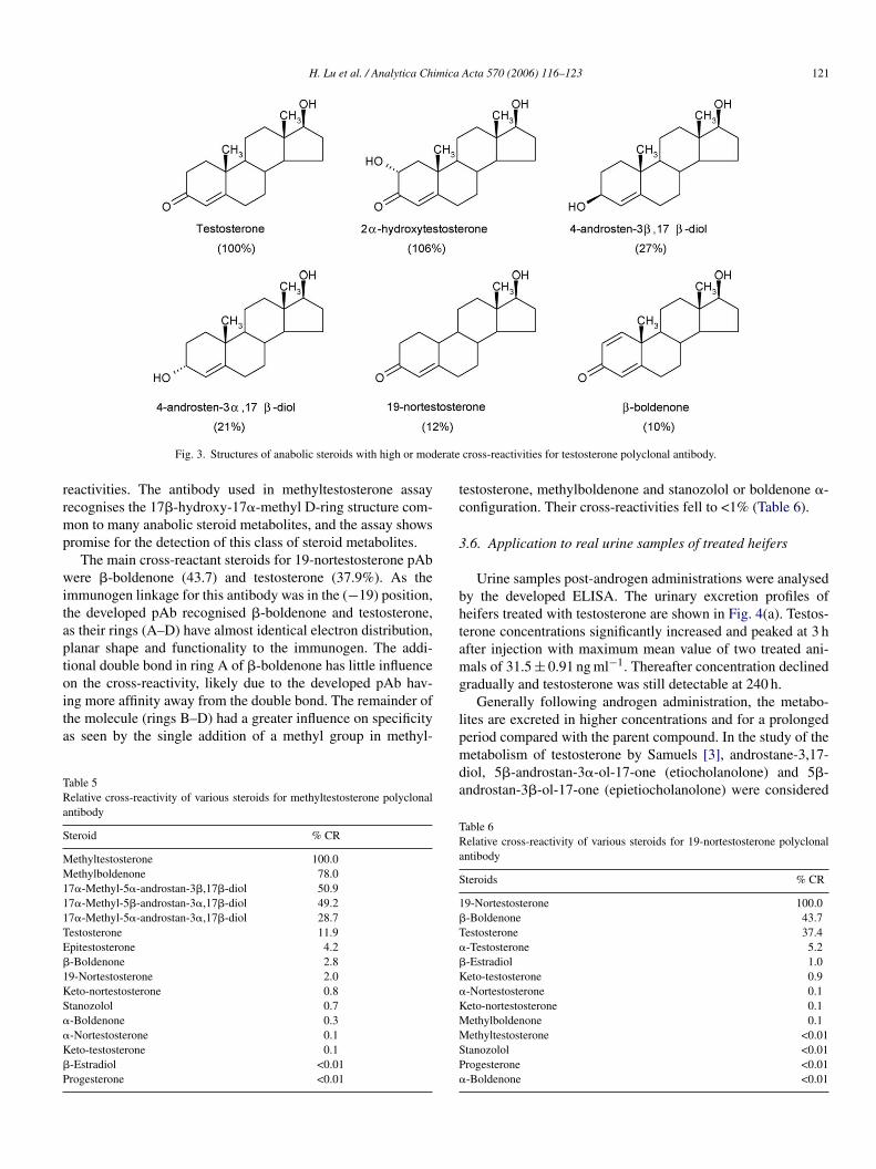
monoclonal antibody. Among the steroid compounds tested(Table 4), testosterone pAb recognised 2�-hydroxytestosteronevery well (105.7). Moderate cross-reactivities (28–10%) wereobserved for 4-androsten-3�,17�-diol, 4-androsten-3�,17�-diol, 19-nortestosterone and �-boldenone. This was quite a nor-mal cross-reactivity pattern for this antibody as the immunogenlinkage for this hapten was in the (−3) position and remain-ing structures (rings B–D) are identical for these steroids withonly the loss of one methyl group in the 19-position for 19-nortestosteorne (Fig. 3). The site at which the protein carrier (e.g.Keyhole Limpet Hemocyanin) is attached to the steroid moleculeimmunogen when generating the antibody largely determinesthe sites at which the antibody will recognise the steroid hapten.The antibody is likely to have the most specificity for sites dis-tant from the position where the protein carrier is linked becauseof steric hindrance [20]. Little cross-reactivity was observed forother steroids tested. Hence the assay using this antibody wouldshow promise for the detection of 17-alkyl-substituted/relatedanabolic steroids. This is desirable in an assay for screening forpotential androgen treatment in meat producing animals.
The significant cross-reactant steroids for methyltestos-terone pAb were methylboldenone (77.9%), 17�-methyl-5�-androstan-3�,17�-diol (50.9%), 17�-methyl-5�-androstan-3�,17�-diol (49.2%) and 17�-methyl-5�-androstan-3�,17�-diol(28.7%). Testosterone, epitestosterone and �-boldenone exhib-ited cross-reactivities from 3 to 12% (Table 5). This was notupt3l�wt
tudy due to the better sensitivity it provided compared withnusual considering the immunogen was linked in the (−3)osition, and thus remaining rings B–D are identical for methyl-estosterone and methylboldenone, 17�-methyl-5�-androstan-�,17�-diol, or 17�-methyl-5�-androstan-3�,17�-diol. Theoss of one methyl group at (−17) position of testosterone and-boldenone contributes to the lower cross-reactivity patternhen compared with 17�-hydroxy-17�-methyl D-ring struc-
ure steroids. Other steroids tested showed less than 2% cross-
H. Lu et al. / Analytica Chimica Acta 570 (2006) 116–123 121
Fig. 3. Structures of anabolic steroids with high or moderate cross-reactivities for testosterone polyclonal antibody.
reactivities. The antibody used in methyltestosterone assayrecognises the 17�-hydroxy-17�-methyl D-ring structure com-mon to many anabolic steroid metabolites, and the assay showspromise for the detection of this class of steroid metabolites.
The main cross-reactant steroids for 19-nortestosterone pAbwere �-boldenone (43.7) and testosterone (37.9%). As theimmunogen linkage for this antibody was in the (−19) position,the developed pAb recognised �-boldenone and testosterone,as their rings (A–D) have almost identical electron distribution,planar shape and functionality to the immunogen. The addi-tional double bond in ring A of �-boldenone has little influenceon the cross-reactivity, likely due to the developed pAb hav-ing more affinity away from the double bond. The remainder ofthe molecule (rings B–D) had a greater influence on specificityas seen by the single addition of a methyl group in methyl-
Table 5Relative cross-reactivity of various steroids for methyltestosterone polyclonalantibody
Steroid % CR
Methyltestosterone 100.0Methylboldenone 78.017�-Methyl-5�-androstan-3�,17�-diol 50.917�-Methyl-5�-androstan-3�,17�-diol 49.217�-Methyl-5�-androstan-3�,17�-diol 28.7Testosterone 11.9E�
1KS�
�
K�
P
testosterone, methylboldenone and stanozolol or boldenone �-configuration. Their cross-reactivities fell to <1% (Table 6).
3.6. Application to real urine samples of treated heifers
Urine samples post-androgen administrations were analysedby the developed ELISA. The urinary excretion profiles ofheifers treated with testosterone are shown in Fig. 4(a). Testos-terone concentrations significantly increased and peaked at 3 hafter injection with maximum mean value of two treated ani-mals of 31.5 ± 0.91 ng ml−1. Thereafter concentration declinedgradually and testosterone was still detectable at 240 h.
Generally following androgen administration, the metabo-lites are excreted in higher concentrations and for a prolongedperiod compared with the parent compound. In the study of themetabolism of testosterone by Samuels [3], androstane-3,17-diol, 5�-androstan-3�-ol-17-one (etiocholanolone) and 5�-androstan-3�-ol-17-one (epietiocholanolone) were considered
Table 6Relative cross-reactivity of various steroids for 19-nortestosterone polyclonalantibody
Steroids % CR
19-Nortestosterone 100.0�-Boldenone 43.7Testosterone 37.4�
�
K�
KMMSP�
pitestosterone 4.2-Boldenone 2.89-Nortestosterone 2.0eto-nortestosterone 0.8tanozolol 0.7-Boldenone 0.3-Nortestosterone 0.1eto-testosterone 0.1-Estradiol <0.01rogesterone <0.01
-Testosterone 5.2-Estradiol 1.0eto-testosterone 0.9-Nortestosterone 0.1eto-nortestosterone 0.1ethylboldenone 0.1ethyltestosterone <0.01
tanozolol <0.01rogesterone <0.01-Boldenone <0.01

122 H. Lu et al. / Analytica Chimica Acta 570 (2006) 116–123
Fig. 4. Urinary excretion profile of testosterone (a), methyltestosterone (b)and 19-nortestosterone (c) in treated heifers, determined using the developedELISAs. Concentrations found from developed calibration curves using 1/10dilution factor (see Fig. 1 (�)), each point represents triplicate measurements.Each colour bar represents two different treated animals.
to be the main metabolites of testosterone in bovine urine follow-ing administration. Other possible major metabolites accordingto the work of several authors [2,21] were 2�-, 6�-, 16�-,16�-hydroxytestosterone, estradiol, etc. As shown in Table 4,our method offers the advantage of detecting both testosteroneand its major metabolites, such as 17-alkyl-substituted anabolicsteroids due to the high cross-reactivity pattern with the antibodyused in the assay. But further D-ring hydroxylated metabolites,such as etiocholanolone, epietiocholanolone, etc. would not bedetected in this assay, as the cross-reactivities for such com-pounds were low.
In the case of methyltestosterone, the urinary excretionmetabolic profile showed the highest concentration observed at6 h (636.1 ± 44.3 ng ml−1) after androgen injection (Fig. 4(b)).The time profiles of two treated animals were similar peaking at6 h with compounds disappearing gradually after 24 h.
A number of studies have investigated the possiblemetabolism pathway of methyltestosterone in human, equineand bovine urine. The metabolism of methyltestosterone inman was investigated in previous work [22,23], identifying17�-methyl-5�-androstane-3�,17�-diol and 17�-methyl-5�-androstane-3�,17�-diol as the main metabolites, whichindicated a A-ring metabolism. An equine urine excretion studyafter oral administration of a (1 + 1) mixture of methyltestos-terone and its deuterized analogue showed that metabolitesbeing excreted as free steroids and as glucuronide or sulphateconjugates. The main phase I metabolic processes appear tobe partial or complete reduction of the 3-oxo-4-ene group,17-epimerization, 15 and 16 hydroxylation and at least twoother undetermined sites, possible the 6 and 11 positions[24]. A study performed by Blokland et al. [4] showed thatthe most dominant metabolite formed after administration ofmethyltestosterone in a heifer was 17�-methyl-5�-androstane-3�,17�-diol with some minor concentrations of isomericcompounds like 17�-methyl-5�-androstane-3�,17�-diol and17�-methyl-5�-androstane-3�,17�-diol.
As shown in Table 5, 17�-hydroxy-17�-methyl D-ring struc-ture common to many anabolic steroid metabolites showedhigh cross-reactivities in the methyltestosterone assay. Henceour method would be expected to detect this class of steroidmetabolites. The methyltestosterone urinary profile matchesthat observed by the standard GC–MS method. Blokland et al.rttespd
m(nosT
nmtEaswaa
4
tmd
eported that both parent compound and its metabolites reachedheir maximum concentration level in urine within 24 h afterreatment of the animals [4]. Existing methods of analysis gen-rally require hydrolysed and extracted procedures before urineample analysis. Our method obviates the necessity of urine sam-le pre-treatment (chemical and/or physical) other than simpleilution.
19-Nortestosterone showed a similar urine excretionetabolism profile with a maximum found from 3 to 9 h
499.5 ± 139.50 ng ml−1). The concentrations declined and 19-ortestosteorne was detectable until 240 h. The time profilesf two treated animals were similar, but concentrations weretrongly influenced by the metabolism of individual animals.his accounts for the large variation in the mean value (Fig. 4(c)).
At present, little is known about the metabolites of 19-ortestosterone nor about their detection. 19-Nortestosteroneetabolites were not determined in this current study. Never-
heless, the concentration of this steroid in urine detected byLISA gave sufficient evidence of illegal 19-nortestosteronedministration to heifers. The aim of screening is to find andelect suspicious samples for further analysis and our methodshich can provide quantitative or semi-quantitative information
re beneficial for the development of strategies to confirm thebuse of these steroids in meat producing animals.
. Conclusion
This work demonstrates the possibility of measuringestosterone, methyltestosterone, 19-nortestosteorne and their
etabolites in bovine urine by ELISA. The limits ofetection were 7.4 ± 2.9 pg ml−1, 26.6 ± 8.2 pg ml−1 and

H. Lu et al. / Analytica Chimica Acta 570 (2006) 116–123 123
13.1 ± 1.9 pg ml−1 for testosterone, methyltestosterone and 19-nortestosterone assay, respectively. Results of repeated analysisof spiked bovine urine samples showed good inter-day preci-sion and accuracy for three androgen assays. Hydrolysis andextraction of urine, which is generally required prior to existingmethods of analysis, was not a prerequisite using our methods. Inparticular, the testosterone assay offers the advantage of detect-ing both testosterone and its major metabolites, such as 17-alkyl-substituted anabolic steroids due to the high cross-reactivitypattern with the antibody used in the assay. The methyltestos-terone assay also detected the 17�-hydroxy-17�-methyl D-ringstructure common to many anabolic steroid metabolites. Theconcentration of 19-nortestosterone in bovine urine detected bydeveloped method does indicate 19-nortestosterone administra-tion to heifers. With simple and fast sample preparation, lowlimits of detection, good repeatability, and the potential of test-ing a large number of samples in a short period of time, thesemethods offer potential primary screening options for detectingandrogen residues in meat producing animals.
Acknowledgements
This projected is funded by EC Quality Life and Manage-ment of Living Resources Programme (QLRT-2000-06170). Theauthors thank Dr. Mark Kreuzer for his helpful discussion.
R
[3] T.P. Samuels, A. Nedderman, M.A. Seymour, E. Houghton, Analyst 123(1998) 2401.
[4] M.H. Blokland, H.J. van Rossum, H.A. Herbold, S.S. Sterk, R.W.Stephany, L.A. van Ginkel, Anal. Chim. Acta 529 (2005) 317.
[5] R. Draisci, L. Palleschi, E. Ferretti, L. Lucentini, P. Cammarata, J. Chro-matogr. A 870 (2000) 511.
[6] V. Popii, G. Baumann, Clin. Chim. Acta 350 (2004) 1.[7] T.Y. Wilke, D.J. Utley, Clin. Chem. 33 (1987) 1372.[8] E.H. Slaats, J.C. Kennedy, H. Kruijswijk, Clin. Chem. 33 (1987) 300.[9] K. Tarun, E. Ali, J. Immunol. Methods 147 (1992) 167.
[10] K.M. Rajkowski, Ch. Hanquez, A. Bouzoumou, N. Cittanova, Clin.Chim. Acta 183 (1989) 197.
[11] C. Hanquez, P. Urios, B. Desfosses, H. Samake, E. Lince, K.M.Rajkowski, N. Cittanova, Clin. Chim. Acta 164 (1989) 71.
[12] E.A.S. Al-Dujaili, Clin. Chim. Acta 364 (2006) 172–179.[13] C. van Peteghem, L. van Look, A. De Guesquiere, J. Chromatogr. B
489 (1989) 219.[14] A.F. Rizzo, E. Alitupa, T. Hirvi, S. Berg, Anal. Chim. Acta 275 (1993)
135.[15] M.P. Oriundi, R. Angeletti, E. Bastiani, C. Nachtmann, K.E. Vanoost-
huyze, C. van Peteghem, Analyst 120 (1995) 577.[16] A. Roda, A.C. Manetta, O. Portanti, M. Mirasoli, M. Guardigli, P. Pasini,
R. Lelli, Luminescence 18 (2003) 72.[17] P. Crabbe, U.J. Meyer, G. Pieraccini, M. O’Keeffe, C. Van Peteghen, J.
Anal. Toxicol. 27 (2003) 213.[18] N.L. Hungerford, B. Sortais, C.G. Smart, A.R. McKinney, D.D. Ridley,
A.M. Stenhouse, C.J. Suann, K.J. Munn, M.N. Sillence, M.D. McLeod,J. Steroid Biochem. Mol. Biol. 96 (2005) 317.
[19] G. Conneely, Ph.D. Thesis, University College Cork, Cork, Ireland,2005.
[20] R. Andrew, Best Practice & Research Clinical Endrocrinology and
[
[[[
eferences
[1] R. Navajas, C. Imaz, D. Carreras, M. Garcıa, M. Perez, C. Rodrıguez,A.F. Rodrıguez, R. Cortes, J. Chromatogr. B 673 (1995) 159.
[2] X.F. Li, M. Ma, A. Cheng, J. Zheng, Y.K. Tam, Anal. Chim. Acta 457(2002) 165.
Metabolism, vol. 15, 2001, p. 1.21] E. Venturelli, A. Cavalleri, G. Secreto, J. Chromatogr. B 671 (1995)
363.22] E.L. Rongone, A. Segaloff, J. Biol. Chem. 237 (1962) 1066.23] W. Schanzer, M. Donike, Anal. Chim. Acta 275 (1993) 23.24] C. Schoene, A.N.R. Nedderman, F. Houghton, Analyst 119 (1994) 2537.