sarah perrett alexander k. buell tuomas p. j. knowles
TRANSCRIPT
Advances in Experimental Medicine and Biology 1174
Sarah PerrettAlexander K. BuellTuomas P. J. Knowles Editors
Biological and Bio-inspired NanomaterialsProperties and Assembly Mechanisms
Advances in Experimental Medicineand Biology
Volume 1174
Editorial Board
IRUN R. COHEN, The Weizmann Institute of Science, Rehovot, IsraelABEL LAJTHA, N.S. Kline Institute for Psychiatric Research,Orangeburg, NY, USAJOHN D. LAMBRIS, University of Pennsylvania, Philadelphia, PA, USARODOLFO PAOLETTI, University of Milan, Milan, ItalyNIMA REZAEI, Children’s Medical Center Hospital, Tehran University ofMedical Sciences, Tehran, Iran
More information about this series at http://www.springer.com/series/5584
Sarah Perrett • Alexander K. BuellTuomas P. J. KnowlesEditors
Biological and Bio-inspiredNanomaterials
Properties and Assembly Mechanisms
123

EditorsSarah PerrettNational Laboratory of BiomacromoleculesInstitute of BiophysicsChinese Academy of SciencesBeijing, China
Alexander K. BuellDepartment of Biotechnologyand BiomedicineTechnical University of DenmarkDTU, Lyngby, Denmark
Tuomas P. J. KnowlesDepartment of ChemistryUniversity of CambridgeCambridge, UK
ISSN 0065-2598 ISSN 2214-8019 (electronic)Advances in Experimental Medicine and BiologyISBN 978-981-13-9790-5 ISBN 978-981-13-9791-2 (eBook)https://doi.org/10.1007/978-981-13-9791-2
© Springer Nature Singapore Pte Ltd. 2019This work is subject to copyright. All rights are reserved by the Publisher, whether the whole or part ofthe material is concerned, specifically the rights of translation, reprinting, reuse of illustrations, recitation,broadcasting, reproduction on microfilms or in any other physical way, and transmission or informationstorage and retrieval, electronic adaptation, computer software, or by similar or dissimilar methodologynow known or hereafter developed.The use of general descriptive names, registered names, trademarks, service marks, etc. in this publicationdoes not imply, even in the absence of a specific statement, that such names are exempt from the relevantprotective laws and regulations and therefore free for general use.The publisher, the authors, and the editors are safe to assume that the advice and information in this bookare believed to be true and accurate at the date of publication. Neither the publisher nor the authors orthe editors give a warranty, express or implied, with respect to the material contained herein or for anyerrors or omissions that may have been made. The publisher remains neutral with regard to jurisdictionalclaims in published maps and institutional affiliations.
This Springer imprint is published by the registered company Springer Nature Singapore Pte Ltd.The registered company address is: 152 Beach Road, #21-01/04 Gateway East, Singapore 189721,Singapore
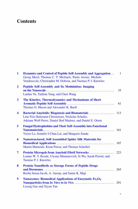
Contents
1 Dynamics and Control of Peptide Self-Assembly and Aggregation . . . 1Georg Meisl, Thomas C. T. Michaels, Paolo Arosio, MicheleVendruscolo, Christopher M. Dobson, and Tuomas P. J. Knowles
2 Peptide Self-Assembly and Its Modulation: Imagingon the Nanoscale . . . . . . . . . . . . . . . . . . . . . . . . . . . . . . . . . . . . . . . . . . . . . . . . . . . . . . . . . . . . 35Lanlan Yu, Yanlian Yang, and Chen Wang
3 The Kinetics, Thermodynamics and Mechanisms of ShortAromatic Peptide Self-Assembly . . . . . . . . . . . . . . . . . . . . . . . . . . . . . . . . . . . . . . . . . . 61Thomas O. Mason and Alexander K. Buell
4 Bacterial Amyloids: Biogenesis and Biomaterials . . . . . . . . . . . . . . . . . . . . . . 113Line Friis Bakmann Christensen, Nicholas Schafer,Adriana Wolf-Perez, Daniel Jhaf Madsen, and Daniel E. Otzen
5 Fungal Hydrophobins and Their Self-Assembly into FunctionalNanomaterials . . . . . . . . . . . . . . . . . . . . . . . . . . . . . . . . . . . . . . . . . . . . . . . . . . . . . . . . . . . . . . . 161Victor Lo, Jennifer I-Chun Lai, and Margaret Sunde
6 Nanostructured, Self-Assembled Spider Silk Materials forBiomedical Applications . . . . . . . . . . . . . . . . . . . . . . . . . . . . . . . . . . . . . . . . . . . . . . . . . . . 187Martin Humenik, Kiran Pawar, and Thomas Scheibel
7 Protein Microgels from Amyloid Fibril Networks . . . . . . . . . . . . . . . . . . . . . . 223Lianne W. Y. Roode, Ulyana Shimanovich, Si Wu, Sarah Perrett, andTuomas P. J. Knowles
8 Protein Nanofibrils as Storage Forms of Peptide Drugsand Hormones . . . . . . . . . . . . . . . . . . . . . . . . . . . . . . . . . . . . . . . . . . . . . . . . . . . . . . . . . . . . . . 265Reeba Susan Jacob, A. Anoop, and Samir K. Maji
9 Nanozymes: Biomedical Applications of Enzymatic Fe3O4Nanoparticles from In Vitro to In Vivo . . . . . . . . . . . . . . . . . . . . . . . . . . . . . . . . . . . 291Lizeng Gao and Xiyun Yan
v

vi Contents
10 Self-Assembly of Ferritin: Structure, Biological Functionand Potential Applications in Nanotechnology . . . . . . . . . . . . . . . . . . . . . . . . . . 313Soumyananda Chakraborti and Pinak Chakrabarti
11 DNA Nanotechnology for Building Sensors, Nanopores andIon-Channels . . . . . . . . . . . . . . . . . . . . . . . . . . . . . . . . . . . . . . . . . . . . . . . . . . . . . . . . . . . . . . . . 331Kerstin Göpfrich and Ulrich F. Keyser
12 Bio Mimicking of Extracellular Matrix . . . . . . . . . . . . . . . . . . . . . . . . . . . . . . . . . . 371Moumita Ghosh, Michal Halperin-Sternfeld,and Lihi Adler-Abramovich
13 Bioinspired Engineering of Organ-on-Chip Devices . . . . . . . . . . . . . . . . . . . . 401Li Wang, Zhongyu Li, Cong Xu, and Jianhua Qin
Chapter 1Dynamics and Control of PeptideSelf-Assembly and Aggregation
Georg Meisl, Thomas C. T. Michaels, Paolo Arosio, Michele Vendruscolo,Christopher M. Dobson, and Tuomas P. J. Knowles
Abstract The aggregation of proteins into fibrillar structures is a central processimplicated in the onset and development of several devastating neuro-degenerativediseases, but can, in contrast to these pathological roles, also fulfil importantbiological functions. In both scenarios, an understanding of the mechanisms bywhich soluble proteins convert to their fibrillar forms represents a fundamentalobjective for molecular sciences. This chapter details the different classes ofmicroscopic processes responsible for this conversion and discusses how theycan be described by a mathematical formulation of the aggregation kinetics. Wepresent easily accessible experimental quantities that allow the determination of thedominant pathways of aggregation, as well as a general strategy to obtain detailedsolutions to the kinetic rate laws that yield the microscopic rate constants of theindividual processes of nucleation and growth. This chapter discusses a frameworkfor a structured approach to address key questions regarding the dynamics ofprotein aggregation and shows how the use of chemical kinetics to tackle complexbiophysical systems can lead to a deeper understanding of the underlying physicaland chemical principles.
Keywords Chemical kinetics · Aggregation mechanisms · Scaling exponent ·Global analysis
G. Meisl (�) · T. C. T. Michaels · M. Vendruscolo · C. M. DobsonDepartment of Chemistry, University of Cambridge, Cambridge, UKe-mail: [email protected]
P. ArosioDepartment of Chemistry and Applied Bioscience, ETH Zurich, Zurich, Switzerland
T. P. J. KnowlesCentre for Misfolding Diseases, Department of Chemistry, University of Cambridge, Cambridge,UK
Cavendish Laboratory, University of Cambridge, Cambridge, UKe-mail: [email protected]
© Springer Nature Singapore Pte Ltd. 2019S. Perrett et al. (eds.), Biological and Bio-inspired Nanomaterials,Advances in Experimental Medicine and Biology 1174,https://doi.org/10.1007/978-981-13-9791-2_1
1
2 G. Meisl et al.
1.1 Introduction
The self-assembly of proteins into ordered linear structures is an important processfor many living systems, for example in the context of the formation of thecytoskeletal filaments. When it occurs in a controlled manner, this process cantherefore be central to the functionality of an organism, but conversely, unwantedfilamentous aggregation of proteins can have devastating effects on an organism’shealth. One such process of particular significance is the aggregation of proteinsinto elongated structures, amyloid fibrils, which may consist of thousands or morecopies of the same protein [1, 2]. Surprisingly, a large variety of unrelated proteinshave the ability to form amyloid structures, and once formed, these entities possessan inherent propensity towards promoting the conversion of further proteins into theamyloid form [3, 4]. The study of protein aggregation has become an importantarea of research largely because the proliferation of amyloid fibrils is closelyassociated with several devastating and increasingly prevalent diseases, includingtype II diabetes, Parkinson’s and Alzheimer’s diseases [5–7]. However, there isalso a number of proteins that self-assemble into fibrillar structures that are notassociated with disease but are functional and essential for living organisms [8–12]. Important examples of such functional protein assemblies include for instancebiofilaments of actin and tubulin, that are key parts of the eukaryotic cytoskeleton[9–11], as well as functional amyloid structures [13] that possess roles as catalyticscaffolds [14], as depots for hormones [15], in the functioning of pathogens [16] oras components of bacterial biofilms [17, 18]. The existence of functional amyloidshas also inspired the use of such structures as functional biomaterials in variousnanotechnological applications [19, 20], a factor that has further contributed to theinterest in understanding how filamentous self-assembly works.
From a biophysical point of view, the formation of filamentous structures fromdispersed proteins represents an elementary form of supra-molecular assembly sinceit is generally homo-molecular in nature [9, 21]. Yet, despite this apparent simplicity,many different molecular-level events contribute to the overall fibril formationprocess and the competition and interplay between these microscopic steps oftenresults in rich dynamical behaviour. Obtaining a molecular-level kinetic descriptionof self-assembling systems is thus a particularly challenging task which involvesconsidering a complex interconnected network of several distinct microscopicsteps, such as nucleation, growth or fragmentation processes [22, 23]. In thischapter we describe in detail how the use of chemical kinetics in the contextof protein aggregation allows one to overcome this challenge. We demonstratethat this approach provides a general strategy for quantifying the rates of theindividual microscopic steps of filamentous growth. This advance illuminates whichparts of the full reaction network determine the aggregation behaviour in a givensystem and which ones can instead be neglected, thus providing a very usefulmechanistic framework for designing strategies for controlling protein aggregationin technological applications or suppressing it for therapeutic purposes.
1 Dynamics and Control of Self-Assembly 3
The chapter is organized as follows. In the first section, we discuss themicroscopic-level processes that contribute to the overall protein aggregationreaction and outline a general approach for mathematically modelling the resultingreaction network. We then consider the interaction and competition between theseindividual processes and describe in detail how such complex scenarios can beunderstood within the framework of kinetic theory. The following section brieflylooks at the application of these kinetic models of protein aggregation in thecontext of data analysis for discovering the dominant microscopic processes inaction. Finally, we conclude with a discussion on how the resulting mechanisticunderstanding of protein aggregation forms the basis of devising rational strategiesto employ inhibitory compounds or modulations of the environmental conditions tocontrol the pathways by which the aggregation reaction proceeds.
1.2 Kinetic Theory of Protein Aggregation
Chemical kinetics provide the mathematical framework for predicting the timecourse of a chemical reaction. In general, a kinetic description of a chemical reactionis derived by breaking the overall process down into a sequence of one or morerelevant steps. The law of mass action then yields differential equations (so calledrate laws) that describe the rates of each one of these individual steps in termsof the concentrations of the species involved. The rate constants are the constantsof proportionality entering such relationships and the reaction orders describe thepower a particular concentration is raised to. Although for simple elementaryreactions the reaction orders correspond to the actual number of species involvedin the reaction, such a simple physical interpretation of reaction orders may notapply to reactions with many steps, such as encountered in the complex modelsdiscussed here. Rate laws emerge directly from a consideration of the various stepsthat constitute the overall reaction and thus represent the best tool for establishingunknown mechanisms. As we will discuss later, if the reaction mechanism isunknown, we can carry out experiments to determine the reaction orders withrespect to each reactant and then try out various trial reaction mechanisms to seewhich one fits best with the experimental data. An important point to recognizehere is that the rate constants and reaction orders need to be constrained by theexperiments. In the following, we apply these concepts from chemical kinetics tothe study of protein aggregation phenomena.
1.2.1 Fundamental Processes in Protein Aggregation
In order to develop a kinetic model of aggregation, it is first necessary to establishthe species and the microscopic-level processes that are likely to be involved inthe overall assembly reaction. In this context, it is important to keep the kinetic
4 G. Meisl et al.
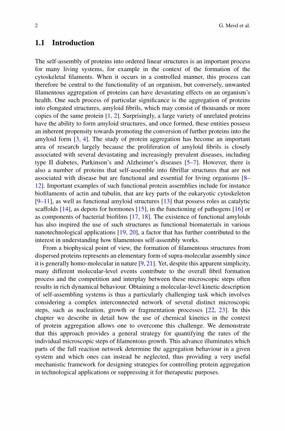
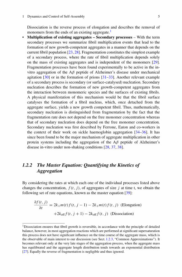
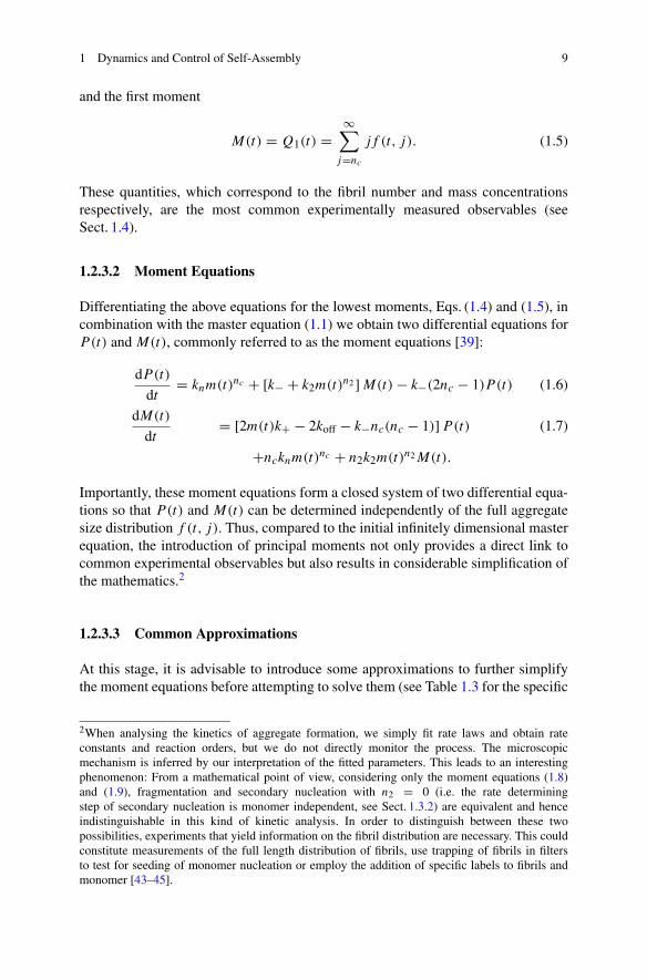
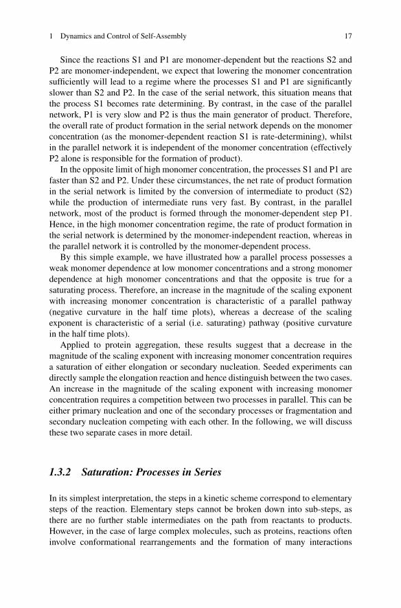
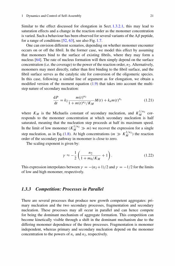
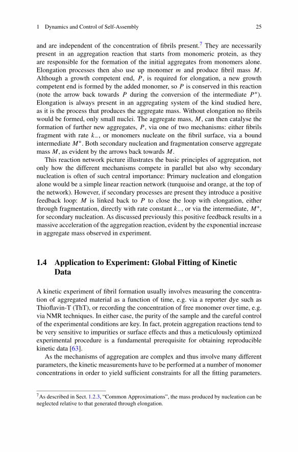
1o nucleation Elongation Fragmentation 2o nucleation
kn k+
koff k- k2
Fig. 1.1 Microscopic processes of aggregation. A schematic depiction of the fundamentalmicroscopic processes of aggregation, also considered in the basic model discussed in Sects. 1.2.2and 1.2.3. Spheres represent monomer, cylinders fibrillar species. Extensions to these processesto account for their multi-step nature, which may become evident under certain conditions, arediscussed in Sect. 1.3
model as minimalistic as possible and only include those microscopic steps that arerequired to explain experimental observations or that are suggested by our physicalintuition of the system. For this reason, we discuss here the simplest kinetic model offilamentous assembly [23], in which filamentous aggregates are described as linearchains of monomers that are formed, grow and multiply according to the followingthree basic categories of processes that form the core parts of the aggregationreaction (see Fig. 1.1 for a schematic visualization of the individual processes):
• De novo formation of aggregates – Primary nucleation – Primary nucleationis the spontaneous formation of a growth-competent aggregate (nucleus) frommonomers alone, without the involvement of fibrillar species. As such, primarynucleation represents always the first event in an aggregation process startingwith monomers only. The primary nucleation step reflects the fact that theformation of small filaments is unfavourable so that aggregates smaller than acertain critical size are unstable; the nucleus represents the smallest aggregatespecies that is more likely to grow than to dissociate back into monomers.Although it involves only monomers, it can, and in many cases does, happenheterogeneously, on surfaces such as for example the air-solution interface orlipid membranes [24, 25]. Removal of specific interfaces or experiments atdifferent surface to volume ratios can give insights into the extent to which suchinterface effects alter the kinetics [26].
• Growth of existing aggregates – Elongation and dissociation – Existingaggregates are capable of further growth through elongation, but may also shrinkthrough dissociation processes. During the elongation reaction, soluble proteinadds onto the ends of an existing fibril thus leading to an increase of the overallaggregate mass. This process typically involves the attachment of monomericprotein to either end of an existing fibril followed by a conformational rear-rangement of the added monomer into a structure with high β-sheet content. Theaddition step is typically rate-determining, but under certain conditions the multi-step nature of the elongation reaction can become apparent (see Sect. 1.3.2).
1 Dynamics and Control of Self-Assembly 5
Dissociation is the reverse process of elongation and describes the removal ofmonomers from the ends of an existing aggregate.1
• Multiplication of existing aggregates – Secondary processes – With the termsecondary processes we summarize fibril multiplication events that lead to theformation of new growth-competent aggregates in a manner that depends on thecurrent fibril population [23, 28]. Fragmentation constitutes the simplest exampleof a secondary process, where the rate of fibril multiplication depends solelyon the mass of existing aggregates and is independent of the monomers [29].Fragmentation processes have been found experimentally to be active in the in-vitro aggregation of the Aβ peptide of Alzheimer’s disease under mechanicalagitation [30] or in the formation of prions [31–33]. Another relevant exampleof a secondary process is secondary (or surface-catalysed) nucleation. Secondarynucleation describes the formation of new growth-competent aggregates fromthe interaction between monomeric species and the surfaces of existing fibrils.A physical manifestation of this mechanism would be that the fibril surfacecatalyses the formation of a fibril nucleus, which, once detached from theaggregate surface, yields a new growth competent fibril. Thus, mathematically,secondary nucleation is distinguished from fragmentation by the fact that thefragmentation rate does not depend on the free monomer concentration whereasthat of secondary nucleation does depend on the free monomer concentration.Secondary nucleation was first described by Ferrone, Eaton and co-workers inthe context of their work on sickle haemoglobin aggregation [34–36]. It hassince been found to be the major mechanism of aggregate multiplication in otherprotein systems including the aggregation of the Aβ peptide of Alzheimer’sdisease in-vitro under non-shaking conditions [28, 37, 38].
1.2.2 The Master Equation: Quantifying the Kinetics ofAggregation
By considering the rates at which each one of the individual processes listed abovechanges the concentration, f (t, j), of aggregates of size j at time t, we obtain thefollowing set of rate equations, known as the master equation [39]:
∂f (t, j)
∂t= 2k+m(t)f (t, j − 1) − 2k+m(t)f (t, j) (Elongation)
+2kofff (t, j + 1) − 2kofff (t, j) (Dissociation)
1Dissociation ensures that fibril growth is reversible, in accordance with the principle of detailedbalance, however, in most aggregation reactions which are performed at significant supersaturationthis process does not have significant influence on the time course of the aggregate mass, which isthe observable of main interest to our discussion (see Sect. 1.2.3, “Common Approximations”). Itbecomes relevant only at the very late stages of the aggregation process, when the aggregate masshas equilibrated and the aggregate length distribution tends towards an exponential distribution[27]. Equally the reverse of fragmentation is negligible and thus ignored.
6 G. Meisl et al.
+2k−∞∑
i=j+1
f (t, i) − k−(j − 1)f (t, j) (Fragmentation)
+knm(t)ncδj,nc (Primary nucleation)
+k2m(t)n2
∞∑
i=nc
if (t, i) δj,n2 (Secondary nucleation) (1.1)
(valid for j ≥ nc)
where δi,j is the Kronecker delta and m(t) is the concentration of monomers.The terms on the right hand side of the master equation have straightforward
interpretation and represent, in order, contributions from elongation, dissociation,fragmentation, primary nucleation and secondary nucleation with the respective rateconstants as summarized in Table 1.1. For example, the rate of secondary nucleationdepends on the monomer concentration raised to the reaction order n2 and theavailable surface area of aggregated protein which is assumed to be proportionalto the aggregate mass concentration,
∑∞j=nc
jf (t, j). The factor of 2 on the firstand second lines of Eq. (1.1) appears because the rate constants for elongation anddissociation are defined per filament end and fibrils grow from both ends. Moreover,implicit in our formulation is the assumption that the rate constants for the variousprocesses are size independent. In particular, the fragmentation rate is assumed tobe uniform along the length of the aggregate, meaning that aggregates are equallylikely to break at each bond between monomers. Physically this behaviour could beenvisioned if fragmentation is governed by random fluctuations, leading to breakageof the bonds holding the fibril together. However, under mechanical stress one couldimagine a fragmentation rate that depends on the total length of the aggregate[41, 42]. In such a case our approach constitutes a mean-field approximation, aslong as the average fibril length is approximately constant in time.
A system in-vivo might be modelled using Eq. (1.1) assuming a constantconcentration of monomeric protein, as the organism constantly replenishes proteinconsumed through the aggregation process. In the majority of cases, however, theaggregation kinetics is measured in an in-vitro context, where the total mass of pro-tein and not the monomer concentration stays constant. Under these circumstances,the conservation of total protein mass mtot = m(t) + ∑∞
j=ncjf (t, j) has to be
enforced, yielding an additional rate equation for m(t):
dm(t)
dt= − d
dt
∞∑
j=nc
jf (t, j). (1.2)
Finally, it is important to notice here that a reaction step (e.g. elongation step)that is effectively written as a single term in the master equation (1.1) may in factconsist of several steps, whose effect can become kinetically apparent under certainenvironmental conditions. This observation will become extremely important as we
1 Dynamics and Control of Self-Assembly 7
Table 1.1 Parameters
Parameter/Units description
m, (m0)/c (Initial) monomer concentration. m(t) is the concentration of free, non-aggregated monomer, called m0 at the beginning of the aggregation reaction.
M, (M0)/c (Initial) fibril mass concentration. M(t) is the mass concentration ofaggregates, i.e. the equivalent monomer concentration if the aggregates werere-dissolved. Its value at the beginning of the reaction is M0, which is 0 in thecase of an unseeded aggregation reaction.
P, (P0)/c (Initial) fibril number concentration. P(t) is the number concentration ofaggregates, proportional to the number concentration of growth competentends, which are the points at which the aggregate can elongate. Its value atthe beginning of the reaction is P0, which is 0 in the case of an unseededaggregation reaction. P is linked to M by the average fibril length, L, viaM/P = L. P is difficult to measure directly but can be estimated from M byusing the average fibril length.
kn/t−1c−nc+1 Primary nucleation rate constant. This appears as knmnc in the rate of
formation of primary nuclei. It has units of time−1 concentration−nc+1.
nc/unit-less Reaction order of primary nucleation. This appears as knmnc in the rate
of formation of primary nuclei. Its simple interpretation, for example in thecontext of classical nucleation theory, is that of a nucleus size, however thisinterpretation is only valid if the reaction is a simple single step process. It isunit-less and typically has a value between 0 and 5.
k+/t−1 c−1 Elongation rate constant. This appears as 2k+mP in the rate of formation ofnew aggregate mass. It has units of time−1 concentration−1.
koff/t−1 Depolymerisation rate constant. This appears as −2koffP in the rate ofaggregate mass formation and is the rate at which monomers are lost from fibrilends. It has units of time−1. This may be a global fitting parameter. However,in most cases it is negligibly small.
k−/t−1 Fragmentation rate constant. This appears as k−M in the rate of formationof new growth competent ends from fragmentation. It has units of time−1.This form of the fragmentation rate assumes that an aggregate is equally likelyto break anywhere along its length, with the time-scale of breaking given by1/k−.
k2/t−1 c−n2 Secondary nucleation rate constant. This appears as k2mn2M in the rate of
formation of secondary nuclei. It has units of time−1 concentration−n2 .
n2/unit-less Reaction order of secondary nucleation. This appears as k2mn2M in the rate
of formation of secondary nuclei. Its simple interpretation, for example in thecontext of classical nucleation theory, is that of a nucleus size, however thisinterpretation is only valid if the reaction is a simple single step process. It isunit-less and typically has a value between 0 and 4 [28, 40], although largervalues are possible.
learn more about saturation effects in Sect. 1.3.2. In order to correctly interpret theresults of our kinetic analysis and develop appropriate extensions of existing models,it is thus important to keep in mind what range of detailed mechanisms will maponto this coarse-grained description and what mechanisms require instead additionalcomplexity. For this reason we have summarized the formal definitions of the kineticmodel in Table 1.2.
8 G. Meisl et al.
Table 1.2 Definitions of the kinetic model of aggregation
Aggregates are linear chains of monomers, no branching occurs (i.e. new aggregates that format the surface detach).
Fibrils can fracture at any point with rate k−. This rate is unchanged throughout the fibril anddoes not depend on its length, i.e. every monomer-monomer bond has an equal probability ofbreaking.
Monomers can attach and detach at the ends of fibrils with rate constants k+ and (koff + k−)
respectively (i.e. koff determines how much more likely a monomer at the end is to break offcompared to the fibril breaking at any other position). These rate constants are independent ofthe size of the aggregate.
Aggregates are unstable below a certain size, nc, and the transient population of aggregatessmaller than nc is hence neglected.
Stable aggregates of size nc can form from monomers with the nucleation rate constant kn. Thisprocess is referred to as primary nucleation.
Stable aggregates of size n2 can form from monomers, heterogeneously on the surface ofexisting aggregates, with the nucleation rate constant k2. This process is referred to as secondarynucleation. It is assumed not to influence the elongation behaviour of the aggregates serving asa nucleation surface.
All rate constants are reaction limited, i.e. the effects of diffusion are not considered explicitly.
Interactions between aggregates are neglected.
1.2.3 Principal Moments and Moment Equations
Being a set of infinitely many coupled and non-linear differential equations, themaster equation (1.1) seems very hard to solve. However, in a data analysis contextthis difficultly does not necessarily preclude its use. In most cases, the full aggregatesize distribution f (t, j) is in fact not easily accessible through experiments. Instead,measurements of average quantities, such as the total fibril mass, are made, whichcan already provide significant insights into the mechanisms of aggregation.
1.2.3.1 Principal Moments
A particularly useful strategy to reduce the complexity of the master equationis given by the introduction of the principal moments of the aggregate lengthdistribution, defined by the equation:
QN(t) =∞∑
j=nc
jNf (t, j). (1.3)
Of particular experimental interest are the zero-th moment
P(t) = Q0(t) =∞∑
j=nc
f (t, j) (1.4)
1 Dynamics and Control of Self-Assembly 9
and the first moment
M(t) = Q1(t) =∞∑
j=nc
jf (t, j). (1.5)
These quantities, which correspond to the fibril number and mass concentrationsrespectively, are the most common experimentally measured observables (seeSect. 1.4).
1.2.3.2 Moment Equations
Differentiating the above equations for the lowest moments, Eqs. (1.4) and (1.5), incombination with the master equation (1.1) we obtain two differential equations forP(t) and M(t), commonly referred to as the moment equations [39]:
dP(t)
dt= knm(t)nc + [k− + k2m(t)n2 ] M(t) − k−(2nc − 1)P (t) (1.6)
dM(t)
dt= [2m(t)k+ − 2koff − k−nc(nc − 1)] P(t) (1.7)
+ncknm(t)nc + n2k2m(t)n2M(t).
Importantly, these moment equations form a closed system of two differential equa-tions so that P(t) and M(t) can be determined independently of the full aggregatesize distribution f (t, j). Thus, compared to the initial infinitely dimensional masterequation, the introduction of principal moments not only provides a direct link tocommon experimental observables but also results in considerable simplification ofthe mathematics.2
1.2.3.3 Common Approximations
At this stage, it is advisable to introduce some approximations to further simplifythe moment equations before attempting to solve them (see Table 1.3 for the specific
2When analysing the kinetics of aggregate formation, we simply fit rate laws and obtain rateconstants and reaction orders, but we do not directly monitor the process. The microscopicmechanism is inferred by our interpretation of the fitted parameters. This leads to an interestingphenomenon: From a mathematical point of view, considering only the moment equations (1.8)and (1.9), fragmentation and secondary nucleation with n2 = 0 (i.e. the rate determiningstep of secondary nucleation is monomer independent, see Sect. 1.3.2) are equivalent and henceindistinguishable in this kind of kinetic analysis. In order to distinguish between these twopossibilities, experiments that yield information on the fibril distribution are necessary. This couldconstitute measurements of the full length distribution of fibrils, use trapping of fibrils in filtersto test for seeding of monomer nucleation or employ the addition of specific labels to fibrils andmonomer [43–45].
10 G. Meisl et al.
Table 1.3 Common approximations
Approximation Description
Mass from nucleation events If long fibrils are produced, i.e. on average significantlybigger than the nucleus sizes n2 and nc, the mass produceddirectly from nucleation events will be negligible. This meanslast two terms in Eq. (1.7) will be negligible. The aggregatesobserved in the context of most aggregating systems usuallyconsist of several thousand monomers, making this a goodapproximation.
Loss through fragmentation If a piece smaller than the nucleus size breaks off the ends itwill re-dissolve and therefore lead to a decrease in both massand number concentration. Because of the large average sizeand the usually small value of the fragmentation constant thiseffect is also negligible. The term last term in equation (1.6)becomes negligible and so does the “k−” term in equation(1.7).
Loss through depolymerisation Depolymerisation is the reverse of the elongation reaction. Ifm∞ denotes the free monomer concentration in equilibrium,i.e. the solubility of the monomer, then the depolymerisationrate is given by koff = k+m∞. Therefore the depolymerisa-tion rate will only become significant, i.e. comparable to theelongation rate, at low monomer concentrations approachingm∞. If the experiment is performed at concentrations wellabove the solubility, where most of the initial monomerconcentration will be incorporated into aggregates, then thedepolymerisation rate can be neglected for the description ofthe kinetics.
details of the various approximations employed here). These approximations arebased on considerations of the relative magnitudes of the rates of certain micro-scopic processes. For example, under typical environmental conditions nucleationprocesses are much slower than elongation; this condition ensures that long fibrilswill form from monomers. For this reason, elongation is the only process thatdepletes monomers significantly and the contribution of nucleation processes to theoverall increase in aggregate mass can be considered to be negligible compared tothat of elongation. These approximations result in the following simplified set ofmoment equations:
dP(t)
dt= knm(t)nc + [
k− + k2m(t)n2]M(t) (1.8)
dM(t)
dt= 2k+m(t)P (t). (1.9)
These equations are the fundamental kinetic equations for filamentous assemblywith secondary pathways and will thus serve as the basis for all descriptions in theremainder of this chapter unless otherwise stated.
1 Dynamics and Control of Self-Assembly 11
1.2.4 Solving the Moment Equations: The Fixed-Point Method
The moment equations (1.8) and (1.9) fully describe the time evolution of commonlymeasured quantities, the aggregate mass and number concentrations. From theseequations, one could, at least in principle, calculate the aggregate number andmass at any point in time, given the values of the rate constants and the boundaryconditions. In practice, the rate constants can be determined for example throughkinetic experiments, whereas the boundary conditions are imposed by the typeof system at hand, most commonly an in-vitro context where the total mass ofprotein stays constant. Unfortunately, exact solutions to the moment equations (1.8)and (1.9) exist only in very few cases, including Oosawa’s solution to filamentousgrowth dominated by primary nucleation [9], and in general one has to rely onapproximation methods in order to find accurate expressions for the aggregate massconcentration, M(t), as a function of time.
One could ask why there is any need to derive analytic solutions to the momentequations in the first place: The closed system of differential equations, (1.8)and (1.9), can simply be integrated numerically to arbitrary accuracy, so whatadvantage is gained by deriving analytic solutions? A minor factor is that analyticsolutions will significantly increase computation speed during data fitting, as numer-ical integration of differential equations can be both computationally expensiveand less robust than evaluation of an analytical function. Much more importantly,the availability of analytic solutions also allows the derivation of other systemrepresentative quantities and provides the basis for a more in depth understandingof the origins of the system’s behaviour. In particular, analytic solutions give ahandle on the relative importance of the different microscopic processes and can beused to derive qualitative constraints on the mechanism, based on easily measurablemacroscopic parameters [37].
A powerful approach to find an approximate solution to the moment equationsis the fixed-point iteration method [46]. The underlying idea of this approach isto reformulate the moment equations as a fixed-point equation and subsequentlyapply the associated fixed-point operator repeatedly to an initial guess. The repeatedapplication of the fixed-point operator will cause the so-constructed series ofapproximative solutions to converge to the exact solution of the moment equations.Making an appropriate choice for the initial guess is particularly important for thesuccess of the fixed-point method, largely because the repeated application of thefixed point operator can easily lead to very complex and thus impractical expressionsfor M(t). We now illustrate these principles on the moment equations (1.8)and (1.9).
To obtain the explicit form of the fixed-point relevant to our case, we integrateEq. (1.9) formally and recast it in terms of an integral operator3:
3Note that the fixed-point operator takes the form of Eq. (1.10) as long as the equation for dM/dt ,Eq. (1.9) remains unchanged.
12 G. Meisl et al.
M(t) = 2k+m0e−2k+∫ t
0 P(τ)dτ
∫ t
0e−2k+
∫ τ0 P(τ )dτ P (τ )dτ, (1.10)
where, for simplicity, we have assumed that only monomers are present initially(M0 = P0 = 0).
To construct an appropriate initial guess for the fixed-point iteration, we makeuse of our physical intuition by realizing that the assumption of constant monomerconcentration will be accurate during the early times of the reaction when thenumber of aggregates is small and the monomers are thus not significantly depleted.This solution, which was originally obtained by Eaton, Ferrone and co-workers,is found by linearising Eqs. (1.8) and (1.9) by enforcing a constant monomerconcentration, m(t) = m0, resulting in a set of linear differential equations thatcan be solved straightforwardly to yield:
Pinit(t) = knmnc0
κsinh(κt) (1.11)
Minit(t) = knmnc0
k−+k2mn20
[cosh(κt) − 1] , (1.12)
where
κ =√
2k+m0(k− + k2m
n20
). (1.13)
Finally, performing one fixed-point iteration by inserting Eq. (1.11) intoEq. (1.10) yields the following closed-form expression for M(t):
M(t)
m0= 1 − exp
(− knm
nc
0
k− + k2mn20
[cosh(κt) − 1]
). (1.14)
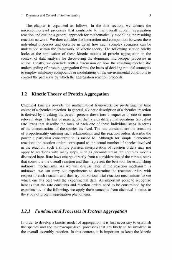
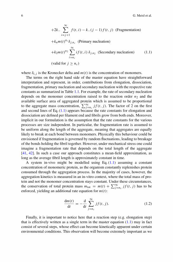
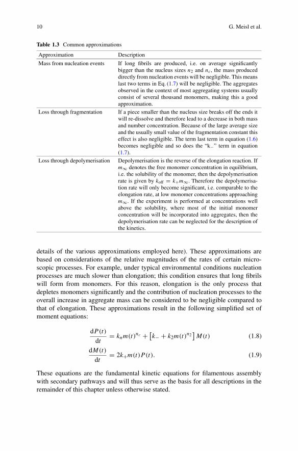
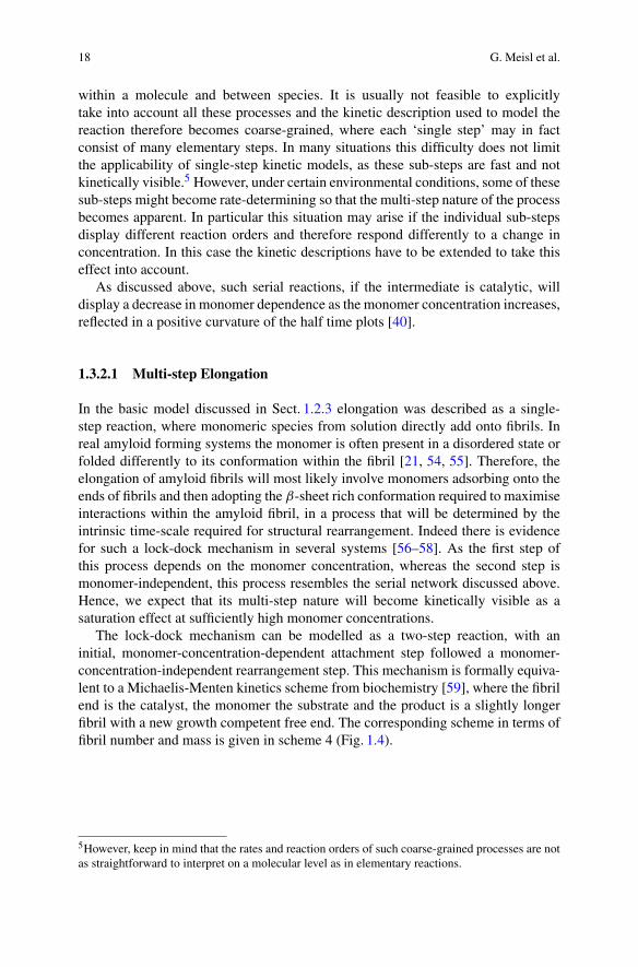
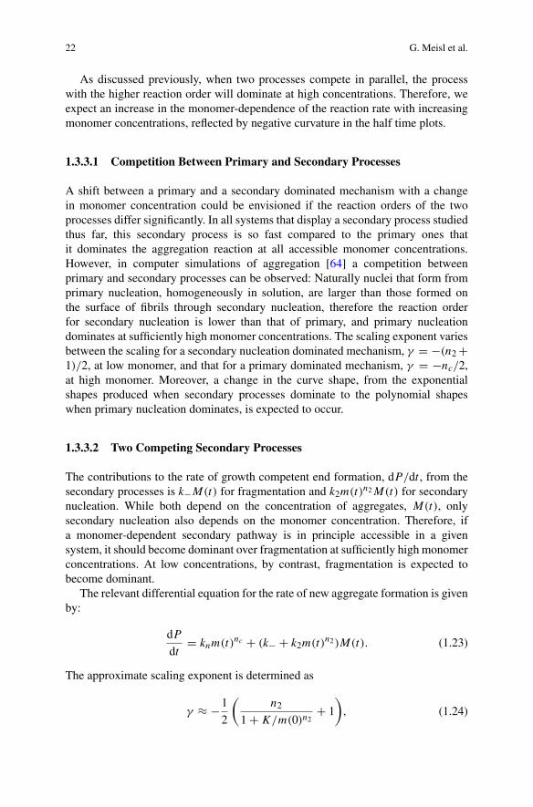
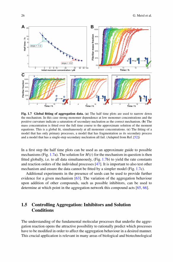
The comparison between Eq. (1.14) and the numerical solution to the momentequations in Fig. 1.2 demonstrates the power of the fixed-point iteration methodfor tackling the mathematical complexity inherent to the description of filamentousgrowth phenomena. With this strategy similar accurate approximate solutions havebeen obtained for various other mechanisms of aggregation; the explicit expressionsfor the various different aggregation mechanisms presented in the following can befound in Meisl et al. [47].
1.2.5 Implications from Integrated Rate Laws
One of the biggest advantages of obtaining analytic solutions to the momentequations is the ability to derive expressions for representative observables thatprovide the basis for an in-depth understanding of the origins of the system’s
1 Dynamics and Control of Self-Assembly 13
numerical solution
first fixed pointiteration
solution oflinearised equation
1.0
0.8
0.6
0.4
0.2
0.0
Fibr
il m
ass
fract
ion
5 51 02100 25Time
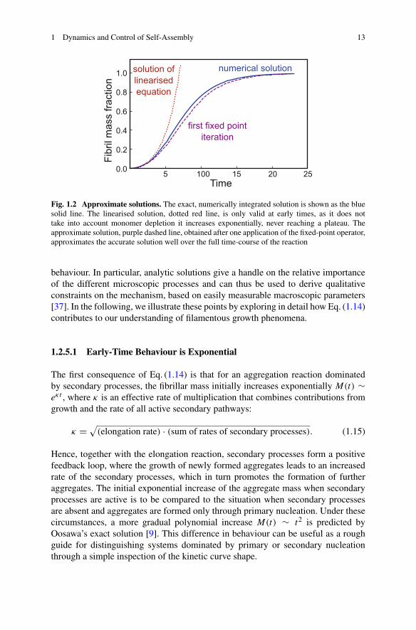
Fig. 1.2 Approximate solutions. The exact, numerically integrated solution is shown as the bluesolid line. The linearised solution, dotted red line, is only valid at early times, as it does nottake into account monomer depletion it increases exponentially, never reaching a plateau. Theapproximate solution, purple dashed line, obtained after one application of the fixed-point operator,approximates the accurate solution well over the full time-course of the reaction
behaviour. In particular, analytic solutions give a handle on the relative importanceof the different microscopic processes and can thus be used to derive qualitativeconstraints on the mechanism, based on easily measurable macroscopic parameters[37]. In the following, we illustrate these points by exploring in detail how Eq. (1.14)contributes to our understanding of filamentous growth phenomena.
1.2.5.1 Early-Time Behaviour is Exponential
The first consequence of Eq. (1.14) is that for an aggregation reaction dominatedby secondary processes, the fibrillar mass initially increases exponentially M(t) ∼eκt , where κ is an effective rate of multiplication that combines contributions fromgrowth and the rate of all active secondary pathways:
κ = √(elongation rate) · (sum of rates of secondary processes). (1.15)
Hence, together with the elongation reaction, secondary processes form a positivefeedback loop, where the growth of newly formed aggregates leads to an increasedrate of the secondary processes, which in turn promotes the formation of furtheraggregates. The initial exponential increase of the aggregate mass when secondaryprocesses are active is to be compared to the situation when secondary processesare absent and aggregates are formed only through primary nucleation. Under thesecircumstances, a more gradual polynomial increase M(t) ∼ t2 is predicted byOosawa’s exact solution [9]. This difference in behaviour can be useful as a roughguide for distinguishing systems dominated by primary or secondary nucleationthrough a simple inspection of the kinetic curve shape.
14 G. Meisl et al.
1.2.5.2 Half-Times and Scaling Exponents
There are many paths by which an aggregation reaction can proceed and a majorchallenge in the analysis of experimental data is to determine which aggregationmechanism is dominant. A fitting of the rate laws for the full network is often notfeasible, therefore strategies to narrow down the number of possible mechanisms arerequired. One qualitative constraint to the underlying mechanism can be obtainedby the analysis of the dependence of the half times on the monomer concentration.In the following, we will discuss how half times provide insights into the topologyof the reaction network and thereby constrain the possible mechanisms in action.
The half time of aggregation, t1/2, is defined as the time at which half thefibrillar mass present at the end of the reaction has been formed. The availabilityof Eq. (1.14) allows us to obtain an approximate expression for this half time bysolving M(t1/2) = m0/2, yielding [48, 49]:
t1/2 = 1
κlog
[log(2)(k− + k2m
n20 )
knmnc
0
]. (1.16)
As expected from the dominance of secondary pathways, t1/2 is found to beinversely proportional to the aggregate multiplication rate κ , even though a weakdependence of the half time on the primary nucleation emerges in form of alogarithmic correction. This result can be rationalized by the fact that the formationof new fibril ends is initially always controlled by primary nucleation even whensecondary processes are dominant at later stages of the reaction. Note that the ideathat in bulk experiments the observed lag time (i.e. the time until an increase inaggregate mass can be detected) is the time to formation of the first nucleus isa common misconception. In almost all bulk systems the first nucleus is formedeffectively immediately, the lag time is instead the time until a sufficient amountof aggregated material has been formed to be detected. First, this is evident fromthe reproducibility of the lag time and lack of stochastic effects, second, a self-consistency check using the fitted values of the nucleation rates shows that anucleation rate far away from that determined from the fitting would be requiredin order to put the system into a regime determined by stochastic nucleation effects[50]. However, stochastic behaviour, where the lag time is indeed determined bythe time until formation of the first nucleus, can be observed if the reactions areperformed in very small volumes, for example nl-sized droplets in a micro-fluidicdevice [51].
When either fragmentation or secondary nucleation is dominant, the dependenceof t1/2 on the monomer concentration, m0, takes approximately the form of a powerlaw t1/2 ≈ m
γ
0 , where γ is referred to as the scaling exponent. A double logarithmicplot of the half time versus monomer concentrations therefore gives a straight linewith slope γ :
γ = d(log(t1/2))
d(log(m0))= m0
d(log(t1/2))
dm0. (1.17)
1 Dynamics and Control of Self-Assembly 15
The exact value of the scaling exponent depends on the monomer-dependence ofthe dominant pathway of aggregation and in general it takes the form:
γ ≈ − (reaction order of elongation) + (reaction order of dominant 2o process)
2,
(1.18)where the factor of 1/2 originates from the square root in κ . For example,fragmentation is monomer independent and so in a system which is dominatedby fragmentation processes (i.e. the contribution from primary and secondarynucleation is negligible) we expect a scaling exponent of γ = −1/2. By contrast,the secondary nucleation step is characterized by a non-zero monomer dependence,hence a system dominated by secondary nucleation would result in a scalingexponent of γ = −(n2 + 1)/2. Because the reaction order of secondary nucleationcommonly takes low integer values around 2, the observation of a scaling exponentsof a large magnitude, e.g. −1.5, is typically indicative of a secondary-nucleation-dominated aggregation mechanism. In the absence of secondary pathways, thescaling exponent is determined by the reaction order of the primary nucleationstep alone, giving γ = −nc/2. Hence, half-time plots as a function of monomerconcentration and the associated scaling exponents are of central importance inestablishing the main mechanism of aggregation of a given protein system.
1.3 The Full Aggregation Network: Interplay andCompetition
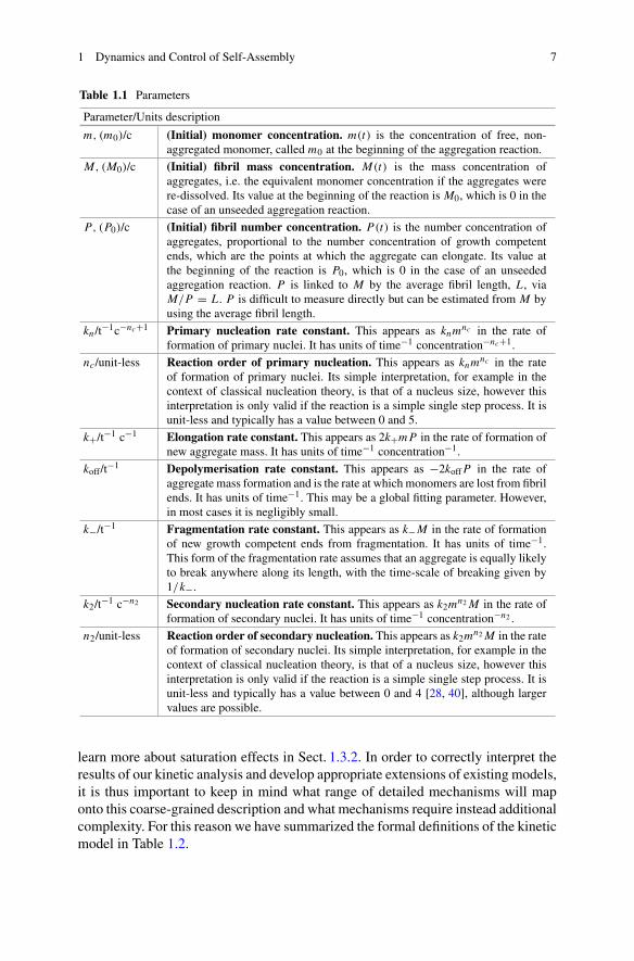
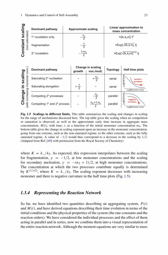
In the previous section, we have demonstrated how the scaling behaviour of the halftime with monomer concentration provides important insights into the nature ofthe dominant mechanism of filamentous assembly through the value of the scalingexponent γ . In the following, we demonstrate that, in addition to its actual value,the monomer concentration dependence of the scaling exponent can provide furtherinsights into the full aggregation network, such as the existence of saturation effectsor competing mechanisms [40].
1.3.1 Monomer Dependence of the Scaling Exponent as aGuide to Complex Mechanisms
The scaling exponent is determined by the reaction orders of the dominant processeson the aggregation pathway. Therefore, a scaling exponent which is independentof the monomer concentration is indicative of the fact that the dominant mech-anism of aggregation is likely to remain unchanged over the range of monomerconcentrations considered. By contrast, a scaling exponent that depends on themonomer concentration (i.e. the scaling exponent, not just the half times, dependon the monomer concentration) is indicative of a change of the reaction order of the
16 G. Meisl et al.
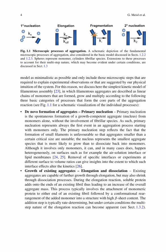
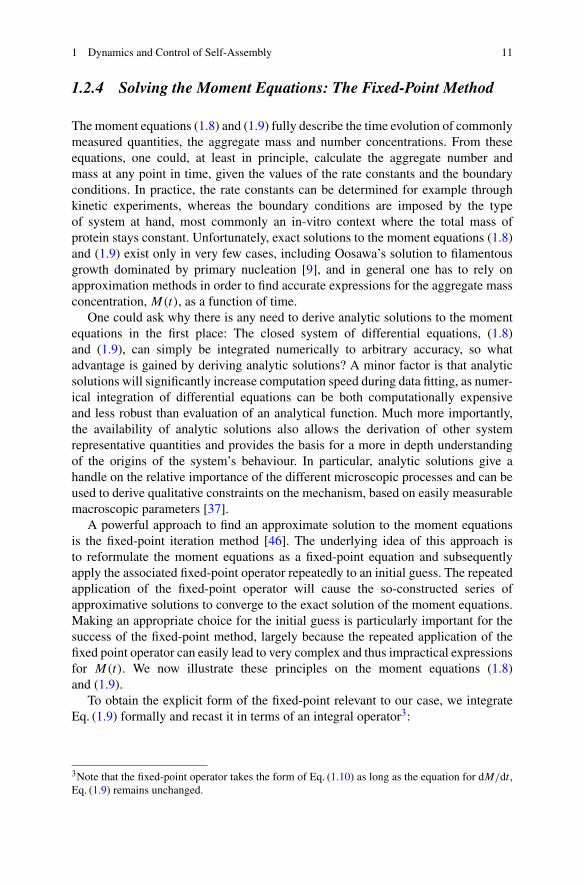
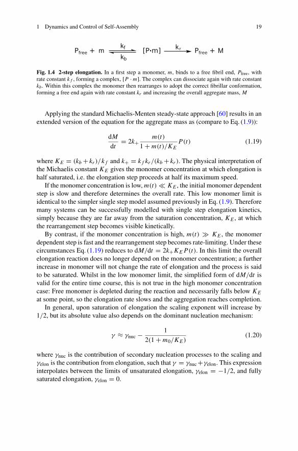
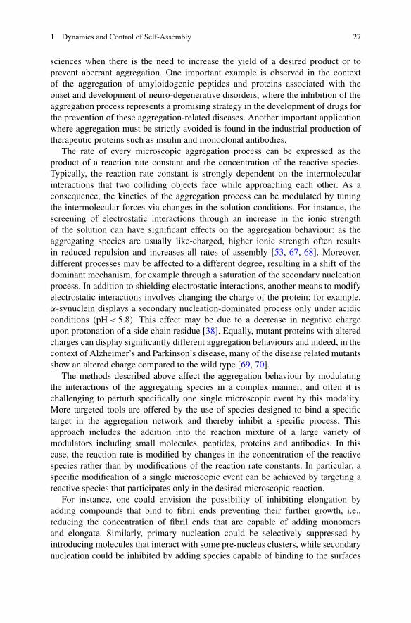
SerialLo
wH
igh
P2P1
P2P1S2
S1
S1
S2
Scal
ing slope = -1.2
slope = -0.2
Hal
f tim
e /
h
10
68
4
2
1
2 4 8 20 40 80Initial monomer concentration / μM
positivecurvature
Concentration dependence decreases(γ less negative) with increasing
Concentration dependence increases(γ more negative) with increasing
Parallel
6
20
10
4
20.6 1 2 4 8
40
negativecurvature
slope = -0.5
slope = -1.7
Initial monomer concentration / μMH
alf t
ime
/ h
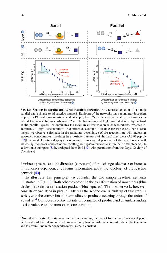
Fig. 1.3 Scaling in parallel and serial reaction networks. A schematic depiction of a simpleparallel and a simple serial reaction network. Each one of the networks has a monomer-dependentstep (S1 or P1) and monomer-independent step (S2 or P2). In the serial network S1 determines therate at low concentrations, whereas S2 is rate-determining at high concentrations. By contrast,in the parallel system P2 dominates the reaction at low monomer concentrations, whereas P1dominates at high concentrations. Experimental examples illustrate the two cases. For a serialsystem we observe a decrease in the monomer dependence of the reaction rate with increasingmonomer concentration, resulting in a positive curvature of the half time plots (Aβ40 peptide[52]). A parallel system displays an increase in monomer dependence of the reaction rate withincreasing monomer concentration, resulting in negative curvature in the half time plots (Aβ42at low ionic strengths [53]). (Adapted from Ref. [40] with permission from the Royal Society ofChemistry)
dominant process and the direction (curvature) of this change (decrease or increasein monomer dependence) contains information about the topology of the reactionnetwork [40].
To illustrate this principle, we consider the two simple reaction networksillustrated in Fig. 1.3. Both schemes describe the transformation of monomers (bluecircles) into the same reaction product (blue squares). The first network, however,consists of two steps in parallel, whereas the second one is built up of two steps inseries, with the conversion of intermediate to product occurring through the action ofa catalyst.4 Our focus is on the net rate of formation of product and on understandingits dependence on the monomer concentration.
4Note that for a simple serial reaction, without catalyst, the rate of formation of product dependson the rates of the individual reactions in a multiplicative fashion, so no saturation effects emergeand the overall monomer dependence will remain constant.
1 Dynamics and Control of Self-Assembly 17
Since the reactions S1 and P1 are monomer-dependent but the reactions S2 andP2 are monomer-independent, we expect that lowering the monomer concentrationsufficiently will lead to a regime where the processes S1 and P1 are significantlyslower than S2 and P2. In the case of the serial network, this situation means thatthe process S1 becomes rate determining. By contrast, in the case of the parallelnetwork, P1 is very slow and P2 is thus the main generator of product. Therefore,the overall rate of product formation in the serial network depends on the monomerconcentration (as the monomer-dependent reaction S1 is rate-determining), whilstin the parallel network it is independent of the monomer concentration (effectivelyP2 alone is responsible for the formation of product).
In the opposite limit of high monomer concentration, the processes S1 and P1 arefaster than S2 and P2. Under these circumstances, the net rate of product formationin the serial network is limited by the conversion of intermediate to product (S2)while the production of intermediate runs very fast. By contrast, in the parallelnetwork, most of the product is formed through the monomer-dependent step P1.Hence, in the high monomer concentration regime, the rate of product formation inthe serial network is determined by the monomer-independent reaction, whereas inthe parallel network it is controlled by the monomer-dependent process.
By this simple example, we have illustrated how a parallel process possesses aweak monomer dependence at low monomer concentrations and a strong monomerdependence at high monomer concentrations and that the opposite is true for asaturating process. Therefore, an increase in the magnitude of the scaling exponentwith increasing monomer concentration is characteristic of a parallel pathway(negative curvature in the half time plots), whereas a decrease of the scalingexponent is characteristic of a serial (i.e. saturating) pathway (positive curvaturein the half time plots).
Applied to protein aggregation, these results suggest that a decrease in themagnitude of the scaling exponent with increasing monomer concentration requiresa saturation of either elongation or secondary nucleation. Seeded experiments candirectly sample the elongation reaction and hence distinguish between the two cases.An increase in the magnitude of the scaling exponent with increasing monomerconcentration requires a competition between two processes in parallel. This can beeither primary nucleation and one of the secondary processes or fragmentation andsecondary nucleation competing with each other. In the following, we will discussthese two separate cases in more detail.
1.3.2 Saturation: Processes in Series
In its simplest interpretation, the steps in a kinetic scheme correspond to elementarysteps of the reaction. Elementary steps cannot be broken down into sub-steps, asthere are no further stable intermediates on the path from reactants to products.However, in the case of large complex molecules, such as proteins, reactions ofteninvolve conformational rearrangements and the formation of many interactions
18 G. Meisl et al.
within a molecule and between species. It is usually not feasible to explicitlytake into account all these processes and the kinetic description used to model thereaction therefore becomes coarse-grained, where each ‘single step’ may in factconsist of many elementary steps. In many situations this difficulty does not limitthe applicability of single-step kinetic models, as these sub-steps are fast and notkinetically visible.5 However, under certain environmental conditions, some of thesesub-steps might become rate-determining so that the multi-step nature of the processbecomes apparent. In particular this situation may arise if the individual sub-stepsdisplay different reaction orders and therefore respond differently to a change inconcentration. In this case the kinetic descriptions have to be extended to take thiseffect into account.
As discussed above, such serial reactions, if the intermediate is catalytic, willdisplay a decrease in monomer dependence as the monomer concentration increases,reflected in a positive curvature of the half time plots [40].
1.3.2.1 Multi-step Elongation
In the basic model discussed in Sect. 1.2.3 elongation was described as a single-step reaction, where monomeric species from solution directly add onto fibrils. Inreal amyloid forming systems the monomer is often present in a disordered state orfolded differently to its conformation within the fibril [21, 54, 55]. Therefore, theelongation of amyloid fibrils will most likely involve monomers adsorbing onto theends of fibrils and then adopting the β-sheet rich conformation required to maximiseinteractions within the amyloid fibril, in a process that will be determined by theintrinsic time-scale required for structural rearrangement. Indeed there is evidencefor such a lock-dock mechanism in several systems [56–58]. As the first step ofthis process depends on the monomer concentration, whereas the second step ismonomer-independent, this process resembles the serial network discussed above.Hence, we expect that its multi-step nature will become kinetically visible as asaturation effect at sufficiently high monomer concentrations.
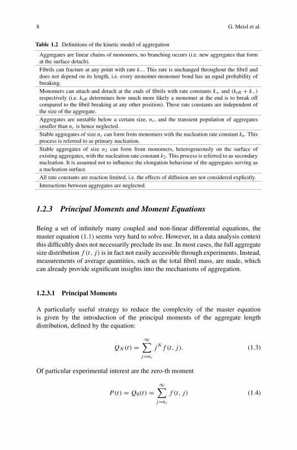
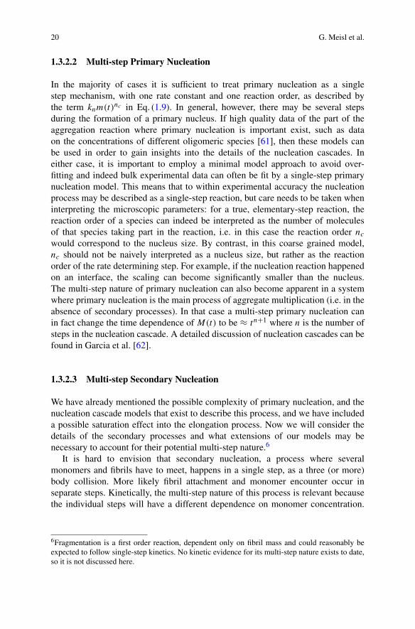
The lock-dock mechanism can be modelled as a two-step reaction, with aninitial, monomer-concentration-dependent attachment step followed a monomer-concentration-independent rearrangement step. This mechanism is formally equiva-lent to a Michaelis-Menten kinetics scheme from biochemistry [59], where the fibrilend is the catalyst, the monomer the substrate and the product is a slightly longerfibril with a new growth competent free end. The corresponding scheme in terms offibril number and mass is given in scheme 4 (Fig. 1.4).
5However, keep in mind that the rates and reaction orders of such coarse-grained processes are notas straightforward to interpret on a molecular level as in elementary reactions.
1 Dynamics and Control of Self-Assembly 19
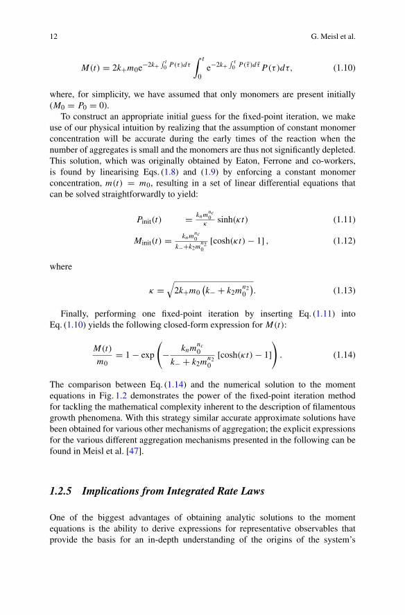
mkf
kb
r MPfree +Pfree + [P m] k
Fig. 1.4 2-step elongation. In a first step a monomer, m, binds to a free fibril end, Pfree, withrate constant kf , forming a complex, [P · m]. The complex can dissociate again with rate constantkb. Within this complex the monomer then rearranges to adopt the correct fibrillar conformation,forming a free end again with rate constant kr and increasing the overall aggregate mass, M
Applying the standard Michaelis-Menten steady-state approach [60] results in anextended version of the equation for the aggregate mass as (compare to Eq. (1.9)):
dM
dt= 2k+
m(t)
1 + m(t)/KE
P (t) (1.19)
where KE = (kb + kr)/kf and k+ = kf kr/(kb + kr). The physical interpretation ofthe Michaelis constant KE gives the monomer concentration at which elongation ishalf saturated, i.e. the elongation step proceeds at half its maximum speed.
If the monomer concentration is low, m(t) � KE , the initial monomer dependentstep is slow and therefore determines the overall rate. This low monomer limit isidentical to the simpler single step model assumed previously in Eq. (1.9). Thereforemany systems can be successfully modelled with single step elongation kinetics,simply because they are far away from the saturation concentration, KE , at whichthe rearrangement step becomes visible kinetically.
By contrast, if the monomer concentration is high, m(t) � KE , the monomerdependent step is fast and the rearrangement step becomes rate-limiting. Under thesecircumstances Eq. (1.19) reduces to dM/dt = 2k+KEP(t). In this limit the overallelongation reaction does no longer depend on the monomer concentration; a furtherincrease in monomer will not change the rate of elongation and the process is saidto be saturated. Whilst in the low monomer limit, the simplified form of dM/dt isvalid for the entire time course, this is not true in the high monomer concentrationcase: Free monomer is depleted during the reaction and necessarily falls below KE
at some point, so the elongation rate slows and the aggregation reaches completion.In general, upon saturation of elongation the scaling exponent will increase by
1/2, but its absolute value also depends on the dominant nucleation mechanism:
γ ≈ γnuc − 1
2(1 + m0/KE)(1.20)
where γnuc is the contribution of secondary nucleation processes to the scaling andγelon is the contribution from elongation, such that γ = γnuc +γelon. This expressioninterpolates between the limits of unsaturated elongation, γelon = −1/2, and fullysaturated elongation, γelon = 0.
20 G. Meisl et al.
1.3.2.2 Multi-step Primary Nucleation
In the majority of cases it is sufficient to treat primary nucleation as a singlestep mechanism, with one rate constant and one reaction order, as described bythe term knm(t)nc in Eq. (1.9). In general, however, there may be several stepsduring the formation of a primary nucleus. If high quality data of the part of theaggregation reaction where primary nucleation is important exist, such as dataon the concentrations of different oligomeric species [61], then these models canbe used in order to gain insights into the details of the nucleation cascades. Ineither case, it is important to employ a minimal model approach to avoid over-fitting and indeed bulk experimental data can often be fit by a single-step primarynucleation model. This means that to within experimental accuracy the nucleationprocess may be described as a single-step reaction, but care needs to be taken wheninterpreting the microscopic parameters: for a true, elementary-step reaction, thereaction order of a species can indeed be interpreted as the number of moleculesof that species taking part in the reaction, i.e. in this case the reaction order nc
would correspond to the nucleus size. By contrast, in this coarse grained model,nc should not be naively interpreted as a nucleus size, but rather as the reactionorder of the rate determining step. For example, if the nucleation reaction happenedon an interface, the scaling can become significantly smaller than the nucleus.The multi-step nature of primary nucleation can also become apparent in a systemwhere primary nucleation is the main process of aggregate multiplication (i.e. in theabsence of secondary processes). In that case a multi-step primary nucleation canin fact change the time dependence of M(t) to be ≈ tn+1 where n is the number ofsteps in the nucleation cascade. A detailed discussion of nucleation cascades can befound in Garcia et al. [62].
1.3.2.3 Multi-step Secondary Nucleation
We have already mentioned the possible complexity of primary nucleation, and thenucleation cascade models that exist to describe this process, and we have includeda possible saturation effect into the elongation process. Now we will consider thedetails of the secondary processes and what extensions of our models may benecessary to account for their potential multi-step nature.6
It is hard to envision that secondary nucleation, a process where severalmonomers and fibrils have to meet, happens in a single step, as a three (or more)body collision. More likely fibril attachment and monomer encounter occur inseparate steps. Kinetically, the multi-step nature of this process is relevant becausethe individual steps will have a different dependence on monomer concentration.
6Fragmentation is a first order reaction, dependent only on fibril mass and could reasonably beexpected to follow single-step kinetics. No kinetic evidence for its multi-step nature exists to date,so it is not discussed here.
1 Dynamics and Control of Self-Assembly 21
Similar to the effect discussed for elongation in Sect. 1.3.2.1, this may lead tosaturation effects and a change in the reaction order as the monomer concentrationis varied. Such a behaviour has been observed for several variants of the Aβ peptide,for a range of conditions [52, 63], see also Fig. 1.7.
One can envision different scenarios, depending on whether monomer encounteroccurs on or off the fibril. In the former case, we model this effect by assumingthat monomers bind to the surface of existing fibrils, where they may form anucleus [64]. The rate of nucleus formation will then simply depend on the surfaceconcentration (i.e. the coverage) to the power of the reaction order, n2. Alternatively,monomers may meet directly, rather than first binding to the fibril surface, and thefibril surface serves as the catalytic site for conversion of the oligomeric species.In this case, following a similar line of argument as for elongation, we obtain amodified version of the moment equation (1.9) that takes into account the multi-step nature of secondary nucleation:
dP
dt= k2
m(t)n2
1 + m(t)n2/KM
M(t) + knm(t)nc (1.21)
where KM is the Michaelis constant of secondary nucleation, and K1/n2M cor-
responds to the monomer concentration at which secondary nucleation is halfsaturated, meaning that the nucleation step proceeds at half its maximum speed.In the limit of low monomer (K1/n2
M � m) we recover the expression for a single
step nucleation, as in Eq. (1.8). At high concentrations (m � K1/n2M ) the reaction
order of the secondary pathway in monomer is close to zero.The scaling exponent is given by:
γ ≈ −1
2
(n2
1 + m0/KM
+ 1
). (1.22)
This expression interpolates between γ = −(n2 +1)/2 and γ = −1/2 for the limitsof low and high monomer, respectively.
1.3.3 Competition: Processes in Parallel
There are several processes that produce new growth competent aggregates: pri-mary nucleation and the two secondary processes, fragmentation and secondarynucleation. These processes may all occur in parallel and can hence competefor being the dominant mechanism of aggregate formation. This competition canbecome kinetically visible through a shift in the dominant mechanism due to thediffering monomer dependence of the three processes. Fragmentation is monomerindependent, whereas primary and secondary nucleation depend on the monomerconcentration to the powers of nc and n2, respectively.
22 G. Meisl et al.
As discussed previously, when two processes compete in parallel, the processwith the higher reaction order will dominate at high concentrations. Therefore, weexpect an increase in the monomer-dependence of the reaction rate with increasingmonomer concentrations, reflected by negative curvature in the half time plots.
1.3.3.1 Competition Between Primary and Secondary Processes
A shift between a primary and a secondary dominated mechanism with a changein monomer concentration could be envisioned if the reaction orders of the twoprocesses differ significantly. In all systems that display a secondary process studiedthus far, this secondary process is so fast compared to the primary ones thatit dominates the aggregation reaction at all accessible monomer concentrations.However, in computer simulations of aggregation [64] a competition betweenprimary and secondary processes can be observed: Naturally nuclei that form fromprimary nucleation, homogeneously in solution, are larger than those formed onthe surface of fibrils through secondary nucleation, therefore the reaction orderfor secondary nucleation is lower than that of primary, and primary nucleationdominates at sufficiently high monomer concentrations. The scaling exponent variesbetween the scaling for a secondary nucleation dominated mechanism, γ = −(n2 +1)/2, at low monomer, and that for a primary dominated mechanism, γ = −nc/2,at high monomer. Moreover, a change in the curve shape, from the exponentialshapes produced when secondary processes dominate to the polynomial shapeswhen primary nucleation dominates, is expected to occur.
1.3.3.2 Two Competing Secondary Processes
The contributions to the rate of growth competent end formation, dP/dt , from thesecondary processes is k−M(t) for fragmentation and k2m(t)n2M(t) for secondarynucleation. While both depend on the concentration of aggregates, M(t), onlysecondary nucleation also depends on the monomer concentration. Therefore, ifa monomer-dependent secondary pathway is in principle accessible in a givensystem, it should become dominant over fragmentation at sufficiently high monomerconcentrations. At low concentrations, by contrast, fragmentation is expected tobecome dominant.
The relevant differential equation for the rate of new aggregate formation is givenby:
dP
dt= knm(t)nc + (k− + k2m(t)n2)M(t). (1.23)
The approximate scaling exponent is determined as
γ ≈ −1
2
(n2
1 + K/m(0)n2+ 1
), (1.24)
1 Dynamics and Control of Self-Assembly 23
Con
stan
t sca
ling
(non
-sat
urat
ed) Dominant pathway Approximate scaling
1o nucelation only nc2
fragmentation 12
2o nucelation 1+n22
Dominant pathwayChange in scaling
Topology Half time plots
Saturating elongation 12+ serial
Saturating 2o nucleation n22+ serial
Competing 2o processes n22- parallel
Competing 1o and 2o process n2 +1-nc2
parallelnegativecurvature
Log[
Hal
f tim
e]
Log[Monomer conc]
positivecurvature
Log[Monomer conc]
Log[
Hal
f tim
e]
Linear approximation tomass concentration
≈Exp[√2k+k-m0 t]
≈Exp[√2k+k2m0 t](n2+1)
growth nuc./mult.
Cha
nge
in s
calin
g(w
ith in
crea
sing
mon
omer
con
c.)
≈2k+knm0 t2nc
Fig. 1.5 Scalings in different limits. This table summarizes the scaling and changes in scalingfor the range of mechanisms discussed here. The top table gives the scaling when no competitionor saturation is observed, as well as the approximate early time increase in aggregate massconcentration, M(t), with time, t , as a function of the initial monomer concentration m0. Thebottom table gives the change in scaling exponent upon an increase in the monomer concentration,going from one extreme, such as the non-saturated regime, to the other extreme, such as the fullysaturated regime. A value of −1/2 would thus correspond to a decrease in the scaling by 1/2.(Adapted from Ref. [40] with permission from the Royal Society of Chemistry)
where K = k−/k2. As expected, this expression interpolates between the scalingfor fragmentation, γ = −1/2, at low monomer concentrations and the scalingfor secondary nucleation, γ = −(n2 + 1)/2, at high monomer concentrations.The concentration at which the two processes contribute equally is determinedby K(1/n2), where K = k−/k2. The scaling exponent decreases with increasingmonomer and there is negative curvature in the half time plots (Fig. 1.5).
1.3.4 Representing the Reaction Network
So far, we have identified two quantities describing an aggregating system, P(t)
and M(t), and have derived equations describing their time evolution in terms of theinitial conditions and the physical properties of the system (the rate constants and thereaction orders). We have considered the individual processes and the effect of themacting in parallel and in series, now we combine them into a visual representation ofthe entire reaction network. Although the moment equations are very similar to mass
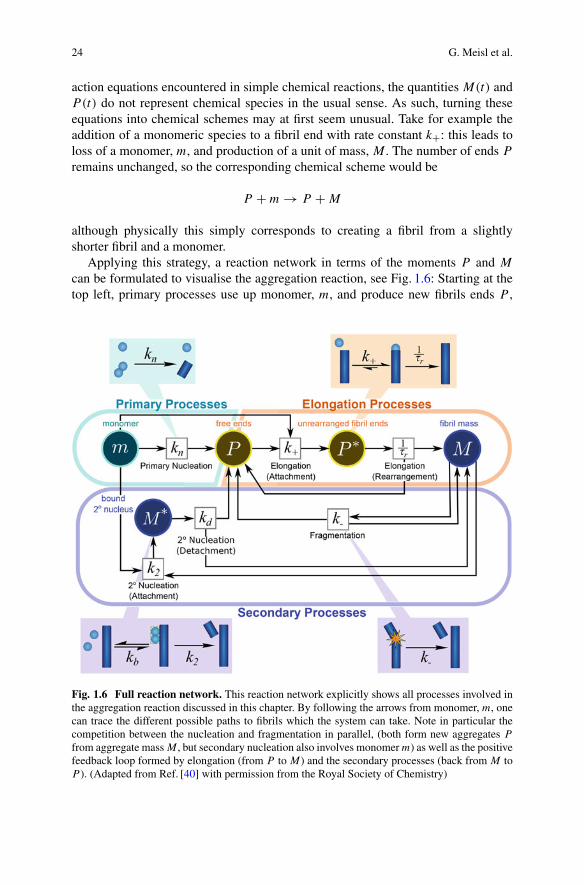
24 G. Meisl et al.
action equations encountered in simple chemical reactions, the quantities M(t) andP(t) do not represent chemical species in the usual sense. As such, turning theseequations into chemical schemes may at first seem unusual. Take for example theaddition of a monomeric species to a fibril end with rate constant k+: this leads toloss of a monomer, m, and production of a unit of mass, M . The number of ends P
remains unchanged, so the corresponding chemical scheme would be
P + m → P + M
although physically this simply corresponds to creating a fibril from a slightlyshorter fibril and a monomer.
Applying this strategy, a reaction network in terms of the moments P and M
can be formulated to visualise the aggregation reaction, see Fig. 1.6: Starting at thetop left, primary processes use up monomer, m, and produce new fibrils ends P ,
Fig. 1.6 Full reaction network. This reaction network explicitly shows all processes involved inthe aggregation reaction discussed in this chapter. By following the arrows from monomer, m, onecan trace the different possible paths to fibrils which the system can take. Note in particular thecompetition between the nucleation and fragmentation in parallel, (both form new aggregates P
from aggregate mass M , but secondary nucleation also involves monomer m) as well as the positivefeedback loop formed by elongation (from P to M) and the secondary processes (back from M toP ). (Adapted from Ref. [40] with permission from the Royal Society of Chemistry)
1 Dynamics and Control of Self-Assembly 25
and are independent of the concentration of fibrils present.7 They are necessarilypresent in an aggregation reaction that starts from monomeric protein, as theyare responsible for the formation of the initial aggregates from monomers alone.Elongation processes then also use up monomer m and produce fibril mass M .Although a growth competent end, P , is required for elongation, a new growthcompetent end is formed by the added monomer, so P is conserved in this reaction(note the arrow back towards P during the conversion of the intermediate P ∗).Elongation is always present in an aggregating system of the kind studied here,as it is the process that produces the aggregate mass. Without elongation no fibrilswould be formed, only small nuclei. The aggregate mass, M , can then catalyse theformation of further new aggregates, P , via one of two mechanisms: either fibrilsfragment with rate k−, or monomers nucleate on the fibril surface, via a boundintermediate M∗. Both secondary nucleation and fragmentation conserve aggregatemass M , as evident by the arrows back towards M .
This reaction network picture illustrates the basic principles of aggregation, notonly how the different mechanisms compete in parallel but also why secondarynucleation is often of such central importance: Primary nucleation and elongationalone would be a simple linear reaction network (turquoise and orange, at the top ofthe network). However, if secondary processes are present they introduce a positivefeedback loop: M is linked back to P to close the loop with elongation, eitherthrough fragmentation, directly with rate constant k−, or via the intermediate, M∗,for secondary nucleation. As discussed previously this positive feedback results in amassive acceleration of the aggregation reaction, evident by the exponential increasein aggregate mass observed in experiment.
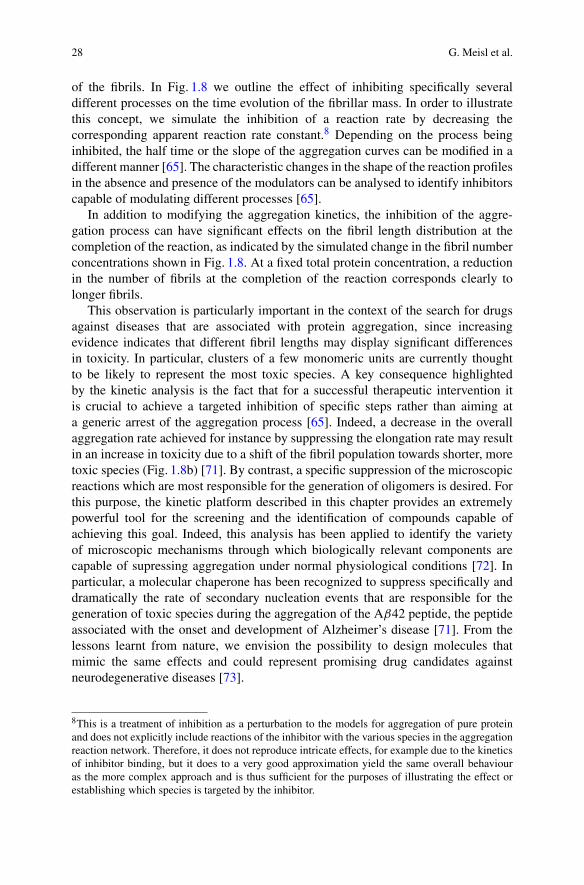
1.4 Application to Experiment: Global Fitting of KineticData
A kinetic experiment of fibril formation usually involves measuring the concentra-tion of aggregated material as a function of time, e.g. via a reporter dye such asThioflavin-T (ThT), or recording the concentration of free monomer over time, e.g.via NMR techniques. In either case, the purity of the sample and the careful controlof the experimental conditions are key. In fact, protein aggregation reactions tend tobe very sensitive to impurities or surface effects and thus a meticulously optimizedexperimental procedure is a fundamental prerequisite for obtaining reproduciblekinetic data [63].
As the mechanisms of aggregation are complex and thus involve many differentparameters, the kinetic measurements have to be performed at a number of monomerconcentrations in order to yield sufficient constraints for all the fitting parameters.
7As described in Sect. 1.2.3, “Common Approximations”, the mass produced by nucleation can beneglected relative to that generated through elongation.
26 G. Meisl et al.
C
Fibr
illar
frac
tion
100%
80%
60%
40%
20%
0%
Time / h 0 4 8 16 18
Time / h 0 4 8 16 18
Time / h 0 4 8 16 18
Time / h
Fibr
illar
mas
s fr
actio
n
100%
80%
60%
40%
20%
0%0 5 10 15 20
B
slope = -1.2
slope = -0.2
H
alf t
ime
/ h
10
6
8
4
2
1
20
2 4 6 8 10 20 40 60 80 100Initial monomer concentration / μM
A
Fig. 1.7 Global fitting of aggregation data. (a) The half time plots are used to narrow downthe mechanism. In this case strong monomer dependence at low monomer concentrations and thepositive curvature indicate a saturation of secondary nucleation as the correct mechanism. (b) Themass concentration is fitted over the full time course to the approximate solution of the momentequations. This is a global fit, simultaneously at all monomer concentrations. (c) The fitting of amodel that has only primary processes, a model that has fragmentation as its secondary processand a model that has a single-step secondary nucleation all fail. (Adapted from Ref. [52])
In a first step the half time plots can be used as an approximate guide to possiblemechanisms (Fig. 1.7a). The solution for M(t) for the mechanism in question is thenfitted globally, i.e. to all data simultaneously, (Fig. 1.7b) to yield the rate constantsand reaction orders of the individual processes [47]. It is important to also test othermechanism and ensure the data cannot be fitted by a simpler model (Fig. 1.7c).
Additional experiments in the presence of seeds can be used to provide furtherevidence for a given mechanism [63]. The variation of the aggregation behaviourupon addition of other compounds, such as possible inhibitors, can be used todetermine at which point in the aggregation network this compound acts [65, 66].
1.5 Controlling Aggregation: Inhibitors and SolutionConditions
The understanding of the fundamental molecular processes that underlie the aggre-gation reaction opens the attractive possibility to rationally predict which processeshave to be modified in order to affect the aggregation behaviour in a desired manner.This crucial application is relevant in many areas of biological and biotechnological
1 Dynamics and Control of Self-Assembly 27
sciences when there is the need to increase the yield of a desired product or toprevent aberrant aggregation. One important example is observed in the contextof the aggregation of amyloidogenic peptides and proteins associated with theonset and development of neuro-degenerative disorders, where the inhibition of theaggregation process represents a promising strategy in the development of drugs forthe prevention of these aggregation-related diseases. Another important applicationwhere aggregation must be strictly avoided is found in the industrial production oftherapeutic proteins such as insulin and monoclonal antibodies.
The rate of every microscopic aggregation process can be expressed as theproduct of a reaction rate constant and the concentration of the reactive species.Typically, the reaction rate constant is strongly dependent on the intermolecularinteractions that two colliding objects face while approaching each other. As aconsequence, the kinetics of the aggregation process can be modulated by tuningthe intermolecular forces via changes in the solution conditions. For instance, thescreening of electrostatic interactions through an increase in the ionic strengthof the solution can have significant effects on the aggregation behaviour: as theaggregating species are usually like-charged, higher ionic strength often resultsin reduced repulsion and increases all rates of assembly [53, 67, 68]. Moreover,different processes may be affected to a different degree, resulting in a shift of thedominant mechanism, for example through a saturation of the secondary nucleationprocess. In addition to shielding electrostatic interactions, another means to modifyelectrostatic interactions involves changing the charge of the protein: for example,α-synuclein displays a secondary nucleation-dominated process only under acidicconditions (pH < 5.8). This effect may be due to a decrease in negative chargeupon protonation of a side chain residue [38]. Equally, mutant proteins with alteredcharges can display significantly different aggregation behaviours and indeed, in thecontext of Alzheimer’s and Parkinson’s disease, many of the disease related mutantsshow an altered charge compared to the wild type [69, 70].
The methods described above affect the aggregation behaviour by modulatingthe interactions of the aggregating species in a complex manner, and often it ischallenging to perturb specifically one single microscopic event by this modality.More targeted tools are offered by the use of species designed to bind a specifictarget in the aggregation network and thereby inhibit a specific process. Thisapproach includes the addition into the reaction mixture of a large variety ofmodulators including small molecules, peptides, proteins and antibodies. In thiscase, the reaction rate is modified by changes in the concentration of the reactivespecies rather than by modifications of the reaction rate constants. In particular, aspecific modification of a single microscopic event can be achieved by targeting areactive species that participates only in the desired microscopic reaction.
For instance, one could envision the possibility of inhibiting elongation byadding compounds that bind to fibril ends preventing their further growth, i.e.,reducing the concentration of fibril ends that are capable of adding monomersand elongate. Similarly, primary nucleation could be selectively suppressed byintroducing molecules that interact with some pre-nucleus clusters, while secondarynucleation could be inhibited by adding species capable of binding to the surfaces
28 G. Meisl et al.
of the fibrils. In Fig. 1.8 we outline the effect of inhibiting specifically severaldifferent processes on the time evolution of the fibrillar mass. In order to illustratethis concept, we simulate the inhibition of a reaction rate by decreasing thecorresponding apparent reaction rate constant.8 Depending on the process beinginhibited, the half time or the slope of the aggregation curves can be modified in adifferent manner [65]. The characteristic changes in the shape of the reaction profilesin the absence and presence of the modulators can be analysed to identify inhibitorscapable of modulating different processes [65].
In addition to modifying the aggregation kinetics, the inhibition of the aggre-gation process can have significant effects on the fibril length distribution at thecompletion of the reaction, as indicated by the simulated change in the fibril numberconcentrations shown in Fig. 1.8. At a fixed total protein concentration, a reductionin the number of fibrils at the completion of the reaction corresponds clearly tolonger fibrils.
This observation is particularly important in the context of the search for drugsagainst diseases that are associated with protein aggregation, since increasingevidence indicates that different fibril lengths may display significant differencesin toxicity. In particular, clusters of a few monomeric units are currently thoughtto be likely to represent the most toxic species. A key consequence highlightedby the kinetic analysis is the fact that for a successful therapeutic intervention itis crucial to achieve a targeted inhibition of specific steps rather than aiming ata generic arrest of the aggregation process [65]. Indeed, a decrease in the overallaggregation rate achieved for instance by suppressing the elongation rate may resultin an increase in toxicity due to a shift of the fibril population towards shorter, moretoxic species (Fig. 1.8b) [71]. By contrast, a specific suppression of the microscopicreactions which are most responsible for the generation of oligomers is desired. Forthis purpose, the kinetic platform described in this chapter provides an extremelypowerful tool for the screening and the identification of compounds capable ofachieving this goal. Indeed, this analysis has been applied to identify the varietyof microscopic mechanisms through which biologically relevant components arecapable of supressing aggregation under normal physiological conditions [72]. Inparticular, a molecular chaperone has been recognized to suppress specifically anddramatically the rate of secondary nucleation events that are responsible for thegeneration of toxic species during the aggregation of the Aβ42 peptide, the peptideassociated with the onset and development of Alzheimer’s disease [71]. From thelessons learnt from nature, we envision the possibility to design molecules thatmimic the same effects and could represent promising drug candidates againstneurodegenerative diseases [73].
8This is a treatment of inhibition as a perturbation to the models for aggregation of pure proteinand does not explicitly include reactions of the inhibitor with the various species in the aggregationreaction network. Therefore, it does not reproduce intricate effects, for example due to the kineticsof inhibitor binding, but it does to a very good approximation yield the same overall behaviouras the more complex approach and is thus sufficient for the purposes of illustrating the effect orestablishing which species is targeted by the inhibitor.
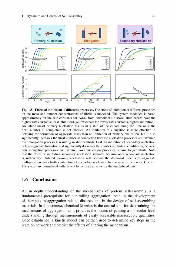
1 Dynamics and Control of Self-Assembly 29
ElongationPrimary Nucleation Secondary Nucleation
increasing inhibition105.7
105.3
105
104.7
k+106
3050 5201 51 02Time
increasing inhibition107
106
105
104
k2108
1.0
0.8
0.6
0.4
0.2
0.0
Fibr
il m
ass
fract
ion
increasing inhibition 110-1
10-2
10-3
10-4
kn
3050 5201 51 02Time
increasing inhibition
305
4
3
2
1
0 0 5201 51 02Time
Rel
ativ
e fib
ril n
umbe
r
increasing inhibition
increasin
g inhibition
Fig. 1.8 Effect of inhibition of different processes. The effect of inhibition of different processeson the mass and number concentrations of fibrils is modelled. The system modelled is basedapproximately on the rate constants for Aβ42 from Alzheimer’s disease. Blue curves have thehighest rate constants (least inhibition), yellow curves the lowest rate constants (highest inhibition).An inhibition of primary nucleation results in a shift of the curves along the time axis; thefibril number at completion is not affected. An inhibition of elongation is more effective indelaying the formation of aggregate mass than an inhibition of primary nucleation, but it alsosignificantly increases the fibril number at completion because nucleation processes are favouredover elongation processes, resulting in shorter fibrils. Last, an inhibition of secondary nucleationdelays aggregate formation and significantly decreases the number of fibrils at equilibrium, becausenow elongation processes are favoured over nucleation processes, giving longer fibrils. Notethat the effect of inhibiting secondary nucleation saturates because once secondary nucleationis sufficiently inhibited, primary nucleation will become the dominant process of aggregatemultiplication and a further inhibition of secondary nucleation has no more effect on the kinetics.The y-axes are normalised with respect to the plateau value for the uninhibited case
1.6 Conclusions
An in depth understanding of the mechanisms of protein self-assembly is afundamental prerequisite for controlling aggregation, both in the developmentof therapies to aggregation-related diseases and in the design of self-assemblingmaterials. In this context, chemical kinetics is the central tool for determining themechanisms of aggregation as it provides the means of gaining a molecular levelunderstanding through measurements of easily accessible macroscopic quantities.Once established, a kinetic model can be then used to determine key steps in thereaction network and predict the effects of altering the mechanism.
30 G. Meisl et al.
Acknowledgements We would like to thank the Swiss National Science Foundation, PeterhouseCollege Cambridge, the European Research Council, the BBSRC, the EPSRC, the NewmanFoundation and Sidney Sussex College Cambridge.
References
1. Sawaya MR, Sambashivan S, Nelson R, Ivanova MI, Sievers SA, Apostol MI, Thompson MJ,Balbirnie M, Wiltzius JJW, McFarlane HT, Madsen A, Riekel C, Eisenberg D (2007) Atomicstructures of amyloid cross-beta spines reveal varied steric zippers. Nature 447(7143):453–457
2. Sunde M, Blake C (1997) The structure of amyloid fibrils by electron microscopy and x-raydiffraction. Adv Protein Chem 50:123–159
3. Knowles TPJ, Vendruscolo M, Dobson CM (2014) The amyloid state and its association withprotein misfolding diseases. Nat Rev Mol Cell Biol 15:384–396
4. Ciryam P, Tartaglia GG, Morimoto RI, Dobson CM, Vendruscolo M (2013) Widespreadaggregation and neurodegenerative diseases are associated with supersaturated proteins. CellRep 5(3):781–790
5. Boller F, Mizutani T, Roessmann U, Gambetti P (1980) Parkinson disease, dementia, andalzheimer disease: clinicopathological correlations. Ann Neurol 7(4):329–335
6. Lansbury PT, Lashuel HA (2006) A century-old debate on protein aggregation and neurode-generation enters the clinic. Nature 443(7113):774–779
7. Bemporad F, Chiti F (2012) Protein misfolded oligomers: experimental approaches, mecha-nism of formation, and structure-toxicity relationships. Chem Biol 19(3):315–327
8. Oosawa F, Kasai M (1962) A theory of linear and helical aggregations of macromolecules. JMol Biol 4:10–21
9. Oosawa F, Asakura S (1975) Thermodynamics of the polymerization of protein. Academic,New York
10. Alberts B, Johnson A, Lewis J, Raff M, Roberts K, Walter P (2002) Molecular biology of thecell, 4th edn. Garland Publishing, New York
11. Hill TL (1987) Linear aggregation theory in cell biology. Springer, New York12. Fowler DM, Koulov AV, Balch WE, Kelly JW (2007) Functional amyloid–from bacteria to
humans. Trends Biochem Sci 32(5):217–22413. Kelly JW, Balch WE (2003) Amyloid as a natural product. J Cell Biol 161:461–46214. Fowler DM, Koulov AV, Alory-Jost C, Marks MS, Balch WE, Kelly JW (2006) Functional
amyloid formation within mammalian tissue. PLoS Biol 4(1):e615. Maji SK, Perrin MH, Sawaya MR, Jessberger S, Vadodaria K, Rissman RA, Singru PS, Nilsson
KPR, Simon R, Schubert D, Eisenberg D, Rivier J, Sawchenko P, Vale W, Riek R (2009)Functional amyloids as natural storage of peptide hormones in pituitary secretory granules.Science 325(5938):328–332
16. Talbot NJ, Kershaw MJ, Wakley GE, De Vries OMH, Wessels JGH, Hamer JE (1996)Mpg1 encodes a fungal hydrophobin involved in surface interactions during infection-relateddevelopment of magnaporthe grisea. Plant Cell 8(6):985–999
17. Romero D, Aguilar C, Losick R, Kolter R (2010) Amyloid fibers provide structural integrity tobacillus subtilis biofilms. Proc Natl Acad Sci U S A 107(5):2230–2234
18. Chapman MR, Robinson LS, Pinkner JS, Roth R, Heuser J, Hammar M, Normark S, HultgrenSJ (2002) Role of escherichia coli curli operons in directing amyloid fiber formation. Science295(5556):851–855
19. Mankar S, Anoop A, Sen S, Maji SK (2011) Nanomaterials: amyloids reflect their brighterside. Nano Rev 2:6032. https://doi.org/10.3402/nano.v2i0.6032
20. Bolisetty S, Mezzenga R (2015) Amyloid-carbon hybrid membranes for universal waterpurification. Nat Nano 11:365–371
1 Dynamics and Control of Self-Assembly 31
21. Dobson CM (1999) Protein misfolding, evolution and disease. Trends Biochem Sci 24(9):329–332
22. Ferrone F (1999) Analysis of protein aggregation kinetics. Methods Enzymol 309:256–27423. Knowles TPJ, Waudby CA, Devlin GL, Cohen SIA, Aguzzi A, Vendruscolo M, Terentjev EM,
Welland ME, Dobson CM (2009) An analytical solution to the kinetics of breakable filamentassembly. Science 326(5959):1533–1537
24. Galvagnion C, Buell AK, Meisl G, Michaels TCT, Vendruscolo M, Knowles TPJ, Dobson CM(2015) Lipid vesicles trigger a-synuclein aggregation by stimulating primary nucleation. NatChem Biol 11:229–234
25. Grigolato F, Colombo C, Ferrari R, Rezabkova L, Arosio P (2017) Mechanistic origin ofthe combined effect of surfaces and mechanical agitation on amyloid formation. ACS Nano11(11):11358–11367. PMID: 29045787
26. Pham CLL, Rey A, Lo V, Soulès M, Ren Q, Meisl G, Knowles TPJ, Kwan AH, Sunde M(2016) Self-assembly of MPG1, a hydrophobin protein from the rice blast fungus that formsfunctional amyloid coatings, occurs by a surface-driven mechanism. Sci Rep 6:25288
27. Michaels TCT, Garcia GA, Knowles TPJ (2014) Asymptotic solutions of the oosawa modelfor the length distribution of biofilaments. J Chem Phys 140(19):194906
28. Ruschak AM, Miranker AD (2007) Fiber-dependent amyloid formation as catalysis of anexisting reaction pathway. Proc Natl Acad Sci U S A 104(30):12341–12346
29. Xue W-F, Hellewell AL, Gosal WS, Homans SW, Hewitt EW, Radford SE (2009) Fibrilfragmentation enhances amyloid cytotoxicity. J Biol Chem 284(49):34272–34282
30. Cohen SIA, Linse S, Luheshi LM, Hellstrand E, White DA, Rajah L, Otzen DE, VendruscoloM, Dobson CM, Knowles TPJ (2013) Proliferation of amyloid-beta42 aggregates occursthrough a secondary nucleation mechanism. Proc Natl Acad Sci 110:9758–9763
31. Collins SR, Douglass A, Vale RD, Weissman JS (2004) Mechanism of prion propagation:amyloid growth occurs by monomer addition. PLoS Biol 2(10):e321
32. Kunes KC, Cox DL, Singh RRP (2005) One-dimensional model of yeast prion aggregation.Phys Rev E Stat Nonlin Soft Matter Phys 72(5 Pt 1):051915
33. Stöhr J, Weinmann N, Wille H, Kaimann T, Nagel-Steger L, Birkmann E, Panza G, PrusinerSB, Eigen M, Riesner D (2008) Mechanisms of prion protein assembly into amyloid. Proc NatlAcad Sci U S A 105(7):2409–2414
34. Ferrone FA, Hofrichter J, Eaton WA (1985) Kinetics of sickle hemoglobin polymerization. I.Studies using temperature-jump and laser photolysis techniques. J Mol Biol 183(4):591–610
35. Ferrone FA, Hofrichter J, Eaton WA (1985) Kinetics of sickle hemoglobin polymerization. II.A double nucleation mechanism. J Mol Biol 183(4):611–631
36. Bishop MF, Ferrone FA (1984) Kinetics of nucleation-controlled polymerization. A perturba-tion treatment for use with a secondary pathway. Biophys J 46(5):631–644
37. Cohen SIA, Vendruscolo M, Dobson CM, Knowles TPJ (2012) From macroscopic measure-ments to microscopic mechanisms of protein aggregation. J Mol Biol 421(2–3):160–171
38. Buell AK, Galvagnion C, Gaspar R, Sparr E, Vendruscolo M, Knowles TPJ, Linse S, DobsonCM (2014) Solution conditions determine the relative importance of nucleation and growthprocesses in α-synuclein aggregation. Proc Natl Acad Sci 111(21):7671–7676
39. Knowles TPJ, Buehler MJ (2011) Nanomechanics of functional and pathological amyloidmaterials. Nat Nanotechnol 6(8):469–479
40. Meisl G, Rajah L, Cohen SAI, Pfammatter M, Saric A, Hellstrand E, Buell AK, AguzziA, Linse S, Vendruscolo M, Dobson CM, Knowles TPJ (2017) Scaling behaviour and rate-determining steps in filamentous self-assembly. Chem Sci 8:7087–7097
41. Krapivsky PL, Redner S, Ben-Naim E (2010) A kinetic view of statistical physics. CambridgeUniversity Press, Leiden
42. Xue W-F, Homans SW, Radford SE (2008) Systematic analysis of nucleation-dependentpolymerization reveals new insights into the mechanism of amyloid self-assembly. Proc NatlAcad Sci U S A 105(26):8926–8931
43. Arosio P, Cedervall T, Knowles TPJ, Linse S (2016) Analysis of the length distribution ofamyloid fibrils by centrifugal sedimentation. Anal Biochem 504:7–13
32 G. Meisl et al.
44. Nasir I, Linse S, Cabaleiro-Lago C (2015) Fluorescent filter-trap assay for amyloid fibril for-mation kinetics in complex solutions. ACS Chem Neurosci (8):1436–1444. PMID: 25946560
45. Gaspar R, Meisl G, Buell AK, Young L, Kaminski CF, Knowles TPJ, Sparr E, Linse S (2017)Secondary nucleation of monomers on fibril surface dominates α-synuclein aggregation andprovides autocatalytic amyloid amplification. Q Rev Biophys 50:E6
46. Bender CM, Orszag SA (1999) Advanced mathematical methods for scientists and engineers.Springer, New York
47. Meisl G, Kirkegaard JB, Arosio P, Michaels TTC, Vendruscolo M, Dobson CM, Linse S,Knowles TPJ (2016) Molecular mechanisms of protein aggregation from global fitting ofkinetic models. Nat Protoc 11(2):252–272
48. Cohen SIA, Vendruscolo M, Welland ME, Dobson CM, Terentjev EM, Knowles TPJ (2011)Nucleated polymerization with secondary pathways. I. Time evolution of the principalmoments. J Chem Phys 135(6):065105
49. Cohen SIA, Vendruscolo M, Dobson CM, Knowles TPJ (2011) Nucleated polymerizationwith secondary pathways. II. Determination of self-consistent solutions to growth processesdescribed by non-linear master equations. J Chem Phys 135(6):065106
50. Arosio P, Knowles TPJ, Linse S (2015) On the lag phase in amyloid fibril formation. PhysChem Chem Phys 17:7606–7618
51. Knowles TPJ, White DA, Abate AR, Agresti JJ, Cohen SIA, Sperling RA, De Genst EJ,Dobson CM, Weitz DA (2011) Observation of spatial propagation of amyloid assembly fromsingle nuclei. Proc Natl Acad Sci U S A 108(36):14746–14751
52. Meisl G, Yang X, Hellstrand E, Frohm B, Kirkegaard JB, Cohen SIA, Dobson CM, Linse S,Knowles TPJ (2014) Differences in nucleation behavior underlie the contrasting aggregationkinetics of the aβ40 and aβ42 peptides. Proc Natl Acad Sci 111:9384–9389
53. Meisl G, Yang X, Dobson CM, Linse S, Knowles TPJ (2017) Modulation of electrostaticinteractions to reveal a reaction network unifying the aggregation behaviour of the aβ42 peptideand its variants. Chem Sci 8:4352–4362
54. Knowles TPJ, Vendruscolo M, Dobson CM (2015) The physical basis of protein misfoldingdisorders. Phys Today 68(3):36
55. Lee J, Culyba EK, Powers ET, Kelly JW (2011) Amyloid-beta forms fibrils by nucleatedconformational conversion of oligomers. Nat Chem Biol 7(9):602–609
56. Scheibel T, Bloom J, Lindquist SL (2004) The elongation of yeast prion fibers involvesseparable steps of association and conversion. Proc Natl Acad Sci U S A 101(8):2287–2292
57. Esler WP, Stimson ER, Jennings JM, Vinters HV, Ghilardi JR, Lee JP, Mantyh PW, MaggioJE (2000) Alzheimer’s disease amyloid propagation by a template-dependent dock-lockmechanism. Biochemistry 39(21):6288–6295
58. Buell AK, Blundell JR, Dobson CM, Welland ME, Terentjev EM, Knowles TPJ (2010)Frequency factors in a landscape model of filamentous protein aggregation. Phys Rev Lett104(22):228101
59. Michaelis L, Menten M (1913) Die kinetik der invertinwirkung. Biochem Z 49:33360. Connors KA (1990) Chemical kinetics: study of reaction rates in solution. Wiley, New York61. Orte A, Clarke R, Balasubramanian S, Klenerman D (2006) Determination of the fraction and
stoichiometry of femtomolar levels of biomolecular complexes in an excess of monomer usingsingle-molecule, two-color coincidence detection. Anal Chem 78(22):7707–7715
62. Garcia GA, Cohen SIA, Dobson CM, Knowles TPJ (2014) Nucleation-conversion-polymerization reactions of biological macromolecules with prenucleation clusters. Phys RevE 89:032712
63. Meisl G, Yang X, Frohm B, Knowles TPJ, Linse S (2016) Quantitative analysis of intrinsicand extrinsic factors in the aggregation mechanism of alzheimer-associated aβ-peptide. SciRep 6:18728
64. Saric A, Buell A, Meisl G, Michaels TCT, Dobson CM, Linse S, Knowles TPJ, Frenkel D(2016) Physical determinants of the self-replication of protein fibrils. Nat Phys 12:874–880
65. Arosio P, Vendruscolo M, Dobson CM, Knowles TPJ (2014) Chemical kinetics for drugdiscovery to combat protein aggregation diseases. Trends Pharmacol Sci 35(3):127–135
1 Dynamics and Control of Self-Assembly 33
66. Abelein A, Graslund A, Danielsson J (2015) Zinc as chaperone-mimicking agent for retarda-tion of amyloid β peptide fibril formation. Proc Natl Acad Sci U S A 112(17):5407–5412
67. Klement K, Wieligmann K, Meinhardt J, Hortschansky P, Richter W, Fändrich M (2007) Effectof different salt ions on the propensity of aggregation and on the structure of Alzheimer’sabeta(1–40) amyloid fibrils. J Mol Biol 373(5):1321–1333
68. Buell AK, Hung P, Salvatella X, Welland ME, Dobson CM, Knowles TPJ (2013) Electrostaticeffects in filamentous protein aggregation. Biophys J 104:1116–1126
69. Flagmeier P, Meisl G, Vendruscolo M, Knowles TPJ, Dobson CM, Buell AK, GalvagnionC (2016) Mutations associated with familial parkinson’s disease alter the initiation andamplification steps of α-synuclein aggregation. Proc Natl Acad Sci U S A 113(37):10328–10333
70. Yang X, Meisl G, Frohm B, Thulin E, Knowles TPJ, Linse S (2018) On the role of sidechainsize and charge in the aggregation of aβ42 with familial mutations. Proc Natl Acad Sci U S A115(26):E5849–E5858
71. Cohen SIA, Arosio P, Presto J, Kurudenkandy FR, Biverstal H, Dolfe L, Dunning C, Yang X,Frohm B, Vendruscolo M, Johansson J, Dobson CM, Fisahn A, Knowles TPJ, Linse S (2015)The molecular chaperone brichos breaks the catalytic cycle that generates toxic ab oligomers.Nat Struct Mol Biol 22:207–213
72. Arosio P, Michaels TCT, Linse S, Månsson C, Emanuelsson C, Presto J, Johansson J,Vendruscolo M, Dobson C, Knowles TPJ (2016) Kinetic analysis reveals the diversity ofmicroscopic mechanisms through which molecular chaperones suppress amyloid formation.Nat Commun 7:10948
73. Habchi J, Arosio P, Perni M, Costa AR, Yagi-Utsumi M, Joshi P, Chia S, Cohen SIA, MüllerMBD, Linse S, Nollen EAA, Dobson CM, Knowles TPJ, Vendruscolo M (2016) An anticancerdrug suppresses the primary nucleation reaction that initiates the production of the toxic aβ42aggregates linked with alzheimer’s disease. Sci Adv 2(2):e1501244
Chapter 2Peptide Self-Assembly and ItsModulation: Imaging on the Nanoscale
Lanlan Yu, Yanlian Yang, and Chen Wang
Abstract This chapter intends to review the progress in obtaining site-specificstructural information for peptide assemblies using scanning tunneling microscopy.The effects on assembly propensity due to mutations and modifications in peptidesequences, small organic molecules and conformational transitions of peptides areidentified. The obtained structural insights into the sequence-dependent assemblypropensity could inspire rational design of peptide architectures at the molecularlevel.
Keywords Peptide assembly · Modulation · Scanning tunneling microscopy
Abbreviations
4Bpy 4,4′-bipyridylAD Alzheimer’s diseaseAFM Atomic force microscopyALS Amyotrophic lateral sclerosisAβ Beta-amyloid peptidesCD Circular dichroismDPE 1,2-di(4-pyridyl)-ethyleneFTIR Fourier transform infrared spectroscopyFTLD Frontotemporal lobar degenerationhIAPP Human islet amyloid polypeptideHOPG Highly oriented pyrolytic graphiteMD Molecular dynamicsNMR Nuclear magnetic resonancePcCu(SO3Na)4 Copper phthalocyanine tetrasulfonate sodium
L. Yu · Y. Yang · C. Wang (�)National Center for Nanoscience and Technology, Chinese Academy of Sciences, Beijing, Chinae-mail: [email protected]
© Springer Nature Singapore Pte Ltd. 2019S. Perrett et al. (eds.), Biological and Bio-inspired Nanomaterials,Advances in Experimental Medicine and Biology 1174,https://doi.org/10.1007/978-981-13-9791-2_2
35
36 L. Yu et al.
STM Scanning tunneling microscopyTDP-43 TAR DNA-binding protein 43TEM Transmission electron microscopyThT Thioflavin TTSE Transmissible spongiform encephalopathyUHV Ultrahigh vacuumVNTRs Variable number of tandem repeatsXRD X-ray diffraction
2.1 Introduction
Peptide assemblies have been drawing extensive interest due to their vital roles inbiological recognition associated with interactions between proteins, interactionsbetween peptide drugs and targets, disease-associated amyloid aggregation, andthe design of novel functional biomaterials. Common to the key elements ofmolecular assembly, inter-peptide interactions include non-covalent interactionssuch as hydrogen bonds, electrostatic interactions (or salt bridges), the hydrophobiceffect, van der Waals interactions and water-mediated hydrogen bonds [1, 2].Representative examples of assembly processes are seen among amyloidal peptideswhich have been extensively investigated due to their relevance to over 20 diseasesincluding Alzheimer’s disease (AD) and Parkinson’s disease (PD). The assembly ofamyloidal peptides generally evolves from oligomers to protofibrils, and protofibrilssubsequently twist together into mature fibrils and plaques [3]. Efforts to unveil thehierarchical assembly pathways and to decode the detailed structural informationwould be beneficial to modulate and manipulate them for the rational design ofbiological materials.
The advances in unraveling molecular mechanisms have contributed to a broadrange of potential applications in areas including controlled release and drug deliv-ery, cell culture, tissue engineering, antimicrobial and biomineralization materials[2, 4–15]. Furthermore, peptide assemblies on surfaces have also been widelyexplored as a reflection of biological and physiological processes in vivo, which arerelevant to membranes and potential applications for biosensors and heterogeneousbiocatalysis, drug delivery, and affinity chromatography [16–18]. It is worth notingthat peptide assemblies on surfaces are typically stabilized through peptide-peptideand peptide-substrate interactions, similar to other widely studied surface-mediatedmolecular assemblies.
To analyze the assembly pathways and structural information, various experi-mental approaches have been attempted. The aggregation propensity and stabilitycan be studied by kinetic measurements such as the thioflavin T (ThT) binding assay[19, 20]. Various spectroscopies, such as circular dichroism (CD) spectroscopy,Fourier transform infrared spectroscopy (FTIR) and ultraviolet Raman spectroscopycan provide information regarding secondary structure [21–24]. Imaging methods,including atomic force microscopy (AFM) and transmission electron microscopy(TEM) can be employed to obtain the morphology and size evolution of the
2 Peptide Self-Assembly and Its Modulation: Imaging on the Nanoscale 37
peptide aggregates [25–27]. In terms of subtle details of peptide assembly structures,X-ray diffraction (XRD) [28–32], nuclear magnetic resonance (NMR) [25, 33–39]and cryo-electron microscopy [31] have been generally applied to obtain high-resolution structural information. However, these techniques are restricted by eitherthe complicated procedures of sample preparation or the nature of peptides suchas solubility and crystallinity. More significantly, the results are confined to anensemble averaging regime and only provide signals from the convolutions of allresidues.
Notably, scanning tunneling microscopy (STM) has been widely exploited toprobe the assembly behavior and single-molecular structure of small molecules[40–48] on surfaces due to its submolecular structural resolution and adaptabilityto various conditions including ambient conditions and ultrahigh vacuum (UHV)conditions. STM offers the advantage of easy sample preparation procedure, andmore importantly, its structural resolution is theoretically down to single molecule,and even amino acid residue-specific resolution. Thus far, STM has successfullydemonstrated its capacity for investigating peptide assemblies ranging from simplemodel peptides [49–51] to complex disease-related peptides [27, 52, 53].
In this chapter, we will firstly introduce the STM investigations on surface-boundpeptide assembly structures with residue-specific resolution. Subsequently, theinfluence of mutations and modifications (phosphorylation/glycosylation) in peptideassemblies and the aggregation propensities are presented, as well as interactionsbetween the peptides with small molecules including terminus modulators and sidegroup modulators. Finally, the correlation of peptide assemblies on surface and insolution and the peptide conformational transitions when adsorbed on surfaces arealso discussed.
2.2 Peptide Self-Assembly Structures on Surfaces
Structural analysis is central to gaining insight into inter-peptide interactions and therelationship between peptide sequence and assembly structures. As a technique withsubmolecular-level resolution, the potential of STM in structural analysis of peptidemolecules and peptide assemblies under various environments including UHV aswell as ambient conditions has been established [49–53].
For UHV-STM investigations, peptide molecules are usually adsorbed ontometal surfaces defined by surface indexes (such as Cu(110) and Au(111)) andat relatively low temperatures to minimize thermally induced fluctuation effectson high-resolution imaging. The smallest peptide molecule comprised of onlytwo amino acid residues (the chiral dipeptide di-L-alanine) was first observed toself-assemble on a metal surface using UHV-STM. Sub-monolayer growth of di-L-alanine on Cu(110) was observed and the peptide was found to nucleate intosmall islands with single or double molecule rows at low coverage (Fig. 2.1a) [51].The dialanines appear as pairs of a smaller and a larger protrusion in STM images,aligned along the [332] direction. Within a single row, the alignment of moleculeswith the same orientation indicates that the –COOH moiety of one molecule binds to
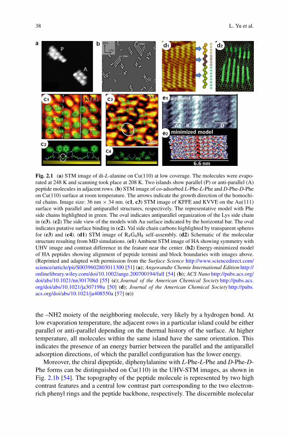
38 L. Yu et al.
Fig. 2.1 (a) STM image of di-L-alanine on Cu(110) at low coverage. The molecules were evapo-rated at 248 K and scanning took place at 208 K. Two islands show parallel (P) or anti-parallel (A)peptide molecules in adjacent rows. (b) STM image of co-adsorbed L-Phe-L-Phe and D-Phe-D-Pheon Cu(110) surface at room temperature. The arrows indicate the growth direction of the homochi-ral chains. Image size: 36 nm × 34 nm. (c1, c3) STM image of KFFE and KVVE on the Au(111)surface with parallel and antiparallel structures, respectively. The representative model with Pheside chains highlighted in green. The oval indicates antiparallel organization of the Lys side chainin (c3). (c2) The side view of the models with Au surface indicated by the horizontal bar. The ovalindicates putative surface binding in (c2). Val side chain carbons highlighted by transparent spheresfor (c3) and (c4). (d1) STM image of R4G4H8 self-assembly. (d2) Schematic of the molecularstructure resulting from MD simulations. (e1) Ambient STM image of HA showing symmetry withUHV image and contrast difference in the feature near the center. (b2) Energy-minimized modelof HA peptides showing alignment of peptide termini and block boundaries with images above.(Reprinted and adapted with permission from the Surface Science http://www.sciencedirect.com/science/article/pii/S0039602803011300 [51] (a); Angewandte Chemie International Edition http://onlinelibrary.wiley.com/doi/10.1002/ange.200700194/full [54] (b); ACS Nano http://pubs.acs.org/doi/abs/10.1021/nn301708d [55] (c); Journal of the American Chemical Society http://pubs.acs.org/doi/abs/10.1021/ja307198u [50] (d); Journal of the American Chemical Society http://pubs.acs.org/doi/abs/10.1021/ja408550a [57] (e))
the –NH2 moiety of the neighboring molecule, very likely by a hydrogen bond. Atlow evaporation temperature, the adjacent rows in a particular island could be eitherparallel or anti-parallel depending on the thermal history of the surface. At highertemperature, all molecules within the same island have the same orientation. Thisindicates the presence of an energy barrier between the parallel and the antiparalleladsorption directions, of which the parallel configuration has the lower energy.
Moreover, the chiral dipeptide, diphenylalanine with L-Phe-L-Phe and D-Phe-D-Phe forms can be distinguished on Cu(110) in the UHV-STM images, as shown inFig. 2.1b [54]. The topography of the peptide molecule is represented by two highcontrast features and a central low contrast part corresponding to the two electron-rich phenyl rings and the peptide backbone, respectively. The discernible molecular
2 Peptide Self-Assembly and Its Modulation: Imaging on the Nanoscale 39
chirality of the adsorbed Phe-Phe molecules indicates that their stereogenic centersare involved in the molecule-surface interactions. Meanwhile, the STM videorecording of two isolated D chirality molecules was performed to attain a dynamicdescription of the chiral-recognition phenomenon, which reveals that the moleculesform an initial pair by experiencing first a change in the adsorption geometryand then go through several further rearrangements to reach the final stable state.Since this chiral recognition occurs only when the two molecules are positionedat a close distance, it implies that these conformational changes could be inducedby mutual intermolecular interaction. The results reveal the dynamic process ofenantioselective molecular interactions resulting from conformational adjustments.
The above illustrated structural resolutions of dipeptides can be also demon-strated in longer peptides on surfaces. The tetrapeptides KFFE and KVVE havebeen reported to be among the minimal amyloid fibril-forming peptide units in vitro.Highly ordered adsorption structures (Fig. 2.1c) were observed on an inert Au(111)surface by STM under UHV conditions [55]. The observations reveal that KFFEand KVVE adopt parallel and antiparallel structures, respectively, which are similarto their arrangements in fibrillar structures. This investigation offers interestingperspectives for gaining single-molecule insights into amyloid fibril formation byusing a surface science approach with STM.
For ambient STM investigations, peptide assemblies can be clearly resolvedunder atmospheric conditions, commonly at room temperature. Highly orientedpyrolytic graphite (HOPG) is usually chosen as the substrate due to its chemicalinertness. More importantly, peptide assemblies at the liquid-solid interface aremore representative of natural states in biological systems in comparison to UHVconditions. Studies have been carried out on the assembly behavior of cyclicpeptides [56], model peptides [49] and disease-related peptides [27, 53] to unveilthe mechanisms of peptide assembly and aggregation. The design and study ofresidue-specific recognition between peptide sequences has been pursued becauseof its significance in understanding of the peptide assembly mechanism andpeptide-peptide interactions. Despite the high resolution capabilities of the STMtechnique, it remains challenging to distinguish individual residues in the peptidesequence because the STM image brightness contrast is a convolution of both theelectronic and topographic structures and affected by the adsorption stability andconformations of the adsorbed molecules. However, assuming that backbones ofpeptides contribute nearly the same in the STM observations, it is feasible to explorethe dependence of the brightness contrast on the side groups.
Such efforts have been reflected in the ambient STM study of homogeneousassemblies of two model peptides, R4G4H8 and F4G4H8 (Fig. 2.1d) [50]. Theseparation between the two adjacent peptide strands was measured to be 4.6 ± 0.2 Å,indicating the formation of β-sheet structure. Combined with the length distributionsof the two peptides, it suggests that both model peptides form parallel β-sheetassembly structures on the HOPG surface. Meanwhile, these amino acid residuescould be distinguished qualitatively depending on the observed brightness in theorder Phe > His > Arg > Gly. Furthermore, all-atom molecular dynamic (MD)simulations were performed to explore the conformational dynamics of the peptide
40 L. Yu et al.
assemblies adsorbed onto a graphene sheet, which shows that the interaction energyof four different residues with the underlying graphite surface follows the sameorder as the STM brightness contrast. This correlation between the brightnesscontrast and the related interaction energy of the different residues may shed lightupon the sequence effects and conformation effects on the peptide assemblies.In another related study, differentiating between histidine and alanine residues(Fig. 2.1e), and side chain orientations in individual histidine residues were alsosuccessfully achieved in the block peptide with the sequence HHHHHAAAAA,by correlating features in STM images with those in energy-optimized modelsin molecular modeling [57]. These pioneering works pave the way for studiesof site-specific interactions between molecular species and peptides, and betweenheterogeneous peptides and protein structures at the single-molecule level.
2.3 Mutation/Modification Effects on Peptide Assemblies
Mutations in peptides, and modifications such as phosphorylation and glycosylationexist widely and play essential roles in biological systems. For surface-boundpeptide assemblies, the chemical nature of the amino acids is crucial for the peptide-peptide and peptide-substrate interactions. It is reasonable that the alteration ofan amino acid side chain or the introduction of phosphoryl or saccharide groups(i.e. phosphorylation or glycosylation) would significantly influence the bindingaffinities and assembly characteristics of peptides.
Mutation effects on disease-associated peptides have drawn considerable interest,especially for the amyloidal peptides related to degenerative diseases and somecancers. For instance, the beta-amyloid peptide (Aβ), the human islet amyloidpolypeptide (hIAPP), the TAR DNA-binding protein 43 (TDP-43) and the prionprotein have been extensively explored with respect to various aspects using miscel-laneous methods due to their close relevance to the following currently intractablediseases: Alzheimer’s disease (AD), type II diabetes mellitus, amyotrophic lateralsclerosis (ALS) and frontotemporal lobar degeneration (FTLD), and transmissiblespongiform encephalopathy (TSE) [58–63]. Mutations found in nature related toAD include Dutch (E22Q) [64], Flemish (A21G) [65], Arctic (E22G) [66], Iowa(D23N) [67], English (H6R) [68], Tottori (D7N) [69], and A2V [70] variants. Thesemutations are associated with the early development of AD and the peptides havehigh amyloid aggregation propensities. Over 30 mutations have also been detectedin the TDP-43 protein among ALS patients [71, 72]. Furthermore, many efforts havebeen made to understand the assembly behavior of mutant analogs of hIAPP as well.However, more studies are still needed to fully understand the complex mechanismsof amyloid assembly and the related neurotoxicity.
Mutational analysis directly compares the differences in behaviour caused by thereplacement of residues at the same site. This approach can provide informationof the effect of certain residues on the assembly kinetics, aggregation propensity,stability and fibril structure. For instance, a particular mutant which has a lower
2 Peptide Self-Assembly and Its Modulation: Imaging on the Nanoscale 41
aggregation propensity could suggest that the original residue contributes to thisproperty to a large extent. Furthermore, single point mutations provide a bridgebetween the nature of a specific amino acid and its role in the physico-chemical andfunctional properties of the peptide.
Herein, the assembly structures of hIAPP-related mutants have been investigatedby using STM, including hIAPP8–37, rat IAPP8–37 (rIAPP8–37), rIAPP8–37R18Hon a HOPG surface [73]. In order to identify the folding sites in the sequence, achaperone molecule, 4,4′-bipyridyl (4Bpy), was introduced to co-assemble with thepeptide to allow identification of the C-terminus of the peptide, due to the strongO−H···N hydrogen bond between the carboxyl terminus of the peptide moleculeand the nitrogen atom of the 4Bpy molecule. With one terminus fixed and labeled,the exact C-terminal region participating in the surface-bound peptide assembly canbe determined by the number of residues, which is obtained from dividing the lengthof C-terminal strand by 0.325 nm, the typical distance of two neighboring residuesin parallel β-sheet structures. Likewise, the N-terminal region can be deduced byrepeating the experiment with the reversed sequence, whereby the sequence isidentical but with reversed N and C termini. This strategy has been readily appliedto investigate the key sites of beta-structural assembly consisting of multiple betamotifs. The high resolution STM images of IAPP analogs present typical lamellarfeatures, as shown in Fig. 2.2. The results of a statistical analysis show that theIAPP8–37 analogs have the common motifs of IAPP8–17 and IAPP26–37 with themost probable folding sites at Ser19/Ser20 and Gly24. The peptides manifest similartendencies to form amyloid fibrils in the N and C termini owing to the similarity ofthe peptide sequences, while the differences caused by changes in specific key sitesdemonstrate the effect of sequence variations. The length distributions of rIAPP8–37are wider compared with hIAPP8–37. This indicates the multiplicity of the rIAPP8–37beta-structure motif because of the six different amino acid residues in the sequence.
To unravel the role of “hot spots” in the beta-like-structure stability, the foldingstructure of rIAPP8–37 R18H was investigated by creating a single point mutation.The length distributions of both rIAPP8–37 R18H and rIAPP37–8 R18H showbimodal characteristics, which shows the direct difference caused by the mutationof His18 to Arg. The smaller components of the length distributions in rIAPP8–37R18H and rIAPP37–8 R18H suggest one more possible key site at Asn21 andGly24, respectively, which indicates the possible extension of the beta-structuremotif at both N and C termini. There is an overlapping possible beta-structuremotif rIAPP20–23 R18H, which could be attributed to the three beta segments inrIAPP8–37 R18H linked by turns at His18-Ser19 and Gly24-Pro25. The extension ofthe beta-structure motif and the possible third beta-structure motif may result fromthe enhanced tendency of beta-structure formation and fibrillogenesis induced by themutation R18H in the peptide rIAPP. This single-point mutation provides evidenceof possible collateral effects on the folding structure, in which the residue His18may have a long-range effect in modulating peptide stacking interactions. Theseresults could open a new window for investigating the assembly polymorphism ofpeptides with multiple β-structure motifs.
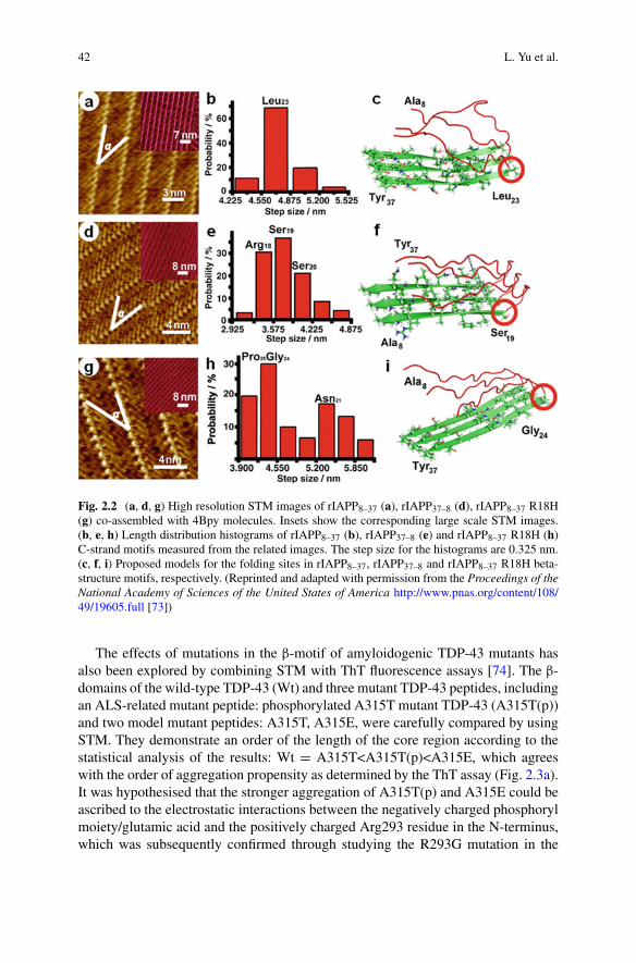
42 L. Yu et al.
Fig. 2.2 (a, d, g) High resolution STM images of rIAPP8–37 (a), rIAPP37–8 (d), rIAPP8–37 R18H(g) co-assembled with 4Bpy molecules. Insets show the corresponding large scale STM images.(b, e, h) Length distribution histograms of rIAPP8–37 (b), rIAPP37–8 (e) and rIAPP8–37 R18H (h)C-strand motifs measured from the related images. The step size for the histograms are 0.325 nm.(c, f, i) Proposed models for the folding sites in rIAPP8–37, rIAPP37–8 and rIAPP8–37 R18H beta-structure motifs, respectively. (Reprinted and adapted with permission from the Proceedings of theNational Academy of Sciences of the United States of America http://www.pnas.org/content/108/49/19605.full [73])
The effects of mutations in the β-motif of amyloidogenic TDP-43 mutants hasalso been explored by combining STM with ThT fluorescence assays [74]. The β-domains of the wild-type TDP-43 (Wt) and three mutant TDP-43 peptides, includingan ALS-related mutant peptide: phosphorylated A315T mutant TDP-43 (A315T(p))and two model mutant peptides: A315T, A315E, were carefully compared by usingSTM. They demonstrate an order of the length of the core region according to thestatistical analysis of the results: Wt = A315T<A315T(p)<A315E, which agreeswith the order of aggregation propensity as determined by the ThT assay (Fig. 2.3a).It was hypothesised that the stronger aggregation of A315T(p) and A315E could beascribed to the electrostatic interactions between the negatively charged phosphorylmoiety/glutamic acid and the positively charged Arg293 residue in the N-terminus,which was subsequently confirmed through studying the R293G mutation in the
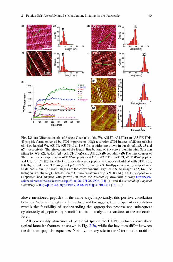
2 Peptide Self-Assembly and Its Modulation: Imaging on the Nanoscale 43
Fig. 2.3 (a) Different lengths of β-sheet C-strands of the Wt, A315T, A315T(p) and A315E TDP-43 peptide forms observed by STM experiments. High resolution STM images of 2D assembliesof 4Bpy-labeled Wt, A315T, A315T(p) and A315E peptides are shown in panels (a1, a3, a5 anda7), respectively. The histograms of the length distributions of the core β-domain with Gaussianfitting for Wt (a2), A315T (a4), A315T(p) (a6) and A315E (a8) peptides. (a9) The time courses ofThT fluorescence experiments of TDP-43 peptides A315E, A315T(p), A315T, Wt TDP-43 peptideand C1, C2, C3. (b) The effect of glycosylation on peptide assemblies identified with STM. (b1,b3) High-resolution STM images of p-VNTR/4Bpy and g-VNTR/4Bpy co-assembly, respectively.Scale bar: 2 nm. The inset images are the corresponding large scale STM images. (b2, b4) Thehistograms of the length distribution of C-terminal strands of p-VNTR and g-VNTR, respectively.(Reprinted and adapted with permission from the Journal of structural Biology http://www.sciencedirect.com/science/article/pii/S1047847712002936 [74] (a) and the Journal of PhysicalChemistry C http://pubs.acs.org/doi/abs/10.1021/acs.jpcc.5b12357 [75] (b))
above mentioned peptides in the same way. Importantly, this positive correlationbetween β-domain length on the surface and the aggregation propensity in solutionreveals the feasibility of understanding the aggregation process and subsequentcytotoxicity of peptides by β-motif structural analysis on surfaces at the molecularlevel.
All coassembly structures of peptide/4Bpy on the HOPG surface above showtypical lamellar features, as shown in Fig. 2.3a, while the key sites differ betweenthe different peptide sequences. Notably, the key site in the C-terminal β-motif of
44 L. Yu et al.
A315T(p), an ALS phosphorylated mutant, is located at Phe316, which is the correctneighboring residue (closer to the C-terminus) to the phosphorylated Thr315. Inanother study, STM investigation of a glycosylated peptide was performed to revealthe glycosylation effect on the peptide assemblies. Glycoprotein MUC1 with avariable number of tandem repeats (VNTRs), serves as a promising target forcancer immunotherapy. The VNTR is a 20-amino acid extracellular domain withthe sequence HGVTSAPDTRPAPGSTAPPA. The assembly structures of pristineVNTR (p-VNTR) and the glycosylated VNTR (g-VNTR) with the glycosylation ofdisaccharide (T antigen) on the Thr9 residue and monosaccharide (Tn antigen) onthe Ser15 residue were directly compared by using STM (Fig. 2.3b) [75]. The p-VNTR peptides form highly homogeneous lamellar-like assemblies on the HOPGsurface, while the g-VNTR peptide assembly shows apparent heterogeneity. Thisdifference in the assembly characteristics provides direct evidence for a glycosy-lation induced destabilization effect on the glycopeptide assemblies. Furthermore,the histogram of the length distribution of C-terminal strands of g-VNTR showstwo peaks (I and II), assigned to key sites Thr16 and Arg10. Notably, key residuesThr16 and Arg10 are located exactly adjacent to the glycosylated residues Ser15 andThr9. This demonstrates that glycosylation can induce a pronounced site-specificdestabilization effect on peptide assemblies. Moreover, disaccharides exert greaterinfluence on the assembly stability than monosaccharides. These results provideinsights into the effects of phosphorylation/glycosylation on peptide assemblies andsequence effects on the local structural stability of proteins.
2.4 Coassembly of Peptides with Small Molecules
A number of small molecules have been exploited as probes to track amyloidaggregation or for modulating the peptide assembly structures and aggregationprocesses. They demonstrate potential applications for the diagnosis and treatmentof conformational diseases caused by protein misfolding. For example, surfactants,copper/zinc ion chelators, well-known bioactive molecules (such as apomorphine,rifamycine, curcumin, porphyrins), and dyes (such as Congo red (CR), thioflavin T(ThT)) and their derivatives have been reported to inhibit amyloid aggregation [76–78]. Unveiling the mechanism of interactions between these molecules and relatedpeptides could provide a theoretical basis for the structure-based drug design andfacilitate the rational design of peptide-organic architectures.
2.4.1 Small Molecules Interacting with the Termini of Peptides
It is well recognized that peptide-peptide interactions are the driving force for pep-tide assembly. Based on similar molecular interactions, introducing peptide-organicmolecule interactions can also provide complementary strategies for studying
2 Peptide Self-Assembly and Its Modulation: Imaging on the Nanoscale 45
peptide assemblies and their interactions and thereby aid in constructing peptide-organic architectures. Such efforts have been reflected in many investigations onthe effects of small molecular modulators of peptide assembly and aggregationprocesses. In particular, it is feasible to tune peptide aggregation by introducinghydrogen bonding interactions with molecular modulators of the peptide termini,such as chaperone-like molecules 4Bpy and 1,2-di(4-pyridyl)-ethylene (DPE) [26,52, 73, 79–81].
Firstly, it is of great importance to clarify the effect of chaperone-like moleculeson the peptide assembly structures by real-time observation. Introducing tip manip-ulation has been demonstrated to be an effective way to rearrange or removemolecules adsorbed onto surfaces and liquid-solid interfaces [82–84]. An amyloidalpeptide Q11 (QQKFQFQFEQQ) was studied as a model peptide. Chaperone-mediated peptide assemblies (Q11/4Bpy) adopt ordered lamellar structures witha homogeneous distribution of the surface-bound peptide lengths, which couldbe attributed to the core segment of the Q11 peptide [85], as shown in Fig.2.4a. With a low-bias-voltage scan, the 4Bpy molecules were removed and theassembled peptides were agitated simultaneously and subsequently formed a newpristine peptide assembly at equilibrium in situ. This pristine peptide assemblypresents relatively disordered structures with a broad and bimodal distributionof core segment lengths. This direct observation reveals the effect of chaperonemolecules on the peptide assembly structures, which could aid construction ofpeptide architectures with various functions.
In another related study, the self-assembly behavior of a critical amyloidalpeptide segment KLVFF (Aβ (16–20)) and its co-assembly behavior with pyridinemodulators (4Bpy and 4′-chloro-2,2′:6′,2′′-terpyridine (Cl-Ter)) was observed andsecondary structure transformation was detected (Fig. 2.4b) [86]. The uniformKLVFF/4Bpy co-assembly was observed to present a parallel β-sheet-like con-formation with sandwich-like striped structures. However, a new close-packedladder-like structure was formed in the KLVFF/Cl-Ter co-assembly. Based onthe conformation, configuration and interaction sites of the modulators, the sec-ondary structure of KLVFF is transformed from a parallel to an antiparallelβ-sheet conformation, which was further verified by FTIR spectroscopy. Moreover,molecular mechanistic investigations on this peptide-terpyridine coassembly furtherreveal the associated contributions from multiple interactions in polymorphicpeptide assemblies with distinct geometrical arrangements. Most importantly, thepeptide-molecule co-assemblies demonstrate a synergistic inhibitory effect onAβ42-induced cytotoxicity [87].
Modulation effects of 4Bpy on AD-related peptide assemblies were also iden-tified with STM. The variation of amino acid residues in the peptide sequenceshows significant effects on the modulation of peptide assemblies as mentionedabove. In particular, the critical effect of the C-terminus of the peptide Aβ42 onits amyloid aggregation has been addressed [88]. Herein, the mechanistic changesof the assembly behaviour of variants of Aβ42 caused by the 4Bpy molecular tetherat its C-terminus was explored (Fig. 2.5) [89]. In the self-assembly of the Aβ42peptide, each β-strand is visible as a bright double-dotted ribbon and its periodicity
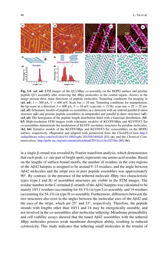
46 L. Yu et al.
Fig. 2.4 (a1, a4) STM images of the Q11/4Bpy co-assembly on the HOPG surface and pristinepeptide Q11 assembly after removing the 4Bpy molecules in the central region. Arrows in theimage present three main directions of peptide molecules. Tunneling conditions for imaging in(a1, a4): I = 300 pA, V = 600 mV. Scale bar = 10 nm. Tunneling conditions for manipulation,the tip scans in a direction, I = 600 pA, V = 10 mV, scan rate = 15 Hz, scan size = 25 × 25 nm.(a2, a5) Schematic models of peptide co-assemblies in a direction with an ordered parallel β-sheetstructure (a2) and pristine peptide assemblies in antiparallel and parallel β-sheet structures (a5).(a3, a6) The histograms of the peptide length distribution fitted with a Gaussian distribution. (b1,b3) High-resolution STM images (with schematic models) of KLVFF/4Bpy and KLVFF/Cl-Terco-assemblies demonstrate the modulation of KLVFF secondary structures by pyridine molecules.(b2, b4) Tentative models of the KLVFF/4Bpy and KLVFF/Cl-Ter coassemblies on the HOPGsurface, respectively. (Reprinted and adapted with permission from the ChemPhysChem http://onlinelibrary.wiley.com/wol1/doi/10.1002/cphc.201500340/full [85] (a), and the Chemical Com-munications http://pubs.rsc.org/en/content/articlehtml/2014/cc/c4cc02748e [86] (b))
in a single β-strand was revealed by Fourier transform analysis, which demonstratesthat each peak, i.e. one pair of bright spots, represents one amino acid residue. Basedon the lengths of surface-bound motifs, the number of residues in the core regionsof the Aβ42 hairpins is assigned to be around 9–15 residues, and the angle betweenAβ42 molecules and the stripe axis in pure peptide assemblies was approximately90◦. By contrast, in the presence of the tethered molecule 4Bpy, two characteristictypes (type I and II) of assembled structures are visible in the STM images. Theresidue number in the C-terminal β-strands of the Aβ42 hairpins was calculated to bemainly 10/11 residues (accounting for 44.1%) in type I co-assembly, and 14 residues(accounting for 54.1%) in type II co-assembly. Furthermore, differences between thetwo structures also exist in the angles between the molecular axes of the Aβ42 andthe axes of the stripe, which are 25◦ and 33◦, respectively. Therefore, the peptidestrands with lengths other than 10/11 and 14 may be energetically unstable, andnot involved in the co-assemblies after molecular tethering. Membrane permeabilityand cell viability assays showed that the tuned Aβ42 assemblies with the tethered4Bpy molecules possess weak membrane disruption ability, resulting in reducedcytotoxicity. This study indicates that tethering small molecules at the termini of
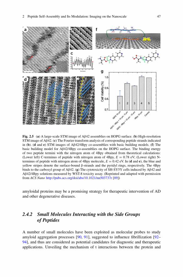
2 Peptide Self-Assembly and Its Modulation: Imaging on the Nanoscale 47
Fig. 2.5 (a) A large-scale STM image of Aβ42 assemblies on HOPG surface. (b) High-resolutionSTM image of Aβ42. (c) The Fourier transform analysis of corresponding peptide strands indicatedin (b). (d and e) STM images of Aβ42/4Bpy co-assemblies with basic building models. (f) Thebasic building model for Aβ42/4Bpy co-assemblies on the HOPG surface. The binding energyof two peptide termini with the nitrogen atom of 4Bpy obtained from theoretical calculations:(Lower left) C-terminus of peptide with nitrogen atom of 4Bpy, E = 0.78 eV; (Lower right) N-terminus of peptide with nitrogen atom of 4Bpy molecule, E = 0.42 eV. In (d and e), the blue andyellow stripes denote the surface-bound β-strands and the pyridyl rings, respectively. The 4Bpybinds to the carboxyl group of Aβ42. (g) The cytotoxicity of SH-SY5Y cells induced by Aβ42 andAβ42/4Bpy solutions measured by WST-8 toxicity assay. (Reprinted and adapted with permissionfrom ACS Nano http://pubs.acs.org/doi/abs/10.1021/nn503737r [89])
amyloidal proteins may be a promising strategy for therapeutic intervention of ADand other degenerative diseases.
2.4.2 Small Molecules Interacting with the Side Groupsof Peptides
A number of small molecules have been exploited as molecular probes to studyamyloid aggregation processes [90, 91], suggested to influence fibrillization [92–94], and thus are considered as potential candidates for diagnostic and therapeuticapplications. Unveiling the mechanism of t interactions between the protein and
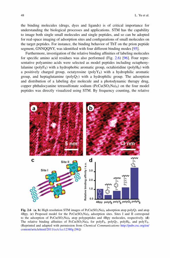
48 L. Yu et al.
the binding molecules (drugs, dyes and ligands) is of critical importance forunderstanding the biological processes and applications. STM has the capabilityto image both single small molecules and single peptides, and so can be adoptedfor real-space imaging of adsorption sites and configurations of small molecules onthe target peptides. For instance, the binding behavior of ThT on the prion peptidesegment, GNNQQNY, was identified with four different binding modes [95].
Furthermore, investigation of the relative binding affinities of labeling moleculesfor specific amino acid residues was also performed (Fig. 2.6) [96]. Four repre-sentative polyamino acids were selected as model peptides including octapheny-lalanine (polyF8) with a hydrophobic aromatic group, octahistidine (polyH8) witha positively charged group, octatyrosine (polyY8) with a hydrophilic aromaticgroup, and heptaglutamine (polyQ7) with a hydrophilic group. The adsorptionand distribution of a labeling dye molecule and a photodynamic therapy drug,copper phthalocyanine tetrasulfonate sodium (PcCu(SO3Na)4) on the four modelpeptides was directly visualized using STM. By frequency counting, the relative
Fig. 2.6 (a, b) High resolution STM images of PcCu(SO3Na)4 adsorption atop polyQ7 and atop4Bpy. (c) Proposed model for the PcCu(SO3Na)4 adsorption sites. Sites I and II correspondto the adsorption of PcCu(SO3Na)4 atop polypeptides and 4Bpy molecules, respectively. (d)The relative binding affinities of PcCu(SO3Na)4 for polyF8, polyQ7, polyH8, and polyY8.(Reprinted and adapted with permission from Chemical Communications http://pubs.rsc.org/en/content/articlehtml/2011/cc/c1cc12380g [96])
2 Peptide Self-Assembly and Its Modulation: Imaging on the Nanoscale 49
binding affinity of the polypeptide and 4Bpy molecules can be obtained. Sincethe affinity of PcCu(SO3Na)4 towards 4Bpy can be considered constant in thesepeptide-organic co-assemblies, the relative binding affinities of PcCu(SO3Na)4 forthe four polypeptides were found to have the ratio (polyH8:polyQ7:polyY8:polyF8= 3.32:1.29:0.79:0.34). The high affinity of PcCu(SO3Na)4 on polyH8 could beascribed to the electrostatic interaction between the positively charged imidazolemoiety with the negatively charged sulfonic acid group, while the low affinity onpolyF8 could be due to the effect of the solvent-accessible surface area of theside chains in water. The binding of dye or drug molecules on model peptidesalso provides important insight into the mechanism of chemical and structuraldependence on specific amino acid residues.
Here, we present a summary of the experimentally observed peptide lengths insurface-bound assemblies, with and without co-assembled small molecules interact-ing with the peptide termini. The data reveal the relationship between the numberof residues in the peptide sequence and the experimentally observed number ofresidues in the peptide assemblies in STM observations (Table 2.1). Short peptidescomprising 2–7 residues can be fully extended in the assembly structures, as shownin Fig. 2.7. It appears that for both homogeneous and heterogeneous sequenceswith less than seven residues, the peptides are fully extended on the surface. Thisobservation indicates the effect of persistence length in peptide structures. In termsof relatively long peptides with more than seven residues, the number of residuesinvolved in assemblies may vary, which suggests pronounced sequence dependence.These observations demonstrate that peptide structures on surfaces could provideinsights into the fundamental mechanisms underlying the sequence dependence ofthe persistent lengths of peptide chains.
Table 2.1 The number of residues in surface-bound peptide assemblies identified by STM
No. Peptide name Fulla Observedb References
1 AA 2 2 Stensgaard [51]2 FF 2 2 Lingenfelder [54]3 KFFE 4 4 Kalashnyk [55]4 KVVE 4 4 Kalashnyk [55]5 KLVFF (Aβ16-20) 5 5 Niu et al. [86]6 A5 5 5 Niu et al. [97]7 GNNQQNY 7 7 Mao et al. [95]8 Q7 7 7 Guo et al. [49]9 Q8 8 7/8 Guo et al. [49]10 Q7 7 7 Wang et al. [96]11 F8 8 8 Wang et al. [96]12 Y8 8 8 Wang et al. [96]13 H8 8 8 Wang et al. [96]
(continued)
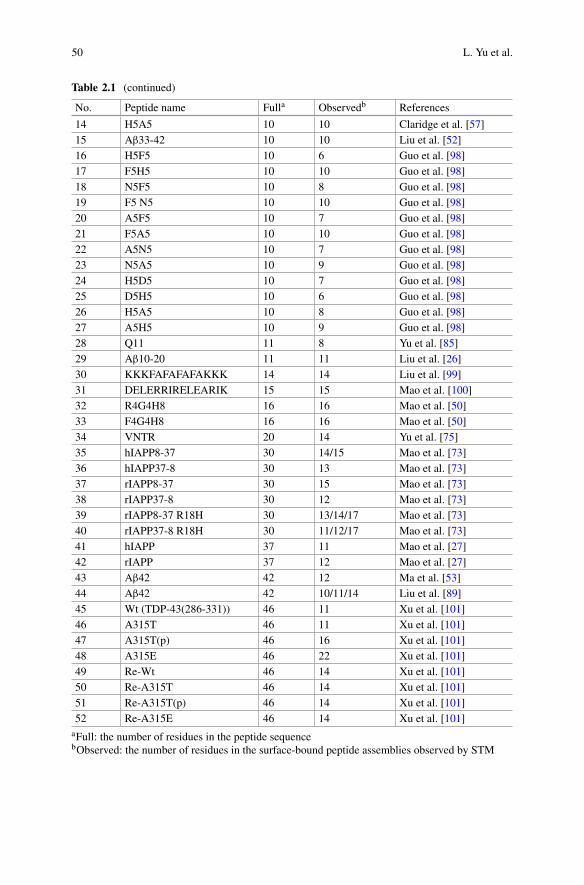
50 L. Yu et al.
Table 2.1 (continued)
No. Peptide name Fulla Observedb References
14 H5A5 10 10 Claridge et al. [57]15 Aβ33-42 10 10 Liu et al. [52]16 H5F5 10 6 Guo et al. [98]17 F5H5 10 10 Guo et al. [98]18 N5F5 10 8 Guo et al. [98]19 F5 N5 10 10 Guo et al. [98]20 A5F5 10 7 Guo et al. [98]21 F5A5 10 10 Guo et al. [98]22 A5N5 10 7 Guo et al. [98]23 N5A5 10 9 Guo et al. [98]24 H5D5 10 7 Guo et al. [98]25 D5H5 10 6 Guo et al. [98]26 H5A5 10 8 Guo et al. [98]27 A5H5 10 9 Guo et al. [98]28 Q11 11 8 Yu et al. [85]29 Aβ10-20 11 11 Liu et al. [26]30 KKKFAFAFAFAKKK 14 14 Liu et al. [99]31 DELERRIRELEARIK 15 15 Mao et al. [100]32 R4G4H8 16 16 Mao et al. [50]33 F4G4H8 16 16 Mao et al. [50]34 VNTR 20 14 Yu et al. [75]35 hIAPP8-37 30 14/15 Mao et al. [73]36 hIAPP37-8 30 13 Mao et al. [73]37 rIAPP8-37 30 15 Mao et al. [73]38 rIAPP37-8 30 12 Mao et al. [73]39 rIAPP8-37 R18H 30 13/14/17 Mao et al. [73]40 rIAPP37-8 R18H 30 11/12/17 Mao et al. [73]41 hIAPP 37 11 Mao et al. [27]42 rIAPP 37 12 Mao et al. [27]43 Aβ42 42 12 Ma et al. [53]44 Aβ42 42 10/11/14 Liu et al. [89]45 Wt (TDP-43(286-331)) 46 11 Xu et al. [101]46 A315T 46 11 Xu et al. [101]47 A315T(p) 46 16 Xu et al. [101]48 A315E 46 22 Xu et al. [101]49 Re-Wt 46 14 Xu et al. [101]50 Re-A315T 46 14 Xu et al. [101]51 Re-A315T(p) 46 14 Xu et al. [101]52 Re-A315E 46 14 Xu et al. [101]
aFull: the number of residues in the peptide sequencebObserved: the number of residues in the surface-bound peptide assemblies observed by STM

2 Peptide Self-Assembly and Its Modulation: Imaging on the Nanoscale 51
Fig. 2.7 The relationship between the number of residues in the full peptide sequence and thenumber of residues observed by STM involved in the peptide assemblies on surfaces. The datapoints on the grey line (y = x) mean that the number of observed residues is equal to its numberof residues in the full peptide sequence, i.e., all the residues in the sequence are participated in thepeptide assembly
2.5 Correlation of Peptide Assemblies on Surfacesand in Solution
Studies of peptide assemblies on surfaces imaged using STM provide structuralinformation and unveil the formation mechanisms, providing constructive guidancefor modulating peptide aggregation and therapeutic applications associated withhydrophobic membranes, including in biological aqueous systems. However, thetwo-dimensional liquid-solid system, which introduces a hydrophobic interface, isdistinct from the three-dimensional solution system. Peptide assembly behavior onsurfaces results from the delicate balance between peptide-peptide, peptide-solventand peptide-substrate interactions [1, 3, 102]. Nevertheless, the main driving forcesfor the assembly behavior could still be peptide-peptide interactions determined bythe physico-chemical properties of amino acid residues in the specific sequence. Asmentioned above, the β-motif length on surfaces and the aggregation propensity inbulk systems have been revealed to be positively correlated.
It is also well recognized from studies on adsorption processes that proteins canundergo conformational rearrangements when adsorbed at liquid-solid interfacesfrom solutions [103–106]. More importantly, the pathogenesis of some degenerativediseases, such as AD, prion diseases, and type 2 diabetes, is associated withan α-to-β conformational transition of part of the protein structure [107–110],which eventually results in the formation of amyloid deposits. The unnaturalconformations in the deposits could involve the formation of elastic layers atinterfaces and also insoluble amyloid aggregates. In the following, the detailedconformational transformation and relationship between secondary structure andassembly behavior in these two systems are reviewed.
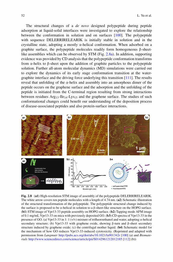
52 L. Yu et al.
The structural changes of a de novo designed polypeptide during peptideadsorption at liquid-solid interfaces were investigated to explore the relationshipbetween the conformation in solution and on surfaces [100]. The polypeptidewith sequence DELERRIRELEARIK is initially stable in solution and in thecrystalline state, adopting a mostly α-helical conformation. When adsorbed on agraphite surface, the polypeptide molecules readily form homogeneous β-sheet-like assemblies which can be observed by STM (Fig. 2.8a). In addition, supportingevidence was provided by CD analysis that the polypeptide conformation transformsfrom α-helix to β-sheet upon the addition of graphite particles to the polypeptidesolution. Further all-atom molecular dynamics (MD) simulations were carried outto explore the dynamics of its early stage conformation transition at the water-graphite interface and the driving force underlying this transition [111]. The resultsreveal that unfolding of the α-helix and assembly into an amorphous dimer of thepeptide occurs on the graphene surface and the adsorption and the unfolding of thepeptide is initiated from the C-terminal region resulting from strong interactionsbetween residues Arg13-Ile14-Lys15 and the graphene surface. The studies of suchconformational changes could benefit our understanding of the deposition processof disease-associated peptides and also protein-surface interactions.
Fig. 2.8 (a1) High-resolution STM image of assembly of the polypeptide DELERRIRELEARIK.The white arrow covers ten peptide molecules with a length of 4.74 nm. (a2) Schematic illustrationof the structural transformation of the polypeptide. The polypeptide structural change induced bythe surface is proposed to be α-helical in solution to a β-sheet-like structure on the HOPG surface.(b1) STM image of Vpr13-33 peptide assembly on HOPG surface. (b2) Tapping mode AFM imageof 0.1 mg/mL Vpr13-33 on mica with previously deposited GO. (b3) CD spectra of Vpr13-33 in thepresence of GO. (a) Vpr13-33 in 1: 1 (v/v) mixture of trifluoroethanol and water, adopting α-helicalsecondary structure; (b) Vpr13-33 with graphene oxide, showing β-turn and β-sheet secondarystructure induced by graphene oxide; (c) the centrifugal mother liquid. (b4) Schematic model forthe mechanism of how GO reduces Vpr13-33-induced cytotoxicity. (Reprinted and adapted withpermission from Langmuir http://pubs.acs.org/doi/abs/10.1021/la901342r [100] (a) and Biomate-rials http://www.sciencedirect.com/science/article/pii/S0142961212012185 [112] (b))
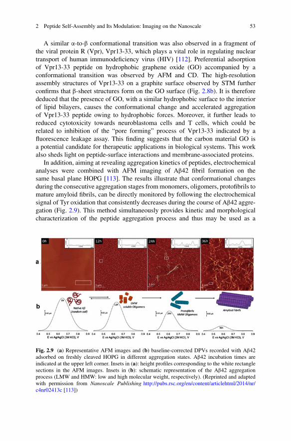
2 Peptide Self-Assembly and Its Modulation: Imaging on the Nanoscale 53
A similar α-to-β conformational transition was also observed in a fragment ofthe viral protein R (Vpr), Vpr13-33, which plays a vital role in regulating nucleartransport of human immunodeficiency virus (HIV) [112]. Preferential adsorptionof Vpr13-33 peptide on hydrophobic graphene oxide (GO) accompanied by aconformational transition was observed by AFM and CD. The high-resolutionassembly structures of Vpr13-33 on a graphite surface observed by STM furtherconfirms that β-sheet structures form on the GO surface (Fig. 2.8b). It is thereforededuced that the presence of GO, with a similar hydrophobic surface to the interiorof lipid bilayers, causes the conformational change and accelerated aggregationof Vpr13-33 peptide owing to hydrophobic forces. Moreover, it further leads toreduced cytotoxicity towards neuroblastoma cells and T cells, which could berelated to inhibition of the “pore forming” process of Vpr13-33 indicated by afluorescence leakage assay. This finding suggests that the carbon material GO isa potential candidate for therapeutic applications in biological systems. This workalso sheds light on peptide-surface interactions and membrane-associated proteins.
In addition, aiming at revealing aggregation kinetics of peptides, electrochemicalanalyses were combined with AFM imaging of Aβ42 fibril formation on thesame basal plane HOPG [113]. The results illustrate that conformational changesduring the consecutive aggregation stages from monomers, oligomers, protofibrils tomature amyloid fibrils, can be directly monitored by following the electrochemicalsignal of Tyr oxidation that consistently decreases during the course of Aβ42 aggre-gation (Fig. 2.9). This method simultaneously provides kinetic and morphologicalcharacterization of the peptide aggregation process and thus may be used as a
Fig. 2.9 (a) Representative AFM images and (b) baseline-corrected DPVs recorded with Aβ42adsorbed on freshly cleaved HOPG in different aggregation states. Aβ42 incubation times areindicated at the upper left corner. Insets in (a): height profiles corresponding to the white rectanglesections in the AFM images. Insets in (b): schematic representation of the Aβ42 aggregationprocess (LMW and HMW: low and high molecular weight, respectively). (Reprinted and adaptedwith permission from Nanoscale Publishing http://pubs.rsc.org/en/content/articlehtml/2014/nr/c4nr02413c [113])
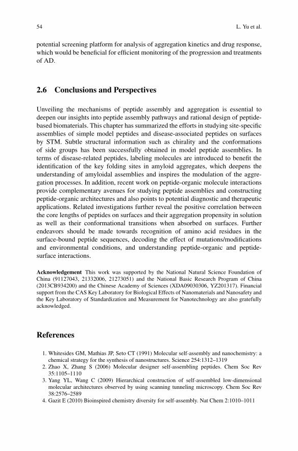
54 L. Yu et al.
potential screening platform for analysis of aggregation kinetics and drug response,which would be beneficial for efficient monitoring of the progression and treatmentsof AD.
2.6 Conclusions and Perspectives
Unveiling the mechanisms of peptide assembly and aggregation is essential todeepen our insights into peptide assembly pathways and rational design of peptide-based biomaterials. This chapter has summarized the efforts in studying site-specificassemblies of simple model peptides and disease-associated peptides on surfacesby STM. Subtle structural information such as chirality and the conformationsof side groups has been successfully obtained in model peptide assemblies. Interms of disease-related peptides, labeling molecules are introduced to benefit theidentification of the key folding sites in amyloid aggregates, which deepens theunderstanding of amyloidal assemblies and inspires the modulation of the aggre-gation processes. In addition, recent work on peptide-organic molecule interactionsprovide complementary avenues for studying peptide assemblies and constructingpeptide-organic architectures and also points to potential diagnostic and therapeuticapplications. Related investigations further reveal the positive correlation betweenthe core lengths of peptides on surfaces and their aggregation propensity in solutionas well as their conformational transitions when absorbed on surfaces. Furtherendeavors should be made towards recognition of amino acid residues in thesurface-bound peptide sequences, decoding the effect of mutations/modificationsand environmental conditions, and understanding peptide-organic and peptide-surface interactions.
Acknowledgement This work was supported by the National Natural Science Foundation ofChina (91127043, 21332006, 21273051) and the National Basic Research Program of China(2013CB934200) and the Chinese Academy of Sciences (XDA09030306, YZ201317). Financialsupport from the CAS Key Laboratory for Biological Effects of Nanomaterials and Nanosafety andthe Key Laboratory of Standardization and Measurement for Nanotechnology are also gratefullyacknowledged.
References
1. Whitesides GM, Mathias JP, Seto CT (1991) Molecular self-assembly and nanochemistry: achemical strategy for the synthesis of nanostructures. Science 254:1312–1319
2. Zhao X, Zhang S (2006) Molecular designer self-assembling peptides. Chem Soc Rev35:1105–1110
3. Yang YL, Wang C (2009) Hierarchical construction of self-assembled low-dimensionalmolecular architectures observed by using scanning tunneling microscopy. Chem Soc Rev38:2576–2589
4. Gazit E (2010) Bioinspired chemistry diversity for self-assembly. Nat Chem 2:1010–1011
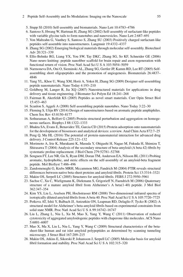
2 Peptide Self-Assembly and Its Modulation: Imaging on the Nanoscale 55
5. Stupp SI (2010) Self-assembly and biomaterials. Nano Lett 10:4783–47866. Santoso S, Hwang W, Hartman H, Zhang SG (2002) Self-assembly of surfactant-like peptides
with variable glycine tails to form nanotubes and nanovesicles. Nano Lett 2:687–6917. Von Maltzahn G, Vauthey S, Santoso S, Zhang SU (2003) Positively charged surfactant-like
peptides self-assemble into nanostructures. Langmuir 19:4332–43378. Zhang SG (2002) Emerging biological materials through molecular self-assembly. Biotechnol
Adv 20:321–3399. Ellis-Behnke RG, Liang YX, You SW, Tay DKC, Zhang SG, So KF, Schneider GE (2006)
Nano neuro knitting: peptide nanofiber scaffold for brain repair and axon regeneration withfunctional return of vision. Proc Natl Acad Sci U S A 103:5054–5059
10. Narmoneva DA, Oni O, Sieminski AL, Zhang SG, Gertler JP, Kamm RD, Lee RT (2005) Self-assembling short oligopeptides and the promotion of angiogenesis. Biomaterials 26:4837–4846
11. Yang YL, Khoe U, Wang XM, Horii A, Yokoi H, Zhang SG (2009) Designer self-assemblingpeptide nanomaterials. Nano Today 4:193–210
12. Goldberg M, Langer R, Jia XQ (2007) Nanostructured materials for applications in drugdelivery and tissue engineering. J Biomater Sci Polym Ed 18:241–268
13. Fairman R, Akerfeldt KS (2005) Peptides as novel smart materials. Curr Opin Struct Biol15:453–463
14. Scanlon S, Aggeli A (2008) Self-assembling peptide nanotubes. Nano Today 3:22–3015. Fleming S, Ulijn RV (2014) Design of nanostructures based on aromatic peptide amphiphiles.
Chem Soc Rev 43:8150–817716. Sethuraman A, Belfort G (2005) Protein structural perturbation and aggregation on homoge-
neous surfaces. Biophys J 88:1322–133317. Bhakta SA, Evans E, Benavidez TE, Garcia CD (2015) Protein adsorption onto nanomaterials
for the development of biosensors and analytical devices: a review. Anal Chim Acta 872:7–2518. Peng Q, Mu HL (2016) The potential of protein-nanomaterial interaction for advanced drug
delivery. J Control Release 225:121–13219. Morimoto A, Irie K, Murakami K, Masuda Y, Ohigashi H, Nagao M, Fukuda H, Shimizu T,
Shirasawa T (2004) Analysis of the secondary structure of beta-amyloid (A beta 42) fibrils bysystematic proline replacement. J Biol Chem 279:52781–52788
20. Senguen FT, Lee NR, Gu X, Ryan DM, Doran TM, Anderson EA, Nilsson BL (2011) Probingaromatic, hydrophobic, and steric effects on the self-assembly of an amyloid-beta fragmentpeptide. Mol BioSyst 7:486–496
21. Zandomeneghi G, Krebs MRH, Mccammon MG, Fandrich M (2004) FTIR reveals structuraldifferences between native beta-sheet proteins and amyloid fibrils. Protein Sci 13:3314–3321
22. Makin OS, Serpell LC (2005) Structures for amyloid fibrils. FEBS J 272:5950–596123. Sachse C, Xu C, Wieligmann K, Diekmann S, Grigorieff N, Faendrich M (2006) Quaternary
structure of a mature amyloid fibril from Alzheimer’s A beta(1-40) peptide. J Mol Biol362:347–354
24. Kim YS, Liu L, Axelsen PH, Hochstrasser RM (2008) Two-dimensional infrared spectra ofisotopically diluted amyloid fibrils from A beta 40. Proc Natl Acad Sci U S A 105:7720–7725
25. Petkova AT, Ishii Y, Balbach JJ, Antzutkin ON, Leapman RD, Delaglio F, Tycko R (2002) Astructural model for Alzheimer’s beta-amyloid fibrils based on experimental constraints fromsolid state NMR. Proc Natl Acad Sci U S A 99:16742–16747
26. Liu L, Zhang L, Niu L, Xu M, Mao X, Yang Y, Wang C (2011) Observation of reducedcytotoxicity of aggregated amyloidogenic peptides with chaperone-like molecules. ACS Nano5:6001–6007
27. Mao X, Ma X, Liu L, Niu L, Yang Y, Wang C (2009) Structural characteristics of the beta-sheet-like human and rat islet amyloid polypeptides as determined by scanning tunnelingmicroscopy. J Struct Biol 167:209–215
28. Makin OS, Atkins E, Sikorski P, Johansson J, Serpell LC (2005) Molecular basis for amyloidfibril formation and stability. Proc Natl Acad Sci U S A 102:315–320
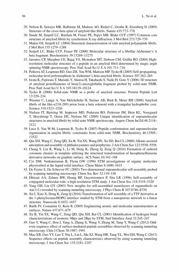
56 L. Yu et al.
29. Nelson R, Sawaya MR, Balbirnie M, Madsen AO, Riekel C, Grothe R, Eisenberg D (2005)Structure of the cross-beta spine of amyloid-like fibrils. Nature 435:773–778
30. Sunde M, Serpell LC, Bartlam M, Fraser PE, Pepys MB, Blake CCF (1997) Common corestructure of amyloid fibrils by synchrotron X-ray diffraction. J Mol Biol 273:729–739
31. Makin OS, Serpell LC (2004) Structural characterisation of islet amyloid polypeptide fibrils.J Mol Biol 335:1279–1288
32. Serpell LC, Blake CCF, Fraser PE (2000) Molecular structure of a fibrillar Alzheimer’s Abeta fragment. Biochemistry 39:13269–13275
33. Jaroniec CP, Macphee CE, Bajaj VS, Mcmahon MT, Dobson CM, Griffin RG (2004) High-resolution molecular structure of a peptide in an amyloid fibril determined by magic anglespinning NMR spectroscopy. Proc Natl Acad Sci U S A 101:711–716
34. Petkova AT, Leapman RD, Guo ZH, Yau WM, Mattson MP, Tycko R (2005) Self-propagating,molecular-level polymorphism in Alzheimer’s beta-amyloid fibrils. Science 307:262–265
35. Iwata K, Fujiwara T, Matsuki Y, Akutsu H, Takahashi S, Naiki H, Goto Y (2006) 3D structureof amyloid protofilaments of beta(2)-microglobulin fragment probed by solid-state NMR.Proc Natl Acad Sci U S A 103:18119–18124
36. Tycko R (2006) Solid-state NMR as a probe of amyloid structure. Protein Peptide Lett13:229–234
37. Wasmer C, Lange A, Van Melckebeke H, Siemer AB, Riek R, Meier BH (2008) Amyloidfibrils of the Het-s(218-289) prion form a beta solenoid with a triangular hydrophobic core.Science 319:1523–1526
38. Nielsen JT, Bjerring M, Jeppesen MD, Pedersen RO, Pedersen JM, Hein KL, VosegaardT, Skrydstrup T, Otzen DE, Nielsen NC (2009) Unique identification of supramolecularstructures in amyloid fibrils by solid-state NMR spectroscopy. Angew Chem Int Ed 48:2118–2121
39. Luca S, Yau W-M, Leapman R, Tycko R (2007) Peptide conformation and supramolecularorganization in amylin fibrils: constraints from solid-state NMR. Biochemistry 46:13505–13522
40. Qiu XH, Wang C, Zeng QD, Xu B, Yin SX, Wang HN, Xu SD, Bai CL (2000) Alkane-assistedadsorption and assembly of phthalocyanines and porphyrins. J Am Chem Soc 122:5550–5556
41. Chang S, Liu R, Wang L, Li M, Deng K, Zheng Q, Zeng Q (2016) Formation of orderedcoronene clusters in template utilizing the structural transformation of hexaphenylbenzenederivative networks on graphite surface. ACS Nano 10:342–348
42. Cyr DM, Venkataraman B, Flynn GW (1996) STM investigations of organic moleculesphysisorbed at the liquid-solid interface. Chem Mater 8:1600–1615
43. De Feyter S, De Schryver FC (2003) Two-dimensional supramolecular self-assembly probedby scanning tunneling microscopy. Chem Soc Rev 32:139–150
44. Dhirani AA, Zehner RW, Hsung RP, Guyotsionnest P, Sita LR (1996) Self-assembly ofconjugated molecular rods: a high-resolution STM study. J Am Chem Soc 118:3319–3320
45. Yang GH, Liu GY (2003) New insights for self-assembled monolayers of organothiols onAu(111) revealed by scanning tunneling microscopy. J Phys Chem B 107:8746–8759
46. Xu J, Xiao X, Deng K, Zeng Q (2016) Transformation of self-assembly of a TTF derivative atthe 1-phenyloctane/HOPG interface studied by STM-from a nanoporous network to a linearstructure. Nanoscale 8:1652–1657
47. Barth JV, Costantini G, Kern K (2005) Engineering atomic and molecular nanostructures atsurfaces. Nature 437:671–679
48. Xu B, Yin SX, Wang C, Zeng QD, Qiu XH, Bai CL (2001) Identification of hydrogen bondcharacterizations of isomeric 4Bpy and 2Bpy by STM. Surf Interface Anal 32:245–247
49. Guo Y, Wang C, Hou J, Yang A, Zhang X, Wang Y, Zhang M, Yang Y, Wang C (2012) Odd-even sequence effect of surface-mediated peptide assemblies observed by scanning tunnelingmicroscopy. Chin J Chem 30:1987–1991
50. Mao XB, Guo YY, Luo Y, Niu L, Liu L, Ma XJ, Wang HB, Yang YL, Wei GH, Wang C (2013)Sequence effects on peptide assembly characteristics observed by using scanning tunnelingmicroscopy. J Am Chem Soc 135:2181–2187
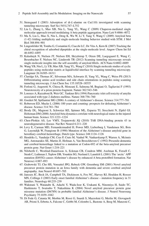
2 Peptide Self-Assembly and Its Modulation: Imaging on the Nanoscale 57
51. Stensgaard I (2003) Adsorption of di-L-alanine on Cu(110) investigated with scanningtunneling microscopy. Surf Sci 545:L747–L752
52. Liu L, Zhang L, Mao XB, Niu L, Yang YL, Wang C (2009) Chaperon-mediated singlemolecular approach toward modulating A beta peptide aggregation. Nano Lett 9:4066–4072
53. Ma X, Liu L, Mao X, Niu L, Deng K, Wu W, Li Y, Yang Y, Wang C (2009) Amyloid beta(1-42) folding multiplicity and single-molecule binding behavior studied with STM. J MolBiol 388:894–901
54. Lingenfelder M, Tomba G, Costantini G, Ciacchi LC, De Vita A, Kern K (2007) Tracking thechiral recognition of adsorbed dipeptides at the single-molecule level. Angew Chem Int Ed46:4492–4495
55. Kalashnyk N, Nielsen JT, Nielsen EH, Skrydstrup T, Otzen DE, Laegsgaard E, Wang C,Besenbacher F, Nielsen NC, Linderoth TR (2012) Scanning tunneling microscopy revealssingle-molecule insights into the self-assembly of amyloid fibrils. ACS Nano 6:6882–6889
56. Wang YB, Niu L, Li YB, Mao XB, Yang YL, Wang C (2010) Single molecule studies of cyclicpeptides using molecular matrix at liquid/solid interface by scanning tunneling microscopy.Langmuir 26:16305–16311
57. Claridge SA, Thomas JC, Silverman MA, Schwartz JJ, Yang YL, Wang C, Weiss PS (2013)Differentiating amino acid residues and side chain orientations in peptides using scanningtunneling microscopy. J Am Chem Soc 135:18528–18535
58. Forloni G, Angeretti N, Chiesa R, Monzani E, Salmona M, Bugiani O, Tagliavini F (1993)Neurotoxicity of a prion protein-fragment. Nature 362:543–546
59. Lorenzo A, Razzaboni B, Weir GC, Yankner BA (1994) Pancreatic-islet cell toxicity of amylinassociated with type-2 diabetes-mellitus. Nature 368:756–760
60. Goedert M, Spillantini MG (2006) A century of Alzheimer’s disease. Science 314:777–78161. Roberson ED, Mucke L (2006) 100 years and counting: prospects for defeating Alzheimer’s
disease. Science 314:781–78462. Brody DL, Magnoni S, Schwetye KE, Spinner ML, Esparza TJ, Stocchetti N, Zipfel GJ,
Holtzman DM (2008) Amyloid-beta dynamics correlate with neurological status in the injuredhuman brain. Science 321:1221–1224
63. Chen-Plotkin AS, Lee VMY, Trojanowski JQ (2010) TAR DNA-binding protein 43 inneurodegenerative disease. Nat Rev Neurol 6:211–220
64. Levy E, Carman MD, Fernandezmadrid IJ, Power MD, Lieberburg I, Vanduinen SG, BotsG, Luyendijk W, Frangione B (1990) Mutation of the Alzheimer’s-disease amyloid gene inhereditary cerebral-hemorrhage, Dutch type. Science 248:1124–1126
65. Hendriks L, Vanduijn CM, Cras P, Cruts M, Vanhul W, Vanharskamp F, Warren A, McinnisMG, Antonarakis SE, Martin JJ, Hofman A, Van Broeckhoven C (1992) Presenile-dementiaand cerebral-hemorrhage linked to a mutation at Codon-692 of the beta-amyloid precursorprotein gene. Nat Genet 1:218–221
66. Nilsberth C, Westlind-Danielsson A, Eckman CB, Condron MM, Axelman K, Forsell C,Stenh C, Luthman J, Teplow DB, Younkin SG, Naslund J, Lannfelt L (2001) The ‘arctic’ APPmutation (E693G) causes Alzheimer’s disease by enhanced A beta protofibril formation. NatNeurosci 4:887–893
67. Grabowski TJ, Cho HS, Vonsattel JPG, Rebeck GW, Greenberg SM (2001) Novel amyloidprecursor protein mutation in an Iowa family with dementia and severe cerebral amyloidangiopathy. Ann Neurol 49:697–705
68. Janssen JC, Beck JA, Campbell TA, Dickinson A, Fox NC, Harvey RJ, Houlden H, RossorMN, Collinge J (2003) Early onset familial Alzheimer’s disease – mutation frequency in 31families. Neurology 60:235–239
69. Wakutani Y, Watanabe K, Adachi Y, Wada-Isoe K, Urakami K, Ninomiya H, Saido TC,Hashimoto T, Iwatsubo T, Nakashima K (2004) Novel amyloid precursor protein genemissense mutation (D678N) in probable familial Alzheimer’s disease. J Neurol NeurosurgPsychiatry 75:1039–1042
70. Di Fede G, Catania M, Morbin M, Rossi G, Suardi S, Mazzoleni G, Merlin M, GiovagnoliAR, Prioni S, Erbetta A, Falcone C, Gobbi M, Colombo L, Bastone A, Beeg M, Manzoni C,
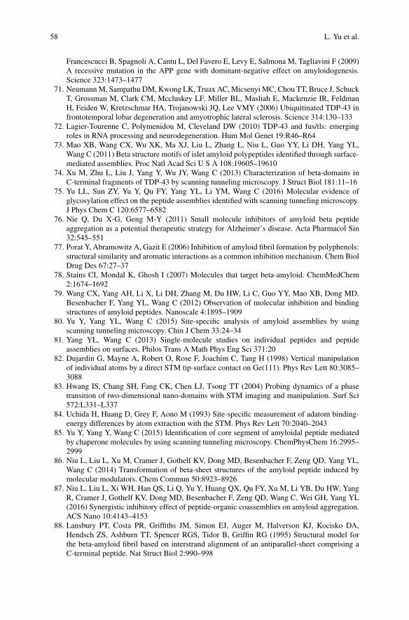
58 L. Yu et al.
Francescucci B, Spagnoli A, Cantu L, Del Favero E, Levy E, Salmona M, Tagliavini F (2009)A recessive mutation in the APP gene with dominant-negative effect on amyloidogenesis.Science 323:1473–1477
71. Neumann M, Sampathu DM, Kwong LK, Truax AC, Micsenyi MC, Chou TT, Bruce J, SchuckT, Grossman M, Clark CM, Mccluskey LF, Miller BL, Masliah E, Mackenzie IR, FeldmanH, Feiden W, Kretzschmar HA, Trojanowski JQ, Lee VMY (2006) Ubiquitinated TDP-43 infrontotemporal lobar degeneration and amyotrophic lateral sclerosis. Science 314:130–133
72. Lagier-Tourenne C, Polymenidou M, Cleveland DW (2010) TDP-43 and fus/tls: emergingroles in RNA processing and neurodegeneration. Hum Mol Genet 19:R46–R64
73. Mao XB, Wang CX, Wu XK, Ma XJ, Liu L, Zhang L, Niu L, Guo YY, Li DH, Yang YL,Wang C (2011) Beta structure motifs of islet amyloid polypeptides identified through surface-mediated assemblies. Proc Natl Acad Sci U S A 108:19605–19610
74. Xu M, Zhu L, Liu J, Yang Y, Wu JY, Wang C (2013) Characterization of beta-domains inC-terminal fragments of TDP-43 by scanning tunneling microscopy. J Struct Biol 181:11–16
75. Yu LL, Sun ZY, Yu Y, Qu FY, Yang YL, Li YM, Wang C (2016) Molecular evidence ofglycosylation effect on the peptide assemblies identified with scanning tunneling microscopy.J Phys Chem C 120:6577–6582
76. Nie Q, Du X-G, Geng M-Y (2011) Small molecule inhibitors of amyloid beta peptideaggregation as a potential therapeutic strategy for Alzheimer’s disease. Acta Pharmacol Sin32:545–551
77. Porat Y, Abramowitz A, Gazit E (2006) Inhibition of amyloid fibril formation by polyphenols:structural similarity and aromatic interactions as a common inhibition mechanism. Chem BiolDrug Des 67:27–37
78. Stains CI, Mondal K, Ghosh I (2007) Molecules that target beta-amyloid. ChemMedChem2:1674–1692
79. Wang CX, Yang AH, Li X, Li DH, Zhang M, Du HW, Li C, Guo YY, Mao XB, Dong MD,Besenbacher F, Yang YL, Wang C (2012) Observation of molecular inhibition and bindingstructures of amyloid peptides. Nanoscale 4:1895–1909
80. Yu Y, Yang YL, Wang C (2015) Site-specific analysis of amyloid assemblies by usingscanning tunneling microscopy. Chin J Chem 33:24–34
81. Yang YL, Wang C (2013) Single-molecule studies on individual peptides and peptideassemblies on surfaces. Philos Trans A Math Phys Eng Sci 371:20
82. Dujardin G, Mayne A, Robert O, Rose F, Joachim C, Tang H (1998) Vertical manipulationof individual atoms by a direct STM tip-surface contact on Ge(111). Phys Rev Lett 80:3085–3088
83. Hwang IS, Chang SH, Fang CK, Chen LJ, Tsong TT (2004) Probing dynamics of a phasetransition of two-dimensional nano-domains with STM imaging and manipulation. Surf Sci572:L331–L337
84. Uchida H, Huang D, Grey F, Aono M (1993) Site-specific measurement of adatom binding-energy differences by atom extraction with the STM. Phys Rev Lett 70:2040–2043
85. Yu Y, Yang Y, Wang C (2015) Identification of core segment of amyloidal peptide mediatedby chaperone molecules by using scanning tunneling microscopy. ChemPhysChem 16:2995–2999
86. Niu L, Liu L, Xu M, Cramer J, Gothelf KV, Dong MD, Besenbacher F, Zeng QD, Yang YL,Wang C (2014) Transformation of beta-sheet structures of the amyloid peptide induced bymolecular modulators. Chem Commun 50:8923–8926
87. Niu L, Liu L, Xi WH, Han QS, Li Q, Yu Y, Huang QX, Qu FY, Xu M, Li YB, Du HW, YangR, Cramer J, Gothelf KV, Dong MD, Besenbacher F, Zeng QD, Wang C, Wei GH, Yang YL(2016) Synergistic inhibitory effect of peptide-organic coassemblies on amyloid aggregation.ACS Nano 10:4143–4153
88. Lansbury PT, Costa PR, Griffiths JM, Simon EJ, Auger M, Halverson KJ, Kocisko DA,Hendsch ZS, Ashburn TT, Spencer RGS, Tidor B, Griffin RG (1995) Structural model forthe beta-amyloid fibril based on interstrand alignment of an antiparallel-sheet comprising aC-terminal peptide. Nat Struct Biol 2:990–998
2 Peptide Self-Assembly and Its Modulation: Imaging on the Nanoscale 59
89. Liu L, Niu L, Xu M, Han QS, Duan HY, Dong MD, Besenbacher F, Wang C, Yang YL (2014)Molecular tethering effect of C-terminus of amyloid peptide A beta 42. ACS Nano 8:9503–9510
90. Ivanova MI, Sawaya MR, Gingery M, Attinger A, Eisenberg D (2004) An amyloid-formingsegment of beta 2-microglobulin suggests a molecular model for the fibril. Proc Natl AcadSci U S A 101:10584–10589
91. Fowler DM, Koulov AV, Balch WE, Kelly JW (2007) Functional amyloid – from bacteria tohumans. Trends Biochem Sci 32:217–224
92. Sato T, Kienlen-Campard P, Ahmed M, Liu W, Li HL, Elliott JI, Aimoto S, ConstantinescuSN, Octave JN, Smith SO (2006) Inhibitors of amyloid toxicity based on beta-sheet packingof A beta 40 and A beta 42. Biochemistry 45:5503–5516
93. Blanchard BJ, Chen A, Rozeboom LM, Stafford KA, Weigele P, Ingram VM (2004) Efficientreversal of Alzheimer’s disease fibril formation and elimination of neurotoxicity by a smallmolecule. Proc Natl Acad Sci U S A 101:14326–14332
94. Heiser V, Scherzinger E, Boeddrich A, Nordhoff E, Lurz R, Schugardt N, Lehrach H, WankerEE (2000) Inhibition of huntingtin fibrillogenesis by specific antibodies and small molecules:implications for Huntington’s disease therapy. Proc Natl Acad Sci U S A 97:6739–6744
95. Mao XB, Guo YY, Wang CX, Zhang M, Ma XJ, Liu L, Niu L, Zeng QD, Yang YL, WangC (2011) Binding modes of thioflavin t molecules to prion peptide assemblies identified byusing scanning tunneling microscopy. ACS Chem Neurosci 2:281–287
96. Wang CX, Mao XB, Yang AH, Niu L, Wang SN, Li DH, Guo YY, Wang YB, Yang YL, WangC (2011) Determination of relative binding affinities of labeling molecules with amino acidsby using scanning tunneling microscopy. Chem Commun 47:10638–10640
97. Niu L, Ma XJ, Liu L, Mao XB, Wu DX, Yang YL, Zeng QD, Wang C (2010) Molec-ularly tuned peptide assemblies at the liquid-solid interface studied by scanning tunnelingmicroscopy. Phys Chem Chem Phys 12:11683–11687
98. Guo YY, Hou JF, Zhang XM, Yang YL, Wang C (2017) Stabilization effect of amino acidside chains in peptide assemblies on graphite studied by scanning tunneling microscopy.Chemphyschem 18:1–10
99. Liu L, Li YB, Xia D, Bortolini C, Zhang S, Yang YL, Pedersen JS, Wang C, BesenbacherF, Dong MD (2015) A self-assembled nanopatch with peptide-organic multilayers andmechanical properties. Nanoscale 7:2250–2254
100. Mao XB, Wang YB, Liu L, Niu L, Yang YL, Wang C (2009) Molecular-level evidenceof the surface-induced transformation of peptide structures revealed by scanning tunnelingmicroscopy. Langmuir 25:8849–8853
101. Xu M, Zhu L, Liu J, Yang Y, Wu JY, Wang C (2013) Characterization of beta-domains inC-terminal fragments of TDP-43 by scanning tunneling microscopy. J Struct Biol 181:11–16
102. Yang YL, Wang C (2009) Solvent effects on two-dimensional molecular self-assembliesinvestigated by using scanning tunneling microscopy. Curr Opin Colloid Interface Sci14:135–147
103. Latour RA, Rini CJ (2002) Theoretical analysis of adsorption thermodynamics for hydropho-bic peptide residues on SAM surfaces of varying functionality. J Biomed Mater Res60:564–577
104. Ostuni E, Grzybowski BA, Mrksich M, Roberts CS, Whitesides GM (2003) Adsorption ofproteins to hydrophobic sites on mixed self-assembled monolayers. Langmuir 19:1861–1872
105. Chakarova SD, Carlsson AE (2004) Model study of protein unfolding by interfaces. Phys RevE 69:021907
106. Sethuraman A, Vedantham G, Imoto T, Przybycien T, Belfort G (2004) Protein unfolding atinterfaces: slow dynamics of alpha-helix to beta-sheet transition. Proteins 56:669–678
107. Jayawickrama D, Zink S, Vandervelde D, Effiong RI, Larive CK (1995) Conformational-analysis of the beta-amyloid peptide fragment, beta(12-28). J Biomol Struct Dyn 13:229–244
108. Mihara H, Takahashi Y, Ueno A (1998) Design of peptides undergoing self-catalytic alpha-to-beta transition and amyloidogenesis. Biopolymers 47:83–92

60 L. Yu et al.
109. Pan KM, Baldwin M, Nguyen J, Gasset M, Serban A, Groth D, Mehlhorn I, Huang ZW,Fletterick RJ, Cohen FE, Prusiner SB (1993) Conversion of alpha-helices into beta-sheetsfeatures in the formation of the scrapie prion proteins. Proc Natl Acad Sci U S A 90:10962–10966
110. Peretz D, Williamson RA, Matsunaga Y, Serban H, Pinilla C, Bastidas RB, RozenshteynR, James TL, Houghten RA, Cohen FE, Prusiner SB, Burton DR (1997) A conformationaltransition at the N terminus of the prion protein features in formation of the scrapie isoform.J Mol Biol 273:614–622
111. Ou L, Luo Y, Wei G (2011) Atomic-level study of adsorption, conformational change, anddimerization of an alpha-helical peptide at graphene surface. J Phys Chem B 115:9813–9822
112. Zhang M, Mao XB, Wang CX, Zeng WF, Zhang CL, Li ZJ, Fang Y, Yang YL, Liang W, WangC (2013) The effect of graphene oxide on conformation change, aggregation and cytotoxicityof HIV-1 regulatory protein (Vpr). Biomaterials 34:1383–1390
113. Lopes P, Xu M, Zhang M, Zhou T, Yang YL, Wang C, Ferapontova EE (2014) Directelectrochemical and AFM detection of amyloid-beta peptide aggregation on basal planeHOPG. Nanoscale 6:7853–7857
Chapter 3The Kinetics, Thermodynamics andMechanisms of Short Aromatic PeptideSelf-Assembly
Thomas O. Mason and Alexander K. Buell
Abstract The self-assembly of short aromatic peptides and peptide derivatives intoa variety of different nano- and microstructures (fibrillar gels, crystals, spheres,plates) is a promising route toward the creation of bio-compatible materials withoften unexpected and useful properties. Furthermore, such simple self-assemblingsystems have been proposed as model systems for the self-assembly of longerpeptides, a process that can be linked to biological function and malfunction.Much effort has been made in the last 15 years to explore the space of peptidesequences, chemical modifications and solvent conditions in order to maximise thediversity of assembly morphologies and properties. However, quantitative studiesof the corresponding mechanisms of, and driving forces for, peptide self-assemblyhave remained relatively scarce until recently. In this chapter we review the currentstate of understanding of the thermodynamic driving forces and self-assemblymechanisms of short aromatic peptides into supramolecular structures. We willfocus on experimental studies of the assembly process and our perspective willbe centered around diphenylalanine (FF), a key motif of the amyloid β sequenceand a paradigmatic self-assembly building block. Our main focus is the basicphysical chemistry and key structural aspects of such systems, and we will alsocompare the mechanism of dipeptide aggregation with that of longer peptidesequences into amyloid fibrils, with discussion on how these mechanisms may berevealed through detailed analysis of growth kinetics, thermodynamics and otherfundamental properties of the aggregation process.
Keywords FF · Aromaticity · Amyloid · Self-assembly · Crystal · Fibril ·Gel · Nucleation · Microfluidics · Biomaterials
T. O. MasonDepartment of Materials and Interfaces, Weizmann Institute of Science, Rehovot, Israel
A. K. Buell (�)Department of Biotechnology and Biomedicine, Technical University of Denmark, DTU,Lyngby, Denmarke-mail: [email protected]
© Springer Nature Singapore Pte Ltd. 2019S. Perrett et al. (eds.), Biological and Bio-inspired Nanomaterials,Advances in Experimental Medicine and Biology 1174,https://doi.org/10.1007/978-981-13-9791-2_3
61
62 T. O. Mason and A. K. Buell
3.1 Introduction
It has been known for decades that (poly)peptides can assemble into orderedsupramolecular structures, such as crystals and fibres [1]. Such assemblies weremostly studied in the context of structural biology (e.g. protein crystals [2]) orcellular biology (e.g. filaments of the cytoskeleton). Only in the second half ofthe twentieth century it became clear that filamentous protein assemblies can alsobe linked to disease processes [3]. A particular type of filamentous assembly, so-called amyloid fibrils, were found in a range of different disorders, ranging fromAlzheimer’s disease to type 2 diabetes [4]. Amyloid fibrils can be formed by a largevariety of unrelated polypeptides and share a common morphology as linear, mostlyunbranched structures of several nanometers in diameter and several micrometersin length. These assemblies are characterised by a high β-sheet content and charac-teristic tinctorial properties and X-ray diffraction pattern [5]. Until recently, mostlylow resolution structural information was available on assemblies from sequenceslonger than about 10 residues, e.g. from limited proteolysis experiments [6] whereasthe possibility to crystallise short (<10 aa) peptides allowed access to atomicallyresolved structures [7]. The recent progress in the production of homogeneoussamples of amyloid fibrils (in vitro [8, 9] and ex vivo [10]) as well as technicalprogress in solid state NMR spectroscopy and cryo-electron microscopy now allowsto define the structures of amyloid fibrils from longer peptide sequences at theindividual amino acid residue level of detail.
The finding that such supramolecular protein structures play a role in diseasestriggered a large scale effort to elucidate the mechanisms and driving forces thatlead to the formation of amyloid fibrils, as well as a search for potential inhibitorsof their formation. In the course of such studies, Gazit and co-workers aimed atidentifying minimal amino acid sequence motifs that would be responsible forthe self-assembly of the proteins implicated in disease [11]. These investigationsultimately led to the isolated study of the diphenylalanine (FF) peptide [12], a centralmotif (position 19 and 20) of the sequence of the amyloid β peptide, the aggregationof which is a hallmark of Alzheimer’s disease [13]. It was found that FF, rather thanassemble into amyloid fibrils, formed crystalline hollow tubes [12], which were latershown to have the same molecular arrangement as the crystals of the FF peptide thestructure of which had been solved a few years earlier [14]. The study of thesestructures really gained momentum when it was shown that the resulting nano- tomicroscale structures could be functionalised in a variety of ways and displayedinteresting mechanical, optical and electrical properties [15–18]. Ultimately, thisline of research converged with the field of short peptide gelators. It had been knownfor some time that, in addition to many other small molecules, short peptides or evenamino acids (aromatic or aliphatic), often terminally capped or functionalised witharomatic protection groups, were capable of self-assembly and gelation [19–21].To-date, hundreds of studies have been published that report on the assembly andstructure formation of short (aromatic) peptides in order to create assemblies withpotentially useful properties. Most of the experimental work in this area in the last
3 Aromatic Peptide Assembly 63
15 years has focused on the creation of new types of materials, whereas fundamentalmechanistic studies have received less attention. The lack of basic physico-chemicalcharacterisation has in some cases led to misinterpretation of observed phenomenaand, more generally, inhibited quantitative studies of the dipeptide systems. Thissituation has changed in the last five years or so, where significant efforts andprogress have been made in the elucidation of the fundamental driving forcesresponsible for the assembly of aromatic dipeptides and their mechanisms ofassembly. We attempt to give a summary of these studies and the advances in theunderstanding of this class of assembly processes. In addition, we will address thequestion as to what we can learn from the self-assembly of short aromatic peptidesabout the assembly characteristics of longer peptide sequences into (disease-relatedor functional) amyloid fibrils. We will start the chapter by reviewing the mostimportant types of intermolecular interactions responsible for peptide self-assembly.The reader familiar with these basic physico-chemical concepts is invited to skip thefirst section.
3.2 The Nature of the Interactions Responsible for PeptideAssembly
A key concept in biologically-relevant self-assembly processes is the reversibilityof the interactions [22, 23] – in contrast to reactions where bonds are formed orbroken by external stimulus, self-assembly relies on the sum total of a number oflower energy interactions being favourable in order to achieve a stable form. Theinteractions in a self-assembly process depend on multi-polar interactions, hydrogenbonding, steric effects, dispersion forces, in addition to ‘forces’ arising from theenvironment in which assembly takes place, most notably for the purposes of thischapter, the hydrophobic effect.
3.2.1 Hydrogen Bonding
Energetically, the strongest interactions beyond the covalent bonds in biologicalsystems are electrostatic in nature – variations in electron density giving rise to aforce between two building blocks that may be attractive or repulsive, in contrast tothe always-attractive dispersion forces. In polypetides, major structural elementsare defined by the pattern of hydrogen bonding between amide linkages theydisplay [24, 25]. The hydrogen bond is a peculiar case of an electrostatic attractionfor both its ubiquity and its strength relative to other intermolecular forces. It occursbetween a dipole of the form δ−
A − Hδ+and an acceptor B, a region of high
electron density typically (but not always) associated with an electronegative atomin a dipole or an anion [26]. B donates electron density to the partially positively
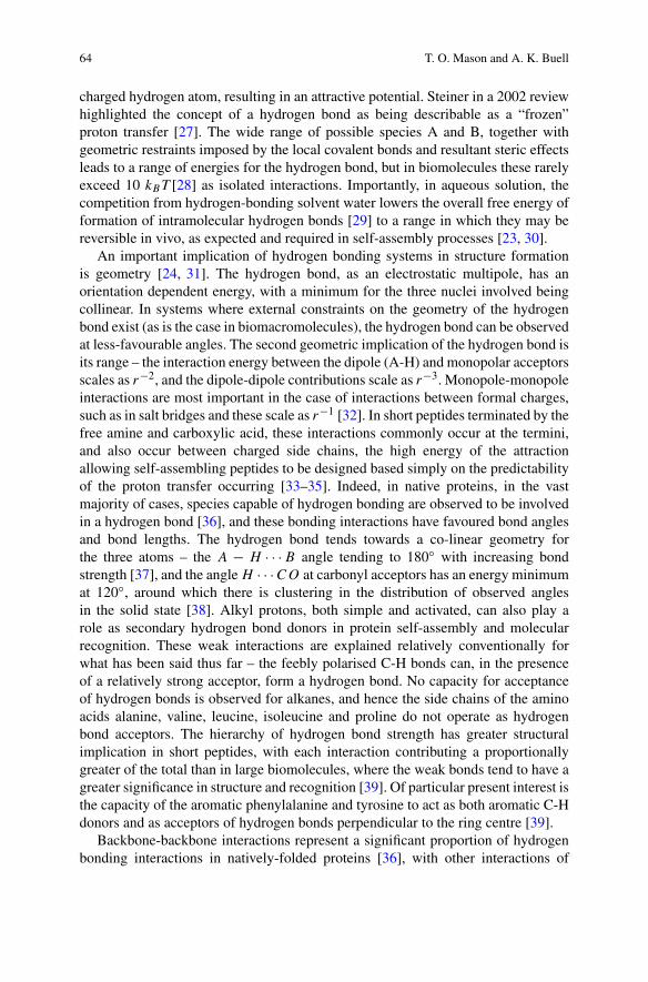
64 T. O. Mason and A. K. Buell
charged hydrogen atom, resulting in an attractive potential. Steiner in a 2002 reviewhighlighted the concept of a hydrogen bond as being describable as a “frozen”proton transfer [27]. The wide range of possible species A and B, together withgeometric restraints imposed by the local covalent bonds and resultant steric effectsleads to a range of energies for the hydrogen bond, but in biomolecules these rarelyexceed 10 kBT [28] as isolated interactions. Importantly, in aqueous solution, thecompetition from hydrogen-bonding solvent water lowers the overall free energy offormation of intramolecular hydrogen bonds [29] to a range in which they may bereversible in vivo, as expected and required in self-assembly processes [23, 30].
An important implication of hydrogen bonding systems in structure formationis geometry [24, 31]. The hydrogen bond, as an electrostatic multipole, has anorientation dependent energy, with a minimum for the three nuclei involved beingcollinear. In systems where external constraints on the geometry of the hydrogenbond exist (as is the case in biomacromolecules), the hydrogen bond can be observedat less-favourable angles. The second geometric implication of the hydrogen bond isits range – the interaction energy between the dipole (A-H) and monopolar acceptorsscales as r−2, and the dipole-dipole contributions scale as r−3. Monopole-monopoleinteractions are most important in the case of interactions between formal charges,such as in salt bridges and these scale as r−1 [32]. In short peptides terminated by thefree amine and carboxylic acid, these interactions commonly occur at the termini,and also occur between charged side chains, the high energy of the attractionallowing self-assembling peptides to be designed based simply on the predictabilityof the proton transfer occurring [33–35]. Indeed, in native proteins, in the vastmajority of cases, species capable of hydrogen bonding are observed to be involvedin a hydrogen bond [36], and these bonding interactions have favoured bond anglesand bond lengths. The hydrogen bond tends towards a co-linear geometry forthe three atoms – the A − H · · · B angle tending to 180◦ with increasing bondstrength [37], and the angle H · · · CO at carbonyl acceptors has an energy minimumat 120◦, around which there is clustering in the distribution of observed anglesin the solid state [38]. Alkyl protons, both simple and activated, can also play arole as secondary hydrogen bond donors in protein self-assembly and molecularrecognition. These weak interactions are explained relatively conventionally forwhat has been said thus far – the feebly polarised C-H bonds can, in the presenceof a relatively strong acceptor, form a hydrogen bond. No capacity for acceptanceof hydrogen bonds is observed for alkanes, and hence the side chains of the aminoacids alanine, valine, leucine, isoleucine and proline do not operate as hydrogenbond acceptors. The hierarchy of hydrogen bond strength has greater structuralimplication in short peptides, with each interaction contributing a proportionallygreater of the total than in large biomolecules, where the weak bonds tend to have agreater significance in structure and recognition [39]. Of particular present interest isthe capacity of the aromatic phenylalanine and tyrosine to act as both aromatic C-Hdonors and as acceptors of hydrogen bonds perpendicular to the ring centre [39].
Backbone-backbone interactions represent a significant proportion of hydrogenbonding interactions in natively-folded proteins [36], with other interactions of
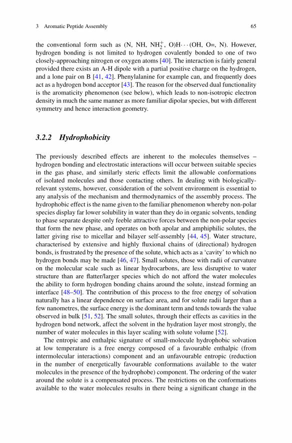
3 Aromatic Peptide Assembly 65
the conventional form such as (N, NH, NH+2 , O)H· · · (OH, O=, N). However,
hydrogen bonding is not limited to hydrogen covalently bonded to one of twoclosely-approaching nitrogen or oxygen atoms [40]. The interaction is fairly generalprovided there exists an A-H dipole with a partial positive charge on the hydrogen,and a lone pair on B [41, 42]. Phenylalanine for example can, and frequently doesact as a hydrogen bond acceptor [43]. The reason for the observed dual functionalityis the aromaticity phenomenon (see below), which leads to non-isotropic electrondensity in much the same manner as more familiar dipolar species, but with differentsymmetry and hence interaction geometry.
3.2.2 Hydrophobicity
The previously described effects are inherent to the molecules themselves –hydrogen bonding and electrostatic interactions will occur between suitable speciesin the gas phase, and similarly steric effects limit the allowable conformationsof isolated molecules and those contacting others. In dealing with biologically-relevant systems, however, consideration of the solvent environment is essential toany analysis of the mechanism and thermodynamics of the assembly process. Thehydrophobic effect is the name given to the familiar phenomenon whereby non-polarspecies display far lower solubility in water than they do in organic solvents, tendingto phase separate despite only feeble attractive forces between the non-polar speciesthat form the new phase, and operates on both apolar and amphiphilic solutes, thelatter giving rise to micellar and bilayer self-assembly [44, 45]. Water structure,characterised by extensive and highly fluxional chains of (directional) hydrogenbonds, is frustrated by the presence of the solute, which acts as a ‘cavity’ to which nohydrogen bonds may be made [46, 47]. Small solutes, those with radii of curvatureon the molecular scale such as linear hydrocarbons, are less disruptive to waterstructure than are flatter/larger species which do not afford the water moleculesthe ability to form hydrogen bonding chains around the solute, instead forming aninterface [48–50]. The contribution of this process to the free energy of solvationnaturally has a linear dependence on surface area, and for solute radii larger than afew nanometres, the surface energy is the dominant term and tends towards the valueobserved in bulk [51, 52]. The small solutes, through their effects as cavities in thehydrogen bond network, affect the solvent in the hydration layer most strongly, thenumber of water molecules in this layer scaling with solute volume [52].
The entropic and enthalpic signature of small-molecule hydrophobic solvationat low temperature is a free energy composed of a favourable enthalpic (fromintermolecular interactions) component and an unfavourable entropic (reductionin the number of energetically favourable conformations available to the watermolecules in the presence of the hydrophobe) component. The ordering of the wateraround the solute is a compensated process. The restrictions on the conformationsavailable to the water molecules results in there being a significant change in the
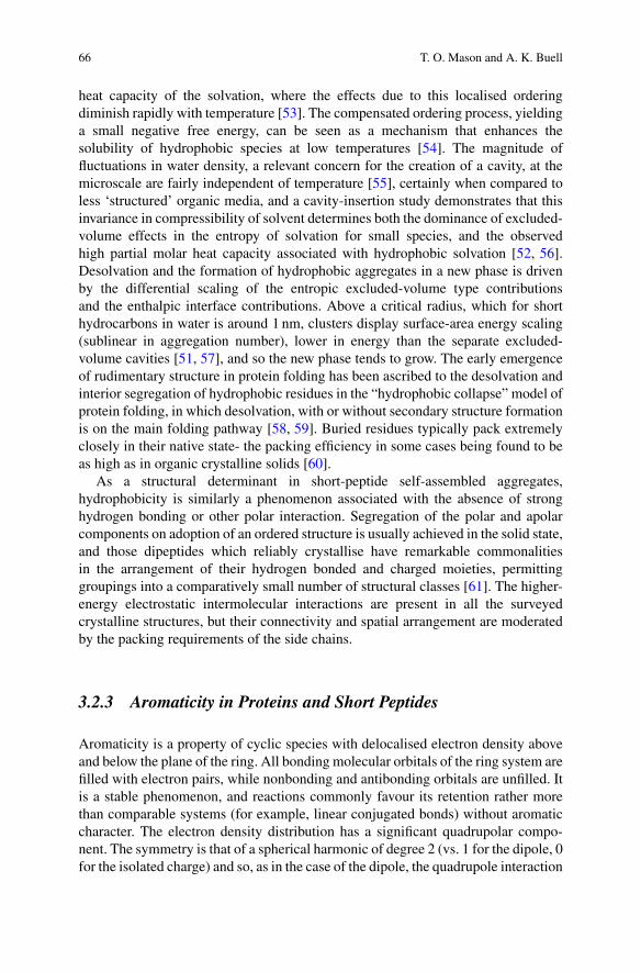
66 T. O. Mason and A. K. Buell
heat capacity of the solvation, where the effects due to this localised orderingdiminish rapidly with temperature [53]. The compensated ordering process, yieldinga small negative free energy, can be seen as a mechanism that enhances thesolubility of hydrophobic species at low temperatures [54]. The magnitude offluctuations in water density, a relevant concern for the creation of a cavity, at themicroscale are fairly independent of temperature [55], certainly when compared toless ‘structured’ organic media, and a cavity-insertion study demonstrates that thisinvariance in compressibility of solvent determines both the dominance of excluded-volume effects in the entropy of solvation for small species, and the observedhigh partial molar heat capacity associated with hydrophobic solvation [52, 56].Desolvation and the formation of hydrophobic aggregates in a new phase is drivenby the differential scaling of the entropic excluded-volume type contributionsand the enthalpic interface contributions. Above a critical radius, which for shorthydrocarbons in water is around 1 nm, clusters display surface-area energy scaling(sublinear in aggregation number), lower in energy than the separate excluded-volume cavities [51, 57], and so the new phase tends to grow. The early emergenceof rudimentary structure in protein folding has been ascribed to the desolvation andinterior segregation of hydrophobic residues in the “hydrophobic collapse” model ofprotein folding, in which desolvation, with or without secondary structure formationis on the main folding pathway [58, 59]. Buried residues typically pack extremelyclosely in their native state- the packing efficiency in some cases being found to beas high as in organic crystalline solids [60].
As a structural determinant in short-peptide self-assembled aggregates,hydrophobicity is similarly a phenomenon associated with the absence of stronghydrogen bonding or other polar interaction. Segregation of the polar and apolarcomponents on adoption of an ordered structure is usually achieved in the solid state,and those dipeptides which reliably crystallise have remarkable commonalitiesin the arrangement of their hydrogen bonded and charged moieties, permittinggroupings into a comparatively small number of structural classes [61]. The higher-energy electrostatic intermolecular interactions are present in all the surveyedcrystalline structures, but their connectivity and spatial arrangement are moderatedby the packing requirements of the side chains.
3.2.3 Aromaticity in Proteins and Short Peptides
Aromaticity is a property of cyclic species with delocalised electron density aboveand below the plane of the ring. All bonding molecular orbitals of the ring system arefilled with electron pairs, while nonbonding and antibonding orbitals are unfilled. Itis a stable phenomenon, and reactions commonly favour its retention rather morethan comparable systems (for example, linear conjugated bonds) without aromaticcharacter. The electron density distribution has a significant quadrupolar compo-nent. The symmetry is that of a spherical harmonic of degree 2 (vs. 1 for the dipole, 0for the isolated charge) and so, as in the case of the dipole, the quadrupole interaction
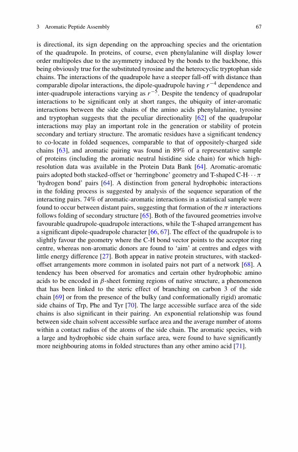
3 Aromatic Peptide Assembly 67
is directional, its sign depending on the approaching species and the orientationof the quadrupole. In proteins, of course, even phenylalanine will display lowerorder multipoles due to the asymmetry induced by the bonds to the backbone, thisbeing obviously true for the substituted tyrosine and the heterocyclic tryptophan sidechains. The interactions of the quadrupole have a steeper fall-off with distance thancomparable dipolar interactions, the dipole-quadrupole having r−4 dependence andinter-quadrupole interactions varying as r−5. Despite the tendency of quadrupolarinteractions to be significant only at short ranges, the ubiquity of inter-aromaticinteractions between the side chains of the amino acids phenylalanine, tyrosineand tryptophan suggests that the peculiar directionality [62] of the quadrupolarinteractions may play an important role in the generation or stability of proteinsecondary and tertiary structure. The aromatic residues have a significant tendencyto co-locate in folded sequences, comparable to that of oppositely-charged sidechains [63], and aromatic pairing was found in 89% of a representative sampleof proteins (including the aromatic neutral histidine side chain) for which high-resolution data was available in the Protein Data Bank [64]. Aromatic-aromaticpairs adopted both stacked-offset or ‘herringbone’ geometry and T-shaped C-H· · · π‘hydrogen bond’ pairs [64]. A distinction from general hydrophobic interactionsin the folding process is suggested by analysis of the sequence separation of theinteracting pairs. 74% of aromatic-aromatic interactions in a statistical sample werefound to occur between distant pairs, suggesting that formation of the π interactionsfollows folding of secondary structure [65]. Both of the favoured geometries involvefavourable quadrupole-quadrupole interactions, while the T-shaped arrangement hasa significant dipole-quadrupole character [66, 67]. The effect of the quadrupole is toslightly favour the geometry where the C-H bond vector points to the acceptor ringcentre, whereas non-aromatic donors are found to ‘aim’ at centres and edges withlittle energy difference [27]. Both appear in native protein structures, with stacked-offset arrangements more common in isolated pairs not part of a network [68]. Atendency has been observed for aromatics and certain other hydrophobic aminoacids to be encoded in β-sheet forming regions of native structure, a phenomenonthat has been linked to the steric effect of branching on carbon 3 of the sidechain [69] or from the presence of the bulky (and conformationally rigid) aromaticside chains of Trp, Phe and Tyr [70]. The large accessible surface area of the sidechains is also significant in their pairing. An exponential relationship was foundbetween side chain solvent accessible surface area and the average number of atomswithin a contact radius of the atoms of the side chain. The aromatic species, witha large and hydrophobic side chain surface area, were found to have significantlymore neighbouring atoms in folded structures than any other amino acid [71].
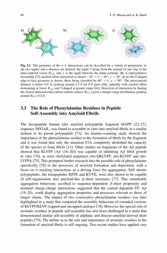
68 T. O. Mason and A. K. Buell
Fig. 3.1 The geometry of the π -π interactions can be described by a variety of parameters, in(a) two angles and a distance are defined, the angle θ being from the normal of one ring to theinter-centroid vector Rcen, and γ is the angle between the plane normals. (b) A representativefavourable [72] stacked-offset interaction is shown – 20◦ < θ < 50◦, γ < 30◦. In (c) the T-shapededge-to-face geometry is shown, these being classified by 60◦ < θ , γ < 90◦. The intercentroiddistance is below 6.5 Å, peaking around 5.3 Å for F-F pairs [68], naturally with stacked offsetdominating at lower Rcen and T-shaped at greater range [64]. Detection of interaction by findingthe closest intermolecular carbon-carbon contact (RCC ) gives a sharper range distribution, peakingaround RCC=3.8 Å
3.3 The Role of Phenylalanine Residues in PeptideSelf-Assembly into Amyloid Fibrils
The hexapeptide human islet amyloid polypeptide fragment hIAPP (22–27),sequence NFGAIL, was found to assemble in vitro into amyloid fibrils in a similarfashion to its parent polypeptide [73]. An alanine-scanning study showed theimportance of the phenylalanine residue to the formation of fibrils by the fragmentand it was found that only the mutation F2A completely abolished the capacityof the species to form fibrils [11]. Other studies on fragments of the Aβ peptideshowed that KLVFF (Aβ (16–20)) was capable of inhibiting Aβ fibril growthin vitro [74], as were cholylated sequences cho-QKLVFF, cho-KLVFF and cho-LVFFA [75]. This prompted further research into the possible role of phenylalaninespecifically [76] in the processes of amyloid formation and deposition, with afocus on π -stacking interactions as a driving force for aggregation. Still shorterpolypeptides, the tetrapeptides KFFE and KVVE, were also shown to be capableof self-organisation into amyloid-like β-sheet structures [77]. This remarkableaggregation behaviour, ascribed to sequence-dependent β-sheet propensity andterminal charge-charge interactions suggested that the central dipeptide FF, Aβ
(19–20), could display aggregation properties and processes relevant to those oflonger chains. The impact of two consecutive phenylalanine residues was laterhighlighted in a study that compared the assembly behaviour of extended versionsof KLVFF/KLVF (capped and uncapped analogs) [78]. However, the special role ofaromatic residues in peptide self-assembly has also been challenged in a study thatdemonstrated similar self-assembly of aliphatic and disease-amyloid derived shortpeptides [79]. The debate as to the role and importance of aromatic residues in theformation of amyloid fibrils is still ongoing. Two recent studies have applied very
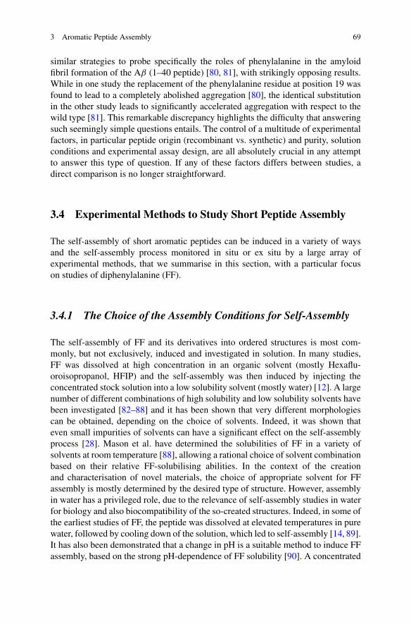
3 Aromatic Peptide Assembly 69
similar strategies to probe specifically the roles of phenylalanine in the amyloidfibril formation of the Aβ (1–40 peptide) [80, 81], with strikingly opposing results.While in one study the replacement of the phenylalanine residue at position 19 wasfound to lead to a completely abolished aggregation [80], the identical substitutionin the other study leads to significantly accelerated aggregation with respect to thewild type [81]. This remarkable discrepancy highlights the difficulty that answeringsuch seemingly simple questions entails. The control of a multitude of experimentalfactors, in particular peptide origin (recombinant vs. synthetic) and purity, solutionconditions and experimental assay design, are all absolutely crucial in any attemptto answer this type of question. If any of these factors differs between studies, adirect comparison is no longer straightforward.
3.4 Experimental Methods to Study Short Peptide Assembly
The self-assembly of short aromatic peptides can be induced in a variety of waysand the self-assembly process monitored in situ or ex situ by a large array ofexperimental methods, that we summarise in this section, with a particular focuson studies of diphenylalanine (FF).
3.4.1 The Choice of the Assembly Conditions for Self-Assembly
The self-assembly of FF and its derivatives into ordered structures is most com-monly, but not exclusively, induced and investigated in solution. In many studies,FF was dissolved at high concentration in an organic solvent (mostly Hexaflu-oroisopropanol, HFIP) and the self-assembly was then induced by injecting theconcentrated stock solution into a low solubility solvent (mostly water) [12]. A largenumber of different combinations of high solubility and low solubility solvents havebeen investigated [82–88] and it has been shown that very different morphologiescan be obtained, depending on the choice of solvents. Indeed, it was shown thateven small impurities of solvents can have a significant effect on the self-assemblyprocess [28]. Mason et al. have determined the solubilities of FF in a variety ofsolvents at room temperature [88], allowing a rational choice of solvent combinationbased on their relative FF-solubilising abilities. In the context of the creationand characterisation of novel materials, the choice of appropriate solvent for FFassembly is mostly determined by the desired type of structure. However, assemblyin water has a privileged role, due to the relevance of self-assembly studies in waterfor biology and also biocompatibility of the so-created structures. Indeed, in some ofthe earliest studies of FF, the peptide was dissolved at elevated temperatures in purewater, followed by cooling down of the solution, which led to self-assembly [14, 89].It has also been demonstrated that a change in pH is a suitable method to induce FFassembly, based on the strong pH-dependence of FF solubility [90]. A concentrated
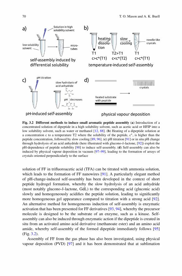
70 T. O. Mason and A. K. Buell
Fig. 3.2 Different methods to induce small aromatic peptide assembly (a) Introduction of aconcentrated solution of dipeptide in a high solubility solvent, such as acetic acid or HFIP into alow solubility solvent, such as water or methanol [12, 88]. (b) Heating of a dipeptide solution ata concentration c to a temperature T2 where the solubility of the peptide, c∗, is higher than thepeptide concentration, followed by slow cooling [89, 96]. (c) pH titration [91] or in situ pH changethrough hydrolysis of an acid anhydride (here illustrated with glucono-δ-lactone, [92]) exploit thepH-dependence of peptide solubility [90] to induce self-assembly. (d) Self-assembly can also beinduced by physical vapour deposition in vacuum [97–99], leading to the formation of arrays ofcrystals oriented perpendicularly to the surface
solution of FF in trifluoroacetic acid (TFA) can be titrated with ammonia solution,which leads to the formation of FF nanowires [91]. A particularly elegant methodof pH-change-induced self-assembly has been developed in the context of shortpeptide hydrogel formation, whereby the slow hydrolysis of an acid anhydride(most notably glucono-δ-lactone, GdL) to the corresponding acid (gluconic acid)slowly and homogeneously acidifies the peptide solution, leading to significantlymore homogeneous gel appearance compared to titration with a strong acid [92].An alternative method for homogeneous induction of self-assembly is enzymaticactivation that has been presented for FF derivatives [93, 94], whereby the precursormolecule is designed to be the substrate of an enzyme, such as a kinase. Self-assembly can also be induced through enzymatic action if the dipeptide is created insitu from an activated amino acid derivative (methanoate ester) and an amino acidamide, whereby self-assembly of the formed dipeptide immediately follows [95](Fig. 3.2).
Assembly of FF from the gas phase has also been investigated, using physicalvapour deposition (PVD) [97] and it has been demonstrated that at sublimation
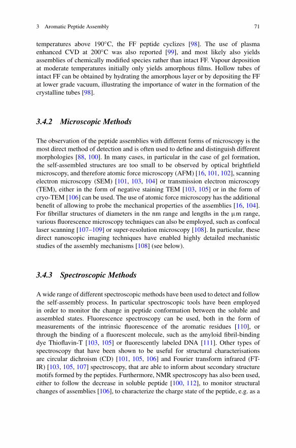
3 Aromatic Peptide Assembly 71
temperatures above 190◦C, the FF peptide cyclizes [98]. The use of plasmaenhanced CVD at 200◦C was also reported [99], and most likely also yieldsassemblies of chemically modified species rather than intact FF. Vapour depositionat moderate temperatures initially only yields amorphous films. Hollow tubes ofintact FF can be obtained by hydrating the amorphous layer or by depositing the FFat lower grade vacuum, illustrating the importance of water in the formation of thecrystalline tubes [98].
3.4.2 Microscopic Methods
The observation of the peptide assemblies with different forms of microscopy is themost direct method of detection and is often used to define and distinguish differentmorphologies [88, 100]. In many cases, in particular in the case of gel formation,the self-assembled structures are too small to be observed by optical brightfieldmicroscopy, and therefore atomic force microscopy (AFM) [16, 101, 102], scanningelectron microscopy (SEM) [101, 103, 104] or transmission electron microscopy(TEM), either in the form of negative staining TEM [103, 105] or in the form ofcryo-TEM [106] can be used. The use of atomic force microscopy has the additionalbenefit of allowing to probe the mechanical properties of the assemblies [16, 104].For fibrillar structures of diameters in the nm range and lengths in the μm range,various fluorescence microscopy techniques can also be employed, such as confocallaser scanning [107–109] or super-resolution microscopy [108]. In particular, thesedirect nanoscopic imaging techniques have enabled highly detailed mechanisticstudies of the assembly mechanisms [108] (see below).
3.4.3 Spectroscopic Methods
A wide range of different spectroscopic methods have been used to detect and followthe self-assembly process. In particular spectroscopic tools have been employedin order to monitor the change in peptide conformation between the soluble andassembled states. Fluorescence spectroscopy can be used, both in the form ofmeasurements of the intrinsic fluorescence of the aromatic residues [110], orthrough the binding of a fluorescent molecule, such as the amyloid fibril-bindingdye Thioflavin-T [103, 105] or fluorescently labeled DNA [111]. Other types ofspectroscopy that have been shown to be useful for structural characterisationsare circular dichroism (CD) [101, 105, 106] and Fourier transform infrared (FT-IR) [103, 105, 107] spectroscopy, that are able to inform about secondary structuremotifs formed by the peptides. Furthermore, NMR spectroscopy has also been used,either to follow the decrease in soluble peptide [100, 112], to monitor structuralchanges of assemblies [106], to characterize the charge state of the peptide, e.g. as a
72 T. O. Mason and A. K. Buell
function of concentration [78], or to study the interactions between assemblies andother species, such as metal ions [113].
Despite not being a spectroscopic method, we include here also mass spectrom-etry, in the form of ion mobility mass spectrometry, as it has been used in order todetect the presence of clusters of aromatic dipeptides in solution [114].
3.4.4 Scattering, Rheological, Calorimetric andConductivity-Based Methods
The often dramatic change in size upon self-assembly makes it possible to follow theself-assembly process by a variety of scattering methods. Dynamic light scattering(DLS) [90, 100], small angle X-ray scattering (SAXS) [115], as well as smallangle neutron scattering (SANS) [107, 116, 117] have been employed to followthe assembly process. In the case of small angle scattering (SAXS and SANS), thedata can sometimes be fitted to a model corresponding to the emergence of rod-likemicelles/fibrils upon the induction of self-assembly [107].
The self-assembly is often associated with a considerable change in mechanicalproperties, in particular in the case of gel-formation. This can be followed by themeasurement of the rheological properties, in particular the ratio between elastic anddissipative behaviour [94, 105, 107, 117]. In the case of orthogonally assembling,self-sorting multi-component gels, the rheological properties provide a measureof the assembly state of the individual components and can be selectively tunedthrough external stimuli [118].
In some cases, the self-assembly reaction can also be directly carried out insidea calorimeter, with the aim to study the thermodynamic signatures of the assemblyreaction. In the case of Boc-FF, the peptide initially forms amorphous sphericalstructures when a concentrated solution in ethanol is diluted into water (see below).These structures can be metastable against transformation into crystals for sufficientamounts of time such that the heat exchange linked to this conversion can bemeasured with a differential scanning calorimeter [100].
Inspired by classical methods for the determination of the critical micelleconcentrations of surfactants, it is also possible to follow different levels of peptideself-assembly by measuring the concentration-dependence of the conductivity ofthe peptide solution [107]. This type of method is based on the fact that theelectrophoretic mobility of assemblies can differ significantly from those of theirbuilding blocks.
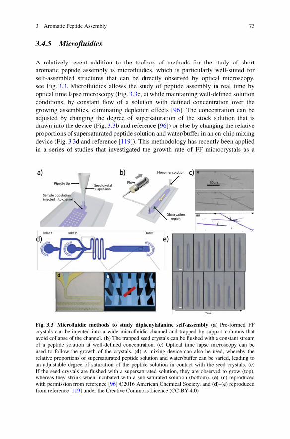
3 Aromatic Peptide Assembly 73
3.4.5 Microfluidics
A relatively recent addition to the toolbox of methods for the study of shortaromatic peptide assembly is microfluidics, which is particularly well-suited forself-assembled structures that can be directly observed by optical microscopy,see Fig. 3.3. Microfluidics allows the study of peptide assembly in real time byoptical time lapse microscopy (Fig. 3.3c, e) while maintaining well-defined solutionconditions, by constant flow of a solution with defined concentration over thegrowing assemblies, eliminating depletion effects [96]. The concentration can beadjusted by changing the degree of supersaturation of the stock solution that isdrawn into the device (Fig. 3.3b and reference [96]) or else by changing the relativeproportions of supersaturated peptide solution and water/buffer in an on-chip mixingdevice (Fig. 3.3d and reference [119]). This methodology has recently been appliedin a series of studies that investigated the growth rate of FF microcrystals as a
Fig. 3.3 Microfluidic methods to study diphenylalanine self-assembly (a) Pre-formed FFcrystals can be injected into a wide microfluidic channel and trapped by support columns thatavoid collapse of the channel. (b) The trapped seed crystals can be flushed with a constant streamof a peptide solution at well-defined concentration. (c) Optical time lapse microscopy can beused to follow the growth of the crystals. (d) A mixing device can also be used, whereby therelative proportions of supersaturated peptide solution and water/buffer can be varied, leading toan adjustable degree of saturation of the peptide solution in contact with the seed crystals. (e)If the seed crystals are flushed with a supersaturated solution, they are observed to grow (top),whereas they shrink when incubated with a sub-saturated solution (bottom). (a)–(c) reproducedwith permission from reference [96] ©2016 American Chemical Society, and (d)–(e) reproducedfrom reference [119] under the Creative Commons Licence (CC-BY-4.0)
74 T. O. Mason and A. K. Buell
function of concentration [96, 119] and temperature [90]. This approach allowsextensive and accurate measurements to be made and the resulting data hascontributed significantly to a better understanding of assembly mechanism of FF(see below).
3.5 Thermodynamic Stability of Peptide Assemblies
The thermodynamics of short aromatic peptide assembly depends crucially on theexact sequence of the peptide, including capping and protection groups, as well asthe solution conditions, i.e. temperature, pH and salt concentration. In the following,we will discuss the thermodynamic stability of crystalline and fibrillar structures andhow they are influenced by various factors.
3.5.1 Thermal Stability of FF Crystals
It has been reported that FF microcrystals are very stable against heating inthe dry state [101]. In the dry state, these structures can withstand temperaturesabove 100◦C and thermogravimetric analysis reveals the loss of water from thecrystal structure [101]. However, at temperatures above 150◦C, it was shownthat chemical decomposition sets in that can lead to the loss of phenylalaninefragments [102]. Furthermore, the FF peptide can cyclize under loss of waterat such high temperatures, which leads to an irreversible collapse of the tubularcrystals [120, 121]. Full thermal decomposition of the peptide film finally occurs attemperatures above 300◦C [101, 122]. On the other hand, amorphous films of FF canbe converted into fibrillar morphology through dry heating at 150◦C [122], possiblythrough rearrangement or, more likely, sublimation of chemically degraded/cyclizedFF, similar to the observed formation of deposits on AFM cantilever observedin an earlier study [102]. This is corroborated by the finding that pre-cyclized,phenylalanine-containing dipeptides can form fibrils and gels [123].
High thermal stability of FF assemblies above 100◦C (in an autoclave) has alsobeen reported in aqueous solution [101]. Only recently, the solubility of FF inwater has been systematically studied as a function of temperature, by measuringthe FF concentration in the supernatant in equilibrium with FF crystals [90]. Itwas found that the solubility of FF dramatically increases with temperature, fromabout 0.5 g/L (1.6 mM) at 10◦C to about 1.9 g/L (6 mM) at 70◦C. Interestingly,phenylalanine shows a similar temperature dependence of its solubility, the absolutesolubility being much higher [124], whereas benzene shows a very weak tem-perature dependence of its solubility in water [125]. It is interesting to note thatbenzene, being a prototypical hydrophobic molecule, has a higher solubility in waterthan the diphenylalanine molecule. The temperature-dependent solubilities of F,FF and benzene are compared in Fig. 3.4. In the light of the reported temperature
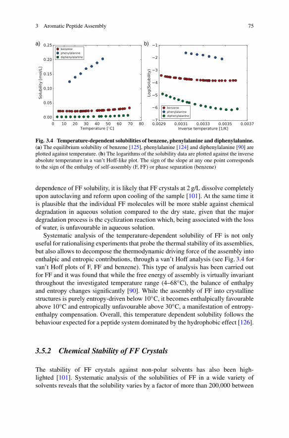
3 Aromatic Peptide Assembly 75
a) b)
Fig. 3.4 Temperature-dependent solubilities of benzene, phenylalanine and diphenylalanine.(a) The equilibrium solubility of benzene [125], phenylalanine [124] and diphenylalanine [90] areplotted against temperature. (b) The logarithms of the solubility data are plotted against the inverseabsolute temperature in a van’t Hoff-like plot. The sign of the slope at any one point correspondsto the sign of the enthalpy of self-assembly (F, FF) or phase separation (benzene)
dependence of FF solubility, it is likely that FF crystals at 2 g/L dissolve completelyupon autoclaving and reform upon cooling of the sample [101]. At the same time itis plausible that the individual FF molecules will be more stable against chemicaldegradation in aqueous solution compared to the dry state, given that the majordegradation process is the cyclization reaction which, being associated with the lossof water, is unfavourable in aqueous solution.
Systematic analysis of the temperature-dependent solubility of FF is not onlyuseful for rationalising experiments that probe the thermal stability of its assemblies,but also allows to decompose the thermodynamic driving force of the assembly intoenthalpic and entropic contributions, through a van’t Hoff analysis (see Fig. 3.4 forvan’t Hoff plots of F, FF and benzene). This type of analysis has been carried outfor FF and it was found that while the free energy of assembly is virtually invariantthroughout the investigated temperature range (4–68◦C), the balance of enthalpyand entropy changes significantly [90]. While the assembly of FF into crystallinestructures is purely entropy-driven below 10◦C, it becomes enthalpically favourableabove 10◦C and entropically unfavourable above 30◦C, a manifestation of entropy-enthalpy compensation. Overall, this temperature dependent solubility follows thebehaviour expected for a peptide system dominated by the hydrophobic effect [126].
3.5.2 Chemical Stability of FF Crystals
The stability of FF crystals against non-polar solvents has also been high-lighted [101]. Systematic analysis of the solubilities of FF in a wide variety ofsolvents reveals that the solubility varies by a factor of more than 200,000 between
76 T. O. Mason and A. K. Buell
the lowest solubility solvent (hexane, 0.002 g/L) and the highest solubility solvent(acetic acid, 430 g/L) investigated [88]. It was also shown that the thermodynamicstability of FF crystals is strongly pH-dependent, with the solubility minimumcoinciding with the pH range where FF occurs as a globally neutral zwitterion [90],and the solubility at pH 1 being approximately ten times higher than at the solubilityminimum [127]. Interestingly, addition of NaCl only marginally changed thesolubility of FF in pure water; only at NaCl concentrations of 1 M and above,the solubility decreases appreciably [90], suggesting that electrostatic screeningeffects do not play important roles in the assembly thermodynamics. The effect ofhigh NaCl concentrations is likely related to the well-known phenomenon of saltingout, that can for example be observed for aqueous solutions of benzene [128].
3.5.3 Non-crystalline Short Aromatic Peptide Assemblies
3.5.3.1 Fibrils and Gels
Structurally, one of the most interesting features of short aromatic peptides is theirability to form fibrils and gels in addition to crystalline structures. In this context,fibrils are defined as highly anisotropic structures with two dimensions of the orderof (a few times) the molecular size and one dimension that can be of the order ofμm, and which do not show long range crystalline order. Fibril/crystal dimorphismis of course not restricted to (aromatic) dipeptides, but is also observed for longersequences, in particular those derived from amyloid fibrils [129, 130]. Whileuncapped zwitterionic dipeptides easily assemble into crystalline structures, cappedversions display a clear preference for assembly into fibrils, even though they canbe crystallised under some conditions [93, 131]. In particular large, bulky aromaticcapping groups, such as fluorenylmethyloxycarbonyl (Fmoc) [132], carboxybenzyl(Cbz) and naphtalene, direct the FF peptide into fibrillar structures [133–135],whereas FF with smaller capping groups (Boc, acetate, amide) is still able to formcrystals [100, 133]. The fibril formation and subsequent gelation of N-terminallycapped aromatic dipeptides has been studied particularly extensively [20, 116, 134–136]. However, whether a gel or a crystal is formed is not exclusively determined bychemical modifications of the FF peptide, but also strongly depends on the solventconditions. In the case of uncapped FF, polar, hydrogen-bonding solvents are foundto favour the formation of crystals [14, 88] and non-polar solvents favouring theformation of gels [82, 85, 86]. A theoretical thermodynamic framework to decidewhether the gel or the crystal is the equilibrium structure has been developed andis based on the geometry of the self-assembling molecule and its solvophilic orsolvophobic properties [137]. It is predicted by this model that the gel, usuallyconsidered a metastable phase compared to the crystal, can in fact correspond to thethermodynamic minimum, depending on the solution conditions. In cases, however,where a system under the same conditions can form both a gel and a crystal, it isusually found that the crystalline state appears after the gel state and corresponds to
3 Aromatic Peptide Assembly 77
the final thermodynamic minimum [100, 106, 135], in agreement with Ostwald’srule of stages [100]. Structurally, gel and crystal are often distinct [100, 135],suggesting that they do not interconvert directly, but that the thermodynamicallymore stable crystal phase nucleates either independently or catalysed by the gel(heterogeneous nucleation).
The most prominent example of an N-terminally capped FF peptide that formsfibrillar gels in aqueous solution is the Fmoc-FF system, which has been charac-terised in detail [20]. It was proposed that the fibrils are stabilised through aromaticstacking interactions, a design principle that has been successfully mimicked withmolecules containing both a phenyl and fluorophenyl group [138]. Notwithstandingthe fact that Fmoc-FF displays very robust fibril formation, it was shown that peptoidsubstitutions (displacement of the phenyl ring from the side chain to the amidenitrogen) of one or both of the phenylalanine residues impede (if one F is changed)or completely abolish (if both F are changed) gel formation [139].
Gel formation in N-terminally capped dipeptides is usually induced by dissolvingthe peptide at elevated pH and then lowering the pH, either through titration withacid or in situ through the hydrolysis of an anhydride (Fig. 3.2 and references [92]and [131]). Gel formation occurs as soon as the pKa value of the C-terminalcarboxy-group is reached, suggesting that it is mainly the globally neutral speciesthat is incorporated into the fibrils. However, through pioneering and detailed pH-titration studies, it could be shown that the initial fibrillar assemblies are not globallyneutral, but consist of molecules with an average degree of ionisation of 0.66 [136].Upon further acidification, subsequent transitions into higher order structures occurthat accompany increasing neutralisation of the peptides. Through such titrationstudies [131, 136], as well as solution state NMR experiments [78] it became clearthat the process of self-assembly actually modulates the pKa of the peptide, withthe pKa of the assembled structures being significantly higher than that of theisolated peptide, whereby the difference can correspond to up to 5 pH units [136].The protonation state of the peptides is also strongly dependent on the total peptideconcentration [78]. If a pH titration is performed on such a peptide system, the self-assembly sets in as soon as appropriate pH values are reached, but the process ofself-assembly is slow on the time scale of the titration experiments and thereforethe system will not be at thermodynamic equilibrium during the titration. Indeed,the titration of self-assembling peptide systems is an ingenious way to access thethermodynamics of gel-forming systems that are otherwise difficult to study. Thismethod was applied to characterize the thermodynamics of fibril and gel formationof the FFDD tetrapeptide and its sequence permutations [110]. Free energies ofself-assembly were determined from titration curves under the assumption thatassemblies only form from neutral species, an assumption, however, which mightnot be fully valid [113, 115, 136]. Nevertheless, from a comparison of the titrationbehaviour of a series of closely related peptides, a relative ordering of the drivingforces for assembly can be deduced [110]. This type of data would otherwisebe difficult to obtain, given the high stability of these fibrils and the resultinglow equilibrium concentration of soluble peptide that are challenging to measuredirectly. In the same study [110], one of the very few quantitative physico-chemical
78 T. O. Mason and A. K. Buell
investigations of aromatic peptide fibril formation published to-date, the kinetics ofassembly was followed by NMR and fluorescence spectroscopy, as well as smallangle neutron scattering and the important conclusion was drawn that the kineticsand the thermodynamics of the assembly of the six homologous peptides are notcorrelated [110].
The thermodynamic stability of different gel-like N-terminally naphthoxyaetyl(NAP)-protected dipeptides was also evaluated qualitatively in the context of astudy that aimed at the creation of a self-assembling system that displays dynamicinstability [95]. The motivation behind this fascinating type of study is to artificiallycreate a system that displays properties similar to biological polymers, in particulartubulin [140]. The competition between enzymatic dipeptide formation, peptideself-assembly and enzymatic peptide hydrolysis was investigated and it was foundthat only in the case of the YF peptide, a gel persisted at equilibrium, due to thehigh thermodynamic stability of the assemblies of this peptide, whereas YY and YLdissolved completely [95].
In addition to pH-drop induced fibril- and gel-formation, it was also shownthat N-terminally naphtalene (as well as Fmoc)-capped peptides can be gelledat high pH by adding salt ions that cross-link the rod-like structures, present atelevated pH (>10) [115]. It was proposed that at high pH, it is not fibrils thatform, but rather worm-like micelles with the deprotonated carboxylic acid groupspointing towards the water. These micelles can then be cross-linked by the additionof metal ions (such as Ca2+). The direct interactions of calcium ions with suchpeptide micelles is confirmed with solution state NMR spectroscopy [113]. Theconcentration-dependent formation of such micelles at pH 10.5 and different peptideconcentrations has been characterized in detail, using a variety of experimentaltechniques [107]. In this context it should be noted that the distinction betweenfibrils and elongated micelles is not simply a matter of semantics; indeed it hasrecently been shown that while peptide fibrils display a formation mechanismcharacterised by nucleated growth, micelles (of a different self-assembling systembased on lipid-like molecules) form without an appreciable nucleation step and doalso not seed further growth [108]. In has been demonstrated that it is possibleto trigger the conversion of a micellar aromatic peptide system into a fibrillarsystem through an external stimulus, such as the dephosphorylation of a tyrosineresidue in the peptide [141]. The removal of the phosphate group renders thepeptide derivative less amphiphilic and hence the micelles less stable. Detailedspectroscopic characterisation of this system suggests that the micelles are notdirectly converted into fibrils, but that they rather dissolve first, followed by a rapidde novo nucleation of fibrils [141].
While most of the hydrogel-forming peptides and peptide derivatives so farreported have been found serendipitously, it has also been shown that coarse grainedsimulations allow to explore the sequence space, at least of tripeptides [142].In this way, several previously unknown gel-forming (in the absence of organicsolvent) tripeptides were predicted and experimentally verified (KYF, KYY, KFFand KYW). The important role of aromatic residues is obvious from this list.
3 Aromatic Peptide Assembly 79
3.5.3.2 Amorphous Materials
In addition to the formation of ordered crystalline and fibrillar structures, shortaromatic peptides and peptide derivatives can also form disordered structures. Whenscreening a range of aromatic dipeptides, such as FW, WY, WF and WW, Rechesand Gazit noticed that only FW was capable of ordered crystalline assembly, inagreement with a later large scale survey of published crystal structures [61], eventhough the crystals were mixed with amorphous assemblies [12]. Interestingly, it hasbeen found that FF itself at highly acidic pH values, where its solubility is very high(see above), also does not form crystalline, but rather dendritic assemblies that arelikely to be disordered at the molecular level [127]. Both the high solubility and theinability to form crystalline assemblies is likely to be linked to the net charge carriedby the peptide at this pH, which would need to be neutralised by the incorporationof a counter ion into the crystal. Intriguingly, the individual phenylalanine moleculecan be crystallised from acidic solution (dilute formic acid) by incorporating twochemically distinct F molecules into the asymmetric unit cell: a neutral F zwitterionand a positively charged F ion associated with a formate molecule [143].
The formation of other types of regularly shaped assemblies that are molecularlydisordered can be observed for a range of N-terminally capped FF derivatives(Boc-FF and Azobenzene-FF (PPA)). The introduction of an ethanol solution ofBoc-FF into water leads to the immediate formation of rather uniform sphericalaggregates (Fig. 3.5 and references [104] and [100]), probably as a result of a de-mixing process. The dependence of the formation of these spherical aggregateson the ethanol and peptide content of the solution was studied systematically(Fig. 3.5c). It was shown that the spheres are amorphous in structure (Fig. 3.5d)and represent metastable assemblies that evolve further into fibrils and finallyinto crystals (Fig. 3.5e, f and reference [100]). The transformation of the spherescan be monitored directly through optical microscopy, dynamic light scattering,NMR spectroscopy, and differential scanning calorimetry (DSC) [100] and theensemble of observations suggests that the spheres are dynamic structures in rapidexchange with monomeric peptide that dissolve once the thermodynamically morestable fibrillar phase has formed, which then grows at the expense of the spheres(Fig. 3.5e). Thermodynamic analysis shows that the system evolves down a gradientin free energy (Fig. 3.5f), as expected for a spontaneous process and that the enthalpychange associated with the dissolution of spheres and simultaneous formationof the gel or crystals shows little temperature dependence [100]. How the netnegative charge of the Boc-FF is accommodated in the final crystal structure isat present unknown. A related system is the N-terminally-azobenzene-capped FF(PPA), which forms amorphous spherical structures upon evaporation from an HFIPsolution [144]. Interestingly, the layer of spherical structures converts into fibrilsupon incubation with water, as was studied by AFM [144]. It is unclear whetherthe spheres assemble directly into fibrils or whether the latter nucleate de novoand grow at the expense of the dynamically dissolving spheres, as in the case ofBoc-FF [100]. The observation that upon contact with water the spheres becomesignificantly smaller [144] suggests the latter possibility as the likely mechanism.
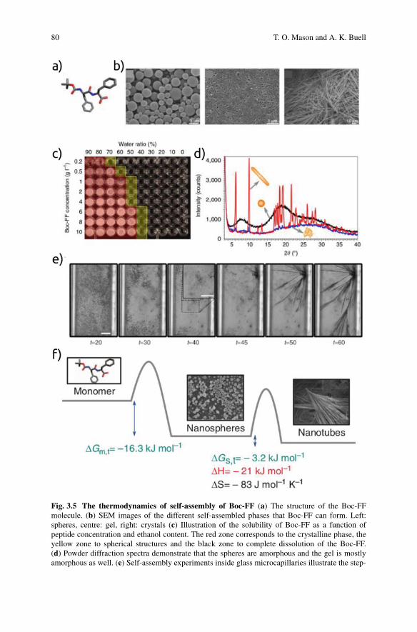
80 T. O. Mason and A. K. Buell
Fig. 3.5 The thermodynamics of self-assembly of Boc-FF (a) The structure of the Boc-FFmolecule. (b) SEM images of the different self-assembled phases that Boc-FF can form. Left:spheres, centre: gel, right: crystals (c) Illustration of the solubility of Boc-FF as a function ofpeptide concentration and ethanol content. The red zone corresponds to the crystalline phase, theyellow zone to spherical structures and the black zone to complete dissolution of the Boc-FF.(d) Powder diffraction spectra demonstrate that the spheres are amorphous and the gel is mostlyamorphous as well. (e) Self-assembly experiments inside glass microcapillaries illustrate the step-
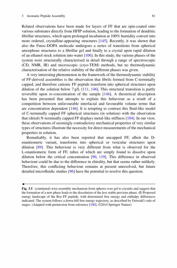
3 Aromatic Peptide Assembly 81
Related observations have been made for layers of FF that are spin-coated ontovarious substrates directly from HFIP solution, leading to the formation of dendriticfibrillar structures, which upon prolonged incubation at 100% humidity convert intomore ordered, crystalline-appearing structures [145]. Recently, it was shown thatalso the Fmoc-DOPA molecule undergoes a series of transitions from sphericalamorphous structures to a fibrillar gel and finally to a crystal upon rapid dilutionof an ethanol stock solution into water [106]. In this study, the various phases of thesystem were structurally characterised in detail through a range of spectroscopic(CD, NMR, IR) and microscopic (cryo-TEM) methods, but no thermodynamiccharacterisation of the relative stability of the different phases was given.
A very interesting phenomenon in the framework of the thermodynamic stabilityof FF-derived assemblies is the observation that fibrils formed from C-terminallycapped, and therefore cationic FF peptide transform into spherical structures upondilution of the solution below 7 g/L [111, 146]. This structural transition is partlyreversible upon re-concentration of the sample [146]. A theoretical descriptionhas been presented that attempts to explain this behaviour as a result of acompetition between unfavourable interfacial and favourable volume terms thatare concentration dependent [146]. It is tempting to contrast this fluid-like modelof C-terminally capped FF spherical structures (in solution) with the observationthat (dried) N-terminally capped FF displays metal-like stiffness [104]. In our view,these observations of seemingly contradictory mechanical properties of very similartypes of structures illustrate the necessity for direct measurements of the mechanicalproperties in solution.
Remarkably, it has also been reported that uncapped FF, albeit the D-enantiomeric variant, transforms into spherical or vesicular structures upondilution [89]. This behaviour is very different from what is observed for theL-enantiomeric form of FF, tubes of which are simply found to dissolve upondilution below the critical concentration [90, 119]. This difference in observedbehaviour could be due to the difference in chirality, but that seems rather unlikely.Therefore, this conflicting behaviour remains at present unresolved, but futuredetailed microfluidic studies [96] have the potential to resolve this question.
�Fig. 3.5 (continued) wise assembly mechanism from spheres over gel to crystals and suggest thatthe formation of a new phase leads to the dissolution of the less stable previous phase. (f) Proposedenergy landscape of the Boc-FF peptide, with determined free energy and enthalpy differencesindicated. The system follows a down-hill free energy trajectory, as described by Ostwald’s rule ofstages. (Adapted with permission from reference [100], ©2014 Springer Nature)
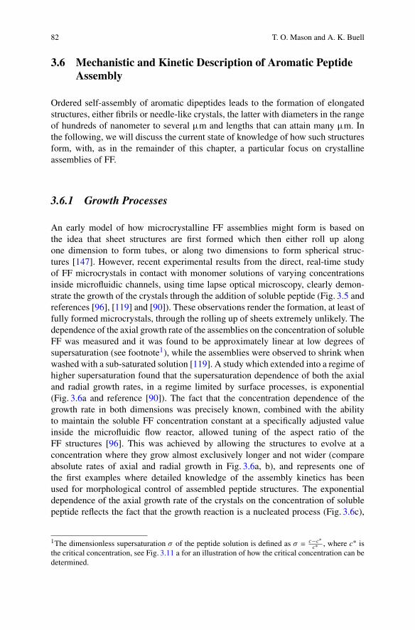
82 T. O. Mason and A. K. Buell
3.6 Mechanistic and Kinetic Description of Aromatic PeptideAssembly
Ordered self-assembly of aromatic dipeptides leads to the formation of elongatedstructures, either fibrils or needle-like crystals, the latter with diameters in the rangeof hundreds of nanometer to several μm and lengths that can attain many μm. Inthe following, we will discuss the current state of knowledge of how such structuresform, with, as in the remainder of this chapter, a particular focus on crystallineassemblies of FF.
3.6.1 Growth Processes
An early model of how microcrystalline FF assemblies might form is based onthe idea that sheet structures are first formed which then either roll up alongone dimension to form tubes, or along two dimensions to form spherical struc-tures [147]. However, recent experimental results from the direct, real-time studyof FF microcrystals in contact with monomer solutions of varying concentrationsinside microfluidic channels, using time lapse optical microscopy, clearly demon-strate the growth of the crystals through the addition of soluble peptide (Fig. 3.5 andreferences [96], [119] and [90]). These observations render the formation, at least offully formed microcrystals, through the rolling up of sheets extremely unlikely. Thedependence of the axial growth rate of the assemblies on the concentration of solubleFF was measured and it was found to be approximately linear at low degrees ofsupersaturation (see footnote1), while the assemblies were observed to shrink whenwashed with a sub-saturated solution [119]. A study which extended into a regime ofhigher supersaturation found that the supersaturation dependence of both the axialand radial growth rates, in a regime limited by surface processes, is exponential(Fig. 3.6a and reference [90]). The fact that the concentration dependence of thegrowth rate in both dimensions was precisely known, combined with the abilityto maintain the soluble FF concentration constant at a specifically adjusted valueinside the microfluidic flow reactor, allowed tuning of the aspect ratio of theFF structures [96]. This was achieved by allowing the structures to evolve at aconcentration where they grow almost exclusively longer and not wider (compareabsolute rates of axial and radial growth in Fig. 3.6a, b), and represents one ofthe first examples where detailed knowledge of the assembly kinetics has beenused for morphological control of assembled peptide structures. The exponentialdependence of the axial growth rate of the crystals on the concentration of solublepeptide reflects the fact that the growth reaction is a nucleated process (Fig. 3.6c),
1The dimensionless supersaturation σ of the peptide solution is defined as σ = c−c∗c∗ , where c∗ is
the critical concentration, see Fig. 3.11 a for an illustration of how the critical concentration can bedetermined.
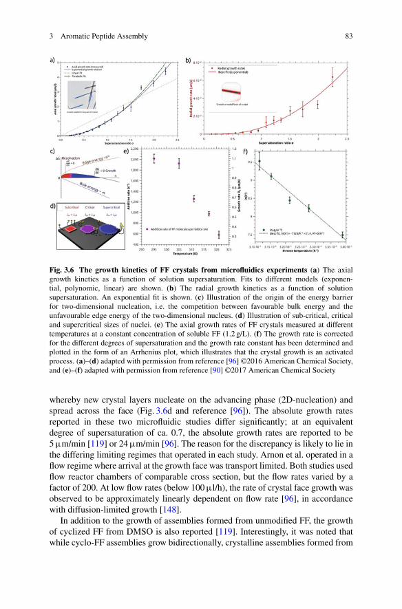
3 Aromatic Peptide Assembly 83
Fig. 3.6 The growth kinetics of FF crystals from microfluidics experiments (a) The axialgrowth kinetics as a function of solution supersaturation. Fits to different models (exponen-tial, polynomic, linear) are shown. (b) The radial growth kinetics as a function of solutionsupersaturation. An exponential fit is shown. (c) Illustration of the origin of the energy barrierfor two-dimensional nucleation, i.e. the competition between favourable bulk energy and theunfavourable edge energy of the two-dimensional nucleus. (d) Illustration of sub-critical, criticaland supercritical sizes of nuclei. (e) The axial growth rates of FF crystals measured at differenttemperatures at a constant concentration of soluble FF (1.2 g/L). (f) The growth rate is correctedfor the different degrees of supersaturation and the growth rate constant has been determined andplotted in the form of an Arrhenius plot, which illustrates that the crystal growth is an activatedprocess. (a)–(d) adapted with permission from reference [96] ©2016 American Chemical Society,and (e)–(f) adapted with permission from reference [90] ©2017 American Chemical Society
whereby new crystal layers nucleate on the advancing phase (2D-nucleation) andspread across the face (Fig. 3.6d and reference [96]). The absolute growth ratesreported in these two microfluidic studies differ significantly; at an equivalentdegree of supersaturation of ca. 0.7, the absolute growth rates are reported to be5 μm/min [119] or 24 μm/min [96]. The reason for the discrepancy is likely to lie inthe differing limiting regimes that operated in each study. Arnon et al. operated in aflow regime where arrival at the growth face was transport limited. Both studies usedflow reactor chambers of comparable cross section, but the flow rates varied by afactor of 200. At low flow rates (below 100 μl/h), the rate of crystal face growth wasobserved to be approximately linearly dependent on flow rate [96], in accordancewith diffusion-limited growth [148].
In addition to the growth of assemblies formed from unmodified FF, the growthof cyclized FF from DMSO is also reported [119]. Interestingly, it was noted thatwhile cyclo-FF assemblies grow bidirectionally, crystalline assemblies formed from
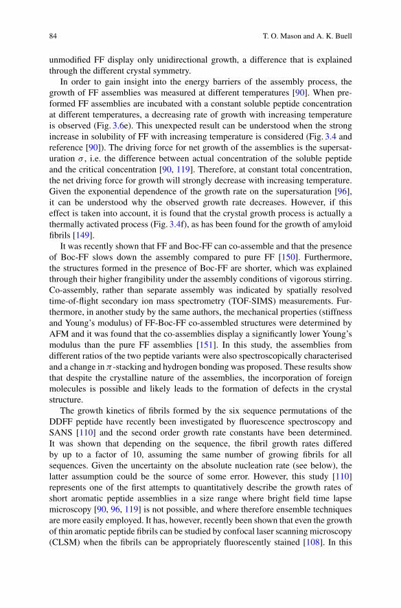
84 T. O. Mason and A. K. Buell
unmodified FF display only unidirectional growth, a difference that is explainedthrough the different crystal symmetry.
In order to gain insight into the energy barriers of the assembly process, thegrowth of FF assemblies was measured at different temperatures [90]. When pre-formed FF assemblies are incubated with a constant soluble peptide concentrationat different temperatures, a decreasing rate of growth with increasing temperatureis observed (Fig. 3.6e). This unexpected result can be understood when the strongincrease in solubility of FF with increasing temperature is considered (Fig. 3.4 andreference [90]). The driving force for net growth of the assemblies is the supersat-uration σ , i.e. the difference between actual concentration of the soluble peptideand the critical concentration [90, 119]. Therefore, at constant total concentration,the net driving force for growth will strongly decrease with increasing temperature.Given the exponential dependence of the growth rate on the supersaturation [96],it can be understood why the observed growth rate decreases. However, if thiseffect is taken into account, it is found that the crystal growth process is actually athermally activated process (Fig. 3.4f), as has been found for the growth of amyloidfibrils [149].
It was recently shown that FF and Boc-FF can co-assemble and that the presenceof Boc-FF slows down the assembly compared to pure FF [150]. Furthermore,the structures formed in the presence of Boc-FF are shorter, which was explainedthrough their higher frangibility under the assembly conditions of vigorous stirring.Co-assembly, rather than separate assembly was indicated by spatially resolvedtime-of-flight secondary ion mass spectrometry (TOF-SIMS) measurements. Fur-thermore, in another study by the same authors, the mechanical properties (stiffnessand Young’s modulus) of FF-Boc-FF co-assembled structures were determined byAFM and it was found that the co-assemblies display a significantly lower Young’smodulus than the pure FF assemblies [151]. In this study, the assemblies fromdifferent ratios of the two peptide variants were also spectroscopically characterisedand a change in π -stacking and hydrogen bonding was proposed. These results showthat despite the crystalline nature of the assemblies, the incorporation of foreignmolecules is possible and likely leads to the formation of defects in the crystalstructure.
The growth kinetics of fibrils formed by the six sequence permutations of theDDFF peptide have recently been investigated by fluorescence spectroscopy andSANS [110] and the second order growth rate constants have been determined.It was shown that depending on the sequence, the fibril growth rates differedby up to a factor of 10, assuming the same number of growing fibrils for allsequences. Given the uncertainty on the absolute nucleation rate (see below), thelatter assumption could be the source of some error. However, this study [110]represents one of the first attempts to quantitatively describe the growth rates ofshort aromatic peptide assemblies in a size range where bright field time lapsemicroscopy [90, 96, 119] is not possible, and where therefore ensemble techniquesare more easily employed. It has, however, recently been shown that even the growthof thin aromatic peptide fibrils can be studied by confocal laser scanning microscopy(CLSM) when the fibrils can be appropriately fluorescently stained [108]. In this
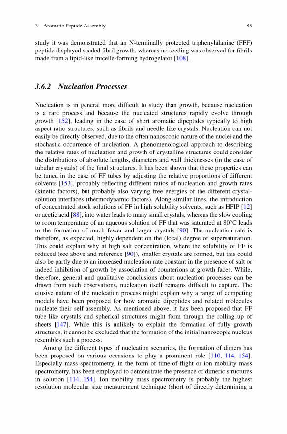
3 Aromatic Peptide Assembly 85
study it was demonstrated that an N-terminally protected triphenylalanine (FFF)peptide displayed seeded fibril growth, whereas no seeding was observed for fibrilsmade from a lipid-like micelle-forming hydrogelator [108].
3.6.2 Nucleation Processes
Nucleation is in general more difficult to study than growth, because nucleationis a rare process and because the nucleated structures rapidly evolve throughgrowth [152], leading in the case of short aromatic dipeptides typically to highaspect ratio structures, such as fibrils and needle-like crystals. Nucleation can noteasily be directly observed, due to the often nanoscopic nature of the nuclei and thestochastic occurrence of nucleation. A phenomenological approach to describingthe relative rates of nucleation and growth of crystalline structures could considerthe distributions of absolute lengths, diameters and wall thicknesses (in the case oftubular crystals) of the final structures. It has been shown that these properties canbe tuned in the case of FF tubes by adjusting the relative proportions of differentsolvents [153], probably reflecting different ratios of nucleation and growth rates(kinetic factors), but probably also varying free energies of the different crystal-solution interfaces (thermodynamic factors). Along similar lines, the introductionof concentrated stock solutions of FF in high solubility solvents, such as HFIP [12]or acetic acid [88], into water leads to many small crystals, whereas the slow coolingto room temperature of an aqueous solution of FF that was saturated at 80◦C leadsto the formation of much fewer and larger crystals [90]. The nucleation rate istherefore, as expected, highly dependent on the (local) degree of supersaturation.This could explain why at high salt concentration, where the solubility of FF isreduced (see above and reference [90]), smaller crystals are formed, but this couldalso be partly due to an increased nucleation rate constant in the presence of salt orindeed inhibition of growth by association of counterions at growth faces. While,therefore, general and qualitative conclusions about nucleation processes can bedrawn from such observations, nucleation itself remains difficult to capture. Theelusive nature of the nucleation process might explain why a range of competingmodels have been proposed for how aromatic dipeptides and related moleculesnucleate their self-assembly. As mentioned above, it has been proposed that FFtube-like crystals and spherical structures might form through the rolling up ofsheets [147]. While this is unlikely to explain the formation of fully growthstructures, it cannot be excluded that the formation of the initial nanoscopic nucleusresembles such a process.
Among the different types of nucleation scenarios, the formation of dimers hasbeen proposed on various occasions to play a prominent role [110, 114, 154].Especially mass spectrometry, in the form of time-of-flight or ion mobility massspectrometry, has been employed to demonstrate the presence of dimeric structuresin solution [114, 154]. Ion mobility mass spectrometry is probably the highestresolution molecular size measurement technique (short of directly determining a
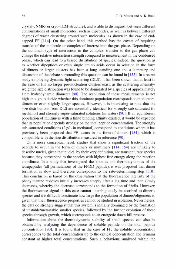
86 T. O. Mason and A. K. Buell
crystal-, NMR- or cryo-TEM-structure), and is able to distinguish between differentconformations of small molecules, such as dipeptides, as well as between differentdegrees of water clustering around such molecules, as shown in the case of end-capped FF [114]. On the other hand, this method has the caveat of requiringtransfer of the molecule or complex of interest into the gas phase. Depending onthe dominant type of interaction in the complex, transfer to the gas phase canchange the relative interaction strength compared to measurement in the condensedphase, which can lead to a biased distribution of species. Indeed, the question asto whether dipeptides or even single amino acids occur in solution in the formof dimers or larger clusters has been a long standing one and a summary anddiscussion of the debate surrounding this question can be found in [155]. In a recentstudy employing dynamic light scattering (DLS), it has been shown that at least inthe case of FF, no larger pre-nucleation clusters exist, as the scattering intensity-weighted size distribution was found to be dominated by a species of approximately1 nm hydrodynamic diameter [90]. The resolution of these measurements is nothigh enough to decide whether this dominant population corresponds to monomers,dimers or even slightly larger species. However, it is interesting to note that thesize distributions from DLS are essentially identical for strongly sub-saturated (inmethanol) and strongly super-saturated solutions (in water) [90]. If an equilibriumpopulation of multimers with a finite binding affinity existed, it would be expectedthat its population depends strongly on the total peptide concentration. The stronglysub-saturated conditions (2 g/L in methanol) correspond to conditions where it haspreviously been proposed that FF occurs in the form of dimers [154], which iscompatible with the size distribution measured in reference [90].
On a more conceptual level, studies that show a significant fraction of thepeptide to occur in the form of dimers or multimers [114, 154] are unlikely todescribe nuclei, given that nuclei, by their very definition, are transient, rare speciesbecause they correspond to the species with highest free energy along the reactioncoordinate. In a study that investigated the kinetics and thermodynamics of sixtetrapeptides (all permutations of the FFDD peptide), it was proposed that dimerformation is slow and therefore corresponds to the rate-determining step [110].This conclusion is based on the observation that the fluorescence intensity of thephenylalanine residues initially increases steeply after a lag time and then slowlydecreases, whereby the decrease corresponds to the formation of fibrils. However,the fluorescence signal in this case cannot unambiguously be ascribed to dimericspecies and it is difficult to estimate how large the population of dimers/oligomers is,given that their fluorescence properties cannot be studied in isolation. Nevertheless,the data do strongly suggest that this system is initially dominated by the formationof unstable/metastable smaller species, followed by the further evolution of thesespecies through growth, which corresponds to an energetic down-hill process.
Information about the thermodynamic stability of small species can also beobtained by analysing the dependence of soluble peptide on the total peptideconcentration [90]. It is found that in the case of FF, the soluble concentrationcorresponds to the total concentration up to the critical concentration and remainsconstant at higher total concentrations. Such a behaviour, analysed within the
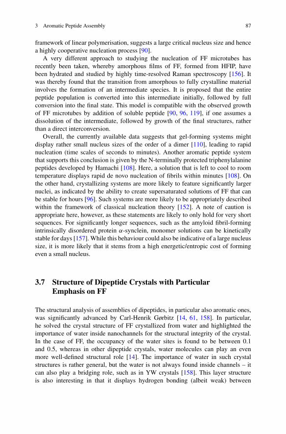
3 Aromatic Peptide Assembly 87
framework of linear polymerisation, suggests a large critical nucleus size and hencea highly cooperative nucleation process [90].
A very different approach to studying the nucleation of FF microtubes hasrecently been taken, whereby amorphous films of FF, formed from HFIP, havebeen hydrated and studied by highly time-resolved Raman spectroscopy [156]. Itwas thereby found that the transition from amorphous to fully crystalline materialinvolves the formation of an intermediate species. It is proposed that the entirepeptide population is converted into this intermediate initially, followed by fullconversion into the final state. This model is compatible with the observed growthof FF microtubes by addition of soluble peptide [90, 96, 119], if one assumes adissolution of the intermediate, followed by growth of the final structures, ratherthan a direct interconversion.
Overall, the currently available data suggests that gel-forming systems mightdisplay rather small nucleus sizes of the order of a dimer [110], leading to rapidnucleation (time scales of seconds to minutes). Another aromatic peptide systemthat supports this conclusion is given by the N-terminally protected triphenylalaninepeptides developed by Hamachi [108]. Here, a solution that is left to cool to roomtemperature displays rapid de novo nucleation of fibrils within minutes [108]. Onthe other hand, crystallizing systems are more likely to feature significantly largernuclei, as indicated by the ability to create supersaturated solutions of FF that canbe stable for hours [96]. Such systems are more likely to be appropriately describedwithin the framework of classical nucleation theory [152]. A note of caution isappropriate here, however, as these statements are likely to only hold for very shortsequences. For significantly longer sequences, such as the amyloid fibril-formingintrinsically disordered protein α-synclein, monomer solutions can be kineticallystable for days [157]. While this behaviour could also be indicative of a large nucleussize, it is more likely that it stems from a high energetic/entropic cost of formingeven a small nucleus.
3.7 Structure of Dipeptide Crystals with ParticularEmphasis on FF
The structural analysis of assemblies of dipeptides, in particular also aromatic ones,was significantly advanced by Carl-Henrik Gørbitz [14, 61, 158]. In particular,he solved the crystal structure of FF crystallized from water and highlighted theimportance of water inside nanochannels for the structural integrity of the crystal.In the case of FF, the occupancy of the water sites is found to be between 0.1and 0.5, whereas in other dipeptide crystals, water molecules can play an evenmore well-defined structural role [14]. The importance of water in such crystalstructures is rather general, but the water is not always found inside channels – itcan also play a bridging role, such as in YW crystals [158]. This layer structureis also interesting in that it displays hydrogen bonding (albeit weak) between
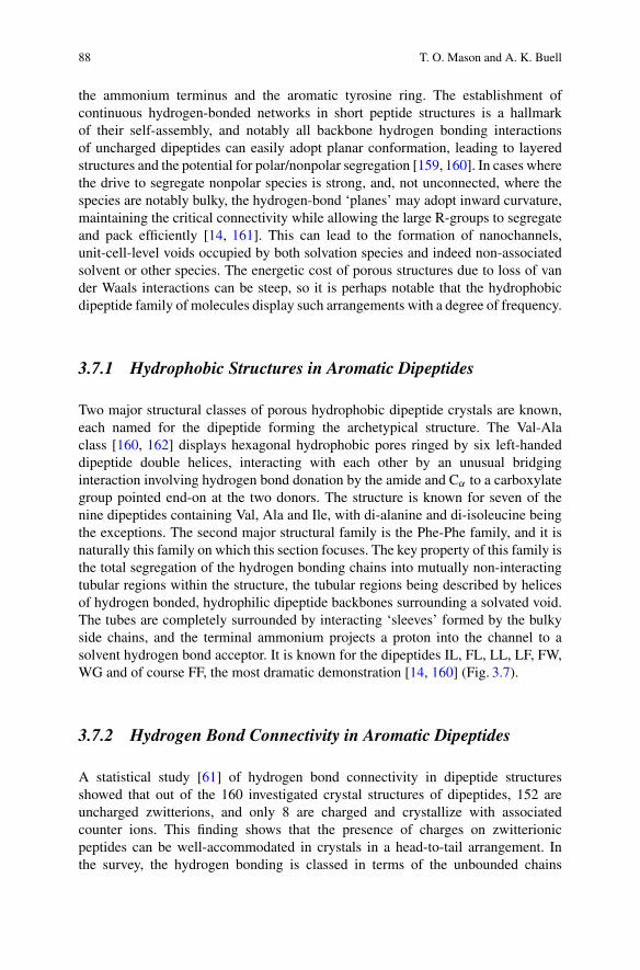
88 T. O. Mason and A. K. Buell
the ammonium terminus and the aromatic tyrosine ring. The establishment ofcontinuous hydrogen-bonded networks in short peptide structures is a hallmarkof their self-assembly, and notably all backbone hydrogen bonding interactionsof uncharged dipeptides can easily adopt planar conformation, leading to layeredstructures and the potential for polar/nonpolar segregation [159, 160]. In cases wherethe drive to segregate nonpolar species is strong, and, not unconnected, where thespecies are notably bulky, the hydrogen-bond ‘planes’ may adopt inward curvature,maintaining the critical connectivity while allowing the large R-groups to segregateand pack efficiently [14, 161]. This can lead to the formation of nanochannels,unit-cell-level voids occupied by both solvation species and indeed non-associatedsolvent or other species. The energetic cost of porous structures due to loss of vander Waals interactions can be steep, so it is perhaps notable that the hydrophobicdipeptide family of molecules display such arrangements with a degree of frequency.
3.7.1 Hydrophobic Structures in Aromatic Dipeptides
Two major structural classes of porous hydrophobic dipeptide crystals are known,each named for the dipeptide forming the archetypical structure. The Val-Alaclass [160, 162] displays hexagonal hydrophobic pores ringed by six left-handeddipeptide double helices, interacting with each other by an unusual bridginginteraction involving hydrogen bond donation by the amide and Cα to a carboxylategroup pointed end-on at the two donors. The structure is known for seven of thenine dipeptides containing Val, Ala and Ile, with di-alanine and di-isoleucine beingthe exceptions. The second major structural family is the Phe-Phe family, and it isnaturally this family on which this section focuses. The key property of this family isthe total segregation of the hydrogen bonding chains into mutually non-interactingtubular regions within the structure, the tubular regions being described by helicesof hydrogen bonded, hydrophilic dipeptide backbones surrounding a solvated void.The tubes are completely surrounded by interacting ‘sleeves’ formed by the bulkyside chains, and the terminal ammonium projects a proton into the channel to asolvent hydrogen bond acceptor. It is known for the dipeptides IL, FL, LL, LF, FW,WG and of course FF, the most dramatic demonstration [14, 160] (Fig. 3.7).
3.7.2 Hydrogen Bond Connectivity in Aromatic Dipeptides
A statistical study [61] of hydrogen bond connectivity in dipeptide structuresshowed that out of the 160 investigated crystal structures of dipeptides, 152 areuncharged zwitterions, and only 8 are charged and crystallize with associatedcounter ions. This finding shows that the presence of charges on zwitterionicpeptides can be well-accommodated in crystals in a head-to-tail arrangement. Inthe survey, the hydrogen bonding is classed in terms of the unbounded chains
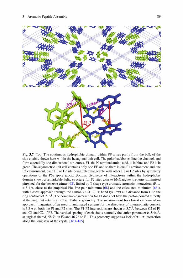
3 Aromatic Peptide Assembly 89
Fig. 3.7 Top: The continuous hydrophobic domain within FF arises partly from the bulk of theside chains, shown here within the hexagonal unit cell. The polar backbones line the channel, andform essentially one-dimensional structures. F1, the N-terminal amino acid, is in blue, and F2 is ingreen. The asymmetric unit cell contains only one FF, and so there is one F1 environment and oneF2 environment, each F1 or F2 site being interchangeable with other F1 or F2 sites by symmetryoperations of the P61 space group. Bottom: Geometry of interactions within the hydrophobicdomain shows a remarkable helix structure for F2 sites akin to McGaughey’s energy-minimisedpinwheel for the benzene trimer [68], linked by T-shape type aromatic-aromatic interactions (Rcen
= 5.1 Å, close to the empirical Phe-Phe pair minimum [68] and the calculated minimum [66]),with closest approach through the carbon 4 C-H· · · π bond (yellow) at a distance from H to thering centroid of 2.9 Å. The comparable interaction for F1 does not have the proton pointed directlyat the ring, but retains an offset T-shape geometry. The measurement for closest carbon-carbonapproach (magenta), often used in automated systems for the discovery of interaromatic contact,is 3.8 Å on both the F1 and F2 sites. The F1-F2 interactions are shown at 3.7 Å between C2 of F1and C1 and C2 of F2. The vertical spacing of each site is naturally the lattice parameter c, 5.46 Å,at angle θ (in red) 58.7◦ on F2 and 46.7◦ on F1. This geometry suggests a lack of π −π interactionalong the long axis of the crystal [163–165]
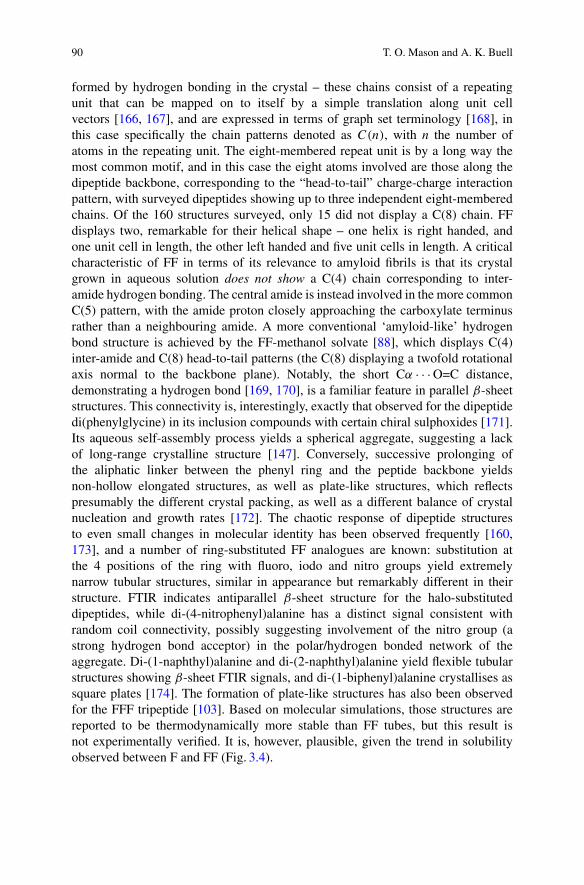
90 T. O. Mason and A. K. Buell
formed by hydrogen bonding in the crystal – these chains consist of a repeatingunit that can be mapped on to itself by a simple translation along unit cellvectors [166, 167], and are expressed in terms of graph set terminology [168], inthis case specifically the chain patterns denoted as C(n), with n the number ofatoms in the repeating unit. The eight-membered repeat unit is by a long way themost common motif, and in this case the eight atoms involved are those along thedipeptide backbone, corresponding to the “head-to-tail” charge-charge interactionpattern, with surveyed dipeptides showing up to three independent eight-memberedchains. Of the 160 structures surveyed, only 15 did not display a C(8) chain. FFdisplays two, remarkable for their helical shape – one helix is right handed, andone unit cell in length, the other left handed and five unit cells in length. A criticalcharacteristic of FF in terms of its relevance to amyloid fibrils is that its crystalgrown in aqueous solution does not show a C(4) chain corresponding to inter-amide hydrogen bonding. The central amide is instead involved in the more commonC(5) pattern, with the amide proton closely approaching the carboxylate terminusrather than a neighbouring amide. A more conventional ‘amyloid-like’ hydrogenbond structure is achieved by the FF-methanol solvate [88], which displays C(4)inter-amide and C(8) head-to-tail patterns (the C(8) displaying a twofold rotationalaxis normal to the backbone plane). Notably, the short Cα · · · O=C distance,demonstrating a hydrogen bond [169, 170], is a familiar feature in parallel β-sheetstructures. This connectivity is, interestingly, exactly that observed for the dipeptidedi(phenylglycine) in its inclusion compounds with certain chiral sulphoxides [171].Its aqueous self-assembly process yields a spherical aggregate, suggesting a lackof long-range crystalline structure [147]. Conversely, successive prolonging ofthe aliphatic linker between the phenyl ring and the peptide backbone yieldsnon-hollow elongated structures, as well as plate-like structures, which reflectspresumably the different crystal packing, as well as a different balance of crystalnucleation and growth rates [172]. The chaotic response of dipeptide structuresto even small changes in molecular identity has been observed frequently [160,173], and a number of ring-substituted FF analogues are known: substitution atthe 4 positions of the ring with fluoro, iodo and nitro groups yield extremelynarrow tubular structures, similar in appearance but remarkably different in theirstructure. FTIR indicates antiparallel β-sheet structure for the halo-substituteddipeptides, while di-(4-nitrophenyl)alanine has a distinct signal consistent withrandom coil connectivity, possibly suggesting involvement of the nitro group (astrong hydrogen bond acceptor) in the polar/hydrogen bonded network of theaggregate. Di-(1-naphthyl)alanine and di-(2-naphthyl)alanine yield flexible tubularstructures showing β-sheet FTIR signals, and di-(1-biphenyl)alanine crystallises assquare plates [174]. The formation of plate-like structures has also been observedfor the FFF tripeptide [103]. Based on molecular simulations, those structures arereported to be thermodynamically more stable than FF tubes, but this result isnot experimentally verified. It is, however, plausible, given the trend in solubilityobserved between F and FF (Fig. 3.4).
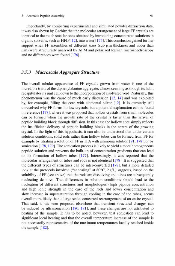
3 Aromatic Peptide Assembly 91
Importantly, by comparing experimental and simulated powder diffraction data,it was also shown by Gørbitz that the molecular arrangement of large FF crystals areidentical to the much smaller ones obtained by introducing concentrated solutions inorganic solvents, such as HFIP [12], into water [175]. This conclusion gained furthersupport when FF assemblies of different sizes (sub μm thickness and wider thanμm) were structurally analysed by AFM and polarized Raman microspectroscopyand no differences were found [176].
3.7.3 Macroscale Aggregate Structure
The overall tubular appearance of FF crystals grown from water is one of theincredible traits of the diphenylalanine aggregate, almost seeming as though its habitrecapitulates its unit cell down to the incorporation of a solvated void! Naturally, thisphenomenon was the cause of much early discussion [12, 14] and was exploitedby, for example, filling the core with elemental silver [12]. It is currently stillunresolved why FF forms hollow crystals, but a potential explanation can be foundin reference [177], where it was proposed that hollow crystals from small moleculescan be formed when the growth rate of the crystal is faster than the arrival ofpeptide building block through diffusion. In this case the hollow core simply reflectsthe insufficient delivery of peptide building blocks in the centre of the growingcrystal. In the light of this hypothesis, it can also be understood that under certainsolution conditions, solid rods rather than hollow tubes can be formed from FF forexample by titrating a solution of FF in TFA with ammonia solution [91, 178], or bysonication [178, 179]. The sonication process is likely to yield a more homogeneouspeptide solution and prevents the built-up of concentration gradients that can leadto the formation of hollow tubes [177]. Interestingly, it was reported that themolecular arrangement of tubes and rods is not identical [178]. It is suggested thatthe different types of structures can be inter-converted [178], but a more detailedlook at the protocols involved (“annealing” at 80◦C, 2 g/L) suggests, based on thesolubility of FF (see above) that the rods are dissolving and tubes are subsequentlynucleating de novo. That differences in solution conditions should lead to thenucleation of different structures and morphologies (high peptide concentrationand high ionic strength in the case of the rods and lower concentration andslow increase in supersaturation through cooling in the case of the tubes) seemsoverall more likely than a large scale, concerted rearrangement of an entire crystal.That said, it has been proposed elsewhere that transient structural changes canbe induced by ultrasonication [180, 181], and these changes are not attributed toheating of the sample. It has to be noted, however, that sonication can lead tosignificant local heating and that the overall temperature increase of the sample isnot necessarily representative of the maximum temperatures locally reached insidethe sample [182].
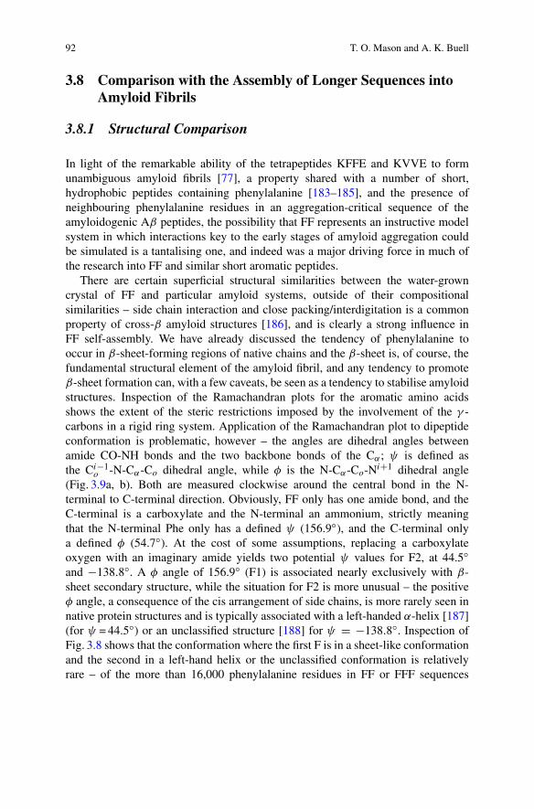
92 T. O. Mason and A. K. Buell
3.8 Comparison with the Assembly of Longer Sequences intoAmyloid Fibrils
3.8.1 Structural Comparison
In light of the remarkable ability of the tetrapeptides KFFE and KVVE to formunambiguous amyloid fibrils [77], a property shared with a number of short,hydrophobic peptides containing phenylalanine [183–185], and the presence ofneighbouring phenylalanine residues in an aggregation-critical sequence of theamyloidogenic Aβ peptides, the possibility that FF represents an instructive modelsystem in which interactions key to the early stages of amyloid aggregation couldbe simulated is a tantalising one, and indeed was a major driving force in much ofthe research into FF and similar short aromatic peptides.
There are certain superficial structural similarities between the water-growncrystal of FF and particular amyloid systems, outside of their compositionalsimilarities – side chain interaction and close packing/interdigitation is a commonproperty of cross-β amyloid structures [186], and is clearly a strong influence inFF self-assembly. We have already discussed the tendency of phenylalanine tooccur in β-sheet-forming regions of native chains and the β-sheet is, of course, thefundamental structural element of the amyloid fibril, and any tendency to promoteβ-sheet formation can, with a few caveats, be seen as a tendency to stabilise amyloidstructures. Inspection of the Ramachandran plots for the aromatic amino acidsshows the extent of the steric restrictions imposed by the involvement of the γ -carbons in a rigid ring system. Application of the Ramachandran plot to dipeptideconformation is problematic, however – the angles are dihedral angles betweenamide CO-NH bonds and the two backbone bonds of the Cα; ψ is defined asthe Ci−1
o -N-Cα-Co dihedral angle, while φ is the N-Cα-Co-Ni+1 dihedral angle(Fig. 3.9a, b). Both are measured clockwise around the central bond in the N-terminal to C-terminal direction. Obviously, FF only has one amide bond, and theC-terminal is a carboxylate and the N-terminal an ammonium, strictly meaningthat the N-terminal Phe only has a defined ψ (156.9◦), and the C-terminal onlya defined φ (54.7◦). At the cost of some assumptions, replacing a carboxylateoxygen with an imaginary amide yields two potential ψ values for F2, at 44.5◦and −138.8◦. A φ angle of 156.9◦ (F1) is associated nearly exclusively with β-sheet secondary structure, while the situation for F2 is more unusual – the positiveφ angle, a consequence of the cis arrangement of side chains, is more rarely seen innative protein structures and is typically associated with a left-handed α-helix [187](for ψ = 44.5◦) or an unclassified structure [188] for ψ = −138.8◦. Inspection ofFig. 3.8 shows that the conformation where the first F is in a sheet-like conformationand the second in a left-hand helix or the unclassified conformation is relativelyrare – of the more than 16,000 phenylalanine residues in FF or FFF sequences
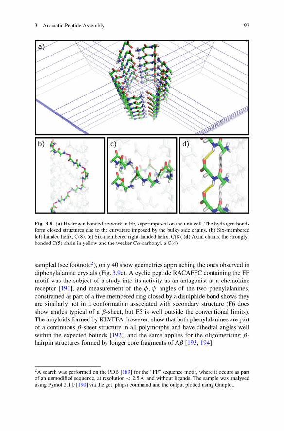
3 Aromatic Peptide Assembly 93
Fig. 3.8 (a) Hydrogen bonded network in FF, superimposed on the unit cell. The hydrogen bondsform closed structures due to the curvature imposed by the bulky side chains. (b) Six-memberedleft-handed helix, C(8). (c) Six-membered right-handed helix, C(8). (d) Axial chains, the strongly-bonded C(5) chain in yellow and the weaker Cα-carbonyl, a C(4)
sampled (see footnote2), only 40 show geometries approaching the ones observed indiphenylalanine crystals (Fig. 3.9c). A cyclic peptide RACAFFC containing the FFmotif was the subject of a study into its activity as an antagonist at a chemokinereceptor [191], and measurement of the φ,ψ angles of the two phenylalanines,constrained as part of a five-membered ring closed by a disulphide bond shows theyare similarly not in a conformation associated with secondary structure (F6 doesshow angles typical of a β-sheet, but F5 is well outside the conventional limits).The amyloids formed by KLVFFA, however, show that both phenylalanines are partof a continuous β-sheet structure in all polymorphs and have dihedral angles wellwithin the expected bounds [192], and the same applies for the oligomerising β-hairpin structures formed by longer core fragments of Aβ [193, 194].
2A search was performed on the PDB [189] for the “FF” sequence motif, where it occurs as partof an unmodified sequence, at resolution < 2.5 Å and without ligands. The sample was analysedusing Pymol 2.1.0 [190] via the get_phipsi command and the output plotted using Gnuplot.
94 T. O. Mason and A. K. Buell
Fig. 3.9 The Ramachandran angles of the FF sequence in a statistical sample (a) Ramachan-dran angle φ, measured in a right-handed fashion in the N-to-C terminal direction. The angleshown is typical of an antiparallel β-sheet. (b) The angle ψ , again with a value typically seenin the antiparallel β-sheet. φ and ψ values are restricted by steric effects to ranges of ‘allowed’values. (c) A statistical sample from the Protein Data Bank of 8044 occurrences of the unmodifiedFF sequence in 3087 PDB entries, collected by the authors for this work. Direction of the chainis shown by the colour gradation. In nature, the sequence has a relatively typical Ramachandrandistribution, with a tendency for a first residue in a helical-type structure to result in a secondresidue with sheet-like conformation. Two black arrows represent the pair of FF sequences inPDB 3ow9 [192], a polymorph of KLVFFA with typical anti-parallel β-sheet conformation (Paneld) The dipeptide crystal has φ1=156.9◦, with ψ1 undefined. The dotted red line shows the φ
angle on F1. The carboxylate terminus of F2 allows the guessing of two possible “ψ angles”,for φ2 = 64.7◦. Recognition of a similar structure with a sheet-like first residue, and a second ina left-handed conformation, reveals the curious fact that FF (Aβ19−20) adopts a similar backbonestructure to PDB entry 5NAO, Aβ1−42, as solved by Wälti [195]. (e) Superposition of the FFcrystalline conformation (green) over the conformation of the sequence in Aβ1−42
The helical connectivity of the hydrogen bonding chains has some similaritiesto a proposed model of polyglutamine aggregation advanced by Perutz et al. [196]contemporaneously with early, but highly influential, reports on the properties ofFF [12], though the β-helix model for polyglutamine species has not stood the test
3 Aromatic Peptide Assembly 95
of time [197, 198]. The parallel arrangement of the two-membered chains might beseen as something of a departure from expected behaviour for short polypeptidesforming amyloid fibrils, due to the increased importance of terminal charge com-plementarity, which forces antiparallel organisation in KFFE and other asymmetricshort peptides with charged residues [77, 199]. A critical factor in the topology ofthe aggregate structure is the twist observed for amyloid fibrils – this phenomenonis due to the steric effects of the chiral backbone [200]. Only if φ + ψ = 0on average along the extended chain in the sheet are the amide groups parallel.In general, the average has a small finite value, resulting in a tendency towards aprogressive deflection of the polar direction of the amide groups along the chain,and hence to a helical twist around the axial direction of the polymeric fibril or β-sheet [201]. This helical twist effectively limits the radial dimension of the fibril, asfibril addition sites further from the central position experience progressively greaterelastic strain. The equilibrium radius of the fibrils arises from the balance betweenthis elastic deformation cost and the free energy of radial addition to the fibril [202].Polymorphism in radial dimension, helical twist pitch, handedness and indeed incoiling of the fibril into nanotube-like ribbons have been observed [203, 204].The limited width, due to mechanical effects, is a key property of amyloid fibrils;indeed, it is a major reason why they have a fibrillar morphology and criticallyit distinguishes the fibril from the crystal. This was shown in a theoretical study,where the free energy as a function of the twist angle was treated as a perturbationin accordance with the Landau theory of phase transitions, a mean-field theorywhereby the perturbation can be expanded as a Taylor series [205]. In the case ofthe twisting of amyloid fibrils, the chirality of the individual chains requires theretention of the odd-powered terms in the expansion which would otherwise causeasymmetry about the transition point, though the linear term is shown to have a zerocoefficient. The analysis in the paper proceeds to identify the stationary points of theseries expansion, with a minimum at zero twist (a crystalline state), and a minimumat a specific twist angle, this twist angle being dependent on the equilibrium twistangle (the twist angle at zero fibril thickness), and the ratio of the torsional springconstant of the fibril (a function of the square of fibril thickness) to the springconstant for rotations between individual chains in the fibril (going as the cube ofthe chain length for long chains). A critical fibril thickness was established, abovewhich the crystalline form would be stable [205]. This critical width scales linearlywith chain length, and is of course also dependent on other material parameters,many of which are common to all amyloid structures. It is to be noted that the originof the asymmetry, the equilibrium chiral twist of the amide backbone, cannot exertinfluence in the dipeptides – only one amide bond exists, and so it is impossiblefor there to be an equilibrium series of amide rotations about the backbone. Theammonium and carboxylate termini are freely rotated, and interact by a monopole-monopole force that, inherently, only has a distance and no directional dependence.
A crystal is defined by its repeating, three-dimensional motif and in principle,any translation along an integer number of unit cell vectors will lead to an identicalenvironment. This property has been confirmed for two distinct FF solvomorphs [88,175] among many other short peptides [61] by virtue of the analysis of X-ray
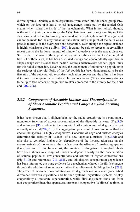
96 T. O. Mason and A. K. Buell
diffractograms. Diphenylalanine crystallises from water into the space group P 61,which on the face of it has a helical appearance, borne out by the angled C(8)chains which spiral the inside of the nanochannels. What is not helical, though,is the vertical (axial) connectivity, the C(5) chain- each step along a multiple of theshort axial unit cell vector brings you to an identical diphenylalanine. This argumentcannot be made for the amyloid axial translation unless the pitch of the helix was aprecise multiple of the hydrogen bond separation. Even though the repeat distanceis highly consistent along a fibril [206], it cannot be said to represent a crystallinerepeat due to the far lower energy of minute fluctuations over the repeat distance.Still harder to equate to the crystalline regime are the radial ‘vectors’ in amyloidfibrils. For these sites, as has been discussed, energy and concomitantly equilibriumshape change with distance from the fibril centre, and there exist defined upper limitsfor the radial dimension. Nevertheless, the attachment of monomeric peptide ontothe surfaces of amyloid fibrils of the Aβ peptide has been demonstrated to be thefirst step of the autocatalytic secondary nucleation process and the affinity has beendetermined from quantitative surface plasmon resonance (SPR) biosensing studiesto be up to two orders of magnitude weaker compared to the affinity for the fibrilend [207, 208].
3.8.2 Comparison of Assembly Kinetics and Thermodynamicsof Short Aromatic Peptides and Longer Amyloid FormingSequences
It has been shown that in diphenylalanine, the radial growth rate is a continuous,monotonic function of excess concentration of the dipeptide in water (Fig. 3.6band reference [96]), while in the amyloid fibril continuous radial growth is notnormally observed [209, 210]. The aggregation process of FF, in common with othercrystalline species, is highly cooperative. Concerns of edge and surface energiesdetermine the stability of ‘islands’ of a new layer at a surface (Fig. 3.6d) andgive rise to complex, higher-order dependence of the incorporation rate on theexcess arrivals of monomer at the surface over the off-rate of resolvating species(Figs. 3.6a and 3.10a). In contrast, the kinetics of elongation of amyloid fibrilshave been shown in a range of studies to depend linearly on the concentrationof soluble peptide at low concentrations and saturate at higher concentrations(Fig. 3.10b and references [211, 212]), and this distinct concentration dependencehas been interpreted as strong evidence for a mechanism whereby the fibrils elongatethrough the addition of monomeric, rather than oligomeric building blocks [211].The effect of monomer concentration on axial growth rate is a readily-identifieddifference between crystalline and fibrillar systems- crystalline systems displaycooperativity at moderate supersaturation, while fibrillar systems transition fromnon-cooperative (linear in supersaturation) to anti-cooperative (sublinear) regimes at
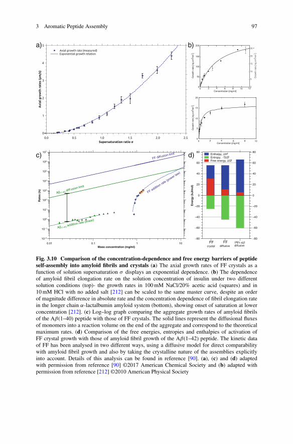
3 Aromatic Peptide Assembly 97
Gro
wth
rate
[ng
cm m
in ]-1
-2
Concentration [mg/ml]
0
5
10
15
20
0 2 4 6 8 10
0
50
100
150
200
Concentration [mg/ml]
0
5
10
15
20
25
Gro
wth
rate
[ng
cm m
in ]-1
-2
Gro
wth
rate
[ng
cm m
in ]-1
-2
121086420
a) b)R
ates
(/s
)
10−2
10−1
100
101
102
103
104
105
106
107
Mass concentration (mg/ml)0.01 0.1 1 10
A (1-40) di usion limit
A (1-40) addition rate (linear)
FF di usion limit
FF addition ra
te (power la
w)
En
erg
y (k
J/m
ol)
−80
−60
−40
−20
0
20
40
60
80
−80
−60
−40
−20
0
20
40
60
80Enthalpy, H‡
Entropy, -T S‡
Free energy, G‡
FF (A 1-42)FFcrystal
c) d)
Fig. 3.10 Comparison of the concentration-dependence and free energy barriers of peptideself-assembly into amyloid fibrils and crystals (a) The axial growth rates of FF crystals as afunction of solution supersaturation σ displays an exponential dependence. (b) The dependenceof amyloid fibril elongation rate on the solution concentration of insulin under two differentsolution conditions (top)- the growth rates in 100 mM NaCl/20% acetic acid (squares) and in10 mM HCl with no added salt [212] can be scaled to the same master curve, despite an orderof magnitude difference in absolute rate and the concentration dependence of fibril elongation ratein the longer chain α-lactalbumin amyloid system (bottom), showing onset of saturation at lowerconcentration [212]. (c) Log–log graph comparing the aggregate growth rates of amyloid fibrilsof the Aβ(1–40) peptide with those of FF crystals. The solid lines represent the diffusional fluxesof monomers into a reaction volume on the end of the aggregate and correspond to the theoreticalmaximum rates. (d) Comparison of the free energies, entropies and enthalpies of activation ofFF crystal growth with those of amyloid fibril growth of the Aβ(1–42) peptide. The kinetic dataof FF has been analysed in two different ways, using a diffusive model for direct comparabilitywith amyloid fibril growth and also by taking the crystalline nature of the assemblies explicitlyinto account. Details of this analysis can be found in reference [90]. (a), (c) and (d) adaptedwith permission from reference [90] ©2017 American Chemical Society and (b) adapted withpermission from reference [212] ©2010 American Physical Society
98 T. O. Mason and A. K. Buell
a supersaturation determined by internal dynamics of the attached monomer duringincorporation [212].
The difference in growth mechanisms between amyloid fibrils and crystalsrenders a direct comparison of the absolute growth rates and in particular of theunderlying free energy barriers not straightforward. Nevertheless, a first idea canbe gained by comparing the absolute rates of growth of these structures with thetheoretical maximum of the assembly rate, the diffusional arrival of the peptide intoa reaction volume of size comparable to the soluble building block. In Fig. 3.10csuch a comparison is shown for the FF peptide and the Aβ(1–40) peptide. Thedifference between the actual rate and the theoretical maximum is approximatelya factor of 103 in the case of Aβ(1–40) across a range of concentrations, and dueto the saturation of fibril elongation this factor increases at high concentrations. Onthe other hand, the exponential dependence of the axial growth rate of FF leads toa strongly decreasing difference between the rates of diffusional arrival and growth.If the difference between maximal and actual rates is interpreted as stemming froma free energy barrier, the latter is therefore concentration dependent [90].
All faces of the crystal have been shown to grow continuously at finite rates insuitable conditions, but another comparison with the amyloid system may be madein that the crystals of FF have extreme aspect ratios, and through careful controlof solution conditions, can be made to grow axially with extremely slow radialgrowth [96], a phenomenon arising from the exponential dependence of growthrate on supersaturation on each face. The observation is explained in terms of theintermolecular interaction chains coplanar with the initial deposition of a new face.In FF, the radial faces may either intersect the helical hydrogen bonded channels,which will readily stabilise incoming FF, or they may present a simple hydrophobicsurface, compare Figs. 3.7 and 3.8. The close-packed hydrophobic surface is arelatively low energy face, and so the borders of new layers on the axial face comeat a rather lower cost than their counterparts on the radial face, which must alwaysexpose unsatisfied pendant hydrogen bonds. As a result of the greater nucleationrate of new layers, the axial face grows faster. Certain theoretical models haveseen success in explaining amyloid nucleation and growth rates in terms of pseudo-one-dimensional crystallisation [213, 214] – crystals where the energy differencebetween the faces is such as to render radial growth impossible for wide rangesof conditions, though it should be mentioned that the Gibbs-Wulff theorem, towhich analogy is made for the equilibrium aggregate shape, is only demonstrablein aggregates capable of rapid equilibration, i.e. very small ones. Kinetic processesare dominant determinants of crystal shape at the macroscale, and the early stagesof polypeptide aggregation frequently involve non-fibrillar oligomeric species, forwhich the crystal theories of amyloid formation do not account.
For shorter species, the line between a crystal and an amyloid fibril can be anarrow one, and the classic examples are the Sup35 fragments GNNQQNY andNNQQNY, a heptapeptide and hexapeptide which, like their parent chain, readilyform amyloid fibrils [129, 215]. Unusually, though, a crystalline polymorph isalso observed in each case at slightly higher monomer concentration, this crystalhaving very similar structure to the fibril as determined by X-ray diffractometry
3 Aromatic Peptide Assembly 99
(the 4.7 Å meridional reflection being particularly notable), and an interestingpoint is made that the crystals were also highly acicular and despite the effortsof the researchers, crystals could not be grown continuously in at least the radialdimension. A microcrystal of NNQQNY is shown, with a width slightly over 1 μm,corresponding to approximately 500 unit cells, and appears to have a somewhatirregular surface, suggesting a high density of grain boundaries parallel to thelong axis [7]. Other short peptide polymorphic systems have been prepared, andthey have a notably wide range of side chains [216] – NNQQ, a tetrapeptidewith purely polar residues, fibrillises, as does penta-glutamine, results in keepingwith observations of the pathology of Huntington’s chorea, a neuropathologicalamyloidosis triggered by extended polyglutamine repeats [217]. The same workestablished a fibrillar state for the hexapeptide SSTSAA, a fairly hydrophilicfragment with short side chains, as well as larger, more hydrophobic species. Akey finding of this extensive survey into amyloidogenic fragments is the conceptof “steric zippers”. These fragments of longer chains are computationally predictedand experimentally confirmed to be amyloidogenic in their own right, and as suchare considered to be potential candidates for “trigger” regions of the parent chains,whose intermolecular interaction can lead to the nucleation of the fibrils [218, 219].The fragments proved capable in many cases of enhancing fibril formation of theparent polypeptide, and vice versa. In the diphenylalanine case, the identity of thedipeptide as Aβ19−20 is frequently remarked upon in studies of the species, andrecent NMR-derived structures of Aβ1−42 show the motif buried in a hydrophobic“pocket”, interestingly in a similar ‘cis’ configuration of the side chains [195, 220].However, a recent cryo-EM study showed F20 oriented out of the hydrophobic‘core’ containing F19 [9]. In this study, the fibrils were formed in 30% acetonitrilesolution. This alteration of the solvent environment may render the projection of F20at the surface less unfavourable than in the aqueous/salt systems of references [195]and [220]. The Ramachandran angles are φF1 = −94.7◦, ψF1 = 152.7◦, φF2 =64.7◦, ψF2 = −172.5◦ in the structure as given by Waelti et al. Interestingly, thesecorrespond very closely to an equivalent FF conformation in the crystal, and a glanceat the structural solution shows the effect of the same forces. The hydrophobicresidues are buried, and indeed interact in a clear herringbone-type chain typicalof the π -π interaction, even at significant cost to the continuity of the hydrogenbonding between successive chains [195]. The effects of hydrophobicity are readilydiscerned from the convoluted structure of the chain in the fibril. Two separatehydrophobic pockets are formed by each of the chains forming the dimeric ‘unitcell’ in the axial direction, one involving the residues 16–22 (KLVFFA), and thesecond involving interdigitating aliphatic side chains from residue 31 onwards.
Comparison of the thermodynamics of dipeptide and polypeptide aggregationis an important step towards rationalising or discounting the employment of shortpeptides as model systems for amyloid propagation. In particular, the desolvationof the aromatic benzyl groups is predicted to contribute a significant negative freeenergy to the process of amyloid fibril formation. In polypeptide systems withhydrophobic residues, the enthalpic component of the free energy of desolvation
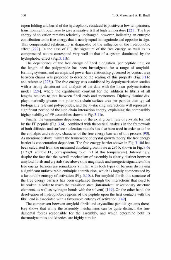
100 T. O. Mason and A. K. Buell
(upon folding and burial of the hydrophobic residues) is positive at low temperature,transitioning through zero to give a negative �H at high temperature [221]. The freeenergy of solvation remains relatively unchanged, however, indicating an entropiccontribution to the free energy that is nearly equal in magnitude and opposite in sign.This compensated relationship is diagnostic of the influence of the hydrophobiceffect [222]. In the case of FF, the signature of the free energy, as well as itscompensated nature correspond very well to that of a system dominated by thehydrophobic effect (Fig. 3.11b).
The dependence of the free energy of fibril elongation, per peptide unit, onthe length of the polypeptide has been investigated for a range of amyloid-forming systems, and an empirical power-law relationship governed by contact areabetween chains was proposed to describe the scaling of this property (Fig. 3.11cand reference [223]). The free energy was established by depolymerisation studieswith a strong denaturant and analysis of the data with the linear polymerisationmodel [224], where the equilibrium constant for the addition to fibrils of alllengths reduces to that between fibril ends and monomers. Diphenylalanine dis-plays markedly greater non-polar side chain surface area per peptide than typicalbiologically relevant polypeptides, and the π -stacking interactions will represent asignificant portion of the side chain interaction energy, explaining the comparablyhigher stability of FF assemblies shown in Fig. 3.11c.
Finally, the temperature dependence of the axial growth rate of crystals formedby the FF peptide (Fig. 3.6f), combined with theoretical analysis in the frameworkof both diffusive and surface nucleation models has also been used in order to definethe enthalpic and entropic character of the free energy barriers of this process [90].As mentioned above, within the framework of crystal growth theory, the free energybarrier is concentration dependent. The free energy barrier shown in Fig. 3.10d hasbeen calculated from the measured absolute growth rate at 295 K shown in Fig. 3.6e(1.2 g/L soluble FF, corresponding to σ ∼1 at this temperature). Interestingly,despite the fact that the overall mechanism of assembly is clearly distinct betweenamyloid fibrils and crystals (see above), the magnitude and energetic signature of thefree energy barriers are remarkably similar, with both types of barriers displayinga significant unfavourable enthalpic contribution, which is largely compensated bya favourable entropy of activation (Fig. 3.10d). For amyloid fibrils this structure ofthe free energy barriers has been explained through the interactions that need tobe broken in order to reach the transition state (intramolecular secondary structureelements, as well as hydrogen bonds with the solvent) [149]. On the other hand, thedesolvation of hydrophobic regions of the peptide upon the first contacts with thefibril end is associated with a favourable entropy of activation [149].
The comparison between amyloid fibrils and crystalline peptide systems there-fore shows that while the assembly mechanisms can be quite distinct, the fun-damental forces responsible for the assembly, and which determine both itsthermodynamics and kinetics, are highly similar.
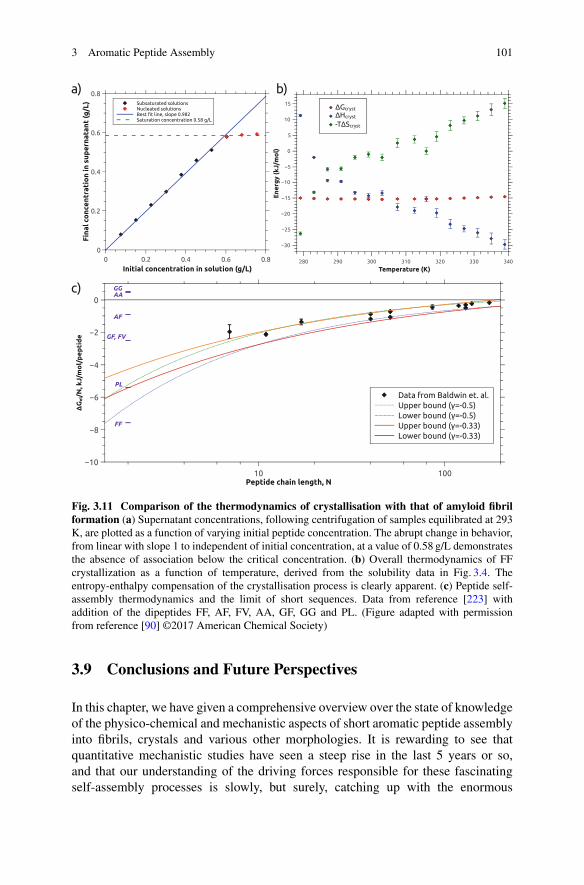
3 Aromatic Peptide Assembly 101
Fig. 3.11 Comparison of the thermodynamics of crystallisation with that of amyloid fibrilformation (a) Supernatant concentrations, following centrifugation of samples equilibrated at 293K, are plotted as a function of varying initial peptide concentration. The abrupt change in behavior,from linear with slope 1 to independent of initial concentration, at a value of 0.58 g/L demonstratesthe absence of association below the critical concentration. (b) Overall thermodynamics of FFcrystallization as a function of temperature, derived from the solubility data in Fig. 3.4. Theentropy-enthalpy compensation of the crystallisation process is clearly apparent. (c) Peptide self-assembly thermodynamics and the limit of short sequences. Data from reference [223] withaddition of the dipeptides FF, AF, FV, AA, GF, GG and PL. (Figure adapted with permissionfrom reference [90] ©2017 American Chemical Society)
3.9 Conclusions and Future Perspectives
In this chapter, we have given a comprehensive overview over the state of knowledgeof the physico-chemical and mechanistic aspects of short aromatic peptide assemblyinto fibrils, crystals and various other morphologies. It is rewarding to see thatquantitative mechanistic studies have seen a steep rise in the last 5 years or so,and that our understanding of the driving forces responsible for these fascinatingself-assembly processes is slowly, but surely, catching up with the enormous
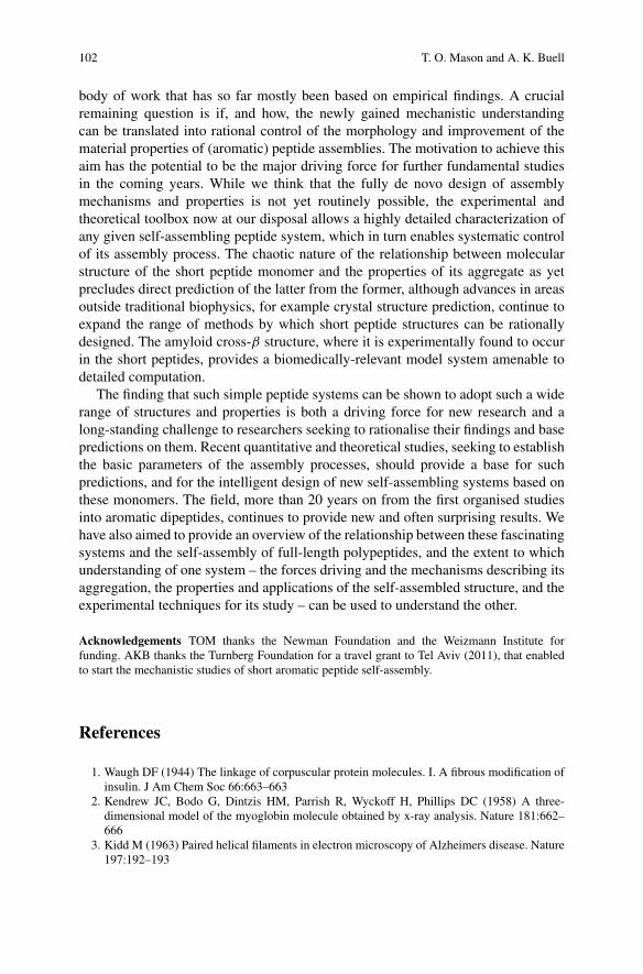
102 T. O. Mason and A. K. Buell
body of work that has so far mostly been based on empirical findings. A crucialremaining question is if, and how, the newly gained mechanistic understandingcan be translated into rational control of the morphology and improvement of thematerial properties of (aromatic) peptide assemblies. The motivation to achieve thisaim has the potential to be the major driving force for further fundamental studiesin the coming years. While we think that the fully de novo design of assemblymechanisms and properties is not yet routinely possible, the experimental andtheoretical toolbox now at our disposal allows a highly detailed characterization ofany given self-assembling peptide system, which in turn enables systematic controlof its assembly process. The chaotic nature of the relationship between molecularstructure of the short peptide monomer and the properties of its aggregate as yetprecludes direct prediction of the latter from the former, although advances in areasoutside traditional biophysics, for example crystal structure prediction, continue toexpand the range of methods by which short peptide structures can be rationallydesigned. The amyloid cross-β structure, where it is experimentally found to occurin the short peptides, provides a biomedically-relevant model system amenable todetailed computation.
The finding that such simple peptide systems can be shown to adopt such a widerange of structures and properties is both a driving force for new research and along-standing challenge to researchers seeking to rationalise their findings and basepredictions on them. Recent quantitative and theoretical studies, seeking to establishthe basic parameters of the assembly processes, should provide a base for suchpredictions, and for the intelligent design of new self-assembling systems based onthese monomers. The field, more than 20 years on from the first organised studiesinto aromatic dipeptides, continues to provide new and often surprising results. Wehave also aimed to provide an overview of the relationship between these fascinatingsystems and the self-assembly of full-length polypeptides, and the extent to whichunderstanding of one system – the forces driving and the mechanisms describing itsaggregation, the properties and applications of the self-assembled structure, and theexperimental techniques for its study – can be used to understand the other.
Acknowledgements TOM thanks the Newman Foundation and the Weizmann Institute forfunding. AKB thanks the Turnberg Foundation for a travel grant to Tel Aviv (2011), that enabledto start the mechanistic studies of short aromatic peptide self-assembly.
References
1. Waugh DF (1944) The linkage of corpuscular protein molecules. I. A fibrous modification ofinsulin. J Am Chem Soc 66:663–663
2. Kendrew JC, Bodo G, Dintzis HM, Parrish R, Wyckoff H, Phillips DC (1958) A three-dimensional model of the myoglobin molecule obtained by x-ray analysis. Nature 181:662–666
3. Kidd M (1963) Paired helical filaments in electron microscopy of Alzheimers disease. Nature197:192–193
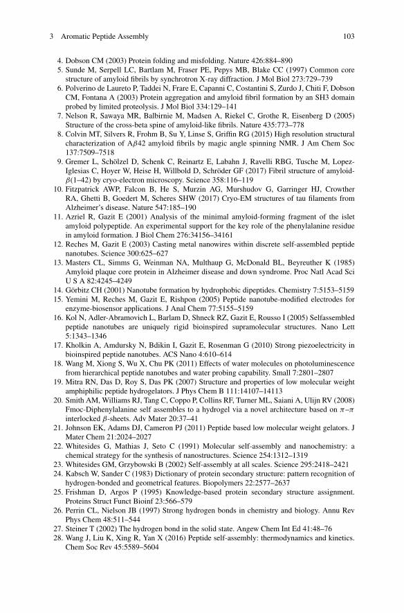
3 Aromatic Peptide Assembly 103
4. Dobson CM (2003) Protein folding and misfolding. Nature 426:884–8905. Sunde M, Serpell LC, Bartlam M, Fraser PE, Pepys MB, Blake CC (1997) Common core
structure of amyloid fibrils by synchrotron X-ray diffraction. J Mol Biol 273:729–7396. Polverino de Laureto P, Taddei N, Frare E, Capanni C, Costantini S, Zurdo J, Chiti F, Dobson
CM, Fontana A (2003) Protein aggregation and amyloid fibril formation by an SH3 domainprobed by limited proteolysis. J Mol Biol 334:129–141
7. Nelson R, Sawaya MR, Balbirnie M, Madsen A, Riekel C, Grothe R, Eisenberg D (2005)Structure of the cross-beta spine of amyloid-like fibrils. Nature 435:773–778
8. Colvin MT, Silvers R, Frohm B, Su Y, Linse S, Griffin RG (2015) High resolution structuralcharacterization of Aβ42 amyloid fibrils by magic angle spinning NMR. J Am Chem Soc137:7509–7518
9. Gremer L, Schölzel D, Schenk C, Reinartz E, Labahn J, Ravelli RBG, Tusche M, Lopez-Iglesias C, Hoyer W, Heise H, Willbold D, Schröder GF (2017) Fibril structure of amyloid-β(1–42) by cryo-electron microscopy. Science 358:116–119
10. Fitzpatrick AWP, Falcon B, He S, Murzin AG, Murshudov G, Garringer HJ, CrowtherRA, Ghetti B, Goedert M, Scheres SHW (2017) Cryo-EM structures of tau filaments fromAlzheimer’s disease. Nature 547:185–190
11. Azriel R, Gazit E (2001) Analysis of the minimal amyloid-forming fragment of the isletamyloid polypeptide. An experimental support for the key role of the phenylalanine residuein amyloid formation. J Biol Chem 276:34156–34161
12. Reches M, Gazit E (2003) Casting metal nanowires within discrete self-assembled peptidenanotubes. Science 300:625–627
13. Masters CL, Simms G, Weinman NA, Multhaup G, McDonald BL, Beyreuther K (1985)Amyloid plaque core protein in Alzheimer disease and down syndrome. Proc Natl Acad SciU S A 82:4245–4249
14. Görbitz CH (2001) Nanotube formation by hydrophobic dipeptides. Chemistry 7:5153–515915. Yemini M, Reches M, Gazit E, Rishpon (2005) Peptide nanotube-modified electrodes for
enzyme-biosensor applications. J Anal Chem 77:5155–515916. Kol N, Adler-Abramovich L, Barlam D, Shneck RZ, Gazit E, Rousso I (2005) Selfassembled
peptide nanotubes are uniquely rigid bioinspired supramolecular structures. Nano Lett5:1343–1346
17. Kholkin A, Amdursky N, Bdikin I, Gazit E, Rosenman G (2010) Strong piezoelectricity inbioinspired peptide nanotubes. ACS Nano 4:610–614
18. Wang M, Xiong S, Wu X, Chu PK (2011) Effects of water molecules on photoluminescencefrom hierarchical peptide nanotubes and water probing capability. Small 7:2801–2807
19. Mitra RN, Das D, Roy S, Das PK (2007) Structure and properties of low molecular weightamphiphilic peptide hydrogelators. J Phys Chem B 111:14107–14113
20. Smith AM, Williams RJ, Tang C, Coppo P, Collins RF, Turner ML, Saiani A, Ulijn RV (2008)Fmoc-Diphenylalanine self assembles to a hydrogel via a novel architecture based on π–π
interlocked β-sheets. Adv Mater 20:37–4121. Johnson EK, Adams DJ, Cameron PJ (2011) Peptide based low molecular weight gelators. J
Mater Chem 21:2024–202722. Whitesides G, Mathias J, Seto C (1991) Molecular self-assembly and nanochemistry: a
chemical strategy for the synthesis of nanostructures. Science 254:1312–131923. Whitesides GM, Grzybowski B (2002) Self-assembly at all scales. Science 295:2418–242124. Kabsch W, Sander C (1983) Dictionary of protein secondary structure: pattern recognition of
hydrogen-bonded and geometrical features. Biopolymers 22:2577–263725. Frishman D, Argos P (1995) Knowledge-based protein secondary structure assignment.
Proteins Struct Funct Bioinf 23:566–57926. Perrin CL, Nielson JB (1997) Strong hydrogen bonds in chemistry and biology. Annu Rev
Phys Chem 48:511–54427. Steiner T (2002) The hydrogen bond in the solid state. Angew Chem Int Ed 41:48–7628. Wang J, Liu K, Xing R, Yan X (2016) Peptide self-assembly: thermodynamics and kinetics.
Chem Soc Rev 45:5589–5604
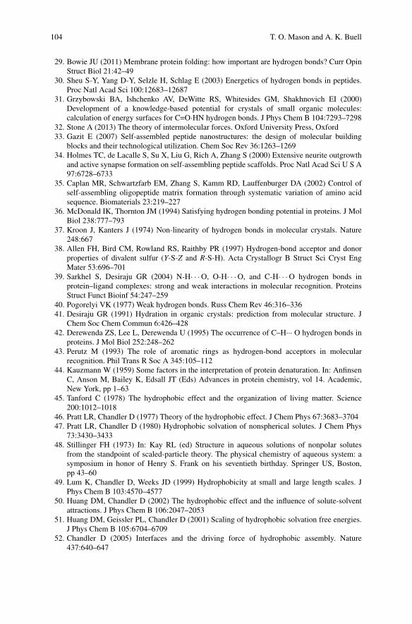
104 T. O. Mason and A. K. Buell
29. Bowie JU (2011) Membrane protein folding: how important are hydrogen bonds? Curr OpinStruct Biol 21:42–49
30. Sheu S-Y, Yang D-Y, Selzle H, Schlag E (2003) Energetics of hydrogen bonds in peptides.Proc Natl Acad Sci 100:12683–12687
31. Grzybowski BA, Ishchenko AV, DeWitte RS, Whitesides GM, Shakhnovich EI (2000)Development of a knowledge-based potential for crystals of small organic molecules:calculation of energy surfaces for C=O·HN hydrogen bonds. J Phys Chem B 104:7293–7298
32. Stone A (2013) The theory of intermolecular forces. Oxford University Press, Oxford33. Gazit E (2007) Self-assembled peptide nanostructures: the design of molecular building
blocks and their technological utilization. Chem Soc Rev 36:1263–126934. Holmes TC, de Lacalle S, Su X, Liu G, Rich A, Zhang S (2000) Extensive neurite outgrowth
and active synapse formation on self-assembling peptide scaffolds. Proc Natl Acad Sci U S A97:6728–6733
35. Caplan MR, Schwartzfarb EM, Zhang S, Kamm RD, Lauffenburger DA (2002) Control ofself-assembling oligopeptide matrix formation through systematic variation of amino acidsequence. Biomaterials 23:219–227
36. McDonald IK, Thornton JM (1994) Satisfying hydrogen bonding potential in proteins. J MolBiol 238:777–793
37. Kroon J, Kanters J (1974) Non-linearity of hydrogen bonds in molecular crystals. Nature248:667
38. Allen FH, Bird CM, Rowland RS, Raithby PR (1997) Hydrogen-bond acceptor and donorproperties of divalent sulfur (Y-S-Z and R-S-H). Acta Crystallogr B Struct Sci Cryst EngMater 53:696–701
39. Sarkhel S, Desiraju GR (2004) N-H· · · O, O-H· · · O, and C-H· · · O hydrogen bonds inprotein–ligand complexes: strong and weak interactions in molecular recognition. ProteinsStruct Funct Bioinf 54:247–259
40. Pogorelyi VK (1977) Weak hydrogen bonds. Russ Chem Rev 46:316–33641. Desiraju GR (1991) Hydration in organic crystals: prediction from molecular structure. J
Chem Soc Chem Commun 6:426–42842. Derewenda ZS, Lee L, Derewenda U (1995) The occurrence of C–H··· O hydrogen bonds in
proteins. J Mol Biol 252:248–26243. Perutz M (1993) The role of aromatic rings as hydrogen-bond acceptors in molecular
recognition. Phil Trans R Soc A 345:105–11244. Kauzmann W (1959) Some factors in the interpretation of protein denaturation. In: Anfinsen
C, Anson M, Bailey K, Edsall JT (Eds) Advances in protein chemistry, vol 14. Academic,New York, pp 1–63
45. Tanford C (1978) The hydrophobic effect and the organization of living matter. Science200:1012–1018
46. Pratt LR, Chandler D (1977) Theory of the hydrophobic effect. J Chem Phys 67:3683–370447. Pratt LR, Chandler D (1980) Hydrophobic solvation of nonspherical solutes. J Chem Phys
73:3430–343348. Stillinger FH (1973) In: Kay RL (ed) Structure in aqueous solutions of nonpolar solutes
from the standpoint of scaled-particle theory. The physical chemistry of aqueous system: asymposium in honor of Henry S. Frank on his seventieth birthday. Springer US, Boston,pp 43–60
49. Lum K, Chandler D, Weeks JD (1999) Hydrophobicity at small and large length scales. JPhys Chem B 103:4570–4577
50. Huang DM, Chandler D (2002) The hydrophobic effect and the influence of solute-solventattractions. J Phys Chem B 106:2047–2053
51. Huang DM, Geissler PL, Chandler D (2001) Scaling of hydrophobic solvation free energies.J Phys Chem B 105:6704–6709
52. Chandler D (2005) Interfaces and the driving force of hydrophobic assembly. Nature437:640–647
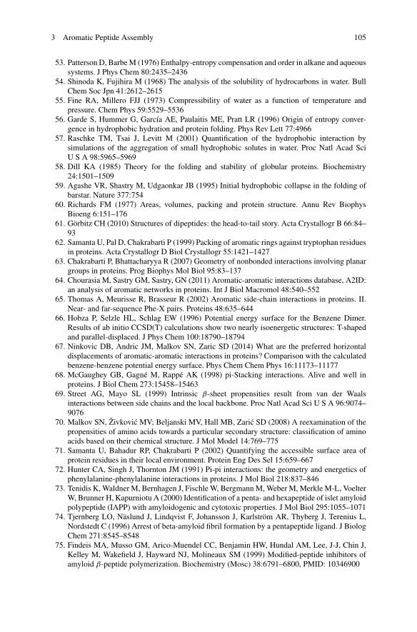
3 Aromatic Peptide Assembly 105
53. Patterson D, Barbe M (1976) Enthalpy-entropy compensation and order in alkane and aqueoussystems. J Phys Chem 80:2435–2436
54. Shinoda K, Fujihira M (1968) The analysis of the solubility of hydrocarbons in water. BullChem Soc Jpn 41:2612–2615
55. Fine RA, Millero FJJ (1973) Compressibility of water as a function of temperature andpressure. Chem Phys 59:5529–5536
56. Garde S, Hummer G, García AE, Paulaitis ME, Pratt LR (1996) Origin of entropy conver-gence in hydrophobic hydration and protein folding. Phys Rev Lett 77:4966
57. Raschke TM, Tsai J, Levitt M (2001) Quantification of the hydrophobic interaction bysimulations of the aggregation of small hydrophobic solutes in water. Proc Natl Acad SciU S A 98:5965–5969
58. Dill KA (1985) Theory for the folding and stability of globular proteins. Biochemistry24:1501–1509
59. Agashe VR, Shastry M, Udgaonkar JB (1995) Initial hydrophobic collapse in the folding ofbarstar. Nature 377:754
60. Richards FM (1977) Areas, volumes, packing and protein structure. Annu Rev BiophysBioeng 6:151–176
61. Görbitz CH (2010) Structures of dipeptides: the head-to-tail story. Acta Crystallogr B 66:84–93
62. Samanta U, Pal D, Chakrabarti P (1999) Packing of aromatic rings against tryptophan residuesin proteins. Acta Crystallogr D Biol Crystallogr 55:1421–1427
63. Chakrabarti P, Bhattacharyya R (2007) Geometry of nonbonded interactions involving planargroups in proteins. Prog Biophys Mol Biol 95:83–137
64. Chourasia M, Sastry GM, Sastry, GN (2011) Aromatic-aromatic interactions database, A2ID:an analysis of aromatic networks in proteins. Int J Biol Macromol 48:540–552
65. Thomas A, Meurisse R, Brasseur R (2002) Aromatic side-chain interactions in proteins. II.Near- and far-sequence Phe-X pairs. Proteins 48:635–644
66. Hobza P, Selzle HL, Schlag EW (1996) Potential energy surface for the Benzene Dimer.Results of ab initio CCSD(T) calculations show two nearly isoenergetic structures: T-shapedand parallel-displaced. J Phys Chem 100:18790–18794
67. Ninkovic DB, Andric JM, Malkov SN, Zaric SD (2014) What are the preferred horizontaldisplacements of aromatic-aromatic interactions in proteins? Comparison with the calculatedbenzene-benzene potential energy surface. Phys Chem Chem Phys 16:11173–11177
68. McGaughey GB, Gagné M, Rappé AK (1998) pi-Stacking interactions. Alive and well inproteins. J Biol Chem 273:15458–15463
69. Street AG, Mayo SL (1999) Intrinsic β-sheet propensities result from van der Waalsinteractions between side chains and the local backbone. Proc Natl Acad Sci U S A 96:9074–9076
70. Malkov SN, Živkovic MV; Beljanski MV, Hall MB, Zaric SD (2008) A reexamination of thepropensities of amino acids towards a particular secondary structure: classification of aminoacids based on their chemical structure. J Mol Model 14:769–775
71. Samanta U, Bahadur RP, Chakrabarti P (2002) Quantifying the accessible surface area ofprotein residues in their local environment. Protein Eng Des Sel 15:659–667
72. Hunter CA, Singh J, Thornton JM (1991) Pi-pi interactions: the geometry and energetics ofphenylalanine-phenylalanine interactions in proteins. J Mol Biol 218:837–846
73. Tenidis K, Waldner M, Bernhagen J, Fischle W, Bergmann M, Weber M, Merkle M-L, VoelterW, Brunner H, Kapurniotu A (2000) Identification of a penta- and hexapeptide of islet amyloidpolypeptide (IAPP) with amyloidogenic and cytotoxic properties. J Mol Biol 295:1055–1071
74. Tjernberg LO, Näslund J, Lindqvist F, Johansson J, Karlström AR, Thyberg J, Terenius L,Nordstedt C (1996) Arrest of beta-amyloid fibril formation by a pentapeptide ligand. J BiologChem 271:8545–8548
75. Findeis MA, Musso GM, Arico-Muendel CC, Benjamin HW, Hundal AM, Lee, J-J, Chin J,Kelley M, Wakefield J, Hayward NJ, Molineaux SM (1999) Modified-peptide inhibitors ofamyloid β-peptide polymerization. Biochemistry (Mosc) 38:6791–6800, PMID: 10346900
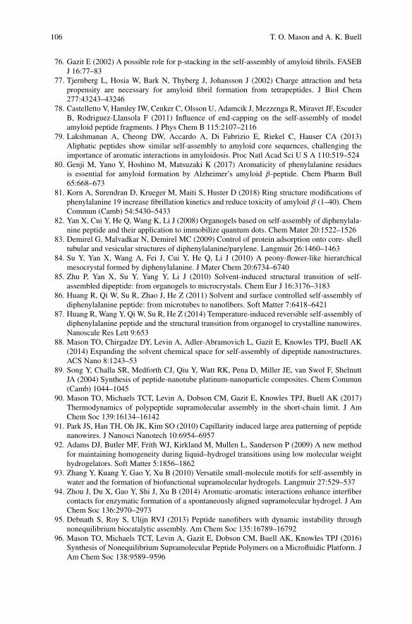
106 T. O. Mason and A. K. Buell
76. Gazit E (2002) A possible role for p-stacking in the self-assembly of amyloid fibrils. FASEBJ 16:77–83
77. Tjernberg L, Hosia W, Bark N, Thyberg J, Johansson J (2002) Charge attraction and betapropensity are necessary for amyloid fibril formation from tetrapeptides. J Biol Chem277:43243–43246
78. Castelletto V, Hamley IW, Cenker C, Olsson U, Adamcik J, Mezzenga R, Miravet JF, EscuderB, Rodriguez-Llansola F (2011) Influence of end-capping on the self-assembly of modelamyloid peptide fragments. J Phys Chem B 115:2107–2116
79. Lakshmanan A, Cheong DW, Accardo A, Di Fabrizio E, Riekel C, Hauser CA (2013)Aliphatic peptides show similar self-assembly to amyloid core sequences, challenging theimportance of aromatic interactions in amyloidosis. Proc Natl Acad Sci U S A 110:519–524
80. Genji M, Yano Y, Hoshino M, Matsuzaki K (2017) Aromaticity of phenylalanine residuesis essential for amyloid formation by Alzheimer’s amyloid β-peptide. Chem Pharm Bull65:668–673
81. Korn A, Surendran D, Krueger M, Maiti S, Huster D (2018) Ring structure modifications ofphenylalanine 19 increase fibrillation kinetics and reduce toxicity of amyloid β (1–40). ChemCommun (Camb) 54:5430–5433
82. Yan X, Cui Y, He Q, Wang K, Li J (2008) Organogels based on self-assembly of diphenylala-nine peptide and their application to immobilize quantum dots. Chem Mater 20:1522–1526
83. Demirel G, Malvadkar N, Demirel MC (2009) Control of protein adsorption onto core- shelltubular and vesicular structures of diphenylalanine/parylene. Langmuir 26:1460–1463
84. Su Y, Yan X, Wang A, Fei J, Cui Y, He Q, Li J (2010) A peony-flower-like hierarchicalmesocrystal formed by diphenylalanine. J Mater Chem 20:6734–6740
85. Zhu P, Yan X, Su Y, Yang Y, Li J (2010) Solvent-induced structural transition of self-assembled dipeptide: from organogels to microcrystals. Chem Eur J 16:3176–3183
86. Huang R, Qi W, Su R, Zhao J, He Z (2011) Solvent and surface controlled self-assembly ofdiphenylalanine peptide: from microtubes to nanofibers. Soft Matter 7:6418–6421
87. Huang R, Wang Y, Qi W, Su R, He Z (2014) Temperature-induced reversible self-assembly ofdiphenylalanine peptide and the structural transition from organogel to crystalline nanowires.Nanoscale Res Lett 9:653
88. Mason TO, Chirgadze DY, Levin A, Adler-Abramovich L, Gazit E, Knowles TPJ, Buell AK(2014) Expanding the solvent chemical space for self-assembly of dipeptide nanostructures.ACS Nano 8:1243–53
89. Song Y, Challa SR, Medforth CJ, Qiu Y, Watt RK, Pena D, Miller JE, van Swol F, ShelnuttJA (2004) Synthesis of peptide-nanotube platinum-nanoparticle composites. Chem Commun(Camb) 1044–1045
90. Mason TO, Michaels TCT, Levin A, Dobson CM, Gazit E, Knowles TPJ, Buell AK (2017)Thermodynamics of polypeptide supramolecular assembly in the short-chain limit. J AmChem Soc 139:16134–16142
91. Park JS, Han TH, Oh JK, Kim SO (2010) Capillarity induced large area patterning of peptidenanowires. J Nanosci Nanotech 10:6954–6957
92. Adams DJ, Butler MF, Frith WJ, Kirkland M, Mullen L, Sanderson P (2009) A new methodfor maintaining homogeneity during liquid–hydrogel transitions using low molecular weighthydrogelators. Soft Matter 5:1856–1862
93. Zhang Y, Kuang Y, Gao Y, Xu B (2010) Versatile small-molecule motifs for self-assembly inwater and the formation of biofunctional supramolecular hydrogels. Langmuir 27:529–537
94. Zhou J, Du X, Gao Y, Shi J, Xu B (2014) Aromatic-aromatic interactions enhance interfibercontacts for enzymatic formation of a spontaneously aligned supramolecular hydrogel. J AmChem Soc 136:2970–2973
95. Debnath S, Roy S, Ulijn RVJ (2013) Peptide nanofibers with dynamic instability throughnonequilibrium biocatalytic assembly. Am Chem Soc 135:16789–16792
96. Mason TO, Michaels TCT, Levin A, Gazit E, Dobson CM, Buell AK, Knowles TPJ (2016)Synthesis of Nonequilibrium Supramolecular Peptide Polymers on a Microfluidic Platform. JAm Chem Soc 138:9589–9596
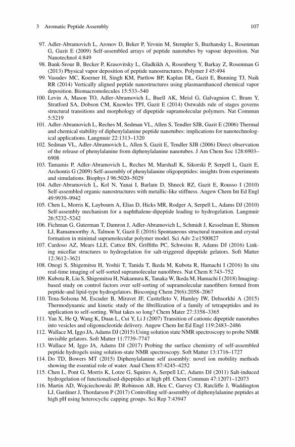
3 Aromatic Peptide Assembly 107
97. Adler-Abramovich L, Aronov D, Beker P, Yevnin M, Stempler S, Buzhansky L, RosenmanG, Gazit E (2009) Self-assembled arrays of peptide nanotubes by vapour deposition. NatNanotechnol 4:849
98. Bank-Srour B, Becker P, Krasovitsky L, Gladkikh A, Rosenberg Y, Barkay Z, Rosenman G(2013) Physical vapor deposition of peptide nanostructures. Polymer J 45:494
99. Vasudev MC, Koerner H, Singh KM, Partlow BP, Kaplan DL, Gazit E, Bunning TJ, NaikRR (2014) Vertically aligned peptide nanostructures using plasmaenhanced chemical vapordeposition. Biomacromolecules 15:533–540
100. Levin A, Mason TO, Adler-Abramovich L, Buell AK, Meisl G, Galvagnion C, Bram Y,Stratford SA, Dobson CM, Knowles TPJ, Gazit E (2014) Ostwalds rule of stages governsstructural transitions and morphology of dipeptide supramolecular polymers. Nat Commun5:5219
101. Adler-Abramovich L, Reches M, Sedman VL, Allen S, Tendler SJB, Gazit E (2006) Thermaland chemical stability of diphenylalanine peptide nanotubes: implications for nanotechnolog-ical applications. Langmuir 22:1313–1320
102. Sedman VL, Adler-Abramovich L, Allen S, Gazit E, Tendler SJB (2006) Direct observationof the release of phenylalanine from diphenylalanine nanotubes. J Am Chem Soc 128:6903–6908
103. Tamamis P, Adler-Abramovich L, Reches M, Marshall K, Sikorski P, Serpell L, Gazit E,Archontis G (2009) Self-assembly of phenylalanine oligopeptides: insights from experimentsand simulations. Biophys J 96:5020–5029
104. Adler-Abramovich L, Kol N, Yanai I, Barlam D, Shneck RZ, Gazit E, Rousso I (2010)Self-assembled organic nanostructures with metallic-like stiffness. Angew Chem Int Ed Engl49:9939–9942
105. Chen L, Morris K, Laybourn A, Elias D, Hicks MR, Rodger A, Serpell L, Adams DJ (2010)Self-assembly mechanism for a naphthalene-dipeptide leading to hydrogelation. Langmuir26:5232–5242
106. Fichman G, Guterman T, Damron J, Adler-Abramovich L, Schmidt J, Kesselman E, ShimonLJ, Ramamoorthy A, Talmon Y, Gazit E (2016) Spontaneous structural transition and crystalformation in minimal supramolecular polymer model. Sci Adv 2:e1500827
107. Cardoso AZ, Mears LLE, Cattoz BN, Griffiths PC, Schweins R, Adams DJ (2016) Link-ing micellar structures to hydrogelation for salt-triggered dipeptide gelators. Soft Matter12:3612–3621
108. Onogi S, Shigemitsu H, Yoshii T, Tanida T, Ikeda M, Kubota R, Hamachi I (2016) In situreal-time imaging of self-sorted supramolecular nanofibres. Nat Chem 8:743–752
109. Kubota R, Liu S, Shigemitsu H, Nakamura K, Tanaka W, Ikeda M, Hamachi I (2018) Imaging-based study on control factors over self-sorting of supramolecular nanofibers formed frompeptide-and lipid-type hydrogelators. Bioconjug Chem 29(6):2058–2067
110. Tena-Solsona M, Escuder B, Miravet JF, Casttelleto V, Hamley IW, Dehsorkhi A (2015)Thermodynamic and kinetic study of the fibrillization of a family of tetrapeptides and itsapplication to self-sorting. What takes so long? Chem Mater 27:3358–3365
111. Yan X, He Q, Wang K, Duan L, Cui Y, Li J (2007) Transition of cationic dipeptide nanotubesinto vesicles and oligonucleotide delivery. Angew Chem Int Ed Engl 119:2483–2486
112. Wallace M, Iggo JA, Adams DJ (2015) Using solution state NMR spectroscopy to probe NMRinvisible gelators. Soft Matter 11:7739–7747
113. Wallace M, Iggo JA, Adams DJ (2017) Probing the surface chemistry of self-assembledpeptide hydrogels using solution-state NMR spectroscopy. Soft Matter 13:1716–1727
114. Do TD, Bowers MT (2015) Diphenylalanine self assembly: novel ion mobility methodsshowing the essential role of water. Anal Chem 87:4245–4252
115. Chen L, Pont G, Morris K, Lotze G, Squires A, Serpell LC, Adams DJ (2011) Salt-inducedhydrogelation of functionalised-dipeptides at high pH. Chem Commun 47:12071–12073
116. Martin AD, Wojciechowski JP, Robinson AB, Heu C, Garvey CJ, Ratcliffe J, WaddingtonLJ, Gardiner J, Thordarson P (2017) Controlling self-assembly of diphenylalanine peptides athigh pH using heterocyclic capping groups. Sci Rep 7:43947
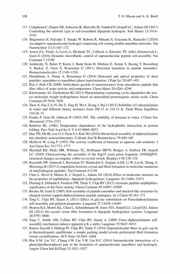
108 T. O. Mason and A. K. Buell
117. Colquhoun C, Draper ER, Schweins R, Marcello M, Vadukul D, Serpell LC, Adams DJ (2017)Controlling the network type in self-assembled dipeptide hydrogels. Soft Matter 13:1914–1919
118. Shigemitsu H, Fujisaku T, Tanaka W, Kubota R, Minami S, Urayama K, Hamachi I (2018)An adaptive supramolecular hydrogel comprising self-sorting double nanofibre networks. NatNanotechnol 1(13):165–172
119. Arnon ZA, Vitalis A, Levin A, Michaels TC, Caflisch A, Knowles TP, Adler-Abramovich L,Gazit E (2016) Dynamic microfluidic control of supramolecular peptide self-assembly. NatCommun 7:13190
120. Amdursky N, Beker P, Koren I, Bank-Srour B, Mishina E, Semin S, Rasing T, RosenbergY, Barkay Z, Gazit E, Rosenman G (2011) Structural transition in peptide nanotubes.Biomacromolecules 12:1349–1354
121. Handelman A, Natan A, Rosenman G (2014) Structural and optical properties of shortpeptides: nanotubes-to-nanofibers phase transformation. J Pept Sci 20:487–493
122. Ryu J, Park CB (2008) Solid-phase growth of nanostructures from amorphous peptide thinfilm: effect of water activity and temperature. Chem Mater 20:4284–4290
123. Kleinsmann AJ, Nachtsheim BJ (2013) Phenylalanine-containing cyclic dipeptides–the low-est molecular weight hydrogelators based on unmodified proteinogenic amino acids. ChemCommun 49:7818–7820
124. Zhou X, Fan J, Li N, Du Z, Ying H, Wu J, Xiong J, Bai J (2012) Solubility of l-phenylalaninein water and different binary mixtures from 288.15 to 318.15 K. Fluid Phase Equilibria316:26–33
125. Franks F, Gent M, Johnson H (1963) 505. The solubility of benzene in water. J Chem Soc(Resumed) 2716–2723
126. Baldwin RL (1986) Temperature dependence of the hydrophobic interaction in proteinfolding. Proc Natl Acad Sci U S A 83:8069–8072
127. Han TH, Oh JK, Lee G-J, Pyun S-I, Kim SO (2010) Hierarchical assembly of diphenylalanineinto dendritic nanoarchitectures. Colloids Surf B Biointerfaces 79:440–445
128. McDevit W, Long F (1952) The activity coefficient of benzene in aqueous salt solutions. JAm Chem Soc 74:1773–1777
129. Marshall KE, Hicks MR, Williams TL, Hoffmann SRVN, Rodger A, Dafforn TR, SerpellLC (2010) Characterizing the assembly of the Sup35 yeast prion fragment, GNNQQNY:structural changes accompany a fiber-to-crystal switch. Biophys J 98:330–338
130. Reynolds NP, Adamcik J, Berryman JT, Handschin S, Zanjani AAH, Li W, Liu K, Zhang A,Mezzenga R (2017) Competition between crystal and fibril formation in molecular mutationsof amyloidogenic peptides. Nat Commun 8:1338
131. Chen L, Revel S, Morris K, C Serpell L, Adams DJ (2010) Effect of molecular structure onthe properties of naphthalene- dipeptide hydrogelators. Langmuir 26:13466–13471
132. Fleming S, Debnath S, Frederix PW, Tuttle T, Ulijn RV (2013) Aromatic peptide amphiphiles:significance of the Fmoc moiety. Chem Commun 49:10587–10589
133. Reches M, Gazit E (2005) Self-assembly of peptide nanotubes and amyloid-like structures bycharged-termini-capped diphenylalanine peptide analogues. Isr J Chem 45:363–371
134. Tang C, Ulijn RV, Saiani A (2011) Effect of glycine substitution on Fmocdiphenylalanineself-assembly and gelation properties. Langmuir 27:14438–14449
135. Houton KA, Morris KL, Chen L, Schmidtmann M, Jones JTA, Serpell LC, Lloyd GO, AdamsDJ (2012) On crystal versus fiber formation in dipeptide hydrogelator systems. Langmuir28:9797–9806
136. Tang C, Smith AM, Collins RF, Ulijn RV, Saiani A (2009) Fmoc-diphenylalanine self-assembly mechanism induces apparent p K a shifts. Langmuir 25:9447–9453
137. Ramos Sasselli I, Halling PJ, Ulijn RV, Tuttle T (2016) Supramolecular fibers in gels can beat thermodynamic equilibrium: a simple packing model reveals preferential fibril formationversus crystallization. ACS Nano 10:2661–2668
138. Hsu S-M, Lin Y-C, Chang J-W, Liu Y-H, Lin H-C (2014) Intramolecular interactions of aphenyl/perfluorophenyl pair in the formation of supramolecular nanofibers and hydrogels.Angew Chem Intl Ed Engl 53:1921–1927
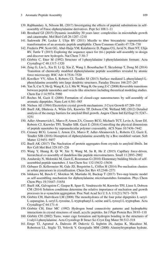
3 Aromatic Peptide Assembly 109
139. Rajbhandary A, Nilsson BL (2017) Investigating the effects of peptoid substitutions in self-assembly of Fmoc-diphenylalanine derivatives. Pept Sci 108:1–11
140. Brouhard GJ (2015) Dynamic instability 30 years later: complexities in microtubule growthand catastrophe. Mol Biol Cell 26:1207–1210
141. Sadownik JW, Leckie J, Ulijn RV (2011) Micelle to fibre biocatalytic supramoleculartransformation of an aromatic peptide amphiphile. Chem Commun (Camb) 47:728–730
142. Frederix PW, Scott GG, Abul-Haija YM, Kalafatovic D, Pappas CG, Javid N, Hunt NT, UlijnRV, Tuttle T (2015) Exploring the sequence space for (tri-) peptide self-assembly to designand discover new hydrogels. Nat Chem 7:30–7
143. Görbitz C, Etter M (1992) Structure of l-phenylalanine l-phenylalaninium formate. ActaCrystallogr C 48:1317–1320
144. Zeng G, Liu L, Xia D, Li Q, Xin Z, Wang J, Besenbacher F, Skrydstrup T, Dong M (2014)Transition of chemically modified diphenylalanine peptide assemblies revealed by atomicforce microscopy. RSC Adv 4:7516–7520
145. Korolkov VV, Allen S, Roberts CJ, Tendler SJ (2013) Surface mediated L-phenylalanyl-L-phenylalanine assembly into large dendritic structures. Faraday Discuss 166:257–267
146. Yan X, Cui Y, He Q, Wang K, Li J, Mu W, Wang B, Ou-yang Z-C (2008) Reversible transitionsbetween peptide nanotubes and vesicle-like structures including theoretical modeling studies.Chem Eur J 14:5974–5980
147. Reches M, Gazit E (2004) Formation of closed-cage nanostructures by self-assembly ofaromatic dipeptides. Nano Lett 4:581–585
148. Nielsen AE (1984) Electrolyte crystal growth mechanisms. J Cryst Growth 67:289–310149. Buell AK, Dhulesia A, White DA, Knowles TP, Dobson CM, Welland ME (2012) Detailed
analysis of the energy barriers for amyloid fibril growth. Angew Chem Intl Ed Engl 51:5247–5251
150. Adler-Abramovich L, Marco P, Arnon ZA, Creasey RCG, Michaels TCT, Levin A, Scurr DJ,Roberts CJ, Knowles TPJ, Tendler SJB, Gazit E (2016) Controlling the physical dimensionsof peptide nanotubes by supramolecular polymer coassembly. ACS Nano 10:7436–7442
151. Creasey RCG, Louzao I, Arnon ZA, Marco P, Adler-Abramovich L, Roberts CJ, Gazit E,Tendler SJB (2016) Disruption of diphenylalanine assembly by a Boc-modified variant. SoftMatter 12:9451–9457
152. Buell AK (2017) The Nucleation of protein aggregates-from crystals to amyloid fibrils. IntRev Cell Mol Biol 329:187–226
153. Wang Y, Huang R, Qi W, Xie Y, Wang M, Su R, He Z (2015) Capillary force-driven,hierarchical co-assembly of dandelion-like peptide microstructures. Small 11:2893–2902
154. Amdursky N, Molotskii M, Gazit E, Rosenman G (2010) Elementary building blocks of self-assembled peptide nanotubes. J Am Chem Soc 132:15632–15636
155. Gebauer D, Kellermeier M, Gale JD, Bergström L, Cölfen H (2014) Pre-nucleation clustersas solute precursors in crystallisation. Chem Soc Rev 43:2348–2371
156. Ishikawa M, Busch C, Motzkus M, Martinho H, Buckup T (2017) Two-step kinetic modelas self-assembling mechanism for diphenylalanine micro/nanotubes formation. Phys ChemChem Phys 19:31647–31654
157. Buell AK, Galvagnion C, Gaspar R, Sparr E, Vendruscolo M, Knowles TPJ, Linse S, DobsonCM (2014) Solution conditions determine the relative importance of nucleation and growthprocesses in α-synuclein aggregation. Proc Natl Acad Sci U S A 111(21):7671–7676
158. Görbitz CH, Hartviksen LM (2008) The monohydrates of the four polar dipeptides L-seryl-L-asparagine, L-seryl-L-tyrosine, L-tryptophanyl-L-serine and L-tyrosyl-L-tryptophan. ActaCrystallogr C 64:171–6
159. Görbitz CH, Etter MC (1992) Hydrogen bond connectivity patterns and hydrophobicinteractions in crystal structures of small, acyclic peptides. Int J Pept Protein Res 39:93–110
160. Görbitz CH (2002) Turns, water cage formation and hydrogen bonding in the structures ofl-valyl-l-phenylalanine. Acta Crystallogr B Struct Sci Cryst Eng Mater 58:512–518
161. Emge TJ, Agrawal A, Dalessio JP, Dukovic G, Inghrim JA, Janjua K, Macaluso M,Robertson LL, Stiglic TJ, Volovik Y, Georgiadis MM (2000) Alaninyltryptophan hydrate,
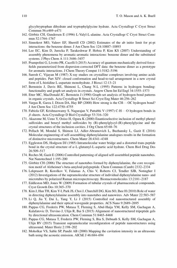
110 T. O. Mason and A. K. Buell
glycyltryptophan dihydrate and tryptophylglycine hydrate. Acta Crystallogr C Cryst StructCommun 56:e469–e471
162. Görbitz CH, Gundersen E (1996) L-Valyl-L-alanine. Acta Crystallogr C Cryst Struct Com-mun 52:1764–1767
163. Sinnokrot MO, Valeev EF, Sherrill CD (2002) Estimates of the ab initio limit for pi-piinteractions: the benzene dimer. J Am Chem Soc 124:10887–10893
164. Lee EC, Kim D, Jurecka P, Tarakeshwar P, Hobza P, Kim KS (2007) Understanding ofassembly phenomena by aromatic-aromatic interactions: benzene dimer and the substitutedsystems. J Phys Chem A 111:3446–3457
165. Prampolini G, Livotto PR, Cacelli I (2015) Accuracy of quantum mechanically derived force-fields parameterized from dispersion-corrected DFT data: the benzene dimer as a prototypefor aromatic interactions. J Chem Theory Comput 11:5182–5196
166. Suresh C, Vijayan M (1987) X-ray studies on crystalline complexes involving amino acidsand peptides. Part XIV: closed conformation and head-to-tail arrangement in a new crystalform of L-histidine L-aspartate monohydrate. J Biosci 12:13–21
167. Bernstein J, Davis RE, Shimoni L, Chang N-L (1995) Patterns in hydrogen bonding:functionality and graph set analysis in crystals. Angew Chem Int Ed Engl 34:1555–1573
168. Etter MC, MacDonald JC, Bernstein J (1990) Graph-set analysis of hydrogen-bond patternsin organic crystals. Acta Crystallogr B Struct Sci Cryst Eng Mater 46:256–262
169. Vargas R, Garza J, Dixon DA, Hay BP (2000) How strong is the CH· · · OC hydrogen bond?J Am Chem Soc 122:4750–4755
170. Fabiola GF, Krishnaswamy S, Nagarajan V, Pattabhi V (1997) C-H· · · O hydrogen bonds inβ-sheets. Acta Crystallogr D Biol Crystallogr 53:316–320
171. Akazome M, Ueno Y, Ooiso H, Ogura K (2000) Enantioselective inclusion of methyl phenylsulfoxides and benzyl methyl sulfoxides by (R)-phenylglycyl-(R)-phenylglycine and thecrystal structures of the inclusion cavities. J Org Chem 65:68–76
172. Pellach M, Mondal S, Shimon LJ, Adler-Abramovich L, Buzhansky L, Gazit E (2016)Molecular engineering of self-assembling diphenylalanine analogues results in the formationof distinctive microstructures. Chem Mater 28:4341–4348
173. Eggleston DS, Hodgson DJ (1985) Intramolecular water bridge and a distorted trans peptidebond in the crystal structure of α-L-glutamyl-L-aspartic acid hydrate. Chem Biol Drug Des26:509–517
174. Reches M, Gazit E (2006) Controlled patterning of aligned self-assembled peptide nanotubes.Nat Nanotechnol 1:195–200
175. Görbitz CH (2006) The structure of nanotubes formed by diphenylalanine, the core recogni-tion motif of Alzheimer’s beta-amyloid polypeptide. Chem Commun (Camb) 2332–2334
176. Lekprasert B, Korolkov V, Falamas A, Chis V, Roberts CJ, Tendler SJB, Notingher I(2012) Investigations of the supramolecular structure of individual diphenylalanine nano- andmicrotubes by polarized Raman microspectroscopy. Biomacromolecules 13:2181–2187
177. Eddleston MD, Jones W (2009) Formation of tubular crystals of pharmaceutical compounds.Cryst Growth Des 10:365–370
178. Kim J, Han TH, Kim Y-I, Park JS, Choi J, Churchill DG, Kim SO, Ihee H (2010) Role of waterin directing diphenylalanine assembly into nanotubes and nanowires. Adv Mater 22:583–587
179. Li Q, Jia Y, Dai L, Yang Y, Li J (2015) Controlled rod nanostructured assembly ofdiphenylalanine and their optical waveguide properties. ACS Nano 9:2689–2695
180. Pappas CG, Frederix PW, Mutasa T, Fleming S, Abul-Haija YM, Kelly SM, Gachagan A,Kalafatovic D, Trevino J, Ulijn R, Bai S (2015) Alignment of nanostructured tripeptide gelsby directional ultrasonication. Chem Commun 51:8465–8468
181. Pappas CG, Mutasa T, Frederix PW, Fleming S, Bia S, Debnath S, Kelly SM, Gachagan A,Ulijn RV (2015) Transient supramolecular reconfiguration of peptide nanostructures usingultrasound. Mater Horiz 2:198–202
182. Moholkar VS, Sable SP, Pandit AB (2000) Mapping the cavitation intensity in an ultrasonicbath using the acoustic emission. AIChE J 46:684–694

3 Aromatic Peptide Assembly 111
183. Tjernberg LO, Callaway DJ, Tjernberg A, Hahne S, Lilliehöök C, Terenius L, Thyberg J,Nordstedt C (1999) A molecular model of Alzheimer amyloid β-peptide fibril formation. JBiol Chem 274:12619–12625
184. Mazor Y, Gilead S, Benhar I, Gazit E (2002) Identification and characterization of a novelmolecular-recognition and self-assembly domain within the islet amyloid polypeptide. J MolBiol 322:1013–1024
185. Gazit E (2005) Mechanisms of amyloid fibril self-assembly and inhibition. Model shortpeptides as a key research tool. FEBS J 272:5971–5978
186. Jahn TR, Makin OS, Morris KL, Marshall KE, Tian P, Sikorski P, Serpell LC (2010) Thecommon architecture of cross-β amyloid. J Mol Biol 395:717–727
187. Carugo O, Djinovic-Carugo K (2013) Half a century of Ramachandran plots. Acta CrystallogrD Biol Crystallogr 69:1333–1341
188. Hollingsworth SA, Karplus PA (2010) A fresh look at the Ramachandran plot and theoccurrence of standard structures in proteins. Biomol Concepts 1:271–283
189. Berman HM, Westbrook J, Feng Z, Gilliland G, Bhat TN, Weissig H, Shindyalov IN, BournePE (2000) The protein data bank. Nucleic Acids Res 28:235–242
190. Schrödinger LLC (2015) The PyMOL Molecular Graphics System, Version 2.1.0191. Di Maro S et al (2016) Exploring the N-terminal region of C-X-C motif chemokine 12
(CXCL12): identification of plasma-stable cyclic peptides as novel, potent C-X-C chemokinereceptor type 4 (CXCR4) antagonists. J Med Chem 59:8369–8380, PMID: 27571038
192. Colletier J-P, Laganowsky A, Landau M, Zhao M, Soriaga AB, Goldschmidt L, Flot D, CascioD, Sawaya MR, Eisenberg D (2011) Molecular basis for amyloid-β polymorphism. Proc NatlAcad Sci U S A 108:16938–16943
193. Spencer RK, Li H, Nowick JS (2014) X-ray crystallographic structures of trimers and higher-order oligomeric assemblies of a peptide derived from Aβ 17–36. J Am Chem Soc 136:5595–5598
194. Kreutzer AG, Hamza IL, Spencer RK, Nowick JS (2016) X-ray crystallographic structuresof a trimer, dodecamer, and annular pore formed by an Aβ17–36 β-hairpin. J Am Chem Soc138:4634–4642, PMID: 26967810
195. Wälti MA, Ravotti F, Arai H, Glabe CG, Wall JS, Böckmann A, Güntert P, Meier BH, Riek R(2016) Atomic-resolution structure of a disease-relevant Aβ (1–42) amyloid fibril. Proc NatlAcad Sci U S A 113:E4976–E4984
196. Perutz MF, Finch JT, Berriman J, Lesk A (2002) Amyloid fibers are water-filled nanotubes.Proc Natl Acad Sci U S A 99:5591–5595
197. Sikorski P, Atkins E (2005) New model for crystalline polyglutamine assemblies and theirconnection with amyloid fibrils. Biomacromolecules 6:425–432
198. Buchanan LE, Carr JK, Fluitt AM, Hoganson AJ, Moran SD, de Pablo JJ, Skinner JL, ZanniMT (2014) Structural motif of polyglutamine amyloid fibrils discerned with mixed-isotopeinfrared spectroscopy. Proc Natl Acad Sci 111:5796–5801
199. Makin OS, Serpell LC (2005) Structures for amyloid fibrils. FEBS J 272:5950–5961200. Harper JD, Lieber CM, Lansbury PT (1997) Atomic force microscopic imaging of seeded
fibril formation and fibril branching by the Alzheimer’s disease amyloid-β protein. ChemBiol 4:951–959
201. Chothia C (1973) Conformation of twisted β-pleated sheets in proteins. J Mol Biol 75:295–302
202. Aggeli A, Nyrkova IA, Bell M, Harding R, Carrick L, McLeish TC, Semenov AN, Boden N(2001) Hierarchical self-assembly of chiral rod-like molecules as a model for peptide β-sheettapes, ribbons, fibrils, and fibers. Proc Natl Acad Sci USA 98:11857–11862
203. Usov I, Adamcik J, Mezzenga R (2013) Polymorphism complexity and handedness inversionin serum albumin amyloid fibrils. ACS Nano 7:10465–10474
204. Wang S-T, Lin Y, Spencer RK, Thomas MR, Nguyen AI, Amdursky N, Pashuck ET,Skaalure SC, Song CY, Parmar PA, Morgan RM, Ercius P, Aloni S, Zuckermann RN, StevensMM (2017) Sequence-dependent self-assembly and structural diversity of Islet amyloidpolypeptide-derived β-sheet fibrils. ACS Nano 11:8579–8589, PMID: 28771324
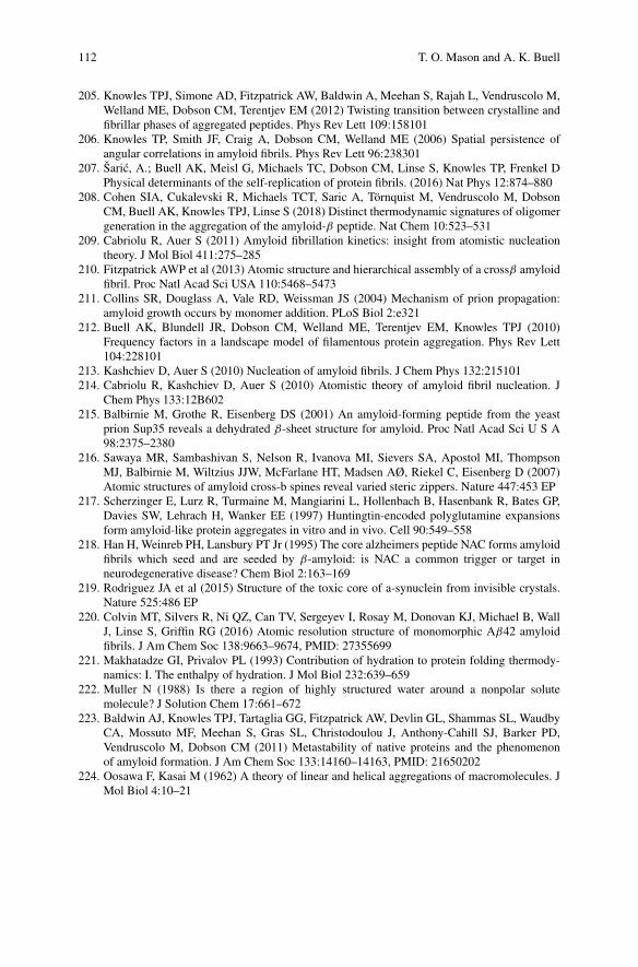
112 T. O. Mason and A. K. Buell
205. Knowles TPJ, Simone AD, Fitzpatrick AW, Baldwin A, Meehan S, Rajah L, Vendruscolo M,Welland ME, Dobson CM, Terentjev EM (2012) Twisting transition between crystalline andfibrillar phases of aggregated peptides. Phys Rev Lett 109:158101
206. Knowles TP, Smith JF, Craig A, Dobson CM, Welland ME (2006) Spatial persistence ofangular correlations in amyloid fibrils. Phys Rev Lett 96:238301
207. Šaric, A.; Buell AK, Meisl G, Michaels TC, Dobson CM, Linse S, Knowles TP, Frenkel DPhysical determinants of the self-replication of protein fibrils. (2016) Nat Phys 12:874–880
208. Cohen SIA, Cukalevski R, Michaels TCT, Saric A, Törnquist M, Vendruscolo M, DobsonCM, Buell AK, Knowles TPJ, Linse S (2018) Distinct thermodynamic signatures of oligomergeneration in the aggregation of the amyloid-β peptide. Nat Chem 10:523–531
209. Cabriolu R, Auer S (2011) Amyloid fibrillation kinetics: insight from atomistic nucleationtheory. J Mol Biol 411:275–285
210. Fitzpatrick AWP et al (2013) Atomic structure and hierarchical assembly of a crossβ amyloidfibril. Proc Natl Acad Sci USA 110:5468–5473
211. Collins SR, Douglass A, Vale RD, Weissman JS (2004) Mechanism of prion propagation:amyloid growth occurs by monomer addition. PLoS Biol 2:e321
212. Buell AK, Blundell JR, Dobson CM, Welland ME, Terentjev EM, Knowles TPJ (2010)Frequency factors in a landscape model of filamentous protein aggregation. Phys Rev Lett104:228101
213. Kashchiev D, Auer S (2010) Nucleation of amyloid fibrils. J Chem Phys 132:215101214. Cabriolu R, Kashchiev D, Auer S (2010) Atomistic theory of amyloid fibril nucleation. J
Chem Phys 133:12B602215. Balbirnie M, Grothe R, Eisenberg DS (2001) An amyloid-forming peptide from the yeast
prion Sup35 reveals a dehydrated β-sheet structure for amyloid. Proc Natl Acad Sci U S A98:2375–2380
216. Sawaya MR, Sambashivan S, Nelson R, Ivanova MI, Sievers SA, Apostol MI, ThompsonMJ, Balbirnie M, Wiltzius JJW, McFarlane HT, Madsen AØ, Riekel C, Eisenberg D (2007)Atomic structures of amyloid cross-b spines reveal varied steric zippers. Nature 447:453 EP
217. Scherzinger E, Lurz R, Turmaine M, Mangiarini L, Hollenbach B, Hasenbank R, Bates GP,Davies SW, Lehrach H, Wanker EE (1997) Huntingtin-encoded polyglutamine expansionsform amyloid-like protein aggregates in vitro and in vivo. Cell 90:549–558
218. Han H, Weinreb PH, Lansbury PT Jr (1995) The core alzheimers peptide NAC forms amyloidfibrils which seed and are seeded by β-amyloid: is NAC a common trigger or target inneurodegenerative disease? Chem Biol 2:163–169
219. Rodriguez JA et al (2015) Structure of the toxic core of a-synuclein from invisible crystals.Nature 525:486 EP
220. Colvin MT, Silvers R, Ni QZ, Can TV, Sergeyev I, Rosay M, Donovan KJ, Michael B, WallJ, Linse S, Griffin RG (2016) Atomic resolution structure of monomorphic Aβ42 amyloidfibrils. J Am Chem Soc 138:9663–9674, PMID: 27355699
221. Makhatadze GI, Privalov PL (1993) Contribution of hydration to protein folding thermody-namics: I. The enthalpy of hydration. J Mol Biol 232:639–659
222. Muller N (1988) Is there a region of highly structured water around a nonpolar solutemolecule? J Solution Chem 17:661–672
223. Baldwin AJ, Knowles TPJ, Tartaglia GG, Fitzpatrick AW, Devlin GL, Shammas SL, WaudbyCA, Mossuto MF, Meehan S, Gras SL, Christodoulou J, Anthony-Cahill SJ, Barker PD,Vendruscolo M, Dobson CM (2011) Metastability of native proteins and the phenomenonof amyloid formation. J Am Chem Soc 133:14160–14163, PMID: 21650202
224. Oosawa F, Kasai M (1962) A theory of linear and helical aggregations of macromolecules. JMol Biol 4:10–21
Chapter 4Bacterial Amyloids: Biogenesisand Biomaterials
Line Friis Bakmann Christensen, Nicholas Schafer, Adriana Wolf-Perez,Daniel Jhaf Madsen, and Daniel E. Otzen
Abstract Functional amyloid (FuBA) is produced by a large fraction of all bacterialspecies and represents a constructive use of the stable amyloid fold, in contrastto the pathological amyloid seen in neurodegenerative diseases. When assembledinto amyloid, FuBA is unusually robust and withstands most chemicals includingdenaturants and SDS. Uses include strengthening of bacterial biofilms, cell-to-cell communication, cell wall construction and even bacterial warfare. Biogenesisis under tight spatio-temporal control, thanks to a simple but efficient secretionsystem which in E. coli, Pseudomonas and other well-studied bacteria includesa major amyloid component that is kept unfolded in the periplasm thanks tochaperones, threaded through the outer membrane via a pore protein and anchoredto the cell surface through a nucleator and possibly other helper proteins. Inthese systems, amyloid formation is promoted through imperfect repeats, but otherevolutionarily unrelated proteins either have no or only partially conserved repeatsor simply consist of small peptides with multiple structural roles. This makesbioinformatics analysis challenging, though the sophisticated amyloid predictiontools developed from research in pathological amyloid together with the steadyincrease in identification of further examples of amyloid will strengthen genomicdata mining. Functional amyloid represents an intriguing source of robust yetbiodegradable materials with new properties, when combining the optimized self-assembly properties of the amyloid component with e.g. peptides with differentbinding properties or surface-reactive protein binders. Sophisticated patterns canalso be obtained by co-incubating bacteria producing different types of amyloid,while amyloid inclusion bodies may lead to slow-release nanopills.
Keywords Curli · Functional amyloid in Pseudomonas · Sequence analysis ·Fusion proteins with binding properties · Screening systems
L. F. B. Christensen · N. Schafer · A. Wolf-Perez · D. J. Madsen · D. E. Otzen (�)iNANO and Department of Molecular Biology and Genetics, Aarhus University, Aarhus,Denmarke-mail: [email protected]
© Springer Nature Singapore Pte Ltd. 2019S. Perrett et al. (eds.), Biological and Bio-inspired Nanomaterials,Advances in Experimental Medicine and Biology 1174,https://doi.org/10.1007/978-981-13-9791-2_4
113
114 L. F. B. Christensen et al.
4.1 Introduction
The amyloid β-sheet consists of a repetitive pattern of β-strands stacking on topof each other to form long fibrillary structure, whose axis is orthogonal to the β-strand (hence “cross- β”). The β-sheet secondary structure arranges the peptidebackbone in an extended conformation, rich in inter-strand hydrogen bonds. Thefaces of the β-sheets can then stack and form very tightly interdigitated and dryinterfaces [1]. Amyloid has long been associated with neurodegenerative diseases.However, it has been known for some time that many bacteria are able to utilizethe amyloid structure for functional purposes such as providing strength to biofilmsor as adhesins or toxins. Particularly within the last few decades, the prevalence ofthese functional bacterial amyloids (FuBAs) in natural habitats and across bacterialphyla has attracted much attention. In this chapter, we will provide a survey of thedifferent FuBA systems described in the literature, discuss ways of identifying themfrom genomic data and present examples of their potential as a source of materialswith new and versatile properties.
4.2 The Curli System: Quality-Conscious and Made to Last
Fibrillar structures, staining positive with Thioflavin T (ThT) and conformationallyspecific antibodies against amyloid fibrils, have been identified in many differentenvironments, ranging from drinking water reservoirs to activated sludge plants [2,3]. One of the most well-characterized of the FuBAs are the curli fibrils producedby E. coli. Curli appear as tangled fibers, 3–6 nm wide and up to several μmlong [4–6]. Curli fibrils are extremely stable – often requiring concentrated formicacid or 1,1,1,3,3,3-hexafluoro-2-propanol (HFIP) to dissolve into monomers – andrich in β-sheets structure [6, 7]. These are characteristics that apply for manydifferent FuBAs [8–11]. The curli fibrils are composed of one major and one minorprotein component called CsgA and CsgB, respectively, which are present in anapproximate ratio of 20:1 in the mature fibrils [12]. Wild-type (WT) CsgB is directedto the cell surface while WT CsgA is secreted from the cells [5]. The mature curlifibrils are especially involved in biofilm formation, where they account for a largeportion of the matrix material, but also in general bacteria physiology [6, 13–16].
The machinery for curli production is dependent on seven proteins encodedby two operons, csgBAC and csgDEFG [17] (Fig. 4.1). This curli system wasinitially identified in some genera of Enterobacteriales [16, 19, 20], but has nowbeen identified across four different phyla [21, 22]. Functional amyloid fibrils areknown to be produced under tight regulation in many different systems [23] toavoid the toxic effects of the oligomers that form as an intermediate in amyloidfibril formation [24–27]. Especially lipid membranes are often sensitive to thesespecies [28]. One of the precautionary measures taken in curli-producing bacteriais the synthesis of the chaperones CsgC and CsgE for handling the amyloidogenic
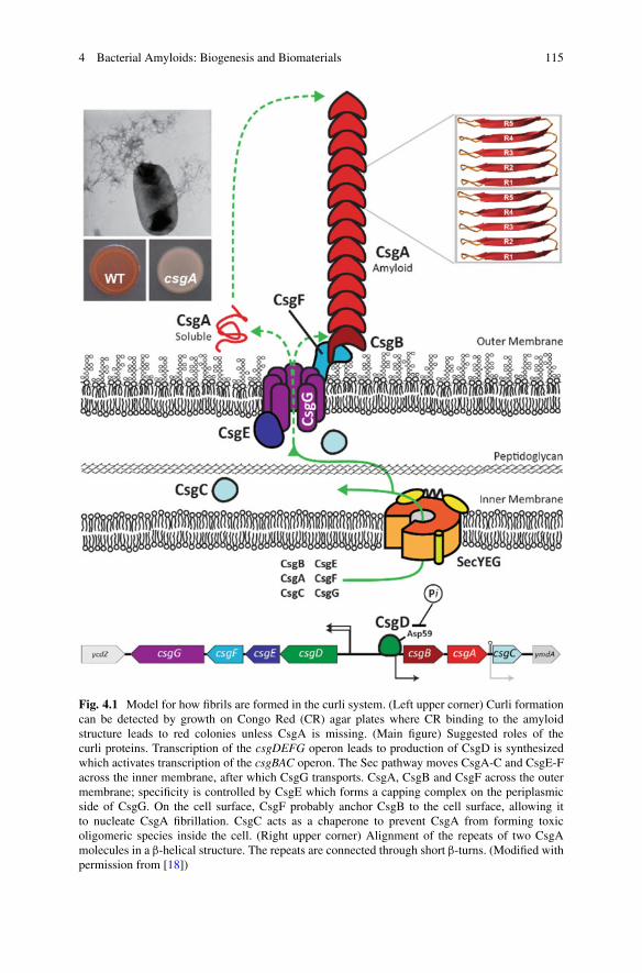
4 Bacterial Amyloids: Biogenesis and Biomaterials 115
Fig. 4.1 Model for how fibrils are formed in the curli system. (Left upper corner) Curli formationcan be detected by growth on Congo Red (CR) agar plates where CR binding to the amyloidstructure leads to red colonies unless CsgA is missing. (Main figure) Suggested roles of thecurli proteins. Transcription of the csgDEFG operon leads to production of CsgD is synthesizedwhich activates transcription of the csgBAC operon. The Sec pathway moves CsgA-C and CsgE-Facross the inner membrane, after which CsgG transports. CsgA, CsgB and CsgF across the outermembrane; specificity is controlled by CsgE which forms a capping complex on the periplasmicside of CsgG. On the cell surface, CsgF probably anchor CsgB to the cell surface, allowing itto nucleate CsgA fibrillation. CsgC acts as a chaperone to prevent CsgA from forming toxicoligomeric species inside the cell. (Right upper corner) Alignment of the repeats of two CsgAmolecules in a β-helical structure. The repeats are connected through short β-turns. (Modified withpermission from [18])
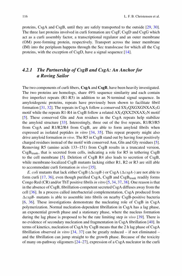
116 L. F. B. Christensen et al.
proteins, CsgA and CsgB, until they are safely transported to the outside [29, 30].The three last proteins involved in curli formation are CsgF, CsgD and CsgG whichact as a curli assembly factor, a transcriptional regulator and an outer membrane(OM) pore-forming protein, respectively. Transport across the inner membrane(IM) into the periplasm happens through the Sec translocase for which all the Csgproteins, with the exception of CsgD, have a signal sequence [14].
4.2.1 The Partnership of CsgB and CsgA: An Anchor fora Roving Sailor
The two components of curli fibers, CsgA and CsgB, have been heavily investigated.The two proteins are homologs, share 49% sequence similarity and each containfive imperfect repeats (R1-R5) in addition to an N-terminal signal sequence. Inamyloidogenic proteins, repeats have previously been shown to facilitate fibrilformation [31, 32]. The repeats in CsgA follow a conserved SX5QXGXGNXAX3Gmotif while the repeats R1-R4 in CsgB follow a related AX3QXX2NXAX3N motif[5]. These conserved Gln and Asn residues in the CsgA repeats help stabilizethe amyloid structure [33]. Interestingly, three out of the five repeats, R1/R3/R5from CsgA and R1/R2/R4 from CsgB, are able to form amyloid fibrils whenexpressed as isolated peptides in vitro [34, 35]. This repeat property might alsodrive amyloid formation in vivo. The R5 in CsgB stand out by having four positivelycharged residues instead of the motif with conserved Asn, Gln and Gly residues [5].Removing R5 (amino acids 133–151) from CsgB results in a truncated version,CsgBtrunk, that is secreted from cells, indicating a role for R5 in tethering CsgBto the cell membrane [5]. Deletion of CsgB R4 also leads to secretion of CsgB,while membrane-localized CsgB mutants lacking either R1, R2 or R3 are still ableto accommodate curli formation in vivo [35].
E. coli mutants that lack either CsgB (�csgB-) or CsgA (�csgA-) are not able toform curli [17, 36], even though purified CsgA, CsgB and CsgBtrunk readily formsCongo Red (CR) and/or ThT positive fibrils in vitro [5, 34, 37, 38]. One reason is thatin the absence of CsgB, fibrillation-competent secreted CsgA diffuses away from thecell [36]. In a process called interbacterial complementation, CsgA produced from�csgB- mutants is able to assemble into fibrils on nearby CsgB-positive bacteria[6, 36]. These investigations demonstrate the nucleating role of CsgB in CsgApolymerization. Normal nucleation-dependent fibrillation in CsgA has a lag phase,an exponential growth phase and a stationary phase, where the nucleus formationduring the lag phase is proposed to be the rate limiting step in vivo [39]. There isno evidence of secondary nucleation and fragmentation in CsgA fibrillation [40]. Interms of kinetics, nucleation of CsgA by CsgB means that the 2 h lag phase of CsgAfibrillation observed in vitro [34, 37] can be greatly reduced – if not eliminated –and the fibrillation can jump straight to the growth phase. Because of the toxicityof many on-pathway oligomers [24–27], expression of a CsgA nucleator in the curli

4 Bacterial Amyloids: Biogenesis and Biomaterials 117
system elegantly minimizes this potential problem of forming toxic oligomers byaccelerating fibril formation.
In addition to WT CsgB protein, CsgBtrunk is also able to seed CsgA fibrillationin vitro, indicating that R5 in CsgB might not be involved in the in vivo nucleationof CsgA [5]. Furthermore, CsgA protein monomers can also be seeded not onlywith pre-formed CsgA fibrils from the same species but also CsgA from othercurli-expressing species [7, 37]. Interestingly, CsgA fibrillates under practicallyany condition in terms of pH and ionic strength [37], possibly emphasizing itsimportance in bacterial life and survival. After secretion of CsgA from the bacteria,which depends on CsgG [17, 41], the protein is largely unstructured [34]. However,upon fibrillating in the presence of CsgB, the secondary structure changes into β-sheet structure both in vitro and in vivo [5, 37]. The structure of in vitro formed CsgAfibrils have been investigated thoroughly with transmission electron microscopy(TEM), electron diffraction, X-ray fiber diffraction and solid state NMR [37, 38].These experimental investigations suggest a cross-β structure of stacked β-helicalsubunits [38]. The β-helical structure was also supported by a study that usedsimulations to identify low-energy structural models of CsgA [42].
4.2.2 All in the Fibril Family: Cooperation Within the CurliOperon
Figure 4.1 summarizes our current understanding of how the different curli proteinswork together to form amyloid. Here we will go through what is known of theindividual components. They reveal an exquisitely controlled nanotechnologicalamyloid generating machinery.
A prerequisite for in vivo CsgA/CsgB expression is a functional CsgD which actsas a positive transcriptional regulator of the csgBAC operon [17]. The C-terminal ofCsgD shows a characteristic helix-turn-helix motif known to enable DNA binding.
CsgC was recently shown to be a potent amyloid inhibitor [29]. The crystalstructure of oxidized CsgC (PDB: 2Y2Y) reveals seven β-strands and a disulphidebond between Cys29 and Cys31 on the same β-strand. CsgC inhibits in vitro CsgAand CsgBtrunk fibrillation, however it is 50 times more effective against CsgAcompared to CsgBtrunk [29]. CsgC inhibits primary nucleation – and to a smallerdegree elongation – of CsgA by inhibiting the formation of some intermediatespecies on the fibrillation pathway [40]. This delays the nucleation rate by a factorof 2–4 times [40]. Mutational studies have shown that especially positively chargedresidues on the protein surface are important for the fibrillation inhibition potencyof CsgC [40]. The difference in the pI of CsgC/CsgH and CsgA (> 2 pH units)is probably responsible for accelerating the encounter between the proteins. Thisis supported by a CsgC homolog from Salmonella typhimurium that shows lesspotency in CsgA inhibition and at the same time has a pI much closer to that of CsgA
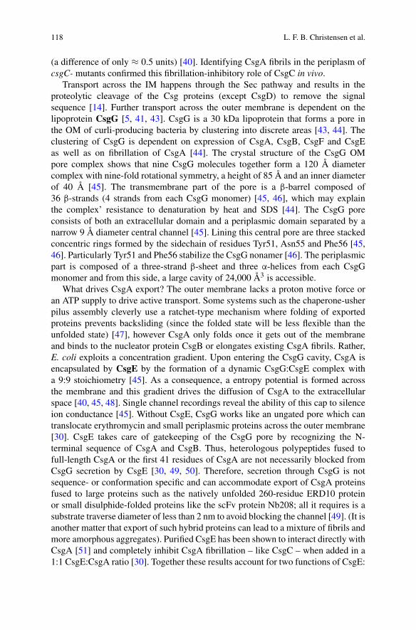
118 L. F. B. Christensen et al.
(a difference of only ≈ 0.5 units) [40]. Identifying CsgA fibrils in the periplasm ofcsgC- mutants confirmed this fibrillation-inhibitory role of CsgC in vivo.
Transport across the IM happens through the Sec pathway and results in theproteolytic cleavage of the Csg proteins (except CsgD) to remove the signalsequence [14]. Further transport across the outer membrane is dependent on thelipoprotein CsgG [5, 41, 43]. CsgG is a 30 kDa lipoprotein that forms a pore inthe OM of curli-producing bacteria by clustering into discrete areas [43, 44]. Theclustering of CsgG is dependent on expression of CsgA, CsgB, CsgF and CsgEas well as on fibrillation of CsgA [44]. The crystal structure of the CsgG OMpore complex shows that nine CsgG molecules together form a 120 Å diametercomplex with nine-fold rotational symmetry, a height of 85 Å and an inner diameterof 40 Å [45]. The transmembrane part of the pore is a β-barrel composed of36 β-strands (4 strands from each CsgG monomer) [45, 46], which may explainthe complex’ resistance to denaturation by heat and SDS [44]. The CsgG poreconsists of both an extracellular domain and a periplasmic domain separated by anarrow 9 Å diameter central channel [45]. Lining this central pore are three stackedconcentric rings formed by the sidechain of residues Tyr51, Asn55 and Phe56 [45,46]. Particularly Tyr51 and Phe56 stabilize the CsgG nonamer [46]. The periplasmicpart is composed of a three-strand β-sheet and three α-helices from each CsgGmonomer and from this side, a large cavity of 24,000 Å3 is accessible.
What drives CsgA export? The outer membrane lacks a proton motive force oran ATP supply to drive active transport. Some systems such as the chaperone-usherpilus assembly cleverly use a ratchet-type mechanism where folding of exportedproteins prevents backsliding (since the folded state will be less flexible than theunfolded state) [47], however CsgA only folds once it gets out of the membraneand binds to the nucleator protein CsgB or elongates existing CsgA fibrils. Rather,E. coli exploits a concentration gradient. Upon entering the CsgG cavity, CsgA isencapsulated by CsgE by the formation of a dynamic CsgG:CsgE complex witha 9:9 stoichiometry [45]. As a consequence, a entropy potential is formed acrossthe membrane and this gradient drives the diffusion of CsgA to the extracellularspace [40, 45, 48]. Single channel recordings reveal the ability of this cap to silenceion conductance [45]. Without CsgE, CsgG works like an ungated pore which cantranslocate erythromycin and small periplasmic proteins across the outer membrane[30]. CsgE takes care of gatekeeping of the CsgG pore by recognizing the N-terminal sequence of CsgA and CsgB. Thus, heterologous polypeptides fused tofull-length CsgA or the first 41 residues of CsgA are not necessarily blocked fromCsgG secretion by CsgE [30, 49, 50]. Therefore, secretion through CsgG is notsequence- or conformation specific and can accommodate export of CsgA proteinsfused to large proteins such as the natively unfolded 260-residue ERD10 proteinor small disulphide-folded proteins like the scFv protein Nb208; all it requires is asubstrate traverse diameter of less than 2 nm to avoid blocking the channel [49]. (It isanother matter that export of such hybrid proteins can lead to a mixture of fibrils andmore amorphous aggregates). Purified CsgE has been shown to interact directly withCsgA [51] and completely inhibit CsgA fibrillation – like CsgC – when added in a1:1 CsgE:CsgA ratio [30]. Together these results account for two functions of CsgE:
4 Bacterial Amyloids: Biogenesis and Biomaterials 119
to ensure some secretion specificity for CsgG and to inhibit self-polymerization ofCsgA through the periplasmic space.
Another Csg protein, the assembly factor CsgF, also interacts with CsgG andis dependent on the membrane protein for localization on the cell surface. CsgGprobably also plays a role in CsgF stability as steady-state levels of CsgF are greatlydecreased in csgG- mutants [52]. CsgF is most likely involved in the localizationand nucleation activity of CsgB because csgF- mutants resemble csgBtrunk- mutantswith large amounts of stable CsgB being released to the cell exterior [5, 6, 52].CsgF also mediates CsgB protease resistance on the cell surface [6, 52]. csgF-mutants are not able to assemble CsgA fibrils [52], however, CsgA produced fromthese mutants is fibrillation-competent and can polymerize on the surface of csgA-mutants [6]. Direct interaction between CsgF and CsgB still needs to be established,but CsgF might interact with R4/R5 of CsgB since removal of these repeats alsoresults in CsgB secretion [35]. The interaction between CsgB and CsgF and possibleinvolvement of CsgF in CsgB nucleus formation might also be facilitated by a highpercentage of Glu and Asn in CsgF (9.2% and 13.4%, respectively), which is similarto CsgA (8.4% and 12.2%) and CsgB (11.5% and 9.2%) [52].
On the cell surface, CsgB is suggested to quickly adopt an R4/R5-dependentamyloid fold that templates CsgA fibrillation [35]. This important role of CsgB R5in both nucleation and membrane-anchoring [5] is especially interesting becausethe isolated R5 peptide does not form fibrils in vitro [35]. Not forming amyloidstructures in the membrane portion of the cells could be another mechanism to avoidforming membrane-permeabilizing oligomers.
An eighth member of the curli family, CsgH, has recently been shown to inhibitamyloid formation [40]. The csgH gene has been observed in many Alphapro-teobacteria where it is always situated next to csgA/B genes [21]. CsgH is – likeCsgC – composed of seven β-strands and the two proteins share great structuralsimilarity (rmsd = 2.6 Å) despite a sequence identity below 20% [40]. CsgH isable to inhibit CsgA fibrillation in a concentration-dependent manner. Interestingly,both CsgH and CsgC are also able to inhibit the unrelated bacterial amyloidogenicprotein FapC [40].
4.2.3 Younger Kid on the Block: Fap Fimbria Are Composedof Mainly FapC
The curli system probably evolved from a common ancestor long time ago [21].Another FuBA system, the Fap system, appears to be evolutionarily younger [53].The fapABCDEF operon encodes 6 proteins [8] and has been found within severalclasses of the phylum Proteobacteria [53]. Fap fimbria were initially identified andpurified from a Pseudomonas strain and were characterized as a FuBA due to thein vivo binding of ThT and amyloid conformational specific antibodies [3] togetherwith the characteristic cross-β structure and extreme stability of purified Fap fibrils
120 L. F. B. Christensen et al.
[8]. Fap fimbria are believed to be involved in adhesion and biofilm formation,since overexpression of Fap results in a highly aggregative phenotype with increasedbiofilm formation [8, 54]. Fap fibrils greatly increase the stiffness (≈ 20-fold) andthe hydrophobicity of the biofilm [55]. Biofilm can, however, still be formed in Fap-negative bacteria. In addition to biofilm formation, Fap fibrils might also play a rolein virulence as over-expression of the fap operon leads to a more mucoid, alginate-rich phenotype which mimics the biofilm formation seen in cystic fibrosis patients[56].
Despite the high stability of Fap fibrils, Dueholm et al. succeeded in purifyingthe major fibril component, which was termed FapC [8]. In a functional genomicsstudy, performed to identify the function of unknown proteins in P. aeruginosa,one of the most attenuated phenotypes towards worm infection was a mutant witha deletion in the fapC gene (NCBI locus tag PA1954) [57]. This again indicatesthat Fap fibrils play an important role in virulence – a function that has also beenproposed for the analogous curli fibrils due to their interactions with different hostproteins [58–60]. Sequencing of the major amyloid subunit, FapC, has shown thatit contains a thrice repeated motif (R1-R3) which – like in the repeats of the curliCsgA and CsgB [5] – have conserved Glu and Asn residues, at least across 18 strains[54]. The R1 and R2 of FapC are 26–27 residues longer than the CsgA repeats whileR3 is 11–12 residues longer. Linkers connect the different FapC repeats and can beup to 275 residues long between R2 and R3 [8] This contrasts with CsgA and CsgBwhich have a tight β-turn composed of 4–5 residues between each repeat [35, 61].Some amyloids depend on interactions between aromatic residues [62, 63], but thelack of any aromatic sidechains in the FapC repeats emphasizes that this is not thecase for these amyloid fibrils. Furthermore, aromatic residues were also found to bedispensable in curli formation [33].
4.2.4 Another Study in Team-Work: The Role of Fap Proteins
The FapC homolog, FapB, has similar, but shorter, repeats (17 residues versusFapC’s 34 and 49) [8] and less variable linker lengths than FapC [53]. Furthermore,the FapB repeats may follow a strand-turn-strand-like motif [54]. Bioinformaticssuggest that FapB is a nucleation factor (like CsgB in the curli system) andbiophysical investigations have identified FapB and FapE as minor constituent ofthe Fap fibrils [54, 56]. In any case, FapB is necessary for Fap biogenesis [55].
The roles of the remaining Fap proteins (A, D, E and F) are not entirely clear andare still under scrutiny. However, there are some clues (Fig. 4.2). As an example,fibrils consisting almost entirely of FapB have been isolated from fapA- mutants,showing not only that FapB is indeed amyloidogenic, but also that FapA probablyhas a regulating role in the assembly of Fap fibrils in vivo [54]. FapA’s functionis unclear, however, as the deletion of the fapA gene is relatively frequent acrossdifferent species [53]. A change in fibril composition has also been observed in the
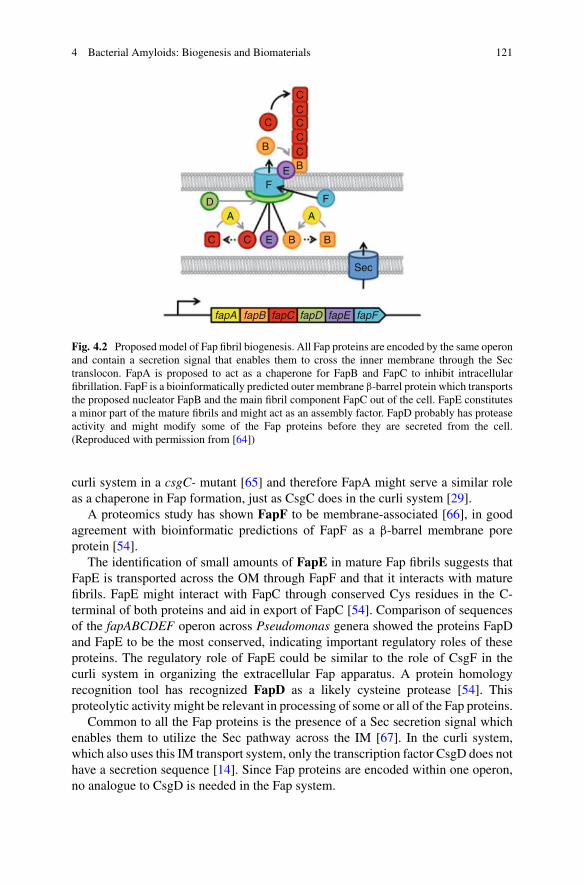
4 Bacterial Amyloids: Biogenesis and Biomaterials 121
C
B
BCCCCC
D
EF
F
AA
B
Sec
fapA fapB fapC fapD fapE fapF
BECC
Fig. 4.2 Proposed model of Fap fibril biogenesis. All Fap proteins are encoded by the same operonand contain a secretion signal that enables them to cross the inner membrane through the Sectranslocon. FapA is proposed to act as a chaperone for FapB and FapC to inhibit intracellularfibrillation. FapF is a bioinformatically predicted outer membrane β-barrel protein which transportsthe proposed nucleator FapB and the main fibril component FapC out of the cell. FapE constitutesa minor part of the mature fibrils and might act as an assembly factor. FapD probably has proteaseactivity and might modify some of the Fap proteins before they are secreted from the cell.(Reproduced with permission from [64])
curli system in a csgC- mutant [65] and therefore FapA might serve a similar roleas a chaperone in Fap formation, just as CsgC does in the curli system [29].
A proteomics study has shown FapF to be membrane-associated [66], in goodagreement with bioinformatic predictions of FapF as a β-barrel membrane poreprotein [54].
The identification of small amounts of FapE in mature Fap fibrils suggests thatFapE is transported across the OM through FapF and that it interacts with maturefibrils. FapE might interact with FapC through conserved Cys residues in the C-terminal of both proteins and aid in export of FapC [54]. Comparison of sequencesof the fapABCDEF operon across Pseudomonas genera showed the proteins FapDand FapE to be the most conserved, indicating important regulatory roles of theseproteins. The regulatory role of FapE could be similar to the role of CsgF in thecurli system in organizing the extracellular Fap apparatus. A protein homologyrecognition tool has recognized FapD as a likely cysteine protease [54]. Thisproteolytic activity might be relevant in processing of some or all of the Fap proteins.
Common to all the Fap proteins is the presence of a Sec secretion signal whichenables them to utilize the Sec pathway across the IM [67]. In the curli system,which also uses this IM transport system, only the transcription factor CsgD does nothave a secretion sequence [14]. Since Fap proteins are encoded within one operon,no analogue to CsgD is needed in the Fap system.
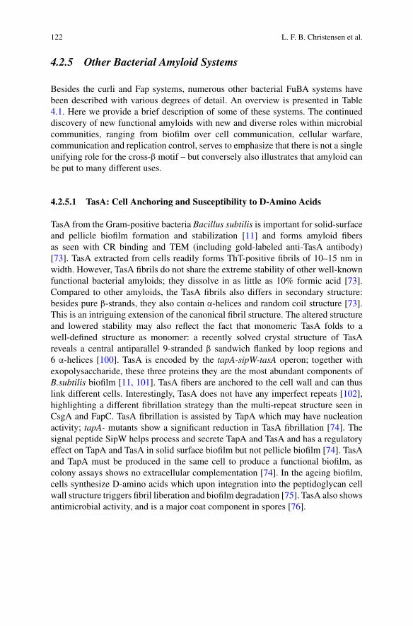
122 L. F. B. Christensen et al.
4.2.5 Other Bacterial Amyloid Systems
Besides the curli and Fap systems, numerous other bacterial FuBA systems havebeen described with various degrees of detail. An overview is presented in Table4.1. Here we provide a brief description of some of these systems. The continueddiscovery of new functional amyloids with new and diverse roles within microbialcommunities, ranging from biofilm over cell communication, cellular warfare,communication and replication control, serves to emphasize that there is not a singleunifying role for the cross-β motif – but conversely also illustrates that amyloid canbe put to many different uses.
4.2.5.1 TasA: Cell Anchoring and Susceptibility to D-Amino Acids
TasA from the Gram-positive bacteria Bacillus subtilis is important for solid-surfaceand pellicle biofilm formation and stabilization [11] and forms amyloid fibersas seen with CR binding and TEM (including gold-labeled anti-TasA antibody)[73]. TasA extracted from cells readily forms ThT-positive fibrils of 10–15 nm inwidth. However, TasA fibrils do not share the extreme stability of other well-knownfunctional bacterial amyloids; they dissolve in as little as 10% formic acid [73].Compared to other amyloids, the TasA fibrils also differs in secondary structure:besides pure β-strands, they also contain α-helices and random coil structure [73].This is an intriguing extension of the canonical fibril structure. The altered structureand lowered stability may also reflect the fact that monomeric TasA folds to awell-defined structure as monomer: a recently solved crystal structure of TasAreveals a central antiparallel 9-stranded β sandwich flanked by loop regions and6 α-helices [100]. TasA is encoded by the tapA-sipW-tasA operon; together withexopolysaccharide, these three proteins they are the most abundant components ofB.subtilis biofilm [11, 101]. TasA fibers are anchored to the cell wall and can thuslink different cells. Interestingly, TasA does not have any imperfect repeats [102],highlighting a different fibrillation strategy than the multi-repeat structure seen inCsgA and FapC. TasA fibrillation is assisted by TapA which may have nucleationactivity; tapA- mutants show a significant reduction in TasA fibrillation [74]. Thesignal peptide SipW helps process and secrete TapA and TasA and has a regulatoryeffect on TapA and TasA in solid surface biofilm but not pellicle biofilm [74]. TasAand TapA must be produced in the same cell to produce a functional biofilm, ascolony assays shows no extracellular complementation [74]. In the ageing biofilm,cells synthesize D-amino acids which upon integration into the peptidoglycan cellwall structure triggers fibril liberation and biofilm degradation [75]. TasA also showsantimicrobial activity, and is a major coat component in spores [76].
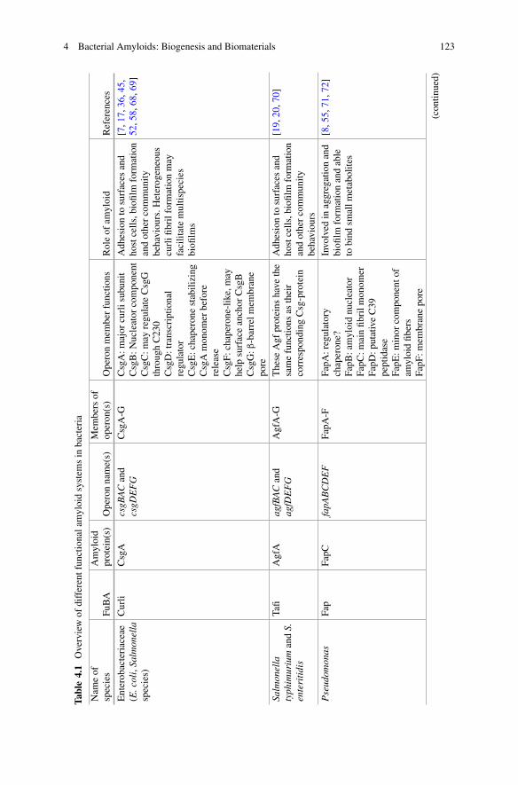
4 Bacterial Amyloids: Biogenesis and Biomaterials 123
Tabl
e4.
1O
verv
iew
ofdi
ffer
entf
unct
iona
lam
yloi
dsy
stem
sin
bact
eria
Nam
eof
spec
ies
FuB
AA
myl
oid
prot
ein(
s)O
pero
nna
me(
s)M
embe
rsof
oper
on(s
)O
pero
nm
embe
rfu
nctio
nsR
ole
ofam
yloi
dR
efer
ence
s
Ent
erob
acte
riac
eae
(E.c
oli,
Salm
onel
lasp
ecie
s)
Cur
liC
sgA
csgB
AC
and
csgD
EF
GC
sgA
-GC
sgA
:maj
orcu
rlis
ubun
itC
sgB
:Nuc
leat
orco
mpo
nent
Csg
C:m
ayre
gula
teC
sgG
thro
ugh
C23
0C
sgD
:tra
nscr
iptio
nal
regu
lato
rC
sgE
:cha
pero
nest
abili
zing
Csg
Am
onom
erbe
fore
rele
ase
Csg
F:ch
aper
one-
like,
may
help
surf
ace
anch
orC
sgB
Csg
G:β
-bar
relm
embr
ane
pore
Adh
esio
nto
surf
aces
and
host
cells
,bio
film
form
atio
nan
dot
her
com
mun
itybe
havi
ours
.Het
erog
eneo
uscu
rlifi
bril
form
atio
nm
ayfa
cilit
ate
mul
tispe
cies
biofi
lms
[7,1
7,36
,45,
52,5
8,68
,69]
Salm
onel
laty
phim
uriu
man
dS.
ente
riti
dis
Tafi
Agf
Aag
fBA
Can
dag
fDE
FG
Agf
A-G
The
seA
gfpr
otei
nsha
veth
esa
me
func
tions
asth
eir
corr
espo
ndin
gC
sg-p
rote
in
Adh
esio
nto
surf
aces
and
host
cells
,bio
film
form
atio
nan
dot
her
com
mun
itybe
havi
ours
[19,
20,7
0]
Pse
udom
onas
Fap
FapC
fapA
BC
DE
FFa
pA-F
FapA
:reg
ulat
ory
chap
eron
e?Fa
pB:a
myl
oid
nucl
eato
rFa
pC:m
ain
fibri
lmon
omer
FapD
:put
ativ
eC
39pe
ptid
ase
FapE
:min
orco
mpo
nent
ofam
yloi
dfib
ers
FapF
:mem
bran
epo
re
Invo
lved
inag
greg
atio
nan
dbi
ofilm
form
atio
nan
dab
leto
bind
smal
lmet
abol
ites
[8,5
5,71
,72]
(con
tinue
d)
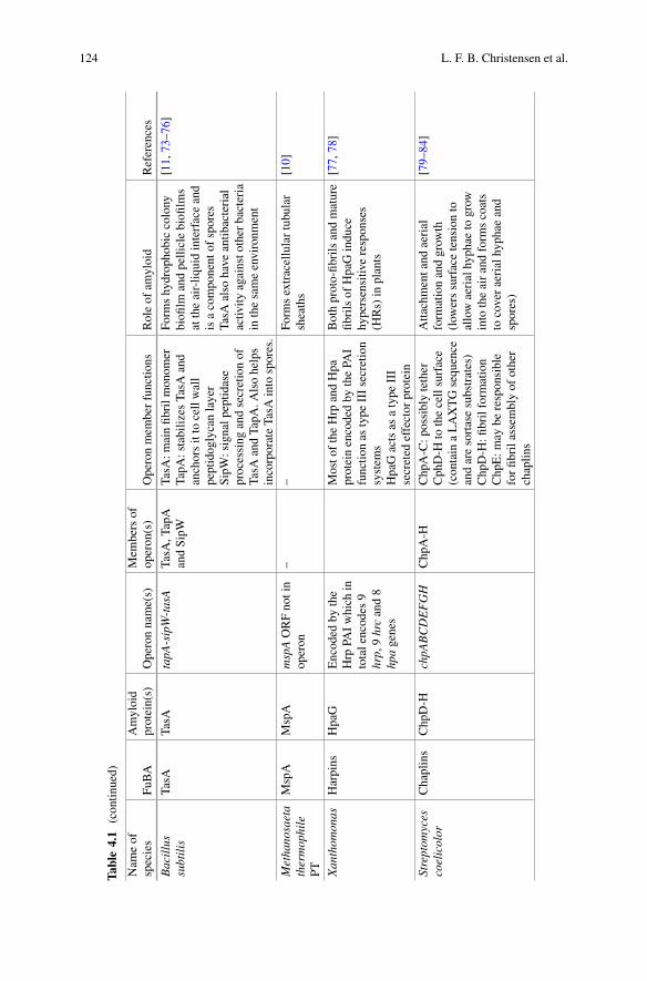
124 L. F. B. Christensen et al.
Tabl
e4.
1(c
ontin
ued)
Nam
eof
spec
ies
FuB
AA
myl
oid
prot
ein(
s)O
pero
nna
me(
s)M
embe
rsof
oper
on(s
)O
pero
nm
embe
rfu
nctio
nsR
ole
ofam
yloi
dR
efer
ence
s
Bac
illu
ssu
btil
isTa
sATa
sAta
pA-s
ipW
-tas
ATa
sA,T
apA
and
SipW
TasA
:mai
nfib
rilm
onom
erTa
pA:s
tabi
lizes
TasA
and
anch
ors
itto
cell
wal
lpe
ptid
ogly
can
laye
rSi
pW:s
igna
lpep
tidas
epr
oces
sing
and
secr
etio
nof
TasA
and
TapA
.Als
ohe
lps
inco
rpor
ate
TasA
into
spor
es.
Form
shy
drop
hobi
cco
lony
biofi
lman
dpe
llicl
ebi
ofilm
sat
the
air-
liqui
din
terf
ace
and
isa
com
pone
ntof
spor
esTa
sAal
soha
vean
tibac
teri
alac
tivity
agai
nsto
ther
bact
eria
inth
esa
me
envi
ronm
ent
[11,
73–7
6]
Met
hano
saet
ath
erm
ophi
lePT
Msp
AM
spA
msp
AO
RF
noti
nop
eron
––
Form
sex
trac
ellu
lar
tubu
lar
shea
ths
[10]
Xan
thom
onas
Har
pins
Hpa
GE
ncod
edby
the
Hrp
PAI
whi
chin
tota
lenc
odes
9hr
p,9
hrc
and
8hp
age
nes
Mos
tof
the
Hrp
and
Hpa
prot
ein
enco
ded
byth
ePA
Ifu
nctio
nas
type
III
secr
etio
nsy
stem
sH
paG
acts
asa
type
III
secr
eted
effe
ctor
prot
ein
Bot
hpr
oto-
fibri
lsan
dm
atur
efib
rils
ofH
paG
indu
cehy
pers
ensi
tive
resp
onse
s(H
Rs)
inpl
ants
[77,
78]
Stre
ptom
yces
coel
icol
orC
hapl
ins
Chp
D-H
chpA
BC
DE
FG
HC
hpA
-HC
hpA
-C:p
ossi
bly
teth
erC
phD
-Hto
the
cell
surf
ace
(con
tain
aL
AX
TG
sequ
ence
and
are
sort
ase
subs
trat
es)
Chp
D-H
:fibr
ilfo
rmat
ion
Chp
E:m
aybe
resp
onsi
ble
for
fibri
lass
embl
yof
othe
rch
aplin
s
Atta
chm
enta
ndae
rial
form
atio
nan
dgr
owth
(low
ers
surf
ace
tens
ion
toal
low
aeri
alhy
phae
togr
owin
toth
eai
ran
dfo
rms
coat
sto
cove
rae
rial
hyph
aean
dsp
ores
)
[79–
84]
4 Bacterial Amyloids: Biogenesis and Biomaterials 125
Myc
obac
teri
umtu
berc
ulos
is(m
aybe
also
inbo
vis
and
aviu
mw
hich
also
enco
dem
tp)
Pili
(cro
ss-β
stru
ctur
ere
mai
nsto
beco
nfirm
ed)
MT
PT
hem
tpge
neis
foun
din
the
H37
Rv
OR
F,de
sign
ated
Rv3
312A
(not
inan
oper
on)
––
Adh
esiv
epr
oper
ties:
inds
tola
min
in.R
ole
inin
fect
ions
?(s
erum
from
tube
rcul
osis
patie
nts
cont
ain
anti-
MPT
antib
odie
s).I
nvol
ved
inbi
ofilm
form
atio
nan
dce
ll-ce
llco
mm
unic
atio
n
[85–
87]
Stre
ptoc
occu
sm
utan
sPi
li(c
ross
-βst
ruct
ure
rem
ains
tobe
confi
rmed
)
P1P1
isen
code
dby
the
spaP
gene
–So
rtas
etr
ansp
eptid
ase
(enc
oded
byth
esr
tAge
ne)
isre
spon
sibl
efo
rte
ther
ing
P1to
the
cell
surf
ace
P1is
invo
lved
inbi
ofilm
form
atio
n.A
myl
oid
isde
tect
edin
dent
alca
ries
[88]
Kle
bsie
lla
pneu
mon
iae
RY
C49
2(h
omol
ogs
are
foun
din
E.c
oli)
K.
pneu
mon
iae:
Mic
roci
nE
492
(Mcc
)E
.col
i:M
icro
cin
24
Mce
A(E
.co
li:m
tfS)
Mce
Ais
loca
ted
insi
dea
3kb
Cla
ID
NA
frag
men
tto
geth
erw
ithits
imm
unity
prot
ein
enco
ded
byth
em
ceB
gene
Mce
A,
Mce
BM
ceA
:has
one
tran
smem
bran
ere
gion
;fo
rms
cyto
toxi
col
igom
ers
orin
activ
efib
rils
invi
voM
ceB
:int
egra
linn
erm
embr
ane
prot
ein
with
thre
epr
edic
ted
tran
smem
bran
ere
gion
s.C
onfe
rsse
lf-p
rote
ctiv
eim
mun
ityto
Mce
A.
Kill
sE
nter
obac
teri
acea
e(c
ompe
titor
sfo
rsa
me
ecol
ogic
alni
che)
with
olig
omer
sth
atfo
rmpo
res
inth
ecy
topl
asm
icm
embr
ane,
caus
ing
leth
allo
ssof
mem
bran
epo
tent
ial
[25,
89–9
2]
Lis
teri
am
onoc
ytog
enes
Lis
teri
loys
inO
(LL
O)
toxi
n
LL
O(e
ncod
edby
the
hly
gene
).
PAI
LIP
I-1
com
pris
essi
xge
nes
(prf
A,p
lcA
,hl
y,m
pl,a
ctA
,pl
cB)
The
prod
ucts
ofL
IPI-
1ar
ere
quir
edfo
rcr
ucia
lste
psin
the
intr
acel
lula
rlif
ecy
cle
ofth
eba
cter
ia
LL
Ool
igom
ers
form
pore
sin
the
mem
bran
eof
phag
olys
osom
es,e
nabl
ing
the
bact
eriu
mto
esca
pein
toth
ecy
toso
l
[93–
95]
(con
tinue
d)
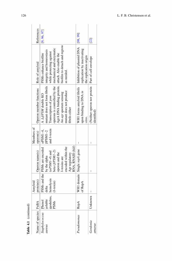
126 L. F. B. Christensen et al.
Tabl
e4.
1(c
ontin
ued)
Nam
eof
spec
ies
FuB
AA
myl
oid
prot
ein(
s)O
pero
nna
me(
s)M
embe
rsof
oper
on(s
)O
pero
nm
embe
rfu
nctio
nsR
ole
ofam
yloi
dR
efer
ence
s
Stap
hylo
cocc
usau
reus
Phen
ols o
lubl
em
odul
ins,
PSM
s
PSM
san
dth
ede
ltahe
mol
ysin
(δ-t
oxin
)
PSM
sar
een
code
dby
the
alph
a(α
PSM
1–4)
and
beta
(βP
SM1–
2)op
eron
and
the
δ-to
xins
are
enco
ded
with
inth
eA
grre
gula
tory
RN
A,R
NA
III
(hld
)
αPS
M1–
4,β
PSM
1–2
and
δ-to
xin
A�
βP
SMdo
uble
KO
mut
antd
oes
notf
orm
fibri
lsT
rans
crip
tion
ofps
mop
eron
sis
cont
rolle
dby
the
Agr
AD
NA
bind
ing
prot
ein.
An
agr
quor
umse
nsin
gm
utan
tdoe
sno
tpro
duce
fibri
lsei
ther
PSM
sen
hanc
ebi
ofilm
inte
grity
and
hom
eost
asis
whi
lepr
otec
ting
agai
nst
mec
hani
cala
nden
zym
atic
atta
ch.A
lso
enab
leth
ebi
ofilm
tode
tach
and
regr
owas
need
ed.
[9,9
6,97
]
Pse
udom
onas
Rep
AW
H1
dom
ain
ofR
epA
Sing
lere
pAge
ne–
WH
1fo
rms
amyl
oid
fibri
lsup
onbi
ndin
gto
DN
Ain
vitr
o.
Inhi
bitio
nof
plas
mid
DN
Are
plic
atio
nby
inac
tivat
ing
the
repl
icat
ion
orig
in
[98,
99]
Gor
doni
aam
arae
Unk
now
n–
––
(Nei
ther
oper
onno
rpr
otei
nid
entifi
ed)
Part
ofce
llen
velo
pe[2
2]
4 Bacterial Amyloids: Biogenesis and Biomaterials 127
4.2.5.2 MspA: The First Archaeal FuBA
MspA (major sheath protein) from the archaea Methanosaeta thermophila PT is thefirst and so far only example of functional amyloid from Archaea. MS/MS revealedthe sheaths to be composed of this single protein [10]. As might be expected, MspAis not evolutionarily related to other known functional amyloids but constitutes anovel amyloid motif that will have to be incorporated into future sequence analyseshunting for functional amyloid [103].
4.2.5.3 Harpins: Green Oligomeric Weapons
Harpins produced by plant pathogenic bacterial species are type III-secreted proteinswhich cause a hypersensitive response (HR) in the intercellular space of leavesin several plant types. The HR is known to be an early defensive strategy of theplant, to stop further growth of plant pathogens by inducing apoptosis [104]. Theharpin protein HpaG of Xanthomonas axonopodia, is a fibril forming protein withimperfect repeats, however HR is induced not by fibrils, but rather by non-amyloidspherical oligomers which form at an earlier stage. Such oligomers are known tobe the cytotoxic species in Alzheimer’s and Parkinson’s Disease due to their cell-permeabilizing properties [105], but HpaG is the only example to our knowledgeof bacteria using this strategy as a functional tool, despite the protein’s amyloid-forming properties.
4.2.5.4 Chaplins: Breaking the Air-Water Interface Barrier
The filamentous bacterium Streptomyces coelicolor produces eight hydrophobicproteins (ChpA-H) which are collectively called chaplins. ChpA-C consist of twochaplin domains (∼ 40 hydrophobic residues) and a C-terminal sorting signal whileChpD-H are composed of a single chaplin domain and an N-terminal secretionsignal peptide. As a mycelium forming bacterium, S. coelicolor forms aerial hyphaewhich give rise to spores that can spread and form new mycelium [106]. ChpA-Hare expressed in the mycelium and in aerial hyphae [107]. They all contain signalsequences for secretion to the cell-wall. When secreted, the chaplins self-assembleat the air/water interface into an amphiphatic amyloid film which reduces surfacetension and promotes spore spreading. This resembles the fungal proteins calledhydrophobins (see Chap. 8). ChpD-H complement cells extracellularly and acceler-ate growth [106]. ChpA-C contain a common motif (LAXTG-Membrane spanningdomain-Charged C-terminal) for anchoring surface proteins to the peptidoglycanin the cell wall covalently, suggesting a role in ChpD-H incorporation in the cellwall [106]. It has recently been suggested that ChpD-H, associated with cellulosefibrils and supported by ChpA-C, are responsible for fimbriae formation at adheringhyphae, and ChpD-H monomers assemble upon contact with assembled fibrils orhydrophobic surfaces [79].
128 L. F. B. Christensen et al.
4.2.5.5 Phenol-Soluble Modulins (PSM): Amyloid or AntimicrobialAgents?
The pathogen Staphylococcus aureus inhabits many bodily cavities and can changefrom benign to pathological within the host. Both states can form biofilm. Functionalamyloid in the form of fibrillar structures have been identified in the biofilm.Unusually, the amyloid consists of small (20–40 residues long) peptides calledphenol soluble modulins (PSMs) [9]. The PSMs fall into 3 highly conserved classes(α, β and δ) which are conserved and expressed from three different operons[9, 96, 97]. As small peptides, the PSMs represent a very different aggregationmotif compared to the imperfect repeats in CsgA and FapC. While monomericPSMs have antimicrobial activity and can disassemble biofilm in a surfactant-like manner (based on agr quorum sensing), fibrillated PSMs stabilize biofilmagainst mechanical and enzymatic attack [108]. Amyloid formation is stimulatedby extracellular DNA (just like the initiator protein RepA involved in plasmidreplication control [98]), suggesting that the antimicrobial effect came first andamyloidogenesis evolved later in an opportunitstic fashion.
4.3 Functional Amyloids in silico
Being able to predict which part of a protein sequence (if any) forms amyloid ishelpful for identifying potential functional amyloid in hitherto uncharted genomes.The following sections will describe the principles behind the bioinformaticsmethods that have been used to identify aggregation and amyloid “hot spots”within protein sequences, how these methods have been used on functional amyloidsequences, and how we can go beyond these sequence-based methods in oursearch for as of yet uncharacterized functional amyloids. Functional amyloid canbe identified based on sequence, evolved characteristics or structural prediction andsimulation, and each of these will be described in more detail.
4.3.1 Predicting Aggregation and Amyloid Propensityof Proteins Based on Sequences
The most widely used methods for detecting aggregation-prone or amyloidogenicsequences were developed mostly with pathological aggregation and amyloidformation in mind [109–120]. These methods have been reviewed several timesalready [121, 122]. An overview of methods is provided in Table 4.2. In this section,we will discuss different principles on which these methods are based.
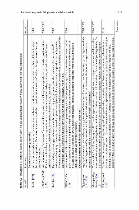
4 Bacterial Amyloids: Biogenesis and Biomaterials 129
Tabl
e4.
2D
escr
iptio
nof
met
hods
used
topr
edic
tam
yloi
dan
dag
greg
atio
npr
open
sity
base
don
prot
ein
sequ
ence
info
rmat
ion
Nam
eaPr
inci
ples
Yea
r(s)
Seco
ndar
yst
ruct
ure
prop
ensi
tySe
cStr
[123
]A
cons
ensu
sse
cond
ary
stru
ctur
epr
edic
tor
that
can
beus
edto
iden
tify
shor
tseq
uenc
esth
atha
vebo
thal
pha-
helic
alan
dbe
ta-s
heet
prop
ensi
ties.
The
sesh
orts
eque
nces
are
dubb
ed“c
onfo
rmat
iona
lsw
itche
s”an
dar
eth
ough
tto
cont
ribu
teto
amyl
oid
form
atio
n.
1988
CSS
P/N
etC
SSP
[120
]A
ttem
pts
toid
entif
y“c
ham
eleo
n”se
quen
ces
that
are
pron
eto
switc
hing
from
alph
a-he
lical
tobe
ta-s
heet
conf
orm
atio
ns.
Em
phas
izes
the
influ
ence
ofte
rtia
ryco
ntac
tson
the
dete
rmin
atio
nof
seco
ndar
yst
ruct
ure,
and
utili
zes
afe
ed-f
orw
ard
artifi
cial
neur
alne
twor
ktr
aine
dus
ing
back
-pro
paga
tion
for
mak
ing
pred
ictio
ns.
2005
,200
9
SAL
SA[1
24]
Use
sa
slid
ing
win
dow
appr
oach
toca
lcul
ate
beta
-str
and
cont
igui
typr
open
sity
for
shor
tam
ino
acid
sequ
ence
s(4
–20
resi
dues
inle
ngth
)by
com
pari
ngbe
ta-s
tran
dpr
open
sity
toal
pha-
helic
alan
dre
vers
e-tu
rnpr
open
sitie
sac
cord
ing
toth
eC
hou
and
Fasm
ansc
ale.
The
thre
shol
dfo
rbe
ing
cons
ider
edam
yloi
doge
nic
was
dete
rmin
edby
com
pari
ngth
eou
tput
ofth
eal
gori
thm
for
alph
a-sy
nucl
ein,
whi
chis
amyl
oido
geni
c,an
dbe
ta-s
ynuc
lein
,whi
chis
not.
2007
BE
TASC
AN
[118
]A
ttem
pts
tofin
dal
lcon
tiguo
usse
gmen
tsin
apr
otei
nse
quen
ceth
atha
vepr
open
sity
tofo
rmbe
ta-s
heet
stru
ctur
esan
dal
lpa
irs
ofse
gmen
tsth
atha
vea
prop
ensi
tyto
pair
inpa
ralle
lbet
aco
nfor
mat
ions
,mak
ing
itw
ells
uite
dto
the
dete
ctio
nof
beta
-hel
ical
stru
ctur
esfo
und
inse
vera
lfun
ctio
nala
myl
oids
.Str
and
and
pair
ing
prop
ensi
ties
wer
ees
timat
edba
sed
onst
atis
tics
ofa
data
base
ofpr
otei
nst
ruct
ures
.
2009
Sequ
ence
patt
erns
and
phys
ico-
chem
ical
prop
erti
esZ
yggr
egat
or[1
16,1
25]
Agg
rega
tion
rate
sar
epr
edic
ted
usin
ga
wei
ghte
dsu
mof
term
sth
atta
kes
into
acco
unth
ydro
phob
icity
,the
degr
eeto
whi
chan
alte
rnat
ing
patte
rnof
hydr
opho
bic
and
hydr
ophi
licre
sidu
esex
ists
inth
ese
quen
ce,e
lect
rost
atic
char
ge,s
econ
dary
stru
ctur
epr
open
sity
,pH
,ion
icco
ncen
trat
ion,
and
prot
ein
conc
entr
atio
n.
2004
,200
8
Mea
npa
ckin
gde
nsity
[126
,12
7]
The
stat
istic
sfo
rpa
ckin
gde
nsity
ofal
l20
resi
due
type
sw
ere
first
colle
cted
from
ada
taba
seof
stru
ctur
esan
dth
ese
valu
esw
ere
used
toca
lcul
ate
apa
ckin
gde
nsity
profi
leus
ing
asl
idin
gw
indo
wof
3–9
resi
dues
.If
the
valu
eof
pack
ing
dens
ityfo
r5
orm
ore
resi
dues
ina
row
exce
edan
empi
rica
llyde
term
ined
thre
shol
d,th
enth
isre
gion
isco
nsid
ered
amyl
oido
geni
c.
2006
,200
7
Fold
Am
yloi
d[1
15]
Stat
istic
son
the
aver
age
pack
ing
dens
ityan
dhy
drog
enbo
ndin
gpr
open
sity
ofal
l20
resi
due
type
sw
ere
colle
cted
from
ada
taba
seof
prot
ein
stru
ctur
es.A
scor
eis
com
pute
dfo
rea
chpe
ptid
eof
ase
quen
ceus
ing
the
pack
ing
dens
ityan
dhy
drog
enbo
ndin
gsc
ores
ofal
lres
idue
sin
asl
idin
gw
indo
w.A
pept
ide
isco
nsid
ered
amyl
oido
geni
cif
the
aver
age
scor
eof
all
resi
dues
with
ina
slid
ing
win
dow
are
abov
ean
empi
rica
llyde
term
ined
thre
shol
dfo
rpa
ckin
gde
nsity
orhy
drog
enbo
ndin
g.
2010 (con
tinue
d)
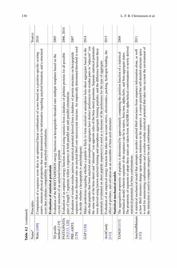
130 L. F. B. Christensen et al.
Tabl
e4.
2(c
ontin
ued)
Nam
eaPr
inci
ples
Yea
r(s)
Wal
tz[1
09]
Com
puta
tion
ofa
sequ
ence
scor
eth
atis
anop
timiz
edlin
ear
com
bina
tion
ofsc
ores
base
don
apo
sitio
n-sp
ecifi
c-sc
orin
gm
atri
x,ni
nete
enph
ysic
alpr
oper
ties
that
wer
ede
term
ined
tobe
info
rmat
ive
rega
rdin
gam
yloi
dfo
rmat
ion,
and
ast
ruct
ural
scor
eth
atev
alua
tes
com
patib
ility
with
amyl
oid
conf
orm
atio
ns.
2010
Eva
luat
ion
ofst
atis
tica
lpot
enti
als
3Dpr
ofile
met
hod
[119
]E
valu
atio
nof
the
RO
SET
TAD
ESI
GN
ener
gyfu
nctio
non
hexa
pept
ides
thre
aded
onto
mul
tiple
tem
plat
esba
sed
onth
ecr
ysta
lstr
uctu
reof
anam
yloi
dfo
rmin
gpe
ptid
e20
05
PAST
A/P
AST
A2.
0[1
13,1
28]
Eva
luat
ion
ofan
empi
rica
lene
rgy
func
tion
deri
ved
usin
gst
atis
tics
from
ada
taba
seof
glob
ular
prot
eins
for
allp
ossi
ble
pair
sof
leng
th7
segm
ents
with
ina
sequ
ence
inbo
thpa
ralle
land
anti-
para
llelo
rien
tatio
ns20
06,2
014
PRE
-AM
YL
[129
]E
valu
atio
nof
resi
due-
resi
due
pair
wis
est
atis
tical
pote
ntia
lder
ived
from
ada
taba
seof
prot
ein
stru
ctur
eson
hexa
pept
ide
sequ
ence
sth
atar
eth
read
edon
toan
amyl
oid
fibri
lcor
em
icro
crys
tals
truc
ture
.An
empi
rica
llyde
term
ined
thre
shol
dis
used
tode
cide
whe
ther
ahe
xape
ptid
eis
amyl
oido
geni
c.
2007
GA
P[1
30]
Mak
espr
edic
tions
rega
rdin
gw
heth
era
pept
ide
islik
ely
tofo
rmam
yloi
dor
amor
phou
sbe
ta-s
heet
aggr
egat
esba
sed
onth
eid
eath
atpe
ptid
esth
atfo
rmam
yloi
dan
dam
orph
ous
aggr
egat
esha
vedi
stin
ctpr
efer
ence
sfo
rre
sidu
epa
irs
in“a
djac
ent”
(on
the
sam
esi
deof
the
beta
-she
et)
and
“alte
rnat
e”(o
nop
posi
tesi
des
ofth
ebe
ta-s
heet
)po
sitio
ns.S
epar
ate
stat
istic
alpo
tent
ials
wer
ede
rive
dus
ing
know
nam
yloi
dfo
rmin
gan
dam
orph
ous
aggr
egat
efo
rmin
gpe
ptid
es,a
ndth
edi
ffer
ence
betw
een
thes
epo
tent
ials
(eva
luat
edon
shor
tpep
tide
sequ
ence
s)is
used
asa
mea
sure
ofth
epr
efer
ence
for
the
type
ofag
greg
ate.
2014
Arc
hCan
dy[1
11]
Eva
luat
ion
ofan
empi
rica
lene
rgy
func
tion
that
take
sin
toac
coun
tste
rics
,ele
ctro
stat
ics,
pack
ing,
hydr
ogen
bond
ing,
the
effe
ctof
prol
ines
,and
solv
atio
non
anen
sem
ble
ofbe
ta-a
rch
stru
ctur
es20
15
Stat
isti
calm
echa
nica
lmod
els
TAN
GO
[117
]T
heag
greg
atio
npr
open
sity
ofpe
ptid
esis
dete
rmin
edby
appr
oxim
atel
yco
mpu
ting
the
part
ition
func
tion
ofa
stat
istic
alm
echa
nica
lmod
elw
here
inse
gmen
tsof
the
sequ
ence
can
bein
nativ
e,be
ta-t
urn,
alph
a-he
lix,a
ndbe
ta-a
ggre
gate
stat
es.
The
prop
ensi
tyof
segm
ents
toad
optt
hese
stat
esar
eta
ken
from
AG
AD
IRfo
ral
pha-
helic
alco
nfor
mat
ions
orne
wly
deri
ved
stat
istic
alpo
tent
ials
for
beta
conf
orm
atio
ns.
2004
Am
yloi
dMut
ants
[131
]A
stat
istic
alm
echa
nica
lmod
elth
atat
tem
pts
topr
edic
tam
yloi
dfib
rils
truc
ture
sfr
omse
quen
cein
form
atio
nal
one,
asw
ell
asho
wfib
rils
truc
ture
sch
ange
upon
mak
ing
mut
atio
nsto
the
sequ
ence
.Aco
arse
-gra
ined
buts
tilld
etai
led
stru
ctur
esp
ace
isus
edfo
ren
umer
atin
gco
nfor
mat
ions
and
are
sidu
e-re
sidu
est
atis
tical
pote
ntia
ltha
ttak
esin
toac
coun
tthe
envi
ronm
ento
fth
ein
tera
ctio
nis
used
toco
mpu
teen
ergi
esfo
rea
chco
nfor
mat
ion.
2011
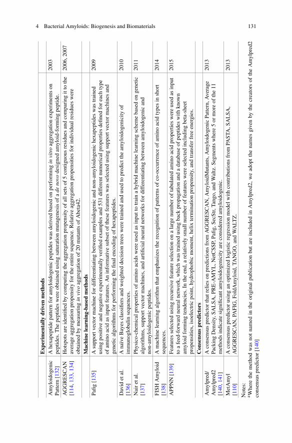
4 Bacterial Amyloids: Biogenesis and Biomaterials 131
Exp
erim
enta
llydr
iven
met
hods
Am
yloi
doge
nic
Patte
rn[1
32]
Ahe
xape
ptid
epa
ttern
for
amyl
oido
geni
cpe
ptid
esw
asde
rive
dba
sed
onpe
rfor
min
gin
vitr
oag
greg
atio
nex
peri
men
tson
pept
ides
.The
pept
ides
wer
eob
tain
edus
ing
satu
ratio
nm
utag
enes
isof
ade
novo
desi
gned
amyl
oid-
form
ing
pept
ide.
2003
AG
GR
ESC
AN
[114
,133
,134
]H
otsp
ots
are
iden
tified
byco
mpu
ting
the
aggr
egat
ion
prop
ensi
tyof
alls
ets
of5
cont
iguo
usre
sidu
esan
dco
mpa
ring
itto
the
aver
age
aggr
egat
ion
prop
ensi
tyfo
rth
een
tire
sequ
ence
.Rel
ativ
eag
greg
atio
npr
open
sitie
sfo
rin
divi
dual
resi
dues
wer
eob
tain
edby
mea
suri
ngin
vivo
aggr
egat
ion
of20
mut
ants
ofA
beta
42.
2006
,200
7
Mac
hine
lear
ning
-bas
edm
etho
dsPa
fig[1
35]
Asu
ppor
tvec
tor
mac
hine
for
diff
eren
tiatin
gbe
twee
nam
yloi
doge
nic
and
non-
amyl
oido
geni
che
xape
ptid
esw
astr
aine
dus
ing
posi
tive
and
nega
tive
expe
rim
enta
llyve
rifie
dex
ampl
esan
d53
1di
ffer
entn
umer
ical
prop
ertie
sde
fined
for
each
type
ofam
ino
acid
asin
putf
eatu
res.
An
info
rmat
ive
subs
etof
thes
efe
atur
esw
asse
lect
edus
ing
supp
ortv
ecto
rm
achi
nes
and
gene
tical
gori
thm
sfo
rpe
rfor
min
gth
efin
alen
codi
ngof
hexa
pept
ides
.
2009
Dav
idet
al.
[136
]A
naïv
eB
ayes
clas
sifie
rsan
dw
eigh
ted
deci
sion
tree
sw
ere
trai
ned
and
used
topr
edic
tthe
amyl
oido
geni
city
ofim
mun
oglo
bulin
sequ
ence
s.20
10
Nai
ret
al.
[137
]Ph
ysic
o-ch
emic
alpr
oper
ties
ofam
ino
acid
sw
ere
used
asin
putt
otr
ain
ahy
brid
mac
hine
lear
ning
sche
me
base
don
gene
tical
gori
thm
s,su
ppor
tvec
tor
mac
hine
s,an
dar
tifici
alne
ural
netw
orks
for
diff
eren
tiatin
gbe
twee
nam
yloi
doge
nic
and
non-
amyl
oido
geni
cpe
ptid
es.
2011
FISH
Am
yloi
d[1
38]
Am
achi
nele
arni
ngal
gori
thm
that
emph
asiz
esth
ere
cogn
ition
ofpa
ttern
sof
co-o
ccur
renc
eof
amin
oac
idty
pes
insh
ort
sequ
ence
s.20
14
APP
NN
[139
]Fe
atur
esse
lect
edus
ing
recu
rsiv
efe
atur
ese
lect
ion
ona
larg
enu
mbe
rof
tabu
late
dam
ino
acid
prop
ertie
sw
ere
used
asin
put
toa
feed
-for
war
dne
ural
netw
ork,
whi
chw
astr
aine
dus
ing
back
prop
agat
ion
and
ada
taba
seof
pept
ides
with
know
nam
yloi
dfo
rmin
gte
nden
cies
.In
the
end,
are
lativ
ely
smal
lnum
ber
offe
atur
esw
ere
sele
cted
incl
udin
gbe
ta-s
heet
prop
ensi
ties,
isoe
lect
ric
poin
t,hy
drop
hobi
cm
omen
t,he
lixte
rmin
atio
npr
open
sity
,and
tran
sfer
free
ener
gies
.
2015
Con
sens
uspr
edic
tors
Am
ylpr
ed/
Am
ylpr
ed2
[140
,141
]
Aco
nsen
sus
pred
icto
rth
atre
lies
onpr
edic
tions
from
AG
GR
ESC
AN
,Am
yloi
dMut
ants
,Am
yloi
doge
nic
Patte
rn,A
vera
gePa
ckin
gD
ensi
ty,S
AL
SA,P
RE
-AM
YL
,Net
CSS
P,Pa
fig,S
ecSt
r,Ta
ngo,
and
Wal
tz.S
egm
ents
whe
re5
orm
ore
ofth
e11
met
hods
indi
cate
sign
ifica
ntam
yloi
doge
nici
tyar
eco
nsid
ered
amyl
oido
geni
c.
2013
Met
Am
yl[1
10]
Aco
nsen
sus
pred
icto
rus
ing
anop
timiz
edlo
gist
icre
gres
sion
mod
elw
ithco
ntri
butio
nsfr
omPA
STA
,SA
LSA
,A
GG
RE
SCA
N,P
AFI
G,F
oldA
myl
oid,
TAN
GO
,and
WA
LTZ
.20
13
Not
es:
a Whe
reth
em
etho
dw
asno
tna
med
inth
eor
igin
alpu
blic
atio
nbu
tar
ein
clud
edin
Am
ylpr
ed2,
we
adop
tth
ena
mes
give
nby
the
crea
tors
ofth
eA
myl
pred
2co
nsen
sus
pred
icto
r[1
40]
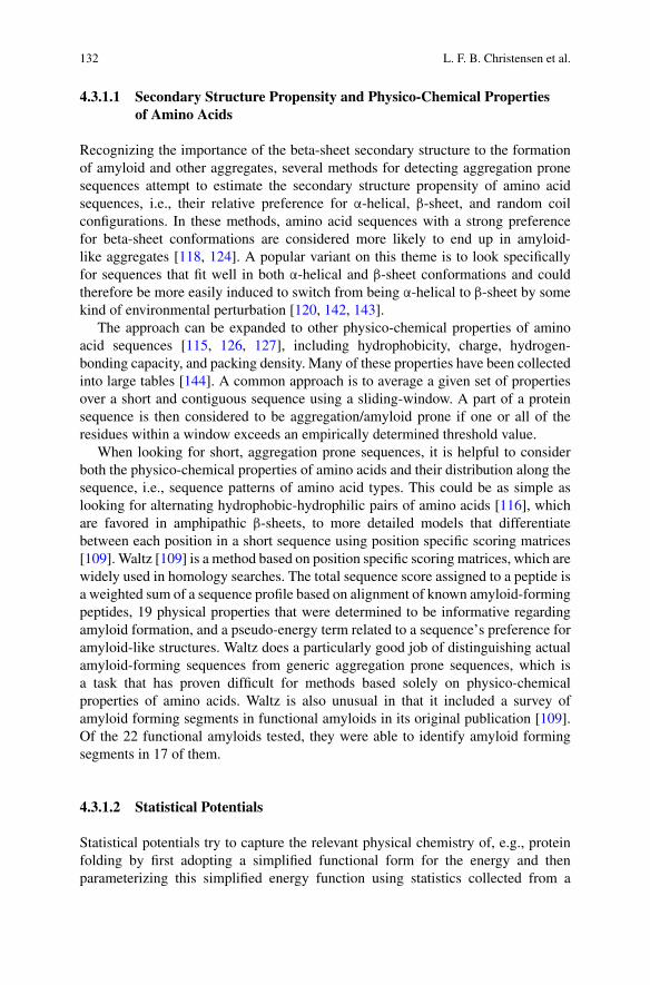
132 L. F. B. Christensen et al.
4.3.1.1 Secondary Structure Propensity and Physico-Chemical Propertiesof Amino Acids
Recognizing the importance of the beta-sheet secondary structure to the formationof amyloid and other aggregates, several methods for detecting aggregation pronesequences attempt to estimate the secondary structure propensity of amino acidsequences, i.e., their relative preference for α-helical, β-sheet, and random coilconfigurations. In these methods, amino acid sequences with a strong preferencefor beta-sheet conformations are considered more likely to end up in amyloid-like aggregates [118, 124]. A popular variant on this theme is to look specificallyfor sequences that fit well in both α-helical and β-sheet conformations and couldtherefore be more easily induced to switch from being α-helical to β-sheet by somekind of environmental perturbation [120, 142, 143].
The approach can be expanded to other physico-chemical properties of aminoacid sequences [115, 126, 127], including hydrophobicity, charge, hydrogen-bonding capacity, and packing density. Many of these properties have been collectedinto large tables [144]. A common approach is to average a given set of propertiesover a short and contiguous sequence using a sliding-window. A part of a proteinsequence is then considered to be aggregation/amyloid prone if one or all of theresidues within a window exceeds an empirically determined threshold value.
When looking for short, aggregation prone sequences, it is helpful to considerboth the physico-chemical properties of amino acids and their distribution along thesequence, i.e., sequence patterns of amino acid types. This could be as simple aslooking for alternating hydrophobic-hydrophilic pairs of amino acids [116], whichare favored in amphipathic β-sheets, to more detailed models that differentiatebetween each position in a short sequence using position specific scoring matrices[109]. Waltz [109] is a method based on position specific scoring matrices, which arewidely used in homology searches. The total sequence score assigned to a peptide isa weighted sum of a sequence profile based on alignment of known amyloid-formingpeptides, 19 physical properties that were determined to be informative regardingamyloid formation, and a pseudo-energy term related to a sequence’s preference foramyloid-like structures. Waltz does a particularly good job of distinguishing actualamyloid-forming sequences from generic aggregation prone sequences, which isa task that has proven difficult for methods based solely on physico-chemicalproperties of amino acids. Waltz is also unusual in that it included a survey ofamyloid forming segments in functional amyloids in its original publication [109].Of the 22 functional amyloids tested, they were able to identify amyloid formingsegments in 17 of them.
4.3.1.2 Statistical Potentials
Statistical potentials try to capture the relevant physical chemistry of, e.g., proteinfolding by first adopting a simplified functional form for the energy and thenparameterizing this simplified energy function using statistics collected from a
4 Bacterial Amyloids: Biogenesis and Biomaterials 133
database of experimentally determined structures. A common simplification schemein the context of proteins is to assume that a large part of the energy can bedecomposed into residue-residue pair interactions rather than explicitly accountingfor the presence of every atom within the residues. Implicit in the parameterizationbased on known structures is the assumption that the proximity of pairs ofamino acids within protein structures is a reflection of their intrinsic propensityfor interaction. Despite these seemingly drastic simplifications and assumptions,statistical potentials have proven useful in many areas of protein science includingstructure prediction and design.
In practice, peptide sequences are threaded onto experimentally determinedamyloid fibril core structures. The statistical potential is then evaluated for a givensequence-structure pair and those sequences with the lowest energies are assumed tobe a good fit and therefore amyloid prone [119, 129]. These methods are reminiscentof early attempts at protein structure prediction and fold recognition based onthreading [145]. Depending on the structure that the sequences are threaded onto,these methods can potentially take into account inter-strand hydrogen bondingenergies, β-sheet stacking energies or even intra-strand hairpin formation [111], butthe fact that only a fixed and finite set of structures is considered in these methodswill always limit their general purpose use.
4.3.1.3 Statistical Mechanical Models
Theoretical studies have shown that the chain entropy of the disordered part of theprotein can be an important factor in favoring particular ensembles of aggregatedconformations [117, 121, 146, 147]. It is therefore useful to develop models thatallow partial ordering of a sequence (where energetic terms are dominant) but alsotake into account disorder from the rest of the chain. After (usually approximately)computing the partition function, it is possible to identify the lowest free energyconformational ensembles, and the ordered parts of the sequence in these ensemblesare understood to be the parts of the sequence that are driving aggregation.
TANGO is a commonly used aggregation predictor that combines secondarystructure propensities and physico-chemical preferences for burial into a statisticalmechanical model for β-sheet aggregation [117]. TANGO has shown promise inpredicting whether a peptide will aggregate as well as the differences in aggregationpropensity when mutations are made to a given sequence, which is particularlyrelevant when investigating disease-causing mutants. A more recently developedstatistical mechanical method attempts to predict not only which parts of thestructure are ordered in aggregates but also the structures of fibrils in significantdetail and how they change upon mutation [131].
134 L. F. B. Christensen et al.
4.3.1.4 Experimentally Driven Methods
The previous methods used data from experiments which were not designedspecifically to determine which protein sequences will aggregate or form amyloid.However, there are also methods based directly on experiments purpose-builtto discover aggregation prone sequences. One approach involves deriving manyvariants of a designed amyloid forming peptide through saturation mutagenesis[132]. The secondary structure content of the peptides was measured by circulardichroism spectroscopy and the peptides’ tendency to form fibrils was evaluated byelectron microscopy after 1 month. The results of these experiments were then usedto derive short sequence patterns that reflected which amino acids were allowed ineach position of amyloid forming peptides. Consequently, these sequence patternscan then be used to rapidly search for potential amyloid forming sequences in largesequence databases. A possible limitation of methods like these is a bias towards theinitial peptide sequence used to derive the short peptides.
Although most aggregation experiments are performed in vitro, ultimately weare interested in aggregation propensity of proteins in vivo. AGGRESCAN [114]is similar to methods that scan sequences looking for short stretches of highlyaggregation prone residues, but is unique in that the aggregation propensities of the20 amino acid residues were obtained by monitoring aggregation via fluorescenceof Abeta-42 variants with single amino acid substitutions in vivo.
4.3.1.5 Machine Learning Methods
Methods discussed thus far are all based on underlying physical principles ofone kind or another, and can be more easily parameterized on the basis oflimited experimental data to provide accurate predictions. Therefore, evaluatingthe accuracy models based on different physical principles can help us to test ourunderstanding of aggregation. However, in some cases, we simply want to know howspecific changes to a sequence will affect aggregation propensity and the detailedmechanisms are of relatively little importance. Any learning problem, includingprediction of aggregation and amyloid propensity from sequence information, wheresufficient labeled data is available, is potentially susceptible to supervised machinelearning techniques [148]. The number of peptides that have been experimentallyvalidated as either fibril forming or non-fibril forming is large and, presumably,still growing [149]. This represents a potentially rich source of training data.Indeed, machine learning and Bayesian statistics based methods such as supportvector machines [135], artificial neural networks [137], and decision trees [136]have already been fruitfully applied to the problem of predicting amyloid andaggregation prone peptide sequences. Although these methods perhaps provide lessdirect physical insight into the problem of amyloid formation than other availablemethods, given enough training data they could eventually become the most reliableway to engineer the aggregation propensity of an amino acid sequence.
4 Bacterial Amyloids: Biogenesis and Biomaterials 135
4.3.1.6 Consensus Predictors
Due in part to the proliferation of methods that can be used to predict aggregationand amyloid propensity on the basis of sequence information, consensus predictorshave become a popular option that allow non-experts to combine results fromdifferent methods and thus hedge their bets when looking for aggregation “hotspots” [110, 140]. The success of consensus methods is likely due to the diversityof principles underlying the constituent methods, many of which we have discussedin this section.
4.3.2 Detecting Amyloid Prone Sequences in FunctionalAmyloids
Although the methods described in the previous section were not derived and havenot been tested primarily with functional amyloid in mind, the structural similaritiesbetween functional and pathological amyloid provide a source of hope that thesemethods can nonetheless be successfully applied in the search for functionalamyloid sequences. Several instances of these methods being applied to functionalamyloids already exist in the literature.
4.3.2.1 Sequence-Based Methods Can Detect Amyloidogenic Segmentsin Biofilm-Associated Proteins
By scanning the sequence of a biofilm associated protein in Staphylococcusepidermidis using a combination of several sequence-based amyloid detectionmethods including AGGRESCAN [114], PASTA [113], and TANGO [117], thepeptide STVTVTF was chosen for further biophysical characterization [150]. Thisseven-residue peptide was shown to spontaneously form amyloid fibers in vitro. Inanother study, the amyloidogenic segments of the yeast cell wall protein Bgl2p werepredicted using FoldAmyloid, TANGO, AGGRESCAN, PASTA, and Waltz [151].Peptides containing these sequences were then synthesized and found to form fibrils.In a similar case, sequence similarity to fimbrial proteins and the amyloid detectionmethods TANGO and AGGRESCAN were used to argue that HfaA might be afunctional amyloid involved in the establishment of extracellular structures [152].
4.3.2.2 Searching for Prion-like Domains Can Uncover PreviouslyUnknown Functional Amyloids
The relationship between amyloids and prions remains to be clearly elucidated.The hallmark of a prion is the ability to convert another prion molecule (with
136 L. F. B. Christensen et al.
the same sequence and perhaps even those with related sequences) from oneconformation to another, typically with functional consequences. One of these statesis often aggregated or amyloid-like. Like amyloids, prions were first recognized ina pathological context but, also like amyloids, in retrospect it seems obvious thatnature would take advantage of these motifs for functional purposes. A statisticalmodel trained on the sequences of proteins that are known to exhibit prion-likebehavior in yeast was used to search against many complete genomes including839 bacterial genomes [153]. The search generated a list of 2200 putative prions(proteins that contain prion-like domains) in bacteria. Of the proteins in bacteriawith annotated function, the prion hits were highly overrepresented in certainfunction classes, including those involved in the establishment of extracellularstructures.
4.3.2.3 The Existence of Imperfect Repeats Is Common to ManyFunctional Amyloids
The sequences of the primary fibril component in the Curli functional amyloid sys-tem, CsgA, and its putative fibril-nucleating component, CsgB, are clearly related,with each containing 5 imperfect repeats. However, according to the consensuspredictor Amylpred2, CsgB has a significantly higher aggregation propensity [154].This is consistent with CsgB’s putative role as a nucleator of aggregation andthe supposition that functional amyloids should have evolved mechanisms forregulating the timing and location of amyloid formation. MspA, the first functionalamyloid described in archaea, was also found to be amyloidogenic according toAmylpred2, and its amyloid nature was confirmed using biophysical techniques[10]. Like many functional amyloids, MspA contains imperfect repeats, as doTapA [155] and FapC [102]. On the basis of sequence alone, the existence ofrepeats containing amyloid motifs may be the single strongest indicator of a likelyfunctional amyloid. The evolved nature of functional amyloids should provideadditional clues when searching for functional amyloids in the genome, however,and some possibilities for leveraging those differences will be discussed in thefollowing section.
4.3.3 Identifying Functional Amyloids Based on Their EvolvedCharacteristics
The fibrils of functional and pathological amyloids share striking structural sim-ilarities, and our knowledge of each type of amyloid can help to inform ourunderstanding of the other. However, the evolutionary forces shaping the sequencesof functional and pathological amyloids could hardly be more different. Functionalamyloids confer selective advantages to their host organisms and typically evolve
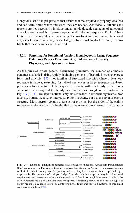
4 Bacterial Amyloids: Biogenesis and Biomaterials 137
alongside a set of helper proteins that ensure that the amyloid is properly localizedand can form fibrils where and when they are needed. Additionally, although thereasons are not necessarily intuitive, many amyloidogenic segments of functionalamyloids are located in imperfect repeats within the full sequence. Each of thesefacts should be useful when searching for as-of-yet uncharacterized functionalamyloids. Given the relatively nascent stage of functional amyloid research, it seemslikely that these searches will bear fruit.
4.3.3.1 Searching for Functional Amyloid Homologues in Large SequenceDatabases Reveals Functional Amyloid Sequence Diversity,Phylogeny, and Operon Structure
As the price of whole genome sequencing plummets, the number of completegenomes available is rising rapidly, including genomes of bacteria known to expressfunctional amyloid [156]. For families of functional amyloids where at least onesequence is known, searching for related sequences in large sequence databasesprovides a fuller picture of the sequence diversity within a family as well as asense of how widespread the family is in the bacterial kingdom, as illustrated inFig. 4.3 [21, 53]. Related functional amyloid sequences in different organisms showdiversity both at the level of individual protein sequences and at the level of operonstructure. Most operons contain a core set of proteins, but the order of the codingsequences in the operon may be shuffled or the orientations inverted. The variation
Fig. 4.3 A taxonomic analysis of bacterial strains based on Functional Amyloid in Pseudomonas(Fap) sequences. The Fap operon typically contains 6 proteins: FapA-FapF. The operon structureis illustrated next to each genus. The primary and secondary fibril components are FapC and FapB,respectively. The presence of multiple “helper” proteins within an operon may be a functionalrequirement and therefore a universal characteristic of functional amyloid operons. If this is thecase, bioinformatic algorithms that look for operons containing amyloids and specific types ofhelper proteins may prove useful in identifying novel functional amyloid systems. (Reproducedwith permission from [53])
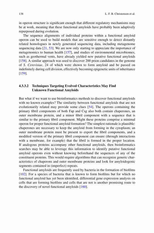
138 L. F. B. Christensen et al.
in operon structure is significant enough that different regulatory mechanisms maybe at work, meaning that these functional amyloids have probably been adaptivelyrepurposed during evolution.
The sequence alignments of individual proteins within a functional amyloidoperon can be used to build models that are sensitive enough to detect distantlyrelated homologues in newly generated sequencing data, including metagenomesequencing data [21, 53]. We are now only starting to appreciate the importance ofmetagenomics to human health [157], and studies of environmental microbiomes,such as geothermal vents, have already yielded new putative functional amyloids[158]. A similar approach was used to discover 200 prion candidates in the genomeof S. Cerevisiae, 24 of which were shown to form amyloid and be passed onindefinitely during cell division, effectively becoming epigenetic units of inheritance[159].
4.3.3.2 Techniques Targeting Evolved Characteristics May FindUnknown Functional Amyloids
But what if we want to use bioinformatics methods to discover functional amyloidswith no known examples? The similarity between functional amyloids that are notevolutionarily related may provide some clues [54]. The operons containing theprimary fibril components of both Fap and Csg also both contain chaperones, anouter membrane protein, and a minor fibril component with a sequence that issimilar to the primary fibril component. Might these proteins comprise a minimaloperon for proper functional amyloid formation? The simplest rationale is plausible:chaperones are necessary to keep the amyloid from forming in the cytoplasm; anouter membrane protein must be present to export the fibril components, and amodified version of the primary fibril component can ensure (through interactionswith a membrane, for example) that the fibril is formed in the proper location.If analogous proteins accompany other functional amyloids, then bioinformaticssearches may be able to leverage this information to identify putative functionalamyloid operons even without knowing beforehand the sequences of any of theconstituent proteins. This would require algorithms that can recognize generic char-acteristics of chaperons and outer membrane proteins and look for amyloidogenicsegments contained in (imperfect) repeats.
Functional amyloids are frequently used by bacteria in the formation of biofilms[102]. For a species of bacteria that is known to form biofilms but for which nofunctional amyloid has yet been identified, differential gene expression analysis oncells that are forming biofilms and cells that are not is another promising route tothe discovery of novel functional amyloids [160].
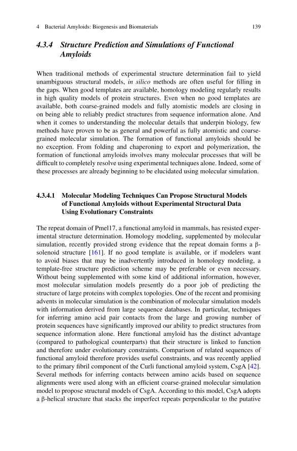
4 Bacterial Amyloids: Biogenesis and Biomaterials 139
4.3.4 Structure Prediction and Simulations of FunctionalAmyloids
When traditional methods of experimental structure determination fail to yieldunambiguous structural models, in silico methods are often useful for filling inthe gaps. When good templates are available, homology modeling regularly resultsin high quality models of protein structures. Even when no good templates areavailable, both coarse-grained models and fully atomistic models are closing inon being able to reliably predict structures from sequence information alone. Andwhen it comes to understanding the molecular details that underpin biology, fewmethods have proven to be as general and powerful as fully atomistic and coarse-grained molecular simulation. The formation of functional amyloids should beno exception. From folding and chaperoning to export and polymerization, theformation of functional amyloids involves many molecular processes that will bedifficult to completely resolve using experimental techniques alone. Indeed, some ofthese processes are already beginning to be elucidated using molecular simulation.
4.3.4.1 Molecular Modeling Techniques Can Propose Structural Modelsof Functional Amyloids without Experimental Structural DataUsing Evolutionary Constraints
The repeat domain of Pmel17, a functional amyloid in mammals, has resisted exper-imental structure determination. Homology modeling, supplemented by molecularsimulation, recently provided strong evidence that the repeat domain forms a β-solenoid structure [161]. If no good template is available, or if modelers wantto avoid biases that may be inadvertently introduced in homology modeling, atemplate-free structure prediction scheme may be preferable or even necessary.Without being supplemented with some kind of additional information, however,most molecular simulation models presently do a poor job of predicting thestructure of large proteins with complex topologies. One of the recent and promisingadvents in molecular simulation is the combination of molecular simulation modelswith information derived from large sequence databases. In particular, techniquesfor inferring amino acid pair contacts from the large and growing number ofprotein sequences have significantly improved our ability to predict structures fromsequence information alone. Here functional amyloid has the distinct advantage(compared to pathological counterparts) that their structure is linked to functionand therefore under evolutionary constraints. Comparison of related sequences offunctional amyloid therefore provides useful constraints, and was recently appliedto the primary fibril component of the Curli functional amyloid system, CsgA [42].Several methods for inferring contacts between amino acids based on sequencealignments were used along with an efficient coarse-grained molecular simulationmodel to propose structural models of CsgA. According to this model, CsgA adoptsa β-helical structure that stacks the imperfect repeats perpendicular to the putative
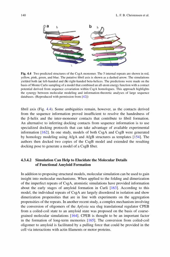
140 L. F. B. Christensen et al.
Fig. 4.4 Two predicted structures of the CsgA monomer. The 5 internal repeats are shown in red,yellow, pink, green, and blue. The putative fibril axis is shown as a dashed arrow. The simulationsyielded both (a) left-handed and (b) right-handed beta-helices. The predictions were made on thebasis of Monte Carlo sampling of a model that combined an all-atom energy function with a contactpotential derived from sequence covariation within CsgA homologues. This approach highlightsthe synergy between molecular modeling and information-theoretic analyses of large sequencedatabases. (Reproduced with permission from [42])
fibril axis (Fig. 4.4). Some ambiguities remain, however, as the contacts derivedfrom the sequence information proved insufficient to resolve the handedness ofthe β-helix and the inter-monomer contacts that contribute to fibril formation.An alternative to inferring docking contacts from sequence information is to usespecialized docking protocols that can take advantage of available experimentalinformation [162]. In one study, models of both CsgA and CsgB were generatedby homology modeling using AfgA and AfgB structures as templates [154]. Theauthors then docked two copies of the CsgB model and extended the resultingdocking pose to generate a model of a CsgB fiber.
4.3.4.2 Simulation Can Help to Elucidate the Molecular Detailsof Functional Amyloid Formation
In addition to proposing structural models, molecular simulation can be used to gaininsight into molecular mechanisms. When applied to the folding and dimerizationof the imperfect repeats of CsgA, atomistic simulations have provided informationabout the early stages of amyloid formation in Curli [163]. According to thismodel, the individual repeats of CsgA are largely disordered in isolation and showdimerization propensities that are in line with experiments on the aggregationpropensities of the repeats. In another recent study, a complex mechanism involvingthe conversion of oligomers of the Aplysia sea slug translational regulator CPEBfrom a coiled-coil state to an amyloid state was proposed on the basis of coarse-grained molecular simulations [164]. CPEB is thought to be an important factorin the formation of long-term memories [165]. The conversion from coiled-coiloligomer to amyloid is facilitated by a pulling force that could be provided in thecell via interactions with actin filaments or motor proteins.
4 Bacterial Amyloids: Biogenesis and Biomaterials 141
4.4 Uses for Functional Amyloid: Brave New Nanomaterials
E. coli’s curli operon makes up a fascinating miniaturized export-and-assemblysystem and holds great potential as a nanotechnological device. The few examplesof controlled amyloid production and export available in the literature all make useof this operon, and we will use the following section to describe results obtained sofar.
4.4.1 C-DAG as a Screen for Amyloid: How to Hijack a RobustAmyloid Export System
It has already been established that proteins can be exported provided their cross-sectional diameter is not larger than 2 nm; provided the protein is maintained inan unfolded state which can be threaded through the CsgG pore, there are noupper limits on protein size [49]. This has been put to elegant use as a bacterialscreening system to identify proteins capable of fibrillation under “amyloid-friendly” conditions in C-DAG (curli-dependent amyloid generator system) [50].Given that an overexpression of CsgG enables efficient secretion of CsgA [43](presumably together with smaller amounts of CsgE and CsgF), it is possible to testany given protein by fusing it to the CsgA signal peptide in a strain overexpressingCsgG and lacking CsgA and CsgB (to avoid spurious nucleation on the surface).In this way, expressed protein is separated from proteins in the cytoplasm and istransported in a (relatively) unfolded conformation through CsgG to accumulate toa high local concentration outside the cell. Here it can aggregate at leisure, possiblystimulated by the anionic lipid environment at the cell surface which is known topromote protein aggregation in general [166, 167]. A number of different proteinshave been shown to form fibrils under these conditions according to standard assays(Congo Red plates, apple-green birefringence, SDS-resistance to unfolding andseeding ability), even proteins such as the yeast prion Sup35 that normally is strictlydependent on the PIN factor to form amyloid [168]. A subsequent screen of all E.coli ORFs using universal primers also identified a novel amyloidogenic protein(FliE from the flagellar basal body). However, the main strength of the systemis probably in the screening of amyloid aggregation modulators by plating cellsexpressing a given protein on solid medium containing Congo Red; a caveat is thatscreening of whole ORF libraries can generate a large number of false positives aswell as various shades of red in the absence of detectable amyloid aggregates [169].The reason for this is unclear, though may be related to CR’s propensity to bind non-amyloid material, as seen for e.g. insulin [170], or even facilitate oligomerization ofnative proteins by binding as a sandwich between two protein molecules [170].
142 L. F. B. Christensen et al.
4.4.1.1 Generating New Binding Properties: How to Hitch a Rideon the Amyloid Ladder
Given the above export abilities, it is obvious to combine CsgA with proteins thatdisplay other functionalities. The simplest function is to bind, and this was earlyon very elegantly exemplified in the BIND (biofilm-integrated nanofiber display)system [171]. The approach that works best is to genetically fuse short peptidebinding sequences to the C-terminus of CsgA with a long linker (GSGGSG) andthen terminate said peptide with another GSGGSG sequence (N-terminal fusionsinterfere with CsgG recognition). The authors demonstrated the versatility of thissystem (Fig. 4.5) by demonstrating binding of E. coli overexpressing these fusionproteins to inorganic surfaces (graphene, carbon nanotube, glass, ice crystals),metals (steel, gold), minerals (hydroxyapatite, zinc sulfide, magnetite) and proteins.In the latter case, CsgA is fused to the protein SpyTag which covalently links to15-kDa SpyCatcher protein [172], allowing generic linkage to any protein that isfused to SpyCatcher. The study by Joshi and coworkers is also worth reading forthe rigor with which they quantify production of amyloid, using both colorimetry(Congo Red absorption; birefringence was not quantitative, probably due to fiberdispersion), formation of extracellular assemblies (whole-cell filtration ELISA withanti-CsgA antibodies), verification of intact protein (extracellular fractions washed
Fig. 4.5 Using fusions of CsgA and peptide binding systems to generate new functionality inbiofilm. Top: The endogenous CsgA is knocked out from an E. coli strain which from theexpression vector produces fusion proteins consisting of CsgA (yellow) and a C-terminallyattached peptide domain (green), as shown in the 3D protein model insert where the CsgA sequencewas threaded onto an AfgA structure. The linker domain is in grey, followed by the SpyTag domainin green. Peptide structure was predicted with PepFold. Bottom: The expressed protein formsamyloid (curli) fibrils on the surface of the E. coli cell and leads to a biofilm with novel function.(Reproduced with permission from [171])
4 Bacterial Amyloids: Biogenesis and Biomaterials 143
in SDS and the pellet dissolved in HFIP to mobilize for MALDI-TOF analysis) andmorphology (SEM, TEM and immunogold labelling with antibodies recognizingthe fusion peptide). Another important aspect is to use the right E. coli strain.The csgA deletion strain (LSR10) does not produce cellulose, avoiding backgroundeffects from CR binding to cellulose [173]. However, for larger-scale production,it is possible to use the K-12 strain PHL628 which does produce cellulose but alsohas a 3.5-fold increased curli operon expression thanks to a single point mutationin OmpR protein [174], so the whole amyloid producing and secreting machineryis boosted. There are remarkable perspectives in this work at several levels. Thesystem is simple and straightforward and requires no real optimization of expressionconstructs, yet provides an amyloid matrix that by virtue of its robustness can likelysurvive cell death to remain behind as a skeletal framework, built from sustainablematerials.
4.4.1.2 Amyloid as Underwater Glue: Fusing CsgA to Mussel FootProteins
Lu and co-workers have extended this approach by fusing CsgA with mussel footproteins Mfp3 and Mfp5 from Mytilus galloprovincialis as CsgA-Mfp3 and Mfp5-CsgA constructs [175]. Mfp proteins are used as underwater adhesives to silica(rock) surfaces; the proteins are activated by enzymatically converting Tyr residuesto the crosslinker 3,4-dihydroxyphenylalanine (DOPA) [176]. By combining theseglue proteins with amyloid, it is possible to generate environmentally robustunderwater glue that can self-heal through self-assembly and form a large fibersurface area that enhances contact area in the adhesive barnacle plaque [177].The hybrid CsgA-Mfp proteins lead to fusion fibrils that were three to fivefoldwider than pure CsgA fibrils, probably due to intermolecular association betweenMfp3 and Mfp5 domains. When hydroxylated, these fibrils had a two to threefoldincrease in adhesion to silica compared to CsgA. Interestingly, a copolymer made bymixing CsgA-Mfp3 and Mfp5-CsgA gave the greatest effect, showing the strongestunderwater adhesion of bio-derived/inspired protein-based underwater adhesives sofar.
4.4.1.3 Controlled Combination of Different Amyloid: The Powerof Riboregulators
The interface with different organic and inorganic surfaces shown in these previousexamples points to a merger of synthetic biology with materials science that willultimately be able to make large-scale programmable living materials. The trick isto engineer genetic logic gates into this system which could enable us to switchon (and off) different biofilms on demand, using appropriate environmental cues.Such a cue could be provided by the extremely sophisticated riboregulator systememployed imaginatively by Lu and co-workers to self-assemble hybrid structures
144 L. F. B. Christensen et al.
Fig. 4.6 Using riboregulators to make controllable variations in fibril patterns. This leads tosynthetic gene circuits where external inducers (aTC or AHL) control protein secretion. (a) Aconstant level of aTc (which drives expression of CsgA) also induces a gradual accumulationof AHL which in term induces expression of CsgAHis. (b) The His-variant can be labelled bygold nanoparticles, and the fraction of fibrils with CsgAHis increases with time. (Reproduced withpermission from [178])
of CsgA at a variety of different length scales [178]. Riboregulators are RNAmolecules that can regulate expression of genes in response to different smallmolecules such as anhydrotetracycline (aTc) or acyl-homoserine lactone (AHL)[179]. The Lu group used two different E. coli strains, regulated by either aTc orAHL, which expressed and secreted CsgA either with or without a His-tail (Fig. 4.6).By growing the two strains together, the production of CsgA and CsgAHis could becontrolled by sequential pulses or different concentrations of AHL versus aTc, sothat the ensuing amyloid fibrils would have different proportions of the two CsgAmolecules, depending on the temporal pattern of pulses [178]. This can also beregulated in a cell-to-cell manner if the aTc-inducible strain producing CsgA alsoproduces AHL constitutively, while the CsgAHis-producing strain is AHL-inducible.As the two strains grow and are induced by aTc, more and more AHL is produced,leading to an increased production (and higher proportion) of CsgAHis. The absoluteproportion will depend on the initial ratio of cell strains. Another elegant variation isgrowth along two opposing aTc and AHL gradients to get mainly CsgA fibrils in oneend and mainly CsgAHis fibrils in the other. Finally, the production of an 8-CsgAconcatemer with a single His tail together with a single-CsgHis can lead to morecomplex His distribution patterns. Gold nanoparticles containing a Nickel-nitrilo-triacetate group can be deposited on these fibrils in different ways, depending on thisCsgA8
His/CsgAHis pattern, leading to either conducting biofilm (long consecutivegold wires) or shorter gold nanorods. Since the properties of gold rods are very
4 Bacterial Amyloids: Biogenesis and Biomaterials 145
sensitive to their size, it is possible to use the protein distribution pattern to tune rodproperties with great versatility. Gold nanoparticles (AuNPs) can even be interfacedand co-assembled with quantum dots (QDs) provided these dots have a spycatcherdomain and the CsgA has a SpyTag domain [178]. This is of interest becausephoton emission properties can be tuned by plasmon-exciton interactions betweenplasmonic AuNPs and fluorescent QDs [180]. As a more general perspective, the useof such orthogonal affinity tags makes it possible to construct patterned scaffoldsfor multi-enzyme systems. The Lu approach also illustrates how it is possible toapply useful properties of multicellular communities or organisms into materialsfabrication using synthetic circuits.
4.4.2 Controlling Amyloid with Co-Factors: The Caseof the Missing Calcium
Apart from these three examples of the use of bona fide bacterial amyloid to generatenew design properties, there are also other examples of bacterial proteins which self-assemble under unusual circumstances. The Repeats-in-toxin (RTX) sequence isa highly conserved nine-peptide Gly- and Asp-rich repeat in the C-terminal Ca2+-binding RTX domain of the P. aeruginosa protease ArpA. This domain is disorderedin the apo-form, but binding of Ca2+ helps fold the RTX domain into compact β-helical structures [181], and thus regulates activity and secretion of these proteins.The Ca2+-binding site provides a useful handle to modulate structure [182]. Whenheterologously expressed in E. coli, the RTX domain can be purified from inclusionbodies as a random coil structure [182]. Addition of Ca2+ induces soluble β-sheetstructure, while the apoform assembles on the hour-scale to form not just fibrils butalso, remarkably, cm2-large highly regular and uniform sheets with flexible edges.Assembly can be arrested (but not reversed) by addition of Ca2+, while EDTAinduces self-assembly. Such scaffolds could incorporate soluble folded proteins asfusion proteins and thus immobilize e.g. enzymatic functions in 2 dimensions (witha density regulated by e.g. different ratios of fusion proteins and RTX-only proteins).The whole RTX domain is important for this self-assembly; a synthetic β-helicalpeptide containing 8 copies of the consensus nine-peptide repeats [183] (lackingthe terminal capping sequences) did not bind Ca2+ or undergo a disorder-to-ordertransition like the full-length RTX domain. Surprisingly, this construct bound La3+which induced self-assembly and fibrillation, turning the whole metal-handle on itshead! Given that the RTX family of exoproteins is associated with virulence of manyhuman pathogens [184], these manipulations are a good example of how to divertthe original pathogen virulence functionality to novel purposes.
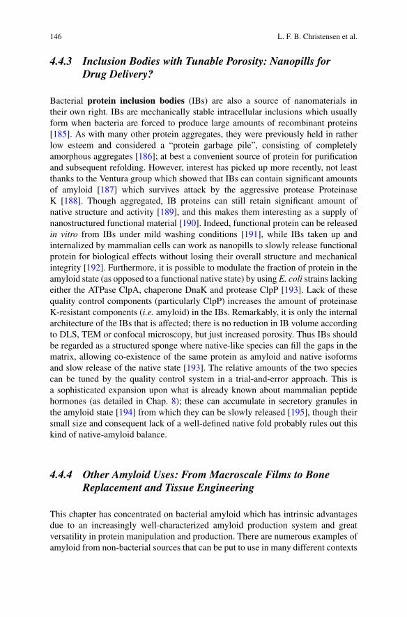
146 L. F. B. Christensen et al.
4.4.3 Inclusion Bodies with Tunable Porosity: Nanopills forDrug Delivery?
Bacterial protein inclusion bodies (IBs) are also a source of nanomaterials intheir own right. IBs are mechanically stable intracellular inclusions which usuallyform when bacteria are forced to produce large amounts of recombinant proteins[185]. As with many other protein aggregates, they were previously held in ratherlow esteem and considered a “protein garbage pile”, consisting of completelyamorphous aggregates [186]; at best a convenient source of protein for purificationand subsequent refolding. However, interest has picked up more recently, not leastthanks to the Ventura group which showed that IBs can contain significant amountsof amyloid [187] which survives attack by the aggressive protease ProteinaseK [188]. Though aggregated, IB proteins can still retain significant amount ofnative structure and activity [189], and this makes them interesting as a supply ofnanostructured functional material [190]. Indeed, functional protein can be releasedin vitro from IBs under mild washing conditions [191], while IBs taken up andinternalized by mammalian cells can work as nanopills to slowly release functionalprotein for biological effects without losing their overall structure and mechanicalintegrity [192]. Furthermore, it is possible to modulate the fraction of protein in theamyloid state (as opposed to a functional native state) by using E. coli strains lackingeither the ATPase ClpA, chaperone DnaK and protease ClpP [193]. Lack of thesequality control components (particularly ClpP) increases the amount of proteinaseK-resistant components (i.e. amyloid) in the IBs. Remarkably, it is only the internalarchitecture of the IBs that is affected; there is no reduction in IB volume accordingto DLS, TEM or confocal microscopy, but just increased porosity. Thus IBs shouldbe regarded as a structured sponge where native-like species can fill the gaps in thematrix, allowing co-existence of the same protein as amyloid and native isoformsand slow release of the native state [193]. The relative amounts of the two speciescan be tuned by the quality control system in a trial-and-error approach. This isa sophisticated expansion upon what is already known about mammalian peptidehormones (as detailed in Chap. 8); these can accumulate in secretory granules inthe amyloid state [194] from which they can be slowly released [195], though theirsmall size and consequent lack of a well-defined native fold probably rules out thiskind of native-amyloid balance.
4.4.4 Other Amyloid Uses: From Macroscale Films to BoneReplacement and Tissue Engineering
This chapter has concentrated on bacterial amyloid which has intrinsic advantagesdue to an increasingly well-characterized amyloid production system and greatversatility in protein manipulation and production. There are numerous examples ofamyloid from non-bacterial sources that can be put to use in many different contexts
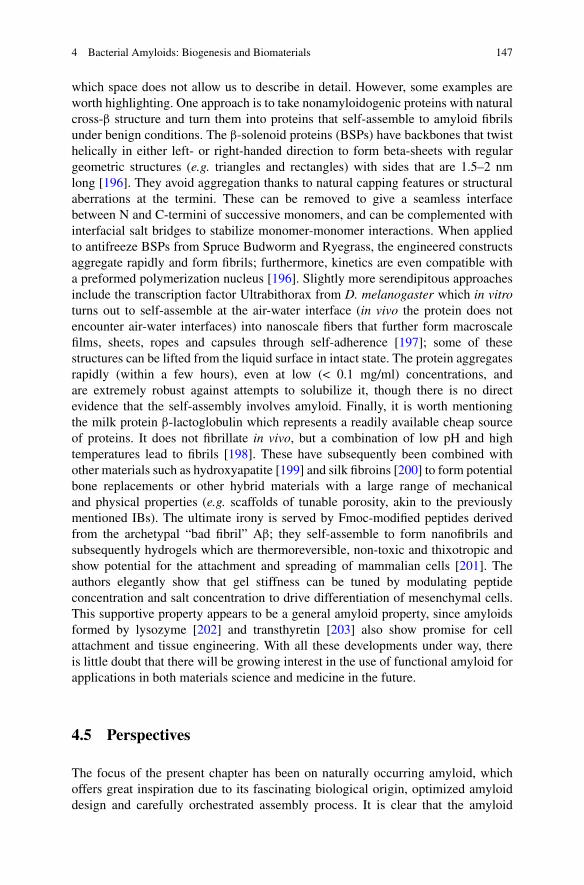
4 Bacterial Amyloids: Biogenesis and Biomaterials 147
which space does not allow us to describe in detail. However, some examples areworth highlighting. One approach is to take nonamyloidogenic proteins with naturalcross-β structure and turn them into proteins that self-assemble to amyloid fibrilsunder benign conditions. The β-solenoid proteins (BSPs) have backbones that twisthelically in either left- or right-handed direction to form beta-sheets with regulargeometric structures (e.g. triangles and rectangles) with sides that are 1.5–2 nmlong [196]. They avoid aggregation thanks to natural capping features or structuralaberrations at the termini. These can be removed to give a seamless interfacebetween N and C-termini of successive monomers, and can be complemented withinterfacial salt bridges to stabilize monomer-monomer interactions. When appliedto antifreeze BSPs from Spruce Budworm and Ryegrass, the engineered constructsaggregate rapidly and form fibrils; furthermore, kinetics are even compatible witha preformed polymerization nucleus [196]. Slightly more serendipitous approachesinclude the transcription factor Ultrabithorax from D. melanogaster which in vitroturns out to self-assemble at the air-water interface (in vivo the protein does notencounter air-water interfaces) into nanoscale fibers that further form macroscalefilms, sheets, ropes and capsules through self-adherence [197]; some of thesestructures can be lifted from the liquid surface in intact state. The protein aggregatesrapidly (within a few hours), even at low (< 0.1 mg/ml) concentrations, andare extremely robust against attempts to solubilize it, though there is no directevidence that the self-assembly involves amyloid. Finally, it is worth mentioningthe milk protein β-lactoglobulin which represents a readily available cheap sourceof proteins. It does not fibrillate in vivo, but a combination of low pH and hightemperatures lead to fibrils [198]. These have subsequently been combined withother materials such as hydroxyapatite [199] and silk fibroins [200] to form potentialbone replacements or other hybrid materials with a large range of mechanicaland physical properties (e.g. scaffolds of tunable porosity, akin to the previouslymentioned IBs). The ultimate irony is served by Fmoc-modified peptides derivedfrom the archetypal “bad fibril” Aβ; they self-assemble to form nanofibrils andsubsequently hydrogels which are thermoreversible, non-toxic and thixotropic andshow potential for the attachment and spreading of mammalian cells [201]. Theauthors elegantly show that gel stiffness can be tuned by modulating peptideconcentration and salt concentration to drive differentiation of mesenchymal cells.This supportive property appears to be a general amyloid property, since amyloidsformed by lysozyme [202] and transthyretin [203] also show promise for cellattachment and tissue engineering. With all these developments under way, thereis little doubt that there will be growing interest in the use of functional amyloid forapplications in both materials science and medicine in the future.
4.5 Perspectives
The focus of the present chapter has been on naturally occurring amyloid, whichoffers great inspiration due to its fascinating biological origin, optimized amyloiddesign and carefully orchestrated assembly process. It is clear that the amyloid
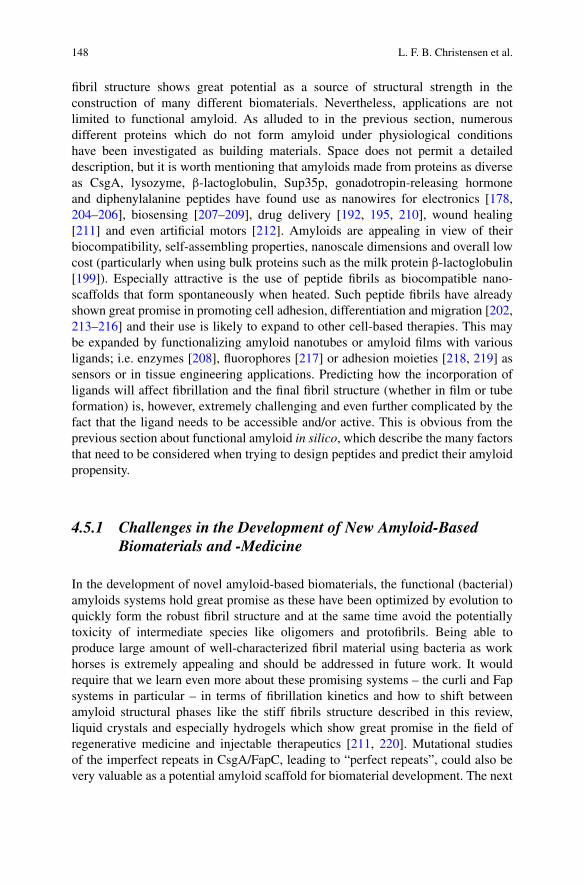
148 L. F. B. Christensen et al.
fibril structure shows great potential as a source of structural strength in theconstruction of many different biomaterials. Nevertheless, applications are notlimited to functional amyloid. As alluded to in the previous section, numerousdifferent proteins which do not form amyloid under physiological conditionshave been investigated as building materials. Space does not permit a detaileddescription, but it is worth mentioning that amyloids made from proteins as diverseas CsgA, lysozyme, β-lactoglobulin, Sup35p, gonadotropin-releasing hormoneand diphenylalanine peptides have found use as nanowires for electronics [178,204–206], biosensing [207–209], drug delivery [192, 195, 210], wound healing[211] and even artificial motors [212]. Amyloids are appealing in view of theirbiocompatibility, self-assembling properties, nanoscale dimensions and overall lowcost (particularly when using bulk proteins such as the milk protein β-lactoglobulin[199]). Especially attractive is the use of peptide fibrils as biocompatible nano-scaffolds that form spontaneously when heated. Such peptide fibrils have alreadyshown great promise in promoting cell adhesion, differentiation and migration [202,213–216] and their use is likely to expand to other cell-based therapies. This maybe expanded by functionalizing amyloid nanotubes or amyloid films with variousligands; i.e. enzymes [208], fluorophores [217] or adhesion moieties [218, 219] assensors or in tissue engineering applications. Predicting how the incorporation ofligands will affect fibrillation and the final fibril structure (whether in film or tubeformation) is, however, extremely challenging and even further complicated by thefact that the ligand needs to be accessible and/or active. This is obvious from theprevious section about functional amyloid in silico, which describe the many factorsthat need to be considered when trying to design peptides and predict their amyloidpropensity.
4.5.1 Challenges in the Development of New Amyloid-BasedBiomaterials and -Medicine
In the development of novel amyloid-based biomaterials, the functional (bacterial)amyloids systems hold great promise as these have been optimized by evolution toquickly form the robust fibril structure and at the same time avoid the potentiallytoxicity of intermediate species like oligomers and protofibrils. Being able toproduce large amount of well-characterized fibril material using bacteria as workhorses is extremely appealing and should be addressed in future work. It wouldrequire that we learn even more about these promising systems – the curli and Fapsystems in particular – in terms of fibrillation kinetics and how to shift betweenamyloid structural phases like the stiff fibrils structure described in this review,liquid crystals and especially hydrogels which show great promise in the field ofregenerative medicine and injectable therapeutics [211, 220]. Mutational studiesof the imperfect repeats in CsgA/FapC, leading to “perfect repeats”, could also bevery valuable as a potential amyloid scaffold for biomaterial development. The next
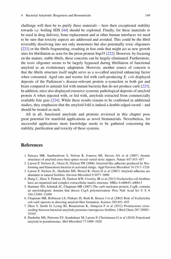
4 Bacterial Amyloids: Biogenesis and Biomaterials 149
challenge will then be to purify these materials – here their exceptional stabilitytowards i.e. boiling SDS [44] should be exploited. Finally, for these materials tobe used in drug delivery, bone replacement and at other human interfaces we needto be sure that toxicity aspects are addressed and avoided. This could be the fibrilreversibly dissolving into not only monomers but also potentially toxic oligomers[221] or the fibrils fragmenting, resulting in free ends that might act as new growthsites for fibrillation as seen for the prion protein Sup35 [222]. However, by focusingon the mature, stable fibrils, these concerns can be largely eliminated. Furthermore,the toxic oligomer seems to be largely bypassed during fibrillation of functionalamyloid as an evolutionary adaptation. However, another source of concern isthat the fibrils structure itself might serve as a so-called amyloid enhancing factorwhen consumed. Aged rats and worms fed with curli-producing E. coli displayeddeposits of the Parkinson’s disease-relevant protein α-synuclein in both gut andbrain compared to animals fed with mutant bacteria that do not produce curli [223].In addition, mice also displayed extensive systemic pathological deposits of amyloidprotein A when injected with, or fed with, amyloids extracted from commerciallyavailable foie gras [224]. While these results remain to be confirmed in additionalstudies, they emphasize that the amyloid fold is indeed a double-edged sword – andshould be treated as such.
All in all, functional amyloids and proteins reviewed in this chapter posegreat potential for manifold applications as novel biomaterials. Nevertheless, forsuccessful applications more knowledge needs to be gathered concerning thestability, purification and toxicity of these systems.
References
1. Sawaya MR, Sambashivan S, Nelson R, Ivanova MI, Sievers SA et al (2007) Atomicstructures of amyloid cross-beta spines reveal varied steric zippers. Nature 447:453–457
2. Larsen P, Nielsen JL, Otzen D, Nielsen PH (2008) Amyloid-like adhesins produced by floc-forming and filamentous bacteria in activated sludge. Appl Environ Microbiol 74:1517–1526
3. Larsen P, Nielsen JL, Dueholm MS, Wetzel R, Otzen D et al (2007) Amyloid adhesins areabundant in natural biofilms. Environ Microbiol 9:3077–3090
4. Hung C, Zhou Y, Pinkner JS, Dodson KW, Crowley JR et al (2013) Escherichia coli biofilmshave an organized and complex extracellular matrix structure. MBio 4:e00645–e00613
5. Hammer ND, Schmidt JC, Chapman MR (2007) The curli nucleator protein, CsgB, containsan amyloidogenic domain that directs CsgA polymerization. Proc Natl Acad Sci U S A104:12494–12499
6. Chapman MR, Robinson LS, Pinkner JS, Roth R, Heuser J et al (2002) Role of Escherichiacoli curli operons in directing amyloid fiber formation. Science 295:851–855
7. Zhou Y, Smith D, Leong BJ, Brannstrom K, Almqvist F et al (2012) Promiscuous cross-seeding between bacterial amyloids promotes interspecies biofilms. J Biol Chem 287:35092–35103
8. Dueholm MS, Petersen SV, Sonderkaer M, Larsen P, Christiansen G et al (2010) Functionalamyloid in pseudomonas. Mol Microbiol 77:1009–1020
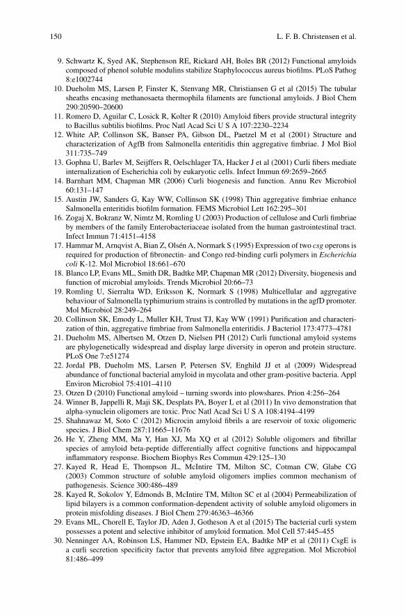
150 L. F. B. Christensen et al.
9. Schwartz K, Syed AK, Stephenson RE, Rickard AH, Boles BR (2012) Functional amyloidscomposed of phenol soluble modulins stabilize Staphylococcus aureus biofilms. PLoS Pathog8:e1002744
10. Dueholm MS, Larsen P, Finster K, Stenvang MR, Christiansen G et al (2015) The tubularsheaths encasing methanosaeta thermophila filaments are functional amyloids. J Biol Chem290:20590–20600
11. Romero D, Aguilar C, Losick R, Kolter R (2010) Amyloid fibers provide structural integrityto Bacillus subtilis biofilms. Proc Natl Acad Sci U S A 107:2230–2234
12. White AP, Collinson SK, Banser PA, Gibson DL, Paetzel M et al (2001) Structure andcharacterization of AgfB from Salmonella enteritidis thin aggregative fimbriae. J Mol Biol311:735–749
13. Gophna U, Barlev M, Seijffers R, Oelschlager TA, Hacker J et al (2001) Curli fibers mediateinternalization of Escherichia coli by eukaryotic cells. Infect Immun 69:2659–2665
14. Barnhart MM, Chapman MR (2006) Curli biogenesis and function. Annu Rev Microbiol60:131–147
15. Austin JW, Sanders G, Kay WW, Collinson SK (1998) Thin aggregative fimbriae enhanceSalmonella enteritidis biofilm formation. FEMS Microbiol Lett 162:295–301
16. Zogaj X, Bokranz W, Nimtz M, Romling U (2003) Production of cellulose and Curli fimbriaeby members of the family Enterobacteriaceae isolated from the human gastrointestinal tract.Infect Immun 71:4151–4158
17. Hammar M, Arnqvist A, Bian Z, Olsén A, Normark S (1995) Expression of two csg operons isrequired for production of fibronectin- and Congo red-binding curli polymers in Escherichiacoli K-12. Mol Microbiol 18:661–670
18. Blanco LP, Evans ML, Smith DR, Badtke MP, Chapman MR (2012) Diversity, biogenesis andfunction of microbial amyloids. Trends Microbiol 20:66–73
19. Romling U, Sierralta WD, Eriksson K, Normark S (1998) Multicellular and aggregativebehaviour of Salmonella typhimurium strains is controlled by mutations in the agfD promoter.Mol Microbiol 28:249–264
20. Collinson SK, Emody L, Muller KH, Trust TJ, Kay WW (1991) Purification and characteri-zation of thin, aggregative fimbriae from Salmonella enteritidis. J Bacteriol 173:4773–4781
21. Dueholm MS, Albertsen M, Otzen D, Nielsen PH (2012) Curli functional amyloid systemsare phylogenetically widespread and display large diversity in operon and protein structure.PLoS One 7:e51274
22. Jordal PB, Dueholm MS, Larsen P, Petersen SV, Enghild JJ et al (2009) Widespreadabundance of functional bacterial amyloid in mycolata and other gram-positive bacteria. ApplEnviron Microbiol 75:4101–4110
23. Otzen D (2010) Functional amyloid – turning swords into plowshares. Prion 4:256–26424. Winner B, Jappelli R, Maji SK, Desplats PA, Boyer L et al (2011) In vivo demonstration that
alpha-synuclein oligomers are toxic. Proc Natl Acad Sci U S A 108:4194–419925. Shahnawaz M, Soto C (2012) Microcin amyloid fibrils a are reservoir of toxic oligomeric
species. J Biol Chem 287:11665–1167626. He Y, Zheng MM, Ma Y, Han XJ, Ma XQ et al (2012) Soluble oligomers and fibrillar
species of amyloid beta-peptide differentially affect cognitive functions and hippocampalinflammatory response. Biochem Biophys Res Commun 429:125–130
27. Kayed R, Head E, Thompson JL, McIntire TM, Milton SC, Cotman CW, Glabe CG(2003) Common structure of soluble amyloid oligomers implies common mechanism ofpathogenesis. Science 300:486–489
28. Kayed R, Sokolov Y, Edmonds B, McIntire TM, Milton SC et al (2004) Permeabilization oflipid bilayers is a common conformation-dependent activity of soluble amyloid oligomers inprotein misfolding diseases. J Biol Chem 279:46363–46366
29. Evans ML, Chorell E, Taylor JD, Aden J, Gotheson A et al (2015) The bacterial curli systempossesses a potent and selective inhibitor of amyloid formation. Mol Cell 57:445–455
30. Nenninger AA, Robinson LS, Hammer ND, Epstein EA, Badtke MP et al (2011) CsgE isa curli secretion specificity factor that prevents amyloid fibre aggregation. Mol Microbiol81:486–499
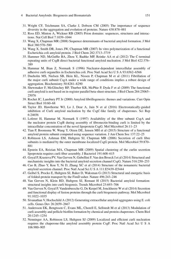
4 Bacterial Amyloids: Biogenesis and Biomaterials 151
31. Wright CF, Teichmann SA, Clarke J, Dobson CM (2005) The importance of sequencediversity in the aggregation and evolution of proteins. Nature 438:878–881
32. Ross ED, Minton A, Wickner RB (2005) Prion domains: sequences, structures and interac-tions. Nat Cell Biol 7:1039–1044
33. Wang X, Chapman MR (2008) Sequence determinants of bacterial amyloid formation. J MolBiol 380:570–580
34. Wang X, Smith DR, Jones JW, Chapman MR (2007) In vitro polymerization of a functionalEscherichia coli amyloid protein. J Biol Chem 282:3713–3719
35. Hammer ND, McGuffie BA, Zhou Y, Badtke MP, Reinke AA et al (2012) The C-terminalrepeating units of CsgB direct bacterial functional amyloid nucleation. J Mol Biol 422:376–389
36. Hammar M, Bian Z, Normark S (1996) Nucleator-dependent intercellular assembly ofadhesive curli organelles in Escherichia coli. Proc Natl Acad Sci U S A 93:6562–6566
37. Dueholm MS, Nielsen SB, Hein KL, Nissen P, Chapman M et al (2011) Fibrillation ofthe major curli subunit CsgA under a wide range of conditions implies a robust design ofaggregation. Biochemistry 50:8281–8290
38. Shewmaker F, McGlinchey RP, Thurber KR, McPhie P, Dyda F et al (2009) The functionalcurli amyloid is not based on in-register parallel beta-sheet structure. J Biol Chem 284:25065–25076
39. Rochet JC, Lansbury PT Jr (2000) Amyloid fibrillogenesis: themes and variations. Curr OpinStruct Biol 10:60–68
40. Taylor JD, Hawthorne WJ, Lo J, Dear A, Jain N et al (2016) Electrostatically-guidedinhibition of Curli amyloid nucleation by the CsgC-like family of chaperones. Sci Rep6:24656
41. Loferer H, Hammar M, Normark S (1997) Availability of the fibre subunit CsgA andthe nucleator protein CsgB during assembly of fibronectin-binding curli is limited by theintracellular concentration of the novel lipoprotein CsgG. Mol Microbiol 26:11–23
42. Tian P, Boomsma W, Wang Y, Otzen DE, Jensen MH et al (2015) Structure of a functionalamyloid protein subunit computed using sequence variation. J Am Chem Soc 137:22–25
43. Robinson LS, Ashman EM, Hultgren SJ, Chapman MR (2006) Secretion of curli fibresubunits is mediated by the outer membrane-localized CsgG protein. Mol Microbiol 59:870–881
44. Epstein EA, Reizian MA, Chapman MR (2009) Spatial clustering of the curlin secretionlipoprotein requires curli fiber assembly. J Bacteriol 191:608–615
45. Goyal P, Krasteva PV, Van Gerven N, Gubellini F, Van den Broeck I et al (2014) Structural andmechanistic insights into the bacterial amyloid secretion channel CsgG. Nature 516:250–253
46. Cao B, Zhao Y, Kou Y, Ni D, Zhang XC et al (2014) Structure of the nonameric bacterialamyloid secretion channel. Proc Natl Acad Sci U S A 111:E5439–E5444
47. Geibel S, Procko E, Hultgren SJ, Baker D, Waksman G (2013) Structural and energetic basisof folded-protein transport by the FimD usher. Nature 496:243–246
48. Van Gerven N, Klein RD, Hultgren SJ, Remaut H (2015) Bacterial amyloid formation:structural insights into curli biogensis. Trends Microbiol 23:693–706
49. Van Gerven N, Goyal P, Vandenbussche G, De Kerpel M, Jonckheere W et al (2014) Secretionand functional display of fusion proteins through the curli biogenesis pathway. Mol Microbiol91:1022–1035
50. Sivanathan V, Hochschild A (2012) Generating extracellular amyloid aggregates using E. colicells. Genes Dev 26:2659–2667
51. Andersson EK, Bengtsson C, Evans ML, Chorell E, Sellstedt M et al (2013) Modulation ofcurli assembly and pellicle biofilm formation by chemical and protein chaperones. Chem Biol20:1245–1254
52. Nenninger AA, Robinson LS, Hultgren SJ (2009) Localized and efficient curli nucleationrequires the chaperone-like amyloid assembly protein CsgF. Proc Natl Acad Sci U S A106:900–905
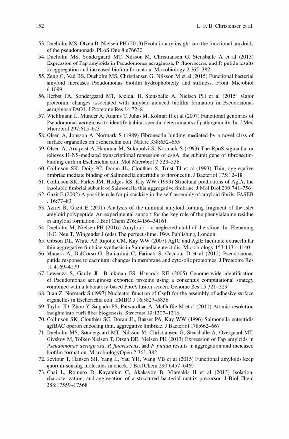
152 L. F. B. Christensen et al.
53. Dueholm MS, Otzen D, Nielsen PH (2013) Evolutionary insight into the functional amyloidsof the pseudomonads. PLoS One 8:e76630
54. Dueholm MS, Sondergaard MT, Nilsson M, Christiansen G, Stensballe A et al (2013)Expression of Fap amyloids in Pseudomonas aeruginosa, P. fluorescens, and P. putida resultsin aggregation and increased biofilm formation. Microbiology 2:365–382
55. Zeng G, Vad BS, Dueholm MS, Christiansen G, Nilsson M et al (2015) Functional bacterialamyloid increases Pseudomonas biofilm hydrophobicity and stiffness. Front Microbiol6:1099
56. Herbst FA, Sondergaard MT, Kjeldal H, Stensballe A, Nielsen PH et al (2015) Majorproteomic changes associated with amyloid-induced biofilm formation in Pseudomonasaeruginosa PAO1. J Proteome Res 14:72–81
57. Wiehlmann L, Munder A, Adams T, Juhas M, Kolmar H et al (2007) Functional genomics ofPseudomonas aeruginosa to identify habitat-specific determinants of pathogenicity. Int J MedMicrobiol 297:615–623
58. Olsen A, Jonsson A, Normark S (1989) Fibronectin binding mediated by a novel class ofsurface organelles on Escherichia coli. Nature 338:652–655
59. Olsen A, Arnqvist A, Hammar M, Sukupolvi S, Normark S (1993) The RpoS sigma factorrelieves H-NS-mediated transcriptional repression of csgA, the subunit gene of fibronectin-binding curli in Escherichia coli. Mol Microbiol 7:523–536
60. Collinson SK, Doig PC, Doran JL, Clouthier S, Trust TJ et al (1993) Thin, aggregativefimbriae mediate binding of Salmonella enteritidis to fibronectin. J Bacteriol 175:12–18
61. Collinson SK, Parker JM, Hodges RS, Kay WW (1999) Structural predictions of AgfA, theinsoluble fimbrial subunit of Salmonella thin aggregative fimbriae. J Mol Biol 290:741–756
62. Gazit E (2002) A possible role for pi-stacking in the self-assembly of amyloid fibrils. FASEBJ 16:77–83
63. Azriel R, Gazit E (2001) Analysis of the minimal amyloid-forming fragment of the isletamyloid polypeptide. An experimental support for the key role of the phenylalanine residuein amyloid formation. J Biol Chem 276:34156–34161
64. Dueholm M, Nielsen PH (2016) Amyloids – a neglected child of the slime. In: FlemmingH-C, Neu T, Wingender J (eds) The perfect slime. IWA Publishing, London
65. Gibson DL, White AP, Rajotte CM, Kay WW (2007) AgfC and AgfE facilitate extracellularthin aggregative fimbriae synthesis in Salmonella enteritidis. Microbiology 153:1131–1140
66. Manara A, DalCorso G, Baliardini C, Farinati S, Cecconi D et al (2012) Pseudomonasputida response to cadmium: changes in membrane and cytosolic proteomes. J Proteome Res11:4169–4179
67. Lewenza S, Gardy JL, Brinkman FS, Hancock RE (2005) Genome-wide identificationof Pseudomonas aeruginosa exported proteins using a consensus computational strategycombined with a laboratory-based PhoA fusion screen. Genome Res 15:321–329
68. Bian Z, Normark S (1997) Nucleator function of CsgB for the assembly of adhesive surfaceorganelles in Escherichia coli. EMBO J 16:5827–5836
69. Taylor JD, Zhou Y, Salgado PS, Patwardhan A, McGuffie M et al (2011) Atomic resolutioninsights into curli fiber biogenesis. Structure 19:1307–1316
70. Collinson SK, Clouthier SC, Doran JL, Banser PA, Kay WW (1996) Salmonella enteritidisagfBAC operon encoding thin, aggregative fimbriae. J Bacteriol 178:662–667
71. Dueholm MS, Søndergaard MT, Nilsson M, Christiansen G, Stensballe A, Overgaard MT,Givskov M, Tolker-Nielsen T, Otzen DE, Nielsen PH (2013) Expression of Fap amyloids inPseudomonas aeruginosa, P. fluorescens, and P. putida results in aggregation and increasedbiofilm formation. MicrobiologyOpen 2:365–382
72. Seviour T, Hansen SH, Yang L, Yau YH, Wang VB et al (2015) Functional amyloids keepquorum-sensing molecules in check. J Biol Chem 290:6457–6469
73. Chai L, Romero D, Kayatekin C, Akabayov B, Vlamakis H et al (2013) Isolation,characterization, and aggregation of a structured bacterial matrix precursor. J Biol Chem288:17559–17568
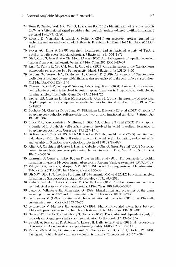
4 Bacterial Amyloids: Biogenesis and Biomaterials 153
74. Terra R, Stanley-Wall NR, Cao G, Lazazzera BA (2012) Identification of Bacillus subtilisSipW as a bifunctional signal peptidase that controls surface-adhered biofilm formation. JBacteriol 194:2781–2790
75. Romero D, Vlamakis H, Losick R, Kolter R (2011) An accessory protein required foranchoring and assembly of amyloid fibres in B. subtilis biofilms. Mol Microbiol 80:1155–1168
76. Stover AG, Driks A (1999) Secretion, localization, and antibacterial activity of TasA, aBacillus subtilis spore-associated protein. J Bacteriol 181:1664–1672
77. Oh J, Kim JG, Jeon E, Yoo CH, Moon JS et al (2007) Amyloidogenesis of type III-dependentharpins from plant pathogenic bacteria. J Biol Chem 282:13601–13609
78. Kim JG, Park BK, Yoo CH, Jeon E, Oh J et al (2003) Characterization of the Xanthomonasaxonopodis pv. glycines Hrp Pathogenicity Island. J Bacteriol 185:3155–3166
79. de Jong W, Wosten HA, Dijkhuizen L, Claessen D (2009) Attachment of Streptomycescoelicolor is mediated by amyloidal fimbriae that are anchored to the cell surface via cellulose.Mol Microbiol 73:1128–1140
80. Claessen D, Rink R, de Jong W, Siebring J, de Vreugd P et al (2003) A novel class of secretedhydrophobic proteins is involved in aerial hyphae formation in Streptomyces coelicolor byforming amyloid-like fibrils. Genes Dev 17:1714–1726
81. Sawyer EB, Claessen D, Haas M, Hurgobin B, Gras SL (2011) The assembly of individualchaplin peptides from Streptomyces coelicolor into functional amyloid fibrils. PLoS One6:e18839
82. Bokhove M, Claessen D, de Jong W, Dijkhuizen L, Boekema EJ et al (2013) Chaplins ofStreptomyces coelicolor self-assemble into two distinct functional amyloids. J Struct Biol184:301–309
83. Elliot MA, Karoonuthaisiri N, Huang J, Bibb MJ, Cohen SN et al (2003) The chaplins:a family of hydrophobic cell-surface proteins involved in aerial mycelium formation inStreptomyces coelicolor. Genes Dev 17:1727–1740
84. Di Berardo C, Capstick DS, Bibb MJ, Findlay KC, Buttner MJ et al (2008) Function andredundancy of the chaplin cell surface proteins in aerial hypha formation, rodlet assembly,and viability in Streptomyces coelicolor. J Bacteriol 190:5879–5889
85. Alteri CJ, Xicohtencatl-Cortes J, Hess S, Caballero-Olin G, Giron JA et al (2007) Mycobac-terium tuberculosis produces pili during human infection. Proc Natl Acad Sci U S A104:5145–5150
86. Ramsugit S, Guma S, Pillay B, Jain P, Larsen MH et al (2013) Pili contribute to biofilmformation in vitro in Mycobacterium tuberculosis. Antonie Van Leeuwenhoek 104:725–735
87. Velayati AA, Farnia P, Masjedi MR (2012) Pili in totally drug resistant MycobacteriumTuberculosis (TDR-TB). Int J Mycobacteriol 1:57–58
88. Oli MW, Otoo HN, Crowley PJ, Heim KP, Nascimento MM et al (2012) Functional amyloidformation by Streptococcus mutans. Microbiology 158:2903–2916
89. Bieler S, Estrada L, Lagos R, Baeza M, Castilla J et al (2005) Amyloid formation modulatesthe biological activity of a bacterial protein. J Biol Chem 280:26880–26885
90. Lagos R, Villanueva JE, Monasterio O (1999) Identification and properties of the genesencoding microcin E492 and its immunity protein. J Bacteriol 181:212–217
91. de Lorenzo V (1984) Isolation and characterization of microcin E492 from Klebsiellapneumoniae. Arch Microbiol 139:72–75
92. de Lorenzo V, Martinez JL, Asensio C (1984) Microcin-mediated interactions betweenKlebsiella pneumoniae and Escherichia coli strains. J Gen Microbiol 130:391–400
93. Gekara NO, Jacobs T, Chakraborty T, Weiss S (2005) The cholesterol-dependent cytolysinlisteriolysin O aggregates rafts via oligomerization. Cell Microbiol 7:1345–1356
94. Bavdek A, Kostanjšek R, Antonini V, Lakey JH, Dalla Serra M et al (2012) pH dependenceof listeriolysin O aggregation and pore-forming ability. FEBS J 279:126–141
95. Vazquez-Boland JA, Dominguez-Bernal G, Gonzalez-Zorn B, Kreft J, Goebel W (2001)Pathogenicity islands and virulence evolution in Listeria. Microbes Infect 3:571–584
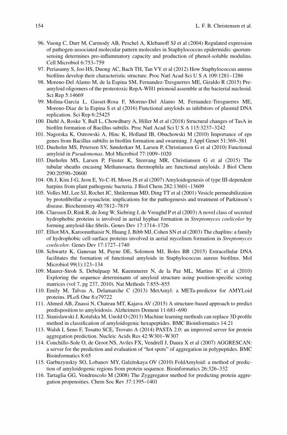
154 L. F. B. Christensen et al.
96. Vuong C, Durr M, Carmody AB, Peschel A, Klebanoff SJ et al (2004) Regulated expressionof pathogen-associated molecular pattern molecules in Staphylococcus epidermidis: quorum-sensing determines pro-inflammatory capacity and production of phenol-soluble modulins.Cell Microbiol 6:753–759
97. Periasamy S, Joo HS, Duong AC, Bach TH, Tan VY et al (2012) How Staphylococcus aureusbiofilms develop their characteristic structure. Proc Natl Acad Sci U S A 109:1281–1286
98. Moreno-Del Alamo M, de la Espina SM, Fernandez-Tresguerres ME, Giraldo R (2015) Pre-amyloid oligomers of the proteotoxic RepA-WH1 prionoid assemble at the bacterial nucleoid.Sci Rep 5:14669
99. Molina-Garcia L, Gasset-Rosa F, Moreno-Del Alamo M, Fernandez-Tresguerres ME,Moreno-Diaz de la Espina S et al (2016) Functional amyloids as inhibitors of plasmid DNAreplication. Sci Rep 6:25425
100. Diehl A, Roske Y, Ball L, Chowdhury A, Hiller M et al (2018) Structural changes of TasA inbiofilm formation of Bacillus subtilis. Proc Natl Acad Sci U S A 115:3237–3242
101. Nagorska K, Ostrowski A, Hinc K, Holland IB, Obuchowski M (2010) Importance of epsgenes from Bacillus subtilis in biofilm formation and swarming. J Appl Genet 51:369–381
102. Dueholm MS, Petersen SV, Sønderkær M, Larsen P, Christiansen G et al (2010) Functionalamyloid in Pseudomonas. Mol Microbiol 77:1009–1020
103. Dueholm MS, Larsen P, Finster K, Stenvang MR, Christiansen G et al (2015) Thetubular sheaths encasing Methanosaeta thermophila are functional amyloids. J Biol Chem290:20590–20600
104. Oh J, Kim J-G, Jeon E, Yo C-H, Moon JS et al (2007) Amyloidogenesis of type III-dependentharpins from plant pathogenic bacteria. J Biol Chem 282:13601–13609
105. Volles MJ, Lee SJ, Rochet JC, Shtilerman MD, Ding TT et al (2001) Vesicle permeabilizationby protofibrillar α-synuclein: implications for the pathogenesis and treatment of Parkinson’sdisease. Biochemistry 40:7812–7819
106. Claessen D, Rink R, de Jong W, Siebring J, de Vreughd P et al (2003) A novel class of secretedhydrophobic proteins is involved in aerial hyphae formation in Streptomyces coelicolor byforming amyloid-like fibrils. Genes Dev 17:1714–1726
107. Elliot MA, Karoonuthaisir N, Huang J, Bibb MJ, Cohen SN et al (2003) The chaplins: a familyof hydrophobic cell-surface proteins involved in aerial mycelium formation in Streptomycescoelicolor. Genes Dev 17:1727–1740
108. Schwartz K, Ganesan M, Payne DE, Solomon MJ, Boles BR (2015) Extracellular DNAfacilitates the formation of functional amyloids in Staphylococcus aureus biofilms. MolMicrobiol 99(1):123–134
109. Maurer-Stroh S, Debulpaep M, Kuemmerer N, de la Paz ML, Martins IC et al (2010)Exploring the sequence determinants of amyloid structure using position-specific scoringmatrices (vol 7, pg 237, 2010). Nat Methods 7:855–855
110. Emily M, Talvas A, Delamarche C (2013) MetAmyl: a METa-predictor for AMYLoidproteins. PLoS One 8:e79722
111. Ahmed AB, Znassi N, Chateau MT, Kajava AV (2015) A structure-based approach to predictpredisposition to amyloidosis. Alzheimers Dement 11:681–690
112. Stanislawski J, Kotulska M, Unold O (2013) Machine learning methods can replace 3D profilemethod in classification of amyloidogenic hexapeptides. BMC Bioinformatics 14:21
113. Walsh I, Seno F, Tosatto SCE, Trovato A (2014) PASTA 2.0: an improved server for proteinaggregation prediction. Nucleic Acids Res 42:W301–W307
114. Conchillo-Sole O, de Groot NS, Aviles FX, Vendrell J, Daura X et al (2007) AGGRESCAN:a server for the prediction and evaluation of “hot spots” of aggregation in polypeptides. BMCBioinformatics 8:65
115. Garbuzynskiy SO, Lobanov MY, Galzitskaya OV (2010) FoldAmyloid: a method of predic-tion of amyloidogenic regions from protein sequence. Bioinformatics 26:326–332
116. Tartaglia GG, Vendruscolo M (2008) The Zyggregator method for predicting protein aggre-gation propensities. Chem Soc Rev 37:1395–1401
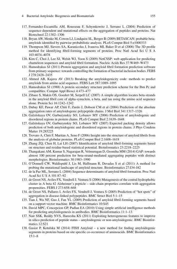
4 Bacterial Amyloids: Biogenesis and Biomaterials 155
117. Fernandez-Escamilla AM, Rousseau F, Schymkowitz J, Serrano L (2004) Prediction ofsequence-dependent and mutational effects on the aggregation of peptides and proteins. NatBiotechnol 22:1302–1306
118. Bryan AW, Menke M, Cowen LJ, Lindquist SL, Berger B (2009) BETASCAN: probable beta-amyloids identified by pairwise probabilistic analysis. PLoS Comput Biol 5:e1000333
119. Thompson MJ, Sievers SA, Karanicolas J, Ivanova MI, Baker D et al (2006) The 3D profilemethod for identifying fibril-forming segments of proteins. Proc Natl Acad Sci U S A103:4074–4078
120. Kim C, Choi J, Lee SJ, Welsh WJ, Yoon S (2009) NetCSSP: web application for predictingchameleon sequences and amyloid fibril formation. Nucleic Acids Res 37:W469–W473
121. Hamodrakas SJ (2011) Protein aggregation and amyloid fibril formation prediction softwarefrom primary sequence: towards controlling the formation of bacterial inclusion bodies. FEBSJ 278:2428–2435
122. Ahmed AB, Kajava AV (2013) Breaking the amyloidogenicity code: methods to predictamyloids from amino acid sequence. FEBS Lett 587:1089–1095
123. Hamodrakas SJ (1988) A protein secondary structure prediction scheme for the Ibm Pc andcompatibles. Comput Appl Biosci 4:473–477
124. Zibaee S, Makin OS, Goedert M, Serpell LC (2007) A simple algorithm locates beta-strandsin the amyloid fibril core of alpha-synuclein, a beta, and tau using the amino acid sequencealone. Protein Sci 16:1242–1242
125. Dubay KF, Pawar AP, Chiti F, Zurdo J, Dobson CM et al (2004) Prediction of the absoluteaggregation rates of amyloidogenic polypeptide chains. J Mol Biol 341:1317–1326
126. Galzitskaya OV, Garbuzynskiy SO, Lobanov MY (2006) Prediction of amyloidogenic anddisordered regions in protein chains. PLoS Comput Biol 2:1639–1648
127. Galzitskaya OV, Garbuzynskiy SO, Lobanov MY (2007) Expected packing density allowsprediction of both amyloidogenic and disordered regions in protein chains. J Phys CondensMatter 19:285225
128. Trovato A, Chiti F, Maritan A, Seno F (2006) Insight into the structure of amyloid fibrils fromthe analysis of globular proteins. PLoS Comput Biol 2:1608–1618
129. Zhang ZQ, Chen H, Lai LH (2007) Identification of amyloid fibril-forming segments basedon structure and residue-based statistical potential. Bioinformatics 23:2218–2225
130. Thangakani AM, Kumar S, Nagarajan R, Velmurugan D, Gromiha MM (2014) GAP: towardsalmost 100 percent prediction for beta-strand-mediated aggregating peptides with distinctmorphologies. Bioinformatics 30:1983–1990
131. O’Donnell CW, Waldispuhl J, Lis M, Halfmann R, Devadas S et al (2011) A method forprobing the mutational landscape of amyloid structure. Bioinformatics 27:I34–I42
132. de la Paz ML, Serrano L (2004) Sequence determinants of amyloid fibril formation. Proc NatlAcad Sci U S A 101:87–92
133. de Groot NS, Aviles FX, Vendrell J, Ventura S (2006) Mutagenesis of the central hydrophobiccluster in A beta 42 Alzheimer’s pepticle – side-chain properties correlate with aggregationpropensities. FEBS J 273:658–668
134. de Groot NS, Pallares I, Aviles FX, Vendrell J, Ventura S (2005) Prediction of “hot spots” ofaggregation in disease-linked polypeptides. BMC Struct Biol 5:1–15
135. Tian J, Wu NF, Guo J, Fan YL (2009) Prediction of amyloid fibril-forming segments basedon a support vector machine. BMC Bioinformatics 10:S45
136. David MPC, Concepcion GP, Padlan EA (2010) Using simple artificial intelligence methodsfor predicting amyloidogenesis in antibodies. BMC Bioinformatics 11:1–13
137. Nair SSK, Reddy NVS, Hareesha KS (2011) Exploiting heterogeneous features to improvein silico prediction of peptide status – amyloidogenic or non-amyloidogenic. BMC Bioinfor-matics 12:S21
138. Gasior P, Kotulska M (2014) FISH Amyloid – a new method for finding amyloidogenicsegments in proteins based on site specific co-occurence of aminoacids. BMC Bioinformatics15:1–8
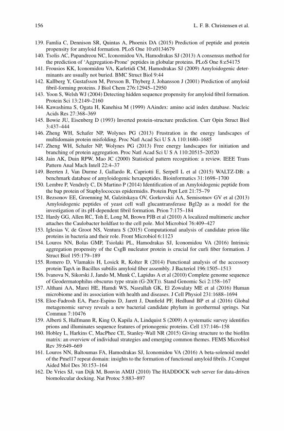
156 L. F. B. Christensen et al.
139. Famlia C, Dennison SR, Quintas A, Phoenix DA (2015) Prediction of peptide and proteinpropensity for amyloid formation. PLoS One 10:e0134679
140. Tsolis AC, Papandreou NC, Iconomidou VA, Hamodrakas SJ (2013) A consensus method forthe prediction of ‘Aggregation-Prone’ peptides in globular proteins. PLoS One 8:e54175
141. Frousios KK, Iconomidou VA, Karletidi CM, Hamodrakas SJ (2009) Amyloidogenic deter-minants are usually not buried. BMC Struct Biol 9:44
142. Kallberg Y, Gustafsson M, Persson B, Thyberg J, Johansson J (2001) Prediction of amyloidfibril-forming proteins. J Biol Chem 276:12945–12950
143. Yoon S, Welsh WJ (2004) Detecting hidden sequence propensity for amyloid fibril formation.Protein Sci 13:2149–2160
144. Kawashima S, Ogata H, Kanehisa M (1999) AAindex: amino acid index database. NucleicAcids Res 27:368–369
145. Bowie JU, Eisenberg D (1993) Inverted protein-structure prediction. Curr Opin Struct Biol3:437–444
146. Zheng WH, Schafer NP, Wolynes PG (2013) Frustration in the energy landscapes ofmultidomain protein misfolding. Proc Natl Acad Sci U S A 110:1680–1685
147. Zheng WH, Schafer NP, Wolynes PG (2013) Free energy landscapes for initiation andbranching of protein aggregation. Proc Natl Acad Sci U S A 110:20515–20520
148. Jain AK, Duin RPW, Mao JC (2000) Statistical pattern recognition: a review. IEEE TransPattern Anal Mach Intell 22:4–37
149. Beerten J, Van Durme J, Gallardo R, Capriotti E, Serpell L et al (2015) WALTZ-DB: abenchmark database of amyloidogenic hexapeptides. Bioinformatics 31:1698–1700
150. Lembre P, Vendrely C, Di Martino P (2014) Identification of an Amyloidogenic peptide fromthe bap protein of Staphylococcus epidermidis. Protein Pept Lett 21:75–79
151. Bezsonov EE, Groenning M, Galzitskaya OV, Gorkovskii AA, Semisotnov GV et al (2013)Amyloidogenic peptides of yeast cell wall glucantransferase Bgl2p as a model for theinvestigation of its pH-dependent fibril formation. Prion 7:175–184
152. Hardy GG, Allen RC, Toh E, Long M, Brown PJB et al (2010) A localized multimeric anchorattaches the Caulobacter holdfast to the cell pole. Mol Microbiol 76:409–427
153. Iglesias V, de Groot NS, Ventura S (2015) Computational analysis of candidate prion-likeproteins in bacteria and their role. Front Microbiol 6:1123
154. Louros NN, Bolas GMP, Tsiolaki PL, Hamodrakas SJ, Iconomidou VA (2016) Intrinsicaggregation propensity of the CsgB nucleator protein is crucial for curli fiber formation. JStruct Biol 195:179–189
155. Romero D, Vlamakis H, Losick R, Kolter R (2014) Functional analysis of the accessoryprotein TapA in Bacillus subtilis amyloid fiber assembly. J Bacteriol 196:1505–1513
156. Ivanova N, Sikorski J, Jando M, Munk C, Lapidus A et al (2010) Complete genome sequenceof Geodermatophilus obscurus type strain (G-20(T)). Stand Genomic Sci 2:158–167
157. Althani AA, Marei HE, Hamdi WS, Nasrallah GK, El Zowalaty ME et al (2016) Humanmicrobiome and its association with health and diseases. J Cell Physiol 231:1688–1694
158. Eloe-Fadrosh EA, Paez-Espino D, Jarett J, Dunfield PF, Hedlund BP et al (2016) Globalmetagenomic survey reveals a new bacterial candidate phylum in geothermal springs. NatCommun 7:10476
159. Alberti S, Halfmann R, King O, Kapila A, Lindquist S (2009) A systematic survey identifiesprions and illuminates sequence features of prionogenic proteins. Cell 137:146–158
160. Hobley L, Harkins C, MacPhee CE, Stanley-Wall NR (2015) Giving structure to the biofilmmatrix: an overview of individual strategies and emerging common themes. FEMS MicrobiolRev 39:649–669
161. Louros NN, Baltoumas FA, Hamodrakas SJ, Iconomidou VA (2016) A beta-solenoid modelof the Pmel17 repeat domain: insights to the formation of functional amyloid fibrils. J ComputAided Mol Des 30:153–164
162. De Vries SJ, van Dijk M, Bonvin AMJJ (2010) The HADDOCK web server for data-drivenbiomolecular docking. Nat Protoc 5:883–897
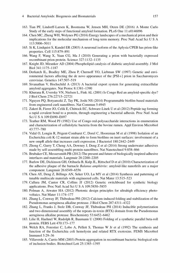
4 Bacterial Amyloids: Biogenesis and Biomaterials 157
163. Tian PF, Lindorff-Larsen K, Boomsma W, Jensen MH, Otzen DE (2016) A Monte CarloStudy of the early steps of functional amyloid formation. PLoS One 11:e0146096
164. Chen MC, Zheng WH, Wolynes PG (2016) Energy landscapes of a mechanical prion and theirimplications for the molecular mechanism of long-term memory. Proc Natl Acad Sci U S A113:5006–5011
165. Si K, Lindquist S, Kandel ER (2003) A neuronal isoform of the Aplysia CPEB has prion-likeproperties. Cell 115:879–891
166. Wang F, Wang X, Yuan CG, Ma J (2010) Generating a prion with bacterially expressedrecombinant prion protein. Science 327:1132–1135
167. Knight JD, Miranker AD (2004) Phospholipid catalysis of diabetic amyloid assembly. J MolBiol 341:1175–1187
168. Derkatch IL, Bradley ME, Zhou P, Chernoff YO, Liebman SW (1997) Genetic and envi-ronmental factors affecting the de novo appearance of the [PSI+] prion in Saccharomycescerevisiae. Genetics 147:507–519
169. Sivanathan V, Hochschild A (2013) A bacterial export system for generating extracellularamyloid aggregates. Nat Protoc 8:1381–1390
170. Khurana R, Uversky VN, Nielsen L, Fink AL (2001) Is Congo Red an amyloid-specific dye?J Biol Chem 276:22715–22721
171. Nguyen PQ, Botyanszki Z, Tay PK, Joshi NS (2014) Programmable biofilm-based materialsfrom engineered curli nanofibres. Nat Commun 5:4945
172. Zakeri B, Fierer JO, Celik E, Chittock EC, Schwarz-Linek U et al (2012) Peptide tag forminga rapid covalent bond to a protein, through engineering a bacterial adhesin. Proc Natl AcadSci U S A 109:E690–E697
173. Teather RM, Wood PJ (1982) Use of Congo red-polysaccharide interactions in enumerationand characterization of cellulolytic bacteria from the bovine rumen. Appl Environ Microbiol43:777–780
174. Vidal O, Longin R, Prigent-Combaret C, Dorel C, Hooreman M et al (1998) Isolation of anEscherichia coli K-12 mutant strain able to form biofilms on inert surfaces: involvement of anew ompR allele that increases curli expression. J Bacteriol 180:2442–2449
175. Zhong C, Gurry T, Cheng AA, Downey J, Deng Z et al (2014) Strong underwater adhesivesmade by self-assembling multi-protein nanofibres. Nat Nanotechnol 9:858–866
176. Brubaker CE, Messersmith PB (2012) The present and future of biologically inspired adhesiveinterfaces and materials. Langmuir 28:2200–2205
177. Barlow DE, Dickinson GH, Orihuela B, Kulp JL, Rittschof D et al (2010) Characterization ofthe adhesive plaque of the barnacle Balanus amphitrite: amyloid-like nanofirils are a majorcomponent. Langmuir 26:6549–6556
178. Chen AY, Deng Z, Billings AN, Seker UO, Lu MY et al (2014) Synthesis and patterning oftunable multiscale materials with engineered cells. Nat Mater 13:515–523
179. Callura JM, Cantor CR, Collins JJ (2012) Genetic switchboard for synthetic biologyapplications. Proc Natl Acad Sci U S A 109:5850–5855
180. Polman A, Atwater HA (2012) Photonic design principles for ultrahigh efficiency photo-voltaics. Nat Mater 11:174–177
181. Zhang L, Conway JF, Thibodeau PH (2012) Calcium-induced folding and stabilization of thePseudomonas aeruginosa alkaline protease. J Biol Chem 287:4311–4322
182. Zhang L, Franks J, Stolz DB, Conway JF, Thibodeau PH (2014) Inducible polymerizationand two-dimensional assembly of the repeats-in-toxin (RTX) domain from the Pseudomonasaeruginosa alkaline protease. Biochemistry 53:6452–6462
183. Lilie H, Haehnel W, Rudolph R, Baumann U (2000) Folding of a synthetic parallel beta-rollprotein. FEBS Lett 470:173–177
184. Welch RA, Forestier C, Lobo A, Pellett S, Thomas W Jr et al (1992) The synthesis andfunction of the Escherichia coli hemolysin and related RTX exotoxins. FEMS MicrobiolImmunol 5:29–36
185. Villaverde A, Carrio MM (2003) Protein aggregation in recombinant bacteria: biological roleof inclusion bodies. Biotechnol Lett 25:1385–1395
158 L. F. B. Christensen et al.
186. Marston FAO (1986) The purification of eukaryotic polypeptides synthesized in Escherichiacoli. Biochem J 240:1–12
187. Carrió M, González-Montalbán N, Vera A, Villaverde A, Ventura S (2005) Amyloid-likeproperties of bacterial inclusion bodies. J Mol Biol 347:1025–1037
188. Morell M, Bravo R, Espargaro A, Sisquella X, Aviles FX et al (2008) Inclusion bodies:specificity in their aggregation process and amyloid-like structure. Biochim Biophys Acta1783:1815–1825
189. Garcia-Fruitos E, Gonzalez-Montalban N, Morell M, Vera A, Ferraz RM et al (2005)Aggregation as bacterial inclusion bodies does not imply inactivation of enzymes andfluorescent proteins. Microb Cell Factories 4:27
190. Mitraki A (2010) Protein aggregation from inclusion bodies to amyloid and biomaterials. AdvProtein Chem Struct Biol 79:89–125
191. Peternel S, Grdadolnik J, Gaberc-Porekar V, Komel R (2008) Engineering inclusion bodiesfor non denaturing extraction of functional proteins. Microb Cell Factories 7:34
192. Vazquez E, Corchero JL, Burgueno JF, Seras-Franzoso J, Kosoy A et al (2012) Functionalinclusion bodies produced in bacteria as naturally occurring nanopills for advanced celltherapies. Adv Mater 24:1742–1747
193. Cano-Garrido O, Rodriguez-Carmona E, Diez-Gil C, Vazquez E, Elizondo E et al (2013)Supramolecular organization of protein-releasing functional amyloids solved in bacterialinclusion bodies. Acta Biomater 9:6134–6142
194. Maji SK, Perrin MH, Sawaya MR, Jessberger S, Vadodaria K et al (2009) Functional amyloidsas natural storage of peptide hormones in pituitary secretory granules. Science 325:328–332
195. Maji SK, Schubert D, Rivier C, Lee S, Rivier JE et al (2008) Amyloid as a depot for theformulation of long-acting drugs. PLoS Biol 6:e17
196. Peralta MD, Karsai A, Ngo A, Sierra C, Fong KT et al (2015) Engineering amyloid fibrilsfrom beta-solenoid proteins for biomaterials applications. ACS Nano 9:449–463
197. Greer AM, Huang Z, Oriakhi A, Lu Y, Lou J et al (2009) The Drosophila transcription factorultrabithorax self-assembles into protein-based biomaterials with multiple morphologies.Biomacromolecules 10:829–837
198. Gosal WS, Clark AH, Ross-Murphy SB (2004) Fibrillar beta-lactoglobulin gels: part 1. Fibrilformation and structure. Biomacromolecules 5:2408–2419
199. Li C, Born AK, Schweizer T, Zenobi-Wong M, Cerruti M et al (2014) Amyloid-hydroxyapatite bone biomimetic composites. Adv Mater 26:3207–3212
200. Ling S, Li C, Adamcik J, Shao Z, Chen X et al (2014) Modulating materials by orthogonallyoriented beta-strands: composites of amyloid and silk fibroin fibrils. Adv Mater 26:4569–4574
201. Jacob RS, Ghosh D, Singh PK, Basu SK, Jha NN et al (2015) Self healing hydrogelscomposed of amyloid nano fibrils for cell culture and stem cell differentiation. Biomaterials54:97–105
202. Reynolds NP, Charnley M, Mezzenga R, Hartley PG (2014) Engineered lysozyme amyloidfibril networks support cellular growth and spreading. Biomacromolecules 15:599–608
203. Reynolds NP, Charnley M, Bongiovanni MN, Hartley PG, Gras SL (2015) Biomimetictopography and chemistry control cell attachment to amyloid fibrils. Biomacromolecules16:1556–1565
204. Malisauskas M, Meskys R, Morozova-Roche LA (2008) Ultrathin silver nanowires producedby amyloid biotemplating. Biotechnol Prog 24:1166–1170
205. Reches M, Gazit E (2003) Casting metal nanowires within discrete self-assembled peptidenanotubes. Science 300:625–627
206. Scheibel T, Parthasarathy R, Sawicki G, Lin XM, Jaeger H et al (2003) Conducting nanowiresbuilt by controlled self-assembly of amyloid fibers and selective metal deposition. Proc NatlAcad Sci U S A 100:4527–4532
207. Knowles TP, Oppenheim TW, Buell AK, Chirgadze DY, Welland ME (2010) Nanostructuredfilms from hierarchical self-assembly of amyloidogenic proteins. Nat Nanotechnol 5:204–207
4 Bacterial Amyloids: Biogenesis and Biomaterials 159
208. Men D, Guo YC, Zhang ZP, Wei HP, Zhou YF et al (2009) Seeding-induced self-assemblingprotein nanowires dramatically increase the sensitivity of immunoassays. Nano Lett 9:2246–2250
209. Men D, Zhang ZP, Guo YC, Zhu DH, Bi LJ et al (2010) An auto-biotinylated bifunctionalprotein nanowire for ultra-sensitive molecular biosensing. Biosens Bioelectron 26:1137–1141
210. Silva RF, Araujo DR, Silva ER, Ando RA, Alves WA (2013) L-diphenylalanine microtubesas a potential drug-delivery system: characterization, release kinetics, and cytotoxicity.Langmuir 29:10205–10212
211. Loo Y, Wong YC, Cai EZ, Ang CH, Raju A et al (2014) Ultrashort peptide nanofibroushydrogels for the acceleration of healing of burn wounds. Biomaterials 35:4805–4814
212. Ikezoe Y, Washino G, Uemura T, Kitagawa S, Matsui H (2012) Autonomous motors of ametal-organic framework powered by reorganization of self-assembled peptides at interfaces.Nat Mater 11:1081–1085
213. Silva GA, Czeisler C, Niece KL, Beniash E, Harrington DA et al (2004) Selective differenti-ation of neural progenitor cells by high-epitope density nanofibers. Science 303:1352–1355
214. Kisiday J, Jin M, Kurz B, Hung H, Semino C et al (2002) Self-assembling peptidehydrogel fosters chondrocyte extracellular matrix production and cell division: implicationsfor cartilage tissue repair. Proc Natl Acad Sci U S A 99:9996–10001
215. Holmes TC, de Lacalle S, Su X, Liu G, Rich A et al (2000) Extensive neurite outgrowth andactive synapse formation on self-assembling peptide scaffolds. Proc Natl Acad Sci U S A97:6728–6733
216. Reynolds NP, Styan KE, Easton CD, Li Y, Waddington L et al (2013) Nanotopographicsurfaces with defined surface chemistries from amyloid fibril networks can control cellattachment. Biomacromolecules 14:2305–2316
217. Baxa U, Speransky V, Steven AC, Wickner RB (2002) Mechanism of inactivation on prionconversion of the Saccharomyces cerevisiae Ure2 protein. Proc Natl Acad Sci U S A 99:5253–5260
218. Gras SL, Tickler AK, Squires AM, Devlin GL, Horton MA et al (2008) Functionalisedamyloid fibrils for roles in cell adhesion. Biomaterials 29:1553–1562
219. Bongiovanni MN, Scanlon DB, Gras SL (2011) Functional fibrils derived from the peptideTTR1-cycloRGDfK that target cell adhesion and spreading. Biomaterials 32:6099–6110
220. Yang Z, Liang G, Wang L, Xu B (2006) Using a kinase/phosphatase switch to regulate asupramolecular hydrogel and forming the supramolecular hydrogel in vivo. J Am Chem Soc128:3038–3043
221. Koffie RM, Meyer-Luehmann M, Hashimoto T, Adams KW, Mielke ML et al (2009)Oligomeric amyloid beta associates with postsynaptic densities and correlates with excitatorysynapse loss near senile plaques. Proc Natl Acad Sci U S A 106:4012–4017
222. Collins SR, Douglass A, Vale RD, Weissman JS (2004) Mechanism of prion propagation:amyloid growth occurs by monomer addition. PLoS Biol 2:e321
223. Chen SG, Stribinskis V, Rane MJ, Demuth DR, Gozal E et al (2016) Exposure to thefunctional bacterial amyloid protein Curli enhances alpha-Synuclein aggregation in agedFischer 344 rats and Caenorhabditis elegans. Sci Rep 6:34477
224. Solomon A, Richey T, Murphy CL, Weiss DT, Wall JS et al (2007) Amyloidogenic potentialof foie gras. Proc Natl Acad Sci U S A 104:10998–11001
Chapter 5Fungal Hydrophobins and TheirSelf-Assembly into FunctionalNanomaterials
Victor Lo, Jennifer I-Chun Lai, and Margaret Sunde
Abstract In recent years, much attention has focused on incorporating biologicaland bio-inspired nanomaterials into various applications that range from func-tionalising surfaces and enhancing biomolecule binding properties, to coatingdrugs for improved bioavailability and delivery. Hydrophobin proteins, whichcan spontaneously assemble into amphipathic layers at hydrophobic:hydrophilicinterfaces, are exciting candidates for use as nanomaterials. These unique pro-teins, which are only expressed by filamentous fungi, have been the focus ofincreasing interest from the biotechnology industry, as evidenced by the sharplygrowing number of hydrophobin-associated publications and patents. Here, weexplore the contribution of different hydrophobins to supporting fungal growth anddevelopment. We describe the key structural elements of hydrophobins and themolecular characteristics that underlie self-assembly of these proteins at interfaces.We outline the multiple roles that hydrophobins can play in supporting aerial growthof filamentous structures, facilitating spore dispersal and preventing an immuneresponse in the infected host. The growing understanding of the hydrophobinprotein structure and self-assembly process highlights the potential for hydrophobinproteins to be engineered for use in a variety of novel applications that requirebiocompatible coatings.
Keywords Hydrophobin · Self-assembly · Functional amyloid · Amphipathic ·Surface active · Biomaterial
V. Lo · J. I-Chun Lai · M. Sunde (�)Discipline of Pharmacology, School of Medical Sciences, Faculty of Medicine and Health,The University of Sydney, Sydney, NSW, Australia
Sydney Nano, NSW, Australiae-mail: [email protected]; [email protected]; [email protected]
© Springer Nature Singapore Pte Ltd. 2019S. Perrett et al. (eds.), Biological and Bio-inspired Nanomaterials,Advances in Experimental Medicine and Biology 1174,https://doi.org/10.1007/978-981-13-9791-2_5
161
162 V. Lo et al.
5.1 Introduction
Filamentous fungi produce and secrete small, amphipathic proteins calledhydrophobins (~70–150 residues) which spontaneously self-assemble into amphi-pathic protein layers at hydrophobic:hydrophilic interfaces. These hydrophobinlayers are functional for fungi because they underpin the ability of filamentousfungi to breach air:water boundaries, where surface tension could otherwiseimpede growth on a microbial scale [1]. They serve to inhibit wetting of aerialspores, allowing for efficient dispersal and they protect spores from the externalenvironment, as well as facilitating attachment to surfaces necessary for infectionof a variety of hosts and germination [2–5].
The ability of the surface-active hydrophobins to self-assemble into robust andbiocompatible amphipathic films makes them attractive candidates for applicationsthat involve interfacial modification and activity, including emulsion stabilisation,biosensor development and surface functionalisation. With an understanding of themanner in which the properties and structures of hydrophobins contribute to fungalgrowth and development, it may be possible to utilise these unique proteins forthe design of functional nanomaterials with a range of exciting biotechnologicalpurposes.
5.2 The Discovery of Hydrophobins
The presence of arrays of fibrillar structures on the surface of spores from wind-dispersed fungi was first reported in the 1960s [6, 7]. The function of the rodletlayer to provide a water repellent coating to the spores was discovered by Dempseyand Beever, when they observed that loss of the rodlet coating, which occurred ina gene knockout of Neurospora crassa, rendered the spores easily wettable [2, 8].The gene was named “eas” and the protein it encodes named EAS, for the “easily”wettable phenotype. Attempts to purify the protein from the spores were challengedby the highly insoluble nature of the rodlet film, which resists solubilisation in avariety of denaturing buffers [2, 9]. In 1995, Templeton and colleagues were ableto solubilise the rodlets in trifluoroacetic acid and determine that the rodlets werecomposed of a single protein with the expected sequence for the gene product ofeas [10].
The term “hydrophobin” was first used by Wösten and Wessels, when theyidentified and characterised the products of genes that are highly expressed duringthe formation of the aerial hyphae and fruiting bodies of Schizophyllum commune[11, 12]. More recent studies that include genome sequence information haveshown that all filamentous fungi produce hydrophobin proteins [13–15]. Membersof the hydrophobin protein family contain eight conserved cysteine residues thatare organised in a distinct pattern (X2–38-C-X5–9-C-C-X11–44-C-X8–23-C-X5–9-C-
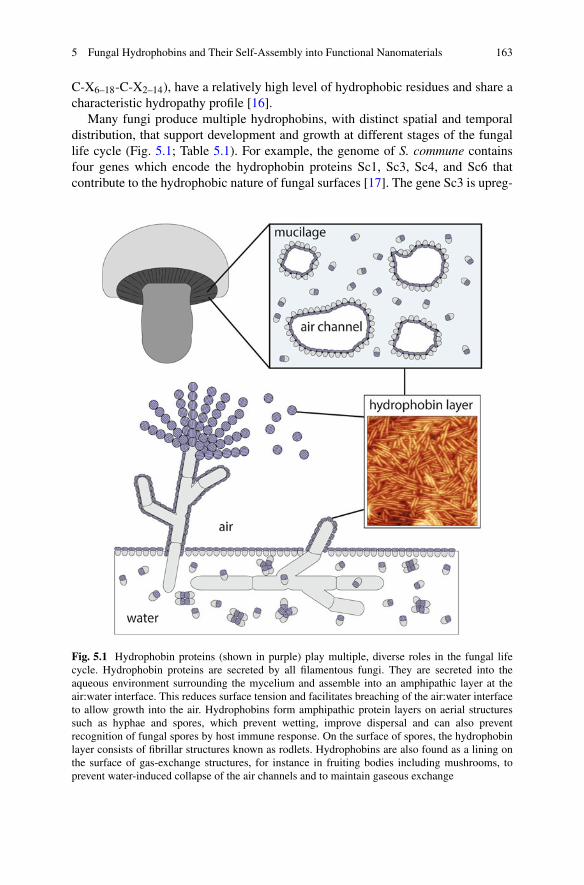
5 Fungal Hydrophobins and Their Self-Assembly into Functional Nanomaterials 163
C-X6–18-C-X2–14), have a relatively high level of hydrophobic residues and share acharacteristic hydropathy profile [16].
Many fungi produce multiple hydrophobins, with distinct spatial and temporaldistribution, that support development and growth at different stages of the fungallife cycle (Fig. 5.1; Table 5.1). For example, the genome of S. commune containsfour genes which encode the hydrophobin proteins Sc1, Sc3, Sc4, and Sc6 thatcontribute to the hydrophobic nature of fungal surfaces [17]. The gene Sc3 is upreg-
Fig. 5.1 Hydrophobin proteins (shown in purple) play multiple, diverse roles in the fungal lifecycle. Hydrophobin proteins are secreted by all filamentous fungi. They are secreted into theaqueous environment surrounding the mycelium and assemble into an amphipathic layer at theair:water interface. This reduces surface tension and facilitates breaching of the air:water interfaceto allow growth into the air. Hydrophobins form amphipathic protein layers on aerial structuressuch as hyphae and spores, which prevent wetting, improve dispersal and can also preventrecognition of fungal spores by host immune response. On the surface of spores, the hydrophobinlayer consists of fibrillar structures known as rodlets. Hydrophobins are also found as a lining onthe surface of gas-exchange structures, for instance in fruiting bodies including mushrooms, toprevent water-induced collapse of the air channels and to maintain gaseous exchange
164 V. Lo et al.
Table 5.1 Hydrophobin proteins discussed in this chapter
Hydrophobin Species Class
EAS Neurospora crassa INC2 IISc1, Sc3, Sc4, Sc6 Schizophyllum commune IHFBI, HFBII Trichoderma reesei IIRodA, DewA, DewB Aspergillus nidulans IDewC, DewD, DewE Intermediate I/IIRodA, RodB, RodC, RodD, RodE Aspergillus fumigatus IHCf1, HCf2, HCf3, HCf4 Cladosporium fulvum IHCf5, HCf6 IIMPG1 Magnaporthe oryzae IMHP1 IIVmh2 Pleurotus ostreatus IHGFI Grifola frondosa IHYD3 Metarhizium brunneum IHYDPt1 Pisolithus tinctorius I
ulated during the formation of aerial hyphae. This hydrophobin functions to lowerthe surface tension of the growth medium and it creates a coat around the aerialhyphae, as the fungi breaches through the medium, that prevents the aerial structuresfrom wetting and thus it facilitates dispersal in air. The hydrophobins Sc1, Sc4, andSc6 support the fruiting bodies, in particular Sc4 forms the hydrophobic film thatlines the gas channels of fruiting bodies to prevent water logging. Aspergillus nidu-lans produces six different hydrophobins, RodA, DewA, DewB, DewC, DewD, andDewE, which can all form amphipathic films and contribute to spore hydrophobicityalthough RodA is the main contributor [18]. The tomato pathogen, Cladosporiumfulvum also secretes six hydrophobins, two of which are present on the aerial hyphaeand the spores, while one is found on young aerial hyphae and another forms a coataround and beneath the fungus, suggesting interactions with the extracellular matrix[19, 20]. In different ways, all hydrophobins support the growth and reproductionof fungi. Some hydrophobins have been shown to be upregulated during nutrient,carbon or nitrogen starvation, while the gene encoding the hydrophobin EAS isactivated by light, the circadian clock and nutrient deficiency [4, 21–24].
5.3 Class I and Class II Hydrophobins
Early studies by Wessels and colleagues showed that members of the hydrophobinfamily could be separated into two distinct classes based on the solubility of theprotein layers and the hydropathy profiles of the proteins: class I and class II [5]. Asmore hydrophobins have been identified and studied in detail, this distinction hasgenerally been maintained and the sequence and structural differences underlyingthe character of the two main classes have been elucidated [13, 14, 25].
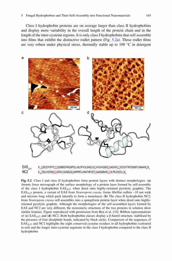
5 Fungal Hydrophobins and Their Self-Assembly into Functional Nanomaterials 165
Class I hydrophobin proteins are on average larger than class II hydrophobinsand display more variability in the overall length of the protein chain and in thelength of the inter-cysteine regions. It is only class I hydrophobins that self-assembleinto films that exhibit the distinctive rodlet pattern (Fig. 5.2a). These rodlet filmsare very robust under physical stress, thermally stable up to 100 ◦C in detergent
Fig. 5.2 Class I and class II hydrophobins form protein layers with distinct morphologies. (a)Atomic force micrograph of the surface morphology of a protein layer formed by self-assemblyof the class I hydrophobin EAS�15 when dried onto highly-oriented pyrolytic graphite. TheEAS�15 protein, a variant of EAS from Neurospora crassa, forms fibrillar rodlets ~10 nm wideand microns long which pack laterally to form a monolayer. (b) The class II hydrophobin NC2,from Neurospora crassa self-assembles into a spongiform protein layer when dried onto highly-oriented pyrolytic graphite. Although the morphologies of the self-assembled layers formed byEAS and NC2 are very different, the monomeric structures of the two proteins in solution showsimilar features. Figure reproduced with permission from Ren et al. [34]. Ribbon representationsof (c) EAS�15 and (d) NC2. Both hydrophobin classes display a β-barrel structure, stabilised bythe presence of four disulphide bonds, indicated by black sticks. Comparison of the sequences ofEAS�15 and NC2 highlights the eight conserved cysteine residues in all hydrophobins (colouredin red) and the longer inter-cysteine segments in the class I hydrophobin compared to the class IIhydrophobin
166 V. Lo et al.
and have been shown to be chemically stable under alkaline wash (3 M NaOH)and acidic wash (3 M HCl) [5, 26]. The rodlets have a diameter of ~10 nm andassemble laterally to form films with a thickness of 2.5–3 nm [26, 27]. They can onlybe depolymerised and dissociated to monomeric protein by treatment with agentssuch as trifluoroacetic acid (TFA) or formic acid [12, 28]. Once dissolved into theirmonomeric form, class I hydrophobins can reassemble into rodlets in the presenceof an appropriate interface [29].
The class I rodlets share many amyloid-like characteristics. Investigations ofEAS and Sc3 showed that the rodlet form of these hydrophobins interacts withthe amyloid-specific dyes, thioflavin-T and Congo red, which are widely used todetect cross-β structures [16, 30]. Further studies with techniques such as circulardichroism and attenuated-total-reflectance Fourier-transform infrared spectroscopy(ATR-FTIR) indicated the presence of β-sheet structure in the rodlet form [16, 27].X-Ray fibre diffraction has confirmed that the class I hydrophobin rodlets from EAScontain a repeating β-structure, similar to amyloid fibrils [31]. The fibre diffractionpattern generated by rodlets displays a reflection at 4.8 Å that arises from the spacingbetween the strands in a β-sheet and an orthogonal reflection at 10–12 Å, generatedby repetition of the spacing between β-sheets parallel to the fibril long axis.
Strikingly, Kershaw and colleagues were able to show that by substituting genesof different hydrophobins (sc3 and sc4 from S. commune, rodA and dewA fromAspergillus nidulans, eas from Neurospora crassa, and ssgA from Metarhiziumanisopliae) into a mutated Magnaporthe oryzae strain lacking the gene for the classI hydrophobin MPG1, it was possible to partially supplement the multiple functionsof MPG1 [32]. This demonstrates that although there is little sequence homologybetween class I hydrophobins from different species, the biological functions theyperform in different fungi are very similar.
Class II hydrophobins are generally smaller than class I hydrophobins, with up to63 residues between cysteines one and eight, instead of up to 105 residues betweenthe outer cysteines in class I hydrophobins. Although they do not form rodlets whenthey self-assemble, class II hydrophobins also spontaneously form amphipathiclayers at hydrophobic:hydrophilic interfaces (Fig. 5.2b). These layers are much lessrobust than class I rodlet-containing layers: they can be disrupted by treatment withalcohols and detergents and the application of mild pressure or temperatures [33,34]. Under certain conditions some level of macromolecular organisation is apparentin class II hydrophobin monolayers. HFBI and HFBII are known to produce highlyorientated crystalline monolayers at the atomic scale under compression in certainexperimental conditions, while in NC2 monolayers a porous network structure hasbeen observed (Fig. 5.2b) [34, 35].
5.4 Structures of Class I and Class II Hydrophobins
When the first three-dimensional structure of a hydrophobin was determined, thatof HFBII by crystallography, it revealed that the protein core consisted of a β-barrelwith four antiparallel β-strands and an external α-helix [36]. Although sequence
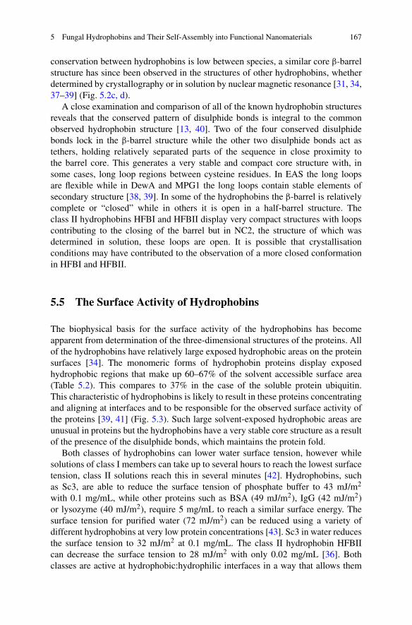
5 Fungal Hydrophobins and Their Self-Assembly into Functional Nanomaterials 167
conservation between hydrophobins is low between species, a similar core β-barrelstructure has since been observed in the structures of other hydrophobins, whetherdetermined by crystallography or in solution by nuclear magnetic resonance [31, 34,37–39] (Fig. 5.2c, d).
A close examination and comparison of all of the known hydrophobin structuresreveals that the conserved pattern of disulphide bonds is integral to the commonobserved hydrophobin structure [13, 40]. Two of the four conserved disulphidebonds lock in the β-barrel structure while the other two disulphide bonds act astethers, holding relatively separated parts of the sequence in close proximity tothe barrel core. This generates a very stable and compact core structure with, insome cases, long loop regions between cysteine residues. In EAS the long loopsare flexible while in DewA and MPG1 the long loops contain stable elements ofsecondary structure [38, 39]. In some of the hydrophobins the β-barrel is relativelycomplete or “closed” while in others it is open in a half-barrel structure. Theclass II hydrophobins HFBI and HFBII display very compact structures with loopscontributing to the closing of the barrel but in NC2, the structure of which wasdetermined in solution, these loops are open. It is possible that crystallisationconditions may have contributed to the observation of a more closed conformationin HFBI and HFBII.
5.5 The Surface Activity of Hydrophobins
The biophysical basis for the surface activity of the hydrophobins has becomeapparent from determination of the three-dimensional structures of the proteins. Allof the hydrophobins have relatively large exposed hydrophobic areas on the proteinsurfaces [34]. The monomeric forms of hydrophobin proteins display exposedhydrophobic regions that make up 60–67% of the solvent accessible surface area(Table 5.2). This compares to 37% in the case of the soluble protein ubiquitin.This characteristic of hydrophobins is likely to result in these proteins concentratingand aligning at interfaces and to be responsible for the observed surface activity ofthe proteins [39, 41] (Fig. 5.3). Such large solvent-exposed hydrophobic areas areunusual in proteins but the hydrophobins have a very stable core structure as a resultof the presence of the disulphide bonds, which maintains the protein fold.
Both classes of hydrophobins can lower water surface tension, however whilesolutions of class I members can take up to several hours to reach the lowest surfacetension, class II solutions reach this in several minutes [42]. Hydrophobins, suchas Sc3, are able to reduce the surface tension of phosphate buffer to 43 mJ/m2
with 0.1 mg/mL, while other proteins such as BSA (49 mJ/m2), IgG (42 mJ/m2)or lysozyme (40 mJ/m2), require 5 mg/mL to reach a similar surface energy. Thesurface tension for purified water (72 mJ/m2) can be reduced using a variety ofdifferent hydrophobins at very low protein concentrations [43]. Sc3 in water reducesthe surface tension to 32 mJ/m2 at 0.1 mg/mL. The class II hydrophobin HFBIIcan decrease the surface tension to 28 mJ/m2 with only 0.02 mg/mL [36]. Bothclasses are active at hydrophobic:hydrophilic interfaces in a way that allows them
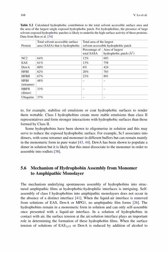
168 V. Lo et al.
Table 5.2 Calculated hydrophobic contribution to the total solvent accessible surface area andthe area of the largest single exposed hydrophobic patch. For hydrophobins, the presence of largesolvent-exposed hydrophobic patches is likely to underlie the high surface activity of these proteins.Data from Ren et al. [34]
ProteinTotal solvent accessible surfacearea (SASA) that is hydrophobic
Total area of the largestsolvent-accessible hydrophobic patchPercentage oftotal SASA
Area of largesthydrophobic patch (Å2)
NC2 64% 12% 693EAS 61% 13% 758DewA 60% 6% 424HFBI 62% 20% 783HFBII 67% 23% 891HFBI(tetramer)
48% – –
HBFII(dimer)
33% – –
Ubiquitin 37% – –
to, for example, stabilise oil emulsions or coat hydrophobic surfaces to renderthem wettable. Class I hydrophobins create more stable emulsions than class IIrepresentatives and form stronger interactions with hydrophobic surfaces than thoseformed by Class II.
Some hydrophobins have been shown to oligomerise in solution and this mayserve to reduce the exposed hydrophobic surface. For example, Sc3 associates intodimers, with some tetramer and monomer in different buffers but can remain mainlyin the monomeric form in pure water [43, 44]. DewA has been shown to populate adimer in solution but it is likely that this must dissociate to the monomer in order toassemble into rodlets [38].
5.6 Mechanism of Hydrophobin Assembly from Monomerto Amphipathic Monolayer
The mechanism underlying spontaneous assembly of hydrophobins into struc-tured amphipathic films at hydrophobic:hydrophilic interfaces is intriguing. Self-assembly of class I hydrophobins into amphipathic monolayers does not occur inthe absence of a distinct interface [41]. When the liquid-air interface is removedfrom solutions of EAS, DewA or MPG1, no amphipathic film forms [26]. Thehydrophobins remain in a monomeric form in solution and can only self-assembleonce presented with a liquid-air interface. In a solution of hydrophobins incontact with air, the surface tension at the air:solution interface plays an importantrole in determining the formation of these hydrophobin films. When the surfacetension of solutions of EAS�15 or DewA is reduced by addition of alcohol to
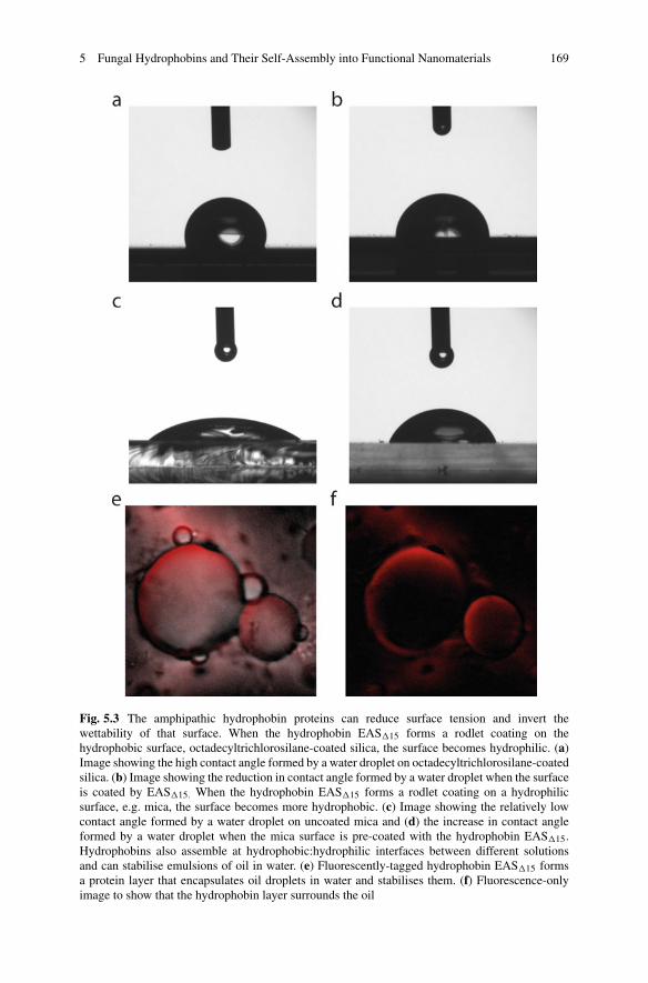
5 Fungal Hydrophobins and Their Self-Assembly into Functional Nanomaterials 169
Fig. 5.3 The amphipathic hydrophobin proteins can reduce surface tension and invert thewettability of that surface. When the hydrophobin EAS�15 forms a rodlet coating on thehydrophobic surface, octadecyltrichlorosilane-coated silica, the surface becomes hydrophilic. (a)Image showing the high contact angle formed by a water droplet on octadecyltrichlorosilane-coatedsilica. (b) Image showing the reduction in contact angle formed by a water droplet when the surfaceis coated by EAS�15. When the hydrophobin EAS�15 forms a rodlet coating on a hydrophilicsurface, e.g. mica, the surface becomes more hydrophobic. (c) Image showing the relatively lowcontact angle formed by a water droplet on uncoated mica and (d) the increase in contact angleformed by a water droplet when the mica surface is pre-coated with the hydrophobin EAS�15.Hydrophobins also assemble at hydrophobic:hydrophilic interfaces between different solutionsand can stabilise emulsions of oil in water. (e) Fluorescently-tagged hydrophobin EAS�15 formsa protein layer that encapsulates oil droplets in water and stabilises them. (f) Fluorescence-onlyimage to show that the hydrophobin layer surrounds the oil
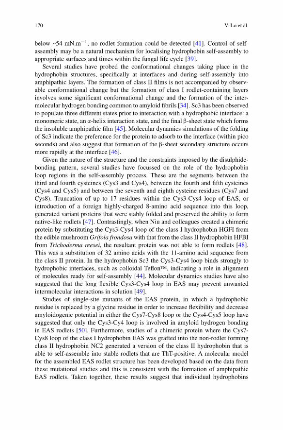
170 V. Lo et al.
below ~54 mN.m−1, no rodlet formation could be detected [41]. Control of self-assembly may be a natural mechanism for localising hydrophobin self-assembly toappropriate surfaces and times within the fungal life cycle [39].
Several studies have probed the conformational changes taking place in thehydrophobin structures, specifically at interfaces and during self-assembly intoamphipathic layers. The formation of class II films is not accompanied by observ-able conformational change but the formation of class I rodlet-containing layersinvolves some significant conformational change and the formation of the inter-molecular hydrogen bonding common to amyloid fibrils [34]. Sc3 has been observedto populate three different states prior to interaction with a hydrophobic interface: amonomeric state, an α-helix interaction state, and the final β-sheet state which formsthe insoluble amphipathic film [45]. Molecular dynamics simulations of the foldingof Sc3 indicate the preference for the protein to adsorb to the interface (within picoseconds) and also suggest that formation of the β-sheet secondary structure occursmore rapidly at the interface [46].
Given the nature of the structure and the constraints imposed by the disulphide-bonding pattern, several studies have focussed on the role of the hydrophobinloop regions in the self-assembly process. These are the segments between thethird and fourth cysteines (Cys3 and Cys4), between the fourth and fifth cysteines(Cys4 and Cys5) and between the seventh and eighth cysteine residues (Cys7 andCys8). Truncation of up to 17 residues within the Cys3-Cys4 loop of EAS, orintroduction of a foreign highly-charged 8-amino acid sequence into this loop,generated variant proteins that were stably folded and preserved the ability to formnative-like rodlets [47]. Contrastingly, when Niu and colleagues created a chimericprotein by substituting the Cys3-Cys4 loop of the class I hydrophobin HGFI fromthe edible mushroom Grifola frondosa with that from the class II hydrophobin HFBIfrom Trichoderma reesei, the resultant protein was not able to form rodlets [48].This was a substitution of 32 amino acids with the 11-amino acid sequence fromthe class II protein. In the hydrophobin Sc3 the Cys3-Cys4 loop binds strongly tohydrophobic interfaces, such as colloidal Teflon™, indicating a role in alignmentof molecules ready for self-assembly [44]. Molecular dynamics studies have alsosuggested that the long flexible Cys3-Cys4 loop in EAS may prevent unwantedintermolecular interactions in solution [49].
Studies of single-site mutants of the EAS protein, in which a hydrophobicresidue is replaced by a glycine residue in order to increase flexibility and decreaseamyloidogenic potential in either the Cys7-Cys8 loop or the Cys4-Cys5 loop havesuggested that only the Cys3-Cy4 loop is involved in amyloid hydrogen bondingin EAS rodlets [50]. Furthermore, studies of a chimeric protein where the Cys7-Cys8 loop of the class I hydrophobin EAS was grafted into the non-rodlet formingclass II hydrophobin NC2 generated a version of the class II hydrophobin that isable to self-assemble into stable rodlets that are ThT-positive. A molecular modelfor the assembled EAS rodlet structure has been developed based on the data fromthese mutational studies and this is consistent with the formation of amphipathicEAS rodlets. Taken together, these results suggest that individual hydrophobins
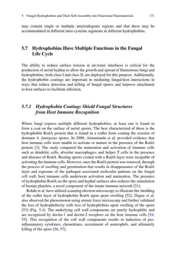
5 Fungal Hydrophobins and Their Self-Assembly into Functional Nanomaterials 171
may contain single or multiple amyloidogenic regions and that these may beaccommodated in different inter-cysteine segments in different hydrophobins.
5.7 Hydrophobins Have Multiple Functions in the FungalLife Cycle
The ability to reduce surface tension at air:water interfaces is critical for theproduction of aerial hyphae to allow the growth and spread of filamentous fungi andhydrophobins, both class I and class II, are deployed for this purpose. Additionally,the hydrophobin coatings are important in mediating fungal:host interactions inways that reduce detection and killing of fungal spores and improve attachmentto host surfaces to facilitate infection.
5.7.1 Hydrophobin Coatings Shield Fungal Structuresfrom Host Immune Recognition
Where fungi express multiple different hydrophobins, at least one is found toform a coat on the surface of aerial spores. The best characterized of these is thehydrophobin RodA protein that is found in a rodlet form coating the exterior ofdormant A. fumigatus spores. In 2009, Aimanianda et al. provided evidence thathost immune cells were unable to activate or mature in the presence of the RodAprotein [3]. The study compared the maturation and activation of immune cellssuch as dendritic cells, alveolar macrophages, and helper T cells in the presenceand absence of RodA. Resting spores coated with a RodA layer were incapable ofactivating the immune cells. However, once the RodA protein was removed, throughthe process of swelling and germination that results in disappearance of the RodAlayer and exposure of the pathogen associated molecular patterns on the fungalcell wall, host immune cells underwent activation and maturation. The presenceof hydrophobin RodA on the spore and hyphal surfaces also reduces the stimulationof human platelets, a novel component of the innate immune network [51].
Rohde et al. have utilised scanning electron microscopy to illustrate the sheddingof the rodlet layer of hydrophobin RodA upon spore swelling [52]. Dague et al.also observed the phenomenon using atomic force microscopy and further validatedthe loss of hydrophobicity with loss of hydrophobins upon swelling of the spore[53] (Fig. 5.4). The underlying cell wall components are purely hydrophilic andare recognized by dectin-1 and dectin-2 receptors on the host immune cells [54,55]. This recognition of the cell wall components results in induction of pro-inflammatory cytokines, chemokines, recruitment of neutrophils, and ultimatelykilling of the spore [56, 57].
172 V. Lo et al.
Fig. 5.4 The outer surface of the spores produced by Aspergillus fumigatus is coated with afibrillar rodlet layer composed of the class I hydrophobin RodA. A series of high-resolution atomicforce micrographs recorded from the surface of a single Aspergillus fumigatus spore during theprocess of spore germination. The highly structured rodlet layer is visible at 1 and 20 min butchanges are apparent at 60 min as the rodlet layer starts to disappear and to be replaced by anamorphous layer. Figure reproduced with permission from Dague et al. [53]
These studies have therefore demonstrated that the RodA protein layer exists asa shield for the underlying fungal cell wall components, including β-glucan andmelanin, which would otherwise serve as antigens and cause continuous immuneactivation and detrimental inflammatory responses. The presence of RodA on thespore surface allows dormant spores to escape detection until conditions becomeconducive for germination [3, 58].
A. fumigatus is an opportunistic human pathogen and, in immune-compromisedindividuals, it is able to germinate within the lung, produce invasive hyphaeand disseminate throughout the bloodstream, causing the difficult-to-treat invasiveaspergillosis. Beauvais et al. have investigated the role of A. fumigatus hydrophobinsin the static aerial growth conditions that resemble the human lung [59]. Thishas revealed an extracellular hydrophobic matrix that supports the fungal cellsin a fungal biofilm community. The matrix consists of galactomannan, α1,3glucan, monosaccharide, polyol, melanin and proteins such as major antigens andhydrophobins. Although RodA is absent from the mycelial stage under both staticaerial or liquid shaken growth conditions, the class I hydrophobin RodB is foundexpressed exclusively in the aerial static culture condition resembling growth inthe lung. The A. fumigatus hydrophobins RodB–RodE continue to be expressed
5 Fungal Hydrophobins and Their Self-Assembly into Functional Nanomaterials 173
throughout biofilm formation, indicating a potential role for these hydrophobins information, maintenance and surface adhesion of the extracellular matrix [60, 61].
5.7.2 Hydrophobins Facilitate Attachment of Fungi to HostCells for Colonisation
The fungus Magnaporthe oryzae causes rice blast, a devastating disease that isresponsible for loss of up to a third of global annual rice production [62]. Thehydrophobin MPG1 is highly expressed during rice blast infections [63]. MPG1is found on the spore surface of M. oryzae spores where it functions to resistwetting and assist adherence to plant tissue [4]. Moreover, MPG1 has been shownto be involved in sensing of a hydrophobic surface, initiation of appressoriumdifferentiation, host penetration and virulence [62]. Knockout or gene silencingstudies of MPG1 resulted in reduced appressorium formation and pathogenicity,and the spores of M. oryzae lacking MPG1 showed a wettable phenotype [64,65]. Silencing of the class II hydrophobin from M. oryzae, MHP1, does not affectpathogenicity [65].
Pham et al. have studied the interaction between the two hydrophobins, MPG1and MHP1, and the cutinase cut2 that is associated with growth through thewaxy rice leaf cuticle [39]. This study demonstrated that assembly of the class Ihydrophobin MPG1 is strictly limited to an interface and the class II hydrophobinMHPI is able to prevent MPG1 assembly by reduction of surface tension in asolution containing both hydrophobins. This suggests that some fungi may producemultiple hydrophobins in order to control the spatial and temporal pattern of rodletformation. The assembly of the hydrophobins was also shown to recruit cutinaseactivity to a hydrophobic surface, potentially offering a mechanism for control ofthe infection interface in rice blast disease.
5.8 Harnessing Hydrophobins for Biotechnological Purposes
Hydrophobins are natural products with properties that are potentially useful fora wide variety of applications. The ability of hydrophobins to self-assemble intobiocompatible amphipathic films at liquid-oil, liquid-liquid, air:liquid and liquid-solid interfaces has underpinned studies of their potential use in biotechnology andthe food and cosmetic industries [12, 66]. Studies of hydrophobins are reportedin multidisciplinary fields, including food technology, pharmacology, chemistryand surface engineering and hydrophobin use in these areas has been heavilypatented in recent years [67]. The number of publications and patents associatedwith hydrophobins shows a striking increase in recent years (Fig. 5.5).
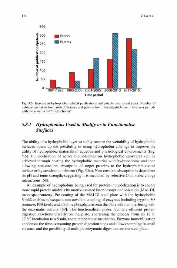
174 V. Lo et al.
Fig. 5.5 Increase in hydrophobin-related publications and patents over recent years. Number ofpublications taken from Web of Science and patents from FreePatentsOnline in five-year periodswith the search word “hydrophobin”
5.8.1 Hydrophobins Used to Modify or to FunctionaliseSurfaces
The ability of a hydrophobin layer to stably reverse the wettability of hydrophobicsurfaces opens up the possibility of using hydrophobin coatings to improve theutility of hydrophobic materials in aqueous and physiological environments (Fig.5.6). Immobilisation of active biomolecules on hydrophobic substrates can beachieved through coating the hydrophobic material with hydrophobins and thenallowing non-covalent absorption of target proteins to the hydrophobin-coatedsurface or by covalent attachment (Fig. 5.6a). Non-covalent absorption is dependenton pH and ionic strength, suggesting it is mediated by selective Coulombic chargeinteractions [68].
An example of hydrophobins being used for protein immobilization is to enablemore rapid protein analysis by matrix-assisted laser desorption/ionization (MALDI)mass spectrometry. Pre-coating of the MALDI steel plate with the hydrophobinVmh2 enables subsequent non-covalent coupling of enzymes including trypsin, V8protease, PNGaseF, and alkaline phosphatase onto the plate without interfering withthe enzymatic activity [69]. The functionalised plates facilitate efficient proteindigestion reactions directly on the plate, shortening the process from an 18 h,37 ◦C incubation to a 5 min, room temperature incubation. Enzyme immobilizationcondenses the time-consuming protein digestion steps and allows sampling in smallvolumes and the possibility of multiple enzymatic digestions on the steel plate.
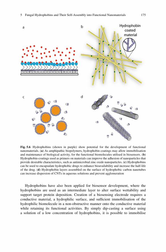
5 Fungal Hydrophobins and Their Self-Assembly into Functional Nanomaterials 175
Fig. 5.6 Hydrophobins (shown in purple) show potential for the development of functionalnanomaterials. (a) As amphipathic biopolymers, hydrophobin coatings may allow immobilisationand maintenance of biological activity, for the functional biomolecules utilised in biosensors. (b)Hydrophobin-coatings used as primers on materials can improve the adhesion of nanoparticles thatprovide desirable characteristics, such as antimicrobial zinc oxide nanoparticles. (c) Hydrophobinscan be used to encapsulate hydrophobic drugs to enhance bioavailability and increase the half-lifeof the drug. (d) Hydrophobin layers assembled on the surface of hydrophobic carbon nanotubescan increase dispersion of CNTs in aqueous solutions and prevent agglomeration
Hydrophobins have also been applied for biosensor development, where thehydrophobins are used as an intermediate layer to alter surface wettability andsupport target protein deposition. Creation of a biosensing electrode requires aconductive material, a hydrophilic surface, and sufficient immobilisation of thehydrophilic biomolecule in a non-obstructive manner onto the conductive materialwhile retaining its functional activities. By simply dip-casting a surface usinga solution of a low concentration of hydrophobins, it is possible to immobilise
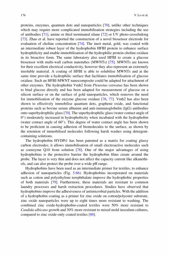
176 V. Lo et al.
proteins, enzymes, quantum dots and nanoparticles [70], unlike other techniqueswhich may require more complicated immobilisation strategies including the useof antibodies [71], amine or thiol terminated silane [72] or UV photo-crosslinking[73]. Zhao et al. have reported the construction of a novel biosensor electrode forevaluation of choline concentration [74]. The inert metal, gold, was coated withan intermediate robust layer of the hydrophobin HFBI protein to enhance surfacehydrophilicity and enable immobilisation of the hydrophilic protein choline oxidasein its bioactive form. The same laboratory also used HFBI to create a glucosebiosensor with multi-wall carbon nanotubes (MWNTs) [75]. MWNTs are knownfor their excellent electrical conductivity, however they also represent an extremelyinsoluble material. A coating of HFBI is able to solubilise MWNTs and at thesame time provide a hydrophilic surface that facilitates immobilisation of glucoseoxidase. Such an HFBI-MWNT nanocomposite could be adapted for attachment ofother enzymes. The hydrophobin Vmh2 from Pleurotus ostreatus has been shownto bind glucose directly and has been adapted for measurement of glucose on asilicon surface or on the surface of gold nanoparticles, which removes the needfor immobilisation of the enzyme glucose oxidase [76, 77]. Vmh2 has also beenshown to effectively immobilise quantum dots, graphene oxide, and functionalproteins such as bovine serum albumin and anti-immunoglobulin (IgG) antibodiesonto superhydrophilic glass [70]. The superhydrophilic glass (water contact angle of0◦) moderately increased in hydrophobicity when incubated with the hydrophobin(water contact angle of 60◦). This degree of water contact angle has been shownto be proficient in causing adhesion of biomolecules to the surface, as shown bythe retention of immobilised molecules following harsh washes using detergent-containing solutions.
The hydrophobin HYDPt1 has been patented as a matrix for coating glassycarbon electrodes; it allows immobilisation of small electroactive molecules suchas coenzyme Q10 from solution [78]. One of the major advantages of usinghydrophobins is the protective barrier the hydrophobin films create around theprobe. The layer is very thin and does not affect the capacity current like alkanethi-ols, and can also protect the probe over a wide pH range.
Hydrophobins have been used as an intermediate primer for textiles, to enhanceadhesion of nanoparticles (Fig. 5.6b). Hydrophobins incorporated on materialssuch as cotton and polyethylene terephthalate improve the hydrophobic propertiesof both materials [79]. Furthermore, these materials are resistant to commonlaundry processes and harsh extraction procedures. Studies have observed thathydrophobins improve the adhesiveness of antimicrobial particles. With the additionof a hydrophobin coating as a primer for zinc oxide on cotton/polyester substrate,zinc oxide nanoparticles were up to eight times more resistant to washing. Thecombined zinc oxide-hydrophobin-coated textiles were 50% more resistant toCandida albicans growth and 30% more resistant to mixed mold inoculum cultures,compared to zinc oxide-only coated textiles [80].
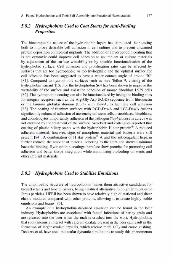
5 Fungal Hydrophobins and Their Self-Assembly into Functional Nanomaterials 177
5.8.2 Hydrophobins Used to Coat Stents for Anti-FoulingProperties
The biocompatible nature of the hydrophobin layers has stimulated their testingboth to improve desirable cell adhesion in cell culture and to prevent unwantedprotein deposition on medical implants. The addition of a hydrophobin coating thatis not cytotoxic could improve cell adhesion to an implant or culture materialsby adjustment of the surface wettability or by specific functionalisation of thehydrophobic surface. Cell adhesion and proliferation rates can be affected bysurfaces that are too hydrophobic or too hydrophilic and the optimal surface forcell adhesion has been suggested to have a water contact angle of around 70◦[81]. Compared to hydrophobic surfaces such as bare Teflon™, coating of thehydrophobin variant TrSc3 or the hydrophobin Sc4 has been shown to improve thewettability of the surface and assist the adhesion of mouse fibroblast L929 cells[82]. The hydrophobin coating can also be functionalized by fusing the binding sitesfor integrin receptors such as the Arg-Gly-Asp (RGD) sequence from fibronectinor the laminin globular domain (LG3) with DewA, to facilitate cell adhesion[83]. The coating of titanium surfaces with RGD-DewA and LG3-DewA fusionssignificantly enhanced adhesion of mesenchymal stem cells, osteoblasts, fibroblasts,and chondrocytes. Importantly, adhesion of the pathogen Staphylococcus aureus wasnot elevated by the treatment of the surface. Weickert and colleagues reported thatcoating of plastic biliary stents with the hydrophobin H star protein® A reducedadherent material; however, signs of amorphous material and bacteria were stillpresent [84]. A combination of H star protein® A and the anticoagulant heparinfurther reduced the amount of material adhering to the stent and showed minimalbacterial binding. Hydrophobin coatings therefore show promise for promoting celladhesion and better tissue integration while minimising biofouling on stents andother implant materials.
5.8.3 Hydrophobins Used to Stabilise Emulsions
The amphipathic structure of hydrophobins makes them attractive candidates forbiosurfactants and bioemulsifiers, being a natural alternative to polymer micelles orJanus particles. HFBII has been shown to have relatively high dilatational and shearelastic modulus compared with other proteins, allowing it to create highly stableemulsions and foams [85].
An example of a hydrophobin-stabilised emulsion can be found in the beerindustry. Hydrophobins are associated with fungal infections of barley grain andare released into the beer when the malt is crushed into the wort. Hydrophobinsthat spontaneously interact with calcium oxalate present in the beer can result in theformation of larger oxalate crystals, which release more CO2 and cause gushing.Deckers et al. have used molecular dynamic simulations to study this phenomenon
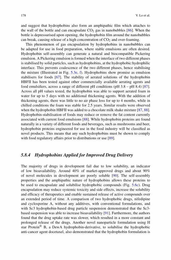
178 V. Lo et al.
and suggest that hydrophobins also form an amphipathic film which attaches tothe wall of the bottle and can encapsulate CO2 gas in nanobubbles [86]. When thebottle is depressurised upon opening, the hydrophobin film around the nanobubblescan break, causing release of a high concentration of CO2 and over-foaming.
This phenomenon of gas encapsulation by hydrophobins in nanobubbles canbe adapted for use in food preparation, where stable emulsions are often desired.Hydrophobin self-assembly can generate a natural and biocompatible Pickeringemulsion. A Pickering emulsion is formed when the interface of two different phasesis stabilised by solid particles, such as hydrophobins, at the hydrophobic:hydrophilicinterface. This prevents coalescence of the two different phases, hence stabilisingthe mixture (Illustrated in Fig. 5.3e, f). Hydrophobins show promise as emulsionstabilisers for foods [67]. The stability of aerated solutions of the hydrophobinHBFII has been tested against other commercially available aerating agents andfood emulsifiers, across a range of different pH conditions (pH 3.8 – pH 8.4) [87].Across all pH values tested, the hydrophobin was able to support aerated foam inwater for up to 5 days with no additional thickening agents. With the addition ofthickening agents, there was little to no air phase loss for up to 4 months, while inchilled conditions the foam was stable for 2.5 years. Similar results were observedwhen the hydrophobin HBFII was added to a chocolate milk shake mixture [87, 88].Hydrophobin-stabilisation of foods may reduce or remove the fat content currentlyassociated with current food emulsions [88]. While hydrophobin proteins are foundnaturally in a variety of different foods and beverages, such as mushrooms and beer,hydrophobin proteins engineered for use in the food industry will be classified asnovel products. This means that any such hydrophobins must be shown to complywith food regulatory affairs prior to distributions or use [89].
5.8.4 Hydrophobins Applied for Improved Drug Delivery
The majority of drugs in development fail due to low solubility, an indicatorof low bioavailability. Around 40% of market-approved drugs and about 90%of novel molecules in development are poorly soluble [90]. The self-assemblyproperties and the amphipathic nature of hydrophobins allows these proteins tobe used to encapsulate and solubilise hydrophobic compounds (Fig. 5.6c). Drugencapsulation may reduce systemic toxicity and side effects, increase the solubilityand efficacy of therapeutics and enable sustained release of active compounds overan extended period of time. A comparison of two hydrophobic drugs, nifedipineand cyclosporine A, without any additives, with conventional formulations, andwith Sc3 hydrophobin-based drug particle suspension demonstrated that the Sc3-based suspension was able to increase bioavailability [91]. Furthermore, the authorsfound that the drug uptake rate was slower, which resulted in a more constant andprolonged release of the drugs. Another novel nanoparticle formulation using Hstar Protein® B, a DewA hydrophobin-derivative, to solubilise the hydrophobicanti-cancer agent docetaxel, also demonstrated that the hydrophobin formulation is
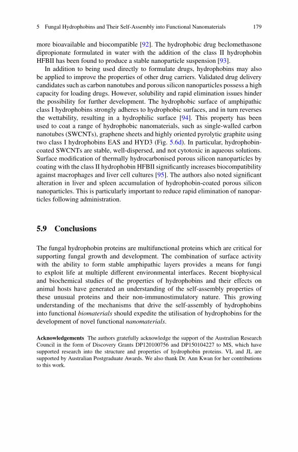
5 Fungal Hydrophobins and Their Self-Assembly into Functional Nanomaterials 179
more bioavailable and biocompatible [92]. The hydrophobic drug beclomethasonedipropionate formulated in water with the addition of the class II hydrophobinHFBII has been found to produce a stable nanoparticle suspension [93].
In addition to being used directly to formulate drugs, hydrophobins may alsobe applied to improve the properties of other drug carriers. Validated drug deliverycandidates such as carbon nanotubes and porous silicon nanoparticles possess a highcapacity for loading drugs. However, solubility and rapid elimination issues hinderthe possibility for further development. The hydrophobic surface of amphipathicclass I hydrophobins strongly adheres to hydrophobic surfaces, and in turn reversesthe wettability, resulting in a hydrophilic surface [94]. This property has beenused to coat a range of hydrophobic nanomaterials, such as single-walled carbonnanotubes (SWCNTs), graphene sheets and highly oriented pyrolytic graphite usingtwo class I hydrophobins EAS and HYD3 (Fig. 5.6d). In particular, hydrophobin-coated SWCNTs are stable, well-dispersed, and not cytotoxic in aqueous solutions.Surface modification of thermally hydrocarbonised porous silicon nanoparticles bycoating with the class II hydrophobin HFBII significantly increases biocompatibilityagainst macrophages and liver cell cultures [95]. The authors also noted significantalteration in liver and spleen accumulation of hydrophobin-coated porous siliconnanoparticles. This is particularly important to reduce rapid elimination of nanopar-ticles following administration.
5.9 Conclusions
The fungal hydrophobin proteins are multifunctional proteins which are critical forsupporting fungal growth and development. The combination of surface activitywith the ability to form stable amphipathic layers provides a means for fungito exploit life at multiple different environmental interfaces. Recent biophysicaland biochemical studies of the properties of hydrophobins and their effects onanimal hosts have generated an understanding of the self-assembly properties ofthese unusual proteins and their non-immunostimulatory nature. This growingunderstanding of the mechanisms that drive the self-assembly of hydrophobinsinto functional biomaterials should expedite the utilisation of hydrophobins for thedevelopment of novel functional nanomaterials.
Acknowledgements The authors gratefully acknowledge the support of the Australian ResearchCouncil in the form of Discovery Grants DP120100756 and DP150104227 to MS, which havesupported research into the structure and properties of hydrophobin proteins. VL and JL aresupported by Australian Postgraduate Awards. We also thank Dr. Ann Kwan for her contributionsto this work.
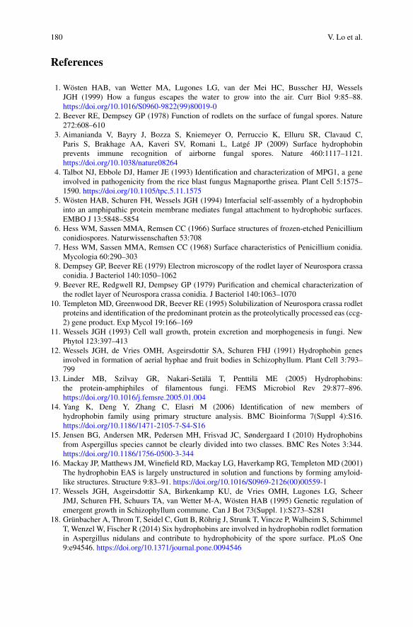
180 V. Lo et al.
References
1. Wösten HAB, van Wetter MA, Lugones LG, van der Mei HC, Busscher HJ, WesselsJGH (1999) How a fungus escapes the water to grow into the air. Curr Biol 9:85–88.https://doi.org/10.1016/S0960-9822(99)80019-0
2. Beever RE, Dempsey GP (1978) Function of rodlets on the surface of fungal spores. Nature272:608–610
3. Aimanianda V, Bayry J, Bozza S, Kniemeyer O, Perruccio K, Elluru SR, Clavaud C,Paris S, Brakhage AA, Kaveri SV, Romani L, Latgé JP (2009) Surface hydrophobinprevents immune recognition of airborne fungal spores. Nature 460:1117–1121.https://doi.org/10.1038/nature08264
4. Talbot NJ, Ebbole DJ, Hamer JE (1993) Identification and characterization of MPG1, a geneinvolved in pathogenicity from the rice blast fungus Magnaporthe grisea. Plant Cell 5:1575–1590. https://doi.org/10.1105/tpc.5.11.1575
5. Wösten HAB, Schuren FH, Wessels JGH (1994) Interfacial self-assembly of a hydrophobininto an amphipathic protein membrane mediates fungal attachment to hydrophobic surfaces.EMBO J 13:5848–5854
6. Hess WM, Sassen MMA, Remsen CC (1966) Surface structures of frozen-etched Penicilliumconidiospores. Naturwissenschaften 53:708
7. Hess WM, Sassen MMA, Remsen CC (1968) Surface characteristics of Penicillium conidia.Mycologia 60:290–303
8. Dempsey GP, Beever RE (1979) Electron microscopy of the rodlet layer of Neurospora crassaconidia. J Bacteriol 140:1050–1062
9. Beever RE, Redgwell RJ, Dempsey GP (1979) Purification and chemical characterization ofthe rodlet layer of Neurospora crassa conidia. J Bacteriol 140:1063–1070
10. Templeton MD, Greenwood DR, Beever RE (1995) Solubilization of Neurospora crassa rodletproteins and identification of the predominant protein as the proteolytically processed eas (ccg-2) gene product. Exp Mycol 19:166–169
11. Wessels JGH (1993) Cell wall growth, protein excretion and morphogenesis in fungi. NewPhytol 123:397–413
12. Wessels JGH, de Vries OMH, Asgeirsdottir SA, Schuren FHJ (1991) Hydrophobin genesinvolved in formation of aerial hyphae and fruit bodies in Schizophyllum. Plant Cell 3:793–799
13. Linder MB, Szilvay GR, Nakari-Setälä T, Penttilä ME (2005) Hydrophobins:the protein-amphiphiles of filamentous fungi. FEMS Microbiol Rev 29:877–896.https://doi.org/10.1016/j.femsre.2005.01.004
14. Yang K, Deng Y, Zhang C, Elasri M (2006) Identification of new members ofhydrophobin family using primary structure analysis. BMC Bioinforma 7(Suppl 4):S16.https://doi.org/10.1186/1471-2105-7-S4-S16
15. Jensen BG, Andersen MR, Pedersen MH, Frisvad JC, Søndergaard I (2010) Hydrophobinsfrom Aspergillus species cannot be clearly divided into two classes. BMC Res Notes 3:344.https://doi.org/10.1186/1756-0500-3-344
16. Mackay JP, Matthews JM, Winefield RD, Mackay LG, Haverkamp RG, Templeton MD (2001)The hydrophobin EAS is largely unstructured in solution and functions by forming amyloid-like structures. Structure 9:83–91. https://doi.org/10.1016/S0969-2126(00)00559-1
17. Wessels JGH, Asgeirsdottir SA, Birkenkamp KU, de Vries OMH, Lugones LG, ScheerJMJ, Schuren FH, Schuurs TA, van Wetter M-A, Wösten HAB (1995) Genetic regulation ofemergent growth in Schizophyllum commune. Can J Bot 73(Suppl. 1):S273–S281
18. Grünbacher A, Throm T, Seidel C, Gutt B, Röhrig J, Strunk T, Vincze P, Walheim S, SchimmelT, Wenzel W, Fischer R (2014) Six hydrophobins are involved in hydrophobin rodlet formationin Aspergillus nidulans and contribute to hydrophobicity of the spore surface. PLoS One9:e94546. https://doi.org/10.1371/journal.pone.0094546
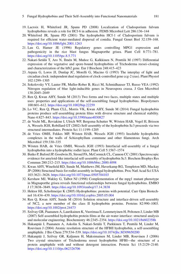
5 Fungal Hydrophobins and Their Self-Assembly into Functional Nanomaterials 181
19. Lacroix H, Whiteford JR, Spanu PD (2008) Localization of Cladosporium fulvumhydrophobins reveals a role for HCf-6 in adhesion. FEMS Microbiol Lett 286:136–144
20. Whiteford JR, Spanu PD (2001) The hydrophobin HCf-1 of Cladosporium fulvum isrequired for efficient water-mediated dispersal of conidia. Fungal Genet Biol 32:159–168.https://doi.org/10.1006/fgbi.2001.1263
21. Lau G, Hamer JE (1996) Regulatory genes controlling MPG1 expression andpathogenicity in the rice blast fungus Magnaporthe grisea. Plant Cell 8:771–781.https://doi.org/10.1105/tpc.8.5.771
22. Nakari-Setälä T, Aro N, Ilmén M, Muñoz G, Kalkkinen N, Penttilä M (1997) Differentialexpression of the vegetative and spore-bound hydrophobins of Trichoderma reesei–cloningand characterization of the hfb2 gene. Eur J Biochem 248:415–423
23. Arpaia G, Loros JJ, Dunlap JC, Morelli G, Macino G (1993) The interplay of light andcircadian clock: independent dual regulation of clock-controlled gene ccg-2 (eas). Plant Physiol102:1299–1305
24. Sokolovsky VY, Lauter FR, Muller-Rober B, Ricci M, Schmidhauser TJ, Russo VEA (1992)Nitrogen regulation of blue light-inducible genes in Neurospora crassa. J Gen Microbiol138:2045–2049
25. Ren Q, Kwan AHY, Sunde M (2013) Two forms and two faces, multiple states and multipleuses: properties and applications of the self-assembling fungal hydrophobins. Biopolymers100:601–612. https://doi.org/10.1002/bip.22259
26. Lo VC, Ren Q, Pham CLL, Morris VK, Kwan AHY, Sunde M (2014) Fungal hydrophobinproteins produce self-assembling protein films with diverse structure and chemical stability.Nano 4:827–843. https://doi.org/10.3390/nano4030827
27. de Vocht ML, Reviakine I, Ulrich WP, Bergsma-Schutter W, Wösten HAB, Vogel H, BrissonA, Wessels JGH, Robillard GT (2002) Self-assembly of the hydrophobin Sc3 proceeds via twostructural intermediates. Protein Sci 11:1199–1205
28. de Vries OMH, Fekkes MP, Wösten HAB, Wessels JGH (1993) Insoluble hydrophobincomplexes in the walls of Schizophyllum commune and other filamentous fungi. ArchMicrobiol 159:330–335
29. Wösten HAB, de Vries OMH, Wessels JGH (1993) Interfacial self-assembly of a fungalhydrophobin into a hydrophobic rodlet layer. Plant Cell 5:1567–1574
30. Butko P, Buford JP, Goodwin JS, Stroud PA, McCormick CL, Cannon CC (2001) Spectroscopicevidence for amyloid-like interfacial self-assembly of hydrophobin Sc3. Biochem Biophys ResCommun 280:212–215. https://doi.org/10.1006/bbrc.2000.4098
31. Kwan AHY, Winefield RD, Sunde M, Matthews JM, Haverkamp RG, Templeton MD, MackayJP (2006) Structural basis for rodlet assembly in fungal hydrophobins. Proc Natl Acad Sci USA103:3621–3626. https://doi.org/10.1073/pnas.0505704103
32. Kershaw MJ, Wakley G, Talbot NJ (1998) Complementation of the mpg1 mutant phenotypein Magnaporthe grisea reveals functional relationships between fungal hydrophobins. EMBOJ 17:3838–3849. https://doi.org/10.1093/emboj/17.14.3838
33. Hektor HJ, Scholtmeijer K (2005) Hydrophobins: proteins with potential. Curr Opin Biotech-nol 16:434–439. https://doi.org/10.1016/j.copbio.2005.05.004
34. Ren Q, Kwan AHY, Sunde M (2014) Solution structure and interface-driven self-assemblyof NC2, a new member of the class II hydrophobin proteins. Proteins 82:990–1003.https://doi.org/10.1002/prot.24473
35. Szilvay GR, Paananen A, Laurikainen K, Vuorimaa E, Lemmetyinen H, Peltonen J, Linder MB(2007) Self-assembled hydrophobin protein films at the air-water interface: structural analysisand molecular engineering. Biochemistry 46:2345–2354. https://doi.org/10.1021/bi602358h
36. Hakanpää J, Paananen A, Askolin S, Nakari-Setälä T, Parkkinen T, Penttilä M, Linder M,Rouvinen J (2004) Atomic resolution structure of the HFBII hydrophobin, a self-assemblingamphiphile. J Bio Chem 279:534–539. https://doi.org/10.1074/jbc.M309650200
37. Hakanpää J, Szilvay GR, Kaljunen H, Maksimainen M, Linder MB, Rouvinen J (2006)Two crystal structures of Trichoderma reesei hydrophobin HFBI—the structure of aprotein amphiphile with and without detergent interaction. Protein Sci 15:2129–2140.https://doi.org/10.1110/ps.062326706
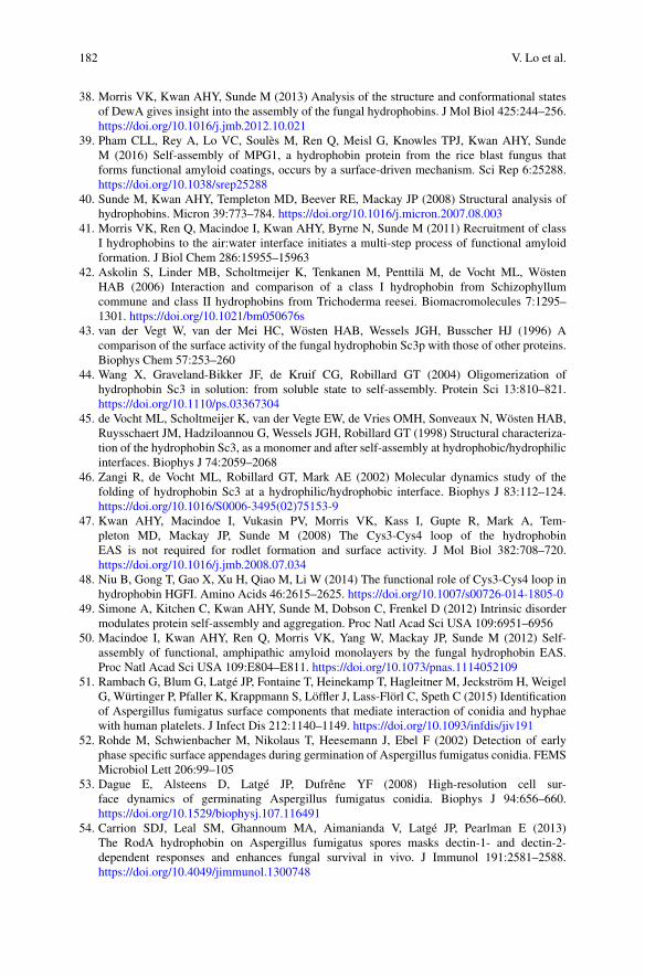
182 V. Lo et al.
38. Morris VK, Kwan AHY, Sunde M (2013) Analysis of the structure and conformational statesof DewA gives insight into the assembly of the fungal hydrophobins. J Mol Biol 425:244–256.https://doi.org/10.1016/j.jmb.2012.10.021
39. Pham CLL, Rey A, Lo VC, Soulès M, Ren Q, Meisl G, Knowles TPJ, Kwan AHY, SundeM (2016) Self-assembly of MPG1, a hydrophobin protein from the rice blast fungus thatforms functional amyloid coatings, occurs by a surface-driven mechanism. Sci Rep 6:25288.https://doi.org/10.1038/srep25288
40. Sunde M, Kwan AHY, Templeton MD, Beever RE, Mackay JP (2008) Structural analysis ofhydrophobins. Micron 39:773–784. https://doi.org/10.1016/j.micron.2007.08.003
41. Morris VK, Ren Q, Macindoe I, Kwan AHY, Byrne N, Sunde M (2011) Recruitment of classI hydrophobins to the air:water interface initiates a multi-step process of functional amyloidformation. J Biol Chem 286:15955–15963
42. Askolin S, Linder MB, Scholtmeijer K, Tenkanen M, Penttilä M, de Vocht ML, WöstenHAB (2006) Interaction and comparison of a class I hydrophobin from Schizophyllumcommune and class II hydrophobins from Trichoderma reesei. Biomacromolecules 7:1295–1301. https://doi.org/10.1021/bm050676s
43. van der Vegt W, van der Mei HC, Wösten HAB, Wessels JGH, Busscher HJ (1996) Acomparison of the surface activity of the fungal hydrophobin Sc3p with those of other proteins.Biophys Chem 57:253–260
44. Wang X, Graveland-Bikker JF, de Kruif CG, Robillard GT (2004) Oligomerization ofhydrophobin Sc3 in solution: from soluble state to self-assembly. Protein Sci 13:810–821.https://doi.org/10.1110/ps.03367304
45. de Vocht ML, Scholtmeijer K, van der Vegte EW, de Vries OMH, Sonveaux N, Wösten HAB,Ruysschaert JM, Hadziloannou G, Wessels JGH, Robillard GT (1998) Structural characteriza-tion of the hydrophobin Sc3, as a monomer and after self-assembly at hydrophobic/hydrophilicinterfaces. Biophys J 74:2059–2068
46. Zangi R, de Vocht ML, Robillard GT, Mark AE (2002) Molecular dynamics study of thefolding of hydrophobin Sc3 at a hydrophilic/hydrophobic interface. Biophys J 83:112–124.https://doi.org/10.1016/S0006-3495(02)75153-9
47. Kwan AHY, Macindoe I, Vukasin PV, Morris VK, Kass I, Gupte R, Mark A, Tem-pleton MD, Mackay JP, Sunde M (2008) The Cys3-Cys4 loop of the hydrophobinEAS is not required for rodlet formation and surface activity. J Mol Biol 382:708–720.https://doi.org/10.1016/j.jmb.2008.07.034
48. Niu B, Gong T, Gao X, Xu H, Qiao M, Li W (2014) The functional role of Cys3-Cys4 loop inhydrophobin HGFI. Amino Acids 46:2615–2625. https://doi.org/10.1007/s00726-014-1805-0
49. Simone A, Kitchen C, Kwan AHY, Sunde M, Dobson C, Frenkel D (2012) Intrinsic disordermodulates protein self-assembly and aggregation. Proc Natl Acad Sci USA 109:6951–6956
50. Macindoe I, Kwan AHY, Ren Q, Morris VK, Yang W, Mackay JP, Sunde M (2012) Self-assembly of functional, amphipathic amyloid monolayers by the fungal hydrophobin EAS.Proc Natl Acad Sci USA 109:E804–E811. https://doi.org/10.1073/pnas.1114052109
51. Rambach G, Blum G, Latgé JP, Fontaine T, Heinekamp T, Hagleitner M, Jeckström H, WeigelG, Würtinger P, Pfaller K, Krappmann S, Löffler J, Lass-Flörl C, Speth C (2015) Identificationof Aspergillus fumigatus surface components that mediate interaction of conidia and hyphaewith human platelets. J Infect Dis 212:1140–1149. https://doi.org/10.1093/infdis/jiv191
52. Rohde M, Schwienbacher M, Nikolaus T, Heesemann J, Ebel F (2002) Detection of earlyphase specific surface appendages during germination of Aspergillus fumigatus conidia. FEMSMicrobiol Lett 206:99–105
53. Dague E, Alsteens D, Latgé JP, Dufrêne YF (2008) High-resolution cell sur-face dynamics of germinating Aspergillus fumigatus conidia. Biophys J 94:656–660.https://doi.org/10.1529/biophysj.107.116491
54. Carrion SDJ, Leal SM, Ghannoum MA, Aimanianda V, Latgé JP, Pearlman E (2013)The RodA hydrophobin on Aspergillus fumigatus spores masks dectin-1- and dectin-2-dependent responses and enhances fungal survival in vivo. J Immunol 191:2581–2588.https://doi.org/10.4049/jimmunol.1300748
5 Fungal Hydrophobins and Their Self-Assembly into Functional Nanomaterials 183
55. Loures FV, Röhm M, Lee CK, Santos E, Wang JP, Specht CA, Calich VLG, Urban CF, LevitzSM (2015) Recognition of Aspergillus fumigatus hyphae by human plasmacytoid dendriticcells is mediated by dectin-2 and results in formation of extracellular traps. PLoS Pathog11:e1004643. https://doi.org/10.1371/journal.ppat.1004643
56. Steele C, Rapaka RR, Metz A, Pop SM, Williams DL, Gordon S, Kolls JK, Brown GD (2005)The beta-glucan receptor dectin-1 recognizes specific morphologies of Aspergillus fumigatus.PLoS Pathog 1:e42. https://doi.org/10.1371/journal.ppat.0010042
57. Khan NS, Kasperkovitz PV, Timmons AK, Mansour MK, Tam JM, Seward MW, Reedy JL,Puranam S, Feliu M, Vyas JM (2016) Dectin-1 controls TLR9 trafficking to phagosomes con-taining β-1,3 glucan. J Immunol 196:2249–2261. https://doi.org/10.4049/jimmunol.1401545
58. Paris S, Debeaupuis JP, Crameri R, Carey M, Charlès F, Prévost MC, Schmitt C, PhilippeB, Latgé JP (2003) Conidial hydrophobins of Aspergillus fumigatus. Appl Environ Microbiol69:1581–1588
59. Beauvais A, Schmidt C, Guadagnini S, Roux P, Perret E, Henry C, Paris S,Mallet A, Prévost MC, Latgé JP (2007) An extracellular matrix glues togetherthe aerial-grown hyphae of Aspergillus fumigatus. Cell Microbiol 9:1588–1600.https://doi.org/10.1111/j.1462-5822.2007.00895.x
60. Bruns S, Seidler M, Albrecht D, Salvenmoser S, Remme N, Hertweck C, Brakhage AA,Kniemeyer O, Müller FMC (2010) Functional genomic profiling of Aspergillus fumigatusbiofilm reveals enhanced production of the mycotoxin gliotoxin. Proteomics 10:3097–3107.https://doi.org/10.1002/pmic.201000129
61. Gibbons JG, Beauvais A, Beau R, McGary KL, Latgé JP, Rokas A (2012) Global transcriptomechanges underlying colony growth in the opportunistic human pathogen Aspergillus fumigatus.Eukaryot Cell 11:68–78. https://doi.org/10.1128/EC.05102-11
62. Skamnioti P, Gurr SJ (2007) Magnaporthe grisea cutinase2 mediates appressorium differ-entiation and host penetration and is required for full virulence. Plant Cell 19:2674–2689.https://doi.org/10.1105/tpc.107.051219
63. Matsumura H, Reich S, Ito A, Saitoh H, Kamoun S, Winter P, Kahl G, Reuter M, KrugerDH, Terauchi R (2003) Gene expression analysis of plant host-pathogen interactions by Super-SAGE. Proc Natl Acad Sci USA 100:15718–15723. https://doi.org/10.1073/pnas.2536670100
64. Talbot NJ, Kershaw MJ, Wakley GE, De Vries OMH, Wessels JGH, Hamer JE (1996)MPG1 encodes a fungal hydrophobin involved in surface interactions during infection-relateddevelopment of Magnaporthe grisea. Plant Cell 8:985–999. https://doi.org/10.1105/tpc.8.6.985
65. Inoue K, Kitaoka H, Park P, Ikeda K (2015) Novel aspects of hydrophobins in wheat isolate ofMagnaporthe oryzae: Mpg1, but not Mhp1, is essential for adhesion and pathogenicity. J GenPlant Pathol 82:18–28. https://doi.org/10.1007/s10327-015-0632-9
66. Khalesi M, Deckers SM, Gebruers K, Vissers L, Verachtert H, Derdelinckx G (2012)Hydrophobins: exceptional proteins for many applicaitons in brewery environment and otherbio-industries. Cerevisia 37:3–9
67. Wösten HAB, Scholtmeijer K (2015) Applications of hydrophobins: current state and perspec-tives. Appl Microbiol Biotechnol 99:1587–1597. https://doi.org/10.1007/s00253-014-6319-x
68. Wang Z, Lienemann M, Qiau M, Linder MB (2010) Mechanisms of protein adhesion on surfacefilms of hydrophobin. Langmuir 26:8491–8496. https://doi.org/10.1021/la101240e
69. Longobardi S, Gravagnuolo AM, Funari R, Della Ventura B, Pane F, Galano E, Amore-sano A, Marino G, Giardina P (2015) A simple MALDI plate functionalization by Vmh2hydrophobin for serial multi-enzymatic protein digestions. Anal Bioanal Chem 407:487–496.https://doi.org/10.1007/s00216-014-8309-3
70. Gravagnuolo AM, Morales-Narváez E, Matos CRS, Longobardi S, Giardina P, MerkoçiA (2015) On-the-spot immobilization of quantum dots, graphene oxide, and proteins viaHydrophobins. Adv Funct Mater 25(38):6084–6092. https://doi.org/10.1002/adfm.201502837
71. Sapsford KE, Medintz IL, Golden JP, Deschamps JR, Uyeda HT, Mattoussi H (2004)Surface-immobilized self-assembled protein-based quantum dot Nanoassemblies. Langmuir20(18):7720–7728. https://doi.org/10.1021/la049263n
184 V. Lo et al.
72. Haddada MB, Blanchard J, Casale S, Krafft J-M, Vallée A, Méthivier C, Bou-jday S (2013) Optimizing the immobilization of gold nanoparticles on functional-ized silicon surfaces: amine- vs thiol-terminated silane. Gold Bull 46(4):335–341.https://doi.org/10.1007/s13404-013-0120-y
73. Alves NJ, Kiziltepe T, Bilgicer B (2012) Oriented surface immobilization of antibodies atthe conserved nucleotide binding site for enhanced antigen detection. Langmuir 28(25):9640–9648. https://doi.org/10.1021/la301887s
74. Zhao ZX, Wang HC, Qin X, Wang XS, Qiao MQ, Anzai JI, Chen Q (2009) Self-assembled filmof hydrophobins on gold surfaces and its application to electrochemical biosensing. ColloidsSurf B Biointerfaces 71:102–106. https://doi.org/10.1016/j.colsurfb.2009.01.011
75. Wang X, Wang H, Huang Y, Zhao Z, Qin X, Wang Y, Miao Z, Chen Q, Qiao M (2010)Noncovalently functionalized multi-wall carbon nanotubes in aqueous solution using thehydrophobin HFBI and their electroanalytical application. Biosens Bioelectron 26:1104–1108.https://doi.org/10.1016/j.bios.2010.08.024
76. Rea I, Giardina P, Longobardi S, Porro F, Casuscelli V, Rendina I, De Stefano L (2012)Hydrophobin Vmh2-glucose complexes self-assemble in nanometric biofilms. J R Soc Inter-face 9:2450–2456. https://doi.org/10.1098/rsif.2012.0217
77. Politi J, De Stefano L, Rea I, Gravagnuolo A, Giardina P, Methivier C, CasaleS, Spadavecchia J (2016) One-pot synthesis of a gold nanoparticle-Vmh2hydrophobin nanobiocomplex for glucose monitoring. Nanotechnology 27:195701.https://doi.org/10.1088/0957-4484/27/19/195701
78. Bilewicz R, Witomski J, Van der Heyden A, Tagu D, Palin B, Rogalska E (2001) Modificationof electrodes with self-assembled Hydrophobin layers. J Phys Chem B 105(40):9772–9777.https://doi.org/10.1021/jp0113782
79. Opwis K, Gutmann JS (2011) Surface modification of textile materials with hydrophobins.Text Res J 81:1594–1602
80. Popescu AC, Stan GE, Duta L, Dorcioman G, Iordache O, Dumitrescu I, Pasuk I, Mihailescu IN(2013) Influence of a hydrophobin underlayer on the structuring and antimicrobial propertiesof ZnO films. J Mater Sci 48:8329–8336
81. Lee JH, Khang G, Lee JW, Lee HB (1998) Interaction of different types of cellson polymer surfaces with wettability gradient. J Colloid Interf Sci 205:323–330.https://doi.org/10.1006/jcis.1998.5688
82. Janssen MI, van Leeuwen MBM, Scholtmeijer K, van Kooten TG, Dijkhuizen L, WöstenHAB (2002) Coating with genetic engineered hydrophobin promotes growth of fibroblasts ona hydrophobic solid. Biomaterials 23:4847–4854
83. Boeuf S, Throm T, Gutt B, Strunk T, Hoffmann M, Seebach E, Mühlberg L, Brocher J,Gotterbarm T, Wenzel W, Fischer R, Richter W (2012) Engineering hydrophobin DewA togenerate surfaces that enhance adhesion of human but not bacterial cells. Acta Biomater8:1037–1047. https://doi.org/10.1016/j.actbio.2011.11.022
84. Weickert U, Wiesend F, Subkowski T, Eickhoff A, Reiss G (2011) Optimizing bil-iary stent patency by coating with hydrophobin alone or hydrophobin and antibi-otics or heparin: an in vitro proof of principle study. Adv Med Sci 56:138–144.https://doi.org/10.2478/v10039-011-0026-y
85. Stanimirova RD, Gurkov TD, Kralchevsky PA, Balashev KT, Stoyanov SD, Pelan EG(2013) Surface pressure and elasticity of hydrophobin HFBII layers on the air-waterinterface: rheology versus structure detected by AFM imaging. Langmuir 29:6053–6067.https://doi.org/10.1021/la4005104
86. Deckers SM, Venken T, Khalesi M, Gebruers K, Baggerman G, Lorgouilloux Y, ShokribousjeinZ, Ilberg V, Schönberger C, Titze J, Verachtert H, Michiels C, Neven H, Delcour J, MartensJ, Derdelinckx G, de Maeyer M (2012) Combined modeling and biophysical characterisationof CO2 interaction with class II hydrophobins: new insight into the mechanism underpinningprimary gushing. J Am Soc Brew Chem 70:249–256
87. Cox AR, Aldred DL, Russell AB (2009) Exceptional stability of food foams using classII hydrophobin HFBII. Food Hydrocoll 23:366–376. https://doi.org/10.1016/j.foodhyd.2008.03.001
5 Fungal Hydrophobins and Their Self-Assembly into Functional Nanomaterials 185
88. Tchuenbou-Magaia FL, Norton IT, Cox PW (2009) Hydrophobins stabilisedair-filled emulsions for the food industry. Food Hydrocoll 23:1877–1885.https://doi.org/10.1016/j.foodhyd.2009.03.005
89. Green A, Littlejohn K, Hooley P, Cox P (2013) Formation and stability of food foams andaerated emulsions: hydrophobins as novel functional ingredients. Curr Opin Colloid InterfaceSci 18(4):292–301
90. Loftsson T, Brewster ME (2010) Pharmaceutical applications of cyclodextrins:basic science and product development. J Pharm Pharmacol 62:1607–1621.https://doi.org/10.1111/j.2042-7158.2010.01030.x
91. Haas Jimoh Akanbi M, Post E, Meter-Arkema A, Rink R, Robillard GT, Wang X,Wösten HAB, Scholtmeijer K (2010) Use of hydrophobins in formulation of waterinsoluble drugs for oral administration. Colloids Surf B Biointerfaces 75:526–531.https://doi.org/10.1016/j.colsurfb.2009.09.030
92. Fang G, Tang B, Liu Z, Gou J, Zhang Y, Xu H, Tang X (2014) Novel hydrophobin-coated docetaxel nanoparticles for intravenous delivery: in vitro characteristics and in vivoperformance. Eur J Pharm Sci 60:1–9. https://doi.org/10.1016/j.ejps.2014.04.016
93. Valo HK, Laaksonen PH, Peltonen LJ, Linder MB, Hirvonen JT, Laaksonen TJ (2010)Multifunctional hydrophobin: toward functional coatings for drug nanoparticles. ACS Nano4:1750–1758. https://doi.org/10.1021/nn9017558
94. Yang W, Ren Q, Wu YN, Morris VK, Rey AA, Braet F, Kwan AHY, Sunde M (2013)Surface functionalization of carbon nanomaterials by self-assembling hydrophobin proteins.Biopolymers 99:84–94. https://doi.org/10.1002/bip.22146
95. Sarparanta M, Bimbo LM, Rytkönen J, Mäkilä E, Laaksonen TJ, Laaksonen P, Nyman M,Salonen J, Linder MB, Hirvonen J, Santos HA, Airaksinen AJ (2012) Intravenous delivery ofhydrophobin-functionalized porous silicon nanoparticles: stability, plasma protein adsorptionand biodistribution. Mol Pharm 9:654–663. https://doi.org/10.1021/mp200611d
Chapter 6Nanostructured, Self-Assembled SpiderSilk Materials for BiomedicalApplications
Martin Humenik, Kiran Pawar, and Thomas Scheibel
Abstract The extraordinary mechanical properties of spider silk fibers resultfrom the interplay of composition, structure and self-assembly of spider silkproteins (spidroins). Genetic approaches enabled the biotechnological productionof recombinant spidroins which have been employed to unravel the self-assemblyand spinning process. Various processing conditions allowed to explore non-natural morphologies including nanofibrils, particles, capsules, hydrogels, filmsor foams. Recombinant spider silk proteins and materials made thereof can beutilized for biomedical applications, such as drug delivery, tissue engineering or3D-biomanufacturing.
Keywords Biofabrication · Drug delivery · Fibers · Genetic engineering ·Recombinant production · Self-assembly · Spider silk · Tissue engineering
6.1 Introduction
Spider webs and spider silk fibers have been fascinating mankind since ages,exemplarily seen in ancient myths about Arachne, a mortal woman who challenged
M. HumenikBiomaterials, Faculty of Engineering Science, University of Bayreuth, Bayreuth, Germany
K. PawarBlusson Spinal Cord Centre, University of British Columbia, Vancouver, Canada
T. Scheibel (�)Bayreuth Center for Colloids and Interfaces (BZKG), University of Bayreuth, Bayreuth, Germany
Research Center Bio-Macromolecules (BIOmac), University of Bayreuth, Bayreuth, Germany
Bayreuth Center for Molecular Biosciences (BZMB), University of Bayreuth, Bayreuth, Germany
Bayreuth Center for Material Science (BayMAT), University of Bayreuth, Bayreuth, Germany
Bavarian Polymer Institute (BPI), University of Bayreuth, Bayreuth, Germanye-mail: [email protected]
© Springer Nature Singapore Pte Ltd. 2019S. Perrett et al. (eds.), Biological and Bio-inspired Nanomaterials,Advances in Experimental Medicine and Biology 1174,https://doi.org/10.1007/978-981-13-9791-2_6
187
188 M. Humenik et al.
Athena in weaving, or in stories about SpiderMan, a mutated young man acquiringspider abilities after a bite of a radioactively modified spider. Apart fiction, severalspecific properties of spider silk webs have been recognized early-on allowingapplications, such as wound coatings in ancient Greece and fishing nets of tribesfrom Papua-New Guinea and Australia [1]. Nowadays, scientific insights into thematerial properties have been gained which allow to mechanistically understandthe assembly of this proteinaceous material, as well as to technically apply it formultiple purposes. Spider silk, like bone, nacre, or wood, reveals a hierarchicallyordered structure, which self-assembles from simple building blocks just uponweak (but numerous) non-covalent interactions, whereas its chemical and structuralcompatibility, probed by evolution from nanoscales to macroscales, results incoherent materials [2].
Here, spider silk composition and fiber structure will be elucidated, includingself-assembly principles underlining its hierarchical setup. We will also presentbioinspired technologies, which on the one hand helped to understand the self-assembly and spinning process, and on the other hand enabled the application ofsilk-based materials. Spider-silk based morphologies, such as nanofibrils, particles,capsules, hydrogels, films, and foams, allowed the development of biomedicalapplications (among others), which are presented in the final section of this bookchapter.
6.2 Natural Spider Silk
Currently more than 48.000 spider species, from tiny Peacock to large Goliathbird-eating spiders, are known, which are divided into 109 families [3], occupyingevery possible niche on Earth and representing one of the most diverse orders of allorganisms. All spider species spin silk threads, proteinaceous materials with quitedifferent composition and structure, which are also produced by a broad range ofother Arthropods [4, 5]. Many spiders weave silk webs of varying architecture,such as funnel, sheet, bola, cob and orb webs, serving different purposes rangingfrom an extension of their sensory system to catching prey to protective shellsof eggs. Orb webs represent the most fascinating and probably the most complexfeatures of silk architecture. They are produced by the suborder Araneomorphaeof the Orbiculariae clade [6] being typically composed of five different silks withspecific mechanical, physical, and chemical properties, which are combined tostop effectively the flying prey by dissipating the kinetic energy of the impact vianonlinear mechanical stress response to minimize possible damages of the web and,at the same time, to keep the pray within the web [7].
Different types of silk proteins are produced by specialized glands in the spiderabdomen [8–10]. Orb weaving spiders of Nephila or Araneus genera show sevenindividual types (one of which represents a glue), including major and minorampullate, flagelliform, tubuliform, aciniform, pyriform and aggregate glands [11].
The frame and radii of the orb web are made of proteins produced in majorampullate (MA) glands, and the respective silk fibers are constantly drawn to be
6 Nanostructured, Self-Assembled Spider Silk Materials for Biomedical Applications 189
also used as a lifeline. Minor ampullate (MI) silk is used as a temporary supportspiral and, together with flagelliform silk, it represents the capture spiral. Junctionsbetween MA and flagelliform fibers are reinforced by pyriform silk, which alsoserves as an attachment cement to anchor the frame on substrates. Aggregate silkis deposited on the capture spiral as a glue. Two silk types are produced to protectoffspring in eggs. The cases are made of tough tubuli- /cylindriform silk (both namesdescribe the same type), whereas a soft layer in the egg interior consists of aciniformsilk, also used in prey-wrapping [6, 12].
6.2.1 Protein Composition of Major Ampullate Silk
Spider silks typically comprise one or more proteins with various molecularweights. They share a general composition of a repetitive core domain withvarying primary sequences among the silk types and small flanking C- and N-terminal domains which are highly conserved between the silk types but alsobetween spider species [11, 13, 14]. Different sequences in the core domain resultconsequently in different mechanical properties of the respective threads rangingfrom extensive rubber-like features of the capture spiral to stiff and tough MA silk[15–17]. Aggregate silk, as the only exception in morphology and composition, isdeposited as glue droplets and composed of at least two small (< 65 kDa), highlyglycosylated proteins and two peptides [18]. In addition to these well describedspidroins [6], recent genomic and proteomic approaches have identified a plethoraof additional proteins associated with silk production, storage and spinning [19].There were more than 600 silk-gland specific transcripts (SSTs) identified by Clarkeet al. in the case of the black widow spider (Latrodectus hesperus) [20]. Themajority of the transcripts showed no annotation, but they may include novel silk-associated proteins (SAPs) [8], such as low molecular weight cysteine-rich proteins(CRPs), which were found in the MA dope as well as in the related fiber formingmacromolecular complexes [21]. Egg case proteins (ECPs) represent a non-spidroinfamily identified in black widow tubuliform glands, and respective fibers made ofTuSp1 are thought to be cross-linked with ECPs [12].
Details of duct morphologies and spinning of particular spider silks can befound elsewhere [6, 22]. Here, we introduce the general but at the same time mostsophisticated features of silk composition, structure and assembly into fibers usingthe example of MA silk, which represents the best studied silk type. The MA glandis the largest one amongst the spider silk glands providing sufficient amounts of thematerial for analysis using X-ray diffraction, NMR, or IR spectroscopy [23–26].
The major ampulla is divided in a long tail (zone A) and a sac (zone B and C)continuing into a tapered S-shaped duct connected to an external spigot [27, 28].In zones A and B proteins are secreted, whereas in zone C they are stored at highconcentration (∼25–50 wt %) [22]. Major ampullate silk proteins can be classifiedinto spidroin 1 and 2 (MaSp1 and MaSp2) with molecular weights often exceeding250 kDa [29–31], but low MW variants consisting of a small nonrepetitive coredomain do exist [32]. The main difference between MaSp1 and MaSp2 is the proline
190 M. Humenik et al.
content, being high in MaSp2 and quite low in MaSp1. In general, Nephila andLatrodectus MA dopes show only 1–2% of proline, whereas Araneus and ArgiopeMA silk contain proline residues in the range of 10–12% [33]. Interestingly, whencomparing native dragline silks, they display similar mechanical performance inthe dry state, but under humid conditions, they reveal pronounced supercontractioncorrelating with the increased proline content. Thus, regulating the ratio of MaSp1and MaSp2 levels enables spiders to alter the material properties of their fibers inresponse to environmental changes [34, 33].
Core domains of MaSps are prevalently composed of glycine (Gly), alanine(Ala), glutamine (Gln), proline (Pro), serine (Ser) and tyrosine (Tyr) residues [35],which are clustered into small motifs, such as poly-alanine stretches (Ala)n (n = 4–12), GA, and GGX (X = Y, L and Q) repeats. MaSp2 contains additionally GPGXXrepeats (X = Q, G, Y) which alternate with GGX [6, 36, 37]. In case of fullysequenced MaSp1 and MaSp2 of L. hesperus [30] and of the wasp spider (Argiopebruennichi) [38], as well as proteomically analyzed MaSps of golden silk orb-weaver Nephila clavipes [39] it has been shown that distinct sequence motifs canbe repeated ~100 times within the core domain. Several phosphorylation sides havebeen also found in MaSps [39, 40].
In solution i.e., in the sac, the repetitive core of MaSps is intrinsically unstruc-tured. NMR and IR spectroscopy studies have shown no distinct secondary struc-tures but only small contributions of polyproline II (PPII) and α-helices [10,25, 41–43]. In contrast, terminal domains represent globular, folded structuresconsisting of bundles of five α-helices [14, 44, 45]. Immunological experiments,using specific antibodies, and mass-spectrometry analysis have shown the presenceof folded C- as well as N-terminal domains in spinning dopes as well as in the fibers[19, 27, 46]. C-terminal domains contain highly conserved Cys residues and areassumed to dimerize in a parallel orientation. N-terminal domains are monomericin the dope and dimerize upon acidification in an antiparallel fashion, as describedin the next section. The sequences of the domains are more hydrophobic than therepetitive core, however their native folds expose prevalently hydrophilic residuesensuring the spidroin solubility [30, 44, 45].
6.2.2 Processing of Spider Silk Proteins into Fibers
In the sac, highly concentrated soluble spidroins are stabilized in the presence ofNaCl and at neutral pH. Along the spinning duct acidification takes place as well asan exchange of chaotropic Na+ and Cl− ions for more kosmotropic K+ and PO4
3−[42, 47–49]. The structure of the C-terminal domain is sensitive to these changes.Destabilization of the structure, partial unfolding, and exposure of its hydrophobiccore have been shown to support aggregation under influence of shear forces [45,50, 51]. In contrast, the monomeric N-terminal domain dimerizes in an antiparallelfashion, driven by electrostatic interactions upon slight acidification to pH 5–6 [52–60]. The repetitive core domain experiences a transformation of random coil into
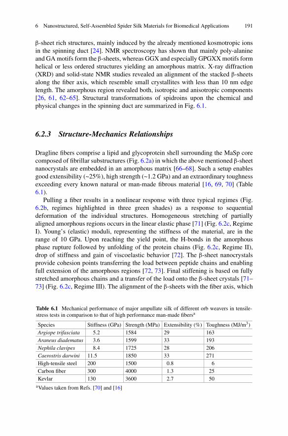
6 Nanostructured, Self-Assembled Spider Silk Materials for Biomedical Applications 191
β-sheet rich structures, mainly induced by the already mentioned kosmotropic ionsin the spinning duct [24]. NMR spectroscopy has shown that mainly poly-alanineand GA motifs form the β-sheets, whereas GGX and especially GPGXX motifs formhelical or less ordered structures yielding an amorphous matrix. X-ray diffraction(XRD) and solid-state NMR studies revealed an alignment of the stacked β-sheetsalong the fiber axis, which resemble small crystallites with less than 10 nm edgelength. The amorphous region revealed both, isotropic and anisotropic components[26, 61, 62–65]. Structural transformations of spidroins upon the chemical andphysical changes in the spinning duct are summarized in Fig. 6.1.
6.2.3 Structure-Mechanics Relationships
Dragline fibers comprise a lipid and glycoprotein shell surrounding the MaSp corecomposed of fibrillar substructures (Fig. 6.2a) in which the above mentioned β-sheetnanocrystals are embedded in an amorphous matrix [66–68]. Such a setup enablesgood extensibility (~25%), high strength (~1.2 GPa) and an extraordinary toughnessexceeding every known natural or man-made fibrous material [16, 69, 70] (Table6.1).
Pulling a fiber results in a nonlinear response with three typical regimes (Fig.6.2b, regimes highlighted in three green shades) as a response to sequentialdeformation of the individual structures. Homogeneous stretching of partiallyaligned amorphous regions occurs in the linear elastic phase [71] (Fig. 6.2c, RegimeI). Young’s (elastic) moduli, representing the stiffness of the material, are in therange of 10 GPa. Upon reaching the yield point, the H-bonds in the amorphousphase rupture followed by unfolding of the protein chains (Fig. 6.2c, Regime II),drop of stiffness and gain of viscoelastic behavior [72]. The β-sheet nanocrystalsprovide cohesion points transferring the load between peptide chains and enablingfull extension of the amorphous regions [72, 73]. Final stiffening is based on fullystretched amorphous chains and a transfer of the load onto the β-sheet crystals [71–73] (Fig. 6.2c, Regime III). The alignment of the β-sheets with the fiber axis, which
Table 6.1 Mechanical performance of major ampullate silk of different orb weavers in tensile-stress tests in comparison to that of high performance man-made fibersa
Species Stiffness (GPa) Strength (MPa) Extensibility (%) Toughness (MJ/m3)Argiope trifasciata 5.2 1584 29 163Araneus diadematus 3.6 1599 33 193Nephila clavipes 8.4 1725 28 206Caerostris darwini 11.5 1850 33 271High-tensile steel 200 1500 0.8 6Carbon fiber 300 4000 1.3 25Kevlar 130 3600 2.7 50
aValues taken from Refs. [70] and [16]
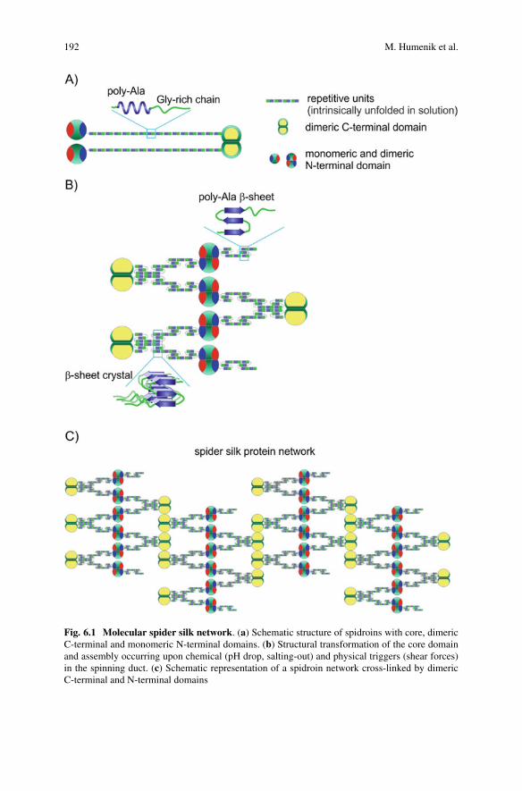
192 M. Humenik et al.
Fig. 6.1 Molecular spider silk network. (a) Schematic structure of spidroins with core, dimericC-terminal and monomeric N-terminal domains. (b) Structural transformation of the core domainand assembly occurring upon chemical (pH drop, salting-out) and physical triggers (shear forces)in the spinning duct. (c) Schematic representation of a spidroin network cross-linked by dimericC-terminal and N-terminal domains
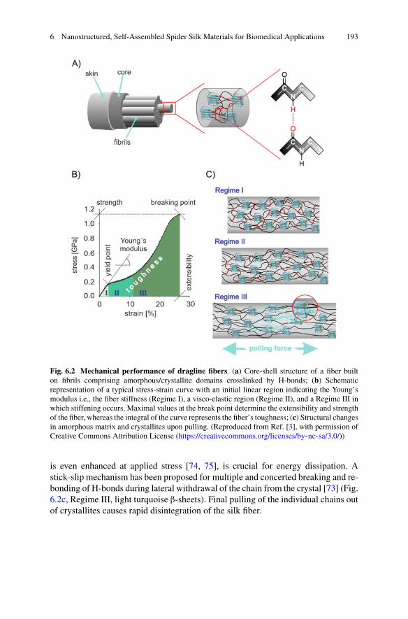
6 Nanostructured, Self-Assembled Spider Silk Materials for Biomedical Applications 193
Fig. 6.2 Mechanical performance of dragline fibers. (a) Core-shell structure of a fiber builton fibrils comprising amorphous/crystallite domains crosslinked by H-bonds; (b) Schematicrepresentation of a typical stress-strain curve with an initial linear region indicating the Young’smodulus i.e., the fiber stiffness (Regime I), a visco-elastic region (Regime II), and a Regime III inwhich stiffening occurs. Maximal values at the break point determine the extensibility and strengthof the fiber, whereas the integral of the curve represents the fiber’s toughness; (c) Structural changesin amorphous matrix and crystallites upon pulling. (Reproduced from Ref. [3], with permission ofCreative Commons Attribution License (https://creativecommons.org/licenses/by-nc-sa/3.0/))
is even enhanced at applied stress [74, 75], is crucial for energy dissipation. Astick-slip mechanism has been proposed for multiple and concerted breaking and re-bonding of H-bonds during lateral withdrawal of the chain from the crystal [73] (Fig.6.2c, Regime III, light turquoise β-sheets). Final pulling of the individual chains outof crystallites causes rapid disintegration of the silk fiber.
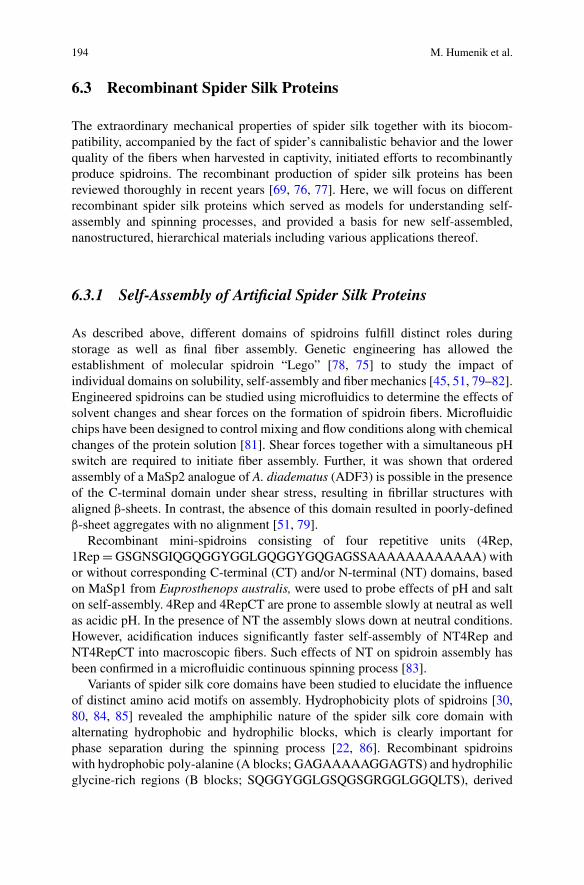
194 M. Humenik et al.
6.3 Recombinant Spider Silk Proteins
The extraordinary mechanical properties of spider silk together with its biocom-patibility, accompanied by the fact of spider’s cannibalistic behavior and the lowerquality of the fibers when harvested in captivity, initiated efforts to recombinantlyproduce spidroins. The recombinant production of spider silk proteins has beenreviewed thoroughly in recent years [69, 76, 77]. Here, we will focus on differentrecombinant spider silk proteins which served as models for understanding self-assembly and spinning processes, and provided a basis for new self-assembled,nanostructured, hierarchical materials including various applications thereof.
6.3.1 Self-Assembly of Artificial Spider Silk Proteins
As described above, different domains of spidroins fulfill distinct roles duringstorage as well as final fiber assembly. Genetic engineering has allowed theestablishment of molecular spidroin “Lego” [78, 75] to study the impact ofindividual domains on solubility, self-assembly and fiber mechanics [45, 51, 79–82].Engineered spidroins can be studied using microfluidics to determine the effects ofsolvent changes and shear forces on the formation of spidroin fibers. Microfluidicchips have been designed to control mixing and flow conditions along with chemicalchanges of the protein solution [81]. Shear forces together with a simultaneous pHswitch are required to initiate fiber assembly. Further, it was shown that orderedassembly of a MaSp2 analogue of A. diadematus (ADF3) is possible in the presenceof the C-terminal domain under shear stress, resulting in fibrillar structures withaligned β-sheets. In contrast, the absence of this domain resulted in poorly-definedβ-sheet aggregates with no alignment [51, 79].
Recombinant mini-spidroins consisting of four repetitive units (4Rep,1Rep = GSGNSGIQGQGGYGGLGQGGYGQGAGSSAAAAAAAAAAAA) withor without corresponding C-terminal (CT) and/or N-terminal (NT) domains, basedon MaSp1 from Euprosthenops australis, were used to probe effects of pH and salton self-assembly. 4Rep and 4RepCT are prone to assemble slowly at neutral as wellas acidic pH. In the presence of NT the assembly slows down at neutral conditions.However, acidification induces significantly faster self-assembly of NT4Rep andNT4RepCT into macroscopic fibers. Such effects of NT on spidroin assembly hasbeen confirmed in a microfluidic continuous spinning process [83].
Variants of spider silk core domains have been studied to elucidate the influenceof distinct amino acid motifs on assembly. Hydrophobicity plots of spidroins [30,80, 84, 85] revealed the amphiphilic nature of the spider silk core domain withalternating hydrophobic and hydrophilic blocks, which is clearly important forphase separation during the spinning process [22, 86]. Recombinant spidroinswith hydrophobic poly-alanine (A blocks; GAGAAAAAGGAGTS) and hydrophilicglycine-rich regions (B blocks; SQGGYGGLGSQGSGRGGLGGQLTS), derived
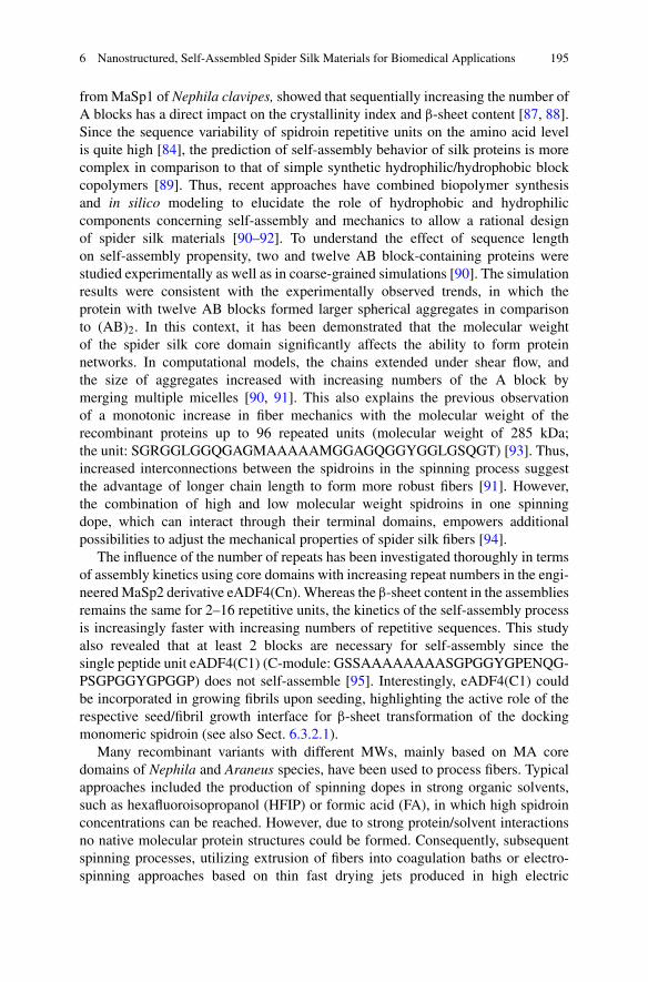
6 Nanostructured, Self-Assembled Spider Silk Materials for Biomedical Applications 195
from MaSp1 of Nephila clavipes, showed that sequentially increasing the number ofA blocks has a direct impact on the crystallinity index and β-sheet content [87, 88].Since the sequence variability of spidroin repetitive units on the amino acid levelis quite high [84], the prediction of self-assembly behavior of silk proteins is morecomplex in comparison to that of simple synthetic hydrophilic/hydrophobic blockcopolymers [89]. Thus, recent approaches have combined biopolymer synthesisand in silico modeling to elucidate the role of hydrophobic and hydrophiliccomponents concerning self-assembly and mechanics to allow a rational designof spider silk materials [90–92]. To understand the effect of sequence lengthon self-assembly propensity, two and twelve AB block-containing proteins werestudied experimentally as well as in coarse-grained simulations [90]. The simulationresults were consistent with the experimentally observed trends, in which theprotein with twelve AB blocks formed larger spherical aggregates in comparisonto (AB)2. In this context, it has been demonstrated that the molecular weightof the spider silk core domain significantly affects the ability to form proteinnetworks. In computational models, the chains extended under shear flow, andthe size of aggregates increased with increasing numbers of the A block bymerging multiple micelles [90, 91]. This also explains the previous observationof a monotonic increase in fiber mechanics with the molecular weight of therecombinant proteins up to 96 repeated units (molecular weight of 285 kDa;the unit: SGRGGLGGQGAGMAAAAAMGGAGQGGYGGLGSQGT) [93]. Thus,increased interconnections between the spidroins in the spinning process suggestthe advantage of longer chain length to form more robust fibers [91]. However,the combination of high and low molecular weight spidroins in one spinningdope, which can interact through their terminal domains, empowers additionalpossibilities to adjust the mechanical properties of spider silk fibers [94].
The influence of the number of repeats has been investigated thoroughly in termsof assembly kinetics using core domains with increasing repeat numbers in the engi-neered MaSp2 derivative eADF4(Cn). Whereas the β-sheet content in the assembliesremains the same for 2–16 repetitive units, the kinetics of the self-assembly processis increasingly faster with increasing numbers of repetitive sequences. This studyalso revealed that at least 2 blocks are necessary for self-assembly since thesingle peptide unit eADF4(C1) (C-module: GSSAAAAAAAASGPGGYGPENQG-PSGPGGYGPGGP) does not self-assemble [95]. Interestingly, eADF4(C1) couldbe incorporated in growing fibrils upon seeding, highlighting the active role of therespective seed/fibril growth interface for β-sheet transformation of the dockingmonomeric spidroin (see also Sect. 6.3.2.1).
Many recombinant variants with different MWs, mainly based on MA coredomains of Nephila and Araneus species, have been used to process fibers. Typicalapproaches included the production of spinning dopes in strong organic solvents,such as hexafluoroisopropanol (HFIP) or formic acid (FA), in which high spidroinconcentrations can be reached. However, due to strong protein/solvent interactionsno native molecular protein structures could be formed. Consequently, subsequentspinning processes, utilizing extrusion of fibers into coagulation baths or electro-spinning approaches based on thin fast drying jets produced in high electric
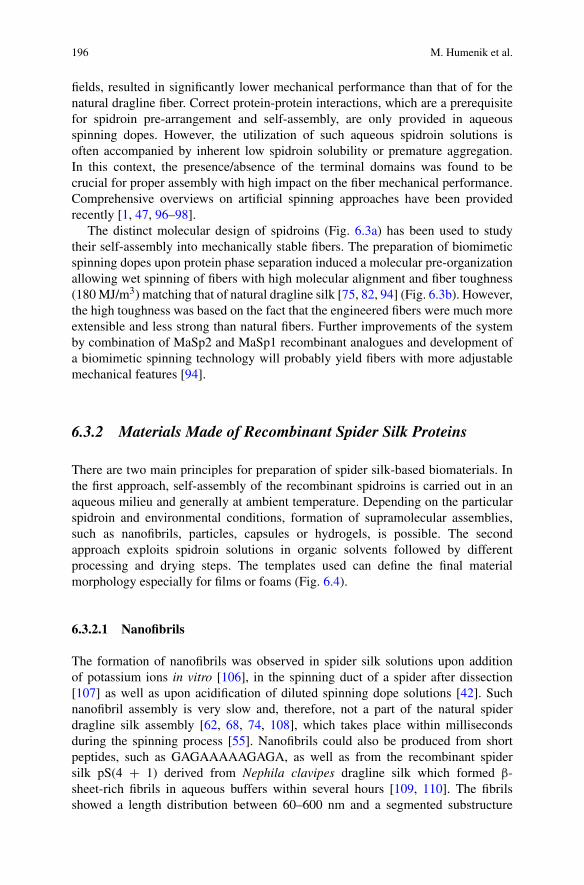
196 M. Humenik et al.
fields, resulted in significantly lower mechanical performance than that of for thenatural dragline fiber. Correct protein-protein interactions, which are a prerequisitefor spidroin pre-arrangement and self-assembly, are only provided in aqueousspinning dopes. However, the utilization of such aqueous spidroin solutions isoften accompanied by inherent low spidroin solubility or premature aggregation.In this context, the presence/absence of the terminal domains was found to becrucial for proper assembly with high impact on the fiber mechanical performance.Comprehensive overviews on artificial spinning approaches have been providedrecently [1, 47, 96–98].
The distinct molecular design of spidroins (Fig. 6.3a) has been used to studytheir self-assembly into mechanically stable fibers. The preparation of biomimeticspinning dopes upon protein phase separation induced a molecular pre-organizationallowing wet spinning of fibers with high molecular alignment and fiber toughness(180 MJ/m3) matching that of natural dragline silk [75, 82, 94] (Fig. 6.3b). However,the high toughness was based on the fact that the engineered fibers were much moreextensible and less strong than natural fibers. Further improvements of the systemby combination of MaSp2 and MaSp1 recombinant analogues and development ofa biomimetic spinning technology will probably yield fibers with more adjustablemechanical features [94].
6.3.2 Materials Made of Recombinant Spider Silk Proteins
There are two main principles for preparation of spider silk-based biomaterials. Inthe first approach, self-assembly of the recombinant spidroins is carried out in anaqueous milieu and generally at ambient temperature. Depending on the particularspidroin and environmental conditions, formation of supramolecular assemblies,such as nanofibrils, particles, capsules or hydrogels, is possible. The secondapproach exploits spidroin solutions in organic solvents followed by differentprocessing and drying steps. The templates used can define the final materialmorphology especially for films or foams (Fig. 6.4).
6.3.2.1 Nanofibrils
The formation of nanofibrils was observed in spider silk solutions upon additionof potassium ions in vitro [106], in the spinning duct of a spider after dissection[107] as well as upon acidification of diluted spinning dope solutions [42]. Suchnanofibril assembly is very slow and, therefore, not a part of the natural spiderdragline silk assembly [62, 68, 74, 108], which takes place within millisecondsduring the spinning process [55]. Nanofibrils could also be produced from shortpeptides, such as GAGAAAAAGAGA, as well as from the recombinant spidersilk pS(4 + 1) derived from Nephila clavipes dragline silk which formed β-sheet-rich fibrils in aqueous buffers within several hours [109, 110]. The fibrilsshowed a length distribution between 60–600 nm and a segmented substructure
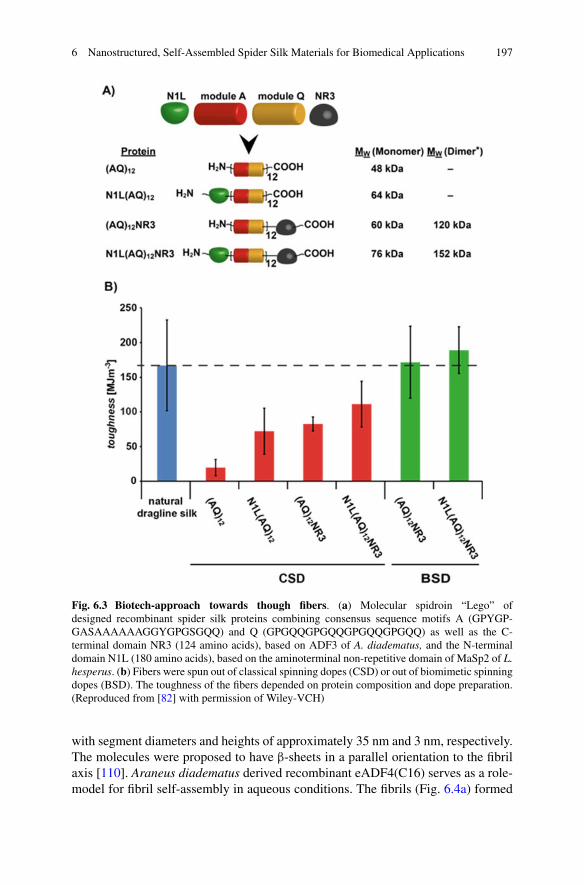
6 Nanostructured, Self-Assembled Spider Silk Materials for Biomedical Applications 197
Fig. 6.3 Biotech-approach towards though fibers. (a) Molecular spidroin “Lego” ofdesigned recombinant spider silk proteins combining consensus sequence motifs A (GPYGP-GASAAAAAAGGYGPGSGQQ) and Q (GPGQQGPGQQGPGQQGPGQQ) as well as the C-terminal domain NR3 (124 amino acids), based on ADF3 of A. diadematus, and the N-terminaldomain N1L (180 amino acids), based on the aminoterminal non-repetitive domain of MaSp2 of L.hesperus. (b) Fibers were spun out of classical spinning dopes (CSD) or out of biomimetic spinningdopes (BSD). The toughness of the fibers depended on protein composition and dope preparation.(Reproduced from [82] with permission of Wiley-VCH)
with segment diameters and heights of approximately 35 nm and 3 nm, respectively.The molecules were proposed to have β-sheets in a parallel orientation to the fibrilaxis [110]. Araneus diadematus derived recombinant eADF4(C16) serves as a role-model for fibril self-assembly in aqueous conditions. The fibrils (Fig. 6.4a) formed
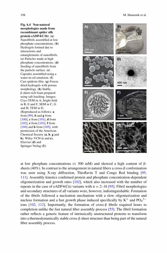
198 M. Humenik et al.
Fig. 6.4 Non-naturalmorphologies made fromrecombinant spider silkprotein eADF4(C16). (a)Nanofibrils assembled at lowphosphate concentrations. (b)Hydrogels formed due tointeractions andentanglements of nanofibrils.(c) Particles made at highphosphate concentrations. (d)Seeding of nanofibrils fromthe particle surface. (e)Capsules assembled using awater-in-oil emulsion. (f)Cast spidroin film. (g) Freezedried hydrogels with porousmorphology. (h) Stable,β-sheet-rich foam preparedusing salt leaching. Images:Cryo-TEM in A; bright fieldin B, E and F; SEM in C, Gand H; TEM in D.(Reproduced as follows: afrom [99], b and g from[100], c from [101], d from[102], e from [103], f from[104] and h from [105], withpermission of the AmericanChemical Society (a, b, g andh), Wiley-VCH (c and e),Elsevier (d) andSpringer-Verlag (f))
at low phosphate concentrations (< 300 mM) and showed a high content of β-sheets (40%). In contrast to the arrangement in natural fibers a cross-β conformationwas seen using X-ray diffraction, Thioflavin T and Congo Red binding [95,111]. Assembly kinetics confirmed protein and phosphate concentration-dependentoligomerization and growth rates [102], which also increased with the number ofrepeats in the case of eADF4(Cn) variants with n = 2–16 [95]. Fibril morphologiesand secondary structures of all variants were, however, indistinguishable. Formationof the fibrils followed a nucleation mechanism with a slow oligomerization andnucleus formation and a fast growth phase induced specifically by K+ and PO4
3−ions [102, 112]. Importantly, the formation of cross-β fibrils required hours tocompletion unlike the fast natural fiber assembly process [55]. The fibril formationrather reflects a generic feature of intrinsically unstructured proteins to transforminto a thermodynamically stable cross-β sheet structure than being part of the naturalfiber assembly process.
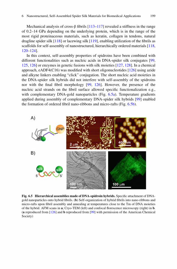
6 Nanostructured, Self-Assembled Spider Silk Materials for Biomedical Applications 199
Mechanical analysis of cross-β fibrils [113–117] revealed a stiffness in the rangeof 0.2–14 GPa depending on the underlying protein, which is in the range of themost rigid proteinaceous materials, such as keratin, collagen in tendons, naturaldragline spider silk [118] or lacewing silk [119], enabling utilization of the fibrils asscaffolds for self-assembly of nanostructured, hierarchically ordered materials [118,120–124].
In this context, self-assembly properties of spidroins have been combined withdifferent functionalities such as nucleic acids in DNA-spider silk conjugates [99,125, 126] or enzymes in genetic fusions with silk moieties [127, 128]. In a chemicalapproach, eADF4(C16) was modified with short oligonucleotides [126] using azideand alkyne linkers enabling “click” conjugation. The short nucleic acid moieties inthe DNA-spider silk hybrids did not interfere with self-assembly of the spidroinsnor with the final fibril morphology [99, 126]. However, the presence of thenucleic acid strands on the fibril surface allowed specific functionalization e.g.,with complementary DNA-gold nanoparticles (Fig. 6.5a). Temperature gradientsapplied during assembly of complementary DNA-spider silk hybrids [99] enabledthe formation of ordered fibril nano-ribbons and micro-rafts (Fig. 6.5b).
Fig. 6.5 Hierarchical assemblies made of DNA-spidroin hybrids. Specific attachment of DNA-gold nanoparticles onto hybrid fibrils. (b) Self-organization of hybrid fibrils into nano-ribbons andmicro-rafts upon fibril assembly and annealing at temperatures close to the Tm of DNA moietiesof the hybrid. AFM scans in a; Cryo-TEM (left) and confocal florescence microscopy (right) in b.(a reproduced from [126] and b reproduced from [99] with permission of the American ChemicalSociety)
200 M. Humenik et al.
6.3.2.2 Hydrogels
Based on nanofibrils (Sect. 6.3.2.1.), spidroins can further assemble into hydrogels(Fig. 6.4b) at protein concentrations above 1% [100, 129–132], representing ahierarchically ordered, supramolecular physical polymer network [133, 134]. Suchhydrogels reveal visco-elastic properties upon shear with maximal shear stressesbetween 200 and 2000 Pa, depending on protein concentration (3–7%) [100]. Goodshape fidelity and comprehensive stress of 140 kPa has been also demonstrated[130, 135]. Spidroin hydrogels typically possess high β-sheet content as well asstructural stability upon exposure to shear forces [100, 130]. Since such spidroinhydrogels showed a shear thinning behavior [131, 135], they are suitable to be usedas injectable or printable materials (see also Sect. 6.5).
6.3.2.3 Particles
High concentrations of phosphate ions (> 400 mM) can promote “salting-out” ofspidroins [136, 137] and induce particle formation (Fig. 6.4c), as demonstratedfor eADF4(C16) [101, 138–140] as well as for different recombinant variants ofNephila clavipes MaSp1 [90, 141, 142] and MaSp2 [143]. The diameters of theparticles can be adjusted by protein and phosphate concentration as well as bymixing intensity [138, 143]. Using ionic liquids as a solvent, nanoparticles of 100–200 nm in diameter can be formed [144]. Similar to cross-β fibrils, all spidroinparticles show a high content of β-sheets [101, 143], which render them chemicallyhighly stable [139].
Spidroin particles substantially swell upon hydration (swelling factor of 2.3)inducing a drastic drop in elastic modulus from GPa in the dry state to MPa inthe wet state [145], depending on the molecular weight of the respective spidroins.Direct force measurements using a colloidal probe technique revealed a polymerbrush-like surface i.e., an ion-permeable particle interface protruding several tenthsof nm into the solution [146, 147]. Interestingly, it has been shown that particlesurfaces can seed fibril growth (Fig. 6.4d) [102].
6.3.2.4 Capsules
The amphiphilic character of spidroins [85] allows fast structural transformationsfrom intrinsically unstructured into β-sheet rich structures at the interface in “water-in-oil” emulsions e.g., in toluene [103, 148] or silicon oil [149]. As a consequence,mechanically stable micrometer-sized capsules (Fig. 6.4e) could be produced inrapid processes and completed within 30 s. The size of these microcapsules (1–30 μm) can be controlled by adjusting the emulsion droplet size through changes inthe emulsion shear rates. The resulting capsule membranes allowed free diffusionof small molecules through the membrane, whereas larger macromolecules wereretained within the capsule (MW cut-off of several thousand Dalton) [148, 149].
6 Nanostructured, Self-Assembled Spider Silk Materials for Biomedical Applications 201
The formation of bowl-shaped (1–3 μm) or giant compound micelles (<50 μm) withporous membranes was observed in case of spidroins in water/2-propanol mixturesdriven by spidroin assembly on entrapped air bubbles i.e., on liquid-air interfaces.The shape, the diameter as well as the β-sheet content increased with the increasingnumbers of poly-Ala blocks (n = 1–6) [87].
6.3.2.5 Films
Films made of recombinant spider silk proteins (Fig. 6.4f) can be processed fromorganic or aqueous solutions driven by drying and template surface effects. Thus,films differ from fibrils, particles or hydrogels which are organized in a “bottom-up”manner.
It has been shown that protein secondary structures of spidroin films cast fromHFIP, hexafluoroacetone or aqueous buffers consist mainly of helices and randomcoil structures [92, 104, 150–154]. Utilization of formic acid as a solvent, however,results in films which are enriched in β-sheet structures [130, 150, 152, 154–156]. The secondary structure content in spidroin films influences their chemicalstability, as amorphous films with low β-sheet content are water-soluble, while highβ-sheet contents support water-stability. Nevertheless, the α-helical and random coilstructures in films processed from HFIP, FA or aqueous solutions can be transformedinto β-sheets upon post-treatment with phosphate buffers [104, 150], or alcohols,such as methanol, ethanol or isopropanol [150, 153, 154, 157], or heat [155, 158,159]. The conformational changes, accompanied by an increased surface roughness[150, 151, 156, 160], mainly occur in the poly-Ala regions as determined by solidstate NMR [154, 155]. X-ray diffraction indicates a wide distribution of β-sheetcrystal sizes from 20 to 400 Å [151]. Interestingly, it is possible to axially align theβ-sheets upon film stretching [154]. β-sheet-rich spidroin films possess enhancedstability against strong protein denaturing agents [150]. Interestingly, non-nativeamino- or carboxy-terminal peptide motifs, such as cell-binding sequences [161,162] or antimicrobial peptides [153], showed no influence on film formation or silkstructure after casting and post-treatment.
Generally, spidroin films show poor mechanical properties including a maximalmechanical stress of 70 MPa and maximal strain typically below 10% [152, 154]depending on the solvents and additives used. Film stretching and concomitantalignment of the crystallites increased the mechanical stress up to 200 MPa andstrain up to 30% [130, 154].
6.3.2.6 Foams and Sponges
Porous foam-like structures were prepared either by foaming of aqueous solutionsof recombinant spidroins [130, 163, 164], freezing/thawing or freeze-drying ofhydrogels [100, 130] or by salt-leaching [105]. Salt-leached foams displayed a highβ-sheet content of 42%, which was induced by the NaCl crystal surface without
202 M. Humenik et al.
an additional post-treatment, simplifying the fabrication process [105]. The foamsshowed interconnected porous morphologies (Fig. 6.4h) with porosities above 90%and pore sizes between 31 and 437 μm, influenced by the size of the salt crystals.The compressive moduli of the salt leached foams were in the range of 0.94–3.24 kPa in a hydrated state, depending on the spidroin concentration employed[105]. Structures obtained by freeze-drying of spidroin hydrogels (lyogels) possesssheet-like morphologies of the pores with diameters ranging from 30 to 200 μm(Fig. 6.4g). Chemically cross-linked hydrogels show highly interconnected sponge-like morphologies after lyophilization, with round pores of 10–20 μm in diameter,independent of the spidroin concentration [100]. Spidroin lyogels have been shownto withstand a stress of up to 300 kPa and strain near 40%, whereas water uptake hasbeen reported at 1600% of the original dry weight [130]. Spidroin sponges producedby hydrogel freezing and thawing show shape-recovery upon several mechanicalloadings in the hydrated state as well as upon rehydration of compressed and driedscaffolds [130].
6.4 Biomedical Applications of Spider Silk
Various material properties, such as biocompatibility, low or no inflammatoryresponse, biodegradability in a defined time frame, and specific structural andmechanical features, are required for biomedical applications.
Native spider silk fibers, providing biocompatibility as well as good mechanicalproperties, were tested already as braided microsurgical sutures [165]. However,drawbacks, as mentioned in the previous section, such as variance in mechanicalperformance of the fibers and low availability of natural spider silk, hamper the largescale application of this material. In contrast, recombinant spider silk, as describedabove, allows scalable production as well as processability into various two andthree dimensional morphologies with adjustable mechanics. These features, alongwith their biocompatibility, qualify recombinant spider silk materials for utilizationin many different biomedical applications [3, 166–168]. Another obvious benefitof the recombinant silk is a possibility to introduce additional peptide motifs withdistinct effects on cell growth, differentiation and migration [140, 161].
Biocompatibility studies of native and recombinant spider silk in vitro or invivo indicated that they are surprisingly well tolerated. Natural spider silk has beenused to test inflammatory response and degradation in split skin wounds [169]. Eggcase silk from spiders was used for subcutaneous implantation in rats to determinethe effect of protease treatment on the degree of inflammation and fibrosis [170].Native spider silk was implanted in vivo to test its biofunctionality showing noadverse effect due to inflammatory response [171, 172]. In the latter, draglinespider silk has been used as an artificial support for nerve regeneration. Thesestudies have shown successful regeneration of damaged tissue and the absence ofadverse reactions, however, a mild inflammatory reaction has been detected. Inanother study, porous scaffolds made of spidroins were well tolerated in mice upon
6 Nanostructured, Self-Assembled Spider Silk Materials for Biomedical Applications 203
subcutaneous implantation. Histological analysis revealed ingrowths of vascularizedconnective tissue and nerve fibers into the scaffold, but also a mild foreign bodyresponse has been reported [173]. Fiber bundles of recombinant spider silk proteinimplanted subcutaneously in rats showed infiltration of newly formed capillaries aswell as ingrowth of fibroblasts [174].
In the following sections, different biomedical applications based on spider silkmaterials will be discussed in more detail.
6.4.1 Drug Delivery and Deposition
The biocompatibility and degradation rate are two important features of biomaterialsin drug delivery applications. The resorption rate of chemically synthesized poly-mers can often be adjusted by controlling the composition of the polymer [175].Silk provides all desirable properties required for sustained drug delivery. Thisrare combination of properties includes aqueous-based processing, biodegradation,biocompatibility, drug stabilization, and robust mechanical properties.
A genetic engineering approach has been used to form block copolymers ofspider silk consensus repeats and poly (L-lysine) domains to form ionic complexesthat are able to deliver plasmid DNA in vitro e.g., into human embryonic kidney(HEK) cells. The DNA complexes were also immobilized on silk films allowingcell transfection. Such silk-based gene carriers could be functionalized with cellmembrane penetrating peptides to enhance the transfection efficiency [176]. Further,the spider silk-poly(L-lysine) variants were functionalized with tumor homingpeptide (THP), and corresponding nanoscale complexes with DNA have beenreported as less cytotoxic and highly target-specific gene carriers [177, 178]. Engi-neered, positively charged spidroin eADF4(κ16) (where all glutamic acid residuesare replaced by lysine residues) showed particle formation indistinguishable tothat of the poly-anionic variant eADF4(C16), described above. Particles made ofeADF4(κ16) were loaded successfully with high molecular weight substances, suchas nucleic acids, with a well-controllable release profile [179]. These examplesdemonstrated the potential of bioengineered silk proteins as a new family ofmolecules to be used for nucleic acid delivery.
Submicron particles or even nanoparticles have been designed as short actingdelivery vehicles and administered through intramuscular, intravenous, subcuta-neous, oral or transdermal routes [180]. Particles with submicron size have uniqueproperties including stability, high surface-to-volume ratio, high carrier-capacity ofbioactive molecules and targeted delivery [181, 182]. Microspheres are commonlyused as drug carriers for long-acting delivery and usually administered intramus-cularly or subcutaneously. Bioengineered spider silks have been mostly exploredin terms of particle formation, characterization and loading/release of model drugs.Recently, submicron particles produced from engineered spider silk eADF4(C16)were used to deliver highly water soluble drugs [139]. However, loading withwater-insoluble drugs is also feasible [183]. Further, crosslinking of spidroins was
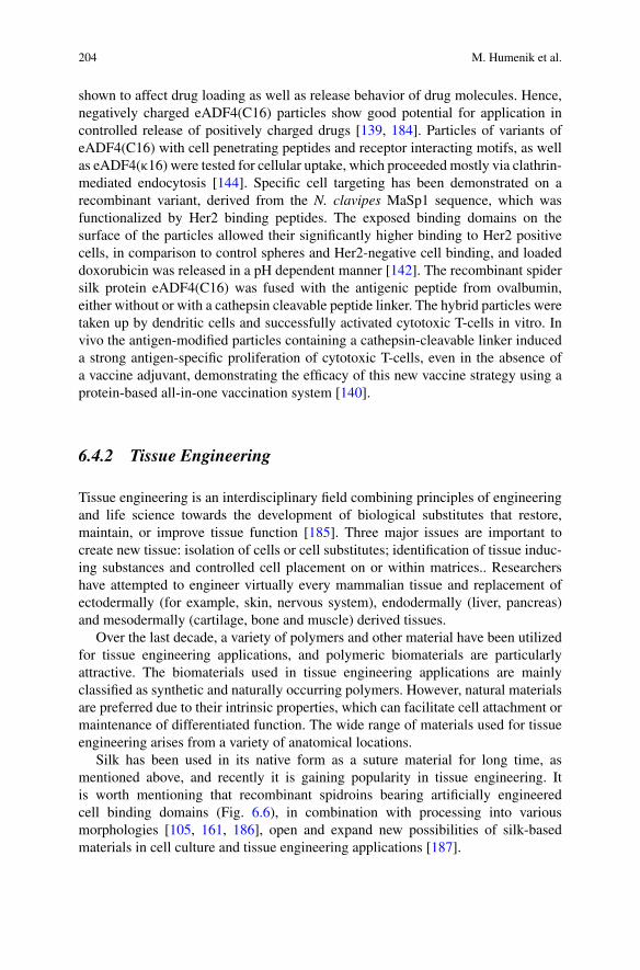
204 M. Humenik et al.
shown to affect drug loading as well as release behavior of drug molecules. Hence,negatively charged eADF4(C16) particles show good potential for application incontrolled release of positively charged drugs [139, 184]. Particles of variants ofeADF4(C16) with cell penetrating peptides and receptor interacting motifs, as wellas eADF4(κ16) were tested for cellular uptake, which proceeded mostly via clathrin-mediated endocytosis [144]. Specific cell targeting has been demonstrated on arecombinant variant, derived from the N. clavipes MaSp1 sequence, which wasfunctionalized by Her2 binding peptides. The exposed binding domains on thesurface of the particles allowed their significantly higher binding to Her2 positivecells, in comparison to control spheres and Her2-negative cell binding, and loadeddoxorubicin was released in a pH dependent manner [142]. The recombinant spidersilk protein eADF4(C16) was fused with the antigenic peptide from ovalbumin,either without or with a cathepsin cleavable peptide linker. The hybrid particles weretaken up by dendritic cells and successfully activated cytotoxic T-cells in vitro. Invivo the antigen-modified particles containing a cathepsin-cleavable linker induceda strong antigen-specific proliferation of cytotoxic T-cells, even in the absence ofa vaccine adjuvant, demonstrating the efficacy of this new vaccine strategy using aprotein-based all-in-one vaccination system [140].
6.4.2 Tissue Engineering
Tissue engineering is an interdisciplinary field combining principles of engineeringand life science towards the development of biological substitutes that restore,maintain, or improve tissue function [185]. Three major issues are important tocreate new tissue: isolation of cells or cell substitutes; identification of tissue induc-ing substances and controlled cell placement on or within matrices.. Researchershave attempted to engineer virtually every mammalian tissue and replacement ofectodermally (for example, skin, nervous system), endodermally (liver, pancreas)and mesodermally (cartilage, bone and muscle) derived tissues.
Over the last decade, a variety of polymers and other material have been utilizedfor tissue engineering applications, and polymeric biomaterials are particularlyattractive. The biomaterials used in tissue engineering applications are mainlyclassified as synthetic and naturally occurring polymers. However, natural materialsare preferred due to their intrinsic properties, which can facilitate cell attachment ormaintenance of differentiated function. The wide range of materials used for tissueengineering arises from a variety of anatomical locations.
Silk has been used in its native form as a suture material for long time, asmentioned above, and recently it is gaining popularity in tissue engineering. Itis worth mentioning that recombinant spidroins bearing artificially engineeredcell binding domains (Fig. 6.6), in combination with processing into variousmorphologies [105, 161, 186], open and expand new possibilities of silk-basedmaterials in cell culture and tissue engineering applications [187].
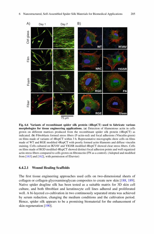
6 Nanostructured, Self-Assembled Spider Silk Materials for Biomedical Applications 205
Fig. 6.6 Variants of recombinant spider silk protein (4RepCT) used to fabricate variousmorphologies for tissue engineering applications. (a) Detection of filamentous actin in cellsgrown on different matrices produced from the recombinant spider silk protein (4RepCT) asindicated. (b) Fibroblasts formed stress fibers (F-actin-red) and focal adhesions (Vinculin-green)on films made of variants of 4RepCT within 3 h. Representative micrographs show cells on filmsmade of WT and RGE modified 4RepCT with poorly formed actin filaments and diffuse vinculinstaining. Cells cultured on IKVAV and YIGSR modified 4RepCT showed clear stress fibers. Cellson films made of RGD modified 4RepCT showed distinct focal adhesion points and well organizedactin stress fibers compared to cells grown on fibronectin (FN as a control). (Adopted and modifiedfrom [163] and [162], with permission of Elsevier)
6.4.2.1 Wound Healing Scaffolds
The first tissue engineering approaches used cells on two-dimensional sheets ofcollagen or collagen-glycosaminoglycan composites to create new skin [188, 189].Native spider dragline silk has been tested as a suitable matrix for 3D skin cellculture, and both fibroblast and keratinocyte cell lines adhered and proliferatedwell. A bi-layered co-cultivation in two continuously separated strata was achievedby serum reduction, changing the medium conditions and the cultivation period.Hence, spider silk appears to be a promising biomaterial for the enhancement ofskin regeneration [190].
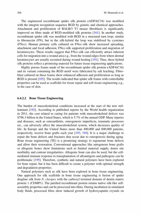
206 M. Humenik et al.
The engineered recombinant spider silk protein eADF4(C16) was modifiedwith the integrin recognition sequence RGD by genetic and chemical approaches.Attachment and proliferation of BALB/3 T3 mouse fibroblasts is significantlyimproved on films made of RGD-modified silk proteins [161]. In another study,recombinant spider silk was modified with RGD in a structural turn loop, similarto fibronectin (FN), but in the silk-hybrid the loop was stabilized by cysteines(FNcc). Human primary cells cultured on FNcc-silk show increased spreading,attachment and focal adhesion. FNcc-silk supported proliferation and migration ofkeratinocytes. These results suggest that FNcc-silk can efficiently attract inherentcells for migration into a wound area e.g., from the wound edges from where dermalkeratinocytes are usually recruited during wound healing [191]. Thus, these hybridsilk proteins reflect a promising material for future tissue engineering applications.
Highly porous foams made of the recombinant spider silk protein eADF4(C16)and its variant containing the RGD motif were fabricated by salt leaching. Fibro-blast cultured on these foams show enhanced adhesion and proliferation as long asRGD is present [105]. The results indicated that spider silk foams with controllableproperties can be used as scaffolds for tissue repair and soft tissue engineering e.g.,in the case of skin.
6.4.2.2 Bone Tissue Engineering
The burden of musculoskeletal conditions increased at the start of the new mil-lennium [192]. According to published reports by the World health organizationin 2011, the cost related to caring for patients with musculoskeletal defects was$796.3 billion in the United States, which is 5.7% of the annual GDP. Many injuriesand diseases, such as osteoarthritis, osteogenesis imperfecta, traumatic processesetc., can adversely affect the musculoskeletal system, which decreases quality oflife. In Europe and the United States more than 400,000 and 600,000 patients,respectively, receive bone grafts each year [193, 194]. It is a major challenge torepair the bone defects and fractures that occur due to osteoporosis during aging.Bone tissue engineering (TE) is a promising strategy to regenerate bone defectsand allow their restoration. Conventional approaches like autogenous bone graftsor allogenic bones show limitations such as limited material supply, donor sitemorbidity and contour irregularities. Allogenic bone can also be used, but the cell-mediated immune response to transplantation of alloantigens and pathogens can beproblematic [195]. Therefore, synthetic and natural polymers have been exploredfor bone repair, but it has been difficult to create a polymer with optimal strengthand degradation properties.
Natural polymers such as silk have been explored in bone tissue engineering.One approach for silk scaffolds in bone tissue engineering is fusion of spiderdragline silk from N. clavipes with the carboxyl terminal domain of dentin matrixprotein 1 (CDMP1). The purified recombinant protein retains native silk-like self-assembly properties and can be processed into films. During incubation in simulatedbody fluids, processed films show induced growth of hydroxyapatite crystals on
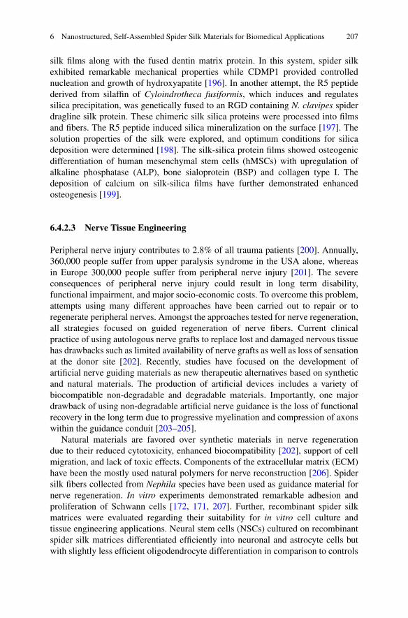
6 Nanostructured, Self-Assembled Spider Silk Materials for Biomedical Applications 207
silk films along with the fused dentin matrix protein. In this system, spider silkexhibited remarkable mechanical properties while CDMP1 provided controllednucleation and growth of hydroxyapatite [196]. In another attempt, the R5 peptidederived from silaffin of Cyloindrotheca fusiformis, which induces and regulatessilica precipitation, was genetically fused to an RGD containing N. clavipes spiderdragline silk protein. These chimeric silk silica proteins were processed into filmsand fibers. The R5 peptide induced silica mineralization on the surface [197]. Thesolution properties of the silk were explored, and optimum conditions for silicadeposition were determined [198]. The silk-silica protein films showed osteogenicdifferentiation of human mesenchymal stem cells (hMSCs) with upregulation ofalkaline phosphatase (ALP), bone sialoprotein (BSP) and collagen type I. Thedeposition of calcium on silk-silica films have further demonstrated enhancedosteogenesis [199].
6.4.2.3 Nerve Tissue Engineering
Peripheral nerve injury contributes to 2.8% of all trauma patients [200]. Annually,360,000 people suffer from upper paralysis syndrome in the USA alone, whereasin Europe 300,000 people suffer from peripheral nerve injury [201]. The severeconsequences of peripheral nerve injury could result in long term disability,functional impairment, and major socio-economic costs. To overcome this problem,attempts using many different approaches have been carried out to repair or toregenerate peripheral nerves. Amongst the approaches tested for nerve regeneration,all strategies focused on guided regeneration of nerve fibers. Current clinicalpractice of using autologous nerve grafts to replace lost and damaged nervous tissuehas drawbacks such as limited availability of nerve grafts as well as loss of sensationat the donor site [202]. Recently, studies have focused on the development ofartificial nerve guiding materials as new therapeutic alternatives based on syntheticand natural materials. The production of artificial devices includes a variety ofbiocompatible non-degradable and degradable materials. Importantly, one majordrawback of using non-degradable artificial nerve guidance is the loss of functionalrecovery in the long term due to progressive myelination and compression of axonswithin the guidance conduit [203–205].
Natural materials are favored over synthetic materials in nerve regenerationdue to their reduced cytotoxicity, enhanced biocompatibility [202], support of cellmigration, and lack of toxic effects. Components of the extracellular matrix (ECM)have been the mostly used natural polymers for nerve reconstruction [206]. Spidersilk fibers collected from Nephila species have been used as guidance material fornerve regeneration. In vitro experiments demonstrated remarkable adhesion andproliferation of Schwann cells [172, 171, 207]. Further, recombinant spider silkmatrices were evaluated regarding their suitability for in vitro cell culture andtissue engineering applications. Neural stem cells (NSCs) cultured on recombinantspider silk matrices differentiated efficiently into neuronal and astrocyte cells butwith slightly less efficient oligodendrocyte differentiation in comparison to controls
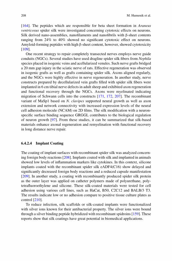
208 M. Humenik et al.
[164]. The peptides which are responsible for beta sheet formation in Araneusventricosus spider silk were investigated concerning cytotoxic effects on neurons.Silk derived nano-assemblies, nanofilaments and nanofibrils with β-sheet contentsranging from 24% to 40% showed no significant cytotoxic effect on neurons.Amyloid-forming peptides with high β-sheet content, however, showed cytotoxicity[109].
One recent strategy to repair completely transected nerves employs nerve guideconduits (NGCs). Several studies have used dragline spider silk fibers from Nephilaspecies placed in isogenic veins and acellularized venules. Such nerve grafts bridgeda 20 mm gap injury in the sciatic nerve of rats. Effective regeneration was observedin isogenic grafts as well as grafts containing spider silk. Axons aligned regularly,and the NGCs were highly effective in nerve regeneration. In another study, nerveconstructs prepared by decellularized vein grafts filled with spider silk fibers wereimplanted in 6 cm tibial nerve defects in adult sheep and exhibited axon regenerationand functional recovery through the NGCs. Axons were myelinated indicatingmigration of Schwann cells into the constructs [171, 172, 207]. The recombinantvariant of MaSp1 based on N. clavipes supported neural growth as well as axonextension and network connectivity with increased expression levels of the neuralcell adhesion molecule (NCAM) on 2D films. The silk modification with a neuron-specific surface binding sequence GRGGL contributes to the biological regulationof neuron growth [97]. From these studies, it can be summarized that silk-basedmaterials enhance axonal regeneration and remyelination with functional recoveryin long distance nerve repair.
6.4.2.4 Implant Coating
The coating of implant surfaces with recombinant spider silk was analyzed concern-ing foreign body reactions [208]. Implants coated with silk and implanted in animalsshowed low levels of inflammation markers like cytokines. In this context, siliconeimplants coated with the recombinant spider silk eADF4(C16) show delayed andsignificantly decreased foreign body reactions and a reduced capsule manifestation[209]. In another study, a coating with recombinantly produced spider silk proteinas the outer layer was applied on catheter polymers made of polyurethane, poly-tetrafluoroethylene and silicone. These silk-coated materials were tested for celladhesion using various cell lines, such as HaCat, B50, C2C12 and BALB/3 T3.The results indicate low or no adhesion compare to positive tissue culture plates ascontrol [210].
To reduce infection, silk scaffolds or silk-coated implants were functionalizedwith silver ions known for their antibacterial property. The silver ions were boundthrough a silver binding peptide hybridized with recombinant spidroins [159]. Thesereports show that silk coatings have great potential in biomedical applications.
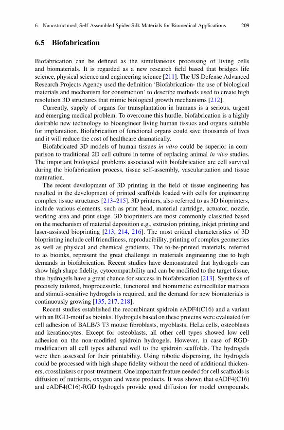
6 Nanostructured, Self-Assembled Spider Silk Materials for Biomedical Applications 209
6.5 Biofabrication
Biofabrication can be defined as the simultaneous processing of living cellsand biomaterials. It is regarded as a new research field based that bridges lifescience, physical science and engineering science [211]. The US Defense AdvancedResearch Projects Agency used the definition ‘Biofabrication- the use of biologicalmaterials and mechanism for construction’ to describe methods used to create highresolution 3D structures that mimic biological growth mechanisms [212].
Currently, supply of organs for transplantation in humans is a serious, urgentand emerging medical problem. To overcome this hurdle, biofabrication is a highlydesirable new technology to bioengineer living human tissues and organs suitablefor implantation. Biofabrication of functional organs could save thousands of livesand it will reduce the cost of healthcare dramatically.
Biofabricated 3D models of human tissues in vitro could be superior in com-parison to traditional 2D cell culture in terms of replacing animal in vivo studies.The important biological problems associated with biofabrication are cell survivalduring the biofabrication process, tissue self-assembly, vascularization and tissuematuration.
The recent development of 3D printing in the field of tissue engineering hasresulted in the development of printed scaffolds loaded with cells for engineeringcomplex tissue structures [213–215]. 3D printers, also referred to as 3D bioprinters,include various elements, such as print head, material cartridge, actuator, nozzle,working area and print stage. 3D bioprinters are most commonly classified basedon the mechanism of material deposition e.g., extrusion printing, inkjet printing andlaser-assisted bioprinting [213, 214, 216]. The most critical characteristics of 3Dbioprinting include cell friendliness, reproducibility, printing of complex geometriesas well as physical and chemical gradients. The to-be-printed materials, referredto as bioinks, represent the great challenge in materials engineering due to highdemands in biofabrication. Recent studies have demonstrated that hydrogels canshow high shape fidelity, cytocompatibility and can be modified to the target tissue,thus hydrogels have a great chance for success in biofabrication [213]. Synthesis ofprecisely tailored, bioprocessible, functional and biomimetic extracellular matricesand stimuli-sensitive hydrogels is required, and the demand for new biomaterials iscontinuously growing [135, 217, 218].
Recent studies established the recombinant spidroin eADF4(C16) and a variantwith an RGD-motif as bioinks. Hydrogels based on these proteins were evaluated forcell adhesion of BALB/3 T3 mouse fibroblasts, myoblasts, HeLa cells, osteoblastsand keratinocytes. Except for osteoblasts, all other cell types showed low celladhesion on the non-modified spidroin hydrogels. However, in case of RGD-modification all cell types adhered well to the spidroin scaffolds. The hydrogelswere then assessed for their printability. Using robotic dispensing, the hydrogelscould be processed with high shape fidelity without the need of additional thicken-ers, crosslinkers or post-treatment. One important feature needed for cell scaffolds isdiffusion of nutrients, oxygen and waste products. It was shown that eADF4(C16)and eADF4(C16)-RGD hydrogels provide good diffusion for model compounds.
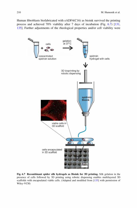
210 M. Humenik et al.
Human fibroblasts biofabricated with eADF4(C16) as bioink survived the printingprocess and achieved 70% viability after 7 days of incubation (Fig. 6.7) [131,135]. Further adjustments of the rheological properties and/or cell viability were
Fig. 6.7 Recombinant spider silk hydrogels as Bioink for 3D printing. Silk gelation in thepresence of cells followed by 3D printing using robotic dispensing enables multilayered 3Dscaffolds with encapsulated viable cells. (Adapted and modified from [135] with permission ofWiley-VCH)
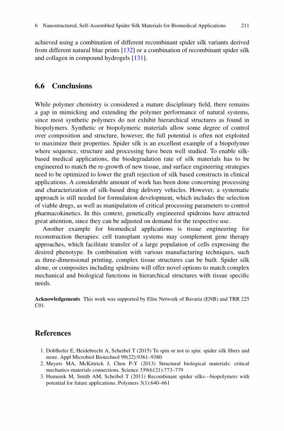
6 Nanostructured, Self-Assembled Spider Silk Materials for Biomedical Applications 211
achieved using a combination of different recombinant spider silk variants derivedfrom different natural blue prints [132] or a combination of recombinant spider silkand collagen in compound hydrogels [131].
6.6 Conclusions
While polymer chemistry is considered a mature disciplinary field, there remainsa gap in mimicking and extending the polymer performance of natural systems,since most synthetic polymers do not exhibit hierarchical structures as found inbiopolymers. Synthetic or biopolymeric materials allow some degree of controlover composition and structure, however, the full potential is often not exploitedto maximize their properties. Spider silk is an excellent example of a biopolymerwhere sequence, structure and processing have been well studied. To enable silk-based medical applications, the biodegradation rate of silk materials has to beengineered to match the re-growth of new tissue, and surface engineering strategiesneed to be optimized to lower the graft rejection of silk based constructs in clinicalapplications. A considerable amount of work has been done concerning processingand characterization of silk-based drug delivery vehicles. However, a systematicapproach is still needed for formulation development, which includes the selectionof viable drugs, as well as manipulation of critical processing parameters to controlpharmacokinetics. In this context, genetically engineered spidroins have attractedgreat attention, since they can be adjusted on demand for the respective use.
Another example for biomedical applications is tissue engineering forreconstruction therapies: cell transplant systems may complement gene therapyapproaches, which facilitate transfer of a large population of cells expressing thedesired phenotype. In combination with various manufacturing techniques, suchas three-dimensional printing, complex tissue structures can be built. Spider silkalone, or composites including spidroins will offer novel options to match complexmechanical and biological functions in hierarchical structures with tissue specificneeds.
Acknowledgements This work was supported by Elite Network of Bavaria (ENB) and TRR 225C01.
References
1. Doblhofer E, Heidebrecht A, Scheibel T (2015) To spin or not to spin: spider silk fibers andmore. Appl Microbiol Biotechnol 99(22):9361–9380
2. Meyers MA, McKittrick J, Chen P-Y (2013) Structural biological materials: criticalmechanics-materials connections. Science 339(6121):773–779
3. Humenik M, Smith AM, Scheibel T (2011) Recombinant spider silks—biopolymers withpotential for future applications. Polymers 3(1):640–661
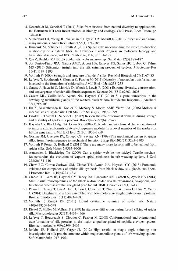
212 M. Humenik et al.
4. Neuenfeldt M, Scheibel T (2014) Silks from insects: from natural diversity to applications.In: Hoffmann KH (ed) Insect molecular biology and ecology. CRC Press, Boca Raton, pp376–400
5. Sutherland TD, Young JH, Weisman S, Hayashi CY, Merritt DJ (2010) Insect silk: one name,many materials. Annu Rev Entomol 55(1):171–188
6. Humenik M, Scheibel T, Smith A (2011) Spider silk: understanding the structure–functionrelationship of a natural fiber. In: Howorka S (ed) Progress in molecular biology andtranslational science, vol 103. Cambridge, MA, pp 131–185
7. Qin Z, Buehler MJ (2013) Spider silk: webs measure up. Nat Mater 12(3):185–1878. dos Santos-Pinto JRA, Garcia AMC, Arcuri HA, Esteves FG, Salles HC, Lubec G, Palma
MS (2016) Silkomics: insight into the silk spinning process of spiders. J Proteome Res15(4):1179–1193
9. Vollrath F (2000) Strength and structure of spiders’ silks. Rev Mol Biotechnol 74(2):67–8310. Lefevre T, Boudreault S, Cloutier C, Pezolet M (2011) Diversity of molecular transformations
involved in the formation of spider silks. J Mol Biol 405(1):238–25311. Gatesy J, Hayashi C, Motriuk D, Woods J, Lewis R (2001) Extreme diversity, conservation,
and convergence of spider silk fibroin sequences. Science 291(5513):2603–260512. Casem ML, Collin MA, Ayoub NA, Hayashi CY (2010) Silk gene transcripts in the
developing tubuliform glands of the western black widow, latrodectus hesperus. J Arachnol38(1):99–103
13. Hu X, Vasanthavada K, Kohler K, McNary S, Moore AMF, Vierra CA (2006) Molecularmechanisms of spider silk. Cell Mol Life Sci 63(17):1986–1999
14. Eisoldt L, Thamm C, Scheibel T (2012) Review the role of terminal domains during storageand assembly of spider silk proteins. Biopolymers 97(6):355–361
15. Hayashi CY, Blackledge TA, Lewis RV (2004) Molecular and mechanical characterization ofaciniform silk: uniformity of iterated sequence modules in a novel member of the spider silkfibroin gene family. Mol Biol Evol 21(10):1950–1959
16. Gosline JM, Guerette PA, Ortlepp CS, Savage KN (1999) The mechanical design of spidersilks: from fibroin sequence to mechanical function. J Exp Biol 202(23):3295–3303
17. Vollrath F, Porter D, Holland C (2011) There are many more lessons still to be learned fromspider silks. Soft Matter 7:9595–9600
18. Agnarsson I, Blackledge TA (2009) Can a spider web be too sticky? Tensile mechan-ics constrains the evolution of capture spiral stickiness in orb-weaving spiders. J Zool278(2):134–140
19. Chaw RC, Correa-Garhwal SM, Clarke TH, Ayoub NA, Hayashi CY (2015) Proteomicevidence for components of spider silk synthesis from black widow silk glands and fibers.J Proteome Res 14(10):4223–4231
20. Clarke TH, Garb JE, Hayashi CY, Haney RA, Lancaster AK, Corbett S, Ayoub NA (2014)Multi-tissue transcriptomics of the black widow spider reveals expansions, co-options, andfunctional processes of the silk gland gene toolkit. BMC Genomics 15(1):1–17
21. Pham T, Chuang T, Lin A, Joo H, Tsai J, Crawford T, Zhao L, Williams C, Hsia Y, VierraC (2014) Dragline silk: a fiber assembled with low-molecular-weight cysteine-rich proteins.Biomacromolecules 15(11):4073–4081
22. Vollrath F, Knight DP (2001) Liquid crystalline spinning of spider silk. Nature410(6828):541–548
23. Riekel C, Müller M, Vollrath F (1999) In situ x-ray diffraction during forced silking of spidersilk. Macromolecules 32(13):4464–4466
24. Lefevre T, Boudreault S, Cloutier C, Pezolet M (2008) Conformational and orientationaltransformation of silk proteins in the major ampullate gland of nephila clavipes spiders.Biomacromolecules 9(9):2399–2407
25. Jenkins JE, Holland GP, Yarger JL (2012) High resolution magic angle spinning nmrinvestigation of silk protein structure within major ampullate glands of orb weaving spiders.Soft Matter 8(6):1947–1954
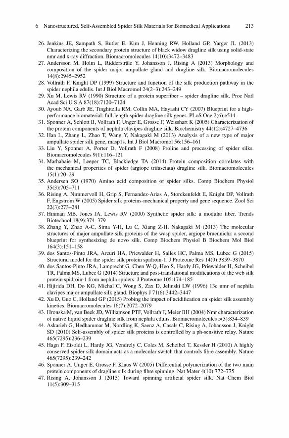
6 Nanostructured, Self-Assembled Spider Silk Materials for Biomedical Applications 213
26. Jenkins JE, Sampath S, Butler E, Kim J, Henning RW, Holland GP, Yarger JL (2013)Characterizing the secondary protein structure of black widow dragline silk using solid-statenmr and x-ray diffraction. Biomacromolecules 14(10):3472–3483
27. Andersson M, Holm L, Ridderstråle Y, Johansson J, Rising A (2013) Morphology andcomposition of the spider major ampullate gland and dragline silk. Biomacromolecules14(8):2945–2952
28. Vollrath F, Knight DP (1999) Structure and function of the silk production pathway in thespider nephila edulis. Int J Biol Macromol 24(2–3):243–249
29. Xu M, Lewis RV (1990) Structure of a protein superfiber – spider dragline silk. Proc NatlAcad Sci U S A 87(18):7120–7124
30. Ayoub NA, Garb JE, Tinghitella RM, Collin MA, Hayashi CY (2007) Blueprint for a high-performance biomaterial: full-length spider dragline silk genes. PLoS One 2(6):e514
31. Sponner A, Schlott B, Vollrath F, Unger E, Grosse F, Weisshart K (2005) Characterization ofthe protein components of nephila clavipes dragline silk. Biochemistry 44(12):4727–4736
32. Han L, Zhang L, Zhao T, Wang Y, Nakagaki M (2013) Analysis of a new type of majorampullate spider silk gene, masp1s. Int J Biol Macromol 56:156–161
33. Liu Y, Sponner A, Porter D, Vollrath F (2008) Proline and processing of spider silks.Biomacromolecules 9(1):116–121
34. Marhabaie M, Leeper TC, Blackledge TA (2014) Protein composition correlates withthe mechanical properties of spider (argiope trifasciata) dragline silk. Biomacromolecules15(1):20–29
35. Andersen SO (1970) Amino acid composition of spider silks. Comp Biochem Physiol35(3):705–711
36. Rising A, Nimmervoll H, Grip S, Fernandez-Arias A, Storckenfeldt E, Knight DP, VollrathF, Engstrom W (2005) Spider silk proteins-mechanical property and gene sequence. Zool Sci22(3):273–281
37. Hinman MB, Jones JA, Lewis RV (2000) Synthetic spider silk: a modular fiber. TrendsBiotechnol 18(9):374–379
38. Zhang Y, Zhao A-C, Sima Y-H, Lu C, Xiang Z-H, Nakagaki M (2013) The molecularstructures of major ampullate silk proteins of the wasp spider, argiope bruennichi: a secondblueprint for synthesizing de novo silk. Comp Biochem Physiol B Biochem Mol Biol164(3):151–158
39. dos Santos-Pinto JRA, Arcuri HA, Priewalder H, Salles HC, Palma MS, Lubec G (2015)Structural model for the spider silk protein spidroin-1. J Proteome Res 14(9):3859–3870
40. dos Santos-Pinto JRA, Lamprecht G, Chen W-Q, Heo S, Hardy JG, Priewalder H, ScheibelTR, Palma MS, Lubec G (2014) Structure and post-translational modifications of the web silkprotein spidroin-1 from nephila spiders. J Proteome 105:174–185
41. Hijirida DH, Do KG, Michal C, Wong S, Zax D, Jelinski LW (1996) 13c nmr of nephilaclavipes major ampullate silk gland. Biophys J 71(6):3442–3447
42. Xu D, Guo C, Holland GP (2015) Probing the impact of acidification on spider silk assemblykinetics. Biomacromolecules 16(7):2072–2079
43. Hronska M, van Beek JD, Williamson PTF, Vollrath F, Meier BH (2004) Nmr characterizationof native liquid spider dragline silk from nephila edulis. Biomacromolecules 5(3):834–839
44. Askarieh G, Hedhammar M, Nordling K, Saenz A, Casals C, Rising A, Johansson J, KnightSD (2010) Self-assembly of spider silk proteins is controlled by a ph-sensitive relay. Nature465(7295):236–239
45. Hagn F, Eisoldt L, Hardy JG, Vendrely C, Coles M, Scheibel T, Kessler H (2010) A highlyconserved spider silk domain acts as a molecular switch that controls fibre assembly. Nature465(7295):239–242
46. Sponner A, Unger E, Grosse F, Klaus W (2005) Differential polymerization of the two mainprotein components of dragline silk during fibre spinning. Nat Mater 4(10):772–775
47. Rising A, Johansson J (2015) Toward spinning artificial spider silk. Nat Chem Biol11(5):309–315
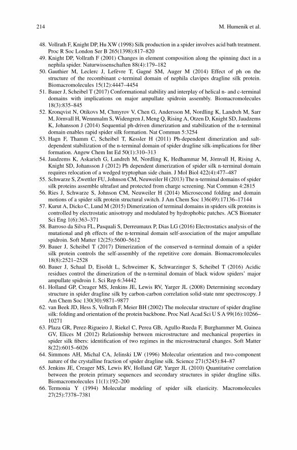
214 M. Humenik et al.
48. Vollrath F, Knight DP, Hu XW (1998) Silk production in a spider involves acid bath treatment.Proc R Soc London Ser B 265(1398):817–820
49. Knight DP, Vollrath F (2001) Changes in element composition along the spinning duct in anephila spider. Naturwissenschaften 88(4):179–182
50. Gauthier M, Leclerc J, Lefèvre T, Gagné SM, Auger M (2014) Effect of ph on thestructure of the recombinant c-terminal domain of nephila clavipes dragline silk protein.Biomacromolecules 15(12):4447–4454
51. Bauer J, Scheibel T (2017) Conformational stability and interplay of helical n- and c-terminaldomains with implications on major ampullate spidroin assembly. Biomacromolecules18(3):835–845
52. Kronqvist N, Otikovs M, Chmyrov V, Chen G, Andersson M, Nordling K, Landreh M, SarrM, Jörnvall H, Wennmalm S, Widengren J, Meng Q, Rising A, Otzen D, Knight SD, JaudzemsK, Johansson J (2014) Sequential ph-driven dimerization and stabilization of the n-terminaldomain enables rapid spider silk formation. Nat Commun 5:3254
53. Hagn F, Thamm C, Scheibel T, Kessler H (2011) Ph-dependent dimerization and salt-dependent stabilization of the n-terminal domain of spider dragline silk-implications for fiberformation. Angew Chem Int Ed 50(1):310–313
54. Jaudzems K, Askarieh G, Landreh M, Nordling K, Hedhammar M, Jörnvall H, Rising A,Knight SD, Johansson J (2012) Ph dependent dimerization of spider silk n-terminal domainrequires relocation of a wedged tryptophan side chain. J Mol Biol 422(4):477–487
55. Schwarze S, Zwettler FU, Johnson CM, Neuweiler H (2013) The n-terminal domains of spidersilk proteins assemble ultrafast and protected from charge screening. Nat Commun 4:2815
56. Ries J, Schwarze S, Johnson CM, Neuweiler H (2014) Microsecond folding and domainmotions of a spider silk protein structural switch. J Am Chem Soc 136(49):17136–17144
57. Kurut A, Dicko C, Lund M (2015) Dimerization of terminal domains in spiders silk proteins iscontrolled by electrostatic anisotropy and modulated by hydrophobic patches. ACS BiomaterSci Eng 1(6):363–371
58. Barroso da Silva FL, Pasquali S, Derreumaux P, Dias LG (2016) Electrostatics analysis of themutational and ph effects of the n-terminal domain self-association of the major ampullatespidroin. Soft Matter 12(25):5600–5612
59. Bauer J, Scheibel T (2017) Dimerization of the conserved n-terminal domain of a spidersilk protein controls the self-assembly of the repetitive core domain. Biomacromolecules18(8):2521–2528
60. Bauer J, Schaal D, Eisoldt L, Schweimer K, Schwarzinger S, Scheibel T (2016) Acidicresidues control the dimerization of the n-terminal domain of black widow spiders’ majorampullate spidroin 1. Sci Rep 6:34442
61. Holland GP, Creager MS, Jenkins JE, Lewis RV, Yarger JL (2008) Determining secondarystructure in spider dragline silk by carbon-carbon correlation solid-state nmr spectroscopy. JAm Chem Soc 130(30):9871–9877
62. van Beek JD, Hess S, Vollrath F, Meier BH (2002) The molecular structure of spider draglinesilk: folding and orientation of the protein backbone. Proc Natl Acad Sci U S A 99(16):10266–10271
63. Plaza GR, Perez-Rigueiro J, Riekel C, Perea GB, Agullo-Rueda F, Burghammer M, GuineaGV, Elices M (2012) Relationship between microstructure and mechanical properties inspider silk fibers: identification of two regimes in the microstructural changes. Soft Matter8(22):6015–6026
64. Simmons AH, Michal CA, Jelinski LW (1996) Molecular orientation and two-componentnature of the crystalline fraction of spider dragline silk. Science 271(5245):84–87
65. Jenkins JE, Creager MS, Lewis RV, Holland GP, Yarger JL (2010) Quantitative correlationbetween the protein primary sequences and secondary structures in spider dragline silks.Biomacromolecules 11(1):192–200
66. Termonia Y (1994) Molecular modeling of spider silk elasticity. Macromolecules27(25):7378–7381
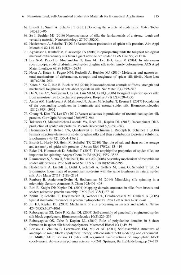
6 Nanostructured, Self-Assembled Spider Silk Materials for Biomedical Applications 215
67. Eisoldt L, Smith A, Scheibel T (2011) Decoding the secrets of spider silk. Mater Today14(3):80–86
68. Su I, Buehler MJ (2016) Nanomechanics of silk: the fundamentals of a strong, tough andversatile material. Nanotechnology 27(30):302001
69. Heidebrecht A, Scheibel T (2013) Recombinant production of spider silk proteins. Adv ApplMicrobiol 82:115–153
70. Agnarsson I, Kuntner M, Blackledge TA (2010) Bioprospecting finds the toughest biologicalmaterial: extraordinary silk from a giant riverine orb spider. PLoS One 5(9):e11234
71. Lee S-M, Pippel E, Moutanabbir O, Kim J-H, Lee H-J, Knez M (2014) In situ ramanspectroscopic study of al-infiltrated spider dragline silk under tensile deformation. ACS ApplMater Interfaces 6(19):16827–16834
72. Nova A, Keten S, Pugno NM, Redaelli A, Buehler MJ (2010) Molecular and nanostruc-tural mechanisms of deformation, strength and toughness of spider silk fibrils. Nano Lett10(7):2626–2634
73. Keten S, Xu Z, Ihle B, Buehler MJ (2010) Nanoconfinement controls stiffness, strength andmechanical toughness of beta-sheet crystals in silk. Nat Mater 9(4):359–367
74. Du N, Liu XY, Narayanan J, Li LA, Lim MLM, Li DQ (2006) Design of superior spider silk:from nanostructure to mechanical properties. Biophys J 91(12):4528–4535
75. Anton AM, Heidebrecht A, Mahmood N, Beiner M, Scheibel T, Kremer F (2017) Foundationof the outstanding toughness in biomimetic and natural spider silk. Biomacromolecules18(12):3954–3962
76. Chung H, Kim TY, Lee SY (2012) Recent advances in production of recombinant spider silkproteins. Curr Opin Biotechnol 23(6):957–964
77. Tokareva O, Michalczechen-Lacerda VA, Rech EL, Kaplan DL (2013) Recombinant DNAproduction of spider silk proteins. Microb Biotechnol 6(6):651–663
78. Huemmerich D, Helsen CW, Quedzuweit S, Oschmann J, Rudolph R, Scheibel T (2004)Primary structure elements of spider dragline silks and their contribution to protein solubility.Biochemistry 43(42):13604–13612
79. Eisoldt L, Hardy JG, Heim M, Scheibel TR (2010) The role of salt and shear on the storageand assembly of spider silk proteins. J Struct Biol 170(2):413–419
80. Exler JH, Hummerich D, Scheibel T (2007) The amphiphilic properties of spider silks areimportant for spinning. Angew Chem Int Ed 46(19):3559–3562
81. Rammensee S, Slotta U, Scheibel T, Bausch AR (2008) Assembly mechanism of recombinantspider silk proteins. Proc Natl Acad Sci U S A 105(18):6590–6595
82. Heidebrecht A, Eisoldt L, Diehl J, Schmidt A, Geffers M, Lang G, Scheibel T (2015)Biomimetic fibers made of recombinant spidroins with the same toughness as natural spidersilk. Adv Mater 27(13):2189–2194
83. Renberg B, Andersson-Svahn H, Hedhammar M (2014) Mimicking silk spinning in amicrochip. Sensors Actuators B Chem 195:404–408
84. Bini E, Knight DP, Kaplan DL (2004) Mapping domain structures in silks from insects andspiders related to protein assembly. J Mol Biol 335(1):27–40
85. Zbilut JP, Scheibel T, Huemmerich D, Webber CL, Colafranceschi M, Giuliani A (2005)Spatial stochastic resonance in protein hydrophobicity. Phys Lett A 346(1–3):33–41
86. Jin HJ, Kaplan DL (2003) Mechanism of silk processing in insects and spiders. Nature424(6952):1057–1061
87. Rabotyagova OS, Cebe P, Kaplan DL (2009) Self-assembly of genetically engineered spidersilk block copolymers. Biomacromolecules 10(2):229–236
88. Rabotyagova OS, Cebe P, Kaplan DL (2010) Role of polyalanine domains in β-sheetformation in spider silk block copolymers. Macromol Biosci 10(1):49–59
89. Borisov O, Zhulina E, Leermakers FM, Müller AE (2011) Self-assembled structures ofamphiphilic ionic block copolymers: theory, self-consistent field modeling and experiment.In: Müller AHE, Borisov O (eds) Self organized nanostructures of amphiphilic blockcopolymers i, Advances in polymer science, vol 241. Springer, Berlin/Heidelberg, pp 57–129
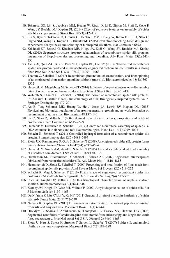
216 M. Humenik et al.
90. Tokareva OS, Lin S, Jacobsen MM, Huang W, Rizzo D, Li D, Simon M, Staii C, Cebe P,Wong JY, Buehler MJ, Kaplan DL (2014) Effect of sequence features on assembly of spidersilk block copolymers. J Struct Biol 186(3):412–419
91. Lin S, Ryu S, Tokareva O, Gronau G, Jacobsen MM, Huang W, Rizzo DJ, Li D, Staii C,Pugno NM, Wong JY, Kaplan DL, Buehler MJ (2015) Predictive modelling-based design andexperiments for synthesis and spinning of bioinspired silk fibres. Nat Commun 6:6892
92. Krishnaji ST, Bratzel G, Kinahan ME, Kluge JA, Staii C, Wong JY, Buehler MJ, KaplanDL (2013) Sequence–structure–property relationships of recombinant spider silk proteins:integration of biopolymer design, processing, and modeling. Adv Funct Mater 23(2):241–253
93. Xia X-X, Qian Z-G, Ki CS, Park YH, Kaplan DL, Lee SY (2010) Native-sized recombinantspider silk protein produced in metabolically engineered escherichia coli results in a strongfiber. Proc Natl Acad Sci U S A 107(32):14059–14063
94. Thamm C, Scheibel T (2017) Recombinant production, characterization, and fiber spinningof an engineered short major ampullate spidroin (masp1s). Biomacromolecules 18(4):1365–1372
95. Humenik M, Magdeburg M, Scheibel T (2014) Influence of repeat numbers on self-assemblyrates of repetitive recombinant spider silk proteins. J Struct Biol 186:431–437
96. Wohlrab S, Thamm C, Scheibel T (2014) The power of recombinant spider silk proteins.In: Asakura T, Miller T (eds) Biotechnology of silk, Biologically-inspired systems, vol 5.Springer, Dordrecht, pp 179–201
97. An B, Tang-Schomer MD, Huang W, He J, Jones JA, Lewis RV, Kaplan DL (2015)Physical and biological regulation of neuron regenerative growth and network formation onrecombinant dragline silks. Biomaterials 48:137–146
98. Fu C, Shao Z, Vollrath F (2009) Animal silks: their structures, properties and artificialproduction. Chem Commun 43:6515–6529
99. Humenik M, Drechsler M, Scheibel T (2014) Controlled hierarchical assembly of spider silk-DNA chimeras into ribbons and raft-like morphologies. Nano Lett 14(7):3999–4004
100. Schacht K, Scheibel T (2011) Controlled hydrogel formation of a recombinant spider silkprotein. Biomacromolecules 12(7):2488–2495
101. Slotta UK, Rammensee S, Gorb S, Scheibel T (2008) An engineered spider silk protein formsmicrospheres. Angew Chem Int Ed 47(24):4592–4594
102. Humenik M, Smith AM, Arndt S, Scheibel T (2015) Ion and seed dependent fibril assemblyof a spidroin core domain. J Struct Biol 191(2):130–138
103. Hermanson KD, Huemmerich D, Scheibel T, Bausch AR (2007) Engineered microcapsulesfabricated from reconstituted spider silk. Adv Mater 19(14):1810–1815
104. Huemmerich D, Slotta U, Scheibel T (2006) Processing and modification of films made fromrecombinant spider silk proteins. Appl Phys A Mater Sci Process 82(2):219–222
105. Schacht K, Vogt J, Scheibel T (2016) Foams made of engineered recombinant spider silkproteins as 3d scaffolds for cell growth. ACS Biomater Sci Eng 2(4):517–525
106. Chen X, Knight DP, Vollrath F (2002) Rheological characterization of nephila spidroinsolution. Biomacromolecules 3(4):644–648
107. Kenney JM, Knight D, Wise MJ, Vollrath F (2002) Amyloidogenic nature of spider silk. EurJ Biochem 269(16):4159–4163
108. Du N, Yang Z, Liu XY, Li Y, Xu HY (2011) Structural origin of the strain-hardening of spidersilk. Adv Funct Mater 21(4):772–778
109. Numata K, Kaplan DL (2011) Differences in cytotoxicity of beta-sheet peptides originatedfrom silk and amyloid beta. Macromol Biosci 11(1):60–64
110. Oroudjev E, Soares J, Arcidiacono S, Thompson JB, Fossey SA, Hansma HG (2002)Segmented nanofibers of spider dragline silk: atomic force microscopy and single-moleculeforce spectroscopy. Proc Natl Acad Sci U S A 99(suppl 2):6460–6465
111. Slotta U, Hess S, Spiess K, Stromer T, Serpell L, Scheibel T (2007) Spider silk and amyloidfibrils: a structural comparison. Macromol Biosci 7(2):183–188
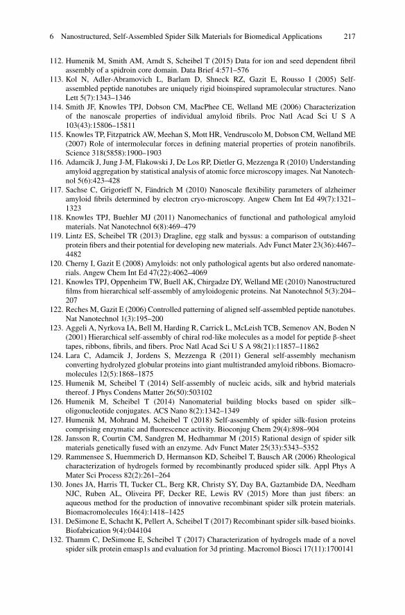
6 Nanostructured, Self-Assembled Spider Silk Materials for Biomedical Applications 217
112. Humenik M, Smith AM, Arndt S, Scheibel T (2015) Data for ion and seed dependent fibrilassembly of a spidroin core domain. Data Brief 4:571–576
113. Kol N, Adler-Abramovich L, Barlam D, Shneck RZ, Gazit E, Rousso I (2005) Self-assembled peptide nanotubes are uniquely rigid bioinspired supramolecular structures. NanoLett 5(7):1343–1346
114. Smith JF, Knowles TPJ, Dobson CM, MacPhee CE, Welland ME (2006) Characterizationof the nanoscale properties of individual amyloid fibrils. Proc Natl Acad Sci U S A103(43):15806–15811
115. Knowles TP, Fitzpatrick AW, Meehan S, Mott HR, Vendruscolo M, Dobson CM, Welland ME(2007) Role of intermolecular forces in defining material properties of protein nanofibrils.Science 318(5858):1900–1903
116. Adamcik J, Jung J-M, Flakowski J, De Los RP, Dietler G, Mezzenga R (2010) Understandingamyloid aggregation by statistical analysis of atomic force microscopy images. Nat Nanotech-nol 5(6):423–428
117. Sachse C, Grigorieff N, Fändrich M (2010) Nanoscale flexibility parameters of alzheimeramyloid fibrils determined by electron cryo-microscopy. Angew Chem Int Ed 49(7):1321–1323
118. Knowles TPJ, Buehler MJ (2011) Nanomechanics of functional and pathological amyloidmaterials. Nat Nanotechnol 6(8):469–479
119. Lintz ES, Scheibel TR (2013) Dragline, egg stalk and byssus: a comparison of outstandingprotein fibers and their potential for developing new materials. Adv Funct Mater 23(36):4467–4482
120. Cherny I, Gazit E (2008) Amyloids: not only pathological agents but also ordered nanomate-rials. Angew Chem Int Ed 47(22):4062–4069
121. Knowles TPJ, Oppenheim TW, Buell AK, Chirgadze DY, Welland ME (2010) Nanostructuredfilms from hierarchical self-assembly of amyloidogenic proteins. Nat Nanotechnol 5(3):204–207
122. Reches M, Gazit E (2006) Controlled patterning of aligned self-assembled peptide nanotubes.Nat Nanotechnol 1(3):195–200
123. Aggeli A, Nyrkova IA, Bell M, Harding R, Carrick L, McLeish TCB, Semenov AN, Boden N(2001) Hierarchical self-assembly of chiral rod-like molecules as a model for peptide β-sheettapes, ribbons, fibrils, and fibers. Proc Natl Acad Sci U S A 98(21):11857–11862
124. Lara C, Adamcik J, Jordens S, Mezzenga R (2011) General self-assembly mechanismconverting hydrolyzed globular proteins into giant multistranded amyloid ribbons. Biomacro-molecules 12(5):1868–1875
125. Humenik M, Scheibel T (2014) Self-assembly of nucleic acids, silk and hybrid materialsthereof. J Phys Condens Matter 26(50):503102
126. Humenik M, Scheibel T (2014) Nanomaterial building blocks based on spider silk–oligonucleotide conjugates. ACS Nano 8(2):1342–1349
127. Humenik M, Mohrand M, Scheibel T (2018) Self-assembly of spider silk-fusion proteinscomprising enzymatic and fluorescence activity. Bioconjug Chem 29(4):898–904
128. Jansson R, Courtin CM, Sandgren M, Hedhammar M (2015) Rational design of spider silkmaterials genetically fused with an enzyme. Adv Funct Mater 25(33):5343–5352
129. Rammensee S, Huemmerich D, Hermanson KD, Scheibel T, Bausch AR (2006) Rheologicalcharacterization of hydrogels formed by recombinantly produced spider silk. Appl Phys AMater Sci Process 82(2):261–264
130. Jones JA, Harris TI, Tucker CL, Berg KR, Christy SY, Day BA, Gaztambide DA, NeedhamNJC, Ruben AL, Oliveira PF, Decker RE, Lewis RV (2015) More than just fibers: anaqueous method for the production of innovative recombinant spider silk protein materials.Biomacromolecules 16(4):1418–1425
131. DeSimone E, Schacht K, Pellert A, Scheibel T (2017) Recombinant spider silk-based bioinks.Biofabrication 9(4):044104
132. Thamm C, DeSimone E, Scheibel T (2017) Characterization of hydrogels made of a novelspider silk protein emasp1s and evaluation for 3d printing. Macromol Biosci 17(11):1700141
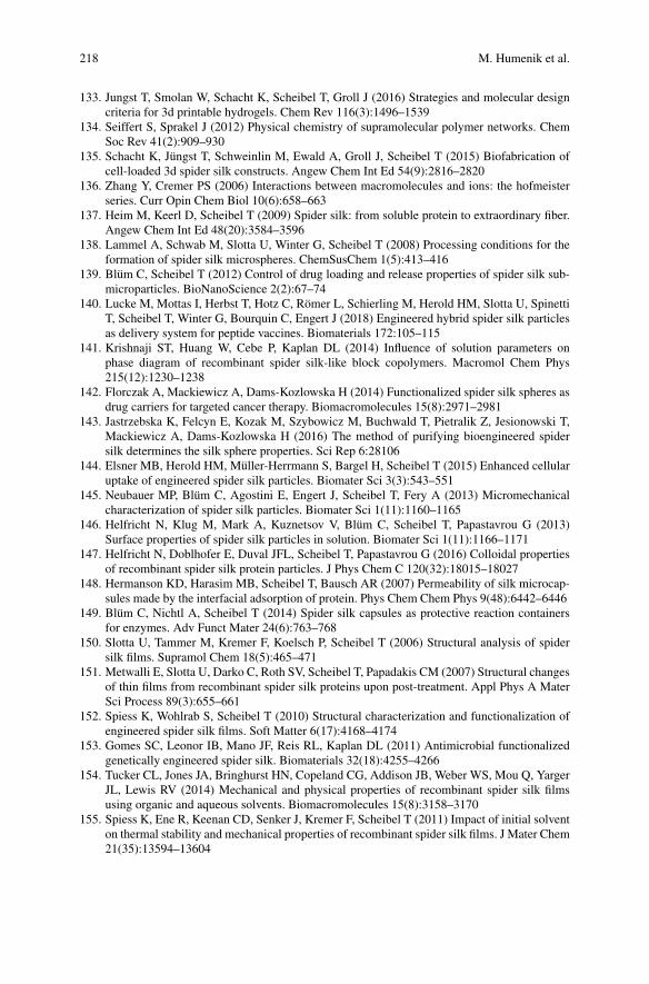
218 M. Humenik et al.
133. Jungst T, Smolan W, Schacht K, Scheibel T, Groll J (2016) Strategies and molecular designcriteria for 3d printable hydrogels. Chem Rev 116(3):1496–1539
134. Seiffert S, Sprakel J (2012) Physical chemistry of supramolecular polymer networks. ChemSoc Rev 41(2):909–930
135. Schacht K, Jüngst T, Schweinlin M, Ewald A, Groll J, Scheibel T (2015) Biofabrication ofcell-loaded 3d spider silk constructs. Angew Chem Int Ed 54(9):2816–2820
136. Zhang Y, Cremer PS (2006) Interactions between macromolecules and ions: the hofmeisterseries. Curr Opin Chem Biol 10(6):658–663
137. Heim M, Keerl D, Scheibel T (2009) Spider silk: from soluble protein to extraordinary fiber.Angew Chem Int Ed 48(20):3584–3596
138. Lammel A, Schwab M, Slotta U, Winter G, Scheibel T (2008) Processing conditions for theformation of spider silk microspheres. ChemSusChem 1(5):413–416
139. Blüm C, Scheibel T (2012) Control of drug loading and release properties of spider silk sub-microparticles. BioNanoScience 2(2):67–74
140. Lucke M, Mottas I, Herbst T, Hotz C, Römer L, Schierling M, Herold HM, Slotta U, SpinettiT, Scheibel T, Winter G, Bourquin C, Engert J (2018) Engineered hybrid spider silk particlesas delivery system for peptide vaccines. Biomaterials 172:105–115
141. Krishnaji ST, Huang W, Cebe P, Kaplan DL (2014) Influence of solution parameters onphase diagram of recombinant spider silk-like block copolymers. Macromol Chem Phys215(12):1230–1238
142. Florczak A, Mackiewicz A, Dams-Kozlowska H (2014) Functionalized spider silk spheres asdrug carriers for targeted cancer therapy. Biomacromolecules 15(8):2971–2981
143. Jastrzebska K, Felcyn E, Kozak M, Szybowicz M, Buchwald T, Pietralik Z, Jesionowski T,Mackiewicz A, Dams-Kozlowska H (2016) The method of purifying bioengineered spidersilk determines the silk sphere properties. Sci Rep 6:28106
144. Elsner MB, Herold HM, Müller-Herrmann S, Bargel H, Scheibel T (2015) Enhanced cellularuptake of engineered spider silk particles. Biomater Sci 3(3):543–551
145. Neubauer MP, Blüm C, Agostini E, Engert J, Scheibel T, Fery A (2013) Micromechanicalcharacterization of spider silk particles. Biomater Sci 1(11):1160–1165
146. Helfricht N, Klug M, Mark A, Kuznetsov V, Blüm C, Scheibel T, Papastavrou G (2013)Surface properties of spider silk particles in solution. Biomater Sci 1(11):1166–1171
147. Helfricht N, Doblhofer E, Duval JFL, Scheibel T, Papastavrou G (2016) Colloidal propertiesof recombinant spider silk protein particles. J Phys Chem C 120(32):18015–18027
148. Hermanson KD, Harasim MB, Scheibel T, Bausch AR (2007) Permeability of silk microcap-sules made by the interfacial adsorption of protein. Phys Chem Chem Phys 9(48):6442–6446
149. Blüm C, Nichtl A, Scheibel T (2014) Spider silk capsules as protective reaction containersfor enzymes. Adv Funct Mater 24(6):763–768
150. Slotta U, Tammer M, Kremer F, Koelsch P, Scheibel T (2006) Structural analysis of spidersilk films. Supramol Chem 18(5):465–471
151. Metwalli E, Slotta U, Darko C, Roth SV, Scheibel T, Papadakis CM (2007) Structural changesof thin films from recombinant spider silk proteins upon post-treatment. Appl Phys A MaterSci Process 89(3):655–661
152. Spiess K, Wohlrab S, Scheibel T (2010) Structural characterization and functionalization ofengineered spider silk films. Soft Matter 6(17):4168–4174
153. Gomes SC, Leonor IB, Mano JF, Reis RL, Kaplan DL (2011) Antimicrobial functionalizedgenetically engineered spider silk. Biomaterials 32(18):4255–4266
154. Tucker CL, Jones JA, Bringhurst HN, Copeland CG, Addison JB, Weber WS, Mou Q, YargerJL, Lewis RV (2014) Mechanical and physical properties of recombinant spider silk filmsusing organic and aqueous solvents. Biomacromolecules 15(8):3158–3170
155. Spiess K, Ene R, Keenan CD, Senker J, Kremer F, Scheibel T (2011) Impact of initial solventon thermal stability and mechanical properties of recombinant spider silk films. J Mater Chem21(35):13594–13604
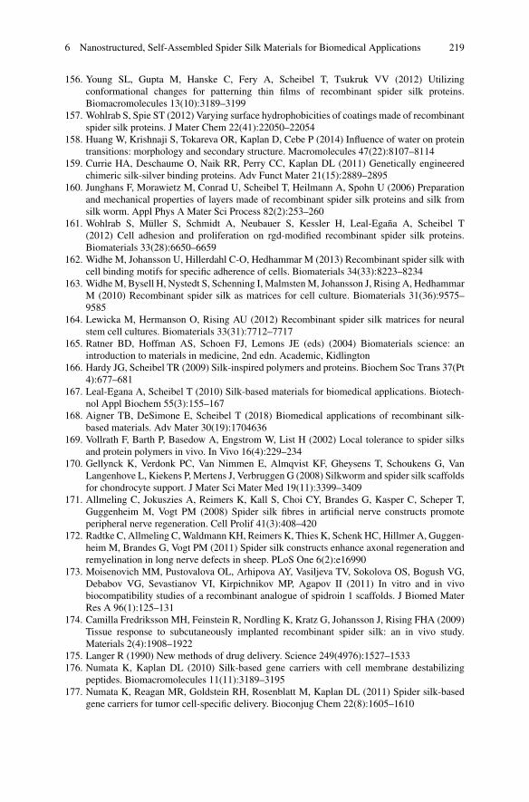
6 Nanostructured, Self-Assembled Spider Silk Materials for Biomedical Applications 219
156. Young SL, Gupta M, Hanske C, Fery A, Scheibel T, Tsukruk VV (2012) Utilizingconformational changes for patterning thin films of recombinant spider silk proteins.Biomacromolecules 13(10):3189–3199
157. Wohlrab S, Spie ST (2012) Varying surface hydrophobicities of coatings made of recombinantspider silk proteins. J Mater Chem 22(41):22050–22054
158. Huang W, Krishnaji S, Tokareva OR, Kaplan D, Cebe P (2014) Influence of water on proteintransitions: morphology and secondary structure. Macromolecules 47(22):8107–8114
159. Currie HA, Deschaume O, Naik RR, Perry CC, Kaplan DL (2011) Genetically engineeredchimeric silk-silver binding proteins. Adv Funct Mater 21(15):2889–2895
160. Junghans F, Morawietz M, Conrad U, Scheibel T, Heilmann A, Spohn U (2006) Preparationand mechanical properties of layers made of recombinant spider silk proteins and silk fromsilk worm. Appl Phys A Mater Sci Process 82(2):253–260
161. Wohlrab S, Müller S, Schmidt A, Neubauer S, Kessler H, Leal-Egaña A, Scheibel T(2012) Cell adhesion and proliferation on rgd-modified recombinant spider silk proteins.Biomaterials 33(28):6650–6659
162. Widhe M, Johansson U, Hillerdahl C-O, Hedhammar M (2013) Recombinant spider silk withcell binding motifs for specific adherence of cells. Biomaterials 34(33):8223–8234
163. Widhe M, Bysell H, Nystedt S, Schenning I, Malmsten M, Johansson J, Rising A, HedhammarM (2010) Recombinant spider silk as matrices for cell culture. Biomaterials 31(36):9575–9585
164. Lewicka M, Hermanson O, Rising AU (2012) Recombinant spider silk matrices for neuralstem cell cultures. Biomaterials 33(31):7712–7717
165. Ratner BD, Hoffman AS, Schoen FJ, Lemons JE (eds) (2004) Biomaterials science: anintroduction to materials in medicine, 2nd edn. Academic, Kidlington
166. Hardy JG, Scheibel TR (2009) Silk-inspired polymers and proteins. Biochem Soc Trans 37(Pt4):677–681
167. Leal-Egana A, Scheibel T (2010) Silk-based materials for biomedical applications. Biotech-nol Appl Biochem 55(3):155–167
168. Aigner TB, DeSimone E, Scheibel T (2018) Biomedical applications of recombinant silk-based materials. Adv Mater 30(19):1704636
169. Vollrath F, Barth P, Basedow A, Engstrom W, List H (2002) Local tolerance to spider silksand protein polymers in vivo. In Vivo 16(4):229–234
170. Gellynck K, Verdonk PC, Van Nimmen E, Almqvist KF, Gheysens T, Schoukens G, VanLangenhove L, Kiekens P, Mertens J, Verbruggen G (2008) Silkworm and spider silk scaffoldsfor chondrocyte support. J Mater Sci Mater Med 19(11):3399–3409
171. Allmeling C, Jokuszies A, Reimers K, Kall S, Choi CY, Brandes G, Kasper C, Scheper T,Guggenheim M, Vogt PM (2008) Spider silk fibres in artificial nerve constructs promoteperipheral nerve regeneration. Cell Prolif 41(3):408–420
172. Radtke C, Allmeling C, Waldmann KH, Reimers K, Thies K, Schenk HC, Hillmer A, Guggen-heim M, Brandes G, Vogt PM (2011) Spider silk constructs enhance axonal regeneration andremyelination in long nerve defects in sheep. PLoS One 6(2):e16990
173. Moisenovich MM, Pustovalova OL, Arhipova AY, Vasiljeva TV, Sokolova OS, Bogush VG,Debabov VG, Sevastianov VI, Kirpichnikov MP, Agapov II (2011) In vitro and in vivobiocompatibility studies of a recombinant analogue of spidroin 1 scaffolds. J Biomed MaterRes A 96(1):125–131
174. Camilla Fredriksson MH, Feinstein R, Nordling K, Kratz G, Johansson J, Rising FHA (2009)Tissue response to subcutaneously implanted recombinant spider silk: an in vivo study.Materials 2(4):1908–1922
175. Langer R (1990) New methods of drug delivery. Science 249(4976):1527–1533176. Numata K, Kaplan DL (2010) Silk-based gene carriers with cell membrane destabilizing
peptides. Biomacromolecules 11(11):3189–3195177. Numata K, Reagan MR, Goldstein RH, Rosenblatt M, Kaplan DL (2011) Spider silk-based
gene carriers for tumor cell-specific delivery. Bioconjug Chem 22(8):1605–1610
220 M. Humenik et al.
178. Numata K, Mieszawska-Czajkowska AJ, Kvenvold LA, Kaplan DL (2012) Silk-basednanocomplexes with tumor-homing peptides for tumor-specific gene delivery. MacromolBiosci 12(1):75–82
179. Doblhofer E, Scheibel T (2015) Engineering of recombinant spider silk proteins allowsdefined uptake and release of substances. J Pharm Sci 104(3):988–994
180. Wang X, Yucel T, Lu Q, Hu X, Kaplan DL (2010) Silk nanospheres and microspheres fromsilk/pva blend films for drug delivery. Biomaterials 31(6):1025–1035
181. Patil Y, Panyam J (2009) Polymeric nanoparticles for sirna delivery and gene silencing. Int JPharm 367(1–2):195–203
182. Sushmita Sundar JK, Kundu SC (2010) Biopolymeric nanoparticles. Sci Technol Adv Mater11(1):1–13
183. Liebmann B, Huemmerich D, Scheibel T, Fehr M (2008) Formulation of poorly water-solublesubstances using self-assembling spider silk protein. Colloids Surf A Physicochem Eng Asp331(1–2):126–132
184. Lammel A, Schwab M, Hofer M, Winter G, Scheibel T (2011) Recombinant spider silkparticles as drug delivery vehicles. Biomaterials 32(8):2233–2240
185. Langer R, Vacanti JP (1993) Tissue engineering. Science 260(5110):920–926186. Schacht K, Scheibel T (2014) Processing of recombinant spider silk proteins into tailor-made
materials for biomaterials applications. Curr Opin Biotechnol 29:62–69187. Petzold J, Aigner TB, Touska F, Zimmermann K, Scheibel T, Engel FB (2017) Surface
features of recombinant spider silk protein eadf4(κ16)-made materials are well-suited forcardiac tissue engineering. Adv Funct Mater 27(36):1701427
188. Bell E, Ehrlich HP, Buttle DJ, Nakatsuji T (1981) Living tissue formed in vitro and acceptedas skin-equivalent tissue of full thickness. Science 211(4486):1052–1054
189. Burke JF, Yannas IV, Quinby WC Jr, Bondoc CC, Jung WK (1981) Successful use of aphysiologically acceptable artificial skin in the treatment of extensive burn injury. Ann Surg194(4):413–428
190. Wendt H, Hillmer A, Reimers K, Kuhbier JW, Schafer-Nolte F, Allmeling C, Kasper C, VogtPM (2011) Artificial skin-culturing of different skin cell lines for generating an artificial skinsubstitute on cross-weaved spider silk fibres. PLoS One 6(7):e21833
191. Widhe M, Shalaly ND, Hedhammar M (2016) A fibronectin mimetic motif improves integrinmediated cell biding to recombinant spider silk matrices. Biomaterials 74:256–266
192. Millennium WSGotBoMCatSotN (2003) The burden of musculoskeletal conditions at thestart of the new millennium. World Health Organ Tech Rep Ser 919:1–218
193. Hing KA (2004) Bone repair in the twenty-first century: biology, chemistry or engineering?Philos Transact A Math Phys Eng Sci 362(1825):2821–2850
194. Parikh SN (2002) Bone graft substitutes in modern orthopedics. Orthopedics 25(11):1301–1309. quiz 1310-1301
195. Mark DE, Hollinger JO, Hastings C Jr, Chen G, Marden LJ, Reddi AH (1990) Repair ofcalvarial nonunions by osteogenin, a bone-inductive protein. Plast Reconstr Surg 86(4):623–630. discussion 631-622
196. Huang J, Wong C, George A, Kaplan DL (2007) The effect of genetically engineered spidersilk-dentin matrix protein 1 chimeric protein on hydroxyapatite nucleation. Biomaterials28(14):2358–2367
197. Wong Po Foo C, Patwardhan SV, Belton DJ, Kitchel B, Anastasiades D, Huang J, Naik RR,Perry CC, Kaplan DL (2006) Novel nanocomposites from spider silk-silica fusion (chimeric)proteins. Proc Natl Acad Sci U S A 103(25):9428–9433
198. Belton DJ, Mieszawska AJ, Currie HA, Kaplan DL, Perry CC (2012) Silk-silica compositesfrom genetically engineered chimeric proteins: materials properties correlate with silicacondensation rate and colloidal stability of the proteins in aqueous solution. Langmuir28(9):4373–4381
199. Mieszawska AJ, Nadkarni LD, Perry CC, Kaplan DL (2010) Nanoscale control of sil-ica particle formation via silk-silica fusion proteins for bone regeneration. Chem Mater22(20):5780–5785
6 Nanostructured, Self-Assembled Spider Silk Materials for Biomedical Applications 221
200. Chen MB, Zhang F, Lineaweaver WC (2006) Luminal fillers in nerve conduits for peripheralnerve repair. Ann Plast Surg 57(4):462–471
201. Ciardelli G, Chiono V (2006) Materials for peripheral nerve regeneration. Macromol Biosci6(1):13–26
202. Evans GR, Brandt K, Katz S, Chauvin P, Otto L, Bogle M, Wang B, Meszlenyi RK, Lu L,Mikos AG, Patrick CW Jr (2002) Bioactive poly(l-lactic acid) conduits seeded with schwanncells for peripheral nerve regeneration. Biomaterials 23(3):841–848
203. Lietz M, Dreesmann L, Hoss M, Oberhoffner S, Schlosshauer B (2006) Neuro tissueengineering of glial nerve guides and the impact of different cell types. Biomaterials27(8):1425–1436
204. Lundborg G, Rosen B, Abrahamson SO, Dahlin L, Danielsen N (1994) Tubular repair of themedian nerve in the human forearm. preliminary findings. J Hand Surg (Br) 19(3):273–276
205. Stanec S, Stanec Z (1998) Reconstruction of upper-extremity peripheral-nerve injuries witheptfe conduits. J Reconstr Microsurg 14(4):227–232
206. Letourneau PC, Condic ML, Snow DM (1994) Interactions of developing neurons with theextracellular matrix. J Neurosci 14(3 Pt 1):915–928
207. Allmeling C, Jokuszies A, Reimers K, Kall S, Vogt PM (2006) Use of spider silk fibres asan innovative material in a biocompatible artificial nerve conduit. J Cell Mol Med 10(3):770–777
208. Borkner CB, Elsner MB, Scheibel T (2014) Coatings and films made of silk proteins. ACSAppl Mater Interfaces 6(18):15611–15625
209. Zeplin PH, Berninger AK, Maksimovikj NC, van Gelder P, Scheibel T, Walles H (2014)Improving the biocompatibility of silicone implants using spider silk coatings: immunohisto-chemical analysis of capsule formation. Handchir Mikrochir Plast Chir 46(6):336–341
210. Borkner CB, Wohlrab S, Möller E, Lang G, Scheibel T (2017) Surface modification ofpolymeric biomaterials using recombinant spider silk proteins. ACS Biomater Sci Eng3(5):767–775
211. Mironov V, Trusk T, Kasyanov V, Little S, Swaja R, Markwald R (2009) Biofabrication: a21st century manufacturing paradigm. Biofabrication 1(2):022001
212. Groll J, Boland T, Blunk T, Burdick JA, Cho DW, Dalton PD, Derby B, Forgacs G, Li Q,Mironov VA, Moroni L, Nakamura M, Shu W, Takeuchi S, Vozzi G, Woodfield TB, XuT, Yoo JJ, Malda J (2016) Biofabrication: reappraising the definition of an evolving field.Biofabrication 8(1):013001
213. Malda J, Visser J, Melchels FP, Jungst T, Hennink WE, Dhert WJ, Groll J, HutmacherDW (2013) 25th anniversary article: engineering hydrogels for biofabrication. Adv Mater25(36):5011–5028
214. Murphy SV, Atala A (2014) 3d bioprinting of tissues and organs. Nat Biotechnol 32(8):773–785
215. Skardal A, Atala A (2015) Biomaterials for integration with 3-d bioprinting. Ann BiomedEng 43(3):730–746
216. Catros S, Fricain JC, Guillotin B, Pippenger B, Bareille R, Remy M, Lebraud E, Desbat B,Amedee J, Guillemot F (2011) Laser-assisted bioprinting for creating on-demand patterns ofhuman osteoprogenitor cells and nano-hydroxyapatite. Biofabrication 3(2):025001
217. Chimene D, Lennox KK, Kaunas RR, Gaharwar AK (2016) Advanced bioinks for 3d printing:a materials science perspective. Ann Biomed Eng 44(6):2090–2102
218. DeSimone E, Schacht K, Jungst T, Groll J, Scheibel T (2015) Biofabrication of 3d constructs:fabrication technologies and spider silk proteins as bioinks. Pure Appl Chem 87(8):737–749
Chapter 7Protein Microgels from Amyloid FibrilNetworks
Lianne W. Y. Roode, Ulyana Shimanovich, Si Wu, Sarah Perrett,and Tuomas P. J. Knowles
Abstract Nanofibrillar forms of amyloidogenic proteins were initially discoveredin the context of protein misfolding and disease but have more recently beenfound at the origin of key biological functionality in many naturally occurringfunctional materials, such as adhesives and biofilm coatings. Their physiologicalroles in nature reflect their great strength and stability, which has led to theexploration of their use as the basis of artificial protein-based functional materials.Particularly for biomedical applications, they represent attractive building blocksfor the development of, for instance, drug carrier agents due to their inherentbiocompatibility and biodegradability. Furthermore, the propensity of proteinsto self-assemble into amyloid fibrils can be exploited under microconfinement,afforded by droplet microfluidic techniques. This approach allows the generationof multi-scale functional microgels that can host biological additives and canbe designed to incorporate additional functionality, such as to aid targeted drugdelivery.
L. W. Y. RoodeCentre for Misfolding Diseases, Department of Chemistry, University of Cambridge,Cambridge, UK
U. Shimanovich (�)Department of Materials and Interfaces, Weizmann Institute of Science, Rehovot, Israele-mail: [email protected]
S. Wu · S. Perrett (�)National Laboratory of Biomacromolecules, CAS Center for Excellence in Biomacromolecules,Institute of Biophysics, Chinese Academy of Sciences, Beijing, China
University of the Chinese Academy of Sciences, Beijing, Chinae-mail: [email protected]
T. P. J. Knowles (�)Centre for Misfolding Diseases, Department of Chemistry, University of Cambridge, Cambridge,UK
Cavendish Laboratory, University of Cambridge, Cambridge, UKe-mail: [email protected]
© Springer Nature Singapore Pte Ltd. 2019S. Perrett et al. (eds.), Biological and Bio-inspired Nanomaterials,Advances in Experimental Medicine and Biology 1174,https://doi.org/10.1007/978-981-13-9791-2_7
223
224 L. W. Y. Roode et al.
Keywords Self-assembled amyloid fibrils · Protein microgels · Dropletmicrofluidics · Drug carrier agents · Functional materials
7.1 Nature of Amyloid Proteins
7.1.1 Introduction
Amyloid fibrils have become a subject of rapidly increasing research interestacross a wide range of scientific disciplines mainly due to their association with agrowing number of debilitating medical disorders, ranging from neurodegenerativedisorders (e.g. Alzheimer’s and Parkinson’s diseases) to systemic amyloidoses [1–5]. A number of human diseases associated with amyloid formation are listed inTable 7.1. The origin of such pathological conditions was found to be associatedwith misfolding of normally soluble, functional peptides and proteins, giving riseto intractable aggregates and their subsequent conversion into mature fibrils, ofwhich the archetypal example are amyloid fibrils. They are highly ordered, thread-like aggregates and their formation is associated with both a loss of function of theproteins involved as well as with the generation of toxic intermediate species thatare formed in the process of self-assembly [6–9].
Although the amyloid form was initially identified within the context of disease,an increasing number of proteins that are not associated with any known pathogenicdisorder have been found to form amyloid-like fibrils. Furthermore, increasing evi-dence has pointed towards amyloid formation being a generic property of proteinsand polypeptide chains, with many polymorphic forms having been identified [6, 8,10]. More recently, amyloid or amyloid-like fibrillar structures (which are definedas functionally beneficial fibrillar constructs structurally similar to amyloids) haveemerged as key functional components in biological materials found in organismsranging from bacteria to mammals [11–16]. These amyloid materials are exploitedfor use in diverse contexts, ranging from adhesives [17–20], catalytic scaffolds[11], bacterial coatings and biofilm formation [13, 21], and mediating epigenetic-information storage and transfer [22–26] (Table 7.2).
The discovery of functional amyloid structures in such a diverse set of rolesin nature has transformed the understanding of the evolutionary requirements anddesign features that favour functional protein assembly versus aberrant association.Although the exact molecular level mechanisms that differentiate toxic amyloidformation and beneficial amyloid species are not fully understood yet, increasingevidence suggests that rather than the mature amyloid fibrils themselves, smallsoluble intermediate species, namely oligomers, are many responsible for thecellular toxicity associated with aberrant protein aggregation [10, 27–33]. In caseswhere amyloid structures are used to generate functional materials it seems thatnature, therefore, has mechanisms in place to control the self-assembly process,ensuring that it goes to completion and that it does not give rise to the accumulationof potentially highly toxic intermediates [12, 15, 21, 34].
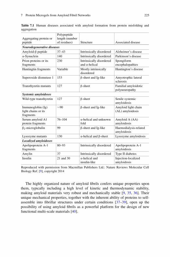
7 Protein Microgels from Amyloid Fibril Networks 225
Table 7.1 Human diseases associated with amyloid formation from protein misfolding andaggregation
Aggregating protein orpeptide
Polypeptidelength (numberof residues) Structure Associated disease
Neurodegenerative diseasesAmyloid-β peptide 37–43 Intrinsically disordered Alzheimer’s diseaseα-Synuclein 140 Intrinsically disordered Parkinson’s diseasePrion proteins or itsfragments
230 Intrinsically disorderedand α-helical
Spongiformencephalopathies
Huntingtin fragments Variable Mostly intrinsicallydisordered
Huntington’s disease
Superoxide dismutase 1 153 β-sheet and Ig-like Amyotrophic lateralsclerosis
Transthyretin mutants 127 β-sheet Familial amyloidoticpolyneuropathy
Systemic amyloidosesWild-type transthyretin 127 β-sheet Senile systemic
amyloidosisImmunoglobin (Ig)light chains or itsfragments
∼90 β-sheet and Ig-like Amyloid light chain(AL) amyloidosis
Serum amyloid A1protein fragments
76–104 α-helical and unknownfold
Amyloid A (AA)amyloidosis
β2-microglobulin 99 β-sheet and Ig-like Haemodialysis-relatedamyloidosis
Lysozyme mutants 130 α-helical and β-sheet Lysozyme amyloidosisLocalized amyloidosesApolipoprotein A-1fragments
80–93 Intrinsically disordered Apolipoprotein A-1amyloidosis
Amylin 37 Intrinsically disordered Type II diabetesInsulin 21 and 30 α-helical and
insulin-likeInjection-localizedamyloidosis
Reproduced with permission from Macmillan Publishers Ltd.: Nature Reviews Molecular CellBiology Ref. [9], copyright 2014
The highly organized nature of amyloid fibrils confers unique properties uponthem, typically including a high level of kinetic and thermodynamic stability,making amyloid materials very robust and mechanically stable [9, 35, 36]. Theirunique mechanical properties, together with the inherent ability of proteins to self-assemble into fibrillar structures under certain conditions [37–39], open up thepossibility of using amyloid fibrils as a powerful platform for the design of newfunctional multi-scale materials [40].
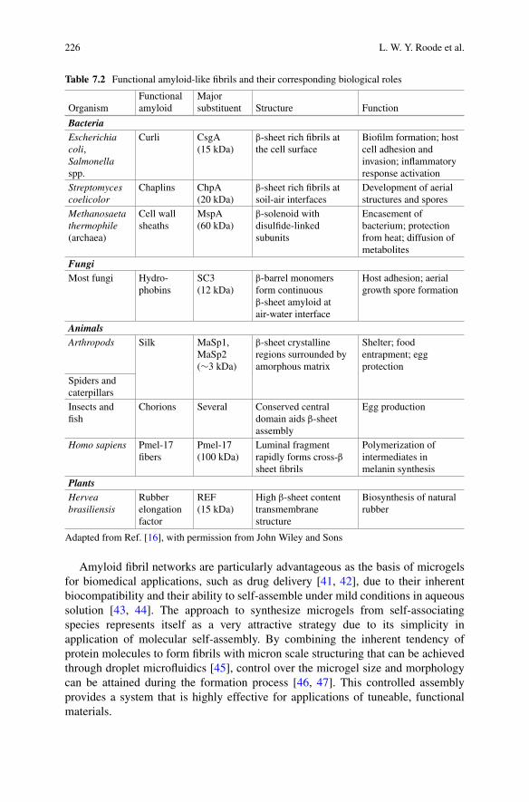
226 L. W. Y. Roode et al.
Table 7.2 Functional amyloid-like fibrils and their corresponding biological roles
OrganismFunctionalamyloid
Majorsubstituent Structure Function
BacteriaEscherichiacoli,Salmonellaspp.
Curli CsgA(15 kDa)
β-sheet rich fibrils atthe cell surface
Biofilm formation; hostcell adhesion andinvasion; inflammatoryresponse activation
Streptomycescoelicolor
Chaplins ChpA(20 kDa)
β-sheet rich fibrils atsoil-air interfaces
Development of aerialstructures and spores
Methanosaetathermophile(archaea)
Cell wallsheaths
MspA(60 kDa)
β-solenoid withdisulfide-linkedsubunits
Encasement ofbacterium; protectionfrom heat; diffusion ofmetabolites
FungiMost fungi Hydro-
phobinsSC3(12 kDa)
β-barrel monomersform continuousβ-sheet amyloid atair-water interface
Host adhesion; aerialgrowth spore formation
AnimalsArthropods Silk MaSp1,
MaSp2(∼3 kDa)
β-sheet crystallineregions surrounded byamorphous matrix
Shelter; foodentrapment; eggprotection
Spiders andcaterpillarsInsects andfish
Chorions Several Conserved centraldomain aids β-sheetassembly
Egg production
Homo sapiens Pmel-17fibers
Pmel-17(100 kDa)
Luminal fragmentrapidly forms cross-βsheet fibrils
Polymerization ofintermediates inmelanin synthesis
PlantsHerveabrasiliensis
Rubberelongationfactor
REF(15 kDa)
High β-sheet contenttransmembranestructure
Biosynthesis of naturalrubber
Adapted from Ref. [16], with permission from John Wiley and Sons
Amyloid fibril networks are particularly advantageous as the basis of microgelsfor biomedical applications, such as drug delivery [41, 42], due to their inherentbiocompatibility and their ability to self-assemble under mild conditions in aqueoussolution [43, 44]. The approach to synthesize microgels from self-associatingspecies represents itself as a very attractive strategy due to its simplicity inapplication of molecular self-assembly. By combining the inherent tendency ofprotein molecules to form fibrils with micron scale structuring that can be achievedthrough droplet microfluidics [45], control over the microgel size and morphologycan be attained during the formation process [46, 47]. This controlled assemblyprovides a system that is highly effective for applications of tuneable, functionalmaterials.
7 Protein Microgels from Amyloid Fibril Networks 227
7.1.2 Detection of Amyloid Structures
The term ‘amyloid’, originally meaning ‘starch like’, is a legacy of its firstobservation more than 150 years ago in systemic amyloidosis, as the depositsobserved in patient’s tissues and organs could be readily stained with iodine, whichis commonly used to detect starch [48, 49]. The repetitive sequence of proteinunits that forms an organized structure includes regular binding pockets for variousdyes by which amyloid fibrils are routinely characterized. Amyloid in human tissueis traditionally visualized by pathologists using the sulfonated azo dye CongoRed, which lends a distinct green birefringence to amyloid deposits under cross-polarized filters [50]. Similarly, in vitro studies generally make use of the fluorescentdye Thioflavin T (ThT), giving rise to increased quantum yield and red shiftedfluorescence of ThT upon binding to amyloid fibrils [51, 52].
Although these dyes are readily available and easy to handle, they do not haveabsolute specificity for amyloid structures, and are thus best used against isolatedproteins [53, 54]. For in vivo studies, it is more appropriate to use conformationallyspecific monoclonal antibodies, which were originally developed for amyloid-βfibrils but were shown to bind to a multitude of amyloid fibrils tested [55, 56]. Inaddition, Kaminski and co-workers reported an intrinsic fluorescence signature ofaggregating polypeptides, including the amyloidogenic human peptides amyloid-β,lysozyme and tau. The intrinsic fluorescence appears to result from electronic levelsthat become available when the polypeptide chain folds into a cross-β sheet structurereminiscent of what has been reported to take place in crystals. This allows for label-free quantification of protein aggregation in vitro [57]. Following these findings, thesame research group developed a Förster resonance energy transfer (FRET) sensorthat exploits the observation that amyloid assemblies can act as energy acceptorsfor fluorescently labelled proteins, thereby permitting the aggregation kinetics ofamyloidogenic proteins to be quantified in a non-invasive manner suitable for bothin vitro and in vivo studies [58, 59].
7.1.3 Structure of Amyloid Fibrils
Amyloid fibrils have been observed for a variety of proteins and peptides [8, 60].Despite the various origins, the fibrils seem to be remarkably similar, having afibrillar morphology at the nanoscale and generic quaternary protein structure onthe molecular scale [61, 62]. A multitude of studies, including solid-state NMRspectroscopy [63–68], single crystal X-ray diffraction analysis [69–71], atomicforce microscopy [72], electron microscopy [73], and cryo-electron microscopy[74–77], have given insight into the atomic level architecture of amyloid fibrils.These studies confirm common structural features, notably a high content ofhydrogen bond-rich β-sheets and the frequent presence of repetitive hydrophobicor polar interactions along the fibrillar axis [8]. The fibrils are observed to consist
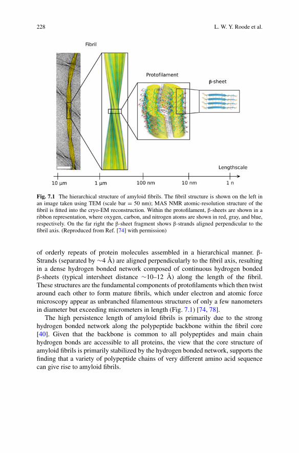
228 L. W. Y. Roode et al.
Fig. 7.1 The hierarchical structure of amyloid fibrils. The fibril structure is shown on the left inan image taken using TEM (scale bar = 50 nm); MAS NMR atomic-resolution structure of thefibril is fitted into the cryo-EM reconstruction. Within the protofilament, β-sheets are shown in aribbon representation, where oxygen, carbon, and nitrogen atoms are shown in red, gray, and blue,respectively. On the far right the β-sheet fragment shows β-strands aligned perpendicular to thefibril axis. (Reproduced from Ref. [74] with permission)
of orderly repeats of protein molecules assembled in a hierarchical manner. β-Strands (separated by ∼4 Å) are aligned perpendicularly to the fibril axis, resultingin a dense hydrogen bonded network composed of continuous hydrogen bondedβ-sheets (typical intersheet distance ∼10–12 Å) along the length of the fibril.These structures are the fundamental components of protofilaments which then twistaround each other to form mature fibrils, which under electron and atomic forcemicroscopy appear as unbranched filamentous structures of only a few nanometersin diameter but exceeding micrometers in length (Fig. 7.1) [74, 78].
The high persistence length of amyloid fibrils is primarily due to the stronghydrogen bonded network along the polypeptide backbone within the fibril core[40]. Given that the backbone is common to all polypeptides and main chainhydrogen bonds are accessible to all proteins, the view that the core structure ofamyloid fibrils is primarily stabilized by the hydrogen bonded network, supports thefinding that a variety of polypeptide chains of very different amino acid sequencecan give rise to amyloid fibrils.
7 Protein Microgels from Amyloid Fibril Networks 229
7.1.4 Self-Assembly and Polymorphism of Amyloid Fibrils
Although all amyloid systems have a common fibrillar architecture, structuralvariations in different amyloid fibrils have been observed. Fibrils may vary in β-strand length, number of β-sheets per protofilament, and conformation (parallel/anti-parallel) as a result of the manner in which different sets of side chains interactand are incorporated into the fibrillar architecture [79, 80]. Unlike the native proteinstate, a single arrangement of a given polypeptide chain in the amyloid core structureis unlikely to provide unique stability relative to all other arrangements. Indeed,the basic amyloid cross-β structure can be adopted through a multitude of possiblestacking geometries of β-sheets, giving rise to a large number of polymorphic forms[8, 40, 74].
In addition to structural variation in fibrils from different polypeptides, polymor-phic forms have been observed for fibrils from the same polypeptide sequence butformed under varying conditions [82–85]. This highlights differences in the detailsof the way in which individual molecules are incorporated into the fibrils, dependingon the assembly environment. While the essential elements of the amyloid foldare mainly determined by inherent physico-chemical properties of the polypeptidechain, as indicated by studies correlating relative aggregation rates with featuressuch as the hydrophobicity and charge [7, 86], detailed morphological variationarises due to secondary structure and long-range interactions. As such, solventconditions such as temperature, pH, ionic strength, and protein concentration aswell as mechanical influences such as agitation influence the assembly process [39,78, 81, 87, 88].
Studies showed that, while under physiological conditions the formation ofamyloid fibrils appeared to be restricted to a subset of proteins, under mildlydenaturing conditions in vitro proteins can be induced to form fibrillar aggregates[6, 10, 37, 89]. These in vitro studies, together with the recognition of functionalamyloidogenic proteins in bacteria and human [12, 13, 90], led to the idea thatthe formation of amyloid structures is not a rare phenomenon associated with asmall number of diseases only, but is rather a generic property of many, if not all,polypeptide chains [8, 10, 91].
The multitude of amyloid fibril forming proteins observed, together with mathe-matical analysis of the mechanism of amyloid formation, suggests that the amyloidform may be determined by kinetic rather than thermodynamic factors. There isincreasing evidence that the native protein state may not always represent theabsolute free energy minima in protein folding processes but rather that the amyloidstate is thermodynamically more stable, though kinetically inaccessible under someconditions [92, 93]. The amyloid state reflects a well-defined structural form ofthe protein that is an alternative to the native state and, unlike the native state,its essential architecture is not encoded by the amino acid sequence, although thepropensity of formation does vary with sequence [8, 9, 94].
230 L. W. Y. Roode et al.
A high kinetic barrier that is associated with amyloid structures makes theformation of the initial amyloid nucleus a slow process. Nevertheless, once formedamyloid fibrils are capable of self-replication through secondary processes whereadditional protein monomers are incorporated into the fibril structure [95–97].Pre-formed fibrillar species, referred to as seeds, can overcome sequence- orenvironment-based structural preferences by acting as a template for further fibrilgrowth; protein monomers added to pre-formed fibril-ends adopt a cross-β con-formation to match that of the protein molecules already present in the aggregate[98, 99]. This process results in rapid fibril formation, analogous to crystallizationphenomena [100], where the seeds alleviate the high kinetic barrier associated withamyloid structures; thus the fibril morphology and structure is propagated [101–103].
7.1.5 Mechanical Properties of Amyloid Fibrils
The highly-ordered structure of amyloid fibrils is intimately reflected by its prop-erties. The comprehensive hydrogen bonding network of the β-sheet core providesstrong intermolecular backbone-backbone interactions between polypeptide chainswithin the fibrils, resulting in great fibril stability [35, 70]. This is exemplifiedby amyloid fibrils being very stable, even under extreme conditions, such ashigh concentrations of (chemical) denaturants, extreme pH environments and hightemperatures [16]. Indeed, amyloid materials possess a remarkably high Young’smodulus and tensile strength [36, 104–106].
In discussing the physical nature of amyloid fibrils, it is informative to comparethem with other types of one-dimensional organic macromolecular and colloidalsystems [40]. Proteins show generic polymeric and colloidal features, and manynon-biological and synthetic polymers exhibit analogous condensed phases, such asfilamentous and particulate gel states [107]. Indeed, some amyloid deposits formedin vivo and fibrils generated in vitro have been found to contain spherulites and otherhigher-order assemblies [108]. These structures are also observed in preparations ofsynthetic polymers, such as polyethylene, and this observation supports the idea thatamyloid fibrils have features similar to those of classical polymers [8].
However, in contrast to classical polymeric systems, the persistence length ofamyloid fibrils shows remarkable independence of the environmental conditions.The persistence length in amyloid fibrils primarily originates from the hydrogenbonding network in the core, which is less affected by solution conditions than theelectrostatic repulsion which is responsible for the persistence length of conven-tional charged polymers [40]. Furthermore, amyloid fibrils possess the properties ofchirality and polarity along their main fibrillar axis, which opens the possibility ofmanipulations via weak electric fields [109, 110]. The chiral, polar and chargednature, together with the intrinsic rigidity, of amyloid fibrils provides complexphysical behaviour in one, two, and three dimensions.
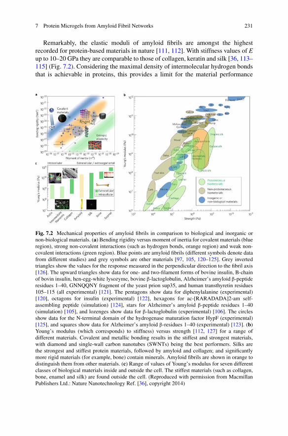
7 Protein Microgels from Amyloid Fibril Networks 231
Remarkably, the elastic moduli of amyloid fibrils are amongst the highestrecorded for protein-based materials in nature [111, 112]. With stiffness values of Eup to 10–20 GPa they are comparable to those of collagen, keratin and silk [36, 113–115] (Fig. 7.2). Considering the maximal density of intermolecular hydrogen bondsthat is achievable in proteins, this provides a limit for the material performance
Fig. 7.2 Mechanical properties of amyloid fibrils in comparison to biological and inorganic ornon-biological materials. (a) Bending rigidity versus moment of inertia for covalent materials (blueregion), strong non-covalent interactions (such as hydrogen bonds, orange region) and weak non-covalent interactions (green region). Blue points are amyloid fibrils (different symbols denote datafrom different studies) and grey symbols are other materials [97, 105, 120–125]. Grey invertedtriangles show the values for the response measured in the perpendicular direction to the fibril axis[126]. The upward triangles show data for one- and two-filament forms of bovine insulin, B-chainof bovin insulin, hen-egg-white lysozyme, bovine β-lactoglobulin, Alzheimer’s amyloid β-peptideresidues 1–40, GNNQQNY fragment of the yeast prion sup35, and human transthyretin residues105–115 (all experimental) [121]. The pentagons show data for diphenylalanine (experimental)[120], octagons for insulin (experimental) [122], hexagons for ac-[RARADADA]2-am self-assembling peptide (simulation) [124], stars for Alzheimer’s amyloid β-peptide residues 1–40(simulation) [105], and lozenges show data for β-lactoglobulin (experimental) [106]. The circlesshow data for the N-terminal domain of the hydrogenase maturation factor HypF (experimental)[125], and squares show data for Alzheimer’s amyloid β-residues 1–40 (experimental) [123]. (b)Young’s modulus (which corresponds) to stiffness) versus strength [112, 127] for a range ofdifferent materials. Covalent and metallic bonding results in the stiffest and strongest materials,with diamond and single-wall carbon nanotubes (SWNTs) being the best performers. Silks arethe strongest and stiffest protein materials, followed by amyloid and collagen; and significantlymore rigid materials (for example, bone) contain minerals. Amyloid fibrils are shown in orange todistinguish them from other materials. (c) Range of values of Young’s modulus for seven differentclasses of biological materials inside and outside the cell. The stiffest materials (such as collagen,bone, enamel and silk) are found outside the cell. (Reproduced with permission from MacmillanPublishers Ltd.: Nature Nanotechnology Ref. [36], copyright 2014)
232 L. W. Y. Roode et al.
that can be obtained from proteinaceous structures [97, 116]. Above this limit,materials contain covalent or metallic interactions which provide significantlyhigher energy density than hydrogen bonds or other weak non-covalent interactions[117]. Therefore nature, through evolution, has optimized proteinaceous materialsto be close to the theoretical performance limits.
It is apparent, however, that the rigidity of intracellular materials, such ascytoskeletal filaments [118], does not reach the maximal values achievable for bio-logical materials. This reflects the requirement of intracellular protein assembly tobe readily reversible under physiological conditions to ensure cellular homeostasis[119]. Indeed, the materials exhibiting the highest moduli (cellulose, bone, keratin,silk) seem to be incompatible with intracellular structure and, instead, are primarilyfound extracellularly in roles where controlled rapid breakdown is not required. Thisphenomenon alludes to nature’s regulation of the formation of such rigid proteinmaterials in functional roles [36].
7.2 Amyloid Proteins for the Development of FunctionalMicrogels
7.2.1 Emerging Applications of Artificial AmyloidProtein-Based Materials and Microgels
The intricate relationship between protein structure and function, which in naturehas been tuned by evolutionary pressures over many millennia, can serve as a fruitfulsource of inspiration for the design of functional materials. The discovery of variousbiologically active amyloid structure-based materials contrasts markedly with theoriginal picture of amyloid fibrils as inherently pathological structures and it hasmotivated the exploration of these fibrils as functional materials. Many of the samecharacteristics that underpin the natural use of amyloid structures, such as the fibril’sextraordinary stability and accurate self-assembly, make them attractive as the basisof artificial functional materials [78, 128].
Within the last decade, artificial fibrillar, proteinaceous materials have beendeveloped with applications including sensors [129–131], opto-electronics [132],biomimetic functional materials [133, 134], cell scaffolds [135–138], sustaineddrug release [139, 140], and food applications [141–145]. Furthermore, compositematerials have been developed [146–149], with active properties making themcapable of for example capturing carbon dioxide [150, 151], removal of radioactivewaste and heavy metal ions from polluted water [152], and continuous flow catalysis[153]. Moreover, the interfacial properties of amyloid fibrils such as adhesiveness –generated via aggregation-induced development of positive surface charge – offereda good biocompatible platform towards biomineralization [154], antimicrobialcoatings [155], wound healing [156], protein crystallization [157], and water-basedphoto/electro-lithography resist [158].
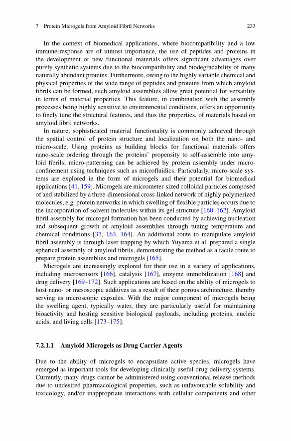
7 Protein Microgels from Amyloid Fibril Networks 233
In the context of biomedical applications, where biocompatibility and a lowimmune-response are of utmost importance, the use of peptides and proteins inthe development of new functional materials offers significant advantages overpurely synthetic systems due to the biocompatibility and biodegradability of manynaturally abundant proteins. Furthermore, owing to the highly variable chemical andphysical properties of the wide range of peptides and proteins from which amyloidfibrils can be formed, such amyloid assemblies allow great potential for versatilityin terms of material properties. This feature, in combination with the assemblyprocesses being highly sensitive to environmental conditions, offers an opportunityto finely tune the structural features, and thus the properties, of materials based onamyloid fibril networks.
In nature, sophisticated material functionality is commonly achieved throughthe spatial control of protein structure and localization on both the nano- andmicro-scale. Using proteins as building blocks for functional materials offersnano-scale ordering through the proteins’ propensity to self-assemble into amy-loid fibrils; micro-patterning can be achieved by protein assembly under micro-confinement using techniques such as microfluidics. Particularly, micro-scale sys-tems are explored in the form of microgels and their potential for biomedicalapplications [41, 159]. Microgels are micrometer-sized colloidal particles composedof and stabilized by a three-dimensional cross-linked network of highly polymerizedmolecules, e.g. protein networks in which swelling of flexible particles occurs due tothe incorporation of solvent molecules within its gel structure [160–162]. Amyloidfibril assembly for microgel formation has been conducted by achieving nucleationand subsequent growth of amyloid assemblies through tuning temperature andchemical conditions [37, 163, 164]. An additional route to manipulate amyloidfibril assembly is through laser trapping by which Yuyama et al. prepared a singlespherical assembly of amyloid fibrils, demonstrating the method as a facile route toprepare protein assemblies and microgels [165].
Microgels are increasingly explored for their use in a variety of applications,including microsensors [166], catalysis [167], enzyme immobilization [168] anddrug delivery [169–172]. Such applications are based on the ability of microgels tohost nano- or mesoscopic additives as a result of their porous architecture, therebyserving as microscopic capsules. With the major component of microgels beingthe swelling agent, typically water, they are particularly useful for maintainingbioactivity and hosting sensitive biological payloads, including proteins, nucleicacids, and living cells [173–175].
7.2.1.1 Amyloid Microgels as Drug Carrier Agents
Due to the ability of microgels to encapsulate active species, microgels haveemerged as important tools for developing clinically useful drug delivery systems.Currently, many drugs cannot be administered using conventional release methodsdue to undesired pharmacological properties, such as unfavourable solubility andtoxicology, and/or inappropriate interactions with cellular components and other
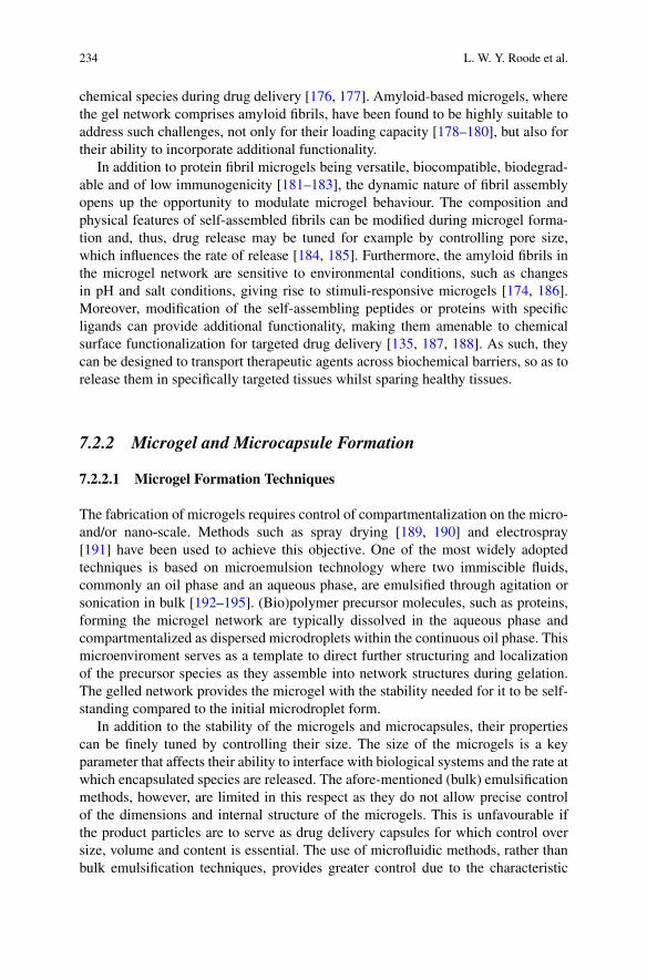
234 L. W. Y. Roode et al.
chemical species during drug delivery [176, 177]. Amyloid-based microgels, wherethe gel network comprises amyloid fibrils, have been found to be highly suitable toaddress such challenges, not only for their loading capacity [178–180], but also fortheir ability to incorporate additional functionality.
In addition to protein fibril microgels being versatile, biocompatible, biodegrad-able and of low immunogenicity [181–183], the dynamic nature of fibril assemblyopens up the opportunity to modulate microgel behaviour. The composition andphysical features of self-assembled fibrils can be modified during microgel forma-tion and, thus, drug release may be tuned for example by controlling pore size,which influences the rate of release [184, 185]. Furthermore, the amyloid fibrils inthe microgel network are sensitive to environmental conditions, such as changesin pH and salt conditions, giving rise to stimuli-responsive microgels [174, 186].Moreover, modification of the self-assembling peptides or proteins with specificligands can provide additional functionality, making them amenable to chemicalsurface functionalization for targeted drug delivery [135, 187, 188]. As such, theycan be designed to transport therapeutic agents across biochemical barriers, so as torelease them in specifically targeted tissues whilst sparing healthy tissues.
7.2.2 Microgel and Microcapsule Formation
7.2.2.1 Microgel Formation Techniques
The fabrication of microgels requires control of compartmentalization on the micro-and/or nano-scale. Methods such as spray drying [189, 190] and electrospray[191] have been used to achieve this objective. One of the most widely adoptedtechniques is based on microemulsion technology where two immiscible fluids,commonly an oil phase and an aqueous phase, are emulsified through agitation orsonication in bulk [192–195]. (Bio)polymer precursor molecules, such as proteins,forming the microgel network are typically dissolved in the aqueous phase andcompartmentalized as dispersed microdroplets within the continuous oil phase. Thismicroenviroment serves as a template to direct further structuring and localizationof the precursor species as they assemble into network structures during gelation.The gelled network provides the microgel with the stability needed for it to be self-standing compared to the initial microdroplet form.
In addition to the stability of the microgels and microcapsules, their propertiescan be finely tuned by controlling their size. The size of the microgels is a keyparameter that affects their ability to interface with biological systems and the rate atwhich encapsulated species are released. The afore-mentioned (bulk) emulsificationmethods, however, are limited in this respect as they do not allow precise controlof the dimensions and internal structure of the microgels. This is unfavourable ifthe product particles are to serve as drug delivery capsules for which control oversize, volume and content is essential. The use of microfluidic methods, rather thanbulk emulsification techniques, provides greater control due to the characteristic
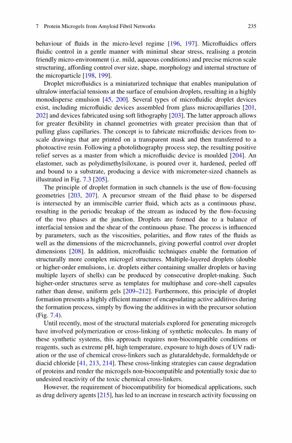
7 Protein Microgels from Amyloid Fibril Networks 235
behaviour of fluids in the micro-level regime [196, 197]. Microfluidics offersfluidic control in a gentle manner with minimal shear stress, realising a proteinfriendly micro-environment (i.e. mild, aqueous conditions) and precise micron scalestructuring, affording control over size, shape, morphology and internal structure ofthe microparticle [198, 199].
Droplet microfluidics is a miniaturized technique that enables manipulation ofultralow interfacial tensions at the surface of emulsion droplets, resulting in a highlymonodisperse emulsion [45, 200]. Several types of microfluidic droplet devicesexist, including microfluidic devices assembled from glass microcapillaries [201,202] and devices fabricated using soft lithography [203]. The latter approach allowsfor greater flexibility in channel geometries with greater precision than that ofpulling glass capillaries. The concept is to fabricate microfluidic devices from to-scale drawings that are printed on a transparent mask and then transferred to aphotoactive resin. Following a photolithography process step, the resulting positiverelief serves as a master from which a microfluidic device is moulded [204]. Anelastomer, such as polydimethylsiloxane, is poured over it, hardened, peeled offand bound to a substrate, producing a device with micrometer-sized channels asillustrated in Fig. 7.3 [205].
The principle of droplet formation in such channels is the use of flow-focusinggeometries [203, 207]. A precursor stream of the fluid phase to be dispersedis intersected by an immiscible carrier fluid, which acts as a continuous phase,resulting in the periodic breakup of the stream as induced by the flow-focusingof the two phases at the junction. Droplets are formed due to a balance ofinterfacial tension and the shear of the continuous phase. The process is influencedby parameters, such as the viscosities, polarities, and flow rates of the fluids aswell as the dimensions of the microchannels, giving powerful control over dropletdimensions [208]. In addition, microfluidic techniques enable the formation ofstructurally more complex microgel structures. Multiple-layered droplets (doubleor higher-order emulsions, i.e. droplets either containing smaller droplets or havingmultiple layers of shells) can be produced by consecutive droplet-making. Suchhigher-order structures serve as templates for multiphase and core-shell capsulesrather than dense, uniform gels [209–212]. Furthermore, this principle of dropletformation presents a highly efficient manner of encapsulating active additives duringthe formation process, simply by flowing the additives in with the precursor solution(Fig. 7.4).
Until recently, most of the structural materials explored for generating microgelshave involved polymerization or cross-linking of synthetic molecules. In many ofthese synthetic systems, this approach requires non-biocompatible conditions orreagents, such as extreme pH, high temperature, exposure to high doses of UV radi-ation or the use of chemical cross-linkers such as glutaraldehyde, formaldehyde ordiacid chloride [41, 213, 214]. These cross-linking strategies can cause degradationof proteins and render the microgels non-biocompatible and potentially toxic due toundesired reactivity of the toxic chemical cross-linkers.
However, the requirement of biocompatibility for biomedical applications, suchas drug delivery agents [215], has led to an increase in research activity focussing on
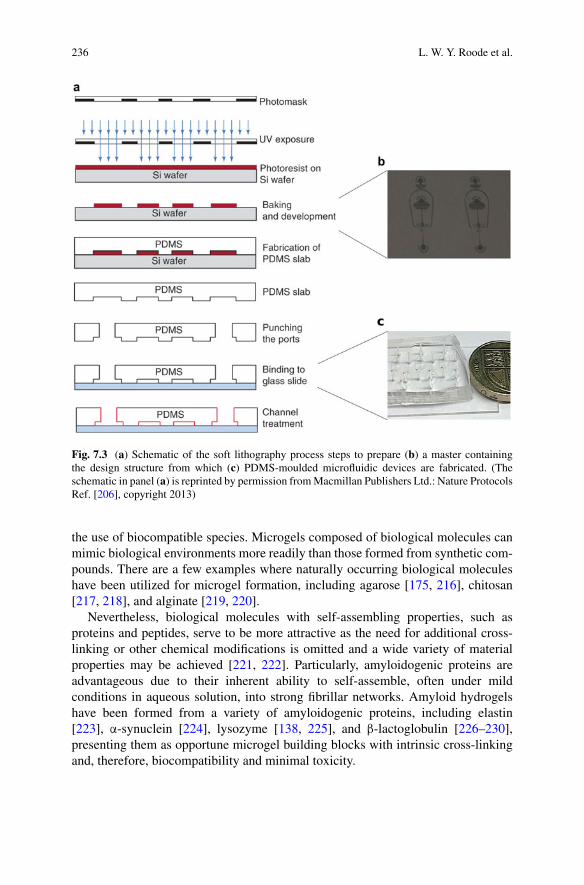
236 L. W. Y. Roode et al.
Fig. 7.3 (a) Schematic of the soft lithography process steps to prepare (b) a master containingthe design structure from which (c) PDMS-moulded microfluidic devices are fabricated. (Theschematic in panel (a) is reprinted by permission from Macmillan Publishers Ltd.: Nature ProtocolsRef. [206], copyright 2013)
the use of biocompatible species. Microgels composed of biological molecules canmimic biological environments more readily than those formed from synthetic com-pounds. There are a few examples where naturally occurring biological moleculeshave been utilized for microgel formation, including agarose [175, 216], chitosan[217, 218], and alginate [219, 220].
Nevertheless, biological molecules with self-assembling properties, such asproteins and peptides, serve to be more attractive as the need for additional cross-linking or other chemical modifications is omitted and a wide variety of materialproperties may be achieved [221, 222]. Particularly, amyloidogenic proteins areadvantageous due to their inherent ability to self-assemble, often under mildconditions in aqueous solution, into strong fibrillar networks. Amyloid hydrogelshave been formed from a variety of amyloidogenic proteins, including elastin[223], α-synuclein [224], lysozyme [138, 225], and β-lactoglobulin [226–230],presenting them as opportune microgel building blocks with intrinsic cross-linkingand, therefore, biocompatibility and minimal toxicity.
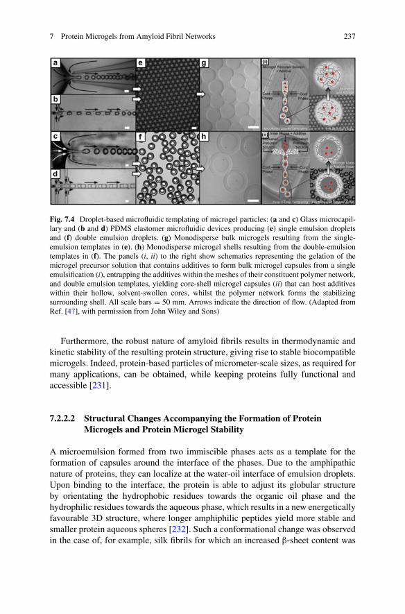
7 Protein Microgels from Amyloid Fibril Networks 237
Fig. 7.4 Droplet-based microfluidic templating of microgel particles: (a and c) Glass microcapil-lary and (b and d) PDMS elastomer microfluidic devices producing (e) single emulsion dropletsand (f) double emulsion droplets. (g) Monodisperse bulk microgels resulting from the single-emulsion templates in (e). (h) Monodisperse microgel shells resulting from the double-emulsiontemplates in (f). The panels (i, ii) to the right show schematics representing the gelation of themicrogel precursor solution that contains additives to form bulk microgel capsules from a singleemulsification (i), entrapping the additives within the meshes of their constituent polymer network,and double emulsion templates, yielding core-shell microgel capsules (ii) that can host additiveswithin their hollow, solvent-swollen cores, whilst the polymer network forms the stabilizingsurrounding shell. All scale bars = 50 mm. Arrows indicate the direction of flow. (Adapted fromRef. [47], with permission from John Wiley and Sons)
Furthermore, the robust nature of amyloid fibrils results in thermodynamic andkinetic stability of the resulting protein structure, giving rise to stable biocompatiblemicrogels. Indeed, protein-based particles of micrometer-scale sizes, as required formany applications, can be obtained, while keeping proteins fully functional andaccessible [231].
7.2.2.2 Structural Changes Accompanying the Formation of ProteinMicrogels and Protein Microgel Stability
A microemulsion formed from two immiscible phases acts as a template for theformation of capsules around the interface of the phases. Due to the amphipathicnature of proteins, they can localize at the water-oil interface of emulsion droplets.Upon binding to the interface, the protein is able to adjust its globular structureby orientating the hydrophobic residues towards the organic oil phase and thehydrophilic residues towards the aqueous phase, which results in a new energeticallyfavourable 3D structure, where longer amphiphilic peptides yield more stable andsmaller protein aqueous spheres [232]. Such a conformational change was observedin the case of, for example, silk fibrils for which an increased β-sheet content was
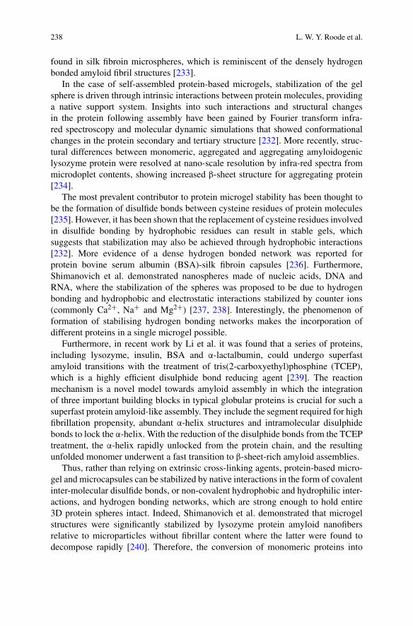
238 L. W. Y. Roode et al.
found in silk fibroin microspheres, which is reminiscent of the densely hydrogenbonded amyloid fibril structures [233].
In the case of self-assembled protein-based microgels, stabilization of the gelsphere is driven through intrinsic interactions between protein molecules, providinga native support system. Insights into such interactions and structural changesin the protein following assembly have been gained by Fourier transform infra-red spectroscopy and molecular dynamic simulations that showed conformationalchanges in the protein secondary and tertiary structure [232]. More recently, struc-tural differences between monomeric, aggregated and aggregating amyloidogeniclysozyme protein were resolved at nano-scale resolution by infra-red spectra frommicrodoplet contents, showing increased β-sheet structure for aggregating protein[234].
The most prevalent contributor to protein microgel stability has been thought tobe the formation of disulfide bonds between cysteine residues of protein molecules[235]. However, it has been shown that the replacement of cysteine residues involvedin disulfide bonding by hydrophobic residues can result in stable gels, whichsuggests that stabilization may also be achieved through hydrophobic interactions[232]. More evidence of a dense hydrogen bonded network was reported forprotein bovine serum albumin (BSA)-silk fibroin capsules [236]. Furthermore,Shimanovich et al. demonstrated nanospheres made of nucleic acids, DNA andRNA, where the stabilization of the spheres was proposed to be due to hydrogenbonding and hydrophobic and electrostatic interactions stabilized by counter ions(commonly Ca2+, Na+ and Mg2+) [237, 238]. Interestingly, the phenomenon offormation of stabilising hydrogen bonding networks makes the incorporation ofdifferent proteins in a single microgel possible.
Furthermore, in recent work by Li et al. it was found that a series of proteins,including lysozyme, insulin, BSA and α-lactalbumin, could undergo superfastamyloid transitions with the treatment of tris(2-carboxyethyl)phosphine (TCEP),which is a highly efficient disulphide bond reducing agent [239]. The reactionmechanism is a novel model towards amyloid assembly in which the integrationof three important building blocks in typical globular proteins is crucial for such asuperfast protein amyloid-like assembly. They include the segment required for highfibrillation propensity, abundant α-helix structures and intramolecular disulphidebonds to lock the α-helix. With the reduction of the disulphide bonds from the TCEPtreatment, the α-helix rapidly unlocked from the protein chain, and the resultingunfolded monomer underwent a fast transition to β-sheet-rich amyloid assemblies.
Thus, rather than relying on extrinsic cross-linking agents, protein-based micro-gel and microcapsules can be stabilized by native interactions in the form of covalentinter-molecular disulfide bonds, or non-covalent hydrophobic and hydrophilic inter-actions, and hydrogen bonding networks, which are strong enough to hold entire3D protein spheres intact. Indeed, Shimanovich et al. demonstrated that microgelstructures were significantly stabilized by lysozyme protein amyloid nanofibersrelative to microparticles without fibrillar content where the latter were found todecompose rapidly [240]. Therefore, the conversion of monomeric proteins into
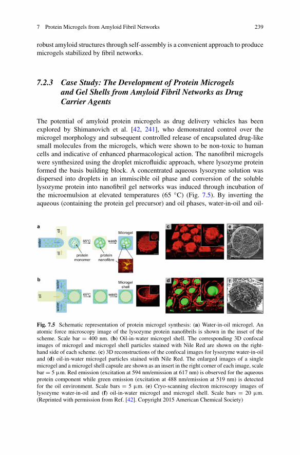
7 Protein Microgels from Amyloid Fibril Networks 239
robust amyloid structures through self-assembly is a convenient approach to producemicrogels stabilized by fibril networks.
7.2.3 Case Study: The Development of Protein Microgelsand Gel Shells from Amyloid Fibril Networks as DrugCarrier Agents
The potential of amyloid protein microgels as drug delivery vehicles has beenexplored by Shimanovich et al. [42, 241], who demonstrated control over themicrogel morphology and subsequent controlled release of encapsulated drug-likesmall molecules from the microgels, which were shown to be non-toxic to humancells and indicative of enhanced pharmacological action. The nanofibril microgelswere synthesized using the droplet microfluidic approach, where lysozyme proteinformed the basis building block. A concentrated aqueous lysozyme solution wasdispersed into droplets in an immiscible oil phase and conversion of the solublelysozyme protein into nanofibril gel networks was induced through incubation ofthe microemulsion at elevated temperatures (65 ◦C) (Fig. 7.5). By inverting theaqueous (containing the protein gel precursor) and oil phases, water-in-oil and oil-
Fig. 7.5 Schematic representation of protein microgel synthesis: (a) Water-in-oil microgel. Anatomic force microscopy image of the lysozyme protein nanofibrils is shown in the inset of thescheme. Scale bar = 400 nm. (b) Oil-in-water microgel shell. The corresponding 3D confocalimages of microgel and microgel shell particles stained with Nile Red are shown on the right-hand side of each scheme. (c) 3D reconstructions of the confocal images for lysozyme water-in-oiland (d) oil-in-water microgel particles stained with Nile Red. The enlarged images of a singlemicrogel and a microgel shell capsule are shown as an insert in the right corner of each image, scalebar = 5 μm. Red emission (excitation at 594 nm/emission at 617 nm) is observed for the aqueousprotein component while green emission (excitation at 488 nm/emission at 519 nm) is detectedfor the oil environment. Scale bars = 5 μm. (e) Cryo-scanning electron microscopy images oflysozyme water-in-oil and (f) oil-in-water microgel and microgel shell. Scale bars = 20 μm.(Reprinted with permission from Ref. [42]. Copyright 2015 American Chemical Society)
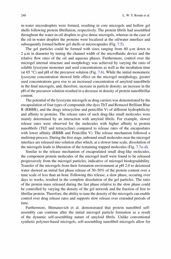
240 L. W. Y. Roode et al.
in-water microdroplets were formed, resulting in core microgels and hollow gelshells following protein fibrillation, respectively. The protein fibrils had assembledthroughout the water-in-oil droplets to give dense microgels, whereas in the case ofthe oil-in-water droplets the proteins were localized at the oil/water interface andsubsequently formed hollow gel shells or microcapsules (Fig. 7.5).
The gel particles could be formed with sizes ranging from 60 μm down to2 μm in diameter by tuning the channel width of the microfluidic device and therelative flow rates of the oil and aqueous phases. Furthermore, control over themicrogel internal structure and morphology was achieved by varying the ratio ofsoluble lysozyme monomer and seed concentrations as well as the incubation time(at 65 ◦C) and pH of the precursor solution (Fig. 7.6). While the initial monomericlysozyme concentration showed little effect on the microgel morphology, greaterseed concentrations gave rise to an increased concentration of amyloid nanofibrilsin the final microgels, and, therefore, increase in particle density; an increase in thepH of the precursor solution resulted in a decrease in density of protein nanofibrillarcontent.
The potential of the lysozyme microgels as drug carriers was demonstrated by theencapsulation of four types of compounds (the dyes ThT and Remazol Brilliant BlueR (RBBR), and the drugs tetracycline and penicillin V) of different hydrophilicityand affinity to proteins. The release rates of such drug-like small molecules weremainly determined by an interaction with amyloid fibrils. For example, slowerrelease rates were observed for the molecules with higher affinity to proteinnanofibrils (ThT and tetracycline) compared to release rates of the encapsulateswith lower affinity (RBBR and Penicillin V). The release mechanism followed amultistep process. During the first stage, unbound small molecules near the microgelinterface are released into solution after which, at a slower time scale, dissolution ofthe microgels leads to liberation of the remaining trapped molecules (Fig. 7.7a–d).
Similar to the release mechanism of encapsulated small drug-like molecules,the component protein molecules of the microgel itself were found to be releasedprogressively from the microgel particles, indicative of microgel biodegradability.Transfer of the microgels from their formation environment at pH 2.0 to deionizedwater showed an initial fast phase release of 30–50% of the protein content over atime scale of less than an hour. Following this release, a slow phase, occuring overdays to weeks, resulted in the complete dissolution of the gel particles. The ratioof the protein mass released during the fast phase relative to the slow phase couldbe controlled by varying the density of the gel network and the fraction of free tofibrillar protein. Therefore, the ability to tune the density of the microgels can enablecontrol over drug release rates and supports slow release over extended periods oftime.
Furthermore, Shimanovich et al. demonstrated that protein nanofibril self-assembly can continue after the initial microgel particle formation as a resultof the dynamic self-assembling nature of amyloid fibrils. Unlike conventionalsynthetic polymer-based microgels, self-assembling nanofibril microgels allow for
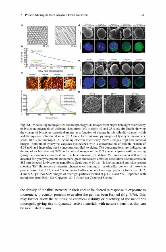
7 Protein Microgels from Amyloid Fibril Networks 241
Fig. 7.6 Modulating microgel size and morphology: (a) Images from bright field light microscopyof lysozyme microgels of different sizes (from left to right: 49 and 23 μm). (b) Graph showingthe change of lysozyme capsule diameter as a function of change in microfluidic channel widthand the aqueous solution:oil ratio. (c) Atomic force microscopy images of lysozyme monomers,seeds, fibrils and microgel. (d) Scanning electron microscopy (SEM) images (top) and confocalimages (bottom) of lysozyme capsules synthesized with a concentration of soluble protein of4.08 mM and increasing seed concentrations (left to right). The concentrations are indicated onthe top of each image. (e) SEM and confocal images of the ThT stained capsule with increasinglysozyme monomer concentration. The blue emission (excitation 350 nm/emission 438 nm) isdetected for lysozyme protein monomers, green fluorescent emission (excitation 450 nm/emission482 nm) detected for lysozyme nanofibrils. Scale bars = 10 μm. (f) Excitation and emission spectrashowing ThT fluorescence intensity change upon binding to nanofibrillar content of lysozymeprotein formed at pH 2, 4 and 5.5 and nanofibrillar content of microgel particles formed at pH 2,4 and 5.5. (g) Cryo-SEM images of microgel particles formed at pH 2, 4 and 5.5. (Reprinted withpermission from Ref. [42]. Copyright 2015 American Chemical Society)
the density of the fibril network in their core to be altered in response to exposure tomonomeric precursor proteins even after the gel has been formed (Fig. 7.7e). Thismay further allow the tailoring of chemical stability or reactivity of the nanofibrilmicrogels, giving rise to dynamic, active materials with network densities that canbe modulated in situ.
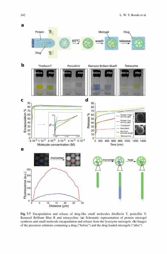
242 L. W. Y. Roode et al.
Fig. 7.7 Encapsulation and release of drug-like small molecules thioflavin T, penicillin V,Remazol Brilliant Blue R and tetracycline: (a) Schematic representation of protein microgelsynthesis and small molecule encapsulation and release from the lysozyme microgels. (b) Imagesof the precursor solutions containing a drug (“before”) and the drug loaded microgels (“after”)
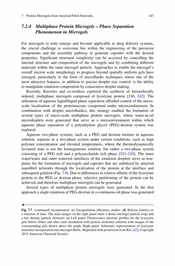
7 Protein Microgels from Amyloid Fibril Networks 243
7.2.4 Multiphase Protein Microgels – Phase SeparationPhenomenon in Microgels
For microgels to truly emerge and become applicable as drug delivery systems,the crucial challenge to overcome lies within the engineering of the precursorcomponents and the assembly pathway to generate capsules with the desiredproperties. Significant structural complexity can be accessed by controlling theinternal structure and composition of the microgels and by combining differentmaterials within the same microgel particle. Approaches to enable the microgel’soverall micron scale morphology to progress beyond spatially uniform gels haveemerged, particularly in the form of microfluidic techniques where one of themost attractive features, in addition to precise droplet size control, is the abilityto manipulate emulsion composition by consecutive droplet making.
Recently, Knowles and co-workers explored the synthesis of hierarchicallyordered, multiphase microgels composed of lysozyme protein [240, 242]. Theutilization of aqueous liquid/liquid phase separation afforded control of the micro-scale localization of the proteinaceous component under microconfinement. Incombination with droplet microfluidics, this strategy enabled the formation ofseveral types of micro-scale multiphase protein microgels, where water-in-oilmicrodroplets were generated that serve as a microenvironment within whichaqueous phase separation of a polyethylene glycol (PEG)-dextran system wasexplored.
Aqueous two-phase systems, such as a PEG and dextran mixture in aqueoussolution, separate in a two-phase system under certain conditions, such as highpolymer concentration and elevated temperatures, where the thermodynamicallyfavoured state is not the homogeneous solution, but rather a two-phase systemconsisting of a PEG rich and a polysaccharide rich phase [243–245]. The innerwater/water and outer water/oil interfaces of the emulsion droplets serve as tem-plates for the formation of microgels and capsules that are stabilized by amyloidnanofibril networks through the localization of the protein at the interface andsubsequent gelation (Fig. 7.8). Due to differences in relative affinity of the lysozymeprotein to the PEG or dextran phase, selective partitioning of the protein can beachieved, and therefore multiphase microgels can be generated.
Several types of multiphase protein microgels were generated. In the firstapproach a single emulsion of PEG-dextran in a continuous oil phase was generated
�Fig. 7.7 (continued) incorporation. (c) Encapsulation efficiency studies. (d) Release kinetics asa function of time. The inset images on the right panel show a dense microgel particle (top) anda low density particle (bottom). (e) Left panel: Fluorescence intensity profiles for the lysozymegels before (blue) and after (red) incubation with protein monomer solution with images of thecorresponding gels shown above the graph. Right panel: Schematic representation of lysozymemonomer incorporation into microgel fibrils. (Reprinted with permission from Ref. [42]. Copyright2015 American Chemical Society)
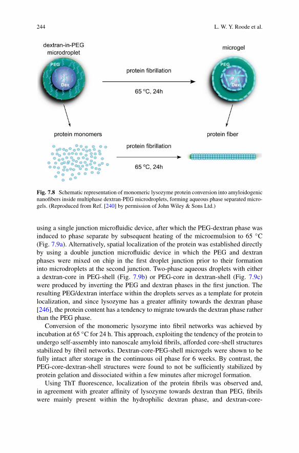
244 L. W. Y. Roode et al.
Fig. 7.8 Schematic representation of monomeric lysozyme protein conversion into amyloidogenicnanofibers inside multiphase dextran-PEG microdroplets, forming aqueous phase separated micro-gels. (Reproduced from Ref. [240] by permission of John Wiley & Sons Ltd.)
using a single junction microfluidic device, after which the PEG-dextran phase wasinduced to phase separate by subsequent heating of the microemulsion to 65 ◦C(Fig. 7.9a). Alternatively, spatial localization of the protein was established directlyby using a double junction microfluidic device in which the PEG and dextranphases were mixed on chip in the first droplet junction prior to their formationinto microdroplets at the second junction. Two-phase aqueous droplets with eithera dextran-core in PEG-shell (Fig. 7.9b) or PEG-core in dextran-shell (Fig. 7.9c)were produced by inverting the PEG and dextran phases in the first junction. Theresulting PEG/dextran interface within the droplets serves as a template for proteinlocalization, and since lysozyme has a greater affinity towards the dextran phase[246], the protein content has a tendency to migrate towards the dextran phase ratherthan the PEG phase.
Conversion of the monomeric lysozyme into fibril networks was achieved byincubation at 65 ◦C for 24 h. This approach, exploiting the tendency of the protein toundergo self-assembly into nanoscale amyloid fibrils, afforded core-shell structuresstabilized by fibril networks. Dextran-core-PEG-shell microgels were shown to befully intact after storage in the continuous oil phase for 6 weeks. By contrast, thePEG-core-dextran-shell structures were found to not be sufficiently stabilized byprotein gelation and dissociated within a few minutes after microgel formation.
Using ThT fluorescence, localization of the protein fibrils was observed and,in agreement with greater affinity of lysozyme towards dextran than PEG, fibrilswere mainly present within the hydrophilic dextran phase, and dextran-core-
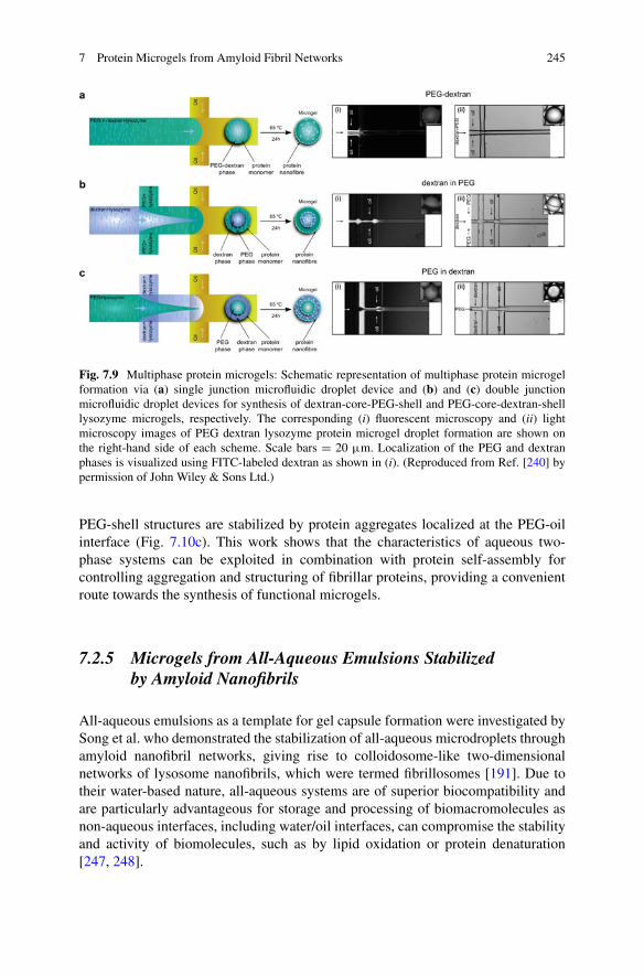
7 Protein Microgels from Amyloid Fibril Networks 245
Fig. 7.9 Multiphase protein microgels: Schematic representation of multiphase protein microgelformation via (a) single junction microfluidic droplet device and (b) and (c) double junctionmicrofluidic droplet devices for synthesis of dextran-core-PEG-shell and PEG-core-dextran-shelllysozyme microgels, respectively. The corresponding (i) fluorescent microscopy and (ii) lightmicroscopy images of PEG dextran lysozyme protein microgel droplet formation are shown onthe right-hand side of each scheme. Scale bars = 20 μm. Localization of the PEG and dextranphases is visualized using FITC-labeled dextran as shown in (i). (Reproduced from Ref. [240] bypermission of John Wiley & Sons Ltd.)
PEG-shell structures are stabilized by protein aggregates localized at the PEG-oilinterface (Fig. 7.10c). This work shows that the characteristics of aqueous two-phase systems can be exploited in combination with protein self-assembly forcontrolling aggregation and structuring of fibrillar proteins, providing a convenientroute towards the synthesis of functional microgels.
7.2.5 Microgels from All-Aqueous Emulsions Stabilizedby Amyloid Nanofibrils
All-aqueous emulsions as a template for gel capsule formation were investigated bySong et al. who demonstrated the stabilization of all-aqueous microdroplets throughamyloid nanofibril networks, giving rise to colloidosome-like two-dimensionalnetworks of lysosome nanofibrils, which were termed fibrillosomes [191]. Due totheir water-based nature, all-aqueous systems are of superior biocompatibility andare particularly advantageous for storage and processing of biomacromolecules asnon-aqueous interfaces, including water/oil interfaces, can compromise the stabilityand activity of biomolecules, such as by lipid oxidation or protein denaturation[247, 248].
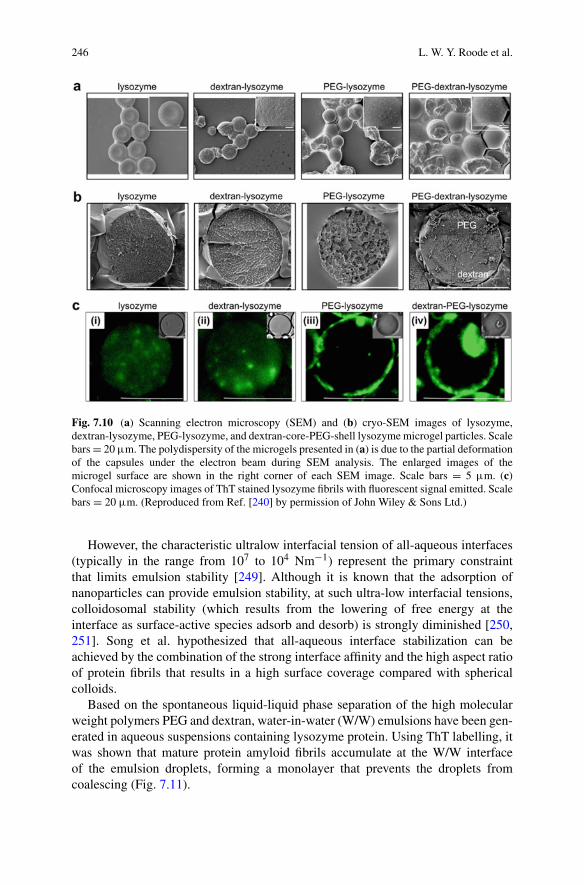
246 L. W. Y. Roode et al.
Fig. 7.10 (a) Scanning electron microscopy (SEM) and (b) cryo-SEM images of lysozyme,dextran-lysozyme, PEG-lysozyme, and dextran-core-PEG-shell lysozyme microgel particles. Scalebars = 20 μm. The polydispersity of the microgels presented in (a) is due to the partial deformationof the capsules under the electron beam during SEM analysis. The enlarged images of themicrogel surface are shown in the right corner of each SEM image. Scale bars = 5 μm. (c)Confocal microscopy images of ThT stained lysozyme fibrils with fluorescent signal emitted. Scalebars = 20 μm. (Reproduced from Ref. [240] by permission of John Wiley & Sons Ltd.)
However, the characteristic ultralow interfacial tension of all-aqueous interfaces(typically in the range from 107 to 104 Nm−1) represent the primary constraintthat limits emulsion stability [249]. Although it is known that the adsorption ofnanoparticles can provide emulsion stability, at such ultra-low interfacial tensions,colloidosomal stability (which results from the lowering of free energy at theinterface as surface-active species adsorb and desorb) is strongly diminished [250,251]. Song et al. hypothesized that all-aqueous interface stabilization can beachieved by the combination of the strong interface affinity and the high aspect ratioof protein fibrils that results in a high surface coverage compared with sphericalcolloids.
Based on the spontaneous liquid-liquid phase separation of the high molecularweight polymers PEG and dextran, water-in-water (W/W) emulsions have been gen-erated in aqueous suspensions containing lysozyme protein. Using ThT labelling, itwas shown that mature protein amyloid fibrils accumulate at the W/W interfaceof the emulsion droplets, forming a monolayer that prevents the droplets fromcoalescing (Fig. 7.11).
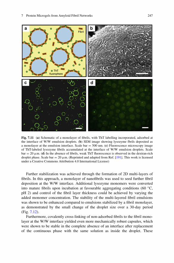
7 Protein Microgels from Amyloid Fibril Networks 247
Fig. 7.11 (a) Schematic of a monolayer of fibrils, with ThT labelling incorporated, adsorbed atthe interface of W/W emulsion droplets. (b) SEM image showing lysozyme fbrils deposited asa monolayer at the emulsion interface. Scale bar = 500 nm. (c) Fluorescence microscopy imageof ThT-labeled lysozyme fibrils accumulated at the interface of W/W emulsion droplets. Scalebar = 20 μm. (d) In the absence of fibrils, weak ThT fluorescence is observed in the dextran-richdroplet phase. Scale bar = 20 μm. (Reprinted and adapted from Ref. [191]. This work is licensedunder a Creative Commons Attribution 4.0 International License)
Further stabilization was achieved through the formation of 2D multi-layers offibrils. In this approach, a monolayer of nanofibrils was used to seed further fibrildeposition at the W/W interface. Additional lysozyme monomers were convertedinto mature fibrils upon incubation at favourable aggregating conditions (60 ◦C,pH 2) and control of the fibril layer thickness could be achieved by varying theadded monomer concentration. The stability of the multi-layered fibril emulsionswas shown to be enhanced compared to emulsions stabilized by a fibril monolayer,as demonstrated by the small change of the droplet size over a 30-day period(Fig. 7.12).
Furthermore, covalently cross-linking of non-adsorbed fibrils to the fibril mono-layer at the W/W interface yielded even more mechanically robust capsules, whichwere shown to be stable in the complete absence of an interface after replacementof the continuous phase with the same solution as inside the droplet. These
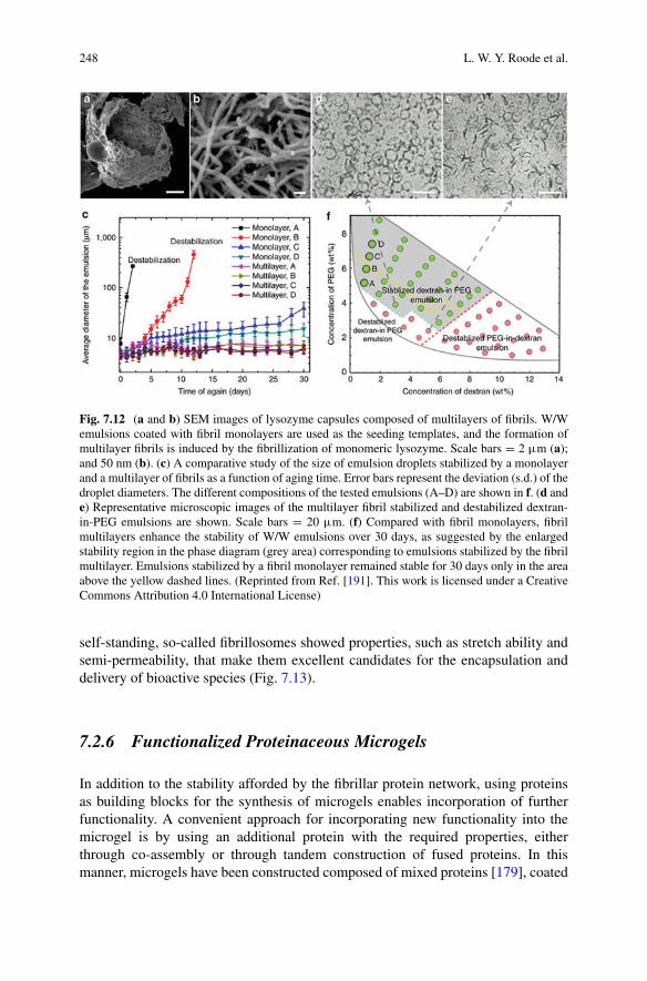
248 L. W. Y. Roode et al.
Fig. 7.12 (a and b) SEM images of lysozyme capsules composed of multilayers of fibrils. W/Wemulsions coated with fibril monolayers are used as the seeding templates, and the formation ofmultilayer fibrils is induced by the fibrillization of monomeric lysozyme. Scale bars = 2 μm (a);and 50 nm (b). (c) A comparative study of the size of emulsion droplets stabilized by a monolayerand a multilayer of fibrils as a function of aging time. Error bars represent the deviation (s.d.) of thedroplet diameters. The different compositions of the tested emulsions (A–D) are shown in f. (d ande) Representative microscopic images of the multilayer fibril stabilized and destabilized dextran-in-PEG emulsions are shown. Scale bars = 20 μm. (f) Compared with fibril monolayers, fibrilmultilayers enhance the stability of W/W emulsions over 30 days, as suggested by the enlargedstability region in the phase diagram (grey area) corresponding to emulsions stabilized by the fibrilmultilayer. Emulsions stabilized by a fibril monolayer remained stable for 30 days only in the areaabove the yellow dashed lines. (Reprinted from Ref. [191]. This work is licensed under a CreativeCommons Attribution 4.0 International License)
self-standing, so-called fibrillosomes showed properties, such as stretch ability andsemi-permeability, that make them excellent candidates for the encapsulation anddelivery of bioactive species (Fig. 7.13).
7.2.6 Functionalized Proteinaceous Microgels
In addition to the stability afforded by the fibrillar protein network, using proteinsas building blocks for the synthesis of microgels enables incorporation of furtherfunctionality. A convenient approach for incorporating new functionality into themicrogel is by using an additional protein with the required properties, eitherthrough co-assembly or through tandem construction of fused proteins. In thismanner, microgels have been constructed composed of mixed proteins [179], coated
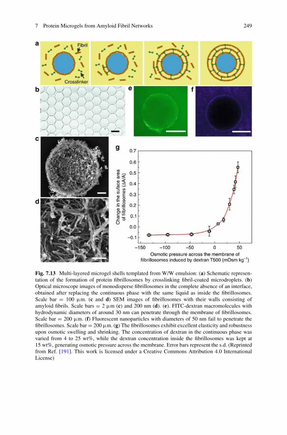
7 Protein Microgels from Amyloid Fibril Networks 249
Fig. 7.13 Multi-layered microgel shells templated from W/W emulsion: (a) Schematic represen-tation of the formation of protein fibrillosomes by crosslinking fibril-coated microdroplets. (b)Optical microscope images of monodisperse fibrillosomes in the complete absence of an interface,obtained after replacing the continuous phase with the same liquid as inside the fibrillosomes.Scale bar = 100 μm. (c and d) SEM images of fibrillosomes with their walls consisting ofamyloid fibrils. Scale bars = 2 μm (c) and 200 nm (d). (e). FITC-dextran macromolecules withhydrodynamic diameters of around 30 nm can penetrate through the membrane of fibrillosomes.Scale bar = 200 μm. (f) Fluorescent nanoparticles with diameters of 50 nm fail to penetrate thefibrillosomes. Scale bar = 200 μm. (g) The fibrillosomes exhibit excellent elasticity and robustnessupon osmotic swelling and shrinking. The concentration of dextran in the continuous phase wasvaried from 4 to 25 wt%, while the dextran concentration inside the fibrillosomes was kept at15 wt%, generating osmotic pressure across the membrane. Error bars represent the s.d. (Reprintedfrom Ref. [191]. This work is licensed under a Creative Commons Attribution 4.0 InternationalLicense)
250 L. W. Y. Roode et al.
with biocompatible polymers [180], and protein spheres conjugated with targetligands, such as folate, for targeted drug delivery [252]. Moreover, Rahimipourand co-workers presented surface-conjugated protein microspheres that captureamyloid-β and inhibit its aggregation and toxicity [253]. In this case, the globularprotein BSA (in its non-aggregating form) was the main structural protein ofwhich the capsule shell was composed and the capsule surface was functionalizedwith the KLVFF peptide [254]. The KLVFF peptide is a known inhibitor of theamyloidogenic Aβ-40 peptide aggregation phenomenon that is associated withAlzheimer’s disease. The BSA-KLVFF conjugated protein capsules show highaffinity and selectivity for Aβ, sequestering the protein and, thereby, reducing itstoxicity [254].
Functionalization of amyloid fibrils via gene fusion has been explored by Perrettand co-workers who demonstrated the immobilization of active enzymes, includingalkaline phosphatase (AP) and horseradish peroxidase (HRP) on amyloid fibrilsformed by the prion domain of the yeast prion protein Ure2 [255]. The gene fusionprocess gives rise to a polypeptide that maintains the ability to self-assemble aswell as to display the desired activity of the fused entity. In following work, Zhouet al. demonstrated the formation of enzymatically active microgels from the Ure2-AP chimeric protein construct. The microgels showed robust material propertiesand their porous architecture allowed for diffusion of reactants and products in andout of the microgels, illustrating the potential for enzyme immobilization and forbiological flow-chemistry or bioactivity assays [231].
The physicochemical properties of the microgel can be further tailored to makethem responsive to environmental cues and thus afford targeted drug deliveryand modulation of release [256, 257]. For instance, engineering protein capsuleswith an external hydrophobic layer can introduce specificity to a target site, aidtransdermal transport, encode a release switch mechanism or enhance the biologicaleffect of the active species delivered through the capsules. As an example, RNAloaded BSA microspheres (where BSA protein appears in its native fold) werecoated with metal (Fe2O3), uncharged polyvinylalcohol (PVA) or positively chargedpolyethyleneimine (PEI) biocompatible synthetic polymers that buried the negativecharge of the BSA. This eliminated the adhesion to the cell membrane, whichwas observed in the case of uncoated capsules, and allowed efficient delivery ofRNA into human osteosarcoma U2OS cancer cells and Trypanosoma parasites.In addition, the Fe2O3 metal coating could allow enhanced contrast in electronmicroscopy for the spatial localization of the Fe2O3 metal coated capsules insidethe parasites and mammalian cancer cells [180]. Furthermore, the transport ofviruses and metal nanoparticles in living cells was shown to be enhanced throughtransfection mediated by non-toxic amyloid fibrils [258–261].
In a further example of tuning of microgel physicochemical properties, Levinet al. synthesized hybrid organic/inorganic microcapsules [262]. Lysozyme compos-ites were functionalized with carboxyl-modified Fe3O4 nanoparticles (NPs), whichhave been used extensively in biological applications due to their biocompatibility,small dimension (<30 nm), ease of characterization, and the rich diversity ofsurface chemistry modifications that can be exploited for biomedical applications

7 Protein Microgels from Amyloid Fibril Networks 251
[263, 264]. Lysozyme and Fe3O4 NPs exhibit a strong interaction that maybe attributed to their electrostatic interactions and there is evidence that NPsmay influence amyloid aggregation kinetics [265, 266]. This effect was furtherexplored for lysozyme self-assembly under the microconfinement of microdropletsin Levin’s work. It was shown that, in the presence of carboxyl-modified Fe3O4NPs, lysozyme fibrillation was inhibited in unseeded conditions, while underseeded conditions, fibrillation occurred that allowed for extensive decoration ofthe fibrillary microcapsules. It was suggested that the lysozyme monomers mayhave a greater affinity to the carboxyl-modified Fe3O4 NPs, which would inhibitfurther protein aggregation and, thus, resulting in less stable microgel structures.In addition, the fibril morphology within the microgels was shown to vary uponprotein fibrillation in the presence of the NPs, indicative of their structure-modifyingand aggregation-modulating role on protein self-assembly. These findings suggest anovel approach for tuning protein-based microgel formation, allowing control overthe morphology and characteristics of the end product [262].
7.3 Conclusions
Although amyloid fibril structures were initially recognized in association withdisease, the increase in research focused on their functional roles in nature andtheir potential as the basis of artificial materials with complex functionality presentthese as excellent building blocks for the development of functional materials withapplications, for example, as drug delivery agents. In addition to their biocom-patibility, the self-assembling ability of proteins can be exploited, in combinationwith microfluidic techniques, to produce multi-scale functional microgels capableof hosting biologically active compounds. The microgel formation process avoidsthe use of harsh reagents or conditions and enables precise control over capsulesize, stability and morphology. The ability to tailor such features is advantageousin the development of biomedical applications, such as drug delivery, as drugrelease can be controlled. Moreover, the use of proteins and peptides as functionalmaterial building blocks allows incorporation of additional functionality throughprotein modification and/or coupling with other functional molecules, enhancingthe versatility of this class of materials as well as leading to responsive microgels,enabling targeted drug delivery. Thus, amyloid fibril microgels represent attractivedesigner materials for biomedical applications, including drug delivery.
References
1. Hardy J, Selkoe DJ (2002) The amyloid hypothesis of Alzheimer’s disease: progress andproblems on the road to therapeutics. Science 297:353–356
2. Haass C, Selkoe DJ (2007) Soluble protein oligomers in neurodegeneration: lessons from theAlzheimer’s amyloid β-peptide. Nat Rev Mol Cell Biol 8:101–112
252 L. W. Y. Roode et al.
3. Ballard C, Gauthier S, Corbett A, Brayne C, Aarsland D, Jones E (2011) Alzheimer’s disease.Lancet Lond Engl 377:1019–1031
4. Querfurth HW, LaFerla FM (2010) Mechanisms of disease Alzheimer’s disease. N Engl JMed 362:329–344
5. Niedowicz DM, Nelson PT, Murphy MP (2011) Alzheimer’s disease: pathological mecha-nisms and recent insights. Curr Neuropharmacol 9:674–684
6. Dobson CM (1999) Protein misfolding, evolution and disease. Trends Biochem Sci 24:329–332
7. Dobson CM (2003) Protein folding and misfolding. Nature 426:884–8908. Chiti F, Dobson CM (2006) Protein misfolding, functional amyloid, and human disease. Annu
Rev Biochem 75:333–3669. Knowles TPJ, Vendruscolo M, Dobson CM (2014) The amyloid state and its association with
protein misfolding diseases. Nat Rev Mol Cell Biol 15:384–39610. Stefani M, Dobson CM (2003) Protein aggregation and aggregate toxicity: new insights into
protein folding, misfolding diseases and biological evolution. J Mol Med 81:678–69911. Fowler DM, Koulov AV, Alory-Jost C, Marks MS, Balch WE, Kelly JW (2006)
Functional amyloid formation within mammalian tissue. PLoS Biol 4(1):100–107.https://doi.org/10.1371/journal.pbio.0040006
12. Fowler DM, Koulov AV, Balch WE, Kelly JW (2007) Functional amyloid – from bacteria tohumans. Trends Biochem Sci 32:217–224
13. Otzen D, Nielsen PH (2008) We find them here, we find them there: functional bacterialamyloid. Cell Mol Life Sci 65:910–927
14. Kelly JW, Balch WE (2003) Amyloid as a natural product. J Cell Biol 161:461–46215. Maji SK, Perrin MH, Sawaya MR et al (2009) Functional amyloids as natural storage of
peptide hormones in pituitary secretory granules. Science 325:328–33216. Bleem A, Daggett V (2017) Structural and functional diversity among amyloid proteins:
agents of disease, building blocks of biology, and implications for molecular engineering.Biotechnol Bioeng 114:7–20
17. Mostaert AS, Higgins MJ, Fukuma T, Rindi F, Jarvis SP (2006) Nanoscale mechanicalcharacterisation of amyloid fibrils discovered in a natural adhesive. J Biol Phys 32:393–401
18. Zhong C, Gurry T, Cheng AA, Downey J, Deng Z, Stultz CM, Lu TK (2014) Strongunderwater adhesives made by self-assembling multi-protein nanofibres. Nat Nanotechnol9:858–866
19. Li C, Qin R, Liu R, Miao S, Yang P (2018) Functional amyloid materials at sur-faces/interfaces. Biomater Sci 6:462–472
20. Wang D, Ha Y, Gu J, Li Q, Zhang L, Yang P (2016) 2D protein supramolecular nanofilm withexceptionally large area and emergent functions. Adv Mater 28:7414–7423
21. Chapman MR, Robinson LS, Pinkner JS, Roth R, Heuser J, Hammar M, Normark S, HultgrenSJ (2002) Role of Escherichia coli curli operons in directing amyloid fiber formation. Science295:851–855
22. Shorter J, Lindquist S (2005) Prions as adaptive conduits of memory and inheritance. Nat RevGenet 6:435–450
23. Krishnan R, Lindquist SL (2005) Structural insights into a yeast prion illuminate nucleationand strain diversity. Nature 435:765–772
24. Lindquist SL, Henikoff S (2002) Self-perpetuating structural states in biology, disease, andgenetics. Proc Natl Acad Sci U S A 99:16377
25. Tanaka M, Collins SR, Toyama BH, Weissman JS (2006) The physical basis of how prionconformations determine strain phenotypes. Nature 442:585–589
26. DePace AH, Weissman JS (2002) Origins and kinetic consequences of diversity in Sup35yeast prion fibers. Nat Struct Mol Biol 9:389–396
27. Lashuel HA, Hartley D, Petre BM, Walz T, Lansbury PT (2002) Neurodegenerative disease:amyloid pores from pathogenic mutations. Nature 418:291–291
7 Protein Microgels from Amyloid Fibril Networks 253
28. Bucciantini M, Giannoni E, Chiti F, Baroni F, Formigli L, Zurdo J, Taddei N, Ramponi G,Dobson CM, Stefani M (2002) Inherent toxicity of aggregates implies a common mechanismfor protein misfolding diseases. Nature 416:507–511
29. Walsh DM, Klyubin I, Fadeeva JV, Cullen WK, Anwyl R, Wolfe MS, Rowan MJ, SelkoeDJ (2002) Naturally secreted oligomers of amyloid β protein potently inhibit hippocampallong-term potentiation in vivo. Nature 416:535–539
30. Caughey B, Peter T, Lansbury J (2003) Protofibirls, pores, fibrils, and neurodegeneration:separating the responsible protein aggregates from the innocent bystanders. Annu RevNeurosci 26:267–298
31. Koffie RM, Meyer-Luehmann M, Hashimoto T et al (2009) Oligomeric amyloid β associateswith postsynaptic densities and correlates with excitatory synapse loss near senile plaques.Proc Natl Acad Sci U S A 106:4012–4017
32. Tomic JL, Pensalfini A, Head E, Glabe CG (2009) Soluble fibrillar oligomer levels areelevated in Alzheimer’s disease brain and correlate with cognitive dysfunction. NeurobiolDis 35:352–358
33. Shankar GM, Bloodgood BL, Townsend M, Walsh DM, Selkoe DJ, Sabatini BL (2007)Natural oligomers of the Alzheimer amyloid-β protein induce reversible synapse loss bymodulating an NMDA-type glutamate receptor-dependent signaling pathway. J Neurosci27:2866–2875
34. Hammer ND, Schmidt JC, Chapman MR (2007) The curli nucleator protein, CsgB, containsan amyloidogenic domain that directs CsgA polymerization. Proc Natl Acad Sci U S A104:12494–12499
35. Knowles TP, Fitzpatrick AW, Meehan S, Mott HR, Vendruscolo M, Dobson CM, Welland ME(2007) Role of intermolecular forces in defining material properties of protein nanofibrils.Science 318:1900–1903
36. Knowles TPJ, Buehler MJ (2011) Nanomechanics of functional and pathological amyloidmaterials. Nat Nanotechnol 6:469–479
37. Chiti F, Webster P, Taddei N, Clark A, Stefani M, Ramponi G, Dobson CM (1999) Designingconditions for in vitro formation of amyloid protofilaments and fibrils. Proc Natl Acad Sci US A 96:3590–3594
38. Chiti F, Dobson CM (2009) Amyloid formation by globular proteins under native conditions.Nat Chem Biol 5:15–22
39. Pawar AP, DuBay KF, Zurdo J, Chiti F, Vendruscolo M, Dobson CM (2005) Predictionof “aggregation-prone” and “aggregation-susceptible” regions in proteins associated withneurodegenerative diseases. J Mol Biol 350:379–392
40. Knowles TPJ, Mezzenga R (2016) Amyloid fibrils as building blocks for natural and artificialfunctional materials. Adv Mater 28:6546–6561
41. Shimanovich U, Bernardes GJ, Knowles TP, Cavaco-Paulo A (2014) Protein micro- and nano-capsules for biomedical applications. Chem Soc Rev 43:1361–1371
42. Shimanovich U, Efimov I, Mason TO et al (2015) Protein microgels from amyloid fibrilnetworks. ACS Nano 9:43–51
43. Cao A, Hu D, Lai L (2004) Formation of amyloid fibrils from fully reduced hen egg whitelysozyme. Protein Sci Publ Protein Soc 13:319–324
44. Kelly JW (2002) Towards an understanding of amyloidogenesis. Nat Struct Mol Biol 9:323–325
45. Teh S-Y, Lin R, Hung L-H, Lee AP (2008) Droplet microfluidics. Lab Chip 8:198–22046. Zhang H, Tumarkin E, Sullan RMA, Walker GC, Kumacheva E (2007) Exploring microfluidic
routes to microgels of biological polymers. Macromol Rapid Commun 28:527–53847. Seiffert S (2013) Microgel capsules tailored by droplet-based microfluidics. ChemPhysChem
14:295–30448. Sipe JD, Cohen AS (2000) Review: history of the amyloid fibril. J Struct Biol 130:88–9849. Buxbaum JN, Linke RP (2012) A molecular history of the amyloidoses. J Mol Biol 421:142–
159
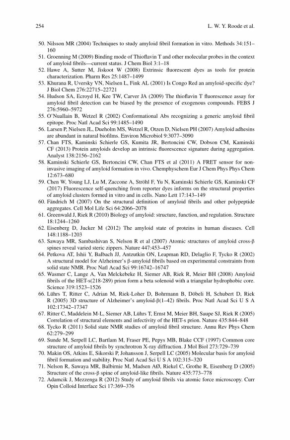
254 L. W. Y. Roode et al.
50. Nilsson MR (2004) Techniques to study amyloid fibril formation in vitro. Methods 34:151–160
51. Groenning M (2009) Binding mode of Thioflavin T and other molecular probes in the contextof amyloid fibrils—current status. J Chem Biol 3:1–18
52. Hawe A, Sutter M, Jiskoot W (2008) Extrinsic fluorescent dyes as tools for proteincharacterization. Pharm Res 25:1487–1499
53. Khurana R, Uversky VN, Nielsen L, Fink AL (2001) Is Congo Red an amyloid-specific dye?J Biol Chem 276:22715–22721
54. Hudson SA, Ecroyd H, Kee TW, Carver JA (2009) The thioflavin T fluorescence assay foramyloid fibril detection can be biased by the presence of exogenous compounds. FEBS J276:5960–5972
55. O’Nuallain B, Wetzel R (2002) Conformational Abs recognizing a generic amyloid fibrilepitope. Proc Natl Acad Sci 99:1485–1490
56. Larsen P, Nielsen JL, Dueholm MS, Wetzel R, Otzen D, Nielsen PH (2007) Amyloid adhesinsare abundant in natural biofilms. Environ Microbiol 9:3077–3090
57. Chan FTS, Kaminski Schierle GS, Kumita JR, Bertoncini CW, Dobson CM, KaminskiCF (2013) Protein amyloids develop an intrinsic fluorescence signature during aggregation.Analyst 138:2156–2162
58. Kaminski Schierle GS, Bertoncini CW, Chan FTS et al (2011) A FRET sensor for non-invasive imaging of amyloid formation in vivo. Chemphyschem Eur J Chem Phys Phys Chem12:673–680
59. Chen W, Young LJ, Lu M, Zaccone A, Ströhl F, Yu N, Kaminski Schierle GS, Kaminski CF(2017) Fluorescence self-quenching from reporter dyes informs on the structural propertiesof amyloid clusters formed in vitro and in cells. Nano Lett 17:143–149
60. Fändrich M (2007) On the structural definition of amyloid fibrils and other polypeptideaggregates. Cell Mol Life Sci 64:2066–2078
61. Greenwald J, Riek R (2010) Biology of amyloid: structure, function, and regulation. Structure18:1244–1260
62. Eisenberg D, Jucker M (2012) The amyloid state of proteins in human diseases. Cell148:1188–1203
63. Sawaya MR, Sambashivan S, Nelson R et al (2007) Atomic structures of amyloid cross-βspines reveal varied steric zippers. Nature 447:453–457
64. Petkova AT, Ishii Y, Balbach JJ, Antzutkin ON, Leapman RD, Delaglio F, Tycko R (2002)A structural model for Alzheimer’s β-amyloid fibrils based on experimental constraints fromsolid state NMR. Proc Natl Acad Sci 99:16742–16747
65. Wasmer C, Lange A, Van Melckebeke H, Siemer AB, Riek R, Meier BH (2008) Amyloidfibrils of the HET-s(218-289) prion form a beta solenoid with a triangular hydrophobic core.Science 319:1523–1526
66. Lührs T, Ritter C, Adrian M, Riek-Loher D, Bohrmann B, Döbeli H, Schubert D, RiekR (2005) 3D structure of Alzheimer’s amyloid-β(1–42) fibrils. Proc Natl Acad Sci U S A102:17342–17347
67. Ritter C, Maddelein M-L, Siemer AB, Lührs T, Ernst M, Meier BH, Saupe SJ, Riek R (2005)Correlation of structural elements and infectivity of the HET-s prion. Nature 435:844–848
68. Tycko R (2011) Solid state NMR studies of amyloid fibril structure. Annu Rev Phys Chem62:279–299
69. Sunde M, Serpell LC, Bartlam M, Fraser PE, Pepys MB, Blake CCF (1997) Common corestructure of amyloid fibrils by synchrotron X-ray diffraction. J Mol Biol 273:729–739
70. Makin OS, Atkins E, Sikorski P, Johansson J, Serpell LC (2005) Molecular basis for amyloidfibril formation and stability. Proc Natl Acad Sci U S A 102:315–320
71. Nelson R, Sawaya MR, Balbirnie M, Madsen AØ, Riekel C, Grothe R, Eisenberg D (2005)Structure of the cross-β spine of amyloid-like fibrils. Nature 435:773–778
72. Adamcik J, Mezzenga R (2012) Study of amyloid fibrils via atomic force microscopy. CurrOpin Colloid Interface Sci 17:369–376
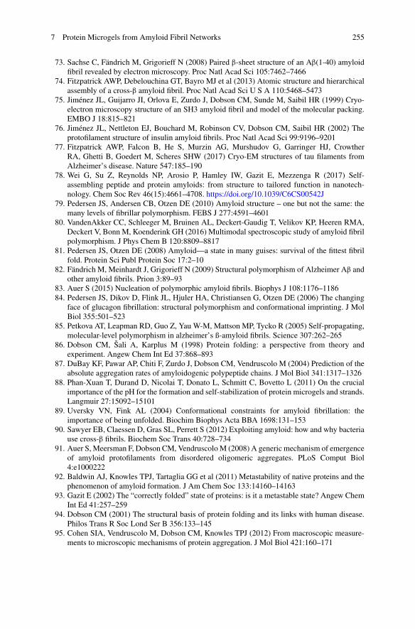
7 Protein Microgels from Amyloid Fibril Networks 255
73. Sachse C, Fändrich M, Grigorieff N (2008) Paired β-sheet structure of an Aβ(1-40) amyloidfibril revealed by electron microscopy. Proc Natl Acad Sci 105:7462–7466
74. Fitzpatrick AWP, Debelouchina GT, Bayro MJ et al (2013) Atomic structure and hierarchicalassembly of a cross-β amyloid fibril. Proc Natl Acad Sci U S A 110:5468–5473
75. Jiménez JL, Guijarro JI, Orlova E, Zurdo J, Dobson CM, Sunde M, Saibil HR (1999) Cryo-electron microscopy structure of an SH3 amyloid fibril and model of the molecular packing.EMBO J 18:815–821
76. Jiménez JL, Nettleton EJ, Bouchard M, Robinson CV, Dobson CM, Saibil HR (2002) Theprotofilament structure of insulin amyloid fibrils. Proc Natl Acad Sci 99:9196–9201
77. Fitzpatrick AWP, Falcon B, He S, Murzin AG, Murshudov G, Garringer HJ, CrowtherRA, Ghetti B, Goedert M, Scheres SHW (2017) Cryo-EM structures of tau filaments fromAlzheimer’s disease. Nature 547:185–190
78. Wei G, Su Z, Reynolds NP, Arosio P, Hamley IW, Gazit E, Mezzenga R (2017) Self-assembling peptide and protein amyloids: from structure to tailored function in nanotech-nology. Chem Soc Rev 46(15):4661–4708. https://doi.org/10.1039/C6CS00542J
79. Pedersen JS, Andersen CB, Otzen DE (2010) Amyloid structure – one but not the same: themany levels of fibrillar polymorphism. FEBS J 277:4591–4601
80. VandenAkker CC, Schleeger M, Bruinen AL, Deckert-Gaudig T, Velikov KP, Heeren RMA,Deckert V, Bonn M, Koenderink GH (2016) Multimodal spectroscopic study of amyloid fibrilpolymorphism. J Phys Chem B 120:8809–8817
81. Pedersen JS, Otzen DE (2008) Amyloid—a state in many guises: survival of the fittest fibrilfold. Protein Sci Publ Protein Soc 17:2–10
82. Fändrich M, Meinhardt J, Grigorieff N (2009) Structural polymorphism of Alzheimer Aβ andother amyloid fibrils. Prion 3:89–93
83. Auer S (2015) Nucleation of polymorphic amyloid fibrils. Biophys J 108:1176–118684. Pedersen JS, Dikov D, Flink JL, Hjuler HA, Christiansen G, Otzen DE (2006) The changing
face of glucagon fibrillation: structural polymorphism and conformational imprinting. J MolBiol 355:501–523
85. Petkova AT, Leapman RD, Guo Z, Yau W-M, Mattson MP, Tycko R (2005) Self-propagating,molecular-level polymorphism in alzheimer’s ß-amyloid fibrils. Science 307:262–265
86. Dobson CM, Šali A, Karplus M (1998) Protein folding: a perspective from theory andexperiment. Angew Chem Int Ed 37:868–893
87. DuBay KF, Pawar AP, Chiti F, Zurdo J, Dobson CM, Vendruscolo M (2004) Prediction of theabsolute aggregation rates of amyloidogenic polypeptide chains. J Mol Biol 341:1317–1326
88. Phan-Xuan T, Durand D, Nicolai T, Donato L, Schmitt C, Bovetto L (2011) On the crucialimportance of the pH for the formation and self-stabilization of protein microgels and strands.Langmuir 27:15092–15101
89. Uversky VN, Fink AL (2004) Conformational constraints for amyloid fibrillation: theimportance of being unfolded. Biochim Biophys Acta BBA 1698:131–153
90. Sawyer EB, Claessen D, Gras SL, Perrett S (2012) Exploiting amyloid: how and why bacteriause cross-β fibrils. Biochem Soc Trans 40:728–734
91. Auer S, Meersman F, Dobson CM, Vendruscolo M (2008) A generic mechanism of emergenceof amyloid protofilaments from disordered oligomeric aggregates. PLoS Comput Biol4:e1000222
92. Baldwin AJ, Knowles TPJ, Tartaglia GG et al (2011) Metastability of native proteins and thephenomenon of amyloid formation. J Am Chem Soc 133:14160–14163
93. Gazit E (2002) The “correctly folded” state of proteins: is it a metastable state? Angew ChemInt Ed 41:257–259
94. Dobson CM (2001) The structural basis of protein folding and its links with human disease.Philos Trans R Soc Lond Ser B 356:133–145
95. Cohen SIA, Vendruscolo M, Dobson CM, Knowles TPJ (2012) From macroscopic measure-ments to microscopic mechanisms of protein aggregation. J Mol Biol 421:160–171
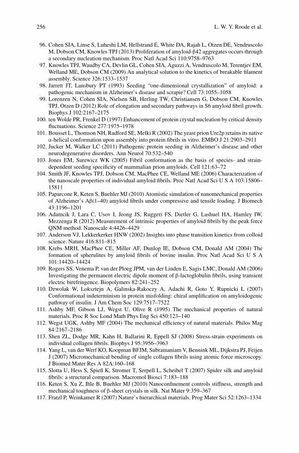
256 L. W. Y. Roode et al.
96. Cohen SIA, Linse S, Luheshi LM, Hellstrand E, White DA, Rajah L, Otzen DE, VendruscoloM, Dobson CM, Knowles TPJ (2013) Proliferation of amyloid-β42 aggregates occurs througha secondary nucleation mechanism. Proc Natl Acad Sci 110:9758–9763
97. Knowles TPJ, Waudby CA, Devlin GL, Cohen SIA, Aguzzi A, Vendruscolo M, Terentjev EM,Welland ME, Dobson CM (2009) An analytical solution to the kinetics of breakable filamentassembly. Science 326:1533–1537
98. Jarrett JT, Lansbury PT (1993) Seeding “one-dimensional crystallization” of amyloid: apathogenic mechanism in Alzheimer’s disease and scrapie? Cell 73:1055–1058
99. Lorenzen N, Cohen SIA, Nielsen SB, Herling TW, Christiansen G, Dobson CM, KnowlesTPJ, Otzen D (2012) Role of elongation and secondary pathways in S6 amyloid fibril growth.Biophys J 102:2167–2175
100. ten Wolde PR, Frenkel D (1997) Enhancement of protein crystal nucleation by critical densityfluctuations. Science 277:1975–1978
101. Bousset L, Thomson NH, Radford SE, Melki R (2002) The yeast prion Ure2p retains its nativeα-helical conformation upon assembly into protein fibrils in vitro. EMBO J 21:2903–2911
102. Jucker M, Walker LC (2011) Pathogenic protein seeding in Alzheimer’s disease and otherneurodegenerative disorders. Ann Neurol 70:532–540
103. Jones EM, Surewicz WK (2005) Fibril conformation as the basis of species- and strain-dependent seeding specificity of mammalian prion amyloids. Cell 121:63–72
104. Smith JF, Knowles TPJ, Dobson CM, MacPhee CE, Welland ME (2006) Characterization ofthe nanoscale properties of individual amyloid fibrils. Proc Natl Acad Sci U S A 103:15806–15811
105. Paparcone R, Keten S, Buehler MJ (2010) Atomistic simulation of nanomechanical propertiesof Alzheimer’s Aβ(1–40) amyloid fibrils under compressive and tensile loading. J Biomech43:1196–1201
106. Adamcik J, Lara C, Usov I, Jeong JS, Ruggeri FS, Dietler G, Lashuel HA, Hamley IW,Mezzenga R (2012) Measurement of intrinsic properties of amyloid fibrils by the peak forceQNM method. Nanoscale 4:4426–4429
107. Anderson VJ, Lekkerkerker HNW (2002) Insights into phase transition kinetics from colloidscience. Nature 416:811–815
108. Krebs MRH, MacPhee CE, Miller AF, Dunlop IE, Dobson CM, Donald AM (2004) Theformation of spherulites by amyloid fibrils of bovine insulin. Proc Natl Acad Sci U S A101:14420–14424
109. Rogers SS, Venema P, van der Ploeg JPM, van der Linden E, Sagis LMC, Donald AM (2006)Investigating the permanent electric dipole moment of β-lactoglobulin fibrils, using transientelectric birefringence. Biopolymers 82:241–252
110. Dzwolak W, Loksztejn A, Galinska-Rakoczy A, Adachi R, Goto Y, Rupnicki L (2007)Conformational indeterminism in protein misfolding: chiral amplification on amyloidogenicpathway of insulin. J Am Chem Soc 129:7517–7522
111. Ashby MF, Gibson LJ, Wegst U, Olive R (1995) The mechanical properties of naturalmaterials. Proc R Soc Lond Math Phys Eng Sci 450:123–140
112. Wegst UGK, Ashby MF (2004) The mechanical efficiency of natural materials. Philos Mag84:2167–2186
113. Shen ZL, Dodge MR, Kahn H, Ballarini R, Eppell SJ (2008) Stress-strain experiments onindividual collagen fibrils. Biophys J 95:3956–3963
114. Yang L, van der Werf KO, Koopman BFJM, Subramaniam V, Bennink ML, Dijkstra PJ, FeijenJ (2007) Micromechanical bending of single collagen fibrils using atomic force microscopy.J Biomed Mater Res A 82A:160–168
115. Slotta U, Hess S, Spieß K, Stromer T, Serpell L, Scheibel T (2007) Spider silk and amyloidfibrils: a structural comparison. Macromol Biosci 7:183–188
116. Keten S, Xu Z, Ihle B, Buehler MJ (2010) Nanoconfinement controls stiffness, strength andmechanical toughness of β-sheet crystals in silk. Nat Mater 9:359–367
117. Fratzl P, Weinkamer R (2007) Nature’s hierarchical materials. Prog Mater Sci 52:1263–1334
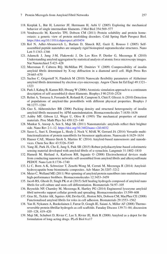
7 Protein Microgels from Amyloid Fibril Networks 257
118. Kreplak L, Bär H, Leterrier JF, Herrmann H, Aebi U (2005) Exploring the mechanicalbehavior of single intermediate filaments. J Mol Biol 354:569–577
119. Vendruscolo M, Knowles TPJ, Dobson CM (2011) Protein solubility and protein home-ostasis: a generic view of protein misfolding disorders. Cold Spring Harb Perspect Biol.https://doi.org/10.1101/cshperspect.a010454
120. Kol N, Adler-Abramovich L, Barlam D, Shneck RZ, Gazit E, Rousso I (2005) Self-assembled peptide nanotubes are uniquely rigid bioinspired supramolecular structures. NanoLett 5:1343–1346
121. Adamcik J, Jung J-M, Flakowski J, De Los Rios P, Dietler G, Mezzenga R (2010)Understanding amyloid aggregation by statistical analysis of atomic force microscopy images.Nat Nanotechnol 5:423–428
122. Meersman F, Cabrera RQ, McMillan PF, Dmitriev V (2009) Compressibility of insulinamyloid fibrils determined by X-ray diffraction in a diamond anvil cell. High Press Res29:665–670
123. Sachse C, Grigorieff N, Fändrich M (2010) Nanoscale flexibility parameters of Alzheimeramyloid fibrils determined by electron cryo-microscopy. Angew Chem Int Ed Engl 49:1321–1323
124. Park J, Kahng B, Kamm RD, Hwang W (2006) Atomistic simulation approach to a continuumdescription of self-assembled β-sheet filaments. Biophys J 90:2510–2524
125. Relini A, Torrassa S, Ferrando R, Rolandi R, Campioni S, Chiti F, Gliozzi A (2010) Detectionof populations of amyloid-like protofibrils with different physical properties. Biophys J98:1277–1284
126. Guo S, Akhremitchev BB (2006) Packing density and structural heterogeneity of insulinamyloid fibrils measured by AFM nanoindentation. Biomacromolecules 7:1630–1636
127. Ashby MF, Gibson LJ, Wegst U, Olive R (1995) The mechanical properties of naturalmaterials. Proc Math Phys Sci 450:123–140
128. Mankar S, Anoop A, Sen S, Maji SK (2011) Nanomaterials: amyloids reflect their brighterside. Nano Rev 2:1–12. https://doi.org/10.3402/nano.v2i0.6032
129. Sasso L, Suei S, Domigan L, Healy J, Nock V, MAK W, Gerrard JA (2014) Versatile multi-functionalization of protein nanofibrils for biosensor applications. Nanoscale 6:1629–1634
130. Hauser CAE, Maurer-Stroh S, Martins IC (2014) Amyloid-based nanosensors and nanode-vices. Chem Soc Rev 43:5326–5345
131. Yang JE, Park JS, Cho E, Jung S, Paik SR (2015) Robust polydiacetylene-based colorimetricsensing material developed with amyloid fibrils of α-synuclein. Langmuir 31:1802–1810
132. Hamedi M, Herland A, Karlsson RH, Inganäs O (2008) Electrochemical devices madefrom conducting nanowire networks self-assembled from amyloid fibrils and alkoxysulfonatePEDOT. Nano Lett 8:1736–1740
133. Li C, Born A-K, Schweizer T, Zenobi-Wong M, Cerruti M, Mezzenga R (2014) Amyloid-hydroxyapatite bone biomimetic composites. Adv Mater 26:3207–3212
134. Meier C, Welland ME (2011) Wet-spinning of amyloid protein nanofibers into multifunctionalhigh-performance biofibers. Biomacromolecules 12:3453–3459
135. Jacob RS, Ghosh D, Singh PK et al (2015) Self healing hydrogels composed of amyloid nanofibrils for cell culture and stem cell differentiation. Biomaterials 54:97–105
136. Reynolds NP, Charnley M, Mezzenga R, Hartley PG (2014) Engineered lysozyme amyloidfibril networks support cellular growth and spreading. Biomacromolecules 15:599–608
137. Gras SL, Tickler AK, Squires AM, Devlin GL, Horton MA, Dobson CM, MacPhee CE (2008)Functionalised amyloid fibrils for roles in cell adhesion. Biomaterials 29:1553–1562
138. Yan H, Nykanen A, Ruokolainen J, Farrar D, Gough JE, Saiani A, Miller AF (2008) Thermo-reversible protein fibrillar hydrogels as cell scaffolds. Faraday Discuss 139:71–84; discussion105–128, 419–420
139. Maji SK, Schubert D, Rivier C, Lee S, Rivier JE, Riek R (2008) Amyloid as a depot for theformulation of long-acting drugs. PLoS Biol 6:e17
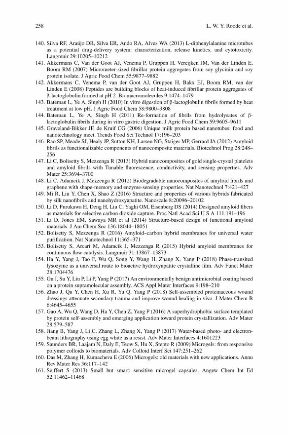
258 L. W. Y. Roode et al.
140. Silva RF, Araújo DR, Silva ER, Ando RA, Alves WA (2013) L-diphenylalanine microtubesas a potential drug-delivery system: characterization, release kinetics, and cytotoxicity.Langmuir 29:10205–10212
141. Akkermans C, Van der Goot AJ, Venema P, Gruppen H, Vereijken JM, Van der Linden E,Boom RM (2007) Micrometer-sized fibrillar protein aggregates from soy glycinin and soyprotein isolate. J Agric Food Chem 55:9877–9882
142. Akkermans C, Venema P, van der Goot AJ, Gruppen H, Bakx EJ, Boom RM, van derLinden E (2008) Peptides are building blocks of heat-induced fibrillar protein aggregates ofβ-lactoglobulin formed at pH 2. Biomacromolecules 9:1474–1479
143. Bateman L, Ye A, Singh H (2010) In vitro digestion of β-lactoglobulin fibrils formed by heattreatment at low pH. J Agric Food Chem 58:9800–9808
144. Bateman L, Ye A, Singh H (2011) Re-formation of fibrils from hydrolysates of β-lactoglobulin fibrils during in vitro gastric digestion. J Agric Food Chem 59:9605–9611
145. Graveland-Bikker JF, de Kruif CG (2006) Unique milk protein based nanotubes: food andnanotechnology meet. Trends Food Sci Technol 17:196–203
146. Rao SP, Meade SJ, Healy JP, Sutton KH, Larsen NG, Staiger MP, Gerrard JA (2012) Amyloidfibrils as functionalizable components of nanocomposite materials. Biotechnol Prog 28:248–256
147. Li C, Bolisetty S, Mezzenga R (2013) Hybrid nanocomposites of gold single-crystal plateletsand amyloid fibrils with Tunable fluorescence, conductivity, and sensing properties. AdvMater 25:3694–3700
148. Li C, Adamcik J, Mezzenga R (2012) Biodegradable nanocomposites of amyloid fibrils andgraphene with shape-memory and enzyme-sensing properties. Nat Nanotechnol 7:421–427
149. Mi R, Liu Y, Chen X, Shao Z (2016) Structure and properties of various hybrids fabricatedby silk nanofibrils and nanohydroxyapatite. Nanoscale 8:20096–20102
150. Li D, Furukawa H, Deng H, Liu C, Yaghi OM, Eisenberg DS (2014) Designed amyloid fibersas materials for selective carbon dioxide capture. Proc Natl Acad Sci U S A 111:191–196
151. Li D, Jones EM, Sawaya MR et al (2014) Structure-based design of functional amyloidmaterials. J Am Chem Soc 136:18044–18051
152. Bolisetty S, Mezzenga R (2016) Amyloid–carbon hybrid membranes for universal waterpurification. Nat Nanotechnol 11:365–371
153. Bolisetty S, Arcari M, Adamcik J, Mezzenga R (2015) Hybrid amyloid membranes forcontinuous flow catalysis. Langmuir 31:13867–13873
154. Ha Y, Yang J, Tao F, Wu Q, Song Y, Wang H, Zhang X, Yang P (2018) Phase-transitedlysozyme as a universal route to bioactive hydroxyapatite crystalline film. Adv Funct Mater28:1704476
155. Gu J, Su Y, Liu P, Li P, Yang P (2017) An environmentally benign antimicrobial coating basedon a protein supramolecular assembly. ACS Appl Mater Interfaces 9:198–210
156. Zhao J, Qu Y, Chen H, Xu R, Yu Q, Yang P (2018) Self-assembled proteinaceous wounddressings attenuate secondary trauma and improve wound healing in vivo. J Mater Chem B6:4645–4655
157. Gao A, Wu Q, Wang D, Ha Y, Chen Z, Yang P (2016) A superhydrophobic surface templatedby protein self-assembly and emerging application toward protein crystallization. Adv Mater28:579–587
158. Jiang B, Yang J, Li C, Zhang L, Zhang X, Yang P (2017) Water-based photo- and electron-beam lithography using egg white as a resist. Adv Mater Interfaces 4:1601223
159. Saunders BR, Laajam N, Daly E, Teow S, Hu X, Stepto R (2009) Microgels: from responsivepolymer colloids to biomaterials. Adv Colloid Interf Sci 147:251–262
160. Das M, Zhang H, Kumacheva E (2006) Microgels: old materials with new applications. AnnuRev Mater Res 36:117–142
161. Seiffert S (2013) Small but smart: sensitive microgel capsules. Angew Chem Int Ed52:11462–11468
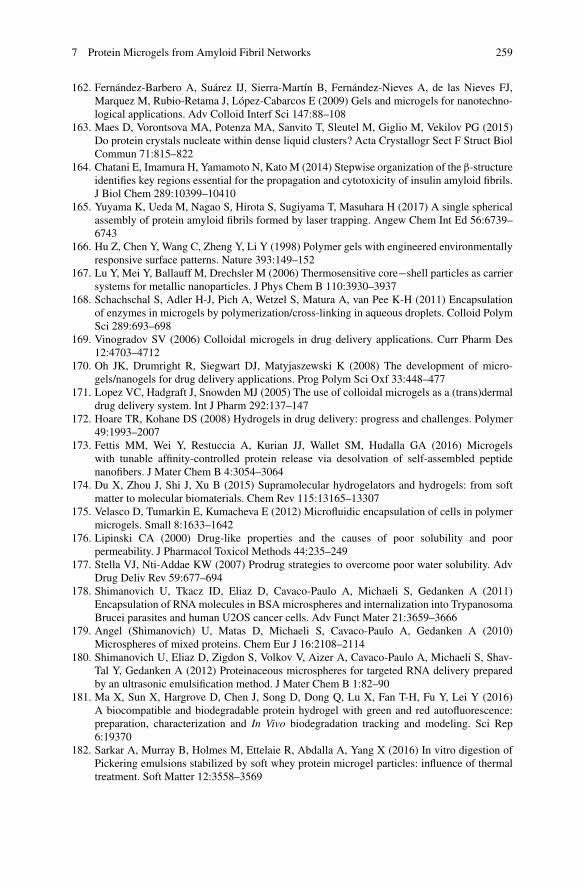
7 Protein Microgels from Amyloid Fibril Networks 259
162. Fernández-Barbero A, Suárez IJ, Sierra-Martín B, Fernández-Nieves A, de las Nieves FJ,Marquez M, Rubio-Retama J, López-Cabarcos E (2009) Gels and microgels for nanotechno-logical applications. Adv Colloid Interf Sci 147:88–108
163. Maes D, Vorontsova MA, Potenza MA, Sanvito T, Sleutel M, Giglio M, Vekilov PG (2015)Do protein crystals nucleate within dense liquid clusters? Acta Crystallogr Sect F Struct BiolCommun 71:815–822
164. Chatani E, Imamura H, Yamamoto N, Kato M (2014) Stepwise organization of the β-structureidentifies key regions essential for the propagation and cytotoxicity of insulin amyloid fibrils.J Biol Chem 289:10399–10410
165. Yuyama K, Ueda M, Nagao S, Hirota S, Sugiyama T, Masuhara H (2017) A single sphericalassembly of protein amyloid fibrils formed by laser trapping. Angew Chem Int Ed 56:6739–6743
166. Hu Z, Chen Y, Wang C, Zheng Y, Li Y (1998) Polymer gels with engineered environmentallyresponsive surface patterns. Nature 393:149–152
167. Lu Y, Mei Y, Ballauff M, Drechsler M (2006) Thermosensitive core−shell particles as carriersystems for metallic nanoparticles. J Phys Chem B 110:3930–3937
168. Schachschal S, Adler H-J, Pich A, Wetzel S, Matura A, van Pee K-H (2011) Encapsulationof enzymes in microgels by polymerization/cross-linking in aqueous droplets. Colloid PolymSci 289:693–698
169. Vinogradov SV (2006) Colloidal microgels in drug delivery applications. Curr Pharm Des12:4703–4712
170. Oh JK, Drumright R, Siegwart DJ, Matyjaszewski K (2008) The development of micro-gels/nanogels for drug delivery applications. Prog Polym Sci Oxf 33:448–477
171. Lopez VC, Hadgraft J, Snowden MJ (2005) The use of colloidal microgels as a (trans)dermaldrug delivery system. Int J Pharm 292:137–147
172. Hoare TR, Kohane DS (2008) Hydrogels in drug delivery: progress and challenges. Polymer49:1993–2007
173. Fettis MM, Wei Y, Restuccia A, Kurian JJ, Wallet SM, Hudalla GA (2016) Microgelswith tunable affinity-controlled protein release via desolvation of self-assembled peptidenanofibers. J Mater Chem B 4:3054–3064
174. Du X, Zhou J, Shi J, Xu B (2015) Supramolecular hydrogelators and hydrogels: from softmatter to molecular biomaterials. Chem Rev 115:13165–13307
175. Velasco D, Tumarkin E, Kumacheva E (2012) Microfluidic encapsulation of cells in polymermicrogels. Small 8:1633–1642
176. Lipinski CA (2000) Drug-like properties and the causes of poor solubility and poorpermeability. J Pharmacol Toxicol Methods 44:235–249
177. Stella VJ, Nti-Addae KW (2007) Prodrug strategies to overcome poor water solubility. AdvDrug Deliv Rev 59:677–694
178. Shimanovich U, Tkacz ID, Eliaz D, Cavaco-Paulo A, Michaeli S, Gedanken A (2011)Encapsulation of RNA molecules in BSA microspheres and internalization into TrypanosomaBrucei parasites and human U2OS cancer cells. Adv Funct Mater 21:3659–3666
179. Angel (Shimanovich) U, Matas D, Michaeli S, Cavaco-Paulo A, Gedanken A (2010)Microspheres of mixed proteins. Chem Eur J 16:2108–2114
180. Shimanovich U, Eliaz D, Zigdon S, Volkov V, Aizer A, Cavaco-Paulo A, Michaeli S, Shav-Tal Y, Gedanken A (2012) Proteinaceous microspheres for targeted RNA delivery preparedby an ultrasonic emulsification method. J Mater Chem B 1:82–90
181. Ma X, Sun X, Hargrove D, Chen J, Song D, Dong Q, Lu X, Fan T-H, Fu Y, Lei Y (2016)A biocompatible and biodegradable protein hydrogel with green and red autofluorescence:preparation, characterization and In Vivo biodegradation tracking and modeling. Sci Rep6:19370
182. Sarkar A, Murray B, Holmes M, Ettelaie R, Abdalla A, Yang X (2016) In vitro digestion ofPickering emulsions stabilized by soft whey protein microgel particles: influence of thermaltreatment. Soft Matter 12:3558–3569
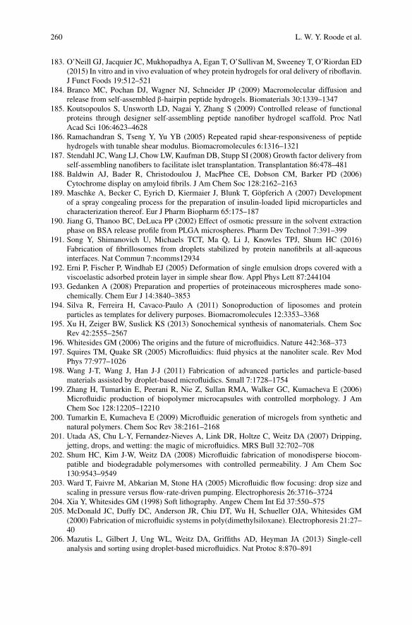
260 L. W. Y. Roode et al.
183. O’Neill GJ, Jacquier JC, Mukhopadhya A, Egan T, O’Sullivan M, Sweeney T, O’Riordan ED(2015) In vitro and in vivo evaluation of whey protein hydrogels for oral delivery of riboflavin.J Funct Foods 19:512–521
184. Branco MC, Pochan DJ, Wagner NJ, Schneider JP (2009) Macromolecular diffusion andrelease from self-assembled β-hairpin peptide hydrogels. Biomaterials 30:1339–1347
185. Koutsopoulos S, Unsworth LD, Nagai Y, Zhang S (2009) Controlled release of functionalproteins through designer self-assembling peptide nanofiber hydrogel scaffold. Proc NatlAcad Sci 106:4623–4628
186. Ramachandran S, Tseng Y, Yu YB (2005) Repeated rapid shear-responsiveness of peptidehydrogels with tunable shear modulus. Biomacromolecules 6:1316–1321
187. Stendahl JC, Wang LJ, Chow LW, Kaufman DB, Stupp SI (2008) Growth factor delivery fromself-assembling nanofibers to facilitate islet transplantation. Transplantation 86:478–481
188. Baldwin AJ, Bader R, Christodoulou J, MacPhee CE, Dobson CM, Barker PD (2006)Cytochrome display on amyloid fibrils. J Am Chem Soc 128:2162–2163
189. Maschke A, Becker C, Eyrich D, Kiermaier J, Blunk T, Göpferich A (2007) Developmentof a spray congealing process for the preparation of insulin-loaded lipid microparticles andcharacterization thereof. Eur J Pharm Biopharm 65:175–187
190. Jiang G, Thanoo BC, DeLuca PP (2002) Effect of osmotic pressure in the solvent extractionphase on BSA release profile from PLGA microspheres. Pharm Dev Technol 7:391–399
191. Song Y, Shimanovich U, Michaels TCT, Ma Q, Li J, Knowles TPJ, Shum HC (2016)Fabrication of fibrillosomes from droplets stabilized by protein nanofibrils at all-aqueousinterfaces. Nat Commun 7:ncomms12934
192. Erni P, Fischer P, Windhab EJ (2005) Deformation of single emulsion drops covered with aviscoelastic adsorbed protein layer in simple shear flow. Appl Phys Lett 87:244104
193. Gedanken A (2008) Preparation and properties of proteinaceous microspheres made sono-chemically. Chem Eur J 14:3840–3853
194. Silva R, Ferreira H, Cavaco-Paulo A (2011) Sonoproduction of liposomes and proteinparticles as templates for delivery purposes. Biomacromolecules 12:3353–3368
195. Xu H, Zeiger BW, Suslick KS (2013) Sonochemical synthesis of nanomaterials. Chem SocRev 42:2555–2567
196. Whitesides GM (2006) The origins and the future of microfluidics. Nature 442:368–373197. Squires TM, Quake SR (2005) Microfluidics: fluid physics at the nanoliter scale. Rev Mod
Phys 77:977–1026198. Wang J-T, Wang J, Han J-J (2011) Fabrication of advanced particles and particle-based
materials assisted by droplet-based microfluidics. Small 7:1728–1754199. Zhang H, Tumarkin E, Peerani R, Nie Z, Sullan RMA, Walker GC, Kumacheva E (2006)
Microfluidic production of biopolymer microcapsules with controlled morphology. J AmChem Soc 128:12205–12210
200. Tumarkin E, Kumacheva E (2009) Microfluidic generation of microgels from synthetic andnatural polymers. Chem Soc Rev 38:2161–2168
201. Utada AS, Chu L-Y, Fernandez-Nieves A, Link DR, Holtze C, Weitz DA (2007) Dripping,jetting, drops, and wetting: the magic of microfluidics. MRS Bull 32:702–708
202. Shum HC, Kim J-W, Weitz DA (2008) Microfluidic fabrication of monodisperse biocom-patible and biodegradable polymersomes with controlled permeability. J Am Chem Soc130:9543–9549
203. Ward T, Faivre M, Abkarian M, Stone HA (2005) Microfluidic flow focusing: drop size andscaling in pressure versus flow-rate-driven pumping. Electrophoresis 26:3716–3724
204. Xia Y, Whitesides GM (1998) Soft lithography. Angew Chem Int Ed 37:550–575205. McDonald JC, Duffy DC, Anderson JR, Chiu DT, Wu H, Schueller OJA, Whitesides GM
(2000) Fabrication of microfluidic systems in poly(dimethylsiloxane). Electrophoresis 21:27–40
206. Mazutis L, Gilbert J, Ung WL, Weitz DA, Griffiths AD, Heyman JA (2013) Single-cellanalysis and sorting using droplet-based microfluidics. Nat Protoc 8:870–891
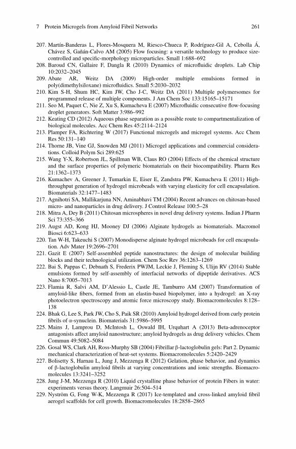
7 Protein Microgels from Amyloid Fibril Networks 261
207. Martín-Banderas L, Flores-Mosquera M, Riesco-Chueca P, Rodríguez-Gil A, Cebolla Á,Chávez S, Gañán-Calvo AM (2005) Flow focusing: a versatile technology to produce size-controlled and specific-morphology microparticles. Small 1:688–692
208. Baroud CN, Gallaire F, Dangla R (2010) Dynamics of microfluidic droplets. Lab Chip10:2032–2045
209. Abate AR, Weitz DA (2009) High-order multiple emulsions formed inpoly(dimethylsiloxane) microfluidics. Small 5:2030–2032
210. Kim S-H, Shum HC, Kim JW, Cho J-C, Weitz DA (2011) Multiple polymersomes forprogrammed release of multiple components. J Am Chem Soc 133:15165–15171
211. Seo M, Paquet C, Nie Z, Xu S, Kumacheva E (2007) Microfluidic consecutive flow-focusingdroplet generators. Soft Matter 3:986–992
212. Keating CD (2012) Aqueous phase separation as a possible route to compartmentalization ofbiological molecules. Acc Chem Res 45:2114–2124
213. Plamper FA, Richtering W (2017) Functional microgels and microgel systems. Acc ChemRes 50:131–140
214. Thorne JB, Vine GJ, Snowden MJ (2011) Microgel applications and commercial considera-tions. Colloid Polym Sci 289:625
215. Wang Y-X, Robertson JL, Spillman WB, Claus RO (2004) Effects of the chemical structureand the surface properties of polymeric biomaterials on their biocompatibility. Pharm Res21:1362–1373
216. Kumachev A, Greener J, Tumarkin E, Eiser E, Zandstra PW, Kumacheva E (2011) High-throughput generation of hydrogel microbeads with varying elasticity for cell encapsulation.Biomaterials 32:1477–1483
217. Agnihotri SA, Mallikarjuna NN, Aminabhavi TM (2004) Recent advances on chitosan-basedmicro- and nanoparticles in drug delivery. J Control Release 100:5–28
218. Mitra A, Dey B (2011) Chitosan microspheres in novel drug delivery systems. Indian J PharmSci 73:355–366
219. Augst AD, Kong HJ, Mooney DJ (2006) Alginate hydrogels as biomaterials. MacromolBiosci 6:623–633
220. Tan W-H, Takeuchi S (2007) Monodisperse alginate hydrogel microbeads for cell encapsula-tion. Adv Mater 19:2696–2701
221. Gazit E (2007) Self-assembled peptide nanostructures: the design of molecular buildingblocks and their technological utilization. Chem Soc Rev 36:1263–1269
222. Bai S, Pappas C, Debnath S, Frederix PWJM, Leckie J, Fleming S, Ulijn RV (2014) Stableemulsions formed by self-assembly of interfacial networks of dipeptide derivatives. ACSNano 8:7005–7013
223. Flamia R, Salvi AM, D’Alessio L, Castle JE, Tamburro AM (2007) Transformation ofamyloid-like fibers, formed from an elastin-based biopolymer, into a hydrogel: an X-rayphotoelectron spectroscopy and atomic force microscopy study. Biomacromolecules 8:128–138
224. Bhak G, Lee S, Park JW, Cho S, Paik SR (2010) Amyloid hydrogel derived from curly proteinfibrils of α-synuclein. Biomaterials 31:5986–5995
225. Mains J, Lamprou D, McIntosh L, Oswald IH, Urquhart A (2013) Beta-adrenoceptorantagonists affect amyloid nanostructure; amyloid hydrogels as drug delivery vehicles. ChemCommun 49:5082–5084
226. Gosal WS, Clark AH, Ross-Murphy SB (2004) Fibrillar β-lactoglobulin gels: Part 2. Dynamicmechanical characterization of heat-set systems. Biomacromolecules 5:2420–2429
227. Bolisetty S, Harnau L, Jung J, Mezzenga R (2012) Gelation, phase behavior, and dynamicsof β-lactoglobulin amyloid fibrils at varying concentrations and ionic strengths. Biomacro-molecules 13:3241–3252
228. Jung J-M, Mezzenga R (2010) Liquid crystalline phase behavior of protein Fibers in water:experiments versus theory. Langmuir 26:504–514
229. Nyström G, Fong W-K, Mezzenga R (2017) Ice-templated and cross-linked amyloid fibrilaerogel scaffolds for cell growth. Biomacromolecules 18:2858–2865
262 L. W. Y. Roode et al.
230. Langton M, Hermansson A-M (1992) Fine-stranded and particulate gels of β-lactoglobulinand whey protein at varying pH. Food Hydrocoll 5:523–539
231. Zhou X-M, Shimanovich U, Herling TW, Wu S, Dobson CM, Knowles TPJ, Perrett S(2015) Enzymatically active microgels from self-assembling protein nanofibrils for microflowchemistry. ACS Nano 9:5772–5781
232. Silva R, Ferreira H, Azoia NG, Shimanovich U, Freddi G, Gedanken A, Cavaco-Paulo A(2012) Insights on the mechanism of formation of protein microspheres in a biphasic system.Mol Pharm 9:3079–3088
233. Shimanovich U, Ruggeri FS, Genst ED et al (2017) Silk micrococoons for protein stabilisationand molecular encapsulation. Nat Commun 8:ncomms15902
234. Müller T, Simone Ruggeri F, Kulik J, Shimanovich U, Mason TO, Knowles TPJ, Dietler G(2014) Nanoscale spatially resolved infrared spectra from single microdroplets. Lab Chip14:1315–1319
235. Avivi S, Gedanken A (2002) S–S bonds are not required for the sonochemical formation ofproteinaceous microspheres: the case of streptavidin. Biochem J 366:705–707
236. Subia B, Kundu SC (2013) Drug loading and release on tumor cells using silk fibroin–albuminnanoparticles as carriers. Nanotechnology 24:035103
237. Shimanovich U, Volkov V, Eliaz D, Aizer A, Michaeli S, Gedanken A (2011) StabilizingRNA by the sonochemical formation of RNA nanospheres. Small 7:1068–1074
238. Shimanovich U, Eliaz D, Aizer A, Vayman I, Michaeli S, Shav-Tal Y, Gedanken A (2011)Sonochemical synthesis of DNA nanospheres. Chembiochem 12:1678–1681
239. Li C, Xu L, Zuo YY, Yang P (2018) Tuning protein assembly pathways through superfastamyloid-like aggregation. Biomater Sci 6:836–841
240. Shimanovich U, Song Y, Brujic J, Shum HC, Knowles TPJ (2015) Multiphase proteinmicrogels. Macromol Biosci 15:501–508
241. Knowles T, Shimanovich U, Dobson C, Weitz D (2016) Protein Capsules242. Volpatti LR, Shimanovich U, Ruggeri FS, Bolisetty S, Müller T, Mason TO, Michaels TCT,
Mezzenga R, Dietler G, Knowles TPJ (2016) Micro- and nanoscale hierarchical structure ofcore–shell protein microgels. J Mater Chem B 4:7989–7999
243. Peters TJ (1987) Partition of cell particles and macromolecules: separation and purificationof biomolecules, cell organelles, membranes and cells in aqueous polymer two phase systemsand their use in biochemical analysis and biotechnology. Cell Biochem Funct 5:233–234
244. Kroner KH, Hustedt H, Granda S, Kula M-R, Introduction by T Alan Hatton (2009) Technicalaspects of separation using aqueous two-phase systems in enzyme isolation processes.Biotechnol Bioeng 104:217–239
245. Diamond AD, Hsu JT (1990) Protein partitioning in PEG/dextran aqueous two-phase systems.AICHE J 36:1017–1024
246. Fele L, Fermeglia M (1996) Partition coefficients of proteins in poly(ethylene glycol) +dextran + water at 298 K. J Chem Eng Data 41:331–334
247. Osborn HT, Akoh CC (2004) Effect of emulsifier type, droplet size, and oil concentration onlipid oxidation in structured lipid-based oil-in-water emulsions. Food Chem 84:451–456
248. Sah H (1999) Stabilization of proteins against methylene chloride/water interface-induceddenaturation and aggregation. J Control Release 58:143–151
249. Liu Y, Lipowsky R, Dimova R (2012) Concentration dependence of the interfacial tension foraqueous two-phase polymer solutions of dextran and polyethylene glycol. Langmuir 28:3831–3839
250. Balakrishnan G, Nicolai T, Benyahia L, Durand D (2012) Particles trapped at the dropletinterface in water-in-water emulsions. Langmuir 28:5921–5926
251. Nguyen BT, Nicolai T, Benyahia L (2013) Stabilization of water-in-water emulsions byaddition of protein particles. Langmuir 29:10658–10664
252. Rollett A, Reiter T, Nogueira P, Cardinale M, Loureiro A, Gomes A, Cavaco-Paulo A,Moreira A, Carmo AM, Guebitz GM (2012) Folic acid-functionalized human serum albuminnanocapsules for targeted drug delivery to chronically activated macrophages. Int J Pharm427:460–466
7 Protein Microgels from Amyloid Fibril Networks 263
253. Richman M, Wilk S, Skirtenko N, Perelman A, Rahimipour S (2011) Surface-modifiedprotein microspheres capture amyloid-β and inhibit its aggregation and toxicity. Chem WeinhBergstr Ger 17:11171–11177
254. Krysmann MJ, Castelletto V, Kelarakis A, Hamley IW, Hule RA, Pochan DJ (2008) Self-assembly and hydrogelation of an amyloid peptide fragment. Biochemistry 47:4597–4605
255. Zhou X-M, Entwistle A, Zhang H, Jackson AP, Mason TO, Shimanovich U, Knowles TPJ,Smith AT, Sawyer EB, Perrett S (2014) Self-assembly of amyloid fibrils that display activeenzymes. ChemCatChem 6:1961–1968
256. Kim J-W, Fernández-Nieves A, Dan N, Utada AS, Marquez M, Weitz DA (2007) Colloidalassembly route for responsive colloidosomes with tunable permeability. Nano Lett 7:2876–2880
257. Li M-H, Keller P (2009) Stimuli-responsive polymer vesicles. Soft Matter 5:927–937258. Yolamanova M, Meier C, Shaytan AK et al (2013) Peptide nanofibrils boost retroviral gene
transfer and provide a rapid means for concentrating viruses. Nat Nanotechnol 8:130–136259. Dai B, Li D, Xi W et al (2015) Tunable assembly of amyloid-forming peptides into nanosheets
as a retrovirus carrier. Proc Natl Acad Sci 112:2996–3001260. Bolisetty S, Boddupalli CS, Handschin S, Chaitanya K, Adamcik J, Saito Y, Manz MG,
Mezzenga R (2014) Amyloid fibrils enhance transport of metal nanoparticles in living cellsand induced cytotoxicity. Biomacromolecules 15:2793–2799
261. Shen Y, Posavec L, Bolisetty S et al (2017) Amyloid fibril systems reduce, stabilize anddeliver bioavailable nanosized iron. Nat Nanotechnol 12:642–647
262. Levin A, Mason TO, Knowles TPJ, Shimanovich U (2017) Self-assembled protein fibril-metaloxide nanocomposites. Isr J Chem 57(7):724–728
263. Sandhu A, Handa H, Abe M (2010) Synthesis and applications of magnetic nanoparticles forbiorecognition and point of care medical diagnostics. Nanotechnology 21:442001
264. Wiogo HTR, Lim M, Bulmus V, Yun J, Amal R (2011) Stabilization of magnetic iron oxidenanoparticles in biological media by eetal bovine serum (FBS). Langmuir 27:843–850
265. Jordens S, Rühs PA, Sieber C, Isa L, Fischer P, Mezzenga R (2014) Bridging the gap betweenthe nanostructural organization and macroscopic interfacial rheology of amyloid fibrils atliquid interfaces. Langmuir 30:10090–10097
266. Linse S, Cabaleiro-Lago C, Xue W-F, Lynch I, Lindman S, Thulin E, Radford SE, DawsonKA (2007) Nucleation of protein fibrillation by nanoparticles. Proc Natl Acad Sci U S A104:8691–8696
Chapter 8Protein Nanofibrils as Storage Formsof Peptide Drugs and Hormones
Reeba Susan Jacob, A. Anoop, and Samir K. Maji
Abstract Amyloids are highly organized cross β-sheet protein nanofibrils thatare associated with both diseases and functions. Thermodynamically amyloids arestable structures as they represent the lowest free energy state that proteins canattain. However, recent studies suggest that amyloid fibrils can be dissociated by achange in environmental parameters such as pH and ionic strength. This reversibilityof amyloids can not only be associated with disease, but function as well. Indisease-associated amyloids, fibrils can act as reservoirs of cytotoxic oligomers.Recently, in higher organisms such as mammals, hormones were found to be storedin amyloid-like state, where these were reported to act as a reservoir of functionalmonomers. These hormone amyloids can dissociate to monomers upon releasefrom the secretory granules, and subsequently bind to their respective receptorsand perform their functions. In this book chapter, we describe in detail how theseprotein nanofibrils represent the densest possible peptide packing and are suitablefor long-term storage. Thus, mimicking the feature of amyloids to release functionalmonomers, it is possible to formulate amyloid-based peptide/protein drugs, whichcan be used for sustained release.
Keywords Amyloid · Functions · Fibrils · Secretory granules · Hormones
8.1 Introduction
Proteins are the workhorses of the cellular machinery and perform a wide array offunctions such as transporting molecules, catalyzing reactions, signal transductionand DNA replication [1]. However, for most proteins to function properly, they mustbe folded correctly into a three-dimensional native conformation [1, 2], which is
R. S. Jacob · A. Anoop · S. K. Maji (�)Department of Biosciences and Bioengineering, Indian Institute of Technology Bombay,Mumbai, Maharashtra, Indiae-mail: [email protected]
© Springer Nature Singapore Pte Ltd. 2019S. Perrett et al. (eds.), Biological and Bio-inspired Nanomaterials,Advances in Experimental Medicine and Biology 1174,https://doi.org/10.1007/978-981-13-9791-2_8
265
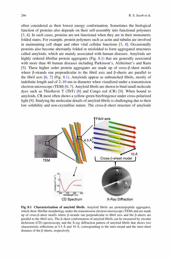
266 R. S. Jacob et al.
often considered as their lowest energy conformation. Sometimes the biologicalfunction of proteins also depends on their self-assembly into functional polymers[3, 4]. In such cases, proteins are not functional when they are in their monomericfolded states. For example, protein polymers such as actin and tubulin are involvedin maintaining cell shape and other vital cellular functions [3, 4]. Occasionallyproteins also become aberrantly folded or misfolded to form aggregated structurescalled amyloids, which are mainly associated with human diseases. Amyloids arehighly ordered fibrillar protein aggregates (Fig. 8.1) that are generally associatedwith more than 40 human diseases including Parkinson’s, Alzheimer’s and Kuru[5]. These higher order protein aggregates are made up of cross-β-sheet motifswhere β-strands run perpendicular to the fibril axis and β-sheets are parallel tothe fibril axis [6, 7] (Fig. 8.1). Amyloids appear as unbranched fibrils, mostly ofindefinite length and of 2–10 nm in diameter when visualized under a transmissionelectron microscope (TEM) [6, 7]. Amyloid fibrils are shown to bind small moleculedyes such as Thioflavin T (ThT) [8] and Congo red (CR) [9]. When bound toamyloids, CR most often shows a yellow-green birefringence under cross-polarizedlight [9]. Studying the molecular details of amyloid fibrils is challenging due to theirlow solubility and non-crystalline nature. The cross-β-sheet structure of amyloids
Fig. 8.1 Characterization of amyloid fibrils. Amyloid fibrils are protein/peptide aggregates,which show fibrillar morphology under the transmission electron microscope (TEM) and are madeup of cross-β-sheet motifs where β-strands run perpendicular to fibril axis and the β-sheets areparallel to the fibril axis. The β-sheet conformation of amyloid fibrils can be measured by circulardichroism (CD) spectroscopy and the X-ray diffraction pattern of amyloid fibrils that shows twocharacteristic reflections at 4.5 Å and 10 Å, corresponding to the inter-strand and the inter-sheetdistance of the β-sheets, respectively
8 Protein Nanofibrils as Storage Forms of Peptide Drugs and Hormones 267
provides them with very high stability as well as resistance to harsh conditions suchas high temperature, wide ranges of pH and proteolytic degradation [10–12].
During aggregation/amyloid formation, both natively structured and unstructuredproteins/peptides undergo misfolding into partially folded intermediates, whicheventually form soluble oligomers and mature fibrils [13]. Recent studies haveshown that soluble oligomers on the pathway of amyloid formation are moretoxic and responsible for cell death occurring in human diseases [14–17]. Theaggregation of protein into amyloid fibrils generally follows nucleation-dependentpolymerization, where three distinct phases are involved. These include the (i) lagphase, where protein monomers associate to form aggregation competent nuclei,but the aggregate mass remains below the threshold for detection (ii) growthphase where monomer addition to nuclei leads to a rapid increase in fibril massand finally the (iii) stationary phase, where fibril formation is completed and thefibrils remain in equilibrium with monomers [18–20]. The nucleation-dependentpolymerization phenomenon in amyloid aggregation is further supported by thefact that addition of preformed fibril seeds (made of the same protein) decreasesthe lag time for fibril formation (self-seeding) [21–23]. Amyloid formation hasthus been considered as an irreversible process, where the fibril is believed tobe a thermodynamically controlled product [24, 25]. This signifies that onceamyloid is formed, its dissociation into monomeric protein/peptide is a seeminglydifficult process. Contrasting this assertion, several research groups have shown thatamyloids could be readily ‘dissolved’ to release their constituent protein/peptideswhen exposed to different pH or simply by dilution [10, 26–28]. Moreover, inthe amyloid aggregation pathway, mature amyloid fibrils remain in equilibriumwith monomers, where the monomeric protein/peptide may constantly exchangewith its fibrillar counterpart in solution (a process known as ‘molecular recycling’)[29]. This dynamic nature of amyloid was nicely demonstrated by Carulla et al.using amyloid fibrils of the bovine phosphatidylinositol-3-kinase SH-3 domain [29].When SH3 domain fibrils were incubated in deuterated buffer for a long time, asubstantial amount of amyloid fibrils were found to be deuterated supporting thatmonomers in amyloid fibrils are getting constantly exchanged with the deuteratedmonomers in solution [29]. In addition to the ‘natural’ phenomenon of molecularrecycling in fibrils, various external factors that can bring about a shift in theequilibrium between the fibrillar and soluble (monomeric/oligomeric) states include(i) protein/peptide concentration, (ii) ionic strength of the buffer, (iii) pH of theenvironment and (iv) the presence of other molecules. The pH-induced fibrildissociation study by Tipping et al. showed that β2 microglobulin amyloid fibrilsdisassemble into toxic oligomers at pH 6.4, whereas the same fibrils dissociate tonative non-toxic monomers at physiological pH 7.4 [30]. Interestingly, a similarobservation was found in another in vitro study, where incubation of α-synucleinfibrils resulted in disaggregation of the fibrils into neurotoxic β-sheet structuredoligomers, which on further incubation formed monomers [31]. Overall, the abovestudies indicate that amyloids can act as a ‘reservoir’ for either monomeric oroligomeric states of proteins.
268 R. S. Jacob et al.
8.2 Functional Amyloids
In the last decade, various reports showed that amyloids cannot only cause humandiseases but are also able to perform native functions in the host organism. Thesenon-disease associated amyloids, involved in important biological functions are col-lectively termed ‘functional amyloids’ [32]. One of the first examples of functionalamyloids came from the discovery of the curli fibrils produced by extracellularlysecreted proteins of the bacterium E. coli [33]. These fibrils were suggested to aidin colonization and biofilm formation of the bacteria. The biofilm is useful for thesurvival of bacteria in extreme environmental conditions such as drastic pH changes,high salt concentration and chemical toxins [34, 35]. Amyloids owing to their abilityto withstand harsh conditions could likely contribute to these characteristics of thebiofilm, being a part of the same. Several other bacteria-associated proteins are alsoreported to polymerize into amyloid fibrils. Examples of these include fimbriaeof Salmonella spp. [34], harpins of X. campestris and P. syringae [36], pili fromM. tuberculosis [37], chaplins from S. coelicolor [38, 39]. Similarly, the amyloidfibrils of the adhesin protein were shown to facilitate yeast cells in colonizationas well as attachment to the host [40]. Furthermore, hydrophobin amyloids, whichprovide support for hyphae/spore formation in fungi [38, 41] and prions of yeast andPodospora anserina that aid in the better survival of the host during stress conditionsare among other examples of fungal functional amyloids [42–45]. Functionalamyloids are also widespread in lower animals such as insects, mollusks etc. Forexample, amyloids of chorion protein can be found in the eggshell of the silk worm,where they protect the growing embryo from environmental hazards [46]. The firstmammalian functional amyloid was discovered by Kelly and co-workers, wherethey showed that amyloid fibrils of Pmel17 inside the melanosome facilitate themelanin polymerization [47]. Recently, it was suggested that pituitary hormonescould also form an amyloid-like structure in vitro and in vivo, which is importantfor hormone storage in secretory granules and its release [26]. In line with thesefindings, designing amyloid-based functional material is shown to be promising fortheir potential application in nano-biotechnology [48, 49]. The following sectionsin this chapter highlight protein nanofibrils as the densest possible peptide-packingstructure suitable for long-term storage. More importantly, it is emphasized here thatby utilizing the feature of amyloids to ‘release functional monomers’, it is possibleto formulate amyloid-based peptide/protein drugs, which can be used for sustainedrelease.
8.3 Amyloids as a Depot for Protein/Peptide Storageand Release
Cells usually synthesize a plethora of proteins for their own activity as well as forsecretion and to act as chemical messages for other cells, an activity which overallleads to a coordinated functioning of tissues and organs. However, deciding which
8 Protein Nanofibrils as Storage Forms of Peptide Drugs and Hormones 269
protein to secrete, when to store and release is a highly regulated and complexprocess in cells. By default, proteins that are to be secreted immediately aftertheir synthesis in ER are transported to the extracellular space through vesiclesthat bud from the trans-Golgi complex [50–53]. Many cells such as liver andmuscle cells use this constitutive route for protein secretion, where the vesiclecontents are released without any external stimulus [54, 55] (Fig. 8.2). However, thesecretion of neuronal/endocrine peptides, growth factors and hormones is tightlyregulated to maintain physiological homeostasis. Such molecules are required tobe stored for long periods in a highly concentrated form until the cells receivesignals to release their contents into the extracellular space [52, 53]. This routeof protein secretion is called “regulated secretion” and is used by several cellsincluding endocrine, neuroendocrine and mast cells [56–58]. Some eukaryotic cellsdisplay both pathways of protein secretion [54]. In such cells, both constitutivelyreleased proteins and regulatory secretory proteins need to be sorted into theirrespective secretory pathways [59]. Although the exact machinery of sorting andsignaling of different protein/peptides to different protein secretory pathway is notclearly understood yet, recent evidence suggests that the formation of amyloid-like structures could be the mechanism for selective aggregation and sorting ofhormones and neuropeptides into regulatory secretory pathway [26]. Therefore, in
Fig. 8.2 Secretory granule biogenesis. This scheme depicts various stages involved in secretorygranule biogenesis, which includes (1) formation of immature secretory granules (ISG), (2)removal of soluble protein by budding (3) homotypic fusion of ISG, (4) remodeling of themembrane of ISG to form MSG
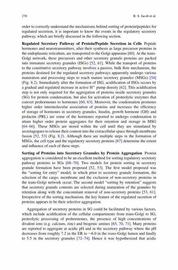
270 R. S. Jacob et al.
order to correctly understand the mechanisms behind sorting of protein/peptides forregulated secretion, it is important to know the events in the regulatory secretorypathway, which are briefly discussed in the following section.
Regulated Secretory Pathway of Protein/Peptide Secretion in Cells Peptidehormones and neurotransmitters, after their synthesis as large precursor proteins inthe endoplasmic reticulum, are transported to the Golgi apparatus [60]. At the trans-Golgi network, these precursors and other secretory granule proteins are packedinto immature secretory granules (ISGs) [52, 61]. While the transport of proteinsto the constitutive secretory pathway involves a passive, bulk flow mechanism, theproteins destined for the regulated secretory pathways apparently undergo variousmaturation and processing steps to reach mature secretory granules (MSGs) [54](Fig. 8.2). Immediately after the formation of ISG, acidification of ISGs occurs bya gradual and regulated increase in active H+ pump density [62]. This acidificationstep is not only required for the aggregation of proteins inside secretory granules(SG) for protein condensation, but also for activation of proteolytic enzymes thatconvert prohormones to hormones [60, 63]. Moreover, the condensation promoteshigher order intermolecular association of proteins and increases the efficiencyof storage of hormones in secretory granules. Insulin, growth hormone (GH) andprolactin (PRL) are some of the hormones reported to undergo condensation toattain higher order protein aggregates for their retention and storage in MSG[64–66]. These MSGs are stored within the cell until they are stimulated bysecretagogues to release their content into the extracellular space through membranefusion [52, 53] (Fig. 8.2). Although there are multiple steps in the formation ofMSGs, the cell type and the regulatory secretory proteins [67] determine the extentand influence of each of these steps.
Sorting of Proteins into Secretory Granules by Protein Aggregation Proteinaggregation is considered to be an excellent method for sorting regulatory secretorypathway proteins to SGs [68–70]. Two models for protein sorting in secretorygranule formation have been proposed [52, 53]. The first model proposed wasthe “sorting for entry” model, in which prior to secretory granule formation, theselection of the cargo, membrane and the exclusion of non-secretory proteins inthe trans-Golgi network occur. The second model “sorting by retention” suggeststhat secretory granule contents are selected during maturation of the granules byretention along with the concomitant removal of non-secretory proteins [53, 61].Irrespective of the sorting mechanism, the key feature of the regulated secretion ofproteins appears to be their selective aggregation.
Aggregation of secretory proteins in SG could be facilitated by various factors,which include acidification of the cellular compartments from trans-Golgi to SG,proteolytic processing of prohormones, the presence of high concentrations ofdivalent ions (e.g. calcium, zinc) and biogenic amines [65, 70, 71]. Many proteinsare reported to aggregate at acidic pH and in the secretory pathway where the pHdecreases from roughly 7.2 in the ER to ∼6.0 in the trans-Golgi lumen and finallyto 5.5 in the secretory granules [72–74]. Hence it was hypothesized that acidic
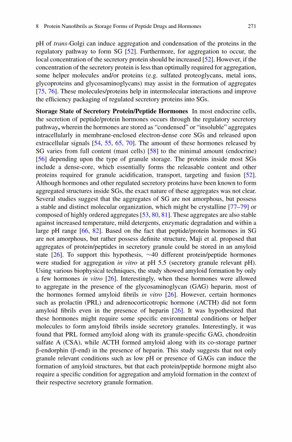
8 Protein Nanofibrils as Storage Forms of Peptide Drugs and Hormones 271
pH of trans-Golgi can induce aggregation and condensation of the proteins in theregulatory pathway to form SG [52]. Furthermore, for aggregation to occur, thelocal concentration of the secretory protein should be increased [52]. However, if theconcentration of the secretory protein is less than optimally required for aggregation,some helper molecules and/or proteins (e.g. sulfated proteoglycans, metal ions,glycoproteins and glycosaminoglycans) may assist in the formation of aggregates[75, 76]. These molecules/proteins help in intermolecular interactions and improvethe efficiency packaging of regulated secretory proteins into SGs.
Storage State of Secretory Protein/Peptide Hormones In most endocrine cells,the secretion of peptide/protein hormones occurs through the regulatory secretorypathway, wherein the hormones are stored as “condensed” or “insoluble” aggregatesintracellularly in membrane-enclosed electron-dense core SGs and released uponextracellular signals [54, 55, 65, 70]. The amount of these hormones released bySG varies from full content (mast cells) [58] to the minimal amount (endocrine)[56] depending upon the type of granule storage. The proteins inside most SGsinclude a dense-core, which essentially forms the releasable content and otherproteins required for granule acidification, transport, targeting and fusion [52].Although hormones and other regulated secretory proteins have been known to formaggregated structures inside SGs, the exact nature of these aggregates was not clear.Several studies suggest that the aggregates of SG are not amorphous, but possessa stable and distinct molecular organization, which might be crystalline [77–79] orcomposed of highly ordered aggregates [53, 80, 81]. These aggregates are also stableagainst increased temperature, mild detergents, enzymatic degradation and within alarge pH range [66, 82]. Based on the fact that peptide/protein hormones in SGare not amorphous, but rather possess definite structure, Maji et al. proposed thataggregates of protein/peptides in secretory granule could be stored in an amyloidstate [26]. To support this hypothesis, ∼40 different protein/peptide hormoneswere studied for aggregation in vitro at pH 5.5 (secretory granule relevant pH).Using various biophysical techniques, the study showed amyloid formation by onlya few hormones in vitro [26]. Interestingly, when these hormones were allowedto aggregate in the presence of the glycosaminoglycan (GAG) heparin, most ofthe hormones formed amyloid fibrils in vitro [26]. However, certain hormonessuch as prolactin (PRL) and adrenocorticotropic hormone (ACTH) did not formamyloid fibrils even in the presence of heparin [26]. It was hypothesized thatthese hormones might require some specific environmental conditions or helpermolecules to form amyloid fibrils inside secretory granules. Interestingly, it wasfound that PRL formed amyloid along with its granule-specific GAG, chondroitinsulfate A (CSA), while ACTH formed amyloid along with its co-storage partnerβ-endorphin (β-end) in the presence of heparin. This study suggests that not onlygranule relevant conditions such as low pH or presence of GAGs can induce theformation of amyloid structures, but that each protein/peptide hormone might alsorequire a specific condition for aggregation and amyloid formation in the context oftheir respective secretory granule formation.
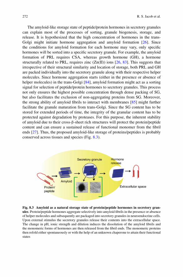
272 R. S. Jacob et al.
The amyloid-like storage state of peptide/protein hormones in secretory granulescan explain most of the processes of sorting, granule biogenesis, storage, andrelease. It is hypothesized that the high concentration of hormones in the trans-Golgi might initiate hormone aggregation and amyloid formation [26]. Sincethe conditions for amyloid formation for each hormone may vary, only specifichormones will be sorted into a specific secretory granule. For example, the amyloidformation of PRL requires CSA, whereas growth hormone (GH), a hormonestructurally related to PRL, requires zinc (Zn(II)) ions [26, 83]. This suggests thatirrespective of their structural similarity and location of storage, both PRL and GHare packed individually into the secretory granule along with their respective helpermolecules. Since hormone aggregation starts (either in the presence or absence ofhelper molecules) in the trans-Golgi [84], amyloid formation might act as a sortingsignal for selection of peptide/protein hormones to secretory granules. This processnot only ensures the highest possible concentration through dense packing of SG,but also facilitates the exclusion of non-aggregating proteins from SG. Moreover,the strong ability of amyloid fibrils to interact with membranes [85] might furtherfacilitate the granule maturation from trans-Golgi. Since the SG content has to bestored for extended periods of time, the integrity of the granular content has to beprotected against degradation by proteases. For this purpose, the inherent stabilityof amyloid due to their cross-β-sheet rich structures will protect the protein/peptidecontent and can ensure a sustained release of functional monomer from the fibrilends [27]. Thus, the proposed amyloid-like storage of proteins/peptides is probablyconserved across tissues and species (Fig. 8.3).
Fig. 8.3 Amyloid as a natural storage state of protein/peptide hormones in secretory gran-ules. Protein/peptide hormones aggregate selectively into amyloid fibrils in the presence or absenceof helper molecules and subsequently are packaged into secretory granules in neuroendocrine cells.Upon external stimulus the secretory granules release their contents into the extracellular space.The change in pH, ionic strength and dilution induces the dissolution of the amyloid fibrils andthe monomeric forms of hormones are then released from the fibril ends. The monomeric proteinsthen refold either spontaneously or with the help of an unknown chaperone to attain their functionalstates
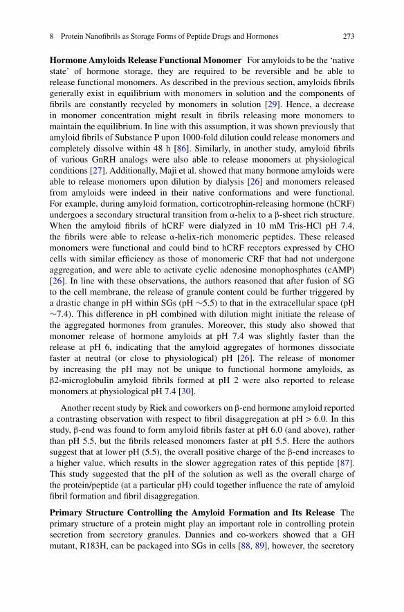
8 Protein Nanofibrils as Storage Forms of Peptide Drugs and Hormones 273
Hormone Amyloids Release Functional Monomer For amyloids to be the ‘nativestate’ of hormone storage, they are required to be reversible and be able torelease functional monomers. As described in the previous section, amyloids fibrilsgenerally exist in equilibrium with monomers in solution and the components offibrils are constantly recycled by monomers in solution [29]. Hence, a decreasein monomer concentration might result in fibrils releasing more monomers tomaintain the equilibrium. In line with this assumption, it was shown previously thatamyloid fibrils of Substance P upon 1000-fold dilution could release monomers andcompletely dissolve within 48 h [86]. Similarly, in another study, amyloid fibrilsof various GnRH analogs were also able to release monomers at physiologicalconditions [27]. Additionally, Maji et al. showed that many hormone amyloids wereable to release monomers upon dilution by dialysis [26] and monomers releasedfrom amyloids were indeed in their native conformations and were functional.For example, during amyloid formation, corticotrophin-releasing hormone (hCRF)undergoes a secondary structural transition from α-helix to a β-sheet rich structure.When the amyloid fibrils of hCRF were dialyzed in 10 mM Tris-HCl pH 7.4,the fibrils were able to release α-helix-rich monomeric peptides. These releasedmonomers were functional and could bind to hCRF receptors expressed by CHOcells with similar efficiency as those of monomeric CRF that had not undergoneaggregation, and were able to activate cyclic adenosine monophosphates (cAMP)[26]. In line with these observations, the authors reasoned that after fusion of SGto the cell membrane, the release of granule content could be further triggered bya drastic change in pH within SGs (pH ∼5.5) to that in the extracellular space (pH∼7.4). This difference in pH combined with dilution might initiate the release ofthe aggregated hormones from granules. Moreover, this study also showed thatmonomer release of hormone amyloids at pH 7.4 was slightly faster than therelease at pH 6, indicating that the amyloid aggregates of hormones dissociatefaster at neutral (or close to physiological) pH [26]. The release of monomerby increasing the pH may not be unique to functional hormone amyloids, asβ2-microglobulin amyloid fibrils formed at pH 2 were also reported to releasemonomers at physiological pH 7.4 [30].
Another recent study by Riek and coworkers on β-end hormone amyloid reporteda contrasting observation with respect to fibril disaggregation at pH > 6.0. In thisstudy, β-end was found to form amyloid fibrils faster at pH 6.0 (and above), ratherthan pH 5.5, but the fibrils released monomers faster at pH 5.5. Here the authorssuggest that at lower pH (5.5), the overall positive charge of the β-end increases toa higher value, which results in the slower aggregation rates of this peptide [87].This study suggested that the pH of the solution as well as the overall charge ofthe protein/peptide (at a particular pH) could together influence the rate of amyloidfibril formation and fibril disaggregation.
Primary Structure Controlling the Amyloid Formation and Its Release Theprimary structure of a protein might play an important role in controlling proteinsecretion from secretory granules. Dannies and co-workers showed that a GHmutant, R183H, can be packaged into SGs in cells [88, 89], however, the secretory
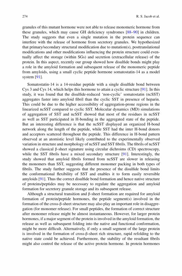
274 R. S. Jacob et al.
granules of this mutant hormone were not able to release monomeric hormone fromthese granules, which may cause GH deficiency syndromes [88–90] in children.The study suggests that even a single mutation in the protein sequence caninterfere with the release of hormone from secretory granules. We hypothesizedthat primary/secondary structural modification due to mutation(s), posttranslationalmodifications and other modifications influencing the protein structure could even-tually affect the storage (within SGs) and secretion (extracellular release) of theprotein. In this aspect, recently our group showed how disulfide bonds might playa role in the amyloid formation and subsequent release of the monomeric peptidefrom amyloids, using a small cyclic peptide hormone somatostatin-14 as a modelsystem [91].
Somatostatin-14 is a 14-residue peptide with a single disulfide bond betweenCys 3 and Cys 14, which helps this hormone to attain a cyclic structure [91]. In thisstudy, it was found that the disulfide-reduced ‘non-cyclic’ somatostatin (ncSST)aggregates faster into amyloid fibril than the cyclic SST in presence of heparin.This could be due to the higher accessibility of aggregation-prone regions in thelinearized ncSST compared to cyclic SST. Molecular dynamics (MD) simulationsof aggregation of SST and ncSST showed that most of the residues in ncSSTas well as SST participated in H-bonding in the aggregated state of the peptide.But an interesting difference is that the ncSST displayed an organized H-bondnetwork along the length of the peptide, while SST had the inter H-bond donorsand acceptors scattered throughout the peptide. This difference in H-bond patternobserved at an atomistic level likely contributed to the experimentally observedvariation in structure and morphology of ncSST and SST fibrils. The fibrils of ncSSTshowed a classical β-sheet signature using circular dichroims (CD) spectroscopy,while the SST fibrils have a mixed secondary structure [91]. Interestingly, thestudy showed that amyloid fibrils formed from ncSST are slower in releasingthe monomers than SST, suggesting different monomer packing in both types offibrils. The study further suggests that the presence of the disulfide bond limitsthe conformational flexibility of SST and enables it to form easily reversibleamyloids [91]. Thus the correct disulfide bond formation and hence native structureof proteins/peptides may be necessary to regulate the aggregation and amyloidformation for secretory granule storage and its subsequent release.
Although a structural transition and β-sheet formation are required for amyloidformation of protein/peptide hormones, the peptide segment(s) involved in theformation of the cross-β-sheet structure may also play an important role in disaggre-gation (for monomer release). For small peptides, the formation of correct structureafter monomer release might be almost instantaneous. However, for larger proteinhormones, if a major segment of the protein is involved in the amyloid formation, therelease as well as subsequent folding into the native and functional conformation,might be more difficult. Alternatively, if only a small segment of the large proteinis involved in the formation of cross-β-sheet rich structure, rapid refolding to thenative state could be achieved. Furthermore, the stability of the resultant fibrilsmight also control the release of the active protein hormone. In protein hormones
8 Protein Nanofibrils as Storage Forms of Peptide Drugs and Hormones 275
such as GH and PRL, the major secondary structure characteristics of the proteinare not altered after fibril formation when measured using CD spectroscopy [26,83]. Both PRL and GH are highly helical proteins and after amyloid formation,these proteins show also a mostly helical conformation, with a slight decrease inhelicity [26, 83]. One of the possible explanations for this observation is that onlya small segment of these proteins might be involved in amyloid formation, whilemost of the other segments remain in their native conformation. This will ensurenot only facile monomer release but also near-instantaneous folding into the nativestate for binding to the receptor and other functions. Several other proteins/peptideswere also previously reported to show the formation of amyloid-like fibrils withoutsignificant secondary structural changes [26, 92–94]. Therefore, we propose that foran amyloid to be an in vivo storage state inside the secretory granules, the structuralchange for amyloid formation may not be aberrant, but rather highly regulated atleast for large proteins, so that substantial amount of native structure could bemaintained within the amyloid state inside secretory granules, facilitating functionalmonomer release. Moreover, the monomer release and folding could be also assistedby chaperones or chaperone-like molecules.
Role of Helper Molecules and Buffer Composition on Amyloid Formationand Release Aggregation-inducing helper molecules (e.g. GAGs, divalent ions)and other biomolecules coexisting within the SG are known to facilitate hormoneaggregation and amyloid formation. Several in vitro aggregation studies of pro-tein/peptide hormones have shown that GAGs such as heparin and chondroitinsulfates induce aggregation [26, 91, 95, 96], wherein the protein and GAGs are usedat particular ratios in solution. Although the exact mechanism of amyloid formationby protein/peptides in the presence of heparin is currently unknown, it is proposedthat heparin’s linear repeat and the electrostatic interactions between protein andGAGs can aid in protein aggregation and amyloid formation. Additionally, a recentstudy by Christensen et al. on prolactin aggregation suggested that the pH of thesurrounding environment could affect the overall charge on the protein hormone,which in turn facilitates the interaction with the helper molecule (GAGs), leadingto its aggregation in vitro [96]. This could be an important mechanism of hormonecondensation inside SGs in vivo. In addition to the role in amyloid formation, theGAGs could also influence the release of monomeric hormones from aggregates.For example, a recent study by Nespovitaya et al. showed that the β-end amyloidfibrils formed in the absence of heparin released monomers in less than one hour,whereas the β-end fibrils formed in the presence of heparin released monomers ina sustained manner up to 24 h [87]. Similarly, a previous study from our groupon the aggregation of glucagon-like peptides 1 and 2 (GLP1 and GLP2), alsoemphasized the role of heparin in modulating monomer release from fibrils [95]. Inthis study, a slower monomer release of GLP1 fibrils was observed, when comparedto GLP2 fibrils, both types of fibrils having been formed in the presence of heparin[95]. Interestingly it was shown that heparin participated in the fibril formation ofGLP1, but not in GLP2. This difference was found to be due to the presence of abasic-non basic-basic (B-X-B) motif present in GLP1 (and not in GLP2), to which
276 R. S. Jacob et al.
heparin could bind to induce fibrillation, and also further aid fibril stabilization.Taken together, these studies suggest that GAGs can significantly influence the fibrilarchitecture, and as a consequence govern the rate of monomer release from amyloidfibrils.
Similar to GAGs, metal ions can also act as helper molecules in proteinaggregation and amyloid formation in the SGs. A recent study from our groupshowed that amyloid fibrils formed in the presence of Zn(II) can release monomericGH in native helical conformation upon dialysis of GH fibrils against 10 mM TrispH 7.4 [83]. Further, this study also showed that GH is co-stored with Zn(II) ionsin vivo, suggesting the role of Zn(II) in both in vivo and in vitro amyloid formationof GH. Another study likewise reported the aggregation of prolactin in the presenceof heparin, chondroitin sulfate A and Zn(II) ions [96]. These studies suggest thathormones require a specific helper molecule (or a combination of molecules) notonly for controlling the formation of individual granules and the content of thegranules but also for the subsequent release of the contents.
In addition to the helper molecules and ions, the buffer constituents of thesolution are also known to be important for amyloid formation. An extensivestudy by Nespovitaya et al. on β-end fibrillation under various physiologicallyrelevant environmental conditions, reported that helper molecules, pH and bufferconstituents could differentially affect the amyloid formation of the peptide [87].In this study, the authors showed that β-end was able to form amyloid by itself inmultivalent ion buffers such as phosphate, sulfate, and citrate but not in ammonium,acetate and Tris. However, the addition of heparin to β-end in ammonium acetatebuffer resulted in its amyloid formation [87]. Nespovitaya et al. also found that theamyloid fibrils formed in multivalent buffers and in the presence of heparin differedin both structure and morphology. This study and previous works by Maji et al. [26,27] overall suggest that the change of the immediate environment that the secretoryproteins encounter along the secretory pathway in terms of pH, GAGs, metal ions,concentration of protein and even ion constituents modulate the amyloid aggregationand subsequent disassembly of hormones.
In line with this evidence, it can be envisioned how the amyloid structure enablesthe densest peptide-packing possible and is suitable for long-term storage. More-over, the common structural signature of cross-β-sheets can by itself act as sortingsignal for packing of hormone/neuropeptide aggregates into secretory granules. Achange in environmental conditions can dissociate these protein nanofibrils andcould release functional monomers, thus enabling amyloid fibrils to act as depotboth in vitro and in vivo.
8.4 Amyloid as Long-Acting Depot Formulations
Developing novel biocompatible scaffolds for drug delivery applications is anemerging area in modern science. Small molecular and protein/peptide drugs haveshort half-life and low bioavailability in vivo. Such drugs are often delivered
8 Protein Nanofibrils as Storage Forms of Peptide Drugs and Hormones 277
through frequent injections or subcutaneous administrations and infusions to preventthem from degradation [97, 98]. To overcome the delivery related problems, newdrug formulations have been created that continuously release and maintain anactive drug concentration over an extended period. In this regard, administratingprotein drugs like insulin and TGF-β3 in self-assembled crystalline form has showncontrolled delivery of these drugs [99, 100]. However, protein crystals tend to befragile and can disintegrate easily, requiring sophisticated methods to preserve andstore them. Hence an alternative peptide/protein self-assembly system needs to beemployed, that can deliver active and functional peptide/protein drugs in a sustainedmanner for an extended period. Since a growing amount of studies have alreadyestablished that not all amyloids are toxic and can serve as functional protein entity,it is possible to imagine that amyloid fibrils can act as a storage depot of variousprotein/peptide hormones. In order to explore this possibility, a study utilized theshort-acting as well as long-acting analogs of gonadotropin-releasing hormone(GnRH) [27]. When these GnRH analogs were incubated in vitro, all long-actinganalogs were found to form fibrillar aggregates that bind to amyloid specific dyesThT and CR. In contrast, only a few short-acting GnRH analogs formed amyloidfibrils in vitro. Furthermore, when the monomer releasing capabilities of theseamyloid fibrils were studied, the long-acting analogs showed sustained release ofmonomeric hormone from amyloids, while the fibrils of short-acting analogs eitherreleased monomer immediately upon dilution (similar to non-aggregated peptide) orreleased very slowly such that effective drug concentration cannot be reached [27].This study clearly indicates that packing and stability of each peptide fibril mightdictate the monomer release, which could possibly be determined by the differencesin their amino acid composition.
Many earlier studies have reported that amyloid fibrils can be stabilized in thepresence of serum amyloid protein P and GAGs [101, 102]. If the protein/peptidedrugs are administered in the form of amyloid fibrils, a more sustained andcontrolled release can be expected, due to the combined effect of the highly orderedstructure of the amyloids and the GAG-mediated stabilization of the fibrils. TheGnRH analogs mentioned earlier were shown to form amyloid fibrils in the presenceas well as absence of heparin [27]. Further, when the release assay was performedwith fibrils formed in the presence or absence of heparin, fibrils formed in thepresence of heparin displayed a slower release pattern, compared to GnRH analogfibrils formed in the absence of heparin. This observation was found to apply forother peptide hormones as well. For instance, two independent studies (as mentionedin the previous section) showed that the amyloid fibrils of β-endorphin or GLP1hormone formed in the presence of heparin [87, 95], displayed a slower monomerrelease profile compared to the respective hormone amyloid formed in the absenceof heparin. These studies suggest that GAGs present in body could also influence theformation, stability as well as controlled release capability of administered peptidefibrils [27, 101–104].
In Vivo Release of Monomers from Hormone Amyloids For amyloids to act asdrug reservoirs, they should release functional monomeric proteins in vivo. While
278 R. S. Jacob et al.
a large range of studies has shown the reversible nature of amyloids in vitro, onlya handful of studies exist, which demonstrate that amyloid can release functionalmonomers in vivo. In one such study, long-acting GnRH analog (L14) monomersin D-Mannitol were injected subcutaneously into rats, which resulted in in vivoamyloid formation at the injection site [27]. GnRH analogs are generally used assuppressing agent of luteinizing hormone (LH) in prostate tumors [105]. The invivo amyloid formation of L14 was confirmed by CR staining and birefringenceof the tissue section on the injection site. Thus, the long-acting capability of L14could be due to its amyloid formation in vivo and release of functional monomersfrom this amyloid depot. This study thereby suggests that the amyloid formation byamyloidogenic peptide hormones could be further facilitated by in vivo conditions,which may also play a role in the controlled release of the peptide drug [27].Moreover, to directly demonstrate that amyloid formation could influence theduration of action of GnRH analogs, a short-acting GnRH analog (S2) was chosen,which did not form amyloid even after 8 days of incubation but formed amyloid afterlong incubation (Fig. 8.4a). Unlike the L14 analog, the introduction of monomericGnRH analog S2 did not form amyloid in vivo at the site of injection [27]. However,after the subcutaneous injection of aged S2 fibrils in rat, the localization of S2 fibrilswas confirmed by CR staining and birefringence at the injected site [27]. When agedS2 fibrils and S2 monomer solutions were subcutaneously injected in a male rat(castrated 10 days prior the experiment), both aged fibrillar and monomeric S2 wereable to suppress (measured by radioimmunoassay) LH in the blood of the animalsafter 24 h of administration. After 48 h of administration, the fibrillar GnRH analogsshowed more LH suppressive activity compared to monomeric GnRH analogs. Itis interesting to note that, although at 72 h, the monomeric S2 almost lost its LHsuppressing activity, the fibrillar S2 was still able to suppress LH significantly atthis time (Fig. 8.4b) [27]. This particular study directly shows the ability of amyloidfibrils to act as drug depot in vivo and further suggests that amyloid formationof protein/peptide drugs can prolong their activity for sustainable drug deliveryapplications (Fig. 8.4c) [27].
Toxicity and Amplification of Toxic Protein Aggregates by Hormone AmyloidsSince amyloids are originally associated with cell death in various neurodegenera-tive diseases such as Alzheimer’s and Parkinson’s, it is a potential concern that thehormone amyloids may exhibit toxicity upon formation in vivo or administrationof hormone amyloids as drug depots. However, recently it was suggested thatamyloid fibrils, the end-products of the aggregation pathway, are the less toxicspecies in protein aggregation reactions and protein/peptide oligomers (pathwayintermediates) are more toxic species, responsible for cell death [14–17]. More-over, it was also suggested that amyloid could be a nontoxic reservoir of toxicprotein/peptide oligomers [14, 106, 107]. Various studies during the last decadesshowed that amyloid fibrils are not only less toxic than smaller aggregated species,but also that many amyloids have evolved in nature to perform native functionsof the host organism [5, 32] (discussed earlier under Sect. 8.2). Moreover, thetoxicity of amyloid fibrils could depend on the protein/peptide sequences that
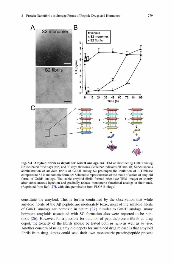
8 Protein Nanofibrils as Storage Forms of Peptide Drugs and Hormones 279
Fig. 8.4 Amyloid fibrils as depots for GnRH analogs. (a) TEM of short-acting GnRH analogS2 incubated for 8 days (top) and 30 days (bottom). Scale bar indicates 200 nm. (b) Subcutaneousadministration of amyloid fibrils of GnRH analog S2 prolonged the inhibition of LH releasecompared to S2 in monomeric form. (c) Schematic representation of the mode of action of amyloidforms of GnRH analogs. The stable amyloid fibrils formed prior (see TEM image) or shortlyafter subcutaneous injection and gradually release monomeric functional analogs at their ends.(Reprinted from Ref. [27], with kind permission from PLOS Biology)
constitute the amyloid. This is further confirmed by the observation that whileamyloid fibrils of the Aβ peptide are moderately toxic, most of the amyloid fibrilsof GnRH analogs are nontoxic in nature [27]. Similar to GnRH analogs, manyhormone amyloids associated with SG formation also were reported to be non-toxic [26]. However, for a possible formulation of peptide/protein fibrils as drugdepot, the toxicity of the fibrils should be tested both in vitro as well as in vivo.Another concern of using amyloid depots for sustained drug release is that amyloidfibrils from drug depots could seed their own monomeric protein/peptide present
280 R. S. Jacob et al.
in the body and amplify the amyloid form. This could be detrimental to the properfunctioning of the normal hormone. However, Maji and colleagues observed thatmature fibrils of long-acting GnRH (L14) could not template aggregation of wildtype GnRH, suggesting that amyloid fibrils of GnRH analogs might be incapableto seed and amplify GnRH amyloid formation [27]. Similarly, it is also possiblethat the amyloid depot of the particular hormone (or analogs) can ‘cross-seed’other protein/peptides associated with diseases and induce their aggregation, therebyincreasing the load of cytotoxic protein/peptide aggregates in the body. However, tocross-seed, both the seed and host protein(s) require high sequence similarity as wellas amyloid forming capability [23, 108, 109]. This could be a probable mechanismby which nature prevents the disease-associated amyloids from cross-seeding thevarious other proteins existing in the body. Nevertheless, one has to test thepossibility of amyloid drug depot to cross-seed other proteins, which are potentiallyamyloidogenic and are associated with diseases. In the study by Maji et al. todemonstrate the safety of GnRH amyloid against cross-seeding of disease associatedprotein aggregation, a small amount (5%) of mature fibrils of L20 GnRH analogwas incubated with the wild-type monomers of α-Synuclein (α-Syn), a proteinassociated with Parkinson’s disease [110–114]. The study showed that althoughα-Syn spontaneously aggregated and formed amyloid fibrils, the fibril seeds oflong-acting hormone L20 did not cross-seed the aggregation and amyloid formationof α-Syn [27]. This study indicates that rationally designing protein analogs thatwould not seed wild-type proteins (to form toxic aggregates) can also be used asa strategy for developing amyloid formulation of peptide/protein drugs. Moreover,under physiological conditions, different proteins are often compartmentalized, thusminimizing the possibility of encounter and interaction between protein amyloid asdepot with other disease-associated proteins such as α-Syn or Aβ.
The reversibility of amyloid fibrils can also be employed for various otherfunctional purposes; for example, in the controlled release of peptide antibiotics.In this regard, Pertinhez et al. have recently shown that the aggregates of anantibody-derived decapeptide KP (killer peptide) can provide targeted delivery aswell as possesses slow release properties [115]. The study showed that disulfide-bonded dimers of the synthetic peptide KP (AKVTMTCSAS) are the active formdisplaying microbicidal activity and these dimers can self-assemble to form higherorder aggregates with fibrillar morphology and β-sheet structure over time [115].The dilution or incubation of these KP aggregates at 35 ◦C resulted in the solubleform. Moreover, similar to other amyloidogenic peptides, the KP also rapidly self-assembles in the presence of GAGs such as 1, 3 β-glucan, and the aggregates formedin presence of this GAG have slow-releasing capability.
In addition to amyloid fibrils, self-assembled protein/peptide oligomers can alsobe used as drug depots. A recent study by Gupta et al. suggested that self-assembledoligomers of insulin can be used as a drug depot, after showing that oligomersformed during the aggregation of insulin were capable of releasing functionalinsulin monomers [116]. In patients with type 1 diabetes, insulin production isgenerally insufficient to control the blood glucose levels. Insulin injections are
8 Protein Nanofibrils as Storage Forms of Peptide Drugs and Hormones 281
administered in these patients for a glycemic control; however, multiple dosagesare required, as the half-life of the injected peptide is short [117]. In their study,Gupta et al. characterized three intermediate states in insulin amyloid formation anddesignated them as supramolecular insulin assembly (SIA) I, II and III. The SIA-II intermediate formed at 12–16 h of incubation of insulin at pH 7.0 was found torelease monomeric insulin at a rate that is needed for maintaining the basal levelof insulin in blood [116]. The intermediates formed at pH 2.0 released monomersat a slow rate and the amyloid fibrils formed at pH 7.0 or 2.0, did not release anymonomers. This observation suggests that the release capability of peptide/proteineither from amyloid or oligomeric assemblies depends on the peptide sequences andthe conditions used for making the assemblies.
Amyloid-Based Biomaterials as Drug Delivery Vehicles Amyloid fibrils assupramolecular assemblies display charged and hydrophobic surfaces, which couldbind both small molecules and proteins [118–121]. The unique surface properties ofamyloid fibril networks can be utilized for entrapping drugs, enzymes and proteins,thus amyloid fibrils could be used for developing drug delivery vehicles. Further,amyloid-based scaffolds or hydrogels can be rationally designed such that they canrelease the encapsulated drugs/protein at the site of action. In this context, Zhangand coworkers have used hydrogels from self-assembling RADA16 peptides asdrug delivery vehicles of small molecules and proteins such as trypsin inhibitor,lysozyme, immunoglobulin G (IgG) and bovine serum albumin (BSA) [122, 123].Moreover, the flanking regions of amyloid-forming peptide sequences can bemodified appropriately such that the designed fibril matrix can deliver its payload attarget sites. For example, Mazza et al. modified an amphiphilic dalargin (an opioidreceptor agonist) derivative with a palmitoyl moiety, which self-assembled into β-sheet fibrils after sonication in water. The intravenous injection of the modifieddalargin nanofibrils allowed it to cross the blood-brain barrier and deliver activedalargin into the brain [124].
Since many small molecule drugs are hydrophobic in nature, an increasingnumber of studies are attempting to encapsulate small-molecule drugs in hydrogelsderived from amyloid fibrils for their successful delivery to target sites. Forexample, a recent study successfully demonstrated the use of protein nanofibrilmicrogels composed of amyloid fibrils for encapsulation and sustained release ofantibiotics [125]. In this work, the nanofibril microgels were produced using awater-in-oil method and were tested for their efficacy to act as carriers of smallmolecules such as dyes and antibiotics. Using this system, it was observed thatthe drug molecules penicillin and tetracycline were encapsulated in the nanofibrilmicrogels and could be released locally, resulting in increased anti-microbial action.Moreover, these microgels were proven to be non-cytotoxic to human cell lines,and thus are biocompatible [125]. Furthermore, the biocompatibility of drugs canbe further enhanced when delivered within an amyloid matrix. For example, Li etal. immobilized carbon nanotubes (NT) within a β-lactoglobulin gel matrix, whichmade the NT biocompatible. The study further showed that NT can be released fromthe protein shell by enzymatic digestion and these NT can be used for killing cancer
282 R. S. Jacob et al.
cells by heating the NT by near infrared light indicating possible applications incancer therapy [126]. Recently, it was also shown that hybrid materials composed ofamyloid fibrils-iron colloidal dispersion could be used as oral iron delivery systems,which increases the bioavailability of iron in vivo [127].
Intriguingly, it is not always the amyloid fibrils that can modulate the biodistri-bution or availability of the encapsulated drug/small molecules. The encapsulateddrugs or small molecules can also modulate the properties of the gel matrix byinteracting with the amyloid fibrils. In a recent study, lysozyme was allowed to formamyloid in the presence of the β1 receptor antagonist drugs, propanolol, timololand atenolol, and it was found that the drugs modulated the fibril architecture,leading to altered release profiles of the drugs [128]. These studies suggest that theinteraction between the various payload and amyloid fibrils can be utilized to finetune the architecture of amyloid-based drug delivery vehicles to obtain desirablerelease profiles. Furthermore, it has been shown that the amyloid fibrils can befabricated into different matrices for various biotechnological applications such asdrug delivery. For example, β2-microglobulin amyloid fibrils were fabricated intoa nanoporous and high mechanical strength protein matrix, which is suggested tobe useful in tissue engineering and drug delivery applications [129]. In anotherexample, hydrogels were prepared from α-synuclein curly-amyloid fibrils and wereused for enzyme entrapment. The enzyme trapped inside the amyloid hydrogel wasable to withstand heat treatment and maintain enzymatic activity, which suggestsa plausible use in therapeutic delivery [130]. Moreover, the entrapping of enzymesand other small molecules inside nano-fibrillar scaffolds of amyloids could improvetheir stability and activity. For example, organophosphate hydrolase (OPH), anenzyme important for bioremediation and chemical detoxification was covalentlycross-linked using glutaraldehyde on bovine insulin fibrils. This enhanced thestability of the encapsulated enzyme at higher temperatures compared to its freecounterpart [131]. Such enzyme encapsulated amyloid hydrogels need not alwaysrelease the enzyme molecule. Since in hydrogels, free diffusion of molecules occurswithin and also with the surrounding environment, the substrate molecules candiffuse inside the hydrogel within the proximity of the enzyme molecules and beconverted into product, which can then diffuse out of the hydrogel. Alternatively,to enhance the stability of enzymes, amyloid-forming peptide segments can beengineered onto medically relevant enzymes such that these engineered enzymescan form amyloid fibrils retaining the enzymatic activity [92]. These engineeredenzymes will be stable and may perform substrate conversion for longer durationthan soluble enzymes.
We, therefore suggest that amyloid-based technology could be used for drugdelivery in two different ways (Fig. 8.5). In one case, the amyloid fibril form ofthe protein/peptide drug could be administered directly, as suggested by previousstudies [27], which will allow the fibrils to release monomers in vivo in a controlledmanner. The other possibility is to construct an amyloid-based scaffold where smallmolecules or drugs can be encapsulated. The latter method can also be used formultiple drug delivery purposes as well as this would provide protein/peptide drugafter dissociation and also will deliver the drug molecules of interest which are
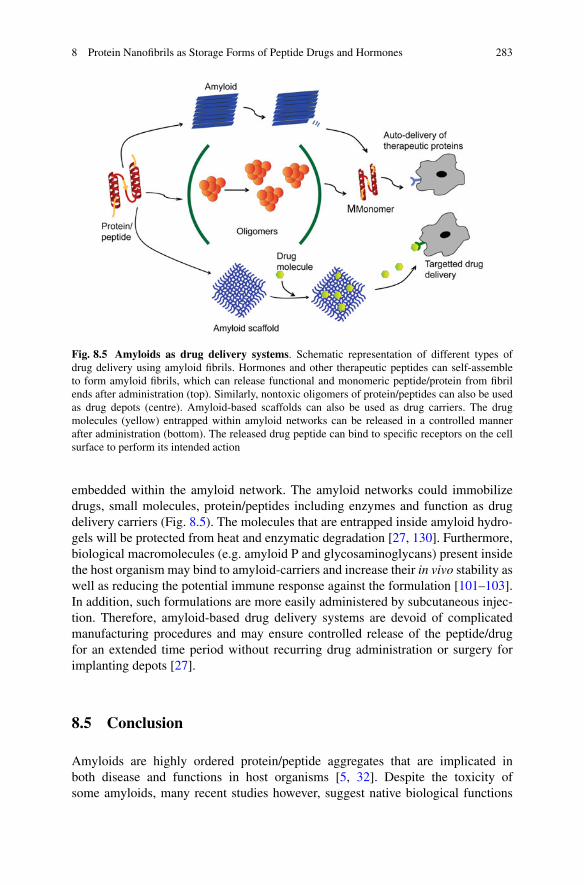
8 Protein Nanofibrils as Storage Forms of Peptide Drugs and Hormones 283
Fig. 8.5 Amyloids as drug delivery systems. Schematic representation of different types ofdrug delivery using amyloid fibrils. Hormones and other therapeutic peptides can self-assembleto form amyloid fibrils, which can release functional and monomeric peptide/protein from fibrilends after administration (top). Similarly, nontoxic oligomers of protein/peptides can also be usedas drug depots (centre). Amyloid-based scaffolds can also be used as drug carriers. The drugmolecules (yellow) entrapped within amyloid networks can be released in a controlled mannerafter administration (bottom). The released drug peptide can bind to specific receptors on the cellsurface to perform its intended action
embedded within the amyloid network. The amyloid networks could immobilizedrugs, small molecules, protein/peptides including enzymes and function as drugdelivery carriers (Fig. 8.5). The molecules that are entrapped inside amyloid hydro-gels will be protected from heat and enzymatic degradation [27, 130]. Furthermore,biological macromolecules (e.g. amyloid P and glycosaminoglycans) present insidethe host organism may bind to amyloid-carriers and increase their in vivo stability aswell as reducing the potential immune response against the formulation [101–103].In addition, such formulations are more easily administered by subcutaneous injec-tion. Therefore, amyloid-based drug delivery systems are devoid of complicatedmanufacturing procedures and may ensure controlled release of the peptide/drugfor an extended time period without recurring drug administration or surgery forimplanting depots [27].
8.5 Conclusion
Amyloids are highly ordered protein/peptide aggregates that are implicated inboth disease and functions in host organisms [5, 32]. Despite the toxicity ofsome amyloids, many recent studies however, suggest native biological functions
284 R. S. Jacob et al.
such as protein/peptide storage inside secretory granules of mammals [26]. Thehigher order structural organization of protein/peptide in a cross-β-sheet richmotif and stability against various harsh physical/chemical conditions such asextreme pHs, temperature and proteases, makes amyloid attractive for versatilenanobiotechnological applications such as drug delivery and tissue engineering[132–134]. Since as few as two amino acid residues are sufficient for amyloid-like assembly [135], tagging short amyloid sequences onto functional proteins canbe exploited for protein formulation similar to the case of GnRH analogs [27].Moreover, many small peptide-based amyloids can also be utilized for encapsulatingdifferent drugs/proteins for their possible delivery. We expect to witness many moreapplications of designed amyloids for diverse biomedical applications in comingyears.
Acknowledgments The authors wish to acknowledge DBT (BT/PR9797/NNT/28/774/2014)Government of India and Wadhwani Research Centre for Bioengineering (WRCB) for financialsupport.
References
1. Jeremy M, Berg JLT, Stryer L (2002) Biochemistry, 5th edn. W H Freeman, New York2. Dobson CM (2004) Principles of protein folding, misfolding and aggregation. Semin Cell
Dev Biol 15(1):3–163. Kirschner M, Mitchison T (1986) Beyond self-assembly: from microtubules to morphogene-
sis. Cell 45(3):329–3424. Fletcher DA, Mullins RD (2010) Cell mechanics and the cytoskeleton. Nature
463(7280):485–4925. Chiti F, Dobson CM (2006) Protein misfolding, functional amyloid, and human disease. Annu
Rev Biochem 75:333–3666. Sunde M, Blake C (1997) The structure of amyloid fibrils by electron microscopy and X-ray
diffraction. Adv Protein Chem 50:123–1597. Sunde M, Serpell LC, Bartlam M, Fraser PE, Pepys MB, Blake CC (1997) Common core
structure of amyloid fibrils by synchrotron X-ray diffraction. J Mol Biol 273(3):729–7398. LeVine H III (1993) Thioflavine T interaction with synthetic Alzheimer’s disease β-amyloid
peptides: detection of amyloid aggregation in solution. Protein Sci 2:404–4109. Westermark GT, Johnson KH, Westermark P (1999) Staining methods for identification of
amyloid in tissue. Methods Enzymol 309:3–2510. Meersman F, Dobson CM (2006) Probing the pressure-temperature stability of amyloid fibrils
provides new insights into their molecular properties. Biochim Biophys Acta 1764(3):452–460
11. Mesquida P, Riener CK, MacPhee CE, McKendry RA (2007) Morphology and mechanicalstability of amyloid-like peptide fibrils. J Mater Sci Mater Med 18(7):1325–1331
12. Zurdo J, Guijarro JI, Dobson CM (2001) Preparation and characterization of purified amyloidfibrils. J Am Chem Soc 123(33):8141–8142
13. Uversky VN, Fink AL (2004) Conformational constraints for amyloid fibrillation: theimportance of being unfolded. Biochim Biophys Acta 1698(2):131–153
14. Haass C, Selkoe DJ (2007) Soluble protein oligomers in neurodegeneration: lessons from theAlzheimer’s amyloid beta-peptide. Nat Rev Mol Cell Biol 8(2):101–112
8 Protein Nanofibrils as Storage Forms of Peptide Drugs and Hormones 285
15. Kayed R, Head E, Thompson JL, McIntire TM, Milton SC, Cotman CW, Glabe CG(2003) Common structure of soluble amyloid oligomers implies common mechanism ofpathogenesis. Science 300(5618):486–489
16. Bucciantini M, Giannoni E, Chiti F, Baroni F, Formigli L, Zurdo J, Taddei N, Ramponi G,Dobson CM, Stefani M (2002) Inherent toxicity of aggregates implies a common mechanismfor protein misfolding diseases. Nature 416(6880):507–511
17. Walsh DM, Klyubin I, Fadeeva JV, Cullen WK, Anwyl R, Wolfe MS, Rowan MJ, SelkoeDJ (2002) Naturally secreted oligomers of amyloid β protein potently inhibit hippocampallong-term potentiation in vivo. Nature 416(6880):535–539
18. Harper JD, Lansbury PT Jr (1997) Models of amyloid seeding in Alzheimer’s disease andscrapie: mechanistic truths and physiological consequences of the time-dependent solubilityof amyloid proteins. Annu Rev Biochem 66:385–407
19. Jarrett JT, Lansbury PT Jr (1993) Seeding “one-dimensional crystallization” of amyloid: apathogenic mechanism in Alzheimer’s disease and scrapie? Cell 73:1055–1058
20. Ferrone F (1999) Analysis of protein aggregation kinetics. Methods Enzymol 309:256–27421. Naiki H, Hashimoto N, Suzuki S, Kimura H, Nakakuki K, Gejyo F (1997) Establishment
of a kinetic model of dialysis-related amyloid fibril extension in vitro. Amyl Int J Exp ClinInvestig 4(4):223–232
22. Serio TR, Cashikar AG, Kowal AS, Sawicki GJ, Moslehi JJ, Serpell L, Arnsdorf MF,Lindquist SL (2000) Nucleated conformational conversion and the replication of conforma-tional information by a prion determinant. Science 289(5483):1317–1321
23. Wright CF, Teichmann SA, Clarke J, Dobson CM (2005) The importance of sequencediversity in the aggregation and evolution of proteins. Nature 438(7069):878–881
24. Wetzel R (2006) Kinetics and thermodynamics of amyloid fibril assembly. Acc Chem Res39(9):671–679
25. Knowles TPJ, Vendruscolo M, Dobson CM (2014) The amyloid state and its association withprotein misfolding diseases. Nat Rev Mol Cell Biol 15(6):384–396
26. Maji SK, Perrin MH, Sawaya MR, Jessberger S, Vadodaria K, Rissman RA, Singru PS,Nilsson KP, Simon R, Schubert D, Eisenberg D, Rivier J, Sawchenko P, Vale W, Riek R (2009)Functional amyloids as natural storage of peptide hormones in pituitary secretory granules.Science 325(5938):328–332
27. Maji SK, Schubert D, Rivier C, Lee S, Rivier JE, Riek R (2008) Amyloid as a depot for theformulation of long-acting drugs. PLoS Biol 6(2):e17
28. MacPhee CE, Dobson CM (2000) Chemical dissection and reassembly of amyloid fibrilsformed by a peptide fragment of transthyretin. J Mol Biol 297(5):1203–1215
29. Carulla N, Caddy GL, Hall DR, Zurdo J, Gairi M, Feliz M, Giralt E, Robinson CV, DobsonCM (2005) Molecular recycling within amyloid fibrils. Nature 436(7050):554–558
30. Tipping KW, Karamanos TK, Jakhria T, Iadanza MG, Goodchild SC, Tuma R, Ranson NA,Hewitt EW, Radford SE (2015) pH-induced molecular shedding drives the formation ofamyloid fibril-derived oligomers. Proc Natl Acad Sci U S A 112(18):5691–5696
31. Cremades N, Cohen SI, Deas E, Abramov AY, Chen AY, Orte A, Sandal M, Clarke RW,Dunne P, Aprile FA, Bertoncini CW, Wood NW, Knowles TP, Dobson CM, Klenerman D(2012) Direct observation of the interconversion of normal and toxic forms of α-synuclein.Cell 149(5):1048–1059
32. Fowler DM, Koulov AV, Balch WE, Kelly JW (2007) Functional amyloid – from bacteria tohumans. Trends Biochem Sci 32(5):217–224
33. Chapman MR, Robinson LS, Pinkner JS, Roth R, Heuser J, Hammar M, Normark S, HultgrenSJ (2002) Role of Escherichia coli curli operons in directing amyloid fiber formation. Science295(5556):851–855
34. Otzen D, Nielsen PH (2008) We find them here, we find them there: functional bacterialamyloid. Cell Mol Life Sci 65(6):910–927
35. Hall-Stoodley L, Costerton JW, Stoodley P (2004) Bacterial biofilms: from the naturalenvironment to infectious diseases. Nat Rev Microbiol 2(2):95–108
286 R. S. Jacob et al.
36. Oh J, Kim JG, Jeon E, Yoo CH, Moon JS, Rhee S, Hwang I (2007) Amyloidogenesis of typeIII-dependent harpins from plant pathogenic bacteria. J Biol Chem 282(18):13601–13609
37. Alteri CJ, Xicohtencatl-Cortes J, Hess S, Caballero-Olin G, Giron JA, Friedman RL (2007)Mycobacterium tuberculosis produces pili during human infection. Proc Natl Acad Sci U SA 104(12):5145–5150
38. Gebbink MF, Claessen D, Bouma B, Dijkhuizen L, Wosten HA (2005) Amyloids – afunctional coat for microorganisms. Nat Rev Microbiol 3(4):333–341
39. Talbot NJ (2003) Aerial morphogenesis: enter the chaplins. Curr Biol 13(18):R696–R69840. Ramsook CB, Tan C, Garcia MC, Fung R, Soybelman G, Henry R, Litewka A, O’Meally S,
Otoo HN, Khalaf RA, Dranginis AM, Gaur NK, Klotz SA, Rauceo JM, Jue CK, Lipke PN(2010) Yeast cell adhesion molecules have functional amyloid-forming sequences. EukaryotCell 9(3):393–404
41. Hammer ND, Wang X, McGuffie BA, Chapman MR (2008) Amyloids: friend or foe? JAlzheimers Dis 13(4):407–419
42. Osherovich LZ, Weissman JS (2002) The utility of prions. Dev Cell 2(2):143–15143. True HL, Lindquist SL (2000) A yeast prion provides a mechanism for genetic variation and
phenotypic diversity. Nature 407(6803):477–48344. Uptain SM, Lindquist S (2002) Prions as protein-based genetic elements. Annu Rev Microbiol
56:703–74145. Chien P, Weissman JS, DePace AH (2004) Emerging principles of conformation-based prion
inheritance. Annu Rev Biochem 73:617–65646. Iconomidou VA, Vriend G, Hamodrakas SJ (2000) Amyloids protect the silkmoth oocyte and
embryo. FEBS Lett 479(3):141–14547. Fowler DM, Koulov AV, Alory-Jost C, Marks MS, Balch WE, Kelly JW (2006) Functional
amyloid formation within mammalian tissue. PLoS Biol 4(1):100–10748. Cherny I, Gazit E (2008) Amyloids: not only pathological agents but also ordered nanomate-
rials. Angew Chem Int Ed Engl 47(22):4062–406949. Knowles TP, Mezzenga R (2016) Amyloid fibrils as building blocks for natural and artificial
functional materials. Adv Mater 28(31):6546–656150. Palade G (1975) Intracellular aspects of the process of protein synthesis. Science
189(4200):347–35851. Lacy PE (1975) Endocrine secretory mechanisms. A review. Am J Pathol 79(1):170–18852. Tooze SA (1998) Biogenesis of secretory granules in the trans-Golgi network of neuroen-
docrine and endocrine cells. Biochim Biophys Acta 1404(1–2):231–24453. Arvan P, Castle D (1998) Sorting and storage during secretory granule biogenesis: looking
backward and looking forward. Biochem J 332(Pt 3):593–61054. Kelly RB (1985) Pathways of protein secretion in eukaryotes. Science 230(4721):25–3255. Kelly RB (1987) Protein transport. from organelle to organelle. Nature 326(6108):14–1556. Stefan Y, Meda P, Neufeld M, Orci L (1987) Stimulation of insulin secretion reveals
heterogeneity of pancreatic B cells in vivo. J Clin Invest 80(1):175–18357. Loh YP, Kim T, Rodriguez YM, Cawley NX (2004) Secretory granule biogenesis and
neuropeptide sorting to the regulated secretory pathway in neuroendocrine cells. J MolNeurosci 22(1–2):63–71
58. Hide I, Bennett JP, Pizzey A, Boonen G, Bar-Sagi D, Gomperts BD, Tatham PE (1993)Degranulation of individual mast cells in response to Ca2+ and guanine nucleotides: an all-or-none event. J Cell Biol 123(3):585–593
59. Turner MD, Rennison ME, Handel SE, Wilde CJ, Burgoyne RD (1992) Proteins are secretedby both constitutive and regulated secretory pathways in lactating mouse mammary epithelialcells. J Cell Biol 117(2):269–278
60. Kim T, Gondre-Lewis MC, Arnaoutova I, Loh YP (2006) Dense-core secretory granulebiogenesis. Physiology (Bethesda) 21:124–133
61. Tooze SA (1991) Biogenesis of secretory granules. Implications arising from the immaturesecretory granule in the regulated pathway of secretion. FEBS Lett 285(2):220–224
8 Protein Nanofibrils as Storage Forms of Peptide Drugs and Hormones 287
62. Wu MM, Grabe M, Adams S, Tsien RY, Moore HP, Machen TE (2001) Mechanisms of pHregulation in the regulated secretory pathway. J Biol Chem 276(35):33027–33035
63. Steiner DF, Smeekens SP, Ohagi S, Chan SJ (1992) The new enzymology of precursorprocessing endoproteases. J Biol Chem 267(33):23435–23438
64. Kuliawat R, Prabakaran D, Arvan P (2000) Proinsulin endoproteolysis confers enhancedtargeting of processed insulin to the regulated secretory pathway. Mol Biol Cell 11(6):1959–1972
65. Dannies PS (2002) Mechanisms for storage of prolactin and growth hormone in secretorygranules. Mol Genet Metab 76(1):6–13
66. Giannattasio G, Zanini A, Meldolesi J (1975) Molecular organization of rat prolactin granules.I. In vitro stability of intact and “membraneless” granules. J Cell Biol 64(1):246–251
67. Morvan J, Tooze SA (2008) Discovery and progress in our understanding of the regulatedsecretory pathway in neuroendocrine cells. Histochem Cell Biol 129(3):243–252
68. Dannies P (2003) Manipulating the reversible aggregation of protein hormones in secretorygranules: potential impact on biopharmaceutical development. BioDrugs 17(5):315–324
69. Dannies PS (1999) Protein hormone storage in secretory granules: mechanisms for concen-tration and sorting. Endocr Rev 20(1):3–21
70. Dannies PS (2001) Concentrating hormones into secretory granules: layers of control. MolCell Endocrinol 177(1–2):87–93
71. Maji SK, Riek R (2010) Formation of secretory granules involves the amyloid structure. In:Rigacci S, Bucciantini M (eds) Functional amyloid aggregation. Kerala, Research Signpost,pp 135–155
72. Hutton JC (1982) The internal pH and membrane potential of the insulin-secretory granule.Biochem J 204(1):171–178
73. Orci L, Ravazzola M, Amherdt M, Madsen O, Perrelet A, Vassalli JD, Anderson RG(1986) Conversion of proinsulin to insulin occurs coordinately with acidification of maturingsecretory vesicles. J Cell Biol 103(6 Pt 1):2273–2281
74. Urbe S, Tooze SA, Barr FA (1997) Formation of secretory vesicles in the biosyntheticpathway. Biochim Biophys Acta 1358(1):6–22
75. Huttner WB, Natori S (1995) Regulated secretion. Helper proteins for neuroendocrinesecretion. Curr Biol 5(3):242–245
76. Kolset SO, Prydz K, Pejler G (2004) Intracellular proteoglycans. Biochem J 379(Pt 2):217–227
77. Miller F, de Harven E, Palade GE (1966) The structure of eosinophil leukocyte granules inrodents and in man. J Cell Biol 31:349–362
78. Howell SL, Fink CJ, Lacy PE (1969) Isolation and properties of secretory granules from ratislets of Langerhans: I. Isolation of a secretory granule fraction. J Cell Biol 41(1):154–161
79. van Grondelle W, Iglesias CL, Coll E, Artzner F, Paternostre M, Lacombe F, Cardus M,Martinez G, Montes M, Cherif-Cheikh R, Valery C (2007) Spontaneous fibrillation of thenative neuropeptide hormone Somatostatin-14. J Struct Biol 160(2):211–223
80. Keeler C, Hodsdon ME, Dannies PS (2004) Is there structural specificity in the reversibleprotein aggregates that are stored in secretory granules? J Mol Neurosci 22(1–2):43–49
81. Arvan P, Zhang BY, Feng L, Liu M, Kuliawat R (2002) Lumenal protein multimerization inthe distal secretory pathway/secretory granules. Curr Opin Cell Biol 14(4):448–453
82. Howell SL, Young DA, Lacy PE (1969) Isolation and properties of secretory granules fromrat islets of Langerhans. 3. Studies of the stability of the isolated beta granules. J Cell Biol41(1):167–176
83. Jacob RS, Das S, Ghosh S, Anoop A, Jha NN, Khan T, Singru P, Kumar A, Maji SK(2016) Amyloid formation of growth hormone in presence of zinc: relevance to its storagein secretory granules. Sci Rep 6:23370
84. Dannies PS (2012) Prolactin and growth hormone aggregates in secretory granules: the needto understand the structure of the aggregate. Endocr Rev 33(2):254–270
85. Gellermann GP, Appel TR, Tannert A, Radestock A, Hortschansky P, Schroeckh V, LeisnerC, Lütkepohl T, Shtrasburg S, Röcken C, Pras M, Linke RP, Diekmann S, Fändrich M (2005)
288 R. S. Jacob et al.
Raft lipids as common components of human extracellular amyloid fibrils. Proc Natl AcadSci U S A 102(18):6297–6302
86. Perry EK, Oakley AE, Candy JM, Perry RH (1981) Properties and possible significance ofsubstance P and insulin fibrils. Neurosci Lett 25(3):321–325
87. Nespovitaya N, Gath J, Barylyuk K, Seuring C, Meier BH, Riek R (2016) Dynamic assemblyand disassembly of functional β-endorphin amyloid fibrils. J Am Chem Soc 138(3):846–856
88. Zhu YL, Conway-Campbell B, Waters MJ, Dannies PS (2002) Prolonged retention afteraggregation into secretory granules of human R183H-growth hormone (GH), a mutant thatcauses autosomal dominant GH deficiency type II. Endocrinology 143(11):4243–4248
89. Deladoey J, Gex G, Vuissoz JM, Strasburger CJ, Wajnrajch MP, Mullis PE (2002) Effect ofdifferent growth hormone (GH) mutants on the regulation of GH-receptor gene transcriptionin a human hepatoma cell line. Eur J Endocrinol 146(4):573–581
90. Deladoey J, Stocker P, Mullis PE (2001) Autosomal dominant GH deficiency due to anArg183His GH-1 gene mutation: clinical and molecular evidence of impaired regulated GHsecretion. J Clin Endocrinol Metab 86(8):3941–3947
91. Anoop A, Ranganathan S, Das Dhaked B, Jha NN, Pratihar S, Ghosh S, Sahay S, KumarS, Das S, Kombrabail M, Agarwal K, Jacob RS, Singru P, Bhaumik P, Padinhateeri R,Kumar A, Maji SK (2014) Elucidating the role of disulfide bond on amyloid formation andfibril reversibility of somatostatin-14: relevance to its storage and secretion. J Biol Chem289(24):16884–16903
92. Sambashivan S, Liu Y, Sawaya MR, Gingery M, Eisenberg D (2005) Amyloid-like fibrilsof ribonuclease A with three-dimensional domain-swapped and native-like structure. Nature437(7056):266–269
93. Ivanova MI, Sawaya MR, Gingery M, Attinger A, Eisenberg D (2004) An amyloid-formingsegment of β2-microglobulin suggests a molecular model for the fibril. Proc Natl Acad Sci US A 101(29):10584–10589
94. Baglioni S, Casamenti F, Bucciantini M, Luheshi LM, Taddei N, Chiti F, Dobson CM, StefaniM (2006) Prefibrillar amyloid aggregates could be generic toxins in higher organisms. JNeurosci 26(31):8160–8167
95. Jha NN, Anoop A, Ranganathan S, Mohite GM, Padinhateeri R, Maji SK (2013) Char-acterization of amyloid formation by glucagon-like peptides: role of basic residues inheparin-mediated aggregation. Biochemistry 52(49):8800–8810
96. Christensen LF, Malmos KG, Christiansen G, Otzen DE (2016) A complex dance: the impor-tance of glycosaminoglycans and zinc in the aggregation of human prolactin. Biochemistry55(26):3674–3684
97. Langer R (2001) Drug delivery. Drugs on target. Science 293(5527):58–5998. Orive G, Hernandez RM, Rodriguez Gascon A, Dominguez-Gil A, Pedraz JL (2003) Drug
delivery in biotechnology: present and future. Curr Opin Biotechnol 14(6):659–66499. Brader ML, Sukumar M, Pekar AH, McClellan DS, Chance RE, Flora DB, Cox AL, Irwin
L, Myers SR (2002) Hybrid insulin cocrystals for controlled release delivery. Nat Biotechnol20(8):800–804
100. Jen A, Madorin K, Vosbeck K, Arvinte T, Merkle HP (2002) Transforming growth factor β-3crystals as reservoirs for slow release of active TGF-β3. J Control Release 78(1–3):25–34
101. Tennent GA, Lovat LB, Pepys MB (1995) Serum amyloid P component prevents proteolysisof the amyloid fibrils of Alzheimer disease and systemic amyloidosis. Proc Natl Acad Sci US A 92(10):4299–4303
102. Suk JY, Zhang F, Balch WE, Linhardt RJ, Kelly JW (2006) Heparin accelerates gelsolinamyloidogenesis. Biochemistry 45(7):2234–2242
103. Guptabansal R, Frederickson RCA, Brunden KR (1995) Proteoglycan-mediated inhibi-tion of Aβ proteolysis – a potential cause of senile plaque accumulation. J Biol Chem270(31):18666–18671
104. Cotman SL, Halfter W, Cole GJ (2000) Agrin binds to beta-amyloid (Aβ), accelerates aβ fibrilformation, and is localized to Aβ deposits in Alzheimer’s disease brain. Mol Cell Neurosci15(2):183–198
8 Protein Nanofibrils as Storage Forms of Peptide Drugs and Hormones 289
105. Herbst KL (2003) Gonadotropin-releasing hormone antagonists. Curr Opin Pharmacol3(6):660–666
106. Kirkitadze MD, Bitan G, Teplow DB (2002) Paradigm shifts in Alzheimer’s disease andother neurodegenerative disorders: the emerging role of oligomeric assemblies. J NeurosciRes 69(5):567–577
107. Shahnawaz M, Soto C (2012) Microcin amyloid fibrils A are reservoir of toxic oligomericspecies. J Biol Chem 287(15):11665–11676
108. Krebs MR, Morozova-Roche LA, Daniel K, Robinson CV, Dobson CM (2004) Observationof sequence specificity in the seeding of protein amyloid fibrils. Protein Sci 13(7):1933–1938
109. Tanaka M, Chien P, Yonekura K, Weissman JS (2005) Mechanism of cross-species priontransmission: an infectious conformation compatible with two highly divergent yeast prionproteins. Cell 121(1):49–62
110. Ghosh D, Singh PK, Sahay S, Jha NN, Jacob RS, Sen S, Kumar A, Riek R, Maji SK (2015)Structure based aggregation studies reveal the presence of helix-rich intermediate during α-Synuclein aggregation. Sci Rep 5:9228
111. Sahay S, Anoop A, Krishnamoorthy G, Maji SK (2014) Site-specific fluorescence dynamicsof α-synuclein fibrils using time-resolved fluorescence studies: effect of familial Parkinson’sdisease-associated mutations. Biochemistry 53(5):807–809
112. Sahay S, Ghosh D, Dwivedi S, Anoop A, Mohite GM, Kombrabail M, KrishnamoorthyG, Maji SK (2015) Familial Parkinson disease-associated mutations alter the site-specificmicroenvironment and dynamics of α-synuclein. J Biol Chem 290(12):7804–7822
113. Ghosh D, Mondal M, Mohite GM, Singh PK, Ranjan P, Anoop A, Ghosh S, Jha NN, Kumar A,Maji SK (2013) The Parkinson’s disease-associated H50Q mutation accelerates α-Synucleinaggregation in vitro. Biochemistry 52(40):6925–6927
114. Ghosh D, Sahay S, Ranjan P, Salot S, Mohite GM, Singh PK, Dwivedi S, Carvalho E, Baner-jee R, Kumar A, Maji SK (2014) The newly discovered Parkinson’s disease associated Finnishmutation (A53E) attenuates α-synuclein aggregation and membrane binding. Biochemistry53(41):6419–6421
115. Pertinhez TA, Conti S, Ferrari E, Magliani W, Spisni A, Polonelli L (2009) Reversible self-assembly: a key feature for a new class of autodelivering therapeutic peptides. Mol Pharm6(3):1036–1039
116. Gupta S, Chattopadhyay T, Pal Singh M, Surolia A (2010) Supramolecular insulin assemblyII for a sustained treatment of type 1 diabetes mellitus. Proc Natl Acad Sci U S A107(30):13246–13251
117. Burn P (2010) Type 1 diabetes. Nature reviews. Drug Discov 9(3):187–188118. Nelson R, Eisenberg D (2006) Recent atomic models of amyloid fibril structure. Curr Opin
Struct Biol 16(2):260–265119. Calamai M, Kumita JR, Mifsud J, Parrini C, Ramazzotti M, Ramponi G, Taddei N, Chiti F,
Dobson CM (2006) Nature and significance of the interactions between amyloid fibrils andbiological polyelectrolytes. Biochemistry 45(42):12806–12815
120. Ghosh D, Dutta P, Chakraborty C, Singh PK, Anoop A, Jha NN, Jacob RS, Mondal M,Mankar S, Das S, Malik S, Maji SK (2014) Complexation of amyloid fibrils with chargedconjugated polymers. Langmuir 30(13):3775–3786
121. Jha NN, Ghosh D, Das S, Anoop A, Jacob RS, Singh PK, Ayyagari N, Namboothiri IN, MajiSK (2016) Effect of curcumin analogs on α-synuclein aggregation and cytotoxicity. Sci Rep6:28511
122. Nagai Y, Unsworth LD, Koutsopoulos S, Zhang S (2006) Slow release of molecules in self-assembling peptide nanofiber scaffold. J Control Release 115(1):18–25
123. Koutsopoulos S, Unsworth LD, Nagai Y, Zhang S (2009) Controlled release of functionalproteins through designer self-assembling peptide nanofiber hydrogel scaffold. Proc NatlAcad Sci U S A 106(12):4623–4628
124. Mazza M, Notman R, Anwar J, Rodger A, Hicks M, Parkinson G, McCarthy D, Daviter T,Moger J, Garrett N, Mead T, Briggs M, Schatzlein AG, Uchegbu IF (2013) Nanofiber-baseddelivery of therapeutic peptides to the brain. ACS Nano 7(2):1016–1026
290 R. S. Jacob et al.
125. Shimanovich U, Efimov I, Mason TO, Flagmeier P, Buell AK, Gedanken A, Linse S,Akerfeldt KS, Dobson CM, Weitz DA, Knowles TP (2015) Protein microgels from amyloidfibril networks. ACS Nano 9(1):43–51
126. Li C, Bolisetty S, Chaitanya K, Adamcik J, Mezzenga R (2013) Tunable carbon nan-otube/protein core-shell nanoparticles with NIR- and enzymatic-responsive cytotoxicity. AdvMater 25(7):1010–1015
127. Shen Y, Posavec L, Bolisetty S, Hilty FM, Nystrom G, Kohlbrecher J, Hilbe M, RossiA, Baumgartner J, Zimmermann MB, Mezzenga R (2017) Amyloid fibril systems reduce,stabilize and deliver bioavailable nanosized iron. Nat Nanotechnol 12(7):642–647
128. Mains J, Lamprou DA, McIntosh L, Oswald ID, Urquhart AJ (2013) Beta-adrenoceptorantagonists affect amyloid nanostructure; amyloid hydrogels as drug delivery vehicles. ChemCommun (Camb) 49(44):5082–5084
129. Ahn M, Kang S, Koo HJ, Lee JH, Lee YS, Paik SR (2010) Nanoporous protein matrix madeof amyloid fibrils of β2-microglobulin. Biotechnol Prog 26(6):1759–1764
130. Bhak G, Lee S, Park JW, Cho S, Paik SR (2010) Amyloid hydrogel derived from curly proteinfibrils of α-synuclein. Biomaterials 31(23):5986–5995
131. Raynes JK, Pearce FG, Meade SJ, Gerrard JA (2011) Immobilization of organophosphatehydrolase on an amyloid fibril nanoscaffold: towards bioremediation and chemical detoxifi-cation. Biotechnol Prog 27(2):360–367
132. Knowles TP, Oppenheim TW, Buell AK, Chirgadze DY, Welland ME (2010) Nanostructuredfilms from hierarchical self-assembly of amyloidogenic proteins. Nat Nanotechnol 5(3):204–207
133. Gras SL (2009) Surface- and solution-based assembly of amyloid fibrils for biomedical andnanotechnology applications. Adv Chem Eng 35:161–209
134. Mankar S, Anoop A, Sen S, Maji SK (2011) Nanomaterials: amyloids reflect their brighterside. Nano Rev 2:6032
135. Reches M, Gazit E (2003) Casting metal nanowires within discrete self-assembled peptidenanotubes. Science 300(5619):625–627
Chapter 9Nanozymes: Biomedical Applicationsof Enzymatic Fe3O4 Nanoparticlesfrom In Vitro to In Vivo
Lizeng Gao and Xiyun Yan
Abstract Fe3O4, also called magnetite, is a naturally occurring mineral andhas been widely used in biomedical applications. However, in the past, all theapplications were based on its excellent magnetic properties and neglected itscatalytic properties. In 2007, we found that Fe3O4 nanoparticles are able toperform intrinsic enzyme-like activities. A specific term, “nanozyme”, is used todescribe the new property of intrinsic enzymatic activity of nanomaterials. Sincethen, Fe3O4 nanoparticles have been used as enzyme mimics, which broadenstheir applications beyond simply their magnetic properties, with applications inbiomedical diagnosis and therapy, environmental monitoring and treatment, thefood industry and chemical synthesis. In this chapter, we will summarize the basicfeatures of Fe3O4 as an enzyme mimetic and its applications in biomedicine.
Keywords Nanozymes · Enzyme-like activity · Enzyme mimetic · Fe3O4 ·Biomedical application
Abbreviations
ABTS 2, 2′-azino-bis (3-ethylbenzthiazoline-6-sulfonic acid)AD Alzheimer’s diseaseAPTES 3-AminopropyltriethoxysilaneCNT Carbon nanotubeDAB 3, 3′-Diaminobenzidine
L. GaoInstitute of Translational Medicine, School of Medicine, Yangzhou University, Yangzhou, Jangsu,China
X. Yan (�)Key Laboratory of Protein and Peptide Pharmaceutical, Institute of Biophysics, Chinese Academyof Sciences, Beijing, Chinae-mail: [email protected]
© Springer Nature Singapore Pte Ltd. 2019S. Perrett et al. (eds.), Biological and Bio-inspired Nanomaterials,Advances in Experimental Medicine and Biology 1174,https://doi.org/10.1007/978-981-13-9791-2_9
291
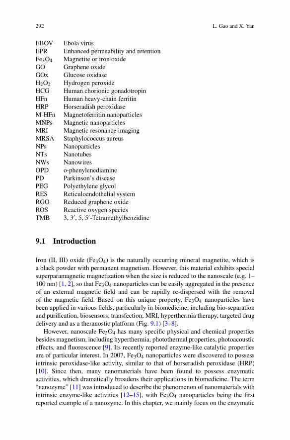
292 L. Gao and X. Yan
EBOV Ebola virusEPR Enhanced permeability and retentionFe3O4 Magnetite or iron oxideGO Graphene oxideGOx Glucose oxidaseH2O2 Hydrogen peroxideHCG Human chorionic gonadotropinHFn Human heavy-chain ferritinHRP Horseradish peroxidaseM-HFn Magnetoferritin nanoparticlesMNPs Magnetic nanoparticlesMRI Magnetic resonance imagingMRSA Staphylococcus aureusNPs NanoparticlesNTs NanotubesNWs NanowiresOPD o-phenylenediaminePD Parkinson’s diseasePEG Polyethylene glycolRES Reticuloendothelial systemRGO Reduced graphene oxideROS Reactive oxygen speciesTMB 3, 3′, 5, 5′-Tetramethylbenzidine
9.1 Introduction
Iron (II, III) oxide (Fe3O4) is the naturally occurring mineral magnetite, which isa black powder with permanent magnetism. However, this material exhibits specialsuperparamagnetic magnetization when the size is reduced to the nanoscale (e.g. 1–100 nm) [1, 2], so that Fe3O4 nanoparticles can be easily aggregated in the presenceof an external magnetic field and can be rapidly re-dispersed with the removalof the magnetic field. Based on this unique property, Fe3O4 nanoparticles havebeen applied in various fields, particularly in biomedicine, including bio-separationand purification, biosensors, transfection, MRI, hyperthermia therapy, targeted drugdelivery and as a theranostic platform (Fig. 9.1) [3–8].
However, nanoscale Fe3O4 has many specific physical and chemical propertiesbesides magnetism, including hyperthermia, photothermal properties, photoacousticeffects, and fluorescence [9]. Its recently reported enzyme-like catalytic propertiesare of particular interest. In 2007, Fe3O4 nanoparticles were discovered to possessintrinsic peroxidase-like activity, similar to that of horseradish peroxidase (HRP)[10]. Since then, many nanomaterials have been found to possess enzymaticactivities, which dramatically broadens their applications in biomedicine. The term“nanozyme” [11] was introduced to describe the phenomenon of nanomaterials withintrinsic enzyme-like activities [12–15], with Fe3O4 nanoparticles being the firstreported example of a nanozyme. In this chapter, we mainly focus on the enzymatic
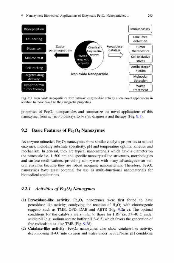
9 Nanozymes: Biomedical Applications of Enzymatic Fe3O4 Nanoparticles. . . 293
Fig. 9.1 Iron oxide nanoparticles with intrinsic enzyme-like activity allow novel applications inaddition to those based on their magnetic properties
properties of Fe3O4 nanoparticles and summarize the novel applications of thisnanozyme, from in vitro bioassays to in vivo diagnosis and therapy (Fig. 9.1).
9.2 Basic Features of Fe3O4 Nanozymes
As enzyme mimetics, Fe3O4 nanozymes show similar catalytic properties to naturalenzymes, including substrate specificity, pH and temperature optima, kinetics andmechanism. In general, they are typical nanomaterials which have a diameter onthe nanoscale i.e. 1–500 nm and specific nanocrystalline structures, morphologiesand surface modifications, providing nanozymes with many advantages over nat-ural enzymes because they are robust inorganic nanomaterials. Therefore, Fe3O4nanozymes have great potential for use as multi-functional nanomaterials forbiomedical applications.
9.2.1 Activities of Fe3O4 Nanozymes
(1) Peroxidase-like activity: Fe3O4 nanozymes were first found to haveperoxidase-like activity, catalyzing the reaction of H2O2 with chromogenicreagents such as TMB, OPD, DAB and ABTS (Fig. 9.2a–c). The optimalconditions for the catalysis are similar to those for HRP i.e. 37–40 C underacidic pH (e.g. sodium acetate buffer pH 3–6.5) which favors the generation offree radicals to oxidize TMB (Fig. 9.2d).
(2) Catalase-like activity: Fe3O4 nanozymes also show catalase-like activity,decomposing H2O2 into oxygen and water under neutral/basic pH conditions
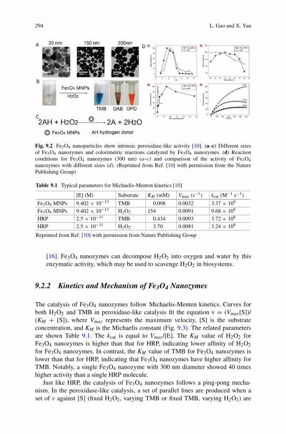
294 L. Gao and X. Yan
Fig. 9.2 Fe3O4 nanoparticles show intrinsic peroxidase-like activity [10]. (a–c) Different sizesof Fe3O4 nanozymes and colorimetric reactions catalyzed by Fe3O4 nanozymes. (d) Reactionconditions for Fe3O4 nanozymes (300 nm) (a–c) and comparison of the activity of Fe3O4nanozymes with different sizes (d). (Reprinted from Ref. [10] with permission from the NaturePublishing Group)
Table 9.1 Typical parameters for Michaelis-Menton kinetics [10]
[E] (M) Substrate KM (mM) Vmax (s−1) kcat (M−1 s−1)
Fe3O4 MNPs 9.402 × 10⁻¹³ TMB 0.098 0.0032 3.37 × 10⁹
Fe3O4 MNPs 9.402 × 10⁻¹³ H2O2 154 0.0091 9.68 × 10⁹
HRP 2.5 × 10⁻¹¹ TMB 0.434 0.0093 3.72 × 10⁸
HRP 2.5 × 10⁻¹¹ H2O2 3.70 0.0081 3.24 × 10⁸
Reprinted from Ref. [10] with permission from Nature Publishing Group
[16]. Fe3O4 nanozymes can decompose H2O2 into oxygen and water by thisenzymatic activity, which may be used to scavenge H2O2 in biosystems.
9.2.2 Kinetics and Mechanism of Fe3O4 Nanozymes
The catalysis of Fe3O4 nanozymes follow Michaelis-Menten kinetics. Curves forboth H2O2 and TMB in peroxidase-like catalysis fit the equation v = (Vmax[S])/(KM + [S]), where Vmax represents the maximum velocity, [S] is the substrateconcentration, and KM is the Michaelis constant (Fig. 9.3). The related parametersare shown Table 9.1. The kcat is equal to Vmax/[E]. The KM value of H2O2 forFe3O4 nanozymes is higher than that for HRP, indicating lower affinity of H2O2for Fe3O4 nanozymes. In contrast, the KM value of TMB for Fe3O4 nanozymes islower than that for HRP, indicating that Fe3O4 nanozymes have higher affinity forTMB. Notably, a single Fe3O4 nanozyme with 300 nm diameter showed 40 timeshigher activity than a single HRP molecule.
Just like HRP, the catalysis of Fe3O4 nanozymes follows a ping-pong mecha-nism. In the peroxidase-like catalysis, a set of parallel lines are produced when aset of v against [S] (fixed H2O2, varying TMB or fixed TMB, varying H2O2) are
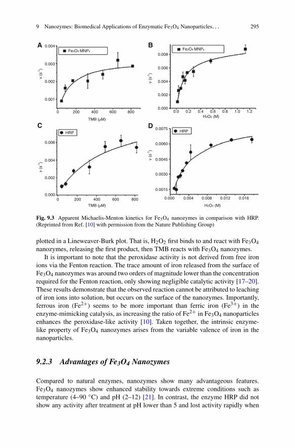
9 Nanozymes: Biomedical Applications of Enzymatic Fe3O4 Nanoparticles. . . 295
0.004
0.003
0.002
0.001
0.006
0.0075
0.0060
0.0045
0.0030
0.0015
0.000 0.004 0.008 0.012 0.016
0.004
0.002
0.0000 200 400 600 800
0.0000.0 0.2 0.4 0.6 0.8 1.0 1.2
0.002
0.004
0.006
0.008
0 200 400
TMB (µM)
HRP HRP
TMB (µM)
Fe3O4 MNPs
A
C
B
D
Fe3O4 MNPs
H2O2 (M)
H2O2 (M)
v (s
-1)
v (s
-1)
v (s
-1)
v (s
-1)
600 800
Fig. 9.3 Apparent Michaelis-Menton kinetics for Fe3O4 nanozymes in comparison with HRP.(Reprinted from Ref. [10] with permission from the Nature Publishing Group)
plotted in a Lineweaver-Burk plot. That is, H2O2 first binds to and react with Fe3O4nanozymes, releasing the first product, then TMB reacts with Fe3O4 nanozymes.
It is important to note that the peroxidase activity is not derived from free ironions via the Fenton reaction. The trace amount of iron released from the surface ofFe3O4 nanozymes was around two orders of magnitude lower than the concentrationrequired for the Fenton reaction, only showing negligible catalytic activity [17–20].These results demonstrate that the observed reaction cannot be attributed to leachingof iron ions into solution, but occurs on the surface of the nanozymes. Importantly,ferrous iron (Fe2+) seems to be more important than ferric iron (Fe3+) in theenzyme-mimicking catalysis, as increasing the ratio of Fe2+ in Fe3O4 nanoparticlesenhances the peroxidase-like activity [10]. Taken together, the intrinsic enzyme-like property of Fe3O4 nanozymes arises from the variable valence of iron in thenanoparticles.
9.2.3 Advantages of Fe3O4 Nanozymes
Compared to natural enzymes, nanozymes show many advantageous features.Fe3O4 nanozymes show enhanced stability towards extreme conditions such astemperature (4–90 ◦C) and pH (2–12) [21]. In contrast, the enzyme HRP did notshow any activity after treatment at pH lower than 5 and lost activity rapidly when
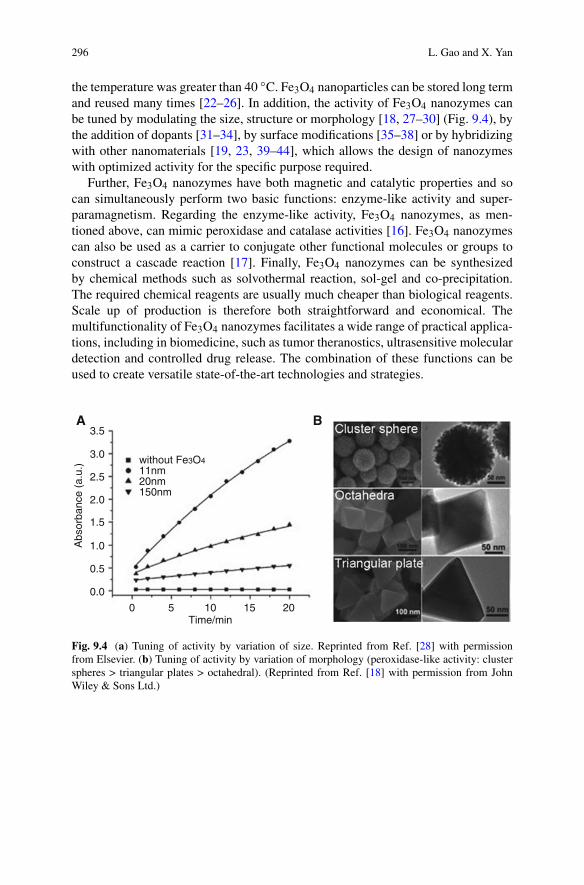
296 L. Gao and X. Yan
the temperature was greater than 40 ◦C. Fe3O4 nanoparticles can be stored long termand reused many times [22–26]. In addition, the activity of Fe3O4 nanozymes canbe tuned by modulating the size, structure or morphology [18, 27–30] (Fig. 9.4), bythe addition of dopants [31–34], by surface modifications [35–38] or by hybridizingwith other nanomaterials [19, 23, 39–44], which allows the design of nanozymeswith optimized activity for the specific purpose required.
Further, Fe3O4 nanozymes have both magnetic and catalytic properties and socan simultaneously perform two basic functions: enzyme-like activity and super-paramagnetism. Regarding the enzyme-like activity, Fe3O4 nanozymes, as men-tioned above, can mimic peroxidase and catalase activities [16]. Fe3O4 nanozymescan also be used as a carrier to conjugate other functional molecules or groups toconstruct a cascade reaction [17]. Finally, Fe3O4 nanozymes can be synthesizedby chemical methods such as solvothermal reaction, sol-gel and co-precipitation.The required chemical reagents are usually much cheaper than biological reagents.Scale up of production is therefore both straightforward and economical. Themultifunctionality of Fe3O4 nanozymes facilitates a wide range of practical applica-tions, including in biomedicine, such as tumor theranostics, ultrasensitive moleculardetection and controlled drug release. The combination of these functions can beused to create versatile state-of-the-art technologies and strategies.
0.0
0 5 10 15Time/min
A B
20
0.5
1.5
2.5
3.5
without Fe3O411nm20nm150nm
1.0
2.0
Abs
orba
nce
(a.u
.)
3.0
Fig. 9.4 (a) Tuning of activity by variation of size. Reprinted from Ref. [28] with permissionfrom Elsevier. (b) Tuning of activity by variation of morphology (peroxidase-like activity: clusterspheres > triangular plates > octahedral). (Reprinted from Ref. [18] with permission from JohnWiley & Sons Ltd.)
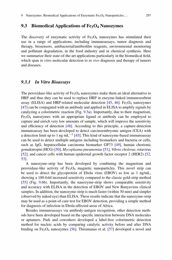
9 Nanozymes: Biomedical Applications of Enzymatic Fe3O4 Nanoparticles. . . 297
9.3 Biomedical Applications of Fe3O4 Nanozymes
The discovery of enzymatic activity of Fe3O4 nanozymes has stimulated theiruse in a range of applications, including immunoassays, tumor diagnosis andtherapy, biosensors, antibacterial/antibiofilm reagents, environmental monitoringand pollutant degradation, in the food industry and in chemical synthesis. Herewe summarize their state-of-the-art applications particularly in the biomedical field,which span in vitro molecular detection to in vivo diagnosis and therapy of tumorsand diseases.
9.3.1 In Vitro Bioassays
The peroxidase-like activity of Fe3O4 nanozymes make them an ideal alternative toHRP and thus they can be used to replace HRP in enzyme-linked immunosorbentassay (ELISA) and HRP-related molecular detection [45, 46]. Fe3O4 nanozymes[47] can be conjugated with an antibody and applied in ELISA to amplify signals bycatalyzing a colorimetric reaction (Fig. 9.5a). Importantly, due to their magnetism,Fe3O4 nanozymes with an appropriate ligand or antibody can be employed tocapture and enrich very low amounts of sample, which will improve the sensitivityand efficiency of detection [48]. According to this principle, a capture-detectionimmunoassay has been developed to detect carcinoembryonic antigen (CEA) witha detection limit up to 1 ng mL−1 [45]. This kind of nanozyme-based immunoassaycan be used to detect multiple antigens including biomarkers and bacteria or cells,such as IgG, hepatocellular carcinoma biomarker GP73 [49], human chorionicgonadotropin (HCG) [50], Mycoplasma pneumoniae [51], Vibrio cholerae, rotavirus[52], and cancer cells with human epidermal growth factor receptor 2 (HER2) [52,53].
A nanozyme-strip has been developed by combining the magnetism andperoxidase-like activity of Fe3O4 magnetic nanoparticles. This novel strip canbe used to detect the glycoprotein of Ebola virus (EBOV) as low as 1 ng/mL,showing a 100-fold increased sensitivity compared to the classic gold-strip method[55] (Fig. 9.6b). Importantly, the nanozyme-strip shows comparable sensitivityand accuracy with ELISA in the detection of EBOV and New Bunyavirus clinicalsamples. In addition, the nanozyme-strip is much faster (within 30 min) and simpler(observed by naked eye) than ELISA. These results indicate that the nanozyme-stripmay be used as a point-of-care test for EBOV detection, providing a simple methodfor diagnosis of infection in Ebola-affected areas of Africa.
Besides immunoassays via antibody-antigen recognition, other detection meth-ods have been developed based on the specific interaction between DNA moleculesor aptamers. Park and coworkers developed a label-free colorimetric detectionmethod for nucleic acids by comparing catalytic activity before and after DNAbinding on Fe3O4 nanozymes [56]. Thiramanas et al. [57] developed a novel and
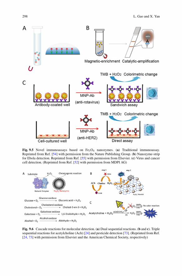
298 L. Gao and X. Yan
Fig. 9.5 Novel immunoassays based on Fe3O4 nanozymes. (a) Traditional immunoassay.Reprinted from Ref. [54] with permission from the Nature Publishing Group. (b) Nanozyme-stripfor Ebola detection. Reprinted from Ref. [55] with permission from Elsevier. (c) Virus and cancercell detection. (Reprinted from Ref. [52] with permission from MDPI AG)
Fig. 9.6 Cascade reactions for molecular detection. (a) Dual sequential reactions. (b and c). Triplesequential reactions for acetylcholine (Ach) [24] and pesticide detection [73]. (Reprinted from Ref.[24, 73] with permission from Elsevier and the American Chemical Society, respectively)
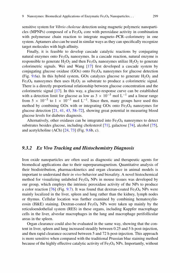
9 Nanozymes: Biomedical Applications of Enzymatic Fe3O4 Nanoparticles. . . 299
sensitive system for Vibrio cholerae detection using magnetic polymeric nanoparti-cles (MPNPs) composed of a Fe3O4 core with peroxidase activity in combinationwith polymerase chain reaction to integrate magneto–PCR–colorimetry in onesystem. Aptamers also can be used in similar ways as they can specifically recognizetarget molecules with high affinity.
Finally, it is feasible to develop cascade catalytic reactions by conjugatingnatural enzymes onto Fe3O4 nanozymes. In a cascade reaction, natural enzyme isresponsible to generate H2O2 and then Fe3O4 nanozymes utilize H2O2 to generatecolorimetric signals. Wei and Wang [17] first developed a cascade system byconjugating glucose oxidase (GOx) onto Fe3O4 nanozymes for glucose detection(Fig. 9.6a). In this hybrid system, GOx catalyzes glucose to generate H2O2 andFe3O4 nanozymes then uses H2O2 as substrate to produce a colorimetric signal.There is a directly proportional relationship between glucose concentration and thecolorimetric signal [17]. In this way, a glucose-response curve can be establishedwith a detection limit for glucose as low as 3 × 10⁻⁵ mol L−1 and a linear rangefrom 5 × 10⁻⁵ to 1 × 10⁻³ mol L−1. Since then, many groups have used thismethod by combining GOx with or integrating GOx onto Fe3O4 nanozymes forglucose detection [21, 41, 43, 58–72], showing great potential in measuring bloodglucose levels for diabetes diagnosis.
Alternatively, other oxidases can be integrated into Fe3O4 nanozymes to detectsubstrates besides glucose, including cholesterol [71], galactose [74], alcohol [75],and acetylcholine (ACh) [24, 73] (Fig. 9.6b, c).
9.3.2 Ex Vivo Tracking and Histochemistry Diagnosis
Iron oxide nanoparticles are often used as diagnostic and therapeutic agents forbiomedical applications due to their superparamagnetism. Quantitative analysis oftheir biodistribution, pharmacokinetics and organ clearance in animal models isimportant to understand their in vivo behavior and biosafety. A novel histochemicalmethod for visualizing unlabeled Fe3O4 NPs in mouse tissues was developed byour group, which employs the intrinsic peroxidase activity of the NPs to producea color reaction [76] (Fig. 9.7). It was found that dextran-coated Fe3O4 NPs weremainly localized in the liver, spleen and lung rather than the kidney, lymph nodesor thymus. Cellular location was further examined by combining hematoxylin-eosin (H&E) staining. Dextran-coated Fe3O4 NPs were taken up mainly by thereticuloendothelial system (RES) in these organs, including Kuppfer macrophagecells in the liver, alveolar macrophages in the lung and macrophage perifollicularareas in the spleen.
Organ clearance could also be evaluated in the same way, showing that the con-tent in liver, spleen and lung increased steadily between 0.25 and 5 h post-injection,and then rapid clearance occurred between 5 and 72 h post-injection. This approachis more sensitive when compared with the traditional Prussian blue staining methodbecause of the highly effective catalytic activity of Fe3O4 NPs. Importantly, without
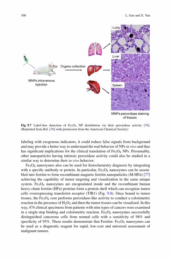
300 L. Gao and X. Yan
Fig. 9.7 Label-free detection of Fe3O4 NP distribution via their peroxidase activity [76].(Reprinted from Ref. [76] with permission from the American Chemical Society)
labeling with exogenous indicators, it could reduce false signals from backgroundand may provide a better way to understand the real behavior of NPs in vivo and thushas significant implications for the clinical translation of Fe3O4 NPs. Presumably,other nanoparticles having intrinsic peroxidase activity could also be studied in asimilar way to determine their in vivo behavior.
Fe3O4 nanozymes also can be used for histochemistry diagnosis by integratingwith a specific antibody or protein. In particular, Fe3O4 nanozymes can be assem-bled into ferritin to form recombinant magneto ferritin nanoparticles (M-HFn) [77]achieving the capability of tumor targeting and visualization in the same uniquesystem. Fe3O4 nanozymes are encapsulated inside and the recombinant humanheavy-chain ferritin (HFn) proteins form a protein shell which can recognize tumorcells overexpressing transferrin receptor (TfR1) (Fig. 9.8). Once bound to tumortissues, the Fe3O4 core performs peroxidase-like activity to conduct a colorimetricreaction in the presence of H2O2 and then the tumor tissues can be visualized. In thisway, 474 clinical specimens from patients with nine types of cancers were examinedin a single-step binding and colorimetric reaction. Fe3O4 nanozymes successfullydistinguished cancerous cells from normal cells with a sensitivity of 98% andspecificity of 95%. These results demonstrate that Ferritin- Fe3O4 nanozymes canbe used as a diagnostic reagent for rapid, low-cost and universal assessment ofmalignant tumors.
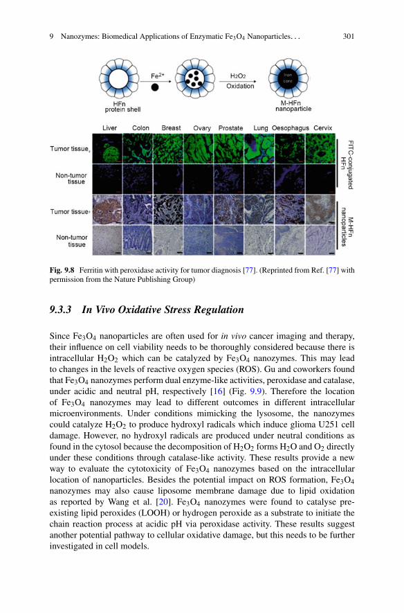
9 Nanozymes: Biomedical Applications of Enzymatic Fe3O4 Nanoparticles. . . 301
Fig. 9.8 Ferritin with peroxidase activity for tumor diagnosis [77]. (Reprinted from Ref. [77] withpermission from the Nature Publishing Group)
9.3.3 In Vivo Oxidative Stress Regulation
Since Fe3O4 nanoparticles are often used for in vivo cancer imaging and therapy,their influence on cell viability needs to be thoroughly considered because there isintracellular H2O2 which can be catalyzed by Fe3O4 nanozymes. This may leadto changes in the levels of reactive oxygen species (ROS). Gu and coworkers foundthat Fe3O4 nanozymes perform dual enzyme-like activities, peroxidase and catalase,under acidic and neutral pH, respectively [16] (Fig. 9.9). Therefore the locationof Fe3O4 nanozymes may lead to different outcomes in different intracellularmicroenvironments. Under conditions mimicking the lysosome, the nanozymescould catalyze H2O2 to produce hydroxyl radicals which induce glioma U251 celldamage. However, no hydroxyl radicals are produced under neutral conditions asfound in the cytosol because the decomposition of H2O2 forms H2O and O2 directlyunder these conditions through catalase-like activity. These results provide a newway to evaluate the cytotoxicity of Fe3O4 nanozymes based on the intracellularlocation of nanoparticles. Besides the potential impact on ROS formation, Fe3O4nanozymes may also cause liposome membrane damage due to lipid oxidationas reported by Wang et al. [20]. Fe3O4 nanozymes were found to catalyse pre-existing lipid peroxides (LOOH) or hydrogen peroxide as a substrate to initiate thechain reaction process at acidic pH via peroxidase activity. These results suggestanother potential pathway to cellular oxidative damage, but this needs to be furtherinvestigated in cell models.
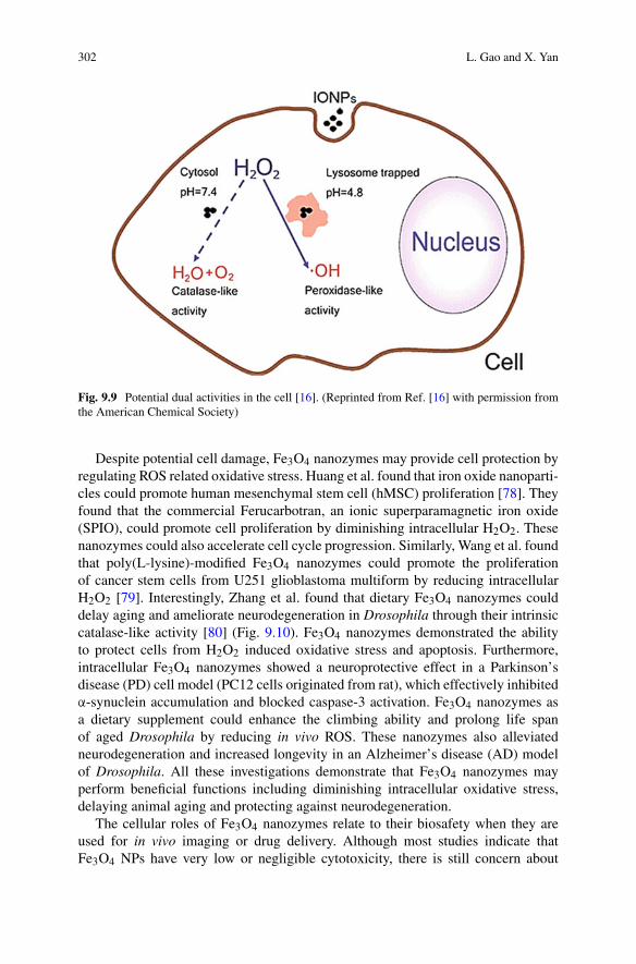
302 L. Gao and X. Yan
Fig. 9.9 Potential dual activities in the cell [16]. (Reprinted from Ref. [16] with permission fromthe American Chemical Society)
Despite potential cell damage, Fe3O4 nanozymes may provide cell protection byregulating ROS related oxidative stress. Huang et al. found that iron oxide nanoparti-cles could promote human mesenchymal stem cell (hMSC) proliferation [78]. Theyfound that the commercial Ferucarbotran, an ionic superparamagnetic iron oxide(SPIO), could promote cell proliferation by diminishing intracellular H2O2. Thesenanozymes could also accelerate cell cycle progression. Similarly, Wang et al. foundthat poly(L-lysine)-modified Fe3O4 nanozymes could promote the proliferationof cancer stem cells from U251 glioblastoma multiform by reducing intracellularH2O2 [79]. Interestingly, Zhang et al. found that dietary Fe3O4 nanozymes coulddelay aging and ameliorate neurodegeneration in Drosophila through their intrinsiccatalase-like activity [80] (Fig. 9.10). Fe3O4 nanozymes demonstrated the abilityto protect cells from H2O2 induced oxidative stress and apoptosis. Furthermore,intracellular Fe3O4 nanozymes showed a neuroprotective effect in a Parkinson’sdisease (PD) cell model (PC12 cells originated from rat), which effectively inhibitedα-synuclein accumulation and blocked caspase-3 activation. Fe3O4 nanozymes asa dietary supplement could enhance the climbing ability and prolong life spanof aged Drosophila by reducing in vivo ROS. These nanozymes also alleviatedneurodegeneration and increased longevity in an Alzheimer’s disease (AD) modelof Drosophila. All these investigations demonstrate that Fe3O4 nanozymes mayperform beneficial functions including diminishing intracellular oxidative stress,delaying animal aging and protecting against neurodegeneration.
The cellular roles of Fe3O4 nanozymes relate to their biosafety when they areused for in vivo imaging or drug delivery. Although most studies indicate thatFe3O4 NPs have very low or negligible cytotoxicity, there is still concern about
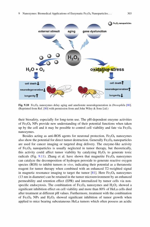
9 Nanozymes: Biomedical Applications of Enzymatic Fe3O4 Nanoparticles. . . 303
Fig. 9.10 Fe3O4 nanozymes delay aging and ameliorate neurodegeneration in Drosophila [80].(Reprinted from Ref. [80] with permission from and John Wiley & Sons Ltd.)
their biosafety, especially for long-term use. The pH-dependent enzyme activitiesof Fe3O4 NPs provide new understanding of their potential functions when takenup by the cell and it may be possible to control cell viability and fate via Fe3O4nanozymes.
Besides acting as anti-ROS agents for neuronal protection, Fe3O4 nanozymesalso show the potential for direct tumor destruction. Generally Fe3O4 nanoparticlesare used for cancer imaging or targeted drug delivery. The enzyme-like activityof Fe3O4 nanoparticles is usually neglected in tumor therapy, but theoretically,this activity could affect tumor viability by catalyzing H2O2 to generate toxicradicals (Fig. 9.11). Zhang et al. have shown that magnetite Fe3O4 nanozymescan catalyze the decomposition of hydrogen peroxide to generate reactive oxygenspecies (ROS) to inhibit tumors in vivo, indicating their potential as a theranosticreagent for tumor therapy when combined with an enhanced T2-weighted signalin magnetic resonance imaging to target the tumor [81]. Here Fe3O4 nanozymes(13 nm in diameter) can be retained in the tumor microenvironment by an enhancedpermeability and retention effect (EPR) and internalized by tumor cells via non-specific endocytosis. The combination of Fe3O4 nanozymes and H2O2 showed asignificant inhibition effect on cell viability and more than 80% of HeLa cells diedafter treatment at different pH values. Furthermore, treatment with the combinationof Fe3O4 NPs and H2O2 showed significant inhibition of tumor growth whenapplied to mice bearing subcutaneous HeLa tumors which often possess an acidic
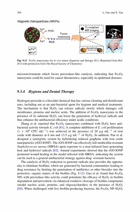
304 L. Gao and X. Yan
Fig. 9.11 Fe3O4 nanozymes for in vivo tumor diagnosis and therapy [81]. (Reprinted from Ref.[81] with permission from The Royal Society of Chemistry)
microenvironment which favors peroxidase-like catalysis, indicating that Fe3O4nanozymes could be used for cancer theranostics, especially in epidermal diseases.
9.3.4 Hygiene and Dental Therapy
Hydrogen peroxide is a biocidal chemical that has various cleaning and disinfectantuses, including use as an anti-bacterial agent for hygiene and medical treatments.The mechanism is that H2O2 can release radicals slowly which damages cellmembranes, proteins and nucleic acids. The addition of Fe3O4 nanozymes in thepresence of its substrate H2O2 can boost the generation of hydroxyl radicals andthus enhance the antibacterial efficiency under acidic conditions.
Zhang et al. reported that Fe3O4 nanozymes combined with H2O2 have anti-bacterial activity towards E. coli [81]. A complete inhibition of E. coli proliferation(1 × 10⁶ CFU mL−1) was achieved in the presence of 20 μg mL−1 of ironoxide with diameter at 6 nm and 13.5 μg mL−1 of H2O2. In addition, Pan et al.designed a synergistic system by hybridizing reduced graphene with iron oxidenanoparticles (rGO-IONP). The rGO-IONP can effectively kill methicillin-resistantStaphylococcus aureus (MRSA) upon exposure to a near-infrared laser generatingheat and hydroxyl radicals [82]. Animal experiments showed that the rGO-IONPpromoted wound healing in the model infected with MRSA, indicating the systemcan be used as a general antibacterial strategy against drug- resistant bacteria.
The catalysis of H2O2 reduction to generate radicals also provides the opportu-nity to eliminate biofilms, which are generated by bacterial communities leading todrug resistance by limiting the penetration of antibiotics or other biocides into theprotective, organic matrix of the biofilm (Fig. 9.12). Gao et al. found that Fe3O4NPs with peroxidase-like activity could potentiate the efficacy of H2O2 in biofilmdegradation and prevention via enhanced oxidative cleavage of biofilm components(model nucleic acids, proteins, and oligosaccharides) in the presence of H2O2[84]. When challenged with live biofilm-producing bacteria, the Fe3O4 NP–H2O2
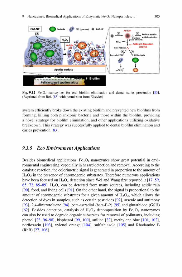
9 Nanozymes: Biomedical Applications of Enzymatic Fe3O4 Nanoparticles. . . 305
Fig. 9.12 Fe3O4 nanozymes for oral biofilm elimination and dental caries prevention [83].(Reprinted from Ref. [83] with permission from Elsevier)
system efficiently broke down the existing biofilm and prevented new biofilms fromforming, killing both planktonic bacteria and those within the biofilm, providinga novel strategy for biofilm elimination, and other applications utilizing oxidativebreakdown. This strategy was successfully applied to dental biofilm elimination andcaries prevention [83].
9.3.5 Eco Environment Applications
Besides biomedical applications, Fe3O4 nanozymes show great potential in envi-ronmental engineering, especially in hazard detection and removal. According to thecatalytic reaction, the colorimetric signal is generated in proportion to the amount ofH2O2 in the presence of chromogenic substrates. Therefore numerous applicationshave been focused on H2O2 detection since Wei and Wang first reported it [17, 59,65, 72, 85–89]. H2O2 can be detected from many sources, including acidic rain[90], food, and living cells [91]. On the other hand, the signal is proportional to theamount of chromogenic substrates for a given amount of H2O2, which allows thedetection of dyes in samples, such as certain pesticides [92], arsenic and antimony[93], 2,4-dinitrotoluene [94], beta-estradiol (beta-E-2) [95] and glutathione (GSH)[62]. Besides detection, catalysis of H2O2 decomposition by Fe3O4 nanozymescan also be used to degrade organic substrates for removal of pollutants, includingphenol [23, 96–98], bisphenol [99, 100], aniline [22], methylene blue [101, 102],norfloxacin [103], xylenol orange [104], sulfathiazole [105] and Rhodamine B(RhB) [27, 106].
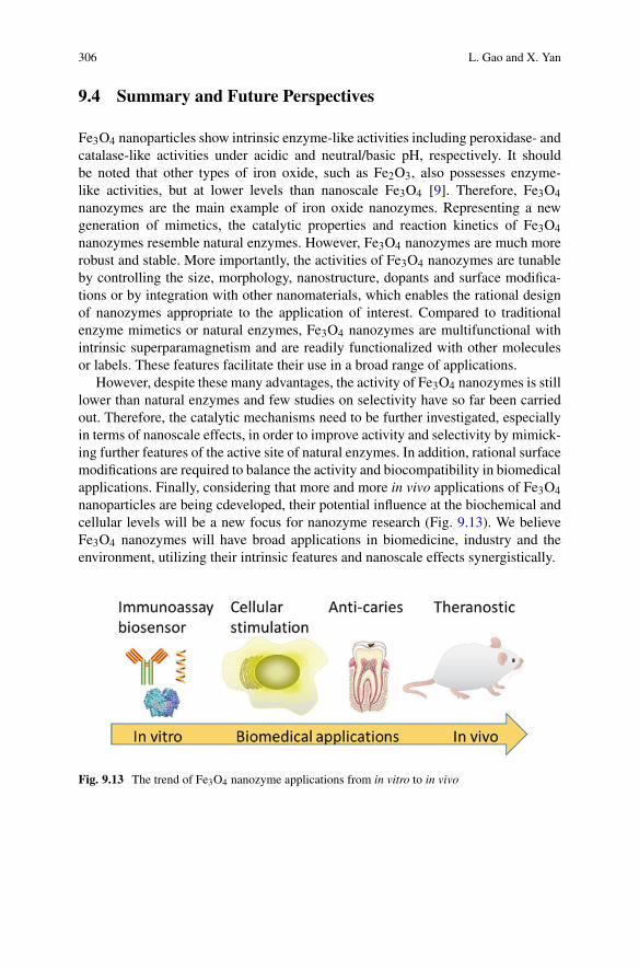
306 L. Gao and X. Yan
9.4 Summary and Future Perspectives
Fe3O4 nanoparticles show intrinsic enzyme-like activities including peroxidase- andcatalase-like activities under acidic and neutral/basic pH, respectively. It shouldbe noted that other types of iron oxide, such as Fe2O3, also possesses enzyme-like activities, but at lower levels than nanoscale Fe3O4 [9]. Therefore, Fe3O4nanozymes are the main example of iron oxide nanozymes. Representing a newgeneration of mimetics, the catalytic properties and reaction kinetics of Fe3O4nanozymes resemble natural enzymes. However, Fe3O4 nanozymes are much morerobust and stable. More importantly, the activities of Fe3O4 nanozymes are tunableby controlling the size, morphology, nanostructure, dopants and surface modifica-tions or by integration with other nanomaterials, which enables the rational designof nanozymes appropriate to the application of interest. Compared to traditionalenzyme mimetics or natural enzymes, Fe3O4 nanozymes are multifunctional withintrinsic superparamagnetism and are readily functionalized with other moleculesor labels. These features facilitate their use in a broad range of applications.
However, despite these many advantages, the activity of Fe3O4 nanozymes is stilllower than natural enzymes and few studies on selectivity have so far been carriedout. Therefore, the catalytic mechanisms need to be further investigated, especiallyin terms of nanoscale effects, in order to improve activity and selectivity by mimick-ing further features of the active site of natural enzymes. In addition, rational surfacemodifications are required to balance the activity and biocompatibility in biomedicalapplications. Finally, considering that more and more in vivo applications of Fe3O4nanoparticles are being cdeveloped, their potential influence at the biochemical andcellular levels will be a new focus for nanozyme research (Fig. 9.13). We believeFe3O4 nanozymes will have broad applications in biomedicine, industry and theenvironment, utilizing their intrinsic features and nanoscale effects synergistically.
Fig. 9.13 The trend of Fe3O4 nanozyme applications from in vitro to in vivo
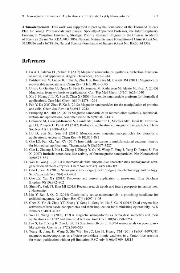
9 Nanozymes: Biomedical Applications of Enzymatic Fe3O4 Nanoparticles. . . 307
Acknowledgement This work was supported in part by the Foundation of the Thousand TalentsPlan for Young Professionals and Jiangsu Specially-Appointed Professor, the InterdisciplinaryFunding at Yangzhou University, Strategic Priority Research Program of the Chinese Academyof Sciences (Grant No. XDA09030306), National Natural Science Foundation of China (Grant No.31530026 and 81671810), Natural Science Foundation of Jiangsu (Grant No. BK20161333).
References
1. Lu AH, Salabas EL, Schuth F (2007) Magnetic nanoparticles: synthesis, protection, function-alization, and application. Angew Chem 46(8):1222–1244
2. Polshettiwar V, Luque R, Fihri A, Zhu HB, Bouhrara M, Bassett JM (2011) Magneticallyrecoverable nanocatalysts. Chem Rev 111(5):3036–3075
3. Unsoy G, Gunduz U, Oprea O, Ficai D, Sonmez M, Radulescu M, Alexie M, Ficai A (2015)Magnetite: from synthesis to applications. Curr Top Med Chem 15(16):1622–1640
4. Xie J, Huang J, Li X, Sun S, Chen X (2009) Iron oxide nanoparticle platform for biomedicalapplications. Curr Med Chem 16(10):1278–1294
5. Pan Y, Du XW, Zhao F, Xu B (2012) Magnetic nanoparticles for the manipulation of proteinsand cells. Chem Soc Rev 41(7):2912–2942
6. Frimpong RA, Hilt JZ (2010) Magnetic nanoparticles in biomedicine: synthesis, functional-ization and applications. Nanomedicine-UK 5(9):1401–1414
7. Colombo M, Carregal-Romero S, Casula MF, Gutierrez L, Morales MP, Bohm IB, Heverha-gen JT, Prosperi D, Parak WJ (2012) Biological applications of magnetic nanoparticles. ChemSoc Rev 41(11):4306–4334
8. Ho D, Sun XL, Sun SH (2011) Monodisperse magnetic nanoparticles for theranosticapplications. Accounts Chem Res 44(10):875–882
9. Gao LZ, Fan KL, Yan XY (2017) Iron oxide nanozyme: a multifunctional enzyme mimeticfor biomedical applications. Theranostics 7(13):3207–3227
10. Gao L, Zhuang J, Nie L, Zhang J, Zhang Y, Gu N, Wang T, Feng J, Yang D, Perrett S, YanX (2007) Intrinsic peroxidase-like activity of ferromagnetic nanoparticles. Nat Nanotechnol2(9):577–583
11. Wei H, Wang E (2013) Nanomaterials with enzyme-like characteristics (nanozymes): next-generation artificial enzymes. Chem Soc Rev 42(14):6060–6093
12. Gao L, Yan X (2016) Nanozymes: an emerging field bridging nanotechnology and biology.Sci China Life Sci 59(4):400–402
13. Gao LZ, Yan XY (2013) Discovery and current application of nanozyme. Prog BiochemBiophys 40(10):892–902
14. Shin HY, Park TJ, Kim MI (2015) Recent research trends and future prospects in nanozymes.J Nanomater
15. Lin Y, Ren J, Qu X (2014) Catalytically active nanomaterials: a promising candidate forartificial enzymes. Acc Chem Res 47(4):1097–1105
16. Chen Z, Yin JJ, Zhou YT, Zhang Y, Song L, Song M, Hu S, Gu N (2012) Dual enzyme-likeactivities of iron oxide nanoparticles and their implication for diminishing cytotoxicity. ACSNano 6(5):4001–4012
17. Wei H, Wang E (2008) Fe3O4 magnetic nanoparticles as peroxidase mimetics and theirapplications in H2O2 and glucose detection. Anal Chem 80(6):2250–2254
18. Liu S, Lu F, Xing R, Zhu JJ (2011) Structural effects of Fe3O4 nanocrystals on peroxidase-like activity. Chemistry 17(2):620–625
19. Wang H, Jiang H, Wang S, Shi WB, He JC, Liu H, Huang YM (2014) Fe3O4-MWCNTmagnetic nanocomposites as efficient peroxidase mimic catalysts in a Fenton-like reactionfor water purification without pH limitation. RSC Adv 4(86):45809–45815
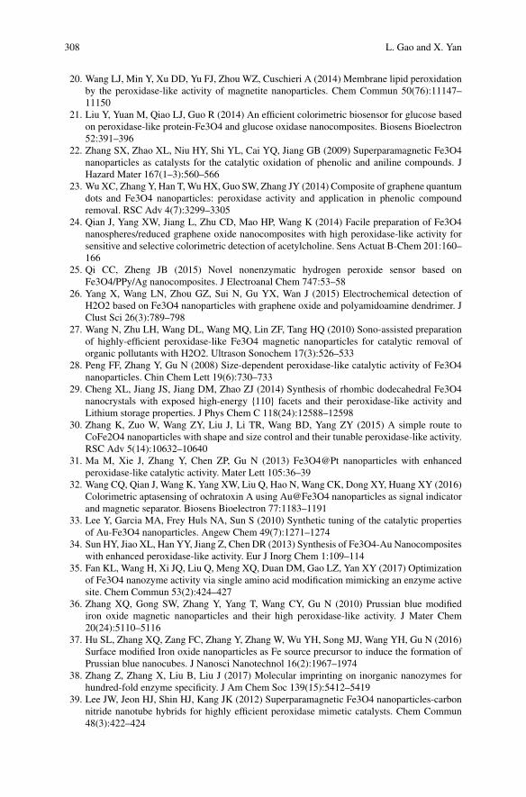
308 L. Gao and X. Yan
20. Wang LJ, Min Y, Xu DD, Yu FJ, Zhou WZ, Cuschieri A (2014) Membrane lipid peroxidationby the peroxidase-like activity of magnetite nanoparticles. Chem Commun 50(76):11147–11150
21. Liu Y, Yuan M, Qiao LJ, Guo R (2014) An efficient colorimetric biosensor for glucose basedon peroxidase-like protein-Fe3O4 and glucose oxidase nanocomposites. Biosens Bioelectron52:391–396
22. Zhang SX, Zhao XL, Niu HY, Shi YL, Cai YQ, Jiang GB (2009) Superparamagnetic Fe3O4nanoparticles as catalysts for the catalytic oxidation of phenolic and aniline compounds. JHazard Mater 167(1–3):560–566
23. Wu XC, Zhang Y, Han T, Wu HX, Guo SW, Zhang JY (2014) Composite of graphene quantumdots and Fe3O4 nanoparticles: peroxidase activity and application in phenolic compoundremoval. RSC Adv 4(7):3299–3305
24. Qian J, Yang XW, Jiang L, Zhu CD, Mao HP, Wang K (2014) Facile preparation of Fe3O4nanospheres/reduced graphene oxide nanocomposites with high peroxidase-like activity forsensitive and selective colorimetric detection of acetylcholine. Sens Actuat B-Chem 201:160–166
25. Qi CC, Zheng JB (2015) Novel nonenzymatic hydrogen peroxide sensor based onFe3O4/PPy/Ag nanocomposites. J Electroanal Chem 747:53–58
26. Yang X, Wang LN, Zhou GZ, Sui N, Gu YX, Wan J (2015) Electrochemical detection ofH2O2 based on Fe3O4 nanoparticles with graphene oxide and polyamidoamine dendrimer. JClust Sci 26(3):789–798
27. Wang N, Zhu LH, Wang DL, Wang MQ, Lin ZF, Tang HQ (2010) Sono-assisted preparationof highly-efficient peroxidase-like Fe3O4 magnetic nanoparticles for catalytic removal oforganic pollutants with H2O2. Ultrason Sonochem 17(3):526–533
28. Peng FF, Zhang Y, Gu N (2008) Size-dependent peroxidase-like catalytic activity of Fe3O4nanoparticles. Chin Chem Lett 19(6):730–733
29. Cheng XL, Jiang JS, Jiang DM, Zhao ZJ (2014) Synthesis of rhombic dodecahedral Fe3O4nanocrystals with exposed high-energy {110} facets and their peroxidase-like activity andLithium storage properties. J Phys Chem C 118(24):12588–12598
30. Zhang K, Zuo W, Wang ZY, Liu J, Li TR, Wang BD, Yang ZY (2015) A simple route toCoFe2O4 nanoparticles with shape and size control and their tunable peroxidase-like activity.RSC Adv 5(14):10632–10640
31. Ma M, Xie J, Zhang Y, Chen ZP, Gu N (2013) Fe3O4@Pt nanoparticles with enhancedperoxidase-like catalytic activity. Mater Lett 105:36–39
32. Wang CQ, Qian J, Wang K, Yang XW, Liu Q, Hao N, Wang CK, Dong XY, Huang XY (2016)Colorimetric aptasensing of ochratoxin A using Au@Fe3O4 nanoparticles as signal indicatorand magnetic separator. Biosens Bioelectron 77:1183–1191
33. Lee Y, Garcia MA, Frey Huls NA, Sun S (2010) Synthetic tuning of the catalytic propertiesof Au-Fe3O4 nanoparticles. Angew Chem 49(7):1271–1274
34. Sun HY, Jiao XL, Han YY, Jiang Z, Chen DR (2013) Synthesis of Fe3O4-Au Nanocompositeswith enhanced peroxidase-like activity. Eur J Inorg Chem 1:109–114
35. Fan KL, Wang H, Xi JQ, Liu Q, Meng XQ, Duan DM, Gao LZ, Yan XY (2017) Optimizationof Fe3O4 nanozyme activity via single amino acid modification mimicking an enzyme activesite. Chem Commun 53(2):424–427
36. Zhang XQ, Gong SW, Zhang Y, Yang T, Wang CY, Gu N (2010) Prussian blue modifiediron oxide magnetic nanoparticles and their high peroxidase-like activity. J Mater Chem20(24):5110–5116
37. Hu SL, Zhang XQ, Zang FC, Zhang Y, Zhang W, Wu YH, Song MJ, Wang YH, Gu N (2016)Surface modified Iron oxide nanoparticles as Fe source precursor to induce the formation ofPrussian blue nanocubes. J Nanosci Nanotechnol 16(2):1967–1974
38. Zhang Z, Zhang X, Liu B, Liu J (2017) Molecular imprinting on inorganic nanozymes forhundred-fold enzyme specificity. J Am Chem Soc 139(15):5412–5419
39. Lee JW, Jeon HJ, Shin HJ, Kang JK (2012) Superparamagnetic Fe3O4 nanoparticles-carbonnitride nanotube hybrids for highly efficient peroxidase mimetic catalysts. Chem Commun48(3):422–424
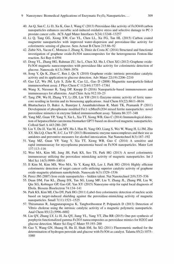
9 Nanozymes: Biomedical Applications of Enzymatic Fe3O4 Nanoparticles. . . 309
40. An Q, Sun C, Li D, Xu K, Guo J, Wang C (2013) Peroxidase-like activity of Fe3O4@carbonnanoparticles enhances ascorbic acid-induced oxidative stress and selective damage to PC-3prostate cancer cells. ACS Appl Mater Interfaces 5(24):13248–13257
41. Li Q, Tang GG, Xiong XW, Cao YL, Chen LL, Xu FG, Tan HL (2015) Carbon coatedmagnetite nanoparticles with improved water-dispersion and peroxidase-like activity forcolorimetric sensing of glucose. Sens Actuat B-Chem 215:86–92
42. Zubir NA, Yacou C, Motuzas J, Zhang X, Diniz da Costa JC (2014) Structural and functionalinvestigation of graphene oxide-Fe3O4 nanocomposites for the heterogeneous Fenton-likereaction. Sci Rep 4:4594
43. Dong YL, Zhang HG, Rahman ZU, Su L, Chen XJ, Hu J, Chen XG (2012) Graphene oxide-Fe3O4 magnetic nanocomposites with peroxidase-like activity for colorimetric detection ofglucose. Nanoscale 4(13):3969–3976
44. Song Y, Qu K, Zhao C, Ren J, Qu X (2010) Graphene oxide: intrinsic peroxidase catalyticactivity and its application to glucose detection. Adv Mater 22(19):2206–2210
45. Gao LZ, Wu JM, Lyle S, Zehr K, Cao LL, Gao D (2008) Magnetite nanoparticle-linkedimmunosorbent assay. J Phys Chem C 112(44):17357–17361
46. Wang X, Niessner R, Tang DP, Knopp D (2016) Nanoparticle-based immunosensors andimmunoassays for aflatoxins. Anal Chim Acta 912:10–23
47. Tang ZW, Wu H, Zhang YY, Li ZH, Lin YH (2011) Enzyme-mimic activity of ferric nano-core residing in ferritin and its biosensing applications. Anal Chem 83(22):8611–8616
48. Bhattacharya D, Baksi A, Banerjee I, Ananthakrishnan R, Maiti TK, Pramanik P (2011)Development of phosphonate modified Fe(1-x)MnxFe2O4 mixed ferrite nanoparticles: novelperoxidase mimetics in enzyme linked immunosorbent assay. Talanta 86:337–348
49. Yang MZ, Guan YP, Yang Y, Xie L, Xia TT, Xiong WB, Guo C (2014) Immunological detec-tion of hepatocellular carcinoma biomarker GP73 based on dissolved magnetic nanoparticles.Colloid Surf A 443:280–285
50. Liu Y, Du JJ, Yan M, Lau MY, Hu J, Han H, Yang OO, Liang S, Wei W, Wang H, Li JM, ZhuXY, Shi LQ, Chen W, Ji C, Lu YF (2013) Biomimetic enzyme nanocomplexes and their use asantidotes and preventive measures for alcohol intoxication. Nat Nanotechnol 8(3):187–192
51. Yang MZ, Guan YP, Yang Y, Xia TT, Xiong WB, Guo C (2014) A sensitive andrapid immunoassay for mycoplasma pneumonia based on Fe3O4 nanoparticles. Mater Lett137:113–116
52. Woo MA, Kim MI, Jung JH, Park KS, Seo TS, Park HG (2013) A novel colorimetricimmunoassay utilizing the peroxidase mimicking activity of magnetic nanoparticles. Int JMol Sci 14(5):9999–10014
53. Il Kim M, Kim MS, Woo MA, Ye Y, Kang KS, Lee J, Park HG (2014) Highly efficientcolorimetric detection of target cancer cells utilizing superior catalytic activity of grapheneoxide-magnetic-platinum nanohybrids. Nanoscale 6(3):1529–1536
54. Perez JM (2007) Iron oxide nanoparticles – hidden talent. Nat Nanotechnol 2(9):535–53655. Duan DM, Fan KL, Zhang DX, Tan SG, Liang MF, Liu Y, Zhang JL, Zhang PH, Liu W,
Qiu XG, Kobinger GP, Gao GF, Yan XY (2015) Nanozyme-strip for rapid local diagnosis ofEbola. Biosens Bioelectron 74:134–141
56. Park KS, Kim MI, Cho DY, Park HG (2011) Label-free colorimetric detection of nucleic acidsbased on target-induced shielding against the peroxidase-mimicking activity of magneticnanoparticles. Small 7(11):1521–1525
57. Thiramanas R, Jangpatarapongsa K, Tangboriboonrat P, Polpanich D (2013) Detection ofVibrio cholerae using the intrinsic catalytic activity of a magnetic polymeric nanoparticle.Anal Chem 85(12):5996–6002
58. Liu QY, Zhang LY, Li H, Jia QY, Jiang YL, Yang YT, Zhu RR (2015) One-pot synthesis ofporphyrin functionalized gamma-Fe2O3 nanocomposites as peroxidase mimics for H2O2 andglucose detection. Mater Sci Eng C-Mater 55:193–200
59. Gao Y, Wang GN, Huang H, Hu JJ, Shah SM, Su XG (2011) Fluorometric method for thedetermination of hydrogen peroxide and glucose with Fe3O4 as catalyst. Talanta 85(2):1075–1080
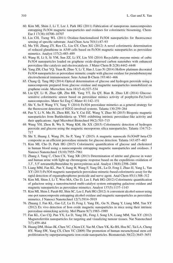
310 L. Gao and X. Yan
60. Kim MI, Shim J, Li T, Lee J, Park HG (2011) Fabrication of nanoporous nanocompositesentrapping Fe3O4 magnetic nanoparticles and oxidases for colorimetric biosensing. Chem-Eur J 17(38):10700–10707
61. Liu CH, Tseng WL (2011) Oxidase-functionalized Fe3O4 nanoparticles for fluorescencesensing of specific substrate. Anal Chim Acta 703(1):87–93
62. Ma YH, Zhang ZY, Ren CL, Liu GY, Chen XG (2012) A novel colorimetric determinationof reduced glutathione in A549 cells based on Fe3O4 magnetic nanoparticles as peroxidasemimetics. Analyst 137(2):485–489
63. Wang H, Li S, Si YM, Sun ZZ, Li SY, Lin YH (2014) Recyclable enzyme mimic of cubicFe3O4 nanoparticles loaded on graphene oxide-dispersed carbon nanotubes with enhancedperoxidase-like catalysis and electrocatalysis. J Mater Chem B 2(28):4442–4448
64. Yang ZH, Chai YQ, Yuan R, Zhuo Y, Li Y, Han J, Liao N (2014) Hollow platinum decoratedFe3O4 nanoparticles as peroxidase mimetic couple with glucose oxidase for pseudobienzymeelectrochemical immunosensor. Sens Actuat B-Chem 193:461–466
65. Chang Q, Tang HQ (2014) Optical determination of glucose and hydrogen peroxide using ananocomposite prepared from glucose oxidase and magnetite nanoparticles immobilized ongraphene oxide. Microchim Acta 181(5–6):527–534
66. Liu QY, Li H, Zhao QR, Zhu RR, Yang YT, Jia QY, Bian B, Zhuo LH (2014) Glucose-sensitive colorimetric sensor based on peroxidase mimics activity of porphyrin-Fe(3)o(4)nanocomposites. Mater Sci Eng C-Mater 41:142–151
67. Shi Y, Su P, Wang YY, Yang Y (2014) Fe3O4 peroxidase mimetics as a general strategy forthe fluorescent detection of H2O2-involved systems. Talanta 130:259–264
68. Pan Y, Li N, Mu JS, Zhou RH, Xu Y, Cui DZ, Wang Y, Zhao M (2015) Biogenic magneticnanoparticles from Burkholderia sp. YN01 exhibiting intrinsic peroxidase-like activity andtheir applications. Appl Microbiol Biotechnol 99(2):703–715
69. Wang YH, Zhou B, Wu S, Wang KM, He XX (2015) Colorimetric detection of hydrogenperoxide and glucose using the magnetic mesoporous silica nanoparticles. Talanta 134:712–717
70. Shi Y, Huang J, Wang JN, Su P, Yang Y (2015) A magnetic nanoscale Fe3O4/P beta-CDcomposite as an efficient peroxidase mimetic for glucose detection. Talanta 143:457–463
71. Kim MI, Cho D, Park HG (2015) Colorimetric quantification of glucose and cholesterolin human blood using a nanocomposite entrapping magnetic nanoparticles and oxidases. JNanosci Nanotechnol 15(10):7955–7961
72. Zhang J, Yang C, Chen CX, Yang XR (2013) Determination of nitrite and glucose in waterand human urine with light-up chromogenic response based on the expeditious oxidation of3,3′, 5,5′-tetramethylbenzidine by peroxynitrous acid. Analyst 138(8):2398–2404
73. Liang MM, Fan KL, Pan Y, Jiang H, Wang F, Yang DL, Lu D, Feng J, Zhao JJ, Yang L, YanXY (2013) Fe3O4 magnetic nanoparticle peroxidase mimetic-based colorimetric assay for therapid detection of organophosphorus pesticide and nerve agent. Anal Chem 85(1):308–312
74. Kim MI, Shim J, Li T, Woo MA, Cho D, Lee J, Park HG (2012) Colorimetric quantificationof galactose using a nanostructured multi-catalyst system entrapping galactose oxidase andmagnetic nanoparticles as peroxidase mimetics. Analyst 137(5):1137–1143
75. Kim MI, Shim J, Parab HJ, Shin SC, Lee J, Park HG (2012) A convenient alcohol sensor usingone-pot nanocomposite entrapping alcohol oxidase and magnetic nanoparticles as peroxidasemimetics. J Nanosci Nanotechnol 12(7):5914–5919
76. Zhuang J, Fan KL, Gao LZ, Lu D, Feng J, Yang DL, Gu N, Zhang Y, Liang MM, Yan XY(2012) Ex vivo detection of Iron oxide magnetic nanoparticles in mice using their intrinsicperoxidase-mimicking activity. Mol Pharm 9(7):1983–1989
77. Fan KL, Cao CQ, Pan YX, Lu D, Yang DL, Feng J, Song LN, Liang MM, Yan XY (2012)Magnetoferritin nanoparticles for targeting and visualizing tumour tissues. Nat Nanotechnol7(7):459–464
78. Huang DM, Hsiao JK, Chen YC, Chien LY, Yao M, Chen YK, Ko BS, Hsu SC, Tai LA, ChengHY, Wang SW, Yang CS, Chen YC (2009) The promotion of human mesenchymal stem cellproliferation by superparamagnetic iron oxide nanoparticles. Biomaterials 30(22):3645–3651
9 Nanozymes: Biomedical Applications of Enzymatic Fe3O4 Nanoparticles. . . 311
79. Wang XQ, Tu Q, Zhao B, An YF, Wang JC, Liu WM, Yuan MS, Ahmed SM, Xu J, LiuR, Zhang YR, Wang JY (2013) Effects of poly(L-lysine)-modified Fe3O4 nanoparticles onendogenous reactive oxygen species in cancer stem cells. Biomaterials 34(4):1155–1169
80. Zhang Y, Wang ZY, Li XJ, Wang L, Yin M, Wang LH, Chen N, Fan CH, Song HY (2016)Dietary Iron oxide nanoparticles delay aging and ameliorate neurodegeneration in Drosophila.Adv Mater 28(7):1387–1393
81. Zhang D, Zhao YX, Gao YJ, Gao FP, Fan YS, Li XJ, Duan ZY, Wang H (2013) Anti-bacterialand in vivo tumor treatment by reactive oxygen species generated by magnetic nanoparticles.J Mater Chem B 1(38):5100–5107
82. Pan WY, Huang CC, Lin TT, Hu HY, Lin WC, Li MJ, Sung HW (2016) Synergisticantibacterial effects of localized heat and oxidative stress caused by hydroxyl radicalsmediated by graphene/iron oxide-based nanocomposites. Nanomed-Nanotechnol 12(2):431–438
83. Gao LZ, Liu Y, Kim D, Li Y, Hwang G, Naha PC, Cormode DP, Koo H (2016) Nanocatalystspromote Streptococcus mutans biofilm matrix degradation and enhance bacterial killing tosuppress dental caries in vivo. Biomaterials 101:272–284
84. Gao L, Giglio KM, Nelson JL, Sondermann H, Travis AJ (2014) Ferromagnetic nanoparticleswith peroxidase-like activity enhance the cleavage of biological macromolecules for biofilmelimination. Nanoscale 6(5):2588–2593
85. You X, Kim J, Pak YK, Pak JJ (2013) Preparation and application of graphene-poly(diallyldimethylammoniumchloride)-Iron oxide nanoparticles buckypaper for hydrogen per-oxide detection. J Nanosci Nanotechno 13(11):7349–7357
86. Gao Y, Wei Z, Li F, Yang ZM, Chen YM, Zrinyi M, Osada Y (2014) Synthesis of amorphology controllable Fe3O4 nanoparticle/hydrogel magnetic nanocomposite inspired bymagnetotactic bacteria and its application in H2O2 detection. Green Chem 16(3):1255–1261
87. Ye YP, Kong T, Yu XF, Wu YK, Zhang K, Wang XP (2012) Enhanced nonenzymatic hydrogenperoxide sensing with reduced graphene oxide/ferroferric oxide nanocomposites. Talanta89:417–421
88. Jiang ZL, Kun L, Ouyang HX, Liang AH, Jiang HS (2011) A simple and sensitivefluorescence quenching method for the determination of H2O2 using Rhodamine B andFe3O4 nanocatalyst. J Fluoresc 21(5):2015–2020
89. Chang Q, Deng KJ, Zhu LH, Jiang GD, Yu C, Tang HQ (2009) Determination of hydrogenperoxide with the aid of peroxidase-like Fe3O4 magnetic nanoparticles as the catalyst.Microchim Acta 165(3–4):299–305
90. Zhuang J, Zhang JB, Gao LZ, Zhang Y, Gu N, Feng J, Yang DL, Yan XY (2008) A novelapplication of iron oxide nanoparticles for detection of hydrogen peroxide in acid rain. MaterLett 62(24):3972–3974
91. Fang HT, Pan YL, Shan WQ, Guo ML, Nie Z, Huang Y, Yao SZ (2014) Enhancednonenzymatic sensing of hydrogen peroxide released from living cells based on Fe3O4/self-reduced graphene nanocomposites. Anal Method-UK 6(15):6073–6081
92. Guan GJ, Yang L, Mei QS, Zhang K, Zhang ZP, Han MY (2012) Chemiluminescenceswitching on peroxidase-like Fe3O4 nanoparticles for selective detection and simultaneousdetermination of various pesticides. Anal Chem 84(21):9492–9497
93. Jia Y, Yu HM, Wu L, Hou XD, Yang L, Zheng CB (2015) Three birds with one Fe3O4nanoparticle: integration of microwave digestion, solid phase extraction, and magnetic sepa-ration for sensitive determination of arsenic and antimony in fish. Anal Chem 87(12):5866–5871
94. Nie DX, Shi GY, Yu YY (2016) Fe3O4 magnetic nanoparticles as peroxidase mimetics usedin colorimetric determination of 2,4-Dinitrotoluene. Chin J Anal Chem 44(2):179–184
95. Wei SL, Li JW, Liu Y (2015) Colourimetric assay for beta-estradiol based on the peroxidase-like activity of Fe3O4@mSiO(2)@HP-beta-CD nanoparticles. RSC Adv 5(130):107670–107679
96. Wang W, Liu Y, Li TL, Zhou MH (2014) Heterogeneous Fenton catalytic degradation ofphenol based on controlled release of magnetic nanoparticles. Chem Eng J 242:1–9
312 L. Gao and X. Yan
97. Zhang JB, Zhuang J, Gao LZ, Zhang Y, Gu N, Feng J, Yang DL, Zhu JD, Yan XY (2008)Decomposing phenol by the hidden talent of ferromagnetic nanoparticles. Chemosphere73(9):1524–1528
98. Wang W, Mao Q, He HH, Zhou MH (2013) Fe3O4 nanoparticles as an efficient heteroge-neous Fenton catalyst for phenol removal at relatively wide pH values. Water Sci Technol68(11):2367–2373
99. Huang RX, Fang ZQ, Fang XB, Tsang EP (2014) Ultrasonic Fenton-like catalytic degradationof bisphenol A by ferroferric oxide (Fe3O4) nanoparticles prepared from steel pickling wasteliquor. J Colloid Interf Sci 436:258–266
100. Huang RX, Fang ZQ, Yan XM, Cheng W (2012) Heterogeneous sono-Fenton catalyticdegradation of bisphenol A by Fe3O4 magnetic nanoparticles under neutral condition. ChemEng J 197:242–249
101. Wang XS, Huang H, Li GQ, Liu Y, Huang JL, Yang DP (2014) Hydrothermal synthesisof 3D hollow porous Fe3O4 microspheres towards catalytic removal of organic pollutants.Nanoscale Res Lett 9:648
102. Zhang XL, He ML, Liu JH, Liao R, Zhao LQ, Xie JR, Wang RJ, Yang ST, Wang HF,Liu YF (2014) Fe3O4@C nanoparticles as high-performance Fenton-like catalyst for dyedecoloration. Chin Sci Bull 59(27):3406–3412
103. Niu HY, Dizhang NH, Meng ZF, Cai YQ (2012) Fast defluorination and removal ofnorfloxacin by alginate/Fe@Fe3O4 core/shell structured nanoparticles. J Hazard Mater227:195–203
104. Zhu MY, Diao GW (2011) Synthesis of porous Fe3O4 nanospheres and its application for thecatalytic degradation of Xylenol Orange. J Phys Chem C 115(39):18923–18934
105. Niu HY, Zhang D, Zhang SX, Zhang XL, Meng ZF, Cai YQ (2011) Humic acid coated Fe3O4magnetic nanoparticles as highly efficient Fenton-like catalyst for complete mineralization ofsulfathiazole. J Hazard Mater 190(1–3):559–565
106. Wang N, Zhu LH, Wang MQ, Wang DL, Tang HQ (2010) Sono-enhanced degradation of dyepollutants with the use of H2O2 activated by Fe3O4 magnetic nanoparticles as peroxidasemimetic. Ultrason Sonochem 17(1):78–83
Chapter 10Self-Assembly of Ferritin: Structure,Biological Function and PotentialApplications in Nanotechnology
Soumyananda Chakraborti and Pinak Chakrabarti
Abstract Protein cages are normally formed by the self-assembly of multipleprotein subunits and ferritin is a typical example of a protein cage structure. Ferritinis a ubiquitous multi-subunit iron storage protein formed by 24 polypeptide chainsthat self-assemble into a hollow, roughly spherical protein cage. Ferritin has externaland internal diameters of approximately 12 nm and 8 nm, respectively. Functionally,ferritin performs iron sequestration and is highly conserved in evolution. Theinterior cavity of ferritin provides a unique reaction vessel to carry out reactionsseparated from the exterior environment. In nature, the cavity is utilized forsequestration of iron and bio-mineralization as a mechanism to render iron inertand safe from the external environment. Material scientists have been inspired bythis system and exploited a range of ferritin superfamily proteins as supramoleculartemplates to encapsulate different carrier molecules ranging from cancer drugs totherapeutic proteins, in addition to using ferritin proteins as well-defined buildingblocks for fabrication. Besides the interior cavity, the exterior surface and sub-unitinterface of ferritin can be modified without affecting ferritin assembly.
Keywords Iron storage protein · Protein self assembly · Ferritin structureand function · Nanotechnology application of ferritin
Brief Description This chapter will describe the self-assembly properties of the iron-carryingprotein ferritin into nanoscale structures and their biological properties as well as their applications.
S. Chakraborti (�)Department of Biochemistry, Bose Institute, Kolkata, India
Malopolska Centre of Biotechnology, Jagiellonian University, Krakow, Polande-mail: [email protected]
P. ChakrabartiDepartment of Biochemistry, Bose Institute, Kolkata, India
© Springer Nature Singapore Pte Ltd. 2019S. Perrett et al. (eds.), Biological and Bio-inspired Nanomaterials,Advances in Experimental Medicine and Biology 1174,https://doi.org/10.1007/978-981-13-9791-2_10
313
314 S. Chakraborti and P. Chakrabarti
10.1 Introduction
Nature uses the self-assembly properties of proteins to produce a wide variety oflarge, complex, and highly symmetric protein architectures [1, 2]. Understandingthe details of self-assembly is not only important for basic science, but thisknowledge is also essential to build ambitious and challenging technologies, suchas programmable nano-machines [3].
Naturally occurring proteins consist of amino acids forming a polypeptide chainwhich is then folded in an energy efficient manner to produce the stable three-dimensional structure of the protein [4]. The thermodynamics of protein folding issuch that some of its residues orientate and project themselves on the surface suchthat they can interact with other proteins [4]. Thus, the information flow definedby the Central Dogma in molecular biology that begins with the genetic codecan be further expanded through protein folding into the realm of protein-proteininteractions [5]. These protein-protein interactions are the basis of self-assemblyevents of large, complex macromolecular protein structures [6].
Self-assembly of proteins can generate a variety of supramolecular structureswith a broad range of biological functions. These complexes include filaments,protein lattices and symmetric cages [7]. Among different supramolecular proteincomplexes, protein cages are probably the most sophisticated protein-based archi-tectures. Their self-assembly from a small number of subunits into symmetrical,monodisperse architectures has inspired scientists from many disciplines. Cagearchitecture is abundant in nature ranging from virus capsids [8] to bacterialmicro-compartments such as the carboxysome [9]. Cage architectures have alsobeen observed in chaperones [10], DNA binding proteins [11]and ferritin proteins[12] (Fig. 10.1). It has been found that the majority of protein cages are hollowand spherical though there are some notable exceptions, and they often possessinternal symmetry, either icosahedral, octahedral or tetrahedral, which plays animportant role in controlling their inter-subunit interactions. In the last two decades,protein cages have been developed as platforms for nanomaterial synthesis, mostlybecause of their remarkable diversity in size, shape and structures. Furthermore,their ease of production in large quantities using biological systems, as well as theirbiocompatibility, makes them an attractive choice for drug delivery, cell specifictargeting and enzyme catalysis [13]. Among protein cages, ferritin is one of the mostcommonly used because of its unique bio-mineralization ability and extraordinarystability [12].
10.2 Historical Perspective
Ferritin was first discovered in 1937 and was isolated from horse spleen. However,the presence of ferritin in human blood serum was determined even much later [14].To date, the ferritin family of proteins is one of the most studied (probably the
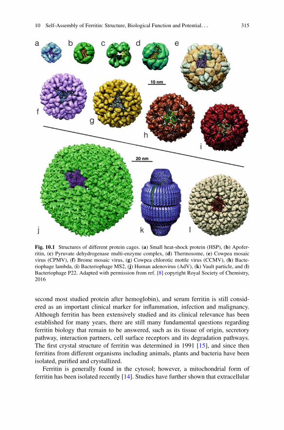
10 Self-Assembly of Ferritin: Structure, Biological Function and Potential. . . 315
Fig. 10.1 Structures of different protein cages. (a) Small heat-shock protein (HSP), (b) Apofer-ritin, (c) Pyruvate dehydrogenase multi-enzyme complex, (d) Thermosome, (e) Cowpea mosaicvirus (CPMV), (f) Brome mosaic virus, (g) Cowpea chlorotic mottle virus (CCMV), (h) Bacte-riophage lambda, (i) Bacteriophage MS2, (j) Human adenovirus (AdV), (k) Vault particle, and (l)Bacteriophage P22. Adapted with permission from ref. [8] copyright Royal Society of Chemistry,2016
second most studied protein after hemoglobin), and serum ferritin is still consid-ered as an important clinical marker for inflammation, infection and malignancy.Although ferritin has been extensively studied and its clinical relevance has beenestablished for many years, there are still many fundamental questions regardingferritin biology that remain to be answered, such as its tissue of origin, secretorypathway, interaction partners, cell surface receptors and its degradation pathways.The first crystal structure of ferritin was determined in 1991 [15], and since thenferritins from different organisms including animals, plants and bacteria have beenisolated, purified and crystallized.
Ferritin is generally found in the cytosol; however, a mitochondrial form offerritin has been isolated recently [14]. Studies have further shown that extracellular
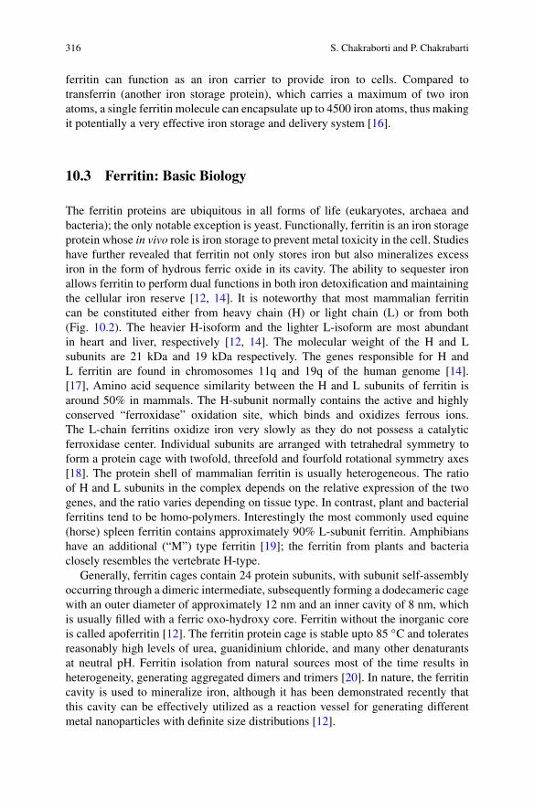
316 S. Chakraborti and P. Chakrabarti
ferritin can function as an iron carrier to provide iron to cells. Compared totransferrin (another iron storage protein), which carries a maximum of two ironatoms, a single ferritin molecule can encapsulate up to 4500 iron atoms, thus makingit potentially a very effective iron storage and delivery system [16].
10.3 Ferritin: Basic Biology
The ferritin proteins are ubiquitous in all forms of life (eukaryotes, archaea andbacteria); the only notable exception is yeast. Functionally, ferritin is an iron storageprotein whose in vivo role is iron storage to prevent metal toxicity in the cell. Studieshave further revealed that ferritin not only stores iron but also mineralizes excessiron in the form of hydrous ferric oxide in its cavity. The ability to sequester ironallows ferritin to perform dual functions in both iron detoxification and maintainingthe cellular iron reserve [12, 14]. It is noteworthy that most mammalian ferritincan be constituted either from heavy chain (H) or light chain (L) or from both(Fig. 10.2). The heavier H-isoform and the lighter L-isoform are most abundantin heart and liver, respectively [12, 14]. The molecular weight of the H and Lsubunits are 21 kDa and 19 kDa respectively. The genes responsible for H andL ferritin are found in chromosomes 11q and 19q of the human genome [14].[17], Amino acid sequence similarity between the H and L subunits of ferritin isaround 50% in mammals. The H-subunit normally contains the active and highlyconserved “ferroxidase” oxidation site, which binds and oxidizes ferrous ions.The L-chain ferritins oxidize iron very slowly as they do not possess a catalyticferroxidase center. Individual subunits are arranged with tetrahedral symmetry toform a protein cage with twofold, threefold and fourfold rotational symmetry axes[18]. The protein shell of mammalian ferritin is usually heterogeneous. The ratioof H and L subunits in the complex depends on the relative expression of the twogenes, and the ratio varies depending on tissue type. In contrast, plant and bacterialferritins tend to be homo-polymers. Interestingly the most commonly used equine(horse) spleen ferritin contains approximately 90% L-subunit ferritin. Amphibianshave an additional (“M”) type ferritin [19]; the ferritin from plants and bacteriaclosely resembles the vertebrate H-type.
Generally, ferritin cages contain 24 protein subunits, with subunit self-assemblyoccurring through a dimeric intermediate, subsequently forming a dodecameric cagewith an outer diameter of approximately 12 nm and an inner cavity of 8 nm, whichis usually filled with a ferric oxo-hydroxy core. Ferritin without the inorganic coreis called apoferritin [12]. The ferritin protein cage is stable upto 85 ◦C and toleratesreasonably high levels of urea, guanidinium chloride, and many other denaturantsat neutral pH. Ferritin isolation from natural sources most of the time results inheterogeneity, generating aggregated dimers and trimers [20]. In nature, the ferritincavity is used to mineralize iron, although it has been demonstrated recently thatthis cavity can be effectively utilized as a reaction vessel for generating differentmetal nanoparticles with definite size distributions [12].
10 Self-Assembly of Ferritin: Structure, Biological Function and Potential. . . 317
Fig. 10.2 Ferritin and ferritin-like protein cages explored from different sources. Here, theferritins isolated from different organisms are illustrated; descriptions are related to number ofsubunits, symmetry and composition. Adapted with permission from ref. [12] copyright AmericanChemical Society, 2015
10.4 Ferritin Protein Family
The ferritin protein superfamily can be divided into three major sub-classes: theclassical ferritins (Ftn), the bacterioferritins (Bfr) and the DNA-binding proteinsfrom starved cells, also known as Dps (Fig. 10.2) [21]. The classical Ftn and Bfrproteins belong to the maxi-ferritin family, as structurally they are slightly larger.In contrast, Dps proteins belong to the mini-ferritin protein family. Interestinglyall three protein sub-families share a four-helix bundle fold. The Bfr proteins seemto possess almost identical quaternary structure to the classical Ftn proteins andboth are assembled from 24 subunits; however, bacterioferritins are only found inarchaea and bacteria. The major difference between Bfr and Ftn lies in possession ofheme moieties: Bfr contains 12 heme groups, compared to classical ferritins, which
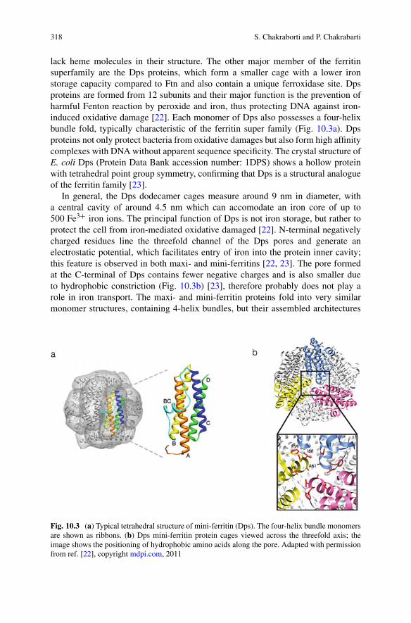
318 S. Chakraborti and P. Chakrabarti
lack heme molecules in their structure. The other major member of the ferritinsuperfamily are the Dps proteins, which form a smaller cage with a lower ironstorage capacity compared to Ftn and also contain a unique ferroxidase site. Dpsproteins are formed from 12 subunits and their major function is the prevention ofharmful Fenton reaction by peroxide and iron, thus protecting DNA against iron-induced oxidative damage [22]. Each monomer of Dps also possesses a four-helixbundle fold, typically characteristic of the ferritin super family (Fig. 10.3a). Dpsproteins not only protect bacteria from oxidative damages but also form high affinitycomplexes with DNA without apparent sequence specificity. The crystal structure ofE. coli Dps (Protein Data Bank accession number: 1DPS) shows a hollow proteinwith tetrahedral point group symmetry, confirming that Dps is a structural analogueof the ferritin family [23].
In general, the Dps dodecamer cages measure around 9 nm in diameter, witha central cavity of around 4.5 nm which can accomodate an iron core of up to500 Fe3+ iron ions. The principal function of Dps is not iron storage, but rather toprotect the cell from iron-mediated oxidative damaged [22]. N-terminal negativelycharged residues line the threefold channel of the Dps pores and generate anelectrostatic potential, which facilitates entry of iron into the protein inner cavity;this feature is observed in both maxi- and mini-ferritins [22, 23]. The pore formedat the C-terminal of Dps contains fewer negative charges and is also smaller dueto hydrophobic constriction (Fig. 10.3b) [23], therefore probably does not play arole in iron transport. The maxi- and mini-ferritin proteins fold into very similarmonomer structures, containing 4-helix bundles, but their assembled architectures
Fig. 10.3 (a) Typical tetrahedral structure of mini-ferritin (Dps). The four-helix bundle monomersare shown as ribbons. (b) Dps mini-ferritin protein cages viewed across the threefold axis; theimage shows the positioning of hydrophobic amino acids along the pore. Adapted with permissionfrom ref. [22], copyright mdpi.com, 2011
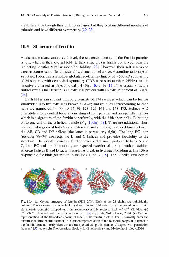
10 Self-Assembly of Ferritin: Structure, Biological Function and Potential. . . 319
are different. Although they both form cages, but they contain different numbers ofsubunits and have different symmetries [22, 23].
10.5 Structure of Ferritin
At the nucleic and amino acid level, the sequence identity of the ferritin proteinsis low, whereas their overall fold (tertiary structure) is highly conserved, possiblyindicating identical/similar monomer folding [22]. However, their self-assembledcage structures can differ considerably, as mentioned above. According to its crystalstructure, H-ferritin is a hollow globular protein machinery of ∼500 kDa consistingof 24 subunits with octahedral symmetry (PDB accession number: 2FHA), and isnegatively charged at physiological pH (Fig. 10.4a, b) [12]. The crystal structurefurther reveals that ferritin is an α-helical protein with an α-helix content of ∼70%[24].
Each H-ferritin subunit normally consists of 174 residues which can be furthersubdivided into five α-helices known as A–E; and residues corresponding to eachhelix are numbered 14–40, 49–76, 96–123, 127–161 and 163–173. Helices A-Dconstitute a long central bundle consisting of four parallel and anti-parallel heliceswhich is a signature of the ferritin superfamily, with the fifth short helix, E, buttingon to one end of the α-helical bundle (Fig. 10.5a) [18]. There are additional shortnon-helical regions at both N- and C-termini and at the right-handed turns betweenthe AB, CD and DE helices (the latter is particularly tight). The long BC loop(residues 78–94) connects the B and C helices and provides flexibility to thestructure. The crystal structure further reveals that most parts of helices A andC, loop BC and the N-terminus, are exposed exterior of the molecular machine,whereas helices B and D faces inwards. A break in hydrogen bonding at His 136 isresponsible for kink generation in the long D helix [18]. The D helix kink occurs
Fig. 10.4 (a) Crystal structure of ferritin (PDB 2ffx). Each of the 24 chains are individuallycolored. The structure is shown looking down the fourfold axis. (b) Structure of ferritin withelectrostatic potential mapped onto the solvent-accessible surface. Red: −5 e−1 kT, blue: +5e−1 kTe−1. Adapted with permission from ref. [56] copyright Wiley Press, 2014. (c) Cartoonrepresentation of the three-fold (polar) channel in the ferritin protein. Fe(II) normally enter theferritin shell through this channel. (d) Cartoon representation of the fourfold (nonpolar) channel inthe ferritin protein; mostly electrons are transported using this channel. Adapted with permissionfrom ref. [57] copyright The American Society for Biochemistry and Molecular Biology, 2016
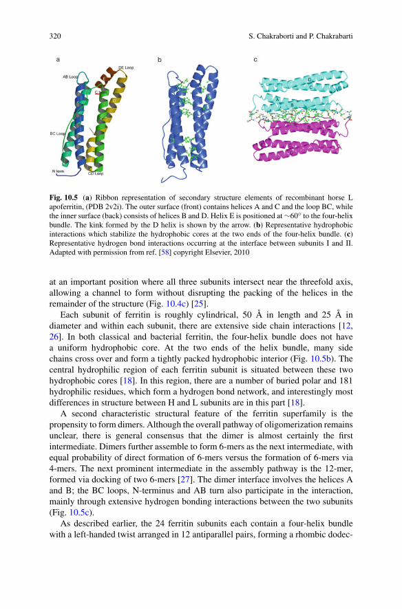
320 S. Chakraborti and P. Chakrabarti
Fig. 10.5 (a) Ribbon representation of secondary structure elements of recombinant horse Lapoferritin, (PDB 2v2i). The outer surface (front) contains helices A and C and the loop BC, whilethe inner surface (back) consists of helices B and D. Helix E is positioned at ∼60◦ to the four-helixbundle. The kink formed by the D helix is shown by the arrow. (b) Representative hydrophobicinteractions which stabilize the hydrophobic cores at the two ends of the four-helix bundle. (c)Representative hydrogen bond interactions occurring at the interface between subunits I and II.Adapted with permission from ref. [58] copyright Elsevier, 2010
at an important position where all three subunits intersect near the threefold axis,allowing a channel to form without disrupting the packing of the helices in theremainder of the structure (Fig. 10.4c) [25].
Each subunit of ferritin is roughly cylindrical, 50 Å in length and 25 Å indiameter and within each subunit, there are extensive side chain interactions [12,26]. In both classical and bacterial ferritin, the four-helix bundle does not havea uniform hydrophobic core. At the two ends of the helix bundle, many sidechains cross over and form a tightly packed hydrophobic interior (Fig. 10.5b). Thecentral hydrophilic region of each ferritin subunit is situated between these twohydrophobic cores [18]. In this region, there are a number of buried polar and 181hydrophilic residues, which form a hydrogen bond network, and interestingly mostdifferences in structure between H and L subunits are in this part [18].
A second characteristic structural feature of the ferritin superfamily is thepropensity to form dimers. Although the overall pathway of oligomerization remainsunclear, there is general consensus that the dimer is almost certainly the firstintermediate. Dimers further assemble to form 6-mers as the next intermediate, withequal probability of direct formation of 6-mers versus the formation of 6-mers via4-mers. The next prominent intermediate in the assembly pathway is the 12-mer,formed via docking of two 6-mers [27]. The dimer interface involves the helices Aand B; the BC loops, N-terminus and AB turn also participate in the interaction,mainly through extensive hydrogen bonding interactions between the two subunits(Fig. 10.5c).
As described earlier, the 24 ferritin subunits each contain a four-helix bundlewith a left-handed twist arranged in 12 antiparallel pairs, forming a rhombic dodec-
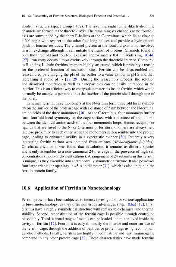
10 Self-Assembly of Ferritin: Structure, Biological Function and Potential. . . 321
ahedron structure (space group F432). The resulting eight funnel-like hydrophilicchannels are formed at the threefold axis. The remaining six channels at the fourfoldaxis are surrounded by the short E-helices at the C-terminus, which lie at close toa 60◦ angle with respect to the other four long helices and provide a hydrophobicpatch of leucine residues. The channel present at the fourfold axis is not involvedin iron exchange although it can initiate the transit of protons. Channels found atboth the threefold and fourfold axes are approximately 0.4 nm wide (Fig. 10.4d)[27]. Iron entry occurs almost exclusively through the threefold interior. Comparedto H-chains, L-chain ferritins are more highly structured, which is probably a reasonfor the preferred location of nucleation sites. Ferritin can be disassembled andreassembled by changing the pH of the buffer to a value as low as pH 2 and thenincreasing it above pH 7 [28, 29]. During the reassembly process, the solutionand dissolved molecules as well as nanoparticles can be easily entrapped in theinterior. This is an efficient way to encapsulate materials inside ferritin, which wouldnormally be unable to penetrate into the interior of the protein shell through one ofthe pores.
In human ferritin, three monomers at the N-termini form threefold local symme-try on the surface of the protein cage with a distance of 5 nm between the N-terminalamino acids of the three monomers [30]. At the C-terminus, four monomers furtherform fourfold local symmetry on the cage surface with a distance of about 1 nmbetween the identical amino acids of the four monomeric loops. Hence, receptors orligands that are fused to the N- or C-termini of ferritin monomers are always heldin close proximity to each other when the monomers self-assemble into the proteincage, leading to enhanced avidity in a synergistic manner [30]. Recently a veryinteresting ferritin variant was obtained from archaea (Archaeoglobus fulgidus).On characterization it was found that in solution, it remains as dimeric speciesand it only assembles to a non-canonical 24-mer cage in the presence of high saltconcentration (mono or divalent cations). Arrangement of 24 subunits in this ferritinis unique, as they assemble into a tetrahedrally symmetric structure. It also possessesfour large triangular openings, ∼45 Å in diameter [31], which is also unique in theferritin protein family.
10.6 Application of Ferritin in Nanotechnology
Ferritin proteins have been subjected to intense investigation for various applicationsin bio-nanotechnology, as they offer numerous advantages (Fig. 10.6a) [12]. First,ferritins have a highly symmetrical structure with remarkable chemical and thermalstability. Second, reconstitution of the ferritin cage is possible through controlledreassembly. Third, a broad range of metals can be loaded and mineralized inside thecavity of ferritin [12]. Fourth, it is easy to modify the interior and outer surface ofthe ferritin cage, through the addition of peptides or protein tags using recombinantgenetic methods. Finally, ferritins are highly biocompatible and less immunogeniccompared to any other protein cage [32]. These characteristics have made ferritins
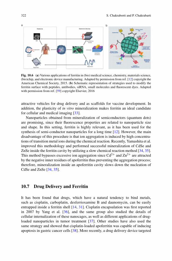
322 S. Chakraborti and P. Chakrabarti
Fig. 10.6 (a) Various applications of ferritin in (bio) medical science, chemistry, materials science,(bio)chip, and electronic device manufacturing. Adapted by permission from ref. [12] copyright theAmerican Chemical Society, 2015. (b) Schematic representation of strategies used to modify theferritin surface with peptides, antibodies, siRNA, small molecules and fluorescent dyes. Adaptedwith permission from ref. [59] copyright Elsevier, 2016
attractive vehicles for drug delivery and as scaffolds for vaccine development. Inaddition, the plasticity of in vitro mineralization makes ferritin an ideal candidatefor cellular and medical imaging [33].
Nanoparticles obtained from mineralization of semiconductors (quantum dots)are promising, since their fluorescence properties are related to nanoparticle sizeand shape. In this setting, ferritin is highly relevant, as it has been used for thesynthesis of semi-conductor nanoparticles for a long time [12]. However, the maindisadvantage of this procedure is that ion aggregation is induced by high concentra-tions of transition metal ions during the chemical reaction. Recently, Yamashita et al.improved this methodology and performed successful mineralization of CdSe andZnSe inside the ferritin cavity by utilizing a slow chemical reaction method [34, 35].This method bypasses excessive ion aggregation since Cd2+ and Zn2+ are attractedby the negative inner residues of apoferritin thus preventing the aggregation process;therefore, mineralization inside an apoferritin cavity slows down the nucleation ofCdSe and ZnSe [34, 35].
10.7 Drug Delivery and Ferritin
It has been found that drugs, which have a natural tendency to bind metals,such as cisplatin, carboplatin, desferrioxamine B and daunomycin, can be easilyentrapped inside a ferritin shell [14, 31]. Cisplatin encapsulation was first reportedin 2007 by Yang et al. [36], and the same group also studied the details ofcellular internalization of these nanocages, as well as different applications of drug-loaded nanoparticles in tumor treatment [37]. Other studies have also used thesame strategy and showed that cisplatin-loaded apoferritin was capable of inducingapoptosis in gastric cancer cells [38]. More recently, a drug delivery device targeted
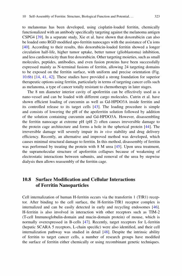
10 Self-Assembly of Ferritin: Structure, Biological Function and Potential. . . 323
to melanomas has been developed, using cisplatin-loaded ferritin, chemicallyfunctionalized with an antibody specifically targeting against the melanoma antigenCSPG4 [39]. In a separate study, Xie et al. have shown that doxorubicin can alsobe loaded onto RGD modified apo-ferritin nanocages with the assistance of Cu ions[40]. According to their results, this doxorubicin-loaded ferritin showed a longercirculation half-life, higher tumor uptake, better tumor (glioblastoma) inhibition,and less cardiotoxicity than free doxorubicin. Other targeting moieties, such as smallmolecules, peptides, antibodies, and even fusion proteins have been successfullyexpressed mainly as N-terminal fusions of ferritin, allowing 24 targeting domainsto be exposed on the ferritin surface, with uniform and precise orientation (Fig.10.6b) [14, 41, 42]. These studies have provided a strong foundation for superiortherapeutic options using ferritin, particularly in terms of targeting cancer cells suchas melanoma, a type of cancer totally resistant to chemotherapy in later stages.
The 8 nm diameter interior cavity of apoferritin can be effectively used as anano-vessel and can be loaded with different cargo molecules. Cutrin et al. haveshown efficient loading of curcumin as well as Gd-HPDO3A inside ferritin andits controlled release to its target cells [43]. The loading procedure is simpleand consists of lowering the pH of the apoferritin solution followed by additionof the solution containing curcumin and Gd-HPDO3A. However, disassemblingthe ferritin nanocage at extreme pH (pH 2) often causes irreversible damage tothe protein cage architecture and forms a hole in the spherical protein [44]. Theirreversible damage will severely impair its in vivo stability and drug deliveryefficiency. Recently, an alternative and improved method was developed, whichcauses minimal structural damage to ferritin. In this method, disassembly of ferritinwas performed by treating the protein with 8 M urea [45]. Upon urea treatment,the supramolecular structure of apoferritin collapses because of weakening ofelectrostatic interactions between subunits, and removal of the urea by stepwisedialysis then allows reassembly of the ferritin cage.
10.8 Surface Modification and Cellular Interactionsof Ferritin Nanoparticles
Cell internalization of human H-ferritin occurs via the transferrin 1 (TfR1) recep-tor. After binding to the cell surface, the H-ferritin-TfR1 receptor complex isinternalized and can be easily detected in early and recycling endosomes [46].H-ferritin is also involved in interaction with other receptors such as TIM-2(T-cell Immunoglobulin-domain and mucin-domain protein) of mouse, which isnormally overexpressed in B-cells [47]. Recently, target receptors for L-ferritin(hepatic SCARA 5 receptors, L-chain specific) were also identified, and their cellinternalization pathway was studied in detail [48]. Despite the intrinsic abilityof ferritin to target cancer cells, a number of research groups have modifiedthe surface of ferritin either chemically or using recombinant genetic techniques.
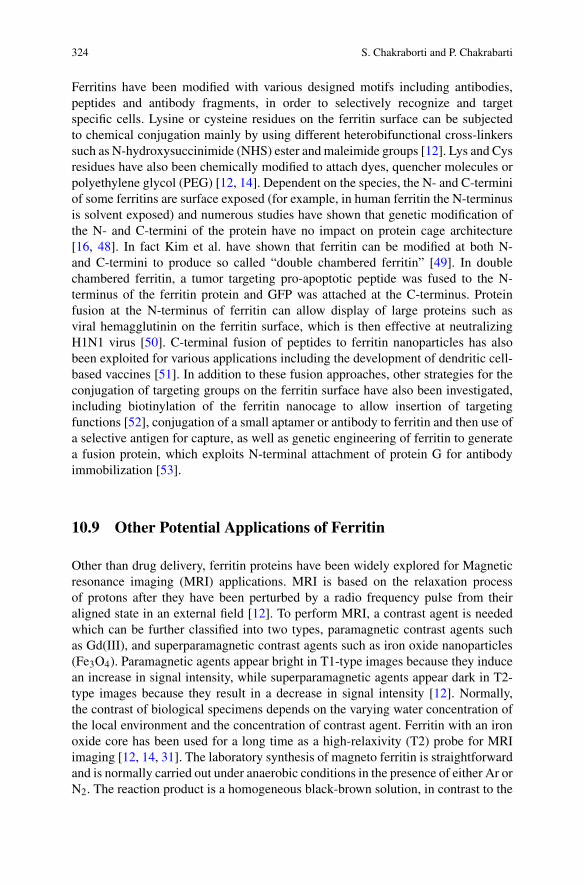
324 S. Chakraborti and P. Chakrabarti
Ferritins have been modified with various designed motifs including antibodies,peptides and antibody fragments, in order to selectively recognize and targetspecific cells. Lysine or cysteine residues on the ferritin surface can be subjectedto chemical conjugation mainly by using different heterobifunctional cross-linkerssuch as N-hydroxysuccinimide (NHS) ester and maleimide groups [12]. Lys and Cysresidues have also been chemically modified to attach dyes, quencher molecules orpolyethylene glycol (PEG) [12, 14]. Dependent on the species, the N- and C-terminiof some ferritins are surface exposed (for example, in human ferritin the N-terminusis solvent exposed) and numerous studies have shown that genetic modification ofthe N- and C-termini of the protein have no impact on protein cage architecture[16, 48]. In fact Kim et al. have shown that ferritin can be modified at both N-and C-termini to produce so called “double chambered ferritin” [49]. In doublechambered ferritin, a tumor targeting pro-apoptotic peptide was fused to the N-terminus of the ferritin protein and GFP was attached at the C-terminus. Proteinfusion at the N-terminus of ferritin can allow display of large proteins such asviral hemagglutinin on the ferritin surface, which is then effective at neutralizingH1N1 virus [50]. C-terminal fusion of peptides to ferritin nanoparticles has alsobeen exploited for various applications including the development of dendritic cell-based vaccines [51]. In addition to these fusion approaches, other strategies for theconjugation of targeting groups on the ferritin surface have also been investigated,including biotinylation of the ferritin nanocage to allow insertion of targetingfunctions [52], conjugation of a small aptamer or antibody to ferritin and then use ofa selective antigen for capture, as well as genetic engineering of ferritin to generatea fusion protein, which exploits N-terminal attachment of protein G for antibodyimmobilization [53].
10.9 Other Potential Applications of Ferritin
Other than drug delivery, ferritin proteins have been widely explored for Magneticresonance imaging (MRI) applications. MRI is based on the relaxation processof protons after they have been perturbed by a radio frequency pulse from theiraligned state in an external field [12]. To perform MRI, a contrast agent is neededwhich can be further classified into two types, paramagnetic contrast agents suchas Gd(III), and superparamagnetic contrast agents such as iron oxide nanoparticles(Fe3O4). Paramagnetic agents appear bright in T1-type images because they inducean increase in signal intensity, while superparamagnetic agents appear dark in T2-type images because they result in a decrease in signal intensity [12]. Normally,the contrast of biological specimens depends on the varying water concentration ofthe local environment and the concentration of contrast agent. Ferritin with an ironoxide core has been used for a long time as a high-relaxivity (T2) probe for MRIimaging [12, 14, 31]. The laboratory synthesis of magneto ferritin is straightforwardand is normally carried out under anaerobic conditions in the presence of either Ar orN2. The reaction product is a homogeneous black-brown solution, in contrast to the
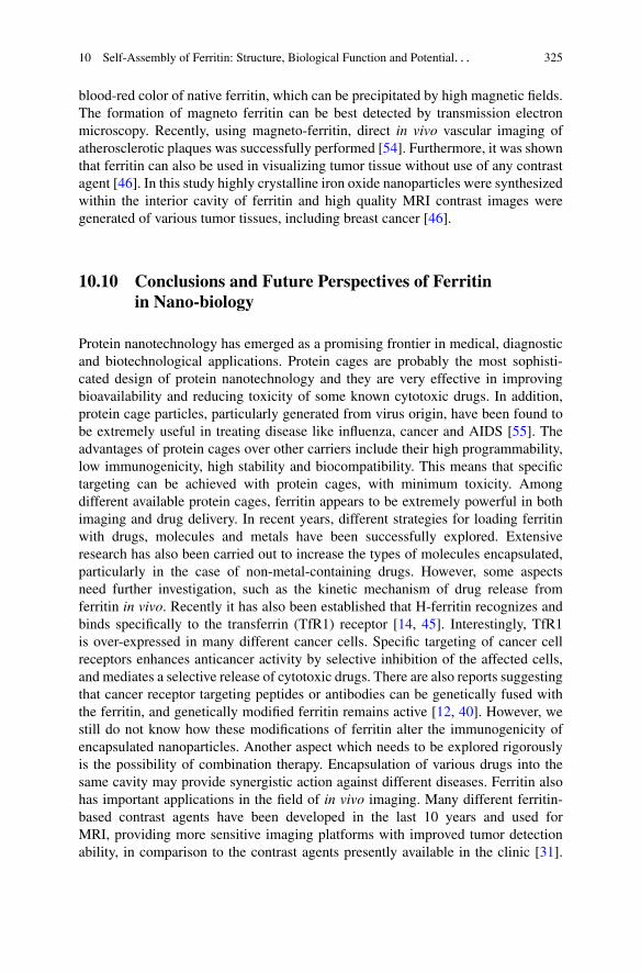
10 Self-Assembly of Ferritin: Structure, Biological Function and Potential. . . 325
blood-red color of native ferritin, which can be precipitated by high magnetic fields.The formation of magneto ferritin can be best detected by transmission electronmicroscopy. Recently, using magneto-ferritin, direct in vivo vascular imaging ofatherosclerotic plaques was successfully performed [54]. Furthermore, it was shownthat ferritin can also be used in visualizing tumor tissue without use of any contrastagent [46]. In this study highly crystalline iron oxide nanoparticles were synthesizedwithin the interior cavity of ferritin and high quality MRI contrast images weregenerated of various tumor tissues, including breast cancer [46].
10.10 Conclusions and Future Perspectives of Ferritinin Nano-biology
Protein nanotechnology has emerged as a promising frontier in medical, diagnosticand biotechnological applications. Protein cages are probably the most sophisti-cated design of protein nanotechnology and they are very effective in improvingbioavailability and reducing toxicity of some known cytotoxic drugs. In addition,protein cage particles, particularly generated from virus origin, have been found tobe extremely useful in treating disease like influenza, cancer and AIDS [55]. Theadvantages of protein cages over other carriers include their high programmability,low immunogenicity, high stability and biocompatibility. This means that specifictargeting can be achieved with protein cages, with minimum toxicity. Amongdifferent available protein cages, ferritin appears to be extremely powerful in bothimaging and drug delivery. In recent years, different strategies for loading ferritinwith drugs, molecules and metals have been successfully explored. Extensiveresearch has also been carried out to increase the types of molecules encapsulated,particularly in the case of non-metal-containing drugs. However, some aspectsneed further investigation, such as the kinetic mechanism of drug release fromferritin in vivo. Recently it has also been established that H-ferritin recognizes andbinds specifically to the transferrin (TfR1) receptor [14, 45]. Interestingly, TfR1is over-expressed in many different cancer cells. Specific targeting of cancer cellreceptors enhances anticancer activity by selective inhibition of the affected cells,and mediates a selective release of cytotoxic drugs. There are also reports suggestingthat cancer receptor targeting peptides or antibodies can be genetically fused withthe ferritin, and genetically modified ferritin remains active [12, 40]. However, westill do not know how these modifications of ferritin alter the immunogenicity ofencapsulated nanoparticles. Another aspect which needs to be explored rigorouslyis the possibility of combination therapy. Encapsulation of various drugs into thesame cavity may provide synergistic action against different diseases. Ferritin alsohas important applications in the field of in vivo imaging. Many different ferritin-based contrast agents have been developed in the last 10 years and used forMRI, providing more sensitive imaging platforms with improved tumor detectionability, in comparison to the contrast agents presently available in the clinic [31].
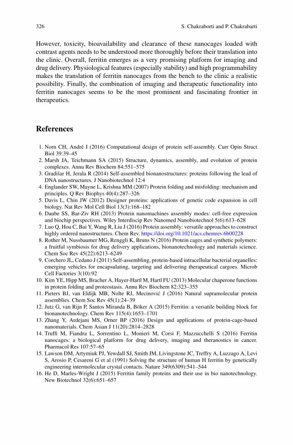
326 S. Chakraborti and P. Chakrabarti
However, toxicity, bioavailability and clearance of these nanocages loaded withcontrast agents needs to be understood more thoroughly before their translation intothe clinic. Overall, ferritin emerges as a very promising platform for imaging anddrug delivery. Physiological features (especially stability) and high programmabilitymakes the translation of ferritin nanocages from the bench to the clinic a realisticpossibility. Finally, the combination of imaging and therapeutic functionality intoferritin nanocages seems to be the most prominent and fascinating frontier intherapeutics.
References
1. Norn CH, André I (2016) Computational design of protein self-assembly. Curr Opin StructBiol 39:39–45
2. Marsh JA, Teichmann SA (2015) Structure, dynamics, assembly, and evolution of proteincomplexes. Annu Rev Biochem 84:551–575
3. Gradišar H, Jerala R (2014) Self-assembled bionanostructures: proteins following the lead ofDNA nanostructures. J Nanobiotechnol 12:4
4. Englander SW, Mayne L, Krishna MM (2007) Protein folding and misfolding: mechanism andprinciples. Q Rev Biophys 40(4):287–326
5. Davis L, Chin JW (2012) Designer proteins: applications of genetic code expansion in cellbiology. Nat Rev Mol Cell Biol 13(3):168–182
6. Daube SS, Bar-Ziv RH (2013) Protein nanomachines assembly modes: cell-free expressionand biochip perspectives. Wiley Interdiscip Rev Nanomed Nanobiotechnol 5(6):613–628
7. Luo Q, Hou C, Bai Y, Wang R, Liu J (2016) Protein assembly: versatile approaches to constructhighly ordered nanostructures. Chem Rev. https://doi.org/10.1021/acs.chemrev.6b00228
8. Rother M, Nussbaumer MG, Renggli K, Bruns N (2016) Protein cages and synthetic polymers:a fruitful symbiosis for drug delivery applications, bionanotechnology and materials science.Chem Soc Rev 45(22):6213–6249
9. Corchero JL, Cedano J (2011) Self-assembling, protein-based intracellular bacterial organelles:emerging vehicles for encapsulating, targeting and delivering therapeutical cargoes. MicrobCell Factories 3(10):92
10. Kim YE, Hipp MS, Bracher A, Hayer-Hartl M, Hartl FU (2013) Molecular chaperone functionsin protein folding and proteostasis. Annu Rev Biochem 82:323–355
11. Pieters BJ, van Eldijk MB, Nolte RJ, Mecinovic J (2016) Natural supramolecular proteinassemblies. Chem Soc Rev 45(1):24–39
12. Jutz G, van Rijn P, Santos Miranda B, Böker A (2015) Ferritin: a versatile building block forbionanotechnology. Chem Rev 115(4):1653–1701
13. Zhang Y, Ardejani MS, Orner BP (2016) Design and applications of protein-cage-basednanomaterials. Chem Asian J 11(20):2814–2828
14. Truffi M, Fiandra L, Sorrentino L, Monieri M, Corsi F, Mazzucchelli S (2016) Ferritinnanocages: a biological platform for drug delivery, imaging and theranostics in cancer.Pharmacol Res 107:57–65
15. Lawson DM, Artymiuk PJ, Yewdall SJ, Smith JM, Livingstone JC, Treffry A, Luzzago A, LeviS, Arosio P, Cesareni G et al (1991) Solving the structure of human H ferritin by geneticallyengineering intermolecular crystal contacts. Nature 349(6309):541–544
16. He D, Marles-Wright J (2015) Ferritin family proteins and their use in bio nanotechnology.New Biotechnol 32(6):651–657
10 Self-Assembly of Ferritin: Structure, Biological Function and Potential. . . 327
17. Worwood M, Brook JD, Cragg SJ, Hellkuhl B, Jones BM, Perera P, Roberts SH, Shaw DJ(1985) Assignment of human ferritin genes to chromosomes 11 and 19q13.3-19qter. HumGenet 69(4):371–374
18. Crichton RR, Declercq JP (2010) X-ray structures of ferritins and related proteins. BiochimBiophys Acta 1800(8):706–718
19. Ha Y, Shi D, Small GW, Theil EC, Allewell NM (1999) Crystal structure of bullfrog M ferritinat 2.8A resolution: analysis of subunit interactions and the binuclear metal center. J Biol InorgChem 4(3):243–256
20. Jutz G, Böker A (2011) Bionanoparticles as functional macromolecular building blocks – anew class of nanomaterials. Polymer 52(2):211–232
21. Andrews SC (2010) The ferritin-like superfamily: evolution of the biological iron storemanfrom a rubrerythrin-like ancestor. Biochim Biophys Acta 1800(8):691–705
22. Zhang Y, Orner BP (2011) Self-assembly in the ferritin nano-cage protein superfamily. Int JMol Sci 12:5406–5421
23. Grant RA, Filman DJ, Finkel SE, Kolter R, Hogle JM (1998) The crystal structure of Dps, aferritin homolog that binds and protects DNA. Nat Struct Biol 5(4):294–303
24. Hasan MR, Tosha T, Theil EC (2008) Ferritin contains less iron (59Fe) in cells when the proteinpores are unfolded by mutation. J Biol Chem 283(46):31394–31400
25. Zang J, Chen H, Zhao G, Wang F, Ren F (2016) Ferritin cage for encapsulation and deliveryof bioactive nutrients: from structure, property to applications. Crit Rev Food Sci Nutr.https://doi.org/10.1080/10408398.2016.1149690
26. Plath LD, Ozdemir A, Aksenov AA, Bier ME (2015) Determination of iron content and dis-persity of intact ferritin by superconducting tunnel junction cryo-detection mass spectrometry.Anal Chem 87(17):8985–8993
27. Sato D, Ohtomo H, Yamada Y, Hikima T, Kurobe A, Fujiwara K, Ikeguchi M (2016)Ferritin assembly revisited: a time-resolved Small-angle X-ray scattering study. Biochemistry55(2):287–293
28. Kim M, Rho Y, Jin KS, Ahn B, Jung S, Kim H, Ree M (2011) pH-dependent structuresof ferritin and apoferritin in solution: disassembly and reassembly. Biomacromolecules12(5):1629–1640
29. Ghisaidoobe AB, Chung SJ (2015) Functionalized protein nanocages as a platform of targetedtherapy and immune detection. Nanomedicine 10(24):3579–3595
30. Uchida M, Kang S, Reichhardt C, Harlen K, Douglas T (2010) The ferritin superfamily:supramolecular templates for materials synthesis. Biochim Biophys Acta 1800(8):834–845
31. Tetter S, Hilvert D (2017) Enzyme encapsulation by a ferritin cage. Angew Chem Int Ed Engl56(47):14933–14936
32. Kim S, Jeon JO, Jun E, Jee J, Jung HK, Lee BH, Kim IS, Kim S (2016) Designingpeptide bunches on nanocage for bispecific or super-affinity targeting. Biomacromolecules17(3):1150–1159
33. Chasteen ND, Harrison PM (1999) Mineralization in ferritin: an efficient means of iron storage.J Struct Biol 126(3):182–194
34. Yamashita I, Hayashi J, Hara M (2004) Bio-template synthesis of uniform CdSe nanoparticlesusing cage-shaped protein apoferritin. Chem Lett 33:1158–1159
35. Iwahori K, Yoshizawa K, Muraoka M, Yamashita I (2005) Fabrication of ZnSe nanoparticlesin the apoferritin cavity by designing a slow chemical reaction system. Inorg Chem 44:6393–6400
36. Yang Z, Wang X, Diao H, Zhang J, Li H, Sun H, Guo Z (2007) Encapsulation of platinumanticancer drugs by apoferritin. Chem Commun 33:3453–3455
37. Xing R, Wang X, Zhang C, Zhang Y, Wang Q, Yang Z, Guo Z (2009) Characterization andcellular uptake of platinum anticancer drugs encapsulated in apoferritin. J Inorg Biochem103(7):1039–1044
328 S. Chakraborti and P. Chakrabarti
38. Ji XT, Huang L, Huang HQ (2012) Construction of nanometer cisplatin core-ferritin (NCC-F)and proteomic analysis of gastric cancer cell apoptosis induced with cisplatin released fromthe NCC-F. J Proteome 75(11):3145–3157
39. Falvo E, Tremante E, Fraioli R, Leonetti C, Zamparelli C, Boffi A, Morea V, Ceci P, GiacominiP (2013) Antibody-drug conjugates: targeting melanoma with cisplatin encapsulated in protein-cage nanoparticles based on human ferritin. Nanoscale 5:12278–12285
40. Zhen Z, Tang W, Chen H, Lin X, Todd T, Wang G, Cowger T, Chen X, Xie J (2013) RGDmodified apoferritin nanoparticles for efficient drug delivery to tumors. ACS Nano 7(6):4830–4837
41. Vannucci L, Falvo E, Fornara M, Micco P, Benada O, Krizan J, Svoboda J, Hulikova-CapkovaK, Morea V, Boffi A, Ceci P (2012) Selective targeting of melanoma by PEG-masked protein-based multifunctional nanoparticles. Int J Nanomed 7:1489–1509
42. Lee JH, Seo HS, Song JA, Kwon KC, Lee EJ, Kim HJ, Lee EB, Cha YJ, Lee J (2013)Proteinticle engineering for accurate 3D diagnosis. ACS Nano 7:10879–10886
43. Cutrin JC, Crich SG, Burghelea D, Dastrù W, Aime S (2013) Curcumin/Gd loaded apoferritin:a novel “theranostic” agent to prevent hepatocellular damage in toxic induced acute hepatitis.Mol Pharm 10(5):2079–2085
44. Kim M, Rho Y, Jin KS, Ahn B, Jung S, Kim H, Ree M (2011) pH-dependent structuresof ferritin and apoferritin in solution: disassembly and reassembly. Biomacromolecules12(5):1629–1640
45. Liang M, Fan K, Zhou M, Duan D, Zheng J, Yang D, Feng J, Yan X (2014) H-ferritin-nanocaged doxorubicin nanoparticles specifically target and kill tumors with a single-doseinjection. Proc Natl Acad Sci USA 111(41):14900–14905
46. Zhao Y, Liang M, Li X, Fan K, Xiao J, Li Y, Shi H, Wang F, Choi HS, Cheng D, Yan X (2016)Bioengineered magnetoferritin nanoprobes for single-dose nuclear-magnetic resonance tumorimaging. ACS Nano 10(4):4184–4191
47. Chen TT, Li L, Chung DH, Allen CD, Torti SV, Torti FM, Cyster JG, Chen CY, Brodsky FM,Niemi EC, Nakamura MC, Seaman WE, Daws MR (2005) TIM-2 is expressed on B cells andin liver and kidney and is a receptor for H-ferritin endocytosis. J Exp Med 202(7):955–965
48. Mendes-Jorge L, Ramos D, Valença A, López-Luppo M, Pires VMR, Catita J, Nacher V,Navarro M, Carretero A, Rodriguez-Baeza A, Ruberte J (2014) L-ferritin binding to Scara5:a new Iron traffic pathway potentially implicated in retinopathy. PLoS One 9(9):e106974.https://doi.org/10.1371/journal.pone.0106974
49. Kim S, Kim GS, Seo J, Gowri Rangaswamy G, So IS, Park RW, Lee BH, Kim IS (2016)Double-chambered ferritin platform: dual-function payloads of cytotoxic peptides and fluores-cent protein. Biomacromolecules 17(1):12–19
50. Kanekiyo M, Wei CJ, Yassine HM, McTamney PM, Boyington JC, Whittle JR, Rao SS, KongWP, Wang L, Nabel GJ (2013) Self-assembling influenza nanoparticle vaccines elicit broadlyneutralizing H1N1 antibodies. Nature 499(7456):102–106
51. Han JA, Kang YJ, Shin C, Ra JS, Shin HH, Hong SY, Do Y, Kang S (2014) Ferritin proteincage nanoparticles as versatile antigen delivery nanoplatforms for dendritic cell (DC)-basedvaccine development. Nanomedicine 10(3):561–569
52. Crich SG, Bussolati B, Tei L, Grange C, Esposito G, Lanzardo S, Camussi G, Aime S (2006)Magnetic resonance visualization of tumor angiogenesis by targeting neural cell adhesionmolecules with the highly sensitivegadolinium-loaded apoferritin probe. Cancer Res 66:9196–9201
53. Hwang M, Lee JW, Lee KE, Lee KH (2013) Think modular: a simple apoferritin-basedplatform for the multifaceted detection of pancreatic cancer. ACS Nano 9:8167–8174
54. Sosnovik DE, Caravan P (2009) Molecular MRI of atherosclerotic plaque with targeted contrastagents. Curr Cardiovasc Imaging Rep 2(2):87–94
55. López-Sagaseta J, Malito E, Rappuoli R, Bottomley MJ (2015) Self-assembling proteinnanoparticles in the design of vaccines. Comput Struct Biotechnol J 14:58–68
56. Beck T, Tetter S, Künzle M, Hilvert D (2015) Construction of Matryoshka-type structures fromsupercharged protein nanocages. Angew Chem Int Ed Engl 54(3):937–940

10 Self-Assembly of Ferritin: Structure, Biological Function and Potential. . . 329
57. Chandramouli B, Bernacchioni C, Di Maio D, Turano P, Brancato G (2016) Electrostatic andstructural bases of Fe2+ translocation through ferritin channels. J Biol Chem 291(49):25617–25628
58. Crichton RR, Declercq JP (2010) Xray structures of ferritins and related proteins. BiochimBiophys Acta 1800(8):706–718
59. Truffi M, Fiandra L, Sorrentino L, Monieri M, Corsi F, Mazzucchelli S (2016) Ferritinnanocages: a biological platform for drug delivery, imaging and theranostics in cancer.Pharmacol Res 107:57–65
Chapter 11DNA Nanotechnology for BuildingSensors, Nanopores and Ion-Channels
Kerstin Göpfrich and Ulrich F. Keyser
Abstract DNA nanotechnology has revolutionised the capabilities to shape andcontrol three-dimensional structures at the nanometre scale. Designer sensors,nanopores and ion-channels built from DNA have great potential for both cross-disciplinary research and applications. Here, we introduce the concept of structuralDNA nanotechnology, including DNA origami, and give an overview of the workflow from design to assembly, characterisation and application of DNA-basedfunctional systems. Chemical functionalisation of DNA has opened up pathways totransform static DNA structures into dynamic nanomechanical sensors. We furtherintroduce nanopore sensing as a powerful label-free single-molecule techniqueand discuss how it can benefit from DNA nanotechnology. Especially excitingis the possibility to create membrane-inserted DNA nanochannels that mimictheir protein-based natural counterparts in form and function. In this chapter wereview the status quo of DNA sensors, nanopores and ion channels, highlightingopportunities and challenges for their future development.
Keywords DNA nanotechnology · DNA origami · Nanopores · Single-moleculesensing · Synthetic ion-channels
K. GöpfrichCavendish Laboratory, University of Cambridge, Cambridge, UK
Department of Cellular Biophysics, Max Planck Institute for Medical Research, Heidelberg,Germanye-mail: [email protected]
U. F. Keyser (�)Cavendish Laboratory, University of Cambridge, Cambridge, UKe-mail: [email protected]
© Springer Nature Singapore Pte Ltd. 2019S. Perrett et al. (eds.), Biological and Bio-inspired Nanomaterials,Advances in Experimental Medicine and Biology 1174,https://doi.org/10.1007/978-981-13-9791-2_11
331
332 K. Göpfrich and U. F. Keyser
11.1 Self-Assembly with DNA
Desoxyribonucleic acid (DNA) is predominantly known as the molecule that carriesgenetic information. Two complementary DNA strands are held together by theWatson-Crick basepairing of adenine (A) with thymine (T) and cytosine (C) withguanine (G) forming the iconic double helix [1]. While nature exploits the basepairing in the replication and expression of genetic information, the same propertymakes DNA an excellent material for molecular self-assembly. With a full helicalturn of 10.5 nm, a pitch of 3.3 nm and an anhydrated diameter of 2 nm, thedimensions of the DNA double helix are inherently suited for precise architecturalcontrol on the nanometre length scale. A linear DNA double helix, however, isof not much use for construction. Transient branched DNA structures have beenfound in nature, for instance as intermediates occurring during the process of geneticrecombination (Holliday junctions) [2, 3]. But only the realisation that it is possibleto engineer stable DNA branches through intelligent sequence matching opened upthe possibility to use DNA beyond its natural purpose as a nanomaterial for designand construction [4, 5]. Over the past 30 years, structural DNA nanotechnology,the science of building with DNA, has been developed tremendously and applied toresearch questions across disciplines thanks to multiple developments:
1. Automated and high throughput synthesis of short DNA strands has becomeavailable.
2. Off-the-shelf and custom-made chemical modifications of DNA have beendeveloped broadening the scope of DNA nanostructures.
3. The self-assembly of DNA nanostructures does not require specialised equip-ment.
4. Computer-aided design tools specifically developed for DNA nanotechnologymake prototyping quick and accessible.
The following sections will give an overview of some of the most relevantachievements in the field of DNA nanotechnology, including DNA origami, andprovide guidance for the design and characterisation of DNA nanostructures. Wewill further discuss possibilities for the functionalisation of DNA.
11.1.1 DNA Lattices and Tiles
In 1982, Nadrian C. Seeman laid the intellectual foundation for the creation oftwo- and three-dimensional lattices from DNA. He realised the possibility tocreate immobile nucleic acid junctions by intelligent sequence design as shown inFig. 11.1a.
If complementary single-stranded DNA overhangs, so-called sticky ends, areextended from this branched structure, it should be possible to link them up as illus-trated in Fig. 11.1b, and eventually to construct crystalline “macromolecular valence
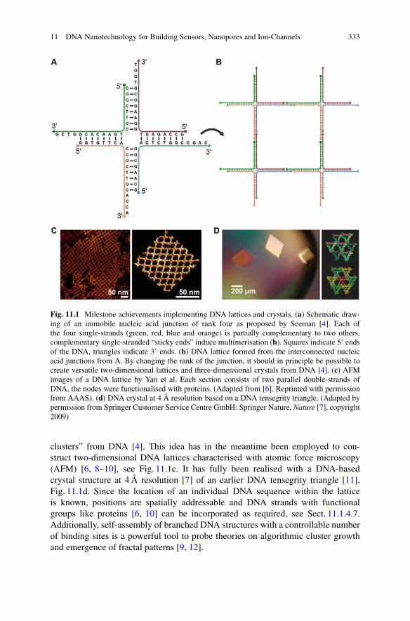
11 DNA Nanotechnology for Building Sensors, Nanopores and Ion-Channels 333
Fig. 11.1 Milestone achievements implementing DNA lattices and crystals. (a) Schematic draw-ing of an immobile nucleic acid junction of rank four as proposed by Seeman [4]. Each ofthe four single-strands (green, red, blue and orange) is partially complementary to two others,complementary single-stranded “sticky ends” induce multimerisation (b). Squares indicate 5’ endsof the DNA, triangles indicate 3’ ends. (b) DNA lattice formed from the interconnected nucleicacid junctions from A. By changing the rank of the junction, it should in principle be possible tocreate versatile two-dimensional lattices and three-dimensional crystals from DNA [4]. (c) AFMimages of a DNA lattice by Yan et al. Each section consists of two parallel double-strands ofDNA, the nodes were functionalised with proteins. (Adapted from [6]. Reprinted with permissionfrom AAAS). (d) DNA crystal at 4 Å resolution based on a DNA tensegrity triangle. (Adapted bypermission from Springer Customer Service Centre GmbH: Springer Nature, Nature [7], copyright2009)
clusters” from DNA [4]. This idea has in the meantime been employed to con-struct two-dimensional DNA lattices characterised with atomic force microscopy(AFM) [6, 8–10], see Fig. 11.1c. It has fully been realised with a DNA-basedcrystal structure at 4 Å resolution [7] of an earlier DNA tensegrity triangle [11],Fig. 11.1d. Since the location of an individual DNA sequence within the latticeis known, positions are spatially addressable and DNA strands with functionalgroups like proteins [6, 10] can be incorporated as required, see Sect. 11.1.4.7.Additionally, self-assembly of branched DNA structures with a controllable numberof binding sites is a powerful tool to probe theories on algorithmic cluster growthand emergence of fractal patterns [9, 12].
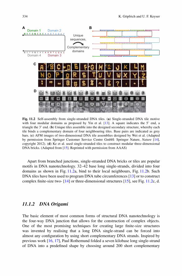
334 K. Göpfrich and U. F. Keyser
ACACGG A A G TGTCGG C G A C
Domain 1 Domain 2Unique
sequences,
Complementary domainsCACGG A A G TGTCGG C G A C
Domain 4 Domain 3
B
C
D
Fig. 11.2 Self-assembly from single-stranded DNA tiles. (a) Single-stranded DNA tile motivewith four modular domains as proposed by Yin et al. [13]. A square indicates the 5’ end, atriangle the 3’ end. (b) Unique tiles assemble into the designed secondary structure, whereby eachtile binds a complementary domain of four neighbouring tiles. Base pairs are indicated as greybars. (c) AFM images of two-dimensional DNA tile assemblies designed by Wei et al. (Adaptedby permission from Springer Customer Service Centre GmbH: Springer Nature, Nature [14],copyright 2012). (d) Ke et al. used single-stranded tiles to construct modular three-dimensionalDNA bricks. (Adapted from [15]. Reprinted with permission from AAAS)
Apart from branched junctions, single-stranded DNA bricks or tiles are popularmotifs in DNA nanotechnology. 32–42 base long single-strands, divided into fourdomains as shown in Fig. 11.2a, bind to their local neighbours, Fig. 11.2b. SuchDNA tiles have been used to program DNA tube circumferences [13] or to constructcomplex finite-size two- [14] or three-dimensional structures [15], see Fig. 11.2c, d.
11.1.2 DNA Origami
The basic element of most common forms of structural DNA nanotechnology isthe four-way DNA junction that allows for the construction of complex objects.One of the most promising techniques for creating large finite-size structureswas invented by realising that a long DNA single-strand can be forced intoalmost any configuration by using short complementary DNA strands. Inspired byprevious work [16, 17], Paul Rothemund folded a seven kilobase long single-strandof DNA into a predefined shape by choosing around 200 short complementary
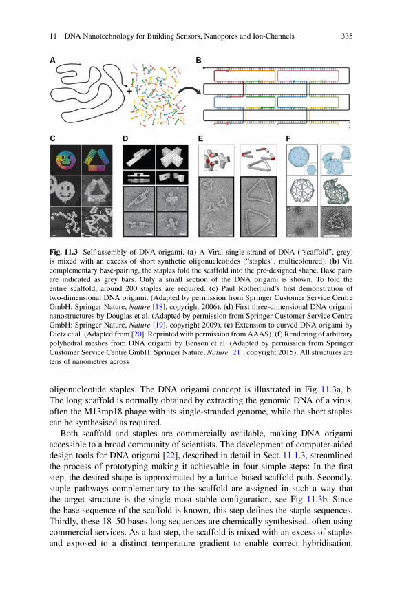
11 DNA Nanotechnology for Building Sensors, Nanopores and Ion-Channels 335
Fig. 11.3 Self-assembly of DNA origami. (a) A Viral single-strand of DNA (“scaffold”, grey)is mixed with an excess of short synthetic oligonucleotides (“staples”, multicoloured). (b) Viacomplementary base-pairing, the staples fold the scaffold into the pre-designed shape. Base pairsare indicated as grey bars. Only a small section of the DNA origami is shown. To fold theentire scaffold, around 200 staples are required. (c) Paul Rothemund’s first demonstration oftwo-dimensional DNA origami. (Adapted by permission from Springer Customer Service CentreGmbH: Springer Nature, Nature [18], copyright 2006). (d) First three-dimensional DNA origaminanostructures by Douglas et al. (Adapted by permission from Springer Customer Service CentreGmbH: Springer Nature, Nature [19], copyright 2009). (e) Extension to curved DNA origami byDietz et al. (Adapted from [20]. Reprinted with permission from AAAS). (f) Rendering of arbitrarypolyhedral meshes from DNA origami by Benson et al. (Adapted by permission from SpringerCustomer Service Centre GmbH: Springer Nature, Nature [21], copyright 2015). All structures aretens of nanometres across
oligonucleotide staples. The DNA origami concept is illustrated in Fig. 11.3a, b.The long scaffold is normally obtained by extracting the genomic DNA of a virus,often the M13mp18 phage with its single-stranded genome, while the short staplescan be synthesised as required.
Both scaffold and staples are commercially available, making DNA origamiaccessible to a broad community of scientists. The development of computer-aideddesign tools for DNA origami [22], described in detail in Sect. 11.1.3, streamlinedthe process of prototyping making it achievable in four simple steps: In the firststep, the desired shape is approximated by a lattice-based scaffold path. Secondly,staple pathways complementary to the scaffold are assigned in such a way thatthe target structure is the single most stable configuration, see Fig. 11.3b. Sincethe base sequence of the scaffold is known, this step defines the staple sequences.Thirdly, these 18–50 bases long sequences are chemically synthesised, often usingcommercial services. As a last step, the scaffold is mixed with an excess of staplesand exposed to a distinct temperature gradient to enable correct hybridisation.
336 K. Göpfrich and U. F. Keyser
This process reliably allows for the creation of microgram quantities of identicalnanostructures in a simple and robust one-pot reaction. With all these tools at hand,it is not surprising that the DNA origami community grew quickly, contributing tonew developments and notable achievements across different disciplines.
Paul Rothemund invented DNA origami in 2006, famously exemplified withthe nanoscale smiley face shown in Fig. 11.3c [18]. Douglas et al. were the firstto create three-dimensional structures from DNA origami, Fig. 11.3d [22]. PossibleDNA geometries were expanded further by Dietz et al. who created the curved DNAstructures in Fig. 11.3e [20], and Benson et al. who demonstrated a general methodfor rendering of polyhedral meshes with DNA origami, Fig. 11.3f [21].
11.1.3 Design and Assembly of DNA Nanostructures
In this section, we discuss the design rules for structural DNA nanotechnology,introduce computational frameworks that facilitate the prototyping process anddiscuss conditions for assembly and storage of DNA nanostructures.
11.1.3.1 Conceiving the Target Shape
The design of a new DNA nanostructure will depend on the functional requirementsfor its envisioned application. The first decision to make is whether to use scaffoldedDNA origami, see Sect. 11.1.2, or a DNA tile approach where the structure isassembled from short single-strands, Sect. 11.1.1. When using a kilobase longscaffold, the size of the DNA nanostructure immediately falls into the megadaltonregime. The achievable concentrations are limited by the scaffold concentration andplacing modifications on the scaffold strand is challenging. Therefore, assemblyfrom short single-strands is useful when the target structure is smaller or hasrepeating structural units like lattices [10] or tubes [13]. Assembly from single-strands, however, becomes less efficient (12–17% yield [14]) for large finite-sizeassemblies, which is why scaffolded DNA origami is then often the method ofchoice. Additionally, many of the software tools described in Sect. 11.1.3.3 havebeen developed specifically for DNA origami making its design relatively straight-forward. The final yield of a DNA origami structure will depend on the design,the assembly conditions and the chosen scaffold. A good scaffold should have littleinternal sequence complementarity and secondary structure, like the M13mp18 viralDNA [18]. An additional benefit of selecting one of a few commonly used scaffoldsis that they can be purchased from vendors like New England Biolabs or Tilibit. Toobtain smaller structures, one can either cut the scaffold to the desired length usingrestriction enzymes [23, 24] or rely on the synthesis of a long custom DNA sequence(e.g. Ultramers from Integrated DNA Technologies), which can then be employedas a scaffold strand.
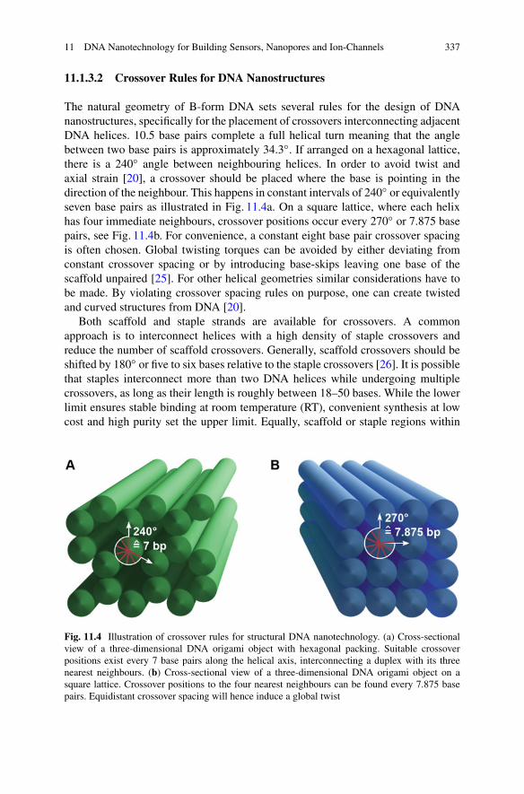
11 DNA Nanotechnology for Building Sensors, Nanopores and Ion-Channels 337
11.1.3.2 Crossover Rules for DNA Nanostructures
The natural geometry of B-form DNA sets several rules for the design of DNAnanostructures, specifically for the placement of crossovers interconnecting adjacentDNA helices. 10.5 base pairs complete a full helical turn meaning that the anglebetween two base pairs is approximately 34.3◦. If arranged on a hexagonal lattice,there is a 240◦ angle between neighbouring helices. In order to avoid twist andaxial strain [20], a crossover should be placed where the base is pointing in thedirection of the neighbour. This happens in constant intervals of 240◦ or equivalentlyseven base pairs as illustrated in Fig. 11.4a. On a square lattice, where each helixhas four immediate neighbours, crossover positions occur every 270◦ or 7.875 basepairs, see Fig. 11.4b. For convenience, a constant eight base pair crossover spacingis often chosen. Global twisting torques can be avoided by either deviating fromconstant crossover spacing or by introducing base-skips leaving one base of thescaffold unpaired [25]. For other helical geometries similar considerations have tobe made. By violating crossover spacing rules on purpose, one can create twistedand curved structures from DNA [20].
Both scaffold and staple strands are available for crossovers. A commonapproach is to interconnect helices with a high density of staple crossovers andreduce the number of scaffold crossovers. Generally, scaffold crossovers should beshifted by 180◦ or five to six bases relative to the staple crossovers [26]. It is possiblethat staples interconnect more than two DNA helices while undergoing multiplecrossovers, as long as their length is roughly between 18–50 bases. While the lowerlimit ensures stable binding at room temperature (RT), convenient synthesis at lowcost and high purity set the upper limit. Equally, scaffold or staple regions within
Fig. 11.4 Illustration of crossover rules for structural DNA nanotechnology. (a) Cross-sectionalview of a three-dimensional DNA origami object with hexagonal packing. Suitable crossoverpositions exist every 7 base pairs along the helical axis, interconnecting a duplex with its threenearest neighbours. (b) Cross-sectional view of a three-dimensional DNA origami object on asquare lattice. Crossover positions to the four nearest neighbours can be found every 7.875 basepairs. Equidistant crossover spacing will hence induce a global twist
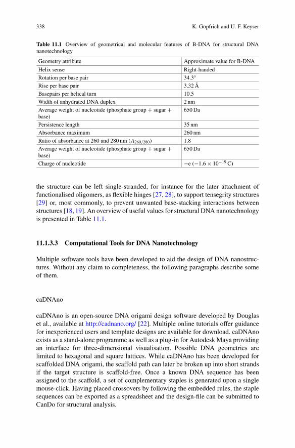
338 K. Göpfrich and U. F. Keyser
Table 11.1 Overview of geometrical and molecular features of B-DNA for structural DNAnanotechnology
Geometry attribute Approximate value for B-DNA
Helix sense Right-handed
Rotation per base pair 34.3◦
Rise per base pair 3.32 Å
Basepairs per helical turn 10.5
Width of anhydrated DNA duplex 2 nm
Average weight of nucleotide (phosphate group + sugar +base)
650 Da
Persistence length 35 nm
Absorbance maximum 260 nm
Ratio of absorbance at 260 and 280 nm (A260/280) 1.8
Average weight of nucleotide (phosphate group + sugar +base)
650 Da
Charge of nucleotide −e (−1.6 × 10⁻¹⁹ C)
the structure can be left single-stranded, for instance for the later attachment offunctionalised oligomers, as flexible hinges [27, 28], to support tensegrity structures[29] or, most commonly, to prevent unwanted base-stacking interactions betweenstructures [18, 19]. An overview of useful values for structural DNA nanotechnologyis presented in Table 11.1.
11.1.3.3 Computational Tools for DNA Nanotechnology
Multiple software tools have been developed to aid the design of DNA nanostruc-tures. Without any claim to completeness, the following paragraphs describe someof them.
caDNAno
caDNAno is an open-source DNA origami design software developed by Douglaset al., available at http://cadnano.org/ [22]. Multiple online tutorials offer guidancefor inexperienced users and template designs are available for download. caDNAnoexists as a stand-alone programme as well as a plug-in for Autodesk Maya providingan interface for three-dimensional visualisation. Possible DNA geometries arelimited to hexagonal and square lattices. While caDNAno has been developed forscaffolded DNA origami, the scaffold path can later be broken up into short strandsif the target structure is scaffold-free. Once a known DNA sequence has beenassigned to the scaffold, a set of complementary staples is generated upon a singlemouse-click. Having placed crossovers by following the embedded rules, the staplesequences can be exported as a spreadsheet and the design-file can be submitted toCanDo for structural analysis.
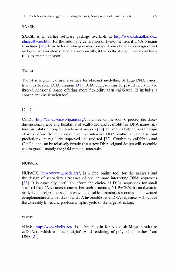
11 DNA Nanotechnology for Building Sensors, Nanopores and Ion-Channels 339
SARSE
SARSE is an earlier software package available at http://www.cdna.dk/index.php/software.html for the automatic generation of two-dimensional DNA origamistructures [30]. It includes a bitmap reader to import any shape as a design objectand generates an atomic model. Conveniently, it tracks the design history and has afully extendible toolbox.
Tiamat
Tiamat is a graphical user interface for efficient modelling of large DNA nanos-tructures beyond DNA origami [31]. DNA duplexes can be placed freely in thethree-dimensional space offering more flexibility than caDNAno. It includes aconvenient visualisation tool.
CanDo
CanDo, http://cando-dna-origami.org/, is a free online tool to predict the three-dimensional shape and flexibility of scaffolded and scaffold-free DNA nanostruc-tures in solution using finite-element analysis [26]. It can thus help to make designchoices before the more cost- and time-intensive DNA synthesis. The structuralpredictions are regularly improved and updated [32]. Combining caDNAno andCanDo, one can be relatively certain that a new DNA origami design will assembleas designed – merely the yield remains uncertain.
NUPACK
NUPACK, http://www.nupack.org/, is a free online tool for the analysis andthe design of secondary structures of one or more interacting DNA sequences[33]. It is especially useful to inform the choice of DNA sequences for smallscaffold-free DNA nanostructures. For such structures, NUPACK’s thermodynamicanalysis can help select sequences without stable secondary structures and unwantedcomplementaries with other strands. A favourable set of DNA sequences will reducethe assembly times and produce a higher yield of the target structure.
vHelix
vHelix, http://www.vhelix.net/, is a free plug-in for Autodesk Maya, similar tocaDNAno, which enables straightforward rendering of polyhedral meshes fromDNA [21].
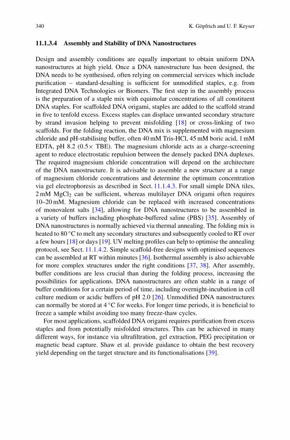
340 K. Göpfrich and U. F. Keyser
11.1.3.4 Assembly and Stability of DNA Nanostructures
Design and assembly conditions are equally important to obtain uniform DNAnanostructures at high yield. Once a DNA nanostructure has been designed, theDNA needs to be synthesised, often relying on commercial services which includepurification – standard-desalting is sufficient for unmodified staples, e.g. fromIntegrated DNA Technologies or Biomers. The first step in the assembly processis the preparation of a staple mix with equimolar concentrations of all constituentDNA staples. For scaffolded DNA origami, staples are added to the scaffold strandin five to tenfold excess. Excess staples can displace unwanted secondary structureby strand invasion helping to prevent misfolding [18] or cross-linking of twoscaffolds. For the folding reaction, the DNA mix is supplemented with magnesiumchloride and pH-stabilising buffer, often 40 mM Tris-HCl, 45 mM boric acid, 1 mMEDTA, pH 8.2 (0.5× TBE). The magnesium chloride acts as a charge-screeningagent to reduce electrostatic repulsion between the densely packed DNA duplexes.The required magnesium chloride concentration will depend on the architectureof the DNA nanostructure. It is advisable to assemble a new structure at a rangeof magnesium chloride concentrations and determine the optimum concentrationvia gel electrophoresis as described in Sect. 11.1.4.3. For small simple DNA tiles,2 mM MgCl2 can be sufficient, whereas multilayer DNA origami often requires10–20 mM. Magnesium chloride can be replaced with increased concentrationsof monovalent salts [34], allowing for DNA nanostructures to be assembled ina variety of buffers including phosphate-buffered saline (PBS) [35]. Assembly ofDNA nanostructures is normally achieved via thermal annealing. The folding mix isheated to 80 ◦C to melt any secondary structures and subsequently cooled to RT overa few hours [18] or days [19]. UV melting profiles can help to optimise the annealingprotocol, see Sect. 11.1.4.2. Simple scaffold-free designs with optimised sequencescan be assembled at RT within minutes [36]. Isothermal assembly is also achievablefor more complex structures under the right conditions [37, 38]. After assembly,buffer conditions are less crucial than during the folding process, increasing thepossibilities for applications. DNA nanostructures are often stable in a range ofbuffer conditions for a certain period of time, including overnight-incubation in cellculture medium or acidic buffers of pH 2.0 [26]. Unmodified DNA nanostructurescan normally be stored at 4 ◦C for weeks. For longer time periods, it is beneficial tofreeze a sample whilst avoiding too many freeze-thaw cycles.
For most applications, scaffolded DNA origami requires purification from excessstaples and from potentially misfolded structures. This can be achieved in manydifferent ways, for instance via ultrafiltration, gel extraction, PEG precipitation ormagnetic bead capture. Shaw et al. provide guidance to obtain the best recoveryyield depending on the target structure and its functionalisations [39].
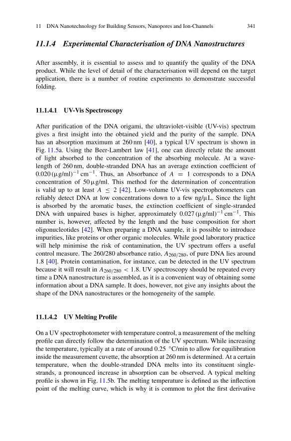
11 DNA Nanotechnology for Building Sensors, Nanopores and Ion-Channels 341
11.1.4 Experimental Characterisation of DNA Nanostructures
After assembly, it is essential to assess and to quantify the quality of the DNAproduct. While the level of detail of the characterisation will depend on the targetapplication, there is a number of routine experiments to demonstrate successfulfolding.
11.1.4.1 UV-Vis Spectroscopy
After purification of the DNA origami, the ultraviolet-visible (UV-vis) spectrumgives a first insight into the obtained yield and the purity of the sample. DNAhas an absorption maximum at 260 nm [40], a typical UV spectrum is shown inFig. 11.5a. Using the Beer-Lambert law [41], one can directly relate the amountof light absorbed to the concentration of the absorbing molecule. At a wave-length of 260 nm, double-stranded DNA has an average extinction coefficient of0.020 (μg/ml)−1 cm−1. Thus, an Absorbance of A = 1 corresponds to a DNAconcentration of 50 μg/ml. This method for the determination of concentrationis valid up to at least A ≤ 2 [42]. Low-volume UV-vis spectrophotometers canreliably detect DNA at low concentrations down to a few ng/μL. Since the lightis absorbed by the aromatic bases, the extinction coefficient of single-strandedDNA with unpaired bases is higher, approximately 0.027 (μg/ml)−1 cm−1. Thisnumber is, however, affected by the length and the base composition for shortoligonucleotides [42]. When preparing a DNA sample, it is possible to introduceimpurities, like proteins or other organic molecules. While good laboratory practicewill help minimise the risk of contamination, the UV spectrum offers a usefulcontrol measure. The 260/280 absorbance ratio, A260/280, of pure DNA lies around1.8 [40]. Protein contamination, for instance, can be detected in the UV spectrumbecause it will result in A260/280 < 1.8. UV spectroscopy should be repeated everytime a DNA nanostructure is assembled, as it is a convenient way of obtaining someinformation about a DNA sample. It does, however, not give any insights about theshape of the DNA nanostructures or the homogeneity of the sample.
11.1.4.2 UV Melting Profile
On a UV spectrophotometer with temperature control, a measurement of the meltingprofile can directly follow the determination of the UV spectrum. While increasingthe temperature, typically at a rate of around 0.25 ◦C/min to allow for equilibrationinside the measurement cuvette, the absorption at 260 nm is determined. At a certaintemperature, when the double-stranded DNA melts into its constituent single-strands, a pronounced increase in absorption can be observed. A typical meltingprofile is shown in Fig. 11.5b. The melting temperature is defined as the inflectionpoint of the melting curve, which is why it is common to plot the first derivative
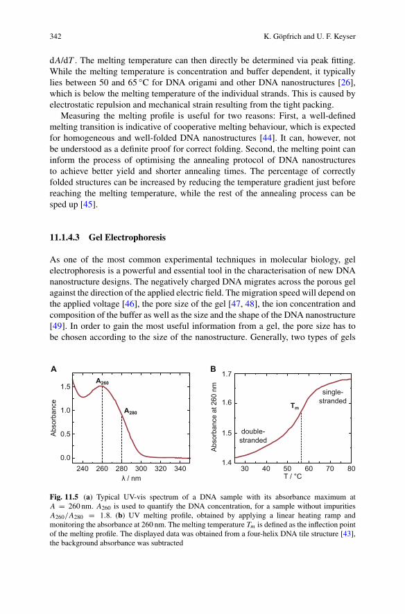
342 K. Göpfrich and U. F. Keyser
dA/dT . The melting temperature can then directly be determined via peak fitting.While the melting temperature is concentration and buffer dependent, it typicallylies between 50 and 65 ◦C for DNA origami and other DNA nanostructures [26],which is below the melting temperature of the individual strands. This is caused byelectrostatic repulsion and mechanical strain resulting from the tight packing.
Measuring the melting profile is useful for two reasons: First, a well-definedmelting transition is indicative of cooperative melting behaviour, which is expectedfor homogeneous and well-folded DNA nanostructures [44]. It can, however, notbe understood as a definite proof for correct folding. Second, the melting point caninform the process of optimising the annealing protocol of DNA nanostructuresto achieve better yield and shorter annealing times. The percentage of correctlyfolded structures can be increased by reducing the temperature gradient just beforereaching the melting temperature, while the rest of the annealing process can besped up [45].
11.1.4.3 Gel Electrophoresis
As one of the most common experimental techniques in molecular biology, gelelectrophoresis is a powerful and essential tool in the characterisation of new DNAnanostructure designs. The negatively charged DNA migrates across the porous gelagainst the direction of the applied electric field. The migration speed will depend onthe applied voltage [46], the pore size of the gel [47, 48], the ion concentration andcomposition of the buffer as well as the size and the shape of the DNA nanostructure[49]. In order to gain the most useful information from a gel, the pore size has tobe chosen according to the size of the nanostructure. Generally, two types of gels
240 260 280 300 320 3400.0
0.5
1.0
1.5
Abso
rban
ce
λ / nm
A260
A280
A
Abso
rban
ce a
t 260
nm
1.4
1.5
1.6
1.7
T / °C30 40 50 60 70 80
double-stranded
Tm
single-stranded
B
Fig. 11.5 (a) Typical UV-vis spectrum of a DNA sample with its absorbance maximum atA = 260 nm. A260 is used to quantify the DNA concentration, for a sample without impuritiesA260/A280 = 1.8. (b) UV melting profile, obtained by applying a linear heating ramp andmonitoring the absorbance at 260 nm. The melting temperature Tm is defined as the inflection pointof the melting profile. The displayed data was obtained from a four-helix DNA tile structure [43],the background absorbance was subtracted
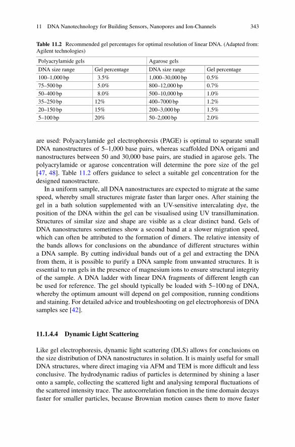
11 DNA Nanotechnology for Building Sensors, Nanopores and Ion-Channels 343
Table 11.2 Recommended gel percentages for optimal resolution of linear DNA. (Adapted from:Agilent technologies)
Polyacrylamide gels Agarose gels
DNA size range Gel percentage DNA size range Gel percentage
100–1,000 bp 3.5% 1,000–30,000 bp 0.5%
75–500 bp 5.0% 800–12,000 bp 0.7%
50–400 bp 8.0% 500–10,000 bp 1.0%
35–250 bp 12% 400–7000 bp 1.2%
20–150 bp 15% 200–3,000 bp 1.5%
5–100 bp 20% 50–2,000 bp 2.0%
are used: Polyacrylamide gel electrophoresis (PAGE) is optimal to separate smallDNA nanostructures of 5–1,000 base pairs, whereas scaffolded DNA origami andnanostructures between 50 and 30,000 base pairs, are studied in agarose gels. Thepolyacrylamide or agarose concentration will determine the pore size of the gel[47, 48]. Table 11.2 offers guidance to select a suitable gel concentration for thedesigned nanostructure.
In a uniform sample, all DNA nanostructures are expected to migrate at the samespeed, whereby small structures migrate faster than larger ones. After staining thegel in a bath solution supplemented with an UV-sensitive intercalating dye, theposition of the DNA within the gel can be visualised using UV transillumination.Structures of similar size and shape are visible as a clear distinct band. Gels ofDNA nanostructures sometimes show a second band at a slower migration speed,which can often be attributed to the formation of dimers. The relative intensity ofthe bands allows for conclusions on the abundance of different structures withina DNA sample. By cutting individual bands out of a gel and extracting the DNAfrom them, it is possible to purify a DNA sample from unwanted structures. It isessential to run gels in the presence of magnesium ions to ensure structural integrityof the sample. A DNA ladder with linear DNA fragments of different length canbe used for reference. The gel should typically be loaded with 5–100 ng of DNA,whereby the optimum amount will depend on gel composition, running conditionsand staining. For detailed advice and troubleshooting on gel electrophoresis of DNAsamples see [42].
11.1.4.4 Dynamic Light Scattering
Like gel electrophoresis, dynamic light scattering (DLS) allows for conclusions onthe size distribution of DNA nanostructures in solution. It is mainly useful for smallDNA structures, where direct imaging via AFM and TEM is more difficult and lessconclusive. The hydrodynamic radius of particles is determined by shining a laseronto a sample, collecting the scattered light and analysing temporal fluctuations ofthe scattered intensity trace. The autocorrelation function in the time domain decaysfaster for smaller particles, because Brownian motion causes them to move faster
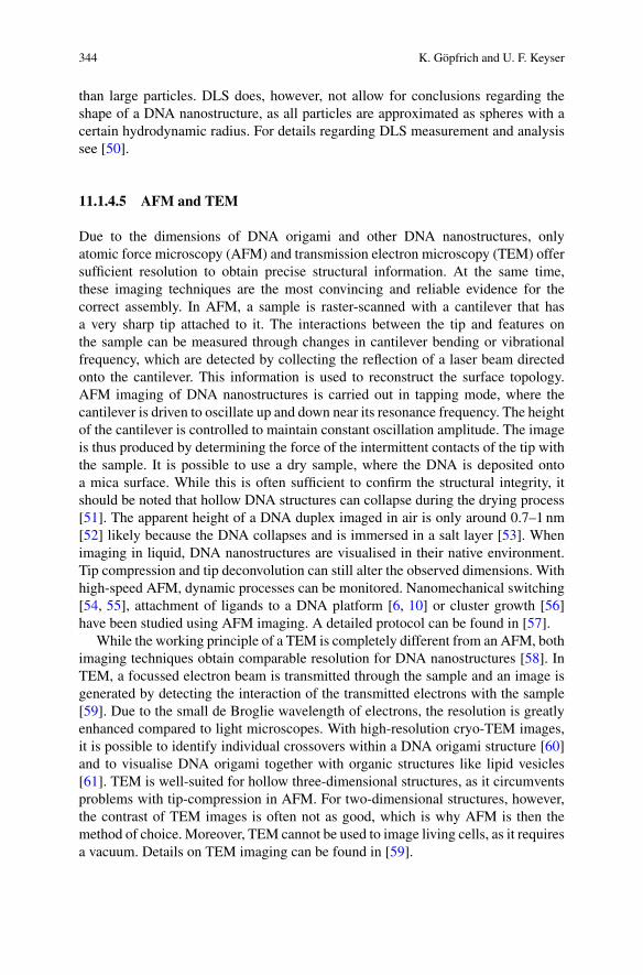
344 K. Göpfrich and U. F. Keyser
than large particles. DLS does, however, not allow for conclusions regarding theshape of a DNA nanostructure, as all particles are approximated as spheres with acertain hydrodynamic radius. For details regarding DLS measurement and analysissee [50].
11.1.4.5 AFM and TEM
Due to the dimensions of DNA origami and other DNA nanostructures, onlyatomic force microscopy (AFM) and transmission electron microscopy (TEM) offersufficient resolution to obtain precise structural information. At the same time,these imaging techniques are the most convincing and reliable evidence for thecorrect assembly. In AFM, a sample is raster-scanned with a cantilever that hasa very sharp tip attached to it. The interactions between the tip and features onthe sample can be measured through changes in cantilever bending or vibrationalfrequency, which are detected by collecting the reflection of a laser beam directedonto the cantilever. This information is used to reconstruct the surface topology.AFM imaging of DNA nanostructures is carried out in tapping mode, where thecantilever is driven to oscillate up and down near its resonance frequency. The heightof the cantilever is controlled to maintain constant oscillation amplitude. The imageis thus produced by determining the force of the intermittent contacts of the tip withthe sample. It is possible to use a dry sample, where the DNA is deposited ontoa mica surface. While this is often sufficient to confirm the structural integrity, itshould be noted that hollow DNA structures can collapse during the drying process[51]. The apparent height of a DNA duplex imaged in air is only around 0.7–1 nm[52] likely because the DNA collapses and is immersed in a salt layer [53]. Whenimaging in liquid, DNA nanostructures are visualised in their native environment.Tip compression and tip deconvolution can still alter the observed dimensions. Withhigh-speed AFM, dynamic processes can be monitored. Nanomechanical switching[54, 55], attachment of ligands to a DNA platform [6, 10] or cluster growth [56]have been studied using AFM imaging. A detailed protocol can be found in [57].
While the working principle of a TEM is completely different from an AFM, bothimaging techniques obtain comparable resolution for DNA nanostructures [58]. InTEM, a focussed electron beam is transmitted through the sample and an image isgenerated by detecting the interaction of the transmitted electrons with the sample[59]. Due to the small de Broglie wavelength of electrons, the resolution is greatlyenhanced compared to light microscopes. With high-resolution cryo-TEM images,it is possible to identify individual crossovers within a DNA origami structure [60]and to visualise DNA origami together with organic structures like lipid vesicles[61]. TEM is well-suited for hollow three-dimensional structures, as it circumventsproblems with tip-compression in AFM. For two-dimensional structures, however,the contrast of TEM images is often not as good, which is why AFM is then themethod of choice. Moreover, TEM cannot be used to image living cells, as it requiresa vacuum. Details on TEM imaging can be found in [59].
11 DNA Nanotechnology for Building Sensors, Nanopores and Ion-Channels 345
11.1.4.6 DNA-PAINT
DNA-PAINT, or “DNA-based point accumulation for imaging in nanoscale topog-raphy”, is a super-resolution imaging method. Super-resolution is obtained bytemporally switching each target between a fluorescence on-state and an off-state,and determining their respective positions with sub-diffraction limit precision.DNA-PAINT uses transient binding of short fluorescently labelled single-strandsof DNA to achieve a resolution down to 5–10 nm [62]. Unlike in AFM and TEM,DNA-PAINT offers chemical specificity due to the required hybridisation and evensingle-stranded DNA can be visualised [63].
11.1.4.7 Functionalisation of DNA
With all the tools at hand, the creation of new shapes from DNA is no longerthe main challenge in the field of structural DNA nanotechnology. We are expe-riencing a shift from mere structures to functional nano-objects with diverseapplications. This is only achievable thanks to the possibility of site-directedchemical modification of DNA with functional groups. Commercially availableamino- or thiol-modified oligonucleotides often serve as a starting point for thecovalent attachment of the selected crosslinker. Proteins and aptamers have beenattached to DNA nanostructures for biomimetic enzyme cascades [64, 65], singlemolecule detection [66], cellular recognition [67, 68] or controlled substrate release[67, 69]. Gold [70–72] and silver nanoparticles [73, 74] and quantum dots [75]have been incorporated into DNA nanostructures for plasmonic applications [76].DNA-based polyhedral meshes [21] have recently been used to assemble crystallinenanoparticle-DNA frameworks [77]. Attachment of carbon nanotubes [78] andmetallisation of DNA structures [79] may lead to the fabrication of new nanoma-terials for electronics. Responsive polymers offer scope for the creation of DNAnanostructures that reversibly change conformation as a response to external stimulisuch as light [72, 80] or pH [81]. Fluorescent dyes and quenchers are commonlyincorporated for visualisation purposes or to monitor conformational changes ofDNA nanostructures, see Sect. 11.2.1. They have also been used to study lightharvesting on DNA platforms [82]. Fluorescently tagged DNA nanostructures havefound commercial use as standards for superresolution microscopy [83]. Lipophilicmodifications, such as cholesterol or porphyrin tags, will be discussed in moredetail in the context of the synthetic DNA-based membrane pores in Sect. 11.3.3.They have further been employed to promote cellular uptake, for two-dimensionalassembly of DNA nanostructure arrays [56], or to scaffold [84] and bend vesicles[85]. Some functionalised custom oligomers, including fluorescent or lipophilictags, can be purchased from vendors like Integrated DNA Technologies or Biomers.Combination of synthetic nucleic acids, like peptide nucleic acid (PNA) [86] XNA[87], with DNA nanostructures may protect them from enzymatic digestion in livingsystems and offers control over the backbone charge [88]. Achieving a sufficientyield of the final purified product is still the main challenge for the integration of
346 K. Göpfrich and U. F. Keyser
many heteroelements into DNA nanostructures. An additional challenge for someapplications, like DNA-based synthetic light harvesting systems [82], is the lack ofcontrol over the orientation of the functional groups. Without chemical modification,dynamic motion of DNA nanostructures can be achieved by strand displacement,where the target strand is equipped with a single-stranded DNA toehold. Theaddition of a DNA strand which binds to the toehold and has higher sequencecomplementarity to the target strand will then replace a previously bound strand[89]. This mechanism has been employed for DNA-based logic gates [90], catalysedreaction circuits [91] or synthetic molecular motors [92].
The long-term prospects of structural DNA nanotechnology will depend on itssuccess in constructing sophisticated nano-machines driving active and functionalbehaviour at a capacity that is unmatched by competing forms of nanotechnology.Until stability, scalability and cost issues are addressed, DNA nanotechnology willmainly be employed to address specific research questions. Yet thanks to the scopeof chemical functionalisation, DNA sensors and nanopores have made remarkableprogress towards viable applications beyond the research community. These will bediscussed in the following section.
11.2 DNA Sensors and Nanopores
Living cells have developed an intriguingly complex sensory machinery to detectchanges in their environment. Molecular control circuits help convert the sensorinformation into a reliable motor response adapting to environmental factors.Remarkably, in vitro DNA nanotechnology has made progress towards constructingmolecular sensory devices. Applications of DNA sensors range from diseasediagnostics to environmental screening or fundamental research. Single-moleculeresolution can be achieved and is highly desirable for precise quantification andwhen a sample is scarce. As of today, DNA-based sensors are one of the mostsuccessful applications of structural DNA nanotechnology. They are either basedon nanomechanical actuation or nanopores for biosensing.
In this section, we will describe the working principle of nanomechanicalDNA sensors, review excising devices and discuss the potential for future ones.We will then introduce nanopore sensors as a label-free single-molecule sensingtechnique and discuss how DNA nanotechnology can enhance this type of sensors.DNA origami nanopores are finally presented as an addition to existing types ofnanopores. Liu et al. [93] and Chandrasekaran et al. [94] have written detailedand comprehensive overviews on DNA-based sensors, we just aim to give a briefoverview.
11 DNA Nanotechnology for Building Sensors, Nanopores and Ion-Channels 347
11.2.1 Nanomechanical DNA-Based Sensors
In a common approach for the fabrication of DNA-based sensors, the DNAnanostructure is designed to undergo a detectable conformation change in responseto a signal. This requires a shift from static structures towards dynamic DNAnanotechnology achieved by chemical functionalisation or strand displacement, seeSect. 11.1.4.7 [92]. Sections of single-stranded DNA can serve as flexible hingesinterconnecting stiff regions of double-stranded DNA. Since the DNA nanostructurecan only amplify the signal, a detection mechanism is still required. The confor-mational change of a larger DNA structure can easily be observed in AFM andTEM as described in Sect. 11.1.4.5. Alternatively, a FRET (Förster resonance energytransfer) pair of fluorescent dyes can be placed on the DNA nanostructure. Thedonor fluorophore is excited and emits at the right wavelength to excite the acceptor.The Stokes-shifted longer-wavelength emission of the acceptor is then detected.Since the energy transfer efficiency between the two fluorophores depends on theirdistance with an inverse 6th-power law, the fluorophore emission is a sensitivemeasure for the conformation of a DNA nanostructure if the dyes are placedappropriately. Using this approach, a straightforward bulk measurement of theemission spectrum is sufficient to detect conformational changes, and surface immo-bilisation is not required. Single-molecule FRET can be carried out in addition.Other detection methods for nanomechanical DNA switches involve fluorescentquenchers [95], nanoparticles, colorimetric and electrochemical sensing techniques,gel electrophoresis [96], microfluidics, optical tweezers [97] or magnetic resonanceimaging (MRI) contrast agents. DNA sensors with picomolar detection limits andover million-fold selectivity have been reported [95].
Nanomechanical DNA sensors can be based on large DNA origami structuresor small functional nucleic acids. Regarding their functionality, they can broadlybe divided into two classes: sensors that detect molecules or ions and sensors thatdetect environmental factors, like temperature or pH.
11.2.1.1 Molecular Sensors
Proteins, DNA and ribonucleic acid (RNA) have been detected employing variationsof DNA origami or smaller nucleic acid-based sensors [98, 99]. The mechanicalresponse of a DNA nanostructure has also been used to assess DNA repair activity[100] or to detect single-base polymorphisms in double-stranded genomic DNA[101]. Not just molecules, also ions have been detected, employing salt-dependentsequence motifs, like the G-quadruplex or metal-ion-bridged duplex nucleic acidsstructures [98, 99, 102–104]. A mercury sensor based on functional nucleic acidswas integrated into dip-stick tests, making it practical for point-of-care diagnostics[95]. Some DNA-based sensors have been employed in vivo for the detection ofmessenger RNA as disease markers [105] or cell surface markers [99, 106]. Theseapproaches may lead to the development of “smart drugs” capable of making
348 K. Göpfrich and U. F. Keyser
autonomous decisions and releasing a molecular payload if appropriate. The cancer-targeting DNA origami drug delivery vehicle by Douglas et al. [69] and the DNAlogic gates for drug release tested in living animals [107] can be seen as proof ofprinciple for such devices.
11.2.1.2 Environmental Sensors
Environmental factors, such as temperature, pH, light or electric field can directlyaffect the conformation of DNA nanostructures or indirectly prompt a mechanicalresponse mediated by functional groups. DNA-based sensors with pH-sensitive i-motifs have successfully been used to map pH changes inside living cells [108, 109].
All sensors described until now can be classified as nanomechanical sensors,where the sensing molecule triggers a mechanical switch. The sensing mechanismof nanopores, described in the next section, does not rely on such a conformationchange.
11.2.2 Nanopores for Single-Molecule Detection
In the first instance, a nanopore is nothing but a nanometre-sized hole in aninsulating material. But by applying a voltage across a nanopore immersed in anelectrolyte solution, one employs it as a powerful, albeit simple single-moleculesensor. The applied voltage induces an ionic current. Charged analytes are driventhrough the pore via electrophoresis, stochastically blocking part of the passage forions. The current blockade signal can be used to detect the analyte, while its durationand magnitude provide information about charge, length and conformation of themolecule [110]. In this way, nanopores can be employed for the label-free detectionof nanometre-sized particles, but the sensing mechanism is applicable across scales– as long as the size of the pore matches the size of the analyte. A Coulter counter,invented by Wallace H. Coulter in 1953 [111], uses the described principle to detect,size and count cells and other particles between 400 nm and 1 mm [112, 113]. Thebrilliantly simple and broadly applicable sensing mechanism based on this resistivepulse principle is illustrated in Fig. 11.6a, b. A big advantage of nanopore sensorscompared to the nanomechanical sensors described in Sect. 11.2.1 is that they can beemployed as multi-purpose sensors. Whereas previous sensors had to be designedfor a specific molecule, nanopores can in principle detect a broad range of differentmolecules in solution.
Bezrukov et al. were the first to demonstrate that a molecular-scale Coultercounter can indeed detect single polymers (polyethylene glycol) in solution [114].The sensing nanopore itself was taken from nature, where nanopores are ubiquitousas molecular gatekeepers of cellular function. Biological protein nanopores areembedded in the membranes of all cells, where they control the selective transportof ions and biomolecules, see Sect. 11.3.1. The challenge for the field of nanopore
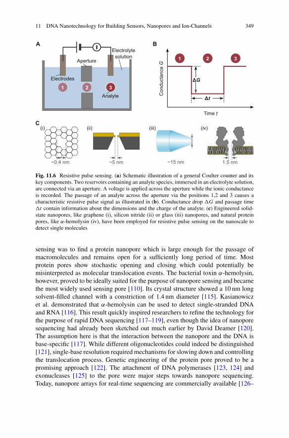
11 DNA Nanotechnology for Building Sensors, Nanopores and Ion-Channels 349
Electrodes
Analyte
BA
1 2 3
Aperture
Electrolytesolution
C(ii)(i) (iv)(iii)
Con
duct
ance
G
Time t
Δt
ΔG
321
~0.4 nm ~5 nm ~15 nm 1.5 nm
)
Fig. 11.6 Resistive pulse sensing. (a) Schematic illustration of a general Coulter counter and itskey components. Two reservoirs containing an analyte species, immersed in an electrolyte solution,are connected via an aperture. A voltage is applied across the aperture while the ionic conductanceis recorded. The passage of an analyte across the aperture via the positions 1,2 and 3 causes acharacteristic resistive pulse signal as illustrated in (b). Conductance drop �G and passage time�t contain information about the dimensions and the charge of the analyte. (c) Engineered solid-state nanopores, like graphene (i), silicon nitride (ii) or glass (iii) nanopores, and natural proteinpores, like α-hemolysin (iv), have been employed for resistive pulse sensing on the nanoscale todetect single molecules
sensing was to find a protein nanopore which is large enough for the passage ofmacromolecules and remains open for a sufficiently long period of time. Mostprotein pores show stochastic opening and closing which could potentially bemisinterpreted as molecular translocation events. The bacterial toxin α-hemolysin,however, proved to be ideally suited for the purpose of nanopore sensing and becamethe most widely used sensing pore [110]. Its crystal structure showed a 10 nm longsolvent-filled channel with a constriction of 1.4 nm diameter [115]. Kasianowiczet al. demonstrated that α-hemolysin can be used to detect single-stranded DNAand RNA [116]. This result quickly inspired researchers to refine the technology forthe purpose of rapid DNA sequencing [117–119], even though the idea of nanoporesequencing had already been sketched out much earlier by David Deamer [120].The assumption here is that the interaction between the nanopore and the DNA isbase-specific [117]. While different oligonucleotides could indeed be distinguished[121], single-base resolution required mechanisms for slowing down and controllingthe translocation process. Genetic engineering of the protein pore proved to be apromising approach [122]. The attachment of DNA polymerases [123, 124] andexonucleases [125] to the pore were major steps towards nanopore sequencing.Today, nanopore arrays for real-time sequencing are commercially available [126–
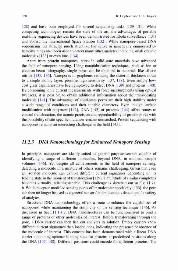
350 K. Göpfrich and U. F. Keyser
128] and have been employed for several sequencing tasks [129–131]. Whilecompeting technologies remain the state of the art, the advantages of portablereal-time sequencing devices have been demonstrated for Ebola surveillance [131]and aboard the International Space Station [132]. While nanopore-based DNAsequencing has attracted much attention, the native or genetically engineered α-hemolysin has also been used to detect many other analytes including small organicmolecules [133] or even ions [134].
Apart from protein nanopores, pores in solid-state materials have advancedthe field of nanopore sensing. Using nanofabrication techniques, such as ion orelectron-beam lithography, single pores can be obtained in materials like siliconnitride [135, 136]. Nanopores in graphene, reducing the material thickness downto a single atomic layer, promise high sensitivity [137, 138]. Even simple low-cost glass capillaries have been employed to detect DNA [139] and proteins [140].By combining ionic current measurements with force measurements using opticaltweezers, it is possible to obtain additional information about the translocatingmolecule [141]. The advantage of solid-state pores are their high stability undera wide range of conditions and their tunable diameters. Even though surfacemodification with polymers [142], DNA [143] or proteins [144] offers routes tocontrol translocation, the atomic precision and reproducibility of protein pores withthe possibility of site-specific mutation remains unmatched. Protein sequencing withnanopores remains an interesting challenge in the field [145].
11.2.3 DNA Nanotechnology for Enhanced Nanopore Sensing
In principle, nanopores are ideally suited as general-purpose sensors capable ofidentifying a range of different molecules, beyond DNA, in minimal samplevolumes [146]. Yet despite all achievements in the field of nanopore sensing,detecting a molecule in a mixture of others remains challenging. Given that evenan isolated molecule can exhibit different current signatures depending on itsfolding state in the moment of translocation [139], a multitude of similar complexesbecomes virtually indistinguishable. This challenge is sketched out in Fig. 11.7a,b. While receptor-modified sensing pores offer molecular specificity [133], the porecan then no longer be used as a general sensor for simultaneous detection of a varietyof analytes.
Structural DNA nanotechnology offers a route to enhance the capabilities ofnanopores, while maintaining the simplicity of the sensing technique [146]. Asdiscussed in Sect. 11.1.4.7, DNA nanostructures can be functionalised to bind arange of proteins or other molecules of interest. Before translocating through thepore, a DNA carrier can then fish out analytes in solution. Empty carriers showdifferent current signatures than loaded ones, indicating the presence or absence ofthe molecule of interest. This concept has been demonstrated with a linear DNAcarrier containing aptamer binding sites for proteins at predefined positions alongthe DNA [147, 148]. Different positions could encode for different proteins. The
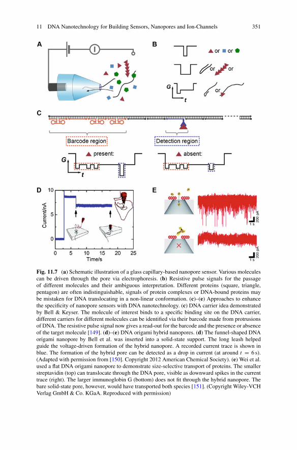
11 DNA Nanotechnology for Building Sensors, Nanopores and Ion-Channels 351
Fig. 11.7 (a) Schematic illustration of a glass capillary-based nanopore sensor. Various moleculescan be driven through the pore via electrophoresis. (b) Resistive pulse signals for the passageof different molecules and their ambiguous interpretation. Different proteins (square, triangle,pentagon) are often indistinguishable, signals of protein complexes or DNA-bound proteins maybe mistaken for DNA translocating in a non-linear conformation. (c)–(e) Approaches to enhancethe specificity of nanopore sensors with DNA nanotechnology. (c) DNA carrier idea demonstratedby Bell & Keyser. The molecule of interest binds to a specific binding site on the DNA carrier,different carriers for different molecules can be identified via their barcode made from protrusionsof DNA. The resistive pulse signal now gives a read-out for the barcode and the presence or absenceof the target molecule [149]. (d)–(e) DNA origami hybrid nanopores. (d) The funnel-shaped DNAorigami nanopore by Bell et al. was inserted into a solid-state support. The long leash helpedguide the voltage-driven formation of the hybrid nanopore. A recorded current trace is shown inblue. The formation of the hybrid pore can be detected as a drop in current (at around t = 6 s).(Adapted with permission from [150]. Copyright 2012 American Chemical Society). (e) Wei et al.used a flat DNA origami nanopore to demonstrate size-selective transport of proteins. The smallerstreptavidin (top) can translocate through the DNA pore, visible as downward spikes in the currenttrace (right). The larger immunoglobin G (bottom) does not fit through the hybrid nanopore. Thebare solid-state pore, however, would have transported both species [151]. (Copyright Wiley-VCHVerlag GmbH & Co. KGaA. Reproduced with permission)
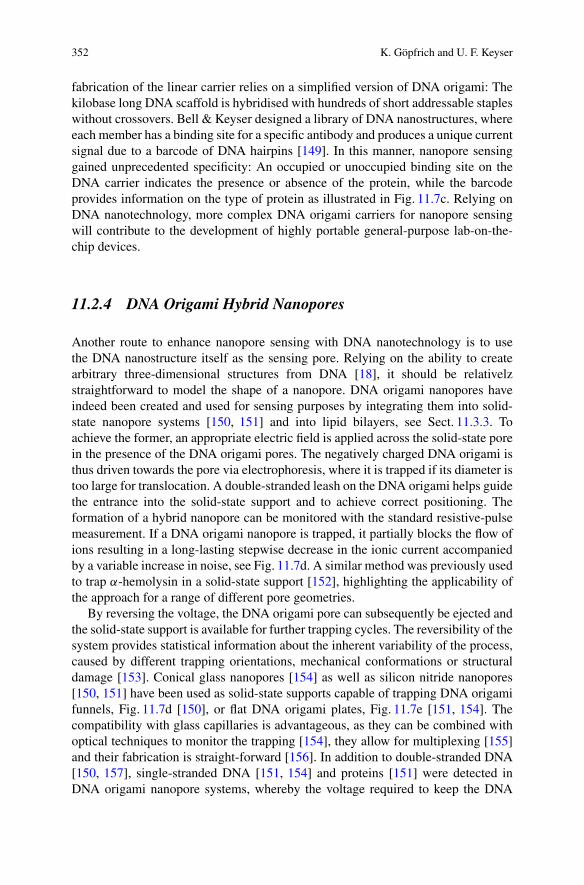
352 K. Göpfrich and U. F. Keyser
fabrication of the linear carrier relies on a simplified version of DNA origami: Thekilobase long DNA scaffold is hybridised with hundreds of short addressable stapleswithout crossovers. Bell & Keyser designed a library of DNA nanostructures, whereeach member has a binding site for a specific antibody and produces a unique currentsignal due to a barcode of DNA hairpins [149]. In this manner, nanopore sensinggained unprecedented specificity: An occupied or unoccupied binding site on theDNA carrier indicates the presence or absence of the protein, while the barcodeprovides information on the type of protein as illustrated in Fig. 11.7c. Relying onDNA nanotechnology, more complex DNA origami carriers for nanopore sensingwill contribute to the development of highly portable general-purpose lab-on-the-chip devices.
11.2.4 DNA Origami Hybrid Nanopores
Another route to enhance nanopore sensing with DNA nanotechnology is to usethe DNA nanostructure itself as the sensing pore. Relying on the ability to createarbitrary three-dimensional structures from DNA [18], it should be relativelzstraightforward to model the shape of a nanopore. DNA origami nanopores haveindeed been created and used for sensing purposes by integrating them into solid-state nanopore systems [150, 151] and into lipid bilayers, see Sect. 11.3.3. Toachieve the former, an appropriate electric field is applied across the solid-state porein the presence of the DNA origami pores. The negatively charged DNA origami isthus driven towards the pore via electrophoresis, where it is trapped if its diameter istoo large for translocation. A double-stranded leash on the DNA origami helps guidethe entrance into the solid-state support and to achieve correct positioning. Theformation of a hybrid nanopore can be monitored with the standard resistive-pulsemeasurement. If a DNA origami nanopore is trapped, it partially blocks the flow ofions resulting in a long-lasting stepwise decrease in the ionic current accompaniedby a variable increase in noise, see Fig. 11.7d. A similar method was previously usedto trap α-hemolysin in a solid-state support [152], highlighting the applicability ofthe approach for a range of different pore geometries.
By reversing the voltage, the DNA origami pore can subsequently be ejected andthe solid-state support is available for further trapping cycles. The reversibility of thesystem provides statistical information about the inherent variability of the process,caused by different trapping orientations, mechanical conformations or structuraldamage [153]. Conical glass nanopores [154] as well as silicon nitride nanopores[150, 151] have been used as solid-state supports capable of trapping DNA origamifunnels, Fig. 11.7d [150], or flat DNA origami plates, Fig. 11.7e [151, 154]. Thecompatibility with glass capillaries is advantageous, as they can be combined withoptical techniques to monitor the trapping [154], they allow for multiplexing [155]and their fabrication is straight-forward [156]. In addition to double-stranded DNA[150, 157], single-stranded DNA [151, 154] and proteins [151] were detected inDNA origami nanopore systems, whereby the voltage required to keep the DNA
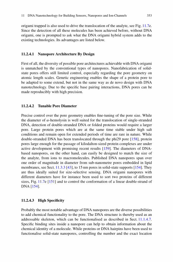
11 DNA Nanotechnology for Building Sensors, Nanopores and Ion-Channels 353
origami trapped is also used to drive the translocation of the analyte, see Fig. 11.7e.Since the detection of all these molecules has been achieved before, without DNAorigami, one is prompted to ask what the DNA origami hybrid system adds to theexisting technologies. Its advantages are listed below.
11.2.4.1 Nanopore Architecture By Design
First of all, the diversity of possible pore architectures achievable with DNA origamiis unmatched by the conventional types of nanopores. Nanofabrication of solid-state pores offers still limited control, especially regarding the pore geometry onatomic length scales. Genetic engineering enables the shape of a protein pore tobe adapted to some extend, but not in the same way as de novo design with DNAnanotechnology. Due to the specific base pairing interactions, DNA pores can bemade reproducibly with high precision.
11.2.4.2 Tunable Pore Diameter
Precise control over the pore geometry enables fine-tuning of the pore size. Whilethe diameter of α-hemolysin is well suited for the translocation of single-strandedDNA, detection of double-stranded DNA or folded proteins would require a largerpore. Large protein pores which are at the same time stable under high saltconditions and remain open for extended periods of time are rare in nature. Whiledouble-stranded DNA has been translocated through the phi29 pore [158], proteinpores large enough for the passage of kilodalton-sized protein complexes are underactive development with promising recent results [159]. The diameters of DNA-based nanopores, on the other hand, can easily be designed to match the size ofthe analyte, from ions to macromolecules. Published DNA nanopores span overone order of magnitude in diameter from sub-nanometre pores embedded in lipidmembranes, see Sect. 11.3.3 [43], to 15 nm pores in solid-state supports [154]. Theyare thus ideally suited for size-selective sensing. DNA origami nanopores withdifferent diameters have for instance been used to sort two proteins of differentsizes, Fig. 11.7e [151] and to control the conformation of a linear double-strand ofDNA [154].
11.2.4.3 High Specificity
Probably the most notable advantage of DNA nanopores are the diverse possibilitiesto add chemical functionality to the pore. The DNA structure is thereby used as anaddressable skeleton, which can be functionalised as described in Sect. 11.1.4.7.Specific binding sites inside a nanopore can help to obtain information about thechemical identity of a molecule. While proteins or DNA hairpins have been used tofunctionalise solid-state nanopores, controlling the number and the exact location
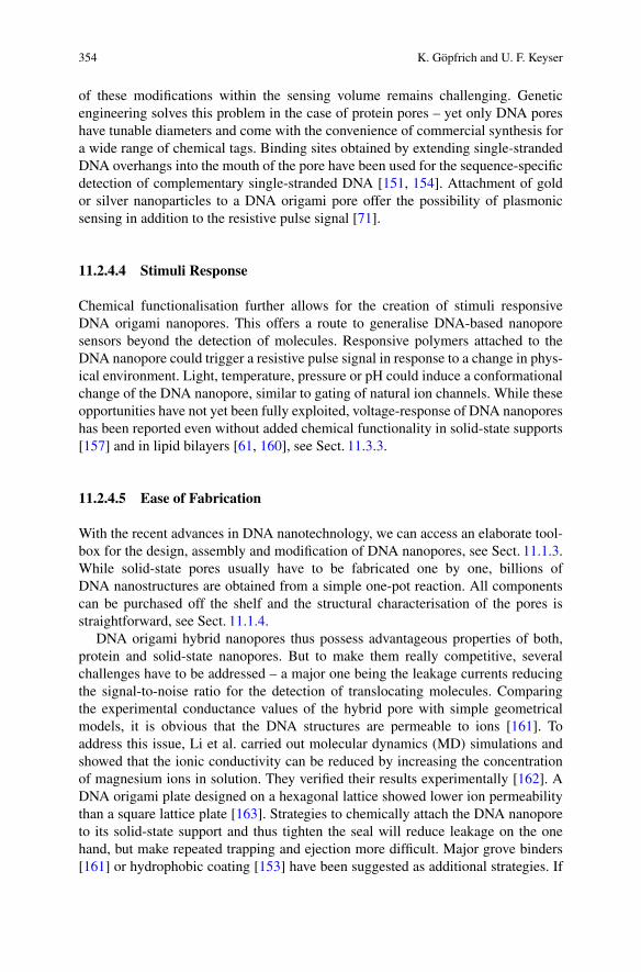
354 K. Göpfrich and U. F. Keyser
of these modifications within the sensing volume remains challenging. Geneticengineering solves this problem in the case of protein pores – yet only DNA poreshave tunable diameters and come with the convenience of commercial synthesis fora wide range of chemical tags. Binding sites obtained by extending single-strandedDNA overhangs into the mouth of the pore have been used for the sequence-specificdetection of complementary single-stranded DNA [151, 154]. Attachment of goldor silver nanoparticles to a DNA origami pore offer the possibility of plasmonicsensing in addition to the resistive pulse signal [71].
11.2.4.4 Stimuli Response
Chemical functionalisation further allows for the creation of stimuli responsiveDNA origami nanopores. This offers a route to generalise DNA-based nanoporesensors beyond the detection of molecules. Responsive polymers attached to theDNA nanopore could trigger a resistive pulse signal in response to a change in phys-ical environment. Light, temperature, pressure or pH could induce a conformationalchange of the DNA nanopore, similar to gating of natural ion channels. While theseopportunities have not yet been fully exploited, voltage-response of DNA nanoporeshas been reported even without added chemical functionality in solid-state supports[157] and in lipid bilayers [61, 160], see Sect. 11.3.3.
11.2.4.5 Ease of Fabrication
With the recent advances in DNA nanotechnology, we can access an elaborate tool-box for the design, assembly and modification of DNA nanopores, see Sect. 11.1.3.While solid-state pores usually have to be fabricated one by one, billions ofDNA nanostructures are obtained from a simple one-pot reaction. All componentscan be purchased off the shelf and the structural characterisation of the pores isstraightforward, see Sect. 11.1.4.
DNA origami hybrid nanopores thus possess advantageous properties of both,protein and solid-state nanopores. But to make them really competitive, severalchallenges have to be addressed – a major one being the leakage currents reducingthe signal-to-noise ratio for the detection of translocating molecules. Comparingthe experimental conductance values of the hybrid pore with simple geometricalmodels, it is obvious that the DNA structures are permeable to ions [161]. Toaddress this issue, Li et al. carried out molecular dynamics (MD) simulations andshowed that the ionic conductivity can be reduced by increasing the concentrationof magnesium ions in solution. They verified their results experimentally [162]. ADNA origami plate designed on a hexagonal lattice showed lower ion permeabilitythan a square lattice plate [163]. Strategies to chemically attach the DNA nanoporeto its solid-state support and thus tighten the seal will reduce leakage on the onehand, but make repeated trapping and ejection more difficult. Major grove binders[161] or hydrophobic coating [153] have been suggested as additional strategies. If
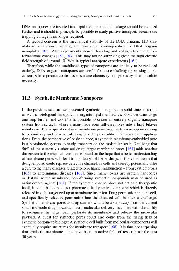
11 DNA Nanotechnology for Building Sensors, Nanopores and Ion-Channels 355
DNA nanopores are inserted into lipid membranes, the leakage should be reducedfurther and it should in principle be possible to study passive transport, because thetrapping voltage is no longer required.
A second concern is the mechanical stability of the DNA origami. MD sim-ulations have shown bending and reversible layer-separation for DNA origaminanoplates [162]. Also experiments showed buckling and voltage-dependent con-formational changes [157, 163]. This may not be surprising given the high electricfield strength of around 107 V/m in typical nanopore experiments [161].
Therefore, while the established types of nanopores are unlikely to be replacedentirely, DNA origami nanopores are useful for more challenging sensing appli-cations where precise control over surface chemistry and geometry is an absolutenecessity.
11.3 Synthetic Membrane Nanopores
In the previous section, we presented synthetic nanopores in solid-state materialsas well as biological nanopores in organic lipid membranes. Now, we want to goone step further and ask if it is possible to create an entirely organic nanoporesystem from scratch, where a man-made pore self-assembles into a lipid bilayermembrane. The scope of synthetic membrane pores reaches from nanopore sensingto biomimicry and beyond, offering broader possibilities for biomedical applica-tions. From the perspective of basic science, a synthetic membrane-embedded poreis a biomimetic system to study transport on the molecular scale. Realising that50% of the currently authorised drugs target membrane pores [164] adds anotherdimension to the research, one that is based on the hope that a better understandingof membrane pores will lead to the design of better drugs. It fuels the dream thatdesigner pores could replace defective channels in cells and thereby potentially offera cure to the many diseases related to ion-channel malfunction – from cystic fibrosis[165] to autoimmune diseases [166]. Since many toxins are protein nanoporesor destabilise the membrane, pore-forming synthetic compounds may be used asantimicrobial agents [167]. If the synthetic channel does not act as a therapeuticitself, it could be coupled to a pharmaceutically active compound which is directlyreleased into the target cell upon membrane insertion. Drug permeation into the cell,and specifically selective permeation into the diseased cell, is often a challenge.Synthetic membrane pores as drug carriers would be a step away from the currentsmall-molecule drugs towards macro-molecular delivery machines with the abilityto recognise the target cell, perforate its membrane and release the molecularpayload. A quest for synthetic pores could also come from the rising field ofsynthetic bottom-up biology: A synthetic cell built from molecular components willeventually require structures for membrane transport [168]. It is thus not surprisingthat synthetic membrane pores have been an active field of research for the past30 years.
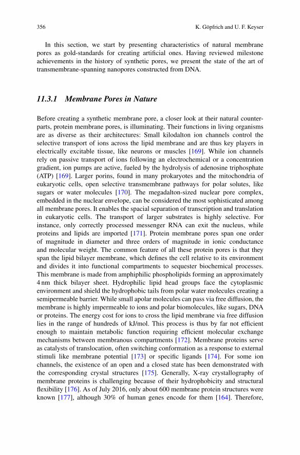
356 K. Göpfrich and U. F. Keyser
In this section, we start by presenting characteristics of natural membranepores as gold-standards for creating artificial ones. Having reviewed milestoneachievements in the history of synthetic pores, we present the state of the art oftransmembrane-spanning nanopores constructed from DNA.
11.3.1 Membrane Pores in Nature
Before creating a synthetic membrane pore, a closer look at their natural counter-parts, protein membrane pores, is illuminating. Their functions in living organismsare as diverse as their architectures: Small kilodalton ion channels control theselective transport of ions across the lipid membrane and are thus key players inelectrically excitable tissue, like neurons or muscles [169]. While ion channelsrely on passive transport of ions following an electrochemical or a concentrationgradient, ion pumps are active, fueled by the hydrolysis of adenosine triphosphate(ATP) [169]. Larger porins, found in many prokaryotes and the mitochondria ofeukaryotic cells, open selective transmembrane pathways for polar solutes, likesugars or water molecules [170]. The megadalton-sized nuclear pore complex,embedded in the nuclear envelope, can be considered the most sophisticated amongall membrane pores. It enables the spacial separation of transcription and translationin eukaryotic cells. The transport of larger substrates is highly selective. Forinstance, only correctly processed messenger RNA can exit the nucleus, whileproteins and lipids are imported [171]. Protein membrane pores span one orderof magnitude in diameter and three orders of magnitude in ionic conductanceand molecular weight. The common feature of all these protein pores is that theyspan the lipid bilayer membrane, which defines the cell relative to its environmentand divides it into functional compartments to sequester biochemical processes.This membrane is made from amphiphilic phospholipids forming an approximately4 nm thick bilayer sheet. Hydrophilic lipid head groups face the cytoplasmicenvironment and shield the hydrophobic tails from polar water molecules creating asemipermeable barrier. While small apolar molecules can pass via free diffusion, themembrane is highly impermeable to ions and polar biomolecules, like sugars, DNAor proteins. The energy cost for ions to cross the lipid membrane via free diffusionlies in the range of hundreds of kJ/mol. This process is thus by far not efficientenough to maintain metabolic function requiring efficient molecular exchangemechanisms between membranous compartments [172]. Membrane proteins serveas catalysts of translocation, often switching conformation as a response to externalstimuli like membrane potential [173] or specific ligands [174]. For some ionchannels, the existence of an open and a closed state has been demonstrated withthe corresponding crystal structures [175]. Generally, X-ray crystallography ofmembrane proteins is challenging because of their hydrophobicity and structuralflexibility [176]. As of July 2016, only about 600 membrane protein structures wereknown [177], although 30% of human genes encode for them [164]. Therefore,
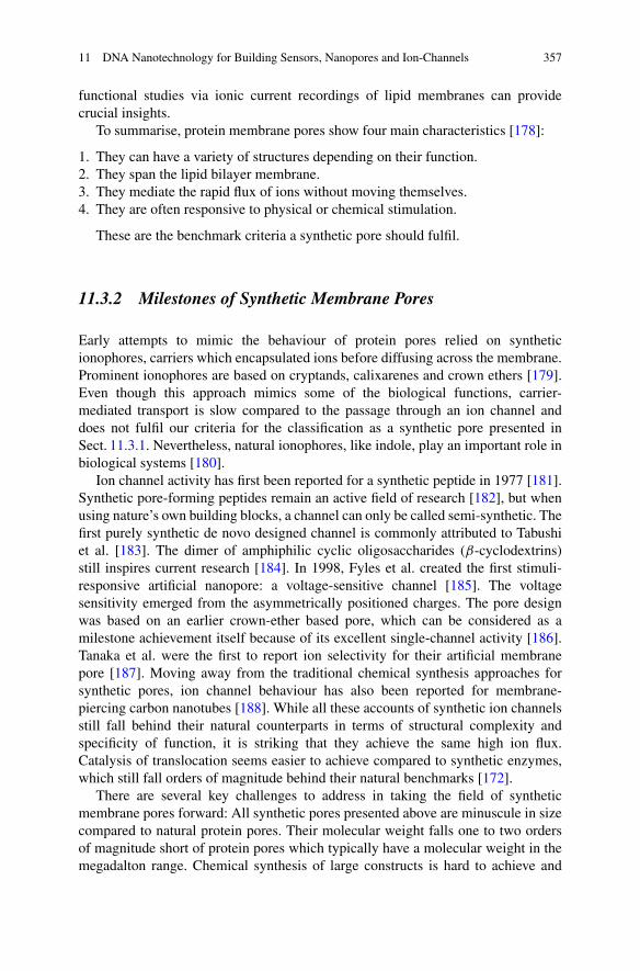
11 DNA Nanotechnology for Building Sensors, Nanopores and Ion-Channels 357
functional studies via ionic current recordings of lipid membranes can providecrucial insights.
To summarise, protein membrane pores show four main characteristics [178]:
1. They can have a variety of structures depending on their function.2. They span the lipid bilayer membrane.3. They mediate the rapid flux of ions without moving themselves.4. They are often responsive to physical or chemical stimulation.
These are the benchmark criteria a synthetic pore should fulfil.
11.3.2 Milestones of Synthetic Membrane Pores
Early attempts to mimic the behaviour of protein pores relied on syntheticionophores, carriers which encapsulated ions before diffusing across the membrane.Prominent ionophores are based on cryptands, calixarenes and crown ethers [179].Even though this approach mimics some of the biological functions, carrier-mediated transport is slow compared to the passage through an ion channel anddoes not fulfil our criteria for the classification as a synthetic pore presented inSect. 11.3.1. Nevertheless, natural ionophores, like indole, play an important role inbiological systems [180].
Ion channel activity has first been reported for a synthetic peptide in 1977 [181].Synthetic pore-forming peptides remain an active field of research [182], but whenusing nature’s own building blocks, a channel can only be called semi-synthetic. Thefirst purely synthetic de novo designed channel is commonly attributed to Tabushiet al. [183]. The dimer of amphiphilic cyclic oligosaccharides (β-cyclodextrins)still inspires current research [184]. In 1998, Fyles et al. created the first stimuli-responsive artificial nanopore: a voltage-sensitive channel [185]. The voltagesensitivity emerged from the asymmetrically positioned charges. The pore designwas based on an earlier crown-ether based pore, which can be considered as amilestone achievement itself because of its excellent single-channel activity [186].Tanaka et al. were the first to report ion selectivity for their artificial membranepore [187]. Moving away from the traditional chemical synthesis approaches forsynthetic pores, ion channel behaviour has also been reported for membrane-piercing carbon nanotubes [188]. While all these accounts of synthetic ion channelsstill fall behind their natural counterparts in terms of structural complexity andspecificity of function, it is striking that they achieve the same high ion flux.Catalysis of translocation seems easier to achieve compared to synthetic enzymes,which still fall orders of magnitude behind their natural benchmarks [172].
There are several key challenges to address in taking the field of syntheticmembrane pores forward: All synthetic pores presented above are minuscule in sizecompared to natural protein pores. Their molecular weight falls one to two ordersof magnitude short of protein pores which typically have a molecular weight in themegadalton range. Chemical synthesis of large constructs is hard to achieve and
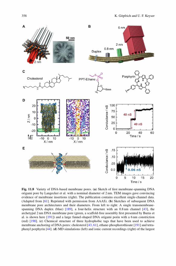
358 K. Göpfrich and U. F. Keyser
Fig. 11.8 Variety of DNA-based membrane pores. (a) Sketch of first membrane-spanning DNAorigami pore by Langecker et al. with a nominal diameter of 2 nm. TEM images gave convincingevidence of membrane insertions (right). The publication contains excellent single-channel data.(Adapted from [61]. Reprinted with permission from AAAS). (b) Sketches of subsequent DNAmembrane pore architectures and their diameters. From left to right: A single transmembrane-spanning DNA duplex (blue) [189], a four-helix structure with an 0.8 nm channel [43], thearchetypal 2 nm DNA membrane pore (green, a scaffold-free assembly first presented by Burns etal. is shown here [191]) and a large funnel-shaped DNA origami porin with a 6 nm constriction(red) [190]. (c) Chemical structure of three hydrophobic tags that have been used to achievemembrane anchoring of DNA pores: cholesterol [43, 61], ethane-phosphorothioate [191] and tetra-phenyl porphyrin [44]. (d) MD simulations (left) and ionic current recordings (right) of the largest
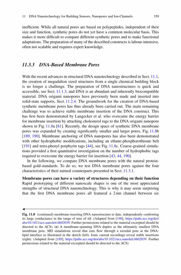
11 DNA Nanotechnology for Building Sensors, Nanopores and Ion-Channels 359
inefficient. While all natural pores are based on polypeptides, independent of theirsize and function, synthetic pores do not yet have a common molecular basis. Thismakes it more difficult to compare different synthetic pores and to make functionaladaptations. The preparation of many of the described constructs is labour-intensive,often not scalable and requires expert knowledge.
11.3.3 DNA-Based Membrane Pores
With the recent advances in structural DNA nanotechnology described in Sect. 11.1,the creation of megadalton sized structures from a single chemical building blockis no longer a challenge. The preparation of DNA nanostructures is quick andaccessible, see Sect. 11.1.3, and DNA is an abundant and inherently biocompatiblematerial. DNA origami nanopores have previously been made and inserted intosolid-state supports, Sect. 11.2.4. The groundwork for the creation of DNA-basedsynthetic membrane pores has thus already been carried out. The main remainingchallenge was to achieve stable membrane insertion of the DNA construct. Thishas first been demonstrated by Langecker et al. who overcame the energy barrierfor membrane insertion by attaching cholesterol tags to the DNA origami nanoporeshown in Fig. 11.8a [61]. Recently, the design space of synthetic DNA membranepores was expanded by creating significantly smaller and larger pores, Fig. 11.8b[189, 190]. Membrane anchoring of DNA nanopores has also been demonstratedwith other hydrophobic modifications, including an ethane-phosphorothioate belt[191] and tetra-phenyl porphyrin tags [44], see Fig. 11.8c. Coarse-grained simula-tions provided a first quantitative investigation on the number of hydrophobic tagsrequired to overcome the energy barrier for insertion [43, 44, 190].
In the following, we compare DNA membrane pores with the natural protein-based gold-standards. To do so, we test DNA membrane pores against the fourcharacteristics of their natural counterparts presented in Sect. 11.3.1.
Membrane pores can have a variety of structures depending on their functionRapid prototyping of different nanoscale shapes is one of the most appreciatedstrengths of structural DNA nanotechnology. This is why it may seem surprisingthat the first DNA membrane pores all featured a 2 nm channel between six
�Fig. 11.8 (continued) membrane-inserting DNA nanostructure to date, independently confirmingits large conductance in the range of tens of nS. (Adapted from [190], https://pubs.acs.org/doi/abs/10.1021/acs.nanolett.6b02039. Further permissions related to the material excerpted should bedirected to the ACS). (e) A membrane-spanning DNA duplex as the ultimately smallest DNAmembrane pore. MD simulations reveal that ions flow through a toroidal pore at the DNA-lipid interface as illustrated in the sketch (left). Ionic current recordings reveal stable insertions(right). (Adapted from [189], https://pubs.acs.org/doi/abs/10.1021/acs.nanolett.6b02039. Furtherpermissions related to the material excerpted should be directed to the ACS)
360 K. Göpfrich and U. F. Keyser
concentrically arranged DNA duplexes [44, 61, 160, 191]. The diameter of sucha pore within the membrane has been confirmed with polyethylene glycol (PEG)-sizing experiments [160]. In the meantime, however, the structural variability ofDNA membrane pores has been demonstrated with the creation of much smalleras well as much larger pores. A DNA channel made from four DNA duplexes hada nominal diameter of 0.8 nm, approaching the dimensions of natural ion channels[43]. Due to its simple design, this DNA channel self-assembled within a minuteat RT. A membrane-inserting DNA origami funnel with a 6 nm construction waspresented as the largest man-made pore to date [190]. It is wider than natural porinsand comparable to the electrical diameter of the nuclear pore complex. Such largearchitectures are difficult to obtain with traditional chemical synthesis described inSect. 11.3.2. Even though the intriguing complexity of natural membrane compo-nents cannot be matched, the diameters of synthetic pores made from DNA spanthe biologically relevant scales. Illustrations of the existing DNA membrane porearchitectures are shown in Fig. 11.8b. Their functional characterisation follows inthe next paragraphs.
Membrane pores span the lipid bilayer membrane TEM imaging, Fig. 11.8a[61], and confocal imaging of fluorescently labelled DNA nanopores [43, 190]revealed their attachment to lipid vesicles. Due to its fluorescent properties,porphyrin has been used for visualisation and membrane anchoring at the sametime [44, 189]. The melting temperature of DNA nanostructures increases uponmembrane insertion [43, 44] and the fluorescence emission of porphyrin shifts [44].It should be noted, however, that these techniques can only confirm membraneattachment – functional studies are required to demonstrate that the DNA poresactually span the lipid bilayer. A study evaluating the relative proportions ofmembrane-spanning versus inactive DNA pores is still lacking.
Membrane pores mediate the rapid flux of ions without moving themselvesSingle-channel ionic current recordings are at the core of membrane pore character-isation – for both natural and synthetic pores. The ionic current passing through asingle channel has for the first time been recorded by Neher and Sackman usingtheir patch-clamp method [192]. A range of techniques have been developed tostudy membrane proteins ex vivo in synthetic membranes providing a simplerenvironment in which uncharacterised channels can be studied in isolation. Allof them rely on the same measurement principle: A lipid membrane is supportedacross an aperture and an electric field is applied across the membrane inducingan ionic current. Magnitude, noise and fluctuations in the recorded ionic currentallow for conclusions on the presence or absence of membrane pores, includingtheir size, stability, and functional properties, such as gating, ligand-binding orion selectivity [193]. Every lipid bilayer experiment starts with the formationof a lipid membrane. These bilayers are gigaohm resistors reducing the flow ofions across the aperture, its formation can be observed as a sudden decrease inthe ionic current. The insertion of a membrane pore, on the other hand, causesthe formation of transmembrane passage for ions and thus a stepwise increasein ionic current. Single-channel recordings of all DNA-based membrane pores
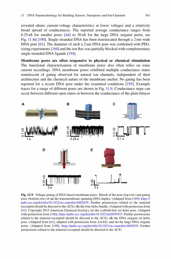
11 DNA Nanotechnology for Building Sensors, Nanopores and Ion-Channels 361
revealed ohmic current-voltage characteristics at lower voltages and a relativelybroad spread of conductances. The reported average conductance ranges from0.25 nS for smaller pores [44] to 30 nS for the large DNA origami porin, seeFig. 11.8d [190]. Single-stranded DNA has been translocated through a 2 nm wideDNA pore [61]. The diameter of such a 2 nm DNA pore was confirmed with PEG-sizing experiments [160] and the ion flux was partially blocked with complementarysingle-stranded DNA ligands [194].
Membrane pores are often responsive to physical or chemical stimulationThe functional characterisation of membrane pores also often relies on ioniccurrent recordings. DNA membrane pores exhibited multiple conductance statesreminiscent of gating observed for natural ion channels, independent of theirarchitecture and the chemical nature of the membrane anchor. No gating has beenreported for a recent DNA pore under the examined conditions [195]. Exampletraces for a range of different pores are shown in Fig. 11.9. Conductance steps canoccur between different open states or between the conductance of the plain bilayer
Fig. 11.9 Voltage-gating of DNA-based membrane pores. Sketch of the pore (top row) and gatingtrace (bottom row) of (a) the transmembrane spanning DNA duplex. (Adapted from [189], https://pubs.acs.org/doi/abs/10.1021/acs.nanolett.6b02039. Further permissions related to the materialexcerpted should be directed to the ACS); (b) the four-helix bundle. (Adapted with permission from[43]. Copyright 2015 American Chemical Society); (c) the scaffold-free six-helix pore. (Adaptedwith permission from [160], https://pubs.acs.org/doi/abs/10.1021/nn5039433. Further permissionsrelated to the material excerpted should be directed to the ACS); (d) the DNA origami six-helixpore. (Adapted from [61], adapted with permission from AAAS), and (e) the large DNA origamiporin. (Adapted from [190], https://pubs.acs.org/doi/abs/10.1021/acs.nanolett.6b02039. Furtherpermissions related to the material excerpted should be directed to the ACS)
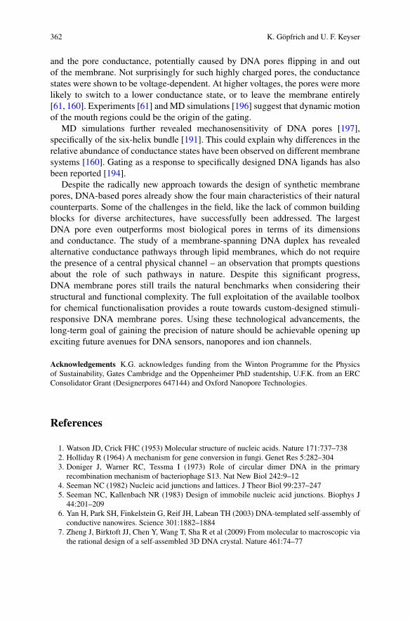
362 K. Göpfrich and U. F. Keyser
and the pore conductance, potentially caused by DNA pores flipping in and outof the membrane. Not surprisingly for such highly charged pores, the conductancestates were shown to be voltage-dependent. At higher voltages, the pores were morelikely to switch to a lower conductance state, or to leave the membrane entirely[61, 160]. Experiments [61] and MD simulations [196] suggest that dynamic motionof the mouth regions could be the origin of the gating.
MD simulations further revealed mechanosensitivity of DNA pores [197],specifically of the six-helix bundle [191]. This could explain why differences in therelative abundance of conductance states have been observed on different membranesystems [160]. Gating as a response to specifically designed DNA ligands has alsobeen reported [194].
Despite the radically new approach towards the design of synthetic membranepores, DNA-based pores already show the four main characteristics of their naturalcounterparts. Some of the challenges in the field, like the lack of common buildingblocks for diverse architectures, have successfully been addressed. The largestDNA pore even outperforms most biological pores in terms of its dimensionsand conductance. The study of a membrane-spanning DNA duplex has revealedalternative conductance pathways through lipid membranes, which do not requirethe presence of a central physical channel – an observation that prompts questionsabout the role of such pathways in nature. Despite this significant progress,DNA membrane pores still trails the natural benchmarks when considering theirstructural and functional complexity. The full exploitation of the available toolboxfor chemical functionalisation provides a route towards custom-designed stimuli-responsive DNA membrane pores. Using these technological advancements, thelong-term goal of gaining the precision of nature should be achievable opening upexciting future avenues for DNA sensors, nanopores and ion channels.
Acknowledgements K.G. acknowledges funding from the Winton Programme for the Physicsof Sustainability, Gates Cambridge and the Oppenheimer PhD studentship, U.F.K. from an ERCConsolidator Grant (Designerpores 647144) and Oxford Nanopore Technologies.
References
1. Watson JD, Crick FHC (1953) Molecular structure of nucleic acids. Nature 171:737–7382. Holliday R (1964) A mechanism for gene conversion in fungi. Genet Res 5:282–3043. Doniger J, Warner RC, Tessma I (1973) Role of circular dimer DNA in the primary
recombination mechanism of bacteriophage S13. Nat New Biol 242:9–124. Seeman NC (1982) Nucleic acid junctions and lattices. J Theor Biol 99:237–2475. Seeman NC, Kallenbach NR (1983) Design of immobile nucleic acid junctions. Biophys J
44:201–2096. Yan H, Park SH, Finkelstein G, Reif JH, Labean TH (2003) DNA-templated self-assembly of
conductive nanowires. Science 301:1882–18847. Zheng J, Birktoft JJ, Chen Y, Wang T, Sha R et al (2009) From molecular to macroscopic via
the rational design of a self-assembled 3D DNA crystal. Nature 461:74–77
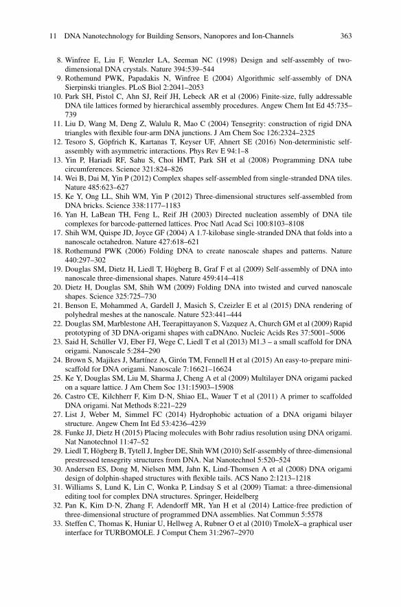
11 DNA Nanotechnology for Building Sensors, Nanopores and Ion-Channels 363
8. Winfree E, Liu F, Wenzler LA, Seeman NC (1998) Design and self-assembly of two-dimensional DNA crystals. Nature 394:539–544
9. Rothemund PWK, Papadakis N, Winfree E (2004) Algorithmic self-assembly of DNASierpinski triangles. PLoS Biol 2:2041–2053
10. Park SH, Pistol C, Ahn SJ, Reif JH, Lebeck AR et al (2006) Finite-size, fully addressableDNA tile lattices formed by hierarchical assembly procedures. Angew Chem Int Ed 45:735–739
11. Liu D, Wang M, Deng Z, Walulu R, Mao C (2004) Tensegrity: construction of rigid DNAtriangles with flexible four-arm DNA junctions. J Am Chem Soc 126:2324–2325
12. Tesoro S, Göpfrich K, Kartanas T, Keyser UF, Ahnert SE (2016) Non-deterministic self-assembly with asymmetric interactions. Phys Rev E 94:1–8
13. Yin P, Hariadi RF, Sahu S, Choi HMT, Park SH et al (2008) Programming DNA tubecircumferences. Science 321:824–826
14. Wei B, Dai M, Yin P (2012) Complex shapes self-assembled from single-stranded DNA tiles.Nature 485:623–627
15. Ke Y, Ong LL, Shih WM, Yin P (2012) Three-dimensional structures self-assembled fromDNA bricks. Science 338:1177–1183
16. Yan H, LaBean TH, Feng L, Reif JH (2003) Directed nucleation assembly of DNA tilecomplexes for barcode-patterned lattices. Proc Natl Acad Sci 100:8103–8108
17. Shih WM, Quispe JD, Joyce GF (2004) A 1.7-kilobase single-stranded DNA that folds into ananoscale octahedron. Nature 427:618–621
18. Rothemund PWK (2006) Folding DNA to create nanoscale shapes and patterns. Nature440:297–302
19. Douglas SM, Dietz H, Liedl T, Högberg B, Graf F et al (2009) Self-assembly of DNA intonanoscale three-dimensional shapes. Nature 459:414–418
20. Dietz H, Douglas SM, Shih WM (2009) Folding DNA into twisted and curved nanoscaleshapes. Science 325:725–730
21. Benson E, Mohammed A, Gardell J, Masich S, Czeizler E et al (2015) DNA rendering ofpolyhedral meshes at the nanoscale. Nature 523:441–444
22. Douglas SM, Marblestone AH, Teerapittayanon S, Vazquez A, Church GM et al (2009) Rapidprototyping of 3D DNA-origami shapes with caDNAno. Nucleic Acids Res 37:5001–5006
23. Said H, Schüller VJ, Eber FJ, Wege C, Liedl T et al (2013) M1.3 – a small scaffold for DNAorigami. Nanoscale 5:284–290
24. Brown S, Majikes J, Martínez A, Girón TM, Fennell H et al (2015) An easy-to-prepare mini-scaffold for DNA origami. Nanoscale 7:16621–16624
25. Ke Y, Douglas SM, Liu M, Sharma J, Cheng A et al (2009) Multilayer DNA origami packedon a square lattice. J Am Chem Soc 131:15903–15908
26. Castro CE, Kilchherr F, Kim D-N, Shiao EL, Wauer T et al (2011) A primer to scaffoldedDNA origami. Nat Methods 8:221–229
27. List J, Weber M, Simmel FC (2014) Hydrophobic actuation of a DNA origami bilayerstructure. Angew Chem Int Ed 53:4236–4239
28. Funke JJ, Dietz H (2015) Placing molecules with Bohr radius resolution using DNA origami.Nat Nanotechnol 11:47–52
29. Liedl T, Högberg B, Tytell J, Ingber DE, Shih WM (2010) Self-assembly of three-dimensionalprestressed tensegrity structures from DNA. Nat Nanotechnol 5:520–524
30. Andersen ES, Dong M, Nielsen MM, Jahn K, Lind-Thomsen A et al (2008) DNA origamidesign of dolphin-shaped structures with flexible tails. ACS Nano 2:1213–1218
31. Williams S, Lund K, Lin C, Wonka P, Lindsay S et al (2009) Tiamat: a three-dimensionalediting tool for complex DNA structures. Springer, Heidelberg
32. Pan K, Kim D-N, Zhang F, Adendorff MR, Yan H et al (2014) Lattice-free prediction ofthree-dimensional structure of programmed DNA assemblies. Nat Commun 5:5578
33. Steffen C, Thomas K, Huniar U, Hellweg A, Rubner O et al (2010) TmoleX–a graphical userinterface for TURBOMOLE. J Comput Chem 31:2967–2970
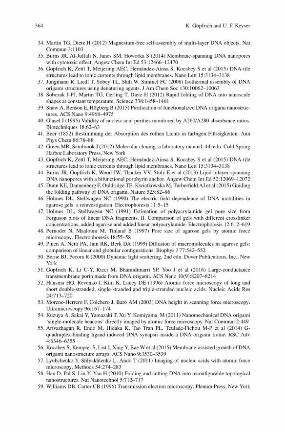
364 K. Göpfrich and U. F. Keyser
34. Martin TG, Dietz H (2012) Magnesium-free self-assembly of multi-layer DNA objects. NatCommun 3:1103
35. Burns JR, Al-Juffali N, Janes SM, Howorka S (2014) Membrane-spanning DNA nanoporeswith cytotoxic effect. Angew Chem Int Ed 53:12466–12470
36. Göpfrich K, Zettl T, Meijering AEC, Hernández-Ainsa S, Kocabey S et al (2015) DNA-tilestructures lead to ionic currents through lipid membranes. Nano Lett 15:3134–3138
37. Jungmann R, Liedl T, Sobey TL, Shih W, Simmel FC (2008) Isothermal assembly of DNAorigami structures using denaturing agents. J Am Chem Soc 130:10062–10063
38. Sobczak J-PJ, Martin TG, Gerling T, Dietz H (2012) Rapid folding of DNA into nanoscaleshapes at constant temperature. Science 338:1458–1461
39. Shaw A, Benson E, Högberg B (2015) Purification of functionalized DNA origami nanostruc-tures. ACS Nano 9:4968–4975
40. Glasel J (1995) Validity of nucleic acid purities monitored by A260/A280 absorbance ratios.Biotechniques 18:62–63
41. Beer (1852) Bestimmung der Absorption des rothen Lichts in farbigen Flüssigkeiten. AnnPhys Chem 86:78–88
42. Green MR, Sambrook J (2012) Molecular cloning: a laboratory manual, 4th edn. Cold SpringHarbor Laboratory Press, New York
43. Göpfrich K, Zettl T, Meijering AEC, Hernández-Ainsa S, Kocabey S et al (2015) DNA-tilestructures lead to ionic currents through lipid membranes. Nano Lett 15:3134–3138
44. Burns JR, Göpfrich K, Wood JW, Thacker VV, Stulz E et al (2013) Lipid-bilayer-spanningDNA nanopores with a bifunctional porphyrin anchor. Angew Chem Int Ed 52:12069–12072
45. Dunn KE, Dannenberg F, Ouldridge TE, Kwiatkowska M, Turberfield AJ et al (2015) Guidingthe folding pathway of DNA origami. Nature 525:82–86
46. Holmes DL, Stellwagen NC (1990) The electric field dependence of DNA mobilities inagarose gels: a reinvestigation. Electrophoresis 11:5–15
47. Holmes DL, Stellwagen NC (1991) Estimation of polyacrylamide gel pore size fromFerguson plots of linear DNA fragments. II. Comparison of gels with different crosslinkerconcentrations, added agarose and added linear polyacrylamide. Electrophoresis 12:612–619
48. Pernodet N, Maaloum M, Tinland B (1997) Pore size of agarose gels by atomic forcemicroscopy. Electrophoresis 18:55–58
49. Pluen A, Netti PA, Jain RK, Berk DA (1999) Diffusion of macromolecules in agarose gels:comparison of linear and globular configurations. Biophys J 77:542–552
50. Berne BJ, Pecora R (2000) Dynamic light scattering, 2nd edn. Dover Publications, Inc., NewYork
51. Göpfrich K, Li C-Y, Ricci M, Bhamidimarri SP, Yoo J et al (2016) Large-conductancetransmembrane porin made from DNA origami. ACS Nano 10(9):8207–8214
52. Hansma HG, Revenko I, Kim K, Laney DE (1996) Atomic force microscopy of long andshort double-stranded, single-stranded and triple-stranded nucleic acids. Nucleic Acids Res24:713–720
53. Moreno-Herrero F, Colchero J, Baró AM (2003) DNA height in scanning force microscopy.Ultramicroscopy 96:167–174
54. Kuzuya A, Sakai Y, Yamazaki T, Xu Y, Komiyama, M (2011) Nanomechanical DNA origami‘single-molecule beacons’ directly imaged by atomic force microscopy. Nat Commun 2:449
55. Arivazhagan R, Endo M, Hidaka K, Tao Tran PL, Teulade-Fichou M-P et al (2014) G-quadruplex-binding ligand-induced DNA synapsis inside a DNA origami frame. RSC Adv4:6346–6355
56. Kocabey S, Kempter S, List J, Xing Y, Bae W et al (2015) Membrane-assisted growth of DNAorigami nanostructure arrays. ACS Nano 9:3530–3539
57. Lyubchenko Y, Shlyakhtenko L, Ando T (2011) Imaging of nucleic acids with atomic forcemicroscopy. Methods 54:274–283
58. Han D, Pal S, Liu Y, Yan H (2010) Folding and cutting DNA into reconfigurable topologicalnanostructures. Nat Nanotechnol 5:712–717
59. Williams DB, Carter CB (1996) Transmission electron microscopy. Plenum Press, New York
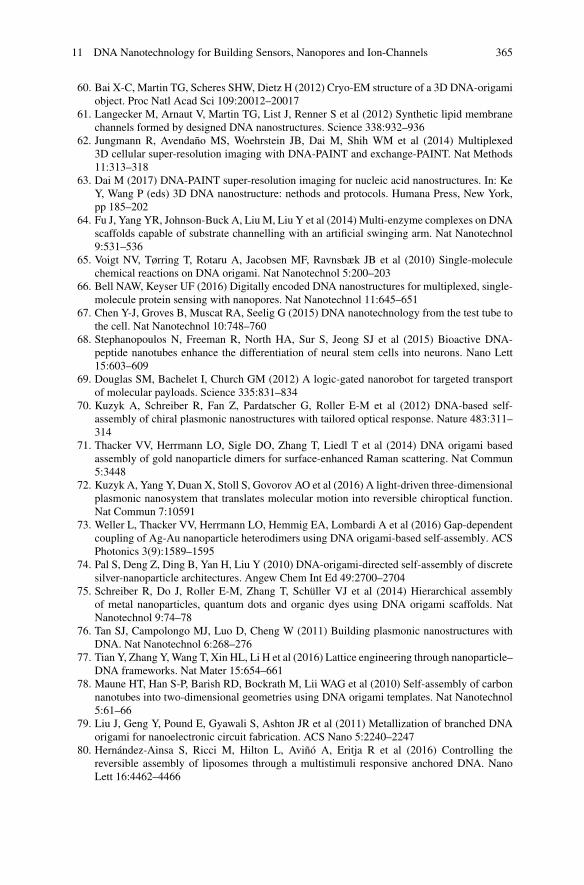
11 DNA Nanotechnology for Building Sensors, Nanopores and Ion-Channels 365
60. Bai X-C, Martin TG, Scheres SHW, Dietz H (2012) Cryo-EM structure of a 3D DNA-origamiobject. Proc Natl Acad Sci 109:20012–20017
61. Langecker M, Arnaut V, Martin TG, List J, Renner S et al (2012) Synthetic lipid membranechannels formed by designed DNA nanostructures. Science 338:932–936
62. Jungmann R, Avendaño MS, Woehrstein JB, Dai M, Shih WM et al (2014) Multiplexed3D cellular super-resolution imaging with DNA-PAINT and exchange-PAINT. Nat Methods11:313–318
63. Dai M (2017) DNA-PAINT super-resolution imaging for nucleic acid nanostructures. In: KeY, Wang P (eds) 3D DNA nanostructure: nethods and protocols. Humana Press, New York,pp 185–202
64. Fu J, Yang YR, Johnson-Buck A, Liu M, Liu Y et al (2014) Multi-enzyme complexes on DNAscaffolds capable of substrate channelling with an artificial swinging arm. Nat Nanotechnol9:531–536
65. Voigt NV, Tørring T, Rotaru A, Jacobsen MF, Ravnsbæk JB et al (2010) Single-moleculechemical reactions on DNA origami. Nat Nanotechnol 5:200–203
66. Bell NAW, Keyser UF (2016) Digitally encoded DNA nanostructures for multiplexed, single-molecule protein sensing with nanopores. Nat Nanotechnol 11:645–651
67. Chen Y-J, Groves B, Muscat RA, Seelig G (2015) DNA nanotechnology from the test tube tothe cell. Nat Nanotechnol 10:748–760
68. Stephanopoulos N, Freeman R, North HA, Sur S, Jeong SJ et al (2015) Bioactive DNA-peptide nanotubes enhance the differentiation of neural stem cells into neurons. Nano Lett15:603–609
69. Douglas SM, Bachelet I, Church GM (2012) A logic-gated nanorobot for targeted transportof molecular payloads. Science 335:831–834
70. Kuzyk A, Schreiber R, Fan Z, Pardatscher G, Roller E-M et al (2012) DNA-based self-assembly of chiral plasmonic nanostructures with tailored optical response. Nature 483:311–314
71. Thacker VV, Herrmann LO, Sigle DO, Zhang T, Liedl T et al (2014) DNA origami basedassembly of gold nanoparticle dimers for surface-enhanced Raman scattering. Nat Commun5:3448
72. Kuzyk A, Yang Y, Duan X, Stoll S, Govorov AO et al (2016) A light-driven three-dimensionalplasmonic nanosystem that translates molecular motion into reversible chiroptical function.Nat Commun 7:10591
73. Weller L, Thacker VV, Herrmann LO, Hemmig EA, Lombardi A et al (2016) Gap-dependentcoupling of Ag-Au nanoparticle heterodimers using DNA origami-based self-assembly. ACSPhotonics 3(9):1589–1595
74. Pal S, Deng Z, Ding B, Yan H, Liu Y (2010) DNA-origami-directed self-assembly of discretesilver-nanoparticle architectures. Angew Chem Int Ed 49:2700–2704
75. Schreiber R, Do J, Roller E-M, Zhang T, Schüller VJ et al (2014) Hierarchical assemblyof metal nanoparticles, quantum dots and organic dyes using DNA origami scaffolds. NatNanotechnol 9:74–78
76. Tan SJ, Campolongo MJ, Luo D, Cheng W (2011) Building plasmonic nanostructures withDNA. Nat Nanotechnol 6:268–276
77. Tian Y, Zhang Y, Wang T, Xin HL, Li H et al (2016) Lattice engineering through nanoparticle–DNA frameworks. Nat Mater 15:654–661
78. Maune HT, Han S-P, Barish RD, Bockrath M, Lii WAG et al (2010) Self-assembly of carbonnanotubes into two-dimensional geometries using DNA origami templates. Nat Nanotechnol5:61–66
79. Liu J, Geng Y, Pound E, Gyawali S, Ashton JR et al (2011) Metallization of branched DNAorigami for nanoelectronic circuit fabrication. ACS Nano 5:2240–2247
80. Hernández-Ainsa S, Ricci M, Hilton L, Aviñó A, Eritja R et al (2016) Controlling thereversible assembly of liposomes through a multistimuli responsive anchored DNA. NanoLett 16:4462–4466
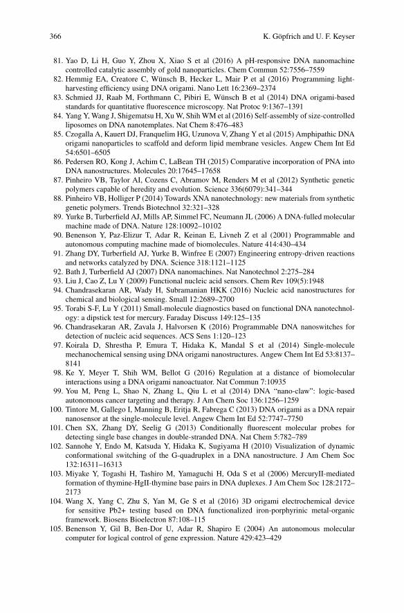
366 K. Göpfrich and U. F. Keyser
81. Yao D, Li H, Guo Y, Zhou X, Xiao S et al (2016) A pH-responsive DNA nanomachinecontrolled catalytic assembly of gold nanoparticles. Chem Commun 52:7556–7559
82. Hemmig EA, Creatore C, Wünsch B, Hecker L, Mair P et al (2016) Programming light-harvesting efficiency using DNA origami. Nano Lett 16:2369–2374
83. Schmied JJ, Raab M, Forthmann C, Pibiri E, Wünsch B et al (2014) DNA origami-basedstandards for quantitative fluorescence microscopy. Nat Protoc 9:1367–1391
84. Yang Y, Wang J, Shigematsu H, Xu W, Shih WM et al (2016) Self-assembly of size-controlledliposomes on DNA nanotemplates. Nat Chem 8:476–483
85. Czogalla A, Kauert DJ, Franquelim HG, Uzunova V, Zhang Y et al (2015) Amphipathic DNAorigami nanoparticles to scaffold and deform lipid membrane vesicles. Angew Chem Int Ed54:6501–6505
86. Pedersen RO, Kong J, Achim C, LaBean TH (2015) Comparative incorporation of PNA intoDNA nanostructures. Molecules 20:17645–17658
87. Pinheiro VB, Taylor AI, Cozens C, Abramov M, Renders M et al (2012) Synthetic geneticpolymers capable of heredity and evolution. Science 336(6079):341–344
88. Pinheiro VB, Holliger P (2014) Towards XNA nanotechnology: new materials from syntheticgenetic polymers. Trends Biotechnol 32:321–328
89. Yurke B, Turberfield AJ, Mills AP, Simmel FC, Neumann JL (2006) A DNA-fulled molecularmachine made of DNA. Nature 128:10092–10102
90. Benenson Y, Paz-Elizur T, Adar R, Keinan E, Livneh Z et al (2001) Programmable andautonomous computing machine made of biomolecules. Nature 414:430–434
91. Zhang DY, Turberfield AJ, Yurke B, Winfree E (2007) Engineering entropy-driven reactionsand networks catalyzed by DNA. Science 318:1121–1125
92. Bath J, Turberfield AJ (2007) DNA nanomachines. Nat Nanotechnol 2:275–28493. Liu J, Cao Z, Lu Y (2009) Functional nucleic acid sensors. Chem Rev 109(5):194894. Chandrasekaran AR, Wady H, Subramanian HKK (2016) Nucleic acid nanostructures for
chemical and biological sensing. Small 12:2689–270095. Torabi S-F, Lu Y (2011) Small-molecule diagnostics based on functional DNA nanotechnol-
ogy: a dipstick test for mercury. Faraday Discuss 149:125–13596. Chandrasekaran AR, Zavala J, Halvorsen K (2016) Programmable DNA nanoswitches for
detection of nucleic acid sequences. ACS Sens 1:120–12397. Koirala D, Shrestha P, Emura T, Hidaka K, Mandal S et al (2014) Single-molecule
mechanochemical sensing using DNA origami nanostructures. Angew Chem Int Ed 53:8137–8141
98. Ke Y, Meyer T, Shih WM, Bellot G (2016) Regulation at a distance of biomolecularinteractions using a DNA origami nanoactuator. Nat Commun 7:10935
99. You M, Peng L, Shao N, Zhang L, Qiu L et al (2014) DNA “nano-claw”: logic-basedautonomous cancer targeting and therapy. J Am Chem Soc 136:1256–1259
100. Tintore M, Gallego I, Manning B, Eritja R, Fabrega C (2013) DNA origami as a DNA repairnanosensor at the single-molecule level. Angew Chem Int Ed 52:7747–7750
101. Chen SX, Zhang DY, Seelig G (2013) Conditionally fluorescent molecular probes fordetecting single base changes in double-stranded DNA. Nat Chem 5:782–789
102. Sannohe Y, Endo M, Katsuda Y, Hidaka K, Sugiyama H (2010) Visualization of dynamicconformational switching of the G-quadruplex in a DNA nanostructure. J Am Chem Soc132:16311–16313
103. Miyake Y, Togashi H, Tashiro M, Yamaguchi H, Oda S et al (2006) MercuryII-mediatedformation of thymine-HgII-thymine base pairs in DNA duplexes. J Am Chem Soc 128:2172–2173
104. Wang X, Yang C, Zhu S, Yan M, Ge S et al (2016) 3D origami electrochemical devicefor sensitive Pb2+ testing based on DNA functionalized iron-porphyrinic metal-organicframework. Biosens Bioelectron 87:108–115
105. Benenson Y, Gil B, Ben-Dor U, Adar R, Shapiro E (2004) An autonomous molecularcomputer for logical control of gene expression. Nature 429:423–429
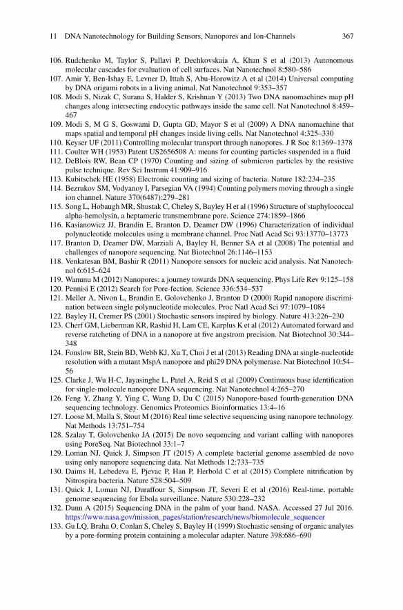
11 DNA Nanotechnology for Building Sensors, Nanopores and Ion-Channels 367
106. Rudchenko M, Taylor S, Pallavi P, Dechkovskaia A, Khan S et al (2013) Autonomousmolecular cascades for evaluation of cell surfaces. Nat Nanotechnol 8:580–586
107. Amir Y, Ben-Ishay E, Levner D, Ittah S, Abu-Horowitz A et al (2014) Universal computingby DNA origami robots in a living animal. Nat Nanotechnol 9:353–357
108. Modi S, Nizak C, Surana S, Halder S, Krishnan Y (2013) Two DNA nanomachines map pHchanges along intersecting endocytic pathways inside the same cell. Nat Nanotechnol 8:459–467
109. Modi S, M G S, Goswami D, Gupta GD, Mayor S et al (2009) A DNA nanomachine thatmaps spatial and temporal pH changes inside living cells. Nat Nanotechnol 4:325–330
110. Keyser UF (2011) Controlling molecular transport through nanopores. J R Soc 8:1369–1378111. Coulter WH (1953) Patent US2656508 A: means for counting particles suspended in a fluid112. DeBlois RW, Bean CP (1970) Counting and sizing of submicron particles by the resistive
pulse technique. Rev Sci Instrum 41:909–916113. Kubitschek HE (1958) Electronic counting and sizing of bacteria. Nature 182:234–235114. Bezrukov SM, Vodyanoy I, Parsegian VA (1994) Counting polymers moving through a single
ion channel. Nature 370(6487):279–281115. Song L, Hobaugh MR, Shustak C, Cheley S, Bayley H et al (1996) Structure of staphylococcal
alpha-hemolysin, a heptameric transmembrane pore. Science 274:1859–1866116. Kasianowicz JJ, Brandin E, Branton D, Deamer DW (1996) Characterization of individual
polynucleotide molecules using a membrane channel. Proc Natl Acad Sci 93:13770–13773117. Branton D, Deamer DW, Marziali A, Bayley H, Benner SA et al (2008) The potential and
challenges of nanopore sequencing. Nat Biotechnol 26:1146–1153118. Venkatesan BM, Bashir R (2011) Nanopore sensors for nucleic acid analysis. Nat Nanotech-
nol 6:615–624119. Wanunu M (2012) Nanopores: a journey towards DNA sequencing. Phys Life Rev 9:125–158120. Pennisi E (2012) Search for Pore-fection. Science 336:534–537121. Meller A, Nivon L, Brandin E, Golovchenko J, Branton D (2000) Rapid nanopore discrimi-
nation between single polynucleotide molecules. Proc Natl Acad Sci 97:1079–1084122. Bayley H, Cremer PS (2001) Stochastic sensors inspired by biology. Nature 413:226–230123. Cherf GM, Lieberman KR, Rashid H, Lam CE, Karplus K et al (2012) Automated forward and
reverse ratcheting of DNA in a nanopore at five angstrom precision. Nat Biotechnol 30:344–348
124. Fonslow BR, Stein BD, Webb KJ, Xu T, Choi J et al (2013) Reading DNA at single-nucleotideresolution with a mutant MspA nanopore and phi29 DNA polymerase. Nat Biotechnol 10:54–56
125. Clarke J, Wu H-C, Jayasinghe L, Patel A, Reid S et al (2009) Continuous base identificationfor single-molecule nanopore DNA sequencing. Nat Nanotechnol 4:265–270
126. Feng Y, Zhang Y, Ying C, Wang D, Du C (2015) Nanopore-based fourth-generation DNAsequencing technology. Genomics Proteomics Bioinformatics 13:4–16
127. Loose M, Malla S, Stout M (2016) Real time selective sequencing using nanopore technology.Nat Methods 13:751–754
128. Szalay T, Golovchenko JA (2015) De novo sequencing and variant calling with nanoporesusing PoreSeq. Nat Biotechnol 33:1–7
129. Loman NJ, Quick J, Simpson JT (2015) A complete bacterial genome assembled de novousing only nanopore sequencing data. Nat Methods 12:733–735
130. Daims H, Lebedeva E, Pjevac P, Han P, Herbold C et al (2015) Complete nitrification byNitrospira bacteria. Nature 528:504–509
131. Quick J, Loman NJ, Duraffour S, Simpson JT, Severi E et al (2016) Real-time, portablegenome sequencing for Ebola surveillance. Nature 530:228–232
132. Dunn A (2015) Sequencing DNA in the palm of your hand. NASA. Accessed 27 Jul 2016.https://www.nasa.gov/mission_pages/station/research/news/biomolecule_sequencer
133. Gu LQ, Braha O, Conlan S, Cheley S, Bayley H (1999) Stochastic sensing of organic analytesby a pore-forming protein containing a molecular adapter. Nature 398:686–690
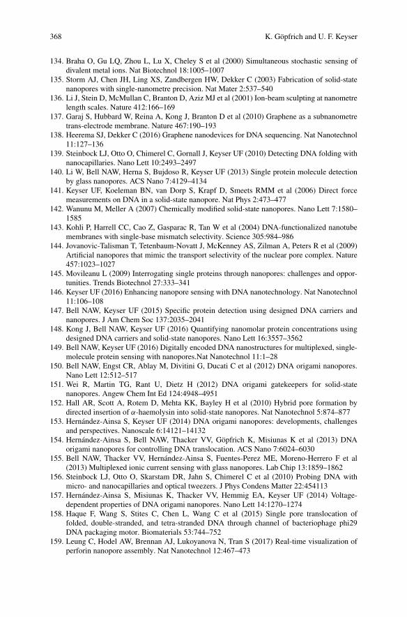
368 K. Göpfrich and U. F. Keyser
134. Braha O, Gu LQ, Zhou L, Lu X, Cheley S et al (2000) Simultaneous stochastic sensing ofdivalent metal ions. Nat Biotechnol 18:1005–1007
135. Storm AJ, Chen JH, Ling XS, Zandbergen HW, Dekker C (2003) Fabrication of solid-statenanopores with single-nanometre precision. Nat Mater 2:537–540
136. Li J, Stein D, McMullan C, Branton D, Aziz MJ et al (2001) Ion-beam sculpting at nanometrelength scales. Nature 412:166–169
137. Garaj S, Hubbard W, Reina A, Kong J, Branton D et al (2010) Graphene as a subnanometretrans-electrode membrane. Nature 467:190–193
138. Heerema SJ, Dekker C (2016) Graphene nanodevices for DNA sequencing. Nat Nanotechnol11:127–136
139. Steinbock LJ, Otto O, Chimerel C, Gornall J, Keyser UF (2010) Detecting DNA folding withnanocapillaries. Nano Lett 10:2493–2497
140. Li W, Bell NAW, Herna S, Bujdoso R, Keyser UF (2013) Single protein molecule detectionby glass nanopores. ACS Nano 7:4129–4134
141. Keyser UF, Koeleman BN, van Dorp S, Krapf D, Smeets RMM et al (2006) Direct forcemeasurements on DNA in a solid-state nanopore. Nat Phys 2:473–477
142. Wanunu M, Meller A (2007) Chemically modified solid-state nanopores. Nano Lett 7:1580–1585
143. Kohli P, Harrell CC, Cao Z, Gasparac R, Tan W et al (2004) DNA-functionalized nanotubemembranes with single-base mismatch selectivity. Science 305:984–986
144. Jovanovic-Talisman T, Tetenbaum-Novatt J, McKenney AS, Zilman A, Peters R et al (2009)Artificial nanopores that mimic the transport selectivity of the nuclear pore complex. Nature457:1023–1027
145. Movileanu L (2009) Interrogating single proteins through nanopores: challenges and oppor-tunities. Trends Biotechnol 27:333–341
146. Keyser UF (2016) Enhancing nanopore sensing with DNA nanotechnology. Nat Nanotechnol11:106–108
147. Bell NAW, Keyser UF (2015) Specific protein detection using designed DNA carriers andnanopores. J Am Chem Soc 137:2035–2041
148. Kong J, Bell NAW, Keyser UF (2016) Quantifying nanomolar protein concentrations usingdesigned DNA carriers and solid-state nanopores. Nano Lett 16:3557–3562
149. Bell NAW, Keyser UF (2016) Digitally encoded DNA nanostructures for multiplexed, single-molecule protein sensing with nanopores.Nat Nanotechnol 11:1–28
150. Bell NAW, Engst CR, Ablay M, Divitini G, Ducati C et al (2012) DNA origami nanopores.Nano Lett 12:512–517
151. Wei R, Martin TG, Rant U, Dietz H (2012) DNA origami gatekeepers for solid-statenanopores. Angew Chem Int Ed 124:4948–4951
152. Hall AR, Scott A, Rotem D, Mehta KK, Bayley H et al (2010) Hybrid pore formation bydirected insertion of α-haemolysin into solid-state nanopores. Nat Nanotechnol 5:874–877
153. Hernández-Ainsa S, Keyser UF (2014) DNA origami nanopores: developments, challengesand perspectives. Nanoscale 6:14121–14132
154. Hernández-Ainsa S, Bell NAW, Thacker VV, Göpfrich K, Misiunas K et al (2013) DNAorigami nanopores for controlling DNA translocation. ACS Nano 7:6024–6030
155. Bell NAW, Thacker VV, Hernández-Ainsa S, Fuentes-Perez ME, Moreno-Herrero F et al(2013) Multiplexed ionic current sensing with glass nanopores. Lab Chip 13:1859–1862
156. Steinbock LJ, Otto O, Skarstam DR, Jahn S, Chimerel C et al (2010) Probing DNA withmicro- and nanocapillaries and optical tweezers. J Phys Condens Matter 22:454113
157. Hernández-Ainsa S, Misiunas K, Thacker VV, Hemmig EA, Keyser UF (2014) Voltage-dependent properties of DNA origami nanopores. Nano Lett 14:1270–1274
158. Haque F, Wang S, Stites C, Chen L, Wang C et al (2015) Single pore translocation offolded, double-stranded, and tetra-stranded DNA through channel of bacteriophage phi29DNA packaging motor. Biomaterials 53:744–752
159. Leung C, Hodel AW, Brennan AJ, Lukoyanova N, Tran S (2017) Real-time visualization ofperforin nanopore assembly. Nat Nanotechnol 12:467–473
11 DNA Nanotechnology for Building Sensors, Nanopores and Ion-Channels 369
160. Seifert A, Göpfrich K, Burns JR, Fertig N, Keyser UF et al (2014) Bilayer-spanning DNAnanopores with voltage-switching between open and closed state. ACS Nano 9:1117–1126
161. Bell NAW, Keyser UF (2014) Nanopores formed by DNA origami: a review. FEBS Lett588:3564–3570
162. Li CY, Hemmig EA, Kong J, Yoo J, Keyser UF et al (2015) Ionic conductivity, structuraldeformation and programmable anisotropy of DNA origami in electric field. ACS Nano9:1420–1433
163. Plesa C, Ananth AN, Linko V, Gulcher C, Katan A et al (2014) Ionic permeability andmechanical properties of DNA origami nanoplates. ACS Nano 8:35–43
164. Baker M (2010) Making membrane proteins for structures: a trillion tiny tweaks. Nat Methods7:429–434
165. Knowles MR, Stutts MJ, Spock A, Fischer N, Gatzy JT et al (1983) Abnormal ion permeationthrough cystic fibrosis. Science 221:1067–1070
166. Feske S (2007) Calcium signalling in lymphocyte activation and disease. Nat Rev Immunol7:690–702
167. Fernandez-Lopez S, Kim HS, Choi EC, Delgado M, Granja JR et al (2001) Antibacterialagents based on the cyclic D,L-alpha-peptide architecture. Nature 412:452–455
168. Czogalla A, Franquelim HG, Schwille P (2016) DNA nanostructures on membranes as toolsfor synthetic biology. Biophys J 110:1698–1707
169. Hille B (2001) Ion channels of excitable membranes. Palgrave Macmillan, Sunderland170. Benz R (ed) (2004) Bacterial and eukaryotic porins WILEY-VCH Verlag GmbH & Co KGaA,
Weinheim171. Rout MP, Aitchison JD, Suprapto A, Hjertaas K, Zhao Y et al (2000) The yeast nuclear pore
complex. J Cell Biol 148:635–652172. Fyles TM (2007) Synthetic ion channels in bilayer membranes. Chem Soc Rev 36:335–347173. Doyle DA, Morais Cabral J, Pfuetzner RA, Kuo A, Gulbis JM et al (1998) The structure of
the potassium channel: molecular basis of K+ conduction and selectivity. Science 280:69–77174. Hucho F, Weise C (2001) Ligand-gated ion channels. Angew Chem Int Ed 40:3100–3116175. Jiang Y, Lee A, Chen J, Cadene M, Chait BT et al (2002) The open pore conformation of
potassium channels. Nature 417:523–546176. Carpenter EP, Beis K, Cameron AD, Iwata S (2008) Overcoming the challenges of membrane
protein crystallography. Curr Opinion Struct Biol 18:581–586177. White SH (1998–2019) Membrane proteins of known 3D structure, Stephen White laboratory,
UC Irvine. Accessed 27 Jul 2016. https://blanco.biomol.uci.edu/mpstruc/178. Sakai N, Matile S (2013) Synthetic ion channels. Langmuir 29:9031–9040179. Pedersen CJ (1988) The discovery of crown ethers (Nobel address). Angew Chem Int Ed
27:1021–1027180. Chimerel C, Field CM, Piñero-Fernandez S, Keyser UF, Summers DK (2012) Indole prevents
Escherichia coli cell division by modulating membrane potential. Biochim Biophys ActaBiomembr 1818:1590–1594
181. Kennedy SJ, Roeske RW, Freeman AR, Watanabe AM, Besche HR (1977) Synthetic peptidesform ion channels in artificial lipid bilayer membranes. Science 196:1341–1342
182. Wallace DP, Tomich JM, Eppler JW, Iwamoto T, Grantham JJ et al (2000) A syntheticchannel-forming peptide induces Cl- secretion: modulation by Ca2+-dependent K+ channels.Biochim Biophys Acta Biomembr 1464:69–82
183. Tabushi I, Kuroda Y, Yokota K (1982) A,B,D,F-tetrasubstituted β-cyclodextrin as artificialchannel compound. Tetrahedron Lett 23:4601–4604
184. Mamad-Hemouch H, Ramoul H, Abou Taha M, Bacri L, Huin C et al (2015) Biomimeticnanotubes based on cyclodextrins for ion-channel applications. Nano Lett 15:7748–7754
185. Fyles TM, Loock D, Zhou X (1998) A voltage-gated ion channel based on a bis-macrocyclicbolaamphiphile. J Am Chem Soc 7863:2997–3003
186. Carmichael VE, Dutton PJ, Fyles TM, James TD, Swan JA et al (1989) Biomimetic iontransport: a functional model of a unimolecular ion channel. J Am Chem Soc 111(2):767–769
370 K. Göpfrich and U. F. Keyser
187. Tanaka Y, Kobuke Y, Sokabe M (1995) A non-peptidic ion channel with K+ selectivity.Angew Chem Int Ed 34:693–694
188. Geng J, Kim K, Zhang J, Escalada A, Tunuguntla R et al (2014) Stochastic transport throughcarbon nanotubes in lipid bilayers and live cell membranes. Nature 514:612–615
189. Göpfrich K, Li C-Y, Mames I, Bhamidimarri SP, Ricci M et al (2016) Ion channels made froma single membrane-spanning DNA duplex. Nano Lett 16:4665–4669
190. Göpfrich K, Li C-Y, Ricci M, Bhamidimarri SP, Yoo J et al (2016) Large-conductancetransmembrane porin made from DNA origami. ACS Nano 10:8207–8214
191. Burns JR, Stulz E, Howorka S (2013) Self-assembled DNA nanopores that span lipid bilayers.Nano Lett 13:2351–2356
192. Neher E, Sakmann B (1976) Single-channel currents recorded from membrane of denervatedfrog muscle fibres. Nature 260:799–802
193. Gutsmann T, Heimburg T, Keyser U, Mahendran KR, Winterhalter M (2015) Protein recon-stitution into freestanding planar lipid membranes for electrophysiological characterization.Nat Protoc 10:188–198
194. Burns JR, Seifert A, Fertig N, Howorka S (2016) A biomimetic DNA-based channel for theligand-controlled transport of charged molecular cargo across a biological membrane. NatNanotechnol 11:152–156
195. Krishnan S, Ziegler D, Arnaut V, Martin TG, Kapsner K et al (2016) Molecular transportthrough large-diameter DNA nanopores. Nat Commun 7:12787
196. Maingi V, Lelimousi M, Howorka S, Sansom MSP (2015) Gating-like motions and wallporosity in a DNA nanopore scaffold revealed by molecular simulations. ACS Nano 9:11209–11217
197. Yoo J, Aksimentiev A (2015) Molecular dynamics of membrane-spanning DNA channels:conductance mechanism, electro-osmotic transport, and mechanical gating. J Phys Chem Lett6:4680–4687
Chapter 12Bio Mimicking of Extracellular Matrix
Moumita Ghosh, Michal Halperin-Sternfeld, and Lihi Adler-Abramovich
Abstract Biomaterials play a critical role in regenerative strategies such as stemcell-based therapies and tissue engineering, aiming to replace, remodel, regenerate,or support damaged tissues and organs. The design of appropriate three-dimensional(3D) scaffolds is crucial for generating bio-inspired replacement tissues. Thesescaffolds are primarily composed of degradable or non-degradable biomaterials andcan be employed as cells, growth factors, or drug carriers. Naturally derived andsynthetic biomaterials have been widely used for these purposes, but the ideal bio-material remains to be found. Researchers from diversified fields have attempted todesign and fabricate novel biomaterials, aiming to find novel theranostic approachesfor tissue engineering and regenerative medicine. Since no single biomaterial hasbeen found to possess all the necessary characteristics for an ideal performance, overthe years scientists have tried to develop composite biomaterials that complementand combine the beneficial properties of multiple materials into a superior matrix.Herein, we highlight the structural features and performance of various biomaterialsand their application in regenerative medicine and for enhanced tissue engineeringapproaches.
Keywords Biomaterials · Extracellular matrix · Scaffolds · Peptides ·Hydrogels · Supramolecular polymers
Abbreviations
3D Three-dimensionalAl AluminabFGF Basic fibroblast growth factor
M. Ghosh · M. Halperin-Sternfeld · L. Adler-Abramovich (�)Department of Oral Biology, The Goldschleger School of Dental Medicine, Sackler Faculty ofMedicine, Tel Aviv University, Tel Aviv, Israele-mail: [email protected]
© Springer Nature Singapore Pte Ltd. 2019S. Perrett et al. (eds.), Biological and Bio-inspired Nanomaterials,Advances in Experimental Medicine and Biology 1174,https://doi.org/10.1007/978-981-13-9791-2_12
371
372 M. Ghosh et al.
BMP-2 Bone morphogenic protein 2CaP Calcium phosphateDOPA 3,4-dihydroxy-L-phenylalanineECM Extracellular matrixFmoc FluorenylmethyloxycarbonylFmocFF Fluorenylmethoxycarbonyl-diphenylalanineGAGs GlycosaminoglycansGDNF Glial cell derived neurotrophic factorMg MagnesiumMn ManganeseMSCs Mesenchymal stem cellsNa SodiumPCL Poly(e-caprolactone)PEG Polyethylene glycolPET Polyethylene terephthalatePGA Polyglycolic acidPGs ProteoglycansPLA Polylactic acidPLGA Polylactic-co-glycolidePMMA Poly(methylmethacrylate)PTFE Poly(tetrafluoroethylene)PU PolyurethanesRP Rapid prototypingSi SiliconTCP Tricalcium phosphateTi TitaniumUV UltravioletVEGF Vascular endothelial growth factorZn ZincZr Zirconium
12.1 Introduction
The term “biomaterial” was initially defined in the first Consensus Conference ofthe European Society for Biomaterials (ESB) in 1976 as “a nonviable materialused in a medical device, intended to interact with biological systems”. However, itwas later modified by the ESB and is currently defined as “a material intended tointerface with biological systems to evaluate, treat, augment or replace any tissue,organ or function of the body”. The change in the definition indicates how thefield of biomaterials has evolved over the years, from merely interacting with thebody to influencing various biological processes, aimed at tissue regeneration. Analternative definition of a biomaterial is a substance that has been engineered to takea form which, alone or as part of a complex system, is used to direct, by controllinginteractions with components of living systems, the course of any therapeutic ordiagnostic procedure, in human or veterinary medicine [1].
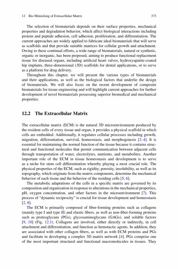
12 Bio Mimicking of Extracellular Matrix 373
The selection of biomaterials depends on their surface properties, mechanicalproperties and degradation behavior, which affect biological interactions includingprotein and peptide adhesion, cell adhesion, proliferation, and differentiation. Thecurrent approaches are widely applied to fabricate ideal biomaterials that will serveas scaffolds and that provide suitable matrices for cellular growth and attachment.Owing to these continual efforts, a wide range of biomaterials, natural or synthetic,organic or inorganic, has been proposed, aiming to produce functional replacementtissue for diseased organs, including artificial heart valves, hydroxyapatite-coatedhip implants, three-dimensional (3D) scaffolds for dental applications, or to serveas a platform for drug delivery.
Throughout this chapter, we will present the various types of biomaterialsand their applications, as well as the biological factors that underlie the designof biomaterials. We will also focus on the recent development of compositebiomaterials for tissue engineering and will highlight current approaches for furtherdevelopment of novel biomaterials possessing superior biomedical and mechanicalproperties.
12.2 The Extracellular Matrix
The extracellular matrix (ECM) is the natural 3D microenvironment produced bythe resident cells of every tissue and organ; it provides a physical scaffold in whichcells are embedded. Additionally, it regulates cellular processes including growth,migration, differentiation, survival, homeostasis, and morphogenesis [2–4]. It isessential for maintaining the normal function of the tissue because it contains struc-tural and functional molecules that permit communication between adjacent cellsthrough transportation of water, electrolytes, nutrients, and metabolites. Anotherimportant role of the ECM in tissue homeostasis and development is to serveas a niche for stem cell differentiation whereby playing a most crucial role. Thephysical properties of the ECM, such as rigidity, porosity, insolubility, as well as itstopography, which originate from the matrix components, determine the mechanicalbehavior of each tissue and the behavior of the residing cells [5, 6].
The metabolic adaptations of the cells in a specific matrix are governed by itscomposition and organization in response to alterations in the mechanical properties,pH, oxygen concentration, and other factors in the microenvironment [7]. Thisprocess of “dynamic reciprocity” is crucial for tissue development and homeostasis[2, 8].
The ECM is primarily composed of fiber-forming proteins such as collagens(mainly type I and type II) and elastic fibers, as well as non-fiber-forming proteinssuch as proteoglycans (PGs), glycosaminoglycans (GAGs), and soluble factors[9, 10] (Fig. 12.1). Collagens are involved, either directly or indirectly, in cellattachment and differentiation, and function as hemotactic agents. In addition, theyare associated with other collagen fibers, as well as with ECM proteins and PGsand facilitate in developing a complex 3D matrix network [4]. PGs comprise oneof the most important structural and functional macromolecules in tissues. They
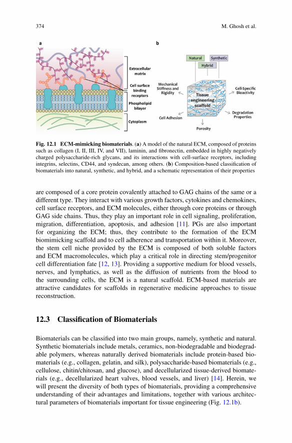
374 M. Ghosh et al.
Fig. 12.1 ECM-mimicking biomaterials. (a) A model of the natural ECM, composed of proteinssuch as collagen (I, II, III, IV, and VII), laminin, and fibronectin, embedded in highly negativelycharged polysaccharide-rich glycans, and its interactions with cell-surface receptors, includingintegrins, selectins, CD44, and syndecan, among others. (b) Composition-based classification ofbiomaterials into natural, synthetic, and hybrid, and a schematic representation of their properties
are composed of a core protein covalently attached to GAG chains of the same or adifferent type. They interact with various growth factors, cytokines and chemokines,cell surface receptors, and ECM molecules, either through core proteins or throughGAG side chains. Thus, they play an important role in cell signaling, proliferation,migration, differentiation, apoptosis, and adhesion [11]. PGs are also importantfor organizing the ECM; thus, they contribute to the formation of the ECMbiomimicking scaffold and to cell adherence and transportation within it. Moreover,the stem cell niche provided by the ECM is composed of both soluble factorsand ECM macromolecules, which play a critical role in directing stem/progenitorcell differentiation fate [12, 13]. Providing a supportive medium for blood vessels,nerves, and lymphatics, as well as the diffusion of nutrients from the blood tothe surrounding cells, the ECM is a natural scaffold. ECM-based materials areattractive candidates for scaffolds in regenerative medicine approaches to tissuereconstruction.
12.3 Classification of Biomaterials
Biomaterials can be classified into two main groups, namely, synthetic and natural.Synthetic biomaterials include metals, ceramics, non-biodegradable and biodegrad-able polymers, whereas naturally derived biomaterials include protein-based bio-materials (e.g., collagen, gelatin, and silk), polysaccharide-based biomaterials (e.g.,cellulose, chitin/chitosan, and glucose), and decellularized tissue-derived biomate-rials (e.g., decellularized heart valves, blood vessels, and liver) [14]. Herein, wewill present the diversity of both types of biomaterials, providing a comprehensiveunderstanding of their advantages and limitations, together with various architec-tural parameters of biomaterials important for tissue engineering (Fig. 12.1b).
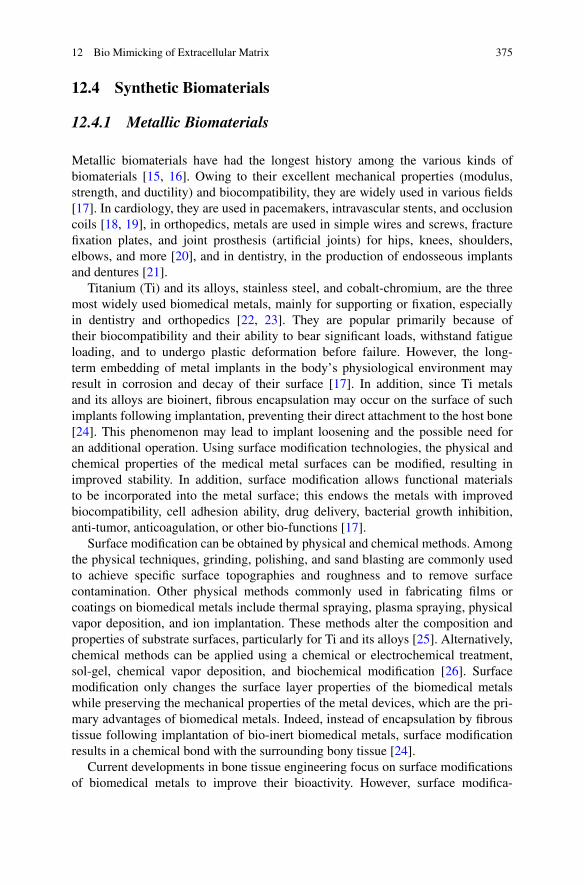
12 Bio Mimicking of Extracellular Matrix 375
12.4 Synthetic Biomaterials
12.4.1 Metallic Biomaterials
Metallic biomaterials have had the longest history among the various kinds ofbiomaterials [15, 16]. Owing to their excellent mechanical properties (modulus,strength, and ductility) and biocompatibility, they are widely used in various fields[17]. In cardiology, they are used in pacemakers, intravascular stents, and occlusioncoils [18, 19], in orthopedics, metals are used in simple wires and screws, fracturefixation plates, and joint prosthesis (artificial joints) for hips, knees, shoulders,elbows, and more [20], and in dentistry, in the production of endosseous implantsand dentures [21].
Titanium (Ti) and its alloys, stainless steel, and cobalt-chromium, are the threemost widely used biomedical metals, mainly for supporting or fixation, especiallyin dentistry and orthopedics [22, 23]. They are popular primarily because oftheir biocompatibility and their ability to bear significant loads, withstand fatigueloading, and to undergo plastic deformation before failure. However, the long-term embedding of metal implants in the body’s physiological environment mayresult in corrosion and decay of their surface [17]. In addition, since Ti metalsand its alloys are bioinert, fibrous encapsulation may occur on the surface of suchimplants following implantation, preventing their direct attachment to the host bone[24]. This phenomenon may lead to implant loosening and the possible need foran additional operation. Using surface modification technologies, the physical andchemical properties of the medical metal surfaces can be modified, resulting inimproved stability. In addition, surface modification allows functional materialsto be incorporated into the metal surface; this endows the metals with improvedbiocompatibility, cell adhesion ability, drug delivery, bacterial growth inhibition,anti-tumor, anticoagulation, or other bio-functions [17].
Surface modification can be obtained by physical and chemical methods. Amongthe physical techniques, grinding, polishing, and sand blasting are commonly usedto achieve specific surface topographies and roughness and to remove surfacecontamination. Other physical methods commonly used in fabricating films orcoatings on biomedical metals include thermal spraying, plasma spraying, physicalvapor deposition, and ion implantation. These methods alter the composition andproperties of substrate surfaces, particularly for Ti and its alloys [25]. Alternatively,chemical methods can be applied using a chemical or electrochemical treatment,sol-gel, chemical vapor deposition, and biochemical modification [26]. Surfacemodification only changes the surface layer properties of the biomedical metalswhile preserving the mechanical properties of the metal devices, which are the pri-mary advantages of biomedical metals. Indeed, instead of encapsulation by fibroustissue following implantation of bio-inert biomedical metals, surface modificationresults in a chemical bond with the surrounding bony tissue [24].
Current developments in bone tissue engineering focus on surface modificationsof biomedical metals to improve their bioactivity. However, surface modifica-
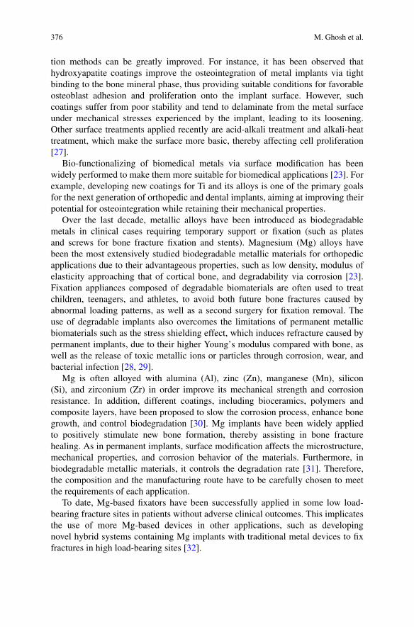
376 M. Ghosh et al.
tion methods can be greatly improved. For instance, it has been observed thathydroxyapatite coatings improve the osteointegration of metal implants via tightbinding to the bone mineral phase, thus providing suitable conditions for favorableosteoblast adhesion and proliferation onto the implant surface. However, suchcoatings suffer from poor stability and tend to delaminate from the metal surfaceunder mechanical stresses experienced by the implant, leading to its loosening.Other surface treatments applied recently are acid-alkali treatment and alkali-heattreatment, which make the surface more basic, thereby affecting cell proliferation[27].
Bio-functionalizing of biomedical metals via surface modification has beenwidely performed to make them more suitable for biomedical applications [23]. Forexample, developing new coatings for Ti and its alloys is one of the primary goalsfor the next generation of orthopedic and dental implants, aiming at improving theirpotential for osteointegration while retaining their mechanical properties.
Over the last decade, metallic alloys have been introduced as biodegradablemetals in clinical cases requiring temporary support or fixation (such as platesand screws for bone fracture fixation and stents). Magnesium (Mg) alloys havebeen the most extensively studied biodegradable metallic materials for orthopedicapplications due to their advantageous properties, such as low density, modulus ofelasticity approaching that of cortical bone, and degradability via corrosion [23].Fixation appliances composed of degradable biomaterials are often used to treatchildren, teenagers, and athletes, to avoid both future bone fractures caused byabnormal loading patterns, as well as a second surgery for fixation removal. Theuse of degradable implants also overcomes the limitations of permanent metallicbiomaterials such as the stress shielding effect, which induces refracture caused bypermanent implants, due to their higher Young’s modulus compared with bone, aswell as the release of toxic metallic ions or particles through corrosion, wear, andbacterial infection [28, 29].
Mg is often alloyed with alumina (Al), zinc (Zn), manganese (Mn), silicon(Si), and zirconium (Zr) in order improve its mechanical strength and corrosionresistance. In addition, different coatings, including bioceramics, polymers andcomposite layers, have been proposed to slow the corrosion process, enhance bonegrowth, and control biodegradation [30]. Mg implants have been widely appliedto positively stimulate new bone formation, thereby assisting in bone fracturehealing. As in permanent implants, surface modification affects the microstructure,mechanical properties, and corrosion behavior of the materials. Furthermore, inbiodegradable metallic materials, it controls the degradation rate [31]. Therefore,the composition and the manufacturing route have to be carefully chosen to meetthe requirements of each application.
To date, Mg-based fixators have been successfully applied in some low load-bearing fracture sites in patients without adverse clinical outcomes. This implicatesthe use of more Mg-based devices in other applications, such as developingnovel hybrid systems containing Mg implants with traditional metal devices to fixfractures in high load-bearing sites [32].
12 Bio Mimicking of Extracellular Matrix 377
12.4.2 Ceramic Biomaterials
Bioceramics are biocompatible ceramic compounds produced in porous or denseforms as well as powders, granulates, coatings, and injectable formulations that canbe used alone or combined with additional organic or polymeric materials for softand hard tissue replacement [33]. They can also be surface modified and designedaccordingly to deliver biologically active substances aimed to repair, maintain andrestore organs and tissues. They are hard, brittle materials with relatively poortensile properties, but exhibit excellent compressive strength, high resistance towear, and favorable low frictional properties in articulation.
Bioceramics can be classified into three major families, based on their tissueresponse: nearly inert (e.g., alumina and zirconia), bioactive (e.g., bioactive glass),and resorbable ceramics [e.g., β- and α-tricalcium phosphate (TCP)] [34]. Inertceramics are widely used as femoral heads and acetabular cups for hip replacementas well as for developing dental implants. However, these materials are presentlynot being applied as scaffolds because their inertness triggers the formation of athick “protective” fibrous capsule on the surface of the implant, thus preventingit from directly binding to the host bone tissue [34]. Since the ability to create astable bond with the host tissue is of utmost importance in selecting bioceramics forscaffold production, bioactive and bioresorbable ceramics are used. Bioceramicsalso gradually degrade over time and are replaced by the natural host tissue, andcan be completely removed once their task of serving as templates for new tissuegeneration has been completed [35].
Based on their microstructure, bioceramics can also be classified into crystallineceramics, glass-ceramics, bioactive glasses, and composites. Crystalline ceramics,including calcium phosphate (CaP), are mainly used as scaffolds for bone tissueregeneration owing to their structure and chemical composition, showing similarityto the inorganic fraction of bones, and their osteoconductivity [36]. In addition,CaP exhibits bioactivity of osteointegration, an intimate physicochemical bondbetween the implant and bone [37]. Porous blocks of various sizes composed ofCaP or Hydroxyapatite, a specific form of CaP with a ratio of 1:67, are currentlyused to repair small-sized bone defects [38, 39]. These blocks can be manipulatedintraoperatively to match the size and contour of the existing defect. Hydroxyapatiteis also used as a porous scaffold for orbital floor repair [40] and as a coating materialof metallic joint prostheses and dental implants [41, 42]. The main drawback of allCaP scaffolds produced in a porous form is their low mechanical properties, such asbrittleness and low fatigue strength, which largely limit their clinical application tolow- or non-load-bearing parts of the skeleton.
Alumina is another crystalline ceramic that has been widely employed tofabricate components of hip and knee joint prostheses (femur head, acetabularcup, and tibial plate), largely due to alumina’s high strength; thus, it is mostsuitable for load-bearing applications, excellent wear resistance, and bioinertness[43]. Porous alumina is primarily used for fabricating orbital implants (sphericalporous scaffolds) for enucleated patients; this may allow for fibro-vascular ingrowth
378 M. Ghosh et al.
through the porous network and it remains in the patient’s anophthalmic socket foran indefinite period, without undergoing degradation [14, 44].
Other forms of bioceramics are bioactive glasses, which are degradable bioma-terials that have been widely used to restore osseous defects due to their uniqueability to bond to host bone, stimulate new bone growth, and induce angiogenesis[45]. Their glass surface allows the formation of a biologically active layer ofhydroxyapatite that provides the bonding interface with host tissues, whereas thedissolution products [e.g., Si, sodium (Na), Ca, P ions] stimulate the cells to producenew tissue [15]. Bioactive glasses can be fabricated by either the conventionalmelting-quenching routes or the sol-gel technique. Finally, the glasses can also bespun to fabricate glass fibers. In the last decade, the phosphate ones, in particular,have attracted increasing interest for soft-tissue engineering applications. They havebeen used as guides for muscle or nerve repair [46], as well as for fabricating glassybone scaffolds [47], and for lung tissue engineering applications [48]. In the lastdecade, a new set of bioactive glasses, characterized by a highly ordered mesoporoustexture, has been developed and studied as a smart platform for the controlled releaseof biomolecules, in situ therapy, and regenerative applications [49, 50].
Glass-ceramics represent the third form of bioceramics, exhibiting superiormechanical properties concerning their parent glasses, specifically their higherelastic modulus, hardness, failure strength, and wear resistance, and are often usedto produce scaffolds.
Like most ceramic materials, a primary disadvantage of bioceramics is theirlow fracture toughness, which could limit their use in load-bearing applications.Furthermore, owing to their high stiffness, they may not be efficient for non-osseous applications, where adequate compliance with soft tissues is needed [51].To overcome these drawbacks, composite scaffolds composed of bioceramics andpolymers have been proposed. Typically, bioceramics are used as fillers or coatingsfor the polymer matrix to improve their mechanical properties. For instance,hydroxyapatite/polyethylene porous composites are currently used for repairingorbital floor fractures [52]. Another example is the use of bioceramics as acoating for metallic materials, such as stainless steel and titanium, for load-bearingprostheses. The bioceramics improve the biocompatibility and reduce the releaseof metal ions into the tissues [14]. Besides their “traditional” use for osseous defectrepair, bioactive glasses and glass-ceramics in their non-porous form have been usedfor non-osseous applications, including wound healing and soft-tissue engineering,owing to their ability to bond to soft tissues and to elicit desirable biologicalresponses, such as angiogenesis. Interestingly, recent studies have highlightedthe suitability of bioactive glass particulates for (i) treating gastric ulcers, (ii)injectable radioactive glasses for killing cancer cells in liver tumors, (iii) glass-polymer composites for cardiac tissue engineering, (iv) regeneration of injuries atthe tendon-to-bone interface, (v) fabricating a scaffold for wound dressing, and (vi)glass/polymer tubes for peripheral nerve regeneration [51, 53–55].
12 Bio Mimicking of Extracellular Matrix 379
12.4.3 Synthetic Biodegradable Polymers
Synthetic biodegradable polymers have gained increasing attention as scaffolds intissue engineering, drug delivery vehicles, biosensors, biomedical adhesives, and forreplacing permanent devices in low-load bearing fracture sites [56–58]. Syntheticpolymers are biocompatible, and their 3D structure and surface features can betailored, resulting in highly controlled and consistent degradation properties andexcellent reproducible mechanical and physical properties, such as tensile strengthand elastic modulus [59]. In addition, they can be produced in large homogeneousquantities and have a long shelf time, as well as a lower price compared withbiological scaffolds.
The current polymers used constructing 3D scaffolds for bone regeneration aresaturated poly-a-hydroxy esters, including polylactic acid (PLA), polyglycolic acid(PGA), and their co-polymer polylactic-co-glycolide (PLGA) [60, 61]. Typicalscaffold forms include meshes, fibers, sponges, and foams, which promote a uniformcell distribution, diffusion of nutrients, and growth of organized cell communities[59]. Different synthetic polymers exhibit varying physicochemical and mechanicalproperties and biodegradation rates. PGA is a hydrophilic polymer exhibiting afast degradation rate, whereas PLA has a structure similar to PGA but has morehydrophobic features. The chemical properties of these polymers allow hydrolyticdegradation by de-esterification. After degradation, the monomeric components ofeach polymer are eliminated by metabolic pathways. Owing to these properties,PLA and PGA have been widely applied in biomedical products and devices,such as degradable sutures [62]. The biochemical properties, such as a faster andmore controllable biodegradation rate, the properties of PLGA, the co-polymerof these two polymers, are improved compared with the pure monomers [63].Generally, for fabricating bone substitute constructs, the co-polymer PLGA ispreferred compared to its constituent homopolymers because of its superior controlof degradation by varying the ratio between its monomers [64]. However, althoughbeing biocompatible, the clinical application of pure PLGA for bone regenerationis limited by its poor osteoconductivity and suboptimal mechanical properties forload-bearing applications. Therefore, combining PLGA with other materials, suchas ceramics and bioactive glass is preferred, to render it more biomimetic, thusenhancing bone regeneration [65]. PLA, PGA, and PLGA are also used as conduitsfor drug delivery [66] and as scaffolds for constructing artificial vessels due totheir high porosity, which allows the diffusion of nutrients upon implantation andsubsequent neovascularization, and their easy handling and fabrication into differentshapes.
Other synthetic polymers have also been used in tissue engineering for a broadrange of biomedical applications [67], including poly(trimethylene carbonate) invascular tissue engineering [68], poly(methylmethacrylate) (PMMA) for producingintraocular lenses [69], poly(e-caprolactone) (PCL) for in situ tissue engineering ofsmall diameter vascular prosthesis [70], polyethylene terephthalate (PET) (Dacron)for hepatic venous reconstruction [71], polyurethanes (PU) in heart valves, vascular
380 M. Ghosh et al.
grafts, catheters, and prostheses [72], polytetrafluoroethylene (PTFE) for smalldiameter vascular grafts [73], and membranes for guided tissue regeneration [74].
The mechanical properties of polymeric biomaterials largely depend on themolecular architecture of the macromolecules, i.e., the structure of the monomericunit, the molecular weight of the polymer, and the morphology exhibited by thepolymers [75].
12.5 Natural Biomaterials
Natural polymers can be considered as the first clinically used biodegradablebiomaterials [76]. They can be classified primarily into proteins (e.g., silk, collagen,gelatin, fibrinogen, elastin, keratin, actin, and myosin), polysaccharides (e.g.,cellulose, amylose, dextran, chitin, and glycosaminoglycans), or polynucleotides(e.g., DNA, RNA) [77]. Natural polymers are soft but flexible materials, whichcan adapt their shape to desired forms through a variety of molding and castingtechniques. They contain specific molecular domains that can support cells atvarious stages of their development and promote rapid biological interactions ofthe scaffold with the surrounding host tissue [78], induce the deposition of cellsand additional ECM, overall, accelerating angiogenesis [79]. Natural polymers thusexhibit several benefits over synthetic polymers, such as degradability, negligibletoxicity, resemblance to native tissues/organs, and the ability to induce tissueremodeling. Owing to their similarity to the ECM as well as their biocompatibilityand biodegradability, natural polymers have been developed as base materials forvarious applications in tissue engineering, including small intestinal submucosa,acellular dermis, bladder acellular matrix grafts, and amniotic membranes.
12.5.1 Collagen
Collagen constitutes approximately one-third of the protein of humans and two-thirds of the dry weight of skin; it plays a vital role in biological functions, such astissue formation, cell attachment, and proliferation. The collagen protein is a helicalpolypeptide composed of repeating sequences of glycine, proline, hydroxyproline,and lysine. As an integral part of the ECM, collagen has great tensile strengthfor tissue growth; thus, it was extensively investigated for biomedical applications[80]. Collagen-based materials are considered to be favorable biomaterials for bothcartilage and bone scaffolds, since collagen type II is the major component inarticular cartilage, and collagen type I is the major component in bone. Basedon the extent of their purification, collagen-based biomaterials can be classifiedinto two categories: (i) decellularized collagen matrices that maintain the originaltissue properties and the ECM structure; and (ii) more refined scaffolds developedby extraction, purification, and collagen polymerization [81]. Different types of
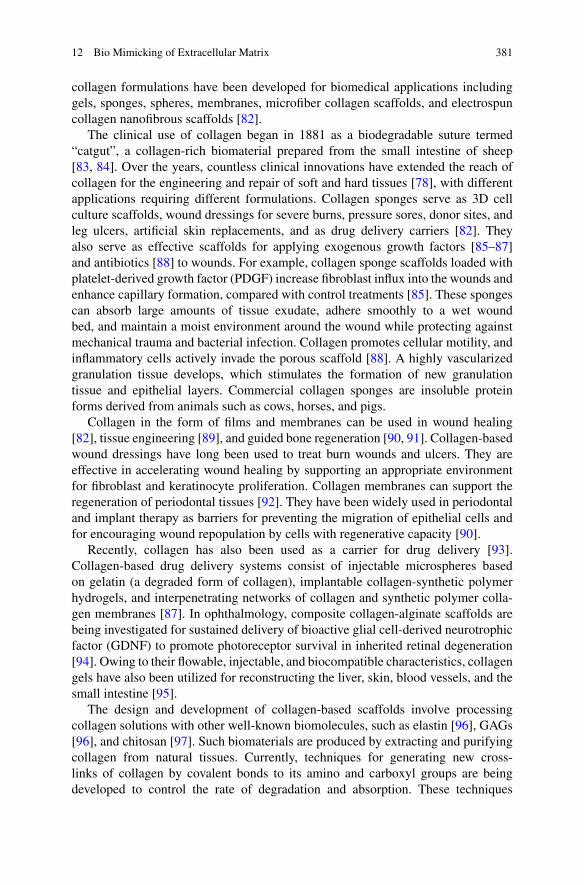
12 Bio Mimicking of Extracellular Matrix 381
collagen formulations have been developed for biomedical applications includinggels, sponges, spheres, membranes, microfiber collagen scaffolds, and electrospuncollagen nanofibrous scaffolds [82].
The clinical use of collagen began in 1881 as a biodegradable suture termed“catgut”, a collagen-rich biomaterial prepared from the small intestine of sheep[83, 84]. Over the years, countless clinical innovations have extended the reach ofcollagen for the engineering and repair of soft and hard tissues [78], with differentapplications requiring different formulations. Collagen sponges serve as 3D cellculture scaffolds, wound dressings for severe burns, pressure sores, donor sites, andleg ulcers, artificial skin replacements, and as drug delivery carriers [82]. Theyalso serve as effective scaffolds for applying exogenous growth factors [85–87]and antibiotics [88] to wounds. For example, collagen sponge scaffolds loaded withplatelet-derived growth factor (PDGF) increase fibroblast influx into the wounds andenhance capillary formation, compared with control treatments [85]. These spongescan absorb large amounts of tissue exudate, adhere smoothly to a wet woundbed, and maintain a moist environment around the wound while protecting againstmechanical trauma and bacterial infection. Collagen promotes cellular motility, andinflammatory cells actively invade the porous scaffold [88]. A highly vascularizedgranulation tissue develops, which stimulates the formation of new granulationtissue and epithelial layers. Commercial collagen sponges are insoluble proteinforms derived from animals such as cows, horses, and pigs.
Collagen in the form of films and membranes can be used in wound healing[82], tissue engineering [89], and guided bone regeneration [90, 91]. Collagen-basedwound dressings have long been used to treat burn wounds and ulcers. They areeffective in accelerating wound healing by supporting an appropriate environmentfor fibroblast and keratinocyte proliferation. Collagen membranes can support theregeneration of periodontal tissues [92]. They have been widely used in periodontaland implant therapy as barriers for preventing the migration of epithelial cells andfor encouraging wound repopulation by cells with regenerative capacity [90].
Recently, collagen has also been used as a carrier for drug delivery [93].Collagen-based drug delivery systems consist of injectable microspheres basedon gelatin (a degraded form of collagen), implantable collagen-synthetic polymerhydrogels, and interpenetrating networks of collagen and synthetic polymer colla-gen membranes [87]. In ophthalmology, composite collagen-alginate scaffolds arebeing investigated for sustained delivery of bioactive glial cell-derived neurotrophicfactor (GDNF) to promote photoreceptor survival in inherited retinal degeneration[94]. Owing to their flowable, injectable, and biocompatible characteristics, collagengels have also been utilized for reconstructing the liver, skin, blood vessels, and thesmall intestine [95].
The design and development of collagen-based scaffolds involve processingcollagen solutions with other well-known biomolecules, such as elastin [96], GAGs[96], and chitosan [97]. Such biomaterials are produced by extracting and purifyingcollagen from natural tissues. Currently, techniques for generating new cross-links of collagen by covalent bonds to its amino and carboxyl groups are beingdeveloped to control the rate of degradation and absorption. These techniques
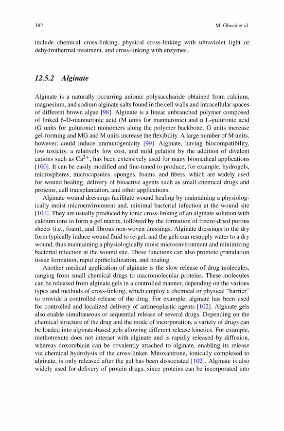
382 M. Ghosh et al.
include chemical cross-linking, physical cross-linking with ultraviolet light ordehydrothermal treatment, and cross-linking with enzymes.
12.5.2 Alginate
Alginate is a naturally occurring anionic polysaccharide obtained from calcium,magnesium, and sodium alginate salts found in the cell walls and intracellular spacesof different brown algae [98]. Alginate is a linear unbranched polymer composedof linked β-D-mannuronic acid (M units for mannuronic) and α L-guluronic acid(G units for guluronic) monomers along the polymer backbone. G units increasegel-forming and MG and M units increase the flexibility. A large number of M units,however, could induce immunogenicity [99]. Alginate, having biocompatibility,low toxicity, a relatively low cost, and mild gelation by the addition of divalentcations such as Ca2+, has been extensively used for many biomedical applications[100]. It can be easily modified and fine-tuned to produce, for example, hydrogels,microspheres, microcapsules, sponges, foams, and fibers, which are widely usedfor wound healing, delivery of bioactive agents such as small chemical drugs andproteins, cell transplantation, and other applications.
Alginate wound dressings facilitate wound healing by maintaining a physiolog-ically moist microenvironment and, minimal bacterial infection at the wound site[101]. They are usually produced by ionic cross-linking of an alginate solution withcalcium ions to form a gel matrix, followed by the formation of freeze-dried poroussheets (i.e., foam), and fibrous non-woven dressings. Alginate dressings in the dryform typically induce wound fluid to re-gel, and the gels can resupply water to a drywound, thus maintaining a physiologically moist microenvironment and minimizingbacterial infection at the wound site. These functions can also promote granulationtissue formation, rapid epithelialization, and healing.
Another medical application of alginate is the slow release of drug molecules,ranging from small chemical drugs to macromolecular proteins. These moleculescan be released from alginate gels in a controlled manner, depending on the varioustypes and methods of cross-linking, which employ a chemical or physical “barrier”to provide a controlled release of the drug. For example, alginate has been usedfor controlled and localized delivery of antineoplastic agents [102]. Alginate gelsalso enable simultaneous or sequential release of several drugs. Depending on thechemical structure of the drug and the mode of incorporation, a variety of drugs canbe loaded into alginate-based gels allowing different release kinetics. For example,methotrexate does not interact with alginate and is rapidly released by diffusion,whereas doxorubicin can be covalently attached to alginate, enabling its releasevia chemical hydrolysis of the cross-linker. Mitoxantrone, ionically complexed toalginate, is only released after the gel has been dissociated [102]. Alginate is alsowidely used for delivery of protein drugs, since proteins can be incorporated into
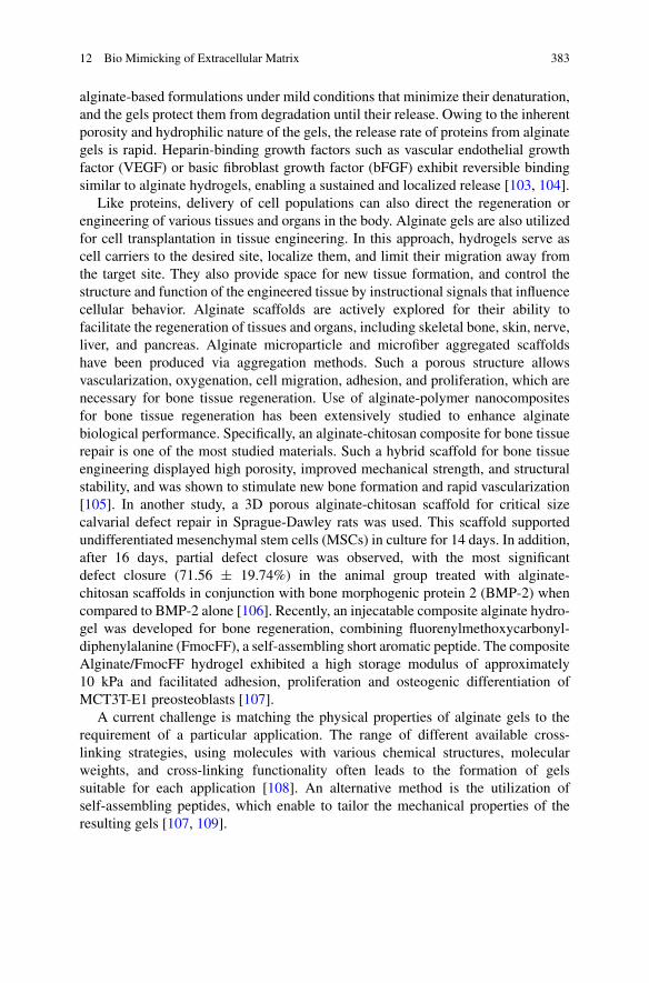
12 Bio Mimicking of Extracellular Matrix 383
alginate-based formulations under mild conditions that minimize their denaturation,and the gels protect them from degradation until their release. Owing to the inherentporosity and hydrophilic nature of the gels, the release rate of proteins from alginategels is rapid. Heparin-binding growth factors such as vascular endothelial growthfactor (VEGF) or basic fibroblast growth factor (bFGF) exhibit reversible bindingsimilar to alginate hydrogels, enabling a sustained and localized release [103, 104].
Like proteins, delivery of cell populations can also direct the regeneration orengineering of various tissues and organs in the body. Alginate gels are also utilizedfor cell transplantation in tissue engineering. In this approach, hydrogels serve ascell carriers to the desired site, localize them, and limit their migration away fromthe target site. They also provide space for new tissue formation, and control thestructure and function of the engineered tissue by instructional signals that influencecellular behavior. Alginate scaffolds are actively explored for their ability tofacilitate the regeneration of tissues and organs, including skeletal bone, skin, nerve,liver, and pancreas. Alginate microparticle and microfiber aggregated scaffoldshave been produced via aggregation methods. Such a porous structure allowsvascularization, oxygenation, cell migration, adhesion, and proliferation, which arenecessary for bone tissue regeneration. Use of alginate-polymer nanocompositesfor bone tissue regeneration has been extensively studied to enhance alginatebiological performance. Specifically, an alginate-chitosan composite for bone tissuerepair is one of the most studied materials. Such a hybrid scaffold for bone tissueengineering displayed high porosity, improved mechanical strength, and structuralstability, and was shown to stimulate new bone formation and rapid vascularization[105]. In another study, a 3D porous alginate-chitosan scaffold for critical sizecalvarial defect repair in Sprague-Dawley rats was used. This scaffold supportedundifferentiated mesenchymal stem cells (MSCs) in culture for 14 days. In addition,after 16 days, partial defect closure was observed, with the most significantdefect closure (71.56 ± 19.74%) in the animal group treated with alginate-chitosan scaffolds in conjunction with bone morphogenic protein 2 (BMP-2) whencompared to BMP-2 alone [106]. Recently, an injecatable composite alginate hydro-gel was developed for bone regeneration, combining fluorenylmethoxycarbonyl-diphenylalanine (FmocFF), a self-assembling short aromatic peptide. The compositeAlginate/FmocFF hydrogel exhibited a high storage modulus of approximately10 kPa and facilitated adhesion, proliferation and osteogenic differentiation ofMCT3T-E1 preosteoblasts [107].
A current challenge is matching the physical properties of alginate gels to therequirement of a particular application. The range of different available cross-linking strategies, using molecules with various chemical structures, molecularweights, and cross-linking functionality often leads to the formation of gelssuitable for each application [108]. An alternative method is the utilization ofself-assembling peptides, which enable to tailor the mechanical properties of theresulting gels [107, 109].
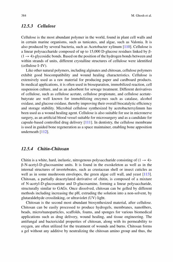
384 M. Ghosh et al.
12.5.3 Cellulose
Cellulose is the most abundant polymer in the world, found in plant cell walls andin certain marine organisms, such as tunicates, and algae, such as Valonia. It isalso produced by several bacteria, such as Acetobacter xylinum [110]. Cellulose isa linear polysaccharide composed of up to 15,000 D-glucose residues linked by β-(1 → 4)-glycosidic bonds. Based on the position of the hydrogen bonds between andwithin strands of units, different crystalline structures of cellulose were identified(cellulose I–IV).
Like other natural polymers, including alginates and chitosan, cellulose polymersexhibit good biocompatibility and wound healing characteristics. Cellulose isextensively used as a raw material for producing paper and cardboard products.In medical applications, it is often used in bioseparation, immobilized reaction, cellsuspension culture, and as an adsorbent for sewage treatment. Different derivativesof cellulose, such as cellulose acetate, cellulose propionate, and cellulose acetate-butyrate are well known for immobilizing enzymes such as catalase, alcoholoxidase, and glucose oxidase, thereby improving their overall biocatalytic efficiencyand storage stability. Microbial cellulose synthesized by acetobacterxylinum hasbeen used as a wound healing agent. Cellulose is also suitable for use in micronervesurgery, as an artificial blood vessel suitable for microsurgery and as a candidate forcapsule-based controlled drug delivery [111]. In dentistry, the cellulose membraneis used in guided bone regeneration as a space maintainer, enabling bone appositionunderneath [112].
12.5.4 Chitin-Chitosan
Chitin is a white, hard, inelastic, nitrogenous polysaccharide consisting of (1 → 4)-β-N-acetyl-D-glucosamine units. It is found in the exoskeleton as well as in theinternal structures of invertebrates, such as crustacean shell or insect cuticles aswell as in some mushroom envelopes, the green algae cell wall, and yeast [113].Chitosan, a partially deacetylated derivative of chitin, is composed of a mixtureof N-acetyl-D-glucosamine and D-glucosamine, forming a linear polysaccharide,structurally similar to GAGs. Once dissolved, chitosan can be gelled by differentmethods including increasing the pH, extruding the solution into a non-solvent, byglutaraldehyde crosslinking, or ultraviolet (UV) light.
Chitosan is the second most abundant biosynthesized material, after cellulose.Chitosan can be easily processed to produce hydrogels, membranes, nanofibers,beads, micro/nanoparticles, scaffolds, foams, and sponges for various biomedicalapplications such as drug delivery, wound healing, and tissue engineering. Theantifungal and bactericidal properties of chitosan, along with its permeability tooxygen, are often utilized for the treatment of wounds and burns. Chitosan formsa gel without any additive by neutralizing the chitosan amino group and thus, the
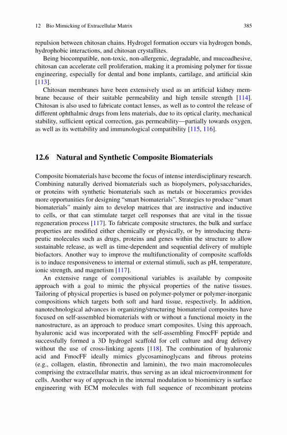
12 Bio Mimicking of Extracellular Matrix 385
repulsion between chitosan chains. Hydrogel formation occurs via hydrogen bonds,hydrophobic interactions, and chitosan crystallites.
Being biocompatible, non-toxic, non-allergenic, degradable, and mucoadhesive,chitosan can accelerate cell proliferation, making it a promising polymer for tissueengineering, especially for dental and bone implants, cartilage, and artificial skin[113].
Chitosan membranes have been extensively used as an artificial kidney mem-brane because of their suitable permeability and high tensile strength [114].Chitosan is also used to fabricate contact lenses, as well as to control the release ofdifferent ophthalmic drugs from lens materials, due to its optical clarity, mechanicalstability, sufficient optical correction, gas permeability—partially towards oxygen,as well as its wettability and immunological compatibility [115, 116].
12.6 Natural and Synthetic Composite Biomaterials
Composite biomaterials have become the focus of intense interdisciplinary research.Combining naturally derived biomaterials such as biopolymers, polysaccharides,or proteins with synthetic biomaterials such as metals or bioceramics providesmore opportunities for designing “smart biomaterials”. Strategies to produce “smartbiomaterials” mainly aim to develop matrices that are instructive and inductiveto cells, or that can stimulate target cell responses that are vital in the tissueregeneration process [117]. To fabricate composite structures, the bulk and surfaceproperties are modified either chemically or physically, or by introducing thera-peutic molecules such as drugs, proteins and genes within the structure to allowsustainable release, as well as time-dependent and sequential delivery of multiplebiofactors. Another way to improve the multifunctionality of composite scaffoldsis to induce responsiveness to internal or external stimuli, such as pH, temperature,ionic strength, and magnetism [117].
An extensive range of compositional variables is available by compositeapproach with a goal to mimic the physical properties of the native tissues.Tailoring of physical properties is based on polymer-polymer or polymer-inorganiccompositions which targets both soft and hard tissue, respectively. In addition,nanotechnological advances in organizing/structuring biomaterial composites havefocused on self-assembled biomaterials with or without a functional moiety in thenanostructure, as an approach to produce smart composites. Using this approach,hyaluronic acid was incorporated with the self-assembling FmocFF peptide andsuccessfully formed a 3D hydrogel scaffold for cell culture and drug deliverywithout the use of cross-linking agents [118]. The combination of hyaluronicacid and FmocFF ideally mimics glycosaminoglycans and fibrous proteins(e.g., collagen, elastin, fibronectin and laminin), the two main macromoleculescomprising the extracellular matrix, thus serving as an ideal microenvironment forcells. Another way of approach in the internal modulation to biomimicry is surfaceengineering with ECM molecules with full sequence of recombinant proteins
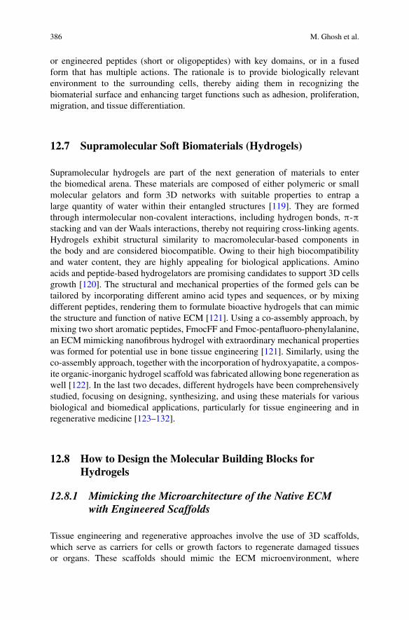
386 M. Ghosh et al.
or engineered peptides (short or oligopeptides) with key domains, or in a fusedform that has multiple actions. The rationale is to provide biologically relevantenvironment to the surrounding cells, thereby aiding them in recognizing thebiomaterial surface and enhancing target functions such as adhesion, proliferation,migration, and tissue differentiation.
12.7 Supramolecular Soft Biomaterials (Hydrogels)
Supramolecular hydrogels are part of the next generation of materials to enterthe biomedical arena. These materials are composed of either polymeric or smallmolecular gelators and form 3D networks with suitable properties to entrap alarge quantity of water within their entangled structures [119]. They are formedthrough intermolecular non-covalent interactions, including hydrogen bonds, π-πstacking and van der Waals interactions, thereby not requiring cross-linking agents.Hydrogels exhibit structural similarity to macromolecular-based components inthe body and are considered biocompatible. Owing to their high biocompatibilityand water content, they are highly appealing for biological applications. Aminoacids and peptide-based hydrogelators are promising candidates to support 3D cellsgrowth [120]. The structural and mechanical properties of the formed gels can betailored by incorporating different amino acid types and sequences, or by mixingdifferent peptides, rendering them to formulate bioactive hydrogels that can mimicthe structure and function of native ECM [121]. Using a co-assembly approach, bymixing two short aromatic peptides, FmocFF and Fmoc-pentafluoro-phenylalanine,an ECM mimicking nanofibrous hydrogel with extraordinary mechanical propertieswas formed for potential use in bone tissue engineering [121]. Similarly, using theco-assembly approach, together with the incorporation of hydroxyapatite, a compos-ite organic-inorganic hydrogel scaffold was fabricated allowing bone regeneration aswell [122]. In the last two decades, different hydrogels have been comprehensivelystudied, focusing on designing, synthesizing, and using these materials for variousbiological and biomedical applications, particularly for tissue engineering and inregenerative medicine [123–132].
12.8 How to Design the Molecular Building Blocks forHydrogels
12.8.1 Mimicking the Microarchitecture of the Native ECMwith Engineered Scaffolds
Tissue engineering and regenerative approaches involve the use of 3D scaffolds,which serve as carriers for cells or growth factors to regenerate damaged tissuesor organs. These scaffolds should mimic the ECM microenvironment, where
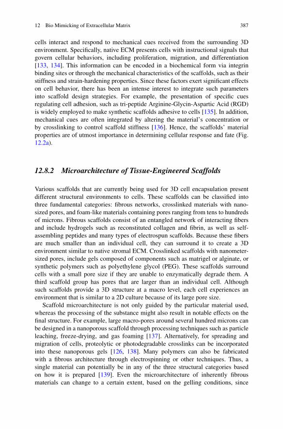
12 Bio Mimicking of Extracellular Matrix 387
cells interact and respond to mechanical cues received from the surrounding 3Denvironment. Specifically, native ECM presents cells with instructional signals thatgovern cellular behaviors, including proliferation, migration, and differentiation[133, 134]. This information can be encoded in a biochemical form via integrinbinding sites or through the mechanical characteristics of the scaffolds, such as theirstiffness and strain-hardening properties. Since these factors exert significant effectson cell behavior, there has been an intense interest to integrate such parametersinto scaffold design strategies. For example, the presentation of specific cuesregulating cell adhesion, such as tri-peptide Arginine-Glycin-Aspartic Acid (RGD)is widely employed to make synthetic scaffolds adhesive to cells [135]. In addition,mechanical cues are often integrated by altering the material’s concentration orby crosslinking to control scaffold stiffness [136]. Hence, the scaffolds’ materialproperties are of utmost importance in determining cellular response and fate (Fig.12.2a).
12.8.2 Microarchitecture of Tissue-Engineered Scaffolds
Various scaffolds that are currently being used for 3D cell encapsulation presentdifferent structural environments to cells. These scaffolds can be classified intothree fundamental categories: fibrous networks, crosslinked materials with nano-sized pores, and foam-like materials containing pores ranging from tens to hundredsof microns. Fibrous scaffolds consist of an entangled network of interacting fibersand include hydrogels such as reconstituted collagen and fibrin, as well as self-assembling peptides and many types of electrospun scaffolds. Because these fibersare much smaller than an individual cell, they can surround it to create a 3Denvironment similar to native stromal ECM. Crosslinked scaffolds with nanometer-sized pores, include gels composed of components such as matrigel or alginate, orsynthetic polymers such as polyethylene glycol (PEG). These scaffolds surroundcells with a small pore size if they are unable to enzymatically degrade them. Athird scaffold group has pores that are larger than an individual cell. Althoughsuch scaffolds provide a 3D structure at a macro level, each cell experiences anenvironment that is similar to a 2D culture because of its large pore size.
Scaffold microarchitecture is not only guided by the particular material used,whereas the processing of the substance might also result in notable effects on thefinal structure. For example, large macro-pores around several hundred microns canbe designed in a nanoporous scaffold through processing techniques such as particleleaching, freeze-drying, and gas foaming [137]. Alternatively, for spreading andmigration of cells, proteolytic or photodegradable crosslinks can be incorporatedinto these nanoporous gels [126, 138]. Many polymers can also be fabricatedwith a fibrous architecture through electrospinning or other techniques. Thus, asingle material can potentially be in any of the three structural categories basedon how it is prepared [139]. Even the microarchitecture of inherently fibrousmaterials can change to a certain extent, based on the gelling conditions, since
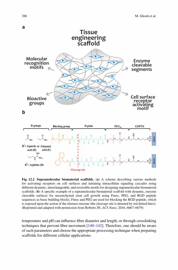
388 M. Ghosh et al.
Fig. 12.2 Supramolecular biomaterial scaffolds. (a) A scheme describing various methodsfor activating receptors on cell surfaces and initiating intracellular signaling cascades usingdifferent dynamic, interchangeable, and reversible motifs for designing supramolecular biomaterialscaffolds. (b) A specific example of a supramolecular biomaterial scaffold with dynamic, enzymecleavable surfaces for mesenchymal stem cell growth using Fmoc, PEG, and RGD peptidesequences as basic building blocks. Fmoc and PEG are used for blocking the RGD peptide, whichis exposed upon the action of the elastase enzyme (the cleavage site is denoted by red dotted lines).(Reprinted and adapted with permission from Roberts JN, ACS Nano, 2016, 6667–6679)
temperature and pH can influence fiber diameter and length, or through crosslinkingtechniques that prevent fiber movement [140–142]. Therefore, one should be awareof such parameters and choose the appropriate processing technique when preparingscaffolds for different cellular applications.
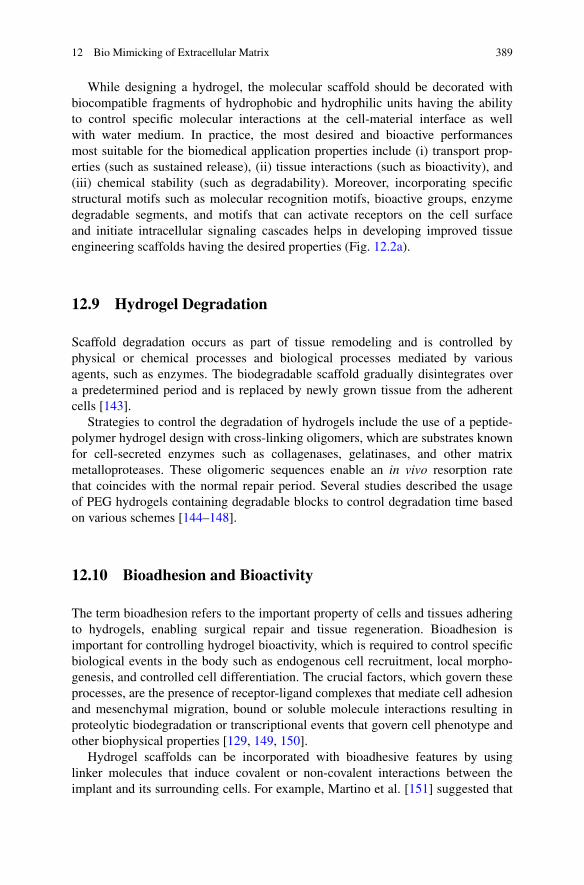
12 Bio Mimicking of Extracellular Matrix 389
While designing a hydrogel, the molecular scaffold should be decorated withbiocompatible fragments of hydrophobic and hydrophilic units having the abilityto control specific molecular interactions at the cell-material interface as wellwith water medium. In practice, the most desired and bioactive performancesmost suitable for the biomedical application properties include (i) transport prop-erties (such as sustained release), (ii) tissue interactions (such as bioactivity), and(iii) chemical stability (such as degradability). Moreover, incorporating specificstructural motifs such as molecular recognition motifs, bioactive groups, enzymedegradable segments, and motifs that can activate receptors on the cell surfaceand initiate intracellular signaling cascades helps in developing improved tissueengineering scaffolds having the desired properties (Fig. 12.2a).
12.9 Hydrogel Degradation
Scaffold degradation occurs as part of tissue remodeling and is controlled byphysical or chemical processes and biological processes mediated by variousagents, such as enzymes. The biodegradable scaffold gradually disintegrates overa predetermined period and is replaced by newly grown tissue from the adherentcells [143].
Strategies to control the degradation of hydrogels include the use of a peptide-polymer hydrogel design with cross-linking oligomers, which are substrates knownfor cell-secreted enzymes such as collagenases, gelatinases, and other matrixmetalloproteases. These oligomeric sequences enable an in vivo resorption ratethat coincides with the normal repair period. Several studies described the usageof PEG hydrogels containing degradable blocks to control degradation time basedon various schemes [144–148].
12.10 Bioadhesion and Bioactivity
The term bioadhesion refers to the important property of cells and tissues adheringto hydrogels, enabling surgical repair and tissue regeneration. Bioadhesion isimportant for controlling hydrogel bioactivity, which is required to control specificbiological events in the body such as endogenous cell recruitment, local morpho-genesis, and controlled cell differentiation. The crucial factors, which govern theseprocesses, are the presence of receptor-ligand complexes that mediate cell adhesionand mesenchymal migration, bound or soluble molecule interactions resulting inproteolytic biodegradation or transcriptional events that govern cell phenotype andother biophysical properties [129, 149, 150].
Hydrogel scaffolds can be incorporated with bioadhesive features by usinglinker molecules that induce covalent or non-covalent interactions between theimplant and its surrounding cells. For example, Martino et al. [151] suggested that
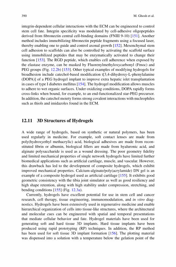
390 M. Ghosh et al.
integrin-dependent cellular interactions with the ECM can be engineered to controlstem cell fate. Integrin specificity was modulated by cell-adhesive oligopeptidesderived from fibronectin central cell-binding domains (FNIII 9-10) [151]. Anothermethod includes immobilizing fibronectin peptide fragments using a focused laser,thereby enabling one to guide and control axonal growth [152]. Mesenchymal stemcell adhesion to scaffolds can also be controlled by activating the scaffold surfaceusing immobilized peptides that may be enzymatically activated to change theirfunction [153]. The RGD peptide, which enables cell adherence when exposed bythe elastase enzyme, can be masked by Fluorenylmethyloxycarbonyl (Fmoc) andPEG groups (Fig. 12.2b) [153]. Other typical examples of modifying hydrogels forbioadhesion include catechol-based modification t[3,4-dihydroxy-L-phenylalanine(DOPA)] of a PEG hydrogel implant to improve extra hepatic islet transplantationin cases of type I diabetes mellitus [154]. The hydrogel modification allows musclesto adhere to wet organic surfaces. Under oxidizing conditions, DOPA rapidly formscross-links when bound, for example, to an end-functionalized star-PEG precursor.In addition, the catechol moiety forms strong covalent interactions with nucleophilessuch as thiols and imidazoles found in the ECM.
12.11 3D Structures of Hydrogels
A wide range of hydrogels, based on synthetic or natural polymers, has beenused regularly in medicine. For example, soft contact lenses are made frompoly(hydroxyethyl methacrylic) acid, biological adhesives are made from recon-stituted fibrin or albumin, biological fillers are made from hyaluronic acid, andalginate polysaccharide is used as a wound dressing. The poor geometry fidelityand limited mechanical properties of single network hydrogels have limited furtherbiomedical applications such as artificial cartilage, muscle, and vascular. However,this drawback has led to the development of composite hydrogels, which exhibitimproved mechanical properties. Calcium-alginate/poly(acrylamide) DN gel is anexample of a composite hydrogel used as artificial cartilage [155]. It exhibits goodgeometric consistency with the tibia joint simulator as well as good resiliency andhigh shape retention, along with high stability under compression, stretching, andbending conditions [155] (Fig. 12.3a).
Currently, hydrogels have excellent potential for use in stem cell and cancerresearch, cell therapy, tissue engineering, immunomodulation, and in vitro diag-nostics. Hydrogels have been extensively used in regenerative medicine and enablehierarchical organization of cells into tissue-like structures, where the architecturaland molecular cues can be engineered with spatial and temporal presentationsthat mediate cellular behavior and fate. Hydrogel materials have been used forgenerating soft and hard tissue 3D implants. Hard tissue implants have beenproduced using rapid prototyping (RP) techniques. In addition, the RP methodhas been used for soft tissue 3D implant formation [156]. The plotting materialwas dispensed into a solution with a temperature below the gelation point of the
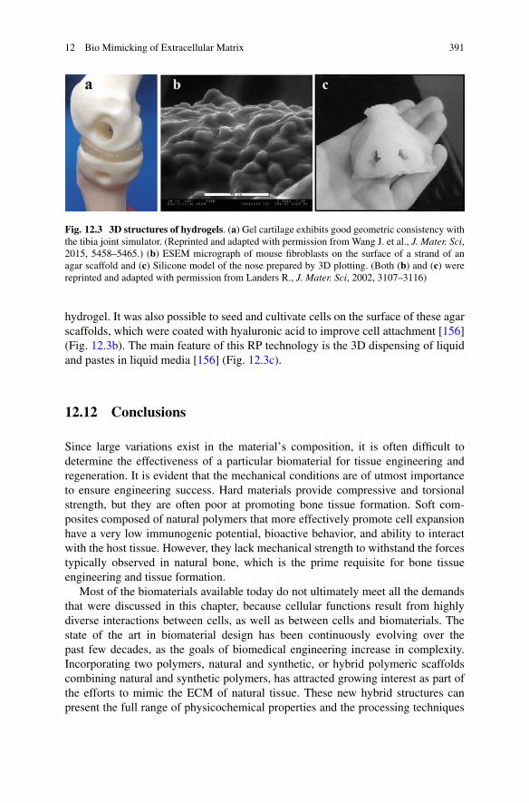
12 Bio Mimicking of Extracellular Matrix 391
Fig. 12.3 3D structures of hydrogels. (a) Gel cartilage exhibits good geometric consistency withthe tibia joint simulator. (Reprinted and adapted with permission from Wang J. et al., J. Mater. Sci,2015, 5458–5465.) (b) ESEM micrograph of mouse fibroblasts on the surface of a strand of anagar scaffold and (c) Silicone model of the nose prepared by 3D plotting. (Both (b) and (c) werereprinted and adapted with permission from Landers R., J. Mater. Sci, 2002, 3107–3116)
hydrogel. It was also possible to seed and cultivate cells on the surface of these agarscaffolds, which were coated with hyaluronic acid to improve cell attachment [156](Fig. 12.3b). The main feature of this RP technology is the 3D dispensing of liquidand pastes in liquid media [156] (Fig. 12.3c).
12.12 Conclusions
Since large variations exist in the material’s composition, it is often difficult todetermine the effectiveness of a particular biomaterial for tissue engineering andregeneration. It is evident that the mechanical conditions are of utmost importanceto ensure engineering success. Hard materials provide compressive and torsionalstrength, but they are often poor at promoting bone tissue formation. Soft com-posites composed of natural polymers that more effectively promote cell expansionhave a very low immunogenic potential, bioactive behavior, and ability to interactwith the host tissue. However, they lack mechanical strength to withstand the forcestypically observed in natural bone, which is the prime requisite for bone tissueengineering and tissue formation.
Most of the biomaterials available today do not ultimately meet all the demandsthat were discussed in this chapter, because cellular functions result from highlydiverse interactions between cells, as well as between cells and biomaterials. Thestate of the art in biomaterial design has been continuously evolving over thepast few decades, as the goals of biomedical engineering increase in complexity.Incorporating two polymers, natural and synthetic, or hybrid polymeric scaffoldscombining natural and synthetic polymers, has attracted growing interest as part ofthe efforts to mimic the ECM of natural tissue. These new hybrid structures canpresent the full range of physicochemical properties and the processing techniques
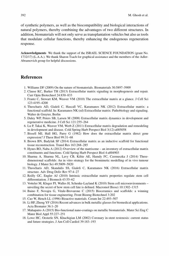
392 M. Ghosh et al.
of synthetic polymers, as well as the biocompatibility and biological interactions ofnatural polymers, thereby combining the advantages of two different structures. Inaddition, biomaterials will not only serve as transplantation vehicles but also as toolsthat modulate cellular functions, thereby enhancing the endogenous regenerationresponse.
Acknowledgments We thank the support of the ISRAEL SCIENCE FOUNDATION (grant No.1732/17) (L.A.A.). We thank Sharon Tsach for graphical assistance and the members of the Adler-Abramovich group for helpful discussions.
References
1. Williams DF (2009) On the nature of biomaterials. Biomaterials 30:5897–59092. Clause KC, Barker TH (2013) Extracellular matrix signaling in morphogenesis and repair.
Curr Opin Biotechnol 24:830–8333. Frantz C, Stewart KM, Weaver VM (2010) The extracellular matrix at a glance. J Cell Sci
123:4195–42004. Theocharis AD, Gialeli C, Hascall VC, Karamanos NK (2012) Extracellular matrix: a
functional scaffold. In: Karamanos NK (ed) Extracellular matrix: Pathobiology and signaling.Walter de Gruyter, Berlin
5. Daley WP, Peters SB, Larsen M (2008) Extracellular matrix dynamics in development andregenerative medicine. J Cell Sci 121:255–264
6. Lu P, Takai K, Weaver VM, Werb Z (2011) Extracellular matrix degradation and remodelingin development and disease. Cold Spring Harb Perspect Biol 3(12):a005058
7. Bissell MJ, Hall HG, Parry G (1982) How does the extracellular matrix direct geneexpression? J Theor Biol 99:31–68
8. Brown BN, Badylak SF (2014) Extracellular matrix as an inductive scaffold for functionaltissue reconstruction. Transl Res 163:268–285
9. Hynes RO, Naba A (2012) Overview of the matrisome – an inventory of extracellular matrixconstituents and functions. Cold Spring Harb Perspect Biol 4:a004903
10. Sharma A, Sharma NL, Lavy CB, Kiltie AE, Hamdy FC, Czernuszka J (2014) Three-dimensional scaffolds: An in vitro strategy for the biomimetic modelling of in vivo tumourbiology. J Mater Sci 49:5809–5820
11. Theocharis AD, Skandalis SS, Gialeli C, Karamanos NK (2016) Extracellular matrixstructure. Adv Drug Deliv Rev 97:4–27
12. Reilly GC, Engler AJ (2010) Intrinsic extracellular matrix properties regulate stem celldifferentiation. J Biomech 43:55–62
13. Votteler M, Kluger PJ, Walles H, Schenke-Layland K (2010) Stem cell microenvironments –unveiling the secret of how stem cell fate is defined. Macromol Biosci 10:1302–1315
14. Baino F, Novajra G, Vitale-Brovarone C (2015) Bioceramics and scaffolds: a winningcombination for tissue engineering. Front Bioeng Biotechnol 3:202
15. Cao W, Hench LL (1996) Bioactive materials. Ceram Int 22:493–50716. Li HF, Zheng YF (2016) Recent advances in bulk metallic glasses for biomedical applications.
Acta Biomater 36:1–2017. Mahapatro A (2015) Bio-functional nano-coatings on metallic biomaterials. Mater Sci Eng C
Mater Biol Appl 55:227–25118. Lowe HC, Oesterle SN, Khachigian LM (2002) Coronary in-stent restenosis: current status
and future strategies. J Am Coll Cardiol 39:183–193
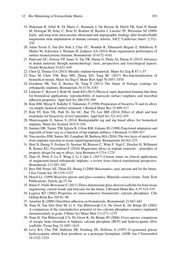
12 Bio Mimicking of Extracellular Matrix 393
19. Waksman R, Erbel R, Di Mario C, Bartunek J, De Bruyne B, Eberli FR, Erne P, HaudeM, Horrigan M, Ilsley C, Bose D, Bonnier H, Koolen J, Luscher TF, Weissman NJ (2009)Early- and long-term intravascular ultrasound and angiographic findings after bioabsorbablemagnesium stent implantation in human coronary arteries. JACC Cardiovasc Interv 2:312–320
20. Amin Yavari S, Van Der Stok J, Chai YC, Wauthle R, Tahmasebi Birgani Z, Habibovic P,Mulier M, Schrooten J, Weinans H, Zadpoor AA (2014) Bone regeneration performance ofsurface-treated porous titanium. Biomaterials 35:6172–6181
21. Padovani GC, Feitosa VP, Sauro S, Tay FR, Duran G, Paula AJ, Duran N (2015) Advancesin dental materials through nanotechnology: facts, perspectives and toxicological aspects.Trends Biotechnol 33:621–636
22. Chen Q, Thouas GA (2015) Metallic implant biomaterials. Mater Sci Eng R 87:1–5723. Xiao M, Chen YM, Biao MN, Zhang XD, Yang BC (2017) Bio-functionalization of
biomedical metals. Mater Sci Eng C Mater Biol Appl 70:1057–107024. Goodman SB, Yao Z, Keeney M, Yang F (2013) The future of biologic coatings for
orthopaedic implants. Biomaterials 34:3174–318325. Lüdecke C, Bossert J, Roth M, Jandt KD (2013) Physical vapor deposited titanium thin films
for biomedical applications: reproducibility of nanoscale surface roughness and microbialadhesion properties. Appl Surf Sci 280:578–589
26. Kim HM, Miyaji F, Kokubo T, Nakamura T (1996) Preparation of bioactive Ti and its alloysvia simple chemical surface treatment. J Biomed Mater Res 32:409–417
27. Kim SY, Kim YK, Park IS, Jin GC, Bae TS, Lee MH (2014) Effect of alkali and heattreatments for bioactivity of tio2 nanotubes. Appl Surf Sci 321:412–419
28. Manivasagam G, Suwas S (2014) Biodegradable mg and mg based alloys for biomedicalimplants. Mater Sci Technol 30:515–520
29. Sumner DR, Turner TM, Igloria R, Urban RM, Galante JO (1998) Functional adaptation andingrowth of bone vary as a function of hip implant stiffness. J Biomech 31:909–917
30. Vasconcelos DM, Santos SG, Lamghari M, Barbosa MA (2016) The two faces of metal ions:from implants rejection to tissue repair/regeneration. Biomaterials 84:262–275
31. Hort N, Huang Y, Fechner D, Stormer M, Blawert C, Witte F, Vogt C, Drucker H, WillumeitR, Kainer KU, Feyerabend F (2010) Magnesium alloys as implant materials – principles ofproperty design for mg-re alloys. Acta Biomater 6:1714–1725
32. Zhao D, Witte F, Lu F, Wang J, Li J, Qin L (2017) Current status on clinical applicationsof magnesium-based orthopaedic implants: a review from clinical translational perspective.Biomaterials 112:287–302
33. Best SM, Porter AE, Thian ES, Huang J (2008) Bioceramics: past, present and for the future.J Eur Ceram Soc 28:1319–1327
34. Hench LL (1999) Bioactive glasses and glass-ceramics, Materials science forum. Trans TechPublications, Zurich, pp 37–64
35. Baino F, Vitale-Brovarone C (2011) Three-dimensional glass-derived scaffolds for bone tissueengineering: current trends and forecasts for the future. J Biomed Mater Res A 97:514–535
36. Legeros RZ (2002) Properties of osteoconductive biomaterials: calcium phosphates. ClinOrthop Relat Res 395:81–98
37. Anselme K (2000) Osteoblast adhesion on biomaterials. Biomaterials 21:667–68138. Yuan H, Van Den Doel M, Li S, Van Blitterswijk CA, De Groot K, De Bruijn JD (2002)
A comparison of the osteoinductive potential of two calcium phosphate ceramics implantedintramuscularly in goats. J Mater Sci Mater Med 13:1271–1275
39. Yuan H, Van Blitterswijk CA, De Groot K, De Bruijn JD (2006) Cross-species comparisonof ectopic bone formation in biphasic calcium phosphate (BCP) and hydroxyapatite (HA)scaffolds. Tissue Eng 12:1607–1615
40. Levy RA, Chu TM, Halloran JW, Feinberg SE, Hollister S (1997) Ct-generated poroushydroxyapatite orbital floor prosthesis as a prototype bioimplant. AJNR Am J Neuroradiol18:1522–1525
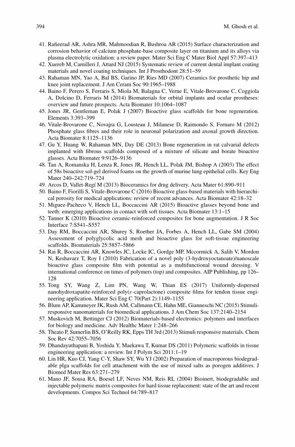
394 M. Ghosh et al.
41. Rafieerad AR, Ashra MR, Mahmoodian R, Bushroa AR (2015) Surface characterization andcorrosion behavior of calcium phosphate-base composite layer on titanium and its alloys viaplasma electrolytic oxidation: a review paper. Mater Sci Eng C Mater Biol Appl 57:397–413
42. Xuereb M, Camilleri J, Attard NJ (2015) Systematic review of current dental implant coatingmaterials and novel coating techniques. Int J Prosthodont 28:51–59
43. Rahaman MN, Yao A, Bal BS, Garino JP, Ries MD (2007) Ceramics for prosthetic hip andknee joint replacement. J Am Ceram Soc 90:1965–1988
44. Baino F, Perero S, Ferraris S, Miola M, Balagna C, Verne E, Vitale-Brovarone C, CoggiolaA, Dolcino D, Ferraris M (2014) Biomaterials for orbital implants and ocular prostheses:overview and future prospects. Acta Biomater 10:1064–1087
45. Jones JR, Gentleman E, Polak J (2007) Bioactive glass scaffolds for bone regeneration.Elements 3:393–399
46. Vitale-Brovarone C, Novajra G, Lousteau J, Milanese D, Raimondo S, Fornaro M (2012)Phosphate glass fibres and their role in neuronal polarization and axonal growth direction.Acta Biomater 8:1125–1136
47. Gu Y, Huang W, Rahaman MN, Day DE (2013) Bone regeneration in rat calvarial defectsimplanted with fibrous scaffolds composed of a mixture of silicate and borate bioactiveglasses. Acta Biomater 9:9126–9136
48. Tan A, Romanska H, Lenza R, Jones JR, Hench LL, Polak JM, Bishop A (2003) The effectof 58s bioactive sol-gel derived foams on the growth of murine lung epithelial cells. Key EngMater 240–242:719–724
49. Arcos D, Vallet-Regí M (2013) Bioceramics for drug delivery. Acta Mater 61:890–91150. Baino F, Fiorilli S, Vitale-Brovarone C (2016) Bioactive glass-based materials with hierarchi-
cal porosity for medical applications: review of recent advances. Acta Biomater 42:18–3251. Miguez-Pacheco V, Hench LL, Boccaccini AR (2015) Bioactive glasses beyond bone and
teeth: emerging applications in contact with soft tissues. Acta Biomater 13:1–1552. Tanner K (2010) Bioactive ceramic-reinforced composites for bone augmentation. J R Soc
Interface 7:S541–S55753. Day RM, Boccaccini AR, Shurey S, Roether JA, Forbes A, Hench LL, Gabe SM (2004)
Assessment of polyglycolic acid mesh and bioactive glass for soft-tissue engineeringscaffolds. Biomaterials 25:5857–5866
54. Rai R, Boccaccini AR, Knowles JC, Locke IC, Gordge MP, Mccormick A, Salih V, MordonN, Keshavarz T, Roy I (2010) Fabrication of a novel poly (3-hydroxyoctanoate)/nanoscalebioactive glass composite film with potential as a multifunctional wound dressing. Vinternational conference on times of polymers (top) and composites. AIP Publishing, pp 126–128
55. Tong SY, Wang Z, Lim PN, Wang W, Thian ES (2017) Uniformly-dispersednanohydroxapatite-reinforced poly(ε-caprolactone) composite films for tendon tissue engi-neering application. Mater Sci Eng C 70(Part 2):1149–1155
56. Blum AP, Kammeyer JK, Rush AM, Callmann CE, Hahn ME, Gianneschi NC (2015) Stimuli-responsive nanomaterials for biomedical applications. J Am Chem Soc 137:2140–2154
57. Muskovich M, Bettinger CJ (2012) Biomaterials-based electronics: polymers and interfacesfor biology and medicine. Adv Healthc Mater 1:248–266
58. Theato P, Sumerlin BS, O’Reilly RK, Epps TH 3rd (2013) Stimuli responsive materials. ChemSoc Rev 42:7055–7056
59. Dhandayuthapani B, Yoshida Y, Maekawa T, Kumar DS (2011) Polymeric scaffolds in tissueengineering application: a review. Int J Polym Sci 2011:1–19
60. Lin HR, Kuo CJ, Yang C-Y, Shaw SY, Wu YJ (2002) Preparation of macroporous biodegrad-able plga scaffolds for cell attachment with the use of mixed salts as porogen additives. JBiomed Mater Res 63:271–279
61. Mano JF, Sousa RA, Boesel LF, Neves NM, Reis RL (2004) Bioinert, biodegradable andinjectable polymeric matrix composites for hard tissue replacement: state of the art and recentdevelopments. Compos Sci Technol 64:789–817
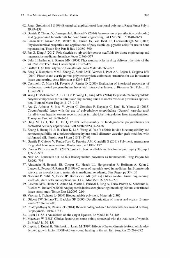
12 Bio Mimicking of Extracellular Matrix 395
62. Jagur-Grodzinski J (1999) Biomedical application of functional polymers. React Funct Polym39:99–138
63. Gentile P, Chiono V, Carmagnola I, Hatton PV (2014) An overview of poly(lactic-co-glycolic)acid (plga)-based biomaterials for bone tissue engineering. Int J Mol Sci 15:3640–3659
64. Lanao RPF, Jonker AM, Wolke JG, Jansen JA, Van Hest JC, Leeuwenburgh SC (2013)Physicochemical properties and applications of poly (lactic-co-glycolic acid) for use in boneregeneration. Tissue Eng Part B Rev 19:380–390
65. Pan Z, Ding J (2012) Poly (lactide-co-glycolide) porous scaffolds for tissue engineering andregenerative medicine. Interface Focus 2:366–377
66. Bala I, Hariharan S, Kumar MN (2004) Plga nanoparticles in drug delivery: the state of theart. Crit Rev Ther Drug Carrier Syst 21:387–422
67. Griffith L (2000) Polymeric biomaterials. Acta Mater 48:263–27768. Song Y, Kamphuis MMJ, Zhang Z, Sterk LMT, Vermes I, Poot AA, Feijen J, Grijpma DW
(2010) Flexible and elastic porous poly(trimethylene carbonate) structures for use in vasculartissue engineering. Acta Biomater 6:1269–1277
69. Cassinelli C, Morra M, Pavesio A, Renier D (2000) Evaluation of interfacial properties ofhyaluronan coated poly(methylmethacrylate) intraocular lenses. J Biomater Sci Polym Ed11:961–977
70. Wang F, Mohammed A, Li C, Ge P, Wang L, King MW (2014) Degradable/non-degradablepolymer composites for in-situ tissue engineering small diameter vascular prosthesis applica-tion. Biomed Mater Eng 24:2127–2133
71. Ara C, Akbulut S, Ince V, Aydin C, Gonultas F, Kayaalp C, Unal B, Yilmaz S (2015)Circumferential fence with the use of polyethylene terephthalate (Dacron) vascular graftfor all-in-one hepatic venous reconstruction in right-lobe living-donor liver transplantation.Transplant Proc 47:1458–1461
72. Ding M, Li J, Tan H, Fu Q (2012) Self-assembly of biodegradable polyurethanes forcontrolled delivery applications. Soft Matter 8:5414–5428
73. Zhang J, Huang H, Ju R, Chen K, Li S, Wang W, Yan Y (2016) In vivo biocompatibility andhemocompatibility of a polytetrafluoroethylene small diameter vascular graft modified withsulfonated silk fibroin. Am J Surg 213(1):87–93
74. Gentile P, Chiono V, Tonda-Turo C, Ferreira AM, Ciardelli G (2011) Polymeric membranesfor guided bone regeneration. Biotechnol J 6:1187–1197
75. Carson JS, Bostrom MP (2007) Synthetic bone scaffolds and fracture repair. Injury 38(Suppl1):S33–S37
76. Nair LS, Laurencin CT (2007) Biodegradable polymers as biomaterials. Prog Polym Sci32:762–798
77. Alexander H, Brunski JB, Cooper SL, Hench LL, Hergenrother R, Hoffman A, Kohn J,Langer R, Peppas N, Ratner B (1996) Classes of materials used in medicine. In: Biomaterialsscience: an introduction to materials in medicine. Academic, San Diego, pp 37–130
78. Nooeaid P, Salih V, Beier JP, Boccaccini AR (2012a) Osteochondral tissue engineering:scaffolds, stem cells and applications. J Cell Mol Med 16:2247–2270
79. Laschke MW, Harder Y, Amon M, Martin I, Farhadi J, Ring A, Torio-Padron N, Schramm R,Rücker M, Junker D (2006) Angiogenesis in tissue engineering: breathing life into constructedtissue substitutes. Tissue Eng 12:2093–2104
80. Vroman I, Tighzert L (2009) Biodegradable polymers. Materials 2:30781. Gilbert TW, Sellaro TL, Badylak SF (2006) Decellularization of tissues and organs. Bioma-
terials 27:3675–368382. Chattopadhyay S, Raines RT (2014) Review collagen-based biomaterials for wound healing.
Biopolymers 101:821–83383. Lister J (1881) An address on the catgut ligature. Br Med J 1:183–18584. Macewen W (1881) Clinical lectures on some points connected with the treatment of wounds.
Br Med J 1:150–15185. Lepisto J, Kujari H, Niinikoski J, Laato M (1994) Effects of heterodimeric isoform of platelet-
derived growth factor PDGF-AB on wound healing in the rat. Eur Surg Res 26:267–272

396 M. Ghosh et al.
86. Marks MG, Doillon C, Silver FH (1991) Effects of fibroblasts and basic fibroblast growthfactor on facilitation of dermal wound healing by type I collagen matrices. J Biomed MaterRes 25:683–696
87. Rao KP (1995) Recent developments of collagen-based materials for medical applicationsand drug delivery systems. J Biomater Sci Polym Ed 7:623–645
88. Wachol-Drewek Z, Pfeiffer M, Scholl E (1996) Comparative investigation of drug deliveryof collagen implants saturated in antibiotic solutions and a sponge containing gentamicin.Biomaterials 17:1733–1738
89. Li X, Xu J, Nicolescu C, Marinelli J, Tien J (2016) Generation, endothelialization, andmicrosurgical suture anastomosis of strong 1-mm-diameter collagen tubes. Tissue Eng Part A23(7–8):335–344
90. Stoecklin-Wasmer C, Rutjes AW, Da Costa BR, Salvi GE, Juni P, Sculean A (2013)Absorbable collagen membranes for periodontal regeneration: a systematic review. J DentRes 92:773–781
91. Wessing B, Urban I, Montero E, Zechner W, Hof M, Alandez Chamorro J, Alandez MartinN, Polizzi G, Meloni S, Sanz M (2016) A multicenter randomized controlled clinical trialusing a new resorbable non-cross-linked collagen membrane for guided bone regeneration atdehisced single implant sites: interim results of a bone augmentation procedure. Clin OralImplants Res 28(11):e218–e226
92. Pitaru S, Tal H, Soldinger M, Grosskopf A, Noff M (1988) Partial regeneration of periodontaltissues using collagen barriers. Initial observations in the canine. J Periodontol 59:380–386
93. An B, Lin YS, Brodsky B (2016) Collagen interactions: drug design and delivery. Adv DrugDeliv Rev 97:69–84
94. Wong FS, Wong CC, Chan BP, Lo AC (2016) Sustained delivery of bioactive GDNF fromcollagen and alginate-based cell-encapsulating gel promoted photoreceptor survival in aninherited retinal degeneration model. PLoS One 11:e0159342
95. Lee KY, Mooney DJ (2001) Hydrogels for tissue engineering. Chem Rev 101:1869–187996. Ellis DL, Yannas IV (1996) Recent advances in tissue synthesis in vivo by use of collagen-
glycosaminoglycan copolymers. Biomaterials 17:291–29997. Wu X, Black L, Santacana-Laffitte G, Patrick CW (2007) Preparation and assessment of
glutaraldehyde-crosslinked collagen–chitosan hydrogels for adipose tissue engineering. JBiomed Mater Res A 81A:59–65
98. Tønnesen HH, Karlsen J (2002) Alginate in drug delivery systems. Drug Dev Ind Pharm28:621–630
99. Kulseng B, Skjåk-Bræk G, Ryan L, Andersson A, King A, Faxvaag A, Espevik T (1999)Transplantation of alginate microcapsules: generation of antibodies against alginates andencapsulated porcine islet-like cell clusters1. Transplantation 67:978–984
100. Wee S, Gombotz WR (1998) Protein release from alginate matrices. Adv Drug Deliv Rev31:267–285
101. Queen D, Orsted H, Sanada H, Sussman G (2004) A dressing history. Int Wound J 1:59–77102. Bouhadir KH, Alsberg E, Mooney DJ (2001) Hydrogels for combination delivery of
antineoplastic agents. Biomaterials 22:2625–2633103. Lee KY, Peters MC, Mooney DJ (2003) Comparison of vascular endothelial growth factor and
basic fibroblast growth factor on angiogenesis in SCID mice. J Control Release 87:49–56104. Silva EA, Mooney DJ (2010) Effects of VEGF temporal and spatial presentation on
angiogenesis. Biomaterials 31:1235–1241105. Li Z, Ramay HR, Hauch KD, Xiao D, Zhang M (2005) Chitosan-alginate hybrid scaffolds for
bone tissue engineering. Biomaterials 26:3919–3928106. Florczyk SJ, Leung M, Li Z, Huang JI, Hopper RA, Zhang M (2013) Evaluation of three-
dimensional porous chitosan-alginate scaffolds in rat calvarial defects for bone regenerationapplications. J Biomed Mater Res A 101:2974–2983
107. Ghosh M, Halperin-Sternfeld M, Grinberg I, Adler-Abramovich L (2019) Injectable alginate-peptide composite hydrogel as a scaffold for bone tissue regeneration. Nanomaterials (Basel)9:497
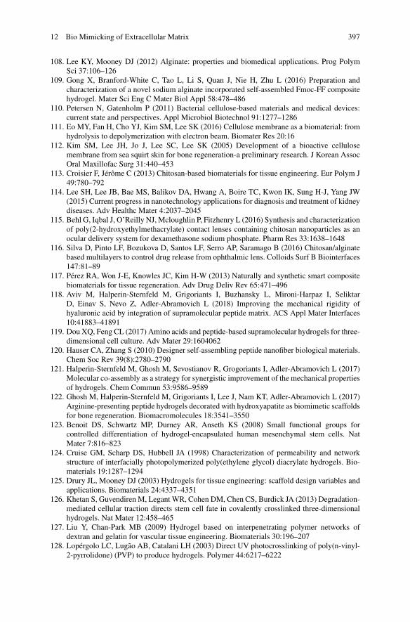
12 Bio Mimicking of Extracellular Matrix 397
108. Lee KY, Mooney DJ (2012) Alginate: properties and biomedical applications. Prog PolymSci 37:106–126
109. Gong X, Branford-White C, Tao L, Li S, Quan J, Nie H, Zhu L (2016) Preparation andcharacterization of a novel sodium alginate incorporated self-assembled Fmoc-FF compositehydrogel. Mater Sci Eng C Mater Biol Appl 58:478–486
110. Petersen N, Gatenholm P (2011) Bacterial cellulose-based materials and medical devices:current state and perspectives. Appl Microbiol Biotechnol 91:1277–1286
111. Eo MY, Fan H, Cho YJ, Kim SM, Lee SK (2016) Cellulose membrane as a biomaterial: fromhydrolysis to depolymerization with electron beam. Biomater Res 20:16
112. Kim SM, Lee JH, Jo J, Lee SC, Lee SK (2005) Development of a bioactive cellulosemembrane from sea squirt skin for bone regeneration-a preliminary research. J Korean AssocOral Maxillofac Surg 31:440–453
113. Croisier F, Jérôme C (2013) Chitosan-based biomaterials for tissue engineering. Eur Polym J49:780–792
114. Lee SH, Lee JB, Bae MS, Balikov DA, Hwang A, Boire TC, Kwon IK, Sung H-J, Yang JW(2015) Current progress in nanotechnology applications for diagnosis and treatment of kidneydiseases. Adv Healthc Mater 4:2037–2045
115. Behl G, Iqbal J, O’Reilly NJ, Mcloughlin P, Fitzhenry L (2016) Synthesis and characterizationof poly(2-hydroxyethylmethacrylate) contact lenses containing chitosan nanoparticles as anocular delivery system for dexamethasone sodium phosphate. Pharm Res 33:1638–1648
116. Silva D, Pinto LF, Bozukova D, Santos LF, Serro AP, Saramago B (2016) Chitosan/alginatebased multilayers to control drug release from ophthalmic lens. Colloids Surf B Biointerfaces147:81–89
117. Pérez RA, Won J-E, Knowles JC, Kim H-W (2013) Naturally and synthetic smart compositebiomaterials for tissue regeneration. Adv Drug Deliv Rev 65:471–496
118. Aviv M, Halperin-Sternfeld M, Grigoriants I, Buzhansky L, Mironi-Harpaz I, SeliktarD, Einav S, Nevo Z, Adler-Abramovich L (2018) Improving the mechanical rigidity ofhyaluronic acid by integration of supramolecular peptide matrix. ACS Appl Mater Interfaces10:41883–41891
119. Dou XQ, Feng CL (2017) Amino acids and peptide-based supramolecular hydrogels for three-dimensional cell culture. Adv Mater 29:1604062
120. Hauser CA, Zhang S (2010) Designer self-assembling peptide nanofiber biological materials.Chem Soc Rev 39(8):2780–2790
121. Halperin-Sternfeld M, Ghosh M, Sevostianov R, Grogoriants I, Adler-Abramovich L (2017)Molecular co-assembly as a strategy for synergistic improvement of the mechanical propertiesof hydrogels. Chem Commun 53:9586–9589
122. Ghosh M, Halperin-Sternfeld M, Grigoriants I, Lee J, Nam KT, Adler-Abramovich L (2017)Arginine-presenting peptide hydrogels decorated with hydroxyapatite as biomimetic scaffoldsfor bone regeneration. Biomacromolecules 18:3541–3550
123. Benoit DS, Schwartz MP, Durney AR, Anseth KS (2008) Small functional groups forcontrolled differentiation of hydrogel-encapsulated human mesenchymal stem cells. NatMater 7:816–823
124. Cruise GM, Scharp DS, Hubbell JA (1998) Characterization of permeability and networkstructure of interfacially photopolymerized poly(ethylene glycol) diacrylate hydrogels. Bio-materials 19:1287–1294
125. Drury JL, Mooney DJ (2003) Hydrogels for tissue engineering: scaffold design variables andapplications. Biomaterials 24:4337–4351
126. Khetan S, Guvendiren M, Legant WR, Cohen DM, Chen CS, Burdick JA (2013) Degradation-mediated cellular traction directs stem cell fate in covalently crosslinked three-dimensionalhydrogels. Nat Mater 12:458–465
127. Liu Y, Chan-Park MB (2009) Hydrogel based on interpenetrating polymer networks ofdextran and gelatin for vascular tissue engineering. Biomaterials 30:196–207
128. Lopérgolo LC, Lugão AB, Catalani LH (2003) Direct UV photocrosslinking of poly(n-vinyl-2-pyrrolidone) (PVP) to produce hydrogels. Polymer 44:6217–6222
398 M. Ghosh et al.
129. Lutolf MP, Hubbell JA (2005) Synthetic biomaterials as instructive extracellular microenvi-ronments for morphogenesis in tissue engineering. Nat Biotechnol 23:47–55
130. Nicodemus GD, Bryant SJ (2008) Cell encapsulation in biodegradable hydrogels for tissueengineering applications. Tissue Eng Part B Rev 14:149–165
131. Peppas NA, Hilt JZ, Khademhosseini A, Langer R (2006) Hydrogels in biology and medicine:from molecular principles to bionanotechnology. Adv Mater 18:1345–1360
132. Tibbitt MW, Anseth KS (2009) Hydrogels as extracellular matrix mimics for 3D cell culture.Biotechnol Bioeng 103:655–663
133. Guilak F, Cohen DM, Estes BT, Gimble JM, Liedtke W, Chen CS (2009) Control of stem cellfate by physical interactions with the extracellular matrix. Cell Stem Cell 5:17–26
134. Guo WH, Frey MT, Burnham NA, Wang YL (2006) Substrate rigidity regulates the formationand maintenance of tissues. Biophys J 90:2213–2220
135. Lowe SB, Tan VT, Soeriyadi AH, Davis TP, Gooding JJ (2014) Synthesis and high-throughputprocessing of polymeric hydrogels for 3d cell culture. Bioconjug Chem 25:1581–1601
136. Nemir S, West JL (2010) Synthetic materials in the study of cell response to substrate rigidity.Ann Biomed Eng 38:2–20
137. Levengood SKL, Zhang M (2014) Chitosan-based scaffolds for bone tissue engineering. JMater Chem B 2:3161–3184
138. Kloxin AM, Kasko AM, Salinas CN, Anseth KS (2009) Photodegradable hydrogels fordynamic tuning of physical and chemical properties. Science 324:59–63
139. Madihally SV, Matthew HW (1999) Porous chitosan scaffolds for tissue engineering.Biomaterials 20:1133–1142
140. Carey SP, Kraning-Rush CM, Williams RM, Reinhart-King CA (2012) Biophysical control ofinvasive tumor cell behavior by extracellular matrix microarchitecture. Biomaterials 33:4157–4165
141. Hayen W, Goebeler M, Kumar S, Riessen R, Nehls V (1999) Hyaluronan stimulates tumorcell migration by modulating the fibrin fiber architecture. J Cell Sci 112(Pt 13):2241–2251
142. Lang NR, Skodzek K, Hurst S, Mainka A, Steinwachs J, Schneider J, Aifantis KE, Fabry B(2015) Biphasic response of cell invasion to matrix stiffness in three-dimensional biopolymernetworks. Acta Biomater 13:61–67
143. Langer R (1994) Biodegradable polymer scaffolds for tissue engineering. Nat Biotechnol12:689–693
144. Deforest CA, Polizzotti BD, Anseth KS (2009) Sequential click reactions for synthesizingand patterning three-dimensional cell microenvironments. Nat Mater 8:659–664
145. Kloxin AM, Kloxin CJ, Bowman CN, Anseth KS (2010) Mechanical properties of cellularlyresponsive hydrogels and their experimental determination. Adv Mater 22:3484–3494
146. Kraehenbuehl TP, Zammaretti P, Van Der Vlies AJ, Schoenmakers RG, Lutolf MP, JaconiME, Hubbell JA (2008) Three-dimensional extracellular matrix-directed cardioprogenitor dif-ferentiation: systematic modulation of a synthetic cell-responsive peg-hydrogel. Biomaterials29:2757–2766
147. Lutolf MP, Lauer-Fields JL, Schmoekel HG, Metters AT, Weber FE, Fields GB, Hubbell JA(2003) Synthetic matrix metalloproteinase-sensitive hydrogels for the conduction of tissueregeneration: engineering cell-invasion characteristics. Proc Natl Acad Sci U S A 100:5413–5418
148. Mckinnon DD, Domaille DW, Brown TE, Kyburz KA, Kiyotake E, Cha JN, Anseth KS (2014)Measuring cellular forces using bis-aliphatic hydrazone crosslinked stress-relaxing hydrogels.Soft Matter 10:9230–9236
149. Discher DE, Mooney DJ, Zandstra PW (2009) Growth factors, matrices, and forces combineand control stem cells. Science 324:1673–1677
150. Hudalla GA, Murphy WL (2011) Biomaterials that regulate growth factor activity viabioinspired interactions. Adv Funct Mater 21:1754–1768
151. Martino MM, Mochizuki M, Rothenfluh DA, Rempel SA, Hubbell JA, Barker TH (2009)Controlling integrin specificity and stem cell differentiation in 2d and 3d environmentsthrough regulation of fibronectin domain stability. Biomaterials 30:1089–1097

12 Bio Mimicking of Extracellular Matrix 399
152. Luo Y, Shoichet MS (2004) A photolabile hydrogel for guided three-dimensional cell growthand migration. Nat Mater 3:249–253
153. Roberts JN, Sahoo JK, Mcnamara LE, Burgess KV, Yang J, Alakpa EV, Anderson HJ, HayJ, Turner LA, Yarwood SJ, Zelzer M, Oreffo RO, Ulijn RV, Dalby MJ (2016) Dynamicsurfaces for the study of mesenchymal stem cell growth through adhesion regulation. ACSNano 10:6667–6679
154. Brubaker CE, Kissler H, Wang LJ, Kaufman DB, Messersmith PB (2010) Biologicalperformance of mussel-inspired adhesive in extrahepatic islet transplantation. Biomaterials31:420–427
155. Wang J, Wei J, Su S, Qiu J, Wang S (2015) Ion-linked double-network hydrogel with hightoughness and stiffness. J Mater Sci 50:5458–5465
156. Landers R, Pfister A, Hübner U, John H, Schmelzeisen R, Mülhaupt R (2002) Fabricationof soft tissue engineering scaffolds by means of rapid prototyping techniques. J Mater Sci37:3107–3116
Chapter 13Bioinspired Engineeringof Organ-on-Chip Devices
Li Wang, Zhongyu Li, Cong Xu, and Jianhua Qin
Abstract The human body can be viewed as an organism consisting of a varietyof cellular and non-cellular materials interacting in a highly ordered manner. Itscomplex and hierarchical nature inspires the multi-level recapitulation of the humanbody in order to gain insights into the inner workings of life. While traditionalcell culture models have led to new insights into the cellular microenvironmentand biological control in vivo, deeper understanding of biological systems andhuman pathophysiology requires the development of novel model systems thatallow for analysis of complex internal and external interactions within the cellularmicroenvironment in a more relevant organ context. Engineering organ-on-chipsystems offers an unprecedented opportunity to unravel the complex and hierar-chical nature of human organs. In this chapter, we first highlight the advances inmicrofluidic platforms that enable engineering of the cellular microenvironment andthe transition from cells-on-chips to organs-on-chips. Then, we introduce the keyfeatures of the emerging organs-on-chips and their proof-of-concept applicationsin biomedical research. We also discuss the challenges and future outlooks of thisstate-of-the-art technology.
Keywords Bioinspired materials · Microfluidics · Organ-on-chip · Cellularmicroenvironment · Disease modeling · Drug testing
L. Wang · Z. Li · C. XuDivision of Biotechnology, Dalian Institute of Chemical Physics, Chinese Academy of Sciences,Dalian, P. R. China
J. Qin (�)Division of Biotechnology, Dalian Institute of Chemical Physics, Chinese Academy of Sciences,Dalian, P. R. China
CAS Center for Excellence in Brain Science and Intelligence Technology, Chinese Academy ofSciences, Shanghai, China
Institute for Stem Cell and Regeneration, Chinese Academy of Sciences, Beijing, China
University of Chinese Academy of Sciences, Beijing, Chinae-mail: [email protected]
© Springer Nature Singapore Pte Ltd. 2019S. Perrett et al. (eds.), Biological and Bio-inspired Nanomaterials,Advances in Experimental Medicine and Biology 1174,https://doi.org/10.1007/978-981-13-9791-2_13
401
402 L. Wang et al.
Abbreviations
ADMET Adsorption, distribution, metabolism, elimination and toxicityBBB Blood-brain-barrierEBs Embryonic bodyECM Extracellular matrixECs Endothelial cellsEMT Epithelial-to mesenchymalESCs Embryonic stem cellsFSS Fluidic shear stressiPSCs Induced pluripotent stem cellsMEMS Micro-electromechanical systemMSC Mesenchymal stem cellsPDMS PolydimethylsiloxanePK/PD Pharmacokinetics and pharmacodynamicsTEER Trans-epithelial electrical resistance
13.1 Introduction
Appropriate model systems drive the development of biological and biomedicalresearch. These model systems seek to recapitulate human physiology and pathol-ogy from the molecular level to the cellular, tissue and organ levels, thus providinginsights into disease etiology, diagnostic therapeutics and disease prevention. Invivo, the body can be viewed as a variety of cellular and non-cellular materialsinteracting in a highly ordered manner. The complex and hierarchical nature ofall living things inspires the multi-level recapitulation of the human body anddevelopment of biological model systems consisting of multiple cell types, withinternal (cell-cell, cell-matrix) and/or external (e.g. cell-environment) interactionsin a more relevant organ and multi-organ context.
Conventionally, animal models are often used to closely recapitulate humanphysiology in a variety of biomedical research areas, but they fail to faithfully mimichuman responses due to the presence of confounding variables and differencesbetween animal and human biology. It is also quite difficult to carry out real-timeobservation, utilization and throughput assays in animal models. While simplisticmodels, such as two dimensional (2D) monocultures of cells in a Petri-dish, havetheir merits to study the biological process of specific cell types, these formatsoften lack cell-cell and cell-matrix interactions that are necessary to maintain anddefine the specific phenotypes of cells. They also fail to mimic the cellular functionsand intercellular communication that is present in tissues or organs. While three-dimensional (3D) cell aggregates and spheroid cultures can improve the cellularfunctions to some extent, they still lack many features that are critical for sustainingorgan development and function, such as spatiotemporal biochemical cues, vascularperfusion, mechanical cues or multiple cell co-cultures. Obviously, most current
13 Bioinspired Engineering of Organ-on-Chip Devices 403
model systems are far from being able to fully reconstitute functions spanningcellular, tissue and organ levels, but it is crucial to develop such biological systemsthat can address specific scientific questions in biomedical research.
Considerable advances in microfabrication and microfluidics technology haveexpanded our ability to culture cells in a tightly controlled complex cellularmicroenvironment in a spatiotemporal manner, thus mimicking the tissue microen-vironment in vivo. The microfluidic culture platform can provide living cells withcontinuously perfused medium in microchannels at the microscale [1–5]. Integrationof microfabrication with microfluidics technologies that enable precise control ofdynamic fluid flow has made it possible to create cellular microenvironments thatpresent cells (e.g. human cell lines, primary cells or stem cells) with appropriateorgan-relevant chemical gradients and dynamic mechanical cues. These new capa-bilities have provided an impetus for the development of alternative cell-based invitro models, which better mimic the complex structures and functional complexityof living organs, termed organs-on-chips [6–9]. These organs-on-chips combinemicrofluidics with bioengineering and cell biology, allowing study of the diversebiological processes and physiopathology of the human body in ways that are notpossible using animal models and conventional 2D or 3D cell culture systems.
In this chapter, we first introduce the microfluidic culture systems that can offerdynamic cell cultures with enhanced capabilities. Then, we introduce the designconsiderations and key components required for engineering the cellular microenvi-ronment using microfluidic chips. We further summarize the emerging transitionfrom cells-on-a-chip to organs-on-a-chip, and the proof-of–concept applicationsof engineered organ-on-chip devices. We also discuss the challenges and futureperspectives of this state-of–the-art technology.
13.2 Microfluidic Cell Culture System
Microfluidic technology allows precise manipulation of fluid in a microscaledevice created with technologies developed by the semiconductor industry andmicro-electromechanical systems (MEMS). Advances in microfabrication and softlithography have enabled microfluidic culture systems to reconstitute dynamic,controlled spatio-temporal physico-chemical microenvironments to mimic in vivoconditions, distinguishing them from the existing cell culture platforms.
Microfluidic culture devices are usually generated by soft lithography pioneeredby Whitesides et al. using polydimethylsiloxane(PDMS) [10, 11]. The classicprocess of PDMS chip fabrication begins from a mould manufactured usingphotoresistor silicon [12, 13]. A mixture of silicone rubber and cross-linking agentis poured into the mould and is then easily peeled off the substrate after cross-linking. The PDMS block can be reversibly sealed to other substrates, such asglass and PDMS simply by direct contact. It is also easy to irreversibly bond thePDMS chip to a PDMS substrate, silicon or glass by plasma oxidizing the PDMSsurface to form Si-O-Si bonds or by using PDMS as glue [14]. These features makeit possible to fabricate multilayer microfluidic chips with flexible microstructure
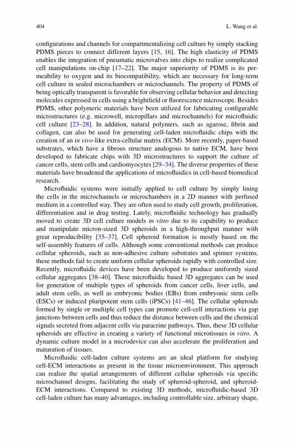
404 L. Wang et al.
configurations and channels for compartmentalizing cell culture by simply stackingPDMS pieces to connect different layers [15, 16]. The high elasticity of PDMSenables the integration of pneumatic microvalves into chips to realize complicatedcell manipulations on-chip [17–22]. The major superiority of PDMS is its per-meability to oxygen and its biocompatibility, which are necessary for long-termcell culture in sealed microchambers or microchannels. The property of PDMS ofbeing optically transparent is favorable for observing cellular behavior and detectingmolecules expressed in cells using a brightfield or fluorescence microscope. BesidesPDMS, other polymeric materials have been utilized for fabricating configurablemicrostructures (e.g. microwell, micropillars and microchannels) for microfluidiccell culture [23–28]. In addition, natural polymers, such as agarose, fibrin andcollagen, can also be used for generating cell-laden microfluidic chips with thecreation of an in vivo-like extra-cellular matrix (ECM). More recently, paper-basedsubstrates, which have a fibrous structure analogous to native ECM, have beendeveloped to fabricate chips with 3D microstructures to support the culture ofcancer cells, stem cells and cardiomyocytes [29–34]. The diverse properties of thesematerials have broadened the applications of microfluidics in cell-based biomedicalresearch.
Microfluidic systems were initially applied to cell culture by simply liningthe cells in the microchannels or microchambers in a 2D manner with perfusedmedium in a controlled way. They are often used to study cell growth, proliferation,differentiation and in drug testing. Lately, microfluidic technology has graduallymoved to create 3D cell culture models in vitro due to its capability to produceand manipulate micron-sized 3D spheroids in a high-throughput manner withgreat reproducibility [35–37]. Cell spheroid formation is mostly based on theself-assembly features of cells. Although some conventional methods can producecellular spheroids, such as non-adhesive culture substrates and spinner systems,these methods fail to create uniform cellular spheroids rapidly with controlled size.Recently, microfluidic devices have been developed to produce uniformly sizedcellular aggregates [38–40]. These microfluidic based 3D aggregates can be usedfor generation of multiple types of spheroids from cancer cells, liver cells, andadult stem cells, as well as embryonic bodies (EBs) from embryonic stem cells(ESCs) or induced pluripotent stem cells (iPSCs) [41–46]. The cellular spheroidsformed by single or multiple cell types can promote cell-cell interactions via gapjunctions between cells and thus reduce the distance between cells and the chemicalsignals secreted from adjacent cells via paracrine pathways. Thus, these 3D cellularspheroids are effective in creating a variety of functional microtissues in vitro. Adynamic culture model in a microdevice can also accelerate the proliferation andmaturation of tissues.
Microfluidic cell-laden culture systems are an ideal platform for studyingcell-ECM interactions as present in the tissue microenvironment. This approachcan realize the spatial arrangements of different cellular spheroids via specificmicrochannel designs, facilitating the study of spheroid-spheroid, and spheroid-ECM interactions. Compared to existing 3D methods, microfluidic-based 3Dcell-laden culture has many advantages, including controllable size, arbitrary shape,
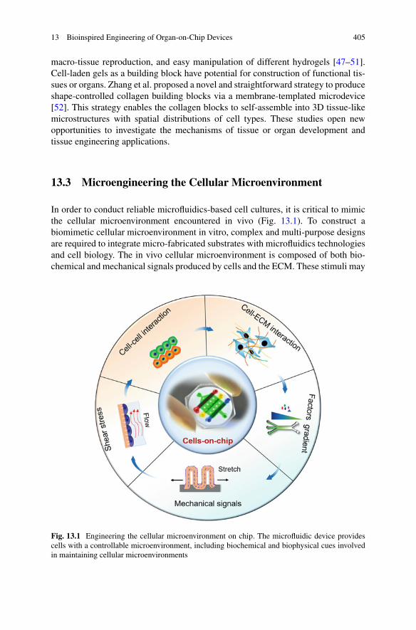
13 Bioinspired Engineering of Organ-on-Chip Devices 405
macro-tissue reproduction, and easy manipulation of different hydrogels [47–51].Cell-laden gels as a building block have potential for construction of functional tis-sues or organs. Zhang et al. proposed a novel and straightforward strategy to produceshape-controlled collagen building blocks via a membrane-templated microdevice[52]. This strategy enables the collagen blocks to self-assemble into 3D tissue-likemicrostructures with spatial distributions of cell types. These studies open newopportunities to investigate the mechanisms of tissue or organ development andtissue engineering applications.
13.3 Microengineering the Cellular Microenvironment
In order to conduct reliable microfluidics-based cell cultures, it is critical to mimicthe cellular microenvironment encountered in vivo (Fig. 13.1). To construct abiomimetic cellular microenvironment in vitro, complex and multi-purpose designsare required to integrate micro-fabricated substrates with microfluidics technologiesand cell biology. The in vivo cellular microenvironment is composed of both bio-chemical and mechanical signals produced by cells and the ECM. These stimuli may
Fig. 13.1 Engineering the cellular microenvironment on chip. The microfluidic device providescells with a controllable microenvironment, including biochemical and biophysical cues involvedin maintaining cellular microenvironments
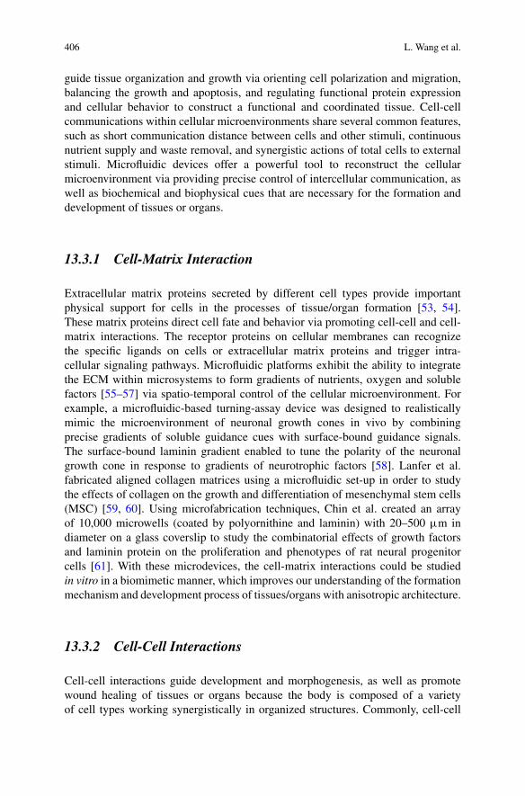
406 L. Wang et al.
guide tissue organization and growth via orienting cell polarization and migration,balancing the growth and apoptosis, and regulating functional protein expressionand cellular behavior to construct a functional and coordinated tissue. Cell-cellcommunications within cellular microenvironments share several common features,such as short communication distance between cells and other stimuli, continuousnutrient supply and waste removal, and synergistic actions of total cells to externalstimuli. Microfluidic devices offer a powerful tool to reconstruct the cellularmicroenvironment via providing precise control of intercellular communication, aswell as biochemical and biophysical cues that are necessary for the formation anddevelopment of tissues or organs.
13.3.1 Cell-Matrix Interaction
Extracellular matrix proteins secreted by different cell types provide importantphysical support for cells in the processes of tissue/organ formation [53, 54].These matrix proteins direct cell fate and behavior via promoting cell-cell and cell-matrix interactions. The receptor proteins on cellular membranes can recognizethe specific ligands on cells or extracellular matrix proteins and trigger intra-cellular signaling pathways. Microfluidic platforms exhibit the ability to integratethe ECM within microsystems to form gradients of nutrients, oxygen and solublefactors [55–57] via spatio-temporal control of the cellular microenvironment. Forexample, a microfluidic-based turning-assay device was designed to realisticallymimic the microenvironment of neuronal growth cones in vivo by combiningprecise gradients of soluble guidance cues with surface-bound guidance signals.The surface-bound laminin gradient enabled to tune the polarity of the neuronalgrowth cone in response to gradients of neurotrophic factors [58]. Lanfer et al.fabricated aligned collagen matrices using a microfluidic set-up in order to studythe effects of collagen on the growth and differentiation of mesenchymal stem cells(MSC) [59, 60]. Using microfabrication techniques, Chin et al. created an arrayof 10,000 microwells (coated by polyornithine and laminin) with 20–500 μm indiameter on a glass coverslip to study the combinatorial effects of growth factorsand laminin protein on the proliferation and phenotypes of rat neural progenitorcells [61]. With these microdevices, the cell-matrix interactions could be studiedin vitro in a biomimetic manner, which improves our understanding of the formationmechanism and development process of tissues/organs with anisotropic architecture.
13.3.2 Cell-Cell Interactions
Cell-cell interactions guide development and morphogenesis, as well as promotewound healing of tissues or organs because the body is composed of a varietyof cell types working synergistically in organized structures. Commonly, cell-cell
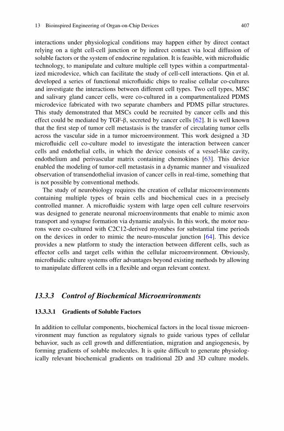
13 Bioinspired Engineering of Organ-on-Chip Devices 407
interactions under physiological conditions may happen either by direct contactrelying on a tight cell-cell junction or by indirect contact via local diffusion ofsoluble factors or the system of endocrine regulation. It is feasible, with microfluidictechnology, to manipulate and culture multiple cell types within a compartmental-ized microdevice, which can facilitate the study of cell-cell interactions. Qin et al.developed a series of functional microfluidic chips to realise cellular co-culturesand investigate the interactions between different cell types. Two cell types, MSCand salivary gland cancer cells, were co-cultured in a compartmentalized PDMSmicrodevice fabricated with two separate chambers and PDMS pillar structures.This study demonstrated that MSCs could be recruited by cancer cells and thiseffect could be mediated by TGF-β, secreted by cancer cells [62]. It is well knownthat the first step of tumor cell metastasis is the transfer of circulating tumor cellsacross the vascular side in a tumor microenvironment. This work designed a 3Dmicrofluidic cell co-culture model to investigate the interaction between cancercells and endothelial cells, in which the device consists of a vessel-like cavity,endothelium and perivascular matrix containing chemokines [63]. This deviceenabled the modeling of tumor-cell metastasis in a dynamic manner and visualizedobservation of transendothelial invasion of cancer cells in real-time, something thatis not possible by conventional methods.
The study of neurobiology requires the creation of cellular microenvironmentscontaining multiple types of brain cells and biochemical cues in a preciselycontrolled manner. A microfluidic system with large open cell culture reservoirswas designed to generate neuronal microenvironments that enable to mimic axontransport and synapse formation via dynamic analysis. In this work, the motor neu-rons were co-cultured with C2C12-derived myotubes for substantial time periodson the devices in order to mimic the neuro-muscular junction [64]. This deviceprovides a new platform to study the interaction between different cells, such aseffector cells and target cells within the cellular microenvironment. Obviously,microfluidic culture systems offer advantages beyond existing methods by allowingto manipulate different cells in a flexible and organ relevant context.
13.3.3 Control of Biochemical Microenvironments
13.3.3.1 Gradients of Soluble Factors
In addition to cellular components, biochemical factors in the local tissue microen-vironment may function as regulatory signals to guide various types of cellularbehavior, such as cell growth and differentiation, migration and angiogenesis, byforming gradients of soluble molecules. It is quite difficult to generate physiolog-ically relevant biochemical gradients on traditional 2D and 3D culture models.
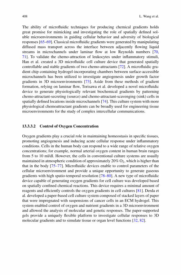
408 L. Wang et al.
The ability of microfluidic techniques for producing chemical gradients holdsgreat promise for mimicking and investigating the role of spatially defined sol-uble microenvironments in guiding cellular behavior and adversity of biologicalresponses [65–69]. Classical microfluidic gradients were generated by manipulatingdiffused mass transport across the interface between adjacently flowing liquidstreams in microchannels under laminar flow at low Reynolds numbers [70,71]. To validate the chemo-attraction of leukocytes under inflammatory stimuli,Han et al. created a 3D microfluidic cell culture device that generated spatiallycontrollable and stable gradients of two chemo-attractants [72]. A microfluidic gra-dient chip containing hydrogel-incorporating chambers between surface-accessiblemicrochannels has been utilized to investigate angiogenesis under growth factorgradients in 3D microenvironments [73]. Aside from these methods of gradientformation, relying on laminar flow, Torisawa et al. developed a novel microfluidicdevice to generate physiologically relevant biochemical gradients by patterningchemo-attractant-secreting (source) and chemo-attractant-scavenging (sink) cells inspatially defined locations inside microchannels [74]. This culture system with morephysiological chemoattractant gradients can be broadly used for engineering tissuemicroenvironments for the study of complex intercellular communications.
13.3.3.2 Control of Oxygen Concentration
Oxygen gradients play a crucial role in maintaining homeostasis in specific tissues,promoting angiogenesis and inducing acute cellular response under inflammatoryconditions. Cells in the human body can respond to a wide range of relative oxygenconcentrations; for example, normal arterial oxygen content in human brain rangesfrom 5 to 10 ml/dl. However, the cells in conventional culture systems are usuallymaintained in atmospheric condition of approximately 20% O2, which is higher thanthat in the body [75–77]. Microfluidic devices enable to control parameters of thecellular microenvironment and provide a unique opportunity to generate gaseousgradients with high spatio-temporal resolution [78–80]. A new type of microfluidicdevice capable of generating oxygen gradients for cell culture was developed basedon spatially confined chemical reactions. This device requires a minimal amount ofreagents and efficiently controls the oxygen gradients in cell cultures [81]. Derda etal. developed a paper-based cell culture system composed of stacked layers of paperthat were impregnated with suspensions of cancer cells in an ECM hydrogel. Thissystem enabled control of oxygen and nutrient gradients in a 3D microenvironmentand allowed the analysis of molecular and genetic responses. The paper-supportedgels provide a uniquely flexible platform to investigate cellular responses to 3Dmolecular gradients and to simulate tissue or organ level functions [32, 82].
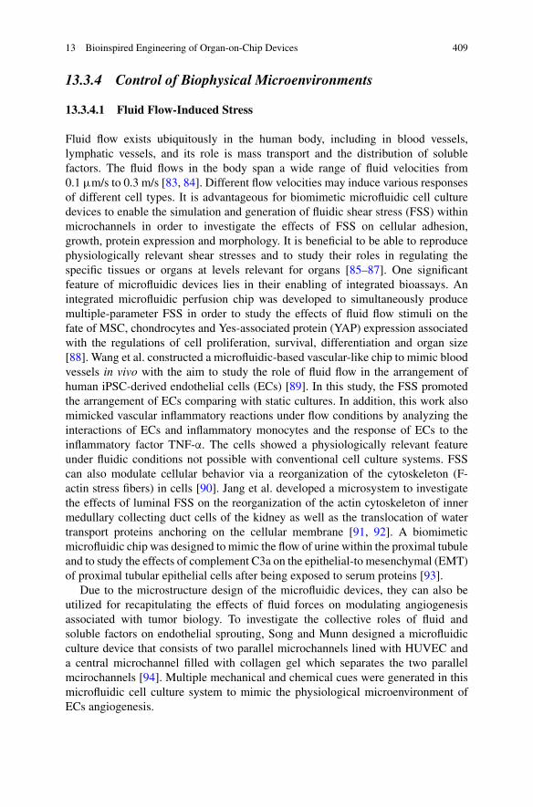
13 Bioinspired Engineering of Organ-on-Chip Devices 409
13.3.4 Control of Biophysical Microenvironments
13.3.4.1 Fluid Flow-Induced Stress
Fluid flow exists ubiquitously in the human body, including in blood vessels,lymphatic vessels, and its role is mass transport and the distribution of solublefactors. The fluid flows in the body span a wide range of fluid velocities from0.1 μm/s to 0.3 m/s [83, 84]. Different flow velocities may induce various responsesof different cell types. It is advantageous for biomimetic microfluidic cell culturedevices to enable the simulation and generation of fluidic shear stress (FSS) withinmicrochannels in order to investigate the effects of FSS on cellular adhesion,growth, protein expression and morphology. It is beneficial to be able to reproducephysiologically relevant shear stresses and to study their roles in regulating thespecific tissues or organs at levels relevant for organs [85–87]. One significantfeature of microfluidic devices lies in their enabling of integrated bioassays. Anintegrated microfluidic perfusion chip was developed to simultaneously producemultiple-parameter FSS in order to study the effects of fluid flow stimuli on thefate of MSC, chondrocytes and Yes-associated protein (YAP) expression associatedwith the regulations of cell proliferation, survival, differentiation and organ size[88]. Wang et al. constructed a microfluidic-based vascular-like chip to mimic bloodvessels in vivo with the aim to study the role of fluid flow in the arrangement ofhuman iPSC-derived endothelial cells (ECs) [89]. In this study, the FSS promotedthe arrangement of ECs comparing with static cultures. In addition, this work alsomimicked vascular inflammatory reactions under flow conditions by analyzing theinteractions of ECs and inflammatory monocytes and the response of ECs to theinflammatory factor TNF-α. The cells showed a physiologically relevant featureunder fluidic conditions not possible with conventional cell culture systems. FSScan also modulate cellular behavior via a reorganization of the cytoskeleton (F-actin stress fibers) in cells [90]. Jang et al. developed a microsystem to investigatethe effects of luminal FSS on the reorganization of the actin cytoskeleton of innermedullary collecting duct cells of the kidney as well as the translocation of watertransport proteins anchoring on the cellular membrane [91, 92]. A biomimeticmicrofluidic chip was designed to mimic the flow of urine within the proximal tubuleand to study the effects of complement C3a on the epithelial-to mesenchymal (EMT)of proximal tubular epithelial cells after being exposed to serum proteins [93].
Due to the microstructure design of the microfluidic devices, they can also beutilized for recapitulating the effects of fluid forces on modulating angiogenesisassociated with tumor biology. To investigate the collective roles of fluid andsoluble factors on endothelial sprouting, Song and Munn designed a microfluidicculture device that consists of two parallel microchannels lined with HUVEC anda central microchannel filled with collagen gel which separates the two parallelmcirochannels [94]. Multiple mechanical and chemical cues were generated in thismicrofluidic cell culture system to mimic the physiological microenvironment ofECs angiogenesis.
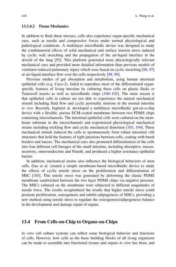
410 L. Wang et al.
13.3.4.2 Tissue Mechanics
In addition to fluid shear stresses, cells also experience organ-specific mechanicalcues, such as tensile and compressive forces under normal physiological andpathological conditions. A multilayer microfluidic device was designed to studythe combinatorial effects of solid mechanical and surface tension stress inducedby cyclic wall stretching and the propagation of the air-liquid interface in thealveoli of the lung [95]. This platform generated more physiologically relevantmechanical cues and provided more detailed information than previous models ofventilator-induced pulmonary injury which were based on cyclic stretching [96, 97]or air-liquid interface flow over the cells respectively [98, 99].
Previous studies of gut absorption and metabolism, using human intestinalepithelial cells (e.g. Caco-2), failed to reproduce most of the differentiated organ-specific features of living intestine by culturing these cells on plastic flasks orTranswell inserts as well as microfluidic chips [100–102]. The main reason isthat epithelial cells in culture are not able to experience the natural mechanicalstimuli including fluid flow and cyclic peristaltic motions in the normal intestinein vivo. Recently, Ingberet al. developed a multilayer microfluidic gut-on-a-chipdevice with a flexible, porous ECM-coated membrane between two PDMS chipscontaining microchannels. The intestinal epithelial cells were cultured on the mem-brane substrate in the microchannels and experienced physiological mechanicalstrains including trickling flow and cyclic mechanical distortion [103, 104]. Thesemechanical stimuli induced the cells to spontaneously form robust intestinal villistructures that hold the features of tight junctions between cells, coating with brushborders and mucus. The mechanical cues also promoted differentiation of the cellsinto four different cell lineages of the small intestine, including absorptive, mucus-secretory, enteroendocrine and Paneth, and produced a higher resistance epithelialbarrier.
In addition, mechanical strains also influence the biological behaviors of stemcells. Gao et al. created a simple membrane-based microfluidic device to studythe effects of cyclic tensile stress on the proliferation and differentiation ofMSC [105]. This tensile stress was generated by deforming the elastic PDMSmembrane sandwiched between the two layer PDMS chips via negative pressure.The MSCs cultured on the membrane were subjected to different magnitudes oftensile force. The results recapitulated the results that higher tensile stress couldpromote proliferation, osteogenesis and inhibit adipogenesis of MSCs, providing anew method using tensile stress to regulate the osteogenesis/adipogenesis balancein the development and damage repair of organs.
13.4 From Cells-on-Chip to Organs-on-Chips
In vitro cell culture systems can reflect some biological behavior and functionsof cells. However, how cells as the basic building blocks of all living organismscan be made to assemble into functional tissues and organs in vitro has been, and
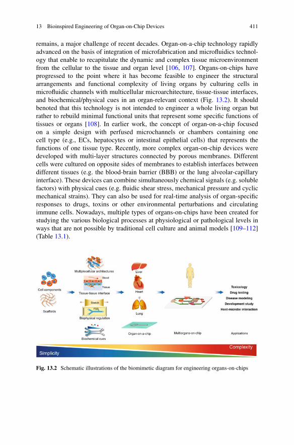
13 Bioinspired Engineering of Organ-on-Chip Devices 411
remains, a major challenge of recent decades. Organ-on-a-chip technology rapidlyadvanced on the basis of integration of microfabrication and microfluidics technol-ogy that enable to recapitulate the dynamic and complex tissue microenvironmentfrom the cellular to the tissue and organ level [106, 107]. Organs-on-chips haveprogressed to the point where it has become feasible to engineer the structuralarrangements and functional complexity of living organs by culturing cells inmicrofluidic channels with multicellular microarchitecture, tissue-tissue interfaces,and biochemical/physical cues in an organ-relevant context (Fig. 13.2). It shouldbenoted that this technology is not intended to engineer a whole living organ butrather to rebuild minimal functional units that represent some specific functions oftissues or organs [108]. In earlier work, the concept of organ-on-a-chip focusedon a simple design with perfused microchannels or chambers containing onecell type (e.g., ECs, hepatocytes or intestinal epithelial cells) that represents thefunctions of one tissue type. Recently, more complex organ-on-chip devices weredeveloped with multi-layer structures connected by porous membranes. Differentcells were cultured on opposite sides of membranes to establish interfaces betweendifferent tissues (e.g. the blood-brain barrier (BBB) or the lung alveolar-capillaryinterface). These devices can combine simultaneously chemical signals (e.g. solublefactors) with physical cues (e.g. fluidic shear stress, mechanical pressure and cyclicmechanical strains). They can also be used for real-time analysis of organ-specificresponses to drugs, toxins or other environmental perturbations and circulatingimmune cells. Nowadays, multiple types of organs-on-chips have been created forstudying the various biological processes at physiological or pathological levels inways that are not possible by traditional cell culture and animal models [109–112](Table 13.1).
Fig. 13.2 Schematic illustrations of the biomimetic diagram for engineering organs-on-chips
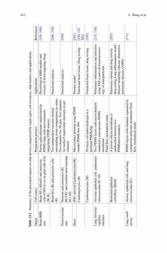
412 L. Wang et al.
Tabl
e13
.1Su
mm
ary
ofth
epr
esen
ted
orga
ns-o
n-ch
ipde
vice
sca
tego
rize
dby
orga
nty
pes,
cell
reso
urce
s,ch
ipfe
atur
esan
dap
plic
atio
ns
Org
anC
ells
ourc
ePr
epar
atio
npr
oces
sA
pplic
atio
nsR
efer
ence
s
Bra
in-B
BB
unit
Bra
inE
Cs
(bE
nd3)
and
astr
ocyt
icce
lls(C
8D1A
)or
ratg
lialc
ells
(C6)
(M,R
)
Poro
uspo
lyca
rbon
ate
sand
wic
hed
byPD
MS
chip
sw
ithm
icro
chan
nels
inte
grat
ing
TE
ER
sens
ors
Phys
iolo
gica
lBB
Bm
odel
san
dto
xici
tyof
brai
n-ta
rget
ing
drug
s[1
59,1
99]
Bra
inE
Cs
(R)
and
astr
ocyt
icce
lls(R
)Tw
oin
depe
nden
tvas
cula
rch
anne
lsu
rrou
ndin
gtis
sue
com
part
men
tin
cent
erTw
och
anne
lsfo
r3D
gels
,Tw
och
anne
lsfo
rm
edia
9tr
apez
oida
lstr
uctu
res
inge
lch
anne
ls
Func
tiona
lana
lysi
s[2
00,2
15]
Neu
rova
scul
arun
itN
euro
nsan
das
troc
ytes
(R)
HU
VE
Can
dce
rebr
alm
icro
vasc
ular
EC
s(H
)
Func
tiona
lana
lysi
s[1
60]
Hea
rtiP
SC-d
eriv
edC
ardi
omyo
cyte
s(H
);M
icro
cont
actp
rint
ing
usin
gPD
MS
stam
psPD
MS
thin
film
Dis
ease
mod
el[2
01]
Car
diom
yocy
tes
(R)
Func
tiona
lhea
rttis
sue;
Dru
gte
stin
g[1
50,1
52,
202]
Car
diom
yocy
tes
(M)
Tri
-lay
erce
ll-la
den
hydr
ogel
ina
perf
used
PMD
Schi
pFu
nctio
nalh
eart
tissu
e;dr
ugte
stin
g[1
45]
Lun
gA
lveo
lar-
capi
llary
inte
rfac
e
Alv
eola
rep
ithel
ialc
ells
,pul
mon
ary
mic
rova
scul
arE
C(H
)Tw
oPD
MS
laye
rsw
ithm
icro
chan
nels
sand
wic
ha
mic
ropo
rous
PDM
Sm
embr
ane.
Flui
dflo
w,m
echa
nica
lstr
ain.
Pulm
onar
yin
flam
mat
ion
and
infe
ctio
nus
ing
TN
F-a
and
coli
bact
eria
,toxi
colo
gyof
nano
part
icul
at
[115
,116
]
Bro
nchi
alep
ithel
ial
cells
(Bea
s-2B
)(H
)A
poro
ussi
licon
mem
bran
eis
sand
wic
hed
betw
een
two
PDM
Smic
roch
anne
ls
Pulm
onar
yed
ema
and
drug
toxi
city
drug
test
ing
Lun
gin
flam
mat
ion
mod
elD
isea
sem
odel
ing
(chr
onic
obst
ruct
ive
pulm
onar
ydi
seas
e,C
OPD
)
[203
]
Lun
gsm
all
airw
ayA
irw
ayep
ithel
ialc
ells
and
lung
mic
rova
scul
arE
Cs.
PDM
Sla
yers
with
chan
nels
sepa
rate
dby
sem
ipor
ous
poly
este
rm
embr
ane
Flui
dflo
w,m
echa
nica
lstr
ain.
[171
]
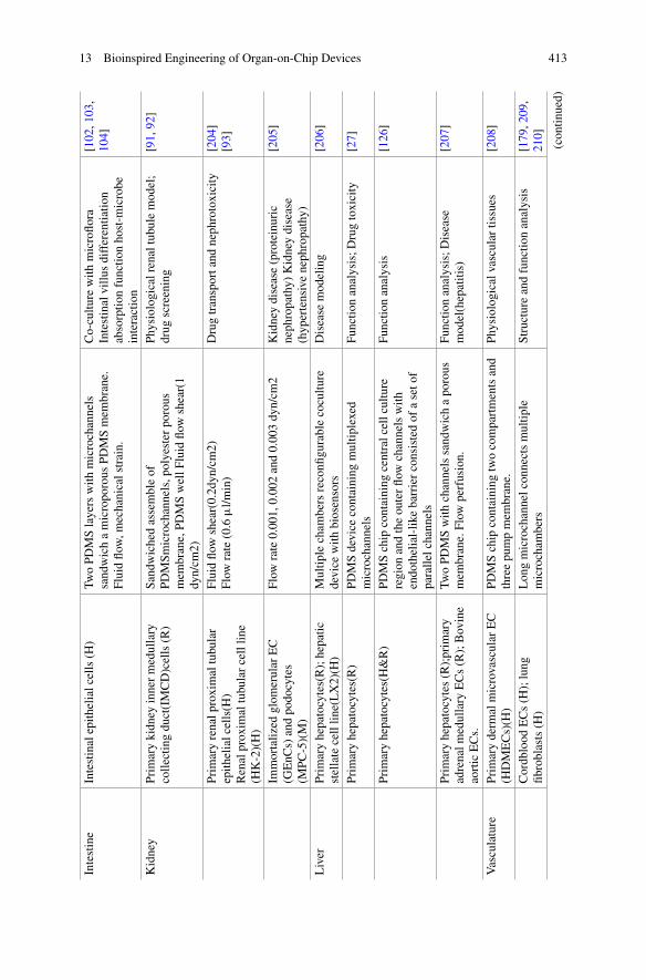
13 Bioinspired Engineering of Organ-on-Chip Devices 413
Inte
stin
eIn
test
inal
epith
elia
lcel
ls(H
)Tw
oPD
MS
laye
rsw
ithm
icro
chan
nels
sand
wic
ha
mic
ropo
rous
PDM
Sm
embr
ane.
Flui
dflo
w,m
echa
nica
lstr
ain.
Co-
cultu
rew
ithm
icro
flora
Inte
stin
alvi
llus
diff
eren
tiatio
nab
sorp
tion
func
tion
host
-mic
robe
inte
ract
ion
[102
,103
,10
4]
Kid
ney
Prim
ary
kidn
eyin
ner
med
ulla
ryco
llect
ing
duct
(IM
CD
)cel
ls(R
)Sa
ndw
iche
das
sem
ble
ofPD
MSm
icro
chan
nels
,pol
yest
erpo
rous
mem
bran
e,PD
MS
wel
lFlu
idflo
wsh
ear(
1dy
n/cm
2)
Phys
iolo
gica
lren
altu
bule
mod
el;
drug
scre
enin
g[9
1,92
]
Prim
ary
rena
lpro
xim
altu
bula
rep
ithel
ialc
ells
(H)
Ren
alpr
oxim
altu
bula
rce
lllin
e(H
K-2
)(H
)
Flui
dflo
wsh
ear(
0.2d
yn/c
m2)
Flow
rate
(0.6
μl/m
in)
Dru
gtr
ansp
orta
ndne
phro
toxi
city
[204
][9
3]
Imm
orta
lized
glom
erul
arE
C(G
EnC
s)an
dpo
docy
tes
(MPC
-5)(
M)
Flow
rate
0.00
1,0.
002
and
0.00
3dy
n/cm
2K
idne
ydi
seas
e(p
rote
inur
icne
phro
path
y)K
idne
ydi
seas
e(h
yper
tens
ive
neph
ropa
thy)
[205
]
Liv
erPr
imar
yhe
pato
cyte
s(R
);he
patic
stel
late
cell
line(
LX
2)(H
)M
ultip
lech
ambe
rsre
confi
gura
ble
cocu
lture
devi
cew
ithbi
osen
sors
Dis
ease
mod
elin
g[2
06]
Prim
ary
hepa
tocy
tes(
R)
PDM
Sde
vice
cont
aini
ngm
ultip
lexe
dm
icro
chan
nels
Func
tion
anal
ysis
;Dru
gto
xici
ty[2
7]
Prim
ary
hepa
tocy
tes(
H&
R)
PDM
Sch
ipco
ntai
ning
cent
ralc
ellc
ultu
rere
gion
and
the
oute
rflo
wch
anne
lsw
ithen
doth
elia
l-lik
eba
rrie
rco
nsis
ted
ofa
seto
fpa
ralle
lcha
nnel
s
Func
tion
anal
ysis
[126
]
Prim
ary
hepa
tocy
tes
(R);
prim
ary
adre
nalm
edul
lary
EC
s(R
);B
ovin
eao
rtic
EC
s.
Two
PDM
Sw
ithch
anne
lssa
ndw
ich
apo
rous
mem
bran
e.Fl
owpe
rfus
ion.
Func
tion
anal
ysis
;Dis
ease
mod
el(h
epat
itis)
[207
]
Vas
cula
ture
Prim
ary
derm
alm
icro
vasc
ular
EC
(HD
ME
Cs)
(H)
PDM
Sch
ipco
ntai
ning
two
com
part
men
tsan
dth
ree
pum
pm
embr
ane.
Phys
iolo
gica
lvas
cula
rtis
sues
[208
]
Cor
dblo
odE
Cs
(H);
lung
fibro
blas
ts(H
)L
ong
mic
roch
anne
lcon
nect
sm
ultip
lem
icro
cham
bers
Stru
ctur
ean
dfu
nctio
nan
alys
is[1
79,2
09,
210]
(con
tinue
d)
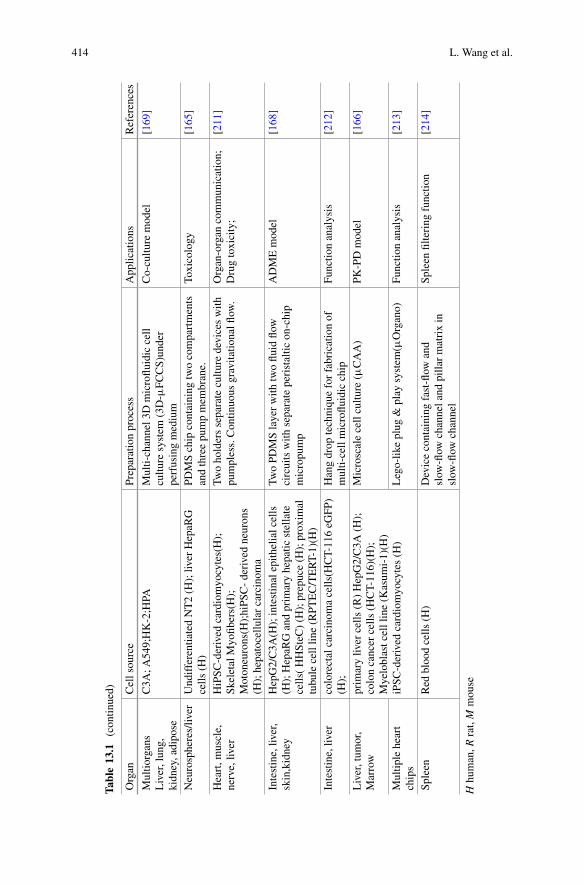
414 L. Wang et al.
Tabl
e13
.1(c
ontin
ued)
Org
anC
ells
ourc
ePr
epar
atio
npr
oces
sA
pplic
atio
nsR
efer
ence
s
Mul
tiorg
ans
Liv
er,l
ung,
kidn
ey,a
dipo
se
C3A
;A54
9;H
K-2
;HPA
Mul
ti-ch
anne
l3D
mic
roflu
idic
cell
cultu
resy
stem
(3D
-μFC
CS)
unde
rpe
rfus
ing
med
ium
Co-
cultu
rem
odel
[169
]
Neu
rosp
here
s/liv
erU
ndif
fere
ntia
ted
NT
2(H
);liv
erH
epaR
Gce
lls(H
)PD
MS
chip
cont
aini
ngtw
oco
mpa
rtm
ents
and
thre
epu
mp
mem
bran
e.To
xico
logy
[165
]
Hea
rt,m
uscl
e,ne
rve,
liver
HiP
SC-d
eriv
edca
rdio
myo
cyte
s(H
);Sk
elet
alM
yofib
ers(
H);
Mot
oneu
rons
(H);
hiPS
C-
deri
ved
neur
ons
(H);
hepa
toce
llula
rca
rcin
oma
Two
hold
ers
sepa
rate
cultu
rede
vice
sw
ithpu
mpl
ess.
Con
tinuo
usgr
avita
tiona
lflow
.O
rgan
-org
anco
mm
unic
atio
n;D
rug
toxi
city
;[2
11]
Inte
stin
e,liv
er,
skin
,kid
ney
Hep
G2/
C3A
(H);
inte
stin
alep
ithel
ialc
ells
(H);
Hep
aRG
and
prim
ary
hepa
ticst
ella
tece
lls(
HH
SteC
)(H
);pr
epuc
e(H
);pr
oxim
altu
bule
cell
line
(RPT
EC
/TE
RT-
1)(H
)
Two
PDM
Sla
yer
with
two
fluid
flow
circ
uits
with
sepa
rate
peri
stal
ticon
-chi
pm
icro
pum
p
AD
ME
mod
el[1
68]
Inte
stin
e,liv
erco
lore
ctal
carc
inom
ace
lls(H
CT-
116
eGFP
)(H
);H
ang
drop
tech
niqu
efo
rfa
bric
atio
nof
mul
ti-ce
llm
icro
fluid
icch
ipFu
nctio
nan
alys
is[2
12]
Liv
er,t
umor
,M
arro
wpr
imar
yliv
erce
lls(R
)H
epG
2/C
3A(H
);co
lon
canc
erce
lls(H
CT-
116)
(H);
Mye
lobl
astc
elll
ine
(Kas
umi-
1)(H
)iP
SC-d
eriv
edca
rdio
myo
cyte
s(H
)
Mic
rosc
ale
cell
cultu
re(μ
CA
A)
PK-P
Dm
odel
[166
]
Mul
tiple
hear
tch
ips
Leg
o-lik
epl
ug&
play
syst
em(μ
Org
ano)
Func
tion
anal
ysis
[213
]
Sple
enR
edbl
ood
cells
(H)
Dev
ice
cont
aini
ngfa
st-fl
owan
dsl
ow-fl
owch
anne
land
pilla
rm
atri
xin
slow
-flow
chan
nel
Sple
enfil
teri
ngfu
nctio
n[2
14]
Hhu
man
,Rra
t,M
mou
se
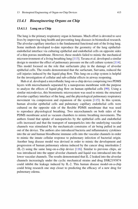
13 Bioinspired Engineering of Organ-on-Chip Devices 415
13.4.1 Bioengineering Organs on Chip
13.4.1.1 Lung on a Chip
The lung is the primary respiratory organ in humans. Much effort is devoted to savelives by improving lung health and preventing lung diseases in biomedical research.The alveolar-capillary interface is the fundamental functional unit of the living lung.Some methods developed to-date reproduce the geometry of the lung epithelial-endothelial interface via culturing epithelial and endothelial cells on opposite sidesof a thin porous membrane. However, these models failed to mimic the mechanicalmicroenvironment of a living breathing lung [113]. Tavana et al. developed a similardesign to monitor the effect of pulmonary pressure on the cell culture system [114].This model focused on the role that surfactants play in the damage of alveolarepithelial cells. The results demonstrated that the surfactant, Survanta, reduced thecell injuries induced by the liquid plug flow. This lung-on-a-chip system is helpfulfor the investigation of cellular and sub-cellular effects in airway reopening.
Huh et al. developed a microfluidic lung-on-a-chip device comprising two PDMSlayers with microchannels separated by a nanoporous membrane with the purposeto analyze the effects of liquid plug flow on human epithelial cells [99]. Using asimilar microdevice, this biomimetic microsystem was used to mimic the structuralalveolar-capillary interface of the lung, and the physiological pulmonary respiratorymovement via compression and expansion of the system [115]. In this system,human alveolar epithelial cells and pulmonary capillary endothelial cells werecultured on the opposite side of the flexible PDMS membrane that was usedto reproduce physiological breathing. Two microchannels on both sides of thePDMS membrane acted as vacuum chambers to mimic breathing movements. Theauthors found that uptake of nanoparticles by the epithelial cells and endothelialcells increased and that the transport of nanoparticles into the underlying vascularchannels was stimulated by the mechanicals constrains of air being pulled in andout of the device. The authors also introduced bacteria and inflammatory cytokinesinto the air and human bloodborne immune cells into the vascular channels in orderto mimic the innate cellular response to pulmonary infection of bacterial origin.Another lung disease model was devised in order to mimic the development andprogression of human pulmonary edema induced by the cancer drug interleukin-2(IL-2) using the same lung-on-a-chip device [116]. Similar to previous chips, airwas introduced into the upper alveolar channels and liquid was introduced into thelower vascular channels. The results demonstrated that IL-2 leaked into the alveolarchannels increasingly under the cyclic mechanical strains and drug DSK2193874could inhibit the leakage induced by IL-2. This human disease model-on-a-chipcould bring research one step closer to predicting the efficacy of a new drug forpulmonary edema.
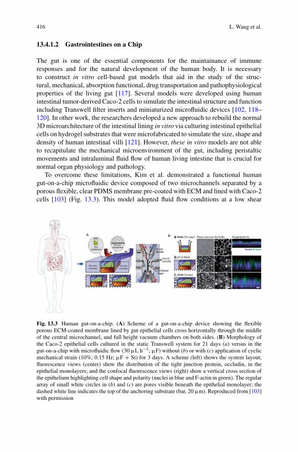
416 L. Wang et al.
13.4.1.2 Gastrointestines on a Chip
The gut is one of the essential components for the maintainance of immuneresponses and for the natural development of the human body. It is necessaryto construct in vitro cell-based gut models that aid in the study of the struc-tural, mechanical, absorption functional, drug transportation and pathophysiologicalproperties of the living gut [117]. Several models were developed using humanintestinal tumor-derived Caco-2 cells to simulate the intestinal structure and functionincluding Transwell filter inserts and miniaturized microfluidic devices [102, 118–120]. In other work, the researchers developed a new approach to rebuild the normal3D microarchitecture of the intestinal lining in vitro via culturing intestinal epithelialcells on hydrogel substrates that were microfabricated to simulate the size, shape anddensity of human intestinal villi [121]. However, these in vitro models are not ableto recapitulate the mechanical microenvironment of the gut, including peristalticmovements and intraluminal fluid flow of human living intestine that is crucial fornormal organ physiology and pathology.
To overcome these limitations, Kim et al. demonstrated a functional humangut-on-a-chip microfluidic device composed of two microchannels separated by aporous flexible, clear PDMS membrane pre-coated with ECM and lined with Caco-2cells [103] (Fig. 13.3). This model adopted fluid flow conditions at a low shear
Fig. 13.3 Human gut-on-a-chip. (A) Scheme of a gut-on-a-chip device showing the flexibleporous ECM-coated membrane lined by gut epithelial cells cross horizontally through the middleof the central microchannel, and full height vacuum chambers on both sides. (B) Morphology ofthe Caco-2 epithelial cells cultured in the static Transwell system for 21 days (a) versus in thegut-on-a-chip with microfluidic flow (30 μL h−1; μF) without (b) or with (c) application of cyclicmechanical strain (10%; 0.15 Hz; μF + St) for 3 days. A scheme (left) shows the system layout;fluorescence views (center) show the distribution of the tight junction protein, occludin, in theepithelial monolayers; and the confocal fluorescence views (right) show a vertical cross section ofthe epithelium highlighting cell shape and polarity (nuclei in blue and F-actin in green). The regulararray of small white circles in (b) and (c) are pores visible beneath the epithelial monolayer; thedashed white line indicates the top of the anchoring substrate (bar, 20 μm). Reproduced from [103]with permission
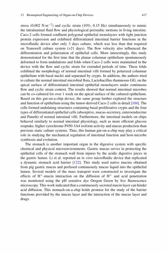
13 Bioinspired Engineering of Organ-on-Chip Devices 417
stress (0.002 N·m−2) and cyclic strain (10%; 0.15 Hz) simultaneously to mimicthe intraluminal fluid flow and physiological peristaltic motions in living intestine.Caco-2 cells formed confluent polygonal epithelial monolayers with tight junctionprotein expression and exhibited differentiated intestinal barrier functions in themicrofluidic device after only 3 days culture, which was less than that requiredon Transwell culture system (>21 days). The flow velocity also influenced thedifferentiation and polarization of epithelial cells. More interestingly, this studydemonstrated for the first time that the planar columnar epithelium spontaneouslydeformed to form undulations and folds when Caco-2 cells were maintained in thedevice with the flow and cyclic strain for extended periods of time. These foldsexhibited the morphology of normal intestinal villi formed by polarized columnarepithelium with basal nuclei and separated by crypts. In addition, the authors triedto culture the normal intestinal microbial flora, Lactobacillus rhamnosus GG, on theapical surface of differentiated intestinal epithelial monolayers under continuousflow and cyclic strain context. The results showed that normal intestinal microbescan be co-cultured for over 1 week on the apical surface of the cultured epithelium.Based on this gut-on-a-chip device, the same group further explored the structureand function of epithelium using the tumor-derived Caco-2 cells in detail [104]. Thecells formed undulating structures containing basal proliferative crypts and the fourtypes of differentiated epithelial cells (absorptive, mucus-secretory, enteroendocrineand Paneth) of normal intestinal villi. Furthermore, the intestinal models on chipsbehaved similarly to normal intestinal physiology, such as more efficient glucosereuptake, higher cytochrome P450-3A4 isoform activity and mucus production thanprevious static culture systems. Thus, this human gut-on-a-chip may play a criticalrole in studying the mechanical regulation of intestinal function and host-microbesymbiosis and evolution.
The stomach is another important organ in the digestive system with specificchemical and physical microenvironments. Gastric mucus serves in protecting theepithelial cells of the stomach wall from injures by the acidic digestive juices inthe gastric lumen. Li et al. reported an in vitro microfluidic device that replicateda dynamic stomach acid barrier [122]. This study used native mucins obtainedfrom pig gastric mucus and perfused continuously mucus liquid into the epitheliallumen. Several models of the mass transport were constructed to investigate theeffects of H+-mucin interaction on the diffusion of H+ and acid penetrationwas monitored using the pH sensitive dye Oregon Green by live fluorescencemicroscopy. This work indicated that a continuously secreted mucin layer can hinderacid diffusion. This stomach-on-a-chip holds promise for the study of the barrierfunctions provided by the mucus layer and the interaction of the mucus layer anddrugs.
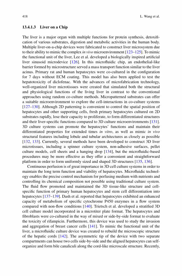
418 L. Wang et al.
13.4.1.3 Liver on a Chip
The liver is a major organ with multiple functions for protein synthesis, detoxifi-cation of various substrates, digestion and metabolic activities in the human body.Multiple liver-on-a-chip devices were fabricated to construct liver microsystem dueto their ability to mimic the complex in vivo microenvironment [123–125]. To mimicthe functional unit of the liver, Lee et al. developed a biologically inspired artificialliver sinusoid microdevice [126]. In this microfluidic chip, an endothelial-likebarrier formed by microstructure served a mass transport function similar to the liveracinus. Primary rat and human hepatocytes were co-cultured in the configurationfor 7 days without ECM coating. This model has also been applied to test thehepatotoxicity of diclofenac. With the advances of microfabrication technology,well-organized liver microtissues were created that simulated both the structuraland physiological functions of the living liver in contrast to the conventionalapproaches using random co-culture methods. Micropatterned substrates can offera suitable microenvironment to explore the cell-interactions in co-culture systems[127–130]. Although 2D patterning is convenient to control the spatial position ofhepatocytes and other supporting cells, fresh primary hepatocytes cultured on 2Dsubstrates rapidly, lose their capacity to proliferate, to form differentiated structuresand their liver-specific functions compared to 3D culture microenvironments [131].3D culture systems can promote the hepatocytes’ functions and maintain theirdifferentiated properties for extended times in vitro, as well as mimic in vivostructural features including lobule and tubular architectures as closely as possible[132, 133]. Currently, several methods have been developed to construct 3D livermicrotissues, including a spinner culture system, non-adhesive surfaces, pelletculture models, cell sheets and a hanging drop [134], but the microfluidic-basedprocedures may be more effective as they offer a convenient and straightforwardplatform in order to form uniformly sized and shaped 3D structures [135, 136].
Continuous perfusion is of great importance in 3D cell culture systems in order tomaintain the long term function and viability of hepatocytes. Microfluidic technol-ogy enables the precise control mechanism for perfusing medium with nutrients andcontrolling its chemical composition not possible using traditional culture system.The fluid flow promoted and maintained the 3D tissue-like structure and cell-specific function of primary human hepatocytes and stem cell differentiation intohepatocytes [137–139]. Dash et al. reported that hepatocytes exhibited an enhancedcapacity of metabolism of specific cytochrome P450 enzymes in a flow systemcompared with non-flow conditions [140]. Trietsch et al. developed a stratified 3Dcell culture model incorporated in a microtiter plate format. The hepatocytes andfibroblasts were co-cultured in the way of mixed or side-by-side format to evaluatethe toxicity of rifampicin. Furthermore, this device was used to study the invasionand aggregation of breast cancer cells [141]. To mimic the functional unit of theliver, a microfluidic culture device was created to rebuild the microscopic structureof the hepatic cords [142]. The asymmetric tip of the device with two separatecompartments can house two cells side-by-side and the aligned hepatocytes can self-organize and form bile canaliculi along the cord-like microscale structure. Recently,
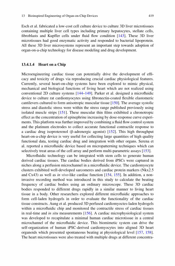
13 Bioinspired Engineering of Organ-on-Chip Devices 419
Esch et al. fabricated a low-cost cell culture device to culture 3D liver microtissuescontaining multiple liver cell types including primary hepatocytes, stellate cells,fibroblasts and Kupffer cells under fluid flow condition [143]. These 3D livermicrotissues had good enzymatic activity and responded to bacterial lipoprotein.All these 3D liver microsystems represent an important step towards adoption oforgan-on-a-chip technology for disease modeling and drug development.
13.4.1.4 Heart on a Chip
Microengineering cardiac tissue can potentially drive the development of effi-cacy and toxicity of drugs via reproducing crucial cardiac physiological features.Currently, several heart-on-chip systems have been explored to mimic physical,mechanical and biological functions of living heart which are not realized usingconventional 2D culture systems [144–149]. Parker et al. designed a microfluidicdevice to culture rat cardiomyocytes using fibronectin-coated flexible elastomericcantilevers cultured to form anisotropic muscular tissue [150]. The average systolicstress and diastolic stress were within the stress range published previously usingisolated muscle strips [151]. These muscular thin films exhibited a chronotropiceffect as the concentration of epinephrine increasing by dose-response curve experi-ments. This platform was further improved by combining a fluid flow control systemand the platinum electrodes to collect accurate functional contractile response toa cardiac drug isoproterenol (β-adrenergic agonist) [152]. This high throughputheart-on-a-chip device is very useful for collecting large quantities of high qualityfunctional data, testing cardiac drug and integration with other organs. Serena etal. reported a microfluidic device based on micropatterning techniques which canselectively treat areas of the cell array and perform multi-parametric assays [153].
Microfluidic technology can be integrated with stem cells to generate humanderived cardiac tissues. The cardiac bodies derived from iPSCs were captured inniches along a perfusion microchannel in a microfluidic device. The cardiomyocyteclusters exhibited well-developed sarcomeres and cardiac protein markers (Nkx2.5and Cx43) as well as in vivo-like cardiac function [154, 155]. In addition, a non-invasive recording method was introduced in this study to calculate the beatingfrequency of cardiac bodies using an ordinary microscope. These 3D cardiacbodies responded to different drugs rapidly in a similar manner to living hearttissue in a body. Other researchers explored different microfluidic approaches toform cell-laden hydrogels in order to evaluate the functionality of the cardiactissue constructs. Aung et al. produced 3D perfused cardiomyoctes-laden hydrogelswithin a microfluidic chip and monitored the contractile stress of cardiac tissuesin real-time and in situ measurements [156]. A cardiac microphysiological systemwas developed to recapitulate a minimal human cardiac microtissue in a centralmicrochannel of the microfluidic device. This biomimetic system can drive theself-organization of human iPSC-derived cardiomyocytes into aligned 3D heartorganoids which presented spontaneous beating at physiological level [157, 158].The heart microtissues were also treated with multiple drugs at different concentra-
420 L. Wang et al.
tions. The results demonstrated that half maximal inhibitory concentration (IC50)and half maximal effective concentration (EC50) values were more consistent withthe data on tissue scale references compared to studies at cellular level.
13.4.1.5 Blood-Brain-Barrier on Chip
The blood-brain-barrier (BBB) is a selective yet dynamic barrier between the centralnervous tissues and the circulatory system, which is formed by microvascularECs, pericytes and the perivascular end-feet of astrocytes. The dysfunction ofthis barrier is associated with brain tumors, Alzheimer’s and neurodegenerativediseases. Reproducing the physiological characteristics and functional responsesof the BBB in a reliable model will greatly promote the development of noveltherapeutics for central nervous system diseases [199, 200]. To mimic the in vivoBBB, a microfluidic BBB (μBBB) was created by lining a fibronectin-coatedpolycarbonate membrane with brain microvascular endothelial cells and astrocyteson the opposite side of the membrane [159]. This BBB exhibited a well-developedbiological function, including higher trans-epithelial electrical resistance (TEER)across the barrier, and being more impermeable to large molecules. A more complexmicrofluidic device of the neurovascular unit was created by co-culturing rat brainmicrovascular ECs and a mixture of three different cell types derived from ratbrain (astrocytes, neurons and microglia) on opposite sides of a porous membrane[160]. The endothelial cells formed a good barrier and the neuronal cells firedinhibitory as well as excitatory potentials after having been cultured for 10 days.Moreover, a tissue-mimetic neuroinflammation model was also established on thischip by stimulating the ECs with inflammatory factor TNF-α for 6 h, leading to thesignificant activation of resident microglia and astrocytes on the neural side.
13.4.1.6 Multiple Organs on a Chip
The advances of single organ-on-a-chip technology highlight the importance ofinteraction between different cell types within the same functional unit of theorgan. More complex microfluidic chips integrating tissue-tissue interfaces andcompartmentalized microchambers connected with microchannels (e.g., blood-brain-barrier, lung alveolar-capillary barrier) make it possible to create multipleorgans on a chip. These multi-organs on a chip are designed to maintain multiplecell types in different culture compartments in one device for long-term cultureand integrate them into one system in order to mimic organ-organ interaction.In the past decade, several multiorgans-on-chip devices have been developed forpotential use in disease modeling and drug toxicity screening applications. Tomimic physiologically relevant organs realistically in vivo, some key aspects needto be considered to design the chip, such as the size of the organs, interactionsbetween different organs and the organ volume-medium ratio (scaling of organsizes and vascular flow in vivo). Wikswo et al. discussed scaling arguments about
13 Bioinspired Engineering of Organ-on-Chip Devices 421
microengineering multi-organs on a chip to guide the design of a universal cell-culture medium without red blood cells [161]. It is mentioned that the prototypedrug metabolized by the liver component could have therapeutic effects. Thus, theliver should be placed upstream of the drug target organs in designing the multi-organs on a chip.
The field of multiorgans-on-chips is explored and investigated for differentpurposes [162]. To study tissue cross-effects, liver and intestinal slices isolatedfrom the same rat were cultured in one microdevice [163]. The liver and intestinalslices remained functional under flow conditions, indicating the potential of thischip for the study of inter-organ interactions. Wagner et al. cultured human primaryhepatocytes and skin biopsies simultaneously in one microfluidic device for up to28 days [164]. These studies demonstrated that the primary tissues from living bodycan maintain their functions in vitro under dynamic culture condition. Moreover, a3D two-organs-on-chip device was created to support the survival of differentiatedneurospheres and liver spheroids in a combined media circuit over 14 days [165].In the study it was suggested that the two combined tissues were more sensitiveto the 2, 5-hexanedione compared to respective single-tissue cultures, maybe dueto the tissue-tissue interactions. As is well established, most orally adminstreddrugs are absorbed and metabolised in the small intestine or the liver and excretedby the kidney. However, it is impossible to mimic these processes in vitro usingconventional cell-based models.
A typical example of a three organs-on-chip device was designed with a 3Dhydrogel culture in which three cell lines were cultured to assess metabolism-dependent cytotoxicity of anti-cancer drugs [166] (Fig. 13.4). In this microsystem,cancer cells, liver cells and myeloblasts were cultured in separate chambersrepresenting cancerous tissue, liver and bone marrow. Tegafur, an oral prodrug of 5-fluorouracil, was introduced into this system to test the cytotoxicity to the three celllines. Compared with conventional 96-well microtiter plates, the micro cell culturesystem enabled to reproduce the metabolism of Tegafur to 5-fluorouracil in the liverand the resulting death of cells caused by 5-fluorouracil, while cells in the 96-wellmicrotiter plate did not display the same results. Esch et al. designed a microscalebody-on-a-chip system containing gastrointestinal tract, liver and other tissues tomimic the oral uptake of nanoparticles. The nanoparticles were transported acrossthe GI epithelium and interacted with liver cells resulting in the damage of the latter[167].
To improve the functions of multiorgans-on-chip, a four-organ-chip microphysio-logical system was proposed to maintain the functionality of four organs containinghuman intestine, liver, skin and kidney over 28 days [168]. Considering the sizeratio of tissues on chip compared to human living organs, the intestine and skintissues on chip are at a size 100,000-fold smaller than their counterpart organs.3D liver microtissues, equivalent to ten liver lobules, were found to mimick liverfunction. Human proximal tubule cell lineage RPTEC/TERT-1 was used to formthe proximal tubule barrier on a polymeric membrane to mimic the metaboliteexcretion. A peristaltic micropump integrated in this chip enables pulsed mediumflow interconnecting the four organs through microchannels. This device considered
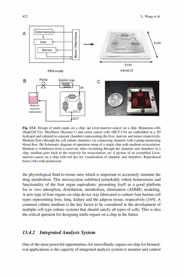
422 L. Wang et al.
Fig. 13.4 Design of multi-ogans on a chip. (a) Liver-marrow-cancer on a chip. Hepatoma cells(HepG2/C3A), Myeblasts (Kasumi-1) and colon cancer cells (HCT-116) are embedded in a 3Dhydrogel and cultured in separate chambers representing the liver, marrow and tumor respectively.Medium flows through the cell culture chambers via connecting channels with a pump mimickingblood flow. (b) Schematic diagram of operation setup of a single chip with medium recirculation.Medium is withdrawn from a reservoir, after circulating through the channels and chambers in achip, medium goes back to the reservoir for recirculation. (c) A picture of an assembled Liver-marrow-cancer on a chip with red dye for visualization of channels and chambers. Reproducedfrom [166] with permission
the physiological fluid-to-tissue ratio which is important to accurately simulate thedrug metabolism. This microsystem exhibited remarkably robust homeostasis andfunctionality of the four organ equivalents, presenting itself as a good platformfor in vitro adsorption, distribution, metabolism, elimination (ADME) modeling.A new type of four organs-on-chip device was fabricated to culture four human celltypes representing liver, lung, kidney and the adipose tissue, respectively [169]. Acommon culture medium is the key factor to be considered in the development ofmultiple cell type culture systems that should satisfy all types of cells. This is alsothe critical question for designing multi-organs on a chip in the future.
13.4.2 Integrated Analysis System
One of the most powerful opportunities for microfluidic organs-on-chip for biomed-ical applications is the capacity of integrated analysis system to monitor and control
13 Bioinspired Engineering of Organ-on-Chip Devices 423
organs in real-time. Existing automated techniques enable us to monitor cellularproliferation, morphology and molecular changes in multiwell plates. However,it is difficult to monitor the complex multiple parameters on organ microsystemsincluding gradients of chemical factors and of oxygen, fluid flow shear, mechanicalcues, matrix composition and metabolites. Currently, several types of microsensorsfor real-time monitoring of organ conditions have provided measurements of basiccell culture parameters including oxygen, pH, temperature, and molecular compo-sition (e.g., glucose and lactate). More specialized sensor measurements are widelyused to monitor the trans-epithelial electrical resistance (TEER) which usuallyexists in biological barriers, such as the blood-brain barrier, intestinal epithelial-capillary barrier and the lung alveolar-capillary barrier. Recent efforts indicate thatTEER measurements in organs-on-chip systems present specific challenges and arehighly sensitive to slight changes in the cell barrier membrane [170]. Hence, theautomated and precise measurements of TEER in a complex organ microsystemwould increase reliability and offer key information on the organ condition andresponses to different drugs.
13.5 Proof-of-Concept Applications of Organs-on-Chip
13.5.1 Disease Modeling
Disease modeling is one of the main applications of organs-on-chip technology,ranging from genetic diseases to infectious, cancerous and degenerative diseases,and is helpful for understanding the disease etiology, diagnostic strategies andeffective therapies. Several proof-of-concept disease models have been reported todate.
13.5.1.1 Inflammatory-Related Diseases
Inflammation reactions are closely related to many diseases and can lead toserious pathological symptoms. Pneumonia is one of most common lung diseases,characterized by its complexity, acute onset and difficulty to control it. Establishingan in vitro model to study the disease mechanism and to be able to screen forefficient drugs is an urgent requirement in order to address this challenging disease.The breathing lung-on-a-chip, as mentioned above, recapitulated the epithelium-endothelium interface under fluid flow and cyclic mechanical strain conditions tomimic the functional unit of the lung. Inflammatory models were developed inthis system by administrating immune activator TNF-α or bacteria which increasedexpression of surface ICAM-1 of endothelial cells and recruitment of humanneutrophils [114]. Based on the lung-on-a-chip system, the same research groupfurther mimicked a pulmonary edema model by the cancer drug interleukin-2 (IL-2)which can cause pulmonary vascular permeability and lung edema [115]. This lung
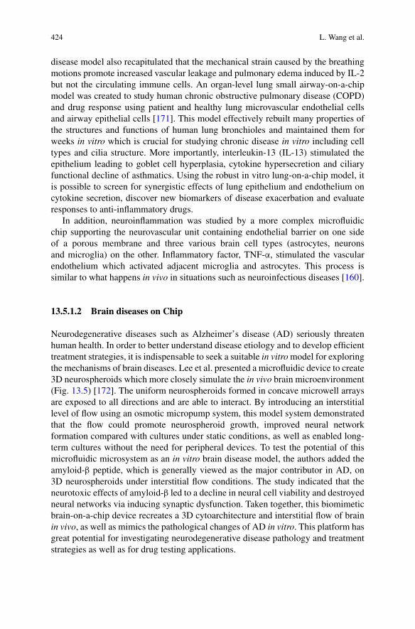
424 L. Wang et al.
disease model also recapitulated that the mechanical strain caused by the breathingmotions promote increased vascular leakage and pulmonary edema induced by IL-2but not the circulating immune cells. An organ-level lung small airway-on-a-chipmodel was created to study human chronic obstructive pulmonary disease (COPD)and drug response using patient and healthy lung microvascular endothelial cellsand airway epithelial cells [171]. This model effectively rebuilt many properties ofthe structures and functions of human lung bronchioles and maintained them forweeks in vitro which is crucial for studying chronic disease in vitro including celltypes and cilia structure. More importantly, interleukin-13 (IL-13) stimulated theepithelium leading to goblet cell hyperplasia, cytokine hypersecretion and ciliaryfunctional decline of asthmatics. Using the robust in vitro lung-on-a-chip model, itis possible to screen for synergistic effects of lung epithelium and endothelium oncytokine secretion, discover new biomarkers of disease exacerbation and evaluateresponses to anti-inflammatory drugs.
In addition, neuroinflammation was studied by a more complex microfluidicchip supporting the neurovascular unit containing endothelial barrier on one sideof a porous membrane and three various brain cell types (astrocytes, neuronsand microglia) on the other. Inflammatory factor, TNF-α, stimulated the vascularendothelium which activated adjacent microglia and astrocytes. This process issimilar to what happens in vivo in situations such as neuroinfectious diseases [160].
13.5.1.2 Brain diseases on Chip
Neurodegenerative diseases such as Alzheimer’s disease (AD) seriously threatenhuman health. In order to better understand disease etiology and to develop efficienttreatment strategies, it is indispensable to seek a suitable in vitro model for exploringthe mechanisms of brain diseases. Lee et al. presented a microfluidic device to create3D neurospheroids which more closely simulate the in vivo brain microenvironment(Fig. 13.5) [172]. The uniform neurospheroids formed in concave microwell arraysare exposed to all directions and are able to interact. By introducing an interstitiallevel of flow using an osmotic micropump system, this model system demonstratedthat the flow could promote neurospheroid growth, improved neural networkformation compared with cultures under static conditions, as well as enabled long-term cultures without the need for peripheral devices. To test the potential of thismicrofluidic microsystem as an in vitro brain disease model, the authors added theamyloid-β peptide, which is generally viewed as the major contributor in AD, on3D neurospheroids under interstitial flow conditions. The study indicated that theneurotoxic effects of amyloid-β led to a decline in neural cell viability and destroyedneural networks via inducing synaptic dysfunction. Taken together, this biomimeticbrain-on-a-chip device recreates a 3D cytoarchitecture and interstitial flow of brainin vivo, as well as mimics the pathological changes of AD in vitro. This platform hasgreat potential for investigating neurodegenerative disease pathology and treatmentstrategies as well as for drug testing applications.
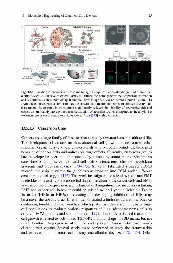
13 Bioinspired Engineering of Organ-on-Chip Devices 425
Fig. 13.5 Creating Alzheimer’s disease modeling on chip. (a) Schematic diagram of a brain-on-a-chip device. A concave microwell array is utilized for homogeneous neurospheroid formationand a continuous flow mimicking interstitial flow is applied via an osmotic pump system. (b)Dynamic culture significantly promotes the growth and function of neurosopheroids. (c) Amyloid-β treatment via an osmotic micropump significantly reduced the viability of neurospheroids andcaused a significantly more pronounced destruction of neural networks, compared to the amyloid-βtreatment under static conditions. Reproduced from [172] with permission
13.5.1.3 Cancers on Chip
Cancers are a large family of diseases that seriously threaten human health and life.The development of cancers involves abnormal cell growth and invasion of otherimportant organs. It is very helpful to establish in vitro models to study the biologicalbehavior of cancer cells and anticancer drug effects. Currently, numerous groupshave developed cancer-on-a-chip models by mimicking tumor microenvironmentsconsisting of complex cell-cell and cell-matrix interactions, chemokine/cytokinegradients and biophysical cues [173–175]. Xu et al. fabricated a bilayer PDMSmicrofluidic chip to mimic the glioblastoma invasion into ECM under differentconcentrations of oxygen [176]. This work investigated the role of hypoxia and EMTin glioblastoma and hypoxia promoted the proliferation of the cancer cells and EMT-associated protein expression, and enhanced cell migration. The mechanism linkingEMT and cancer cell behavior could be related to the Hypoxia-Inducible Factor1α or 2α (HIF1α or HIF2α), indicating that developing inhibitors of HIFs maybe a novel therapeutic drug. Li et al. demonstrated a high throughput microdevicecontaining tunable cell micro-niches, which performs flow-based analysis of largecell populations to evaluate various responses of lung adenocarcinoma cells todifferent ECM proteins and soluble factors [177]. This study indicated that tumor-cell growth is related to TGF-β and TGF-βR2 inhibitor drugs in a 3D matrix but notin a 2D culture. Angiogenesis of tumors is a key step of tumor metastasis towardsdistant target organs. Several works were performed to study the intravasationand extravasation of tumor cells using microfluidic devices [178, 179]. Other

426 L. Wang et al.
studies have utilized similar microfluidic technology to investigate the molecularmechanisms of cancer cell-immune cell crosstalk [180].
13.5.2 Drug Testing
13.5.2.1 Efficacy and Toxicity Testing
One challenge of drug development is the poor efficacy and unexpected toxicityin clinical trials caused by an absence of predicted therapeutic effects. The mainreason for undesired outcomes is that existing approaches fail to accurately predictin vivo drug efficacy before clinical application. Human organ-on-a-chip systemsthat model human physiological and pathological functional units of living organsprovide a promising tool to address the limitations of existing methods. They allowto reconstruct and pharmacologically modulate key aetiologies and clinical relevantresponses at various levels of biological complexity and to test unanticipatedoff-target toxicity. As described above, a heart-on-a-chip composed of 20 ratcardiomyocyte thin films was utilized to evaluate the inotropic effects of the β-adrenergic agonist isoproterenol which are similar to those previously determinedin rats [152]. A recent study reported a micro-engineered 3D model of EMTduring cancer progression for testing drug efficacy [181]. This model demonstratedEMT-induced tumor dispersion and phenotypic changes by culturing lung tumorspheroids in a matrix gel close to microchannels inhabited with endothelial cells.In addition, 12 drugs, including approved drugs, as well as prospective drugs thatare in the early discovery pipeline, were perfused into the vascular microchannelsto evaluate the potential of this model as a drug screening platform. The abilityof these drugs to inhibit EMT was tested by direct visualization of the cancerspheroids. The results showed that efficacious concentrations derived from thecancer-on-a-chip were higher than that from the 2D system, but are also closerto the range of effective drug concentrations determined in clinical trials. Severalmicro-engineered models of breast cancer and multiple organ models of uterinecancer, bone marrow and liver have also demonstrated a similar difference betweeneffective drug concentrations determined by organs-on-chip platforms and thoseby conventional 2D culture systems [182, 183]. To investigate the communicationbetween different brain regions, organotypic brain slices from rat hippocampus andentorhinal cortex were cultured in compartments interconnected by microchannels[184]. A glutamate receptor antagonist (kynurenic acid) was introduced to onemicrochannel in order to selectively inhibit the spontaneous electrical excitationof the treated brain tissue but not on the other brain slice. This design allows forthe selective pharmacological administration of only one tissue and evaluation of itseffects across the synaptic connection.
Recently, Qin et al. developed a novel in vivo-like 3D blood-brain barrier modelthat replicates the complex multicellular architecture, functions and mechanicalproperties of the BBB in vivo [185] (Fig. 13.6). The BBB model encompassedessential components, including primary brain microvascular endothelial cells,
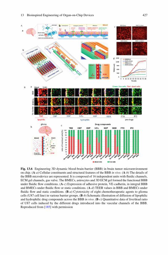
13 Bioinspired Engineering of Organ-on-Chip Devices 427
Fig. 13.6 Engineering 3D dynamic blood-brain-barrier (BBB) in brain tumor microenvironmenton chip. (A-a) Cellular constituents and structural features of the BBB in vivo. (A-b) The details ofthe BBB microdevice are represented. It is composed of 16 independent units with fluidic channels,ECM gel channels, gas valve. The BMECs, astrocytes and 3D ECM gel formed the functional BBBunder fluidic flow conditions. (A-c) Expression of adhesive protein, VE-cadherin, in integral BBBand BMECs under fluidic flow or static conditions. (A-d) TEER values in BBB and BMECs underfluidic flow and static conditions. (B-a) Cytotoxicity of eight chemotherapeutic agents to gliomacells (U87 cell line) in various barrier groups. (B-b) Schematic illustration of diffusion of lipophilicand hydrophilic drug compounds across the BBB in vivo. (B-c) Quantitative data of live/dead ratioof U87 cells induced by the different drugs introduced into the vascular channels of the BBB.Reproduced from [185] with permission
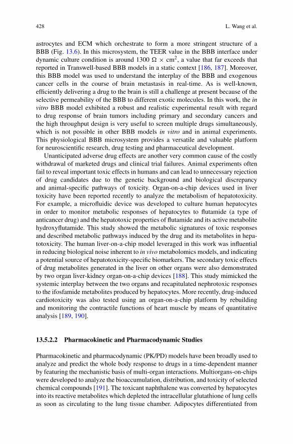
428 L. Wang et al.
astrocytes and ECM which orchestrate to form a more stringent structure of aBBB (Fig. 13.6). In this microsystem, the TEER value in the BBB interface underdynamic culture condition is around 1300 � × cm2, a value that far exceeds thatreported in Transwell-based BBB models in a static context [186, 187]. Moreover,this BBB model was used to understand the interplay of the BBB and exogenouscancer cells in the course of brain metastasis in real-time. As is well-known,efficiently delivering a drug to the brain is still a challenge at present because of theselective permeability of the BBB to different exotic molecules. In this work, the invitro BBB model exhibited a robust and realistic experimental result with regardto drug response of brain tumors including primary and secondary cancers andthe high throughput design is very useful to screen multiple drugs simultaneously,which is not possible in other BBB models in vitro and in animal experiments.This physiological BBB microsystem provides a versatile and valuable platformfor neuroscientific research, drug testing and pharmaceutical development.
Unanticipated adverse drug effects are another very common cause of the costlywithdrawal of marketed drugs and clinical trial failures. Animal experiments oftenfail to reveal important toxic effects in humans and can lead to unnecessary rejectionof drug candidates due to the genetic background and biological discrepancyand animal-specific pathways of toxicity. Organ-on-a-chip devices used in livertoxicity have been reported recently to analyze the metabolism of hepatotoxicity.For example, a microfluidic device was developed to culture human hepatocytesin order to monitor metabolic responses of hepatocytes to flutamide (a type ofanticancer drug) and the hepatotoxic properties of flutamide and its active metabolitehydroxyflutamide. This study showed the metabolic signatures of toxic responsesand described metabolic pathways induced by the drug and its metabolites in hepa-totoxicity. The human liver-on-a-chip model leveraged in this work was influentialin reducing biological noise inherent to in vivo metabolomics models, and indicatinga potential source of hepatotoxicity-specific biomarkers. The secondary toxic effectsof drug metabolites generated in the liver on other organs were also demonstratedby two organ liver-kidney organ-on-a-chip devices [188]. This study mimicked thesystemic interplay between the two organs and recapitulated nephrotoxic responsesto the ifosfamide metabolites produced by hepatocytes. More recently, drug-inducedcardiotoxicity was also tested using an organ-on-a-chip platform by rebuildingand monitoring the contractile functions of heart muscle by means of quantitativeanalysis [189, 190].
13.5.2.2 Pharmacokinetic and Pharmacodynamic Studies
Pharmacokinetic and pharmacodynamic (PK/PD) models have been broadly used toanalyze and predict the whole body response to drugs in a time-dependent mannerby featuring the mechanistic basis of multi-organ interactions. Multiorgans-on-chipswere developed to analyze the bioaccumulation, distribution, and toxicity of selectedchemical compounds [191]. The toxicant naphthalene was converted by hepatocytesinto its reactive metabolites which depleted the intracellular glutathione of lung cellsas soon as circulating to the lung tissue chamber. Adipocytes differentiated from
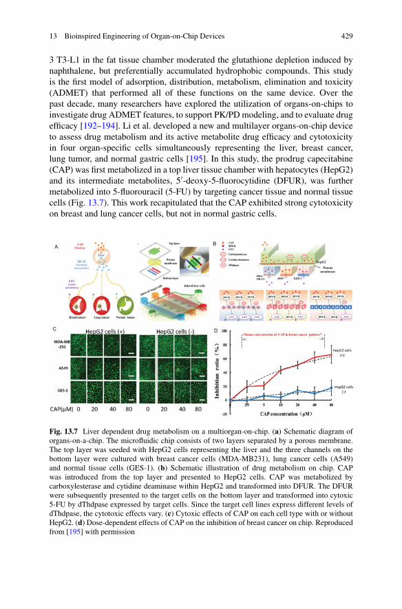
13 Bioinspired Engineering of Organ-on-Chip Devices 429
3 T3-L1 in the fat tissue chamber moderated the glutathione depletion induced bynaphthalene, but preferentially accumulated hydrophobic compounds. This studyis the first model of adsorption, distribution, metabolism, elimination and toxicity(ADMET) that performed all of these functions on the same device. Over thepast decade, many researchers have explored the utilization of organs-on-chips toinvestigate drug ADMET features, to support PK/PD modeling, and to evaluate drugefficacy [192–194]. Li et al. developed a new and multilayer organs-on-chip deviceto assess drug metabolism and its active metabolite drug efficacy and cytotoxicityin four organ-specific cells simultaneously representing the liver, breast cancer,lung tumor, and normal gastric cells [195]. In this study, the prodrug capecitabine(CAP) was first metabolized in a top liver tissue chamber with hepatocytes (HepG2)and its intermediate metabolites, 5′-deoxy-5-fluorocytidine (DFUR), was furthermetabolized into 5-fluorouracil (5-FU) by targeting cancer tissue and normal tissuecells (Fig. 13.7). This work recapitulated that the CAP exhibited strong cytotoxicityon breast and lung cancer cells, but not in normal gastric cells.
Fig. 13.7 Liver dependent drug metabolism on a multiorgan-on-chip. (a) Schematic diagram oforgans-on-a-chip. The microfluidic chip consists of two layers separated by a porous membrane.The top layer was seeded with HepG2 cells representing the liver and the three channels on thebottom layer were cultured with breast cancer cells (MDA-MB231), lung cancer cells (A549)and normal tissue cells (GES-1). (b) Schematic illustration of drug metabolism on chip. CAPwas introduced from the top layer and presented to HepG2 cells. CAP was metabolized bycarboxylesterase and cytidine deaminase within HepG2 and transformed into DFUR. The DFURwere subsequently presented to the target cells on the bottom layer and transformed into cytoxic5-FU by dThdpase expressed by target cells. Since the target cell lines express different levels ofdThdpase, the cytotoxic effects vary. (c) Cytoxic effects of CAP on each cell type with or withoutHepG2. (d) Dose-dependent effects of CAP on the inhibition of breast cancer on chip. Reproducedfrom [195] with permission
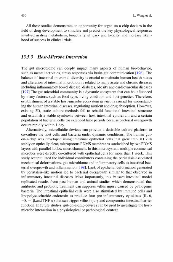
430 L. Wang et al.
All these studies demonstrate an opportunity for organ-on-a-chip devices in thefield of drug development to simulate and predict the key physiological responsesinvolved in drug metabolism, bioactivity, efficacy and toxicity, and increase likeli-hood of success in clinical trials.
13.5.3 Host-Microbe Interaction
The gut microbiome can deeply impact many aspects of human bio-behavior,such as mental activities, stress responses via brain-gut communication [196]. Thebalance of intestinal microbial diversity is crucial to maintain human health statusand alteration of intestinal microbiota is related to many acute and chronic diseasesincluding inflammatory bowel disease, diabetes, obesity and cardiovascular diseases[197].The gut microbial community is a dynamic ecosystem that can be influencedby many factors, such as food type, living condition and host genetics. Therefore,establishment of a stable host-microbe ecosystem in vitro is crucial for understand-ing the human intestinal diseases, regulating nutrient and drug absorption. However,existing 2D, static culture methods fail to rebuild functional intestinal structureand establish a stable symbiosis between host intestinal epithelium and a certainpopulation of bacterial cells for extended time periods because bacterial overgrowthoccurs rapidly within 1 day.
Alternatively, microfluidic devices can provide a desirable culture platform toco-culture the host cells and bacteria under dynamic conditions. The human gut-on-a-chip was developed using intestinal epithelial cells that grew into 3D villistably on optically clear, microporous PDMS membranes sandwiched by two PDMSlayers with parallel hollow microchannels. In this microsystem, multiple commensalmicrobes were directly co-cultured with epithelial cells for more than 1 week. Thisstudy recapitulated the individual contributors containing the peristalsis-associatedmechanical deformations, gut microbiome and inflammatory cells to intestinal bac-terial overgrowth and inflammation [198]. Lack of epithelial deformation generatedby peristalsis-like motion led to bacterial overgrowth similar to that observed ininflammatory intestinal diseases. Most importantly, this in vitro intestinal modelreplicated results from past human and animal studies which demonstrated thatantibiotic and probiotic treatment can suppress villus injury caused by pathogenicbacteria. The intestinal epithelial cells were also stimulated by immune cells andlipopolysaccharide endotoxin to produce four pro-inflammatory cytokines (IL-6,−8, −1β,and TNF-α) that can trigger villus injury and compromise intestinal barrierfunction. In future studies, gut-on-a-chip devices can be used to investigate the host-microbe interaction in a physiological or pathological context.
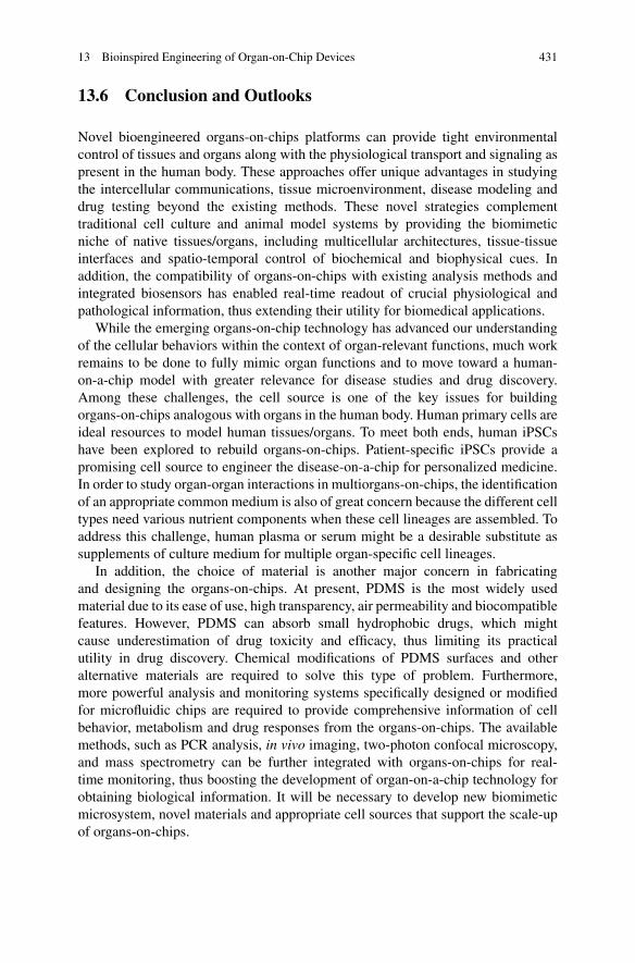
13 Bioinspired Engineering of Organ-on-Chip Devices 431
13.6 Conclusion and Outlooks
Novel bioengineered organs-on-chips platforms can provide tight environmentalcontrol of tissues and organs along with the physiological transport and signaling aspresent in the human body. These approaches offer unique advantages in studyingthe intercellular communications, tissue microenvironment, disease modeling anddrug testing beyond the existing methods. These novel strategies complementtraditional cell culture and animal model systems by providing the biomimeticniche of native tissues/organs, including multicellular architectures, tissue-tissueinterfaces and spatio-temporal control of biochemical and biophysical cues. Inaddition, the compatibility of organs-on-chips with existing analysis methods andintegrated biosensors has enabled real-time readout of crucial physiological andpathological information, thus extending their utility for biomedical applications.
While the emerging organs-on-chip technology has advanced our understandingof the cellular behaviors within the context of organ-relevant functions, much workremains to be done to fully mimic organ functions and to move toward a human-on-a-chip model with greater relevance for disease studies and drug discovery.Among these challenges, the cell source is one of the key issues for buildingorgans-on-chips analogous with organs in the human body. Human primary cells areideal resources to model human tissues/organs. To meet both ends, human iPSCshave been explored to rebuild organs-on-chips. Patient-specific iPSCs provide apromising cell source to engineer the disease-on-a-chip for personalized medicine.In order to study organ-organ interactions in multiorgans-on-chips, the identificationof an appropriate common medium is also of great concern because the different celltypes need various nutrient components when these cell lineages are assembled. Toaddress this challenge, human plasma or serum might be a desirable substitute assupplements of culture medium for multiple organ-specific cell lineages.
In addition, the choice of material is another major concern in fabricatingand designing the organs-on-chips. At present, PDMS is the most widely usedmaterial due to its ease of use, high transparency, air permeability and biocompatiblefeatures. However, PDMS can absorb small hydrophobic drugs, which mightcause underestimation of drug toxicity and efficacy, thus limiting its practicalutility in drug discovery. Chemical modifications of PDMS surfaces and otheralternative materials are required to solve this type of problem. Furthermore,more powerful analysis and monitoring systems specifically designed or modifiedfor microfluidic chips are required to provide comprehensive information of cellbehavior, metabolism and drug responses from the organs-on-chips. The availablemethods, such as PCR analysis, in vivo imaging, two-photon confocal microscopy,and mass spectrometry can be further integrated with organs-on-chips for real-time monitoring, thus boosting the development of organ-on-a-chip technology forobtaining biological information. It will be necessary to develop new biomimeticmicrosystem, novel materials and appropriate cell sources that support the scale-upof organs-on-chips.
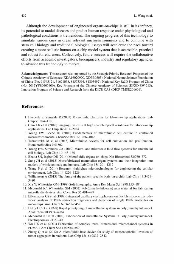
432 L. Wang et al.
Although the development of engineered organs-on-chips is still in its infancy,its potential to model diseases and predict human response under physiological andpathological conditions is tremendous. The ongoing progress of this technology tosimulate various cues in organ relevant microenvironments and to combine withstem cell biology and traditional biological assays will accelerate the pace towardcreating a more realistic human-on-a-chip model system that is accessible, practicaland robust for end users. Collectively, future success will require the collaborativeefforts from academic investigators, bioengineers, industry and regulatory agenciesto advance this technology to market.
Acknowledgments This research was supported by the Strategic Priority Research Program of theChinese Academy of Sciences (XDA16020900, XDPB0305), National Nature Science Foundationof China (No. 91543121, 31671038, 81573394, 81803492), National Key R&D Program of China(No. 2017YFB0405400), Key Program of the Chinese Academy of Sciences (KFZD-SW-213),Innovation Program of Science and Research from the DICP, CAS (DICP TMSR201601).
References
1. Haeberle S, Zengerle R (2007) Microfluidic platforms for lab-on-a-chip applications. LabChip 7:1094–1110
2. Chin LK et al (2016) Imaging live cells at high spatiotemporal resolution for lab-on-a-chipapplications. Lab Chip 16:2014–2024
3. Young EW, Beebe DJ (2010) Fundamentals of microfluidic cell culture in controlledmicroenvironments. ChemSoc Rev 39:1036–1048
4. Tehranirokh M et al (2013) Microfluidic devices for cell cultivation and proliferation.Biomicrofluidics 7:51502
5. Young EW, Simmons CA (2010) Macro- and microscale fluid flow systems for endothelialcell biology. Lab Chip 10:143–160
6. Bhatia SN, Ingber DE (2014) Microfluidic organs-on-chips. Nat Biotechnol 32:760–7727. Sung JH et al (2013) Microfabricated mammalian organ systems and their integration into
models of whole animals and humans. Lab Chip 13:1201–12128. Tseng P et al (2014) Research highlights: microtechnologies for engineering the cellular
environment. Lab Chip 14:1226–12299. Williamson A (2013) The future of the patient-specific body-on-a-chip. Lab Chip 13:3471–
348010. Xia Y, Whitesides GM (1998) Soft lithography. Annu Rev Mater Sci 1998:153–18411. Mcdonald JC, Whitesides GM (2002) Poly(dimethylsiloxane) as a material for fabricating
microfluidic devices. Acc Chem Res 35:491–49912. Effenhauser CS et al (1997) Integrated capillary electrophoresis on flexible silicone microde-
vices: analysis of DNA restriction fragments and detection of single DNA molecules onmicrochips. Anal Chem 69:3451–3457
13. Duffy DC et al (1998) Rapid prototyping of microfluidic systems in poly(dimethylsiloxane).Anal Chem 70:4974–4984
14. Mcdonald JC et al (2000) Fabrication of microfluidic Systems in Poly(dimethylsiloxane).Electrophoresis 21:27–40
15. Wu HK et al (2003) Fabrication of complex three- dimensional microchannel systems inPDMS. J Am Chem Soc 125:554–559
16. Zhang Q et al (2012) A microfluidic-base device for study of transendothelial invasion oftumor aggregates in realtiem. Lab Chip 12(16):2837–2842
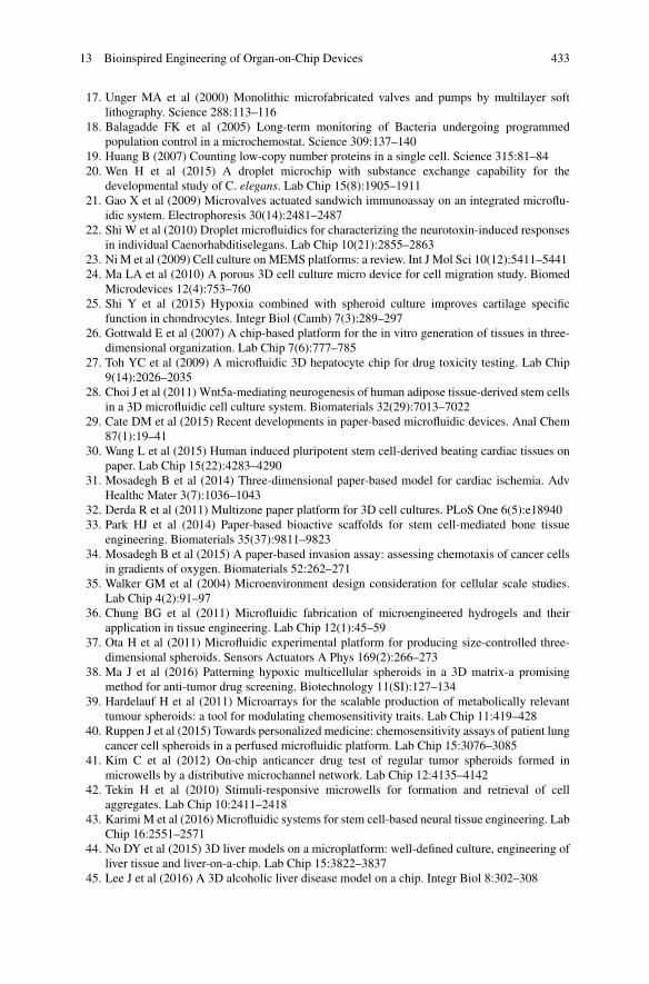
13 Bioinspired Engineering of Organ-on-Chip Devices 433
17. Unger MA et al (2000) Monolithic microfabricated valves and pumps by multilayer softlithography. Science 288:113–116
18. Balagadde FK et al (2005) Long-term monitoring of Bacteria undergoing programmedpopulation control in a microchemostat. Science 309:137–140
19. Huang B (2007) Counting low-copy number proteins in a single cell. Science 315:81–8420. Wen H et al (2015) A droplet microchip with substance exchange capability for the
developmental study of C. elegans. Lab Chip 15(8):1905–191121. Gao X et al (2009) Microvalves actuated sandwich immunoassay on an integrated microflu-
idic system. Electrophoresis 30(14):2481–248722. Shi W et al (2010) Droplet microfluidics for characterizing the neurotoxin-induced responses
in individual Caenorhabditiselegans. Lab Chip 10(21):2855–286323. Ni M et al (2009) Cell culture on MEMS platforms: a review. Int J Mol Sci 10(12):5411–544124. Ma LA et al (2010) A porous 3D cell culture micro device for cell migration study. Biomed
Microdevices 12(4):753–76025. Shi Y et al (2015) Hypoxia combined with spheroid culture improves cartilage specific
function in chondrocytes. Integr Biol (Camb) 7(3):289–29726. Gottwald E et al (2007) A chip-based platform for the in vitro generation of tissues in three-
dimensional organization. Lab Chip 7(6):777–78527. Toh YC et al (2009) A microfluidic 3D hepatocyte chip for drug toxicity testing. Lab Chip
9(14):2026–203528. Choi J et al (2011) Wnt5a-mediating neurogenesis of human adipose tissue-derived stem cells
in a 3D microfluidic cell culture system. Biomaterials 32(29):7013–702229. Cate DM et al (2015) Recent developments in paper-based microfluidic devices. Anal Chem
87(1):19–4130. Wang L et al (2015) Human induced pluripotent stem cell-derived beating cardiac tissues on
paper. Lab Chip 15(22):4283–429031. Mosadegh B et al (2014) Three-dimensional paper-based model for cardiac ischemia. Adv
Healthc Mater 3(7):1036–104332. Derda R et al (2011) Multizone paper platform for 3D cell cultures. PLoS One 6(5):e1894033. Park HJ et al (2014) Paper-based bioactive scaffolds for stem cell-mediated bone tissue
engineering. Biomaterials 35(37):9811–982334. Mosadegh B et al (2015) A paper-based invasion assay: assessing chemotaxis of cancer cells
in gradients of oxygen. Biomaterials 52:262–27135. Walker GM et al (2004) Microenvironment design consideration for cellular scale studies.
Lab Chip 4(2):91–9736. Chung BG et al (2011) Microfluidic fabrication of microengineered hydrogels and their
application in tissue engineering. Lab Chip 12(1):45–5937. Ota H et al (2011) Microfluidic experimental platform for producing size-controlled three-
dimensional spheroids. Sensors Actuators A Phys 169(2):266–27338. Ma J et al (2016) Patterning hypoxic multicellular spheroids in a 3D matrix-a promising
method for anti-tumor drug screening. Biotechnology 11(SI):127–13439. Hardelauf H et al (2011) Microarrays for the scalable production of metabolically relevant
tumour spheroids: a tool for modulating chemosensitivity traits. Lab Chip 11:419–42840. Ruppen J et al (2015) Towards personalized medicine: chemosensitivity assays of patient lung
cancer cell spheroids in a perfused microfluidic platform. Lab Chip 15:3076–308541. Kim C et al (2012) On-chip anticancer drug test of regular tumor spheroids formed in
microwells by a distributive microchannel network. Lab Chip 12:4135–414242. Tekin H et al (2010) Stimuli-responsive microwells for formation and retrieval of cell
aggregates. Lab Chip 10:2411–241843. Karimi M et al (2016) Microfluidic systems for stem cell-based neural tissue engineering. Lab
Chip 16:2551–257144. No DY et al (2015) 3D liver models on a microplatform: well-defined culture, engineering of
liver tissue and liver-on-a-chip. Lab Chip 15:3822–383745. Lee J et al (2016) A 3D alcoholic liver disease model on a chip. Integr Biol 8:302–308
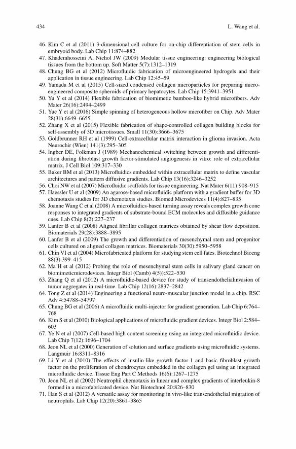
434 L. Wang et al.
46. Kim C et al (2011) 3-dimensional cell culture for on-chip differentiation of stem cells inembryoid body. Lab Chip 11:874–882
47. Khademhosseini A, Nichol JW (2009) Modular tissue engineering: engineering biologicaltissues from the bottom up. Soft Matter 5(7):1312–1319
48. Chung BG et al (2012) Microfluidic fabrication of microengineered hydrogels and theirapplication in tissue engineering. Lab Chip 12:45–59
49. Yamada M et al (2015) Cell-sized condensed collagen microparticles for preparing micro-engineered composite spheroids of primary hepatocytes. Lab Chip 15:3941–3951
50. Yu Y et al (2014) Flexible fabrication of biomimetic bamboo-like hybrid microfibers. AdvMater 26(16):2494–2499
51. Yue Y et al (2016) Simple spinning of heterogeneous hollow microfiber on Chip. Adv Mater28(31):6649–6655
52. Zhang X et al (2015) Flexible fabrication of shape-controlled collagen building blocks forself-assembly of 3D microtissues. Small 11(30):3666–3675
53. Goldbrunner RH et al (1999) Cell-extracellular matrix interaction in glioma invasion. ActaNeurochir (Wien) 141(3):295–305
54. Ingber DE, Folkman J (1989) Mechanochemical switching between growth and differenti-ation during fibroblast growth factor-stimulated angiogenesis in vitro: role of extracellularmatrix. J Cell Biol 109:317–330
55. Baker BM et al (2013) Microfluidics embedded within extracellular matrix to define vasculararchitectures and pattern diffusive gradients. Lab Chip 13(16):3246–3252
56. Choi NW et al (2007) Microfluidic scaffolds for tissue engineering. Nat Mater 6(11):908–91557. Haessler U et al (2009) An agarose-based microfluidic platform with a gradient buffer for 3D
chemotaxis studies for 3D chemotaxis studies. Biomed Microdevices 11(4):827–83558. Joanne Wang C et al (2008) A microfluidics-based turning assay reveals complex growth cone
responses to integrated gradients of substrate-bound ECM molecules and diffusible guidancecues. Lab Chip 8(2):227–237
59. Lanfer B et al (2008) Aligned fibrillar collagen matrices obtained by shear flow deposition.Biomaterials 29(28):3888–3895
60. Lanfer B et al (2009) The growth and differentiation of mesenchymal stem and progenitorcells cultured on aligned collagen matrices. Biomaterials 30(30):5950–5958
61. Chin VI et al (2004) Microfabricated platform for studying stem cell fates. Biotechnol Bioeng88(3):399–415
62. Ma H et al (2012) Probing the role of mesenchymal stem cells in salivary gland cancer onbiomimeticmicrodevices. Integr Biol (Camb) 4(5)):522–530
63. Zhang Q et al (2012) A microfluidic-based device for study of transendothelialinvasion oftumor aggregates in real-time. Lab Chip 12(16):2837–2842
64. Tong Z et al (2014) Engineering a functional neuro-muscular junction model in a chip. RSCAdv 4:54788–54797
65. Chung BG et al (2006) A microfluidic multi-injector for gradient generation. Lab Chip 6:764–768
66. Kim S et al (2010) Biological applications of microfluidic gradient devices. Integr Biol 2:584–603
67. Ye N et al (2007) Cell-based high content screening using an integrated microfluidic device.Lab Chip 7(12):1696–1704
68. Jeon NL et al (2000) Generation of solution and surface gradients using microfluidic systems.Langmuir 16:8311–8316
69. Li Y et al (2010) The effects of insulin-like growth factor-1 and basic fibroblast growthfactor on the proliferation of chondrocytes embedded in the collagen gel using an integratedmicrofluidic device. Tissue Eng Part C Methods 16(6):1267–1275
70. Jeon NL et al (2002) Neutrophil chemotaxis in linear and complex gradients of interleukin-8formed in a microfabricated device. Nat Biotechnol 20:826–830
71. Han S et al (2012) A versatile assay for monitoring in vivo-like transendothelial migration ofneutrophils. Lab Chip 12(20):3861–3865
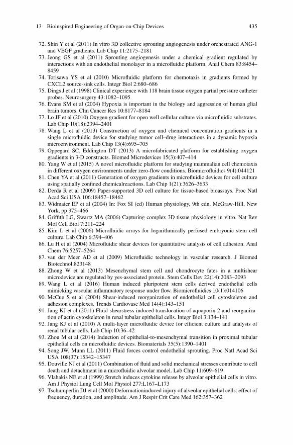
13 Bioinspired Engineering of Organ-on-Chip Devices 435
72. Shin Y et al (2011) In vitro 3D collective sprouting angiogenesis under orchestrated ANG-1and VEGF gradients. Lab Chip 11:2175–2181
73. Jeong GS et al (2011) Sprouting angiogenesis under a chemical gradient regulated byinteractions with an endothelial monolayer in a microfluidic platform. Anal Chem 83:8454–8459
74. Torisawa YS et al (2010) Microfluidic platform for chemotaxis in gradients formed byCXCL2 source-sink cells. Integr Biol 2:680–686
75. Dings J et al (1998) Clinical experience with 118 brain tissue oxygen partial pressure catheterprobes. Neurosurgery 43:1082–1095
76. Evans SM et al (2004) Hypoxia is important in the biology and aggression of human glialbrain tumors. Clin Cancer Res 10:8177–8184
77. Lo JF et al (2010) Oxygen gradient for open well cellular culture via microfluidic substrates.Lab Chip 10(18):2394–2401
78. Wang L et al (2013) Construction of oxygen and chemical concentration gradients in asingle microfluidic device for studying tumor cell–drug interactions in a dynamic hypoxiamicroenvironment. Lab Chip 13(4):695–705
79. Oppegard SC, Eddington DT (2013) A microfabricated platform for establishing oxygengradients in 3-D constructs. Biomed Microdevices 15(3):407–414
80. Yang W et al (2015) A novel microfluidic platform for studying mammalian cell chemotaxisin different oxygen environments under zero-flow conditions. Biomicrofluidics 9(4):044121
81. Chen YA et al (2011) Generation of oxygen gradients in microfluidic devices for cell cultureusing spatially confined chemicalreactions. Lab Chip 1(21):3626–3633
82. Derda R et al (2009) Paper-supported 3D cell culture for tissue-based bioassays. Proc NatlAcad Sci USA 106:18457–18462
83. Widmaier EP et al (2004) In: Fox SI (ed) Human physiology, 9th edn. McGraw-Hill, NewYork, pp 375–466
84. Griffith LG, Swartz MA (2006) Capturing complex 3D tissue physiology in vitro. Nat RevMol Cell Biol 7:211–224
85. Kim L et al (2006) Microfluidic arrays for logarithmically perfused embryonic stem cellculture. Lab Chip 6:394–406
86. Lu H et al (2004) Microfluidic shear devices for quantitative analysis of cell adhesion. AnalChem 76:5257–5264
87. van der Meer AD et al (2009) Microfluidic technology in vascular research. J BiomedBiotechnol:823148
88. Zhong W et al (2013) Mesenchymal stem cell and chondrocyte fates in a multishearmicrodevice are regulated by yes-associated protein. Stem Cells Dev 22(14):2083–2093
89. Wang L et al (2016) Human induced pluripotent stem cells derived endothelial cellsmimicking vascular inflammatory response under flow. Biomicrofluidics 10(1):014106
90. McCue S et al (2004) Shear-induced reorganization of endothelial cell cytoskeleton andadhesion complexes. Trends Cardiovasc Med 14(4):143–151
91. Jang KJ et al (2011) Fluid-shearstress-induced translocation of aquaporin-2 and reorganiza-tion of actin cytoskeleton in renal tubular epithelial cells. Integr Biol 3:134–141
92. Jang KJ et al (2010) A multi-layer microfluidic device for efficient culture and analysis ofrenal tubular cells. Lab Chip 10:36–42
93. Zhou M et al (2014) Induction of epithelial-to-mesenchymal transition in proximal tubularepithelial cells on microfluidic devices. Biomaterials 35(5):1390–1401
94. Song JW, Munn LL (2011) Fluid forces control endothelial sprouting. Proc Natl Acad SciUSA 108(37):15342–15347
95. Douville NJ et al (2011) Combination of fluid and solid mechanical stresses contribute to celldeath and detachment in a microfluidic alveolar model. Lab Chip 11:609–619
96. Vlahakis NE et al (1999) Stretch induces cytokine release by alveolar epithelial cells in vitro.Am J Physiol Lung Cell Mol Physiol 277:L167–L173
97. Tschumperlin DJ et al (2000) Deformationinduced injury of alveolar epithelial cells: effect offrequency, duration, and amplitude. Am J Respir Crit Care Med 162:357–362
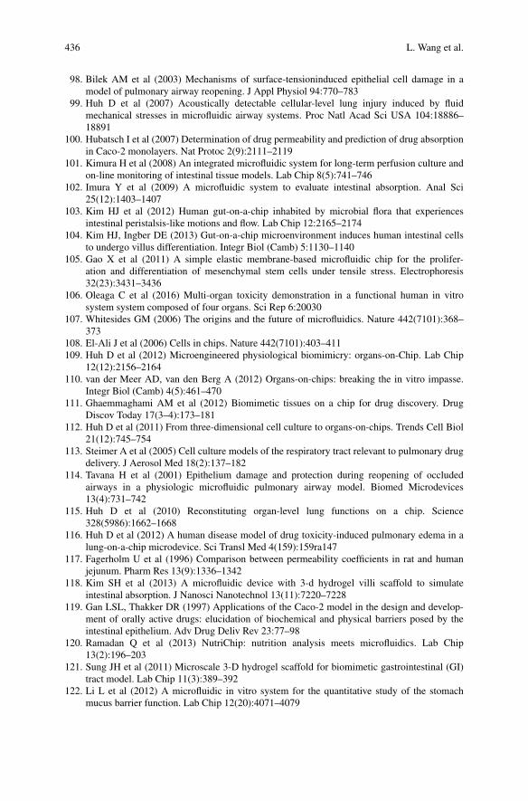
436 L. Wang et al.
98. Bilek AM et al (2003) Mechanisms of surface-tensioninduced epithelial cell damage in amodel of pulmonary airway reopening. J Appl Physiol 94:770–783
99. Huh D et al (2007) Acoustically detectable cellular-level lung injury induced by fluidmechanical stresses in microfluidic airway systems. Proc Natl Acad Sci USA 104:18886–18891
100. Hubatsch I et al (2007) Determination of drug permeability and prediction of drug absorptionin Caco-2 monolayers. Nat Protoc 2(9):2111–2119
101. Kimura H et al (2008) An integrated microfluidic system for long-term perfusion culture andon-line monitoring of intestinal tissue models. Lab Chip 8(5):741–746
102. Imura Y et al (2009) A microfluidic system to evaluate intestinal absorption. Anal Sci25(12):1403–1407
103. Kim HJ et al (2012) Human gut-on-a-chip inhabited by microbial flora that experiencesintestinal peristalsis-like motions and flow. Lab Chip 12:2165–2174
104. Kim HJ, Ingber DE (2013) Gut-on-a-chip microenvironment induces human intestinal cellsto undergo villus differentiation. Integr Biol (Camb) 5:1130–1140
105. Gao X et al (2011) A simple elastic membrane-based microfluidic chip for the prolifer-ation and differentiation of mesenchymal stem cells under tensile stress. Electrophoresis32(23):3431–3436
106. Oleaga C et al (2016) Multi-organ toxicity demonstration in a functional human in vitrosystem system composed of four organs. Sci Rep 6:20030
107. Whitesides GM (2006) The origins and the future of microfluidics. Nature 442(7101):368–373
108. El-Ali J et al (2006) Cells in chips. Nature 442(7101):403–411109. Huh D et al (2012) Microengineered physiological biomimicry: organs-on-Chip. Lab Chip
12(12):2156–2164110. van der Meer AD, van den Berg A (2012) Organs-on-chips: breaking the in vitro impasse.
Integr Biol (Camb) 4(5):461–470111. Ghaemmaghami AM et al (2012) Biomimetic tissues on a chip for drug discovery. Drug
Discov Today 17(3–4):173–181112. Huh D et al (2011) From three-dimensional cell culture to organs-on-chips. Trends Cell Biol
21(12):745–754113. Steimer A et al (2005) Cell culture models of the respiratory tract relevant to pulmonary drug
delivery. J Aerosol Med 18(2):137–182114. Tavana H et al (2001) Epithelium damage and protection during reopening of occluded
airways in a physiologic microfluidic pulmonary airway model. Biomed Microdevices13(4):731–742
115. Huh D et al (2010) Reconstituting organ-level lung functions on a chip. Science328(5986):1662–1668
116. Huh D et al (2012) A human disease model of drug toxicity-induced pulmonary edema in alung-on-a-chip microdevice. Sci Transl Med 4(159):159ra147
117. Fagerholm U et al (1996) Comparison between permeability coefficients in rat and humanjejunum. Pharm Res 13(9):1336–1342
118. Kim SH et al (2013) A microfluidic device with 3-d hydrogel villi scaffold to simulateintestinal absorption. J Nanosci Nanotechnol 13(11):7220–7228
119. Gan LSL, Thakker DR (1997) Applications of the Caco-2 model in the design and develop-ment of orally active drugs: elucidation of biochemical and physical barriers posed by theintestinal epithelium. Adv Drug Deliv Rev 23:77–98
120. Ramadan Q et al (2013) NutriChip: nutrition analysis meets microfluidics. Lab Chip13(2):196–203
121. Sung JH et al (2011) Microscale 3-D hydrogel scaffold for biomimetic gastrointestinal (GI)tract model. Lab Chip 11(3):389–392
122. Li L et al (2012) A microfluidic in vitro system for the quantitative study of the stomachmucus barrier function. Lab Chip 12(20):4071–4079
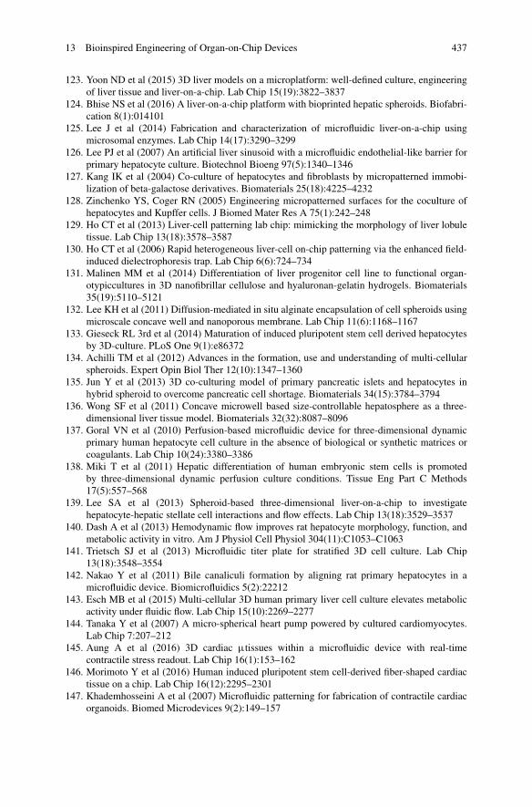
13 Bioinspired Engineering of Organ-on-Chip Devices 437
123. Yoon ND et al (2015) 3D liver models on a microplatform: well-defined culture, engineeringof liver tissue and liver-on-a-chip. Lab Chip 15(19):3822–3837
124. Bhise NS et al (2016) A liver-on-a-chip platform with bioprinted hepatic spheroids. Biofabri-cation 8(1):014101
125. Lee J et al (2014) Fabrication and characterization of microfluidic liver-on-a-chip usingmicrosomal enzymes. Lab Chip 14(17):3290–3299
126. Lee PJ et al (2007) An artificial liver sinusoid with a microfluidic endothelial-like barrier forprimary hepatocyte culture. Biotechnol Bioeng 97(5):1340–1346
127. Kang IK et al (2004) Co-culture of hepatocytes and fibroblasts by micropatterned immobi-lization of beta-galactose derivatives. Biomaterials 25(18):4225–4232
128. Zinchenko YS, Coger RN (2005) Engineering micropatterned surfaces for the coculture ofhepatocytes and Kupffer cells. J Biomed Mater Res A 75(1):242–248
129. Ho CT et al (2013) Liver-cell patterning lab chip: mimicking the morphology of liver lobuletissue. Lab Chip 13(18):3578–3587
130. Ho CT et al (2006) Rapid heterogeneous liver-cell on-chip patterning via the enhanced field-induced dielectrophoresis trap. Lab Chip 6(6):724–734
131. Malinen MM et al (2014) Differentiation of liver progenitor cell line to functional organ-otypiccultures in 3D nanofibrillar cellulose and hyaluronan-gelatin hydrogels. Biomaterials35(19):5110–5121
132. Lee KH et al (2011) Diffusion-mediated in situ alginate encapsulation of cell spheroids usingmicroscale concave well and nanoporous membrane. Lab Chip 11(6):1168–1167
133. Gieseck RL 3rd et al (2014) Maturation of induced pluripotent stem cell derived hepatocytesby 3D-culture. PLoS One 9(1):e86372
134. Achilli TM et al (2012) Advances in the formation, use and understanding of multi-cellularspheroids. Expert Opin Biol Ther 12(10):1347–1360
135. Jun Y et al (2013) 3D co-culturing model of primary pancreatic islets and hepatocytes inhybrid spheroid to overcome pancreatic cell shortage. Biomaterials 34(15):3784–3794
136. Wong SF et al (2011) Concave microwell based size-controllable hepatosphere as a three-dimensional liver tissue model. Biomaterials 32(32):8087–8096
137. Goral VN et al (2010) Perfusion-based microfluidic device for three-dimensional dynamicprimary human hepatocyte cell culture in the absence of biological or synthetic matrices orcoagulants. Lab Chip 10(24):3380–3386
138. Miki T et al (2011) Hepatic differentiation of human embryonic stem cells is promotedby three-dimensional dynamic perfusion culture conditions. Tissue Eng Part C Methods17(5):557–568
139. Lee SA et al (2013) Spheroid-based three-dimensional liver-on-a-chip to investigatehepatocyte-hepatic stellate cell interactions and flow effects. Lab Chip 13(18):3529–3537
140. Dash A et al (2013) Hemodynamic flow improves rat hepatocyte morphology, function, andmetabolic activity in vitro. Am J Physiol Cell Physiol 304(11):C1053–C1063
141. Trietsch SJ et al (2013) Microfluidic titer plate for stratified 3D cell culture. Lab Chip13(18):3548–3554
142. Nakao Y et al (2011) Bile canaliculi formation by aligning rat primary hepatocytes in amicrofluidic device. Biomicrofluidics 5(2):22212
143. Esch MB et al (2015) Multi-cellular 3D human primary liver cell culture elevates metabolicactivity under fluidic flow. Lab Chip 15(10):2269–2277
144. Tanaka Y et al (2007) A micro-spherical heart pump powered by cultured cardiomyocytes.Lab Chip 7:207–212
145. Aung A et al (2016) 3D cardiac μtissues within a microfluidic device with real-timecontractile stress readout. Lab Chip 16(1):153–162
146. Morimoto Y et al (2016) Human induced pluripotent stem cell-derived fiber-shaped cardiactissue on a chip. Lab Chip 16(12):2295–2301
147. Khademhosseini A et al (2007) Microfluidic patterning for fabrication of contractile cardiacorganoids. Biomed Microdevices 9(2):149–157
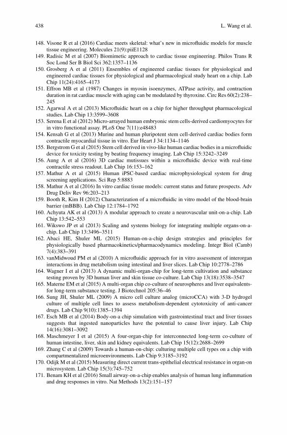
438 L. Wang et al.
148. Visone R et al (2016) Cardiac meets skeletal: what’s new in microfluidic models for muscletissue engineering. Molecules 21(9):piiE1128
149. Radisic M et al (2007) Biomimetic approach to cardiac tissue engineering. Philos Trans RSoc Lond Ser B Biol Sci 362:1357–1136
150. Grosberg A et al (2011) Ensembles of engineered cardiac tissues for physiological andengineered cardiac tissues for physiological and pharmacological study heart on a chip. LabChip 11(24):4165–4173
151. Effron MB et al (1987) Changes in myosin isoenzymes, ATPase activity, and contractionduration in rat cardiac muscle with aging can be modulated by thyroxine. Circ Res 60(2):238–245
152. Agarwal A et al (2013) Microfluidic heart on a chip for higher throughput pharmacologicalstudies. Lab Chip 13:3599–3608
153. Serena E et al (2012) Micro-arrayed human embryonic stem cells-derived cardiomyocytes forin vitro functional assay. PLoS One 7(11):e48483
154. Kensah G et al (2013) Murine and human pluripotent stem cell-derived cardiac bodies formcontractile myocardial tissue in vitro. Eur Heart J 34:1134–1146
155. Bergstrom G et al (2015) Stem cell derived in vivo-like human cardiac bodies in a microfluidicdevice for toxicity testing by beating frequency imaging. Lab Chip 15:3242–3249
156. Aung A et al (2016) 3D cardiac mutissues within a microfluidic device with real-timecontractile stress readout. Lab Chip 16:153–162
157. Mathur A et al (2015) Human iPSC-based cardiac microphysiological system for drugscreening applications. Sci Rep 5:8883
158. Mathur A et al (2016) In vitro cardiac tissue models: current status and future prospects. AdvDrug Deliv Rev 96:203–213
159. Booth R, Kim H (2012) Characterization of a microfluidic in vitro model of the blood-brainbarrier (mBBB). Lab Chip 12:1784–1792
160. Achyuta AK et al (2013) A modular approach to create a neurovascular unit-on-a-chip. LabChip 13:542–553
161. Wikswo JP et al (2013) Scaling and systems biology for integrating multiple organs-on-a-chip. Lab Chip 13:3496–3511
162. Abaci HE, Shuler ML (2015) Human-on-a-chip design strategies and principles forphysiologically based pharmacokinetics/pharmacodynamics modeling. Integr Biol (Camb)7(4):383–391
163. vanMidwoud PM et al (2010) A microfluidic approach for in vitro assessment of interorganinteractions in drug metabolism using intestinal and liver slices. Lab Chip 10:2778–2786
164. Wagner I et al (2013) A dynamic multi-organ-chip for long-term cultivation and substancetesting proven by 3D human liver and skin tissue co-culture. Lab Chip 13(18):3538–3547
165. Materne EM et al (2015) A multi-organ chip co-culture of neurospheres and liver equivalents-for long-term substance testing. J Biotechnol 205:36–46
166. Sung JH, Shuler ML (2009) A micro cell culture analog (microCCA) with 3-D hydrogelculture of multiple cell lines to assess metabolism-dependent cytotoxicity of anti-cancerdrugs. Lab Chip 9(10):1385–1394
167. Esch MB et al (2014) Body-on-a chip simulation with gastrointestinal tract and liver tissuessuggests that ingested nanoparticles have the potential to cause liver injury. Lab Chip14(16):3081–3092
168. Maschmeyer I et al (2015) A four-organ-chip for interconnected long-term co-culture ofhuman intestine, liver, skin and kidney equivalents. Lab Chip 15(12):2688–2699
169. Zhang C et al (2009) Towards a human-on-chip: culturing multiple cell types on a chip withcompartmentalized microenvironments. Lab Chip 9:3185–3192
170. Odijk M et al (2015) Measuring direct current trans-epithelial electrical resistance in organ-onmicrosystem. Lab Chip 15(3):745–752
171. Benam KH et al (2016) Small airway-on-a-chip enables analysis of human lung inflammationand drug responses in vitro. Nat Methods 13(2):151–157
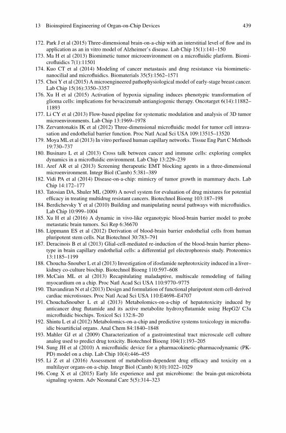
13 Bioinspired Engineering of Organ-on-Chip Devices 439
172. Park J et al (2015) Three-dimensional brain-on-a-chip with an interstitial level of flow and itsapplication as an in vitro model of Alzheimer’s disease. Lab Chip 15(1):141–150
173. Ma H et al (2013) Biomimetic tumor microenvironment on a microfluidic platform. Biomi-crofluidics 7(1):11501
174. Kuo CT et al (2014) Modeling of cancer metastasis and drug resistance via biomimetic-nanocilial and microfluidics. Biomaterials 35(5):1562–1571
175. Choi Y et al (2015) A microengineered pathophysiological model of early-stage breast cancer.Lab Chip 15(16):3350–3357
176. Xu H et al (2015) Activation of hypoxia signaling induces phenotypic transformation ofglioma cells: implications for bevacizumab antiangiogenic therapy. Oncotarget 6(14):11882–11893
177. Li CY et al (2013) Flow-based pipeline for systematic modulation and analysis of 3D tumormicroenvironments. Lab Chip 13:1969–1978
178. Zervantonakis IK et al (2012) Three-dimensional microfluidic model for tumor cell intrava-sation and endothelial barrier function. Proc Natl Acad Sci USA 109:13515–13520
179. Moya ML et al (2013) In vitro perfused human capillary networks. Tissue Eng Part C Methods19:730–737
180. Businaro L et al (2013) Cross talk between cancer and immune cells: exploring complexdynamics in a microfluidic environment. Lab Chip 13:229–239
181. Aref AR et al (2013) Screening therapeutic EMT blocking agents in a three-dimensionalmicroenvironment. Integr Biol (Camb) 5:381–389
182. Vidi PA et al (2014) Disease-on-a-chip: mimicry of tumor growth in mammary ducts. LabChip 14:172–177
183. Tatosian DA, Shuler ML (2009) A novel system for evaluation of drug mixtures for potentialefficacy in treating multidrug resistant cancers. Biotechnol Bioeng 103:187–198
184. Berdichevsky Y et al (2010) Building and manipulating neural pathways with microfluidics.Lab Chip 10:999–1004
185. Xu H et al (2016) A dynamic in vivo-like organotypic blood-brain barrier model to probemetastatic brain tumors. Sci Rep 6:36670
186. Lippmann ES et al (2012) Derivation of blood-brain barrier endothelial cells from humanpluripotent stem cells. Nat Biotechnol 30:783–791
187. Deracinois B et al (2013) Glial-cell-mediated re-induction of the blood-brain barrier pheno-type in brain capillary endothelial cells: a differential gel electrophoresis study. Proteomics13:1185–1199
188. Choucha-Snouber L et al (2013) Investigation of ifosfamide nephrotoxicity induced in a liver–kidney co-culture biochip. Biotechnol Bioeng 110:597–608
189. McCain ML et al (2013) Recapitulating maladaptive, multiscale remodeling of failingmyocardium on a chip. Proc Natl Acad Sci USA 110:9770–9775
190. Thavandiran N et al (2013) Design and formulation of functional pluripotent stem cell-derivedcardiac microtissues. Proc Natl Acad Sci USA 110:E4698–E4707
191. ChouchaSnouber L et al (2013) Metabolomics-on-a-chip of hepatotoxicity induced byanticancer drug flutamide and its active metabolite hydroxyflutamide using HepG2/ C3amicrofluidic biochips. Toxicol Sci 132:8–20
192. Shintu L et al (2012) Metabolomics-on-a-chip and predictive systems toxicology in microflu-idic bioartificial organs. Anal Chem 84:1840–1848
193. Mahler GJ et al (2009) Characterization of a gastrointestinal tract microscale cell cultureanalog used to predict drug toxicity. Biotechnol Bioeng 104(1):193–205
194. Sung JH et al (2010) A microfluidic device for a pharmacokinetic-pharmacodynamic (PK-PD) model on a chip. Lab Chip 10(4):446–455
195. Li Z et al (2016) Assessment of metabolism-dependent drug efficacy and toxicity on amultilayer organs-on-a-chip. Integr Biol (Camb) 8(10):1022–1029
196. Cong X et al (2015) Early life experience and gut microbiome: the brain-gut-microbiotasignaling system. Adv Neonatal Care 5(5):314–323
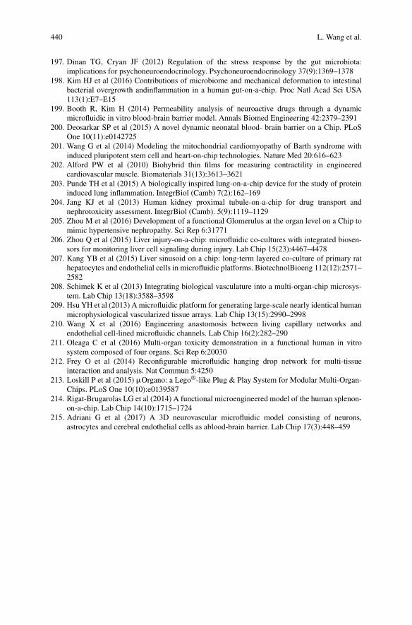
440 L. Wang et al.
197. Dinan TG, Cryan JF (2012) Regulation of the stress response by the gut microbiota:implications for psychoneuroendocrinology. Psychoneuroendocrinology 37(9):1369–1378
198. Kim HJ et al (2016) Contributions of microbiome and mechanical deformation to intestinalbacterial overgrowth andinflammation in a human gut-on-a-chip. Proc Natl Acad Sci USA113(1):E7–E15
199. Booth R, Kim H (2014) Permeability analysis of neuroactive drugs through a dynamicmicrofluidic in vitro blood-brain barrier model. Annals Biomed Engineering 42:2379–2391
200. Deosarkar SP et al (2015) A novel dynamic neonatal blood- brain barrier on a Chip. PLoSOne 10(11):e0142725
201. Wang G et al (2014) Modeling the mitochondrial cardiomyopathy of Barth syndrome withinduced pluripotent stem cell and heart-on-chip technologies. Nature Med 20:616–623
202. Alford PW et al (2010) Biohybrid thin films for measuring contractility in engineeredcardiovascular muscle. Biomaterials 31(13):3613–3621
203. Punde TH et al (2015) A biologically inspired lung-on-a-chip device for the study of proteininduced lung inflammation. IntegrBiol (Camb) 7(2):162–169
204. Jang KJ et al (2013) Human kidney proximal tubule-on-a-chip for drug transport andnephrotoxicity assessment. IntegrBiol (Camb). 5(9):1119–1129
205. Zhou M et al (2016) Development of a functional Glomerulus at the organ level on a Chip tomimic hypertensive nephropathy. Sci Rep 6:31771
206. Zhou Q et al (2015) Liver injury-on-a-chip: microfluidic co-cultures with integrated biosen-sors for monitoring liver cell signaling during injury. Lab Chip 15(23):4467–4478
207. Kang YB et al (2015) Liver sinusoid on a chip: long-term layered co-culture of primary rathepatocytes and endothelial cells in microfluidic platforms. BiotechnolBioeng 112(12):2571–2582
208. Schimek K et al (2013) Integrating biological vasculature into a multi-organ-chip microsys-tem. Lab Chip 13(18):3588–3598
209. Hsu YH et al (2013) A microfluidic platform for generating large-scale nearly identical humanmicrophysiological vascularized tissue arrays. Lab Chip 13(15):2990–2998
210. Wang X et al (2016) Engineering anastomosis between living capillary networks andendothelial cell-lined microfluidic channels. Lab Chip 16(2):282–290
211. Oleaga C et al (2016) Multi-organ toxicity demonstration in a functional human in vitrosystem composed of four organs. Sci Rep 6:20030
212. Frey O et al (2014) Reconfigurable microfluidic hanging drop network for multi-tissueinteraction and analysis. Nat Commun 5:4250
213. Loskill P et al (2015) μOrgano: a Lego®-like Plug & Play System for Modular Multi-Organ-Chips. PLoS One 10(10):e0139587
214. Rigat-Brugarolas LG et al (2014) A functional microengineered model of the human splenon-on-a-chip. Lab Chip 14(10):1715–1724
215. Adriani G et al (2017) A 3D neurovascular microfluidic model consisting of neurons,astrocytes and cerebral endothelial cells as ablood-brain barrier. Lab Chip 17(3):448–459