rios jmrcm 06
TRANSCRIPT
Abstract The contractile cycle of striated muscles,
skeletal and cardiac, is controlled by a cytosolic [Ca2+]
transient that requires rapid movements of the ion
through channels in the sarcoplasmic reticulum (SR).
A functional signature of these channels is their closure
after a stereotyped time lapse of Ca2+ release. In
cardiac muscle there is abundant evidence that termi-
nation of release is mediated by depletion of the Ca2+
store, even if the linkage mechanism remains un-
known. By contrast, in skeletal muscle the mechanisms
of release termination are not understood. This article
reviews measurements of store depletion, the experi-
mental evidence for dependence of Ca2+ release on the
[Ca2+] level inside the SR, as well as tests of the
molecular nature of putative intra-store Ca2+ sensors.
Because Ca2+ sparks exhibit the basic release termi-
nation mechanism, much attention is dedicated to the
studies of store depletion caused by sparks and its
relationship with termination of sparks. The review
notes the striking differences in volume, content and
buffering power of the stores in cardiac vs. skeletal
muscle, differences that explain why functional deple-
tion is much greater for cardiac than skeletal muscle
stores. Because in skeletal muscle store depletion is
minimal and reduction in store [Ca2+] does not appear
to greatly inhibit Ca2+ release, it is concluded that
decrease in free SR [Ca2+] does not mediate physio-
logical termination of Ca2+ release in this type of
muscle. In spite of the apparent absence of store
depletion and its putative channel closing effect, ter-
mination of Ca2+ sparks is faster and more robust in
skeletal than cardiac muscle. A gating role of a hypo-
thetical ‘‘proximate store’’ constituted by polymers of
calsequestrin and associated proteins is invoked in an
attempt to preserve a role for store depletion and unify
mechanisms in both types of striated muscle.
Keywords Skeletal muscle Æ Cardiac muscle ÆSarcoplasmic reticulum Æ Calcium sparks Æ Calsequestrin
Robust mechanisms terminate intracellular Ca2+
release in striated muscles
Skeletal and cardiac muscles share a striated structure,
which reflects an organized arrangement of contractile
filaments and membrane-bound organelles. The struc-
tural similarity reflects an ability for contraction and
relaxation that is fast by comparison with that of
smooth muscle. The striated muscles also share a basic
switching on or off by Ca2+ ions, moving into the
cytosol largely from intracellular stores to bind to an
allosteric regulatory protein on the contractile fila-
ments. By contrast, the role of Ca2+ in smooth muscle
is more nuanced, to the point that its release from
stores causes relaxation (Wray et al. 2005).
The functional cycles of both types of striated
muscle demand full relaxation, which requires robust
closure of the Ca2+ release channels. Figure 1 shows
E. Rıos (&) Æ B. S. Launikonis Æ L. Royer Æ J. ZhouSection of Cellular Signaling, Department of MolecularBiophysics and Physiology, Rush University School ofMedicine, 1750 W. Harrison St. Suite 1279JS,Chicago, IL 60612, USAe-mail: [email protected]
G. BrumDepartamento de Biofısica, Facultad de Medicina,Universidad de la Republica, Gral. Flores 2125,Montevideo, Uruguay
J Muscle Res Cell Motil (2006) 27:337–350
DOI 10.1007/s10974-006-9082-5
123
REVIEW
The elusive role of store depletion in the control of intracellularcalcium release
E. Rıos Æ B. S. Launikonis Æ L. Royer ÆG. Brum Æ J. Zhou
Received: 22 May 2006 / Accepted: 26 June 2006 / Published online: 25 August 2006� Springer Science+Business Media B.V. 2006

the evolution of Ca2+ release flux (back-calculated
from Ca2+ transients), in skeletal and cardiac muscle,
during a voltage clamp depolarizing pulse. The records
show that all muscles have a provision to stop Ca2+
release, even under continuous membrane depolar-
ization, a stop that manifests itself as a sharp peak
followed by decay in the evolution of the cytosolic Ca2+
transient.
The fast deterministic closure of Ca2+ release
channels capable of activation by Ca2+ (CICR) con-
stitutes a ‘‘paradox of control’’, a paradox also mani-
fested in the fact that Ca2+ release has a graded
dependence on the primary stimulus (which is depo-
larization in skeletal muscle, and Ca2+ influx via ICa in
the heart).
A way out of this paradox was provided by Ca2+
sparks (Cheng et al. 1993), which result from syn-
chronous opening of a variable number of clustered
channels (Gonzalez et al. 2000; Wang et al. 2004). The
source clusters were identified with ‘‘couplons’’, com-
prising in the cardiac case all the channels in one dyad
and in the skeletal case the channels on one side of a
junctional segment of t tubule and its associated SR
(Stern et al. 1997). If such sources are sufficiently
separated they can respond individually in a regener-
ative manner, mediated by CICR, while the global
response remains graded. This ‘‘local control’’ theory
identifies a crucial component of any explanation, the
fact that the sources are multiple and separate, but
leaves fundamental questions unanswered.
In cardiac muscle, the limitation of activation during a
spark to less than ten channels out of a two-hundred
channel couplon remains unexplained (Stern and Cheng
2004). It is accepted that cardiac SR Ca2+ overload
sharply increases opening of release channels. Largely as
an extension of this effect, SR depletion is believed to
close channels and together with an ill-defined in-
activation process terminate release in the heart.
Skeletal muscle presents two problems; in the
amphibian less than 5 channels are involved in a spark
if the fiber is intact, and up to 30 if the membrane is
disrupted (Rıos et al. 1999; Baylor et al.2002; Chandler
et al. 2003; rev. by Baylor 2005). What stops the acti-
vation from fully invading couplons of up to 100
channels?
In mammalian skeletal muscle the signal is instead
composed of ‘‘embers’’ produced by channels opening
individually (Csernoch et al. 2004). Sparks occur, but
usually under artificial circumstances, which include
membrane permeabilization (Kirsch et al. 2001; Zhou
et al. 2003), excess of ROS (Isaeva and Shirokova 2003;
Isaeva et al. 2005), osmotic disruption or strenuous
exercise (Pan et al. 2002). There is, however, a strong
spontaneous termination, patent in the whole cell
record (Fig. 1b). It has not been possible to reproduce
this global response based on the summation of indi-
vidual embers (Csernoch et al. 2004). By analogy with
the cardiac role, here again it is possible that depletion
translates the joint activity of many channels to a signal
that terminates release.
This article will review existing measurements of
depletion in skeletal muscle, as well as the evidence for
gating effects of depletion that enable rapid channel
closure. Cardiac muscle, where these issues have been
assiduously studied, will be reviewed summarily as a
term of comparison.
50 ms
10 m
M/m
s50
mM
/ms
450 ms
3 m
M/m
s
a
b
c
Fig. 1 The time course of Ca2+ release flux during sustainedmembrane depolarization to a highly activating potential.(a) frog semitendinosus. (b) rat EDL. (c) Guinea pig ventricularmyocyte. Depolarizing pulse lasted 80 ms (a, b) or 150 ms (c).Panel C is part of Fig. 3 of Sipido and Wier (1991), reproducedwith permission. Note in every case the fast decay of flux afterand early peak
338 J Muscle Res Cell Motil (2006) 27:337–350
123

The activating and inhibitory sites on Ca2+ release
channels/ryanodine receptors
The ryanodine receptor is modulated by the endoge-
nous ligands Ca2+, Mg2+, H+, ATP and inorganic
phosphate (rev. by Meissner 1994). Of critical impor-
tance to EC coupling is the modulation of the ryano-
dine receptor by Ca2+ and Mg2+. The modulation by
cytosolic Ca2+ constitutes both a paradigm and a con-
founding factor for the establishment of the putative
role of intra-SR Ca2+.
In vertebrate skeletal muscle there are two types of
sites on the ryanodine receptor that bind both Ca2+ and
Mg2+. There is a high affinity Ca2+-activation site
(KD ~ 1 lM for Ca2+ and ~ 25 lM for Mg2+; Zhou
et al. 2004) and a low affinity site (KD ~ 1 mM) that
keeps the ryanodine receptor shut when either Ca2+ or
Mg2+ are bound (Meissner 1994; Laver et al. 1997).
The low affinity inhibitory site will be at least half-
saturated at normal resting myoplasmic [Mg2+] (Lamb
and Stephenson 1992). Ca2+ binding to the high affinity
Ca2+-activation site causes the ryanodine receptor to
open —the CICR mechanism. Mg2+ present in the
myoplasm competes with activator Ca2+ at this site
limiting the rate at which Ca2+ can occupy it and elicit
activation.
The control by Ca2+ and Mg2+ sites also varies with
taxonomic class. Skinned fibre studies first showed that
mammalian skeletal muscle was less susceptible to
CICR than amphibian skeletal muscle (Endo 1985).
Under more physiological conditions, Shirokova et al.
(1996) found markedly different waveforms of voltage-
induced Ca2+ release in mammalian and amphibian
skeletal muscle. In particular the ratio of peak over
steady release levels was greater in frog muscle
(Fig. 1). Additionally, the ratio of peak over steady
release exhibited a clear maximum at intermediate
membrane depolarization in the frog, while it was
essentially voltage-independent in the rat. The greater
peak of Ca2+ release in the frog was attributed to
CICR. The failure to image Ca2+ sparks in voltage-
pulsed rat muscle under the same conditions that
produced sparks in frog muscle (Shirokova et al. 1998),
together with the finding of embers in response to low
voltage depolarization in the rat (Csernoch et al. 2004)
and mouse (Csernoch et al. 2006) confirmed the dis-
parity. The paucity of CICR in mammalian muscle was
also evident in comparisons of release elicited by
caffeine and membrane depolarization (Lamb et al.
2001). The need for a lesser peak of Ca2+ release in the
mammal is attributed to the diffusional advantage
derived from the presence of two triad junctions in
every sarcomere of mammalian muscle, while muscles
of other taxonomic classes only have one (Shirokova
et al. 1996, after a suggestion by Elizabeth Stephen-
son).
In skinned fibres, conditions that produce Ca2+
sparks have now been found for mammalian skeletal
muscle (Kirsch et al. 2001; Zhou et al. 2003). However,
they are consistent with lack of CICR (Zhou et al.
2004). In frog muscle, there is a strong negative cor-
relation between spark frequency and [Mg2+] in the
lM to mM range. This is indicative of competition with
Ca2+ for the CICR activation site on the ryanodine
receptor. Similar increases in [Mg2+] affect little the
ability of rat skinned fibres to produce sparks. In fact,
sparks can be observed in rat muscle in the presence of
> 5 mM Mg2+ (Zhou et al. 2004). Thus sparks are
produced under conditions that saturate with Mg2+
both the activating and inhibitory sites of the mam-
malian ryanodine receptor.
These results suggest that even though ryanodine
receptors present in skeletal muscle (of the molecular
isoform 1, homologous to the amphibian a isoform)
possess the Ca2+/Mg2+ activation and inhibitory sites,
their physiology in situ includes other Ca2+ release
modulators. It has been suggested that isoform b of the
RyR, abundant in the amphibian, is responsible for the
CICR response (Rıos and Zhou 2004; Zhou et al.
2004), while isoform a in the amphibian and the
homologous RyR1 in the mammal have their ability to
respond to Ca2+ strongly inhibited by SR accessory
proteins (Murayama and Ogawa 2004) and the DHPR
(Lee et al. 2004; Zhou et al. 2006). SR proteins that
may inhibit aRyR and RyR1 include FKBP, calse-
questrin and triadin.
Structure and biochemistry of the Ca2+ stores provide
clues for the role of depletion
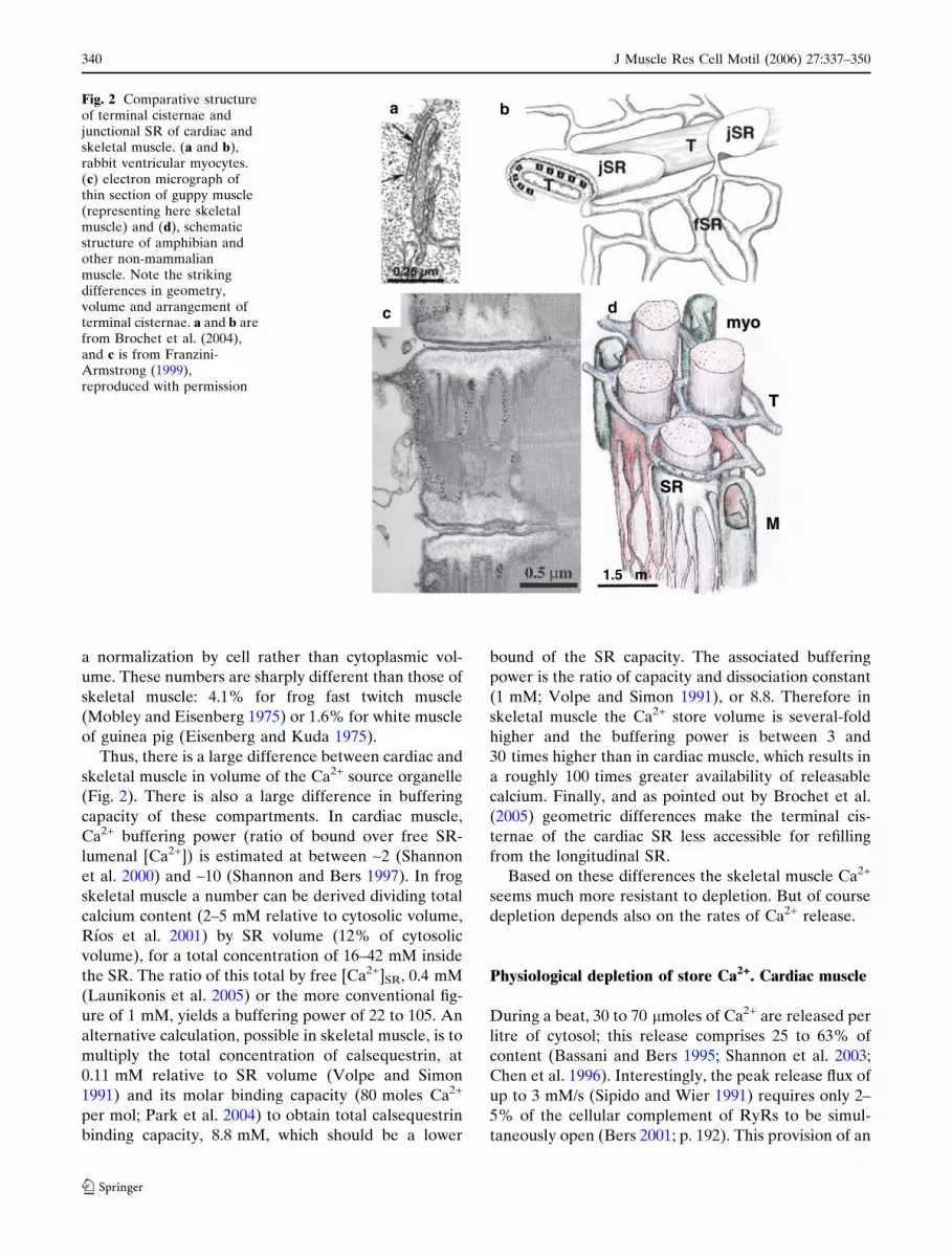
In rabbit ventricular myocytes the junctional (j) SR
forms flat terminal cisternae that appear in thin sec-
tions as linear profiles (Fig. 2a), with an average
diameter of 592 nm (Brochet et al. 2005). The interiors
of the cisternae are electron dense because of the
presence of calsequestrin. The cisternae, described as
‘‘pancake-shaped’’ (Fig. 2b), form junctions with t tu-
bules near the Z lines. Each cisterna has 66 RyRs on
average, grouped in one or two clusters. Estimates of
relative volume are varied. According to Brochet et al.
(2005) the jSR and free SR occupy 2.0% and 2.2%,
respectively, of the ‘‘cytoplasmic’’ volume (total vol-
ume minus mitochondria, nuclei, and SR volumes). jSR
of (rat) ventricle is instead 0.3% in measurements of
Page et al. (1971), with the lower number due in part to
J Muscle Res Cell Motil (2006) 27:337–350 339
123
a normalization by cell rather than cytoplasmic vol-
ume. These numbers are sharply different than those of
skeletal muscle: 4.1% for frog fast twitch muscle
(Mobley and Eisenberg 1975) or 1.6% for white muscle
of guinea pig (Eisenberg and Kuda 1975).
Thus, there is a large difference between cardiac and
skeletal muscle in volume of the Ca2+ source organelle
(Fig. 2). There is also a large difference in buffering
capacity of these compartments. In cardiac muscle,
Ca2+ buffering power (ratio of bound over free SR-
lumenal [Ca2+]) is estimated at between ~2 (Shannon
et al. 2000) and ~10 (Shannon and Bers 1997). In frog
skeletal muscle a number can be derived dividing total
calcium content (2–5 mM relative to cytosolic volume,
Rıos et al. 2001) by SR volume (12% of cytosolic
volume), for a total concentration of 16–42 mM inside
the SR. The ratio of this total by free [Ca2+]SR, 0.4 mM
(Launikonis et al. 2005) or the more conventional fig-
ure of 1 mM, yields a buffering power of 22 to 105. An
alternative calculation, possible in skeletal muscle, is to
multiply the total concentration of calsequestrin, at
0.11 mM relative to SR volume (Volpe and Simon
1991) and its molar binding capacity (80 moles Ca2+
per mol; Park et al. 2004) to obtain total calsequestrin
binding capacity, 8.8 mM, which should be a lower
bound of the SR capacity. The associated buffering
power is the ratio of capacity and dissociation constant
(1 mM; Volpe and Simon 1991), or 8.8. Therefore in
skeletal muscle the Ca2+ store volume is several-fold
higher and the buffering power is between 3 and
30 times higher than in cardiac muscle, which results in
a roughly 100 times greater availability of releasable
calcium. Finally, and as pointed out by Brochet et al.
(2005) geometric differences make the terminal cis-
ternae of the cardiac SR less accessible for refilling
from the longitudinal SR.
Based on these differences the skeletal muscle Ca2+
seems much more resistant to depletion. But of course
depletion depends also on the rates of Ca2+ release.
Physiological depletion of store Ca2+. Cardiac muscle
During a beat, 30 to 70 lmoles of Ca2+ are released per
litre of cytosol; this release comprises 25 to 63% of
content (Bassani and Bers 1995; Shannon et al. 2003;
Chen et al. 1996). Interestingly, the peak release flux of
up to 3 mM/s (Sipido and Wier 1991) requires only 2–
5% of the cellular complement of RyRs to be simul-
taneously open (Bers 2001; p. 192). This provision of an
myomyo
T T
MM
SRSR
1.5 µm
a b
c d
Fig. 2 Comparative structureof terminal cisternae andjunctional SR of cardiac andskeletal muscle. (a and b),rabbit ventricular myocytes.(c) electron micrograph ofthin section of guppy muscle(representing here skeletalmuscle) and (d), schematicstructure of amphibian andother non-mammalianmuscle. Note the strikingdifferences in geometry,volume and arrangement ofterminal cisternae. a and b arefrom Brochet et al. (2004),and c is from Franzini-Armstrong (1999),reproduced with permission
340 J Muscle Res Cell Motil (2006) 27:337–350
123
overabundant functional reserve of channels also re-
quires the presence of a tight control mechanism,
which usually keeps most of the channels closed during
release activity.
Recently it was possible to measure the depletion
locally associated with Ca2+ sparks in cardiac muscle.
From the measurement of ‘‘blinks’’ of fluo-5N inside
the SR, the nadir of reduction of free [Ca2+] was esti-
mated at 54% of resting value, while the reduction in
calsequestrin-bound calcium was put at 28% (Brochet
et al. 2005).
Thus in the heart Ca2+ release causes a large fractional
depletion, in spite of the fact that a large functional re-
serve of channels remains inactive. Therefore, it is pos-
sible to envision for cardiac muscle mechanisms that
derive a robust signal for termination of release from
lumenal SR depletion or other variables that directly
depend on lumenal [Ca2+] levels. Such mechanisms have
been sought, and apparently found.
The effects of calcium load on activation of release
in cardiac muscle
Many observations reveal various activating effects of
elevated [Ca2+]SR and inhibitory effects of reduced
[Ca2+]SR. Activating effects have been described since
Fabiato and Fabiato (1972), including the production
of Ca2+ waves and periodic contractions, and an in-
crease in spark frequency. Diaz et al. (1997) could
predict Ca2+ wave production assuming that sponta-
neous release occurs whenever [Ca2+]SR surpasses a
fixed threshold. Wave propagation appears to be due
to three effects of the increased store [Ca2+]: an
enhancement in the sensitivity of channels to activation
by cytosolic Ca2+, a reduction of the inhibition that
usually follows release and the increase in release flux,
which directly favors Ca2+-dependent activation (Bers
2001, p. 232).
The activating effect is very non-linear (Shannon
et al. 2000) —the dependence between fractional re-
lease and load has a pronounced upward curvature.
This non-linearity may be intrinsic to the dependency
of activation with lumenal [Ca2+] or it may be indirect,
a consequence of positive feedback at the cytosolic side
of the channels.
These global effects also have local manifestations,
chiefly the increase in frequency of Ca2+ sparks with
total SR [Ca2+]. A most compelling demonstration of
the effect is in the transient nature of the reduction of
spark frequency that ensues in the continuous presence
of the channel blocker tetracaine, even when the drug
is used at concentrations that fully eliminate sparks
when first applied (Gyorke et al. 1997).
The activation effect of lumenal Ca2+ is also sup-
ported by work with channels reconstituted in bilayers.
As reviewed by Gyorke et al. (2002), the evidence
supports both an indirect ‘‘feed-through’’ effect of lu-
menal Ca2+ acting on the cytosolic side, and a direct
effect of Ca2+ on the lumenal (trans) side of the
channels. The joint evidence strongly indicates that the
activation is due to lumenal Ca2+ acting on the lumenal
face of channels or associated proteins.
There is also evidence of a converse effect: reduc-
tion of release activity associated with SR depletion. It
is useful to summarize the effect of lumenal Ca2+ as a
change in Po of the generic release channel and then
analyze this effect into its kinetic components. The
change in steady Po must include an increase in the
opening transition rate kON (as suggested by the in-
crease in frequency of local events and the reduction of
mean closed time in bilayer-reconstituted channels).
Additionally, it could include a reduction in the closing
rate kOFF. If this was the case, then the converse would
apply, depletion would increase kOFF, literally termi-
nating release. In that case the effect of lumenal Ca2+
could provide a mechanism of termination of sparks
and spontaneous reduction of Ca2+ release (‘‘inactiva-
tion’’) at the cell-wide level.
Several lines of evidence support release termina-
tion by store depletion; most compelling are the effects
of extrinsic Ca2+ buffers, including maleate, citrate and
ADA. They increase the total calcium content in the
SR in direct proportion to their affinity for Ca2+, and
increase amount of global Ca2+ release and amplitude
of Ca2+ sparks by prolongation of release time (i.e.,
sparks rise time) exclusive of other mechanisms
(Terentyev et al. 2002). The striking correlation among
buffer affinity, SR load, duration of cell-wide release
and rise time of sparks constitutes the strongest evi-
dence to this date for a determinant role of depletion in
termination of Ca2+ release. Sobie et al. (2002) antici-
pated this result with a model that assumed the Ca2+
affinity of the cytosolic activator site to be linearly
dependent on the SR lumenal [Ca2+]. The lumenal
Ca2+ dependence of activator affinity plus the rapid
spread of gating state in an allosterically connected
cluster were sufficient to simulate realistic termination
of sparks, provided that the store operated as relatively
isolated organelles of limited capacity. While all the
assumptions in the model of Sobie et al. (2002) have
not been confirmed in detail, it is now clear that the
terminal cisternae of cardiac SR do in fact deplete
strongly during individual sparks (see previous sec-
tion) and therefore depletion is taken to be a critical
J Muscle Res Cell Motil (2006) 27:337–350 341
123
determinant of spark termination in the heart. This
dependence has been rationalized as the consequence
of Ca2+ binding to an intra-SR ‘‘Ca2+ sensor’’, which
may not necessarily be on the RyR molecule.
Molecular nature of the lumenal ‘‘Ca2+ sensor’’
The lumenal Ca2+ sensor remains unidentified. Be-
cause the Po of purified channels in bilayers was un-
changed upon trans [Ca2+] increase (Gyorke et al.
2004), the action of SR-lumenal Ca2+ on the release
channels may be indirect. Native channels showed in-
stead a marked sensitivity of Po to lumenal [Ca2+],
which suggests that the lumenal Ca2+ sensor is part of a
protein or complex of proteins that are lost during the
purification process.
In striated muscles SR Ca2+ is buffered mostly by
calsequestrin, a low-affinity Ca2+ binding protein
whose role in the regulation of SR Ca2+ release is un-
der intense investigation. Modification of the cellular
levels of calsequestrin produced conflicting results. In
transgenic mice the overexpression of calsequestrin
resulted in diminished Ca2+ transients and contrac-
tions, even though the SR Ca2+ content was signifi-
cantly higher (Wang et al. 2000). An opposite result
was described by Terentyev et al. (2003). Using ade-
noviruses to overexpress calsequestrin in cardiomyo-
cytes these authors obtained results similar to the ones
observed applying exogenous buffers (Terentyev et al.
2002). The magnitude of both ICa-induced Ca2+ tran-
sients and spontaneous Ca2+ sparks were increased,
which was attributed to a slower termination of Ca2+
release from the SR. Also, as observed with the
exogenous buffers, the recovery time for repetitive
Ca2+ sparks was longer, another result consistent with
the existence of a threshold [Ca2+]SR above which Ca2+
sparks are generated spontaneously.
Due to its role as a calcium buffer in the SR lumen,
calsequestrin is an obvious candidate sensor. However,
its addition caused no change in the activity of purified
RyRs in bilayers (Gyorke et al. 2004).
The junctional SR protein triadin 1 physically links
calsequestrin to the lumenal side of the release channel
(Guo and Campbell 1995; Zhang et al. 1997). The
adenovirus-mediated overexpression of triadin 1 in
cardiomyocytes has been shown to enhance cardiac EC
coupling (Terentyev et al. 2005). In particular the Po of
native channels reconstituted from myocytes overex-
pressing triadin 1 was 5-fold higher than that of chan-
nels from control cells. Moreover, when these cells
overexpressed a triadin mutant lacking the domain
responsible for interaction with calsequestrin, the Po
was unchanged. Addition of triadin and/or junctin to
the trans side of the bilayer resulted in a significant
increase in the open probability of the purified channel
(Gyorke et al. 2004). However the channel was still
unresponsive to changes in trans Ca2+. A subsequent
addition of calsequestrin inhibited the channel activity
but restored the ability of the ryanodine receptor to
respond to changes in luminal Ca2+. These results
suggest that the function requires a supramolecular
complex in which calsequestrin act as the sensor, while
triadin and/or junctin are responsible for the interac-
tion between calsequestrin and ryanodine receptor.
The impressive series of studies by Gyorke and
colleagues took a dramatic turn with the demonstra-
tion of de-stabilizing effects of an antisense-induced
reduction in calsequestrin content. The intervention
resulted in spontaneous Ca2+ release and membrane
depolarization associated with the early termination of
a refractoriness that prevents re-entry, which was in
turn traced to an accelerated restoration of [Ca2+]SR
after its depletion during a beat. These observations
(Terentyev et al. 2003; Viatchenko-Karpinsky et al.
2004) provide a clear pathogenesis mechanism for
hereditary arrhythmias associated with mutations of
the human CSQ2 gene (the syndrome named cate-
cholaminergic polymorphic ventricular tachycardia or
CPVT; Lahat et al. 2001) and suggest compelling
approaches to the treatment of this syndrome.
It is surprising to see that these steady advances in
the understanding of Ca2+ control in the heart cannot
be transferred to skeletal muscle in a straightforward
way.
Depletion of store Ca2+ in skeletal muscle
In frog skeletal muscle, release by individual action
potentials (quantified from the amount of H+ displaced
from EGTA introduced in the cytosol) reached 14% in
fiber segments (Pape et al. 1995). This was in good
agreement with a figure of 10%, obtained with the first
three-dimensional model of calcium movements in
skeletal muscle cells (Cannell and Allen 1984).
In toad muscle, prolonged tetanic activity resulted in
reduction of contractile force (fatigue) which was
accompanied by a reduction in releasable Ca2+ store
content to approximately 40% of resting level (Kab-
bara and Allen 1999). The same authors later used
fluo-5N to directly evaluate SR content and found that
[Ca2+]SR falls by 40% in a 500 ms 100 Hz tetanus
(Kabbara and Allen 2001). These measurements are
342 J Muscle Res Cell Motil (2006) 27:337–350
123
again in rough agreement with the 3-D model of
Cannell and Allen (1984). More recently, using SEER
of mag-indo-1 in manually skinned frog fibers Lau-
nikonis et al. (2006a) measured a depletion of 10%
upon release caused by a single twitch elicited by
electrical stimulation. In these skinned fibers the
t tubular system reseals and recovers the ability to
generate action potentials, therefore the flux and
amount of Ca2+ release are presumably close to those
in a physiological twitch. Finally Rudolf et al. (1996)
estimated a 17% reduction by a single twitch in mouse
tibialis anterior expressing the biosensor D1ER (Pal-
mer et al. 2004). In summary, estimates of depletion
caused by a single twitch range from 10 to 17% in frogs
and mammals, but repeated twitches and tetani may
accumulate much greater depletion. By comparison,
voltage clamp depolarization that activates Ca2+
release maximally, causes a flux waveform that reaches
a peak (and thereafter decays rapidly) in ~5 ms. Before
peak flux, some 250 lM (or 10% of content) may be
released. Therefore, whether release is elicited by
action potentials or clamp depolarization, the sponta-
neous process of termination is in full force when
depletion has only reached the 10–20% range. Deple-
tion of such small magnitude could hardly have a
determinant role in release termination.
The local store depletion left behind by the Ca2+
release underlying a spark was first brought into focus
by evidence that fluorescence in sparks increases mo-
noexponentially up to a peak, and at this point makes a
sharp break to start a rapid decay (Lacampagne et al.
1999). Such time course was interpreted to reflect a
roughly constant flux during the rise time of the spark.
This inference was supported by modelling in which a
constant flux active during the rise time was adequate
to stimulate fluorescence measured in sparks (Baylor
et al. 2002; Chandler et al. 2003). Also in agreement
was the computation of rate of signal mass production,
a quantity proportional to release flux under most
conditions (Zhuge et al. 2002, 2004; Zhou et al. 2005),
in large sparks recorded at high temporal resolution
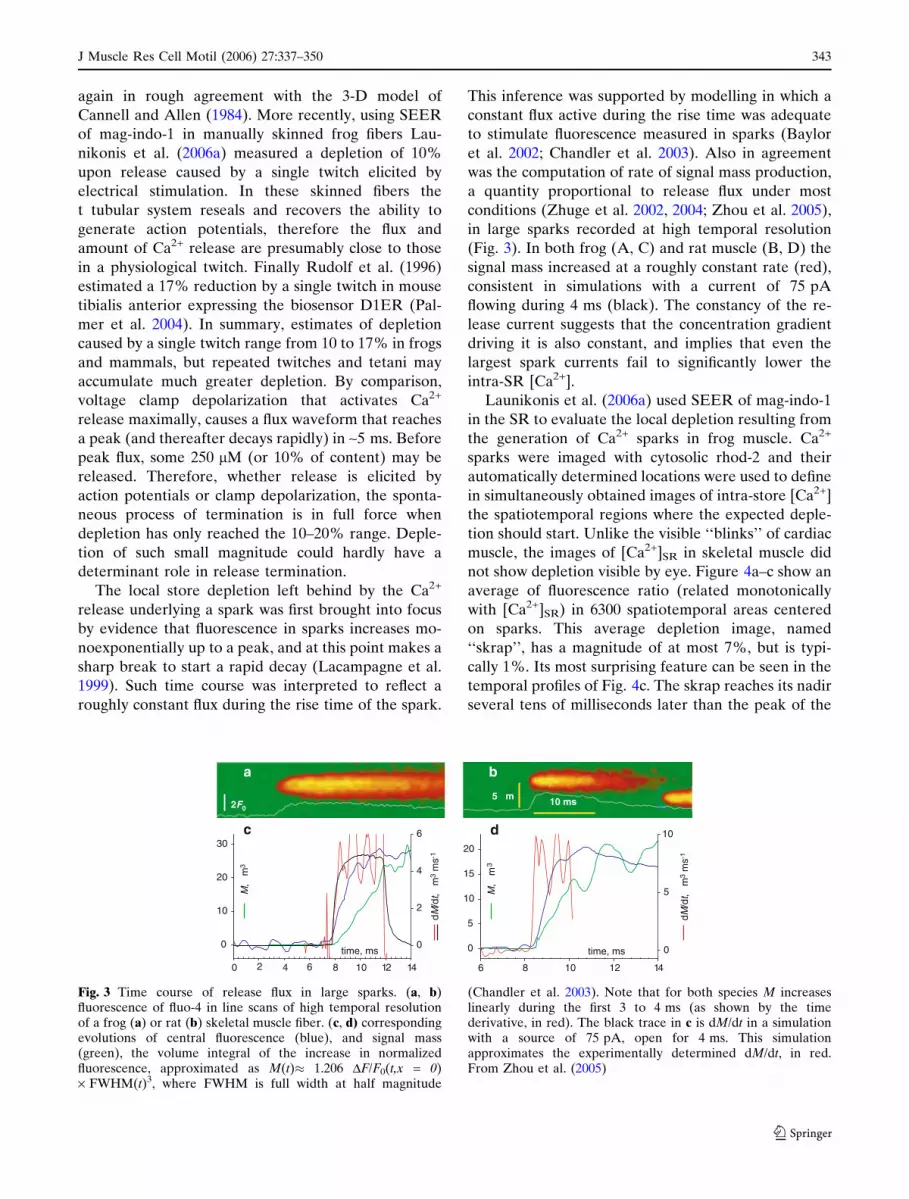
(Fig. 3). In both frog (A, C) and rat muscle (B, D) the
signal mass increased at a roughly constant rate (red),
consistent in simulations with a current of 75 pA
flowing during 4 ms (black). The constancy of the re-
lease current suggests that the concentration gradient
driving it is also constant, and implies that even the
largest spark currents fail to significantly lower the
intra-SR [Ca2+].
Launikonis et al. (2006a) used SEER of mag-indo-1
in the SR to evaluate the local depletion resulting from
the generation of Ca2+ sparks in frog muscle. Ca2+
sparks were imaged with cytosolic rhod-2 and their
automatically determined locations were used to define
in simultaneously obtained images of intra-store [Ca2+]
the spatiotemporal regions where the expected deple-
tion should start. Unlike the visible ‘‘blinks’’ of cardiac
muscle, the images of [Ca2+]SR in skeletal muscle did
not show depletion visible by eye. Figure 4a–c show an
average of fluorescence ratio (related monotonically
with [Ca2+]SR) in 6300 spatiotemporal areas centered
on sparks. This average depletion image, named
‘‘skrap’’, has a magnitude of at most 7%, but is typi-
cally 1%. Its most surprising feature can be seen in the
temporal profiles of Fig. 4c. The skrap reaches its nadir
several tens of milliseconds later than the peak of the
12 14
0
10
20
30
0
2
4
6
M, µ
m3
dM/d
t, µm
3m
s-1
c
time, ms
6 8 10 12 14
0
5
10
15
20
0
5
10
M, µ
m3
dM/d
t, µm
3m
s-1
d
time, ms
ba
5 µm 10 ms2F0
0 2 4 6 8 10
Fig. 3 Time course of release flux in large sparks. (a, b)fluorescence of fluo-4 in line scans of high temporal resolutionof a frog (a) or rat (b) skeletal muscle fiber. (c, d) correspondingevolutions of central fluorescence (blue), and signal mass(green), the volume integral of the increase in normalizedfluorescence, approximated as M(t)� 1.206 DF/F0(t,x = 0)· FWHM(t)3, where FWHM is full width at half magnitude
(Chandler et al. 2003). Note that for both species M increaseslinearly during the first 3 to 4 ms (as shown by the timederivative, in red). The black trace in c is dM/dt in a simulationwith a source of 75 pA, open for 4 ms. This simulationapproximates the experimentally determined dM/dt, in red.From Zhou et al. (2005)
J Muscle Res Cell Motil (2006) 27:337–350 343
123

time, ms
200 400 600
R /
R0
0.98
0.99
1.00
F3/
F3,
01.0
1.5
2.0
time, ms
150 200 250
R /
R0
0.98
0.99
1.00
dM/d
t, m
s-1
0
2
4
ac
b
e
time, ms
150 180 2100.0
0.9
1.0
0
5
1
d
R /
R0
Flu
x
10 µm
Fig. 4 Various measurements of SR depletion accompanyingCa2+ release into the cytosol. (a) the average ‘‘skrap’’, image ofthe local deficit in [Ca2+] left inside the store by the release thatunderlies a spark. The vertical coordinate of linescan images aand e is space and the horizontal coordinate is time. This image isan average of those corresponding to 6300 individual sparksdetected in frog muscle fibers stained with rhod-2 in the cytosoland mag-indo-1 in the SR. It displays the ratio R(x,t) offluorescence excited by different laser lines in two differentranges of emission of the dye in the SR (a ratio that variesmonotonically with [Ca2+]SR, as detailed in Launikonis et al.2006). (b) blue: temporal profile of image (a). Red, temporalprofile of the average of all spark images, obtained from thefluorescence of rhod-2 simultaneously with the images of[Ca2+]SR. Both plots are represented normalized to their initial,pre-spark values. The exponential decay of the skrap, after nadirhas an initial amplitude of 0.015 and a s of 251 ms. (c) spark and
skrap profiles at an expanded temporal scale. Green, the rate ofproduction of spark mass M, which is proportional to the flux ofCa2+ release underlying the spark. Note that the depletionreaches nadir ~50 ms after the end of Ca2+ release. (d) temporalprofiles of depletion and Ca2+ release flux, measured as in a–c, ina frog muscle fiber that released Ca2+ upon an action potential.Note similar delay of the nadir of depletion. a–d are fromLaunikonis et al. (2006), with slight modifications. (e) circles,ratio of fluorescence at two emission ranges in an intact mousetibialis anterior fiber expressing D1ER, averaged over the fiberand 15 twitches repeated at 1 Hz. Bars are an average of fibershortening. Inset: ratio of linescans during contraction. Bar,50 lm. Note that the nadir of depletion occurs at the same timeas the peak of contractile displacement, or tens of ms later thanthe peak of the cytosolic Ca2+ transient. From Rudolf et al.(2006), reproduced with permission.
344 J Muscle Res Cell Motil (2006) 27:337–350
123
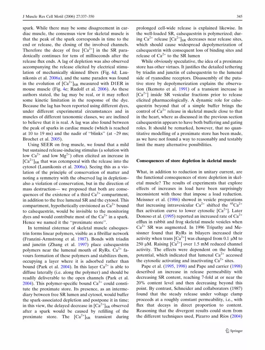
spark. While there may be some disagreement in car-
diac muscle, the consensus view for skeletal muscle is
that the peak of the spark corresponds in time to the
end or release, the closing of the involved channels.
Therefore the decay of free [Ca2+] in the SR para-
doxically continues for tens of milliseconds after the
release flux ends. A lag of depletion was also observed
accompanying the release elicited by electrical stimu-
lation of mechanically skinned fibers (Fig. 4d; Lau-
nikonis et al. 2006a), and the same paradox was found
in the evolution of [Ca2+]SR measured with D1ER in
mouse muscle (Fig. 4e; Rudolf et al. 2006). As these
authors stated, the lag may be real, or it may reflect
some kinetic limitation in the response of the dye.
Because the lag has been reported using different dyes,
under different experimental circumstances and in
muscles of different taxonomic classes, we are inclined
to believe that it is real. A lag was also found between
the peak of sparks in cardiac muscle (which is reached
at 10 to 19 ms) and the nadir of ‘‘blinks’’ (at ~29 ms;
Brochet et al. 2005).
Using SEER on frog muscle, we found that a mild
but sustained release-inducing stimulus (a solution with
low Ca2+ and low Mg2+) often elicited an increase in
[Ca2+]SR that was cotemporal with the release into the
cytosol (Launikonis et al. 2006a). Seeing this as a vio-
lation of the principle of conservation of matter and
noting a symmetry with the observed lag in depletion–
also a violation of conservation, but in the direction of
mass destruction— we proposed that both are conse-
quences of the existence of a third Ca2+ compartment,
in addition to the free lumenal SR and the cytosol. This
compartment, hypothetically envisioned as Ca2+ bound
to calsequestrin, would be invisible to the monitoring
dyes and would contribute most of the Ca2+ in a spark.
Hence we named it the ‘‘proximate store’’.
In terminal cisternae of skeletal muscle calseques-
trin forms linear polymers, visible as a fibrillar network
(Franzini-Armstrong et al. 1987). Bonds with triadin
and junctin (Zhang et al. 1997) place calsequestrin
polymers near the lumenal mouth of RyRs. Ca2+ fa-
vours formation of these polymers and stabilizes them,
occupying a layer where it is adsorbed rather than
bound (Park et al. 2004). In this layer Ca2+ is able to
diffuse laterally (i.e. along the polymer) and should be
readily deliverable to the open channels (Park et al.
2004). This polymer-specific bound Ca2+ could consti-
tute the proximate store. Its presence, as an interme-
diary between free SR lumen and cytosol, would buffer
the spark-associated depletion and postpone it in time;
in this view, the delayed decrease in [Ca2+]SR observed
after a spark would be caused by refilling of the
proximate store. The [Ca2+]SR transient during
prolonged cell-wide release is explained likewise. In
the well-loaded SR, calsequestrin is polymerized; dur-
ing Ca2+ release [Ca2+]SR decreases near release sites,
which should cause widespread depolymerization of
calsequestrin with consequent loss of binding sites and
release of Ca2+ to the SR lumen
While obviously speculative, the idea of a proximate
store has other virtues. It justifies the detailed tethering
by triadin and junctin of calsequestrin to the lumenal
side of ryanodine receptors. Disassembly of the puta-
tive store by depolymerization explains the observa-
tion (Ikemoto et al. 1991) of a transient increase in
[Ca2+] inside SR vesicular fractions prior to release
elicited pharmacologically. A dynamic role for calse-
questrin beyond that of a simple buffer brings the
control of Ca2+ release in skeletal muscle close to that
in the heart, where as discussed in the previous section
calsequestrin appears to have both buffering and gating
roles. It should be remarked, however, that no quan-
titative modelling of a proximate store has been made,
as we have not found a way to reasonably and testably
limit the many alternative possibilities.
Consequences of store depletion in skeletal muscle
What, in addition to reduction in unitary current, are
the functional consequences of store depletion in skel-
etal muscle? The results of experiments that explore
effects of increases in load have been surprisingly
inconsistent with those that impose a load reduction.
Meissner et al. (1986) showed in vesicle preparations
that increasing intravesicular Ca2+ shifted the 45Ca2+
flux activation curve to lower cytosolic [Ca2+]. Later
Donoso et al. (1995) reported an increased rate of Ca2+
efflux in rabbit and frog skeletal muscle vesicles when
Ca2+ SR was augmented. In 1996 Tripathy and Me-
sissner found that RyRs in bilayers increased their
activity when trans [Ca2+] was changed from 0.1 lM to
250 lM. Raising [Ca2+] over 1.5 mM reduced channel
activity. The effects were dependent on the holding
potential, which indicated that lumenal Ca2+ accessed
the cytosolic activating and inactivating Ca2+ sites.
Pape et al. (1995, 1998) and Pape and carrier (1998)
described an increase in release permeability with
decreasing SR content, reaching 7-fold at or near the
20% content level and then decreasing beyond this
point. By contrast, Schneider and collaborators (1987)
found that the steady release under voltage clamp
proceeds at a roughly constant permeability, i.e., with
flux that decays in direct proportion to content.
Reasoning that the divergent results could stem from
the different techniques used, Pizarro and Rıos (2004)
J Muscle Res Cell Motil (2006) 27:337–350 345
123
combined the direct measurement of [Ca2+]cyto of
Schneider et al. (1987) with the evaluation of Ca2+
release by EGTA and Phenol Red used by Pape and
colleagues, trying also to distinguish the effects on the
peak of the waveform of release flux from those on the
steady level reached later (Fig. 1A). Their results
confirmed qualitatively that permeability increases
with progressive depletion; the increase however af-
fected only the steady flux level, it was substantially
lower than reported before and was essentially negli-
gible unless SR content was reduced by 30% or more
(a feature also described by Pape and Carrier 1998).
Therefore, the effect of store depletion in skeletal
muscle is one of promotion of cell-wide Ca2+ release, in
sharp contrast with that found in heart muscle. Pizarro
and Rıos (2004) additionally found a rough constancy
of the early peak of release flux during a voltage pulse,
and a post-peak decay that became slower and less
pronounced as SR load decreased. These features im-
ply that the effect only occurs after the establishment
of full flux –or channel opening— and therefore indi-
cate a cytosolic locus of Ca2+ action. The mechanism
probably consists in relief of Ca2+-dependent channel
closing (Ca2+-dependent inactivation or CDI) as
[Ca2+]cyto near open channels cannot reach the usual
high values if the store is depleted.
Launikonis et al. (2006b) studied in frog muscle the
effects of changes in [Ca2+]SR on the properties of Ca2+
sparks occurring under mild stimulation. Changes in
[Ca2+]SR were imposed by manipulation of [Ca2+]cyto
and measured with SEER of Mag-Indo-1; to neutralize
the effect of cytosolic calcium (reported by Zhou et al.
2005), sparks were always imaged in 100 nM [Ca2+]cyto.
The increase in [Ca2+]SR from an average of 250 lM to
about 400 lM was accompanied by an increase in
spark frequency by about 70%, and the effect reversed
upon the inverse change in [Ca2+]SR. However, the
power of the effect was weak, as attested by a corre-
lation coefficient q2 of less than 0.06. While the average
increase in [Ca2+]SR was modest in these experiments,
in individual cases it was greater than 3-fold. Not even
the largest increases in [Ca2+]SR were able to induce
the sharp increases in release activity observed in car-
diomyocytes under similar changes.
After the considerations above it seems clear that
reduction of free SR Ca2+ per se cannot provide a ro-
bust mechanism of release termination, global or local,
in skeletal muscle. It is not just that the magnitude of
depletion caused by physiological activity is limited,
but substantial imposed depletion has either release-
promoting effects (under voltage stimulation) or causes
weak inhibition (in sparks activated by mild promoters
of CICR).
These observations, largely carried out in non-
mammalian muscle, leave the termination mechanism
of Ca2+ release totally unsettled. The problem may be
less critical in mammalian muscle, where there are no
spontaneous Ca2+ sparks and the elementary local
events of Ca2+ release are instead ‘‘embers’’, which
reflect individual channel openings controlled by
membrane depolarization (Csernoch et al. 2003, 2006).
Even in mammals, though, the flux of Ca2+ release
spontaneously decays after an early peak (Fig. 1B), and
it has not been possible to quantitatively reconstitute
the cell-wide release waveform as a sum of embers. A
‘‘termination principle’’ is missing in the mammal as
well.
Calsequestrin and accessory proteins in skeletal muscle
Gating effects of calsequestrin on skeletal muscle
RyRs were first described by Kawasaki and Kasai
(1994) as a loss of the activation by Ca2+ of release
from heavy SR vesicles upon a treatment that induced
loss of calsequestrin from the vesicles. A role of triadin
in the effect was first postulated by Ohkura et al.
(1998) also from observations on heavy SR, while
Szegedi et al. (2001) found that only a dephosphoryl-
ated form of calsequestrin promoted opening of
reconstituted channels. More recently Beard et al.
(2002 2005) reproduced for reconstituted skeletal
muscle channels most of the effects described for car-
diac channels. The evidence suggests both a direct
facilitation of channel opening by Ca2+ on the lumenal
side (demonstrated on purified RyRs) and a basal
calsequestrin-mediated inhibition, which depends on
the presence of triadin or junctin and is relieved upon
increasing lumenal [Ca2+]. This relief appears as a
[Ca2+]—dependent activation that reaches maximum
at 1 mM lumenal [Ca2+]. The concentration range of
these effects is therefore adequate for their physio-
logical relevance. Wei et al. (2006) added to this pic-
ture evidence that calsequestrin depolymerizes and
detaches from the junctional SR membrane upon
prolonged exposure to low (100 lM) lumenal [Ca2+],
while calsequestrin monomers remain attached. In this
situation the native regulation is lost and channels re-
spond with increase in Po to reduction of lumenal
[Ca2+], a response that could underlie the increase in
steady permeability induced by depolarization in (frog)
fibers with strongly depleted SR (Pape et al. 1995,
1998; Pizarro and Rıos 2004). In spite of these occa-
sional agreements, cellular observations cannot yet be
explained on the basis of the interactions with associ-
ated proteins demonstrated in subcellular systems.
346 J Muscle Res Cell Motil (2006) 27:337–350
123

Genetic manipulation of the calsequestrin endow-
ment has given additional insights. Paolini et al. (2005)
preliminarily showed that a mouse null for the skeletal
isoform of calsequestrin (CSQ1), which was viable, had
terminal cisternae with greatly altered structural
properties. The properties (some of which were shared
with cardiac jSR) included flattened cisternae with a
junctional membrane that contains multiple –rather
than double—rows of RyRs, often forming longitudi-
nally oriented, multi-layered junctions with t tubules.
Surprisingly, K.O. mice did not show many differences
in behaviour, and their EDL muscles –apparently
devoid of any calsequestrin—did not exhibit changes in
twitch amplitude and had an increased resistance to
fatigue (Paolini et al. 2006). These results imply that
CSQ1 is important in cellular ontogenesis, but also
underscore the shortcomings of the knockout approach
to genetic engineering, which allows compensatory
changes the most time to develop.
A more conservative approach is transient gene
silencing; Wang et al. (2006) used small interference
RNA to reduce expression of either or both calse-
questrin isoforms in the myogenic C2C12 cell line,
which usually expresses both isoforms. Again here the
results were surprising: the CSQ1 knockdown myotu-
bes, with less than 10% expression of the skeletal iso-
form, had normal release of Ca2+ in response to various
stimuli, while a comparable reduction in CSQ2 did
diminish the response. The reduced response in turn
was traced to a lower density of SERCA1 and ryano-
dine receptors, associated with lower Ca2+ uptake and
release in the CSQ2 knockdown myotubes. The tran-
sient gene silencing approach therefore failed to isolate
an effect of the lack of calsequestrin, but was consis-
tent, in a way, with the surprisingly modest effects of
CSQ1 ablation on contractile performance of these
muscle models (Paolini et al. 2006). In view of these
results it is again clear that a full reduction of cellular
EC coupling to its elementary molecular interactions is
far from being achieved.
Conclusions and speculation
The available evidence leads to a number of conclu-
sions:
(i) There are extraordinary differences in fluxes of
Ca2+ release and reuptake between skeletal and
cardiac muscle, as well as in the spatial extent,
volume and buffering capacity of the storage
organelles that define these fluxes. The differences
seem to be in correspondence with very different
functional demands for the two systems. Accord-
ingly, one should not expect major similarities in
the degree of physiological store depletion, or, by
extension, in the functional role of depletion in
skeletal and cardiac muscle.
(ii) In agreement with the first conclusion, the loss of
Ca2+ after one action potential is ~50% in cardiac
muscle, while in skeletal muscle it is 15%.
(iii) a robust termination of release is a common
feature of all striated muscles. A role for
depletion in this termination process seems
well-established for cardiac muscle only. Stern
and Cheng (2004) concluded that the termina-
tion of Ca2+ release is the central problem in the
field of cardiac muscle EC coupling. From the
vantage point of our comparison between stri-
ated muscles we must add several qualifications
to agree. First, in view of the abundantly dem-
onstrated effects of extrinsic buffers and calse-
questrin, the central role of depletion should not
be in doubt for cardiac muscle. What remains
unclear is how a 50% depletion of the store can
bring about channel closure robustly, every time.
Thus, from the standpoint of cardiac muscle, the
issue is lack of good theories, rather than data.
Moving on to skeletal muscle, the functional role
of depletion appears weak, not just because the
magnitude of the depletion caused by a single
action potential is modest, but also because
(iv) measured effects of increased load are modest
(a weak promotion of sparks) and
(v) the effect of major store depletion, an increase in
the steady permeability reached during long-
lasting depolarizations, is contradictory with a
role in termination of Ca2+ release.
These largely negative conclusions regarding the
role of depletion in skeletal muscle appear incon-
gruous with the consensus that prevails in cardiac
muscle. The dissonance seems even greater if one
recalls that release termination is sharper, more ro-
bust, in frog skeletal muscle, where sparks have a
narrowly distributed rise time centered at 4 ms, than
in cardiac muscle, where spark rise times are
10–15 ms and open channel times are thought to
surpass the time to peak. Thus, Ca2+ sparks termi-
nate earlier and more conclusively in the frog, where
depletion is negligible, than in heart muscle, where
depletion is substantial.
One possible interpretation of this discrepancy is
that the termination mechanism operative in the heart
is insufficient for skeletal muscle, which therefore must
J Muscle Res Cell Motil (2006) 27:337–350 347
123

have developed a much better terminator of its own.
Ca2+-dependent inactivation, or CDI, could be such
mechanism. Inactivation by the elevated [Ca2+]cyto
near open channels could both prevent continued
propagation of activity by acting on closed channels
and terminate Ca2+ release by working on open chan-
nels. This mechanism is especially attractive because
elevated Ca2+ has a much greater inhibitory effect on
skeletal than cardiac muscle channels in bilayer stud-
ies. CDI predicts among other properties a negative
correlation between local [Ca2+]cyto and rise time in
Ca2+ sparks. Spark amplitude is an imperfect measure
of local [Ca2+]cyto, due to off-focus errors and limited
kinetic response of dyes. A negative correlation be-
tween spark amplitude and rise time has not been
clearly demonstrated; an absence of correlation is
found instead (Lacampagne et al. 1999; Rıos et al.
1999). This result is in itself evidence of auto-
inactivation, because the absence of effects by local
[Ca2+]cyto should result in a positive correlation with
rise time ([Ca2+]cyto would rise progressively at
increasing channel open times). Together with the
evidence from global measurements (reviewed by Pi-
zarro and Rıos 2004), this property of sparks constitute
indirect evidence of a release-terminating effect of
cytosolic Ca2+.
In addition to a fully different termination strategy
valid for skeletal muscle only, it is possible to imagine
a shared mechanism that upholds a role of depletion
and operates in parallel with CDI. The hypothesis is
an extension of the notion that Ca2+ ions adsorbed to
linear polymers of calsequestrin constitute a proxi-
mate store (Launikonis et al. 2006a). We propose that
depletion of the proximate store contributes to the
robust signal needed for channel closure, and that the
effect is mediated by depolymerization of calseques-
trin. Given the structural differences, in cardiac
muscle calsequestrin depletion would essentially
course in parallel with substantial depletion of free
SR-lumenal Ca2+, while depletion of the lumen of the
larger and better buffered skeletal muscle SR would
be smaller and could be delayed. This speculation
therefore accounts for both the differences in mag-
nitude and time course of the local reduction in free
SR [Ca2+] and the essential similarity of interactions
of skeletal and cardiac RyRs with intra-SR proteins
and Ca2+ in bilayer-reconstituted systems. Better
biosensors, alternative genetic manipulations and
quantitative modeling will put these speculations to
the test within the next few years. In the meantime,
the sobering conclusion is that the role of store con-
tent in the control of skeletal muscle EC coupling
remains unknown.
Acknowledgements We are grateful to Gil Wier, ClaraFranzini-Armstrong, Heping Cheng, Tullio Pozzan and RudigerRudolf for kindly allowing us the use of their figures, and to TomShannon for discussion regarding the SR buffers and other as-pects of cardiac calcium control. Work supported by grants fromthe National Institutes of Health/National Institute of Arthritisand Musculoskeletal and Skin Diseases, USA and the Programade Desarrollo de Ciencias Basicas, Uruguay. B.S.L. was the re-cipient of a C.J. Martin Fellowship of the National Health andMedical Research Council, Australia
References
Bassani RA, Bers DM (1995) Rate of diastolic Ca release fromthe sarcoplasmic reticulum of intact rabbit and rat ventric-ular myocytes. Biophys J 68:2015–2022
Baylor SM (2005) Calcium sparks in skeletal muscle fibers. CellCalcium 37(6):513–530
Baylor SM, Hollingworth S, Chandler WK (2002) Comparison ofsimulated and measured calcium sparks in intact skeletalmuscle fibers of the frog. J Gen Physiol 120:349–368
Beard NA, Casarotto MG, Wei L, Varsanyi M, Laver DR,Dulhunty AF (2005) Regulation of ryanodine receptors bycalsequestrin: effect of high luminal Ca2+ and phosphoryla-tion. Biophys J 88:3444–3454
Beard NA, Sakowska MM, Dulhunty AF, Laver DR (2002)Calsequestrin is an inhibitor of skeletal muscle ryanodinereceptor calcium release channels. Biophys J 82:310–320
Bers DM (2001) Excitation–Contraction Coupling and cardiaccontractile force. 2nd edn. Kluwer Academic Publishers
Brochet DX, Yang D, Di Maio A, Lederer WJ, Franzini-Arm-strong C, Cheng H (2005) Ca2+ blinks: rapid nanoscopicstore calcium signaling. Proc Natl Acad Sci USA102(8):3099–3104
Cannell MB, Allen DG (1984) Model of calcium movementsduring activation in the sarcomere of frog skeletal muscle.Biophys J 45:913–925
Chandler WK, Hollingworth S, Baylor SM (2003) Simulation ofcalcium sparks in cut skeletal muscle fibers of the frog.J Gen Physiol 121(4):311–324
Chen W, Steenbergen C, Levy LA, Vance J, London RE, Mur-phy E (1996) Measurement of free Ca2+ in sarcoplasmicreticulum in perfused rabbit heart loaded with 1,2-bis(2-amino-5,6-difluorophenoxy)ethane-N,N,N¢,N¢-tetraaceticacid by 19F NMR. J Biol Chem 271(13):7398–7403
Cheng H, Lederer WJ, Cannell MB (1993) Calcium sparks:elementary events underlying excitation–contraction cou-pling in heart muscle. Science 262:740–744
Csernoch L, Pouvreau S, Jacquemond V (2006) Voltage-acti-vated calcium release events in mouse skeletal muscle fibers.Biophys J 90:68a
Csernoch L, Zhou J, Stern MD, Brum G, Rıos E (2004) Theelementary events of Ca2+ release elicited by membranedepolarization in mammalian muscle. J Physiol 557:43–58
Diaz ME, Trafford AW, O¢ Neill SC, Eisner DA (1997) Mea-surement of sarcoplasmic reticulum Ca2+ content and sar-colemmal Ca2+ fluxes in isolated rat ventricular myocytesduring spontaneous Ca2+ release. J Physiol 501(Pt 1):3–16
Donoso P, Prieto H, Hidalgo C (1995) Luminal calcium regulatescalcium release in triads isolated from frog and rabbitskeletal muscle. Biophys J 68(2):507–515
Eisenberg BR, Kuda AM (1975) Stereological analysis ofmammalian skeletal muscle. II. White vastus muscle of theadult guinea pig. J Ultrastruct Res 51(2):176–187
348 J Muscle Res Cell Motil (2006) 27:337–350
123
Endo M (1985) Calcium release from sarcoplasmic reticulum.Physiol Rev 25:181–230
Fabiato A, Fabiato F (1972) Excitation-contraction coupling ofisolated cardiac fibers with disrupted or closed sarcolemmas.Calcium-dependent cyclic and tonic contractions. Circ Res31(3):293–307
Franzini-Armstrong C (1999) The sarcoplasmic reticulum andthe control of muscle contraction. FASEB J. 13(Suppl 2):S266–S270
Franzini-Armstrong C, Kenney LJ, Varriano-Marston E (1987)The structure of calsequestrin in triads of vertebrate skeletalmuscle: a deep-etch study. J Cell Biol 105(1):49–56
Gonzalez A, Kirsch WG, Shirokova N, Pizarro G, Brum G,Pessah IN, Stern MD, Cheng H, Rıos E. (2000) Involvementof multiple intracellular release channels in calcium sparksof skeletal muscle. Proc Natl Acad Sci USA 97:4380–4385
Guo W, Campbell KP (1995) Association of triadin with theryanodine receptor and calsequestrin in the lumen of thesarcoplasmic reticulum. J Biol Chem 270:9027–9030
Gyorke I, Hester N, Jones LR, Gyorke S (2004) The role ofcalsequestrin, triadin, and junctin in conferring cardiacryanodine receptor responsiveness to luminal calcium.Biophys J 86:2121–2128
Gyorke S, Lukyanenko V, Gyorke I. (1997) Dual effects of tet-racaine on spontaneous calcium release in rat ventricularmyocytes. J Physiol 500(Pt 2):297–309
Gyorke S, Gyorke I, Lukyanenko V, Terentyev D, Viatchenko-Karpinski S, Wiesner TF (2002) Regulation of sarcoplasmicreticulum calcium release by luminal calcium in cardiacmuscle. Front Biosci 7:d1454–d163
Ikemoto N, Antoniu B, Kang JJ, Meszaros LG, Ronjat M (1991)Intravesicular calcium transient during calcium release fromsarcoplasmic reticulum. Biochemistry 30(21):5230–5237
Isaeva EV, Shkryl VM, Shirokova N (2005) Mitochondrial redoxstate and Ca2+ sparks in permeabilized mammalian skeletalmuscle. J Physiol 565:855–872
Isaeva EV, Shirokova N (2003) Metabolic regulation of Ca2+
release in permeabilized mammalian skeletal muscle fibres.J Physiol 547:453–462
Kawasaki T, Kasai M (1994) Regulation of calcium channel insarcoplasmic reticulum by calsequestrin. Biochem BiophysRes Commun 199(3):1120–1127
Kabbara AA, Allen DG (2001) The use of the indicator fluo-5Nto measure sarcoplasmic reticulum calcium in single musclefibres of the cane toad. J Physiol 534(Pt 1):87–97
Kabbara AA, Allen DG (1999) The role of calcium stores infatigue of isolated single muscle fibres from the cane toad.J Physiol 519(Pt 1):169–176
Kirsch WG, Uttenweiler D, Fink RHA (2001) Spark- and em-ber-like elementary Ca2+ release events in skinned fibres ofadult mammalian skeletal muscle. J Physiol 537:379–389
Lacampagne A, Ward CW, Klein MG, Schneider MF (1999)Time course of individual Ca2+ sparks in frog skeletalmuscle recorded at high time resolution. J Gen Physiol113:187–198
Lahat H, Pras E, Olender T, Avidan N, Ben-Asher E, Man O,Levy-Nissenbaum E, Khoury A, Lorber A, Goldman B,Lancet D, Eldar M (2001) A missense mutation in a highlyconserved region of CASQ2 is associated with autosomalrecessive catecholamine-induced polymorphic ventriculartachycardia in Bedouin families from Israel. Am J HumGenet 69:1378–1384
Lamb GD, Cellini MA, Stephenson DG (2001) Different Ca2+
releasing action of caffeine and depolarisation in skeletalmuscle fibres of the rat. J Physiol 531:715–728
Lamb GD, Stephenson DG (1992) Importance of Mg2+ in exci-tation-contraction coupling. NIPS 7:270–274
Launikonis BS, Zhou J, Royer L, Shannon TR, Brum G, Rıos E(2005) Confocal imaging of [Ca2+] in cellular organelles bySEER, shifted excitation and emission rationing. J Physiol567:523–543
Launikonis BS, Zhou J, Royer L, Shannon TR, Brum G, Rıos E(2006a) Depletion ‘‘skraps’’ and dynamic buffering insidethe cellular Ca2+ store. Proc Natl Acad Sci USA 103:2982–2987
Launikonis BS, Zhou J, Santiago D, Brum G, Rıos E (2006b)The changes in Ca2+ sparks associated with measuredmodifications of intra-store Ca2+ concentration in skeletalmuscle. J Gen Physiol (In press)
Laver DR, Baynes TM, Dulhunty AF (1997) Magnesium inhi-bition of ryanodine-receptor calcium channels: evidence fortwo independent mechanisms. J Membr Biol 156(3):213–229
Lee EH, Lopez JR, Li J, Protasi F, Pessah IN, Kim do H, AllenPD (2004) Conformational coupling of DHPR, RyR1 inskeletal myotubes is influenced by long-range allosterism:evidence for a negative regulatory module. Am J PhysiolCell Physiol 286(1):C179–C189
Meissner G (1994) Ryanodine receptor/Ca2+ release channelsand their regulation by endogeneous effectors. Annu RevPhysiol 56:485–508
Meissner G, Darling E, Eveleth J (1986) Kinetics of rapid Ca2+
release by sarcoplasmic reticulum. Effects of Ca2+, Mg2+,
and adenine nucleotides. Biochemistry 25(1):236–244Mobley BA, Eisenberg BR (1975) Sizes of components in frog
skeletal muscle measured by methods of stereology. J GenPhysiol 66(1):31–45
Murayama T, Ogawa Y (2004) RyR1 exhibits lower gain ofCICR activity than RyR3 in the SR: evidence for selectivestabilization of RyR1 channel. Am J Physiol Cell Physiol287:C36–C45
Ogawa Y, Murayama T, Kurebayashi N (2002) Ryanodinereceptor isoforms of non-mammalian skeletal muscle. FrontBiosci 7:d1184–d1194
Ohkura M, Furukawa K, Fujimori H, Kuruma A, Kawano S,Hiraoka M, Kuniyasu A, Nakayama H, Ohizumi Y (1998)Dual regulation of the skeletal muscle ryanodine receptor bytriadin and calsequestrin. Biochemistry 37(37):12987–1293
Page E, McCallister LP, Power B (1971) Stereological mea-surements of cardiac ultrastructures implicated in excita-tion–contraction coupling. Proc Natl Acad Sci USA68(7):1465–1466
Pan Z, Yang D, Nagaraj RY, Nosek TA, Nishi M, Takeshima H,Cheng H, Ma J (2002) Dysfunction of store-operated cal-cium channel in muscle cells lacking mg29. Nat Cell Biol4(5):379–383
Pape PC, Jong DS, Chandler WK (1998) Effects of partial sar-coplasmic reticulum calcium depletion on calcium release infrog cut muscle fibers equilibrated with 20 mM EGTA.J Gen Physiol 112(3):263–295
Pape PC, Carrier N (1998) Effect of sarcoplasmic reticulum (SR)calcium content on SR calcium release elicited by smallvoltage-clamp depolarizations in frog cut skeletal musclefibers equilibrated with 20 mM EGTA. J Gen Physiol112:161–179
Pape PC, Jong D-S, Chandler WK (1995) Calcium release and itsvoltage dependence in frog cut muscle fibers equilibratedwith 20 mM EGTA. J Gen Physiol 106:259–336
Palmer AE, Jin C, Reed JC, Tsien RY (2004) Bcl-2-mediatedalterations in endoplasmic reticulum Ca2+ analyzed with animproved genetically encoded fluorescent sensor. Proc NatlAcad Sci USA 101(50):17404–17409
J Muscle Res Cell Motil (2006) 27:337–350 349
123

Paolini C, Allen PD, Protasi F (2005) Cardiac-like calciumrelease units in skeletal muscle fibers expressing uniquelythe cardiac isoforms of calsequestrin. Biophs J 88: 322a
Paolini C, Quarta M, Nori A, Volpe P, Reggian C, Allen PD,Protasi F (2006) Any role for calsequestrin in EC coupling?Initial studies on a skeletal calsequestrin knock-out mouse.Biophys J meeting supplement:5a
Park H, Park IY, Kim E, Youn B, Fields K, Dunker AK, Kang C(2004) Comparing skeletal and cardiac calsequestrin struc-tures and their calcium binding: a proposed mechanism forcoupled calcium binding and protein polymerization. J BiolChem 279:18026–18033
Pizarro G, Rıos E (2004) How source content determinesintracellular Ca2+ release kinetics. Simultaneous measure-ment of [Ca2+] transients and [H+] displacement in skeletalmuscle. J Gen Physiol 124(3):239–258
Rıos E, Shirokova N, Kirsch WG, Pizarro G, Stern MD,Cheng H, Gonzalez A (2001) A preferred amplitude ofcalcium sparks in skeletal muscle. Biophys J 80(1):169–183
Rıos E, Zhou J (2004) Control of dual isoforms of Ca2+ releasechannels in muscle. Biol Res 37(4):583–591
Rıos E, Stern MD, Gonzalez A, Pizarro G, Shirokova N (1999)Calcium release flux underlying Ca2+ sparks of frog skeletalmuscle. J Gen Physiol 114:31–48
Rudolf R, Magalhaes PJ, Pozzan T. (2006) Direct in vivo moni-toring of sarcoplasmic reticulum Ca2+ and cytosolic cAMPdynamics in mouse skeletal muscle. J Cell Biol 173(2):187–193
Schneider MF, Simon BJ, Szucs G (1987) Depletion of calciumfrom the sarcoplasmic reticulum during calcium release infrog skeletal muscle. J Physiol 392:167–192
Shannon TR, Bers DM (1997) Assessment of intra-SR free [Ca]and buffering in rat heart. Biophys J 73(3):1524–1531
Shannon TR, Ginsburg KS, Bers DM (2000) Potentiation offractional sarcoplasmic reticulum calcium release by totaland free intra-sarcoplasmic reticulum calcium concentra-tion. Biophys J 78(1):334–343
Shannon TR, Guo T, Bers DM (2003) Ca2+ scraps: local depletionsof free [Ca2+] in cardiac sarcoplasmic reticulum during con-tractions leave substantial Ca2+ reserve. Circ Res 93:40–45
Shirokova N, Garcia J, Pizarro G, Rıos E (1996) Ca2+ releasefrom the sarcoplasmic reticulum compared in amphibianand mammalian skeletal muscle. J Gen Physiol 107(1):1–18
Shirokova N, Garcia J, Rıos E (1998) Local calcium release inmammalian skeletal muscle. J Physiol 512:377–384
Sipido KR, Wier WG (1991) Flux of Ca2+ across the sarcoplas-mic reticulum of guinea-pig cardiac cells during excitation–contraction coupling. J Physiol 435:605–630
Sobie EA, Dilly KW, Cruz JD, Lederer WJ, Jafri MS (2002)Termination of Ca2+ sparks: an investigative mathematicalmodel of calcium-induced calcium release. Biophys J 83:59–78
Stern MD, Cheng H (2004) Putting out the fire: what terminatescalcium-induced calcium release in cardiac muscle? CellCalcium 35:591–601
Stern MD, Pizarro G, Rıos E (1997) Local control model ofexcitation–contraction coupling in skeletal muscle. J GenPhysiol 110:415–440
Szegedi C, Sarkozi S, Herzog A, Jona I, Varsanyi M (1999) Calse-questrin: more than ‘only’ a luminal Ca2+ buffer inside thesarcoplasmic reticulum. Biochem J 337(Pt 1): 19–22
Terentyev D, Cala SE, Houle TD, Viatchenko-Karpinski S,Gyorke I, Terentyeva R, Williams SC, Gyorke S (2005)Triadin overexpression stimulates excitation–contractioncoupling and increases predisposition to cellular arrhytmiain cardiac myocytes. Circ Res 96:651–658
Terentyev D, Viatchenko-Karpinski S, Gyorke I, Volpe P, Wil-liams SC, Gyorke S (2003) Calsequestrin determines the
functional size and stability of cardiac intracellular calciumstores: mechanism for hereditary arrhythmia. Proc NatlAcad Scien USA 100:11759–11764
Terentyev D, Viatchenko-Karpinski S, Valdivia HH, EscobarAL, Gyorke S (2002) Luminal Ca2+ controls terminationand refractory behavior of Ca2+-induced Ca2+ release incardiac myocytes. Circ Res 91(5):414–420
Tripathy A, Meissner G (1996) Sarcoplasmic reticulum lumenalCa2+ has access to cytosolic activation and inactivationsites of skeletal muscle Ca2+ release channel. BiophysJ 70(6):2600–2615
Viatchenko-Karpinski S, Terentyev D, Gyorke I, Terentyeva R,Volpe P, Priori SG, Napolitano C, Nori A, Williams SC,Gyorke S (2004) Abnormal calcium signaling and suddencardiac death associated with mutation of calsequestrin. CircRes 94(4):471–477
Volpe P, Simon BJ (1991) The bulk of Ca2+ released to themyoplasm is free in the sarcoplasmic reticulum and does notunbind from calsequestrin. FEBS Lett 278(2):274–278
Wang SQ, Stern MD, Rıos E, Cheng H (2004) The quantalnature of Ca2+ sparks and in situ operation of the ryanodinereceptor array in cardiac cells. Proc Natl Acad Sci USA101:3979–3984
Wang W, Cleemann L, Jones LR, Morad M (2000) Modulationof focal and global Ca2+ release in calsequestrin-overex-pressing mouse cardiomyocytes. J Physiol 524:399–414
Wang Y, Xu L, Duan H, Pasek DA, Eu JP, Meissner G (2006)Knocking down type 2 but not type 1 calsequestrin reducescalcium sequestration and release in C2C12 skeletal musclemyotubes. J Biol Chem (In press) doi/10.1074/jbc.M600090200
Wei L, Varsanyi M, Dulhunty AF, Beard NA (2006) The con-formation of calsequestrin determines its ability to regulateskeletal ryanodine receptors. Biophys J (In press)doi:10.1529/biophysj.106.082610
Wray S, Burdyga T, Noble K (2005) Calcium signalling insmooth muscle. Cell Calcium 38:397–407
Zhang L, Kelley J, Schmeisser G, Kobayashi YM, Jones LR(1997) Complex formation between junctin, triadin, calse-questrin, and the ryanodine receptor. Proteins of the cardiacjunctional sarcoplasmic reticulum membrane. J Biol Chem272:23389–23397
Zhou J, Brum G, Gonzalez A, Launikonis BS, Stern MD, Rıos E(2003) Ca2+ sparks and embers of mammalian muscle.Properties of the sources. J Gen Physiol 122:95–114
Zhou J, Launikonis BS, Rıos E, Brum G (2004) Regulation ofCa2+ sparks by Ca2+ and Mg2+ in mammalian and amphibianmuscle. An RyR isoform-specific role in excitation–con-traction coupling? J Gen Physiol 124:409–428
Zhou J, Brum G, Gonzalez A, Launikonis BS, Stern MD,Rios E. (2005) Concerted vs. sequential. Two activationpatterns of vast arrays of intracellular Ca2+ channels inmuscle. J Gen Physiol 126:301–309
Zhou J, Yi J, Royer L, Launikonis BS, Gonzalez A, Garcia J,Rıos E (2006) A probable role of dihydropyridine receptors inrepression of Ca2+ sparks demonstrated in cultured mam-malian muscle. Am J Physiol Cell Physiol 290:C539–C553
Zhuge R, Fogarty KE, Tuft RA, Walsh JV Jr (2002) Spontane-ous transient outward currents arise from microdomainswhere BK channels are exposed to a mean Ca2+ concen-tration on the order of 10 lM during a Ca2+ spark. J GenPhysiol 120(1):15–27
Zhuge R, Fogarty KE, Tuft RA, Lifshitz LM, Sayar K, Walsh JVJr (2000) Dynamics of signaling between Ca2+ sparks andCa2+- activated K(+) channels studied with a novel image-based method for direct intracellular measurement ofryanodine receptor Ca2+ current. J Gen Physiol 116:845–864
350 J Muscle Res Cell Motil (2006) 27:337–350
123