rigid 2d/3d slice-to-volume registration and its application on fluoroscopic ct images
TRANSCRIPT
Stochastic rank correlation: A robust merit function for 2D/3Dregistration of image data obtained at different energies
Wolfgang Birkfellnera)Center for Biomedical Engineering and Physics, Medical University Vienna, Waehringer Guertel18-20 AKH 4L, A-1090 Vienna, Austria
Markus StockDepartment of Radiotherapy, Division of Medical Radiation Physics, Medical University Vienna,Waehringer Guertel 18-20 AKH 4L, A-1090 Vienna, Austria
Michael Figl, Christelle Gendrin, Johann Hummel, and Shuo DongCenter for Biomedical Engineering and Physics, Medical University Vienna, Waehringer Guertel18-20 AKH 4L, A-1090 Vienna, Austria
Joachim KettenbachDepartment of Radiology, Medical University Vienna, Waehringer Guertel 18-20 AKH 4L, A-1090Vienna, Austria
Dietmar GeorgDepartment of Radiotherapy, Division of Medical Radiation Physics, Medical University Vienna,Waehringer Guertel 18-20 AKH 4L, A-1090 Vienna, Austria
Helmar BergmannCenter for Biomedical Engineering and Physics, Medical University Vienna, Waehringer Guertel18-20 AKH 4L, A-1090 Vienna, Austria
AbstractIn this article, the authors evaluate a merit function for 2D/3D registration called stochastic rankcorrelation (SRC). SRC is characterized by the fact that differences in image intensity do notinfluence the registration result; it therefore combines the numerical advantages of crosscorrelation (CC)-type merit functions with the flexibility of mutual-information-type meritfunctions. The basic idea is that registration is achieved on a random subset of the image, whichallows for an efficient computation of Spearman’s rank correlation coefficient. This measure is, bynature, invariant to monotonic intensity transforms in the images under comparison, which rendersit an ideal solution for intramodal images acquired at different energy levels as encountered inintrafractional kV imaging in image-guided radiotherapy. Initial evaluation was undertaken usinga 2D/3D registration reference image dataset of a cadaver spine. Even with no radiometriccalibration, SRC shows a significant improvement in robustness and stability compared to CC.Pattern intensity, another merit function that was evaluated for comparison, gave rather poorresults due to its limited convergence range. The time required for SRC with 5% image contentcompares well to the other merit functions; increasing the image content does not significantlyinfluence the algorithm accuracy. The authors conclude that SRC is a promising measure for 2D/3D registration in IGRT and image-guided therapy in general.
© 2009 American Association of Physicists in Medicinea)Author to whom correspondence should be addressed. [email protected] .
UKPMC Funders GroupAuthor ManuscriptMed Phys. Author manuscript; available in PMC 2010 April 19.
Published in final edited form as:Med Phys. 2009 August ; 36(8): 3420–3428.
UKPM
C Funders G
roup Author Manuscript
UKPM
C Funders G
roup Author Manuscript
I. INTRODUCTIONExact estimation of patient and target position is a crucial issue in modern fractionatedradiation therapy since accurate dose delivery over the entire period of treatment ismandatory for therapeutic success. The current interest in image-guided radiation therapy(IGRT) has been stimulated by the advent of novel radiation therapy facilities withintegrated kilovoltage (kV)-imaging units1,2 and the broad availability of semiconductorelectronic image detectors.3-5 In the context of daily patient position verification, 2D/3Dregistration6-8 was always considered as the technology of choice since its introduction tomedical imaging. Here, a 2D projection image (usually an x ray) is iteratively compared to aperspective volume rendering derived from volumetric 3D imaging. In the case of patientpositioning in radiation therapy, typically megavoltage (MV) electronic portal images (EPIs)or, more recently, a kV x ray and digitally rendered radiographs (DRRs) derived fromcomputed tomography (CT) volume data are used. Additional information from otherimaging sources can also be integrated in such a volume.9 The latest generation of linearaccelerators (LINACs) provides x-ray imaging equipment mounted together with thetreatment system for intrafractional planar x-ray and cone beam CT imaging.1
As far as 2D/3D registration in general is concerned, the main issues remain algorithmrobustness and execution speed of the iterative registration process. A number of possiblemerit functions, rendering algorithms for the generation of DRRs and optimization strategieswere proposed. These include the following.
• Various merit functions like cross correlation (CC),6,7 local normalized correlation(LNC),10 pattern intensity (PI),7 gradient-based methods, Zernike momentdecompositions,11 and possibly the most frequent due to its popularity for 3D/3Dmultimodal volume registration, mutual information (MI) and its variants.12-14,7,15-17
• Fast DRR rendering algorithms, such as shear-warp algorithms,18 light fieldrendering,19 ray casting using subregions of the images,20 estimation ofhistograms from stochastic ray casting,21 graphics processor unit (GPU)accelerated methods,22,23 and splat-rendering approaches.24
• Decoupling25 or pseudodecoupling26 of degrees of freedom which accelerates theoptimization process by utilizing special projection geometries.
The properties of merit functions need further discussion since this article deals with a noveltype of similarity measure that has advantageous properties for the 2D/3D registration ofimages with substantial intensity discrepancies. In general, one can categorize meritfunctions as follows:
• Merit functions for intramodal registration: Images of similar histogram content areregistered by establishing a relationship between pixel intensities. This can be doneby computing a correlation measure between the corresponding pixel locations by,for instance, CC, or by minimizing the entropy in a difference image—PI being oneof the best known measures of this kind.
• Merit functions for intermodal registration: Here, the assessment of pixelsimilarities is replaced by the likelihood for a pixel position being occupied.Different intensities or even color-coded information can be used as the base datafor the histograms (considered a probability distribution in this case), and thecomparison of histograms yields a merit function. MI, the measure that gives thedegree of mutual statistical dependence of two random variables, is the meritfunction of choice in this case.
Birkfellner et al. Page 2
Med Phys. Author manuscript; available in PMC 2010 April 19.
UKPM
C Funders G
roup Author Manuscript
UKPM
C Funders G
roup Author Manuscript
Usually, in 2D/3D registration, intramodal images are registered, although efforts to registermagnetic resonance (MR) volume images and DRRs exist28 or are subject to research forslice-to-volume registration.16,17,29,30 With regard to the daily verification of patientposition in IGRT, we are dealing with a quasi-intramodal matching problem; image intensityand histogram content are highly disparate due to the different energies and detectorcharacteristics used for CT kV imaging from either the LINAC-integrated cone beam CT ora diagnostic CT image from another machine; both may provide the volume data foriterative DRR rendering in the 2D/3D registration process. The x-ray imaging from theLINAC-integrated imager in the treatment room provides the reference image. However, thephysical process—the attenuation of ionizing radiation by tissue—is the same in both cases.
The robustness of the 2D/3D registration process is governed by the global shape of themerit function. A wide convergence radius (sometimes also referred to as “capture range” inliterature) with a smooth concave shape is desirable for optimum convergence even in thepresence of large initial misregistrations. Too many local or false global minima increase therisk of registration failure, whereas sharp, spike-shaped merit functions may give accurateresults in the case of convergence but require the use of time-intensive global optimizationalgorithms.17
In a recent study on slice-to-volume registration of FluoroCT and CT imaging,17 we foundCC to be an excellent merit function for 2D/3D matching if an exact linear relationshipexists between pixels in the images under comparison. This was also confirmed by otherrecent studies10,16 but careful radiometric calibration of image histograms when renderingthe DRR is necessary to achieve good results.10 The reason for this constraint of strictlinearity is clear from the very definition of CC and is further addressed in Sec. II. MI, onthe other hand, appears suitable for the problem of 2D/3D matching of imaging dataobtained at different energies but was of questionable stability due to the sparse histogrampopulation resulting in false global minima at the boundaries of the optimization range.7,17
In this article, we introduce stochastic rank correlation (SRC), a novel merit function for 2D/3D registration of images which exhibit a continuous monotonic relationship between pixelintensities. In particular, we have tested SRC on a reference dataset of a cadaver spine.27
II. MATERIALS AND METHODSII.A. Cross correlation, rank correlation, and stochastic rank correlation
The CC-merit function is, in fact, the Pearson product moment correlation coefficient thatmeasures the level of linear dependence between two variables measured on the samesubject. It is defined as
(1)
where Iij denotes the pixel grayscale values of the base image and the match image,respectively. is the average grayscale pixel value of the image. It is a well-known fact frombasic statistics that the product moment correlation coefficient determines an exact linearrelationship between the grayscale pixel values in IBase and IMatch. Another property of is evident from Eq. (1). Variances are derived as deviations from the average value , whichis a measure prone to error if outliers are present. In the case of medical image registration,
Birkfellner et al. Page 3
Med Phys. Author manuscript; available in PMC 2010 April 19.
UKPM
C Funders G
roup Author Manuscript
UKPM
C Funders G
roup Author Manuscript

high intensity artifacts jeopardize registration success. Careful radiometric calibration, forinstance, by means of histogram equalization, can somewhat compensate for this problem.10
For variables measured on an ordinal level, statistics provides Spearman’s rank correlation(RC) coefficient, a measure for assessing arbitrary monotonic functions. It does not require alinear relationship, and the variables need not be measured on a metric scale. For thepurpose of medical image registration, a merit function based on RC can be defined asfollows:
• Copy all pixel grayscale values γ, together with their coordinates in a3×N matrix R, where N is the number of pixels in the images IBase—the reference xray and IMatch—the DRR. Two “raw score” matrices, RBase and RMatch, are theresult.
• Rearrange RBase and RMatch in such a manner that the values γ are ordered; the
result is two “ranked score” lists, and . The first row of matrix R′ can bethought of as a “longitudinal histogram” whose number of classes equals the graylevel depth of the image. The associated pixel coordinates are in the second andthird rows of R′. The number of entries with identical values of γ would representthe height of the column in a conventional histogram. In practice, this orderingprocess uses a hash-table procedure so that the pixel coordinates for the gray valuesare preserved. In our implementation, we have used introsort, the sorting procedureimplemented in the STL of the g++ compiler. The process is of complexity order
in the worst case. The rank ρ of the coordinate is the column index inthis sorted array.
• Strictly speaking, pixels with identical grayscale values γ will have tied ranks—within a class in the longitudinal histogram, the rank is therefore arbitrary and aresult of the sorting process; statistics demand that in such a case, the average rank
for all identical values of γ is computed, which is simply given as. This process is of the order of . We have found
this step to be of little relevance in practice. Using the current index of the sortedcoordinates as rank ρ should work as well.
• Discard the first row with grayscale values γ and replace it with the average rank. What remains is two submatrices r for the coordinate values of IBase and
IMatch, which are ordered according to their associated (and now abandoned) valueγ. In a simplified version of the algorithm where the computation of average ranks
is omitted, the rank ρ of the pixel coordinates is given by the index in thematrices R′.
• For each pixel coordinate , n ∈ {1, …, N} in rBase, the rank ρMatchm, m ∈ {1,…, N} of the corresponding pixel position in rMatch is sought, and the squared rank
difference is recorded for each pixel. In the worst case, this is
a process of complexity order .
• Compute the merit function based on the rank correlation coefficient as
(2)
Birkfellner et al. Page 4
Med Phys. Author manuscript; available in PMC 2010 April 19.
UKPM
C Funders G
roup Author Manuscript
UKPM
C Funders G
roup Author Manuscript

Note that is defined in such a manner that totally uncorrelated images would deliver amaximum value, and perfectly correlated images give a minimum so that a convexoptimization problem is given. For obtaining a concave minimization problem, one maydefine
(3)
• Minimize iteratively for by producing new DRRs with different parameters.
The advantages of using RC rather than CC are evident.
• A linear relationship between pixel intensities is not necessary; thereforeradiometric calibration can be avoided. In fact, pixel intensities are discarded at anearly stage of merit function evaluation.
• Mean pixel intensity is not used for assessment of relative deviations in pixelintensities; therefore, RC is expected to be a more robust merit function if highintensity artifacts are present. In general, rank correlation is considered a measurerobust against outliers.
The disadvantage of RC is also obvious. The processes of raw score matrix sorting andcomparison of ranks is of considerable complexity and requires a large amount of computingtime.
The basic idea of SRC is to obtain an estimate of the raw score lists, RBase and RMatch, bysampling the two matrices at randomly selected but identical pixel positions in IBase andIMatch. In practice, one determines a mask of pixel positions prior to algorithm initializationfor both images. This mask is computed from a uniform distribution using a random numbergenerator. It is of utmost importance to emphasize that the random mask is constant duringthe registration process and identical for both images; hereby, the rank correlationcoefficient is always computed from the image intensities at the same pixel location in RBaseand RMatch. The effect of this operation is that the number of pixels N can be reducedsignificantly. The performance of such a merit function
(4)
is assessed in this article on a reference 2D/3D DRR/x-ray dataset of a cadaver spine.27
Figure 1 gives an overview of the 2D/3D registration process using stochastic rankcorrelation.
II.B. Algorithm evaluationEvaluation of the algorithm took place with x-ray/DRR datasets of an available referencedataset for 2D/3D registration of a spine segment.27 The phantom consists of a spinesegment with five vertebral bodies. Image data are provided viahttp://www.isi.uu.nl/Research/Databases/GS/. For the evaluation, we used the CT data andthe lateral fluoroscopic image from the example data. The CT was acquired using a 16-rowmultislice scanner (MX8000) at a tube voltage of 80 kV and the x ray was taken with aIntegris BV5000 (both from Philips Medical Systems. Best, The Netherlands) atapproximately 60 kV according to Ref. 27. Our main interest for applying SRC stems fromimage-guided radiotherapy with LINAC-mounted kV-imaging systems such as the Elekta
Birkfellner et al. Page 5
Med Phys. Author manuscript; available in PMC 2010 April 19.
UKPM
C Funders G
roup Author Manuscript
UKPM
C Funders G
roup Author Manuscript

Synergy system. Since the kV-imaging unit of such a system is rigidly attached to theLINAC, only one projection can be acquired at a time—therefore we confined ourselves to2D/3D registration using only a single projection. The coordinate system used forregistration in our implementation is fixed to the imaging plane, as opposed to the localcoordinate system used in Ref. 27. For this reason, the gold standard position data weretransformed to the convention as used in our setup. Furthermore, the CT scan wasinterpolated from its original resolution of 0.30664×0.30664×0.492 mm3 to a homogeneousresolution of 0.5 mm3 using ANALYZE AVW 9.0. The reference x ray was alsointerpolated from its original resolution of 0.6271 mm2 to a resolution of 0.5 mm2 andcropped to a 256×256 matrix. The distance of the x-ray focus and the imaging plane is802.37 mm. Finally, both the x-ray image and the CT were saved in 8 bit depth, which isalso the original depth of the provided CT scan and the x-ray image. Figure 2 shows thereference x ray together with six DRRs rendered from the CT data. The splat-renderingalgorithm used here24 requires a minimum threshold for perspective volume rendering—sixdifferent intensity thresholds of 40, 50, 60, 70, 80, and 90 (out of 256) gray values were usedfor the evaluation of the algorithm with changing histogram content. Since the CT scan doesnot feature 12 bit depth, translating these thresholds to Hounsfield units (HU) is notpossible; when transforming the 8 bit gray values to HU, one can estimate the minimumthresholds for DRR rendering to be in the range of −384–416 HU.
Using the known registration parameters, we evaluated the capture range and the accuracyachievable by performing 145 registrations with varying starting positions. Following asimple power analysis, the sample number should be sufficient for giving a significantresult. The correct parameters were distorted by a random number. The range ofdisplacements was chosen to be ±5° and ±10 mm. These displacements should also reflectthe typical range of motion encountered in radiotherapy, for instance, in lung irradiation.5
After registration, the position of the maximum voxel coordinate usingthe found registration parameters was computed, and the result was compared to the positionof that voxel as given by the gold standard parameters. The absolute difference was recordedas the target registration error (TRE). We did not select the results in terms of successful andunsuccessful registrations; only those results where the optimization did not converge after afixed set of iterations were removed. This problem, however, only occurred for one of themerit functions under consideration.
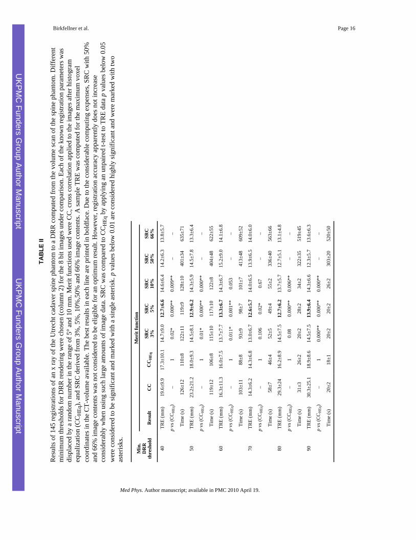
The merit functions under comparison were CC, CC after histogram equalization of theDRR and the x ray, and SRC using 3%, 5%, and 10%. Furthermore, SRC was also computedfor 50% and 66% of image content—these results are, however, to be considered out ofcompetition since the time required for computing the merit function becomes unacceptable.The results were recorded, and a t-test for independent samples was carried out for the TREusing SPSS 17.0. The resulting p values are given in Sec. III.
PI was also included. PI is known to give excellent results for 2D/3D registration,7 butsuffers from a very limited convergence range, which was also confirmed by our ownexperience.17,23,31,32 The merit function operates on a difference image Idiff and is given as
(5)
Since PI operates on a difference image, the threshold for DRR rendering also appearscritical since the disorder of the difference image is evaluated as a registration measure. Forour method of evaluation, we used PI together with histogram equalization. It turned out that
Birkfellner et al. Page 6
Med Phys. Author manuscript; available in PMC 2010 April 19.
UKPM
C Funders G
roup Author Manuscript
UKPM
C Funders G
roup Author Manuscript
the algorithm converged only for approximately 50% of all trials, with very varyingaccuracies when using internal parameters of σ =10 and r=3 or r=5. A detailed description ofthe algorithm can be found in Ref. 7.
MI, the other merit function that is of known usefulness when dealing with multimodal 3D/3D registration problems, is a natural choice for images with different histogram content.However, it is also known for its problematic behavior in the case of 2D/3D registration dueto the sparse population of the joint histogram.7,17 MI is highly dependent on the chosenimplementation.5 In our case, a simple approach where the probability distribution wasderived from a histogram with 32 bins was also of limited success. Therefore, MI wasfinally excluded from the experiments, which are strictly aiming at quasi-intramodal 2D/3Dregistration. When registering x-ray data to volume data from another modality but CT, thecase is of course different.
II.C. ImplementationThe algorithm was implemented using GCC 4.1.2 on Fedora 8 on a Dell Precision 490personal computer. User interfaces were programed using the Qt 3.3 toolkit (Trolltech,Norway). Basic image processing functionality was added using the ANALYZEAVW 9.0 library(BIR, Mayo Clinic, Rochester/MN).33 In principle, the software setup was adopted from thesetup used in Ref. 17. DRRs were generated using the wobbled splatting algorithm asdescribed in Ref. 24. The Downhill–Simplex algorithm used for optimization in the iterativeregistration process used was taken from the GSL package (Free Software Foundation,Boston, MA). Registration takes place in such manner that after convergence of thealgorithm, the result is used as a starting point for another trial. After three subsequentregistrations, the result was considered to be final.
III. RESULTSIn a first trial, the stability and accuracy of the implementation was tested. For this purpose,a DRR was rendered at a threshold of 70, and the result was fed into the program as the xray. The known initial position used for rendering the DRR was distorted by randompositions of 5° and 10 mm in each coordinate. Fifteen registrations were carried out. Such anevaluation on a phantom image gives only little information on the actual performance of analgorithm, but it gives a figure of the quality of the implementation including theoptimization algorithm. For this ideal configuration, no significant differences in themaximum TRE were found. All merit functions converged properly. PI suffers from itssmall range of convergence, which affects the overall algorithm accuracy. The detailedresults can be found in Table I.
The crucial evaluation is the registration of the spine phantom dataset; the results can befound in Table II. Here, SRC using 3%–10% of image content always outperformed CC andCCHEq. For CC, results were really bad, especially when using another treshold than 70,which gave the most homogeneous results for CC, CCHEq, and SRC. Maximum TRE forCCHEq ranged from 14.3±6.8 mm when using DRR threshold 70 to 18.9±8.6 mm atthreshold 90. The results for SRC (excluding the registration efforts when using 50% and66% image content, which did not show any improvement compared to SRC using 5% or10% image content) ranged from 12.6±5.7 mm (SRC 5%, DRR threshold 70) to 14.7±9.0mm (SRC 3%, DRR threshold 40). For all trials, the best results were provided by SRC. It isthe very nature of the volume-driven splat-rendering routine to perform better for higherthresholds,24 which results in faster registration if a higher threshold is chosen. Detailedresults for the time requirements are given in Table II; however, SRC for 3% and 5% imagecontents required usually 20–120 s, which is similar to the performance of CC and CCHEq.
Birkfellner et al. Page 7
Med Phys. Author manuscript; available in PMC 2010 April 19.
UKPM
C Funders G
roup Author Manuscript
UKPM
C Funders G
roup Author Manuscript
SRC with 50% or 66% of image content took up to 10 min and is therefore only of academicinterest.
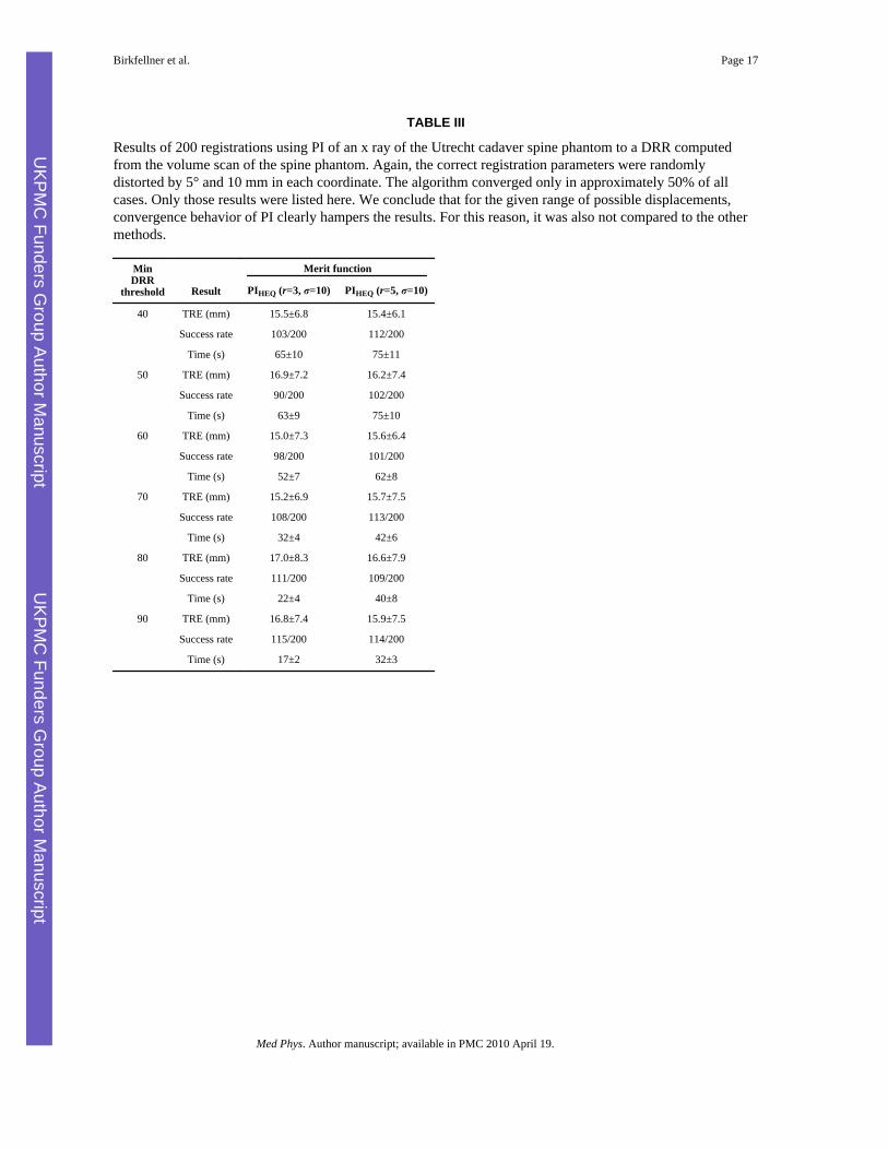
PI showed only limited stability and was terminated in almost 50% of all trials. Beyond that,a tendency to converge to local minima was evident, which spoiled the registration results.Maximum TRE for those cases where convergence was achieved ranged from 15.0±7.3 mm(PI with r=3, σ=10 at DRR threshold 60 to 17.0±8.3 mm for the same parameters at DRRthreshold 80. The results are given in Table III.
IV. DISCUSSION AND CONCLUSIONSIn this article we present a merit function suitable for 2D/3D registration of intramodal datasets acquired with x-ray-based equipment. The specific strength of SRC is the independenceof pixel intensity differences in the image histograms as long as they can be modeled asmonotonic transforms. Thus, radiometric calibration is unnecessary. However, SRC also haslimitations by design. As it is the case with most intensity-based merit functions, depth ofthe images is crucial; if only a few actual values of possible gray values γ are available, thenumber of tied ranks will be considerable, resulting in an inconclusive minimum for themerit function. It is also necessary to use images with lots of content—an x ray showing asmall structure surrounded by air will be definitely result in only a few random pixels withdifferent ranks. Furthermore, noise may affect the performance of SRC, as it is the case withmost other intensity-based merit functions. In this article, we have not examined theinfluence of noise on SRC. However, the evaluation was carried out with image dataacquired on standard imaging equipment, and the splat-rendering routine used24 producesimages stricken with Gaussian noise by definition, which is reduced by applying a spatiallow pass filter prior to registration of the DRR to the x ray. We therefore assume that thenoise level encountered here represents a real-life scenario.
Our results demonstrate that, in the given setup, SRC outperforms both classical CC and CCwith histogram equalization of the images under comparison; in the case of SRC, any kindof fine tuning in histogram space is not necessary by definition. Therefore, SRC is ideallysuited for registration tasks involving image data obtained at different energies, and theexperimental data on the spine phantom support this conclusion. However, a patient studyusing actual IGRT equipment is a pending task. Furthermore, the robustness of SRC in thepresence of high intensity artifacts is an open question. It is in the very nature of the rankcorrelation coefficient that it is more robust against outliers; the extent of this theoreticaladvantage is to be examined.
The use of stochastic sampled image information for merit function design is not new; inRef. 21, a similar approach was used to derive a MI-based merit function while reducingtime requirements for computing the DRR. We have not explored this approach sincewobbled splatting,24 as a volume-driven method, does not necessarily benefit fromrendering just a few pixels. On the other hand, it is more efficient compared to raycasting,and it offers huge potential for GPU-based implementation.22 A direct comparison of apartial raycasting approach as proposed in Ref. 21 and splat rendering is, however, also anopen task.
One possibly astonishing fact is the range of error encountered in the experiments and thefact that PI does not perform as good as one might expect from the literature. The reason forthis behavior lies in the fact that the initial misalignment in the experimental series is largerthan the convergence range typically used in most evaluations. The very limitedconvergence range of PI, however, is also known and documented.23,31,17,37 In Ref. 27, aconvergence range of only a few millimeters is considered to give safe results. With tumor
Birkfellner et al. Page 8
Med Phys. Author manuscript; available in PMC 2010 April 19.
UKPM
C Funders G
roup Author Manuscript
UKPM
C Funders G
roup Author Manuscript
motion tracking being our objective, it makes little sense to distinguish between failed andsuccessful registrations—only those results where the optimization algorithm did notconverge were excluded; the range of motions possible used in this evaluation is realistic,and beyond that, the TRE computed gives a worst case scenario. PI, a merit function thatgives a very sharp minimum, here is a victim of this setup. It gives very good results forsmall displacements, but outside the convergence range, it does not even provide a gradientthat might finally point in the appropriate direction. Future research work, for instance, onimproved optimization schemes as proposed in Ref. 31, is, however, necessary to improveconvergence behavior and algorithm accuracy. Since it is not possible to compute analyticalderivatives of a rank correlation type of merit function, options for alternative optimizationschemes are somewhat narrowed down.
Another problem we encountered is the little availability of reference datasets and referencealgorithm implementations. The spine dataset27 is a good initial effort in this direction butsuffers from limitations such as its reduced intensity range and the small size of thespecimen; a pending task for the community is therefore the development of morerepresentative phantoms, which are to be provided together with gold standard data andreference implementations of the registration algorithms. Given the methodological natureof this article, an exhaustive evaluation with more merit functions is therefore also a pendingtask.
The other question is the intended use of inter- and intrafractional imaging in combinationwith 2D/3D registration in IGRT. The most obvious application—exact positioning of thepatient prior to treatment—is widely discussed and well known both in image-guidedtherapy6-8,20,21,25,16,17,28 and radiation oncology.2-5,19,10,26 For this purpose, several x-rayimages from various positions or even a cone beam CT can be made, the latter providing theknown advantages of a full 3D volume. However, numerous tumor locations requireintrafractional position monitoring due to organ motion during irradiation. It appears as ifthe advent of on-board imaging units has further stimulated this field of research, whichspans approaches from motion prediction34,35 to permanent x-ray monitoring of implantedfiducial markers36 and registration.37-41 2D/3D registration using only one projection42 asin our experimental series appears to be a promising method of choice to achieve this goal ofminimizing dose delivered outside the target volume despite the additional irradiation fromthe low-energy x-ray system. With this perspective in mind, we have designed ourexperiments; here, the sensitivity and reliability of motion detection is of greater interestthan the absolute accuracy (which can be achieved by performing a 3D/3D registration ofthe cone beam CT and the diagnostic CT prior to irradiation anyhow). The future will showwhether image-based motion correction at a high rate can compete with motion sensingusing miniature active43 and passive44 tracking solutions. To achieve this goal, a massiveincrease in registration speed, which may be achievable using GPU-based parallelapproaches is necessary.
AcknowledgmentsANALYZEAVW and AVW were provided courtesy of Dr. R. A. Robb, BIR, Mayo Clinic, Rochester/MN. The authors aregrateful to Mary Mc Allister, MD, Johns Hopkins University Hospital, Baltimore, for manuscript assistance. Thespine dataset and ground truth for the 2D-3D registration used in this work was provided by the Image SciencesInstitute, University Medical Center Utrecht, The Netherlands. This work was supported by the Eurasia-PacificUninet Foundation and the Austrian Science Foundation FWF Grant Nos. P19931 and L503.
References1. Dawson LA, Jaffray DA. Advances in image-guided radiation therapy. J. Clin. Oncol 2007;25(8):
938–946. [PubMed: 17350942]
Birkfellner et al. Page 9
Med Phys. Author manuscript; available in PMC 2010 April 19.
UKPM
C Funders G
roup Author Manuscript
UKPM
C Funders G
roup Author Manuscript

2. Elshaikh M, Ljungman M, Haken R. Ten, Lichter AS. Advances in radiation oncology. Annu. Rev.Med 2006;57:19–31. [PubMed: 16409134]
3. Jans HS, Syme AM, Rathee S, Fallone BG. 3D interfractional patient position verification using2D-3D registration of orthogonal images. Med. Phys 2006;33(5):1420–1439. [PubMed: 16752578]
4. Meyer J, Richter A, Baier K, Wilbert J, Guckenberger M, Flentje M. Tracking moving objects withmegavoltage portal imaging: A feasibility study. Med. Phys 2006;33(5):1275–1280. [PubMed:16752562]
5. Künzler T, Grezdo J, Bogner J, Birkfellner W, Georg D. Registration of DRRs and portal images forverification of stereotactic body radiotherapy: A feasibility study in lung cancer treatment. Phys.Med. Biol 2007;52(8):2157–2170. [PubMed: 17404461]
6. Lemieux L, Jagoe R, Fish DR, Kitchen ND, Thomas DGT. A patient-to-computed-tomographyimage registration method based on digitally reconstructed radiographs. Med. Phys 1994;21(11):1749–1760. [PubMed: 7891637]
7. Penney GP, Weese J, Little JA, Desmedt P, Hill DLG, Hawkes DJ. A comparison of similaritymeasures for use in 2-D-3-D medical image registration. IEEE Trans. Med. Imaging 1998;17(4):586–595. [PubMed: 9845314]
8. Turgeon GA, Lehmann G, Guiraudon G, Drangova M, Holdsworth D, Peters T. 2D-3D registrationof coronary angiograms for cardiac procedure planning and guidance. Med. Phys 2005;32(12):3737–3749. [PubMed: 16475773]
9. Balter JM, Kessler ML. Imaging and alignment for image-guided radiation therapy. J. Clin. Oncol2007;25(8):931–937. [PubMed: 17350941]
10. Khamene A, Bloch P, Wein W, Svatos M, Sauer F. Automatic registration of portal images andvolumetric CT for patient positioning in radiation therapy. Med. Image Anal 2006;10:96–112.[PubMed: 16150629]
11. Dong S, Kettenbach J, Hinterleitner I, Bergmann H, Birkfellner W. The Zernike expansion—anexample of a merit function for 2D/3D registration based on orthogonal functions. MICCAI2008;11(2):964–971. [PubMed: 18982698]
12. Maes F, Collignon A, Vandermeulen D, Marchal G, Suetens P. Multimodality image registrationby maximization of mutual information. IEEE Trans. Med. Imaging 1997;16(2):187–198.[PubMed: 9101328]
13. Wells WM III, Viola P, Atsumi H, Nakajima S, Kikinis R. Multi-modal volume registration bymaximization of mutual information. Med. Image Anal 1996;1(1):35–51. [PubMed: 9873920]
14. Studholme C, Hill DL, Hawkes DJ. Automated 3D registration of MR and CT images of the head.Med. Image Anal 1996;1(2):163–175. [PubMed: 9873927]
15. Pluim JP, Maintz JB, Viergever MA. Mutual-information-based registration of medical images: Asurvey. IEEE Trans. Med. Imaging 2003;22(8):986–1004. [PubMed: 12906253]
16. Fei B, Duerk JL, Boll DT, Lewin JS, Wilson DL. Slice-to-volume registration and its potentialapplication to interventional MRI-guided radio-frequency thermal ablation of prostate cancer.IEEE Trans. Med. Imaging 2003;22(4):515–525. [PubMed: 12774897]
17. Birkfellner W, Figl M, Kettenbach J, Hummel J, Homolka P, Schernthaner R, Nau T, Bergmann H.Rigid 2D/3D slice-to-volume registration and its application on fluoroscopic CT images. Med.Phys 2007;34(1):246–255. [PubMed: 17278510]
18. Lacroute, P.; Levoy, M. Fast volume rendering using a shear-warp factorization of the viewingtransformation. Proceedings of SIGGRAPH ’94; 1994; New York: ACM; p. 451-458.
19. Russakoff DB, Rohlfing T, Mori K, Rueckert D, Ho A, Adler JR Jr. Maurer CR Jr. Fast generationof digitally reconstructed radiographs using attenuation fields with application to 2D-3D imageregistration. IEEE Trans. Med. Imaging 2005;24(11):1441–1454. [PubMed: 16279081]
20. Knaan D, Joskowicz L. Effective intensity-based 2D/3D rigid registration between fluoroscopic x-ray and CT. MICCAI 2003;6(1):351–358.
21. Zöllei, L.; Grimson, E.; Norbash, A.; Wells, W. 2D-3D rigid registration of x-ray fluoroscopy andCT images using mutual information and sparsely sampled histogram estimators. Proceedings ofIEEE CVPR; 2001; New York: IEEE Computer Society;
22. Spoerk J, Bergmann H, Wanschitz F, Dong S, Birkfellner W. Fast DRR splat rendering usingcommon consumer graphics hardware. Med. Phys 2007;34(11):4302–4308. [PubMed: 18072495]
Birkfellner et al. Page 10
Med Phys. Author manuscript; available in PMC 2010 April 19.
UKPM
C Funders G
roup Author Manuscript
UKPM
C Funders G
roup Author Manuscript

23. Kubias A, Deinzer F, Feldmann T, Paulus D, Schreiber B, Brunner T. 2D/3D image registration onthe GPU. Pattern Recognition and Image Analysis 2008;18(3):381–389.
24. Birkfellner W, Seemann R, Figl M, Hummel J, Ede C, Homolka P, Yang X, Niederer P, BergmannH. Wobbled splatting—a fast perspective colume rendering method for simulation of x-ray imagesfrom CT. Phys. Med. Biol 2005;50(9):N73–N84. [PubMed: 15843725]
25. Birkfellner W, Wirth J, Burgstaller W, Baumann B, Staedele H, Hammer B, Gellrich NC, JacobAL, Regazzoni P, Messmer P. A faster method for 2D/3D medical image registration. Phys. Med.Biol 2003;48(16):2665–2679. [PubMed: 12974581]
26. Andrews DW, Bednarz G, Evans JJ, Downes B. A review of 3 current radiosurgery systems. Surg.Neurol 2006;66(6):559–564. [PubMed: 17145309]
27. van de Kraats EB, Penney GP, Tomazevic D, van Walsum T, Niessen WJ. Standardized evaluationmethodology for 2-D-3-D registration. IEEE Trans. Med. Imaging 2005;24(9):1177–1189.[PubMed: 16156355]
28. Tomazevic D, Likar B, Pernus F. 3-D/2-D registration by integrating 2-D information in 3-D. IEEETrans. Med. Imaging 2006;25(1):17–27. [PubMed: 16398411]
29. Frühwald L, Kettenbach J, Figl M, Hummel J, Bergmann H, Birkfellner W. A comparative studyon manual and automatic slice-to-volume registration of CT images. Eur. Radiol. (in press).
30. Archip N, Statli P, Morrison P, Jolesz F, Warfield SK, Silverman S. Non-rigid registration of pre-procedural MR images with intra-procedural unenhanced CT-images for improved targeting oftumors during liver radiofrequency ablations. MICCAI 2007;10(2):969–977. [PubMed: 18044662]
31. Dey J, Napel S. Targeted 2D/3D registration using ray normalization and a hybrid optimizer. Med.Phys 2006;33(12):4730–4738. [PubMed: 17278825]
32. Russakoff, DB.; Rohlfing, T.; Ho, A.; Kim, DH.; Shahidi, R.; Adler, JR., Jr.; Maurer, CR, Jr..Evaluation of intensity-based 2D-3D spine image registration using clinical gold-standard data.Proceedings of the second International Workshop on Biomedical Image Registration; 2003;Philadelphia, PA: Springer; p. 151-160.Paper No. LNCS 2717
33. Robb RA, Hanson DP, Karwoski RA, Larson AG, Workman EL, Stacy MC. Analyze: Acomprehensive, operator-interactive software package for multidimensional medical image displayand analysis. Comput. Med. Imaging Graph 1989;13(6):433–454. [PubMed: 2688869]
34. Tewatia D, Zhang T, Tome W, Paliwal B, Metha M. Clinical implementation of target tracking bybreathing synchronized delivery. Med. Phys 2006;33(11):4330–4336. [PubMed: 17153412]
35. Sharp GC, Jiang SB, Shimizu S, Shirato H. Prediction of respiratory tumour motion for real-timeimage-guided radiotherapy. Phys. Med. Biol 2004;49(3):425–440. [PubMed: 15012011]
36. Tang X, Sharp GC, Jiang SB. Fluoroscopic tracking of multiple implanted fiducial markers usingmultiple object tracking. Phys. Med. Biol 2007;52:4081–4098. [PubMed: 17664596]
37. Rohlfing T, Denzler J, Grassl C, Russakoff DB, Maurer CR Jr. Markerless real-time 3-D targetregion tracking by motion backprojection from projection images. IEEE Trans. Med. Imaging2005;24(11):1455–1468. [PubMed: 16279082]
38. Zeng R, Fessler JA, Balter JM. Estimating 3-D respiratory motion from orbiting views bytomographic image registration. IEEE Trans. Med. Imaging 2007;26(2):153–163. [PubMed:17304730]
39. Lehmann GC, Holdsworth DW, Drangova M. Angle-independent measure of motion for image-based gating in 3D coronary angiography. Med. Phys 2006;33(5):1311–1320. [PubMed:16752566]
40. Shimizu S, Shirato H, Ogura S, Akita-Dosaka H, Kitamura K, Nishioka T, Kagei K, Nishimura M,Miyasaka K. Detection of lung tumor movement in real-time tumor-tracking radiotherapy. Int. J.Radiat. Oncol., Biol., Phys 2001;51(2):304–310. [PubMed: 11567803]
41. Shirato H, Shimizu S, Kitamura K, Onimaru R. Organ motion in image-guided radiotherapy:Lessons from real-time tumor tracking radiotherapy. Int. J. Clin. Oncol 2007;12(1):8–16.[PubMed: 17380435]
42. Suh Y, Dieterich S, Keall PJ. Geometric uncertainty of 2D projection imaging in monitoring 3Dtumor motion. Phys. Med. Biol 2007;52:3439–3454. [PubMed: 17664553]
43. Hummel J, Figl M, Kollmann C, Bergmann H, Birkfellner W. Evaluation of a miniatureelectromagnetic position tracker. Med. Phys 2002;29(10):2205–2212. [PubMed: 12408292]
Birkfellner et al. Page 11
Med Phys. Author manuscript; available in PMC 2010 April 19.
UKPM
C Funders G
roup Author Manuscript
UKPM
C Funders G
roup Author Manuscript

44. Kupelian P, Willoughby T, Mahadevan A, Djemil T, Weinstein G, Jani S, Enke C, Solberg T,Flores N, Liu D, Beyer D, Levine L. Multi-institutional clinical experience with the CalypsoSystem in localization and continuous, real-time monitoring of the prostate gland during externalradiotherapy. Int. J. Radiat. Oncol., Biol., Phys 2007;67(4):1088–1098. [PubMed: 17187940]
Birkfellner et al. Page 12
Med Phys. Author manuscript; available in PMC 2010 April 19.
UKPM
C Funders G
roup Author Manuscript
UKPM
C Funders G
roup Author Manuscript
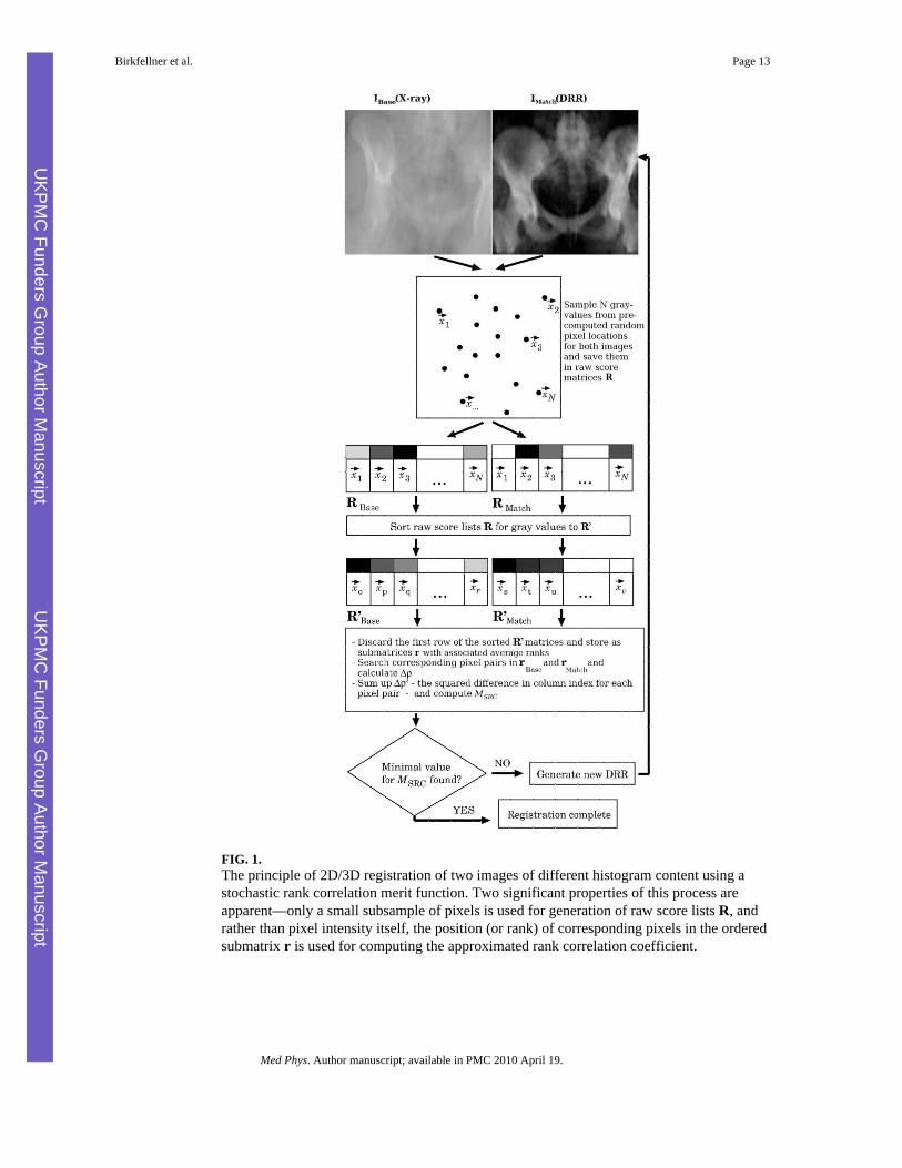
FIG. 1.The principle of 2D/3D registration of two images of different histogram content using astochastic rank correlation merit function. Two significant properties of this process areapparent—only a small subsample of pixels is used for generation of raw score lists R, andrather than pixel intensity itself, the position (or rank) of corresponding pixels in the orderedsubmatrix r is used for computing the approximated rank correlation coefficient.
Birkfellner et al. Page 13
Med Phys. Author manuscript; available in PMC 2010 April 19.
UKPM
C Funders G
roup Author Manuscript
UKPM
C Funders G
roup Author Manuscript
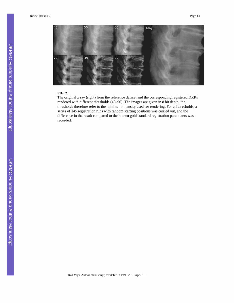
FIG. 2.The original x ray (right) from the reference dataset and the corresponding registered DRRsrendered with different thresholds (40–90). The images are given in 8 bit depth; thethresholds therefore refer to the minimum intensity used for rendering. For all thresholds, aseries of 145 registration runs with random starting positions was carried out, and thedifference in the result compared to the known gold standard registration parameters wasrecorded.
Birkfellner et al. Page 14
Med Phys. Author manuscript; available in PMC 2010 April 19.
UKPM
C Funders G
roup Author Manuscript
UKPM
C Funders G
roup Author Manuscript

UKPM
C Funders G
roup Author Manuscript
UKPM
C Funders G
roup Author Manuscript
Birkfellner et al. Page 15
Tabl
e I
Reg
istra
tion
resu
lts fo
r an
idea
l sim
ulat
ed D
RR
; the
se d
ata
give
an
idea
of t
he a
ccur
acy
to b
e ex
pect
ed fr
om th
e re
fere
nce
spin
e da
tase
t, th
e pr
ojec
tion
geom
etry
use
d, a
nd id
eal c
ircum
stan
ces g
iven
the
impl
emen
tatio
n of
use
d fo
r the
exp
erim
ents
. As e
xpec
ted,
CC
and
SR
C p
erfo
rm si
mila
r und
er su
chid
eal c
ondi
tions
. A ra
nge
of p
ossi
ble
disp
lace
men
ts fr
om th
e kn
own
proj
ectio
n pa
ram
eter
s of 5
° and
10
mm
was
use
d. T
he T
RE
was
com
pute
d fo
r the
max
imum
vox
el c
oord
inat
e av
aila
ble
in th
e vo
lum
e.
Mer
it fu
nctio
n
Res
ult
CC
SRC
3%SR
C5%
SRC
10%
SRC
50%
SRC
66%
PIr=
3, σ
=10
PIr=
5, σ
=10
TRE
(mm
)6.
9±4.
57.
5±1.
78.
0±4.
46.
8±3.
37.
9±4.
16.
8±3.
712
.5±6
.812
.3±6
.8
Med Phys. Author manuscript; available in PMC 2010 April 19.
UKPM
C Funders G
roup Author Manuscript
UKPM
C Funders G
roup Author Manuscript
Birkfellner et al. Page 16
TAB
LE II
Res
ults
of 1
45 re
gist
ratio
ns o
f an
x ra
y of
the
Utre
cht c
adav
er sp
ine
phan
tom
to a
DR
R c
ompu
ted
from
the
volu
me
scan
of t
he sp
ine
phan
tom
. Diff
eren
tm
inim
um th
resh
olds
for D
RR
rend
erin
g w
ere
chos
en (c
olum
n 2)
for t
he 8
bit
imag
es u
nder
com
paris
on. E
ach
of th
e kn
own
regi
stra
tion
para
met
ers w
asdi
spla
ced
by a
rand
om n
umbe
r in
the
rang
e of
5° a
nd 1
0 m
m. M
erit
func
tions
use
d w
ere
CC
, cro
ss c
orre
latio
n ap
plie
d to
the
imag
es a
fter h
isto
gram
equa
lizat
ion
(CC
HEq
), an
d SR
C d
eriv
ed fr
om 3
%, 5
%, 1
0%,5
0% a
nd 6
6% im
age
cont
ents
. A sa
mpl
e TR
E w
as c
ompu
ted
for t
he m
axim
um v
oxel
coor
dina
tes i
n th
e C
T-vo
lum
e av
aila
ble.
The
bes
t res
ults
in e
ach
line
are
prin
ted
in b
oldf
ace.
Due
to th
e co
nsid
erab
le c
ompu
ting
expe
nses
, SR
C w
ith 5
0%an
d 66
% im
age
cont
ents
was
not
con
side
red
to b
e el
igib
le fo
r an
optim
um re
sult.
How
ever
, reg
istra
tion
accu
racy
app
aren
tly d
oes n
ot in
crea
seco
nsid
erab
ly w
hen
usin
g su
ch la
rge
amou
nts o
f im
age
data
. SR
C w
as c
ompa
red
to C
CH
Eq b
y ap
plyi
ng a
n un
paire
d t-t
est t
o TR
E da
ta p
val
ues b
elow
0.0
5w
ere
cons
ider
ed to
be
sign
ifica
nt a
nd m
arke
d w
ith a
sing
le a
ster
isk.
p v
alue
s bel
ow 0
.01
are
cons
ider
ed h
ighl
y si
gnifi
cant
and
wer
e m
arke
d w
ith tw
oas
teris
ks.
Min
.D
RR
thre
shol
d
Mer
it fu
nctio
n
Res
ult
CC
CC
HE
qSR
C3%
SRC
5%SR
C10
%SR
C50
%SR
C66
%
40TR
E (m
m)
19.6
±9.9
17.3
±10.
114
.7±9
.012
.7±6
.614
.6±6
.414
.2±6
.313
.8±5
.7
p vs
(CC
HEq
)–
10.
02*
0.00
0**
0.00
9**
––
Tim
e (s
)12
6±12
110±
812
2±11
119±
912
8±10
401±
3463
5±71
50TR
E (m
m)
23.2
±21.
218
.0±9
.314
.5±8
.112
.9±6
.214
.3±5
.914
.5±7
.813
.3±6
.4
p vs
(CC
HEq
)–
10.
01*
0.00
0**
0.00
0**
––
Tim
e (s
)11
9±12
106±
811
5±10
117±
1012
2±8
404±
4862
2±55
60TR
E (m
m)
16.3
±11.
316
.0±7
.513
.7±7
.713
.3±6
.714
.3±6
.715
.2±9
.014
.1±6
.8
p vs
(CC
HEq
)–
10.
011*
0.00
1**
0.05
3–
–
Tim
e (s
)10
3±11
88±8
93±9
98±7
101±
741
3±48
609±
52
70TR
E (m
m)
14.3
±6.2
14.3
±6.8
13.0
±6.7
12.6
±5.7
14.0
±6.5
13.9
±6.5
14.0
±6.0
p vs
(CC
HEq
)–
10.
106
0.02
*0.
67–
–
Tim
e (s
)58
±746
±452
±549
±455
±233
6±40
563±
66
80TR
E (m
m)
29.3
±24
16.2
±8.9
14.5
±7.5
12.7
±6.2
13.7
±5.7
12.7
±5.1
13.1
±4.8
p vs
(CC
HEq
)–
10.
080.
000*
*0.
006*
*–
–
Tim
e (s
)31
±326
±220
±228
±234
±232
2±35
519±
45
90TR
E (m
m)
30.3
±25.
118
.9±8
.614
.5±7
.513
.9±6
.414
.3±6
.612
.3±5
.713
.6±6
.3
p vs
(CC
HEq
)–
10.
000*
*0.
000*
*0.
000*
*–
–
Tim
e (s
)20
±218
±120
±220
±226
±230
3±20
520±
50
Med Phys. Author manuscript; available in PMC 2010 April 19.
UKPM
C Funders G
roup Author Manuscript
UKPM
C Funders G
roup Author Manuscript
Birkfellner et al. Page 17
TABLE III
Results of 200 registrations using PI of an x ray of the Utrecht cadaver spine phantom to a DRR computedfrom the volume scan of the spine phantom. Again, the correct registration parameters were randomlydistorted by 5° and 10 mm in each coordinate. The algorithm converged only in approximately 50% of allcases. Only those results were listed here. We conclude that for the given range of possible displacements,convergence behavior of PI clearly hampers the results. For this reason, it was also not compared to the othermethods.
MinDRR
threshold
Merit function
Result PIHEQ (r=3, σ=10) PIHEQ (r=5, σ=10)
40 TRE (mm) 15.5±6.8 15.4±6.1
Success rate 103/200 112/200
Time (s) 65±10 75±11
50 TRE (mm) 16.9±7.2 16.2±7.4
Success rate 90/200 102/200
Time (s) 63±9 75±10
60 TRE (mm) 15.0±7.3 15.6±6.4
Success rate 98/200 101/200
Time (s) 52±7 62±8
70 TRE (mm) 15.2±6.9 15.7±7.5
Success rate 108/200 113/200
Time (s) 32±4 42±6
80 TRE (mm) 17.0±8.3 16.6±7.9
Success rate 111/200 109/200
Time (s) 22±4 40±8
90 TRE (mm) 16.8±7.4 15.9±7.5
Success rate 115/200 114/200
Time (s) 17±2 32±3
Med Phys. Author manuscript; available in PMC 2010 April 19.