ribosomal hibernation-associated factors in escherichia coli
TRANSCRIPT
�����������������
Citation: Maki, Y.; Yoshida, H.
Ribosomal Hibernation-Associated
Factors in Escherichia coli.
Microorganisms 2022, 10, 33. https://
doi.org/10.3390/microorganisms
10010033
Academic Editor: José Marques
Andrade
Received: 1 November 2021
Accepted: 21 December 2021
Published: 24 December 2021
Publisher’s Note: MDPI stays neutral
with regard to jurisdictional claims in
published maps and institutional affil-
iations.
Copyright: © 2021 by the authors.
Licensee MDPI, Basel, Switzerland.
This article is an open access article
distributed under the terms and
conditions of the Creative Commons
Attribution (CC BY) license (https://
creativecommons.org/licenses/by/
4.0/).
microorganisms
Review
Ribosomal Hibernation-Associated Factors in Escherichia coli
Yasushi Maki and Hideji Yoshida *
Department of Physics, Osaka Medical and Pharmaceutical University, Takatsuki 569-8686, Japan;[email protected]* Correspondence: [email protected]; Tel.: +81-72-684-7023
Abstract: Bacteria convert active 70S ribosomes to inactive 100S ribosomes to survive under variousstress conditions. This state, in which the ribosome loses its translational activity, is known as riboso-mal hibernation. In gammaproteobacteria such as Escherichia coli, ribosome modulation factor andhibernation-promoting factor are involved in forming 100S ribosomes. The expression of ribosomemodulation factor is regulated by (p)ppGpp (which is induced by amino acid starvation), cAMP-CRP(which is stimulated by reduced metabolic energy), and transcription factors involved in biofilmformation. This indicates that the formation of 100S ribosomes is an important strategy for bacterialsurvival under various stress conditions. In recent years, the structures of 100S ribosomes fromvarious bacteria have been reported, enhancing our understanding of the 100S ribosome. Here, wepresent previous findings on the 100S ribosome and related proteins and describe the stress-responsepathways involved in ribosomal hibernation.
Keywords: 100S ribosome; ribosome modulation factor; hibernation-promoting factor; ribosomalhibernation; stress response
1. Introduction
Bacteria have developed sophisticated adaptive systems to survive during various en-vironmental changes, including nutrient starvation, temperature shock, osmolarity changes,and rapid pH changes. These systems include inhibition of cell growth, reduction of cellvolume, changes in cell shape, compression of nucleoids, changes in the cell wall com-position, and changes in cytoplasmic components [1]. These environmental adaptationsmay result in the formation of biofilms encased in extracellular polymers [2] or the con-version of bacteria into persister cells with reduced vitality [3], making them resistantto antibiotics and responsible for chronic infections [4–7]. To further combat these stressconditions, bacteria utilize a system to dimerize and inactivate ribosomes [8]. In somegammaproteobacteria, such as Escherichia coli and Vibrio cholerae, two ribosomal proteinfactors, ribosome modulation factor (RMF) and hibernation-promoting factor (HPF), bindto the 70S ribosome to produce its dimeric form, the 100S ribosome. In most other bacteria,including Staphylococcus aureus, Lactobacillus paracasei, and Thermus thermophilus, the 100Sribosome is formed through binding of a long-form of HPF to the 70S ribosome [9–11].A ribosomal hibernation model has been proposed in which the 100S ribosome loses itstranslational activity to reduce cellular energy consumption and functions as a reservoirfor protecting ribosomes from degradation by RNases [12–15]. Recently, high-resolutionstructures of 100S ribosomes from various bacteria have been reported, improving theunderstanding of ribosome hibernation [16–20].
In this review, we describe previous findings on the 100S ribosome and related proteinfactors in E. coli, as shown in Table 1. We attempted to integrate the hibernation stage intothe ribosome cycle and discuss the elaborate stress response pathway involved in bacterialtranslational control.
Microorganisms 2022, 10, 33. https://doi.org/10.3390/microorganisms10010033 https://www.mdpi.com/journal/microorganisms
Microorganisms 2022, 10, 33 2 of 11
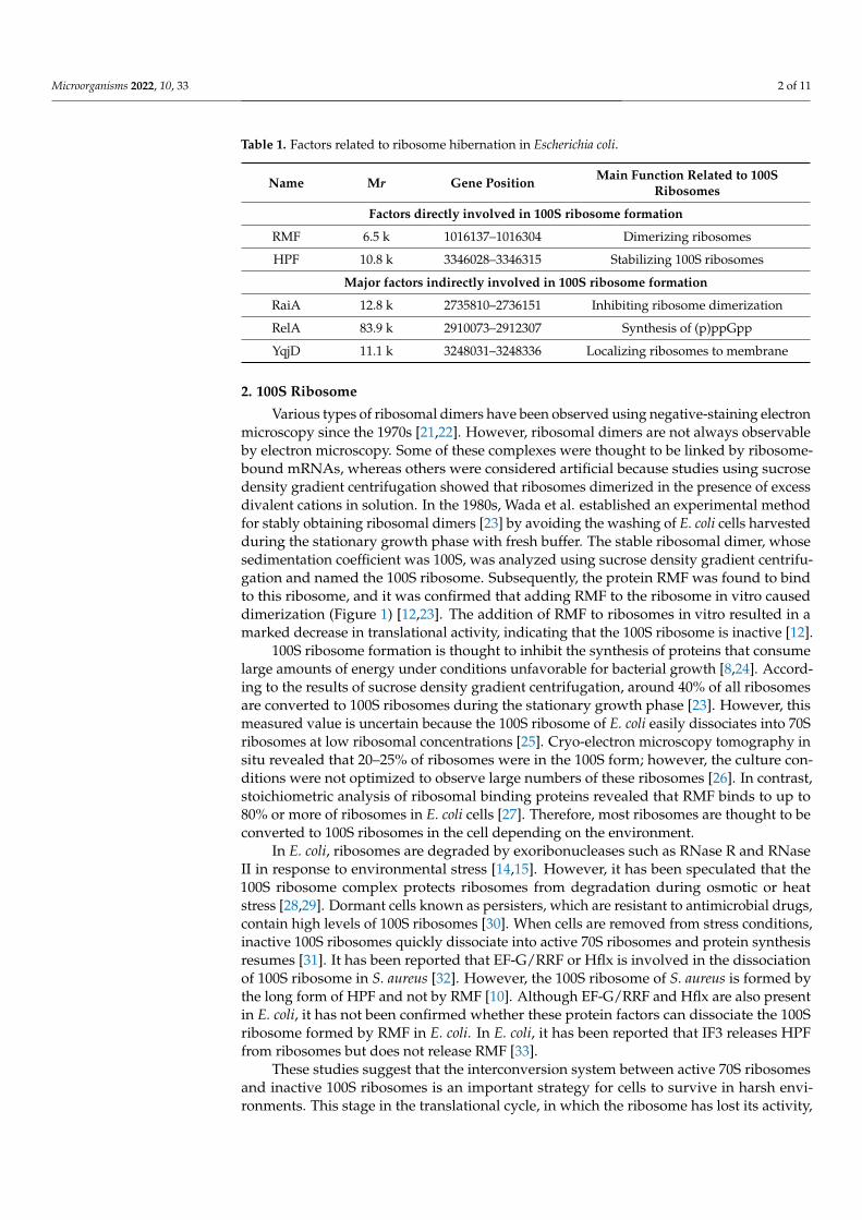
Table 1. Factors related to ribosome hibernation in Escherichia coli.
Name Mr Gene Position Main Function Related to 100SRibosomes
Factors directly involved in 100S ribosome formation
RMF 6.5 k 1016137–1016304 Dimerizing ribosomes
HPF 10.8 k 3346028–3346315 Stabilizing 100S ribosomes
Major factors indirectly involved in 100S ribosome formation
RaiA 12.8 k 2735810–2736151 Inhibiting ribosome dimerization
RelA 83.9 k 2910073–2912307 Synthesis of (p)ppGpp
YqjD 11.1 k 3248031–3248336 Localizing ribosomes to membrane
2. 100S Ribosome
Various types of ribosomal dimers have been observed using negative-staining electronmicroscopy since the 1970s [21,22]. However, ribosomal dimers are not always observableby electron microscopy. Some of these complexes were thought to be linked by ribosome-bound mRNAs, whereas others were considered artificial because studies using sucrosedensity gradient centrifugation showed that ribosomes dimerized in the presence of excessdivalent cations in solution. In the 1980s, Wada et al. established an experimental methodfor stably obtaining ribosomal dimers [23] by avoiding the washing of E. coli cells harvestedduring the stationary growth phase with fresh buffer. The stable ribosomal dimer, whosesedimentation coefficient was 100S, was analyzed using sucrose density gradient centrifu-gation and named the 100S ribosome. Subsequently, the protein RMF was found to bindto this ribosome, and it was confirmed that adding RMF to the ribosome in vitro causeddimerization (Figure 1) [12,23]. The addition of RMF to ribosomes in vitro resulted in amarked decrease in translational activity, indicating that the 100S ribosome is inactive [12].
100S ribosome formation is thought to inhibit the synthesis of proteins that consumelarge amounts of energy under conditions unfavorable for bacterial growth [8,24]. Accord-ing to the results of sucrose density gradient centrifugation, around 40% of all ribosomesare converted to 100S ribosomes during the stationary growth phase [23]. However, thismeasured value is uncertain because the 100S ribosome of E. coli easily dissociates into 70Sribosomes at low ribosomal concentrations [25]. Cryo-electron microscopy tomography insitu revealed that 20–25% of ribosomes were in the 100S form; however, the culture con-ditions were not optimized to observe large numbers of these ribosomes [26]. In contrast,stoichiometric analysis of ribosomal binding proteins revealed that RMF binds to up to80% or more of ribosomes in E. coli cells [27]. Therefore, most ribosomes are thought to beconverted to 100S ribosomes in the cell depending on the environment.
In E. coli, ribosomes are degraded by exoribonucleases such as RNase R and RNaseII in response to environmental stress [14,15]. However, it has been speculated that the100S ribosome complex protects ribosomes from degradation during osmotic or heatstress [28,29]. Dormant cells known as persisters, which are resistant to antimicrobial drugs,contain high levels of 100S ribosomes [30]. When cells are removed from stress conditions,inactive 100S ribosomes quickly dissociate into active 70S ribosomes and protein synthesisresumes [31]. It has been reported that EF-G/RRF or Hflx is involved in the dissociationof 100S ribosome in S. aureus [32]. However, the 100S ribosome of S. aureus is formed bythe long form of HPF and not by RMF [10]. Although EF-G/RRF and Hflx are also presentin E. coli, it has not been confirmed whether these protein factors can dissociate the 100Sribosome formed by RMF in E. coli. In E. coli, it has been reported that IF3 releases HPFfrom ribosomes but does not release RMF [33].
These studies suggest that the interconversion system between active 70S ribosomesand inactive 100S ribosomes is an important strategy for cells to survive in harsh envi-ronments. This stage in the translational cycle, in which the ribosome has lost its activity,
Microorganisms 2022, 10, 33 3 of 11
is named the hibernation stage, and ribosomes in this stage are known as hibernationribosomes [12].
Microorganisms 2022, 10, x FOR PEER REVIEW 3 of 11
named the hibernation stage, and ribosomes in this stage are known as hibernation ribo-somes [12].
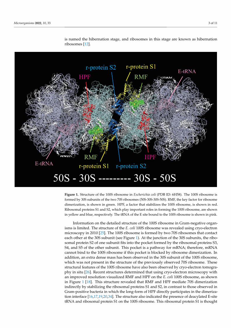
Information on the detailed structure of the 100S ribosome in Gram-negative organ-isms is limited. The structure of the E. coli 100S ribosome was revealed using cryo-electron microscopy in 2010 [25]. The 100S ribosome is formed by two 70S ribosomes that contact each other at the 30S subunit (see Figure 1). At the junction of the 30S subunits, the ribo-somal protein S2 of one subunit fits into the pocket formed by the ribosomal proteins S3, S4, and S5 of the other subunit. This pocket is a pathway for mRNA; therefore, mRNA cannot bind to the 100S ribosome if this pocket is blocked by ribosome dimerization. In addition, an extra dense mass has been observed in the 30S subunit of the 100S ribosome, which was not present in the structure of the previously observed 70S ribosome. These structural features of the 100S ribosome have also been observed by cryo-electron tomog-raphy in situ [26]. Recent structures determined that using cryo-electron microscopy with an improved resolution visualized RMF and HPF on the E. coli 100S ribosome, as shown in Figure 1 [18]. This structure revealed that RMF and HPF mediate 70S dimerization in-directly by stabilizing the ribosomal proteins S1 and S2, in contrast to those observed in Gram-positive bacteria in which the long-form of HPF directly participates in the dimeri-zation interface [16,17,19,20,34]. The structure also indicated the presence of deacylated E-site tRNA and ribosomal protein S1 on the 100S ribosome. This ribosomal protein S1 is thought to be the extra-dense mass mentioned above. Interestingly, S1 has an inactive structure for translation initiation and contacts the RMF [18].
Figure 1. Structure of the 100S ribosome in Escherichia coli (PDB ID: 6H58). The 100S ribosome is formed by 30S subunits of the two 70S ribosomes (50S-30S-30S-50S). RMF, the key factor for ribosome dimerization, is shown in green. HPF, a factor that stabilizes the 100S ribosome, is shown in red. Ribosomal proteins S1 and S2, which play important roles in forming the 100S ribosome, are shown in yellow and blue, respectively. The tRNA of the E site bound to the 100S ribosome is shown in pink.
Figure 1. Structure of the 100S ribosome in Escherichia coli (PDB ID: 6H58). The 100S ribosome isformed by 30S subunits of the two 70S ribosomes (50S-30S-30S-50S). RMF, the key factor for ribosomedimerization, is shown in green. HPF, a factor that stabilizes the 100S ribosome, is shown in red.Ribosomal proteins S1 and S2, which play important roles in forming the 100S ribosome, are shownin yellow and blue, respectively. The tRNA of the E site bound to the 100S ribosome is shown in pink.
Information on the detailed structure of the 100S ribosome in Gram-negative organ-isms is limited. The structure of the E. coli 100S ribosome was revealed using cryo-electronmicroscopy in 2010 [25]. The 100S ribosome is formed by two 70S ribosomes that contacteach other at the 30S subunit (see Figure 1). At the junction of the 30S subunits, the ribo-somal protein S2 of one subunit fits into the pocket formed by the ribosomal proteins S3,S4, and S5 of the other subunit. This pocket is a pathway for mRNA; therefore, mRNAcannot bind to the 100S ribosome if this pocket is blocked by ribosome dimerization. Inaddition, an extra dense mass has been observed in the 30S subunit of the 100S ribosome,which was not present in the structure of the previously observed 70S ribosome. Thesestructural features of the 100S ribosome have also been observed by cryo-electron tomogra-phy in situ [26]. Recent structures determined that using cryo-electron microscopy withan improved resolution visualized RMF and HPF on the E. coli 100S ribosome, as shownin Figure 1 [18]. This structure revealed that RMF and HPF mediate 70S dimerizationindirectly by stabilizing the ribosomal proteins S1 and S2, in contrast to those observed inGram-positive bacteria in which the long-form of HPF directly participates in the dimeriza-tion interface [16,17,19,20,34]. The structure also indicated the presence of deacylated E-sitetRNA and ribosomal protein S1 on the 100S ribosome. This ribosomal protein S1 is thought
Microorganisms 2022, 10, 33 4 of 11
to be the extra-dense mass mentioned above. Interestingly, S1 has an inactive structure fortranslation initiation and contacts the RMF [18].
3. Preparation of Ribosomal Hibernation3.1. Synthesis of (p)ppGpp on the Ribosome by RelA
Expression of the rmf gene is known to be controlled by guanosine tetraphosphateand pentaphosphate, collectively known as (p)ppGpp [35]. Depletion of essential nutrientsin the environment is among the most serious threats to bacteria. A central component ofadaptation to this stress is the stringent response [36] driven by (p)ppGpp, which playsnumerous roles in regulating cell growth rates and adapting to the environment [37].(p)ppGpp regulates transcriptional activity and decreases ribosome biosynthesis for envi-ronmental adaptation by interacting with RNA polymerase [38,39]. Intracellular (p)ppGpplevels are regulated by enzymes belonging to the RelA/SpoT homolog family [40]. SpoTis a bifunctional enzyme with strong hydrolytic activity and weak (p)ppGpp synthesisactivity (<8> in Figure 2) [41], whereas RelA is a ribosomal factor with (p)ppGpp synthesisactivity (see Table 1) [42]. When the supply of amino acids is limited by nutrient starvation,deacylated tRNA binds to the A site of the 70S ribosome (<6> in Figure 2). RelA is activatedby binding to deacetylated tRNA-bound 70S ribosomes [43]. Activated RelA transfers thepyrophosphoryl group from ATP to GTP or GDP on the ribosome, synthesizing pppGpp orppGpp, respectively (<7> in Figure 2). To understand how RelA synthesizes (p)ppGpp onthe deacylated tRNA-bound 70S ribosome, the high-resolution structure of the entire 70SRelA deacyl-tRNA complex was analyzed [44–46]. The structural analysis results showedthat the interaction between RelA and the ribosome caused conformational changes inboth the 30S and 50S subunits during (p)ppGpp synthesis. (p)ppGpp is involved in pro-moting transcription of the rmf gene and strongly affects 100S ribosome formation (<9> inFigure 2) [35]. This sequence of events (amino acid starvation→ synthesis of (p)ppGppby RelA→ expression of RMF by (p)ppGpp→ formation of 100S ribosome by RMF→reduction of translational activity by 100S ribosome) is congruent with the response tostarvation stress.
3.2. Changes in Ribosomes Because of Growth Phase Transition
The established ribosome cycle in bacteria consists of initiation, elongation, termina-tion, and recycling stages (<1>–<4> in Figure 2). In the exponential growth phase, whennutrients are sufficient, ribosomes dissociate into subunits in a GTP-dependent reactioninvolving ribosome recycling factor and elongation factor G after the recycling phase.Thereafter, initiation factor 3 (IF3) binds to the 30S subunit to stabilize the dissociation forthe next round of translation (<5> in Figure 2) [47]. However, ribosomes are less likely to bebound by IF3 as the growth phase transitions from the exponential phase to the stationaryphase [48]. In contrast, when RMF is added to ribosomes prepared from cells duringthe exponential phase in vitro, 100S ribosomes are less likely to form than when they areprepared from cells during the stationary phase [48]. These results indicate that ribosomesin the exponential and stationary phases are different. Structural analysis showed that theribosomal protein S1 binds to 100S ribosomes, which contacts the RMF [18]. It is knownthat this ribosomal protein facilitates the unwinding and placement of the start codon ofmRNA [49]. Interestingly, the S1 protein on the 100S ribosome has an inactive and compactconformation that differs from previously resolved structures. Moreover, the S1 proteinhas been reported to interact with IF3 [33,50]. The ribosomal protein S1 may play a rolein regulating the binding of IF3 to ribosomes and initiating translation or RMF binding toribosomes and dimerizing them during the transition from the exponential phase to thestationary phase.
Microorganisms 2022, 10, 33 5 of 11
Microorganisms 2022, 10, x FOR PEER REVIEW 4 of 11
3. Preparation of Ribosomal Hibernation 3.1. Synthesis of (p)ppGpp on the Ribosome by RelA
Expression of the rmf gene is known to be controlled by guanosine tetraphosphate and pentaphosphate, collectively known as (p)ppGpp [35]. Depletion of essential nutri-ents in the environment is among the most serious threats to bacteria. A central compo-nent of adaptation to this stress is the stringent response [36] driven by (p)ppGpp, which plays numerous roles in regulating cell growth rates and adapting to the environment [37]. (p)ppGpp regulates transcriptional activity and decreases ribosome biosynthesis for environmental adaptation by interacting with RNA polymerase [38,39]. Intracellular (p)ppGpp levels are regulated by enzymes belonging to the RelA/SpoT homolog family [40]. SpoT is a bifunctional enzyme with strong hydrolytic activity and weak (p)ppGpp synthesis activity (<8> in Figure 2) [41], whereas RelA is a ribosomal factor with (p)ppGpp synthesis activity (see Table 1) [42]. When the supply of amino acids is limited by nutrient starvation, deacylated tRNA binds to the A site of the 70S ribosome (<6> in Figure 2). RelA is activated by binding to deacetylated tRNA-bound 70S ribosomes [43]. Activated RelA transfers the pyrophosphoryl group from ATP to GTP or GDP on the ribosome, synthe-sizing pppGpp or ppGpp, respectively (<7> in Figure 2). To understand how RelA synthe-sizes (p)ppGpp on the deacylated tRNA-bound 70S ribosome, the high-resolution struc-ture of the entire 70S RelA deacyl-tRNA complex was analyzed [44–46]. The structural analysis results showed that the interaction between RelA and the ribosome caused con-formational changes in both the 30S and 50S subunits during (p)ppGpp synthesis. (p)ppGpp is involved in promoting transcription of the rmf gene and strongly affects 100S ribosome formation (<9> in Figure 2) [35]. This sequence of events (amino acid starvation → synthesis of (p)ppGpp by RelA → expression of RMF by (p)ppGpp → formation of 100S ribosome by RMF → reduction of translational activity by 100S ribosome) is congru-ent with the response to starvation stress.
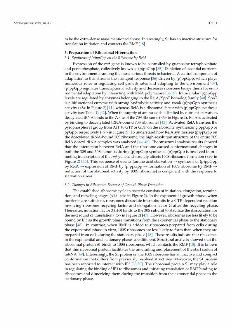
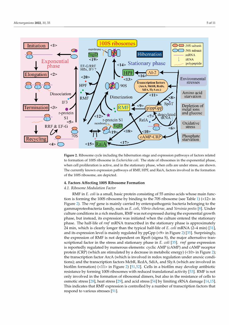
Figure 2. Ribosome cycle including the hibernation stage and expression pathways of factors related to formation of 100S ribosome in Escherichia coli. The state of ribosomes in the exponential phase, when cell proliferation is active, and in the stationary phase, when cells are under stress, are shown.
Figure 2. Ribosome cycle including the hibernation stage and expression pathways of factors relatedto formation of 100S ribosome in Escherichia coli. The state of ribosomes in the exponential phase,when cell proliferation is active, and in the stationary phase, when cells are under stress, are shown.The currently known expression pathways of RMF, HPF, and RaiA, factors involved in the formationof the 100S ribosome, are depicted.
4. Factors Affecting 100S Ribosome Formation4.1. Ribosome Modulation Factor
RMF in E. coli is a small, basic protein consisting of 55 amino acids whose main func-tion is forming the 100S ribosome by binding to the 70S ribosome (see Table 1) (<12> inFigure 2). The rmf gene is mainly carried by enteropathogenic bacteria belonging to thegammaproteobacteria family, such as E. coli, Vibrio cholerae, and Yersinia pestis [8]. Underculture conditions in a rich medium, RMF was not expressed during the exponential growthphase, but instead, its expression was initiated when the culture entered the stationaryphase. The half-life of rmf mRNA transcribed in the stationary phase is approximately24 min, which is clearly longer than the typical half-life of E. coli mRNA (2–4 min) [31],and its expression level is mainly regulated by ppGpp (<9> in Figure 2) [35]. Surprisingly,the expression of RMF is not dependent on RpoS (sigma S), the major alternative tran-scriptional factor in the stress and stationary phase in E. coli [35]. rmf gene expressionis reportedly regulated by numerous elements: cyclic AMP (cAMP) and cAMP receptorprotein (CRP) (which are stimulated by a decrease in metabolic energy) (<10> in Figure 2);the transcription factor ArcA (which is involved in redox regulation under anoxic condi-tions); and the transcription factors McbR, RcdA, SdiA, and SlyA (which are involved inbiofilm formation) (<11> in Figure 2) [51,52]. Cells in a biofilm may develop antibioticresistance by forming 100S ribosomes with reduced translational activity [53]. RMF is notonly involved in the formation of ribosomal dimers, but also in the resistance of cells toosmotic stress [28], heat stress [29], and acid stress [54] by limiting rRNA damage [14,15].This indicates that RMF expression is controlled by a number of transcription factors thatrespond to various stresses [51].
Microorganisms 2022, 10, 33 6 of 11
The binding sites of RMF on the ribosome have been investigated by protein–proteincrosslinking [13], chemical probing [55], and structural analyses [18,56]. The results of thesestructural analyses indicated that RMF binds near the anti-Shine-Dalgarno region of the30S subunit (see Figure 1). The structure of the RMF consists of two helices connectedby a linker, as determined in nuclear magnetic resonance analysis (PDB ID: 2jRM), X-raycrystallography (PDB ID: 4V8G), and cryo-electron microscopy (PDB ID: 6H4N). Thirteenamino acids of RMF are completely conserved and functionally important, as replacementof one of these residues with alanine suppressed ribosome dimerization [57]. Examinationof the functional sites using mutants of RMF revealed that R3, K5, and R11 in the N-terminaldomain contribute significantly to ribosome binding. Analysis of the 100S ribosome bycryo-electron microscopy showed that R3 and K5 interact with helix 28 of 16S rRNA [18].In addition, G23 and R45 are important for ribosome dimerization because although RMFswith mutations in these amino acids bind to ribosomes, they cannot form dimers [57]. Thus,RMF contains multiple functional sites for ribosome binding and ribosome dimerization.
4.2. RaiA and HPF
Protein Y, the gene product of yfiA, was identified as a factor binding to the inter-subunit position of the 30S, which stabilizes the 70S monomer of E. coli ribosomes underlow Mg2+ conditions in vitro [58]. Subsequently, this protein was found to inhibit cell-freetranslation of mRNA in cell extracts of E. coli and be induced under cold-shock stress.Thus, protein Y was renamed as ribosome-associated inhibitor A (RaiA) and its genename (yfiA) as raiA, as shown in Table 1 [59]. It has also been reported that RaiA reducestranslation errors [60].
RaiA and its paralogous protein YhbH were found to be associated with hibernatingribosomes in the stationary phase of E. coli cells and were released from translating ribo-somes soon after starved cells were transferred into a fresh medium. This indicates thatthese proteins are involved in the storage form of ribosomes in the stationary phase, atleast in E. coli, as is the case with RMF [61]. YhbH appeared to promote the formation of100S ribosomes by converting the immature 90S dimer containing RMF; hence, the proteinand its gene name were renamed as HPF and hpf, respectively, as shown in Table 1 (<14>in Figure 2) [62]. Although both RaiA and HPF are encoded in the E. coli genome and arehighly homologous to each other, HPF binds to 100S ribosomes with RMF in contrast toRaiA binding to the 70S monomer in the ribosomal fraction (<15> in Figure 2) [61].
Crystallographic and chemical probing analyses revealed that RaiA blocks the peptidyl-tRNA site (P site) and aminoacyl-tRNA site (A site) of the ribosome and inhibits translationinitiation [63]. It was suggested that the HPF binding site overlaps with RaiA because of thehigh identity of these sequences and their similar structures in solution [64]; nevertheless,HPF promotes 100S formation with RMF, in contrast to RaiA, which prevents dimer forma-tion [62]. The 3D structure of HPF and YfiA on the ribosome showed that these proteinsare bound in the channel between the head and body of the 30S subunit where tRNAsand mRNA bind during protein synthesis [56]. It was also suggested that RaiA inhibits100S formation using its C-terminal short tail, which blocks binding of RMF and preventsRMF-induced dimer formation. Recently, a high-resolution structure of hibernating 100Sribosomes from E. coli was reported with well-resolved electron densities for HPF and RMF,revealing a direct interaction of HPF with E-site tRNA, as shown in Figure 1 [18].
Some information on the expression and regulatory mechanisms of the genes ofRaiA and HPF in E. coli is known. Expression of both proteins is induced under starvedconditions [61], including nitrogen starvation [65]. It has also been reported that themRNA abundance of hpf is increased by the signal of autoinducer 2 (<16> in Figure 2) [66].RaiA was induced under cold-shock treatment [59] and downregulated by FNR, a globalregulator of anaerobic metabolism [67]. RaiA and RMF expression is induced by cAMP inresponse to glucose starvation (<17>, <10> in Figure 2) [51] and by starvation alarmone(p)ppGpp (<18>, <9> in Figure 2) [68].
Microorganisms 2022, 10, 33 7 of 11
Recently, some results concerning functions of these proteins and storage ribosomeshave been reported in several bacterial species. The HPF- or RaiA-bound storage ribosomesof E. coli exhibit resistance to unfolded protein-mediated subunit dissociation and subse-quent degradation by cellular ribonucleases, with the intrinsic chaperon activity retainedto assist in protein folding [69]. The absence of HPF results in the loss of some proteinsfrom the ribosome during incubation in the stationary phase [70,71]; furthermore, HPFprotects ribosomes against degradation in the absence of mRNA by blocking the attackof ribonuclease [72–74]. Although these results include cases of specific species except forgammaproteobacteria, the inter-subunit proteins of non-active ribosomes may typicallyprotect the machinery from degradation under stress conditions.
4.3. YqjD
YqjD, whose physiological function was unknown, has been reported to be involvedin biofilm formation [75]. This protein is not expressed when rpoS is deleted, indicatingthat its expression is regulated by the sigma factor RpoS, which is responsible for thetranscription of stationary-phase specific genes. YqjD is a membrane-binding protein with atransmembrane helix in the C-terminal region and associates with 70S and 100S ribosomesat the N-terminal region (see Table 1) [27]. Therefore, it was concluded that YqjD anchorssome ribosomes to the membrane during the stationary phase (<19> in Figure 2). yqjDhas been found only in closely related species of E. coli, such as Salmonella typhimuriumaand Shigella flexneri. Escherichia coli possesses two paralogous proteins of YqjD, ElaB, andYgaM. These paralogous proteins have transmembrane helix and ribosome binding activity,which are expressed during the stationary phase as observed for YqjD [27]. One cell hasthree paralogous proteins, YqjD, ElaB, and YgaM, which may be an important strategy forlocalizing part of the stationary-phase ribosomes to the membrane.
5. Conclusions
The ribosome cycle in bacteria consists of initiation, elongation, termination, andrecycling stages, as shown in Figure 2. During the exponential growth phase, the ribosomeprogresses step-by-step in this cycle (<1>→ <2>→ <3>→ <4>→ <5>→<1> in Figure 2),during which proteins are synthesized. However, the protein synthesis activity in bacteriais inhibited under stressful conditions, such as amino acid starvation. Upon amino acidstarvation, the number of deacylated tRNAs initially increases; these tRNAs bind to the Asite of the ribosome (<6> in Figure 2). Once bound, RelA binds to the ribosome and initiatesthe synthesis of (p)ppGpp (<7> in Figure 2). Under stress conditions, such as fatty acidstarvation, carbon source starvation, phosphate starvation, and hyperosmotic shock, SpoTsynthesizes (p)ppGpp (<8> in Figure 2). Transcription of the rmf gene is mainly inducedby (p)ppGpp, which is synthesized by RelA and/or SpoT (<9> in Figure 2) and regulatedby the cAMP-activated global transcriptional regulator CRP (<10> in Figure 2) and thetranscription factors involved in biofilm formation McbR, RcdA, SdiA, and SlyA (<11>in Figure 2).
RMF, translated as a protein factor, competes with IF3 for binding to ribosomes thathave completed the recycling step (determining whether the ribosome proceeds to <5> or<12> in Figure 2). Prior to this event, as the E. coli culture transitions from the exponentialphase to the stationary phase, changes to the ribosome make it less likely to bind to IF3 andmore likely to form the 100S ribosome mediated by RMF. As ribosomal proteins, S1 andIF3 are bound to the 30S subunit in the initiation stage and inactive S1 and RMF are boundto the 100S ribosome, S1 protein may be involved in the ribosomal changes describedabove. The binding of RMF results in the formation of a 70S dimer, which is recognized asa particle with a sedimentation coefficient of 90S in sucrose density gradient centrifugation(<13> in Figure 2). The binding of HPF to the unstable 90S particle transforms it into amature 100S ribosome (<14> in Figure 2). HPF expression is known to be controlled byautoinducer 2. In contrast, RaiA, a paralog of HPF, suppresses the formation of the 100Sribosome by binding to the 70S ribosome (<15> in Figure 2). Its expression is under the
Microorganisms 2022, 10, 33 8 of 11
control of (p)ppGpp (<18> in Figure 2) and cAMP-CRP (<17> in Figure 2) as well as RMF.Some of these 70S and 100S ribosomes, which have lost their translation activity duringthe stationary phase, are localized near the membrane by YqjD (<19> in Figure 2). Whenthe surrounding environment of the cell is improved, the 100S ribosome is immediatelyconverted to an active 70S ribosome by releasing RMF and HPF, and the ribosome cycleresumes (<20> in Figure 2). This ribosomal hibernation system is thought to be suitablefor the life cycle of enterobacteria, which are released from starvation and multiply at oncewhen their host eats.
The genes for these ribosomal hibernation factors are scattered throughout the E. coligenome (see Table 1) and use different expression pathways (see Figure 2). The expressionlevels of these proteins may be regulated according to the type of stress. The formationof 100S ribosomes is a key strategy for bacterial survival under various stress conditions,which protects bacteria in the presence of antibiotics. Therefore, determining the expressionmechanism of protein factors, such as RMF and HPF, which contribute to the formation of100S ribosomes, will be useful for considering the effective use of antibiotics.
Author Contributions: H.Y. wrote sections on the 100S ribosome and RMF; Y.M. wrote the sectionon RaiA and HPF. All authors have read and agreed to the published version of the manuscript.
Funding: This work was supported by The Intramural Program for Collaborative Research Projectssupported by the Center for Medical Research and Development, OMPU (2021-35-18): Matching fundsubsidy and Grant-in-Aid for Scientific Research (JP20K07489) from MEXT (Ministry of Education,Culture, Sports, Science and Technology).
Institutional Review Board Statement: Not applicable.
Informed Consent Statement: Not applicable.
Data Availability Statement: Not applicable.
Acknowledgments: We are grateful to S. Furuike (Osaka Medical and Pharmaceutical University)for his support.
Conflicts of Interest: The authors declare no conflict of interest.
References1. Kolter, R.; Siegele, D.A.; Tormo, A. The stationary phase of the bacterial life cycle. Annu. Rev. Microbiol. 1993, 47, 855–874.
[CrossRef] [PubMed]2. Costerton, J.W.; Stewart, P.S.; Greenberg, E.P. Bacterial biofilms: A common cause of persistent infections. Science 1999, 284,
1318–1322. [CrossRef] [PubMed]3. Wilmaerts, D.; Windels, E.M.; Verstraeten, N.; Michiels, J. General mechanisms leading to persister formation and awakening.
Trends Genet. 2019, 35, 401–411. [CrossRef] [PubMed]4. Stewart, P.S.; Costerton, J.W. Antibiotic resistance of bacteria in biofilms. Lancet 2001, 358, 135–138. [CrossRef]5. Verstraeten, N.; Knapen, W.; Fauvart, M.; Michiels, J. A Historical Perspective on Bacterial Persistence. Methods Mol. Biol. 2016,
1333, 3–13. [CrossRef] [PubMed]6. Issakhanian, L.; Behzadi, P. Antimicrobial agents and urinary tract infections. Curr. Pharm. Des. 2019, 25, 1409–1423. [CrossRef]
[PubMed]7. Behzadi, P.; García-Perdomo, H.A.; Karpinski, T.M.; Issakhanian, L. Metallo-ß-lactamases: A review. Mol. Biol. Rep. 2020, 47,
6281–6294. [CrossRef]8. Yoshida, H.; Wada, A. The 100S ribosome: Ribosomal hibernation induced by stress. WIREs RNA 2014, 5, 723–732. [CrossRef]9. Ueta, M.; Ohniwa, R.L.; Yoshida, H.; Maki, Y.; Wada, C.; Wada, A. Role of HPF (hibernation promoting factor) in translational
activity in Escherichia coli. J. Biochem. 2008, 143, 425–433. [CrossRef]10. Ueta, M.; Wada, C.; Wada, A. Formation of 100S ribosomes in Staphylococcus aureus by the hibernation promoting factor homolog
Sa HPF. Genes Cells 2010, 15, 43–58. [CrossRef] [PubMed]11. Ueta, M.; Wada, C.; Daifuku, T.; Sako, Y.; Bessho, Y.; Kitamura, A.; Ohniwa, R.L.; Morikawa, K.; Yoshida, H.; Kato, T.; et al.
Conservation of two distinct types of 100S ribosome in bacteria. Genes Cells 2013, 18, 554–574. [CrossRef]12. Wada, A.; Igarashi, K.; Yoshimura, S.; Aimoto, S.; Ishihama, A. Ribosome modulation factor: Stationary growth phase-specific
inhibitor of ribosome functions from Escherichia coli. Biochem. Biophys. Res. Commun. 1995, 214, 410–417. [CrossRef] [PubMed]13. Yoshida, H.; Maki, Y.; Kato, H.; Fujisawa, H.; Izutsu, K.; Wada, C.; Wada, A. The ribosome modulation factor (RMF) binding site
on the 100S ribosome of Escherichia coli. J. Biochem. 2002, 132, 983–989. [CrossRef]
Microorganisms 2022, 10, 33 9 of 11
14. Sulthana, S.; Quesada, E.; Deutscher, M.P. RNase II regulates RNase PH and is essential for cell survival during starvation andstationary phase. RNA 2017, 23, 1456–1464. [CrossRef]
15. Dos Santos, R.F.; Quendera, A.P.; Boavida, S.; Seixas, A.F.; Arraiano, C.M.; Andrade, J.M. Major 3′-5′ exoribonucleases in themetabolism of coding and non-coding RNA. Prog. Mol. Biol. Transl. Sci. 2018, 159, 101–155. [CrossRef]
16. Franken, L.E.; Oostergetel, G.T.; Pijning, T.; Puri, P.; Arkhipova, V.; Boekema, E.J.; Poolman, B.; Guskov, A. A General mechanismof ribosome dimerization revealed by single-particle cryo-electron microscopy. Nat. Commun. 2017, 8, 722. [CrossRef] [PubMed]
17. Beckert, B.; Abdelshahid, M.; Schäfer, H.; Steinchen, W.; Arenz, S.; Berninghausen, O.; Beckmann, R.; Bange, G.; Turgay, K.;Wilson, D.N. Structure of the Bacillus Subtilis hibernating 100S ribosome reveals the basis for 70S dimerization. EMBO J. 2017, 36,2061–2072. [CrossRef] [PubMed]
18. Beckert, B.; Turk, M.; Czech, A.; Berninghausen, O.; Beckmann, R.; Ignatova, Z.; Plitzko, J.M.; Wilson, D.N. Structure of ahibernating 100S ribosome reveals an inactive conformation of the ribosomal protein S1. Nat. Microbiol. 2018, 3, 1115–1121.[CrossRef]
19. Matzov, D.; Aibara, S.; Basu, A.; Zimmerman, E.; Bashan, A.; Yap, M.-N.F.; Amunts, A.; Yonath, A.E. The cryo-EM structure ofhibernating 100S ribosome dimer from pathogenic Staphylococcus aureus. Nat. Commun. 2017, 8, 723. [CrossRef] [PubMed]
20. Khusainov, I.; Vicens, Q.; Ayupov, R.; Usachev, K.; Myasnikov, A.; Simonetti, A.; Validov, S.; Kieffer, B.; Yusupova, G.; Yusupov,M.; et al. Structures and dynamics of hibernating ribosomes from Staphylococcus aureus mediated by intermolecular interactionsof HPF. EMBO J. 2017, 36, 2073–2087. [CrossRef]
21. Lake, J.A. Ribosome structure determined by electron microscopy of Escherichia coli small subunits, large subunits and monomericribosomes. J. Mol. Biol. 1976, 105, 131–159. [CrossRef]
22. Lake, J.A. Ribosomal subunit orientations determined in the monomeric ribosome by single and by double-labeling immuneelectron microscopy. J. Mol. Biol. 1982, 161, 89–106. [CrossRef]
23. Wada, A.; Yamazaki, Y.; Fujita, N.; Ishihama, A. Structure and probable genetic location of a “ribosome modulation factor”associated with 100S ribosomes in stationary-phase Escherichia coli cells. Proc. Natl. Aca. Sci. USA 1990, 87, 2657–2661. [CrossRef][PubMed]
24. Prossliner, T.; Skovbo Winther, K.; Sørensen, M.A.; Gerdes, K. Ribosome hibernation. Annu. Rev. Genet. 2018, 52, 321–348.[CrossRef] [PubMed]
25. Kato, T.; Yoshida, H.; Miyata, T.; Maki, Y.; Wada, A.; Namba, K. Structure of the 100S ribosome in the hibernation stage revealedby electron cryomicroscopy. Structure 2010, 18, 719–724. [CrossRef] [PubMed]
26. Ortiz, J.O.; Brandt, F.; Matias, V.R.F.; Sennels, L.; Rappsilber, J.; Scheres, S.H.W.; Eibauer, M.; Hartl, F.U.; Baumeister, W. Structureof hibernating ribosomes studied by cryoelectron tomography in vitro and in situ. J. Cell Biol. 2010, 190, 613–621. [CrossRef][PubMed]
27. Yoshida, H.; Maki, Y.; Furuike, S.; Sakai, A.; Ueta, M.; Wada, A. YqjD is an inner membrane protein associated with stationary-phase ribosomes in Escherichia coli. J. Bacteriol. 2012, 194, 4178–4183. [CrossRef] [PubMed]
28. Garay-Arroyo, A.; Colmenero-Flores, J.M.; Garciarrubio, A.; Covarrubias, A.A. Highly hydrophilic proteins in prokaryotes andeukaryotes are common during conditions of water deficit. J. Biol. Chem. 2000, 275, 5668–5674. [CrossRef] [PubMed]
29. Niven, G.W. Ribosome modulation factor protects Escherichia coli during heat stress, but this may not be dependent on ribosomedimerisation. Arch. Microbiol. 2004, 182, 60–66. [CrossRef]
30. Song, S.; Wood, T.K. ppGpp ribosome dimerization model for bacterial persister formation and resuscitation. Biochem. Biophys.Res. Commun. 2020, 523, 281–286. [CrossRef]
31. Aiso, T.; Yoshida, H.; Wada, A.; Ohki, R. Modulation of MRNA stability participates in stationary-phase-specific expression ofribosome modulation factor. J. Bacteriol. 2005, 187, 1951–1958. [CrossRef] [PubMed]
32. Basu, A.; Shields, K.E.; Yap, M.-N.F. The hibernating 100S complex is a target of ribosome-recycling factor and elongation factor Gin Staphylococcus aureus. J. Biol. Chem. 2020, 295, 6053–6063. [CrossRef]
33. Moll, I.; Resch, A.; Bläsi, U. Discrimination of 5′-terminal start codons by translation initiation factor 3 is mediated by ribosomalprotein S1. FEBS Lett. 1998, 436, 213–217. [CrossRef]
34. Flygaard, R.K.; Boegholm, N.; Yusupov, M.; Jenner, L.B. Cryo-EM structure of the hibernating Thermus thermophilus 100Sribosome reveals a protein-mediated dimerization mechanism. Nat. Commun. 2018, 9, 4179. [CrossRef] [PubMed]
35. Izutsu, K.; Wada, A.; Wada, C. Expression of ribosome modulation factor (RMF) in Escherichia coli requires ppGpp. Genes Cells2001, 6, 665–676. [CrossRef] [PubMed]
36. Potrykus, K.; Cashel, M. (p)ppGpp: Still magical? Annu. Rev. Microbiol. 2008, 62, 35–51. [CrossRef] [PubMed]37. Patacq, C.; Chaudet, N.; Létisse, F. Crucial role of ppGpp in the resilience of Escherichia coli to growth disruption. mSphere 2020, 5,
e01132-20. [CrossRef]38. Cachel, M.; Gentry, D.R.; Hernandez, V.J.; Vinella, D. Escherichia Coli and Salmonella: Cellular and Molecular Biology, 2nd ed.;
Neidhardt, F.C., Ed.; ASM Press: Washington, DC, USA, 1996; ISBN 978-1-55581-084-9.39. Lemke, J.J.; Sanchez-Vazquez, P.; Burgos, H.L.; Hedberg, G.; Ross, W.; Gourse, R.L. Direct regulation of Escherichia coli ribosomal
protein promoters by the transcription factors ppGpp and DksA. Proc. Natl. Acad. Sci. USA 2011, 108, 5712–5717. [CrossRef][PubMed]
40. Atkinson, G.C.; Tenson, T.; Hauryliuk, V. The RelA/SpoT homolog (RSH) superfamily: Distribution and functional evolution ofppGpp synthetases and hydrolases across the tree of life. PLoS ONE 2011, 6, e23479. [CrossRef] [PubMed]
Microorganisms 2022, 10, 33 10 of 11
41. Xiao, H.; Kalman, M.; Ikehara, K.; Zemel, S.; Glaser, G.; Cashel, M. Residual guanosine 3′,5′-bispyrophosphate synthetic activityof RelA null mutants can be eliminated by SpoT null mutations. J. Biol. Chem. 1991, 266, 5980–5990. [CrossRef]
42. Shyp, V.; Tankov, S.; Ermakov, A.; Kudrin, P.; English, B.P.; Ehrenberg, M.; Tenson, T.; Elf, J.; Hauryliuk, V. Positive allostericfeedback regulation of the stringent response enzyme RelA by its product. EMBO Rep. 2012, 13, 835–839. [CrossRef] [PubMed]
43. Wendrich, T.M.; Blaha, G.; Wilson, D.N.; Marahiel, M.A.; Nierhaus, K.H. Dissection of the mechanism for the stringent factorRelA. Mol. Cell 2002, 10, 779–788. [CrossRef]
44. Arenz, S.; Abdelshahid, M.; Sohmen, D.; Payoe, R.; Starosta, A.L.; Berninghausen, O.; Hauryliuk, V.; Beckmann, R.; Wilson, D.N.The stringent factor RelA adopts an open conformation on the ribosome to stimulate ppGpp synthesis. Nucleic Acids Res. 2016, 44,6471–6481. [CrossRef] [PubMed]
45. Brown, A.; Fernández, I.S.; Gordiyenko, Y.; Ramakrishnan, V. Ribosome-dependent activation of stringent control. Nature 2016,534, 277–280. [CrossRef] [PubMed]
46. Loveland, A.B.; Bah, E.; Madireddy, R.; Zhang, Y.; Brilot, A.F.; Grigorieff, N.; Korostelev, A.A. Ribosome•RelA structures revealthe mechanism of stringent response activation. eLife 2016, 5, e17029. [CrossRef] [PubMed]
47. Karimi, R.; Pavlov, M.Y.; Buckingham, R.H.; Ehrenberg, M. Novel roles for classical factors at the interface between translationtermination and initiation. Mol. Cell 1999, 3, 601–609. [CrossRef]
48. Yoshida, H.; Ueta, M.; Maki, Y.; Sakai, A.; Wada, A. Activities of Escherichia coli ribosomes in IF3 and RMF change to prepare 100Sribosome formation on entering the stationary growth phase. Genes Cells 2009, 14, 271–280. [CrossRef]
49. Qu, X.; Lancaster, L.; Noller, H.F.; Bustamante, C.; Tinoco, I. Ribosomal protein S1 unwinds double-stranded RNA in multiplesteps. Proc. Natl. Acad. Sci. USA 2012, 109, 14458–14463. [CrossRef] [PubMed]
50. Laughrea, M.; Tam, J. Interaction of ribosomal protein S1 and initiation factor IF3 with the 3’ major domain and the decoding siteof the 30S subunit of Escherichia coli. Biochemistry 1991, 30, 11412–11420. [CrossRef]
51. Shimada, T.; Yoshida, H.; Ishihama, A. Involvement of cyclic AMP receptor protein in regulation of the rmf gene encoding theribosome modulation factor in Escherichia coli. J. Bacteriol. 2013, 195, 2212–2219. [CrossRef] [PubMed]
52. Yoshida, H.; Shimada, T.; Ishihama, A. Coordinated hibernation of transcriptional and translational apparatus during growthtransition of Escherichia coli to stationary phase. mSystems 2018, 3, e00057-18. [CrossRef] [PubMed]
53. Boehm, A.; Steiner, S.; Zaehringer, F.; Casanova, A.; Hamburger, F.; Ritz, D.; Keck, W.; Ackermann, M.; Schirmer, T.; Jenal, U.Second messenger signalling governs Escherichia coli biofilm induction upon ribosomal stress. Mol. Microbiol. 2009, 72, 1500–1516.[CrossRef] [PubMed]
54. El-Sharoud, W.M.; Niven, G.W. The activity of ribosome modulation factor during growth of Escherichia coli under acidicconditions. Arch. Microbiol. 2005, 184, 18–24. [CrossRef] [PubMed]
55. Yoshida, H.; Yamamoto, H.; Uchiumi, T.; Wada, A. RMF inactivates ribosomes by covering the peptidyl transferase centre andentrance of peptide exit tunnel. Genes Cells 2004, 9, 271–278. [CrossRef] [PubMed]
56. Polikanov, Y.S.; Blaha, G.M.; Steitz, T.A. How hibernation factors RMF, HPF, and YfiA turn off protein synthesis. Science 2012, 336,915–918. [CrossRef] [PubMed]
57. Yoshida, H.; Nakayama, H.; Maki, Y.; Ueta, M.; Wada, C.; Wada, A. Functional sites of ribosome modulation factor (RMF)involved in the formation of 100S ribosome. Front. Mol. Biosci. 2021, 8, 661691. [CrossRef] [PubMed]
58. Agafonov, D.E.; Kolb, V.A.; Nazimov, I.V.; Spirin, A.S. A protein residing at the subunit interface of the bacterial ribosome. Proc.Natl. Acad. Sci. USA 1999, 96, 12345–12349. [CrossRef]
59. Agafonov, D.E.; Kolb, V.A.; Spirin, A.S. Ribosome-associated protein that inhibits translation at the aminoacyl-TRNA bindingstage. EMBO Rep. 2001, 2, 399–402. [CrossRef]
60. Agafonov, D.E.; Spirin, A.S. The ribosome-associated inhibitor A reduces translation errors. Biochem. Biophys. Res. Commun. 2004,320, 354–358. [CrossRef]
61. Maki, Y.; Yoshida, H.; Wada, A. Two proteins, YfiA and YhbH, associated with resting ribosomes in stationary phase Escherichiacoli. Genes Cells 2000, 5, 965–974. [CrossRef]
62. Ueta, M.; Yoshida, H.; Wada, C.; Baba, T.; Mori, H.; Wada, A. Ribosome binding proteins YhbH and YfiA have opposite functionsduring 100S formation in the stationary phase of Escherichia coli. Genes Cells 2005, 10, 1103–1112. [CrossRef]
63. Vila-Sanjurjo, A.; Schuwirth, B.-S.; Hau, C.W.; Cate, J.H.D. Structural basis for the control of translation initiation during stress.Nat. Struct. Mol. Biol. 2004, 11, 1054–1059. [CrossRef]
64. Sato, A.; Watanabe, T.; Maki, Y.; Ueta, M.; Yoshida, H.; Ito, Y.; Wada, A.; Mishima, M. Solution structure of the E. coli ribosomehibernation promoting factor HPF: Implications for the relationship between structure and function. Biochem. Biophys. Res.Commun. 2009, 389, 580–585. [CrossRef] [PubMed]
65. Sanchuki, H.B.S.; Gravina, F.; Rodrigues, T.E.; Gerhardt, E.C.M.; Pedrosa, F.O.; Souza, E.M.; Raittz, R.T.; Valdameri, G.; de Souza,G.A.; Huergo, L.F. Dynamics of the Escherichia coli Proteome in Response to Nitrogen Starvation and Entry into the StationaryPhase. Biochim. Biophys. Acta (BBA) Proteins Proteom. 2017, 1865, 344–352. [CrossRef]
66. DeLisa, M.P.; Wu, C.-F.; Wang, L.; Valdes, J.J.; Bentley, W.E. DNA microarray-based identification of genes controlled byautoinducer 2-stimulated quorum sensing in Escherichia coli. J. Bacteriol. 2001, 183, 5239–5247. [CrossRef] [PubMed]
67. Salmon, K.; Hung, S.; Mekjian, K.; Baldi, P.; Hatfield, G.W.; Gunsalus, R.P. Global gene expression profiling in Escherichia coli K12.The effects of oxygen availability and FNR. J. Biol. Chem. 2003, 278, 29837–29855. [CrossRef]
Microorganisms 2022, 10, 33 11 of 11
68. Durfee, T.; Hansen, A.-M.; Zhi, H.; Blattner, F.R.; Jin, D.J. Transcription profiling of the stringent response in Escherichia coli. J.Bacteriol. 2008, 190, 1084–1096. [CrossRef] [PubMed]
69. Ferdosh, S.; Banerjee, S.; Pathak, B.K.; Sengupta, J.; Barat, C. Hibernating ribosomes exhibit chaperoning activity but can resistunfolded protein-mediated subunit dissociation. FEBS J. 2021, 288, 1305–1324. [CrossRef]
70. Theng, S.; Williamson, K.S.; Franklin, M.J. Role of hibernation promoting factor in ribosomal protein stability during Pseudomonasaeruginosa dormancy. Int. J. Mol. Sci. 2020, 21, 9494. [CrossRef] [PubMed]
71. Feaga, H.A.; Kopylov, M.; Kim, J.K.; Jovanovic, M.; Dworkin, J. Ribosome dimerization protects the small subunit. J. Bacteriol.2020, 202, e00009-20. [CrossRef] [PubMed]
72. Feaga, H.A.; Dworkin, J. Transcription regulates ribosome hibernation. Mol. Microbiol. 2021, 116, 663–673. [CrossRef] [PubMed]73. Prossliner, T.; Gerdes, K.; Sørensen, M.A.; Winther, K.S. Hibernation factors directly block ribonucleases from entering the
ribosome in response to starvation. Nucleic Acids Res. 2021, 49, 2226–2239. [CrossRef]74. Liponska, A.; Yap, M.-N.F. Hibernation-promoting factor sequesters Staphylococcus aureus ribosomes to antagonize RNase
R-mediated nucleolytic degradation. mBio 2021, 12, e00334-21. [CrossRef]75. Yang, X.; Ma, Q.; Wood, T.K. The R1 conjugative plasmid increases Escherichia coli biofilm formation through an envelope stress
response. Appl. Environ. Microbiol. 2008, 74, 2690–2699. [CrossRef] [PubMed]