rhombencephalic neural crest segmentation is preserved throughout craniofacial ontogeny, development...
TRANSCRIPT
3229Development 122, 3229-3242 (1996)Printed in Great Britain © The Company of Biologists Limited 1996DEV2076
Rhombencephalic neural crest segmentation is preserved throughout
craniofacial ontogeny
Georgy Köntges and Andrew Lumsden
MRC Brain Development Programme, Department of Developmental Neurobiology, UMDS, Guy’s Hospital, London SE1 9RT, UK
To investigate the influence of hindbrain segmentation oncraniofacial patterning we have studied the long term fateof neural crest (NC) subpopulations of individual rhom-bomeres (r), using quail-chick chimeras. Mapping of allskeletal and muscle connective tissues developing fromthese small regions revealed several novel features of thecranial neural crest. First, the mandibular arch skeletonhas a composite origin in which the proximal elements arer1+r2 derived, whereas more distal ones are exclusivelymidbrain derived. The most proximal region of the lowerjaw is derived from second arch (r4) NC. Second, both thelower jaw and tongue skeleton display an organisationwhich precisely reflects the rostrocaudal order ofsegmental crest deployment from the embryonichindbrain. Third, cryptic intraskeletal boundaries, whichdo not correspond to anatomical landmarks, form sharplydefined interfaces between r1+r2, r4 and r6+r7 crest. Cellsthat survive the early apoptotic elimination of premigra-tory NC in r3 and r5 are restricted to tiny contributionswithin the 2nd arch (r4) skeleton. Fourth, a highly con-
strained pattern of cranial skeletomuscular connectivitywas found that precisely respects the positional origin of itsconstitutive crest: each rhombomeric population remainscoherent throughout ontogeny, forming both the connec-tive tissues of specific muscles and their respective attach-ment sites onto the neuro- and viscerocranium. Finally,focal clusters of crest cells, confined to the attachment sitesof branchial muscles, intrude into the otherwise mesoder-mal cranial base. In the viscerocranium, an equally strict,rhombomere-specific matching of muscle connectivetissues and their attachment sites is found for all branchialand tongue (hypoglossal) muscles. This coherence ofsegmental crest populations explains how cranial skeleto-muscular pattern can be implemented and conserveddespite evolutionary changes in the shapes of skeletalelements.
Key words: connective tissues, muscle patterning, fate mapping,chick-quail chimeras, rhombomeres, neural crest, segmentation,vertebrate head, evolution
SUMMARY
INTRODUCTION
Patterning tasks for a developing vertebrate head include thedefinition of skeletal and muscular shapes and the establishmentof a precise network of skeletomuscular connections. It has longbeen suspected that cranial neural crest (NC), a novel featureof craniates (Gans and Northcutt, 1983), might play animportant role in these two aspects of head patterning. Regionalfate maps have already revealed the extensive contribution ofcranial NC versus mesoderm (Couly et al., 1993; Le Lièvre andLe Douarin, 1975; Le Lièvre, 1978; Noden, 1983b, 1988), butits specific role in patterning has remained elusive.
The idea that regional diversity in the head emerges fromearly differences in the morphogenetic specification of indi-vidual NC populations was deduced from hetero- and isotopicgrafting experiments in amphibian (Hörstadius and Sellman,1946; Wagner, 1949) and avian (Noden, 1983a) embryos. Butthese experimental results have remained controversialbecause no premigratory NC subpopulations other than themandibular one seems to be specified with respect to subse-quent pattern. Furthermore, even mandibular crest does notproduce a complete set of (mandibular) skeletal derivativeswhen grafted heterotopically.
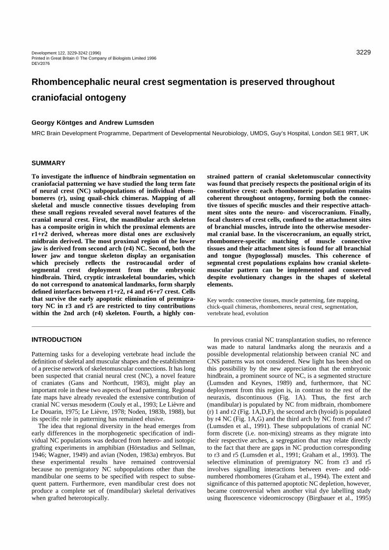
In previous cranial NC transplantation studies, no referencewas made to natural landmarks along the neuraxis and apossible developmental relationship between cranial NC andCNS patterns was not considered. New light has been shed onthis possibility by the new appreciation that the embryonichindbrain, a prominent source of NC, is a segmented structure(Lumsden and Keynes, 1989) and, furthermore, that NCdeployment from this region is, in contrast to the rest of theneuraxis, discontinuous (Fig. 1A). Thus, the first arch(mandibular) is populated by NC from midbrain, rhombomere(r) 1 and r2 (Fig. 1A,D,F), the second arch (hyoid) is populatedby r4 NC (Fig. 1A,G) and the third arch by NC from r6 and r7(Lumsden et al., 1991). These subpopulations of cranial NCform discrete (i.e. non-mixing) streams as they migrate intotheir respective arches, a segregation that may relate directlyto the fact that there are gaps in NC production correspondingto r3 and r5 (Lumsden et al., 1991; Graham et al., 1993). Theselective elimination of premigratory NC from r3 and r5involves signalling interactions between even- and odd-numbered rhombomeres (Graham et al., 1994). The extent andsignificance of this patterned apoptotic NC depletion, however,became controversial when another vital dye labelling studyusing fluorescence videomicroscopy (Birgbauer et al., 1995)
3230 G. Köntges and A. Lumsden
Fig. 1. Embryonic origin and fate of individual cranial neuralcrest subpopulations in the chick embryo at st 14 (A) and E10(B). A is modified after Lumsden et al. (1991). The otic andtongue regions are detailed in Figs 2A, 4. (C-G) Relativerhombomeric origin and localisation of quail neural crest cells isidentical to that reported by Lumsden et al. (1991) for chick NC,using DiI. As in all other photographs quail cells as detected bythe Q¢PN antibody, are HRP-labelled and dark brown.(C) Dorsal view of st 19 chimeric hindbrain, r1 being isotopicallyreplaced by quail r1 at st 8+. Compare with E for accuracy ofr1/r2 grafting at st 8+/9−. (D) Lateral view of same embryoshowing quail NC cells in the trigeminal exit point (asteriskadjacent to r2), the trigeminal nerve, mandibular and maxillaryprocess of branchial arch 1 (b1) and behind the eye (cf. Lumsdenet al., 1991). No NC cells are found in other branchial arches(b2). (E) Dorsal view of st. 15 chimeric hindbrain, r2 wasreplaced by quail r2 at st 9−. (F) Lateral view of same embryoshowing quail r2-NC cells restricted (arrows) to the mandibularprocess of the 1st arch (b1) (cf. Lumsden et al., 1991). No cellsare found in the 2nd branchial arch (b2). (G) Lateral view of a st.15 chimera with r4 being replaced by quail r4 at st. 9. The 2ndbranchial arch (hyoid, b2) is entirely filled by postmigratory quailcrest cells from r4 which do not migrate to any other branchialarch (cf. Lumsden et al., 1991). Some crest cells in the roofplateof the hindbrain migrate posteriorly, but do not enter otherbranchial arches (b1,3,4).
3231Rhombomeric neural crest fate map
revealed a peculiar behavior of the surviving NC cells in r3and r5: the NC progenitor region appears to spread in a ros-trocaudal direction such that cells originating in r3 and r5escape apoptosis and exit the neural tube at the axial levels ofr2, 4 and 6. This observation seemed to counter the notion thatpositional values on the rostrocaudal axis of the hindbrain (andtherefore also within its emanating NC populations) are alreadyfixed at the stage of NC emigration.
We wanted to assess the impact of hindbrain segmentationon craniofacial patterning and, specifically the significance ofr3 and r5 NC cell depletion; for these purposes it was necessaryto fate map each rhombomeric NC subpopulation - using thequail marker (Le Douarin, 1969) to allow long-term detection.Earlier studies on NC have focused almost exclusively on howthe shapes of visceroskeletal elements are defined. The other,equally important facet of head patterning, the establishmentof skeletomuscular connectivity, however, has received littleattention. We have, therefore, scanned chimeric heads for quailcells in all skeletal as well as muscle connective tissue deriv-atives. The resulting rhombomeric NC fate map provides acomplete set of morphological criteria for estimating morpho-genetic specification of each rhombomeric NC subpopulation.Furthermore, it defines the embryological framework for amore refined interpretation of functional studies involving twohomeobox-containing genes, Hoxa-2 and Otx-2. Their nullmutation results in distinct and highly specific craniofacial phe-notypes (Rijli et al., 1993; Gendron-Maguire et al., 1993;Matsuo et al., 1995).
We find that most elements of the visceral skeleton are com-posites of a multi-rhombomeric NC origin. Within thesecompound elements, however, the original neuraxial order ofcrest deployment from the hindbrain is strictly maintained.Moreover, single-rhombomere resolution enabled us toobserve an underlying, highly constrained pattern of skeleto-muscular connectivity, which precisely relates to the rhom-bomeric origin of the individual crest subpopulations: eachremains coherent throughout ontogeny, forming the connectivetissue of individual branchial and tongue muscles as well astheir respective attachment sites on both the NC-derivedvisceral skeleton (viscerocranium) and the mesodermallyderived cranial base (neurocranium). Thus, cranial muscle con-nective tissues derived from a specific rhombomeric level arealways exclusively attached to skeletal regions of the sameorigin. This striking match between skeletal elements and theconnective tissues of their attached muscles addresses a fun-damental question of head patterning - how do cranial musclesbecome specifically localised onto homogeneous lookingskeletal elements?
MATERIALS AND METHODS
Rhombomere transplantationsSingle rhombomeres were grafted isotopically and isochronicallyfrom quail to chick prior to neural crest emigration at HH stages 8+(5somites) to 9+/10− (8-9 somites), depending on the axial level of thegraft. Quail (Coturnix coturnix japonica) and Rhode Island Red hen’seggs from commercial sources were incubated at 37°C in a humidi-fied atmosphere to HH stages 8+ to 9+. Quail donor embryos wereremoved from eggs, pinned out on a sylgard-coated dish in Howard’sRinger. Whole single rhombomeres with very small flaps of adjacentectoderm were dissected out using flame-sharpened tungsten needles
(100 µm wire) and unilaterally marked for later orientation with fineglass tips carrying 1% Nile Blue (Serva) in 2% agarose. For rhom-bomeres 3-7 visible boundary constrictions were used as landmarksfor microsurgery. As crest emigration from r1 and r2 precedes r1/2boundary formation, the presumptive r1/2 territory in st 8+/9−embryos was determined as follows. The future r2/3 boundary wasestimated by counting 5 presumptive rhombomere lengths (90 µmeach) from the level of the first somite (adjacent presumptive r7) tothe front using an ocular micrometer. The neuroepithelial r1+2territory delimited by this presumptive r2/3 boundary and the alreadyvisible mid-/hindbrain boundary was then bisected into pieces ofequal rostrocaudal length (presumptive r1 and r2, respectively). Forthe grafts of midbrain NC at stage 8+ a 150 µm long piece of neu-roepithelium in front of the mid-/hindbrain boundary was excised. Toexclude mesodermal contamination, rhombomeres were carefullycleaned from adherent cells after treatment with dispase I(Boehringer-Mannheim, 1 mg/ml in L-15 medium; Gibco) for 20minutes at room temperature, then washed and transplanted isotopi-cally into exactly stage-matched chick hosts. To prepare hosts, theeggs were windowed and the embryos visualised by a small sub-blas-todermal injection of India ink (Pelican yellow) made with a fine glassmouth pipette. A small hole was made through the vitelline membranelateral to the site of operation and a single host rhombomere corre-sponding to the graft was excised with needles, leaving the notochordand pharyngeal endoderm intact. We used the same procedure andlandmarks to determine rhombomeric positions in the host asdescribed above for the donor. The quail graft was manoeuvred intoplace, with correct rostrocaudal orientation as determined by unilat-eral Nile Blue staining. The accuracy of this grafting procedure is doc-umented in Fig. 1C-G. After moistening, the egg was sealed with elec-trical tape and reincubated for 3 hours. Operated embryos were theninspected and if the graft had not healed in properly, they werediscarded. The chimeras were killed at E10, when the head is fullypatterned (Tonkoff, 1900; McClearn and Noden, 1988), their headsfixed in Serra’s fixative (ethanol 100%:formalin 37%:acetic acid,6:3:1) overnight, dehydrated and embedded in Paraplast.
Assay and analysis of chimeras364 operations were performed, 63 embryos obtained at E10 (viability21%) and 42 chimeras at HH stages 13-19 (viability 70%). Eachchimeric E10 head was paraffin wax embedded and cut into 10 µmhorizontal sections. To detect quail nuclei, the majority of sectionswere processed with the quail-specific Q¢PN antibody (Develop-mental Studies Hybridoma Bank, University of Iowa), diluted 1:50,overnight. A peroxidase-conjugated goat anti-mouse IgG secondaryantibody (Jackson) was used at 1:250 dilution for 1 hour, DABstaining performed for 20 minutes, endogenous peroxidase activitybeing blocked with 0.1% H2O2 in PBS and non-specific bindingprevented with 5% FCS. To investigate early phases of crest migrationand to test whether the chick-quail chimera technique yields the sameresults as previously obtained by focal DiI labelling of NC (Lumsdenet al., 1991), 42 chimeras were harvested at HH st 13-19, fixedovernight in 3.5% paraformaldehyde and processed as wholemountswith the Q¢PN antibody (for 5 days at 4°C), all solutions containing1% Triton X-100 (Sigma) to make tissues more permeable. Fig. 1C-G show that the NC migration pathways and locations in st 14-19embryos are identical to those reported by Lumsden et al. (1991; cf.their Fig. 1). These controls gave us reason to believe that the chimeratechnique is also valid at later stages of development. Any possibledelays due to embryonic healing thus appear negligible and do notsystematically affect NC migratory pathways. Whole-mounts andsections were viewed on a Zeiss Axiophot with Nomarski opticsallowing unequivocal identification of brown quail cells, cartilage,bone, muscle and connective tissues without couterstaining. Somesections were processed with standard Lison’s hematoxylin nuclearstain (Lison, 1954; Couly et al., 1993), which shows quail nucleoli inblack, cartilage in blue and bone in red. All chimeras were then
3232 G. Köntges and A. Lumsden
Table 1. Number of chimeras analysedNumber of
Type of HH stage Beginning of chimeras analysed at
isotopic graft of operation crest emigration* E10 st 13-19
caudal 150 µm 8+ 9 3 5of midbrain r1 8+ 9+/10 5 7r2 9− 9+/10 5 7r3 9(+) 10+ 5 6r4 9(+) 10+ 5 6r5 9+ 10+ 4 6r6 9+/10− 10+ 5 5r7 10− 10+ 6
*Data from Tosney (1982) as judged by timing of basal laminafragmentation and changed ECM properties above emerging CNCpopulations.
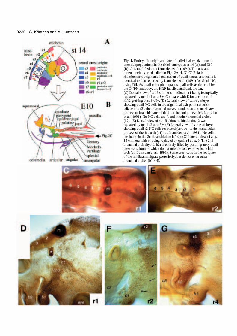
ateral aspect of the otic region at E10. Colour coding as in Fig. 1A. Redrcates the invisible boundary between 1st and 2nd arch neural crestrticular region of the lower jaw. Note the branchial arch specificatching of muscle connective tissues and their respective skeletal jawsites. (B) Horizontal section through the quadratoarticular joint of an chimera (grafted at st 8+). In the mandibular arch skeletonic crest (from r1+2) is restricted to proximal parts such as articular androunding the jaw joints. (C) Horizontal section through distal jawhimera in which the caudal 150 µm of midbrain/crest were isotopically
st 8+. Only midbrain crest forms the distal part of the jaw, ie. Meckel’sd dentary. In C cartilage is blue, bone is red and quail nuclei are black.
subjected to a rigorous screen for morphological abnormalities. Thosewhose skeletal or muscular elements showed any deformations/dis-placements or whose peripheral cranial nerve courses were aberrantwere excluded from further analysis. After this preselection, 46embryos were left, 38 of which were single rhombomere grafts (Table1) and 8 were rhombomere-pair grafts (posterior midbrain+r1, n=1;r1+2, n=1; r2+3, n=3; r4+5, n=3). The latter grafts were made as addi-tional controls in order to assess how the surgical procedure in singlerhombomere grafts might disturb the rhombomeric interactionsresponsible for NC apoptosis over r3 and r5 (Graham et al., 1993),thus creating excess crest deployment from odd-numbered rhom-bomeres. If this had been the case, we would have found the addedskeletal territories occupied by crest from single r2- and r3 graftsbeing more widespread than that occupied by crest from r2+3 inrhombomere-pair grafts. This was not observed (data not shown) andconvinced us that additional crest production from r3 and r5 due tosurgical artifacts was negligible.
As the grafts were bilateral, we assayed all 46 headson both body sides for the presence of quail NC cellsin 65 locations of the skeleton (braincase, visceralskeleton) and of all branchial and hypoglossal muscleconnective tissues. When crest cells were present in aspecific location in 80% of all embryos examined, wecharted this location as being crest-derived from thespecific origin. Its extent was superimposed in colour-coding onto camera lucida drawings of chick headsusing Adobe Photoshop, as shown below. The cameralucida template drawings of cleared E10 chick headsstained for cartilage/bone with Alcian Blue/AlizarinRed were performed with ink on tracing paper andthen scanned in. The skeletal nomenclature wasadopted from Jollie (1957) and Baumel (1979) andthat of the musculature from Barnikol (1952), McClearn and Noden (1988), Kallius (1905) and Zweers(1974).
RESULTS
Rhombomeric organisation of the avianjaw apparatusThe aim of this study was to determine the con-tribution made by the neural crest of eachrhombomere to the avian jaw and tongueapparatus.
The avian jaw comprises a complex pattern ofupper and lower elements, which are linkedtogether and to the brain capsule (neurocranium)by joints and branchial muscles. The main upperjaw element is the pterygoquadrate, which hasfour articulations with surrounding elements: tothe palatine bone (the roof of the palate) anteri-orly, to the otic region (squamosal) posteriorly,to the quadratojugal/jugal bone laterally and tothe lower jaw ventrally (Figs 1B, 2). The lowerjaw skeleton in the embryo is composed ofMeckel’s cartilage, covered by several dermalbones (dentary, splenial, surangular, angular,gonial); the proximal end of Meckel’s cartilage,forming the jaw joint, later ossifies endochon-drally as the articular. The articular bone has twoprocesses which serve as muscle attachmentsites: the M.pterygoideus (N.V.) inserts onto themedially located internal process and the
Fig. 2. (A) Larrow demawithin the a(=colour) mattachment E10 r1 crestrhombomerquadrate surregion of a creplaced at cartilage an
3233Rhombomeric neural crest fate map
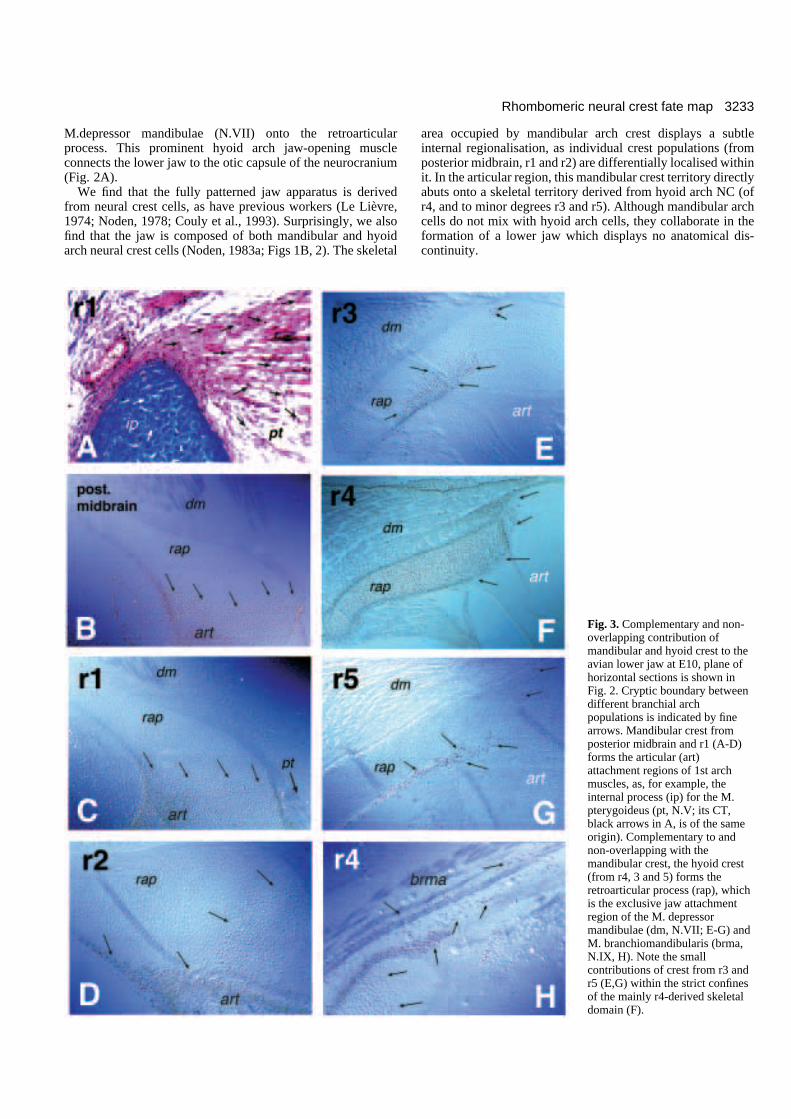
M.depressor mandibulae (N.VII) onto the retroarticularprocess. This prominent hyoid arch jaw-opening muscleconnects the lower jaw to the otic capsule of the neurocranium(Fig. 2A).
We find that the fully patterned jaw apparatus is derivedfrom neural crest cells, as have previous workers (Le Lièvre,1974; Noden, 1978; Couly et al., 1993). Surprisingly, we alsofind that the jaw is composed of both mandibular and hyoidarch neural crest cells (Noden, 1983a; Figs 1B, 2). The skeletal
area occupied by mandibular arch crest displays a subtleinternal regionalisation, as individual crest populations (fromposterior midbrain, r1 and r2) are differentially localised withinit. In the articular region, this mandibular crest territory directlyabuts onto a skeletal territory derived from hyoid arch NC (ofr4, and to minor degrees r3 and r5). Although mandibular archcells do not mix with hyoid arch cells, they collaborate in theformation of a lower jaw which displays no anatomical dis-continuity.
Fig. 3. Complementary and non-overlapping contribution ofmandibular and hyoid crest to theavian lower jaw at E10, plane ofhorizontal sections is shown inFig. 2. Cryptic boundary betweendifferent branchial archpopulations is indicated by finearrows. Mandibular crest fromposterior midbrain and r1 (A-D)forms the articular (art)attachment regions of 1st archmuscles, as, for example, theinternal process (ip) for the M.pterygoideus (pt, N.V; its CT,black arrows in A, is of the sameorigin). Complementary to andnon-overlapping with themandibular crest, the hyoid crest(from r4, 3 and 5) forms theretroarticular process (rap), whichis the exclusive jaw attachmentregion of the M. depressormandibulae (dm, N.VII; E-G) andM. branchiomandibularis (brma,N.IX, H). Note the smallcontributions of crest from r3 andr5 (E,G) within the strict confinesof the mainly r4-derived skeletaldomain (F).
3234 G. Köntges and A. Lumsden
e avian tongue skeleton and its associated branchial and tongueote the internal zonation of the basihyoid and the branchial arch
ing of muscle connective tissues and their respective skeletal attachment
Underlying combinatorial composition of themandibular arch skeletonThe crest populations emanating from posterior midbrain, r1and r2 occupy the adult mandibular arch skeletal territory inan overlapping, combinatorial manner. Distal elements, suchas the upper jaw bones (maxilla, palatine, jugal), the major partof Meckel’s cartilage and its overlying dermal bones (dentary,splenial), are derived solely from midbrain crest (Fig. 1D, pinkin Fig. 1B), whereas the more proximal elements, centeredaround the jaw joint and close to the middle ear (articular,angular, surangular, quadrate, quadratojugal), are formedmainly by crest from r1 and 2 (Fig. 2B; yellow, green in Fig.2A; cf. Noden, 1978). In the upper jaw, r1-derived NC is foundthroughout the pterygoquadrate, whereas midbrain crest isfound only at its dorsal margin and articulations (orange in Fig.2A) and r2 crest (green) where it preferentially surrounds theventral articulations with lower jaw and quadratojugal.Although the boundaries between these individual crest popu-lations are not absolutely sharp throughout the mandibular archcrest territory, cells from r1 and r2 were never found rostral tothe rear quarter of the lower jaw.
Mandibular and hyoid arch crest cooperate in theformation of the jaw skeletonWhile the crest from the midbrain and r1+ r2 merge into eachother we find no mixing between these mandibular populationsand those from the crest derived from r4 (+r3,r5). The quailmarker reveals a distinct border between the mandibular archcrest populations (Fig. 3A-D) and the hyoid arch r4 (+r3, r5)crest population (Fig. 3E-H). This sharp demarcation betweenmandibular and hyoid crest populations (red arrow in Fig. 2A)does not coincide with any anatomical separation betweenskeletal elements, but traverses the articular region internally.Furthermore, this cryptic border separates the mandibularcrest-derived insertion regions of all trigeminal (N.V) musclesfrom the hyoid crest-derived insertion region of facial (N.VII)muscles. The latter attach exclusively to the retroarticularprocess which is formed solely by hyoid crest from r4 (and toa smaller degree r3, r5) in all chimeras examined (Fig. 3E-G).The spatial extent of hyoid crest in the retroarticular process isexactly complementary to and non-overlapping with that of mandibularcrest (compare arrows in Fig. 3A-Dwith those in 3E-H).
Crest from r3 was confined to asmall contribution to the V2/3 andVII ganglia, the Schwann cell sheathsof the peripheral trigeminal (V2/3)and facial (VII) nerve branches andthe hyoid skeletal elements (retroar-ticular process and columella, Figs3E, 7E,F). In the latter case, r3 crestis always localised in small scatteredareas within the precisely demarcatedconfines of r4 crest (Fig. 3E) and isalways excluded from the mandibu-lar crest territory. Similarly, the evensmaller r5 crest population is alsorestricted to the r4 territory (Fig. 3G)and peripheral nerve sheaths ofNn.VI, VII and IX. We could not
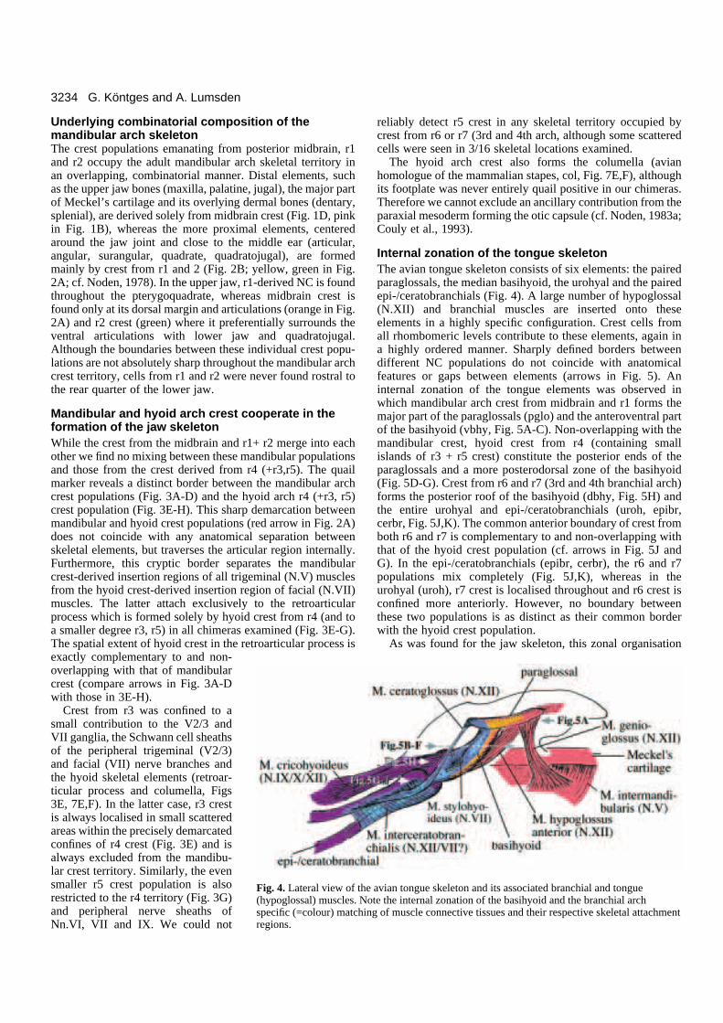
Fig. 4. Lateral view of th(hypoglossal) muscles. Nspecific (=colour) matchregions.
reliably detect r5 crest in any skeletal territory occupied bycrest from r6 or r7 (3rd and 4th arch, although some scatteredcells were seen in 3/16 skeletal locations examined.
The hyoid arch crest also forms the columella (avianhomologue of the mammalian stapes, col, Fig. 7E,F), althoughits footplate was never entirely quail positive in our chimeras.Therefore we cannot exclude an ancillary contribution from theparaxial mesoderm forming the otic capsule (cf. Noden, 1983a;Couly et al., 1993).
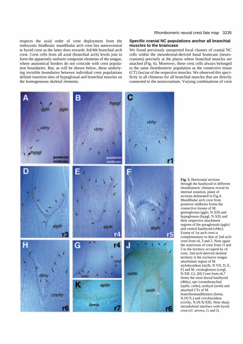
Internal zonation of the tongue skeletonThe avian tongue skeleton consists of six elements: the pairedparaglossals, the median basihyoid, the urohyal and the pairedepi-/ceratobranchials (Fig. 4). A large number of hypoglossal(N.XII) and branchial muscles are inserted onto theseelements in a highly specific configuration. Crest cells fromall rhombomeric levels contribute to these elements, again ina highly ordered manner. Sharply defined borders betweendifferent NC populations do not coincide with anatomicalfeatures or gaps between elements (arrows in Fig. 5). Aninternal zonation of the tongue elements was observed inwhich mandibular arch crest from midbrain and r1 forms themajor part of the paraglossals (pglo) and the anteroventral partof the basihyoid (vbhy, Fig. 5A-C). Non-overlapping with themandibular crest, hyoid crest from r4 (containing smallislands of r3 + r5 crest) constitute the posterior ends of theparaglossals and a more posterodorsal zone of the basihyoid(Fig. 5D-G). Crest from r6 and r7 (3rd and 4th branchial arch)forms the posterior roof of the basihyoid (dbhy, Fig. 5H) andthe entire urohyal and epi-/ceratobranchials (uroh, epibr,cerbr, Fig. 5J,K). The common anterior boundary of crest fromboth r6 and r7 is complementary to and non-overlapping withthat of the hyoid crest population (cf. arrows in Fig. 5J andG). In the epi-/ceratobranchials (epibr, cerbr), the r6 and r7populations mix completely (Fig. 5J,K), whereas in theurohyal (uroh), r7 crest is localised throughout and r6 crest isconfined more anteriorly. However, no boundary betweenthese two populations is as distinct as their common borderwith the hyoid crest population.
As was found for the jaw skeleton, this zonal organisation
3235Rhombomeric neural crest fate map
respects the axial order of crest deployment from theembryonic hindbrain: mandibular arch crest lies anteroventralto hyoid crest as the latter does towards 3rd/4th branchial archcrest. Crest cells from all axial (branchial arch) levels join toform the apparently uniform composite elements of the tongue,where anatomical borders do not coincide with crest popula-tion boundaries. But, as will be shown below, these underly-ing invisible boundaries between individual crest populationsdelimit insertion sites of hypoglossal and branchial muscles onthe homogeneous skeletal elements.
vbhy vbhy
dbhy
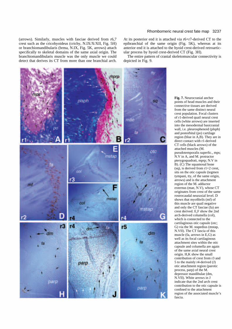
Specific cranial NC populations anchor all branchialmuscles to the braincaseWe found previously unreported focal clusters of cranial NCcells within the mesodermal-derived basal braincase (neuro-cranium) precisely at the places where branchial muscles areattached (Fig. 6). Moreover, these crest cells always belongedto the same rhombomeric population as the connective tissue(CT) fasciae of the respective muscles. We observed this speci-ficity in all chimeras for all branchial muscles that are directlyconnected to the neurocranium. Varying combinations of crest
Fig. 5. Horizontal sectionsthrough the basihyoid in differentrhombomeric chimeras reveal itsinternal zonation, plane ofsections delineated in Fig.4.Mandibular arch crest fromposterior midbrain forms theconnective tissues of M.genioglossus (gglo, N.XII) andhypoglossus (hypgl, N.XII) andtheir respective attachmentregions of the paraglossals (pglo)and ventral basihyoid (vbhy).Extent of 1st arch crest iscomplementary to that of 2nd archcrest from r4, 3 and 5. Note againthe restriction of crest from r3 and5 to the territory occupied by r4crest. 2nd arch-derived skeletalterritory is the exclusive tongueattachment region of M.stylohyoideus (stylh, N.VII, D, E,F) and M. ceratoglossus (cergl,N.XII, G). (H) Crest from r6,7forms the most dorsal basihyoid(dbhy), epi-/ceratobranchial(epibr, cerbr), urohyal (uroh) andattached CTs of M.branchiomandibularis (brma,N.IX/X.) and cricohyoideus(crichy, N.IX/X/XII). Note sharpintraskeletal interface with hyoidcrest (cf. arrows, G and J).
vbhy
3236 G. Köntges and A. Lumsden
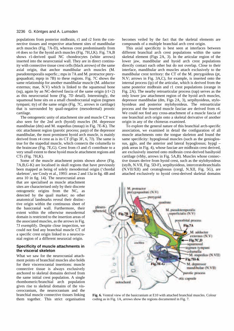
ew of the basicranium at E10 with attached branchial muscles. Colour1A, arrows show the regions documented in Fig. 7.
populations from posterior midbrain, r1 and r2 form the con-nective tissues and respective attachment sites of mandibulararch muscles (Fig. 7A-D), whereas crest predominantly fromr4 does so for the hyoid arch muscles (Fig. 7H,J,K). Fig. 7A,Bshows r1-derived quail NC chondrocytes (white arrows)inserted into the neurocranial wall. They are in direct continu-ity with connective tissue crest cells (black arrows) of the sameaxial origin, that anchor mandibular arch muscles (M.pseudotemporalis superfic.; mps in 7A and M. protractor ptery-goquadrati; mprp in 7B) to these regions. Fig. 7C shows thesame relationship for another mandibular muscle (M. adductorexternus; mae, N.V) which is linked to the squamosal bone(sq), again by an NC-derived fascia of the same origin (r1+2)as this neurocranial bone (Fig. 7D detail). Interestingly, thesquamosal bone sits on a small chondrocranial region (tegmentympani; tty) of the same origin (Fig. 7C, arrows in cartilage)that is surrounded by quail-negative (mesodermal) prooticcartilage.
The ontogenetic unity of attachment site and muscle CT wasalso seen for the 2nd arch (hyoid) muscles (M. depressormandibulae (dm) and M. stapedius (mstap) in Fig. 7E-K). Theotic attachment region (parotic process; parp) of the depressormandibulae, the most prominent hyoid arch muscle, is mainlyderived from r4 crest as is its CT (Figs 3F, 6, 7J). The same istrue for the stapedial muscle, which connects the columella tothe braincase (Fig. 7E,G). Crest from r3 and r5 contribute to avery small extent to these hyoid muscle attachment regions andCTs (Fig. 7H,K).
Some of the muscle attachment points shown above (Fig.7A,B,G-K) are localised in skull regions that have previouslybeen mapped as being of solely mesodermal origin (‘chordalskeleton’, see Couly et al., 1993: areas 2 and 13a in fig. 4B andarea 10 in fig. 14). The neurocranial areasthat are specialised as muscle attachmentsites are characterised only by their discreteontogenetic origins from the NC, asdetected by the quail marker; no otheranatomical landmarks reveal their distinc-tive origin within the continuous sheet ofthe basicranial wall. Furthermore, theirextent within the otherwise mesodermaldomain is restricted to the insertion areas ofthe associated muscles, as the arrows in Fig.7J exemplify. Despite close inspection, wecould not find any branchial muscle CT ofa specific crest origin linked to a neurocra-nial region of a different neuraxial origin.
Specificity of muscle attachments inthe visceral skeleton What we saw for the neurocranial attach-ment points of branchial muscles also holdsfor their viscerocranial insertions: muscleconnective tissue is always exclusivelyanchored to skeletal domains derived fromthe same initial crest population. A singlerhombomeric/branchial arch populationgives rise to skeletal domains of the vis-cerocranium, the neurocranium and thebranchial muscle connective tissues linkingthem together. This strict organisation
Fig. 6. Ventral vicoding as in Fig.
becomes veiled by the fact that the skeletal elements arecompounds of a multiple branchial arch crest origin.
This axial specificity is best seen at interfaces betweendifferent branchial arch crest populations within the sameskeletal element (Figs 2A, 3). In the articular region of thelower jaw, mandibular and hyoid arch crest populationsdirectly contact each other but do not overlap. Close to theirinterface, mandibular arch muscles attach exclusively to themandibular crest territory: the CT of the M. pterygoideus (pt,N.V; arrows in Fig. 3A,C), for example, is inserted onto theinternal process (ip) of the articular, which is derived from thesame posterior midbrain and r1 crest populations (orange inFig. 2A). The nearby retroarticular process (rap) serves as theonly lower jaw attachment region of the hyoid arch muscles,depressor mandibulae (dm, Figs 2A, 3), serpihyoideus, stylo-hyoideus and posterior mylohyoideus. The retroarticularprocess and the inserted muscle fasciae are derived from r4.We could not find any cross-attachment of a muscle fascia ofone branchial arch origin onto a skeletal derivative of anotherorigin in any of the chimeras examined.
To explore the general nature of this branchial arch-specificassociation, we examined in detail the configuration of allmuscle attachments onto the tongue skeleton and found thesame specificity: hypoglossal muscles (such as the genioglos-sus, gglo, and the anterior and lateral hypoglossus; hypgl –pink areas in Fig. 4), whose fasciae are midbrain crest derived,are exclusively inserted onto midbrain crest-derived basihyoidcartilage (vbhy, arrows in Fig. 5A,B). Muscles whose connec-tive tissues derive from hyoid crest, such as the stylohyoideus(stylh, N.VII, Fig. 5D-F), serpihyoideus, interceratobranchialis(N.VII/XII) and ceratoglossus (cergl, N.XII, Fig. 5G), areattached exclusively to hyoid crest-derived skeletal domains
3237Rhombomeric neural crest fate map
(arrows). Similarly, muscles with fasciae derived from r6,7crest such as the cricohyoideus (crichy, N.IX/X/XII, Fig. 5H)or branchiomandibularis (brma, N.IX, Fig. 5K, arrows) attachspecifically to skeletal domains of the same axial origin. Thebranchiomandibularis muscle was the only muscle we coulddetect that derives its CT from more than one branchial arch.
At its posterior end it is attached via r6+r7-derived CT to theepibranchial of the same origin (Fig. 5K), whereas at itsanterior end it is attached to the hyoid crest-derived retroartic-ular process by hyoid crest-derived CT (Fig. 3H).
The entire pattern of cranial skeletomuscular connectivity isdepicted in Fig. 9.
Fig. 7. Neurocranial anchorpoints of head muscles and theirconnective tissues are derivedfrom the same distinct neuralcrest population. Focal clustersof r1-derived quail neural crestcells (white arrows) are insertedinto the mesodermal basicranialwall, i.e. pleurosphenoid (plsph)and postorbital (po) cartilageregion (blue in A,B). They are indirect contact with r1-derivedCT cells (black arrows) of theattached muscles (M.pseudotemporalis superfic., mps;N.V in A, and M. protractorpterygoquadrati, mprp; N.V inB). (C) The squamosal bone(sq), is derived from r1+2 crest,sits on the otic capsule (tegmentympani, tty, of the same origin,arrows) and is the attachmentregion of the M. adductorexternus (mae, N.V), whose CToriginates from crest of the samerostrocaudal neuraxial level. Dshows that myofibrils (mf) ofthis muscle are quail negativeand only the CT fasciae (fa) arecrest derived. E,F show the 2ndarch-derived columella (col),which is connected to thecartilaginous otic capsule (otc;G) via the M. stapedius (mstap,N.VII). The CT fascia of thismuscle (fa, arrows in E,G) aswell as its focal cartilaginousattachment sites within the oticcapsule and columella are againof the same axial neural crestorigin. H,K show the smallcontribution of crest from r3 and5 to the mainly r4-derived (J)otic attachment region (paroticprocess, parp) of the M.depressor mandibulae (dm,N.VII). White arrows in Jindicate that the 2nd arch crestcontribution to the otic capsule isconfined to the attachmentregion of the associated muscle’sfascia.
3238 G. Köntges and A. Lumsden
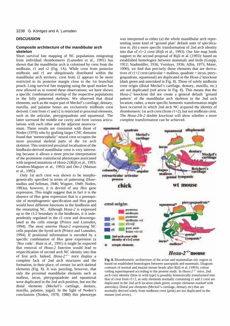
hombomeric architecture of the avian and mammalian otic region as established homologies between sauropsids and mammals. Diagram of normal and mutant mouse heads after Rijli et al. (1993), colouruperimposed according to the present study. In Hoxa-2−/− mice, 2ndst identity (blue in wild type) is possibly homeotically transformed intorest from r1+2, as only elements normally containing r1 and 2 crest areed in the 2nd arch location (dark green, ectopic elements marked with). Distal jaw elements (Meckel’s cartilage, dentary etc) that are
y derived solely from midbrain crest (pink) are not duplicated in thered arrow).
DISCUSSION
Composite architecture of the mandibular archskeleton Short survival fate mapping of NC populations emigratingfrom individual rhombomeres (Lumsden et al., 1991) hasshown that the mandibular arch is colonised by crest from themidbrain, r1 and r2 (Fig. 1A). While crest from posteriormidbrain and r1 are ubiquitously distributed within themandibular arch territory, crest from r2 appears to be morerestricted to its posterior margin close to the 1st branchialpouch. Long survival fate mapping using the quail marker hasnow allowed us to extend these observations; we have showna specific combinatorial overlap of the respective populationsin the fully patterned skeleton. We observed that distalelements, such as the major part of Meckel’s cartilage, dentary,maxilla, and palatine bones are exclusively midbrain crestderived. Crest from r1 and r2 is restricted to proximal elements,such as the articular, pterygoquadrate and squamosal. Thelatter surround the middle ear cavity and form various articu-lations with each other and the adjacent neurocra-nium. These results are consistent with those ofNoden (1978) who by grafting larger CNC domainsfound that ‘metencephalic’ neural crest occupies themore proximal skeletal parts of the 1st archskeleton. This restricted proximal localisation of thehindbrain-derived mandibular crest is very interest-ing because it allows a more precise interpretationof the prominent craniofacial phenotypes associatedwith targeted mutations of Hoxa-2 (Rijli et al., 1993;Gendron-Maguire et al., 1993) and Otx-2 (Matsuoet al., 1995).
Only 1st arch crest was shown to be morpho-genetically specified in terms of patterning (Hoer-stadius and Sellman, 1946; Wagner, 1949; Noden,1983a), however, it is devoid of any Hox geneexpression. This might suggest that in fact it is theabsence of Hox gene expression that is a prerequi-site of morphogenetic specification and Hox geneswould have different functions in the hindbrain andthe emanating NC. Although Hoxa-2 is expressedup to the r1/2 boundary in the hindbrain, it is inde-pendently regulated in the r2 crest and downregu-lated as the cells emerge (Prince and Lumsden,1994). The most anterior Hoxa-2 expressing NCcells populate the hyoid arch (Prince and Lumsden,1994). If positional information is encoded by aspecific combination of Hox gene expression (a‘Hox code’, Hunt et al., 1991) it might be expectedthat removal of Hoxa-2 function would lead torespecification of second arch NC identity into thatof first arch. Indeed, Hoxa-2−/− mice display acomplete lack of 2nd arch structures and theformation, in their place, of certain 1st arch skeletalelements (Fig. 8). It was puzzling, however, thatonly the proximal mandibular elements such asmalleus, incus, pterygoquadrate and squamosalwere duplicated in the 2nd arch position, but not thedistal elements (Meckel’s cartilage, dentary,maxilla, palatine, jugal). In the light of Noden’sconclusions (Noden, 1978, 1988) this phenotype
Fig. 8. Rbased oncontourscoding sarch crethat of cduplicatasterisksnormallmutant (
was interpreted as either (a) the whole mandibular arch repre-senting some kind of ‘ground plan’ default state of specifica-tion or, (b) a more specific transformation of 2nd arch identityinto that of r1+2 crest (Rijli et al., 1993). Our fate map lendssupport to the second proposal of Rijli et al. (1993): based onestablished homologies between mammals and birds (Gaupp,1912; Stadtmüller, 1936; Versluys, 1936; Allin, 1975; Maier,1990), we find that precisely those elements that are deriva-tives of r1+2 crest (articular = malleus, quadrate = incus, ptery-goquadrate, squamosal) are duplicated in the Hoxa-2 knockout(dark green and asterisked in Fig. 8). Those of solely midbraincrest origin (distal Meckel’s cartilage, dentary, maxilla, etc.)are not duplicated (red arrow in Fig. 8). This means that theHoxa-2 knockout did not create a general default ‘groundpattern’ of the mandibular arch skeleton in the 2nd archlocation; rather, a more specific homeotic transformation mighthave occurred in which 2nd arch NC acquired the identity ofrhombomeric 1st arch crest (from r1+2), but not midbrain crest.The Hoxa-2/b-2 double knockout will show whether a morecomplete transformation can be achieved.
3239Rhombomeric neural crest fate map
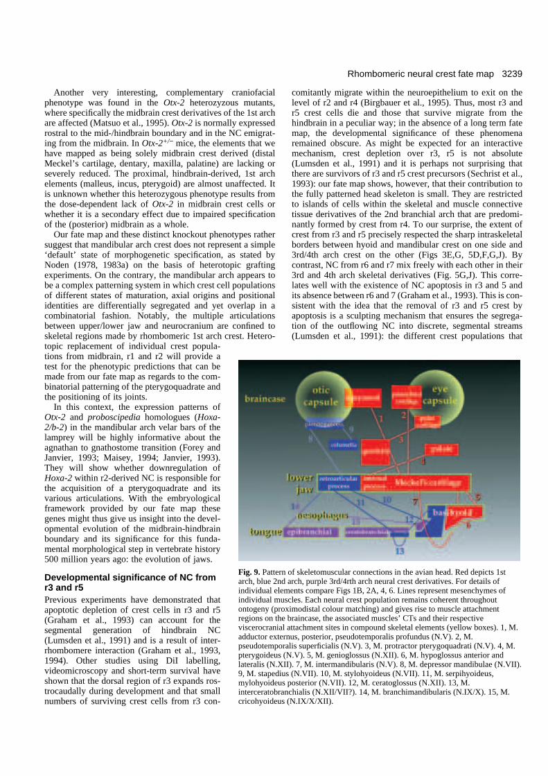
n of skeletomuscular connections in the avian head. Red depicts 1std arch, purple 3rd/4rth arch neural crest derivatives. For details ofements compare Figs 1B, 2A, 4, 6. Lines represent mesenchymes ofuscles. Each neural crest population remains coherent throughoutoximodistal colour matching) and gives rise to muscle attachmente braincase, the associated muscles’ CTs and their respectivel attachment sites in compound skeletal elements (yellow boxes). 1, M.
ernus, posterior, pseudotemporalis profundus (N.V). 2, M.ralis superficialis (N.V). 3, M. protractor pterygoquadrati (N.V). 4, M. (N.V). 5, M. genioglossus (N.XII). 6, M. hypoglossus anterior andII). 7, M. intermandibularis (N.V). 8, M. depressor mandibulae (N.VII).
ius (N.VII). 10, M. stylohyoideus (N.VII). 11, M. serpihyoideus,s posterior (N.VII). 12, M. ceratoglossus (N.XII). 13, M.anchialis (N.XII/VII?). 14, M. branchimandibularis (N.IX/X). 15, M.s (N.IX/X/XII).
Another very interesting, complementary craniofacialphenotype was found in the Otx-2 heterozyzous mutants,where specifically the midbrain crest derivatives of the 1st archare affected (Matsuo et al., 1995). Otx-2 is normally expressedrostral to the mid-/hindbrain boundary and in the NC emigrat-ing from the midbrain. In Otx-2+/− mice, the elements that wehave mapped as being solely midbrain crest derived (distalMeckel’s cartilage, dentary, maxilla, palatine) are lacking orseverely reduced. The proximal, hindbrain-derived, 1st archelements (malleus, incus, pterygoid) are almost unaffected. Itis unknown whether this heterozygous phenotype results fromthe dose-dependent lack of Otx-2 in midbrain crest cells orwhether it is a secondary effect due to impaired specificationof the (posterior) midbrain as a whole.
Our fate map and these distinct knockout phenotypes rathersuggest that mandibular arch crest does not represent a simple‘default’ state of morphogenetic specification, as stated byNoden (1978, 1983a) on the basis of heterotopic graftingexperiments. On the contrary, the mandibular arch appears tobe a complex patterning system in which crest cell populationsof different states of maturation, axial origins and positionalidentities are differentially segregated and yet overlap in acombinatorial fashion. Notably, the multiple articulationsbetween upper/lower jaw and neurocranium are confined toskeletal regions made by rhombomeric 1st arch crest. Hetero-topic replacement of individual crest popula-tions from midbrain, r1 and r2 will provide atest for the phenotypic predictions that can bemade from our fate map as regards to the com-binatorial patterning of the pterygoquadrate andthe positioning of its joints.
In this context, the expression patterns ofOtx-2 and proboscipedia homologues (Hoxa-2/b-2) in the mandibular arch velar bars of thelamprey will be highly informative about theagnathan to gnathostome transition (Forey andJanvier, 1993; Maisey, 1994; Janvier, 1993).They will show whether downregulation ofHoxa-2 within r2-derived NC is responsible forthe acquisition of a pterygoquadrate and itsvarious articulations. With the embryologicalframework provided by our fate map thesegenes might thus give us insight into the devel-opmental evolution of the midbrain-hindbrainboundary and its significance for this funda-mental morphological step in vertebrate history500 million years ago: the evolution of jaws.
Developmental significance of NC fromr3 and r5Previous experiments have demonstrated thatapoptotic depletion of crest cells in r3 and r5(Graham et al., 1993) can account for thesegmental generation of hindbrain NC(Lumsden et al., 1991) and is a result of inter-rhombomere interaction (Graham et al., 1993,1994). Other studies using DiI labelling,videomicroscopy and short-term survival haveshown that the dorsal region of r3 expands ros-trocaudally during development and that smallnumbers of surviving crest cells from r3 con-
Fig. 9. Patterarch, blue 2nindividual elindividual montogeny (prregions on thviscerocraniaadductor extpseudotempopterygoideuslateralis (N.X9, M. stapedmylohyoideuinterceratobrcricohyoideu
comitantly migrate within the neuroepithelium to exit on thelevel of r2 and r4 (Birgbauer et al., 1995). Thus, most r3 andr5 crest cells die and those that survive migrate from thehindbrain in a peculiar way; in the absence of a long term fatemap, the developmental significance of these phenomenaremained obscure. As might be expected for an interactivemechanism, crest depletion over r3, r5 is not absolute(Lumsden et al., 1991) and it is perhaps not surprising thatthere are survivors of r3 and r5 crest precursors (Sechrist et al.,1993): our fate map shows, however, that their contribution tothe fully patterned head skeleton is small. They are restrictedto islands of cells within the skeletal and muscle connectivetissue derivatives of the 2nd branchial arch that are predomi-nantly formed by crest from r4. To our surprise, the extent ofcrest from r3 and r5 precisely respected the sharp intraskeletalborders between hyoid and mandibular crest on one side and3rd/4th arch crest on the other (Figs 3E,G, 5D,F,G,J). Bycontrast, NC from r6 and r7 mix freely with each other in their3rd and 4th arch skeletal derivatives (Fig. 5G,J). This corre-lates well with the existence of NC apoptosis in r3 and 5 andits absence between r6 and 7 (Graham et al., 1993). This is con-sistent with the idea that the removal of r3 and r5 crest byapoptosis is a sculpting mechanism that ensures the segrega-tion of the outflowing NC into discrete, segmental streams(Lumsden et al., 1991): the different crest populations that
3240 G. Köntges and A. Lumsden
emigrate from even-numbered rhombomeres and colonizedifferent branchial arches are kept apart during migration bycrest-free spacing regions.
Compound nature of the visceral skeleton and itscryptic axial organisationIndividual elements of the visceral skeleton are compositestructures derived from crest cells of multiple branchial archorigin. The boundaries between adjacent crest populations donot coincide with the borders of anatomical structures; rather,they span them as invisible interfaces. Noden (1983a) notedthat the retroarticular process and the basihyoid are derivedfrom ‘myelencephalic’ crest – although he claimed in an earlierpaper (Noden, 1978) that the tongue skeleton is mandibulararch crest. These and other studies (Le Lièvre, 1974, 1978;LeLièvre and LeDouarin, 1975; Noden, 1983b, 1988) precededthe appreciation that segmentation is a fundamental feature ofearly hindbrain development (Lumsden and Keynes, 1989)which has consequences for the deployment of individual crestpopulations (Lumsden et al., 1991). Without reference torhombomere boundaries as natural landmarks for transplanta-tion, these previous studies neither required nor attempted thehigh spatial resolution provided by the present study. This cannow clarify the contradictory issues raised by Noden’s work(1978, 1983a,b).
We find that all of the branchial arches contribute to thetongue skeleton in a veiled, albeit highly organised manner:individual mandibular crest populations mix with each otherin varying degrees but do not mix with crest colonising thehyoid arch, although both contribute to the lower jaw. Thisis not an artefact of selective, species-specific adhesive prop-erties of quail versus chick cells: if it were, then we wouldhave found clear segregation also between (e.g.) r1 and r2populations.
Despite their heterogeneous axial origins, however, jaw andtongue are homogeneous in shape. They display a cryptic mul-tilayered organisation which precisely maintains the initialaxial order of crest deployment from the embryonic hindbrain:mandibular crest comes to lie rostral to hyoid crest and hyoidcrest rostral to 3rd/4th branchial arch crest throughoutontogeny.
The compound nature of the visceral skeleton raises sub-stantial objections to the idea that the whole pattern ofbranchial elements is encoded in individual crest populationsprior to their emigration (Noden 1983a). The concept of pre-migratory crest specification was developed by Hörstadius andSellman (1946), Wagner (1949) and Noden (1983a) on thebasis of grafting experiments of mandibular arch only but notmore posterior crest populations. The latter, however, aresupposed to be the developmentally specified ones becausethey express varying combinations of Hox genes, whereasmandibular arch crest is devoid of any Hox gene expression.The anatomical homogeneity of branchial componentssuggests that other patterning mechanisms, spanning the wholerostrocaudal axis, act on different crest populations and recruitthem into composite elements. By defining precisely the axiallevels of the grafts, our fate map enables us to make predic-tions about the effect of whole rhombomere excisions and het-erotopic crest replacements on these compound elements.These will conclusively answer the debated question of pre-migratory NC specification: whether it implies information on
skeletal shapes or only on the rhombomere-specific pattern ofskeletomuscular connectivity.
The compound nature of visceroskeletal elements hasimportant functional consequences: it provides the mechanicalbasis for an integration of different branchial arch muscleactivities. These are controlled by different rhombomere pairs(Lumsden and Keynes, 1989; Fortin et al., 1995) and ofteneven work antagonistically on elements of a common insertion(Bock, 1964; Zweers, 1974). For example, the internal processand other mandibular crest-derived regions of the lower jawserve as attachment sites for the jaw-closing adductor muscles,which are innervated by the trigeminal neurons of r2 and r3.Similarly, the hyoid crest-derived retroarticular process is theinsertion site of the 2nd arch depressor mandibulae, whichopens the jaw and which is innervated by facial motor neuronsin r4 and r5. Even the branchiomandibularis, the 3rd archmuscle which acts as a tongue protractor, is inserted onto theretroarticular process via hyoid crest-derived connective tissueand attaches to the epibranchial cartilage via r6,7-derived con-nective tissue. Complex, often even antagonistic activities ofsuch different muscles that are inserted onto common skeletalstructures are of vital functional significance for a vertebrate.They require a highly constrained ontogenetic patterningmechanism which defines the precise location of every muscleinsertion site onto the skeleton.
Specific crest populations anchor branchialmuscles to the neuro- and viscerocraniumThe most striking aspect of the underlying skeletomuscularorganisation which we found, is the specificity with whichmuscles attach to skeletal domains of the same axial origin.This specificity is astonishing because these insertion sites onapparently uniform skeletal elements show no further anatom-ical specialisations that might distinguish them from eachother, apart from their rhombomeric origin.
The triple contribution of single crest populations to thebasal, mesoderm-derived neurocranium, muscle connectivetissues and visceroskeletal insertions answers an old questionof craniofacial pattern formation, which was rejuvenated byNoden (1986; McClearn and Noden, 1988): how do branchialmuscles and hypoglossal muscles get anchored to specificregions within the homogeneous looking neuro- and viscero-cranium? Do mesodermal and NC cells, which both contributeto the neurocranium, have different functions?
Distinct NC populations, as focally inserted muscular anchorpoints within the uniform mesodermal wall of the basicranium,indicate a specific function of crest versus paraxial headmesoderm in setting up skeletomuscular connectivity. Fur-thermore, our fate map discloses a highly specific pattern ofskeletomuscular connections in which individual cranial NCpopulations remain coherent throughout ontogeny (Fig. 9);being veiled anatomically by the compound nature of the vis-ceroskeletal elements, this underlying pattern could only berevealed at single rhombomere resolution. To our surprise, thissame strict skeletomuscular specificity applies also tohypoglossal muscle attachment points. Previous mappingstudies (Noden, 1983b; Couly et al., 1993) have shown that themyocytes and the innervation of these muscles are derivedfrom the much more posterior axial levels of the first fivesomites. Their skeletal attachment fasciae, however, arederived from the more anterior axial levels of cranial NC.
3241Rhombomeric neural crest fate map
As has been shown for the limb (Chevallier and Kieny,1982) and also suggested for the head (Noden, 1983a, 1986),our study supports the view that the connective tissue is thepatterning source for muscles, irrespective of the axial originof their myoblasts. Furthermore, we show how the coherenceof individual crest populations which ensheathe the paraxialhead mesoderm (Noden, 1988) could implement the specificpattern of skeletomuscular connections; the crest populationsseem to interact with adjacent embryonic tissues to create acommon patterning system for the visceral and the tongueskeleton. Individual shapes of branchial elements would thenbe defined by other patterning mechanisms acting on the wholerostrocaudal and proximodistal axis, whereas precise skeleto-muscular connectivity is defined by the axial origin of indi-vidual crest populations. Heterotopic transplantation of theseindividual crest populations respecting the axial levels asdefined by our study are now required to test this supposition.
Coherence of rhombomeric NC populationsthroughout ontogenyThe persistent coherence of rhombomeric crest populations onthe proximodistal axis and their strictly maintained order onthe anteroposterior axis may have profound implications forthe evolution of diverse head morphologies. These axial con-straints would allow variation in the shapes of visceroskeletalelements without ever rendering them non-functional, becauseskeletal domains would always remain linked to the neurocra-nium via muscle connective tissue of the same axial origin.Such changes occurred, for instance, during the early evolutionof mammals when the 2nd arch-derived retroarticular processand attached M. depressor mandibulae were lost and the M.stylohyoideus (N.VII) shifted its attachment point from thelower jaw (Fig. 2A) to the styloid process of the otic capsule(Gaupp, 1912; Allin, 1975).
Within the developmental framework of strictly maintainedaxial order, Dlx genes, for instance, which are differentiallyexpressed on the proximodistal axes of the branchial arches,might play a role in proximodistal specification (Qiu et al.,1995). Molecular evolution of proximodistal branchial archspecifiers might then account for evolutionary transformationsof viscerocranial and muscular structures and their relativepositions with respect to the neurocranium: a scenario whichis experimentally testable by misexpressing these genes on theproximodistal axis of the branchial arches.
Our fate map provides a reference for a detailed analysis offuture gene-knockouts with specific craniofacial phenotypesand also for a wider comparative approach in other species. Byfocussing attention on individual rhombomeric crest popula-tions as the ultimate basis of homology between their deriva-tives, such comparative studies will help to elucidate whichmolecular pathways have changed during evolution and thatare thereby responsible for the fascinating diversity of ver-tebrate head structure.
We thank Anthony Graham and Ian McKay for carefully readingthe manuscript and Per Ahlberg (BMNH, London) for stimulating dis-cussions on early vertebrate head evolution. This work was supportedby the Studienstiftung des Deutschen Volkes (FRG), the MRC andHHMI. G. K. is in receipt of a PhD studentship by UMDS Bursaryand, previously, from the Studienstiftung des Deutschen Volkes. A. L. is an International Research Scholar of the HHMI.
REFERENCES
Allin, E. F. (1975). Evolution of the mammalian middle ear. J. Morph. 147,403-438.
Barnikol, A. (1952). Korrelationen in der Ausgestaltung der Schädelform beiVögeln. Morphol. Jahrb. 92, 373-414
Baumel, J. J. (1979). Nomina anatomica avium., New York: Academic Press. Birgbauer, E., Sechrist, J., Bronner-Fraser, M. and Fraser, S. (1995).
Rhombomeric origin and rostrocaudal reassortment of neural crest cellsrevealed by intravital microscopy. Development 121, 935-945.
Bock, W. J. (1964). Kinetics of the avian skull. J. Morph. 114, 1-42.Chevallier, A. and Kieny, M. (1982). On the role of connective tissue in the
patterning of the chick limb musculature. Wilhelm Roux Arch. Dev. Biol. 191,277-280.
Couly, G. F., Coltey, P. M. and Le Douarin, N. (1993). The triple origin ofskull in higher vertebrates: a study in quail-chick chimeras. Development117, 409-429.
Forey, P. and Janvier, P. (1993). Agnathans and the origin of jawedvertebrates. Nature 361, 129-134.
Fortin, G., Kato, F., Lumsden, A. and Champagnat, J. (1995). Rhythmgeneration in the segmented hindbrain of chick embryos. J. Physiol. 486.3,735-744.
Gans, C. and Northcutt, R. G. (1983). Neural crest and the origin ofvertebrates: a new head. Science 220, 268-273.
Gaupp, E. (1912). Die Reichertsche Theorie (Hammer-, Amboss- undKieferfrage). Arch. Anat. Entwickl. gesch. Jg. 1912 Supplement, 1-416.
Gendron-Maguire, M., Mallo, M., Zhang, M. and Gridley, T. (1993). Hoxa-2 mutant mice exhibit homeotic transformation of skeletal elements derivedfrom cranial neural crest. Cell 75, 1317-1331.
Graham, A., Heyman, I. and Lumsden, A. (1993). Even-numberedrhombomeres control the apoptotic elimination of neural crest cells fromodd-numbered rhombomeres in the chick hindbrain. Development 119, 233-245.
Graham, A., Francis-West, P., Brickell, P. and Lumsden, A. (1994). Thesignalling molecule Bmp-4 mediates apoptosis in the rhombencephalicneural crest. Nature 372, 684-686.
Hörstadius, S. and Sellman, S. (1946). Experimentelle Untersuchungen überdie Determination des knorpeligen Kopfskelettes bei Urodelen. Nov. Act.Reg. Soc. Scient. Ups. Ser. IV., 13(8), 1-170.
Hunt, P., Giulisano, M., Cook, M., Sham, M.-H., Faiella, A., Wilkinson, D.,Boncinelli, E. and Krumlauf, R. (1991). A distinct Hox code for thebranchial region of the vertebrate head. Nature 353, 861-864.
Janvier, P. (1993). Patterns of diversity in the skull of jawless fishes. In TheSkull (ed. Hanken, J. and Hall, B.K.), Vol. 2, pp. 131-188. Chicago:University of Chicago Press.
Jollie, M. T. (1957). The head skeleton of the chicken and remarks on theanatomy of this region in other birds. J. Morph. 100, 389-436.
Kallius, E. (1905). Beiträge zur Entwickelung der Zunge, II. Teil, Vögel, Anas.Anat. Hefte, 1. Abt., 28, 308-579.
Le Douarin, N. (1969). Particularités du noyau interphasique chez la caillejaponaise (Coturnix coturnix japonica). Bull. Biol. Fr. Belg. 103, 435-452.
Le Lièvre, C. S. (1974). Rôle des cellules mésectodermiques issues des crêtesneurales céphaliques dans la formation des arcs branchiaux et du squeletteviscéral. J. Embryol. Exp. Morph. 31, 453-477.
Le Lièvre, C. S. (1978). Participation of neural crest-derived cells in thegenesis of the skull in birds. J. Embryol. Exp. Morph. 47, 17-37.
Le Lièvre, C. S. and Le Douarin, N. (1975). Mesenchymal derivatives of theneural crest: analysis of chimaeric quail and chick embryos. J. Embryo. Exp.Morph. 34, 125-154.
Lison, L. (1954). Alcian blue G with chlorantine fast red 5B; a technique forselective staining of mucopolysaccharids. Stain Techn. 29, 131-138.
Lumsden, A. and Keynes, R. (1989). Segmental patterns of neuronaldevelopment in the chick hindbrain. Nature 337, 424-428.
Lumsden, A., Sprawson, N. and Graham, A. (1991). Segmental origin andmigration of neural crest cells in the hindbrain region of the chick embryo.Development 113, 1281-1291.
Maier, W. (1990). Phylogeny and ontogeny of mammalian middle earstructures. Netherl. J. Zool. 40 (1-2), 55-74.
Maisey, J. G. (1994). Gnathostomes (jawed vertebrates). In Major Features OfVertebrate Evolution, Short Courses In Palaeontology 7, pp. 1-56. APublication of The Palaeontological Society.
Matsuo, I., Kuratani, S., Kimura, C., Takeda, N. and Aizawa, S. (1995)Mouse Otx2 functions in the formation and patterning of rostral head. GenesDev. 9, 2646-2658.
3242 G. Köntges and A. Lumsden
Mc Clearn, D. and Noden, D. (1988). Ontogeny of architectural complexity inembryonic quail visceral arch muscles. Am. J. Anat. 183, 277-293.
Noden, D. (1978). The control of avian cephalic neural crestcytodifferentiation, I. skeletal and connective tissues. Dev. Biol. 67, 296-312.
Noden, D. (1983a). The role of the neural crest in patterning of avian cranialskeletal, connective and muscle tissues. Dev. Biol. 96, 144-165.
Noden, D. (1983b). The embryonic origins of avian cephalic and cervicalmuscles and associated connective tissues. Am. J. Anat. 168, 257-276.
Noden, D. (1986). Patterning of avian craniofacial muscles. Dev. Biol. 116,347-356.
Noden, D. (1988). Interactions and fates of avian craniofacial mesenchyme.Development 103 Supplement, 121-140.
Prince, V. and Lumsden, A. (1994). Hoxa-2 expression in normal andtransposed rhombomeres: independent regulation in the neural tube andneural crest. Development 120, 911-923.
Qiu, M., Bulfone, A., Martinez, S., Meneses, J. J., Shimamura, K.,Pedersen, R. and Rubenstein, J. L. R. (1995). Null mutation of Dlx-2results in abnormal morphogenesis of proximal first and second branchialarch derivatrives and abnormal differentiation in the forebrain. Genes Dev. 9,2523-2538.
Rijli, F. M., Mark, M., Lakkaraju, S., Dierich, A., Dolle, P., and Chambon,P. (1993). A homeotic transformation is generated in the rostral branchialregion of the head by disruption of Hoxa-2, which acts as a selector gene. Cell75, 1333-49.
Sechrist, J., Serbedzija, G. N., Scherson, T., Fraser, S.E. and Bronner-Fraser, M. (1993). Segmental migration of the hindbrain neural crest doesnot arise from its segmental generation. Development 118, 691-703.
Stadtmüller, F. (1936). Kranium und Visceralskelett der Säugetiere. InHandbuch der vergleichenden Anatomie der Wirbeltiere (eds. L. Bolk, E.Göppert, E. Kallius, and W. Lubosch), Vol. 4, pp. 839-1016. Berlin: Urbanund Schwarzenberg.
Tonkoff, W. (1900). Zur Entwicklungsgeschichte des Hühnerschädels. Anat.Anz. 18, 297-304.
Tosney, K. W. (1982). The segregation and early migration of cranial neuralcrest cells in the avian embryo. Dev. Biol. 89, 13-24.
Versluys, V. J. (1936). Kranium und Visceralskelett der Sauropsiden. InHandbuch der vergleichenden Anatomie der Wirbeltiere (eds. L. Bolk, E.Göppert, E. Kallius and W. Lubosch), Vol. 4, pp. 699-808. Berlin: Urban undSchwarzenberg.
Wagner, G. (1949). Die Bedeutung der Neuralleiste für die Kopfgestaltung derAmphibienlarven. Rev. Suisse Zool. 56(33), 519-620.
Zweers, G.A. (1974). Structure, movement, and myography of the feedingapparatus of the mallard (Anas platyrhynchos L.) A study in functionalanatomy. Netherl. J. Zool. 24, 323-467.
(Accepted 4 July 1996)