reversible information flow across the medial temporal lobe: the hippocampus links cortical modules...
TRANSCRIPT
Behavioral/Cognitive
Reversible Information Flow across the Medial TemporalLobe: The Hippocampus Links Cortical Modules duringMemory Retrieval
Bernhard P. Staresina, Elisa Cooper, and Richard N. HensonMedical Research Council Cognition and Brain Sciences Unit, Cambridge CB2 7EF, United Kingdom
A simple cue can be sufficient to elicit vivid recollection of a past episode. Theoretical models suggest that upon perceiving such a cue,disparate episodic elements held in neocortex are retrieved through hippocampal pattern completion. We tested this fundamentalassumption by applying functional magnetic resonance imaging (fMRI) while objects or scenes were used to cue participants’ recall ofpreviously paired scenes or objects, respectively. We first demonstrate functional segregation within the medial temporal lobe (MTL),showing domain specificity in perirhinal and parahippocampal cortices (for object-processing vs scene-processing, respectively), butdomain generality in the hippocampus (retrieval of both stimulus types). Critically, using fMRI latency analysis and dynamic causalmodeling, we go on to demonstrate functional integration between these MTL regions during successful memory retrieval, with reversiblesignal flow from the cue region to the target region via the hippocampus. This supports the claim that the human hippocampus providesthe vital associative link that integrates information held in different parts of cortex.
IntroductionHow does the sight of a vase on one’s desk rekindle a memory of themarket stall in which it was purchased? Theoretical accounts andcomputational models posit that after initial binding, a partial re-trieval cue will elicit a pattern completion process that reinstates theconstituents of the original experience. Importantly, this process isthought to be accomplished by the hippocampus, a key region of themedial temporal lobe (MTL) memory system (Marr, 1971; Teylerand DiScenna, 1986; Treves and Rolls, 1994; Norman and O’Reilly,2003; Teyler and Rudy, 2007). For instance, according to the “hip-pocampal memory indexing theory,” the actual contents of a mem-ory representation are held in cortex, and these cortical regions aredynamically linked by the hippocampus during successful memoryretrieval (Teyler and DiScenna, 1986; Teyler and Rudy, 2007). Al-though neuropsychological studies have shown clear evidence forthe destructive effects of hippocampal damage on episodic memory(Squire, 1992; Yonelinas et al., 2002; Mayes et al., 2004, 2007; Squireet al., 2004; Vann et al., 2009), these findings cannot answer how thehippocampus dynamically interacts with cortical modules duringintact episodic memory retrieval.
However, understanding the MTL’s network dynamics (or“functional integration”) during memory retrieval first requires
understanding the separate contributions (or “functional segre-gation”) among its subregions. While there is broad consensusabout the role of the hippocampus (HIPP) in associative encod-ing and retrieval (for reviews, see Squire et al., 2004; Davachi,2006; Eichenbaum et al., 2007; Mayes et al., 2007), controversystill surrounds the putative roles of perirhinal (PrC) and parahip-pocampal cortex (PhC) (comprising the anterior and posteriorportions of the parahippocampal gyrus, respectively). While“process-based” accounts emphasize differential contributionsof PrC and PhC to familiarity-based versus recollection-basedrecognition (Aggleton and Brown, 1999; Diana et al., 2007;Eichenbaum et al., 2007; Mayes et al., 2007), more recent“domain-based” accounts, building largely on neuroanatomicaldata (Suzuki and Amaral, 1994; Burwell and Amaral, 1998;Lavenex and Amaral, 2000), emphasize different stimulus prop-erties processed by PrC and PhC (Davachi, 2006; Graham et al.,2010; Wixted and Squire, 2011). A key prediction of suchdomain-based accounts is that both PrC and PhC may contributeto recollection, but differentially so as a function of the stimulusmaterial used to define successful memory performance.
In the current study, we used a novel object-scene cued recallparadigm (Fig. 1) to first assess whether PrC and PhC differen-tially process object- and scene-related information, respectively(consistent with domain-based accounts). Importantly, we thenset out to reveal how these stimulus-specific contributions mightbe dynamically integrated during object-scene recall, andwhether there is indeed a role of HIPP in linking the episodicelements held in cortex.
Materials and MethodsParticipants. Twenty (11 female) right-handed native English speakerswith normal or corrected-to-normal vision participated in the experi-ment (mean age: 25 years, range: 22–32). Informed consent was obtained
Received May 11, 2013; revised June 26, 2013; accepted July 22, 2013.Author contributions: B.P.S. and R.N.H. designed research; B.P.S. and E.C. performed research; B.P.S. and R.N.H.
analyzed data; B.P.S. and R.N.H. wrote the paper.This work was supported by a Sir Henry Wellcome Postdoctoral Fellowship to B.P.S. and the UK Medical Research
Council Program MC_A060_5PR10 to R.N.H. We thank Mike Anderson for helpful discussion.The authors declare no competing financial interests.Correspondence should be addressed to Dr. Bernhard Staresina, MRC Cognition and Brain Sciences Unit, 15
Chaucer Road, Cambridge CB2 7EF, UK. E-mail: [email protected]:10.1523/JNEUROSCI.1987-13.2013
Copyright © 2013 the authors 0270-6474/13/3314184-09$15.00/0
14184 • The Journal of Neuroscience, August 28, 2013 • 33(35):14184 –14192
in a manner approved by a local Psychological Research Committee andparticipants were paid for their participation.
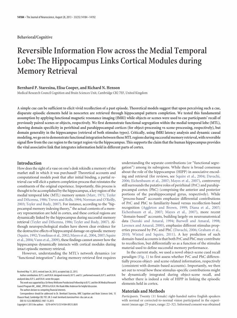
Experimental design. The stimulus material consisted of 384 color pic-tures (Konkle et al., 2010), half of which (192) depicted objects and halfof which depicted scenes (16 additional pictures were used for practice).For each of the two stimulus categories (objects, scenes), there were twosimilar exemplars for each of 96 subcategories (e.g., a glass of red wineand a glass of white wine for the object subcategory “wine glass” or ascene with a volcano emitting lava and scene with a volcano emitting anash cloud for the scene subcategory “volcano”). As detailed below, thetwo exemplars per subcategory were used to enforce attention to event-specific details during encoding and retrieval. The stimulus material wascounterbalanced so that half of the participants were presented with set 1of subcategory exemplars during the first half of the experiment and withset 2 during the second half (and vice versa for the other half of theparticipants). Thus, no two exemplars of a given subcategory were pre-sented within the same encoding-retrieval cycle.
The experiment consisted of six runs, with each run containing threeblocks: an encoding block, a delay block, and a retrieval block (Fig. 1).Scanning was performed continuously across the three blocks, with shortunscanned breaks between runs. Only retrieval data are reported here.The experiment was presented via the Psychophysics Toolbox (Brainard,1997) implemented in MATLAB. During each encoding block, partici-pants were presented with 32 unique object-scene pairs. The pairing ofobjects and scenes was randomized across participants. Object and scenepictures were each presented in a 250 � 250 pixels frame placed to the leftand right of the screen center. During half of the trials (16, randomlyselected), the object appeared to the left of the screen center and the sceneto the right, with the reverse order during the other half of the trials. Thetrial duration was 4 s, and for the last 0.5 s the picture pair was replacedwith a fixation cross (responses were still recorded), alerting participantsthat another trial would appear shortly. The encoding task was to indicatevia button press whether the given object-scene pair is plausible or im-plausible, i.e., likely to appear in real life or nature (Staresina and Davachi,2006). “Plausible” responses were given with the index finger and “im-plausible” responses with the middle finger. Across participants, use ofleft versus right hand was counterbalanced (but the finger assignment
was held constant). Object-scene encoding tri-als were intermixed with an active baselinecondition (Stark and Squire, 2001). Here, ran-dom numbers between 0 and 100 were shown,and participants pressed the index finger keyfor even numbers and the middle finger key forodd numbers. As soon as a response was given,another random number was shown. The re-sponse time for each number was self-pacedand participants were encouraged to performthis task as fast as possible without sacrificingaccuracy. Each encoding block lasted �3 min.
After the last encoding trial, participants sawa transition screen for 16 s, alerting them to theupcoming delay block. During the delay block,participants again performed the odd/evennumbers task described above for 2 min. Odd/even response accuracy was reported to partic-ipants on the computer screen following thecompletion of the task to encourage accuracy.
At the end of the delay block, another 16 stransition screen alerted participants to the up-coming retrieval block. Each retrieval blockconsisted of 32 trials, each trial lasting 6 s. For agiven trial, participants saw either the object orthe scene of a given object-scene pair from theprevious encoding block and were asked to in-dicate whether they remembered the corre-sponding paired associate (“recall”; indexfinger) or not (“forgot”; middle finger). Half ofthe cues (16) were object pictures, the otherhalf scene pictures. Across the 32 retrieval tri-als, each cue type (object cue or scene cue) was
presented in mini-blocks of eight consecutive trials (A-B-A-B), with arandom assignment of object and scene cue trials to A and B in each run.Participants received the following instructions regarding “recall” re-sponses: “Remembering the associate means that you could describe it insuch a way that another person who has not seen the stimuli can pick thecorrect stimulus based on your description. Keep in mind that there aremultiple exemplars per category, so your description has to be as detailedas possible.” As mentioned above, there were only two exemplars persubcategory. To ensure that participants gave “recall” responses whenthey indeed recalled the correct paired associate, we asked them to ver-bally describe the target after �10% of the “recall” responses. In partic-ular, three catch trials per block were randomly determined beforehand(e.g., trial 5, 14, and 32 for block 1). If the participant did not give a recallresponse on a designated catch trial, the next trial on which a recallresponse was given served as a catch trial. This means that for someblocks, there were �3 catch trials (e.g., if the participant indicated forgoton trial 32 (the last trial in a block) in the example above so that noalternative catch trial could be chosen). Catch trials started after the 6 strial period, showing a 2 s warning screen (“prepare to describe theassociated image…”) followed by a 10 s period during which a verbalresponse was recorded (“please describe the associated image”), followedagain by a 6 s fade-out screen (“prepare to continue with the experi-ment”). For scoring purposes, verbal responses were classified as accu-rate if they encompassed the target image’s basic level label as well assome characteristic feature (e.g., “a glass of red wine” in the exampleabove). As during the encoding block, retrieval trials were intermixedwith odd/even number baseline trials. The retrieval block lasted �6 min.
For the imaging analysis, the four conditions of interest were as fol-lows: (1) object cue, target scene recalled (O-S(R)), (2) object cue, targetscene forgotten (O-S(F)), (3) scene cue, object target recalled (S-O(R)),and (4) scene cue, object target forgotten (S-O(F)).
Magnetic resonance imaging scanning details. Scanning was performedon a 3 T Siemens Tim Trio magnetic resonance imaging (MRI) systemusing a 32-channel whole-head coil. Functional data were acquired usinga gradient-echo, echo-planar pulse sequence (TR � 1000 ms, TE � 30ms, 16 horizontal slices oriented parallel to the hippocampal axis, de-
Figure 1. Experimental paradigm. During encoding, participants were presented with pairs of trial-unique object and sceneimages. During retrieval, participants were either cued with an object or with a scene, and indicated whether they could recall thecorresponding target. O-S(R): object cue, scene target recalled; O-S(F): object cue, scene target forgotten; S-O(R): scene cue, objecttarget recalled; S-O(F): scene cue, object target forgotten.
Staresina et al. • Functional Integration across the MTL J. Neurosci., August 28, 2013 • 33(35):14184 –14192 • 14185
scending slice acquisition, 3 � 3 � 3 mm voxel size, 0.75 mm interslicegap, 702 volume acquisitions per run). The first 7 volumes of each runwere discarded to allow for magnetic field stabilization. High-resolution(1 � 1 � 1 mm) T1-weighted (MP-RAGE) images were collected foranatomical visualization. Foam padding was used to minimize head mo-tion. Visual stimuli were projected onto a screen that was viewed througha mirror, and responses were collected with magnet-compatible buttonboxes placed under the participant’s hands.
The active baseline task (odd/even-task; Stark and Squire, 2001) com-prised a fourth of the total scanning time. The sequence of encoding/retrieval trials and the variable number of baseline trials waspseudorandom and optimized for rapid event-related functional MRI(fMRI; using the “optseq” algorithm; Dale, 1999).
fMRI analysis. Data were analyzed using SPM8 (http://www.fil.ion.ucl.ac.uk/spm/). During preprocessing, images were corrected for differ-ences in slice acquisition timing, followed by motion correction across allruns. Neural activity for the conditions of interest (O-S(R), O-S(F),S-O(R), S-O(F)) was modeled as an impulse (delta function) in a designmatrix that concatenated all retrieval blocks and included nuisance re-gressors for invalid trials, head movement, low-frequency scanner drift,and run means. Additionally, the 10 s overt speech plus the surrounding2 s fade-in and 6 s fade-out periods of catch trials were modeled asuser-specified nuisance regressors (using unconvolved stick functionsfor each volume). For the conventional general linear model (GLM)analysis, condition onsets were convolved using a single, canonical he-modynamic response function (HRF), as provided in SPM8. The result-ing �-parameter estimates were then averaged across voxels within eachregion of interest (ROI; see below) in the participant’s native space, andthe resulting values were used in repeated-measures ANOVAs and t tests.For ANOVA factors with more than one numerator degree of freedom(df), we used a Greenhouse–Geisser df-correction for nonsphericity ofthe error.
Extraction of time-resolved blood oxygenation level-dependent(BOLD) data was based on the same design matrix, but condition re-sponses were modeled via a finite impulse response (FIR) basis set (ratherthan the canonical HRF), with 20 bins and a 1 s bin-width equal to the TR(and converted to percentage signal change via the MarsBaR toolbox;Brett et al., 2002). We focused on the evoked BOLD response, corre-sponding to the first 11 bins, or 0.5–11.5 s (given that data were aligned tothe middle slice acquired). These FIR parameter estimates were averagedacross voxels within each ROI in the participant’s native space.
We analyzed the data in PrC, PhC, and HIPP using hand-drawn,participant-specific ROIs, based on the individual structural image. An-atomical demarcation was done according to Insausti et al. (1998) andPruessner et al. (2002). As there were no hemispheric differences (datanot shown), we combined left and right hemisphere ROIs. Specifically,data were separately extracted for left and right hemisphere ROIs andcollapsed before entering analyses. Note that no spatial smoothing wasperformed on the data, ensuring that there was minimal signal overlapbetween the regions.
Nonlinear HRF fitting. To estimate the BOLD onset latency, we fit themodel:
y � g�t, p1, p2, p3� � e,
where y is the vector of the above 11 FIR parameter estimates across timepoints t � [0.5 1.5 2.5 3.5 4.5 5.5 6.5 7.5 8.5 9.5 10.5 11.5] from a givenROI and condition of a given participant, e is random error, and g is anonlinear function with parameters p1–3. Given the positively skewednature of the BOLD response, we defined g as a Gamma function:
g�t, p1, p2, p3� � p1 � G�t � p2, 6/p3, dt/p3�,
where p1 is a scaling parameter (amplitude), p2 is the onset latency, and p3
is a dispersion factor that affects the shape and scale of a gamma proba-bility density function ( G) over time t, relative to a peak of 6 s whensampled every dt � 1 s here (Evans et al., 1993).
In general, these parameters are fit numerically by an iterative algo-rithm that maximizes some goodness of fit (GOF) metric between thedata (y) and fitted response (g�t, p1, p2, p3�). However, this GOF metric
can have local maxima, particularly with noisy data for some participantsand ROIs, and particularly when some parameters (such as onset latencyand dispersion) have correlated effects on the fitted response. To accom-modate this, we regularized the problem by imposing Gaussian priors onthe parameters and on the noise, corresponding to a variational Bayesianapproach that can be solved by maximizing a free-energy metric thatapproximates the model evidence (probability of the data given the model;Friston et al., 2007). In this case, e is assumed to be drawn from a zero-mean Gaussian distribution with variance p0. The prior mean of the onsetlatency ( p2) was 0 s, the prior mean of the dispersion factor ( p3) was 1,and the prior mean of the amplitude ( p1) was set to the mean peakresponse over all regions, conditions, and participants (0.76% signalchange). The variance of each of these four priors for parameters p0-3 wasvaried over a range [0.01 0.1 0.5 1 5 10 100], and the maximal free-energyused to select the best of the resulting 7 4 � 2401 models. This optimalmodel, which had prior variances of 10 for p0, 0.5 for p1, 0.5 for p2, and 0.1for p3, was then used to estimate the posterior mean of the parametersreported in the main text.
Dynamic causal modeling. Dynamic casual modeling (DCM) was per-formed using version DCM10 in SPM8, using the same model describedabove (except that the inputs had duration of 2 s, to allow sufficientsensitivity to modulation of connections (Henson et al., 2013). The vol-umes of interest were defined based on both anatomical and functionalcriteria. First, only voxels within the anatomically defined ROIs (PrC,PhC, HIPP) were considered. Second, within each ROI, we chose the 15voxels with the strongest univariate effect sizes based on contrasts withina GLM: S-O(R) versus S-O(F) for PrC, O-S(R) versus O-S(F) for PhC,and R versus F (collapsed across object-cue and scene-cue trials) forHIPP. Note that this selection step merely served to identify the voxelsmost responsive within a given region and does not bias the subsequentDCM analysis to show any of the observed directionality effects. The top15 voxels were identified for left and right hemisphere regions separatelyand then combined for the DCM analysis.
Bayesian model selection was performed using a random effectsmodel, as described by Stephan et al. (2009). This allows estimation of the“exceedance probability,” i.e., the extent to which each model is morelikely than any other model tested to have generated the data from arandomly selected participant. The choice of models is described below.
ResultsBehavioral resultsDuring retrieval, the proportion of scene recall and forgot re-sponses when cued with an object were 49.8 and 49.4%, respec-tively (with no response given on 0.8% of the trials). Thecorresponding proportion for object recall when cued with ascene was 43.6 and 54.8%, respectively (with no response givenon the remaining 1.6% of the trials). Importantly, reaction timesfor recall responses did not differ statistically between object-cueand scene-cue trials (2.23 s vs 2.16 s; t(19) � 1.24, p � 0.23). Onrandomly interspersed catch trials (where participants verballydescribed their memory after giving a recall keypress), the answerwas correct on 98% of the trials when cued with an object and on96% of the trials when cued with a scene, demonstrating thatparticipants indeed recalled the correct target when indicating so.The average numbers of trials for our conditions of interest were48 for O-S(R) (range 33– 69), 47 for O-S(F) (range 26 – 62), 42 forS-O(R) (range 25– 61), and 52 for S-O(F) (range 35–70).
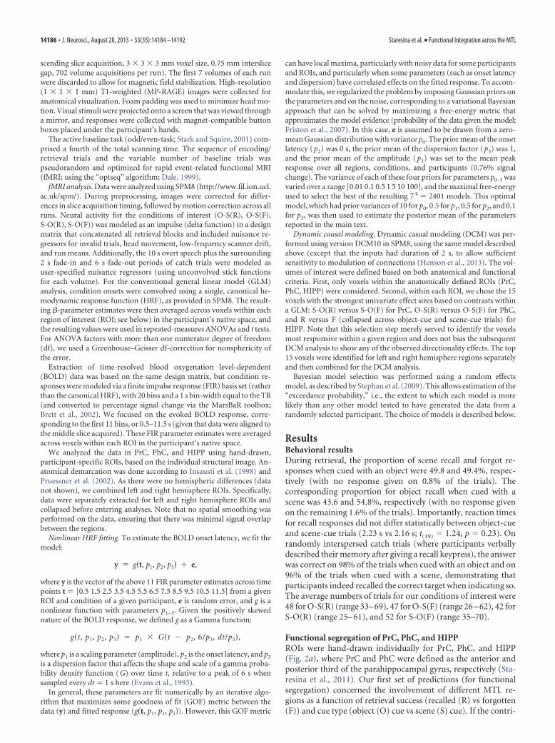
Functional segregation of PrC, PhC, and HIPPROIs were hand-drawn individually for PrC, PhC, and HIPP(Fig. 2a), where PrC and PhC were defined as the anterior andposterior third of the parahippocampal gyrus, respectively (Sta-resina et al., 2011). Our first set of predictions (for functionalsegregation) concerned the involvement of different MTL re-gions as a function of retrieval success (recalled (R) vs forgotten(F)) and cue type (object (O) cue vs scene (S) cue). If the contri-
14186 • J. Neurosci., August 28, 2013 • 33(35):14184 –14192 Staresina et al. • Functional Integration across the MTL
butions of PrC and PhC are domain specific, we would expectPrC to show a greater response during trials in which objectinformation is represented, regardless of whether the object isperceived as the cue (O-S(R) and O-S(F)), or retrieved as thetarget after being cued with a scene image (S-O(R)), comparedwith when no object information is perceived or retrieved(S-O(F)). The same logic applies to PhC: an increased re-sponse would be expected whenever scene information isperceived (S-O(R) and S-O(F)), or retrieved from memory(O-S(R)), compared with when no scene information is per-ceived or retrieved (O-S(F)).
Using a conventional analysis of the parameter estimate for acanonical HRF derived from a GLM (see Materials and Meth-ods), a repeated-measures ANOVA with the factors Region (PrC,PhC, HIPP), Cue Type (object, scene), and Memory (R, F)showed a highly significant three-way Region � Cue Type �Memory interaction (F(1.41,26.87) � 110.33, p � 0.001). Subsidiaryrepeated-measures ANOVAs conducted separately for each re-gion showed a significant Cue Type � Memory interaction inPrC (F(1,19) � 8.39, p � 0.009) and in PhC (F(1,19) � 94.86, p �0.001), but only a significant main effect of Memory in HIPP
(F(1,19) � 95.17, p � 0.001; Cue Type �Memory interaction, F(1,19) � 0.31, p �0.583). The pattern of significant pairwisedifferences is shown in Figure 2. In sum-mary, PrC showed a significant memoryeffect (greater response to recalled thanforgotten trials) for recalling objects, butnot for recalling scenes; PhC showed a sig-nificant memory effect for recallingscenes, but not for recalling objects; andHIPP showed a significant memory effectfor recalling both objects and scenes. Thisthree-way interaction constitutes compel-ling evidence for functional segregation inthe MTL: while PrC and PhC contribu-tions are domain specific, driven by objectand scene representations, respectively(either as the perceived cue or as the re-trieved target), the contribution of HIPPis domain general and driven by successversus failure of associative recall. Thesame pattern of significant ROI resultswas obtained when allowing for latencydifferences (see below) by using instead asthe dependent variable: (1) the percentagesignal change from the peak time point ofeach region and condition or (2) the am-plitude parameter from nonlinear fittingof the HRF.
Latencies of evoked responses withinPrC and PhCIf PrC and PhC provide domain-specificcontributions, their relative engagementover time would be expected to vary as afunction of the cue–target relationship.For instance, if PrC holds object represen-tations, engagement of this region shouldoccur earlier when an object serves as thecue than when an object is successfullyretrieved as the target, assuming that in-formation must undergo additional pro-
cessing stages when retrieved from memory relative to beingperceived in the environment. Likewise, engagement of PhCshould occur earlier when a scene serves as the cue than when ascene is the successfully retrieved target. Correspondingly, wepredicted an earlier response for O-S(R) relative to S-O(R) inPrC, but an earlier response for S-O(R) relative to O-S(R) in PhC,reflecting a reversal of the relative temporal ordering of condi-tions across regions.
Evidence for this prediction was apparent when plotting thetrial-averaged time courses of the evoked BOLD response every1 s (Fig. 3): while the greatest BOLD response in PrC occurredduring the fifth TR for the O-S(R) condition (when averagingresponses across participants), the greatest mean response for theS-O(R) condition occurred later, in the sixth TR (Fig. 3a). Theopposite pattern can be seen in PhC (Fig. 3b). To assess thisstatistically, we used nonlinear fitting of an HRF that was explic-itly parameterized by its amplitude, onset latency, and dispersion(see Materials and Methods), and compared the onset latencyestimates across conditions and regions. In PrC, the average onsetlatency (relative to the stimulus onset at 0 s) was 0.38 s for O-S(R)and 0.52 s for S-O(R). In PhC, on the other hand, the average
Figure 2. a, ROIs, manually drawn for each participant (shown here for one example participant): PrC (blue), PhC (red), andHIPP (purple). b, Mean (�SEM) GLM parameter estimates in PrC (bottom left), PhC (bottom right), and HIPP (top) for successfulversus unsuccessful recall as a function of cue and target stimulus (N � 20). Results show a three-way dissociation, where PrC andPhC are driven in a domain-specific fashion by object and scene representations, respectively (as the perceived cue or the retrievedtarget). Conversely, HIPP activation is driven in a domain-general fashion by success versus failure of recall, regardless of stimulustype. O-S(R): object cue, scene target recalled; O-S(F): object cue, scene target forgotten; S-O(R): scene cue, object target recalled;S-O(F): scene cue, object target forgotten. *p � 0.05, two-tailed paired t tests.
Staresina et al. • Functional Integration across the MTL J. Neurosci., August 28, 2013 • 33(35):14184 –14192 • 14187
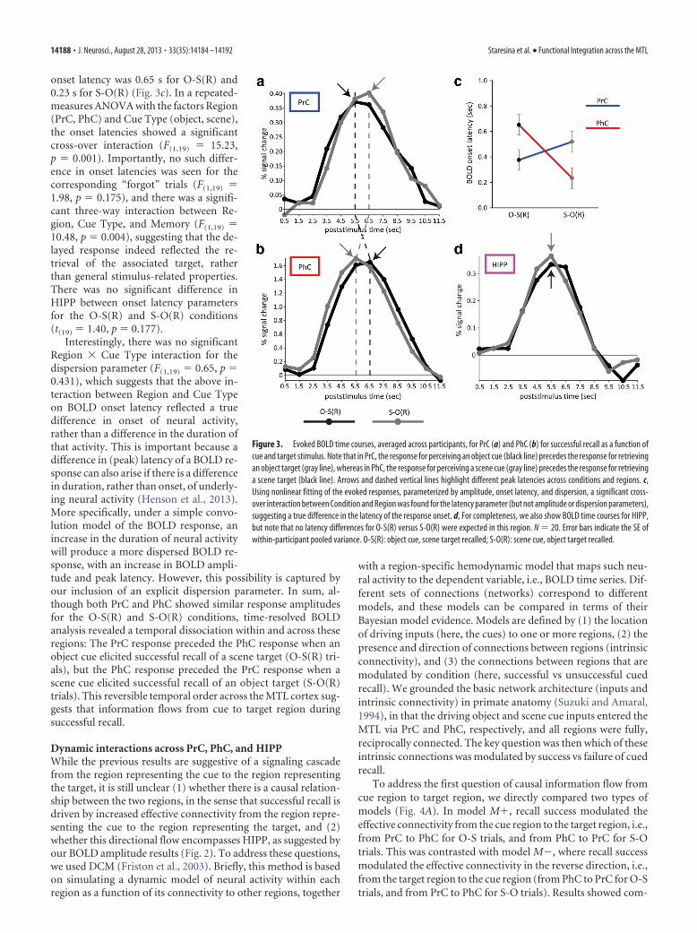
onset latency was 0.65 s for O-S(R) and0.23 s for S-O(R) (Fig. 3c). In a repeated-measures ANOVA with the factors Region(PrC, PhC) and Cue Type (object, scene),the onset latencies showed a significantcross-over interaction (F(1,19) � 15.23,p � 0.001). Importantly, no such differ-ence in onset latencies was seen for thecorresponding “forgot” trials (F(1,19) �1.98, p � 0.175), and there was a signifi-cant three-way interaction between Re-gion, Cue Type, and Memory (F(1,19) �10.48, p � 0.004), suggesting that the de-layed response indeed reflected the re-trieval of the associated target, ratherthan general stimulus-related properties.There was no significant difference inHIPP between onset latency parametersfor the O-S(R) and S-O(R) conditions(t(19) � 1.40, p � 0.177).
Interestingly, there was no significantRegion � Cue Type interaction for thedispersion parameter (F(1,19) � 0.65, p �0.431), which suggests that the above in-teraction between Region and Cue Typeon BOLD onset latency reflected a truedifference in onset of neural activity,rather than a difference in the duration ofthat activity. This is important because adifference in (peak) latency of a BOLD re-sponse can also arise if there is a differencein duration, rather than onset, of underly-ing neural activity (Henson et al., 2013).More specifically, under a simple convo-lution model of the BOLD response, anincrease in the duration of neural activitywill produce a more dispersed BOLD re-sponse, with an increase in BOLD ampli-tude and peak latency. However, this possibility is captured byour inclusion of an explicit dispersion parameter. In sum, al-though both PrC and PhC showed similar response amplitudesfor the O-S(R) and S-O(R) conditions, time-resolved BOLDanalysis revealed a temporal dissociation within and across theseregions: The PrC response preceded the PhC response when anobject cue elicited successful recall of a scene target (O-S(R) tri-als), but the PhC response preceded the PrC response when ascene cue elicited successful recall of an object target (S-O(R)trials). This reversible temporal order across the MTL cortex sug-gests that information flows from cue to target region duringsuccessful recall.
Dynamic interactions across PrC, PhC, and HIPPWhile the previous results are suggestive of a signaling cascadefrom the region representing the cue to the region representingthe target, it is still unclear (1) whether there is a causal relation-ship between the two regions, in the sense that successful recall isdriven by increased effective connectivity from the region repre-senting the cue to the region representing the target, and (2)whether this directional flow encompasses HIPP, as suggested byour BOLD amplitude results (Fig. 2). To address these questions,we used DCM (Friston et al., 2003). Briefly, this method is basedon simulating a dynamic model of neural activity within eachregion as a function of its connectivity to other regions, together
with a region-specific hemodynamic model that maps such neu-ral activity to the dependent variable, i.e., BOLD time series. Dif-ferent sets of connections (networks) correspond to differentmodels, and these models can be compared in terms of theirBayesian model evidence. Models are defined by (1) the locationof driving inputs (here, the cues) to one or more regions, (2) thepresence and direction of connections between regions (intrinsicconnectivity), and (3) the connections between regions that aremodulated by condition (here, successful vs unsuccessful cuedrecall). We grounded the basic network architecture (inputs andintrinsic connectivity) in primate anatomy (Suzuki and Amaral,1994), in that the driving object and scene cue inputs entered theMTL via PrC and PhC, respectively, and all regions were fully,reciprocally connected. The key question was then which of theseintrinsic connections was modulated by success vs failure of cuedrecall.
To address the first question of causal information flow fromcue region to target region, we directly compared two types ofmodels (Fig. 4A). In model M�, recall success modulated theeffective connectivity from the cue region to the target region, i.e.,from PrC to PhC for O-S trials, and from PhC to PrC for S-Otrials. This was contrasted with model M, where recall successmodulated the effective connectivity in the reverse direction, i.e.,from the target region to the cue region (from PhC to PrC for O-Strials, and from PrC to PhC for S-O trials). Results showed com-
Figure 3. Evoked BOLD time courses, averaged across participants, for PrC (a) and PhC (b) for successful recall as a function ofcue and target stimulus. Note that in PrC, the response for perceiving an object cue (black line) precedes the response for retrievingan object target (gray line), whereas in PhC, the response for perceiving a scene cue (gray line) precedes the response for retrievinga scene target (black line). Arrows and dashed vertical lines highlight different peak latencies across conditions and regions. c,Using nonlinear fitting of the evoked responses, parameterized by amplitude, onset latency, and dispersion, a significant cross-over interaction between Condition and Region was found for the latency parameter (but not amplitude or dispersion parameters),suggesting a true difference in the latency of the response onset. d, For completeness, we also show BOLD time courses for HIPP,but note that no latency differences for O-S(R) versus S-O(R) were expected in this region. N � 20. Error bars indicate the SE ofwithin-participant pooled variance. O-S(R): object cue, scene target recalled; S-O(R): scene cue, object target recalled.
14188 • J. Neurosci., August 28, 2013 • 33(35):14184 –14192 Staresina et al. • Functional Integration across the MTL
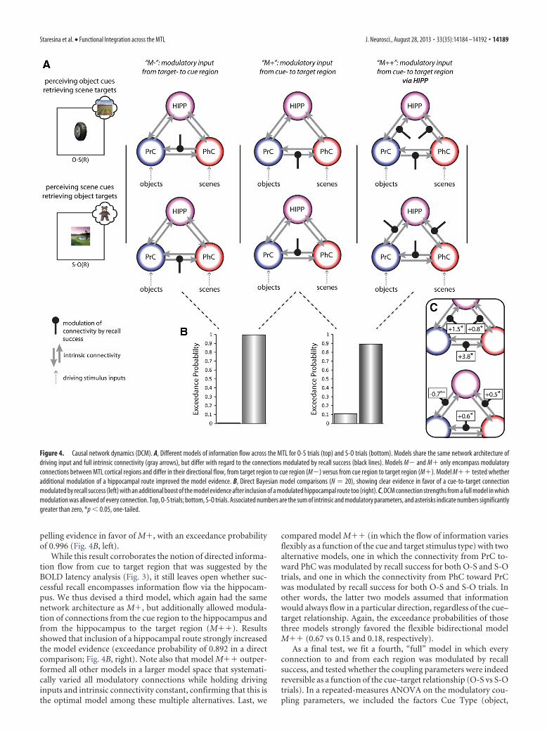
pelling evidence in favor of M�, with an exceedance probabilityof 0.996 (Fig. 4B, left).
While this result corroborates the notion of directed informa-tion flow from cue to target region that was suggested by theBOLD latency analysis (Fig. 3), it still leaves open whether suc-cessful recall encompasses information flow via the hippocam-pus. We thus devised a third model, which again had the samenetwork architecture as M�, but additionally allowed modula-tion of connections from the cue region to the hippocampus andfrom the hippocampus to the target region (M��). Resultsshowed that inclusion of a hippocampal route strongly increasedthe model evidence (exceedance probability of 0.892 in a directcomparison; Fig. 4B, right). Note also that model M�� outper-formed all other models in a larger model space that systemati-cally varied all modulatory connections while holding drivinginputs and intrinsic connectivity constant, confirming that this isthe optimal model among these multiple alternatives. Last, we
compared model M�� (in which the flow of information variesflexibly as a function of the cue and target stimulus type) with twoalternative models, one in which the connectivity from PrC to-ward PhC was modulated by recall success for both O-S and S-Otrials, and one in which the connectivity from PhC toward PrCwas modulated by recall success for both O-S and S-O trials. Inother words, the latter two models assumed that informationwould always flow in a particular direction, regardless of the cue–target relationship. Again, the exceedance probabilities of thosethree models strongly favored the flexible bidirectional modelM�� (0.67 vs 0.15 and 0.18, respectively).
As a final test, we fit a fourth, “full” model in which everyconnection to and from each region was modulated by recallsuccess, and tested whether the coupling parameters were indeedreversible as a function of the cue–target relationship (O-S vs S-Otrials). In a repeated-measures ANOVA on the modulatory cou-pling parameters, we included the factors Cue Type (object,
Figure 4. Causal network dynamics (DCM). A, Different models of information flow across the MTL for O-S trials (top) and S-O trials (bottom). Models share the same network architecture ofdriving input and full intrinsic connectivity (gray arrows), but differ with regard to the connections modulated by recall success (black lines). Models M and M� only encompass modulatoryconnections between MTL cortical regions and differ in their directional flow, from target region to cue region (M) versus from cue region to target region (M�). Model M�� tested whetheradditional modulation of a hippocampal route improved the model evidence. B, Direct Bayesian model comparisons (N � 20), showing clear evidence in favor of a cue-to-target connectionmodulated by recall success (left) with an additional boost of the model evidence after inclusion of a modulated hippocampal route too (right). C, DCM connection strengths from a full model in whichmodulation was allowed of every connection. Top, O-S trials; bottom, S-O trials. Associated numbers are the sum of intrinsic and modulatory parameters, and asterisks indicate numbers significantlygreater than zero, *p � 0.05, one-tailed.
Staresina et al. • Functional Integration across the MTL J. Neurosci., August 28, 2013 • 33(35):14184 –14192 • 14189
scene) and Direction (from PrC toward PhC vs from PhC towardPrC, averaging across PrC-PhC, PrC-HIPP, and PhC-HIPP con-nections). Critically, we observed a significant Cue Type � Di-rection interaction (F(1,19) � 37.92, p � 0.001), due to asignificant increase in effective connectivity from PrC towardPhC (compared with PhC toward PrC) during O-S(R) trials(t(19) � 5.25, p � 0.001), but a significant increase in effectiveconnectivity from PhC toward PrC (compared with PrC towardPhC) during S-O(R) trials (t(19) � 4.08, p � 0.001). Indeed, whentesting the individual connection strengths during successful re-call (the sum of the intrinsic and modulatory connection param-eters), five of the six modulations in the forward direction (fromcue region toward target region) were significantly greater thanzero (Fig. 4C). This included the output connection from HIPPto PhC during O-S trials, though the output connection fromHIPP to PrC during S-O trials did not significantly differ fromzero (see Discussion).
To summarize our DCM analysis: following the observationof a reversal of relative response latencies across PrC and PhC(Fig. 3), we obtained further evidence for a causal dynamic rela-tionship between PrC and PhC, such that PrC drives activation inPhC when PrC represents the cue and PhC represents the target,but PhC drives PrC when the cue-target assignment is reversed.We then went on to demonstrate that this directional flow fromcue toward target region is better captured by adding a furtherindirect route via the hippocampus. This was demonstrated bothacross models that differed in which connections were modu-lated by successful recall, and across coupling parameters withina fully modulated model. Relating back to the pattern of func-tional segregation (Fig. 2), these results suggest that the hip-pocampus flexibly links domain-specific representations in MTLcortex during successful recall.
DiscussionEver since the hallmark case of patient H.M. (Scoville and Milner,1957), whose episodic memory was devastated by a large lesion tohis MTLs, memory research has primarily focused on teasingapart the contributions of different regions within the MTL (Co-hen and Eichenbaum, 1993; Aggleton and Brown, 1999; Cohen etal., 1999; Norman and O’Reilly, 2003; Eichenbaum, 2004, 2007;Squire et al., 2004; Henson, 2005; Davachi, 2006; Diana et al.,2007; Mayes et al., 2007). However, controversy about the preciseprinciples of functional segregation has hindered progress on thearguably more important question of how these regions dynam-ically interact to enable our rich and integrated episodic memo-ries (functional integration). The question of functionalintegration is not unique to memory research; it is a fundamentalchallenge in neuroscience that emerges whenever specializedmodules must be integrated to enable coherent perception,thought, and action (Zeki and Shipp, 1988; Edelman, 1993; Fris-ton, 2002; Macaluso and Driver, 2005). In the current study, wefirst showed a pattern of functional segregation across MTL re-gions that supports recent neuroanatomically based accounts ofMTL functions. Building on this division of labor, we then pro-ceeded to assess how the separate contributions are dynamicallyintegrated during successful recall.
Functional segregation–three-way dissociation in thecontributions of PrC, PhC, and HIPPAs mentioned in the Introduction, recent efforts to capture thedivision of labor among MTL regions have emphasized the ana-tomical inputs and stimulus representations processed by theseregions (Lee et al., 2005; Buffalo et al., 2006; Davachi, 2006; Diana
et al., 2007; Graham et al., 2010; Staresina et al., 2011; Wixted andSquire, 2011; Liang et al., 2013). Regarding the MTL cortex, ourcurrent data provide strong support for this view (Fig. 2). En-gagement of PrC and PhC was driven by processing of objects orscenes, respectively, regardless of whether their preferred stimuliwere perceived as a cue, or retrieved as a target. Regarding theretrieval effects, it is interesting to note that despite the stronginteraction of Cue Type � Memory in both regions (reflectingdifferential recall effects for each region’s preferred stimulustype), there was a numerical trend in PrC toward a recall effect forscene targets. This pattern is reminiscent of an fMRI study thatassessed MTL activation during encoding of objects and spatiallocations (Buffalo et al., 2006) and found only spatial encodingeffects in PhC, but both object and spatial encoding effects in PrC(albeit stronger effects for objects). Moreover, a recent study us-ing a cued recall paradigm in which objects were used as itemsand scenes were used as contexts (Hannula et al., 2013) foundrecall effects in PhC only when retrieving the scene context, butrecall effects in PrC both when retrieving the object item and thescene context. Collectively, these results suggest that the assign-ment of PrC to object processing versus PhC to scene processingmay not be perfectly symmetrical. One explanation might be thatat conventional fMRI resolutions, PrC may include signal fromthe adjacent entorhinal cortex, which processes both spatial andnonspatial representations along its mediolateral gradient(Schultz et al., 2012). Higher resolution imaging would be neededto address this possibility. Another explanation (as suggested byBuffalo et al., 2006) might be that there is stronger anatomicalinput from PhC to PrC than vice versa (Suzuki and Amaral,1994). The stronger direct connections from PhC to PrC thanfrom PrC to PhC may also explain why modulation of outputfrom HIPP to PrC in the DCM analysis did not reach significancefor scene cues (see Results).
On a related note, it is worth considering that the relativelystrict criterion for recall responses likely induced a fairly conser-vative response bias, such that forgot responses may include lessconfident target recall and/or different levels of stimulus famil-iarity. Likewise, in searching for the associated target, partici-pants may continue to mentally generate and scan multipleexemplars from the target category during F trials. While suchtransient stimulus representations are unlikely to achieve thesame representational fidelity as successfully retrieved targets,they may still engage PrC and PhC to certain levels. This wouldexplain the clear above-baseline activation levels of S-O(F) andO-S(F) trials in PrC and PhC (Fig. 2), respectively. However, thekey finding with regard to PrC and PhC activation levels is theirdissociation in supporting recall of object targets versus scenetargets, respectively, consistent with a role of these regions indomain-specific retrieval.
Unlike in the MTL cortex, we observed no domain specificityin HIPP; rather, HIPP engagement reflected success versus failureof cued recall regardless of the stimulus-type being recalled. Thisis in agreement with the idea that HIPP contributions are domaingeneral (Cohen and Eichenbaum, 1993; Eichenbaum, 2004; Da-vachi, 2006; Staresina and Davachi, 2008; Konkel and Cohen,2009; Kumaran et al., 2012), and is again consistent with themultimodal array of anatomical inputs this region receives (Su-zuki and Amaral, 1994; Lavenex and Amaral, 2000; van Strien etal., 2009). There is abundant evidence for the role of HIPP inassociative binding/pattern completion (for reviews, see Cohenand Eichenbaum, 1993; Aggleton and Brown, 1999; Squire et al.,2004; Davachi, 2006; Eichenbaum et al., 2007; Mayes et al., 2007;Konkel and Cohen, 2009), but our data are the first to reveal how
14190 • J. Neurosci., August 28, 2013 • 33(35):14184 –14192 Staresina et al. • Functional Integration across the MTL
recall modulates the functional connectivity of HIPP with otherMTL structures. This is arguably more direct evidence for a roleof HIPP in pattern completion than has been furnished by previ-ous activation analyses. To be explicit, while our results on func-tional segregation across the MTL corroborate and extendpervious findings, the novel aspect of the current study is that webuild on these different contributions to ask how their dynamicinterplay enables memory retrieval, as elaborated below.
Network dynamics across the MTL: from functionalsegregation to integrationGiven the stimulus-specific contributions of PrC and PhC, wefirst asked whether their engagement reflects different stages inthe MTL signaling cascade during episodic retrieval. Specifically,we hypothesized that during O-S(R) trials, PrC activation reflectsprocessing of the object cue, whereas PhC activation reflects re-trieval of the scene target. Similarly, during S-O(R) trials, PhCactivation should reflect processing of the scene cue, whereas PrCactivation reflects retrieval of the object target. Given that the cue,by definition, precedes the recalled item in a cued-recall para-digm (and given that the response latency for recalled responseswas �2 s; see Results), one would expect these different functionsto be expressed with different temporal profiles: engagement ofthe region representing the perceived cue should precede engage-ment of the region representing the retrieved target. As illustratedin Figure 3, the data show: during O-S(R) trials, the PrC BOLDresponse preceded the PhC response, whereas during S-O(R) tri-als, the PhC BOLD response preceded the PrC response.
Is the BOLD response sensitive enough to reveal temporaldifferences across conditions at such a short timescale? Despitethe tacit assumption that the fMRI signal is proportional to neu-ral firing rates, skepticism is warranted when interpreting BOLDtime course effects (Friston et al., 2000; Heeger and Ress, 2002;Logothetis and Wandell, 2004). Therefore, any main effect ofRegion would be difficult to interpret due to potentially differentneural-to-BOLD mappings across regions, and any main effect ofCue Type would be difficult to interpret due to potentially differ-ent dynamics earlier in object-processing-pathways versus scene-processing-pathways. Importantly, however, the cross-overinteraction of Region � Cue Type we observed here (Fig. 3c) rulesout any such region-specific or processing-pathway explana-tions. Furthermore, although it is difficult to infer backward fromBOLD latency differences to underlying neural latency differ-ences, our BOLD latency findings were obtained from nonlinearfitting of a model that included separate parametrization of onsetdelay, amplitude, and dispersion–allowing for more confidentinterpretation of differences in the BOLD onset latency parame-ter in terms of neural onset latency. Indeed, in a previous fMRIstudy, BOLD latency differences in PrC and HIPP across differentmemory retrieval conditions were directly confirmed by intracra-nial electroencephalography recordings (Staresina et al., 2012).
While the latency of the trial averaged-evoked BOLD responseis suggestive of a directional interplay between PrC and PhC dur-ing successful cued recall, simple latency differences are only in-direct evidence for a causal relationship between the regionrepresenting the cue stimulus and the region representing thetarget stimulus. Such causality is better inferred from temporaldependencies between regions across the whole fMRI time series,as in DCM. Furthermore, the latency analysis did not illuminatethe putative role of HIPP as a pattern completer in this cue-targetcascade (Marr, 1971; Teyler and DiScenna, 1986; Treves andRolls, 1994; Norman and O’Reilly, 2003; Teyler and Rudy, 2007).Indeed, recent studies using direct electrophysiological record-
ings in humans have shown results partly consistent with thenotion of HIPP as the interface between cue and target: a sourceretrieval signal was shown to be initiated in HIPP in response to apreceding old/new signal in PrC (Staresina et al., 2012), and anincrease in HIPP firing rates and gamma power was shown toprecede free recall of target items (Sederberg et al., 2007; Gelbard-Sagiv et al., 2008). However, due to restricted coverage of corticalsites in those studies, the network dynamics between HIPP andstimulus-specific MTL cortical regions (PrC and PhC) duringsuccessful recall has remained elusive. Here, we included HIPP ina DCM model to explicitly test for changes in effective connec-tivity between all three MTL regions during successful memoryretrieval. First and foremost, our DCM results provided clearevidence in favor of the expected flow of information across theMTL cortex (Fig. 4), i.e., from PrC toward PhC when perceivingan object and retrieving an associated scene, and in the oppositedirection (from PhC toward PrC) when perceiving a scene andretrieving an associated object. Second, a model that additionallyincorporated an additional indirect transmission route, (1) fromthe cortical region representing the cue to HIPP and (2) fromHIPP to the cortical region representing the target, further out-performed the model in which only direct PrC–PhC connectionswere modulated. Again, this directional flow including the hip-pocampal route was reversible as a function of the cue-targetrelationship on a given trial. These results are consistent with thenotion that HIPP serves as the site of associative recall/patterncompletion, and that different parts of the MTL cortex serve asstimulus-specific input and output modules.
ReferencesAggleton JP, Brown MW (1999) Episodic memory, amnesia, and the
hippocampal-anterior thalamic axis. Behav Brain Sci 22:425– 444; discus-sion 444 – 489. Medline
Brainard DH (1997) The Psychophysics Toolbox. Spat Vis 10:433– 436.CrossRef Medline
Brett M, Anton JL, Valabregue R, Poline JB (2002) Region of interest anal-ysis using an SPM toolbox. Neuroimage 16:S497.
Buffalo EA, Bellgowan PS, Martin A (2006) Distinct roles for medial tem-poral lobe structures in memory for objects and their locations. LearnMem 13:638 – 643. CrossRef Medline
Burwell RD, Amaral DG (1998) Cortical afferents of the perirhinal, postrhi-nal, and entorhinal cortices of the rat. J Comp Neurol 398:179 –205.CrossRef Medline
Cohen NJ, Eichenbaum HE (1993) Memory, amnesia, and the hippocampalsystem. Cambridge, MA: MIT.
Cohen NJ, Ryan J, Hunt C, Romine L, Wszalek T, Nash C (1999) Hip-pocampal system and declarative (relational) memory: summarizing thedata from functional neuroimaging studies. Hippocampus 9:83–98.CrossRef Medline
Dale AM (1999) Optimal experimental design for event-related fMRI. HumBrain Mapp 8:109 –114. CrossRef Medline
Davachi L (2006) Item, context and relational episodic encoding in humans.Curr Opin Neurobiol 16:693–700. CrossRef Medline
Diana RA, Yonelinas AP, Ranganath C (2007) Imaging recollection and fa-miliarity in the medial temporal lobe: a three-component model. TrendsCogn Sci 11:379 –386. CrossRef Medline
Edelman GM (1993) Neural Darwinism: selection and reentrant signalingin higher brain function. Neuron 10:115–125. CrossRef Medline
Eichenbaum H (2004) Hippocampus: cognitive processes and neural repre-sentations that underlie declarative memory. Neuron 44:109 –120.CrossRef Medline
Eichenbaum H, Yonelinas AP, Ranganath C (2007) The medial temporallobe and recognition memory. Annu Rev Neurosci 30:123–152. CrossRefMedline
Evans M, Hastings N, Peacock B (1993) Statistical distributions (Ed 2). NewYork: Wiley.
Friston K (2002) Functional integration and inference in the brain. ProgNeurobiol 68:113–143. CrossRef Medline
Staresina et al. • Functional Integration across the MTL J. Neurosci., August 28, 2013 • 33(35):14184 –14192 • 14191
Friston KJ, Mechelli A, Turner R, Price CJ (2000) Nonlinear responses infMRI: the Balloon model, Volterra kernels, and other hemodynamics.Neuroimage 12:466 – 477. CrossRef Medline
Friston KJ, Harrison L, Penny W (2003) Dynamic causal modelling. Neuro-image 19:1273–1302. CrossRef Medline
Friston K, Mattout J, Trujillo-Barreto N, Ashburner J, Penny W (2007)Variational free energy and the Laplace approximation. Neuroimage 34:220 –234. CrossRef Medline
Gelbard-Sagiv H, Mukamel R, Harel M, Malach R, Fried I (2008) Internallygenerated reactivation of single neurons in human hippocampus duringfree recall. Science 322:96 –101. CrossRef Medline
Graham KS, Barense MD, Lee AC (2010) Going beyond LTM in the MTL: asynthesis of neuropsychological and neuroimaging findings on the role ofthe medial temporal lobe in memory and perception. Neuropsychologia48:831– 853. CrossRef Medline
Hannula DE, Libby LA, Yonelinas AP, Ranganath C (2013) Medial tempo-ral lobe contributions to cued retrieval of items and contexts. Neuropsy-chologia pii: S0028-3932(13)00053-5. CrossRef Medline
Heeger DJ, Ress D (2002) What does fMRI tell us about neuronal activity?Nat Rev Neurosci 3:142–151. CrossRef Medline
Henson R (2005) A mini-review of fMRI studies of human medial temporallobe activity associated with recognition memory. Q J Exp Psychol B58:340 –360. CrossRef Medline
Henson R, Wakeman D, Phillips C, Rowe J (2013) Effective connectivitybetween OFA and FFA during face perception: DCM of evoked MEG,EEG and fMRI responses. Hum Brain Mapp, in press.
Insausti R, Juottonen K, Soininen H, Insausti AM, Partanen K, Vainio P,Laakso MP, Pitkanen A (1998) MR volumetric analysis of the humanentorhinal, perirhinal, and temporopolar cortices. AJNR Am J Neurora-diol 19:659 – 671. Medline
Konkel A, Cohen NJ (2009) Relational memory and the hippocampus: rep-resentations and methods. Front Neurosci 3:166 –174. CrossRef Medline
Konkle T, Brady TF, Alvarez GA, Oliva A (2010) Scene memory is moredetailed than you think the role of categories in visual long-term memory.Psychol Sci 21:1551–1556. CrossRef Medline
Kumaran D, Melo HL, Duzel E (2012) The emergence and representation ofknowledge about social and nonsocial hierarchies. Neuron 76:653– 666.CrossRef Medline
Lavenex P, Amaral DG (2000) Hippocampal-neocortical interaction: a hi-erarchy of associativity. Hippocampus 10:420 – 430. CrossRef Medline
Lee AC, Barense MD, Graham KS (2005) The contribution of the humanmedial temporal lobe to perception: bridging the gap between animal andhuman studies. Q J Exp Psychol B 58:300 –325. CrossRef Medline
Liang JC, Wagner AD, Preston AR (2013) Content representation in thehuman medial temporal lobe. Cereb Cortex 23:80 –96. CrossRef Medline
Logothetis NK, Wandell BA (2004) Interpreting the BOLD signal. Annu RevPhysiol 66:735–769. CrossRef Medline
Macaluso E, Driver J (2005) Multisensory spatial interactions: a windowonto functional integration in the human brain. Trends Neurosci 28:264 –271. CrossRef Medline
Marr D (1971) Simple memory: a theory for archicortex. Philos Trans R SocLond B, Biol Sci 262:23– 81. CrossRef Medline
Mayes AR, Holdstock JS, Isaac CL, Montaldi D, Grigor J, Gummer A, CarigaP, Downes JJ, Tsivilis D, Gaffan D, Gong Q, Norman KA (2004) Asso-ciative recognition in a patient with selective hippocampal lesions andrelatively normal item recognition. Hippocampus 14:763–784. CrossRefMedline
Mayes A, Montaldi D, Migo E (2007) Associative memory and the medialtemporal lobes. Trends Cogn Sci 11:126 –135. CrossRef Medline
Norman KA, O’Reilly RC (2003) Modeling hippocampal and neocorticalcontributions to recognition memory: a complementary-learning-systems approach. Psychol Rev 110:611– 646. CrossRef Medline
Pruessner JC, Kohler S, Crane J, Pruessner M, Lord C, Byrne A, Kabani N,
Collins DL, Evans AC (2002) Volumetry of temporopolar, perirhinal,entorhinal and parahippocampal cortex from high-resolution MR im-ages: considering the variability of the collateral sulcus. Cereb Cortex12:1342–1353. CrossRef Medline
Schultz H, Sommer T, Peters J (2012) Direct evidence for domain-sensitivefunctional subregions in human entorhinal cortex. J Neurosci 32:4716 –4723. CrossRef Medline
Scoville WB, Milner B (1957) Loss of recent memory after bilateral hip-pocampal lesions. J Neurol Neurosurg Psychiatry 20:11–21. CrossRefMedline
Sederberg PB, Schulze-Bonhage A, Madsen JR, Bromfield EB, Litt B, BrandtA, Kahana MJ (2007) Gamma oscillations distinguish true from falsememories. Psychol Sci 18:927–932. CrossRef Medline
Squire LR (1992) Memory and the hippocampus: a synthesis from findingswith rats, monkeys, and humans. Psychol Rev 99:195–231. CrossRefMedline
Squire LR, Stark CE, Clark RE (2004) The medial temporal lobe. Annu RevNeurosci 27:279 –306. CrossRef Medline
Staresina BP, Davachi L (2006) Differential encoding mechanisms for sub-sequent associative recognition and free recall. J Neurosci 26:9162–9172.CrossRef Medline
Staresina BP, Davachi L (2008) Selective and shared contributions of thehippocampus and perirhinal cortex to episodic item and associative en-coding. J Cogn Neurosci 20:1478 –1489. CrossRef Medline
Staresina BP, Duncan KD, Davachi L (2011) Perirhinal and parahippocam-pal cortices differentially contribute to later recollection of object- andscene-related event details. J Neurosci 31:8739 – 8747. CrossRef Medline
Staresina BP, Fell J, Do Lam AT, Axmacher N, Henson RN (2012) Memorysignals are temporally dissociated in and across human hippocampus andperirhinal cortex. Nat Neurosci 15:1167–1173. CrossRef Medline
Stark CE, Squire LR (2001) When zero is not zero: the problem of am-biguous baseline conditions in fMRI. Proc Natl Acad Sci U S A 98:12760 –12766. CrossRef Medline
Stephan KE, Penny WD, Daunizeau J, Moran RJ, Friston KJ (2009) Bayesianmodel selection for group studies. Neuroimage 46:1004 –1017. CrossRefMedline
Suzuki WA, Amaral DG (1994) Perirhinal and parahippocampal cortices ofthe macaque monkey: cortical afferents. J Comp Neurol 350:497–533.CrossRef Medline
Teyler TJ, DiScenna P (1986) The hippocampal memory indexing theory.Behav Neurosci 100:147–154. CrossRef Medline
Teyler TJ, Rudy JW (2007) The hippocampal indexing theory and episodicmemory: updating the index. Hippocampus 17:1158 –1169. CrossRefMedline
Treves A, Rolls ET (1994) Computational analysis of the role of the hip-pocampus in memory. Hippocampus 4:374 –391. CrossRef Medline
van Strien NM, Cappaert NL, Witter MP (2009) The anatomy of memory:an interactive overview of the parahippocampal– hippocampal network.Nat Rev Neurosci 10:272–282. CrossRef Medline
Vann SD, Tsivilis D, Denby CE, Quamme JR, Yonelinas AP, Aggleton JP,Montaldi D, Mayes AR (2009) Impaired recollection but spared famil-iarity in patients with extended hippocampal system damage revealed by3 convergent methods. Proc Natl Acad Sci U S A 106:5442–5447.CrossRef Medline
Wixted JT, Squire LR (2011) The medial temporal lobe and the attributes ofmemory. Trends Cogn Sci 15:210 –217. CrossRef Medline
Yonelinas AP, Kroll NE, Quamme JR, Lazzara MM, Sauve MJ, Widaman KF,Knight RT (2002) Effects of extensive temporal lobe damage or mildhypoxia on recollection and familiarity. Nat Neurosci 5:1236 –1241.CrossRef Medline
Zeki S, Shipp S (1988) The functional logic of cortical connections. Nature335:311–317. CrossRef Medline
14192 • J. Neurosci., August 28, 2013 • 33(35):14184 –14192 Staresina et al. • Functional Integration across the MTL