result of ender's nail in diaphyseal fracture of shaft femur and tibia in children
TRANSCRIPT

ISSN 2320-5407 International Journal of Advanced Research (2015), Volume 3, Issue 12, 1123 – 1144
1123
Journal homepage: http://www.journalijar.com INTERNATIONAL JOURNAL
OF ADVANCED RESEARCH
RESEARCH ARTICLE
Result of ender’s nail in diaphyseal fracture of shaft femur and tibia in children
Dr. Harshadkumar A Patel
1, Dr. Rajesh A Solanki
2, Dr. Chirag G Prajapati
3, Dr. Punit Tank
4, Dr. Nirav
Rathi5, Dr. Hiren Bhabhor
6, Dr. Janak Mistry
6
1. 3rd year resident doctor, Department of orthopaedics, B.J. Medical College, Ahmedabad.
2. Additional Professor, Department of orthopaedics , B.J. Medical college, Ahmedabad.
3. 3rd year resident doctor, Department of orthopaedics, B.J. Medical College, Ahmedabad.
3. 3rd year resident doctor, Department of orthopaedics, B.J. Medical College, Ahmedabad.
4. 2rd year resident doctor, Department of orthopaedics, B.J. Medical College, Ahmedabad.
5. Ist year resident doctor, Department of orthopaedics, B.J. Medical College, Ahmedabad)
6. Ist year resident doctor, Department of orthopaedics, B.J. Medical College, Ahmedabad)
Manuscript Info Abstract
Manuscript History:
Received: 12 October 2015
Final Accepted: 25 November 2015
Published Online: December 2015
Key words: Ender’s nail , Elastic
intramedullary nail, Ender’s nail
in children ,TENS, Shaft femur
fracture in children,Tibial Shaft fracture in children, Long bone
fracture, long bone fracture in
children
*Corresponding Author
Dr. Harshadkumar A Patel
Introduction
“Fracture of the shaft of the femur and tibia” is relatively frequent injury in children.
Majority of these fractures occur as a result of high velocity injury that leads to
fracture of femur and / or tibia by tremendous force. The high velocity injury may be
direct or indirect, such as that sustained in automobile accident or fall from height.
Whatever the method of treatment, the goals should be to stabilize the fracture, to control length and alignment, to promote bone healing and to minimize the morbidity
and complications for the child and his/her family. Recently in our institution, ender’s
nail fixation for diaphyseal fracture of femur and tibia is an ideal method of surgical
treatment with satisfactory results with minimum complications.
Material and Method
In this age group, we have evaluated total 50 patients, out of which 30 patients have
fracture of shaft femur and 20 patients have fracture of shaft tibia.
All children and adolescent patients between 5-16 years of age with diaphyseal fractures of femur and / or tibia admitted in tertiary center in government
setup - meeting the inclusion and the exclusion criteria ( as given below) during the
study period were the subjects for the study.Patient were regularly followed up
radiologically. Final outcome is measured with Flynn’s criteria.
Results
68% of the patients were boys. Fall down was the most common mode of
injury accounting for 28 (56.0%) cases Average duration of stay in hospital was 5.18
days. Superficial infection was seen in 1(2.0%) case. 1(2.0%) patient had shortening. Union was achieved in <3 months in 40 (80%) of the patients with average time to
union being 11.9 weeks.
Conclusion
Because of early weight bearing, rapid healing and minimal disturbance of bone growth, Ender’s nail may be considered to be a physiological method of treatment.

ISSN 2320-5407 International Journal of Advanced Research (2015), Volume 3, Issue 12, 1123 – 1144
707
Use of Ender’s nails for definitive stabilization of femoral and tibial shaft fractures in
children is a reliable, minimally invasive, and physeal-protective treatment method. Our study results provide new evidence that expands the inclusion criteria for this
treatment and shows that Ender’s nails can be successfully used regardless of
fracture location and fracture pattern. Copy Right, IJAR, 2015,. All rights reserved
INTRODUCTION “Fracture of the shaft of the femur and tibia” is relatively frequent injury in children. Majority of these fractures
occur as a result of high velocity injury that leads to fracture of femur and / or tibia by tremendous force. The high
velocity injury may be direct or indirect, such as that sustained in automobile accident or fall from height.
Aim of the treatment in this fracture is to give an adequate reduction and alignment and carry out normal activities
as early as possible in order to reduce psychological and economical burden to parents.
Whatever the method of treatment, the goals should be to stabilize the fracture, to control length and alignment, to
promote bone healing and to minimize the morbidity and complications for the child and his/her family.
Recently in our institution, ender’s nail fixation for diaphyseal fracture of femur and tibia is an ideal method of
surgical treatment with satisfactory results with minimum complications.
MATERIALS AND METHODS
Aim is to evaluate the results of operative treatment - outcome, safety, efficacy of paediatric femoral and
tibial diaphyseal fractures in the age group between 5 to 16 years treated by closed reduction and internal
fixation with Ender’s nails. In this age group, we have evaluated total 50 patients, out of which 30 patients
have fracture of shaft femur and 20 patients have fracture of shaft tibia.
All children and adolescent patients between 5-16 years of age with diaphyseal fractures of femur and /
or tibia admitted in tertiary center in government setup - meeting the inclusion and the exclusion criteria ( as given
below) during the study period were the subjects for the study .
Inclusion criteria:
1. 5-16 years of age
2. Diaphyseal fractures
3. Closed fractures
Exclusion criteria:
1. Metaphyseal fractures
2. Compound fractures
3. Pathological fractures
As soon as the patient was brought to casualty, patient’s airway, breathing and circulation were assessed. Then a
complete survey was carried out to rule out other significant injuries. Plain radiographs of AP and lateral views of -
the thigh including hip and knee joints OR – the leg including knee and ankle to assess the extent of fracture
comminution, the geometry and the dimensions of the fracture.
On admission to ward, a detailed history was taken, relating to the age, sex, and occupation, mode of injury,
past and associated medical illness. Routine investigations were done for all patients.
Patients were operated as early as possible once the general condition of the patient was stable and patient was fit
for surgery.

ISSN 2320-5407 International Journal of Advanced Research (2015), Volume 3, Issue 12, 1123 – 1144
1124
After prior informed consent, a pre-operative anaesthetic evaluation is done. Pre-op planning of surgery is made.
PREOPERATIVE PLANNING
Nail width:
The diameter of the individual nail is selected as per
1) Flynn et al’s formula.
Diameter of nail= width of the narrowest point of the medullary canal on AP and LATERAL view X 0.4mm
2) Intra operative assessment
Diameter of the nail is chosen so that each nail occupies atleast 1/3rd
o r 4 0 % o f the medullary cavity.
Nail length:
Lay one of the selected nails over the thigh / leg, and determine that it is of the appropriate length by fluoroscopy.
The nail for femur should extend from the level of the distal femoral physis to a point approximately 2 cm distal to
the capital femoral physis and 1cm distal to the greater trochanteric physis and for tibia it should extend 2cm from
the proximal physis till 5mm proximal to the distal physis.
PROCEDURE FOR ENDER’S NAILING OF DIAPHYSEAL FRACTURE OF FEMUR
RETROGRADE FIXATION
General / Spinal anesthesia is administered, and patient is placed in supine on a radiolucent table. The operative
extremity is then prepared and draped free. Identify the physis by fluoroscopy, and mark its location on the
skin. A 2 to 2.5 cm longitudinal skin incision was made over the medial and lateral surface of the distal femur,
starting 2 cm proximal to the distal femoral epiphyseal plate ; a haemostat was used to split the soft tissue down to
the bone, following which a 3.2 mm drill bit was used at a point 2.5 cm proximal to the distal femoral growth plate
to open the cortex at a right angle ; the drill was then inclined 10° to the distal femoral cortex. A nail was introduced
with a T-handle by rotation movements of the wrist.
Under image intensifier control, the nail was driven with rotatory movement or with a hammer to the fracture site
which was aligned to anatomical or near anatomical position with proper attention to limb rotation and length. By
rotation movements of the T-handle with or without limb manipulation, the nail was directed to the proximal
fragment which was pushed into better alignment by the nail. At the same time the second nail was advanced to
enter the proximal fragment and in the meantime any traction was released to avoid any distraction, and both nails
were pushed further till their tips became fixed into the cancellous bone of the proximal femoral metaphysis without
reaching the epiphyseal plate. The tips of the nail that entered the lateral femoral cortex should come to rest just
distal to the trochanteric epiphysis. The opposite nail should be at the same level towards the calcar region; too
short nails should be avoided.
The two-nail construct should be in a symmetrical alignment face to face with the maximum curvature of the nails
at the level of the fracture.
Distally the nails were leaving only 0.5 - 1 cm outside the cortex. The extra osseous portion of the nails was kept as
it was or slightly bent away from the bone to facilitate removal later on. In all cases care was taken to use nails
with similar diameters, to use the largest possible diameter, and to use the double C construct to ensure a 3-point
fixation.

ISSN 2320-5407 International Journal of Advanced Research (2015), Volume 3, Issue 12, 1123 – 1144
1125
PROCEDURE FOR ENDER’S NAILING OF DIAPHYSEAL FRACTURE OF TIBIA ANTEGRADE
/ RETROGRADE FIXATION
General / Spinal anesthesia is administered, and patient is placed in supine on a radiolucent table. The operative
extremity is then prepared and draped free. Under fluoroscopy, the fracture site and proximal tibial physis are
marked. The starting point for nail insertion is 1.5–2.0 cm distal to the physis, sufficiently posterior in the sagittal
plane to avoid injury to the tibial tubercle apophysis. A longitudinal 2 cm incision is made on both the lateral and
medial side of the tibia metaphysis just proximal to the desired bony entry point. Using a hemostat, the soft tissues
are bluntly dissected down to bone. Based on preoperative measurements, an appropriately sized implant is selected
so that the nail diameter is 40% of the diameter of the narrowest portion of the medullary canal. A drill roughly
0.5 mm larger than the selected nail is then used to open the cortex at the nail entry site; angling the drill
distally down the shaft facilitates nail entry. Both nails are then inserted through the entry holes and advanced to
the level of the fracture site.
Under fluoroscopic guidance, the fracture is reduced in both the coronal and sagittal planes, and the first nail is
advanced past the fracture site. If proper intramedullary position of the nail distal to the fracture site is confirmed
on antero-posterior and lateral views, then the second nail is tapped across the fracture site. Both nails are
advanced until the tips lie just proximal to the distal tibial physis. Fluoroscopy is again used to confirm proper
fracture reduction as well as nail position.
To minimize soft tissue irritation, the nails should be advanced until <1cm of nail lies outside of bone. Care is
taken not to bend the nails away from the bone, as we have found that this increases nail prominence and
subsequent skin irritation. The two incisions for nail entry are closed in a layered fashion, and the wounds are well
padded with gauze.
Postoperative Care:
Patients were kept nil by mouth for 4 to 6 hours post operatively.
IV fluids / blood transfusions were given as needed.
Analgesics were given according to the needs of the patient.
The limb was kept elevated over a pillow with slab or splint.
IV antibiotics were continued for 3 days and switched over to oral antibiotics on the 3rd
day and
continued till the 12th
day and patients were discharged on 3rd
or 4th day.
Sutures were removed on the 12th
postoperative day and patients were discharged.
Post-operatively, patients are immobilized with long leg cast for femur fracture or above knee POP cast for
tibia fracture for 6 weeks and such immobilization was continued for another 2-3 weeks based on radiological
assessment.
The period of immobilization was followed by active hip and knee / knee and ankle mobilization with non-weight
bearing crutch walking.
Full weight bearing is started by 8 - 12 weeks depending on the fracture configuration and callus response.
FOLLOW UP
Assessment done at 6, 12 and 24 weeks.
At each follow up patients are assessed clinically, radiologically and the complications are noted.
CLINICAL ASSESMENT
1) Pain 2) Range of movements

ISSN 2320-5407 International Journal of Advanced Research (2015), Volume 3, Issue 12, 1123 – 1144
1126
3) Measurement of limb length – noted for shortening / lengthening
4) Time of weight bearing
Partial weight bearing (in weeks)
Complete weight bearing (in weeks)
RADIOLOGICAL ASSESSMENT
X-ray thigh full length with hip and knee joints – AP and LATERAL views and X-ray leg full length with knee and
ankle- AP and LATERAL.
Alignment sagittal/coronal angulation (in degrees - <10 or >10) rotational mal-alignment (in degrees - <10 or >10).
Circumferential callus formation – good / adequate / poor.
Visibility of fracture line – seen clearly / masked / not seen.
COMPLICATIONS
Minor complications – a) when they resolved without additional surgery b) not resulting in long
term morbidity.
Major complications – a) when further operation was required b) long term morbidity ensued.
MINOR COMPLICATIONS:
a. Pain at the site of nail insertion
b. Minor angulation (< 100
– sagittal/coronal; <100
rotational mal-alignment) at final follow-
up (24 weeks)
c. Minor leg length discrepancy(< 2cm – shortening/lengthening) at final follow-up (24 weeks)
d. Inflammatory reaction to nails
e. Superficial infection at site of nail insertion
f. Delayed union
2. MAJOR COMPLICATIONS
3. 1. Angulation exceeding the guidelines (>100
– sagittal/coronal; or > 100
rotational mal-
alignment) at final follow-up
4. 2. Leg length discrepancy exceeding the guidelines (>2cm – shortening/lengthening) at final follow-up
5. 3. Deep infection
6. 4. Loss of reduction requiring new reduction or surgery
7. 5. Surgery to revise nail placement
8. 6. Compartment syndrome requiring surgery
9. 7. Neurological damage after nailing
10. 8. Delayed or nonunion leading to revision
11. The final outcome based on the above observations is done as per Flynn’s criteria. Flynn’s criteria.
12. Flynn et al [1],[2]
RESULTS
Excellent Satisfactory Poor
VARIABLES at 24 weeks

ISSN 2320-5407 International Journal of Advanced Research (2015), Volume 3, Issue 12, 1123 – 1144
1127
Limb-length inequality < 1.0 cm < 2.0 cm > 2.0 cm
Mal-alignment 5 degrees 10 degrees >10 degrees
Unresolved pain Absent Absent Present
Other complications None Minor and
resolved
Major and
lasting
morbidity
13. Table:1 Flynn et al criteria
14. ADDITIONAL VARIABLES included in our study
15.
Variables
Outcome
Excellent Satisfactory Poor
Range of movemnts Full range Mild restriction Moderate –severe
restriction
Time for union 8– 12 weeks 13– 18 weeks >18 weeks
Unsupported weight
bearing 8– 12 weeks 13– 18 weeks >18 weeks
16. Table 2: Advanced Criteria
17. Excellent : when there was anatomical or near anatomical alignment, no leg length discrepancy with no
preoperative problems.
18. Satisfactory : when there was acceptable alignment and leg length with resolution of
preoperative problems.
19. Poor : in the presence of unacceptable alignment or leg length with unresolved
preoperative problems.[2]
20. Statistical Analysis :
21. Descriptive statistics like numbers, percentages, average and standard deviations were used. Data was
presented in the form of tables and graphs wherever necessary.
22. Inferential statistical tests like Chi- square and Fisher’s exact probability test were applied to know the
association between incidence of complications and clinical variables.
Dameron and Thompson outlined seven principles of paediatric femoral shaft fracture care.
1. The simplest form of satisfactory treatment is the best.
2. The initial treatment should be permanent treatment whenever possible.
3. Perfect anatomical reduction is not essential for perfect function.

ISSN 2320-5407 International Journal of Advanced Research (2015), Volume 3, Issue 12, 1123 – 1144
1128
4. Restoration of alignment is more important than position of fragments with respect to one another.
5. More potential growth equals more probable restoration of normal architecture because of
remodeling.
6. Over treatment is usually worse than under treatment.
7. Injured limb should be kept in Thomas splint with skin traction before definitive therapy is begun.
Table : 3 : Acceptable Angulation[3]
Age Varus / Valgus
(Degree)
Anterior /
Posterior
(Degree)
Shortening
(mm)
Birth to 2 years 30 30 15
2-5 years 15 20 20
6-10 years 10 15 15
11 years to
maturity 5 10 10
Treatment of tibial shaft fractures in children
Intramedullary nailing of long bone fractures in the skeletally immature has gained widespread popularity because
of its clinical effectiveness and low risk of complications. Advantages include closed insertion, preservation of the
fracture hematoma, and a physeal sparing entry point.[4]
Table 4: Acceptable Alignment of a Pediatric Diaphyseal Tibial Fracture
Patient Age <8 Years >8 Years
Valgus 5 A0 5A
0
Varus 10A0
5A0
Anterior angulation 10A0
5A0
Posterior angulation 5A0
0A0
Shortening 10mm 5mm
Rotation 5A0
5A0
ISSN 2320-5407 International Journal of Advanced Research (2015), Volume 3, Issue 12, 1123 – 1144
1129
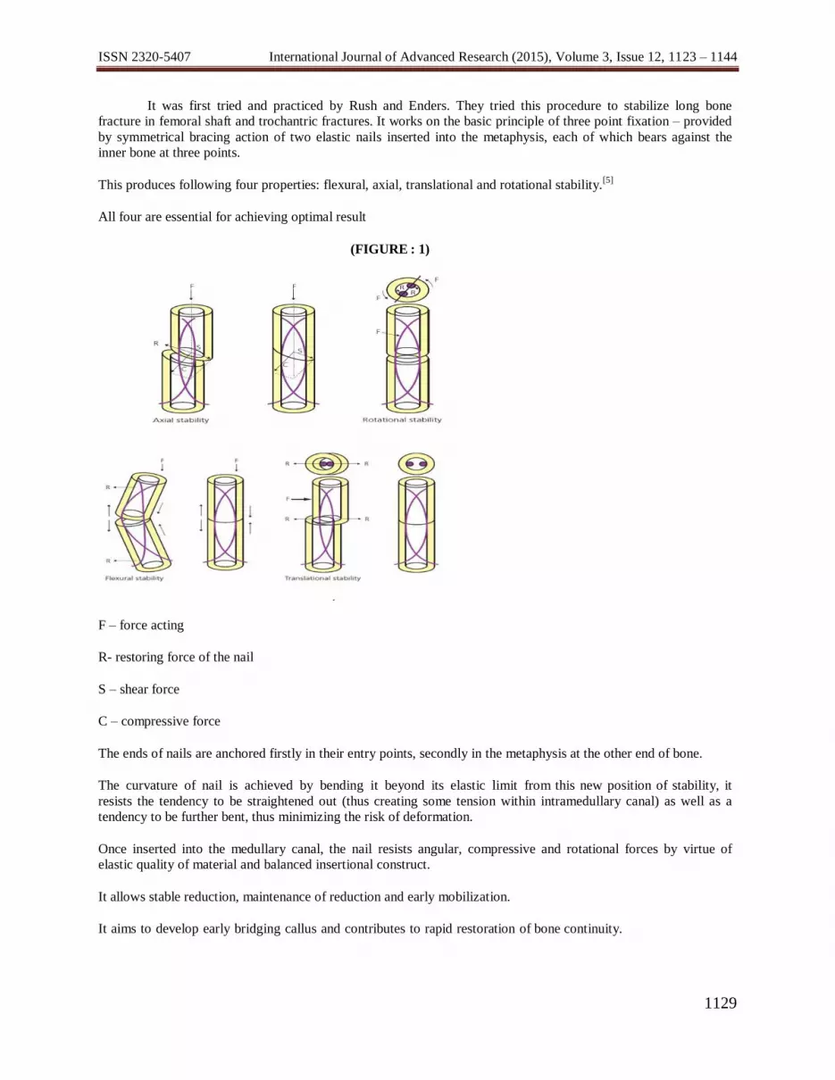
It was first tried and practiced by Rush and Enders. They tried this procedure to stabilize long bone
fracture in femoral shaft and trochantric fractures. It works on the basic principle of three point fixation – provided
by symmetrical bracing action of two elastic nails inserted into the metaphysis, each of which bears against the
inner bone at three points.
This produces following four properties: flexural, axial, translational and rotational stability.[5]
All four are essential for achieving optimal result
(FIGURE : 1)
F – force acting
R- restoring force of the nail
S – shear force
C – compressive force
The ends of nails are anchored firstly in their entry points, secondly in the metaphysis at the other end of bone.
The curvature of nail is achieved by bending it beyond its elastic limit from this new position of stability, it
resists the tendency to be straightened out (thus creating some tension within intramedullary canal) as well as a
tendency to be further bent, thus minimizing the risk of deformation.
Once inserted into the medullary canal, the nail resists angular, compressive and rotational forces by virtue of
elastic quality of material and balanced insertional construct.
It allows stable reduction, maintenance of reduction and early mobilization.
It aims to develop early bridging callus and contributes to rapid restoration of bone continuity.

ISSN 2320-5407 International Journal of Advanced Research (2015), Volume 3, Issue 12, 1123 – 1144
1130
It has advantage over other surgical methods particularly in this age group because it is simple , is a load sharing
internal splint that doesn’t violate open physis, allows early mobilization and maintains alignment.[6]
Micro-motion provided by the elasticity of the fixation promotes faster external bridging callus formation.
The periosteum is not disturbed and being a closed procedure there is no disturbance of fracture hematoma, there
by less risk of infection.[6]
All the current available elastic nails have tapered ends to allow satisfactory sliding down on insertion along inner
surface of diaphysis without impacting on opposite cortex.
Only correct tensioning of nail can fulfill the dynamic principles of this method. It is based on circular muscle
mantle and restoring force of pre-stressed nails, which repeatedly bring the fragment back into anatomical
position.When fracture of the distal part of diaphysis is to be fixed with ender’s nail, the entry points must be at
opposite ends of bone.
The incorrect insertion points can have various negative effects like internal tension and imbalance of fracture
stability and fixation.
Entry points that are too diaphyseal, damage the musculature during insertion and removal. The nails that are left
too long cause severe muscle and skin irritation and breakdown. Injury to perichondral ring and growth plates
may occur at time and formation of entry point as well as nail insertion and may lead to growth arrest.
Always 2 nails of same thickness should be used, to avoid valgus and varus or axial deformity which may be due to
different restoring force.
Difficulties with fracture reduction as well as advancing the 2nd
nail may require the surgeon to rotate the
nail more than 180 degree. This may lead to one nail being turned around the other- the corkscrew phenomenon. In
such cases it is rotationally and axially unstable.
OBSERVATIONS AND RESULTS
Study design: An outcome of surgical study with 50 patients with Diaphyseal fractures is undertaken to study the
outcome of ender’s nails fixation in Lower limb.
Table 5: Age distribution of patients studied
Age in years Number of
patients
%
5-8 17 34%
9-12 22 44%
13-16 11 22%
Total 50 100.0
Out of 50 patients, there were 34 (68%) male patients and 16 (32%) female patients in our study.

ISSN 2320-5407 International Journal of Advanced Research (2015), Volume 3, Issue 12, 1123 – 1144
1131
Patients with shaft femur and tibia fracture had history of either RTA or fall down, with more chance of history fall
down (56%) than RTA(44%).
In our study , we have found 30 patients with shaft femur fracture and 20 patients with shaft tibia fracture.
In our study, we have found that 64% patients have right side affection of limb and 36% patients have left side
affection of limb.
Our study shows that there are more patients of transverse fracture than other variety , being transverse fracture 56
% , oblique fracture 32% and spiral fracture 22%.
Figure: 2: Level of fracture
There are 24 % patients having proximal 1/3rd
fracture, 54% patients having middle 1/3rd
fracture and 22%
patients having distal 1/3rd
fracture in our study.
In our study, we have found that most of the patients(68%) were operated within 2 days of trauma, 30% patients
were operated within 2-4 days and 2% patients were operated in 5 or more days.
In our study, we have found that most of the patients (54%)
were discharged within 4 days, 32% patients were discharged in 5-8 days and only 14% patients need hospital stay
more than 8 days.
Table 6: Time for union on radiological finding
0
10
20
30
40
50
60
Upper Middle Lower
percentage
percentage
Time for union
Number of
patients
%

ISSN 2320-5407 International Journal of Advanced Research (2015), Volume 3, Issue 12, 1123 – 1144
1132
In our study, most of the patients (80%) achieves union of fractures on radiological finding within 12 weeks.
Table 7: Range of movements at 24 weeks
Range of
movements(degrees) Number of patients %
Full range 48 96
Mild restriction 2 4
Moderate restriction 0 0
Severe restriction 0 0
Total 50 100
There were 80% patients started full weight bearing within 12 weeks and only 20% started full weight bearing
between 12 -24 weeks.
Table 8 a: Complications
Minor Major Nil Total
No.of Patients 12 0 38 50
Percentage 24 0 76 100
Table 8.B. Complications
< / = 12 weeks 40 80.0
>12 – 18 weeks 8 16
>18 – 24 weeks 2 4
Total 50 100.0

ISSN 2320-5407 International Journal of Advanced Research (2015), Volume 3, Issue 12, 1123 – 1144
1133
Complications No. of cases Percentage
1) Pain 8 16
2) Infection 1 2
2.1) Superficial
2.2) Deep - -
3) Inflammatory reaction - -
4) Delayed union and non union - -
5) Limb lengthening
-
- 5.1) < 2 cm
5.2) > 2 cm
6) Limb shortening 1 2
6.1) < 2 cm
6.2) > 2 cm
7) Nail back out - -
8) Mal alignment 1 2
8.1) Varus angulation
8.2) Valgus angulation - -
8.3) Anterior angulation - -
8.4) Posterior angulation - -
8.5) Rotational malalignment - -
9) Bursa at the tip of the nail - -
10) Sinking of the nail into the medullary cavity 1 2
Table 9: Outcome (according to Flynn’s criteria)
Outcome
Number of
patients
(n=50)
%

ISSN 2320-5407 International Journal of Advanced Research (2015), Volume 3, Issue 12, 1123 – 1144
1134
Excellent 47 94.0
Satisfactory 3 6
Poor 0 0.0
In our study, 96% patients have excellent result and only 4% patients have satisfactory result and no patients have
poor result.
Table 10: Outcome for additional variables in the present study
OUTCOME EXCELLENT
(%)
SATISFACTORY (%) POOR
(%)
VARIABLES
Range of movements 96 4 -
Time for union 80 16 4
Unsupported weight bearing 80 16 4
Table 11: Association of Incidence of complications with clinical variables studied
Clinical variables
Total number
of patients
(n=50)
Complications(Minor)
P value Absent
(n=38)
Present
(n=12)
Age in years
• 5-8 17(34%) 13(34.2%) 4(33.3%)
0.961 • 9-12 22(44%) 17(44.7%) 5(41.7%)
• 13-16 11(22%) 8(21%) 3(25%)
Gender
• Male 34(68%) 28(73.7%) 6(50%)
0.238 • Female 16(32%) 10(26.3%) 6(50%)
Mode of Injury

ISSN 2320-5407 International Journal of Advanced Research (2015), Volume 3, Issue 12, 1123 – 1144
1135
• RTA 22(44%) 19(50%) 3(25%) 0.235
• Fall down 28(56%) 19(50%) 9(75%)
Bone affected
• Femur 30(60%) 21(55.26%) 9(75%) 0.379
• Tibia 20(40%) 17(44.73%) 3(25%)
Pattern of fracture
• Transverse 23(46%) 21(55.26%) 2(16.7%) 0.08
• Oblique 16(32%) 13(34.21%) 3(25%)
• Spiral 11(22%) 4(10.5%) 7(58.3%)
Time interval between
trauma & surgery
• < 2days 34(68%) 23(60.52%) 11(91.7%) 0.230
• 3-4 days 15(30%) 14(36.84%) 1(8.3%)
• 5-7 days 1(2%) 1(2.63%) 0(0%)
There was no significant association observed between clinical variables (Age, Gender, Mode of
injury, Bone affected, Pattern of Fracture and Time Interval between trauma and surgery) and Incidence of
complications.
DISCUSSION
Age incidence:
In the present study 17(34.0%) of the patients were 5-8 years, 22 (44.0%)were 9 to 12 years and 11(22.0%) were
13 to 16 years age group with the average age being 10.16 years.
J.M. Flynn et al studied children ranged from 3-16 years with a mean of 10.3 years.
Wudbhav N Sankar et al
studied children ranged from 7.2-16 years with a mean of 12.2 years.[4]
William G. Cole et al study of plaster casting in fracture femur mean age is 5 yr.
Sex incidence:
There were 16(32%) girls and 34 (68%) boys in the present study. The sex incidence is comparable to other
studies in the literature.
In their study Singh and Kumar et al. out of 112 cases, had 77 (69%)
Male patient and 35 (31%) female patients.
William G. Cole et al studied 191 patients with 113(68.58%) male and 58(30.36%) girls.
Mode of Injury:

ISSN 2320-5407 International Journal of Advanced Research (2015), Volume 3, Issue 12, 1123 – 1144
1136
In the present study RTA was the most common mode of injury accounting for
22 (44.0%) cases and fall down accounted for 28 (56%) of the cases.
J. M. Flynn et. al, in their study assessing 234 cases, 136(58.1%) were following RTAs, 46(19.6%) were
following self fall and remaining 43(28.8%) were as a result of fall from height.[2]
William G. Cole et al, in their study have 38(21.1%) patients with history of RTA and 131(72.7%) patients with
history of fall down.
Bone affected
We studied 30(60.0%) femoral and 20(40%) tibial fractures.
In their study, Gamal El-Adl et al. had 48 (65.7%) femoral and 25 (34.3%) tibial fractures.[8]
Pattern of Facture:
In our study, transverse fractures accounted for 23(46.0%) cases, oblique fractures - 16(32.0%), spiral
fractures – 11(22.0%) and there were no comminuted and segmental fractures.
In their study J. M. Flynn et al. out of 2 2 9 femoral fractures studied 1 1 4 (48.7%) were
transverse fractures, communited fractures- 28 (12%), oblique fractures - 47(20%), spiral fractures –
29(12%) and 14 (6%) were butterfly fractures.
Wudbhav N. Sankar studied 19 tibial shaft fractures out of which 9 (47.3%) were transverse, 7 (36.8%) were
oblique, 2 (10.5%) were spiral and 1 (5.2%) was communited.[4]
William G. Cole et al studied 191 patients with femoral shaft fracture, in which 40(20.9%) had transverse fracture,
113(59.13%) had spiral fracture and 38(19.9%) patients had oblique fracture.
Table 12 : Pattern of fracture (comparison of our study with other)
STUDIES
PATTERN OF FRACTURE (%)
Transverse Communited Oblique Spiral Segmental
Pesent study 46.0 - 32.0 22.0 -
J. M. Flynn et al. 48.7 12 20 12 -
Wudbhav N.
Sankar et al.
47.3 5.2 36.8 10.5 -
Level of Fracture:

ISSN 2320-5407 International Journal of Advanced Research (2015), Volume 3, Issue 12, 1123 – 1144
1137
Fractures involving the middle 1/3rd accounted for 27 (54.0%) cases, proximal 1/3rd
– 12 (24.0%) and distal
1/3rd
– 11 (22.0%) of cases in our study.
In their study J. M. Flynn et al among 233 femoral shaft fractures, 33 fractures were in the proximal 1/3rd
,
165 in the middle 1/3rd
and 35 were in the distal 1/3rd
.
Wudbhav N. Sankar studied 19 tibial shaft
fractures out of which 15 were middle 1/3rd
, 2 – proximal 1/3rd
and 2 were distal 1/3rd
.[4]
Time interval between trauma and surgery:
In the present series, 34 (68.0%) patients underwent surgery within 2 days after trauma, 15(30.0%) in 3 – 4
days, 1(2.0%) in 5 and more day.– She was operated 8 days after trauma (admission) as she has history of head
injury .
Average duration between trauma and surgery was 1.86 days in the study Gamal el adl operated 56.1% of cases
between 3-4 days after injury, 21.2% cases between 3 -4 days and 22.7% cases after 7 days.[8]
William G. Cole et al in their casting study, 74.34% patients were applied cast on day 1, 18.9% patients were
applied cast on day 2 and remaining 6.8% patients were applied cast after 2 days.
Duration of stay in the hospital:
The duration of stay in the hospital ≤ 4 days for 27 (54.0%) patients, 5-7 days for 16 (32.0%) and 8 or more days
for 7 (14.0 %) patients.
Among the 7 patients who stayed for more than 8 days, had mostly problem regarding head injury and open
fracture in whom we were stayed few days for operation to prevent postoperative infection.
The average duration of hospital stay in the present study is 5.18 days. The mean hospital stay was 5.7 days in
Gamal El-Adl et al. study.[8]
Most of the patients about 86% had hospital stay of <4 days and remaining 14% patients had hospital stay >4 days
in William G. Cole et al. study.
Table 13 : Duration of hospital stay (comparison between our study and other)
STUDIES AVERAGE DURATION OF STAY IN HOSPITAL
(in days)
Present study 5.18
Gamal El-Adl et al.[8]
5.7

ISSN 2320-5407 International Journal of Advanced Research (2015), Volume 3, Issue 12, 1123 – 1144
1138
Compared to the above study conducted, average duration of hospital stay was less in our study i.e. 5.18 days. The
reduced hospital stay in our series is because of proper selection of Patients, stable fixation and less incidence of
complications.
Time for union:
In our study union was achieved in <3 months in 40 (80%) of the patients and 3 – 4.5 months in 10 (20%). Average
time to union was 11.9 weeks.
In study of Gamal El-Adl et al. average time to union was 85 days (12weeks), with range between 42 to 140 days
for flexible intramedullary nailing.
Table 14 :Time of union (comparison between our study and other)
STUDIES TIME OF UNION (in weeks)
Present study 11.9
Gamal El-Adl et al.[8]
12
In our study, closed reduction of the fracture, leading to preservation of fracture hematoma, improved
biomechanical stability and minimal soft tissue dissection led to rapid union of the fracture compared to
compression plate fixation.
Table 15 :Average time of full weight bearing (comparison between our study and other)
STUDIES
AVERAGE TIME OF FULL
WEIGHT BEARING
(in weeks)
Present study 12.6
Wudbhav N.Sankar.[4]
8.65
Table 16 :Complication (comparison between our study and other)
COMPLICATIONS PRESENT
STUDY
(% incidence)
PREVIOUS STUDIES (%
incidence)

ISSN 2320-5407 International Journal of Advanced Research (2015), Volume 3, Issue 12, 1123 – 1144
1139
Pain at the site of nail insertion 16 16.2 J.M.Flynn et al. [1],[2]
Superficial infection 2.0 1.7 J.M.Flynn et al. [1],[2]
Range of motion 4.0 0.9 J.M.Flynn et al. [1],[2]
LIMB LENGTH
DISCREPANCY (minor)
Lengthening - 0
.
0
Wudbhav[4]
[ ] [ ] [ ]
N.Sankar
Shortening 2.0 0
.
5
Wudbhav[4]
4[4]
N.Sankar
MALALLIGNMENT(minor)
Varus / Valgus 2.0 -
.
3
J.M.Flynn et al[1]
Anteroposterior - - Wudbhav[4]
[ ]
N.Sankar
Rotational deformities - -
.
2
Gammal et Al [8]
Table 17 : Assesment of Outcome (comparison between our study and other)
STUDIES
OUTCOME
EXCELLENT
(%)
SATISFACTORY
(%)
POOR
(%)
Present study 94.0 6.0 -
Gamal El Adl et al.[8]
75.8 24.2 -
J.M.Flynn et al.[1]
65 25 10
Wudbhav N.Sankar[4]
63.15 31.57 5.26
SUMMARY
Thirty patients with diaphyseal fractures of the femur (30) and twenty patients with fracture tibia (20) were treated
with ender’s nailing between September 2010 to September 2014 in tertiary center at government setup.
Children and adolescents aged between 5 to 16 years were included in the study. 34.0% of patients were between
5-8 years, 44.0% of patients in between 9-12 years and 22.0% of patients in between 13 to 16 years age group with
the average age being 10.16 years. 68% of the patients were boys. Fall down was the most common
mode of injury accounting for 28 (56.0%) cases followed by RTA - 22 (44.0%)[9]

ISSN 2320-5407 International Journal of Advanced Research (2015), Volume 3, Issue 12, 1123 – 1144
1140
Transverse fractures accounted for 23(46.0%) cases, oblique fractures 16(32.0%) and spiral fractures –
11(22.0%).
Fractures involving the middle 1/3rd accounted for 27 (54.0%) cases. All the patients were prepared and
operated as early as possible once the general condition was stable and the patient was fit for surgery.
The average duration between trauma and surgery was 1.86 days with 34 (68%.0) patients undergoing surgery
within 2 days and between 3 to 7 days (32.0%).
43 (86.0%) cases were immobilized (with long leg cast for femur fracture / above knee POP cast for tibia fracture)
postoperatively for 6-8 weeks and such immobilization was for 9 weeks in rest of the 7 (14.0%) of the cases.
Average duration of stay in hospital was 5.18 days.
Union was achieved in <3 months in 40 (80%) of the patients with average time to union being 11.9 weeks.
Unsupported full weight bearing walking was started in < 3 months for 40 (80%) of the patients.
All patients had full range of hip and ankle motion in the present study and 2 (6.66%) patients had mild restriction
in knee flexion at 12 weeks
8(16.0%) had developed pain at site of nail insertion during follow up evaluation, all of which resolved by the end
of 12 weeks follow up.
Superficial infection was seen in 1(2.0%) case. 1(2.0%) patient had shortening
No patient in our study had major limb length discrepancy (i.e. > ± 2cm). Nail back out was not seen in any of the
cases. 1(2.0%) patient presented with varus(40
) angulation, no patient presented with valgus angulation and no
patients had antero-posterior angulation or rotational mal-alignment.[10]
The development of the Ender’s nail fixation method has put an end to criticism of the surgical treatment of
paediatric long bone fractures, as it avoids any growth disturbance by preserving the epiphyseal growth plate, it
avoids bone damage or weakening through the elasticity of the construct, which provides a load sharing,
biocompatible internal splint, and finally it entails a minimal risk of bone infection.[11]
CONCLUSION
Based on our experience and results, we conclude that ENDER’S NAILING technique is an ideal method for
treatment of pediatric femoral and tibial diaphyseal fractures. It gives elastic mobility promoting rapid union at
fractures site and stability which is ideal for early mobilization. It gives lower complication rate, good outcome
when compared with other methods of treatment.
It is a simple, easy, rapid, reliable and effective method for management of paediatric femoral and tibial fractures
between the age of 5 to 16 years, with shorter operative time, lesser blood loss, lesser radiation exposure, shorter
hospital stay, and reasonable time to bone healing.
Because of early weight bearing, rapid healing and minimal disturbance of bone growth, Ender’s nail may be
considered to be a physiological method of treatment.
Use of Ender’s nails for definitive stabilization of femoral and tibial shaft fractures in children is a reliable,
minimally invasive, and physeal-protective treatment method. Our study results provide new evidence that expands
the inclusion criteria for this treatment and shows that Ender’s nails can be successfully used regardless of
fracture location and fracture pattern.

ISSN 2320-5407 International Journal of Advanced Research (2015), Volume 3, Issue 12, 1123 – 1144
1141
BIBLIOGRAPHY
1. Flynn JM, Skaggs DL, Sponseller PD, Ganley TJ, Kay RM, Kellie Leitch KK. The operative
management of pediatric fractures of the lower extremity. J Bone Joint Surg Am 2002;84:288-300.
2. Flynn JM, Hresko T, Reynolds RA, Blaiser RD, Davidson R, Kasser J. Titanium elastic nails for
pediatric femur fractures - a multicenter studyof early results with analysis of complications. J Pediatr
Orthop 2001;21(1):4-8.
3. Robert W.Bucholz, James D.Heckman, Charles Court –Brown. Rockwood and green’s fractures in
children. (2006) 6:22 (896-934)
4. Wudbhav N. Sankar, Kristofer J. Jones, B. David Horn, and Lawrence Wells. Titanium elastic
nails for pediatric tibial shaft fractures. J Child Orthop 2007 November; 1(5):281-286
5. S.Terry Canale, James H.Beaty. Campbell’s operative Orthopaedics. (2007) 11:23 (1651-1666)
6. KC Saikia, SK Bhuyan, TD Bhattacharya, SP Saikia. Titanium elastic nailing in femoral
diaphyseal fractures of children in 6-16 years of age. Indian J Orthop 2007; 41:381-385.
7. Mann DC, Weddington J. and Davenport K. "Closed elastic nailing of femoral shaft fractures in
adolescents". J Pediatr Orthop 1986; 6 (6): 651-5.
8. Gamal El-Adl, Mohamed F. Mostafa, Mohamed A. Khalil, Ahmed Enan. Titanium elastic nail
fixation for paediatric femoral and tibial fractures. Acta Orthop. Belg 2009; 75: 512-520
9. Heybel1y M, Muratli HH, Çeleb L, Gülηek S, Biηimoglu A. The results of intramedullary fixation with
titanium elastic nails in children with femoral fractures. Acta Orthop Traumatol Turc 2004;38:178-187.
10. Moroz L, Launay F, Kocher MS, Newton PO, Frick SL, Sponseller PD, Flynn JM. Titanium elastic
nailing of fractures of the femur in children: predictors of complications and poor outcome. J
Bone Joint Surg Br 2006; 88:1361-1366
11. Ligier JN, Metaizeau JP, Prevot J, Lascombes P. Elastic stable intramedullary nailing of femoral shaft
fractures in children. J Bone Joint Surg [Br] 1988;70-B:74-7.
RADIOLOGICAL & CLINICAL PHOTOGRAPHS
(FIGURE : 2 )
CASE : FRACTURE FEMUR
PRE – OP

ISSN 2320-5407 International Journal of Advanced Research (2015), Volume 3, Issue 12, 1123 – 1144
1142
POST
FOLLOW UP : 12 weeks
CLINICAL PHOTOS
CASE : FRACTURE TIBIA (FIGURE : 3 )

ISSN 2320-5407 International Journal of Advanced Research (2015), Volume 3, Issue 12, 1123 – 1144
1143
PRE - OP
FOLLOW UP : 12 Weeks
KNEE FLEXION KNEE EXTENSION
ANKLE DORSI FLEXION ANKLE PLANTAR FLEXION

ISSN 2320-5407 International Journal of Advanced Research (2015), Volume 3, Issue 12, 1123 – 1144
1144
SQUATING
CLINICAL PHOTOS