response of atrial fibrillation to pulmonary vein antrum isolation is directly related to resumption...
TRANSCRIPT
Response of Atrial Fibrillation to Pulmonary Vein AntrumIsolation Is Directly Related to Resumption and Delay of
Pulmonary Vein ConductionAtul Verma, MD; Fethi Kilicaslan, MD; Ennio Pisano, MD; Nassir F. Marrouche, MD;
Raffaele Fanelli, MD; Johannes Brachmann, MD; Jens Geunther, MD; Domenico Potenza, MD;David O. Martin, MD, MPH; Jennifer Cummings, MD; J. David Burkhardt, MD; Walid Saliba, MD;
Robert A. Schweikert, MD; Andrea Natale, MD
Background—The role of pulmonary vein (PV) isolation in ablative treatment of atrial fibrillation (AF) has been debatedin conflicting reports. We sought to compare PV conduction in patients who had no AF recurrence (group I), patientswho could maintain sinus rhythm on antiarrhythmic medication (group II), and patients who had recurrent AF despiteantiarrhythmic medication (group III) after PV antrum isolation (PVAI).
Methods and Results—PV conduction was examined in consecutive patients undergoing second PVAI for AF recurrence.We also recruited some patients cured of AF to undergo a repeat, limited electrophysiological study at �3 months afterPVAI. All patients underwent PVAI with an intracardiac echocardiography (ICE)–guided approach with completeisolation of all 4 PV antra (PVA). The number of PVs with recurrent conduction and the shortest atrial to PV (A-PV)conduction delay was measured with the use of consistent Lasso positions defined by ICE. Late AF recurrence wasdefined as AF �2 months after PVAI with the patient off medications. Patients in groups I (n�26), II (n�37), and III(n�44) did not differ at baseline (38% permanent AF; ejection fraction 53�6%). Recurrence of PV–left atrial (LA)conduction was seen in 1.7�0.8 and 2.2�0.8 PVAs for groups II and III but only in 0.2�0.4 for group I (P�0.02). Inpatients with recurrent PV-LA conduction, the A-PV delay increased from the first to second procedure by 69�47% forgroup III, 267�110% for group II, and 473�71% for group I (P�0.001). When pacing was at a faster rate, A-PV blockdeveloped in all 5 of the group I patients with recurrent PV-LA conduction.
Conclusions—The majority of patients with drug-free cure show no PV-LA conduction recurrence. Substantial A-PV delayis seen in patients able to maintain sinus rhythm on antiarrhythmic medication or cured of AF compared with patientswho fail PVAI. (Circulation. 2005;112:627-635.)
Key Words: ablation � atrial fibrillation � pulmonary veins � recurrence
It is accepted that atrial fibrillation (AF) is frequentlyinitiated by triggering foci located within the pulmonary
vein (PV) antra (PVA).1 Techniques of ablating AF havetherefore targeted radiofrequency lesions at the interfacebetween the left atrium (LA) and the PVA.2 However, thepathophysiology of AF can be quite complex. Triggering focifor AF initiation may occur from non-PV locations in aminority of patients.3 Furthermore, formation of stable rotorsat the interface between the PVA and the posterior LA wallmay be critical to AF perpetuation.4 Therefore, whereas someapproaches have emphasized the necessity of electricallyisolating the PVs for procedural success,2,5,6 others have usedan empirical anatomic approach targeting the “substrate” forAF maintenance without seeking to achieve PV isolation.7
Our group, in particular, has always targeted electric isolationof all 4 PVA using a technique guided by intracardiacechocardiography (ICE).8
The importance of PV isolation to procedural outcome isnot well known, and reports published to date have hadconflicting conclusions.9–13 However, if electric isolation ofthe PVA is not critical to procedural outcome, there should beno relationship between PV conduction and the status of thepatient’s AF after ablation. In this study we examined PVconduction in patients who had previously undergone PVantrum isolation (PVAI) using an ICE-guided technique. Wesought to characterize and compare PV conduction in patientswho (1) had no AF recurrence, (2) could maintain sinusrhythm using antiarrhythmic medication ineffective before
Received January 3, 2005; revision received April 14, 2005; accepted April 20, 2005.From the Section of Cardiovascular Electrophysiology, Department of Cardiology, Cleveland Clinic Foundation, Cleveland, Ohio (A.V., F.K., N.F.M.,
D.O.M., J.C., J.D.B., W.S., R.A.S., A.N.); Department of Cardiology, Casa Sollivero della Soffrenza, S. Giovanni Rotondo, Italy (E.P., R.F., D.P.); andDepartment of Cardiology, Klinikum Coburg, Coburg, Germany (J.B., J.G.).
Correspondence to Andrea Natale, MD, Section of Pacing and Electrophysiology, Center for Atrial Fibrillation, Cleveland Clinic Foundation, Desk F15, 9500 Euclid Ave, Cleveland, OH 44195. E-mail [email protected]
© 2005 American Heart Association, Inc.
Circulation is available at http://www.circulationaha.org DOI: 10.1161/CIRCULATIONAHA.104.533190
627
Arrhythmia/Electrophysiology
by guest on December 16, 2014http://circ.ahajournals.org/Downloaded from
ablation, and (3) had recurrent AF not controlled by antiar-rhythmic medication.
MethodsStudy PopulationWe examined all patients undergoing second PVAI procedures forAF recurrence from September 2003 until March 2004. All patientshad undergone a first PVAI procedure within the preceding 6months. Recurrence was defined as AF occurring beyond 2 monthsafter PVAI. Patients were initially selected for PVAI because ofsymptomatic AF, which was paroxysmal, persistent, or permanentand refractory to �2 antiarrhythmic medications. Patients withpreexisting extensive LA scar detected at their initial procedure wereexcluded from this study because of previous data showing theunique altered substrate in this small subset of patients (representingnot more than 6% of our total AF ablation population).14 Extensivescarring was defined exactly according to the previously publishedcriteria of absence of atrial electrogram seen in all 10 poles of adecapolar circular mapping catheter in at least 3 distinct positions inthe LA.14
Over this same time period, we recruited patients who were curedof AF after a first PVAI procedure to undergo a repeat, limitedelectrophysiological study to assess conduction within the PVA.These patients all underwent their repeat procedure at least 6 monthsafter initial PVAI; again, patients with preexisting LA scar wereexcluded for the aforementioned reasons.
Patients were recruited from 3 participating institutions. Of the107 patients studied, 65 were from the Cleveland Clinic, 25 fromCasa Sollievo della Sofferenza (S. Giovanni Rotondo, Italy), and 17from Klinikum Coburg (Coburg, Germany). Patients from all 3institutions underwent the identical procedural protocol describedbelow. All patients had to sign a written informed consent beforeundergoing both initial and repeat electrophysiological and ablationprocedures. The collection of these data was performed in accor-dance with the ethical guidelines of each institution’s review board.
PVAI ProcedureAll patients underwent PVAI with the use of an ICE-guidedtechnique, which is summarized here but described in extensivedetail elsewhere.8 After femoral venous access was obtained, a 10F64-element phased-array ultrasound imaging catheter (Siemens AG)was positioned in the right atrium. A 14-pole recording catheter (TZMedical) was placed in the coronary sinus via right internal jugularaccess. Under ICE guidance, a decapolar circular (Lasso) mappingcatheter and an 8-mm-tip ablation catheter (Biosense Webster) wereadvanced into the LA via 2 transseptal punctures. The patient wassystemically anticoagulated with intravenous heparin to maintain anactivated clotting time (ACT) of 350 to 400 seconds. Before the firsttransseptal puncture, a 140-IU/kg heparin bolus was given, and aninfusion of 15 IU/kg/h was started. An additional 70 IU/kg bolus wasgiven before the second transseptal puncture. The heparin infusionwas then titrated to maintain the target ACT. ICE was used to definethe PVA and guide sequential placement of the Lasso catheter in allpositions surrounding (and outside of) each PV. Radiofrequency(RF) ablation was performed wherever PV potentials were recordedaround the PVA. RF energy output was limited to a maximum of 70W and 55 coulombs and was titrated according to microbubbleformation detected by ICE. The end point of ablation was completeelectric disconnection of the PVA from the LA. This was consideredto be achieved when no PV potentials could be recorded along theantrum or inside the vein by the Lasso catheter during sinus rhythmor coronary sinus pacing. At the end of the procedure, all 4 PVAwere extensively remapped with the Lasso catheter to check for anypersisting PV potentials, and, if necessary, further ablation wasperformed to eliminate these. All 4 PVs and the superior vena cava(SVC) were isolated in every patient. The SVC was isolated byplacing the Lasso catheter at the SVC–right atrial junction defined byICE. High-output pacing from the ablation catheter was used tolocalize the phrenic nerve to avoid phrenic injury during ablation.
During the baseline ablation, we did not routinely look for non-PVfoci apart from routinely isolating the SVC. No ablation linesbetween the mitral annulus and PVs were performed.
Repeat Electrophysiological StudyFor patients cured of AF but agreeing to undergo repeat electrophys-iological study, a limited procedure was performed compared withthe original PVAI. As described for the PVAI procedure, the ICEimaging catheter was advanced via femoral venous access into theright atrium, and a 14-pole catheter was placed in the coronary sinus.A single transseptal puncture was performed under direct ICEvisualization to minimize the risk of transseptal access (we have hadno complications related to transseptal access in our last 1000procedures). The patient was heparinized to maintain an ACT of 350to 400 seconds. With the use of a single 8F sheath to cross theseptum, a decapolar Lasso catheter was advanced into the LA andused to map all 4 PVA with and without pacing from the coronarysinus.
In all 3 groups, during the second ablation/electrophysiologicalprocedure, intravenous isoproterenol infusion (up to 20 �g/min) andboluses of adenosine were given to encourage PV or non-PV triggerfiring and to assess whether they indeed led to AF initiation.
During both initial and repeat procedures, we determined the atrialto PV interval (A-PV). To ensure consistency in the A-PV measure-ment, we positioned the Lasso catheter at the end of the tubularsegment of each PV as defined by ICE. Using this ICE definition, wecould ensure that measurements of the A-PV interval were beingperformed at uniform locations. For the left and right-sided veins,A-PV was defined as the time interval from the onset of the localatrial potential to the earliest or latest PV potential recorded by anyof the 10 Lasso catheter electrodes. For consistency, the shortestA-PV interval recorded by any of the 10 electrodes was taken as theA-PV interval for that particular vein.
If non-PV triggers for AF were identified during the study,3-dimensional electroanatomic mapping with the use of the CARTOsystem (Biosense Webster) was used to assist in mapping andablation of the focus. A subset of patients had complete voltagemapping of the LA performed at their repeat procedure with the useof CARTO as part of another study protocol. For these voltage maps,scar was defined as a bipolar voltage of �0.5 mV indistinguishablefrom noise.15 The CARTO system is able to measure the distancebetween any 2 points. With the assumption that a scarred segment ofLA could be divided into multiple smaller rectangular or trapezoidalshapes, the area of the segment could be approximated by summingthe areas of the smaller shapes. This segment was then expressed asa percentage of the total approximated LA surface area (assessed bythe same technique) excluding the tubular portion of the PVs. Asimilar technique has been previously reported for mapping the leftventricle.16
Follow-UpAfter initial PVAI, patients continued anticoagulation with warfarinto maintain an international normalized ratio of 2.0 to 3.0 for aminimum of 4 months. In all patients, antiarrhythmic medicationswere continued for 2 months after ablation and were chosen from oneof sotalol, propafenone, flecainide, or dofetilide. Amiodarone wasnot used after ablation and was discontinued 4 to 5 months before theinitial ablation procedure. Antiarrhythmic medications were discon-tinued in all patients after 2 months. Patients had spiral CT scans 3months after ablation to assess PV stenosis.
Late recurrence of AF was defined as AF occurring beyond 2months after PVAI. Thus, success was defined as a lack of late AFrecurrence with the patient off antiarrhythmic medication. If late AFrecurrence occurred, antiarrhythmic medications were restarted todetermine whether sinus rhythm could be maintained, but patientswere still offered a second ablation procedure to achieve a true cure(sinus rhythm without the use of antiarrhythmic medication). Todetermine recurrence, all patients wore rhythm transmitters for aminimum of 3 months after PVAI, and this was extended by another3 months for patients with early AF recurrences. Patients were askedto record events any time they experienced symptoms and to
628 Circulation August 2, 2005
by guest on December 16, 2014http://circ.ahajournals.org/Downloaded from
routinely send 1 to 3 routine transmissions per day even in theabsence of symptoms. Patients had scheduled clinical visits, 12-leadECG, and 48-hour Holter monitoring at 3, 6, and 12 months afterablation. Interrogation of implanted devices was also used (whenavailable) to confirm arrhythmia recurrence. Patients were alsoadvised to report any recurrence of symptoms to the clinic, at whichpoint 48-hour Holter monitoring was performed. Recurrences werebased on patient reporting, rhythm transmitter, Holter, and/or ECGdata.
Patients were categorized into 3 groups according to late AFrecurrence. Group I included patients who had no recurrence afterinitial PVAI. Group II consisted of patients who experienced late AFrecurrence after initial PVAI but could maintain sinus rhythm onantiarrhythmic medication. Group III consisted of patients who hadlate AF recurrence and remained unresponsive to antiarrhythmicmedication.
Statistical AnalysisAll data are reported as mean�SD for continuous variables and aspercentage values for categorical variables unless otherwise indi-cated. Comparison of continuous variables among the 3 groups wasmade with the use of 1-way ANOVA. If the ANOVA analysis wassignificant, multiple comparisons between subgroups were per-formed with the Tukey procedure. Comparison of continuous vari-ables between 2 groups was made with the independent samplesStudent t test. Categorical variables were compared with �2 analysis.A-PV intervals are given for the individual PVs as well as the overallpercent change in A-PV interval from the first to second procedurefor all PVs. Overall percent change was compared between groupswith ANOVA and t test analysis (as above). A probability cutoff of�0.05 was considered significant for all statistical determinations.
Results
Patient CharacteristicsBaseline patient characteristics in each of groups I, II, and IIIare summarized in Table 1. There were no significantdifferences in patient characteristics among the 3 groups. Thedistribution of patients with paroxysmal, persistent, andpermanent AF was similar in all 3 groups. Importantly, thetotal amount of RF delivery time at the initial PVAI proce-
dure was not different in the 3 groups, suggesting that theamount of ablation performed was similar in the 3 groups.The proportions of patients with structural heart disease, LAsize, and ejection fraction were also similar in all 3 groups.
Number of PVA With Recurrent ConductionAccording to our technique, all 4 PVA were isolated for allpatients during the initial ablation procedure. Recurrence ofPV-LA conduction was seen in a larger number of PVA inpatients who experienced AF recurrence after PVAI com-pared with patients who did not. Recurrence was seen in1.7�0.8 PVA for group II patients and 2.2�0.8 PVA forgroup III patients. This was significantly higher than the0.2�0.4 PVA showing recurrence in group I patients(P�0.021).
The number of patients experiencing 1, 2, 3, and 4 PVArecurrences is detailed in Table 2. In groups II and III, 95%and 100% of patients had recurrence of conduction seen in atleast 1 PVA, respectively. Recurrence in only 1 culprit PVAwas sufficient to cause AF recurrence in 12 of 37 patients(32%) in group II and 6 of 44 patients (14%) in group III. Themajority of patients in group I (81%) did not show recurrenceof PV-LA conduction in any of the PVA.
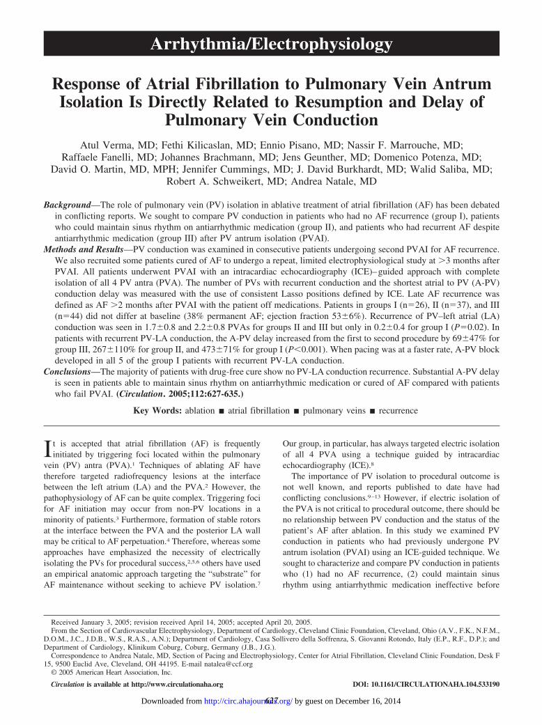
Two patients (5%) in group II did not show recurrentconduction in any of the PVs. In both of these cases, focaltriggers for AF were found in non-PV sites within the LA. In1 patient, the site was located in the anterior roof of the LA.Figure 1 is a CARTO activation map of this patient showingthe anterior roof location of the premature atrial firing. Focalablation at this site (2 RF lesions) was required to prevent AFinitiation on isoproterenol despite the extensive scar on theposterior LA wall from the previous ablation. In the otherpatient, a focal non-PV trigger was mapped and successfullyablated along the LA septum (1 RF lesion). Again, this focalablation prevented AF initiation despite extensive scarringfrom the previous ablation.
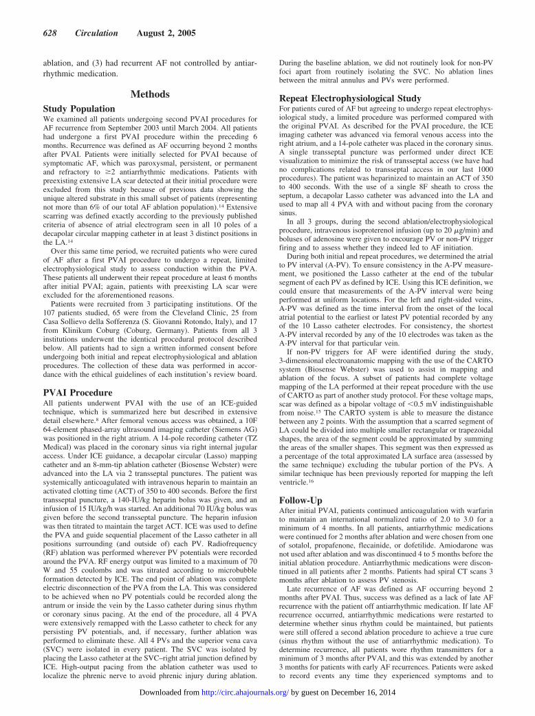
LA Voltage MappingAt the time of the second study, voltage mapping of the LAwas performed with the use of CARTO in 6 of 26 group Ipatients (23%), 14 of 37 group II patients (38%), and 18 of 44group III patients (41%). The size of the low-voltage areacreated from the previous PVAI did not differ among the 3groups. Low “scar” voltages occupied 38�8%, 40�12%, and
TABLE 1. Baseline Patient Characteristics
Group I Group II Group III P
Patients, n 26 37 44 NA
Age, y 55�9 54�13 58�7 0.32
Male sex, n (%) 19 (73) 25 (68) 31 (71) 0.48
AF frequency, n (%)
Paroxysmal 11 (42) 16 (43) 15 (35) 0.11*
Persistent 6 (23) 8 (22) 10 (23)
Permanent 9 (35) 13 (33) 19 (43)
AF duration, y 7�7 7�7 9�7 0.18
No. failed AAM 2.4�0.8 2.6�0.9 3.1�0.9 0.09
Structural heart disease, n (%) 6 (23) 10 (27) 13 (30) 0.09
LA size, cm 4.3�0.6 4.3�0.5 4.4�0.9 0.26
Ejection fraction, % 54�7 53�4 53�9 0.61
Initial procedure
Total duration, min 166�40 170�42 169�48 0.35
RF time, min 45�25 46�25 45�28 0.69
AAM indicates antiarrhythmic medication; NA, not applicable.*P represents comparison of distribution of AF frequency in each group.
TABLE 2. Distribution of Recurrent PVA Number byPatient Groups
No. Recurrent PVA Group I Group II Group III
0 21 (81) 2 (5) 0 (0)
1 5 (19) 12 (32) 6 (14)
2 0 (0) 17 (46) 25 (57)
3 0 (0) 6 (16) 11 (25)
4 0 (0) 0 (0) 2 (4)
Numbers in each group column represent the number of patients with aspecific number of recurrent PVA. Percentages are shown in parentheses.
Verma et al Resumption of PV Conduction Predicts AF Recurrence 629
by guest on December 16, 2014http://circ.ahajournals.org/Downloaded from
41�10% of the estimated total LA surface area (Figure 2) ingroup I, II, and III patients, respectively (P�0.34).
A-PV Conduction DelayA-PV intervals were significantly delayed from the first tosecond procedure in patients who could maintain sinusrhythm with/without antiarrhythmic medication comparedwith patients who failed PVAI. The A-PV intervals for eachof the 4 PVs and the percent change from the first to secondprocedure are detailed in Table 3, except for the right superiorPV and right inferior PV for group I. This is because none ofthe patients in group I had recurrence in the right superior PVand only 1 had recurrence in the right inferior PV, therebypreventing statistical analysis. The A-PV delay was signifi-cantly different among the 3 groups (P�0.001). For all PVs,the A-PV interval increased by 69�47% for group IIIpatients from the first to second procedure. However, thedelay in PV-LA conduction was much longer for group IIpatients, with an A-PV increase of 267�110%. In theminority of group I patients who demonstrated conductionrecurrence in a single PVA, the delay in PV-LA conductionwas longer still, with an overall A-PV increase of 473�71%.
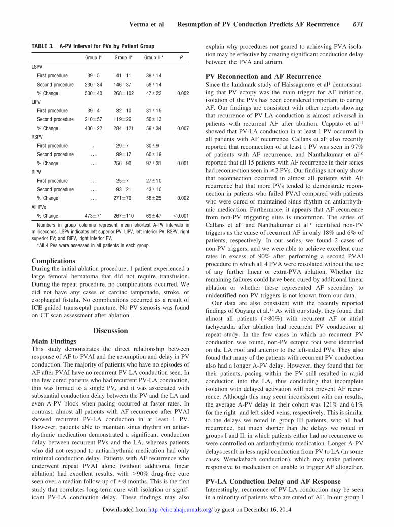
In fact, when pacing occurred at a faster rate, A-PV block wasobserved in all 5 (100%) of the patients with recurrent PV-LAconduction in group I (Figure 3). A-PV block was notobserved in any group II or III patients. Figures 4 and 5demonstrate representative examples of A-PV delay record-ings from patients in groups II and III.
Follow-UpAll of the patients in group II and III underwent repeat PVAIto reisolate those PVA that demonstrated recurrent conduc-tion. Repeat ablation was not performed on those patientswho were cured (group I). Repeat PVAI was performed inexactly the same manner as the initial ablation procedure. Inall cases, only limited ablation in specific segments aroundthe antral-LA interface was required for repeat isolation.Mean RF time was only 5.3�2.7 minutes for these repeatprocedures. Median follow-up duration for the patients whounderwent repeat PVAI was 8.3 months (range, 6 to 12months) since the second procedure. Of the 37 patients ingroup II, 36 (97%) have remained free of AF while offantiarrhythmic medication since their second PVAI. In groupIII, 40 of 44 patients (91%) have remained free of AF whileoff antiarrhythmic medication since their second PVAI.
Figure 1. Three-dimensional electroanatomic CARTO maps of the LA (anterior view [left], posterior view [right]) showing the activationsequence of an ectopic atrial focus in a patient who had a previous PVAI procedure 5 months before. The gray areas represent regionsof scar created by the initial PVAI procedure. In this case, the ablated regions represent 42% of the total LA surface area. This patientexperienced AF recurrence after initial PVAI that was suppressed by antiarrhythmic medication. During this second procedure, norecurrent conduction between the pulmonary veins and the atrium was found. However, AF was initiated by an ectopic atrial focus fir-ing from the LA roof (arrow), with red representing the earliest site of activation of the focus. Two RF lesions at this site successfullyeliminated the ectopic trigger and prevented AF recurrence.
Figure 2. Three-dimensional electroanatomicCARTO maps of the LA (anterior view [right],posterior view [left]) showing the extent ofscarring created by the first PVAI ablationprocedure in a patient who had AF recur-rence after the first procedure that was notcontrolled with antiarrhythmic medication(group III). The gray regions represent scarcreated by the first ablation as determinedby absent or very low voltage amplitude. Inthis patient, the ablation scar represents39% of the total LA surface area, which istypical of the scar area created by the abla-tion procedure. The size of scar seen at thetime of the second procedure did not differsignificantly between the 3 groups studied(see text for details). The colored tubes rep-resent the tubular portions of the PVs.
630 Circulation August 2, 2005
by guest on December 16, 2014http://circ.ahajournals.org/Downloaded from
ComplicationsDuring the initial ablation procedure, 1 patient experienced alarge femoral hematoma that did not require transfusion.During the repeat procedure, no complications occurred. Wedid not have any cases of cardiac tamponade, stroke, oresophageal fistula. No complications occurred as a result ofICE-guided transseptal puncture. No PV stenosis was foundon CT scan assessment after ablation.
DiscussionMain FindingsThis study demonstrates the direct relationship betweenresponse of AF to PVAI and the resumption and delay in PVconduction. The majority of patients who have no episodes ofAF after PVAI have no recurrent PV-LA conduction seen. Inthe few cured patients who had recurrent PV-LA conduction,this was limited to a single PV, and it was associated withsubstantial conduction delay between the PV and the LA andeven A-PV block when pacing occurred at faster rates. Incontrast, almost all patients with AF recurrence after PVAIshowed recurrent PV-LA conduction in at least 1 PV.However, patients able to maintain sinus rhythm on antiar-rhythmic medication demonstrated a significant conductiondelay between recurrent PVs and the LA, whereas patientswho did not respond to antiarrhythmic medication had onlyminimal conduction delay. Patients with AF recurrence whounderwent repeat PVAI alone (without additional linearablation) had excellent results, with �90% drug-free cureseen over a median follow-up of �8 months. This is the firststudy that correlates long-term cure with isolation or signif-icant PV-LA conduction delay. These findings may also
explain why procedures not geared to achieving PVA isola-tion may be effective by creating significant conduction delaybetween the PVA and atrium.
PV Reconnection and AF RecurrenceSince the landmark study of Haissaguerre et al1 demonstrat-ing that PV ectopy was the main trigger for AF initiation,isolation of the PVs has been considered important to curingAF. Our findings are consistent with other reports showingthat recurrence of PV-LA conduction is almost universal inpatients with recurrent AF after ablation. Cappato et al11
showed that PV-LA conduction in at least 1 PV occurred inall patients with AF recurrence. Callans et al9 also recentlyreported that reconnection of at least 1 PV was seen in 97%of patients with AF recurrence, and Nanthakumar et al10
reported that all 15 patients with AF recurrence in their serieshad reconnection seen in �2 PVs. Our findings not only showthat reconnection occurred in almost all patients with AFrecurrence but that more PVs tended to demonstrate recon-nection in patients who failed PVAI compared with patientswho were cured or maintained sinus rhythm on antiarrhyth-mic medication. Furthermore, it appears that AF recurrencefrom non-PV triggering sites is uncommon. The series ofCallans et al9 and Nanthakumar et al10 identified non-PVtriggers as the cause of recurrent AF in only 18% and 6% ofpatients, respectively. In our series, we found 2 cases ofnon-PV triggers, and we were able to achieve excellent curerates in excess of 90% after performing a second PVAIprocedure in which all 4 PVA were reisolated without the useof any further linear or extra-PVA ablation. Whether theremaining failures could have been cured by additional linearablation or whether these represented AF secondary tounidentified non-PV triggers is not known from our data.
Our data are also consistent with the recently reportedfindings of Ouyang et al.17 As with our study, they found thatalmost all patients (�80%) with recurrent AF or atrialtachycardia after ablation had recurrent PV conduction atrepeat study. In the few cases in which no recurrent PVconduction was found, non-PV ectopic foci were identifiedon the LA roof and anterior to the left-sided PVs. They alsofound that many of the patients with recurrent PV conductionalso had a longer A-PV delay. However, they found that fortheir patients, pacing within the PV still resulted in rapidconduction into the LA, thus concluding that incompleteisolation with delayed activation will not prevent AF recur-rence. Although this may seem inconsistent with our results,the average A-PV delay in their cohort was 121% and 61%for the right- and left-sided veins, respectively. This is similarto the delays we noted in group III patients, who all hadrecurrence, but much shorter than the delays we noted ingroups I and II, in which patients either had no recurrence orwere controlled on antiarrhythmic medication. Longer A-PVdelays result in less rapid conduction from PV to LA (in somecases, Wenckebach conduction), which may make patientsresponsive to medication or unable to trigger AF altogether.
PV-LA Conduction Delay and AF ResponseInterestingly, recurrence of PV-LA conduction may be seenin a minority of patients who are cured of AF. In our group I
TABLE 3. A-PV Interval for PVs by Patient Group
Group I* Group II* Group III* P
LSPV
First procedure 39�5 41�11 39�14
Second procedure 230�34 146�37 58�14
% Change 500�40 268�102 47�22 0.002
LIPV
First procedure 39�4 32�10 31�15
Second procedure 210�57 119�26 50�13
% Change 430�22 284�121 59�34 0.007
RSPV
First procedure � � � 29�7 30�9
Second procedure � � � 99�17 60�19
% Change � � � 256�90 97�31 0.001
RIPV
First procedure � � � 25�7 27�10
Second procedure � � � 93�21 43�10
% Change � � � 271�79 58�25 0.002
All PVs
% Change 473�71 267�110 69�47 �0.001
Numbers in group columns represent mean shortest A-PV intervals inmilliseconds. LSPV indicates left superior PV; LIPV, left inferior PV; RSPV, rightsuperior PV; and RIPV, right inferior PV.
*All 4 PVs were assessed in all patients in each group.
Verma et al Resumption of PV Conduction Predicts AF Recurrence 631
by guest on December 16, 2014http://circ.ahajournals.org/Downloaded from
patients, 19% demonstrated recurrence of PV-LA conductionin 1 PV. However, although conduction may have recurred, itwas substantially delayed compared with the initial conduc-tion time before ablation. Although triggering impulses maystill conduct into the LA, the delay in conduction time mayrender the impulse incapable of initiating AF. Indeed, withfaster pacing rates, A-PV block developed in all of thesepatients. Reconnection of 1 or more PVs has been reported inup to 32% of patients who achieve a drug-free cure fromAF,11 but as with our findings, the conduction was signifi-cantly delayed compared with baseline.
Furthermore, PV-LA conduction delay seems to predictresponsiveness to antiarrhythmic medication, and it waspresent even in some of the patients who had been cured.Patients who had AF recurrence but were able to maintainsinus rhythm on antiarrhythmic medication had significantlymore PV-LA conduction delay than those patients withrecurrence despite antiarrhythmic medication. Because of thealready delayed conduction, antiarrhythmic medication mayslow conduction even further, potentially causing exit blockof the triggering impulses and preventing the developmentof AF.
At first glance, our study may appear inconsistent withother studies that have suggested that achieving PV isolation
is not a prerequisite for procedural success of AF abla-tion.12,13 However, the main difference between our study andthese other reports is in the definition of “cure.” We haveused a lack of AF recurrence in patients off antiarrhythmicmedication as our standard definition of cure. However, in theother series, many of the patients classified as having suc-cessful outcomes were on antiarrhythmic medication (40% to63%), making the off-drug cure rates 30% to 38%.12,13 As wehave shown, PV isolation is not required to be able tomaintain sinus rhythm on antiarrhythmic medication afterablation; only a delay in PV-LA conduction seems to beneeded for antiarrhythmic medication responsiveness. Thus,although these other reports describe full PV isolation in only20% to 40% of their ablated patients, PV-LA conductiondelay was likely present in most patients, explaining the goodresponse to antiarrhythmic medication. Indeed, one of thesereports used an empirical anatomic ablation approach guidedby electroanatomic mapping and reduction of electrogramamplitude.12 Although complete PV isolation is achieved inonly a minority of cases with the use of this technique, thetechnique results in substantial PV-LA conduction delay inalmost all patients.18 Had complete PV isolation beenachieved in these other series, it is possible that the cure rates
Figure 3. Recording of both surface ECG and intracardiac electrograms during pacing from the coronary sinus (CS) os in a patientcured of AF by a PVAI procedure performed 7 months before. The Lasso catheter (Ls) is placed along the anterior segment of the leftsuperior PVA. There is considerable conduction delay between the far-field LA (A) potential (diagonal arrows) and the local high-frequency PV potential (*). During pacing from the CS os at 500 ms, there is Wenckebach conduction of the A-PV delay. The A-PVinterval gradually lengthens from 340 to 380 ms until there is entrance block into the PV (Block). Gain on the intracardiac signals is5000 with the use of the Prucka recording system (GE Prucka).
632 Circulation August 2, 2005
by guest on December 16, 2014http://circ.ahajournals.org/Downloaded from
in patients off antiarrhythmic medication would have beenhigher.
Long-Term Success of PV Isolation–Based ProceduresInitial studies of segmental PV isolation for AF ablationshowed variable, and often poor, success rates,19 suggestingthat PV isolation was not adequate to achieve proceduralsuccess. However, many of these early approaches onlytargeted “arrhythmogenic” or easily accessible PVs instead ofmore modern approaches that empirically target all of theveins. It is often difficult to identify single, arrhythmogenicPVs, and even after a culprit vein is ablated, triggers fromother PVs may become unmasked.20 Furthermore, many ofthese techniques ablated within the tubular portion of the PV,excluding the posterior, funnel-shaped extension of the PV,which we refer to as the antrum.8 Thus, large regions of theveins were not being isolated. Finally, out of concern forstenosis, power output was often limited to a very lowwattage (�30 W), making recurrent PV conduction common-
place. Thanks to improved monitoring techniques, manyoperators now use higher power outputs and broader circum-ferential ablation, making isolation of the entire PVA moreeffective.
Some have suggested that ablation around the PVs pre-vents AF not by isolation of PV triggers but rather bymodification of the atrial substrate, thereby preventing AFperpetuation.21 However, the strong relationship betweenPV-LA conduction and AF response to PVAI in this studywould argue against this concept. Furthermore, in the 2patients in whom AF recurred despite successful isolation ofall of the PVA, AF still recurred despite extensive scarringand “modification” of the posterior LA wall. Focal non-PVtriggers were responsible for the AF recurrence, and onlyablating and removing these focal triggers prevented AF.There was also no difference in the extent of RF ablationinitially performed in patients who failed PVAI versus thosewho were cured or were responsive to antiarrhythmic medi-cation. Initial PVAI RF time was the same for all groups, andthe extent of scar created by the ablation was also the same in
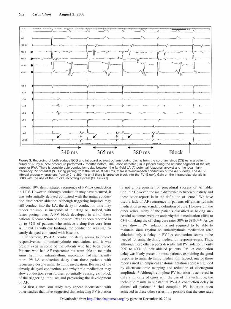
Figure 4. Recording of both surface ECG and intracardiac electrograms during sinus rhythm in a patient with AF recurrence after PVAI6 months before but able to maintain sinus rhythm on antiarrhythmic medication (sotalol). The Lasso catheter (Ls) is placed along theanterior segment of the left superior PVA as visualized by ICE (inset). There is considerable conduction delay between the far-field LA(A) potential and the local high-frequency PV potential. The longest measured A-PV interval was 180 ms in Ls 1 to 2, and the shortestwas 136 ms in Ls 2 to 3. This delay was considerably longer than the baseline A-PV interval in this vein before the first PVAI, whichwas only 26 ms. Gain on the intracardiac signals is 5000 with the use of the Prucka recording system (GE Prucka). LUPV indicates leftupper pulmonary vein; LLPV, left lower pulmonary vein.
Verma et al Resumption of PV Conduction Predicts AF Recurrence 633
by guest on December 16, 2014http://circ.ahajournals.org/Downloaded from
the 3 groups. During repeat PVAIs, only limited, focusedablation was required (�6 minutes of RF time) to producelasting cure, which is unlikely to have “modified” the LAsubstrate any more than the initial ablation procedure.
Despite improved PVAI techniques, our 2-procedure suc-cess rate still falls short of 100%. Possible explanations forthis include ongoing recurrence of PV-LA conduction or thepresence of non–PV-related triggers for AF that are notidentified during the initial procedures.
ConclusionsResumption and delay of PV conduction is directly related tothe response of AF to PVAI. The majority of patients withdrug-free cure show no recurrence of PV-LA conduction,whereas all patients with recurrent AF show PV-LA recon-nection. Substantial delay in PV-LA conduction is seen inpatients able to maintain sinus rhythm on antiarrhythmicmedication after ablation and in the minority of patients withdrug-free cure who have recurrent PV-LA conduction in asingle PV.
AcknowledgmentDr Verma was supported by a fellowship award from the Heart andStroke Foundation of Canada.
References1. Haissaguerre M, Jais P, Shah DC, Takahashi A, Hocini M, Quiniou G,
Garrigue S, Le Mouroux A, Le Metayer P, Clementy J. Spontaneous
initiation of atrial fibrillation by ectopic beats originating in the pulmo-nary veins. N Engl J Med. 1998;339:659–666.
2. Marrouche NF, Martin DO, Wazni O, Gillinov AM, Klein A, BhargavaM, Saad E, Bash D, Yamada H, Jaber W, Schweikert R, Tchou P,Abdul-Karim A, Saliba W, Natale A. Phased-array intracardiac echocar-diography monitoring during pulmonary vein isolation in patients withatrial fibrillation: impact on outcome and complications. Circulation.2003;107:2710–2716.
3. Lin WS, Tai CT, Hsieh MH, Tsai CF, Lin YK, Tsao HM, Huang JL, YuWC, Yang SP, Ding YA, Chang MS, Chen SA. Catheter ablation ofparoxysmal atrial fibrillation initiated by non-pulmonary vein ectopy.Circulation. 2003;107:3176-3183.
4. Jalife J. Rotors and spiral waves in atrial fibrillation. J CardiovascElectrophysiol. 2003;14:776–780.
5. Ouyang F, Bansch D, Ernst S, Schaumann A, Hachiya H, Chen M, ChunJ, Falk P, Khanedani A, Antz M, Kuck KH. Complete isolation of leftatrium surrounding the pulmonary veins: new insights from thedouble-Lasso technique in paroxysmal atrial fibrillation. Circulation.2004;110:2090–2096.
6. Haissaguerre M, Sanders P, Hocini M, Hsu LF, Shah DC, Scavee C,Takahashi Y, Rotter M, Pasquie JL, Garrigue S, Clementy J, Jais P.Changes in atrial fibrillation cycle length and inducibility during catheterablation and their relation to outcome. Circulation. 2004;109:3007–3013.
7. Nademanee K, McKenzie J, Kosar E, Schwab M, Sunsaneewitayakul B,Vasavakul T, Khunnawat C, Ngarmukos T. A new approach for catheterablation of atrial fibrillation: mapping of the electrophysiologic substrate.J Am Coll Cardiol. 2004;43:2044–2053.
8. Verma A, Marrouche NF, Natale A. Pulmonary vein antrum isolation:intracardiac echocardiography–guided technique. J Cardiovasc Electro-physiol. 2004;15:1335–1340.
9. Callans DJ, Gerstenfeld EP, Dixit S, Zado E, Vanderhoff M, Ren JF,Marchlinski FE. Efficacy of repeat pulmonary vein isolation procedures
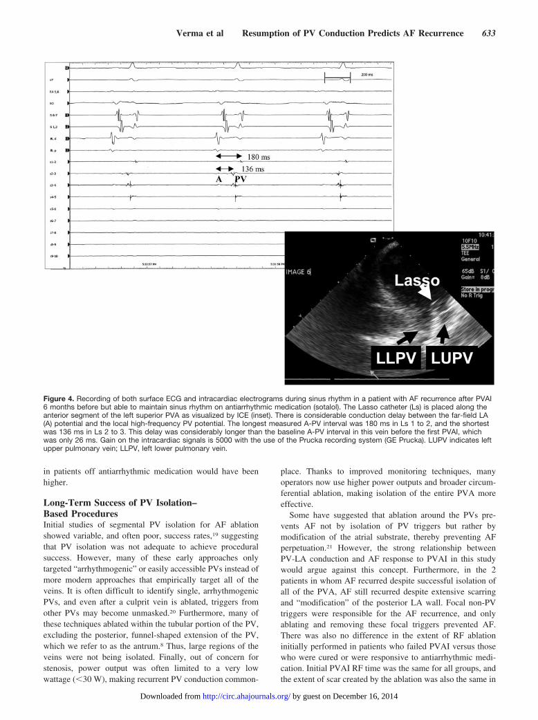
Figure 5. Recording of both surface ECG and intracardiac electrograms during sinus rhythm in a patient with AF recurrence after PVAI6 months before and inability to maintain sinus rhythm even with antiarrhythmic medications. The Lasso catheter (Ls) is placed alongthe anterior segment of the left superior PVA. There is almost no delay between the far-field LA potential (A) and the local high-frequency PV potential. The shortest measured A-PV interval was 36 ms, which was not significantly different from the A-PV intervalmeasured in this vein before the first PVAI (32 ms). Gain on the intracardiac signals is 5000 with the use of the Prucka recording sys-tem (GE Prucka).
634 Circulation August 2, 2005
by guest on December 16, 2014http://circ.ahajournals.org/Downloaded from
in patients with recurrent atrial fibrillation. J Cardiovasc Electrophysiol.2004;15:1050–1055.
10. Nanthakumar K, Plumb VJ, Epstein AE, Veenhuyzen GD, Link D, KayGN. Resumption of electrical conduction in previously isolated pulmo-nary veins: rationale for a different strategy? Circulation. 2004;109:1226–1229.
11. Cappato R, Negroni S, Pecora D, Bentivegna S, Lupo PP, Carolei A,Esposito C, Furlanello F, De Ambroggi L. Prospective assessment of lateconduction recurrence across radiofrequency lesions producing electricaldisconnection at the pulmonary vein ostium in patients with atrial fibril-lation. Circulation. 2003;108:1599–1604.
12. Kottkamp H, Tanner H, Kobza R, Schirdewahn P, Dorszewski A,Gerds-Li JH, Carbucicchio C, Piorkowski C, Hindricks G. Time coursesand quantitative analysis of atrial fibrillation episode number and durationafter circular plus linear left atrial lesions: trigger elimination or substratemodification: early or delayed cure? J Am Coll Cardiol. 2004;44:869–877.
13. Stabile G, Turco P, La Rocca V, Nocerino P, Stabile E, De Simone A. Ispulmonary vein isolation necessary for curing atrial fibrillation? Circu-lation. 2003;108:657–660.
14. Verma A, Wazni OM, Marrouche NF, Martin DO, Kilicaslan F, Minor S,Schweikert RA, Saliba W, Cummings J, Burkhardt JD, Bhargava M,Belden WA, Abdul-Karim A, Natale A. Pre-existent left atrial scarring inpatients undergoing pulmonary vein antrum isolation: an independentpredictor of procedural failure. J Am Coll Cardiol. 2005;45:285–292.
15. Sanders P, Morton JB, Davidson NC, Spence SJ, Vohra JK, Sparks PB,Kalman JM. Electrical remodeling of the atria in congestive heart failure:
electrophysiological and electroanatomic mapping in humans. Circu-lation. 2003;108:1461–1468.
16. Marchlinski FE, Callans DJ, Gottlieb CD, Zado E. Linear ablation lesionsfor control of unmappable ventricular tachycardia in patients with ische-mic and nonischemic cardiomyopathy. Circulation. 2000;101:1288–1296.
17. Ouyang F, Antz M, Ernst S, Hachiya H, Mavrakis H, Deger FT,Schaumann A, Chun J, Falk P, Hennig D, Liu X, Bansch D, Kuck KH.Recovered pulmonary vein conduction as a dominant factor for recurrentatrial tachyarrhythmias after complete circular isolation of the pulmonaryveins: lessons from double Lasso technique. Circulation. 2005;111:127–135.
18. Pappone C, Oreto G, Rosanio S, Vicedomini G, Tocchi M, Gugliotta F,Salvati A, Dicandia C, Calabro MP, Mazzone P, Ficarra E, Di Gioia C,Gulletta S, Nardi S, Santinelli V, Benussi S, Alfieri O. Atrial electro-anatomic remodeling after circumferential radiofrequency pulmonaryvein ablation: efficacy of an anatomic approach in a large cohort ofpatients with atrial fibrillation. Circulation. 2001;104:2539–2544.
19. Finta B, Haines DE. Catheter ablation therapy for atrial fibrillation.Cardiol Clin. 2004;22:127–145, ix.
20. Haissaguerre M, Jais P, Shah DC, Garrigue S, Takahashi A, Lavergne T,Hocini M, Peng JT, Roudaut R, Clementy J. Electrophysiological endpoint for catheter ablation of atrial fibrillation initiated from multiplepulmonary venous foci. Circulation. 2000;101:1409–1417.
21. Oral H, Scharf C, Chugh A, Hall B, Cheung P, Good E, Veerareddy S,Pelosi F Jr, Morady F. Catheter ablation for paroxysmal atrial fibrillation:segmental pulmonary vein ostial ablation versus left atrial ablation. Cir-culation. 2003;108:2355–2360.
Verma et al Resumption of PV Conduction Predicts AF Recurrence 635
by guest on December 16, 2014http://circ.ahajournals.org/Downloaded from
Burkhardt, Walid Saliba, Robert A. Schweikert and Andrea NataleBrachmann, Jens Geunther, Domenico Potenza, David O. Martin, Jennifer Cummings, J. David Atul Verma, Fethi Kilicaslan, Ennio Pisano, Nassir F. Marrouche, Raffaele Fanelli, Johannes
Resumption and Delay of Pulmonary Vein ConductionResponse of Atrial Fibrillation to Pulmonary Vein Antrum Isolation Is Directly Related to
Print ISSN: 0009-7322. Online ISSN: 1524-4539 Copyright © 2005 American Heart Association, Inc. All rights reserved.
is published by the American Heart Association, 7272 Greenville Avenue, Dallas, TX 75231Circulation doi: 10.1161/CIRCULATIONAHA.104.533190
2005;112:627-635Circulation.
http://circ.ahajournals.org/content/112/5/627World Wide Web at:
The online version of this article, along with updated information and services, is located on the
http://circ.ahajournals.org//subscriptions/
is online at: Circulation Information about subscribing to Subscriptions:
http://www.lww.com/reprints Information about reprints can be found online at: Reprints:
document. Permissions and Rights Question and Answer this process is available in the
click Request Permissions in the middle column of the Web page under Services. Further information aboutOffice. Once the online version of the published article for which permission is being requested is located,
can be obtained via RightsLink, a service of the Copyright Clearance Center, not the EditorialCirculationin Requests for permissions to reproduce figures, tables, or portions of articles originally publishedPermissions:
by guest on December 16, 2014http://circ.ahajournals.org/Downloaded from