requirement of a leucine residue for (apical) membrane expression of type iib napi cotransporters
TRANSCRIPT

Requirement of a leucine residue for (apical)membrane expression of type IIbNaPi cotransportersZoubida Karim-Jimenez*, Nati Hernando*, Jurg Biber, and Heini Murer†
Institute of Physiology, Zurich University, Zurich, CH-8057, Switzerland
Communicated by Gerhard Giebisch, Yale University School of Medicine, New Haven, CT, December 28, 1999 (received for review July 28, 1999)
Type II NaPi cotransporters mediate epithelial phosphate (Pi) re-absorption. In mammals the type IIb protein is expressed in thesmall intestinal apical membrane and other epithelia; it is notexpressed in the renal proximal tubule where we find the type IIaisoform. To look for molecular determinant(s) involved in apicalexpression of type IIb cotransporters, we have made deletionmutations within the C-terminal tails of mouse IIb (mIIb) andhuman IIb (hIIb) transporter proteins. The constructs were fused tothe enhanced green fluorescent protein and transiently trans-fected into intestinal CaCo2-cells. Both mIIb and hIIb were locatedexclusively in the apical membrane of the cells. For mIIb, theremoval of a cysteine cluster or the last three amino acids (TVF) hadno effect on the location of the protein. However, truncation at thelevel of the conserved L691y689 prevented the apical membraneexpression of both mIIb and hIIb, respectively, and the mutatedproteins were located in endosomal and lysosomal structures. Asimilar expression pattern of the mIIb and hIIb constructs wasfound in renal proximal tubular opossum kidney cells. Our datasuggest that L691y689 is involved in mechanisms leading to anapical expression of type IIb NaPi cotransporters.
epithelial polarity u enhanced green fluorescent protein
The asymmetrical distribution of transporter proteins in theplasma membrane of epithelial cells is essential for the
vectorial transport of solutes. Type II NaPi cotransporters arelocated in the apical (brush border) membrane and mediate Pitransport in different epithelia (1, 2). The type IIb NaPi co-transporter is located in small intestine, type II alveolar pneu-mocytes, and other tissues, whereas the type IIa isoform isexpressed mainly in the renal proximal tubule (3, 4).
The type IIa and type IIb transporters are homologousproteins ('70% similarity) with most of the differences in thecytoplasmic-oriented termini (3). In a recent study on transfec-tions with type IIa and type IIb proteins we found that the typeIIb protein is exclusively apically expressed in several cell linesof different epithelial origin whereas the type IIa transporter isexpressed only in renal epithelial cell lines, and proper apicallocation is observed only in opossum kidney (OK) cells (prox-imal tubular origin) (5). This finding indicates that besidesmolecular motifs at the level of the protein the cellular contextmay crucially contribute to proper polarized expression.
The mechanism(s) by which newly synthesized proteins areexpressed at either of the two plasma membrane domains are notfully understood; ‘‘targeting’’ andyor ‘‘scaffolding’’ mechanismscan be involved. Newly synthesized apical and basolateral pro-teins seem to be transported together from the endoplasmicreticulum (ER) to the trans-Golgi network and then to thesubapical compartment (SAC). SAC is an important interme-diate in the delivery of proteins to specific early endosomesdestined either to the apical or the basolateral membrane (6).The mechanisms by which SAC is able to discriminate betweenapical and basolateral endosomes are not clear. Two basolateralsorting motifs located in the cytoplasmic tail of transmembraneproteins have been identified: tyrosine and di-leucine-based
motifs (7, 8). The presence of a basolateral determinant withintransmembrane domains also has been suggested (9). For apicalsorting, several mechanisms have been proposed. One involvesthe interaction of proteins with rafts, sphingolipids, and choles-terol-rich and detergent-insoluble membranes (10). Anotherdepends on the presence of apical sorting signals in the cyto-plasmic domain of membrane proteins (11, 12). Furthermore,the involvement of ecto- and transmembrane domains also hasbeen suggested (13–15).
The aim of the present study was to obtain information aboutmolecular determinants involved in the apical expression of typeIIb NaPi cotransporters. The present experiments show that aconserved L691 (mouse IIb, mIIb)y689 (human IIb, hIIb) isessential for the (apical) membrane localization of type IIb NaPicotransporters. This leucine residue therefore could be part of atargeting motif andyor a motif required for membrane stabili-zation of the transporter protein.
MethodsNaPi-pEGFP Construction and Mutagenesis. The cDNAs encodingthe wild-type (WT) mIIb and hIIb cotransporters were sub-cloned into the red-shifted enhanced green fluorescent protein(EGFP) variant vector pEGFP-C1 (CLONTECH). Briefly, sin-gle restriction sites were introduced at the 59 and 39 ends of theWT cDNAs through PCR: SacI–SalI for mIIb and SalI–BamHIfor hIIb. The cDNAs then were ligated into the pEGFP-C1,which was digested by the same restriction enzymes. AllcDNAs were fused at the carboxyl terminal end of the EGFP(NaPi-pEGFP).
The truncated mutants were constructed by introducing thestop codon TAG at different positions within the C-terminal tailof the WT cDNAs (Fig. 1). For these constructs, site-directedmutagenesis was performed by using NaPi-pEGFP as templatesand complementary sense and antisense primers, both of themcontaining the stop codon in the middle of their sequence. AfterPCRs, the templates were digested by Dnp1.
To generate mIIbD5C, which lacks the five-cysteine residueslocated in the C-terminal tail of mIIb, a ThaI site was introducedat position 618 of the mIIb cDNA, which originally contains thesame site at position 622 (Fig. 1). The nucleotides encoding thefive cysteines then were removed by digesting the mutated cDNAwith ThaI. After purification, mIIbD5C was inserted intopEGFP-C1 as described for mIIbWT.
All mutations were confirmed by sequencing.
Abbreviations: EGFP, enhanced green fluorescent protein; mIIb, mouse IIb; hIIb, human IIb;ER, endoplasmic reticulum; SAC, subapical compartment; WT, wild type; OK, opossumkidney.
*Z.K.-J. and N.H. contributed equally to this work.
†To whom reprint requests should be addressed at: Physiologisches Institut, UniversitatZurich-Irchel, Winterthurerstrasse 190, Zurich, CH-8057, Switzerland. E-mail: [email protected].
The publication costs of this article were defrayed in part by page charge payment. Thisarticle must therefore be hereby marked “advertisement” in accordance with 18 U.S.C.§1734 solely to indicate this fact.
2916–2921 u PNAS u March 14, 2000 u vol. 97 u no. 6

Cell Culture and Transient Transfections. Colon adenocarcinomacells (CaCo2 cells) were grown in DMEM complemented with20% FCS, 44 mM NaHCO3, 20 mM Hepes, 4 mM L-glutamine,1% nonessential amino acids, 100 unitsyml of penicillin, and 100mgyml streptomycin, at 37°C in 10% CO2-90% air. OK cellswere grown in DMEMyHam’s F-12 medium (1:1) comple-mented with 10% FCS, 22 mM NaHCO3, 20 mM Hepes, 2 mML-glutamine, 50 unitsyml of penicillin, and 50 mgyml strepto-mycin, at 37°C in 5% CO2-95% air. For transfections, CaCo2 andOK cells were grown on coverslips in 35-mm dishes. Subcon-
fluent cultures were transfected with 3 ml of Fugene (BoehringerMannhein) and 1 mg plasmids. Cells were processed for analysisby microscopy once they reached confluency, 2 or 3 days aftertransfection. At least five independent experiments were per-formed on transiently transfected cells. Attempts to obtain stablytransfected cell lines expressing the EGFP-fused cotransporterhave failed, though it was previously possible to establish celllines containing the WT NaPi-2 cotransporter (16, 17).
Analysis by Confocal Microscopy. Confluent cells were fixed, andthe actin andyor expressed proteins were stained as described(5). The immunodetection of hIIb was performed with a rabbitpolyclonal antibody raised against an N-terminal peptide (3). Forthe immunodetection of ER, trans-Golgi network, endosomes,and lysosomes, we used mAbs against human p63, giantin,transferrin receptor, and lamp1, respectively (18–22). The cov-erslips were mounted by using Dako-glycerol containing 2.5%1.4-diazabicyclo-(2.2.2) octane (Sigma) as a fading retardant.Confocal images were taken by using a Leica TCSSP (Wetzlar,Germany) laser scan microscope equipped with a 363 oilimmersion objective.
Results and DiscussionSimilar to the type IIa transporter (23, 24), the type IIb NaPicotransporter most likely has eight transmembrane domains,with cytoplasmic N and C termini (3). Most of the differencesbetween both transporters lay within the cytoplasmic tails (3).
The type IIb proteins showed proper apical expression aftertransfection in different epithelial cell lines (CaCo2, OK, LLC-PK1, and MDCK cells) whereas for the type IIa transporter a‘‘correct’’ apical expression was obtained only in renal proximaltubular OK cells (5). To identify potential subdomains (motifs)involved in the apical expression of the type IIb transporterprotein, we performed transient transfection experiments withtruncations in the C-terminal tail of mIIb and hIIb cotransport-
Fig. 1. Schematic representation of WT and mutants of type IIb NaPi co-transporters. The N-terminus (N-Ter), the transmembrane (TD), and the C-terminus (C-Ter) domains are indicated in open, filled, and striped bars,respectively. The numbers given at the beginning or the end of the barsindicate the position of the amino acids. The spaces indicate the removal of theamino acids. Also shown are the different sequences of the C-terminal tails ofmIIb and hIIb cotransporters.
Fig. 2. Expression of hIIb cotransporter in CaCo2 cells. Cells were transfected with EGFP-fused hIIb cotransporter or EGFP plasmid and processed for confocalmicroscopy. (a–c) Focal planes. (d–i) xy cross-sections. (a, d, and g) Cells transfected with empty plasmid. (b, e, and h) Cells transfected with the EGFP-fused hIIb.(c, f, and i) Cells transfected with EGFP-fused hIIb and stained with anti-hIIb antibody. The endogenous EGFP fluorescence is shown in green and actin and anti-hIIbantibody staining in red. In a–c the fluorescence signals were merged.
Karim-Jimenez et al. PNAS u March 14, 2000 u vol. 97 u no. 6 u 2917
PHYS
IOLO
GY
ers (Fig. 1). As indicated in Methods, we were unable to establishstably transfected cell lines.
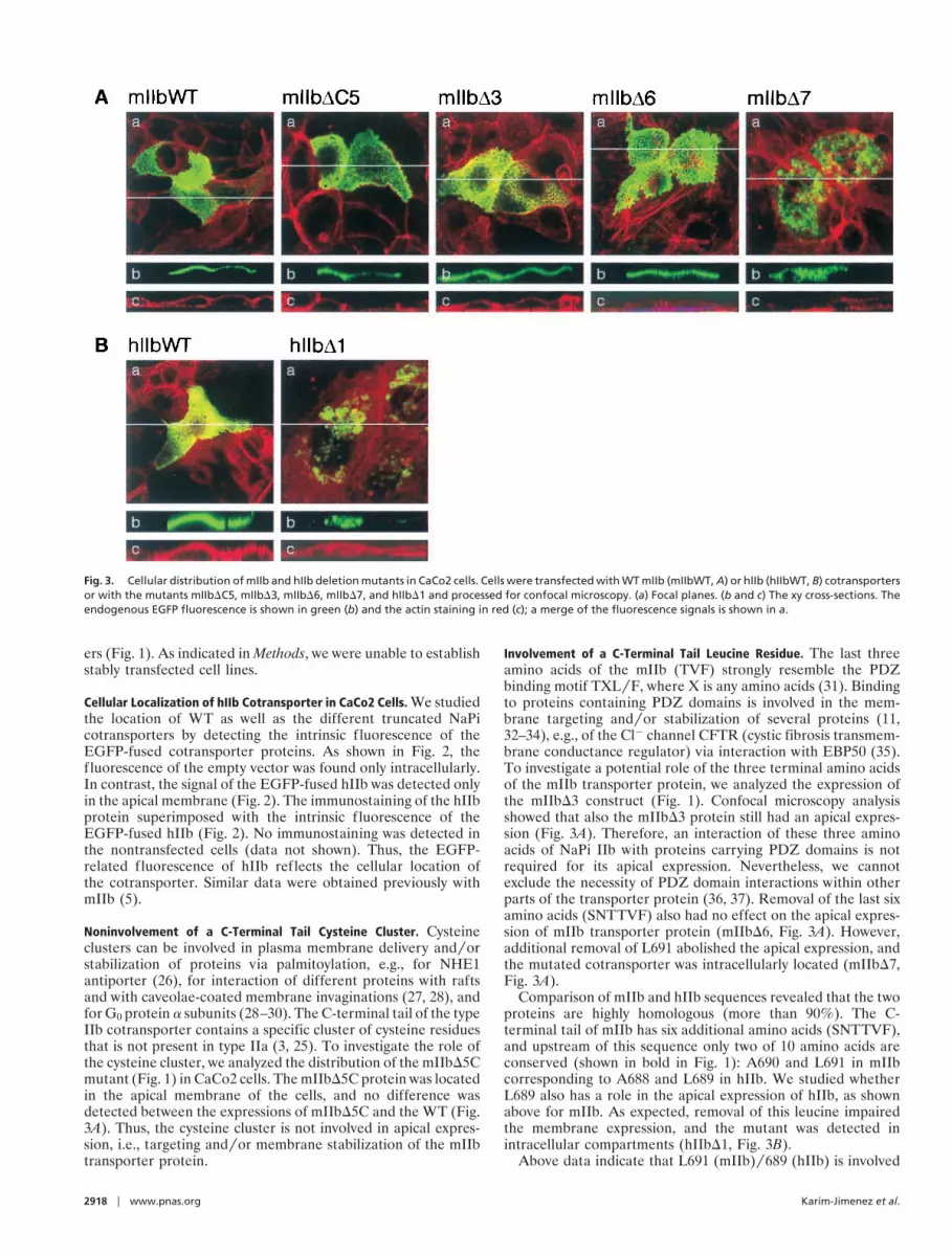
Cellular Localization of hIIb Cotransporter in CaCo2 Cells. We studiedthe location of WT as well as the different truncated NaPicotransporters by detecting the intrinsic f luorescence of theEGFP-fused cotransporter proteins. As shown in Fig. 2, thefluorescence of the empty vector was found only intracellularly.In contrast, the signal of the EGFP-fused hIIb was detected onlyin the apical membrane (Fig. 2). The immunostaining of the hIIbprotein superimposed with the intrinsic f luorescence of theEGFP-fused hIIb (Fig. 2). No immunostaining was detected inthe nontransfected cells (data not shown). Thus, the EGFP-related fluorescence of hIIb reflects the cellular location ofthe cotransporter. Similar data were obtained previously withmIIb (5).
Noninvolvement of a C-Terminal Tail Cysteine Cluster. Cysteineclusters can be involved in plasma membrane delivery andyorstabilization of proteins via palmitoylation, e.g., for NHE1antiporter (26), for interaction of different proteins with raftsand with caveolae-coated membrane invaginations (27, 28), andfor G0 protein a subunits (28–30). The C-terminal tail of the typeIIb cotransporter contains a specific cluster of cysteine residuesthat is not present in type IIa (3, 25). To investigate the role ofthe cysteine cluster, we analyzed the distribution of the mIIbD5Cmutant (Fig. 1) in CaCo2 cells. The mIIbD5C protein was locatedin the apical membrane of the cells, and no difference wasdetected between the expressions of mIIbD5C and the WT (Fig.3A). Thus, the cysteine cluster is not involved in apical expres-sion, i.e., targeting andyor membrane stabilization of the mIIbtransporter protein.
Involvement of a C-Terminal Tail Leucine Residue. The last threeamino acids of the mIIb (TVF) strongly resemble the PDZbinding motif TXLyF, where X is any amino acids (31). Bindingto proteins containing PDZ domains is involved in the mem-brane targeting andyor stabilization of several proteins (11,32–34), e.g., of the Cl2 channel CFTR (cystic fibrosis transmem-brane conductance regulator) via interaction with EBP50 (35).To investigate a potential role of the three terminal amino acidsof the mIIb transporter protein, we analyzed the expression ofthe mIIbD3 construct (Fig. 1). Confocal microscopy analysisshowed that also the mIIbD3 protein still had an apical expres-sion (Fig. 3A). Therefore, an interaction of these three aminoacids of NaPi IIb with proteins carrying PDZ domains is notrequired for its apical expression. Nevertheless, we cannotexclude the necessity of PDZ domain interactions within otherparts of the transporter protein (36, 37). Removal of the last sixamino acids (SNTTVF) also had no effect on the apical expres-sion of mIIb transporter protein (mIIbD6, Fig. 3A). However,additional removal of L691 abolished the apical expression, andthe mutated cotransporter was intracellularly located (mIIbD7,Fig. 3A).
Comparison of mIIb and hIIb sequences revealed that the twoproteins are highly homologous (more than 90%). The C-terminal tail of mIIb has six additional amino acids (SNTTVF),and upstream of this sequence only two of 10 amino acids areconserved (shown in bold in Fig. 1): A690 and L691 in mIIbcorresponding to A688 and L689 in hIIb. We studied whetherL689 also has a role in the apical expression of hIIb, as shownabove for mIIb. As expected, removal of this leucine impairedthe membrane expression, and the mutant was detected inintracellular compartments (hIIbD1, Fig. 3B).
Above data indicate that L691 (mIIb)y689 (hIIb) is involved
Fig. 3. Cellular distribution of mIIb and hIIb deletion mutants in CaCo2 cells. Cells were transfected with WT mIIb (mIIbWT, A) or hIIb (hIIbWT, B) cotransportersor with the mutants mIIbDC5, mIIbD3, mIIbD6, mIIbD7, and hIIbD1 and processed for confocal microscopy. (a) Focal planes. (b and c) The xy cross-sections. Theendogenous EGFP fluorescence is shown in green (b) and the actin staining in red (c); a merge of the fluorescence signals is shown in a.
2918 u www.pnas.org Karim-Jimenez et al.

in the routing to or in the stabilization of the IIb transporters atthe apical membrane. Leucine residues within dileucine motifsare important for endocytosis and basolateral targeting (38). Inaddition, it has been shown that leucine residues are involved inprotein–protein interaction, e.g., in the interaction of the b2-adrenergic receptor with the regulatory factor NHERF (39).
Intracellular Fate of Misslocated Transporters (mIIbD7yhIIbD1). Weattempted to identify the intracellular compartment where theIIb transporters are accumulated after removal of the L691y689.For that purpose, we studied the colocalization of the WT andmutant proteins with proteins residing in the ER (p63), the Golgiapparatus (giantin), the endosomes (transferrin receptor), andthe lysosomes (lamp1) (18–22). As shown in Fig. 4, neither theWT nor the mIIbD7 mutant colocalized with p63 or giantin.Unlike the WT, mIIbD7 strongly colocalized with the lysosomalmarker lamp1 (Fig. 4). We also observed partial colocalizationof mIIbD7 with the transferrin receptor, an endosomal marker(Fig. 4). Similar colocalizations were detected with the hIIbD1mutant (data not shown).
The ER and Golgi compartments are involved in ‘‘qualitycontrol,’’ and missfolded proteins are unable to leave these com-partments (40–42). The above results suggest that removal of theL691y689 yields a cotransporter that is conformationaly competentto leave the ER and Golgi. The detection of both mIIbD7 andhIIbD1 in lysosomal compartments indicated that the truncated
cotransporters are targeted to degradation. Based on the partialcolocalization of the truncated cotransporters with the transferrinreceptor, two possibilities for lysosomal location can be envisioned:(i) Transferrin receptors are present in earlyyrecycling endosomesbut not in the late endosomalylysosomal pathway (43). Thus,localization in earlyyrecycling endosomes could suggest that themutants mIIbD7yhIIbD1 were expressed at the plasma membranebut were quickly internalized because of their instability in theplasma membrane. Such a mechanism has been reported for theepithelial g-aminobutyric acid transporter (BGT-1) (44). (ii) On theother hand, the colocalization of the transferrin receptor and themIIbD7yhIIbD1 proteins also could be explained by their presencein the SAC (45). From the SAC, transferrin receptors could berouted to the membrane whereas the mutated cotransporters couldbe targeted to the lysosomes. Therefore, the L691y689 truncationscould affect either the targeting andyor the stability of the type IIbNaPi cotransporter in the membrane. Because we did not observean apical location of mIIbD7yhIIbD1 proteins in several experi-ments, we favor the interpretation of inappropriate apical deliveryas contributing to a major extent to endosomal and finally lysosomallocation.
L689y691 Also Is Involved in Apical Expression of Type IIb Proteins inOK Cells. To test whether the removal of L691y689 affects the apicalexpression of the type IIb cotransporter independent of the cellularenvironment (cellular context), we studied the expression of the
Fig. 4. Colocalization of mIIb and mIIbD7 with different intracellular compartment markers in CaCo2 cells. Cells were transfected with WT mIIb cotransporter(mIIbWT) or the mutant mIIbD7. They were stained with antibodies againts p63 (ER), giantin (Golgi), transferrin receptor (Endosomes), and lamp1 (Lysosomes)and processed for confocal microscopy. (a) The focal planes. (b) The xy cross-sections. The endogenous EGFP fluorescence is shown in green, the antibodiesstainings in red, and the overlapping of both in yellow.
Karim-Jimenez et al. PNAS u March 14, 2000 u vol. 97 u no. 6 u 2919
PHYS
IOLO
GY

WT and several mutants in a renal cell line (OK cells). Wepreviously have shown that the mIIb cotransporter was mostlyapically expressed in OK cells, although a weak basolateral stainingwas observed in about 30% of transfected cells (5, 17). We foundthat mIIb, mIIbD3, and mIIbD6 had a similar apical expressionpattern, whereas the mutant mIIbD7 was intracellularly detected(Fig. 5A), as indicated by the lack of the mosaic pattern character-istic for the apical surface of OK cells (5). The same results wereobserved for hIIb cotransporter (Fig. 5B), which indicates thatL689y691 also determines the apical expression in OK cells.Because of the lack of antibodies crossreacting with OK cellproteins residing in intracellular compartments we could not char-acterize the intracellular fate of the truncated transporters in OKcells, but assume similar behavior as in CaCo2 cells.
In summary, the L691y689 seems to be crucial for the apical
membrane expression of IIb NaPi cotransporters. Its role isbeyond the maturation of the protein in ER and Golgi andprimarily might involve an altered membrane delivery althougha reduced apical membrane stability also can contribute. Finally,the cysteine cluster has no apparent function in the apicalexpression of IIb NaPi cotransporters.
We thank Hans-Peter Hauri and Paul Cannon (Roche Bioscience) forproviding us with the antibodies for the different intracellular compart-ments and with hNaPi IIb, U. Ziegler for helpful technical advice in theanalysis by the confocal microscope, and C. Gasser for assistance inpreparing the figures. This work was supported by Swiss National ScienceFoundation Grant (to H.M.), the Hartmann Muller-Stiftung (Zurich),the Olga Mayenflsch-Stiftung, the Schwerzerische Bank-Gesellschaft(Zurich; Bu70417–1), and Novartis Foundation.
1. Murer, H. & Biber, J. (1994) Curr. Opin. Nephrol. Hypertens. 3, 504–510.2. Murer, H. & Biber, J. (1997) Pflugers Arch. 433, 379–389.3. Hilfiker, H., Hattenhauer, O., Traebert, M., Forster, I., Murer, H. & Biber, J.
(1998) Proc. Natl. Acad. Sci. USA 95, 14564–14569.4. Traebert, M., Hattenhauer, O., Murer, H., Kaissling, B. & Biber, J. (1999)
Am. J. Physiol. 277, L868–L873.5. Hernando, N., Sheikh, S., Karim-Jimenez, Z., Galliker, H., Forgo, J., Biber, J.
& Murer, H. (2000) Am. J. Physiol., in press.6. van Ijzendoorn, S. C. D. & Hoekstra, D. (1999) Trends Cell Biol. 9, 144–149.7. Mellman, I. (1995) Cold Spring Harb. Symp. Quant. Biol. 60, 745–752.8. Roush, D. L., Gottardi, C. J., Naim, H. Y., Roth, M. G. & Caplan, M. J. (1998)
J. Biol. Chem. 273, 26862–26869.9. Alonso, M. A., Fan, L. & Alarcon, B. (1997) J. Biol. Chem. 272, 30748–30752.
10. Simons, K. & Ikonen, E. (1997) Nature (London) 387, 569–572.11. Muth, T. R., Ahn, J. & Caplan, M. J. (1998) J. Biol. Chem. 273, 25616–25627.12. Chuang, J. Z. & Sung, C. H. (1998) J. Cell. Biol. 142, 1245–1256.13. Corbeil, D., Boileau, G., Lemay, G. & Crine, P. (1992) J. Biol. Chem. 267,
2798–2801.
14. Dunbar, L. A., Courtois-Coutry, N., Roush, D. L., Muth, T. R., Gottardi, C. J.,Rajendran, V., Geibel, J., Kashgarian, M. & Caplan, M. J. (1998) Acta Physiol.Scand. Suppl., 643, 289–295.
15. Jacob, R., Preuss, U., Panzer, P., Alfalah, M., Quack, S., Roth, M. G., Naim,H. & Naim, H. Y. (1999) J. Biol. Chem. 274, 8061–8067.
16. Quabius, E. S., Murer, H. & Biber, J. (1996) Am. J. Physiol. 270, F220–F228.17. Pfister, M. F., Lederer, E., Forgo, J., Ziegler, U., Lotscher, M., Quabius, E. S.,
Biber, J. & Murer, H. (1997) J. Biol. Chem. 272, 20125–20130.18. Schweizer, A., Fransen, J. A., Bachi, T., Ginsel, L. & Hauri, H. P. (1988) J. Cell.
Biol. 107, 1643–1653.19. Linstedt, A. D. & Hauri, H. P. (1993) Mol. Biol. Cell 4, 679–693.20. Schweizer, A., Ericsson, M., Bachi, T., Griffiths, G. & Hauri, H. P. (1993)
J. Cell. Sci. 104, 671–683.21. Vollenweider, F., Kappeler, F., Itin, C. & Hauri, H. P. (1998) J. Cell. Biol. 142,
377–389.22. Andersson, H., Kappeler, F. & Hauri, H. P. (1999) J. Biol. Chem. 274,
15080–15084.23. Magagnin, S., Werner, A., Markovich, D., Sorribas, V., Stange, G., Biber, J. &
Fig. 5. Cellular distribution of mIIb and hIIb deletion mutants in OK cells. Cells were transfected with WT mIIb (mIIbWT, A) or hIIb (hIIbWT, B) cotransportersor with the mutants mIIbDC5, mIIbD3, mIIbD6, mIIbD7, and hIIbD1 and processed for confocal microscopy. (a) Focal planes. (b and c) The xy cross-sections. Theendogenous EGFP fluorescence is shown in green (b) and the actin staining in red (c); a merger of the fluorescence signals is shown in a.
2920 u www.pnas.org Karim-Jimenez et al.

Murer, H. (1993) Proc. Natl. Acad. Sci. USA 90, 5979–5983.24. Lambert, G., Traebert, M., Hernando, N., Biber, J. & Murer, H. (1999) Pflugers
Arch. 437, 972–978.25. Feild, J. A., Zhang, L., Brun, K. A., Brooks, D. P. & Edwards, R. M. (1999)
Biochem. Biophys. Res. Commun. 258, 578–582.26. Wang, H., Singh, D. & Fliegel, L. (1998) Arch. Biochem. Biophys. 358, 116–124.27. Shenoy-Scaria, A. M., Dietzen, D. J., Kwong, J., Link, D. C. & Lublin, D. M.
(1994) J. Cell. Biol. 126, 353–363.28. Milligan, G., Parenti, M. & Magee, A. I. (1995) Trends Biochem. Sci. 20,
181–187.29. Mumby, S. M., Kleuss, C. & Gilman, A. G. (1994) Proc. Natl. Acad. Sci. USA
91, 2800–2804.30. Zhang, W., Trible, R. P. & Samelson, L. E. (1998) Immunity 9, 239–246.31. Songyang, Z., Fanning, A. S., Fu, C., Xu, J., Marfatia, S. M., Chishti, A. H.,
Crompton, A., Chan, A. C., Anderson, J. M. & Cantley, L. C. (1997) Science275, 73–77.
32. Kim, E., Niethammer, M., Rothschild, A., Jan, Y. N. & Sheng, M. (1995) Nature(London) 378, 85–88.
33. Kornau, H. C., Schenker, L. T., Kennedy, M. B. & Seeburg, P. H. (1995) Science269, 1737–1740.
34. Panzer, P., Preuss, U., Joberty, G. & Naim, H. Y. (1998) J. Biol. Chem. 273,13861–13869.
35. Short, D. B., Trotter, K. W., Reczek, D., Kreda, S. M., Bretscher, A., Boucher,R. C., Stutts, M. J. & Milgram, S. L. (1998) J. Biol. Chem. 273, 19797–19801.
36. Cuppen, E., Gerrits, H., Pepers, B., Wieringa, B. & Hendriks, W. (1998) Mol.Biol. Cell 9, 671–683.
37. van Huizen, R., Miller, K., Chen, D. M., Li, Y., Lai, Z. C., Raab, R. W., Stark,W. S., Shortridge, R. D. & Li, M. (1998) EMBO J. 17, 2285–2297.
38. Mellman, I. (1996) Annu. Rev. Cell Dev. Biol. 12, 575–625.39. Hall, R. A., Premont, R. T., Chow, C. W., Blitzer, J. T., Pitcher, J. A., Claing,
A., Stoffel, R. H., Barak, L. S., Shenolikar, S., Weinman, E. J., et al. (1998)Nature (London) 392, 626–630.
40. Kopito, R. R. (1997) Cell 88, 427–430.41. Saraste, J. & Kuismanen, E. (1992) Semin. Cell. Biol. 3, 343–355.42. Hammond, C. & Helenius, A. (1994) J. Cell. Biol. 126, 41–52.43. Mukherjee, S., Ghosh, R. N. & Maxfield, F. R. (1997) Physiol. Rev. 77, 759–803.44. Perego, C., Vanoni, C., Villa, A., Longhi, R., Kaech, S. M., Frohli, E., Hajnal,
A., Kim, S. K. & Pietrini, G. (1999) EMBO J. 18, 2384–2393.45. Odorizzi, G., Pearse, A., Domingo, D., Trowbridge, I. S. & Hopkins, C. R.
(1996) J. Cell. Biol. 135, 139–152.
Karim-Jimenez et al. PNAS u March 14, 2000 u vol. 97 u no. 6 u 2921
PHYS
IOLO
GY