removal of a single pore subcomplex results in vertebrate nuclei devoid of nuclear pores
TRANSCRIPT
Molecular Cell, Vol. 11, 853–864, April, 2003, Copyright 2003 by Cell Press
Removal of a Single Pore SubcomplexResults in Vertebrate Nuclei Devoid of Nuclear Pores
ruses often opportunistically alter the rules of nucleartrafficking in order to import their genomes or inhibitcellular mRNA export (Cullen, 2001; Conti and Izaur-
Amnon Harel,1,6 Arturo V. Orjalo,1,6
Thomas Vincent,1,3 Aurelie Lachish-Zalait,2
Sanjay Vasu,1,4 Sundeep Shah,1,5 Ella Zimmerman,2
ralde, 2001).Michael Elbaum,2 and Douglass J. Forbes1,*The vertebrate NPC contains multiple structural do-1Section of Cell and Developmental Biology
mains (Figure 1A). These include cytoplasmic filaments,Division of Biology 0347thin nuclear and cytoplasmic rings, a scaffold of eightUniversity of California, San Diegolarge spokes, a central transporter region, and a “bas-La Jolla, California 92093ket” of nuclear filaments (Hinshaw et al., 1992; Yang et2 Department of Materials and Interfacesal., 1998; Goldberg et al., 1997; Rout et al., 2000). TheWeizmann Institute of Sciencecrucial central transporter region contains nucleoporinsRehovot 76100with abundant phenylalanine-glycine (FG) repeats. InIsraelvertebrates, these are the Nup62/58/54/45 proteins (Fig-3 Isis Pharmaceuticalsure 1A, dark green) (Vasu et al., 2001). Transport recep-4 Quorex Pharmaceuticalstors enter the central region through affinity for these FGCarlsbad, California 92008repeats, while other proteins are excluded. The precise5 Aurora Biosciencesmechanism of translocation remains a source of debateSan Diego, California 92121(Rout et al., 2000; Ribbeck and Gorlich, 2002; Siebrasseand Peters, 2002).
Given the importance of the nuclear pore complex,Summary an understanding of its molecular structure has lagged
significantly. This results from the massive size of theThe vertebrate nuclear pore complex, 30 times the NPC (120 million daltons in vertebrates; 60 MDa insize of a ribosome, assembles from a library of soluble yeast), the complexity of its traffic, and the fact thatsubunits and two membrane proteins. Using immuno- its full complement of �30 proteins was only recentlydepletion of Xenopus nuclear reconstitution extracts, revealed (Reichelt et al., 1990; Rout et al., 2000; Millerit has previously been possible to assemble nuclei and Forbes, 2000; Cronshaw et al., 2002). Yeast NPClacking pore subunits tied to protein import, export, proteins differ substantially from those of vertebratesor mRNA export. However, these altered pores all still (Vasu and Forbes, 2001). However, in both, only a fewpossessed the bulk of pore structure. Here, we immu- components are integral membrane proteins, while thenodeplete a single subunit, the Nup107-160 complex, majority are recruited from the cytoplasm in dozens ofusing antibodies to Nup85 and Nup133, two of its com- copies as small preformed subunits (Matsuoka et al.,ponents. The resulting reconstituted nuclei are se- 1999). The contacts between the subunits, their locationverely defective for NLS import and DNA replication. in the NPC, and the mechanism of NPC assembly re-Strikingly, they show a profound defect for every main, to a large extent, speculative.tested nucleoporin. Even the integral membrane pro- Recently, a new vertebrate NPC subcomplex was dis-teins POM121 and gp210 are absent or unorganized. covered by virtue of its interaction with Nup153 andScanning electron microscopy reveals pore-free nu- Nup98 (Vasu et al., 2001), the two most critical nucleo-clei, while addback of the Nup107-160 complex re- porins for mRNA export (see Bastos et al., 1996; Powersstores functional pores. We conclude that the Nup107- et al., 1997; Ullman et al., 1999; Dimaano et al., 2001;160 complex is a pivotal determinant for vertebrate Griffis et al., 2002; and references therein). This new
subcomplex minimally was found to contain five pro-nuclear pore complex assembly.teins: Nup160, Nup133, Nup107, Nup96, and sec13 (Bel-gareh et al., 2001; Vasu et al. 2001; see also Fontoura et
Introduction al., 1999). Previous names for the complex are combinedhere for clarity into a compound name, the Nup107-160
The nuclear pore complex (NPC) is a significant barrier complex. This complex localizes to both sides of theto proteins requiring import, and mRNAs, tRNAs, and NPC by electron microscopy (Belgareh et al., 2001). Do-ribosomes requiring export. For much of nucleocy- mains of two of the proteins, Nup160 and Nup133, havetoplasmic traffic, receptors of the importin-� family ferry strong dominant-negative effects on mRNA export incargo through the NPC. Distinct from this, mRNAs also vivo (Vasu et al., 2001). Surprisingly, a fraction of theemploy other proteins for their export (Gorlich and Ku- Nup107-160 complex migrates to the kinetochores dur-tay, 1999; Conti and Izaurralde, 2001; Feldherr et al., ing mitosis, implying that the complex has a separate2001; Weis, 2002; Shamsher et al., 2002). Traffic through mitotic role (Belgareh et al., 2001; Lyman and Gerace,the NPC is globally regulated by the small GTPase Ran 2001).(Sazer and Dasso, 2000; Damelin and Silver, 2000; Kalab Members of the vertebrate Nup107-160 complex haveet al., 2002; Macara, 2002; Schwoebel et al., 2002). Vi- homology to the yeast Nup84 complex (Nup84p,
Nup85p, Nup120p, Nup145Cp, sec13, and seh1). Yeastmutants show mRNA accumulation and NPC clustering*Correspondence: [email protected]
6 These authors contributed equally to this work. (Doye et al., 1994; Aitchison et al., 1995; Goldstein et
Molecular Cell854
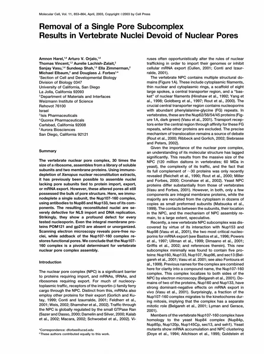
Figure 1. Metazoan Nup85, a New Component of the Nup107-160 Complex
(A) A model of vertebrate NPC structure, showing the cytoplasmic filaments (dark blue), cytoplasmic ring (blue), central scaffold spokes(yellow-green), central transporter (dark green), and nuclear ring and nuclear pore basket (red). The approximate location of a subset ofnucleoporins is shown.(B) Anti-mouse Nup85 antibody recognizes a single �70 kDa protein in HeLa cell extract (lane 1, 1 �l, 10 �g total protein), rat liver nuclei (lane2, 1 �l; �12,000 nuclei), and in Xenopus egg cytosol (lane 3, Ext, 0.1 �l) but not membranes (lane 4, 0.1 �l). Markers are 200, 120, 90, 68, 53,and 36 kDa.(C) Immunoprecipitation from Xenopus egg cytosol using anti-mNup85 (lane 2) and preimmune sera (lane 1). The blot was probed with anti-mNup85.(D) Immunoprecipitation from HeLa cell extracts using anti-mNup85 (lane 2) and preimmune sera (lane 1). The blot was cut horizontally andprobed with mAb414 (to detect Nup358, Nup214, and Nup153) and individual antisera to Nup160, Nup 133, Nup85, sec13, and Gle2. The FGnucleoporins and Gle2 were absent from these immunoprecipitates (lane 2), as were Nup62, Nup93, Nup98, Nup155, and Nup205 (data notshown).(E) Immunoprecipitation from HeLa cell extracts using anti-hNup133 (lane 2) and preimmune sera (lane 1); the blot was probed with anti-hNup133 and anti-mNup85 antisera.(F) Nup85 is present in assembled pores. Annulate lamellae were assembled, and the pores were purified (Miller and Forbes, 2000) and partiallysolubilized before immunoprecipitation with anti-mNup85. Nup85 was immunoprecipitated from purified pores by anti-Nup85 (lane 2).
al., 1996; Li et al., 1995; Siniossoglou et al., 1996, 2000; complex forms a Y-shaped structure, and addition ofS.c. Nup133 lengthens this Y to �40 nm (SiniossoglouLutzmann et al., 2002, and references therein). Although
no single Nup84 complex gene is essential, mutations et al., 2000; Lutzmann et al., 2002). The complex theoreti-cally could form part of the basket filaments, a subunitin any two cause synthetic lethality. The yeast Nup84
Nup107-160 Complex Is Pivotal for NPC Assembly855
of the rings, a portion of the spokes, or some other Immunofluorescence with the anti-70 kDa antibodyproduced a punctate nuclear rim stain (Figure 2A), whilestructure of the yeast NPC.
In vertebrates, nuclear reconstitution has proven to be transfection of a myc-tagged version showed incorpora-tion into NPCs (Figure 2B). We conclude that the �70a powerful tool for assigning function to nucleoporins.
Nuclei reconstituted in vitro from Xenopus egg extracts kDa mouse protein and its relatives are indeed verte-brate Nup85. The Xenopus Nup85 protein sequencecontain double nuclear membranes and nuclear pore
complexes and show robust import, DNA replication, (data not shown) has �55% identity to mouse Nup85,explaining its strong crossreactivity with the anti-mouseand pol III transcription (Forbes et al., 1983; Lohka and
Masui, 1983; Blow and Laskey, 1986; Newport, 1987; Nup85 antibody (Figures 1B, 1C, and 1F). Near the endof our study, a proteomic analysis of NPC proteins solu-Ullman and Forbes, 1995; Zhang and Clarke, 2000).
When egg extracts are immunodepleted of the central bilized from rat nuclear envelopes observed a rat Nup85homolog and termed the human relative hNup75 (Cron-transporter components, Nup62/58/54/45, the resulting
nuclei are defective for import (Finlay et al., 1991). In shaw et al., 2002). Our data indicate that we have identi-fied Nup85 homologs from S. pombe, Drosophila, C.contrast, when nuclei are reconstituted without the cyto-
plasmic ring protein Nup214 or the basket protein elegans, rice, Arabidopsis, and humans, with sizes rang-ing from 68 to 85 kDa (see Experimental Procedures).Nup98, both known to play a role in export, the nuclei
have no defect in import (Powers et al., 1995; Grandi For clarity and consistency with yeast, we collectivelyterm the proteins Nup85. Metazoan Nup85 is quite dis-et al., 1997; Walther et al., 2002). Nuclei reconstituted
without Nup153 are found to lack the basket-associated similar from yeast Nup85, having only 13%–14% iden-tity. Structurally, human Nup85 is �-helical (GOR4 Pro-proteins Nup98, Nup93, and Tpr, while nuclei missing
Nup358 lack cytoplasmic filaments (Walther et al., 2001, gram) and contains no obvious conserved domains orFG repeats.2002). Thus, in each of these cases, removal of a
nucleoporin and its immediate neighbors chips away atNPC structure but does not affect it at its central core. Nup85 Is Present at the Kinetochores
In search of essential building blocks of the vertebrate of Mitotic Chromosomesnuclear pore, we examined the Nup107-160 complex. At interphase, the Nup107-160 complex plays a role inUsing immunodepletion and nuclear reconstitution, we mRNA export (Vasu et al., 2001). During mitosis, how-have unexpectedly identified the biochemical equivalent ever, a fraction of the complex moves to the kineto-of a null mutant for the vertebrate nuclear pore. Absence chores (Belgareh et al., 2001). We found Nup85 to showof the Nup107-160 complex gives rise to nuclei with the same strong mitotic association with kinetochoresstrikingly severe defects in NPC structure and assembly. (Figures 2C and 2D). Thus, Nup85 is concluded to be
available for participation with the Nup107-160 complexin its as yet unknown role at the mitotic kinetochores.Results
Antibodies to Vertebrate Nup85, a New Component Anti-Nup85-Depleted Nuclei Are Deficientin NPC Number and Importof the Nup107-160 Complex
One arm of the yeast Y-shaped Nup84 complex is com- Recent advances in metazoan cells have used transfec-tion of small double-stranded interfering RNAs (RNAi)posed of Nup85 and seh1 (Siniossoglou et al., 2000;
Lutzmann et al., 2002). The related vertebrate Nup107- to target the destruction of specific mRNAs (Elbashir etal., 2001). We initially attempted to knock out Nup85 in160 complex was puzzling in that there was no readily
apparent Nup85 candidate, either by simple BLAST HeLa cells using RNAi. A large but incomplete reductionin the Nup85 nuclear rim stain was indeed observedsearches or silver staining (Vasu et al., 2001; Belgareh
et al., 2001). When a more stringent PSI-BLAST search (Figure 2E). Interestingly, the FG nucleoporins whichmap throughout the length of the NPC were also greatlywas done, however, we identified mouse ESTs which
could be pieced together to encode a putative ORF of reduced (Figure 2F). This suggested that the removal ofNup85 might cause extensive defects in the NPC. These656 aa. A PSI-BLAST search with this ORF sequence
revealed relatives from humans to S. pombe ranging in results, while provocative, represented only a partialknockout (see also Boehmer et al., 2003). The desire forsize from 598–733 aa, as well as the S. cerevisiae Nup85
(744 aa). a more complete knockout led us to attack the role ofNup85 at the level of pore complex assembly.This mouse ORF was cloned, expressed as protein,
and antibody raised. The antibody recognized a single Nuclear reconstitution can be accomplished in vitroby combining chromatin with the membrane and cytosol�70 kDa band in humans, rats, and Xenopus (Figures
1B and 1C). It further coimmunoprecipitated Nup160, fractions of a Xenopus egg lysate. Antibody to Nup85was used here to immunodeplete Nup85 and its associ-Nup133, and sec13, three known members of the verte-
brate Nup107-160 complex (Figure 1D), but not other ated proteins from the cytosol, while preimmune IgG wasused for mock-depletion. The great majority of Nup85nucleoporins. Anti-Nup133 antibody conversely coimmu-
noprecipitated the 70 kDa protein (Figure 1E). Pull-downs was removed by immunodepletion (Figure 3A, lane 2).Strikingly, Nup133 and Nup160 were also largely re-with the Nup153 beads originally used to identify the
Nup107-160 complex in Xenopus (Vasu et al., 2001) were moved (Figure 3C, lane 2). In addition, sec13 was par-tially depleted (Figure 3C), consistent with its presencealso found to contain the 70 kDa protein (data not shown).
Most tellingly, the �70 kDa protein was present in puri- both in the Nup107-160 complex (depleted) and a sepa-rate vesicular trafficking complex (not depleted) (Shay-fied Xenopus pores (Figure 1F) (Miller and Forbes, 2000).
Molecular Cell856
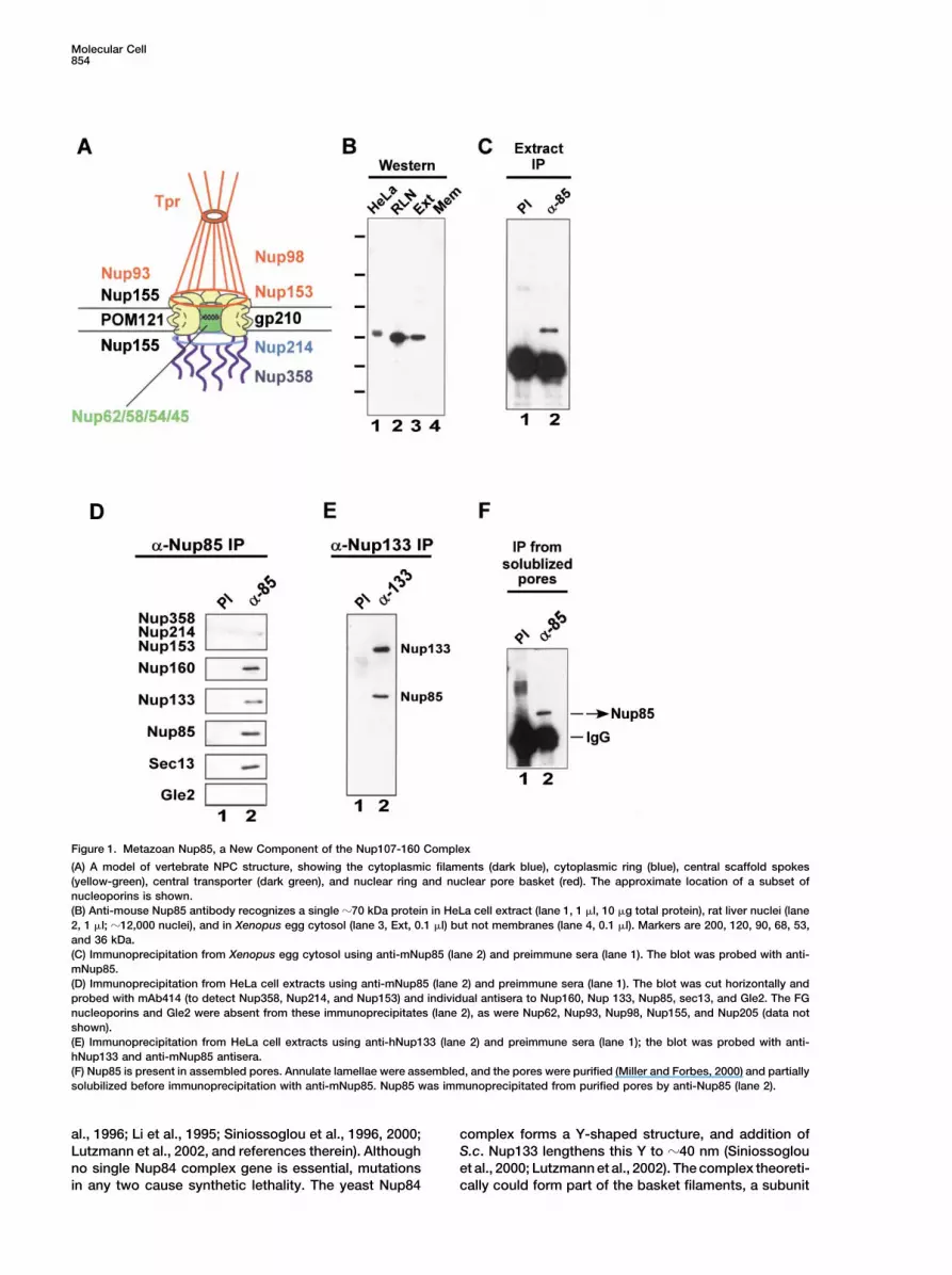
Figure 2. Nup85 Is Present in NPCs and onMitotic Kinetochores
(A) Immunofluorescence with anti-mNup85antibody on HeLa cells; the inset shows amagnified portion, revealing the punctate nu-clear rim.(B) Transfection of myc-tagged mNup85 intoHeLa cells and immunofluorescence with anti-myc antibodies. Mouse Nup85 localizes to hu-man NPCs. The inset shows a magnified por-tion of the nuclear rim and individual NPCs.(C and D) A mitotic cell from the experimentin (A) is shown stained with anti-mNup85 (C)and the DNA dye DAPI (D). A fraction of Nup85localizes to the kinetochores.(E and F) RNAi was performed on HeLa cellsusing dsNup85 oligomers. At 72 hr, RNAigreatly reduced the amount of Nup85 presentin the NPCs, as assessed using anti-Nup85antibodies (Nup85), and prevented the major-ity of FG nucleoporins from associating withnuclear NPCs, as assessed with mAb414 an-tibody (414). The scale bar represents 10 �min (A) and (B) and (E) and (F), 5 �m in (C) and(D), and �2.7 �m in the insets for (A) and (B).
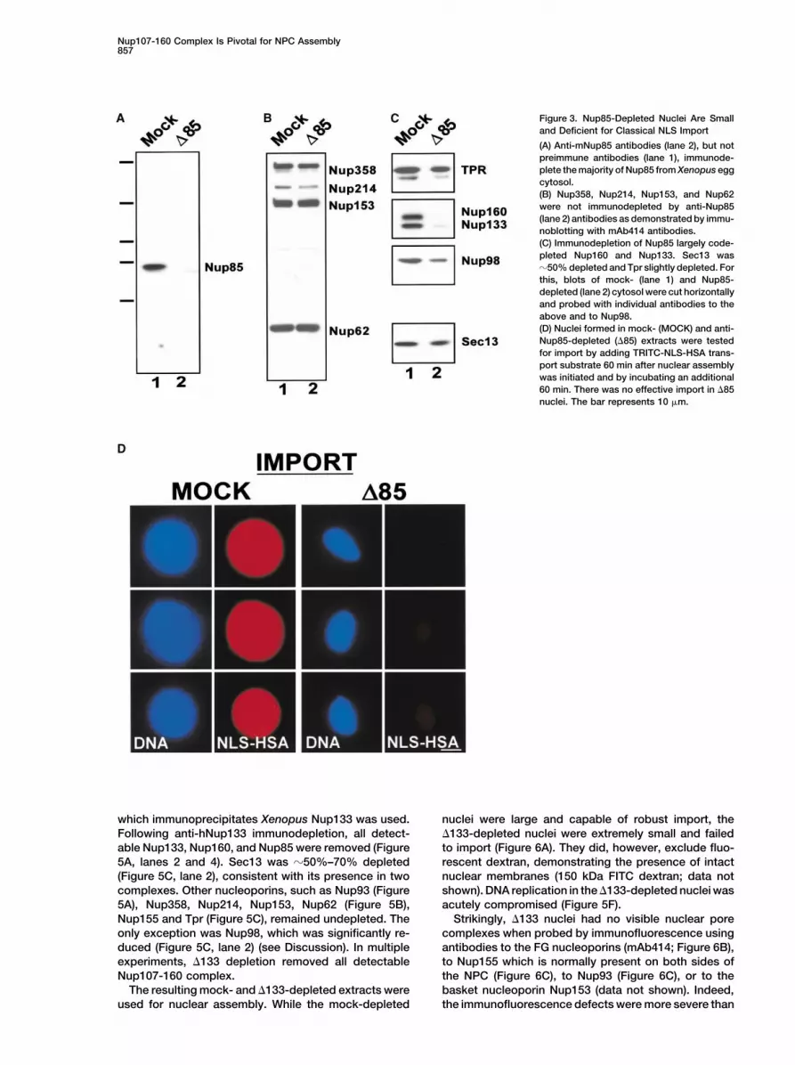
witz et al., 1995; Vasu et al., 2001; Cronshaw et al., clear rim on mock-depleted nuclei (Figures 4A and 4B,MOCK), but showed a dramatically reduced stain on2002). The FG-nucleoporins Nup358, Nup214, Nup153,
and Nup62 were undepleted (Figure 3B, lane 2), as were �85-depleted nuclei, with very few visible NPCs (Figures4A and 4B, �85). Moreover, individual antibodies to themultiple other nuclear proteins, including PCNA, im-
portin � and �, transportin, Nup93, Nup155, and Nup205 basket nucleoporin Nup153 or the cytoplasmic ring/fila-ment protein Nup214 failed to stain �85-depleted nuclei(data not shown). Nup98 and Tpr showed a limited
depletion (Figure 3C, lane 2; see also below). Overall, (Figure 4C). Thus, anti-Nup85 removal of the Nup107-160 complex, although not complete, resulted in nucleiwe conclude that anti-Nup85 depletion removes the ma-
jority of the Nup107-160 complex, as determined by the which not only showed a large decrease in NPC number,but lacked basket and cytoplasmic ring/filament pro-simultaneous decrease in Nup85, Nup133, and Nup160.
To assemble nuclei lacking the Nup107-160 complex, teins.anti-Nup85-depleted cytosol (�85) was combined withchromatin and membrane vesicles and incubated for 60min to allow for nuclear growth. Fluorescently labeled Complete Immunodepletion of the Nup107-160
Complex Yields Nuclei Devoid of NPCsnuclear import substrate (TRITC-NLS-HSA) was thenadded. The �85-depleted nuclei proved to be quite small In the anti-Nup85 depletion above a small amount of
Nup85, Nup133, and potentially other complex membersand almost completely defective for classical NLS im-port (Figure 3D). Monoclonal mAb414, which recognizes remained (Figure 3C). To attempt to more completely
deplete the complex, an antibody to human Nup133FG-containing nucleoporins, gave a bright punctate nu-
Nup107-160 Complex Is Pivotal for NPC Assembly857
Figure 3. Nup85-Depleted Nuclei Are Smalland Deficient for Classical NLS Import
(A) Anti-mNup85 antibodies (lane 2), but notpreimmune antibodies (lane 1), immunode-plete the majority of Nup85 from Xenopus eggcytosol.(B) Nup358, Nup214, Nup153, and Nup62were not immunodepleted by anti-Nup85(lane 2) antibodies as demonstrated by immu-noblotting with mAb414 antibodies.(C) Immunodepletion of Nup85 largely code-pleted Nup160 and Nup133. Sec13 was�50% depleted and Tpr slightly depleted. Forthis, blots of mock- (lane 1) and Nup85-depleted (lane 2) cytosol were cut horizontallyand probed with individual antibodies to theabove and to Nup98.(D) Nuclei formed in mock- (MOCK) and anti-Nup85-depleted (�85) extracts were testedfor import by adding TRITC-NLS-HSA trans-port substrate 60 min after nuclear assemblywas initiated and by incubating an additional60 min. There was no effective import in �85nuclei. The bar represents 10 �m.
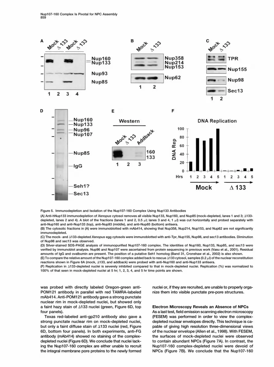
which immunoprecipitates Xenopus Nup133 was used. nuclei were large and capable of robust import, the�133-depleted nuclei were extremely small and failedFollowing anti-hNup133 immunodepletion, all detect-
able Nup133, Nup160, and Nup85 were removed (Figure to import (Figure 6A). They did, however, exclude fluo-rescent dextran, demonstrating the presence of intact5A, lanes 2 and 4). Sec13 was �50%–70% depleted
(Figure 5C, lane 2), consistent with its presence in two nuclear membranes (150 kDa FITC dextran; data notshown). DNA replication in the �133-depleted nuclei wascomplexes. Other nucleoporins, such as Nup93 (Figure
5A), Nup358, Nup214, Nup153, Nup62 (Figure 5B), acutely compromised (Figure 5F).Strikingly, �133 nuclei had no visible nuclear poreNup155 and Tpr (Figure 5C), remained undepleted. The
only exception was Nup98, which was significantly re- complexes when probed by immunofluorescence usingantibodies to the FG nucleoporins (mAb414; Figure 6B),duced (Figure 5C, lane 2) (see Discussion). In multiple
experiments, �133 depletion removed all detectable to Nup155 which is normally present on both sides ofthe NPC (Figure 6C), to Nup93 (Figure 6C), or to theNup107-160 complex.
The resulting mock- and �133-depleted extracts were basket nucleoporin Nup153 (data not shown). Indeed,the immunofluorescence defects were more severe thanused for nuclear assembly. While the mock-depleted

Molecular Cell858
Figure 4. Nuclei Formed in Nup85-Depleted Extract Are Impaired in NPC Assembly
(A) Nuclei reconstituted in mock-depleted (MOCK) and anti-Nup85-depleted cytosol (�85) for 90 min were fixed and stained 60 min withdirectly labeled Oregon green-mAb414 antibody (mock-414 or �85-414). �85-depleted nuclei were small and stained poorly with the mAb414,indicating a defect in NPC assembly.(B) Higher magnification views of (A), showing portions of the nuclear rims of mock-depleted (mock-414, 1X exposure) and �85-depleted(�85-414, 3X exposure) nuclei. A longer exposure was required to see the more lightly staining pores of �85-depleted nuclei.(C) Mock-depleted nuclei and �85-depleted nuclei were assembled 60 min, fixed, and subjected to immunofluorescence with anti-Nup214(�214) and anti-Nup153 (�153) antibodies and TRITC-labeled secondary antibody. The Nup85-depleted nuclei lack Nup214 and Nup153. Thebar represents 3 �m in (A) through (C).
those seen in �85 depleted nuclei, in that there were no These last results indicate that the defects are specificallydue to the absence of the Nup107-160 complex.NPCs stained (Figures 6B and 6C).
When small amounts of highly purified Nup107-160complex (Figures 5D and 5E) were added back to the Defects in Integral Membrane Pore
Protein Localization�133-depleted extract at t � 0, this complex was ableto restore import function in the resulting nuclei (Figure We next tested for presence of the integral membrane
proteins of the NPC, POM121 and gp210, in the depleted6A, addback). Moreover, abundant NPCs that stained withanti-FG antibody were now present (Figure 6B, addback). nuclei (Hallberg et al., 1993; Greber et al., 1990). POM121
Nup107-160 Complex Is Pivotal for NPC Assembly859
Figure 5. Immunodepletion and Isolation of the Nup107-160 Complex Using Nup133 Antibodies
(A) Anti-hNup133 immunodepletion of Xenopus cytosol removes all visible Nup133, Nup160, and Nup85 (mock-depleted, lanes 1 and 3; �133-depleted, lanes 2 and 4). A blot of the fractions (lanes 1 and 2, 0.5 �l; lanes 3 and 4, 1 �l) was cut horizontally and probed separately withanti-Nup160 and anti-Nup133 (top), anti-Nup93 (middle), and anti-Nup85 (bottom) antisera.(B) The cytosolic fractions in (A) were immunoblotted with mAb414, showing that Nup358, Nup214, Nup153, and Nup62 are not significantlyimmunodepleted.(C) The mock- and �133-depleted Xenopus egg cytosols were immunoblotted with anti-Tpr, Nup155, Nup98, and sec13 antibodies. Diminutionof Nup98 and sec13 was observed.(D) Silver-stained SDS-PAGE analysis of immunopurified Nup107-160 complex. The identities of Nup160, Nup133, Nup85, and sec13 wereverified by immunoblot analysis. Nup96 and Nup107 were ascertained from protein sequencing in previous work (Vasu et al., 2001). Residualamounts of IgG and ovalbumin are present. The position of a putative Seh1 homolog (Band 31, Cronshaw et al., 2002) is also shown.(E) To compare the relative amount of the Nup107-160 complex added back to rescue �133 cytosol, samples (0.2 �l) of the nuclear reconstitutionreactions shown in Figure 6A (mock, �133, and addback) were probed with anti-Nup160 and anti-Nup133 antisera.(F) Replication in �133-depleted nuclei is severely inhibited compared to that in mock-depleted nuclei. Replication (%) was normalized to100% of that seen in mock-depleted nuclei at 5 hr; 1, 2, 3, 4, and 5 hr time points are shown.
was probed with directly labeled Oregon-green anti- nuclei or, if they are recruited, are unable to properly orga-nize them into visible punctate pre-pore structures.POM121 antibody in parallel with red TAMRA-labeled
mAb414. Anti-POM121 antibody gave a strong punctatenuclear rim in mock-depleted nuclei, but showed onlya faint hazy stain of �133 nuclei (green, Figure 6D, top Electron Microscopy Reveals an Absence of NPCs
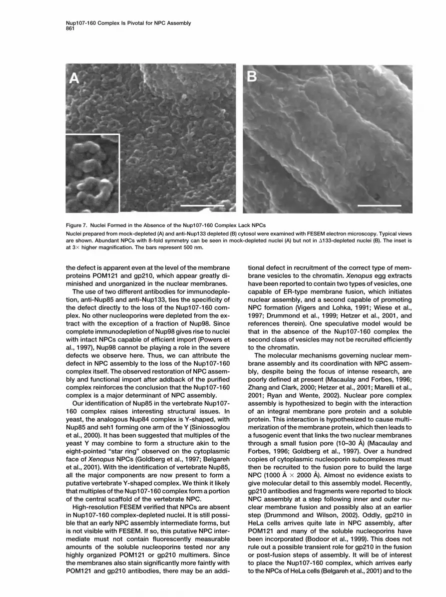
As a last test, field emission scanning electron microscopyfour panels).Texas red-labeled anti-gp210 antibody also gave a (FESEM) was performed in order to view the complex-
depleted nuclear envelopes directly. This technique is ca-strong punctate nuclear rim on mock-depleted nuclei,but only a faint diffuse stain of �133 nuclei (red, Figure pable of giving high resolution three-dimensional views
of the nuclear envelope (Allen et al., 1998). With FESEM,6D, bottom four panels). In both experiments, anti-FGantibody (mAb414) showed no staining of the complex- the surfaces of mock-depleted nuclei were observed
to contain abundant NPCs (Figure 7A). In contrast, thedepleted nuclei (Figure 6D). We conclude that nuclei lack-ing the Nup107-160 complex are either unable to recruit Nup107-160 complex-depleted nuclei were devoid of
NPCs (Figure 7B). We conclude that the Nup107-160the integral membrane pore proteins to the newly formed

Molecular Cell860
Figure 6. Nuclei Lacking the Nup107-160 Complex Are Defective for Multiple Nucleoporins Including the POM121 and gp210 Integral MembraneNucleoporins
(A) Anti-Nup133-depleted nuclei are defective for import of TRITC-NLS–HSA (�133), but can be rescued by addback of dilute purified Nup107-160 complex (addback). TRITC-NLS-HSA was added 60 min after nuclear assembly was initiated and visualized 15 min later.(B) Nuclei from an experiment similar to that in (A) were stained with Oregon green-labeled mAb414. No staining of mAb414 on the �133-depleted nuclei (�133) was observed. However, a bright punctate NPC stain was observed in �133-depleted nuclei to which purified Nup107-160 complex had been added at t � 0 (addback).(C) Nuclei lacking the Nup107-160 complex were completely devoid of a nuclear rim stain for Nup155 and Nup93. Only a faint residualintranuclear stain was observed. For this, nuclei were assembled 60 min, fixed, and processed for indirect immunofluorescence.(D) The NPC integral membrane proteins POM121 and gp210 appeared either absent or unorganized into punctate structures in nuclei lackingthe Nup107-160 complex (�133). For this, mock- and �133-depleted nuclei were assembled 60 min, fixed, and then stained with TAMRA-labeled mAb414 and Oregon green-labeled anti-POM121 (top four panels), or Oregon green-labeled mAb414 and Texas red-labeled anti-gp210 (bottom four panels). With either anti-POM121 or anti-gp210 antibodies, only faint diffuse membrane stain was observed in �133-depleted nuclei, while with mAb414, fluorescence was absent altogether. The bars represent 10 �m.
complex-depleted nuclei are defective for the earliest complex. Nuclei reconstituted in vitro lacking this com-plex contain nuclear membranes, but are small and virtu-visible steps in NPC assembly.ally incapable of NLS import and DNA replication. Strik-
Discussion ingly, these defects stem from the fact that the depletednuclei are unable to assemble or maintain any nuclearpore complexes. This profound defect in pore assemblyIn this study, we have identified an essential core ele-
ment of the vertebrate nuclear pore, the Nup107-160 is clearly visible by FESEM microscopy. The full extent of
Nup107-160 Complex Is Pivotal for NPC Assembly861
Figure 7. Nuclei Formed in the Absence of the Nup107-160 Complex Lack NPCs
Nuclei prepared from mock-depleted (A) and anti-Nup133 depleted (B) cytosol were examined with FESEM electron microscopy. Typical viewsare shown. Abundant NPCs with 8-fold symmetry can be seen in mock-depleted nuclei (A) but not in �133-depleted nuclei (B). The inset isat 3� higher magnification. The bars represent 500 nm.
the defect is apparent even at the level of the membrane tional defect in recruitment of the correct type of mem-brane vesicles to the chromatin. Xenopus egg extractsproteins POM121 and gp210, which appear greatly di-
minished and unorganized in the nuclear membranes. have been reported to contain two types of vesicles, onecapable of ER-type membrane fusion, which initiatesThe use of two different antibodies for immunodeple-
tion, anti-Nup85 and anti-Nup133, ties the specificity of nuclear assembly, and a second capable of promotingNPC formation (Vigers and Lohka, 1991; Wiese et al.,the defect directly to the loss of the Nup107-160 com-
plex. No other nucleoporins were depleted from the ex- 1997; Drummond et al., 1999; Hetzer et al., 2001, andreferences therein). One speculative model would betract with the exception of a fraction of Nup98. Since
complete immunodepletion of Nup98 gives rise to nuclei that in the absence of the Nup107-160 complex thesecond class of vesicles may not be recruited efficientlywith intact NPCs capable of efficient import (Powers et
al., 1997), Nup98 cannot be playing a role in the severe to the chromatin.The molecular mechanisms governing nuclear mem-defects we observe here. Thus, we can attribute the
defect in NPC assembly to the loss of the Nup107-160 brane assembly and its coordination with NPC assem-bly, despite being the focus of intense research, arecomplex itself. The observed restoration of NPC assem-
bly and functional import after addback of the purified poorly defined at present (Macaulay and Forbes, 1996;Zhang and Clark, 2000; Hetzer et al., 2001; Marelli et al.,complex reinforces the conclusion that the Nup107-160
complex is a major determinant of NPC assembly. 2001; Ryan and Wente, 2002). Nuclear pore complexassembly is hypothesized to begin with the interactionOur identification of Nup85 in the vertebrate Nup107-
160 complex raises interesting structural issues. In of an integral membrane pore protein and a solubleprotein. This interaction is hypothesized to cause multi-yeast, the analogous Nup84 complex is Y-shaped, with
Nup85 and seh1 forming one arm of the Y (Siniossoglou merization of the membrane protein, which then leads toa fusogenic event that links the two nuclear membraneset al., 2000). It has been suggested that multiples of the
yeast Y may combine to form a structure akin to the through a small fusion pore (10–30 A) (Macaulay andForbes, 1996; Goldberg et al., 1997). Over a hundredeight-pointed “star ring” observed on the cytoplasmic
face of Xenopus NPCs (Goldberg et al., 1997; Belgareh copies of cytoplasmic nucleoporin subcomplexes mustthen be recruited to the fusion pore to build the largeet al., 2001). With the identification of vertebrate Nup85,
all the major components are now present to form a NPC (1000 A � 2000 A). Almost no evidence exists togive molecular detail to this assembly model. Recently,putative vertebrate Y-shaped complex. We think it likely
that multiples of the Nup107-160 complex form a portion gp210 antibodies and fragments were reported to blockNPC assembly at a step following inner and outer nu-of the central scaffold of the vertebrate NPC.
High-resolution FESEM verified that NPCs are absent clear membrane fusion and possibly also at an earlierstep (Drummond and Wilson, 2002). Oddly, gp210 inin Nup107-160 complex-depleted nuclei. It is still possi-
ble that an early NPC assembly intermediate forms, but HeLa cells arrives quite late in NPC assembly, afterPOM121 and many of the soluble nucleoporins haveis not visible with FESEM. If so, this putative NPC inter-
mediate must not contain fluorescently measurable been incorporated (Bodoor et al., 1999). This does notrule out a possible transient role for gp210 in the fusionamounts of the soluble nucleoporins tested nor any
highly organized POM121 or gp210 multimers. Since or post-fusion steps of assembly. It will be of interestto place the Nup107-160 complex, which arrives earlythe membranes also stain significantly more faintly with
POM121 and gp210 antibodies, there may be an addi- to the NPCs of HeLa cells (Belgareh et al., 2001) and to the

Molecular Cell862
portin (Transduction Laboratories); anti-Xenopus POM121 (A.H. andchromatin in Xenopus reconstitution assays (R. Chan,D.J.F., unpublished data); anti-Xenopus Nup153 (Shah et al., 1998);A.V.O., and D.J.F., unpublished data), into this NPC as-anti-Xenopus Nup214 (Shah et al., 1998); anti-Xenopus gp210 lume-sembly model.nal domain (R. Chan and D.J.F., unpublished data); and anti-hsec13p
In summary, we have found that reconstitution of nu- antibody (see Vasu et al., 2001, for details). Indirect immunofluores-clei lacking the Nup107-160 complex results in the most cence and immunoblot analysis were done as in Shah et al. (1998).severe defect in NPC assembly yet observed, i.e., the
Immunodepletion and Nuclear Reconstitutionfull loss of the nuclear pore complex. We conclude thatCytosolic and membrane vesicle fractions of Xenopus egg extractsthe Nup107-160 complex is an essential core elementwere prepared as in Powers et al. (1995), except for the use of 500
of the NPC and is used very early in the decision to form mM KCl in the membrane wash buffer to remove residual amounts ofa nuclear pore. The Nup107-160 complex thus addition- soluble nucleoporins. The membranes were stored in 10 �l aliquotsally offers an attractive point for regulation of nuclear at �80�C, and used as a 20X stock. For immunodepletion, 300 �g
of anti-Nup85, 600 �g of anti-Nup133 IgG, or equal amounts ofpore complex assembly.preimmune IgG from the same rabbits were bound to 60 �l of proteinA-Sepharose overnight. The bound antibody beads were blockedExperimental Procedureswith 20 mg/ml BSA in PBS buffer and washed two to three timeswith ELB (Powers et al., 1995). Egg cytosol (200 �l) was added tocDNA Cloning and Antibody Production30 �l of antibody or preimmune IgG beads and tumbled 1 hr (4�C),A BLAST search with S. cerevisiae Nup85p (744 aa; gi 6322502)giving a ratio of 1.5 (or 3.0 for Nup133) �g antibody/�l cytosol,revealed a potential S. pombe homolog of 675 aa with 19% identity,for two consecutive depletions. The immuno- or mock-depleted
but no vertebrate homologs. An iterative PSI-BLAST search, whichcytosols were used for nuclear reconstitution by mixing with mem-
uses the residues conserved between the two yeast Nup85sbrane vesicles and demembranated sperm chromatin (Powers et
(Altschul et al., 1997), identified short mouse ESTs which could beal., 1995). DNA replication was assayed as in Grandi et al. (1997).
combined to give a 656 aa ORF. A cDNA clone encoding this ORF For indirect immunofluorescence, reconstituted nuclei were fixed,was prepared by RT-PCR of mouse 3T3 total RNA and cloned into pelleted onto poly-lysine-coated coverslips (Macaulay and Forbes,pET28a. His-tagged putative mouse Nup85 protein was expressed 1996), and probed with various antibodies. For direct immunofluo-in E. coli BL21/DE3 and used for rabbit immunization. Relatives rescence, mAb414 or affinity-purified anti-POM121 or -gp210 anti-of mouse Nup85 (gi 12856972) were found, in many cases as the bodies were coupled to succinimidyl ester derivatives of Oregongenomes were completed, in humans (656 aa; gi 10434102), Dro- green 488, Tetra-methyl-rhodamine (TAMRA), or Texas-redX (Mo-sophila (668 aa; gi 7302655), C. elegans (598 aa; gi 17508241), Arabi- lecular Probes, Eugene, OR). For this, nuclei were fixed with 3%dopsis thaliana (713 aa; gi 18418112), rice (733 aa; gi 21902047), formaldehyde, quenched with glycine, washed, and mixed with anti-Yarrowia lipolytica (708 aa; gi 18076820), and S. pombe (675 aa; gi body. To assess nuclear import, TRITC-SV40 NLS-HSA transport19112685). substrate was added 60 min after the start of assembly and visual-
Antibodies were raised to recombinant human Nup133 containing ized 15–30 min later. For addback experiments, the Nup107-160all but seven C-terminal amino acids (aa 1–1149) (Vasu et al., 2001) complex was purified by large-scale anti-Nup133 immunoprecipita-and affinity purified as in Shah et al. (1998). Preimmune IgG from tion from Xenopus egg cytosol. For this, affinity-purified antibodythe same rabbits was purified with protein A-Sepharose. Immuno- was coupled to protein A-Sepharose, blocked with ovalbumin, andprecipitations were performed as in Shah et al. (1998) using affinity- incubated in egg cytosol (2 ml) diluted in 50 ml PBS. The beadspurified IgG to mNup85 (aa 1–656), hNup133 (aa 1–1149), or preim- were washed extensively with PBS, once with 1 M NaCl, 50 mMmune antisera (1 �g) from the same rabbits. HeLa cells, grown in a Tris-HCl, and 1 mM DTT (pH 7.5), then eluted with 1 M MgCl2, 1 mM100 mm plate, were resuspended in 500 �l HMN buffer (20 mM DTT (pH 6.5). Purified complex was subjected to step dialysis intoHEPES [pH 7.5], 2.5 mM MgCl2, and 150 mM NaCl), passed through ELB minus sucrose and tested by adding 1/6 volume of highly puri-a 22-gauge needle ten times, and incubated with ovalbumin-blocked fied complex (or buffer) to a �133 extract, followed by nuclear recon-
stitution and assessment of import and mAb414 staining.antibody beads. Rat liver nuclei (12,000/�l) were prepared as inFinlay et al. (1991). Purified pores from Xenopus annulate lamellae
Field Emission Scanning Electron Microscopy(Miller and Forbes, 2000) were partially solubilized in 0.5 M NaCl,Mock- and anti-Nup133 depleted nuclei were assembled for 60 min0.5% Tween-20 in HM buffer (see above), diluted 10� in HMN, andas above, then prepared essentially as in Allen et al. (1998). Samplesimmunoprecipitated.were critical point dried from ultra-dry CO2 (BAL-TEC CPD 030),sputter coated with 3.4 nm chromium (EMITECH K575X), and exam-Transfection, Immunofluorescence, and RNAiined using a Philips XL30 ESEM FEG field emission scanning elec-HeLa cells on coverslips were transfected 16 hr with mouse Nup85tron microscope.in a pCS2MT vector using Effectene (QIAGEN). The cells were meth-
anol fixed (15 min �20�C), permeabilized (PBS/0.2% Triton X-100),Acknowledgmentsblocked (10 min, PBS/0.2% Triton X-100/5% fetal calf serum), and
incubated with anti-c-myc tag antibody (9E10; Calbiochem; 1 hr).The authors thank S. Allen for sequence searches, V. Delmar, M. Avi-Coverslips, mounted with Vectashield (Vector Laboratories), wereguetero, Zhongsheng You, and K. Wilson of U.C.S.D. for help withvisualized with a Zeiss Axioskop 2 microscope (63� objective). Im-experimental techniques, and R. Chan for use of her gp210 antibody
munofluorescence on interphase and mitotic HeLa cells was as inprior to publication. M.E. thanks Terry Allen for generous instruction
Vasu et al. (2001). For RNAi, HeLa cells were transfected for 3 daysin FESEM. This work was funded by a grant from the NIH (RO1
using 0.84 �g of Nup85 oligomer duplex (target: AACCCCTGGAGM33279) to D.J.F., a United States-Israel Binational Science Founda-
CAACATCTTGTT) or control luciferase oligomer duplex (Xeragon; tion grant to M.E. and D.J.F., funding from the Marcus Sieff FoundationHuntsville, AL) in Oligofectamine (Invitrogen), followed by methanol to M.E., and GANN Predoctoral Fellowships to A.V.O. and T.V.fixation and immunofluorescence. DNA was stained with Hoechst33258 or DAPI dye. Received: November 25, 2002
Revised: March 7, 2003Antibodies Accepted: March 13, 2003The antibodies used included affinity-purified anti-mouse Nup85 (aa Published: April 17, 20031–656); anti-human Nup133 (this study). Anti-human Nup160, anti-rat Nup98 (aa 43–470), and anti-rat Nup155 (aa 295–578) were de- Referencesscribed in Vasu et al. (2001). Also used were anti-Tpr (Shah et al.,1998); anti-hNup205 and anti-Nup93 (Miller and Forbes, 2000); anti- Aitchison, J.D., Blobel, G., and Rout, M.P. (1995). Nup120p: A yeastmouse Gle2 (a gift from M. Powers, Emory University, Atlanta, GA), nucleoporin required for nuclear pore distribution and mRNA trans-
port. J. Cell Biol. 131, 1659–1675.mAb414 (Covance); anti-importin �, anti-importin �, and anti-trans-
Nup107-160 Complex Is Pivotal for NPC Assembly863
Allen, T.D., Rutherford, S.Z., Bennion, G.R., Wiese, C., Riepert, S., evidence for structural intermediates in nuclear pore complex as-sembly. J. Cell Sci. 110, 409–420.Kiseleva, E., and Goldberg, M.W. (1998). Three-dimensional surface
structure analysis of the nucleus. In Methods in Cell Biology, Vol. Goldstein, A.L., Snay, C.A., Heath, C.V., and Cole, C.N. (1996). Pleio-53, M. Berrios, ed. (San Diego: Academic Press), pp. 125–138. tropic nuclear defects associated with a conditional allele of the
novel nucleoporin Rat9p/Nup85p. Mol. Biol. Cell 7, 917–934.Altschul, S.F., Madden, T.L., Schaffer, A.A., Zhang, J., Zhang, Z.,Miller, W., and Lipman, D.J. (1997). Gapped BLAST and PSI-BLAST: Gorlich, D., and Kutay, U. (1999). Transport between the cell nucleusa new generation of protein database search programs. Nucleic and the cytoplasm. Annu. Rev. Cell Dev. Biol. 15, 607–660.Acids Res. 25, 3389–3402.
Grandi, P., Dang, T., Pane, N., Shevchenko, A., Mann, M., Forbes,Bastos, R., Lin, A., Enarson, M., and Burke, B. (1996). Targeting and D., and Hurt, E. (1997). Nup93, a vertebrate homologue of yeastfunction in mRNA export of nuclear pore complex protein Nup153. Nic96p, forms a complex with a novel 205-kDa protein and is re-J. Cell Biol. 134, 1141–1156. quired for correct nuclear pore assembly. Mol. Biol. Cell 8, 2017–
2038.Belgareh, N., Rabut, G., Bai, S.W., van Overbeek, M., Beaudouin,J., Daigle, N., Zatsepina, O.V., Pasteau, F., Labas, V., Fromont- Greber, U.F., Senior, A., and Gerace, L. (1990). A major glycoproteinRacine, M., et al. (2001). An evolutionarily conserved nuclear pore of the nuclear pore complex is a membrane-spanning polypeptidesubcomplex, which redistributes in part to kinetochores in mamma- with a large lumenal domain and a small cytoplasmic tail. EMBO J.lian cells. J. Cell Biol. 152, 1147–1160. 9, 1495–1502.Blow, J.J., and Laskey, R.A. (1986). Initiation of DNA replication in Griffis, E.R., Altan, N., Lippincott-Schwartz, J., and Powers, M.A.nuclei and purified DNA by a cell-free extract of Xenopus eggs. Cell (2002). Nup98 is a mobile nucleoporin with transcription-dependent47, 577–587. dynamics. Mol. Biol. Cell 13, 1282–1297.Bodoor, K., Shaikh, S., Salina, D., Raharjo, W.H., Bastos, R., Lohka, Hallberg, E., Wozniak, R.W., and Blobel, G. (1993). An integral mem-M., and Burke, B. (1999). Sequential recruitment of NPC proteins brane protein of the pore membrane domain of the nuclear envelopeto the nuclear periphery at the end of mitosis. J. Cell Sci. 112, contains a nucleoporin-like region. J. Cell Biol. 122, 513–521.2253–2264. Hetzer, M., Meyer, H.H., Walther, T.C., Bilbao-Cortes, D., Warren,Boehmer, T., Enninga, J., Dales, S., Blobel, G., and Zhong, H. (2003). G., and Mattaj, I.W. (2001). Distinct AAA-ATPase p97 complexesDepletion of a single nucleoporin, Nup107, prevents the assembly function in discrete steps of nuclear assembly. Nat. Cell Biol. 3,of a subset of nucleoporins into the nuclear pore complex. Proc. 1086–1091.Natl. Acad. Sci. USA 100, 981–985. Hinshaw, J.E., Carragher, B.O., and Milligan, R.A. (1992). Architec-Conti, E., and Izaurralde, E. (2001). Nucleocytoplasmic transport ture and design of the nuclear pore complex. Cell 69, 1133–1141.enters the atomic age. Curr. Opin. Cell Biol. 13, 310–319. Kalab, P., Weis, K., and Heald, R. (2002). Visualization of a Ran-GTPCronshaw, J.M., Krutchinsky, A.N., Zhang, W., Chait, B.T., and Ma- gradient in interphase and mitotic Xenopus egg extracts. Sciencetunis, M.J. (2002). Proteomic analysis of the mammalian nuclear 295, 2452–2456.pore complex. J. Cell Biol. 158, 915–927. Li, O., Heath, C.V., Amberg, D.C., Dockendorff, T.C., Copeland, C.S.,Cullen, B.R. (2001). Journey to the center of the cell. Cell 105, Snyder, M., and Cole, C.N. (1995). Mutation or deletion of the S.697–700. cerevisiae RAT3/NUP133 gene causes temperature-dependent nu-
clear accumulation of poly(A) RNA and constitutive clustering ofDamelin, M., and Silver, P.A. (2000). Mapping interactions betweennuclear pore complexes. Mol. Biol. Cell 6, 401–417.nuclear transport factors in living cells reveals pathways through
the nuclear pore complex. Mol. Cell 5, 133–140. Lohka, M.J., and Masui, Y. (1983). Formation in vitro of sperm pronu-clei and mitotic chromosomes induced by amphibian ooplasmicDimaano, C., Bal, J.R., Prunuske, A.J., and Ullman, K.S. (2001). RNAcomponents. Science 220, 719–721.association defines a functionally conserved domain in the nuclear
protein Nup153. J. Biol. Chem. 276, 45349–45357. Lutzmann, M., Kunze, R., Buerer, A., Aebi, U., and Hurt, E. (2002).Modular self-assembly of a Y-shaped multiprotein complex fromDoye, V., Wepf, R., and Hurt, E.C. (1994). A novel nuclear pore proteinseven nucleoporins. EMBO J. 21, 387–397.Nup133p with distinct roles in poly(A) RNA transport and nuclearLyman, S.K., and Gerace, L. (2001). Nuclear pore complexes: dynam-pore distribution. EMBO J. 13, 6062–6075.ics in unexpected places. J. Cell Biol. 154, 17–20.Drummond, S.P., and Wilson, K.L. (2002). Interference with the cyto-Macara, I.G. (2002). Why FRET about Ran? Dev. Cell 2, 379–380.plasmic tail of gp210 disrupts “close apposition” of nuclear mem-
branes and blocks nuclear pore dilation. J. Cell Biol. 158, 53–62. Macaulay, C., and Forbes, D.J. (1996). Assembly of the nuclearpore: biochemically distinct steps revealed with NEM, GTPS, andDrummond, S., Ferrigno, P., Lyon, C., Murphy, J., Goldberg, M.,BAPTA. J. Cell Biol. 132, 5–20.Allen, T., Smythe, C., and Hutchison, C.J. (1999). Temporal differ-
ences in the appearance of NEP-B78 and an LBR-like protein during Marelli, M., Lusk, C.P., Chan, H., Aitchison, J.D., and Wozniak, R.W.Xenopus nuclear envelope reassembly reflect the ordered recruit- (2001). A link between the synthesis of nucleoporins and the biogen-ment of functionally discrete vesicle types. J. Cell Biol. 144, 225–240. esis of the nuclear envelope. J. Cell Biol. 153, 709–724.Elbashir, S.M., Harborth, J., Lendeckel, W., Yalcin, A., Weber, K., Matsuoka, Y., Takagi, M., Ban, T., Miyazaki, M., Yamamoto, T.,and Tuschl, T. (2001). Duplexes of 21-nucleotide RNAs mediate RNA Kondo, Y., and Yoneda, Y. (1999). Identification and characterizationinterference in cultured mammalian cells. Nature 411, 494–498. of nuclear pore subcomplexes in mitotic extract of human somatic
cells. Biochem. Biophys. Res. Commun. 254, 417–423.Feldherr, C.M., Akin, D., and Cohen, R.J. (2001). Regulation of func-tional nuclear pore size in fibroblasts. J. Cell Sci. 114, 4621–4627. Miller, B.R., and Forbes, D.J. (2000). Purification of the vertebrate
nuclear pore by biochemical criteria. Traffic 1, 941–951.Finlay, D.R., Meier, E., Bradley, P., Horecka, J., and Forbes, D.J.(1991). A complex of nuclear pore proteins required for pore func- Newport, J. (1987). Nuclear reconstitution in vitro: stages of assem-tion. J. Cell Biol. 114, 169–183. bly around protein-free DNA. Cell 48, 205–217.
Fontoura, B.M., Blobel, G., and Matunis, M.J. (1999). A conserved Powers, M.A., Macaulay, C., Masiarz, F.R., and Forbes, D.J. (1995).biogenesis pathway for nucleoporins: proteolytic processing of a Reconstituted nuclei depleted of a vertebrate GLFG nuclear pore186-kilodalton precursor generates Nup98 and the novel nucleo- protein, p97 (Nup98), import but are defective in nuclear growth andporin, Nup96. J. Cell Biol. 144, 1097–1112. replication. J. Cell Biol. 128, 721–736.
Forbes, D.J., Kirschner, M.W., and Newport, J.W. (1983). Spontane- Powers, M.A., Forbes, D.J., Dahlberg, J.E., and Lund, E. (1997). Theous formation of nucleus-like structures around bacteriophage DNA vertebrate GLFG nucleoporin, Nup98, is an essential component ofmicroinjected into Xenopus eggs. Cell 34, 13–23. multiple RNA export pathways. J. Cell Biol. 136, 241–250.
Reichelt, R., Holzenburg, A., Buhle, E.L., Jr., Jarnik, M., Engel, A.,Goldberg, M.W., Wiese, C., Allen, T.D., and Wilson, K.L. (1997). Dim-ples, pores, star-rings, and thin rings on growing nuclear envelopes: and Aebi, U. (1990). Correlation between structure and mass distri-
Molecular Cell864
bution of the nuclear pore complex and of distinct pore complex Zhang, C., and Clarke, P.R. (2000). Chromatin-independent nuclearenvelope assembly induced by Ran GTPase in Xenopus egg ex-components. J. Cell Biol. 110, 883–894.tracts. Science 288, 1429–1432.Ribbeck, K., and Gorlich, D. (2002). The permeability barrier of nu-
clear pore complexes appears to operate via hydrophobic exclusion.EMBO J. 21, 2664–2671.
Rout, M.P., Aitchison, J.D., Suprapto, A., Hjertaas, K., Zhao, Y., andChait, B.T. (2000). The yeast nuclear pore complex. Composition,architecture, and transport mechanism. J. Cell Biol. 148, 635–652.
Ryan, K.J., and Wente, S.R. (2002). Isolation and characterizationof new Saccharomyces cerevisiae mutants perturbed in nuclearpore complex assembly. BMC Genet. 3, 17.
Sazer, S., and Dasso, M. (2000). The ran decathlon: multiple rolesof Ran. J. Cell Sci. 113, 1111–1118.
Schwoebel, E.D., Ho, T.H., and Moore, M.S. (2002). The mechanismof inhibition of Ran-dependent nuclear transport by cellular ATPdepletion. J. Cell Biol. 157, 963–974.
Shah, S., Tugendreich, S., and Forbes, D. (1998). Major binding sitesfor the nuclear import receptor are the internal nucleoporin Nup153and the adjacent nuclear filament protein Tpr. J. Cell Biol. 141,31–49.
Shamsher, M.K., Ploski, J., and Radu, A. (2002). Karyopherin beta2B participates in mRNA export from the nucleus. Proc. Natl. Acad.Sci. USA 99, 14195–14199.
Shaywitz, D.A., Orci, L., Ravazzola, M., Swaroop, A., and Kaiser,C.A. (1995). Human SEC13Rp functions in yeast and is located ontransport vesicles budding from the endoplasmic reticulum. J. CellBiol. 128, 769–777.
Siebrasse, J.P., and Peters, R. (2002). Rapid translocation of NTF2through the nuclear pore of isolated nuclei and nuclear envelopes.EMBO Rep. 3, 887–892.
Siniossoglou, S., Wimmer, C., Rieger, M., Doye, V., Tekotte, H.,Weise, C., Emig, S., Segref, A., and Hurt, E.C. (1996). A novel complexof nucleoporins, which includes sec13p and a sec13p homologue,is essential for normal nuclear pores. Cell 84, 265–275.
Siniossoglou, S., Lutzmann, M., Santos-Rosa, H., Leonard, K.,Mueller, S., Aebi, U., and Hurt, E. (2000). Structure and assembly ofthe Nup84p complex. J. Cell Biol. 149, 41–54.
Ullman, K.S., and Forbes, D.J. (1995). RNA polymerase III transcrip-tion in synthetic nuclei assembled in vitro from defined DNA tem-plates. Mol. Cell. Biol. 15, 4873–4883.
Ullman, K., Shah, S., Powers, M., and Forbes, D. (1999). The nucleo-porin Nup153 plays a critical role in multiple types of nuclear export.Mol. Biol. Cell 10, 649–664.
Vasu, S., and Forbes, D.J. (2001). Nuclear pores and nuclear assem-bly. Curr. Opin. Cell Biol. 13, 363–375.
Vasu, S., Shah, S., Orjalo, A., Park, M., Fischer, W.H., and Forbes,D.J. (2001). Novel vertebrate nucleoporins Nup133 and Nup160 playa role in mRNA export. J. Cell Biol. 155, 339–354.
Vigers, G.P., and Lohka, M.J. (1991). A distinct vesicle populationtargets membranes and pore complexes to the nuclear envelope inXenopus eggs. J. Cell Biol. 112, 545–556.
Walther, T.C., Fornerod, M., Pickersgill, H., Goldberg, M., Allen, T.D.,and Mattaj, I.W. (2001). The nucleoporin Nup153 is required fornuclear pore basket formation, nuclear pore complex anchoring andimport of a subset of nuclear proteins. EMBO J. 20, 5703–5714.
Walther, T.C., Pickersgill, H.S., Cordes, V.C., Goldberg, M.W., Allen,T.D., Mattaj, I.W., and Fornerod, M. (2002). The cytoplasmic fila-ments of the nuclear pore complex are dispensable for selectivenuclear protein import. J. Cell Biol. 158, 63–77.
Weis, K. (2002). Nucleocytoplasmic transport: cargo traffickingacross the border. Curr. Opin. Cell Biol. 14, 328–335.
Wiese, C., Goldberg, M.W., Allen, T.D., and Wilson, K.L. (1997). Nu-clear envelope assembly in Xenopus extracts visualized by scanningEM reveals a transport-dependent ‘envelope smoothing’ event. J.Cell Sci. 110, 1489–1502.
Yang, Q., Rout, M.P., and Akey, C.W. (1998). Three-dimensionalarchitecture of the isolated yeast nuclear pore complex: functionaland evolutionary implications. Mol. Cell 1, 223–234.