cyclostreptin binds covalently to microtubule pores and lumenal taxoid binding sites
TRANSCRIPT
Cyclostreptin binds covalently to microtubule pores andlumenal taxoid binding sitesRuben M Buey1,9, Enrique Calvo2,9, Isabel Barasoain1, Oriol Pineda3, Michael C Edler4, Ruth Matesanz1,Gemma Cerezo5, Christopher D Vanderwal6, Billy W Day7, Erik J Sorensen8, Juan Antonio Lopez2,Jose Manuel Andreu1, Ernest Hamel4 & J Fernando Dıaz1
Cyclostreptin (1), a natural product from Streptomyces sp. 9885, irreversibly stabilizes cellular microtubules, causes cell cyclearrest, evades drug resistance mediated by P-glycoprotein in a tumor cell line and potently inhibits paclitaxel binding tomicrotubules, yet it only weakly induces tubulin assembly. In trying to understand this paradox, we observed irreversible bindingof synthetic cyclostreptin to tubulin. This results from formation of covalent crosslinks to b-tubulin in cellular microtubules andmicrotubules formed from purified tubulin in a 1:1 total stoichiometry distributed between Thr220 (at the outer surface of a porein the microtubule wall) and Asn228 (at the lumenal paclitaxel site). Unpolymerized tubulin was only labeled at Thr220. Thus,the pore region of b-tubulin is an undescribed binding site that (i) elucidates the mechanism by which taxoid-site compoundsreach the kinetically unfavorable lumenal site and (ii) explains how taxoid-site drugs induce microtubule formation from dimericand oligomeric tubulin.
The mitotic spindle is an important target in cancer chemotherapy1.Spindle function is dependent on microtubule dynamics, whichinvolves stochastic gain and loss of ab-tubulin heterodimers frommicrotubule ends. Growth or shortening depends on the activationstate of tubulin, which is controlled by the exchangeable nucleotidebound to the b subunit. ‘‘Activated’’ tubulin-GTP adds to microtubuleends, whereas ‘‘deactivated’’ tubulin-GDP dissociates from polymerends and does not normally polymerize into microtubules.
Compounds that bind to tubulin arrest cells in mitosis and causeapoptosis. These agents interfere with microtubule dynamics even atintracellular concentrations far below the tubulin concentration2.Inhibitors of assembly (such as colchicine; 2) inactivate tubulin,thereby preventing microtubule formation. In contrast, microtubulestabilizing agents (MSAs) such as paclitaxel bind preferentially toassembled tubulin, thereby minimizing dissociation of tubulin-GDPfrom microtubule ends3. MSAs also induce assembly of the otherwiseinactive tubulin-GDP4. In recent years, many structurally diversetaxoid-site MSAs have been discovered, including epothilone B anddiscodermolide, which are biochemically more potent than paclitaxel5,and cyclostreptin (FR182877; Fig. 1a), which, though apparently lessactive than paclitaxel, has unusual biochemical properties6.
Paclitaxel-stabilized, zinc-induced sheets of antiparallel tubulinprotofilaments have been used for construction of a model of tubulin
with bound paclitaxel7. After fitting this model into electron densitymicrotubule maps, investigators concluded that paclitaxel binds tob-tubulin facing the microtubule lumen8. This model is supported bytubulin mutation data from cells that are resistant to paclitaxeland epothilones9.
Two points remain obscure about the biochemical mechanism bywhich taxoid-site ligands induce tubulin assembly. First, exami-nation of the binding kinetics of paclitaxel10 yields a high kineticassociation constant (kf = 3.6 � 106 M–1 s–1), which is inconsistentwith a relatively inaccessible lumenal binding site. An exposed site isalso supported by microtubule-associated protein (MAP)-mediatedreduction in the paclitaxel binding rate10 and by the binding ofantifluorescein antibody to a fluorescein-labeled taxoid bound tomicrotubules11. These findings led us to propose an initial bindingsite for paclitaxel on the outer microtubule wall near pore type I,which is close to the lumenal site, followed by translocation of thedrug to its lumenal site10. Because there is 1:1 stoichiometry forpaclitaxel binding to ab-tubulin, the two sites must be mutuallyexclusive, perhaps with a shared element.
Second, although apparently the paclitaxel site exists only inassembled microtubules12, taxoid-site drugs can induce microtubuleassembly under conditions in which tubulin is normally unable toassemble; thus, no binding sites can be demonstrated. Therefore, given
Received 20 July 2006; accepted 8 December 2006; published online 7 January 2007; doi:10.1038/nchembio853
1Centro de Investigaciones Biologicas, Consejo Superior de Investigaciones Cientıficas, Ramiro de Maeztu 9, Madrid 28040, Spain. 2Unidad de Proteomica, CentroNacional de Investigaciones Cardiovasculares, Melchor Fernandez Almagro 3, Madrid 28029, Spain. 3Departament de Quımica Organica, Facultat de Quımica,Universitat de Barcelona, Av. Diagonal 647, Barcelona 08028, Spain. 4Toxicology and Pharmacology Branch, Developmental Therapeutics Program, Division of CancerTreatment and Diagnosis, National Cancer Institute at Frederick, National Institutes of Health, Frederick, Maryland 21702, USA. 5PharmaMar, S.A. Avda. de los Reyes1, Colmenar Viejo 28770, Spain. 6Department of Chemistry, University of California, Irvine, California 92697, USA. 7Departments of Pharmaceutical Sciences andChemistry, University of Pittsburgh, Pittsburgh, Pennsylvania 15261, USA. 8Department of Chemistry, Princeton University, Princeton, New Jersey 08544, USA.9These authors contributed equally to this work. Correspondence should be addressed to J.F.D. ([email protected]).
NATURE CHEMICAL BIOLOGY VOLUME 3 NUMBER 2 FEBRUARY 2007 1 1 7
ART ICL ES

that assembly occurs, we postulated that there is a low-affinity bindingsite in tubulin dimers or oligomers that cannot be demonstratedbecause of low paclitaxel solubility12.
Although cyclostreptin13 weakly stimulates tubulin assembly, itavidly binds to microtubules, thereby strongly inhibiting the bindingof other MSAs to polymer. In addition, cyclostreptin-stabilizedmicrotubules disassemble at 0 1C more slowly than paclitaxel-stabilized microtubules6. We have now found that cyclostreptininteracts covalently both in vitro and in cells with polymerized tubulin,blocking the binding of even the most potent taxoid-site ligands.Moreover, cyclostreptin is fully active in multidrug-resistant (MDR)ovarian carcinoma (A2780/AD) cells overexpressing P-glycoprotein,which indicates that covalent binding might be a way to overcomeMDR. Using HPLC-MS, we found that two amino acid residues,Thr220 and Asn228, are modified in polymerized b-tubulin (Asn228is near the taxoid site facing the microtubule lumen; Thr220 abutspore type I), but only Thr220 is modified when cyclostreptin interactswith unpolymerized tubulin.
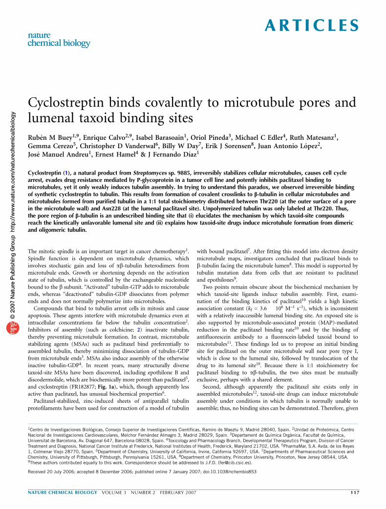
RESULTSCyclostreptin binds irreversibly to microtubulesBecause cyclostreptin strongly inhibits binding of taxoid-site ligandsto microtubules, we tried to detect microtubule-bound cyclostreptin.We analyzed cyclostreptin-treated microtubules by HPLC-MS. Con-trols showed that cyclostreptin should have been detectable, but thepellet extracts were devoid of the compound (Fig. 1b). Ligand in thesupernatant was greatly reduced following incubation of cyclostreptinwith microtubules. In the absence of degradation products, cyclo-streptin must have reacted irreversibly with the protein.
Cyclostreptin blocks binding of taxoid-site ligandsWe examined competition among taxoid-site ligands having differentaffinities for microtubules (discodermolide (3), epothilone B (4),epothilone A (5), cyclostreptin and paclitaxel (6); Kd at 35 1C of0.18, 1.3, 28, 49 and 70 nM, respectively)5,6,14. The competitionexperiments were performed by adding labeled competitor before orafter unlabeled ligand (Table 1). All ligands except cyclostreptinbehaved as expected if binding reversibly to the same site (order ofligand addition had no effect). With cyclostreptin, the feeble inhibition
of discodermolide and epothilone B binding and the strong inhibitionof paclitaxel binding without a preincubation became near totalinhibition with a preincubation. We also examined the binding tomicrotubules of 7-O-[N-(2,7-difluoro-4-fluoresceincarbonyl)-L-ala-nyl]paclitaxel (Flutax-2; 7), a fluorescent analog of paclitaxel15,which bound with an apparent Kd of 14 nM, but not to microtubulespreincubated with cyclostreptin (Fig. 1c).
Cyclostreptin and taxanes bind by different mechanismsCyclostreptin can displace Flutax-2 from stabilized microtubules withan apparent Ka similar to those of paclitaxel and docetaxel (8)6. Todetermine whether the same mechanism is involved15, we comparedthe effects of docetaxel and cyclostreptin on the kinetics of Flutax-2dissociation (Fig. 1d).
Docetaxel displaces Flutax-2 from microtubules because kf[doce-taxel] 44 Flutax-2 dissociation rate constant kr (ref. 15). With anyexcess [docetaxel], every empty site is rapidly filled by docetaxel(Fig. 1d). The Flutax-2 kr determined here was 8.9 ± 0.3 � 10–3 s–1
at 2 mM and 20 mM docetaxel, which is in agreement with thepreviously determined value15 of 7.10 ± 4 � 10–3 s–1 (error is s.e.m.for both values).
Cyclostreptin was very different. Although 2–40 mM cyclostreptinfully displaced bound Flutax-2 following monoexponential kinetic cur-ves (Fig. 1d), the observed dissociation rate constant (kobs) increasedwith cyclostreptin concentration until it equaled the kr of Flutax-2obtained with docetaxel (Fig. 1d). This indicates that the overall reac-tion is limited by the rate of cyclostreptin binding to microtubules. Theexplanation that cyclostreptin binds relatively slowly, with rebinding offree Flutax-2, is inconsistent with the monoexponential kinetic curves.These indicate a constant reaction rate, whereas increasing Flutax-2concentration should cause the apparent kr to decrease over time.
An alternative explanation is that cyclostreptin binds to the Flutax-2–microtubule complex and induces Flutax-2 dissociation:
TF + C.k1
k�1
TFC.k2
k�2
TC + F
where T represents the taxoid site in microtubules, C is cyclostreptinand F is Flutax-2. In this case the observed reaction step would be thesecond, with monophasic kinetics at steady state (concentration of
Cyclostreptin (1) Epoxidized cyclostreptin (9) Reduced cyclostreptin (10)
OO
HO OH
OO
OO
HOOH
O
OO
HO2
1
17OH
a b c
d
Cyclostreptin DocetaxelCyclostreptin
DMSO
1.2
1.0
0.8
0.6
Inte
nsity
(a.
u.)
0.4
0.2
0.020 21 22 23
Time (min)24 25 26
1.0
0.8
0.6
[Flu
tax-
2]bo
und/
[Flu
tax-
2]to
tal
0.4
0.2
0.0
[Sites]free (M)10–610–710–810–9
0.22
0.20
0.16
0.14
Ani
sotr
opy
0.10
0.06
0.040 1,000 2,000 3,000 4,000 5,000 6,000
Time (s)
0.18
0.12
0.08
Ani
sotr
opy
Time (s)
0.050.100.150.200.25
050
01,
000
1,50
0
[Cyclostreptin](µM)
Kob
s (×
10–3
s–1
)
00 10 20 30 40 50
1234567
O
Figure 1 Biochemistry of the cyclostreptin-microtubule interaction. (a) Chemical structure
of cyclostreptin and analogs. (b) HPLC-MS analysis of cyclostreptin extracted from pellets
(black) and supernatants (red) after incubation with (solid lines) and without (dashed
lines) stabilized microtubules. a.u., arbitrary units. (c) Binding of Flutax-2 to stabilized
microtubules preincubated with cyclostreptin or DMSO. Error bars are s.e.m. (d) Kinetics
of displacement of Flutax-2 from microtubules by cyclostreptin at 2 mM (black), 5 mM
(yellow), 10 mM (magenta), 20 mM (green), 40 mM (blue) or 2% DMSO (orange). Left
inset: displacement of Flutax-2 by docetaxel at 2 mM (black solid line) or 20 mM (red
dotted line). Right inset: dependence of the observed rate constant on [cyclostreptin],
solid line fit of the data to the kinetic model proposed.
ART ICL ES
11 8 VOLUME 3 NUMBER 2 FEBRUARY 2007 NATURE CHEMICAL BIOLOGY

TCF constant during the experiment) and the rate dependent on[cyclostreptin]. Curve fitting16 to this kinetic model yielded a k2 valueof 0.008 ± 0.002 s–1 (which is in agreement with the 0.006 s–1 value forFlutax-2 dissociation at 25 1C with docetaxel15), a negligible k–1 valueand a k1 value of 350 ± 20 M–1 s–1 (errors are s.e.m.). These valuesindicate that an irreversible reaction has occurred.
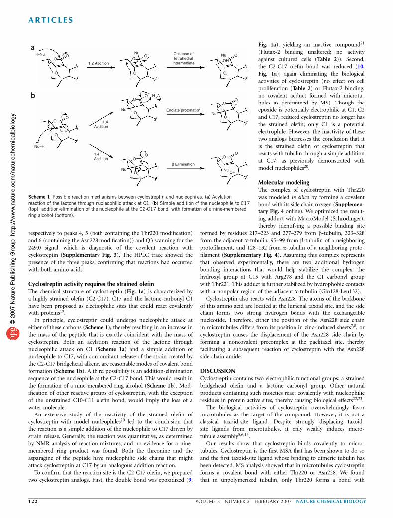
Cellular effects of cyclostreptinWe performed competition experiments between cyclostreptin andFlutax-2 using unfixed cytoskeletons from potoroo kidney (PtK2)cells. When cyclostreptin or paclitaxel wasadded to cytoskeletons with bound Flutax-2,both compounds displaced Flutax-2. PtK2cytoskeletons preincubated with paclitaxel,but not those preincubated with cyclostrep-tin, bound Flutax-2 (Fig. 2a–c), therebydemonstrating irreversible binding of cyclo-streptin to the cytoskeletons.
Similarly, there were differences in theeffects of cyclostreptin and paclitaxel oncellular microtubules. We observed partialrecovery of the microtubule network in pacli-taxel-treated PtK2 cells, but the effects ofcyclostreptin were irreversible. Cells wereincubated for 7 h with 2 or 5 mM cyclo-streptin or 10 mM paclitaxel, extensivelywashed and incubated for 16 h or 48 hlonger. Untreated cells had typical micro-
tubule networks and normal nuclei after 7 h (Fig. 2d). Cells incubatedfor 7 h with ligand had microtubule bundles (Fig. 2e,f), and a smallpercentage of them were micronucleated (4% of the cells) or arrestedin mitosis with multiple asters.
After washing and another 16 h in culture, untreated cells hadtypical microtubule networks (Fig. 2g) and normal nuclei, whereas inthe treated cells the cyclostreptin effect had progressed: the bundleswere unchanged (Fig. 2h), and cells with micronucleation or multi-polar spindles had increased in number to about 70%. With paclitaxel,microtubules had reverted to a normal appearance (Fig. 2i), but
Table 1 Dependence on order of ligand addition for the binding of discodermolide, paclitaxel and epothilone B to the taxoid site
Binding a of 2 mM
[3H]Discodermolide [14C]Epothilone B [3H]Paclitaxel
A B A B A B
(20 mM) Percentage of radioactive compound boundb ± s.d.
Cyclostreptin 96 ± 4 19 ± 6 79 ± 10 12 ± 1 60 ± 2 25 ± 2
Epothilone A 95 ± 1 96 ± 5 49 ± 1 49 ± 1 46 ± 3 49 ± 1
Epothilone B 76 ± 1 79 ± 5 9 ± 2 9 ± 1 31 ± 1 33 ± 4
Discodermolide 27 ± 1 31 ± 10 ND ND ND ND
aA columns: the radiolabeled ligand was added before the nonradiolabeled competitor. B columns: the nonradiolabeled competitor was added before the radiolabeled ligand. bThe values representthe amount of radiolabeled compound bound in the presence of the competitor relative to the amount of compound bound in its absence. ND, not determined.
a b c
d e f
g h i
Figure 2 Inhibition of binding of Flutax-2 to
PtK2 cytoskeletons by preincubation with
cyclostreptin and irreversibility of microtubule
effects on PtK2 cells grown in cyclostreptin.
(a–c) Cytoskeletons were preincubated with
DMSO (a), 10 mM cyclostreptin (b) or 10 mM
paclitaxel (c) and stained with Flutax-2. Insets:
Flutax-2–stained mitotic spindles from same
cytoskeleton preparations. (d–f) Cells cultured
for 7 h in the presence of DMSO (d), 5 mM
cyclostreptin (e) or 10 mM paclitaxel (f) andimmunostained. (g–i) Cells washed after
7 h, left in culture for 16 h and immunostained;
shown are the DMSO treatment (g), cyclostreptin
treatment (h) and paclitaxel treatment (i). Scale
bar represents 10 mm, and all panels and insets
have the same magnification.
ART ICL ES
NATURE CHEMICAL BIOLOGY VOLUME 3 NUMBER 2 FEBRUARY 2007 1 1 9

the number of micronucleated cells had increased to about 70%. Aftera 48-h recovery, cyclostreptin-treated cells had somewhat sparsermicrotubule bundles but an unchanged number of micronucleatedcells. After 48 h without drug, paclitaxel-treated cells continued torecover, with micronucleation reduced to about 40%.
We examined the PtK2 cells for DNA content by flow cytometry(Supplementary Table 1 online), and these data also indicated a largerrecovery after paclitaxel versus cyclostreptin treatment, with a largerpopulation of G2+M cells persisting after cyclostreptin treatment.
We tested the cytotoxicity of cyclostreptin in paclitaxel-resistant andnonresistant carcinoma cell lines (Table 2). In the nonresistant line,cyclostreptin was around 40 times less active than paclitaxel. However,PTX10 and PTX22, two lines that are resistant to paclitaxel as a resultof tubulin mutations, showed lower relative resistance to cyclostreptin(relative resistance index: 27 and 17 for paclitaxel, respectively, and 5and 1.3 for cyclostreptin, respectively). Moreover, cyclostreptin washighly active in the MDR line, with a resistance factor of 1.2, as com-pared with resistance factors of 600–900 for paclitaxel and docetaxel.
We performed cell cycle analysis in PtK2, A549, A2780 and A2780/AD cells incubated for 24 h with paclitaxel or cyclostreptin. The lowestconcentrations that gave maximal accumulation of cells in the G2+Mphase were 0.5 mM, 20 nM, 15 nM and 1 mM, respectively, forpaclitaxel, whereas for cyclostreptin this concentration was 100 nMin each of the four cell lines. Thus, cyclostreptin was more active thanpaclitaxel in the cell lines that are least sensitive to paclitaxel (PtK2 andA2780/AD) (Supplementary Table 1).
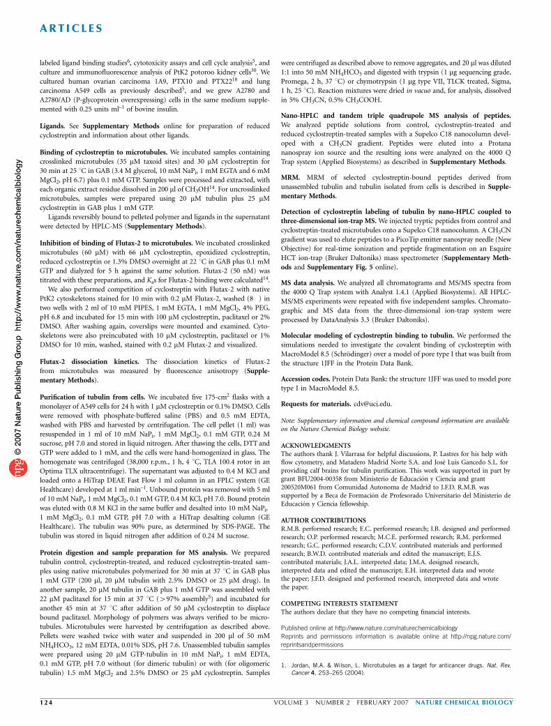
Characterization of cyclostreptin binding site by MSMS chromatograms of digested tubulin samples derived fromuntreated (Fig. 3a) or cyclostreptin-treated (Fig. 3b) microtubulesshowed differential, intense peaks corresponding to tubulin peptidesproducing a cyclostreptin-derived fragment ion at m/z 249.0 (seeMethods; peaks 1–2 and 3–7 in the trypsin- and chymotrypsin-digested samples, respectively). These signals were absent in controlsamples (Fig. 3a). MS/MS-based peptide sequencing (SupplementaryFig. 1 online) of peaks 1–7 demonstrated that all peptide sequencesmap into b-tubulin219–243 (Fig. 3c). This sequence contains part of thetaxoid site7. Peaks 1 and 2 correspond, respectively, to oxidized andnonoxidized cyclostreptin-modified 219-LTTPTYGDLNHLVSATMSGVTTCLR-243. The cyclostreptin-tagged peptide appeared in triply andquadruply charged form in the analyzed mass range. Peaks 3–7correspond to the sequences indicated (Fig. 3c). A comprehensivestudy of the fragmentation spectrum from the parent ion correspond-ing to peak 2 revealed b-tubulin219–243 to be the binding site for
cyclostreptin (Supplementary Fig. 1), but the 43 kDa size of thispeptide made the precise modification site uncertain. Analysis of thefragmentation spectrum from b-tubulin220–230 (peak 5) (Supplemen-tary Fig. 1) and b-tubulin220–235 (peak 7) revealed that Thr220 forms acovalent bond with cyclostreptin. Analysis of the fragmentationspectra from the ions corresponding to peaks 3, 4 and 6 (Supple-mentary Fig. 1) demonstrated a second modification site: Asn228.No doubly labeled peptides were found, so the covalent reactions withThr220 and Asn228 are mutually exclusive. Although relativeareas measured from chromatographic peaks corresponding to chy-motryptic peptides from cyclostreptin-treated samples (Fig. 3b) indi-cate a more extensive reaction with asparagine than with threonine,ionization of chromatographically separated peptides is substantiallyinfluenced by the composition of the pool of accompanying pep-tides eluted at a given retention time. The relative reactivity of thetwo amino acid residues thus cannot be determined reliably byMS analysis.
However, the chromatographic signal corresponding to 219-LTTPTYGDLNHLVSATMSGVTTCLR-243 showed that there is a 1:1stoichiometry for the interaction of cyclostreptin with ab-tubulin inmicrotubules (Supplementary Fig. 2 online). In the extracted ionchromatography (EIC) for triply charged ions, this peptide was presentin one of two alternative forms. Without cyclostreptin, unmodified219-LTTPTYGDLNHLVSATMSGVTTCLR-243 (m/z 884.4 Da) wasobserved. When cyclostreptin was included in the reaction mixture,only the corresponding peptide with one cyclostreptin added (m/z1,017.6 Da) was observed. Thus, all b-tubulin subunits in the micro-tubules were modified by addition of a single cyclostreptin.
We performed similar experiments under nonpolymerizing reactionconditions, in which either the heterodimer or small oligomericspecies predominate, depending on the Mg2+ concentration. Analysisof precursor ion scanning experiments demonstrated the presence ofweak chromatographic peaks 1, 2 and 5, but did not detect peaks 3, 4,6 and 7. Thus, formation of only small amounts of the Thr220 adductin nonpolymerized tubulin was detectable (from peak intensity, lessthan 5% with oligomeric tubulin and less than 0.5% with hetero-dimeric tubulin). To verify these results and increase sensitivity, wealso analyzed samples by multiple reaction monitoring (MRM) scanmode, fixing the Q3 quadrupole for the detection of the diagnostic ionat m/z 249.0 Da. Ions at m/z 1,017.6, 636.8, 816.4 and 779.4 (at the m/zof respective chromatographic peaks 2, 4, 5 and 6) were isolated andfragmented. Only precursors and fragmentation spectra from ionscorresponding to peaks 2 and 5 were detected (Fig. 3b), which againunambiguously demonstrates the Thr220 modification. We found no
Table 2 Effects of cyclostreptin compared with paclitaxel and docetaxel on the growth of human carcinoma cells
IC50 (nM) ± s.d.a
Cell line
Compound A2780 A2780/AD 1A9 PTX10 PTX22 A549
Paclitaxel 1.0 ± 0.3 900 ± 200 (900)b 1.1 ± 0.2 30 ± 9 (27) 19 ± 5 (17) 3.6 ± 0.4
Docetaxel 0.5 ± 0.1 285 ± 60 (570) 0.6 ± 0.1 ND ND 7.2 ± 0.3
Cyclostreptin 43.5 ± 4 51 ± 12 (1.2) 44 ± 6 240 ± 50 (5) 58 ± 7 (1.3) 45.5 ± 11
Epoxidized cyclostreptin Inactive Inactive Inactive Inactive Inactive Inactive
Reduced cyclostreptin Inactive Inactive Inactive Inactive Inactive Inactive
aIC50 (half-maximal inhibitory concentration) values determined in the ovarian carcinoma lines A2780 (parental line), A2780/AD (an MDR line overexpressing P-glycoprotein), 1A9 (a clone ofA2780), and PTX10 and PTX22 (paclitaxel-resistant tubulin mutants derived from 1A9), and in the non-small-cell lung carcinoma line A549. IC50 values were obtained in at least four independentexperiments. bThe numbers in parentheses are the calculated relative resistance values, obtained by dividing the IC50 value of the resistant line by the IC50 value of the parental line. ND, notdetermined. Inactive, no inhibition at 5 mM.
ART ICL ES
12 0 VOLUME 3 NUMBER 2 FEBRUARY 2007 NATURE CHEMICAL BIOLOGY

evidence for ions corresponding to peaks 4 and 6, which contain themodified Asn228. The MRM experiments confirmed that themodified peaks are ten-fold more intense in peptides derived fromoligomeric versus dimeric tubulin samples.
To verify that the Thr220 modification found in microtubules doesnot occur in oligomers before microtubule assembly, microtubulesstabilized by paclitaxel at a concentration (22 mM paclitaxel; 20 mMtubulin) much greater than the 1 mM drug required to suppressmicrotubule dynamics17 were incubated with excess cyclostreptin.This resulted in cyclostreptin displacing paclitaxel from the preformedmicrotubules. Because paclitaxel binding to microtubules is reversible,cyclostreptin should bind to transiently unoccupied sites, progressivelydisplacing the bound paclitaxel. Therefore, the very same labeling asoccurred in the previous experiment when cyclostreptin was addeddirectly to the microtubule assembly mixture is expected, unless theThr220 modification occurs in oligomers before microtubule assem-bly, in which case only labeling at Asn228 should be observed. InMRM experiments, ions at m/z 1,017.6, 636.8, 816.4 and 779.4 (peaks2, 4, 5 and 6, respectively) were detected, whereas in precursor ionscanning experiments, using the ion at m/z 249.0 as the diagnosticsignal, we also found tubulin-derived peptides corresponding to peaks3 and 7. Thus, the modifications at Thr220 and Asn228 wereunambiguously documented again. We therefore conclude that theThr220 modification must occur during the binding of cyclostreptinto microtubules.
Cyclostreptin labeling of cellular tubulinTo verify that the tubulin-cyclostreptin adduct is formed in treatedcells, we incubated A549 cells for 24 h with 1 mM cyclostreptin orDMSO, isolated their cytoskeletons and digested them with trypsin.We analyzed the digestion mixture by HPLC, with detection by MRM
scan mode for the m/z of the cyclostreptin-labeled form of 219-LTTPTYGDLNHLVSATMSGVTTCLR-243 (not shown). A peak at theexpected retention time for the labeled peptide was detected, whichindicates formation of the adduct in living cells.
To determine the proportion of cellular tubulin that had reacted withcyclostreptin, we purified tubulin from cyclostreptin-treated and con-trol A549 cells using a one-step ion-exchange procedure. This methodyielded tubulin of 490% purity (Supplementary Fig. 3 online), whichis suitable for MS procedures. After treatment with trypsin, the reactionmixture was subjected to HPLC, with analysis in the MRM scan modewith Q1 scanning for the m/z of unmodified and cyclostreptin-linked219-LTTPTYGDLNHLVSATMSGVTTCLR-243 (peak 2) and Q3 scan-ning for a common peptide in the y-fragmentation series of bothlabeled and unlabeled peptide (Fig. 3d). Whereas tubulin from controlcells showed only one peak, with an m/z of 884.4 (which correspondsto triply charged unmodified peptide), tubulin from cyclostreptin-treated cells showed two peaks. The major peak, with an m/z of1,017.6, corresponded to the triply charged peptide crosslinked tocyclostreptin, and the minor peak was identical to the unmodifiedpeptide from control cells. The areas of the peaks derived from tubulinin control and cyclostreptin-treated cells indicated that about 60% ofthe tubulin from treated cells contained the adduct. About 60% oftubulin in paclitaxel-treated 1A9 cells forms polymer18, a proportionthat is not very different from the proportion of tubulin that reactswith cyclostreptin in A549 cells. This suggests near total formation ofcyclostreptin adduct in the microtubules of treated A549 cells.
Further, we found that both Thr220 and Asn228 were covalentlymodified in the tubulin we obtained from cyclostreptin-treated cells.We did this by digesting the isolated tubulin with chymotrypsin. Thedigest was subjected to HPLC, with analysis in the MRM mode withQ1 scanning for signals at m/z 636.8, 816.4 and 779.4 (corresponding
Trya
b
c
d
TIC + prec (249)Tubulin
1.0 × 10⁵
0.025 35 45 55 65
25 35 45 55 65
Chy
TIC + prec (249) 219- -243Peaks 1 and 2
Peak 3
Peak 5Peak 6
Peak 4
Peak 7
Tubulin2.3 × 105
4.8 × 10⁵
2.6 × 10⁵
0.0
1.8 × 10³
2.0 × 10³
1.0 × 10³
0.0
9.0 × 10⁴
4.5 × 10⁴
0.0
3.8 × 10⁴
4.0 × 10⁴
2.0 × 10⁴
0.0
2.7 × 10⁵
2.0 × 10⁵
0.0
Inte
nsity
(c.
p.s.
)
0.020 30 40 50 60
TIC + prec (249)Tubulin + cyclostreptin
TIC + prec (249)Tubulin + cyclostreptin
DT + cyclostreptin+MRM 1,017.6 (Q1) 249.0 (Q3)
DT + cyclostreptin+MRM 636.8; 816.4; 779.4 (Q1) 249.0 (Q3)
20
31
2 4
5
6
7
30 40 50 60
25
25 45 652
35 45 55 65Time (min)
20
20 40
5
60
30 40 50 60
219- -243
219- -243
Inte
nsity
(c.
p.s.
)
+MRM 1,017.6; 884.4 (Q1) 836.4 (Q3)
Untreated cellsCyclostreptin-treated cells
60%
Cs
Time (min)
15 35 55 75
Figure 3 MS analyses of cyclostreptin binding to tubulin. (a,b) Total ion chromatogram (TIC) of the precursor ion scanning of fragment at m/z 249.0 from
control (a) or cyclostreptin-treated (b) tubulin samples, digested with trypsin (Try) or chymotrypsin (Chy). The lower panels of b show MRM experiments with
unassembled, cyclostreptin-treated dimeric tubulin (DT) samples. The upper insets show MRM results for the corresponding dimeric tubulin control samples.
Black arrows indicate the retention time expected for signals from peaks 2 (left, trypsin) and 5 (right, chymotrypsin). (c) Sequence of b-tubulin219-243
containing the amino acids Thr220 and Asn228 labeled with cyclostreptin (we use the sequence nomenclature of ref. 7, which is used extensively in
molecular modeling studies. The two cyclostreptin-modified amino acids are actually Thr218 and Asn226 in b-tubulin31, and the tryptic peptide actually
spans residues 217 to 241). Black triangles indicate the theoretical chymotrypsin cleavage sites within the tryptic peptide. (d) MRM experiments with
trypsin-digested tubulin extracted from untreated or cyclostreptin-treated cells. Cs, the cyclostreptin tag.
ART ICL ES
NATURE CHEMICAL BIOLOGY VOLUME 3 NUMBER 2 FEBRUARY 2007 1 2 1
respectively to peaks 4, 5 (both containing the Thr220 modification)and 6 (containing the Asn228 modification)) and Q3 scanning for the249.0 signal, which is diagnostic of the covalent reaction withcyclostreptin (Supplementary Fig. 3). The HPLC trace showed thepresence of the three peaks, confirming that reactions had occurredwith both amino acids.
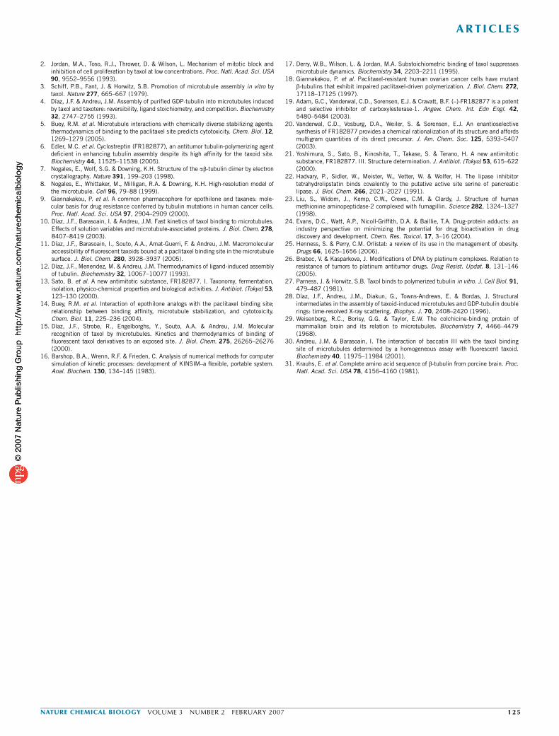
Cyclostreptin activity requires the strained olefinThe chemical structure of cyclostreptin (Fig. 1a) is characterized bya highly strained olefin (C2-C17). C17 and the lactone carbonyl C1have been proposed as electrophilic sites that could react covalentlywith proteins19.
In principle, cyclostreptin could undergo nucleophilic attack ateither of these carbons (Scheme 1), thereby resulting in an increase inthe mass of the peptide that is exactly coincident with the mass ofcyclostreptin. Both an acylation reaction of the lactone throughnucleophilic attack on C1 (Scheme 1a) and a simple addition ofnucleophile to C17, with concomitant release of the strain created bythe C2-C17 bridgehead alkene, are reasonable modes of covalent bondformation (Scheme 1b). A third possibility is an addition-eliminationsequence of the nucleophile at the C2-C17 bond. This would result inthe formation of a nine-membered ring alcohol (Scheme 1b). Mod-ification of other reactive groups of cyclostreptin, with the exceptionof the unstrained C10-C11 olefin bond, would imply the loss of awater molecule.
An extensive study of the reactivity of the strained olefin ofcyclostreptin with model nucleophiles20 led to the conclusion thatthe reaction is a simple addition of the nucleophile to C17 driven bystrain release. Generally, the reaction was quantitative, as determinedby NMR analysis of reaction mixtures, and no evidence for a nine-membered ring product was found. Both the threonine and theasparagine of the peptide have nucleophilic side chains that mightattack cyclostreptin at C17 by an analogous addition reaction.
To confirm that the reaction site is the C2-C17 olefin, we preparedtwo cyclostreptin analogs. First, the double bond was epoxidized (9,
Fig. 1a), yielding an inactive compound21
(Flutax-2 binding unaltered; no activityagainst cultured cells (Table 2)). Second,the C2-C17 olefin bond was reduced (10,Fig. 1a), again eliminating the biologicalactivities of cyclostreptin (no effect on cellproliferation (Table 2) or Flutax-2 binding;no covalent adduct formed with microtu-bules as determined by MS). Though theepoxide is potentially electrophilic at C1, C2and C17, reduced cyclostreptin no longer hasthe strained olefin; only C1 is a potentialelectrophile. However, the inactivity of thesetwo analogs buttresses the conclusion that itis the strained olefin of cyclostreptin thatreacts with tubulin through a simple additionat C17, as previously demonstrated withmodel nucleophiles20.
Molecular modelingThe complex of cyclostreptin with Thr220was modeled in silico by forming a covalentbond with its side chain oxygen (Supplemen-tary Fig. 4 online). We optimized the result-ing adduct with MacroModel (Schrodinger),thereby identifying a possible binding site
formed by residues 217–223 and 277–279 from b-tubulin, 323–328from the adjacent a-tubulin, 95–99 from b-tubulin of a neighboringprotofilament, and 128–132 from a-tubulin of a neighboring proto-filament (Supplementary Fig. 4). Assuming this complex representsthat observed experimentally, there are two additional hydrogenbonding interactions that would help stabilize the complex: thehydroxyl group at C15 with Arg278 and the C1 carbonyl groupwith Thr221. This adduct is further stabilized by hydrophobic contactswith a nonpolar region of the adjacent a-tubulin (Gln128-Leu132).
Cyclostreptin also reacts with Asn228. The atoms of the backboneof this amino acid are located at the lumenal taxoid site, and the sidechain forms two strong hydrogen bonds with the exchangeablenucleotide. Therefore, either the position of the Asn228 side chainin microtubules differs from its position in zinc-induced sheets7,8, orcyclostreptin causes the displacement of the Asn228 side chain byforming a noncovalent precomplex at the paclitaxel site, therebyfacilitating a subsequent reaction of cyclostreptin with the Asn228side chain amide.
DISCUSSIONCyclostreptin contains two electrophilic functional groups: a strainedbridgehead olefin and a lactone carbonyl group. Other naturalproducts containing such moieties react covalently with nucleophilicresidues in protein active sites, thereby causing biological effects22,23.
The biological activities of cyclostreptin overwhelmingly favormicrotubules as the target of the compound. However, it is not aclassical taxoid-site ligand. Despite strongly displacing taxoid-site ligands from microtubules, it only weakly induces micro-tubule assembly5,6,13.
Our results show that cyclostreptin binds covalently to micro-tubules. Cyclostreptin is the first MSA that has been shown to do soand the first taxoid-site ligand whose binding to dimeric tubulin hasbeen detected. MS analysis showed that in microtubules cyclostreptinforms a covalent bond with either Thr220 or Asn228. We foundthat in unpolymerized tubulin, only Thr220 forms a bond with
1,2 Addition
1,4Addition
1,4Addition
OO
O
O
OO
O
O
O
O OO
OH
O
H
H–A
Nu–H
OH
Nu Nu
NuNu
Nu Nu
Collapse oftetrahedral
intermediate
Enolate protonation
β Elimination
O–
O
O–
O
O
O
O
O
O
H-Nua
b
Scheme 1 Possible reaction mechanisms between cyclostreptin and nucleophiles. (a) Acylation
reaction of the lactone through nucleophilic attack at C1. (b) Simple addition of the nucleophile to C17
(top); addition-elimination of the nucleophile at the C2-C17 bond, with formation of a nine-membered
ring alcohol (bottom).
ART ICL ES
12 2 VOLUME 3 NUMBER 2 FEBRUARY 2007 NATURE CHEMICAL BIOLOGY

cyclostreptin, and this covalent interaction is much less extensive thanthe reaction with microtubules. We also demonstrated that bothcrosslinks to b-tubulin form in cyclostreptin-treated cells. Covalentbond formation explains the unusual biochemical properties ofcyclostreptin, such as the distinct requirement for higher temperaturesfor assembly induction and binding to polymer and the high stabilityof cyclostreptin polymer to disassembly at 0 1C6. Once covalentlybound to microtubules, cyclostreptin cannot dissociate and themicrotubules cannot disassemble, as they are more stable thanuntreated control and paclitaxel-induced microtubules.
Similarly, we found that PtK2 cellular microtubule bundles inducedby cyclostreptin are more stable than those induced by paclitaxel.Although cyclostreptin is 40-fold less cytotoxic than paclitaxel, itretains its activity in cells that are resistant to paclitaxel by over-expression of P-glycoprotein or by expression of mutant b-tubulins,becoming more potent than paclitaxel in the former case. Therefore,formation of a covalent tubulin adduct can be a mechanism to escapethese mechanisms of resistance, at least in cultured cells. Thoughagents that react covalently with specific targets can be too toxic foruse in humans24, some covalently reactive compounds are successfullyused in the clinic. Examples are the antiobesity drug tetrahydrolip-statin, which reacts with pancreatic lipase25, and the anticancerplatinum agents, which react with DNA26. In the MDR A2780/ADcells, we found that the covalent adduct of cyclostreptin with tubulincan escape the pump. With the tubulin mutants, cyclostreptin affinityfor the altered tubulins might be modified. Alternatively, the longertime frame involved in cell culture studies might still lead toirreversible tubulin inactivation by covalent bond formation, even ifthe tubulins have reduced affinity for cyclostreptin. In summary,because the formation of the adduct is a kinetically controlled,irreversible process, resistant tumor cells cannot escape the effect ofcyclostreptin by reducing its affinity for the target or by enhancingdrug efflux, which suggests that the design of ligands that covalentlyreact with the taxoid site or other targets might be an effective way toaddress drug resistance.
The discovery of the covalent reactions of cyclostreptin with tubulinand microtubules provides new insights into two obscure aspects ofthe mechanism of ligand binding to the taxoid site. First, modeling ofpaclitaxel-stabilized zinc sheet protofilaments into a microtubulestructure led to the conclusion that the taxoid site is on b-tubulinadjacent to the microtubule lumen7,8. In contrast, kinetic measure-ments, effects of MAPs and antibody binding10,11,15 indicated an easilyaccessible site, most logically on the outer microtubule surface. Toreconcile these observations, we proposed an exterior taxoid bindingsite to which ligands bind initially, before transfer to the lumenalsite10. The two sites could not be occupied simultaneously because ofthe observed 1:1 binding stoichiometry (similar stoichiometry hasbeen found for discodermolide (M.C.E., unpublished data) and forepothilone A and epothilone B binding14). The proposed site was atpore type I, including b-tubulin residues Phe214, Thr220, Thr221 andPro222 in the H6-H7 loop10. We have now shown that the taxoid-siteagent cyclostreptin reacts covalently with Thr220 at the proposed poretype I site, and it also reacts with Asn228, a residue at the lumenal site.
The proposed two-step mechanism is consistent with the two-stepkinetics of binding of fluorescent taxoids to microtubules15. Step oneis a bimolecular reaction with a micromolar Kd and a kf of about 106
M–1 s–1, which is most consistent with a diffusion-limited reactioninvolving an exposed site on the microtubule surface, such as the poretype I site containing Thr220. Step two is a monomolecular reactionwith a Keq B20, which indicates that about 5% of the bound ligandremains at the external site. The data obtained with the fluorescent
taxoids indicate that the first step involves binding of the taxoidmoiety without immobilization of the fluorescein group, as there is noincrease in fluorescence anisotropy. This is consistent with the taxoidmoiety binding at Thr220, because when the taxoid and fluorescentmoieties are separated by a longer linker, the fluorescein moiety is fullyaccessible to antibodies11.
The hypothesis that binding at the pore type I site, as manifested bythe reaction of cyclostreptin with Thr220, precedes binding of taxoid-site agents to the lumenal site is supported by the fact that covalentbinding of cyclostreptin to microtubules abolishes subsequent bindingof every taxoid-site agent examined (Table 1; Fig. 1c). In the Flutax-2experiment, we incubated 50 nM taxoid with up to 1 mM taxoid sitespreincubated with cyclostreptin, and Flutax-2 binding was completelyabolished. Even if only 1% of the taxoid sites had been accessible toFlutax-2, a change in the fluorescence anisotropy signal would havebeen detectable. Cyclostreptin reacts with tubulin in microtubules in a1:1 proportion, and therefore such microtubules, as the MS data show,would contain ab dimers with the b-tubulin modified at both Thr220and Asn228. Though dimers modified at Asn228 would not havebound Flutax-2 because the taxoid site would have been at least parti-ally occupied by the cyclostreptin moiety, one can imagine other routesto the lumenal taxoid site that would be available as a result of modi-fication at Thr220. For example, Flutax-2 might reach the lumenthrough type II pores or through microtubule ends. The failure to de-tect even low levels of Flutax-2 binding indicates that an initial inter-action at the pore type I site is an obligatory route to the lumenal site.
Second, previous studies12,27 have shown that paclitaxel binds avidlyto microtubules but not to ab heterodimers. However, paclitaxel andother taxoid-site agents initiate assembly in tubulin solutions in whichno microtubules (that is, presumptive binding sites) exist. It has beenproposed that paclitaxel might bind to tubulin oligomers28, therebyleading to assembly under otherwise unfavorable conditions. In thepresent work, we found a low level of cyclostreptin covalent reactivitywith Thr220 in dimeric and oligomeric tubulin, under conditions inwhich microtubule assembly is prevented by keeping the free Mg2+
concentration lower than that required to assemble microtubules with20 mM tubulin12. This demonstrates an MSA binding site in non-assembled tubulin. No reaction with Asn228 was observed, whichindicates that the lumenal site is not present without microtubuleformation. The binding affinity of MSAs for the site containing Thr220in ab heterodimers and oligomers must be very low, but binding tothis site might initiate an assembly reaction, with creation of the higheraffinity lumenal site and stabilization of nascent polymer. The irrever-sible covalent reaction of cyclostreptin with Thr220 allows trapping ofthe binding complex and detection of this evanescent species.
The assembly process might involve transformation of some ele-ments from acting as components of the external site into acting aspart of the lumenal site. However, the Thr220 cyclostreptin adductforms under all reaction conditions examined, including in pre-formed, paclitaxel-stabilized microtubules following paclitaxel displa-cement by cyclostreptin. Thus, the external pore type I site must alsoexist to some extent after assembly. Overall, the data presented heredemonstrate the existence of an MSA binding site in unassembledtubulin and support the hypothesis that taxoids reach the lumenal sitethrough transient binding to the pore type I site.
METHODSPreviously described methodologies. Previous papers provide details for
preparation of tubulin29, cyclostreptin20, epoxidized cyclostreptin21 and glutar-
aldehyde-stabilized microtubules, and also for quantitation of the taxoid sites of
glutaraldehyde-stabilized microtubules10,14, electron microscopy, radioactively
ART ICL ES
NATURE CHEMICAL BIOLOGY VOLUME 3 NUMBER 2 FEBRUARY 2007 1 2 3
labeled ligand binding studies6, cytotoxicity assays and cell cycle analysis5, and
culture and immunofluorescence analysis of PtK2 potoroo kidney cells30. We
cultured human ovarian carcinoma 1A9, PTX10 and PTX2218 and lung
carcinoma A549 cells as previously described5, and we grew A2780 and
A2780/AD (P-glycoprotein overexpressing) cells in the same medium supple-
mented with 0.25 units ml–1 of bovine insulin.
Ligands. See Supplementary Methods online for preparation of reduced
cyclostreptin and information about other ligands.
Binding of cyclostreptin to microtubules. We incubated samples containing
crosslinked microtubules (35 mM taxoid sites) and 30 mM cyclostreptin for
30 min at 25 1C in GAB (3.4 M glycerol, 10 mM NaPi, 1 mM EGTA and 6 mM
MgCl2, pH 6.7) plus 0.1 mM GTP. Samples were processed and extracted, with
each organic extract residue dissolved in 200 ml of CH3OH14. For uncrosslinked
microtubules, samples were prepared using 20 mM tubulin plus 25 mM
cyclostreptin in GAB plus 1 mM GTP.
Ligands reversibly bound to pelleted polymer and ligands in the supernatant
were detected by HPLC-MS (Supplementary Methods).
Inhibition of binding of Flutax-2 to microtubules. We incubated crosslinked
microtubules (60 mM) with 66 mM cyclostreptin, epoxidized cyclostreptin,
reduced cyclostreptin or 1.3% DMSO overnight at 22 1C in GAB plus 0.1 mM
GTP and dialyzed for 5 h against the same solution. Flutax-2 (50 nM) was
titrated with these preparations, and Kas for Flutax-2 binding were calculated14.
We also performed competition of cyclostreptin with Flutax-2 with native
PtK2 cytoskeletons stained for 10 min with 0.2 mM Flutax-2, washed (8�) in
two wells with 2 ml of 10 mM PIPES, 1 mM EGTA, 1 mM MgCl2, 4% PEG,
pH 6.8 and incubated for 15 min with 100 mM cyclostreptin, paclitaxel or 2%
DMSO. After washing again, coverslips were mounted and examined. Cyto-
skeletons were also preincubated with 10 mM cyclostreptin, paclitaxel or 1%
DMSO for 10 min, washed, stained with 0.2 mM Flutax-2 and visualized.
Flutax-2 dissociation kinetics. The dissociation kinetics of Flutax-2
from microtubules was measured by fluorescence anisotropy (Supple-
mentary Methods).
Purification of tubulin from cells. We incubated five 175-cm2 flasks with a
monolayer of A549 cells for 24 h with 1 mM cyclostreptin or 0.1% DMSO. Cells
were removed with phosphate-buffered saline (PBS) and 0.5 mM EDTA,
washed with PBS and harvested by centrifugation. The cell pellet (1 ml) was
resuspended in 1 ml of 10 mM NaPi, 1 mM MgCl2, 0.1 mM GTP, 0.24 M
sucrose, pH 7.0 and stored in liquid nitrogen. After thawing the cells, DTT and
GTP were added to 1 mM, and the cells were hand-homogenized in glass. The
homogenate was centrifuged (38,000 r.p.m., 1 h, 4 1C, TLA 100.4 rotor in an
Optima TLX ultracentrifuge). The supernatant was adjusted to 0.4 M KCl and
loaded onto a HiTrap DEAE Fast Flow 1 ml column in an FPLC system (GE
Healthcare) developed at 1 ml min–1. Unbound protein was removed with 5 ml
of 10 mM NaPi, 1 mM MgCl2, 0.1 mM GTP, 0.4 M KCl, pH 7.0. Bound protein
was eluted with 0.8 M KCl in the same buffer and desalted into 10 mM NaPi,
1 mM MgCl2, 0.1 mM GTP, pH 7.0 with a HiTrap desalting column (GE
Healthcare). The tubulin was 90% pure, as determined by SDS-PAGE. The
tubulin was stored in liquid nitrogen after addition of 0.24 M sucrose.
Protein digestion and sample preparation for MS analysis. We prepared
tubulin control, cyclostreptin-treated, and reduced cyclostreptin-treated sam-
ples using native microtubules polymerized for 30 min at 37 1C in GAB plus
1 mM GTP (200 ml, 20 mM tubulin with 2.5% DMSO or 25 mM drug). In
another sample, 20 mM tubulin in GAB plus 1 mM GTP was assembled with
22 mM paclitaxel for 15 min at 37 1C (497% assembly5) and incubated for
another 45 min at 37 1C after addition of 50 mM cyclostreptin to displace
bound paclitaxel. Morphology of polymers was always verified to be micro-
tubules. Microtubules were harvested by centrifugation as described above.
Pellets were washed twice with water and suspended in 200 ml of 50 mM
NH4HCO3, 12 mM EDTA, 0.01% SDS, pH 7.6. Unassembled tubulin samples
were prepared using 20 mM GTP-tubulin in 10 mM NaPi, 1 mM EDTA,
0.1 mM GTP, pH 7.0 without (for dimeric tubulin) or with (for oligomeric
tubulin) 1.5 mM MgCl2 and 2.5% DMSO or 25 mM cyclostreptin. Samples
were centrifuged as described above to remove aggregates, and 20 ml was diluted
1:1 into 50 mM NH4HCO3 and digested with trypsin (1 mg sequencing grade,
Promega, 2 h, 37 1C) or chymotrypsin (1 mg type VII, TLCK treated, Sigma,
1 h, 25 1C). Reaction mixtures were dried in vacuo and, for analysis, dissolved
in 5% CH3CN, 0.5% CH3COOH.
Nano-HPLC and tandem triple quadrupole MS analysis of peptides.
We analyzed peptide solutions from control, cyclostreptin-treated and
reduced cyclostreptin-treated samples with a Supelco C18 nanocolumn devel-
oped with a CH3CN gradient. Peptides were eluted into a Protana
nanospray ion source and the resulting ions were analyzed on the 4000 Q
Trap system (Applied Biosystems) as described in Supplementary Methods.
MRM. MRM of selected cyclostreptin-bound peptides derived from
unassembled tubulin and tubulin isolated from cells is described in Supple-
mentary Methods.
Detection of cyclostreptin labeling of tubulin by nano-HPLC coupled to
three-dimensional ion-trap MS. We injected tryptic peptides from control and
cyclostreptin-treated microtubules onto a Supelco C18 nanocolumn. A CH3CN
gradient was used to elute peptides to a PicoTip emitter nanospray needle (New
Objective) for real-time ionization and peptide fragmentation on an Esquire
HCT ion-trap (Bruker Daltoniks) mass spectrometer (Supplementary Meth-
ods and Supplementary Fig. 5 online).
MS data analysis. We analyzed all chromatograms and MS/MS spectra from
the 4000 Q Trap system with Analyst 1.4.1 (Applied Biosystems). All HPLC-
MS/MS experiments were repeated with five independent samples. Chromato-
graphic and MS data from the three-dimensional ion-trap system were
processed by DataAnalysis 3.3 (Bruker Daltoniks).
Molecular modeling of cyclostreptin binding to tubulin. We performed the
simulations needed to investigate the covalent binding of cyclostreptin with
MacroModel 8.5 (Schrodinger) over a model of pore type I that was built from
the structure 1JFF in the Protein Data Bank.
Accession codes. Protein Data Bank: the structure 1JFF was used to model pore
type I in MacroModel 8.5.
Requests for materials. [email protected].
Note: Supplementary information and chemical compound information are availableon the Nature Chemical Biology website.
ACKNOWLEDGMENTSThe authors thank J. Vilarrasa for helpful discussions, P. Lastres for his help withflow cytometry, and Matadero Madrid Norte S.A. and Jose Luis Gancedo S.L. forproviding calf brains for tubulin purification. This work was supported in part bygrant BFU2004-00358 from Ministerio de Educacion y Ciencia and grant200520M061 from Comunidad Autonoma de Madrid to J.F.D. R.M.B. wassupported by a Beca de Formacion de Profesorado Universitario del Ministerio deEducacion y Ciencia fellowship.
AUTHOR CONTRIBUTIONSR.M.B. performed research; E.C. performed research; I.B. designed and performedresearch; O.P. performed research; M.C.E. performed research; R.M. performedresearch; G.C. performed research; C.D.V. contributed materials and performedresearch; B.W.D. contributed materials and edited the manuscript; E.J.S.contributed materials; J.A.L. interpreted data; J.M.A. designed research,interpreted data and edited the manuscript; E.H. interpreted data and wrotethe paper; J.F.D. designed and performed research, interpreted data and wrotethe paper.
COMPETING INTERESTS STATEMENTThe authors declare that they have no competing financial interests.
Published online at http://www.nature.com/naturechemicalbiology
Reprints and permissions information is available online at http://npg.nature.com/
reprintsandpermissions
1. Jordan, M.A. & Wilson, L. Microtubules as a target for anticancer drugs. Nat. Rev.Cancer 4, 253–265 (2004).
ART ICL ES
12 4 VOLUME 3 NUMBER 2 FEBRUARY 2007 NATURE CHEMICAL BIOLOGY
2. Jordan, M.A., Toso, R.J., Thrower, D. & Wilson, L. Mechanism of mitotic block andinhibition of cell proliferation by taxol at low concentrations. Proc. Natl. Acad. Sci. USA90, 9552–9556 (1993).
3. Schiff, P.B., Fant, J. & Horwitz, S.B. Promotion of microtubule assembly in vitro bytaxol. Nature 277, 665–667 (1979).
4. Dıaz, J.F. & Andreu, J.M. Assembly of purified GDP-tubulin into microtubules inducedby taxol and taxotere: reversibility, ligand stoichiometry, and competition. Biochemistry32, 2747–2755 (1993).
5. Buey, R.M. et al. Microtubule interactions with chemically diverse stabilizing agents:thermodynamics of binding to the paclitaxel site predicts cytotoxicity. Chem. Biol. 12,1269–1279 (2005).
6. Edler, M.C. et al. Cyclostreptin (FR182877), an antitumor tubulin-polymerizing agentdeficient in enhancing tubulin assembly despite its high affinity for the taxoid site.Biochemistry 44, 11525–11538 (2005).
7. Nogales, E., Wolf, S.G. & Downing, K.H. Structure of the ab-tubulin dimer by electroncrystallography. Nature 391, 199–203 (1998).
8. Nogales, E., Whittaker, M., Milligan, R.A. & Downing, K.H. High-resolution model ofthe microtubule. Cell 96, 79–88 (1999).
9. Giannakakou, P. et al. A common pharmacophore for epothilone and taxanes: mole-cular basis for drug resistance conferred by tubulin mutations in human cancer cells.Proc. Natl. Acad. Sci. USA 97, 2904–2909 (2000).
10. Dıaz, J.F., Barasoain, I. & Andreu, J.M. Fast kinetics of taxol binding to microtubules.Effects of solution variables and microtubule-associated proteins. J. Biol. Chem. 278,8407–8419 (2003).
11. Dıaz, J.F., Barasoain, I., Souto, A.A., Amat-Guerri, F. & Andreu, J.M. Macromolecularaccessibility of fluorescent taxoids bound at a paclitaxel binding site in the microtubulesurface. J. Biol. Chem. 280, 3928–3937 (2005).
12. Dıaz, J.F., Menendez, M. & Andreu, J.M. Thermodynamics of ligand-induced assemblyof tubulin. Biochemistry 32, 10067–10077 (1993).
13. Sato, B. et al. A new antimitotic substance, FR182877. I. Taxonomy, fermentation,isolation, physico-chemical properties and biological activities. J. Antibiot. (Tokyo) 53,123–130 (2000).
14. Buey, R.M. et al. Interaction of epothilone analogs with the paclitaxel binding site;relationship between binding affinity, microtubule stabilization, and cytotoxicity.Chem. Biol. 11, 225–236 (2004).
15. Dıaz, J.F., Strobe, R., Engelborghs, Y., Souto, A.A. & Andreu, J.M. Molecularrecognition of taxol by microtubules. Kinetics and thermodynamics of binding offluorescent taxol derivatives to an exposed site. J. Biol. Chem. 275, 26265–26276(2000).
16. Barshop, B.A., Wrenn, R.F. & Frieden, C. Analysis of numerical methods for computersimulation of kinetic processes: development of KINSIM–a flexible, portable system.Anal. Biochem. 130, 134–145 (1983).
17. Derry, W.B., Wilson, L. & Jordan, M.A. Substoichiometric binding of taxol suppressesmicrotubule dynamics. Biochemistry 34, 2203–2211 (1995).
18. Giannakakou, P. et al. Paclitaxel-resistant human ovarian cancer cells have mutantb-tubulins that exhibit impaired paclitaxel-driven polymerization. J. Biol. Chem. 272,17118–17125 (1997).
19. Adam, G.C., Vanderwal, C.D., Sorensen, E.J. & Cravatt, B.F. (–)-FR182877 is a potentand selective inhibitor of carboxylesterase-1. Angew. Chem. Int. Edn Engl. 42,5480–5484 (2003).
20. Vanderwal, C.D., Vosburg, D.A., Weiler, S. & Sorensen, E.J. An enantioselectivesynthesis of FR182877 provides a chemical rationalization of its structure and affordsmultigram quantities of its direct precursor. J. Am. Chem. Soc. 125, 5393–5407(2003).
21. Yoshimura, S., Sato, B., Kinoshita, T., Takase, S. & Terano, H. A new antimitoticsubstance, FR182877. III. Structure determination. J. Antibiot. (Tokyo) 53, 615–622(2000).
22. Hadvary, P., Sidler, W., Meister, W., Vetter, W. & Wolfer, H. The lipase inhibitortetrahydrolipstatin binds covalently to the putative active site serine of pancreaticlipase. J. Biol. Chem. 266, 2021–2027 (1991).
23. Liu, S., Widom, J., Kemp, C.W., Crews, C.M. & Clardy, J. Structure of humanmethionine aminopeptidase-2 complexed with fumagillin. Science 282, 1324–1327(1998).
24. Evans, D.C., Watt, A.P., Nicoll-Griffith, D.A. & Baillie, T.A. Drug-protein adducts: anindustry perspective on minimizing the potential for drug bioactivation in drugdiscovery and development. Chem. Res. Toxicol. 17, 3–16 (2004).
25. Henness, S. & Perry, C.M. Orlistat: a review of its use in the management of obesity.Drugs 66, 1625–1656 (2006).
26. Brabec, V. & Kasparkova, J. Modifications of DNA by platinum complexes. Relation toresistance of tumors to platinum antitumor drugs. Drug Resist. Updat. 8, 131–146(2005).
27. Parness, J. & Horwitz, S.B. Taxol binds to polymerized tubulin in vitro. J. Cell Biol. 91,479–487 (1981).
28. Dıaz, J.F., Andreu, J.M., Diakun, G., Towns-Andrews, E. & Bordas, J. Structuralintermediates in the assembly of taxoid-induced microtubules and GDP-tubulin doublerings: time-resolved X-ray scattering. Biophys. J. 70, 2408–2420 (1996).
29. Weisenberg, R.C., Borisy, G.G. & Taylor, E.W. The colchicine-binding protein ofmammalian brain and its relation to microtubules. Biochemistry 7, 4466–4479(1968).
30. Andreu, J.M. & Barasoain, I. The interaction of baccatin III with the taxol bindingsite of microtubules determined by a homogeneous assay with fluorescent taxoid.Biochemistry 40, 11975–11984 (2001).
31. Krauhs, E. et al. Complete amino acid sequence of b-tubulin from porcine brain. Proc.Natl. Acad. Sci. USA 78, 4156–4160 (1981).
ART ICL ES
NATURE CHEMICAL BIOLOGY VOLUME 3 NUMBER 2 FEBRUARY 2007 1 2 5