remediation of dyes in water using green synthesized
TRANSCRIPT
Remediation of Dyes in Water using Green Synthesized Nanoparticles (NPs)
International Journal of Plant and Environment, Volume 6 Issue 1 (2020)68
Ab s t r Ac tWith the rapid growth of population and increasing urbanization and industrialization, environmental pollution is becoming a serious concern worldwide. Different type of pollutants is released into the water bodies enormously from the expansive range of industries. Among all the pollutants, dyes are the major used noxious waste discharged by these productions. Even at minute content (< 1ppm), dyes are posing a detrimental threat to the ecosystem and human health risks. Recently, nanotechnology has emerged as an efficient technology for the remediation of environmental pollutants from water. Green synthesis of nanoparticles (NPs) can be done to degrade molecules of dyes in wastewater. Various nanoparticles such as iron, palladium, and cerium dioxide using Camellia sinensis, Boswellta serrata, and Azardirachta indica extracts have been reported successfully for the remediation of various dyes like rhodamine B, methylene blue, etc. Removal of dyes from the wastewater using green synthesized nanoparticles with the help of microbes or plant extracts is a sustainable technique, i.e., by the use of this technique, our environment will not get polluted, and its quality will also be maintained. The present review discusses the classification of dyes, nanoparticle formation by using microbes and plant extract, and, finally, the remediation of dye using such nanoparticles.Keywords: Dye pollution, Green synthesis, Nanoparticles, Nanotechnology, Remediation.International Journal of Plant and Environment (2020); ISSN: 2454-1117 (Print), 2455-202X (Online)
Remediation of Dyes in Water using Green Synthesized Nanoparticles.Akanksha Pandey*, Pooja Shukla, Pankaj Kumar Srivastava DOI: 10.18811/ijpen.v6i01.08
In t r o d u c t I o n
Along with growing urbanization, industrialization, and rapid advancement of population, environmental pollution, and
other crucial difficulties are becoming critical globally. Various types of pollutants discharged from the diverse industries are the major source of pollution (Yang et al., 2010; Tang et al., 2014; Chen et al., 2015; Wu et al., 2017; He et al., 2018). These industrial pollutants modify the quality of water bodies. Wastewater discharges from chemical industries contain toxic substances such as heavy metal ions, dyes, and organic pollutants. One of the harshest water pollution sources is dye effluent discharge from various industries (Mua and Wang, 2016). A large amount of dyes is produced annually from the different industries such as textile, paint, cosmetic, paper, plastic, leather, agricultural research, hair coloring, photo-electrochemical cell, light-harvesting array, pharmaceutical and nutrition industries. However, the exact estimation of the amount of dyes that are discharged from these industries is a tough task (Khataeea and Kasiri, 2010). Approximately 70,000 tons is the worldwide annual production of dyes, whose commercial types are around 100,000 (Robinson et al., 2001). Discharge of dyes into water bodies without proper treatment has caused serious problems to aquatic life and human health (McKay, 1982; Shimada et al., 2010; Khan et al., 2013).
Dyes are commonly observed to be carcinogenic and mutagenic, as well as pernicious in nature. In the natural environment, dyes retain their color and structural stability as well as show strong resistance to microbial degradation under the exposure to sweat, sunlight, soil and bacteria (Banat et al., 1996; Robinson et al., 2001; Baban et al., 2010; Pathakoti et al., 2018). Rate of photosynthesis reduces because these dyes prevent the penetration of light across the water bodies, therefore the level of dissolved oxygen of entire aquatic
REVIEW ARTICLE
Environmental Technologies, CSIR-National Botanical Research Institute, Rana Pratap Marg, Lucknow-226001, Uttar Pradesh, India*Corresponding author: Ms. Akanksha Pandey, Environmental Technologies, CSIR-National Botanical Research Institute, Rana Pratap Marg, Lucknow-226001, Uttar Pradesh, India; Mobile: +91-7755873137; Email: [email protected] to cite this article: Pandey, A., Shukla, P. and Srivastava, P. K. (2020). Remediation of Dyes in Water using Green Synthesized Nanoparticles. International Journal of Plant and Environment, 6(1): 68-84.Source of support: NilConflict of interest: NoneSubmitted: 24/10/2019 Accepted: 25/01/2020 Published: 31/01/2020
ecosystem getting affected which cause aesthetic damage to the water bodies (Imran et al., 2015; Hassan and Carr, 2018; Lellis et al., 2019). When dyes enter into the drinking water system, they impart serious health hazards. Dye effluent contains chemicals that are carcinogenic, mutagenic, or teratogenic, which can cause damage to genetic material in various organisms (Weisburger, 2002; Umbuzeiro et al., 2005). In humans, some dyes have been linked to bladder cancer; dysfunction of the kidneys, reproductive system, liver, brain and central nervous system (Medvedev et al., 1988; Kadirvelu et al., 2003; Dinçer et al., 2007; Shen et al., 2009). Only 1.0 mg L-1 of dye concentration in drinking water could impart a significant color, making it unfit for human consumption (Malik et al., 2007). So the remediation of dyes is essential even though at a very minute concentration of theirs is present in the environment.
Dyes have complex structures, stable, non-biodegradable, and remaining ecosystems for a longer time. For their complete degradation, there is a constant need to have an effective
Remediation of Dyes in Water using Green Synthesized Nanoparticles
International Journal of Plant and Environment, Volume 6 Issue 1 (2020) 69
method that can efficiently remove these dyes from wastewater. However, such a method for dye removal has remained a challenge for scientists because conventional methods for dye removal were not very efficient and economical. Many conventional treatment methods applied for the remediation of dyes from wastewater include physical decolorization (sedimentation, filtration adsorption, and reverse osmosis), chemical decolorization (neutralization, recovery, chemical oxidation, ion exchange methods), and biological decolorization by using bacteria, fungi, and actinomyces (Morshedi et al., 2013). The drawback of physical methods is high operational cost, while drawback for the chemical method is the production of a concentrated sludge (Robinson et al., 2002). The biological method takes a long time for degradation, and for some dyes, this method has a low degradation efficiency. Moreover, the management and start-up of biological techniques is a tricky process (Lin et al., 2008; Shan et al., 2008; Yadav et al., 2012). Consequently, there is an urgently needed technique for dye remediation, which should be cost-effective, eco-friendly, and have high removal efficiency for decolorization of wastewater discharges from the various industries.
Nanotechnology is an emerging field that is being used in several applications to improve the quality of the environment. It is a field of applied science that is concerned with materials and systems whose structure and components exhibit novel and significantly improved biological, chemical, and physical properties, owing to their nanosized structure. Generally, nanotechnology refers to materials of size 100 nanometer (nm) or smaller in at least one dimension, and it involves the development of materials or devices in this size range (Rawtani et al., 2018). Various nanomaterials, such as a nanoparticle, nanofiber, nanotubes, nanowire, nanorods, nanoribbon, etc. are used for remedial techniques. But out of these, nanoparticles are of great scientific interest for the remediation because of their small size and relatively large reactive surface area. Different methods such as physical, chemical, biological, and hybrid processes are used to synthesize myriad nanoparticles (Pandey et al., 2016; Rawtani et al., 2017; Tharmavaram et al., 2017, 2018; Rawtani et al., 2019). However, physical methods are too expensive, while chemical methods create some adverse effects on our environment and are very harmful to living organisms as well as human health (Panigrahi et al., 2004; Narayanan and Sakthivel, 2010; Thakkar et al., 2010). These methods are not much suitable for wide-scale production because of demerits such as high preparation costs, consumption of extraordinary energy, use of hazardous organic solvents, production of hazardous intermediates, and harmful waste products, which leads to environmental pollution and several biological risks. During the formation of nanoparticles, aggregation of particles occurs due to attractive forces between the nanoparticles. Consequently, there is a requirement to add some capping agents to prevent aggregation and attain the uniformity of the product. All these incidents are responsible for the necessity of improved or appropriate alternative technology, which is consistently good for the development of nanoparticles and also is environmentally friendly. For the remediation of different pollutants from the environment, green technology is recommended as the superior-most technique because in this technique biogenic substances or natural substances such as
plants or microbes are used as a reducer and capping agents. Nanoparticles derived from green synthesis using microbe or plant extract have no toxic substances, and their by-products are also eco-friendly. So as far as remedial processes are concerned, it is evident that green synthesis of nanoparticles is the best technique.
In this regard, this review focuses on the recent developments for the remediation of dyes from industrial wastewater by using nanoparticles, which are synthesized by greener routes involving extract of plants, microbes, and other natural products. Previous studies majorly focused on the remediation of various dyes using nanoparticles synthesized by physical and chemical methods. The novelty of present review summarizes the classification of dyes of various aspects such as on their sources, attached chromophore and their applications in various sectors, synthesis of nanoparticles with the help of green technology, and these nanoparticles applied for the remediation of various dyes which was not focused by the past reviews.
cAt e g o r I z At I o n o f dye s
Organic compounds which are have colored substances called as dyes. When these colored substances or dyes applied to some substrates, these colored substances get tied up or bind to these substrates chemically and impart permanent color to these substrates. Any type of substrate such as fur, hair, paper, plastics, cosmetics, textiles is used for the coloration from dyes. The imparted color is not removed by washing with water, detergents, or exposure to light. Dyes are used by diverse industries such as plastic, paint, textiles, food, etc. (Chincholi et al., 2014; Yagub et al., 2014). Various types of manner occur for the categorization of dyes such as categorized by the material source, i.e., dyes obtained from which kind of source, naturally or synthetically; categorization by the attached chromophores; and categorization by application (Fig. 1).
Categorization by the Material SourceThis type of categorization is very familiar. According to this categorization, dyes are of two types: one is a natural dye, and the other is a synthetic dye.
Natural dyesThese dyes or colorants are derived from plants, animals, and minerals. As the name suggests, natural dyes are derived naturally from diverse plants or plant parts (wood, root, bark,
Fig. 1: Dye classification

Remediation of Dyes in Water using Green Synthesized Nanoparticles (NPs)
International Journal of Plant and Environment, Volume 6 Issue 1 (2020)70
and stem), fungi, and lichens. Some minerals (sangraj, lajerd, gem, sindur, and sajeda) are also used as natural dyes.
Synthetic dyesNow a day, synthetic dyes are used extensively in almost all places. The production of synthetic dyes is economical, as well as they are very bright in color, and their application to textile is uncomplicated. These reasons make synthetic dye’s usage to be ubiquitous. Classes of synthetic dyes are acid dyes, azo dyes, basic dyes, and mordant dyes.
Categorization by the Attached ChromophoreDyes are categorized by chromophores, which are found in the structure of dyes.
Azo chromophoreAzo dyes have gained the most significant among all the dyes available. They are extensively useful in industries such as textiles, food, and leather. In the structure of these dyes, the azo group is present, which is normally named as azo. Sometimes, the dye may also contain two, three, four, and more azo groups, which are named as disazo, trisazo, tetrakisazo, and polyazo, respectively. Azo dyes can supply yellow/red and blue/brown dyes mostly. Various azo dyes are solvent yellow 14, disperse red 13, disperse blue, reactive brown 1, acid black 1, direct green 26, and direct black 19.
Anthraquinone chromophoreThis class of dyes is the second most significant of all the dyes. Some dyes of anthraquinone are very oldest, and they were used in the process of mummification. Almost all the major natural red dyes are anthraquinones (Gordon and Gregor, 1983). Anthraquinone group, which is generally colorless, is present in the ring structure, and their position defines the anthraquinone dyes. When these dyes are used commercially, some amino or hydroxyl groups are added in the ring structure at α position. Anthraquinone dye imparts the combined properties, and generally, these dyes are used for the shades of red and blue.
Indigoid chromophoreFor the coloration of different textiles such as cotton and wool indigoid, dyes are very utilitarian. Many indigoid dyes have been synthesized using only indigo. These dyes are used mainly for denim jeans and also jackets for the coloration. Various expensive, branded, and luxurious clothes were dyed-through these dyes. Some dyes of this class were so costly that poor community was not capable of affording garments that were dyed with these dyes.
Nitroso and nitro dyesNitroso dyes are those compounds which carry chromophore named as nitroso and -OH as auxochrome. This nitroso group involved in a carbon or nitrogen atom. Sometimes, this nitroso group in some substances gets involved with an oxygen atom, and then named as nitrites, while sometimes called nitrosyls when involved with a metal ion. The molecules of these dyes were perfect for the penetration of polyester fibers, for example, disperse dyes. These dyes are also very useful for the coloration of papers, for example: Acid Green 1.
Triarylmethane dyesTriarylmethane dyes were produced synthetically and derived from the triphenylmethane. Auxochromes (amino, hydroxyl) present in these dye are responsible for their deep color. Some examples of these are methyl violet, malachite green, and phenol dyes.
Categorization by the Application
Reactive dyesThe reactive group is found in the structure of reactive dyes; that is why these dyes are named as reactive dyes. These are the only dyes that carry the reactive groups in their structure, and this reactive group is responsible for the establishment of a covalent bond between the dye molecule and respective fiber such as cotton, wool, and nylon.
Disperse dyesThe solubility of disperse dyes in water is very low, and are sometimes called insoluble or non-ionic dyes. Polyesters, nylon, acrylic are the target fibers for dyeing with these dyes. The requirements at the process of dying are that these dyes need a dispersing agent, high temperature, and acidic condition. Disperse dyes are derived from nitro, anthraquinone, and azo groups.
Direct dyesGenerally, direct dyes are also termed as substantive dyes, i.e., these dyes are attracted to the textiles by some physical forces. Substantivity is quantified by the proportion or the degree of attraction that occurs between the molecules of dyes and the textile. These dyes are soluble in water, and during the process of dying, these dyes need an alkaline medium.
Vat dyesCellulose fibers are the target fibers for dyeing by using vat dyes. Vat dyes are poorly dissolved in water, but for the dyeing process, it needs to be in its soluble form, which is attained by the aid of reducing agents (e.g., sodium dithionite). After attaining its soluble form, they attached to the respective fiber and imparted the strong color to the fabric.
Sulfur dyesLike vat dyes, sulfur dyes are also water-insoluble, and these dyes also require reducing agents (sodium sulfide, sodium hydrosulfide) for attaining their soluble form. During the process of dying, the reducing agent aid in the dissolution of dye particles and facilitate the absorption of dyes into the fabric.
Cationic (basic) dyesThese dyes are also named as cationic dyes. The reason behind the name of cationic dye is the color production. At the time of ionization of the salts of organic bases, the positively charged cation and negatively charged anion are produced, and the produced cation is responsible for the production of color. The uses of basic dyes are mainly in the textile industry, where they are employed to color the fabrics. The target fibers of these dyes are wool, silk, cells/tissues of humans (e.g., safranin and crystal violet).

Remediation of Dyes in Water using Green Synthesized Nanoparticles
International Journal of Plant and Environment, Volume 6 Issue 1 (2020) 71
Acid dyesThe solubility of acid dyes is very high in water. The target fibers are silk, wool, and protein. Various types of bonds (van der Waals, ionic, and hydrogen) are established by the molecules of dyes with the respective fibers.
Solvent dyesSolvent dyes are those dyes which do not solubilize in water and are much soluble in organic solvents. The uses of solvent dyes are that they utilize the coloration of organic solvent, waxes, fuels, plastics, and glass. Most of the solvent dyes are azo dyes, e.g., red and yellow dyes, and anthraquinone dyes, e.g., green and blue.
gr e e n syn t h e s I s o f nA n o pA r t I c l e s
Nanoparticles can be synthesized by various methods that are categorized into two approaches: top to down and bottom to up (Figs 2 and 3). By the employment of the top-down approach, the nanoparticle is synthesized by the reduction of size from minute particles, and these minute particles are formed from the bulk material. Different techniques utilized in the nanoparticle formation through this approach include arc discharge, pulsed laser ablation, spray pyrolysis, evaporation-condensation, and lithography (Rafique et al., 2017). These are the physical methods, where physical forces are involved in the attraction of nanoscale particles and the formation of a large, stable, well-defined nanostructure. Limitations of the physical method involve the use of expensive equipment for the synthesis, high temperature, and pressure (Chandrasekaran et al., 2016), large space area for setting up of instruments, defective surface formation, and low production rate.
By the employment of the bottom-up approach, nanoparticles are obtained by the use of chemical and biological methods. Under this approach, atoms clump and form clusters or new nuclei, which at last develop into a nanoparticle. Different techniques occur in the nanoparticle synthesis by chemical methods such as solvothermal, pyrolysis, co-precipitation, sonochemical, and electrochemical (Ealias and Saravanakumar, 2017). Chemical synthesis methods are not eco-friendly and involve the usage of toxic chemicals, formation of hazardous by-products, which create biological risk and contamination from precursor chemicals (Thakkar et al., 2010; Vijayan et al., 2016). On the whole, the conclusion is that these conventional methods for nanoparticle synthesis have certain drawbacks at the time of the fabrication process of nanoparticles. In addition, the major limitation is when these synthesized nanoparticles are applied to certain fields like medical, agriculture, where they create toxicity and alter the quality of our ecosystem (Ahmed et
al., 2016). Consequently, there is a growing exigency to establish clean, non-toxic, and environment-friendly procedures for nanoparticle synthesis.
Nanoparticle synthesis by green route is fascinating all investigators because the use of the green route for the generation of nanoparticles has overcome all the downsides or limits which come when the chemical or physical method is adopted for the development of nanoparticles. In the chemical methods, chemicals which were used in the process of nanoparticle synthesis were too expensive, and also, they are toxic as well as their by-products are also very hazardous, but if we select the green route for nanoparticle genesis, this problem is controlled because, in the development of nanoparticles through green route, the process is completed by the use of non-toxic natural products. Physical methods of nanoparticle synthesis consume more energy, which was also not good, and that can also be overcome by green synthesis. The generation of nanoparticles by the green route method or biological method adopts the bottom-up approach. In the process of synthesizing nanoparticles from the biological method, three most important selections are required: choice of the solvent medium which is for the development of nanoparticles; choice of environment-friendly reducer; and choice of stabilizer agent which acts as capping agent (Narayanan and Sakthivel, 2011; Singh et al., 2011).
For the production of advanced nanoparticles, nature has provided many ways, and sometimes these biogenic or natural products which are helpful in the generation of nanoparticles are termed as the laboratory of the natural products especially in the fabrication of metallic and metal oxide nanoparticles (Sharma et al., 2019). The biological approach includes different types of microorganisms such as bacteria (Shivaji et al., 2010), fungi (Chan and Mat Don, 2013), yeast (Kumar et al., 2011), and plant extract (Akhtar et al., 2013) (Fig. 4).
Nanoparticle Synthesis using MicroorganismsIn the generation or synthesis of diverse type of nanoparticles, different microbes have acted as utilitarian agents in the development of desirably sized nanoparticles. Different microbes are used in the process of nanoparticle genesis due to their simplicity to work, medium for the evolution of microbes is cheap, and they are maintainable. In previous years, the synthesis of nanoparticles using microbes has enlarged comprehensively due to its immense application. The generation of metallic nanoparticles through microbes is an appropriate approach. Gold, silver, and cadmium sulfide nanoparticles are extensively
Fig. 3: Synthesis of nanoparticles by various methodsFig. 2: Approaches for nanoparticle synthesis

Remediation of Dyes in Water using Green Synthesized Nanoparticles (NPs)
International Journal of Plant and Environment, Volume 6 Issue 1 (2020)72
synthesized by microbial cells as these cells are considered a potential bio-factories for these nanoparticles fabrication. Various biomolecules (enzymes, vitamins, polysaccharides, amino acids) perform as a capping agent and reducing agent for the generation of nanoparticles. These biomolecules can be obtained from the extracts of microbes. It seems that microbes play a role in providing the nucleation centers and establish conditions for obtaining highly disperse nanoparticle systems. Microbes prevent aggregation and have the potential to immobilize nanoparticles by providing a viscous medium.
The precise mechanism behind the construction of nanoparticles via microbes is not known up until now because, for the duration of the fabrication of nanoparticles, diverse microbes respond in a different way. Inorganic materials are produced by microbes either intracellularly or extracellularly, but the mechanisms are different with different microbial cells. For the intercellular genesis of nanoparticles, the cell wall of microorganisms performs an extensive character. Microbes possess enzymes in the cell wall, which help in the metal ion reduction, and these cations get fixed with the negatively charged cell wall. This is the intercellular manner of development of nanoparticle. Fungi also secrete the enzyme named as nitrate reductase, which promotes the metal ion reduction and helps the formation of nanoparticles extracellularly (Joerger et al., 2001; Nair and Pradeep 2002; Durán et al., 2005; He et al., 2007; Kumar et al., 2007a,b; Ingle et al., 2008).
First of all, the nucleation of clusters in the bacterial cell stake place for the generation of nanoparticles (Shivaji et al., 2010). In the second step of the development of nanoparticles, the bacterial cell and the clusters of metal bind electrostatically to each other. After binding, the formed nanoparticles travel into the bacterial cell wall. Several bacterial species are reported for the fabrication of silver nanoparticles, such as Aeromonas sp. (Fu et al., 2006), Enterobacter cloacae (Shahverdi et al., 2007), Bacillus subtilis (Saifuddin et al., 2009), Pseudomonas stutzeri AG259 (Klaus et al., 1999), Proteus mirabilis (Nasrin et al., 2009), Cornebacterium sp. (Huang et al., 2007), Plectonema boryanum (Lengke et al.,
2007), and Lactobacillus sp. (Armendariz et al., 2004). Regarding the fabrication of gold nanoparticles, some reported bacterial species are Rhodo pseudomonas capsulate (Bai et al., 2009), Marinobacter Pelagius sp. (Joerger et al., 2000), Lactobacillus sp. (Tom et al., 2003), Pseudomonas aeruginosa (Nayantara and Kaur, 2018), Stenotrophomonas malophilia (Sharma et al., 2012), E. coli K12 (Srivastava et al., 2013), Geobacillus sp. strain ID17 (Narayanan and Sakthivel, 2008), Thermomonospora sp. (Kasthuri et al., 2008), Rhodococcus sp. (Park et al., 2011), and Delftia acidovorans (Johnston et al., 2013).
Some other nanoparticles; synthesis has also been reported in previous literature by using bacterial strains such as cadmium sulphide (CdS NPs) nanoparticles using Klebsiella aerogenes (Holmes et al., 1995), Escherichia coli (Sweeney et al., 2004), Rhodobacter sphaeroides (Bai et al., 2009); Palladium (Pd) nanoparticles by Clostridium butyricum, Citrobacter braakii, Klebsiella pneumoniae, Enterococcus faecium, Escherichia coli, Bacteroides vulgatus (Hennebel et al., 2011), and Desulfovibrio desulfuricans (Yong et al., 2002). Serratia sp. for copper oxide (CuO, Cu2O, Cu4O3) nanoparticles, Actinobacters spp. for iron oxide (Fe3O4) nanoparticles, Lactobacilli for Titanium dioxide (TiO2) nanoparticles, Actinobacters spp. for silicon/silica nanoparticles, Shewanella putrefaciens for uranium dioxide (UO2) nanoparticles, Lactobacillus sporoge for zinc oxide (ZnO) nanoparticles (Durán and Seabra, 2012). Selenium and tellurium nanoparticles have been synthesized by Stenotrophomonas maltophilia and Ochrobactrum sp. (Zonaro et al., 2015).
Several species of fungi have been reported successful in the previous studies, and it has been observed that fungal cells assist in fabricating monodispersed nanoparticles. Fungal cells are rich sources of enzymes, which make them a tremendous contender for the production of nanoparticles. As compared to bacteria, fungi synthesize a great amount of nanoparticles because fungi secrete more amount of proteins, which leads to higher productivity of nanoparticles. Various fungal species were used in the production of silver (Ag) nanoparticles, such as Verticillium spp. (Mukherjee et al., 2001), Aspergillus fumigatus (Bhainsa and D’souza, 2006), Aspergillus flavus (Vigneshwaran et al., 2007), white-rot fungi (Gudikandula et al., 2017), Aspergillus terreus (Li et al., 2012), Fusarium oxysporum (Ahmad et al., 2003c), Humicola sp. (Syed et al., 2013), Macrophomina phaseolina (Chowdhury et al., 2014). Gold nanoparticles (AuNPs) have been synthesized by Fusarium oxysporum (Mukherjee et al. 2002), Collitotrichum sp. (Shankar et al., 2003), Trichothecium sp. (Ahmad et al., 2005), Verticillium luteoalbum (Erasmus et al., 2014), and Aspergillusoryzae var. viridis (Binupriya et al., 2010). Fusarium oxysporum fungal species are also used in some other nanoparticles’ synthesis such as cadmium sulfide (Ahmad et al., 2002), silica, titanium (Bansal et al., 2005), cadmium selenide (CdSe) (Kumar et al., 2007a), zinc oxide (Baskar et al., 2017), zirconia (Bansal et al., 2004), bismuth oxide (Bi2O3). Apart from this, nanoparticles of iron oxide (Fe3O4) from Fusarium oxysporum and Verticillium sp., antimony trioxide (Sb2O3) from Saccharomyces cerevisiae (Durán and Seabra, 2012), magnetite from the species of Fusarium oxysporum and Verticillium sp. (Bharde et al., 2006).
Algae is also used in the biosynthesis of nanoparticles because algae have the ability to accumulate heavy metals. Algae is very beneficial in multiple fields because a kind of
Fig. 4: Advantages of green synthesized nanoparticles

Remediation of Dyes in Water using Green Synthesized Nanoparticles
International Journal of Plant and Environment, Volume 6 Issue 1 (2020) 73
polysaccharide named fucoidans, which is secreted from the cell wall of algae, is very advantageous since it has properties such as anti-cancerous, anti-viral agent, slow aging agent, and anti-inflammatory. Metabolites are present in the algal cells, which perform the role of stabilizing and reducing agents for attaining the size of NPs at the range of nanometer. Various algal species were reported successfully in the generation of diverse nanoparticles. Silver (Ag) NPs were produced by Chaetomorpha linum (Kannan et al., 2013), Pterocladia capillacae, Jania rubins, Ulva faciata, and Colpmenia sinusa (El-Rafie et al., 2013), Hypnea musciformis (Selvam and Sivakumar, 2015), and Enteromorpha flexuosa (Yousefzadi et al., 2014). Sargassum muticum was used in the production of gold (Au) NPs (Namvar et al., 2015), Tetraselmis kochinensis (Senapati et al., 2012), Ecklonia cava (Ghodake and Lee, 2011), Chlorella vulgaris (Annamalai and Nallamuthu, 2015), Padina gymnospora (Singh et al., 2013), and Fucus vesiculosus (Mata et al., 2009). Sargassum myriocystum, Caulerpa peltata, and Hypnea valencia were used in the production of zinc oxide (ZnO) NPs (Nagarajan et al., 2013), Gracilaria gracilis (Francavilla et al., 2014). Ferric oxide (Fe3O4) NPs have been synthesized by Sargassum muticum (Mahdavi et al., 2013), copper oxide NPs by Bifurcaria bifurcate (Abboud et al., 2014), cadmium sulfide NPs by Phaeodactylum tricornutum (Scarano and Morelli, 2003), and ferrihydrite NPs by Euglena gracilis (Brayner et al., 2012).
In the fabrication process of various types of nanoparticles, actinomycetes are also a very fabulous candidate because they have the characters of both fungi and bacteria, and also the genetic modification is very facile for the development of desirably sized nanoparticles. Metallic nanoparticles are produced in large numbers with the aid of actinomycetes. Gold nanoparticles were synthesized by Thermomonospora sp. (Ahmad et al., 2003b) and Rhodococcus sp. (Ahmad et al., 2003a). Streptomyces hygroscopicus (Husseiny et al., 2007), Gordonia amarae (Montes et al., 2011), Gordonia amicalis (Baker and Satish, 2015), Streptomyces fulvissimus (Balagurunathan et al., 2011), Streptomyces sp. (Meysam et al., 2015), and Streptomyces viridogens (Kumar et al., 2011). Actinomycetes such as Streptomyces sp., Pilimeliacolu mellifera, and Rhodococcus sp. have been used in the development of silver (Ag) nanoparticles (Patrycja et al., 2016). Various species of yeast are also used in nanoparticle synthesis. Some reported literature for gold nanoparticles is Pichia jadinii (Gericke and Pinches, 2006), Yarrowia lipolytica 3589 (Ganesh Babu and Gunasekaran, 2009), Hansenula anomala (Waghmare et al., 2014), and Candida guilliermondii (Tripathi et al., 2014), Magnusiomycesingens (Venkatesan et al., 2014). Cadmium
sulfide nanoparticles synthesized by Candida glabarata, and Schizosaccaromyces pombe (Agnihotri et al., 2009). Amorphous iron phosphate NPs by Saccharomyces cerevisiae (He et al., 2009). Saccharomyces cerevisiae was also used in titanium dioxide nanoparticle (Jha et al., 2009a) and antimony trioxide (Sb2O3) nanoparticles’synthesis (Jha et al., 2009b).
Nanoparticle Synthesis using Plant ExtractThe synthesis of plant-based nanoparticles is a simple one-step process. In the plant-based nanoparticle fabrication, a wide range of green reducing and capping agents are used, which can be cost-effective, biocompatible, non-hazardous, and eco-friendly. Biomolecules and the existence of functional groups in the extracts of plant aid in the generation of nanoparticles because during the synthesis process, they perform the role of capping and reducing agents.
As compare to microbes, plant extract could be an efficient approach according to previous reports (Iravani, 2011) because the plant-based synthesis of nanoparticles produces highly stabilized nanoparticles in a single step process and in a very short duration.
Silver nanoparticles were synthesized from the solution of silver nitrate with the help of the various type of plant extracts such as Alternanthera dentate leaf extract (Kumar et al., 2014), Acorus calamus rhizome (Nakkala et al., 2014), tea extract (Suna et al., 2014), Vitis vinifera fruit extract (Gnanajobitha et al., 2013), Salvadora persica stem extract (Tahir et al., 2015), Vasaka (Justicia adhatoda L.) leaf extract (Bose and Chatterjee, 2015), and beetroot extract (Bindhu and Umadevi, 2015). Gold nanoparticles have been synthesized by Coleus amboinicus, Dillenia indica fruit extract, tuber extract of Dioscorea bulbifera, and leaf extract of Euphorbia hirta, Zingiber officinale, Mentha piperita. Zinc oxide nanoparticles have been synthesized by various plant parts like leaf, stem, root, fruit, and seed. Bio-reduction involves reducing metal ions or metal oxides to 0-valence metal NPs. Other fabulous plant species also reported successfully such as Calatropis gigantean, Plectranthus amboinicus, Agathosma betulina, Vitex negundo, Nephelium lappaceum, Azadirachta indica, Moringa oleifera, Plectranthus amboinicus, and Anisochilus carnosus (Agarwal et al., 2017; Kumar et al., 2017; Nadeem et al., 2017).
Fig. 6: Nanoparticle synthesis using plant extractFig. 5: Nanoparticle synthesis using microbes
Remediation of Dyes in Water using Green Synthesized Nanoparticles (NPs)
International Journal of Plant and Environment, Volume 6 Issue 1 (2020)74
re m e d I At I o n o f dye s by gr e e n syn t h e s I z e d nA n o pA r t I c l e s
According to Barizãoa et al. (2020), tartrazine and Bordeaux red dye can be removed by iron oxide nanoparticles using two agro-industry residues: Cucurbita moschata leaves and Beta vulgaris stalks. The synthesized nanoparticles with an estimated diameter of 2 and 20 nm, were observed by diffraction peaks in X-ray Diffraction (XRD). In the adsorption process for dye, equilibrium was obtained after 1,200 minutes for tartrazine and 240 minutes for Bordeaux red when Cucurbita moschata leaves were applied in the dye solution. When Beta vulgaris stalks were used, equilibrium reached after 180 minutes for tartrazine and 120 minutes for Bordeaux red. Kouhbanani et al. (2019) also reported a successful generation of iron oxide nanoparticles. These nanoparticles were synthesized using aqueous leaf extract of Teucrium polium in size range of 5.68 to 30.29 nm, which was characterized using particle size analysis (PSA). The green synthesized iron oxide nanoparticles IONPs were able to decolorize methyl orange dye with 73.6% efficiency in a 6-hour reaction. The maximum rate of methyl orange degradation occurred with IONPs catalyzed H2O2 after 6-hour with 73.6% efficiency. Decolorization of methyl dye became possible due to the combination of IONPs and H2O2. This combination caused the release of free hydroxyl radical (OH·) that attacked and cleaved the azo bond (-N=N-) found in the methyl orange, which led to the decolorization of dye solution (Kouhbanani et al., 2019). Another study of dye decolorization using iron oxide nanoparticles (Fe2O3NPs) was reported by Bibi et al., in which the nanoparticles were successfully fabricated via green route using pomegranate (Punica granatum) seeds extract, which were confirmed by UV-Vis., XRD, EDX, SEM, and AFM techniques. The shape (semi-spherical) and size (25–55 nm) of produced nanoparticles was confirmed with the aid of scanning electron microscopy (SEM). Fe2O3 NPs showed excellent photocatalytic activity against reactive blue under UV light irradiation, and maximum degradation of 95.08% was achieved with 56 minutes of reaction time. It was observed that the absorption peak of dye decreased rapidly as a function of UV irradiation time, which was due to the breakdown of the chromophore group in the dye. The dye was degraded up to 95.08% in 56 minutes of reaction time (Bibia et al., 2019). According to Ismail et al. (2019), Duranta erecta, flowering shrub’s fruit extract, was used in the synthesis of copper nanoparticles. In the existence of NaBH4, these nanoparticles played a great role in discoloration of methyl orange and congo red dye. The reduction of both dyes followed the pseudo-first-order reaction. In the presence of NaBH4 and CuNPs, the reduction of methyl orange achieved 96% in 4 minutes and 90.35% reduction of congo red in 5 minutes. This CuNPs reusability was checked four times for methyl orange. For the first time, the degradation rate achieved was 96% in 4 minutes, then for the second time, it took 6 minutes for the 95% degradation, and 10 and 15 minutes for 95% in the third and fourth time (Ismail et al., 2019).
Fatimah et al. (2020) fabricated iron oxide (Fe3O4 and Fe2O3) nanoparticle (10–80 nm) by using Parkia speciosa Hassk pod extract. These nanoparticles contained magnetic property as well as showed a great reduction rate (98%) of bromophenol
blue dye under both UV and visible light exposure. The photocatalytic reduction rate of bromophenol blue was affected by the formation of hydroxyl radical, which formed after the addition of H2O2. The addition of H2O2 significantly accelerated the degradation rate, and faster degradation occurred under UV light as compared with that under visible light. In the research work of Kolya et al. (2015), silver (Ag) nanoparticles (11–15 nm) were produced by the use of leaf extract of Amaranthus gangeticus Linn through the solution of silver nitrate (AgNO3). The produced silver (Ag) nanoparticles exhibited great degradation towards congo red dye. Silver nanoparticles were also synthesized using an extract of Clitoria ternatea pods with an average size of 62.51 nm. Methylene blue dye degraded in the presence of NaBH4 with the aid of green synthesized silver nanoparticles. The decolorization of dye occurred from blue color to completely vanish within 18 minutes. For the degradation process, silver nanoparticles help in transferring the electron from BH4
- to methylene blue dye (Varadavenkatesan et al., 2019).
As per Wang et al. (2018), Klebsiella oxytoca GS-4-08 is an anaerobic bacteria that was used in the generation of palladium nanoparticles (5–20 nm) in the existence of glucose. These bio-Pd nanoparticles were efficient for the removal of azo dyes (methyl orange, acid blue 113, reactive black 5, and acid red 1). The reduction efficiency was about 96.54 ± 0.23% in 24 hours. The enhancement of the reduction rate of azo dyes obtained by the use of anthraquinone-2-disulfonate (AQS). The reduction rate in the presence of AQS achieved at 68.55 ± 0.21% only in 2 hours, while in the absence of AQS, the reduction rate was only 58.35 ± 0.45%. According to Kora and Rastogi (2016), Palladium nanoparticles are also produced from palladium chloride (PdCl2) via gum olibanum (Boswellia serrata) with an average size of about 6.6 nm. By the use of these nanoparticles, the reduction in the concentration of the synthetic dyes such as rhodamine B, coomassie brilliant blue G-250, and methylene blue with NaBH4 was observed. The whole reduction process completed within 2 minutes, with a color change from yellow to colorless.
According to Ismail et al. (2018), silver nanoparticles were synthesized by taro (Colocasi aesculenta) plant rhizome powder with a mean size of about 68 ± 12 nm. These nanoparticles showed high degradation efficiency towards the organic azo dyes such as methyl orange (MO), congo red (CR), methyl red (MR), and rhodamine B (RhB) by NaBH4. 100% degradation of methyl orange was achieved in the presence of NaBH4 with green synthesized silver nanoparticles only in 7 minutes. The discoloration of congo red (96.9%)was achieved in 12 minutes by NaBH4 in the presence of active catalysts silver (Ag) nanoparticles. Sodium borohydride in the presence of catalyst reduced congo red molecule at the azo sites (-N=N-) by producing hydrazine derivative compounds. Similarly, 96.29% of methyl red reduction took 14 minutes, and 97.78% reduction of rhodamine B took just 6 minutes. The prepared silver nanoparticles also showed a great reduction of the mixtures of dyes. Complete reduction obtained of the mixture of methyl orange and methyl red only in 9 and 10 minutes for the mixture of methyl orange, methyl red, and congo red. The general hypothesis behind nanoparticle fabrication and dye removal is shown in Fig. 7. Some other literature works have been shown in Table 1.

Remediation of Dyes in Water using Green Synthesized Nanoparticles
International Journal of Plant and Environment, Volume 6 Issue 1 (2020) 75
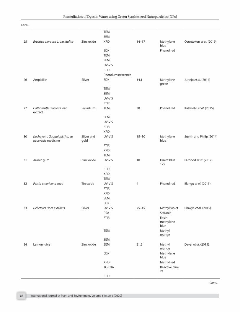
Table 1: Green synthesized nanoparticles for dye remediation
S. No. Plant extract/ microorganism
Nanoparticle (NPs)
Characterization techniques
Size/ diameter of NPs (nm) Targeted dye References
1 Cucurbita moschata leaves Iron oxide UV-VIS 2–20 Tartrazine Barizãoa et al. (2020)Beta vulgaris stalks FTIR Bordeaux red
TEMXRD
2 Leaf extract of Teucrium polium Iron oxide TEM 5.68–30.29 Methyl orange
Kouhbanani et al. (2019)
PSAXRDFTIRVSMTGA
3 Punica granatum seeds extract Iron oxide UV-VIS 25–55 Reactive blue Bibia et al. (2019)XRDEDXSEMAFM
4 Fruit extract of Duranta erecta Copper UV-VIS 70 Methyl orange
Ismail et al. (2019)
XRD Congo redEDX
Fig. 7: Hypothesis behind synthesis of nanoparticle and dye remediation
Cont...

Remediation of Dyes in Water using Green Synthesized Nanoparticles (NPs)
International Journal of Plant and Environment, Volume 6 Issue 1 (2020)76
FTIRSEM
5 Parkia speciosa Hassk pod extract
Iron XRD 10–80 Bromophenol blue
Fatimah et al. (2020)
SEMTEM
6 Leaf extract of Amarranthus gangeticus
Silver HR-TEM 11–15 Congo red Kolya et al. (2015)
SAEDUV-VISFTIR
7 Extract of Clitoria ternatea pods Silver UV-VIS 62.51 Methylene blue
Varadavenkatesan et al. (2019)
SEMXRDFTIREDX
8 Klebsiella oxytoca GS-4-08 Palladium TEM 5.20 Azo dyes Wang et al. (2018)XRD
9 Boswellia serrata Palladium UV-VIS 6.6 ± 1.5 Coomassie brilliant blue G-250
Kora and Rastogi (2016)
DLS Rhodamine BTEM Methylene
blueXRDFTIR
10 Taro (Colocasia esculenta) plant rhizome powder
Silver SEM 68 ± 12 Methyl orange
Ismail et al. (2018)
EDX Congo redXPS Methyl redXRD Rhodamine B
11 Camellia sinensis tea extract Iron XRF 20–100 Methylene blue
Carvalho and Carvalho (2017)
TGA Methyl orange
TEM Bromothymol blue
12 Palm dates fruit Silver-iron bimetallic NPs
UV-VIS 5–40 Bromothymol blue
Al-Asfar et al. (2018)
TEMEDX
13 Extracts of green tea leaves Iron TEM 40–60 Methylene blue
Shahwana et al. (2011)
SEM Methyl orange
EDXXRDFTIR
14 extract of Cupressus sempervirens
Iron TEM 19 Methyl orange
Ebrahiminezhad et al. (2017)
FTIRUV-VISXRD
Cont...
Cont...

Remediation of Dyes in Water using Green Synthesized Nanoparticles
International Journal of Plant and Environment, Volume 6 Issue 1 (2020) 77
15 Green tea Iron TEM Remazol brilliant Blue R
Truskewycza et al. (2016)
SEM Direct red 80EDXFTIRZeta potential
16 Green tea Iron UV-VIS 50–60 Malachite green
Abbassi et al. (2013)
17 Leaf extract of Azadirachta indica
Cerium dioxide
XPS 10–15 Rhodamine B Sharma et al. (2017)
DSCTGAUV-VISXRDSEMTEM
18 Angelica gigas ribbed stem extracts
Silver and gold
FTIR 20–50 Eosin Y Chokkalingam et al. (2019)
EDX Malachite green
XRDFTIRUV-VISPSA
20 Leaf extract of Camellia sinensis Zinc Oxide UV-VIS 60 Malachite green
Batool et al. (2018)
FTIRXRDSEM
21 Mulberry leaves Iron DLS 47.70 Methylene blue
Lim et al. (2018)
SEM Methyl orange
FTIRUV-VISZetasizer
22 Datura leaf extract Iron SEM 326–327 Solo chromo black (SCB)
Raju et al. (2017)
UV-VIS23 Zanthoxylum armatum leaves Silver UV-VIS 15–50 Safranine O Jyoti and Singh (2016)
FTIR Methyl redSEM Methyl
orangeTEM Methylene
blueSAEDXRDEDX
24 Gymnema sylvestre extract Silver UV-VIS 95.2 Methylene blue
Kumar et al. (2019)
FTIRXRD
Cont...
Cont...
Remediation of Dyes in Water using Green Synthesized Nanoparticles (NPs)
International Journal of Plant and Environment, Volume 6 Issue 1 (2020)78
TEMSEM
25 Brassica oleracea L. var. italica Zinc oxide XRD 14–17 Methylene blue
Osuntokun et al. (2019)
EDX Phenol redTEMSEMUV-VISFTIRPhotoluminescence
26 Ampicillin Silver EDX 14.1 Methylene green
Junejo et al. (2014)
TEMSEMUV-VISFTIR
27 Catharanthus roseus leaf extract
Palladium TEM 38 Phenol red Kalaiselvi et al. (2015)
SEMUV-VISFTIRXRD
30 Kashayam, Guggulutiktha, an ayurvedic medicine
Silver and gold
UV-VIS 15–50 Methylene blue
Suvith and Philip (2014)
FTIRXRDTEM
31 Arabic gum Zinc oxide UV-VIS 10 Direct blue 129
Fardood et al. (2017)
FTIRXRDTEM
32 Persia americana seed Tin oxide UV-VIS 4 Phenol red Elango et al. (2015)FTIRXRDSEMEDX
33 Helicteres isora extracts Silver UV-VIS 25–45 Methyl violet Bhakya et al. (2015)PSA SafraninFTIR Eosin
methylene blue
TEM Methyl orange
SEM34 Lemon juice Zinc oxide SEM 21.5 Methyl
orangeDavar et al. (2015)
EDX Methylene blue
XRD Methyl red TG-DTA Reactive blue
21 FTIR
Cont...
Cont...
Remediation of Dyes in Water using Green Synthesized Nanoparticles
International Journal of Plant and Environment, Volume 6 Issue 1 (2020) 79
UV-VIS Photoluminescence
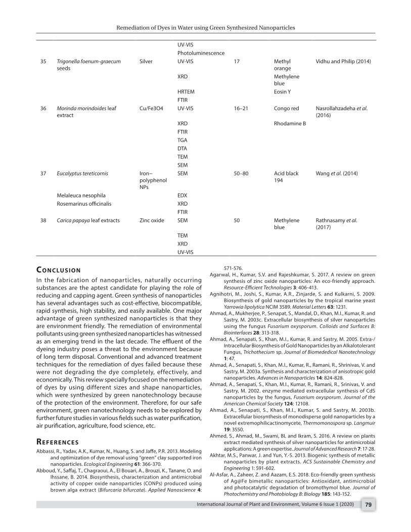
35 Trigonella foenum-graecum seeds
Silver UV-VIS 17 Methyl orange
Vidhu and Philip (2014)
XRD Methylene blue
HRTEM Eosin YFTIR
36 Morinda morindoides leaf extract
Cu/Fe3O4 UV-VIS 16–21 Congo red Nasrollahzadeha et al. (2016)
XRD Rhodamine BFTIRTGADTATEMSEM
37 Eucalyptus tereticornis Iron−polyphenol NPs
SEM 50–80 Acid black 194
Wang et al. (2014)
Melaleuca nesophila EDXRosemarinus officinalis XRD
FTIR38 Carica papaya leaf extracts Zinc oxide SEM 50 Methylene
blueRathnasamy et al. (2017)
TEMXRDUV-VIS
co n c lu s I o n
In the fabrication of nanoparticles, naturally occurring substances are the aptest candidate for playing the role of reducing and capping agent. Green synthesis of nanoparticles has several advantages such as cost-effective, biocompatible, rapid synthesis, high stability, and easily available. One major advantage of green synthesized nanoparticles is that they are environment friendly. The remediation of environmental pollutants using green synthesized nanoparticles has witnessed as an emerging trend in the last decade. The effluent of the dyeing industry poses a threat to the environment because of long term disposal. Conventional and advanced treatment techniques for the remediation of dyes failed because these were not degrading the dye completely, effectively, and economically. This review specially focused on the remediation of dyes by using different sizes and shape nanoparticles, which were synthesized by green nanotechnology because of the protection of the environment. Therefore, for our safe environment, green nanotechnology needs to be explored by further future studies in various fields such as water purification, air purification, agriculture, food science, etc.
re f e r e n c e sAbbassi, R., Yadav, A.K., Kumar, N., Huang, S. and Jaffe, P.R. 2013. Modeling
and optimization of dye removal using “green” clay supported iron nanoparticles. Ecological Engineering 61: 366-370.
Abboud, Y., Saffaj, T., Chagraoui, A., El Bouari, A., Brouzi, K., Tanane, O. and Ihssane, B. 2014. Biosynthesis, characterization and antimicrobial activity of copper oxide nanoparticles (CONPs) produced using brown alga extract (Bifurcaria bifurcata). Applied Nanoscience 4:
571-576.Agarwal, H., Kumar, S.V. and Rajeshkumar, S. 2017. A review on green
synthesis of zinc oxide nanoparticles: An eco-friendly approach. Resource-Efficient Technologies 3: 406-413.
Agnihotri, M., Joshi, S., Kumar, A.R., Zinjarde, S. and Kulkarni, S. 2009. Biosynthesis of gold nanoparticles by the tropical marine yeast Yarrowia lipolytica NCIM 3589. Material Letters 63: 1231.
Ahmad, A., Mukherjee, P., Senapat, S., Mandal, D., Khan, M.I., Kumar, R. and Sastry, M. 2003c. Extracellular biosynthesis of silver nanoparticles using the fungus Fusarium oxysporum. Colloids and Surfaces B: Biointerfaces 28: 313-318.
Ahmad, A., Senapati, S., Khan, M.I., Kumar, R. and Sastry, M. 2005. Extra-/Intracellular Biosynthesis of Gold Nanoparticles by an Alkalotolerant Fungus, Trichothecium sp. Journal of Biomededical Nanotechnology 1: 47.
Ahmad, A., Senapati, S., Khan, M.I., Kumar, R., Ramani, R., Shrinivas, V. and Sastry, M. 2003a. Synthesis and characterization of anisotropic gold nanoparticles. Advances in Nanoparticles 14: 824-828.
Ahmad, A., Senapati, S., Khan, M.I., Kumar, R., Ramani, R., Srinivas, V. and Sastry, M. 2002. enzyme mediated extracellular synthesis of CdS nanoparticles by the fungus, Fusarium oxysporum. Journal of the American Chemical Society 124: 12108.
Ahmad, A., Senapati, S., Khan, M.I., Kumar, S. and Sastry, M. 2003b. Extracellular biosynthesis of monodisperse gold nanoparticles by a novel extremophilicactinomycete, Thermomonospora sp. Langmuir 19: 3550.
Ahmed, S., Ahmad, M., Swami, BL and Ikram, S. 2016. A review on plants extract mediated synthesis of silver nanoparticles for antimicrobial applications: A green expertise. Journal of Advanced Research 7: 17-28.
Akhtar, M.S., Panwar, J. and Yun, Y.-S. 2013. Biogenic synthesis of metallic nanoparticles by plant extracts. ACS Sustainable Chemistry and Engineering 1: 591-602.
Al-Asfar, A., Zaheer, Z. and Aazam, E.S. 2018. Eco-friendly green synthesis of Ag@Fe bimetallic nanoparticles: Antioxidant, antimicrobial and photocatalytic degradation of bromothymol blue. Journal of Photochemistry and Photobiology B: Biology 185: 143-152.

Remediation of Dyes in Water using Green Synthesized Nanoparticles (NPs)
International Journal of Plant and Environment, Volume 6 Issue 1 (2020)80
Annamalai, J. and Nallamuthu, T. 2015. Characterization of biosynthesized gold nanoparticles from aqueous extract of Chlorella vulgaris and their anti-pathogenic properties. Applied Nanoscience 5: 603-607.
Armendariz, V., Herrera, I., Peraltavidea, J.R., Jose-Yacaman, M., Troiani, H., Santiago, P., Jorge, L. and Torresdey, G. 2004. Size controlled gold nanoparticle formation by Avenasativa biomass: use of plants in nanobiotechnology. Journal of Nanoparticle Research 6: 377-382.
Baban, A., Yediler, A. and Ciliz, N.K. 2010. Integrated water management and CP implementation for wool and textile blend processes. Clean-Soil, Air, Water 38(1): 84-90.
Bai, H., Zhang, Z., Guo, Y. and Jia, W. 2009. Biological synthesis of size-controlled cadmium sulfidenanoparticles using immobilized Rhodobacter sphaeroides. Nanoscale Research Letters 4: 717-723.
Baker, S. and Satish, S. 2015. Biosynthesis of gold nanoparticles by Pseudomonas veronii AS41G inhabiting Annona squamosa L. Spectrochimica Acta Part A: Molecular and Biomolecular Spectroscopy 150: 691-695.
Balagurunathan, M.R., Ramaswamy, B.R. and Velmurugan, D. 2011. Biosynthesis of gold nanoparticles from actinomycetes Streptomyces viridogens strain (HM10). Indian Journal of Biochemistry and Biophysics 48: 331-335.
Banat, I.M., Nigam, P. and Marchant, R. 1996. Microbial decolorization of textile-dye containing effluents: a review. Bioresource Technology 58: 217-227.
Bansal, V., Rautaray, D., Ahmad, A. and Sastry, M. 2004. Biosynthesis of zirconiananoparticles using the fungus Fusarium oxysporum. Journal of Materials Chemistry 14: 3303.
Bansal, V., Rautaray, D., Bharde, A., Ahire, K., Sanyal, A., Ahmad, A. and Sastry, M. 2005. Fungus-mediated biosynthesis of silica and titania particles. Journal of Materials Chemistry 15: 2583.
Barizãoa A.C.de L., Silvab, M.F., Andradeb, M., Britoc, F.C., Gomesc, R.G. and Bergamascob, R. 2020. Green synthesis of iron oxide nanoparticles for tartrazine and bordeaux red dye removal. Journal of Environmental Chemical Engineering 8: 103618.
Baskar, G., Chandhuru, J., Sheraz Fahad, K. and Praveen, AS 2017. Anticancer activity of iron oxide nanobiocomposite of fungal asparaginase. International Journal of Modern Science and Technology 2: 98-104.
Batool, M., Qureshi, Z. and Basir, A. 2018. Removal of melachite green dye by using zinc oxide nanoparticles prepared by the green synthesis by using Camellia sinensis (Green Tea) leafs extract. Archives of Nanomedicine: Open Access Journal 1: 96-101.
Bhainsa, K.C. and D’souza, S. 2006. Extracellular biosynthesis of silver nanoparticles using the fungus Aspergillus fumigatus. Colloids and Surfaces B: Biointerfaces 47: 160-164.
Bhakya, S., Muthukrishnan, S., Sukumaran, M., Muthukumar, M. and Kumar, T.S. 2015. Catalytic degradation of organic dyes using synthesized silver nanoparticles: A green approach. Journal of Bioremediation and Biodegradation 6: 1-9.
Bharde, A., Rautaray, D., Bansal, V., Ahmad, A., Sarkar, I., Yusuf, S.M., Sanyal, M. and Sastry, M. 2006. Extracellular biosynthesis of magnetite using fungi. Small 2: 135.
Bibia, I., Nazar, N., Ata, S., Sultan, M., Ali, A., Abbas, A., Jilanid, K., Kamale, S., Sarimf, F.M., Khang, M.I., Jalalh, F. and Iqbal, M. 2019. Green synthesis of iron oxide nanoparticles using pomegranate seeds extract and photocatalytic activity evaluation for the degradation of textile dye. Journal of Materials and Technology 8: 6115-6124.
Bindhu, M.R. and Umadevi, M. 2015. Antibacterial and catalytic activities of green synthesized silver nanoparticles. SpectrochimicaActa Part A: Molecular and Biomolecular Spectroscopy 135: 373-378.
Binupriya, A.R., Sathishkumar, M., Vijayaraghavan, K. and Yun, S.-I. 2010. Bioreduction of trivalent aurum to nano-crystalline gold particles by active andinactive cells and cell-free extract of Aspergillus oryzae var. viridis. Journal of Hazardous Materials 177: 539.
Bose, D. and Chatterjee, S. 2015. Antibacterial activity of green synthesized silver nanoparticles using vasaka (Justicia adhatoda L.) leaf extract. Indian Journal of Microbiology 55: 163-167.
Brayner, R., Coradin, T., Beaunier, P., Greneche, J.-M., Djediat, C., Yepremian, C., Coute, A. and Fievet, F. 2012. Intracellular biosynthesis of superparamagnetic 2-lines ferrihydrite nanoparticles using Euglena
gracilis microalgae. Colloids and Surfaces B: Biointerfaces 93: 20-23.Carvalho, S. and Carvalho, N. 2017. Dye degradation by green heterogeneous
Fenton catalysts prepared in presence of Camellia sinensis. Journal of Environmental Management 187: 82-88.
Chan, Y.S. and Mat Don, M. 2013. Biosynthesis and structural characterization of Ag nanoparticles from white rot fungi. Materials Science and Engineering 33: 282-288.
Chandrasekaran, R., Gnanasekar, S., Seetharaman, P., Keppanan, R., Arockiaswamy, W. and Sivaperumal, S. 2016. Formulation of Carica papaya latex-functionalized silver nanoparticles for its improved antibacterial and anticancer applications. Journal of Molecular Liquids 219: 232-238.
Chen, M., Xu, P., Zeng, G., Yang, C., Huang, D. and Zhang, J. 2015. Bioremediation of soils contaminated with polycyclic aromatic hydrocarbons, petroleum, pesticides, chlorophenols and heavy metalsbycomposting: applications, microbes and future research needs. Biotechnology Advances 33: 745-755.
Chincholi, M., Sagwekar, P., Nagaria, C., Kulkarni, S. and Dhokpande, S. 2014. Removal of dye by adsorption on variousadsorbents: A review. International Journal of Science, Engineering and Technology Research 3: 835-840.
Ch o k k al in gam, M . , Rup a , E . J . , Hu o b, Y. , M athi y a lagana , R . , Anandapadmanaban, G., Ahna, JC, Parka, JK, Lub, J. and Yanga, D.C. 2019. Photocatalytic degradation of industrial dyes using Ag and Au nanoparticles synthesized from Angelica gigas ribbed stem extracts. Optik-International Journal for Light and Electron Optics 185: 1213-1219.
Chowdhury, S., Basu, A. and Kundu, S. 2014. Green synthesis of protein capped silver nanoparticles from phytopathogenic fungus Macrophomina phaseolina (Tassi) Goid with antimicrobial properties against multidrug-resistant bacteria. Nanoscale Research Letters 9: 365.
Davar, F., Majedi, A. and Mirzaei, A. 2015. Green synthesis of ZnO nanoparticles and its application in the degradation of some dyes. Journal of the American Ceramic Society 98: 1739-1746.
Dinçer, A.R., Günes, Y., Karakaya, N. and Günes, E. 2007. Comparison of activated carbon and bottom ash for removal of reactive dye from aqueous solution. Jourmal of Bioresource and Technology 98: 834-839.
Durán, N. and Seabra, A.B. 2012. Metallic oxide nanoparticles: state of the art in biogenicsyntheses and their mechanisms. Applied Microbiology and Biotechnology 95: 275-288.
Durán, N., Marcato, P.D., Alves, O., Souza, G.I. and Esposito, E. 2005. Mechanistic aspects of biosynthesis of silver nanoparticles by several Fusarium oxysporum strains. Journal of Nanobiotechnology 3: 8.
Ealias, A.M. and Saravanakumar, M.P. 2017. A review on the classification, characterisation, synthesis of nanoparticles and their application. IOP Conf. Series: Materials Science and Engineering 263: 032019.
Ebrahiminezhad, A., Taghizadeh, S., Ghasemi, Y. and Berenjian, A. 2017. Green synthesized nanoclusters of ultrasmall zero valent iron nanoparticles as a novel dye removing material. Science of the Total Environment 621: 1527-1532.
Elango, G., Kumaran, S.M., Kumar, S.S., Muthuraja, S., and Roopan, S.M. 2015. Green synthesis of SnO2 nanoparticles and its photocatalytic activity of phenolsulfonphthalein dye. Spectrochimica Acta Part A: Molecular and Biomolecular Spectroscopy 145: 176-180.
El-Rafie, H.M., El-Rafie, M.H. and Zahran, MK 2013. Green synthesis of silver nanoparticles using polysaccharides extracted from marine macro algae. Carbohydrate Polymers 96(2): 403-410.
Erasmus, M., Cason, E.D., Marwijk, J.V., Botes, E., Gericke, M. and Heerden, EV 2014. Gold nanoparticle synthesis using the thermophilic bacterium Thermus scotoductus SA-01 and the purificationand characterization of its unusual gold reducing protein. Gold Bulletin 47: 245-253.
Fardood, S.T., Ramazani, A., Moradi, S. and Asiabi, P.A. 2017. Green synthesis of zinc oxide nanoparticles using Arabic gum and photocatalytic degradation of direct blue 129 dye under visible light. Journal of Materials Science: Materials in Electronics 28: 13596-13601.
Fatimah, I., Pratiwi, E.Z. and Wicaksono, W.P. 2020. Synthesis of magnetic nanoparticles using Parkia Speciosa Hassk pod extract and photocatalytic activity for Bromophenol blue degradation. The Egyptian Journal of Aquatic Research 46: 35-40.
Remediation of Dyes in Water using Green Synthesized Nanoparticles
International Journal of Plant and Environment, Volume 6 Issue 1 (2020) 81
Francavilla, M., Pineda, A., Romero, A.A., Colmenares, J.C., Vargas, C., Monteleone, M. and Luque, R. 2014. Efficient and simple reactive milling preparation of photocatalytically active porous ZnO nanostructures using biomass derived polysaccharides. Green Chemistry 16: 2876-2885.
Fu, M., Li, Q., Sun, D., Lu, Y., He, N., Deng, X., Wang, H. and Huang, J. 2006. Rapid preparation process of silver nanoparticles by bioreduction and their characterizations. Chinese Journal of Chemical Engineering 14: 114-117.
Ganesh Babu, MM and Gunasekaran, P. 2009. Production and structural characterization of crystalline silver nanoparticles from Bacillus cereus isolate. Colloids and Surfaces B: Biointerfaces 74: 191-195.
Gericke, M. and Pinches, A. 2006. Microbial production of gold nanoparticles. Gold Bulletin 39: 22.
Ghodake, G. and Lee, D.S. 2011. Biological synthesis of gold nanoparticles using the aqueous extract of the brown algae Laminaria japonica. Journal of Nanoelectronics and Optoelectronics 6: 268-271.
Gnanajobitha, G., Paulkumar, K., Vanaja, M., Rajeshkumar, S., Malarkodi, C., Annadurai, G. and Kannan, C. 2013. Fruit-mediated synthesis of silver nanoparticles using Vitis vinifera and evaluation of their antimicrobial efficacy. Journal of Nanostructure in Chemistry 3: 1-6.
Gordon, P.F. and Gregor, P. 1983. Organic Chemistry in Color. Springer-Verlag, Berlin.
Gudikandula, K., Vadapally, P. and Charyaa, M.A.S. 2017. Biogenic synthesis of silver nanoparticles from white rot fungi: Their characterization and antibacterial studies. OpenNano 2: 64-78.
Hassan, MM and Carr, C.M. 2018. A critical review on recentadvancements of the removal of reactive dyes from dyehouseeffluent by ion-exchange adsorbents. Chemosphere 209: 201-219.
He, K., Chen, G., Zeng, G., Chen, A., Huang, Z., Shi, J., Huang, T., Peng, M. and Hu, L. 2018. Three-dimensional graphene supported catalysts for organic dyes degradation. Applied Catalysis B: Environmental 228: 19-28.
He, S., Guo, Z., Zhang, Y., Zhang, S., Wang, J. and Gu, N. 2007. Biosynthesis of gold nanoparticles using the bacteria Rhodopseudomonas capsulate. Materials Letter 61: 3984.
He, W., Zhou, W.J., Wang, Y.J., Zhang, X.D., Zhao, H.S., Li, ZM and Yan, S.P. 2009. Biomineralization of iron phosphate nanoparticles in yeast cell. Material Science and Engineering 4: 1348.
Hennebel, T., Nevel, S.V., Verschuere, S., Corte, S.D., Gusseme, B.D., Cuvelier, C., Fitts, J.P., Lelie, D.V.D., Boon, N. and Verstraete, W. 2011. Palladium nanoparticles produced by fermentatively cultivated bacteria as catalyst for diatrizoate removal with biogenic hydrogen. Applied Microbiology and Biotechnology 91: 1435-1445.
Holmes, J.D., Smith, P.R., Evans-Gowing, R., Richardson, D.J., Russell, D.A. and Sodeau, J.R. 1995. Energy-dispersive X-ray analysisof the extracellular cadmium sulfide crystallites of Klebsiella aerogenes. Archives of Microbiology 163: 143.
Huang, J., Li, Q., Sun, D., Lu, Y., Su, Y., Yang, X., Wang, H., Wang, Y., Shao, W. and He, N. 2007. Biosynthesis of silver and gold nanoparticles by novel sundried Cinnamomum camphora leaf. Nanotechnology 18: 105104.
Husseiny, M.I . , Aziz, M.A.E. , Badr, Y. and Mahmoud, M.A. 2007. Biosynthesis of gold nanoparticles using Pseudomonas aeruginosa. SpectrochimicaActa Part A: Molecular and Biomolecular Spectroscopy 67: 1003-1006.
Imran, M., Crowley, D.E., Khalid, A., Hussain, S., Mumtaz, MW and Arshad, M. 2015. Microbial biotechnology for decolorizationof textile wastewaters. Reviews in Environmental Science and Biotechnology 14: 73-92.
Ingle, A., Gade, A., Pierrat, S., Sonnichsen, C.M.K. and Rai, M.K. 2008. Mycosynthesis of silver nanoparticles using the fungus Fusarium acuminatum and its activity against some human pathogenic bacteria. Current Nanoscience 4: 141.
Iravani, S. 2011. Green synthesis of metal nanoparticles using plants. Green Chemistry 13: 2638-2650.
Ismail, M., Gul, S., Khan, M.I., Ali Khan, M., Asiri, A.M. and Khan, S.B. 2019. Green synthesis of zerovalent copper nanoparticles for efficient reduction of toxic azo dyes congo red and methyl orange. Green Process Synthesis 8: 135-143.
Ismail, M., Khan, M.I., Khan, S.B., Akhtard, K., Ali Khan, M. and Asirib, A.M. 2018. Catalytic reduction of picric acid, nitrophenols and organic azo dyes via green synthesized plant supported Ag nanoparticles. Journal of Molecular Liquids 268: 87-101.
Jha, A.K., Prasad, K. and Kulkarni, A.R. 2009a. Synthesis of TiO2 nanoparticles using microorganisms. Colloids and Surfaces B: Biointerfaces 71: 226-229.
Jha, A.K., Prasad, K. and Prasad, K. 2009b. A green low-cost biosynthesis of Sb2O3 nanoparticles. Biochemical Engineering Journal 43: 303.
Joerger, R., Klaus, T. and Granqvist, C.G. 2000. Biologically produced silver carboncomposite materials for optically function althin-film coatings. Advanced Materials 12: 407.
Joerger, T.K., Joerger, R., Olsson, E. and Granqvist, C.G. 2001. Bacteria as workers in the living factory: Metal-accumulating bacteria and their potential for materials science. Trends in Biotechnology 19: 15-20.
Johnston, C.W., Wyatt, M.A., Li, X., Ibrahim, A. and Shuster, J. 2013. Gold biomineralization by a metallophore from a gold-associated microbe. Nature Chemical Biology 9: 241-243.
Junejo, Y., Sirajuddin, Baykal, A., Safdar, M. and Balouch, A. 2014. A novel green synthesis and characterization of Ag NPs with its ultra-rapid catalytic reduction of methyl green dye. Applied Surface Science 290: 499-503.
Jyoti, K. and Singh, A. 2016. Green synthesis of nanostructured silver particles and their catalytic application in dye degradation. Journal of Genetic Engineering and Biotechnology 14: 311-317.
Kadirvelu, K., Kavipriya, M., Karthika, C., Radhika, M., Vennilamani, N. and Pattabhi, S. 2003. Utilization of various agricultural wastes for activated carbon preparation and application for the removal of dyes and metal ions from aqueous solutions. Journal of Bioresource Technology 87: 129-132.
Kalaiselvi, A., Roopan, S.M., Madhumitha, G., Ramalingam, C. and Elango, G. 2015. Synthesis and characterization of palladium nanoparticles using Catharanthus roseus leaf extract and its application in the photocatalytic degradation. Spectrochimica Acta Part A: Molecular and Biomolecular Spectroscopy 135: 116-119.
Kannan, R.R., Arumugam, R., Ramya, D., Manivannan, K. and Anantharaman, P. 2013. Green synthesis of silver nanoparticles using marine macroalga Chaetomorpha linum. Applied Nanoscience 3: 229-233.
Kasthuri, J., Kathiravan, K. and Rajendiran, N. 2008. Phyllanthin-assisted biosynthesis of silver and gold nanoparticles: A novel biological approach. Journal of Nanoparticle Research 11: 1075-1085.
Khan, R., Bhawana, P. and Fulekar, M.H. 2013. Microbial decolorization and degradation of synthetic dyes: A review. Reviews in Environmental Science and Biotechnology 12: 75-97.
Khataeea, A.R. and Kasiri, M.B. 2010. Photocatalytic degradation of organic dyes in the presence of nanostructured titanium dioxide: Influence of the chemical structure of dyes. Journal of Molecular Catalysis A: Chemical 328: 8-26.
Klaus, T., Joerger, R., Olsson, E. and Granqvist, C.-G. 1999. Silverbased crystalline nanoparticles, microbially fabricated. Proceedings of the National Academy of Sciences of the United States of America 96: 13611-13614.
Kolya, H., Maiti, P., Pandey, A. and Tripathy, T. 2015. Green synthesis of silver nanoparticles with antimicrobial and azo dye (Congo red) degradation properties using Amaranthus gangeticus Linn. leaf extract. Journal of Analytical Science and Technology 6: 1-7.
Kora, A.J. and Rastogi, L. 2016. Catalytic degradation of anthropogenic dye pollutants using palladium nanoparticles synthesized by gum olibanum, aglucuronoarabinogalactanbiopolymer. Industrial Crops and Products 81: 1-10.
Kouhbanani, M.A.J., Beheshtkhoo, N., Taghizadeh, S., Amani, A.M. and Alimardani, V. 2019. One-step green synthesis and characterization of iron oxide nanoparticles using aqueous leaf extract of Teucriumpolium and their catalytic application in dye degradation. Advances in Natural Sciences: Nanoscience and Nanotechnology 10: 015007.
Kumar, B., Smita, K., Cumbal, L. and Debut, A. 2017. Green synthesis of silver nanoparticles using Andean blackberry fruit extract. Saudi Journal of Biological Sciences 24: 45-50.
Kumar, D.A., Palanichamy, V. and Roopan, S.M. 2014. Green synthesis of
Remediation of Dyes in Water using Green Synthesized Nanoparticles (NPs)
International Journal of Plant and Environment, Volume 6 Issue 1 (2020)82
silver nanoparticles using Alternantheradentata leaf extract at room temperature and their antimicrobial activity. Spectrochimica Acta Part A: Molecular and Biomolecular Spectroscopy 127: 168-171.
Kumar, K.S., Amutha, R., Arumugam, P., Berchmans, S., 2011. Synthesis of gold nanoparticles: an ecofriendly approach using Hansenula anomala. ACS Applied Materials and Interfaces 3: 1418-1425.
Kumar, M.S., Supraja, N. and David. 2019. Photocatalytic degradation of methylene blue using silver nanoparticles synthesized from Gymnema sylvestre and antimicrobial assay. Novel Research in Sciences 2: 1-7.
Kumar, S.A., Abyaneh, M.K., Gosavi, S.W., Kulkarni, S.K., Pasricha, R., Ahmad, A. and Khan, M.I. 2007a. Nitrate reductase-mediated synthesis of silver nanoparticles from AgNO3. Biotechnology Letters 29: 439.
Kumar, S.A., Ayoobul, A.A., Absar, A. and Khan, M.I. 2007b. Extracellular biosynthesis of CdSe quantum dots by the fungus, Fusarium Oxysporum. Jounal of Biomedical Nanotechnology 3: 190-194.
Kumar, S.K., Arumugam, RAP and Berchmans, S. 2011. Synthesis of gold nanoparticles: an ecofriendly approach using Hansenula anomala. ACS Applied Materials and Interfaces 3: 1418-1425.
Lellis, B., Fávaro-Polonio, C.Z., Pamphile, JA and Polonio, J.C. 2019. Effects of textile dyes on health and the environmentand bioremediation potential of living organisms. Biotechnology Research and Innovation 3: 275-290.
Lengke, F.M., Fleet, E.M. and Southam, G. 2007. Biosynthesis of silver nanoparticles by filamentous cyanobacteria from a silver (I) nitrate complex. Langmuir 23: 2694-2699.
Li, G., He, D., Qian, Y., Guan, B., Gao, S., Cui, Y., Yokoyama, K. and Wang, L. 2012. Fungus mediated green synthesis of silver nanoparticles using Aspergillus terreus. International Journal of Molecular Sciences 13: 466-476.
Lim, S.N., Ng, W.M., Lim, J.K. and Che, H.X. 2018. Performance of mulberry leaves mediated green synthesis zero-valent iron nanoparticles in dye removal. International Journal of Engineering and Technology 7: 113-117.
Lin, Y.T., Weng, C.H. and Chen, F.Y. 2008. Effective removal of AB24 dye by nano/micro- size zero-valent iron. Separation and Purification Technology 64: 26-30.
Mahdavi, M., Namvar, F., Ahmad, M. and Mohamad, R. 2013. Green biosynthesis and characterization of magnetic iron oxide (Fe3O4) nanoparticles using seaweed (Sargassummuticum) aqueous extract. Molecules 18: 5954.
Malik, R., Ramteke, D.S. and Wate, SR 2007. Adsorption of malachite green on groundnut shell waste based powdered activated carbon. Waste Management 27: 1129-1138.
Mata, Y.N., Torres, E., Blazquez, M.L., Ballester, A., Gonzalez, F. and Munoz, J.A. 2009. Gold (III) biosorption and bioreduction with the brown alga Fucusvesiculosus. Journal of Hazardous Materials 166: 612-618.
McKay, G. 1982. Adsorption of dyestuffs from aqueous solutions with activated carbon I: equilibrium and batch contact-time studies. Journal of Chemical Technology and Biotechnology 32: 759-772.
Medvedev, Z.A., Crowne, HM and Medvedev, M.N. 1988. Age related variations of hepato carcinogenic effect of azo dye (3’-MDAB) as linked to the level of hepatocyte polyploidization. Mechanisms of Ageing Development 46: 159-174.
Meysam, S.N., Bonjar, G.H.S. and Khaleghi, N. 2015. Biosynthesis of gold nanoparticles by Streptomyces fulvissimus. Nanomedicine Journal 2: 153-159.
Montes, M.O., Mayoral, A., Deepak, F.L., Parsons, J.G., Jose-Yacama, M., Videa, J.R.P. and Torresdey, J.L.G. 2011. Anisotropic gold nanoparticles and gold plates biosynthesis using alfalfa extracts. Journal of Nanoparticle Research 13: 3113-3121.
Morshedi, D., Mohammadia, Z., Mashhadi, M., Boojarb, A. and Aliakbaria, F. 2013. Using protein nanofibrils to remove azo dyes from aqueous solution by the coagulation process. Colloids and Surfaces B: Biointerfaces 112: 245-254.
Mua, B. and Wang, A. 2016. Adsorption of dyes onto palygorskite and its composites: A review. Journal of Environmental Chemical Engineering 4: 1274-1294.
Mukherjee, P., Ahmad, A., Mandal, D., Senapati, S., Sainkar, S.R., Khan, M.I.,
Parishcha, R., Ajaykumar, P., Alam, M. and Kumar, R. 2001. Fungus-mediated synthesis of silver nanoparticles and their immobilization in the mycelial matrix: a novel biological approach to nanoparticle synthesis. Nano Letters 1: 515-519.
Mukherjee, P., Senapati, S., Mandal, D., Ahmad, A., Khan, M.I., Kumar, R. and Sastry, M. 2002. Extracellular synthesis of gold nanoparticles by the fungus Fusarium oxysporum. ChemBioChem Combining Chemistry and Biology 3: 461.
Nadeem, M., Abbasi, B.H., Younas, M., Ahmad, W. and Khan, T. 2017. A review of the green syntheses and anti-microbial applications of gold nanoparticles. Green Chemistry Letters and Reviews 10: 216-227.
Nagarajan, S., Arumugam, K. and Kuppusamy, K. 2013. Extracellular synthesis of zinc oxide nanoparticle using seaweeds of Gulf of Mannar, India. Journal of Nanobiotechnology 11: 39.
Nair, B. and Pradeep, T. 2002. Coalescence of nanoclusters and formation of submicron crystallites assisted by Lactobacillus strains. Crystal Growth Design 2: 293-298.
Nakkala, J.R., Mata, R., Gupta, A.K. and Sadras, S.R. 2014. Biological activities of green silver nanoparticles synthesized with Acorouscalamus rhizome extract. European Journal of Medicinal Chemistry 85: 784-794.
Namvar, F., Azizi, S., Ahmad, M., Shameli, K., Mohamad, R., Mahdavi, M. and Tahir, P. 2015. Green synthesis and characterization of gold nanoparticles using the marine macroalgaeSargassummuticum. Research on Chemical Intermediates 41: 5723-5730.
Narayanan, K.B. and Sakthivel, N. 2008. Coriander leaf mediated biosynthesis of gold nanoparticles. Materials Letters 62: 4588-4590.
Narayanan, K.B. and Sakthivel, N. 2010. Biological synthesis of metal nanoparticles by microbes. Advances in Colloid and Interface Science 156: 1-13.
Narayanan, K.B. and Sakthivel, N. 2011. Green synthesis of biogenic metal nanoparticles by terrestrial and aquatic phototrophic and heterotrophic eukaryotes and biocompatible agents. Advances in Colloid and Interface Science 169: 59-79.
Nasrin, S., Donya, G., Ali, E., Hosein, J., Reza, F.M. and Aziz, M.F. 2009. Intra/extracellular biosynthesis of silver nanoparticles by an autochthonous strain of Proteus mirabilis isolated from photographic waste. Journal of Biomedical Nanotechnology 5: 247-253.
Nasrollahzadeha, M., Ataroda, M. and Sajadiba, S.M. 2016. Green synthesis of the Cu/Fe3O4 nanoparticles using Morinda morindoides leaf aqueous extract: A highly efficient magneticallyseparable catalyst for the reduction of organic dyes in aqueousmedium at room temperature. Applied Surface Science 364: 636-644.
Nayantara and Kaur, P. 2018. Biosynthesis of nanoparticles using eco-friendlyfactories and their role in plant pathogenicity: areview. Biotechnology Research and Innovation 2: 63-73.
Osuntokun, J., Onwudiwe, D.C. and Ebenso, E.E. 2019. Green synthesis of ZnO nanoparticles using aqueous Brassica oleracea L. var. italica and the photocatalytic activity. Green Chemistry Letters and Reviews 12(4): 444-457.
Pandey, G., Rawtani, D. and Agrawal, Y.K. 2016. Aspects of nanoelectronics in materials development. In: Kar, A. (Ed.), Nanoelectronics and Materials Development, InTech, https://doi.org/10.5772/64414
Panigrahi, S., Kundu, S., Ghosh, S.K., Nath, S. and Pal, T. 2004. General method of synthesis for metal nanoparticles. Journal of Nanoparticle Research 6: 411-414.
Park, Y., Hong, Y.N. and Weyers, A., Kim, Y.S. and Linhardt, R.J. 2011. Polysaccharides and phytochemicals: a natural reservoir for the green synthesis of gold and silver nanoparticles. IET Nanobiotechnology 5: 69 78.
Pathakoti, K., Manubolu, M. and Hwang, H.M. 2018. Nanotechnology Applications for Environmental Industry. Handbook of Nanomaterials for Industrial Application pp. 894-907.
Patrycja, G., Magdalena, W., Dnyaneshwar, R., Sagar, T., Hanna, D. and Mahendra, R. 2016. Synthesis of silver nanoparticles from two acidophilic strains of Pilimeliacolu mellifera subsp. pallida and their antibacterial activities. Journal of Basic Microbiology 56: 541-556.
Rafique, M., Sadaf, I., Rafiquea, M.S. and Tahirb, M.B. 2017. A review on green synthesis of silver nanoparticles and their applications. Artificial Cells Nanomedicine and Biotechnology 45: 1272-1291.
Remediation of Dyes in Water using Green Synthesized Nanoparticles
International Journal of Plant and Environment, Volume 6 Issue 1 (2020) 83
Raju, Ch. AI, Chakravarthy, Ch., Sujatha, V., Satti Babu, K., Ratna Raju, P. and Prem, K. 2017. Studies on green synthesis of iron nanoparticle for Solo Chrome Black (SCB) dye decolorization. International Research Journal of Engineering and Technology 04: 1656-1663.
Rathnasamy, R., Thangasamy, P., Thangamuthu, R., Sampath, S. and Alagan, V. 2017. Green synthesis of ZnO nanoparticles using Carica papaya leaf extracts for photocatalytic and photovoltaic applications. Journal of Materials Science: Materials in Electronics 28: 10374-10381.
Rawtani, D., Khatri, N., Tyagi, S. and Pandey, G. 2018. Nanotechnology-based recent approaches for sensing and remediation of pesticides. Journal of Environmental Management 206: 749-762.
Rawtani, D., Pandey, G., Tharmavaram, M., Pathak, P., Akkireddy, S. and Agrawal, Y.K. 2017. Development of a novel ‘nanocarrier’ system based on halloysitenanotubes to overcome the complexation of ciprofloxacin with iron: An in vitro approach. Applied Clay Science 150: 293-302.
Rawtani, D., Tharmavaram, M., Pandey, G. and Hussain, C.M. 2019. Functionalized nanomaterial for forensic sample analysis. Trends in Analytical Chemistry 120: 115661.
Robinson, T., Chandran, B. and Nigam, P. 2002. Removal of dyes from and artificial textile dye effluent by two agricultural waste residues, corncob and barley husk. Environment International 28: 29-33.
Robinson, T., McMullan, G., Marchant, R. and Nigam, P. 2001. Remediation of dyes in textile effluent: a critical review on current treatment technologies with a proposed alternative. Bioresource Technology 77: 247-255.
Saifuddin, N., Wong, C. and Yasumira, A. 2009. Rapid biosynthesis of silver nanoparticles using culture supernatant of bacteria with microwave irradiation. Journal of Chemistry 6: 61-70.
Scarano, G. and Morelli, E. 2003. Properties of phytochelatin-coated CdS nanocrystallites formed in a marine phytoplanktonic alga (Phaeodactylum tricornutum Bohlin) in response to Cd. Plant Science 165: 803-810.
Selvam, GG and Sivakumar, K. 2015. Phycosynthesis of silver nanoparticles and photocatalytic degradation of methyl orange dye using silver (Ag) nanoparticles synthesized from Hypnea musciformis (Wulfen) J.V. Lamouroux. Applied Nanoscience 5: 617-622.
Senapati, S., Syed, A., Moeez, S., Kumar, A. and Ahmad, A. 2012. Intracellular synthesis of gold nanoparticles using alga Tetraselmisko chinensis. Material Letters 79: 116-118.
Shahverdi, A.R., Minaeian, S., Shahverdi, H.R., Jamalifar, H. and Nohi, A.-A. 2007. Rapid synthesis of silver nanoparticles using culture supernatants of Enterobacteria: A novel biological approach. Process Biochemistry 42: 919-923.
Shahwana, T., Abu Sirriah, S., Nairata, M., Boyacıb, E., Eroglub, A.E., Scottc, T.B. and Hallamc, K.R. 2011. Green synthesis of iron nanoparticles and their application as a Fenton-like catalyst for the degradation of aqueous cationic and anionic dyes. Chemical Engineering Journal 172: 258- 266.
Shan, Z.Z., Fu, L.J., Chao, T., Fang, Z.Q., Tian, HJ and GuiBin, J. 2008. Rapid decolorization of water soluble azo-dyes by nano sized zero-valent iron immobilized on the exchange resin. Science in China Series B: Chemistry 51: 186-192.
Shankar, S.S., Ahmad, A., Pasrichaa, R. and Sastry, M. 2003. Bioreduction of chloroaurate ions by geranium leaves and its endophytic fungus yields gold nanoparticles of different shapes. Journal of Materials Chemistry 13: 1822.
Sharma, D., Kanchi, S. and Bisetty, K. 2019. Biogenic synthesis of nanoparticles: A review. Arabian Journal of Chemistry 12: 3576-3600.
Sharma, J.K., Srivastava, P., Ameen, S., Akhtar, M.S., Sengupta, S.K. and Singh, G. 2017. Phytoconstituents assisted green synthesis of cerium oxide nanoparticles for thermal decomposition and dye remediation. Materials Research Bulletin 91: 98-110.
Sharma, N., Pinnaka, A.K., Raje, M., Fnu, A., Bhattacharyya, M.S. and Choudhury, A.R. 2012. Exploitation of marine bacteria for production of gold nanoparticles. Microbial Cell Factories 11: 86.
Shen, D., Fan, J., Zhou, W., Gao, B., Yue, Q. and Kang, Q. 2009. Adsorption kinetics and isotherm of anionic dyes onto organo-bentonite from single and multisolute systems. Journal of Hazardous Material 172:
99-107.Shimada, C., Sasaki, K.Y.F., Sato, I. and Tsudua, S. 2010. Differential colon
DNA damage induced by azo food additives between rats and mice. Journal of Toxicological Science 35: 547-554.
Shivaji, S., Madhu, S. and Singh, S. 2010. Extracellular synthesis of antibacterial silver nanoparticles using psychrophilic bacteria. Process Biochemistry 46: 1800-1807.
Singh, M., Kalaivani, R., Manikandan, S., Sangeetha, N. and Kumaraguru, A.K. 2013. Facile green synthesis of variable metallic gold nanoparticle using Padina gymnospora, a brown marine macroalga. Applied Nanoscience 3: 145-151.
Singh, M., Manikandan, S. and Kumaraguru, A.K. 2011. Nanoparticles: A new technology with wide applications. Research Journal of Nanoscience and Nanotechnology 1: 1-11.
Srivastava, S.K., Yamada, R., Ogino, C. and Kondo, A. 2013. Biogenic synthesis and characterization of goldnanoparticles by Escherichia coli K12 and itsheterogeneous catalysis in degradation of4-nitrophenol. Nanoscale Research Letters 8: 70.
Suna, Q., Cai, X., Li, J., Zheng, M., Chenb, Z. and Yu, CP 2014. Green synthesis of silver nanoparticles using tea leaf extract and evaluation of their stability and antibacterial activity. Colloids and Surfaces A: Physicochemical and Engineering Aspects 444: 226-223.
Suvith, V.S. and Philip, D. 2014. Catalytic degradation of methylene blue using biosynthesized gold and silver nanoparticles. Spectrochimica Acta Part A: Molecular and Biomolecular Spectroscopy 118: 526-532.
Sweeney, R.Y., Mao, C., Gao, X., Burt, J.L., Belcher, A.M., Georgiou, G. and Iverson, B.L. 2004. Bacterial biosynthesisof cadmium sulfide nanocrystals. Chemistry and Boilogy 11: 1553.
Syed, A., Saraswati, S., Kundu, GC and Ahmad, A. 2013. Biological synthesis of silver nanoparticles using the fungus Humicola sp. and evaluation of their cytoxicity using normal and cancer cell lines. Spectrochimica Acta Part A: Molecular and Biomolecular Spectroscopy 114: 144-147.
Tahir, K., Nazir, S., Li, B., Khan, A.U., Khan, Z.U.H., Ahmad, A. and Khan, F.U. 2015. An efficient photo catalytic activity of green synthesized silver nanoparticles using Salvadorapersica stem extract. Separation and Purification Technology 150: 316-324.
Tang, W.W., Zeng, G.M., Gong, J.L., Liang, J., Xu, P., Zhang, C. and Huang, B.B. 2014. Impact of humic/fulvic acid on the removal of heavy metals from aqueous solutions using nanomaterials: A review. Science of the Total Environment 468-469: 1014-1027.
Thakkar, K.N., Mhatre, S.S. and Parikh, R.Y. 2010. Biological synthesis of metallic nanoparticles. Nanomedicine 6: 257-262.
Tharmavaram, M., Pandey, G. and Rawtani, D. 2018. Surface modified halloysite nanotubes: A flexible interface for biological, environmental and catalytic applications. Advances in Colloid and Interface Science 261: 82-101.
Tharmavaram, M., Rawtani, D. and Pandey, G. 2017. Fabrication routes for one-dimensional nanostructures via block copolymers. Nano Convergence 4: 12.
Tom, R.T., Nair, A.S., Singh, N., Aslam, M. 2003. Freely dispersible Au@TiO2, Au@ZrO2, Ag@TiO2, andAg@ZrO2 core-shell nanoparticles: one-step synthesis, characterization, spectroscopy, and optical limiting properties. Langmuir 19: 3439-3445.
Tripathi, R.M., Gupta, R.K., Singh, P., Bhadwal, A.S., Shrivastava, A., Kumar, N., Shrivastav, B.R. 2014. Ultra-sensitive detection of mercury (II) ions in water sample using gold nanoparticles synthesised by Trichoderma harzianum and their mechanistic approach. Sensors Actuators B: Chemical 204: 637-646.
Truskewycza, A., Shuklab, R. and Balla, A.S. 2016. Iron nanoparticles synthesized using green tea extracts for the fenton-like degradation of concentrated dye mixtures at elevated temperatures. Journal of Environmental Chemical Engineering 4: 4409-4417.
Umbuzeiro, G.A., Freeman, H., Warren, S.H., Kummrow, F. and Claxton, L.D. 2005. Mutagenicity evaluation of the commercial product CI Disperse Blue 291 using different protocols of the Salmonella assay. Food and Chemical Toxicology 43: 49-56.
Varadavenkatesan, T., Vinayagam, R. and Selvaraj, R. 2019. Green synthesis and structural characterization of silver nanoparticles synthesized using the pod extract of Clitoria ternatea and its application towards

Remediation of Dyes in Water using Green Synthesized Nanoparticles (NPs)
International Journal of Plant and Environment, Volume 6 Issue 1 (2020)84
dye degradation. Materials Today: Proceedings 23: 27-29.Venkatesan, J., Manivasagan, P., Kim, S.K., Kirthi, A.V., Marimuthu, S. and
Rahuman, A.A. 2014. Marine algae-mediated synthesis of gold nanoparticles using a novel Ecklonia cava. Bioprocess and Biosystems Engineering 37: 1591-1597.
Vidhu, V.K. and Philip, D. 2014. Catalytic degradation of organic dyes using biosynthesized silvernanoparticles. Micron 56: 54-62.
Vigneshwaran, N., Ashtaputre, N., Varadarajan, P., Nachane, R., Paralikar, K. and Balasubramanya, R. 2007. Biological synthesis of silver nanoparticles using the fungus Aspergillus flavus. Materials Letters 61: 1413-1418.
Vijayan, S.R., Santhiyagu, P., Ramasamy, R., Arivalagan, P., Kumar, G. and Ethiraj, K. 2016. Seaweeds: A resource for marine bionanotechnology. Enzyme and Microbial Technology 95: 45-57.
Waghmare, S.S., Arvind, M.D. and Sadowski, Z. 2014. Biosynthesis, optimization, purification and characterization of gold nanoparticles. African Journal of Microbiology Research 8: 138-146.
Wang, P., Song, Y., Fan, H. and Yu, L. 2018. Bioreduction of azo dyes was enhanced by in-situ biogenic palladium nanoparticles. Bioresource Technology 266: 176-180.
Wang, Z., Fang, C. and Megharaj, M. 2014. Characterization of iron−polyphenol nanoparticles synthesized by three plant extracts and their fenton oxidation of azo dye. ACS Sustainable Chemistry and Engineering 2: 1022-1025.
Weisburger, J.H. 2002. Comments on the history and importance of aromatic and heterocyclic amines in public health. Mutation Research-
Fundamental of Molecular Mechanics and Mutagenesis 506-507: 9-20.Wu, H., Lai, C., Zeng, G., Liang, J., Chen, J., Xu, J., Dai, J., Li, X., Liu, J. and
Chen, M. 2017. The interactions of composting and biochar and their implications for soil amendment and pollution remediation: A review. Critical Reviews in Biotechnology 37: 754-764.
Yadav, A.K., Jena, S., Acharya, B.C. and Mishra, B.K. 2012. Removal of azo dye in innovative constructed wetlands: influence of iron scrap and sulfate reducing bacterial enrichment. Ecological Engineering 49: 53-58.
Yagub, M.T., Sen, T.K., Afroze, S. and Ang, H.M. 2014. Dye and its removal from aqueous solution by adsorption: A review. Advances in Colloid and Interface Science 209: 172-184.
Yang, C., Chen, H., Zeng, G., Yu, G. and Luo, S. 2010. Biomass accumulation and control strategies in gas biofiltration. Biotechnology Advances 28: 531-540.
Yong, P., Rowsen, N.A., Farr, J.P.G., Harris, I .R. and Macaskie, L.E. 2002. Bioreduction and biocrystallization of palladium by Desulfovibriodesulfuricans NCIMB 8307. Biotechnology and Bioengineering 80: 369.
Yousefzadi, M., Rahimi, Z. and Ghafori, V. 2014. The green synthesis, characterization and antimicrobial activities of silver nanoparticles synthesized from green alga Enteromorphaflexuosa (Wulfen) J. Agardh. Materials Letters 137: 1-4.
Zonaro, E., Lampis, S., Turner, R.J., Junaid, S., Qazi, S. and Vallini, G. 2015. Biogenic selenium and tellurium nanoparticles synthesized by environmental microbial isolates efficaciously inhibit bacterial planktonic cultures and biofilms. Frontiers in Microbiology 16: 584.