regulation of the osmotin gene promoter
TRANSCRIPT
The Plant Cell, Vol. 4, 513-524, May 1992 O 1992 American Society of Plant Physiologists
RESEARCH ARTICLE
Regulation of the Osmotin Gene Promoter
Andrzej K. Kononowicz,a*cil Donald E. Nelson,a Narendra K. Singh,b Paul M. Hasegawa,a and Ray A. Bressana a Center for Plant Environmental Stress Physiology, 1165 Department of Horticulture, Purdue University, West Lafayette, Indiana 47907-1 165
Department of Botany and Microbiology, Auburn University, Auburn, Alabama 36849 Department of Plant Cytology and Cytochemistry, University of Lodz, Banacha 12/16, 90-237 Lodz, Poland
By introducing a chimeric gene fusion of the osmotin promoter and b-glucuronidase into tobacco by Agrobacterium- mediated transformation, we have demonstrated a very specific pattern of temporal and spatial regulation of the osmotin promoter during normal plant development and after adaptation to NaCI. We have found that the osmotin promoter has a very high natural level of activity in mature pollen grains during anther dehiscence and in pericarp tissue at the final, desiccating stages of fruit development. GUS activity was rapidly lost after pollen germination. The osmotin promoter thus appears to be unique among active pollen promoters described to date in that it is active only in dehydrated pollen. The osmotin promoter was also active in corolla tissue at the onset of senescence. Adaptation of plants to NaCl highly stimulated osmotin promoter activity in epidermal and cortex parenchyma cells in the root elongation zone; in epidermis and xylem parenchyma cells in stem internodes; and in epidermis, mesophyll, and xylem parenchyma cells in developed leaves. The spatial and temporal expression pattern of the osmotin gene appears consistent with both osmotic and pathogen defense functions of the gene.
INTRODUCTION
Since we first reported its isolation (Singh et al., 1985), osmo- tin (OSM) and OSM-like proteins or the genes encoding these proteins have been extensively studied in severa1 laboratories (Singh et al., 1987a, 1989a, 1989b; LaRosaet al., 1989; Meeks- Wagner et al., 1989; Grosset et al., 1990; Neale et al., 1990; Roberts and Selitrennikoff, 1990; Stintzi et al., 1991; Woloshuk et al., 1991). OSM is a group of cationic proteins that exist in at least two forms, one with a pl of 7.8 and the other with a pl of greater than 8.2; they differ slightly in molecular weight (Singh et al., 1985, 1987a). We showed that OSM is synthe- sized as a preprotein with a molecular weight of 26,380 and that the molecular weight of its mature form is 23,984 (Singh et al., 1989b). OSM exhibits a very high level of sequence ho- mology with other proteins, including the sweet protein thaumatin, potato pathogenesis-related (PR) protein C, tobacco PR-S, and the maize a-amylase/trypsin inhibitor (Richardson et al., 1987; Singh et al., 1987a).
On the basis of similarities in amino acid sequence and in expression pattern, OSM has been classified as a member of the PR-5-type proteins of tobacco (BOI, 1988; Brederode et al., 1991; Linthorst, 1991). Further studies have shown that the synthesis and accumulation of OSM mRNA are develop- mentally regulated and controlled by at least six hormonal or
To whom correspondence should be addressed.
environmental signals, including abscisic acid (ABA), ethyl- ene, tobacco mosaic virus infection, salinity, desiccation, and wounding in both cultured cells and whole plants of tobacco (LaRosaet al., 1985,1987; Singh et al., 1987b). However, sub- stantial accumulation of the OSM protein itself occurs only in response to osmotic stress and ethylene in tobacco cultivar W38, which shows no hypersensitive response (LaRosa et al., 1992).
Much of the cellular OSM protein was found by immunocy- tochemistry to be localized in electron-dense (tanninlike) inclusions in the vacuoles. A very low amount of OSM was detected in the ground cytoplasm, but there was no preferential localization of this protein within the cytoplasm. Occasionally, OSM-positive colloidal gold particles were found attached to the tonoplast, but the organelles, plasma membrane, and cell wall were devoid of any gold particles (Singh et al., 1987a). OSM has also been detected in the extracellular matrix of plant cells, although it is not known whether the extracellular OSM is the same as that found in the vacuole or whether it is per- haps an acidic form as are other extracellular PR proteins (Casas et al., 1992).
Transcriptional activation of the OSM promoter was inves- tigated in transformed tobacco plants carrying copies of a fusion of the cloned OSM promoter to the P-glucuronidase (GUS) reporter gene (Nelson et al., 1992). In young seedlings,

514 The Plant Cell
the OSM promoter was found to be transcriptionally activated by exogenous ABA and ethylene. However, the sensitivity of the OSM promoter to ABA decreased with age in both roots and shoots of young seedlings, whereas ethylene-induced pro- moter activity remained the same over this developmental time period. It was also shown that NaCl shock activated the OSM promoter and that this effect seemed to be mediated through ABA. On the other hand, wounding, which also activated the OSM promoter, seemed to exert its effect through ethylene (Nelson et al., 1992).
To elucidate the function of the OSM gene, several studies have focused on the organ and tissue localization of OSM mRNA and the encoded protein. A high rate of synthesis and accumulation of OSM was found in roots and in “outer stem tissue,” which is composed mainly of the epidermis (Singh et al., 1987b). Only low levels of OSM were found in the flower bud, stigma, style, and ovary of mature flowers, and in the cor- tex and pith of stem. The OSM gene product was undetectable in seeds and barely detectable in leaf tissues (LaRosa et al., 1989, 1992; Singh et al., 1989a, 1989b). Also, developmental regulation of the synthesis and accumulation of OSM tran- scripts has been demonstrated (Meeks-Wagner et al., 1989; Neale et al., 1990). In tobacco cultivar Samsun NN, FB7-2 tran- scripts encoding the OSM protein occur in apices of prefloral and floral plants, in stem internode segments, and in leaves of plants possessing an immature inflorescence, with higher levels in older leaves and internodes (Meeks-Wagner et al., 1989; Neale et al., 1990). OSM mRNA also accumulates to high levels in root tissues and 2-week-old seedlings and to moderate levels in floral organs at various stages during de- velopment (Neale et al., 1990).
In this study, we report the results obtained from histochem- ical analyses of GUS expression in specific cell types of transgenic tobacco plants containing an OSM promoter::GUS chimeric gene, which is illustrated in Figure 1. We also report the pattern of OSM promoter activity in these plants after adap- tation to a moderately saline (171 mM NaCI) environment. Our data demonstrate that the OSM promoter had the highest level of activity in mature desiccated pollen grains and in placenta and pericarp tissues of the mature fruit. The OSM promoter activity was stimulated during adaptation to NaCl in specific cells and tissues of roots and in xylem parenchyma cells of both stem and leaves.
RESULTS
Patterns of Expression of 0SM::GUS Gene Fusion in Vegetative Organs of Unadapted Plants
GUS activity was analyzed in the longitudinal and transverse paraffin sections of roots, in stem internode segments (basal, middle, and apical segments), and in leaves at different stages of development. Low GUS activity was-detected in tissues of roots, stems, leaves, and reproductive organs of transformed
RB osmotin 1.R
n 8
Figure 1. 0SM::GUS Fusion Gene Used for Transformation.
RB, right border; LB, left border; neo, neomycin phosphotransferase II gene; gusA, GUS reporter gene.
unadapted plants. Although osmotin and other basic PR pro- teins are considered to accumulate in healthy roots, we found that the expression of the 0SM::GUS gene was quite low in unstressed hydroponically grown roots. However, high GUS activity was detected in roots of these plants when grown in either soil or vermiculite (data not shown). A higher level of the GUS activity was observed in the epidermis of both roots (in the elongation zone), as shown in Figure 2A, and trichomes and epidermis of basal leaves and stems, as shown in Figures 3D to 3F. No GUS activity was detected in either vascular or parenchymatous tissues of any vegetative plant organs. It must be emphasized that some minor variation in GUS activity (i.e., differences in color intensity but not localization) was observed between analyzed organ. samples.
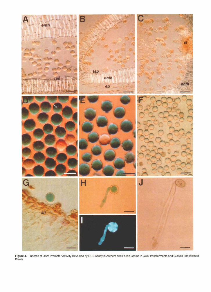
High GUS Activity in Mature Pollen Grains after Anther Dehiscence
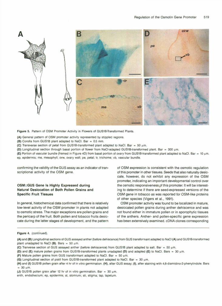
GUS activity was analyzed in tissues of different parts of flowers at several developmental stages. Only mature pollen grains and epidermis of petals showed GUS activity, as shown in Figures 4E and 5C, respectively. No GUS activity was observed in any other floral tissue by histochemical GUS assays.
Expression of the 0SM::GUS gene in pollen grains occurred only after anther dehiscence (Figures 4B to 4E). Approximately 57% of the pollen grains expressed moderate or high levels of GUS activity. Another 30% showed low GUS activity. This variability in degree of expression could be the result of segre- gation of thefour 0SM::GUS genes carried by GUSlB plants. GUS activity decreased rapidly after the initiation of pollen ger- mination in vitro (Figures 4G to a). After 24 hr of culture on pollen germination medium, almost ali (93%) of the pollen grains showed no GUS activity. At no stage of development or germination was moderate or high GUS activity found in pollen grains from nontransformed tobacco plants or plants transformed with a construct consisting of the GUS coding se- quence without the OSM promoter (Figures 4A and 4F). Only occasionally were pale blue-stained pollen grains observed in these plants.
When mature pollen grains from transformed 0SM::GUS plants were incubated in a GUS assay mixture with 20% meth- anol, a treatment that according to Kosugi et al. (1990) eliminates the intrinsic GUS activity in tobacco plants, color
\ \
Regulation of the Osmotin Gene Promoter 515
intensity was slightly reduced, but the percentage of GUS- positive grains did not change (~90%). GUS activity was not detected in pollen grains from nontransformed plants and plants transformed with GUS coding sequence alone (without pro- moter) when GUS assay was conducted in the presence of 20% methanol.
lnduction of the OSM Promoter during Fruit Development
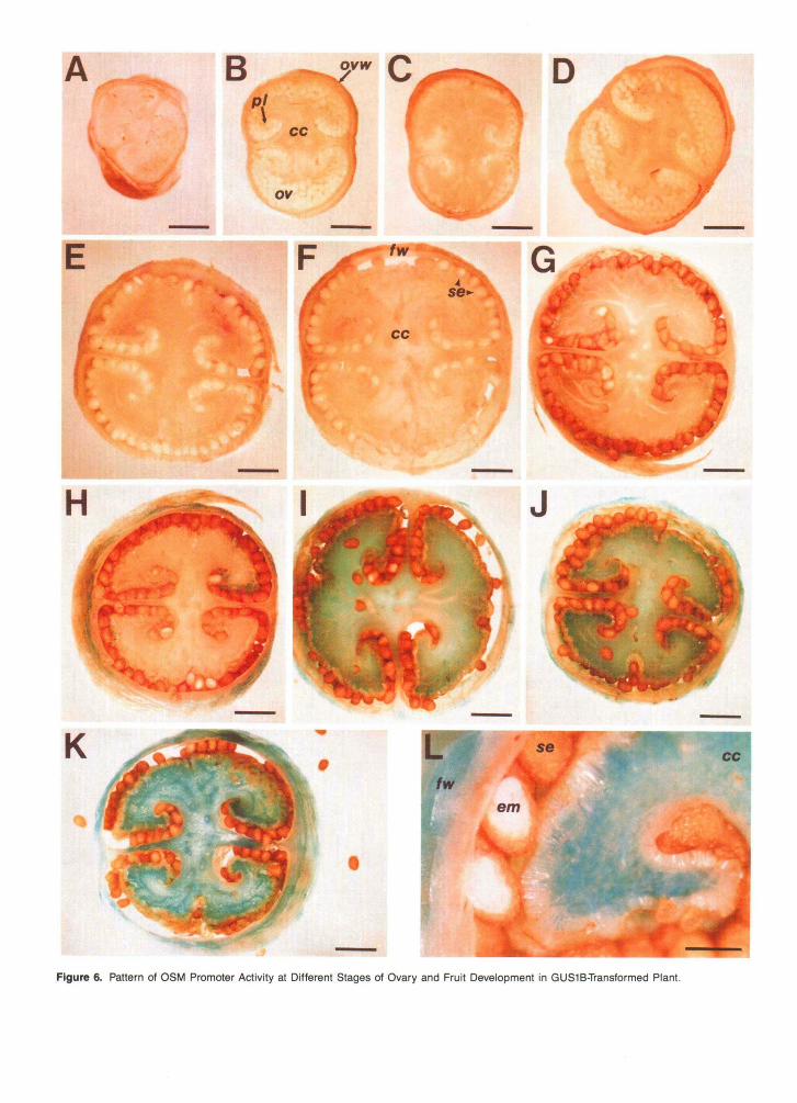
Before fertilization and fruit formation, the only tissue of the ovary that expressed GUS activity was the vascular tissue in the basal portion of this floral organ, as shown in Figures 5D and 5E. In the ovary of both NaCI-adapted and NaCCunadapted 0SM::GUS-transformed tobacco plants, induction of GUS ac- tivity and a gradual increase in GUS intensity were observed at the final stages of fruit development as desiccation of the fruit began, as shown in Figures 6A to 6L. Interestingly, a GUS-positive reaction was found only in the maternal tissues of developing fruit (pericarp and placenta) during desiccation, but not in seeds at any stage of development, including ma- ture, desiccated seeds (Figures 6G to 6L).
Adaptation to NaCl lnduces and/or Stimulates Activity of the OSM Promoter
Long-term adaptation of 0SM::GUS transgenic plants to moderate concentrations of NaCl(l71 mM) modulated the ac- tivity of the OSM promoter in temporal and spatial contexts. Paraffin sections were made of roots, stems, and leaves at different stages of development from both types of transgenic plants as well as nontransformed tobacco plants that were adapted for 30,60, and 90 days to NaCl and used to localize GUS activity. Histochemical analysis of GUS activity was also performed at different stages of flower development, before and after pollination (stages 1,3,5, 7, 8, 9, 11, and 12 accord- ing to the designation proposed by Koltunow et al. [1990]), and at different stages of fruit formation. GUS activity was care- fully monitored in pollen grains during their development and in vitro germination.
Stem Adaptation to NaCl
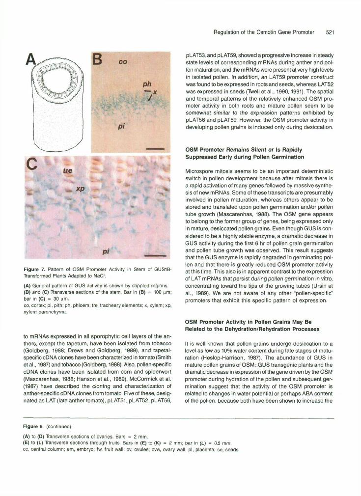
Adaptation to NaCl also increased OSM promoter activity in stem internodes at all stages of plant development. The GUS- positive reaction was restricted to the epidermis and the xylem parenchyma of the stem, but was also occasionally observed in the cortex parenchyma, as shown in Figures 7A to 7C. The pattern of expression or GUS level in these tissues did not change significantly after the initial induction by NaCI.
Leaf Adaptation to NaCl
Although increased activity of the OSM promoter was found in the trichomes, epidermis, mesophyll, and xylem parenchyma of fully developed (expanded) leaves after adaptation to NaCI, the strongest GUS-positive reaction was found in leaf tips (Figures 3A, 3G, and 3J). In young leaves, lower GUS activity was observed in the trichomes. No GUS activity was observed in either shoot apical meristems or leaf primordia. There was a consistent gradient of low to high OSM promoter expression in the youngest to the oldest leaves, and this was confirmed with GUS fluorometric assays using 4-methylumbelliferyl P-o-glucuronide substrate (data not shown).
Adaptation to NaCl in Reproductive Structures
Adaptation of transgenic 0SM::GUS plants to NaCl significantly affects OSM promoter activity in flowers. Moderate GUS ac- tivity after salt adaptation was found in vascular bundles of the basal part of the ovary, the epidermal layers and the mesophyll of the top (limb) region of petals (Figures 5AtO 5C), and in the epidermis and mesophyll of the sepal tips.
A high level of 0SM::GUS gene expression was found in mature pollen grains of adapted and unadapted plants at an- ther dehiscence (Figures 4D and 4E). The GUS-positive reaction, however, was visible only in mature, "dry" pollen grains and decreased dramatically during the early stages of in vitro germination (Figures 4H to 4J).
DlSCUSSlON Adaptation to NaCl in Roots
0SM::GUS transgenic plants adapted to grow in NaCl for 30 days showed increased GUS activity in root elongation zones (epidermis and root cortex parenchyma) but not in root apical meristems (Figure 26). After prolonged (60 days) adaptation of plants to NaCI, GUS activity was also observed in the en- dodermis, cortex, and tissues of the vascular cylinder within the root elongation zone (Figures 2C to 2K). A GUS-positive reaction was never found either in root meristems (Figures 2G and 2K) or in any tissue of the differentiated (basal) zone of the root (Figure 2D).
We have found that the GUS reporter gene when driven by the OSM promoter exhibits organ-, tissue-, and cell-specific expression patterns. Furthermore, adaptation of 0SM::GUS transgenic plants to NaCl resulted in a significant increase in GUS gene expression in specific cells and tissues of roots, stems, and mature leaves. The differential patterns of OSM promoter activity were mostly consistent with less detailed anal- yses of the distribution of OSM mRNA and protein in plant tissues reported earlier (Singh et al., 1987a, 1987b; LaRosa et al., 1989; Meeks-Wagner et al., 1989; Neale et al., 1990),
i% H
vc
K
re
amre
Figure 2. Pattern of OSM Promoter Activity Revealed by GUS Activity in Roots of GUS1B-Transformed Plants.
B f/v
vb me
. •eph\ ft*. - - • • » . / - - '
me
;."•j
—fr
^^^fBftfjf
H
me
ep
iph—
me
tr
K epA
Figure 3. Pattern of OSM Promoter Activity Revealed by GUS Activity in Leaves of Tobacco Transformants.
(A) General pattern of GUS activity in fully expanded leaves of GUS1B-transformed plants (stippled area represents region of GUS activity).(B) and (C) Transverse sections of leaves from plants transformed with the GUS coding sequence alone. Bars = 100 um.(D) Portion of leaf of GUS1B-transformed plant unadapted to NaCI. Bar = 0.7 mm.(E) and (F) Transverse sections of leaf blade and petiole from GUS1B-transformed plant unadapted to NaCI. Bars = 50 um.(G) Portion of leaf tip of fully expanded leaf from GUS1B-transformed plant adapted to NaCI. Bar = 1 mm.(H) to (M) Transverse sections of fully expanded leaves from GUS1B plants adapted to NaCI. (H) shows leaf blade; bar = 50 um. (I) shows petiole;bar = 1 mm. (J) shows leaf tip; bar = 100 um. (K) shows leaf blade; bar = 50 um. (L) and (M) show portions of vascular bundle; bar in (L)= 30 um and bar in (M) = 10 um.ep, epidermis; eph, external phloem; iph, internal phloem; me, mesophyll; tr, trichome; tre, tracheary element; vb, vascular bundle; x, xylem;xp, xylem parenchyma.
Figure 2. (continued).
(A) Roots from plants unadapted to NaCI. Bar = 1.25 mm.(B) Roots from plants adapted to 171 mM NaCI for 30 days. Bar = 1.25 mm.(C) Roots from plants adapted to 171 mM NaCI for 60 days. Bar = 0.7 mm.(D) to (H) Longitudinal sections through different root zones from plants adapted to NaCI. Bars = 50 urn.(I) to (K) Transverse sections through different zones of roots from plants adapted to NaCI. Bars = 50 um.(D), (E), (I), and (J), differentiated zone; (F), elongation zone; (G) and (K), meristematic zone; (H), root cap.am, apical meristem; co, root cortex; ep, epidermis; ph, phloem; px, protoxylem; re, root cap; vc, vascular cylinder; xp, xylem parenchyma.
Benth
St
Figure 4. Patterns of OSM Promoter Activity Revealed by GUS Assay in Anthers and Pollen Grains in GUS Transformants and GUS1B-TransformedPlants.
Regulation of the Osmotin Gene Promoter 519
me vb
Figure 5. Pattern of OSM Promoter Activity in Flowers of GUS1B-Transformed Plants.
(A) General pattern of OSM promoter activity represented by stippled regions.(B) Corolla from GUS1B plant adapted to NaCI. Bar = 0.5 mm.(C) Transverse section of petal from GUSlB-transformed plant adapted to NaCI. Bar = 50 urn.(D) Longitudinal section through basal portion of flower from NaCI-adapted GUS1B-transformed plant. Bar = 300 urn.(E) Portion of vascular bundle (framed in Figure 4D) from basal portion of ovary from GUSlB-transformed plant adapted to NaCI. Barep, epidermis; me, mesophyll; ovw, ovary wall; pe, petal; tr, trichome; vb, vascular bundle.
10 urn.
confirming the validity of the GUS assay as an indicator of tran-scriptional activity of the OSM gene.
OSM::GUS Gene Is Highly Expressed duringNatural Desiccation of Both Pollen Grains andSpecific Fruit Tissues
In general, histochemical data confirmed that there is relativelylow-level activity of the OSM promoter in plants not adaptedto osmotic stress. The major exceptions are pollen grains andthe pericarp of the fruit. Both pollen and tobacco fruits desic-cate during the latter stages of development, and the pattern
of OSM expression is consistent with the osmotic regulationof this promoter in other tissues. Seeds that also naturally desic-cate, however, do not exhibit any expression of the OSMpromoter, indicating an important developmental control overthe osmotic responsiveness of this promoter. It will be interest-ing to determine if there are seed-expressed versions of theOSM gene in tobacco as was reported for OSM-like proteinsof other species (Vigers et al., 1991).
OSM promoter activity was found to be localized in mature,desiccated pollen grains during anther dehiscence and wasnot found either in immature pollen or in sporophytic tissuesof the anthers. Anther- and pollen-specific gene expressionhas been extensively examined. cDNA clones corresponding
Figure 4. (continued).
(A) and (B) Longitudinal sections of GUS assayed anther (before dehiscence) from GUS transformant adapted to NaCI (A) and GUSlB-transformedplant unadapted to NaCI (B). Bars = 50 urn.(C) Tranverse section of GUS assayed anther (before dehiscence) from GUS1B plant adapted to salt. Bar = 50 urn.(D) and (E) mature pollen grains from GUSlB-transformed plants unadapted (D) and adapted (E) to NaCI. Bars = 30 nm.(F) Mature pollen grains from GUS transformant adapted to NaCI. Bar = 50 urn.(G) Longitudinal section of pistil from GUSlB-transformed plant adapted to NaCI. Bar = 30 urn.(H) and (I) GUS1B pollen grain after 4 hr of in vitro germination. (H), after GUS assay; (I), after staining with 4,6-diamidino-2-phenylindole. Bars= 30 urn.(J) GUS1B pollen grain after 12 hr of in vitro germination. Bar = 30 urn.enth, endothecium; ep, epidermis; st, stomium; sti, stigma; tap, tapetum.
fw
cc
Figure 6. Pattern of OSM Promoter Activity at Different Stages of Ovary and Fruit Development in GUS1B-Transformed Plant.
Regulation of the Osmotin Gene Promoter 521
B CO
Ph
Pi
xp
Pi
Figure 7. Pattern of OSM Promoter Activity in Stem of GUS1B-Transformed Plants Adapted to NaCI.(A) General pattern of GUS activity is shown by stippled regions.(B) and (C) Transverse sections of the stem. Bar in (B) = 100 urn;bar in (C) = 30 urn.co, cortex; pi, pith; ph, phloem; tre, tracheary elements; x, xylem; xp,xylem parenchyma.
to mRNAs expressed in all sporophytic cell layers of the an-thers, except the tapetum, have been isolated from tobacco(Goldberg, 1988; Drews and Goldberg, 1989), and tapetal-specific cDNA clones have been characterized in tomato (Smithetal., 1987) and tobacco (Goldberg, 1988). Also, pollen-specificcDNA clones have been isolated from corn and spiderwort(Mascarenhas, 1988; Hanson et al., 1989). McCormick et al.(1987) have described the cloning and characterization ofanther-specific cDNA clones from tomato. Five of these, desig-nated as LAT (late anther tomato), pLAT51, pLAT52, pLAT56,
pLAT53, and pLAT59, showed a progressive increase in steadystate levels of corresponding mRNAs during anther and pol-len maturation, and the mRNAs were present at very high levelsin isolated pollen. In addition, an LAT59 promoter constructwas found to be expressed in roots and seeds, whereas LAT52was expressed in seeds (Twell et al., 1990,1991). The spatialand temporal patterns of the relatively enhanced OSM pro-moter activity in both roots and mature pollen seem to besomewhat similar to the expression patterns exhibited bypLAT56 and pLAT59. However, the OSM promoter activity indeveloping pollen grains is induced only during desiccation.
OSM Promoter Remains Silent or Is RapidlySuppressed Early during Pollen Germination
Microspore mitosis seems to be an important deterministicswitch in pollen development because after mitosis there isa rapid activation of many genes followed by massive synthe-sis of new mRNAs. Some of these transcripts are presumablyinvolved in pollen maturation, whereas others appear to bestored and translated upon pollen germination and/or pollentube growth (Mascarenhas, 1988). The OSM gene appearsto belong to the former group of genes, being expressed onlyin mature, desiccated pollen grains. Even though GUS is con-sidered to be a highly stable enzyme, a dramatic decrease inGUS activity during the first 6 hr of pollen grain germinationand pollen tube growth was observed. This result suggeststhat the GUS enzyme is rapidly degraded in germinating pol-len and that there is greatly reduced OSM promoter activityat this time. This also is in apparent contrast to the expressionof LAT mRNAs that persist during pollen germination in vitro,concentrating toward the tips of the growing tubes (Ursin etal., 1989). We are not aware of any other "pollen-specific"promoters that exhibit this specific pattern of expression.
OSM Promoter Activity in Pollen Grains May BeRelated to the Dehydration/Rehydration Processes
It is well known that pollen grains undergo desiccation to alevel as low as 10% water content during late stages of matu-ration (Heslop-Harrison, 1987). The abundance of GUS inmature pollen grains of OSM::GUS transgenic plants and thedramatic decrease in expression of the gene driven by the OSMpromoter during hydration of the pollen and subsequent ger-mination suggest that the activity of the OSM promoter isrelated to changes in water potential or perhaps ABA contentof the pollen, because both have been shown to increase the
Figure 6. (continued).
(A) to (D) Transverse sections of ovaries. Bars = 2 mm.(E) to (L) Transverse sections through fruits. Bars in (E) to (K) = 2 mm; bar in (L) = 0.5 mm.cc, central column; em, embryo; fw, fruit wall; ov, ovules; ovw, ovary wall; pi, placenta; se, seeds.
522 The Plant Cell
levels of OSM mRNA in other tissues (LaRosa et al., 1992). It is not known, however, if ABA levels increase during pollen desiccation. Although ABA levels are known to increase dur- ing seed desiccation, this apparent increase of ABA does not activate the OSM promoter in seeds.
OSM May Be lnvolved in Reorganization of Membranes Affecting Permeability
Recently, Roberts and Selitrennikoff (1990) and Vigers et al. (1991) reported the isolation of an antifungal protein from corn (zeamatin) whose N-terminal amino acid sequence was con- siderably similar to that of OSM. According to these authors, zeamatin appears to act by permeabilizing the plasma mem- branes of fungi. This hypothesis was also suggested by Woloshuk et al. (1991); they postulated that because of the hy- drophobic nature of the AP24 protein (identified as OSM), it may interact with the plasma membrane. Such interactions could disrupt the membrane of the fungi causing lysis and in- hibition of hyphal growth. Electron microscopy studies of the dehydration and hydration of gramineaceous pollen led Heslop- Harrison (1979) to suggest that the membranes of pollen cells are likely to be ineffective as an osmotic barrier because their organization and/or permeability changes during dehydration. During pollen grain germination, upon initial hydration, the in- tegrity of the pollen cell membrane is restored, presumably leading to the recovery of its semipermeability properties and capacity for controlling the movement of solutes. Restoration of membrane integrity is an essential prerequisite for pollen germination, and the inability of the membrane to regain its normal structure is one of the causes of the loss of pollen via- bility (Simon, 1974; Shivanna and Heslop-Harrison, 1981; Heslop-Harrison, 1987).
We have observed (Singh et al., 1987a; Nelson et ai., 1988) that the OSM protein associates with the tonoplast and plasma membrane of tobacco cells but not chloroplast or mitochon- drial membranes. Also, Yen et al. (1991) have reported the identification of an OSM-like protein that is associated with the plasma membrane from the common ice plant, a halophyte. It is therefore possible that OSM participates in the dehydra- tion protection of membranes of desiccated pollen cells and that this protection involves the reorganization of membranes in the desiccated state, causing them to become potentially leaky if their normal structure is not restored during rehydra- tion. If this is indeed the case, it could explain why the protein product of a gene that is dramatically regulated by desicca- tion or osmotic stress is so apparently active in the induction of leakiness of fungal membranes (Vigers et al., 1991). The OSM gene may have evolved as part of a mechanism caus- ing permeabilization that protects pollen membranes during dehydration. Later, this gene may have been modified to al- low sequestration of the insoluble protein in vacuoles so that the OSM protein released from the vacuole by pathogen at- tack would specifically permeabilize the vulnerable hydrated
plasma membranes of pathogens. It will be important to de- termine the intracellular localization of the OSM protein in these different tissues to test this hypothesis.
The Pattern of OSM Promoter Activity in NaCI-Adapted Plants 1s Consistent with 60th an Osmotic Adaptatlon Mechanism and a Pathogen Defense Mechanism
60th the temporal and spatial patterns of OSM promoter ac- tivity observed here have considerable similarity to the map of Na+ and K+ transport and distribution in whole plants con- structed by Wolf et al. (1991). These similarities are related to low K+/high Na+ and include the following observations:
(1) There is an increase in GUS promoter activity, particularly in root, stem, and part of leaf tissues during and after adap- tation to high salt.
(2) Relatively high expression of GUS activity was found in roots in general and compared with other plant organs and specifically in the root elongation zone but not in the root tip meristem.
(3) Higher GUS activity was observed in older compared with younger leaves and stem internodes.
(4) No GUS activity was detected in apical meristems and leaf primordia.
(5) High GUS activity could be seen in xylem parenchyma of both stem and leaves.
This similarity between Na+ and K+ distribution within whole plants and the pattern of OSM promoter activity tends to support the hypothesis that the expression of the OSM gene is an adaptive response of plants to osmotic stress. Yet, sev- era1 aspects of the developmental pattern of OSM promoter activity also suggest that the OSM has a natural role in de- fense against pathogens, especially those pathogens that may induce symptoms of osmotic stress such as bacterial or fun- gal wilt pathogens. For instance, these pathogens first tend to attack root tissues near or at the zone of cell elongation. They also can spread to and disrupt the vascular tissues. Both sites exhibit relatively active 0SM::GUS expression.
The OSM gene may be osmotically responsive so that the defense mechanism of plants will recognize the induced os- motic stress symptoms of severa1 pathogens such as bacterial and fungal wilt pathogens. This recognition of the pathogen by its induction of stress symptoms rather than by a specific chemical signal released by the pathogen would make it vir- tually impossible for the pathogen to evolve a counter-defense mechanism to avoid specific recognition by the host. Such a defensive response can be envisaged as the plant’s “second line” of defense, which is more slowly activated at a later stage of pathogen attack but would not rely on the early but easily “mutatable” specific elicitors produced by specific pathogens.
Regulation of the Osmotin Gene Promoter 523
METHODS
Plant Transformation
A DNAfragment containing 1.8 kb of sequence in the B’direction from the transcription start of the osmotin (OSM) gene (Nelson et al., 1992) was inserted in the BamHl site of pBI101.1, which is at the 5’end of the gene encoding P-glucuronidase (GUS) (Jefferson et al., 1987). This construct was transferred to Agrobacterium tumefaciens and introduced into Nicotiana tabacum cv W38 by A. tumefaciens-mediated tobacco leaf disc transformation. Plants were regenerated directly from the leaf discs. Severa1 independent primary transformants were obtained, and three were examined histologically. Data obtained from only one of them, designated GUSlB, which contained four copies of the OSM gene construct and expressed the highest leve1 of GUS activity (Nelson et al., 1992), are reported here. However, the pattern of expression in the other two plants was consistent with that observed with GUSlB. Tobacco plants transformed with a construct consisting of only the GUS coding sequence (GUS transformants) were used as control plants.
Growth and Adaptation of Tobacco Plants to NaCl
Vegetatively propagated transgenic tobacco plants carrying the 0SM::GUS chimericgene or the GUS gene without the OSM promoter were grown hydroponically in quarter-strength MS salts (Murashige and Skoog, 1962) and adapted stepwise to a concentration of 170 mM NaCl by increasing the NaCl concentration by 42.5 mM every 3 days. Plants were grown in the presence of 170 mM NaCl for 4, 8, and 12 weeks before tissue samples were taken for GUS analyses. Hydro- ponically cultured plants of the same age grown without NaCl were used as unadapted controls.
Histochemical Assay for GUS Enzyme Activity
The GUS enzyme assay was used to measure the relative activity of the OSM promoter in 0SM::GUS gene fusion transformants. Histo- c,hemical staining for GUS activity was performed according to the procedure of Jefferson et al. (1987), with the modifications proposed by Koltunow et al. (1990). Samples of plant tissues and organs were fixed for 15 min under vacuum in 0.1 M sodium phosphate buffer, pH 7.0, containing 0.1% formaldehyde, 0.1% Triton X-100, and 0.1% 0-mercaptoethanol. Samples were then rinsed severa1 times with 0.1 M phosphate buffer containing 0.1% 0-mercaptoethanol, followed by a rinse with 0.05 M phosphate buffer, pH 7.0. For the GUS reaction as- say, a buffered solution (0.05 M sodium phosphate buffer, pH 7.0) of 1 mM 5-bromo-4-chloro-3-indolyl-~-D-glucuronic acid cyclohexylam- monium salt (Biosynth AG, Staat, Switzerland) containing 0.1% 0-mercaptoethanol and 0.1% Triton X-100 was used. After the GUS reaction, organ or tissue samples were fixed for 4 hr in a 2% glutaralde- hyde solution in 0.05 M phosphate buffer, pH 7.2, or 10% formaldehyde, 20% ethanol, 5% acetic acid. Chlorophyll was extracted from tissues during dehydration in an ethanol series prior to embedding in Tissue Prep 2 embedding wax (Fisher Scientific, Fair Lawn, NJ). Paraffin sec- tions (10- to 50-vm-thick) were cut and deparaffinized. The slides were viewed and photographed using a microscope (Optiphot model; Nikon, Tokyo) set for bright-field illumination or Nomarski differential interfer- ente contrast.
For pollen grains, along with the procedure described above, the GUS assay proposed by Kosugi et al. (1990) was used to eliminate intrinsic GUS activity by the addition of methanol at 20% volume to the GUS assay mixture.
In Vitro Germination of Pollen Grains
Pollen was collected from flowers at anthesis and germinated on mi- croscopic slides coated with solidified medium containing 3 mM H3B03, 1.7 mM Ca(NO&, 10% sucrose, and 0.7% agar at pH 5.8 (Bino et al., 1987). Pollen grains were germinated at 24OC in the dark in a humid chamber prior to GUS assay.
ACKNOWLEDGMENTS
We thank Jean Clithero for excellent technical assistance and Drs. Ana Casas and Kashchandra G. Raghothama for critically reading the manuscript. This research was partially supported by National Science Foundation Grant No. DCB-90005216. This is journal paper No. 13356 of the Purdue University Agricultura1 Experiment Station.
Received January 30, 1992; accepted March 17, 1992.
REFERENCES
Bino, R.J., Hille, J., and Franken, J. (1987). Kanamycin resistance during in vitro development of pollen from transgenic tomato plants. Plant Cell Rep. 6, 333-336.
BOI, J.F. (1988). Structure and expression of plant genes encoding pathogenesis-related proteins. In Temporal and Spatial Regulation of Plant Genes, D.P.S. Verma and R.B. Goldberg, eds (New York: Springer-Verlag), pp. 201-221.
Brederode, F.T., Linthorst, H.J.M., and BOI, J.F. (1991). Differential induction of acquired resistance and PR gene expression in tobacco by virus infection, ethephon treatment, UV light and wounding. Plant MOI. Biol. 17, 1117-1125.
Casas, A.M., Nelson, D.E., Raghothama, K.G., Paino DUrzo, M., Singh, N.K., Bressan, R.A., and Hasegawa, P.M. (1992). Expres- sion of osmotin-like genes in the halophyte Atriplex nummularia L. Plant Physiol. 99, in press.
Drews, G.N., and Goldberg, R.B. (1989). Genetic control of flower development. Trends Genet. 5, 256-261.
Goldberg, R.B. (1988). Plants: Nove1 developmental processes. Science
Grosset, J., Meyer, Y., Chartier, Y., Kauffmann, S., Legrand, M., and Fritig, B. (1990). Tobacco mesoppyll protoplasts synthesize 1,3- P-glucanase, chitinases and “osmotins” during in vitro culture. Plant Physiol. 92, 520-527.
Hanson, D.D., Hamilton, D.A., Travis, J.L., Bashe, D.M., and Mascarenhas, J.P. (1989). Characterization of a pollen-specific cDNA clone from Zea mays and its expression. Plant Cell 1, 173-179.
240, 1460-1467.
524 The Plant Cell
Heslop-Harrison, J. (1979). An interpretation of the hydrodynamics of pollen. Am. J. Bot. 66, 737-743.
Heslop-Harrison, J. (1987). Pollen germination and pollen-tube growth. Int. Rev. Cytol. 107, 1-78.
Jefferson, R.A., Kavanagh, T.A., and Bevan, M.W. (1987). GUS fu- sion: 0-Glucuronidase is a sensitive and versatile fusion marker in higher plants. EMBO J. 6, 3901-3907.
Koltunow, A.M., Truettner, J., Cox, K.H., Wallmth, M., and Goldberg, R.B. (1990). Different temporal and spatial gene expression patterns occur during anther development. Plant Cell 2, 1201-1224.
Kosugi, S., Ohashi, Y., Nakajima, K., and Arai, Y. (1990). An improved assay for B-glucuronidase in transformed cells: Methanol almost com- pletely suppresses a putative endogenous p-glucuronidase activity. Plant Sci. 70, 133-140.
LaRosa, P.C., Handa, A.K., Hasegawa, P.M., and Bressan, R.A. (1985). Abscisic acid accelerated adaptation of cultured tobacco cells to salt. Plant Physiol. 79, 138-142.
LaRosa, C.P., Hasegawa, P.M., Rhodes, D., Clithero, J.M., Watad, A.-E.A., and Bressan, R.A. (1987). Abscisic acid stimulated osmotic adjustment and its involvement in adaptation of tobacco cells to NaCI. Plant Physiol. 85, 174-185.
LaRosa, P.C., Singh, N.K., Hasegawa, EM., and Bressan, R.A. (1989). Stable NaCl tolerance of tobacco cells is associated with enhanced accumulation of OSM. Plant Physiol. 91, 855-861.
LaRosa, P.C., Chen, L., Nelson, D.E., Singh, N.K., Hasegawa, P.M., and Bressan, R.A. (1992). Osmotin gene expression is post- transcriptionally regulated. Plant Physiol, in press.
Linthorst, H.J.M. (1991). Pathogenesis-related proteins of plants. Crit. Rev. Plant Sci. 10, 123-150.
Mascarenhas, J.P. (1988). Anther- and pollen-expressed genes. In Tem- poral and Spatial Regulation of Plant Genes, D.P.S. Verma and R.B. Goldberg, eds (New York: Springer-Verlag), pp. 97-115.
McCormick, S., Smith, A., Gasser, C., Sachs, K., Hinchee, M., Horsch, R., and Fraley, R. (1987). The identification of genes spe- cifically expressed in reproductive organs of tomato. In Tomato Biotechnology, D. Nevins and R. Jones, eds (New York: Alan Liss),
Meeks-Wagner, D.R., Dennis, E.S., Tran Thanh Van, K., and Peacock, W.J. (1989). Tobacco genes expressed during in vitro flo- ral initiation and their expression during normal plant development. Plant Cell 1, 25-35.
Murashige, T., and Skoog, F. (1962). A revised medium for rapid growth and bio-assays with tobacco tissue cultures. Physiol. Plant. 15, 473-497.
Neale, A.D., Wahleithner, J.A., Lund, M., Bonnett, H.T., Kelly, A., Meeks-Wagner, D.R., Peacock, W.J., and Dennis, E.S. (1990). Chitinase, p1,3-glucanase, osmotin, and extensin are expressed in tobacco explants during flower formation. Plant Cell 2, 673-684.
Nelson, D.E., Singh, N.K., Reuveni, M., Bracker, C.E., Hasegawa, P.M., and Bressan, R.A. (1988). lntracellular localization of osmo- tin. Plant Physiol. 86 (suppl.), 90 (abstr.).
Nelson, D.E., Raghothama, K.G., Singh, N.K., Hasegawa, P.M., and Bressan, R.A. (1992). Analysis of structure and transcriptional ac- tivation of an OSM gene. Plant MOI. Biol., in press.
Richardson, M., Valdes-Rodriguez, S., and Blanco-Labria, A. (1987). A possible function for thaumatin and a TMV-induced protein sug- gested by homology to a maize inhibitor. Nature 327, 432-434.
'
pp. 255-265.
Roberts, W.K., and Selitrennikoff, C.P. (1990). Zeamatin, an antifungal protein from maize with membrane-permeabilizing activity. J. Gen. Microbiol. 136, 1771-1778.
Shivanna, K.R., and Heslop-Harrison, J. (1981). Membrane state and pollen viability. Ann. Bot. 47, 759-770.
Simon, E.W. (1974). Phospholipids and plant membrane permeabil- ity. New Phytol. 73, 377-420.
Singh, N.K., Handa, A.K., Hasegawa, P.M., and Bressan, R.A. (1985). Proteins associated with adaptation of cultured tobacco cells to NaCI. Plant Physiol. 79, 126-137.
Singh, N.K., Bracker, C.A., Hasegawa, P.M., Handa, A.K., Buckel, S., Hermodson, M.A., Pfankoch, E., Regnier, F.E., and Bressan, R.A. (1987a). Characterization of OSM. Plant Physiol. 85,529-536.
Singh, N.K., LaRosa, P.C., Handa, A.K., Hasegawa, P.M., and Bressan, R.A. (1987b). Hormonal regulation of protein synthesis associated with salt tolerance in plant cells. Proc. Natl. Acad. Sci.
Singh, N.K., Nelson, D.E., Kuhn, D., Hasegawa, P.M., and Bressan, R.A. (1989a). Molecular cloning of OSM and regulation of its ex- pression by ABA and adaptation to low water potential. Plant Physiol.
Singh, N.K., Nelson, D.E., LaRosa, P.C., Bracker, C.E., Handa, A.K., Hasegawa, P.M., and Bressan, R.A. (1989b). Osmotin: A protein associated with osmotic stress adaptation in plant cells. In NATO AS1 Series, Vol. G19, Environmental Stress in Plants, J.H. Cherry, ed (Heidelberg: Springer-Verlag), pp. 67-87,
Smith, A., Hinchee, M.A., and Horsch, R. (1987). Cell and tissue specific expression localized by in situ RNA hybridization in floral tissues. Plant MOI. Biol. Rep. 5, 237-241.
Stintzi, A., Heitz, T., Kauffmann, S., Legrand, M., and Fritig, B. (1991). ldentification of a basic pathogenesis-regulated thaumatin- like protein of virus-infected tobacco as OSM. Physiol. MOI. Plant Pathol. 38, 137-146.
Twell, D., Yamaguchi, J., and McCormick, S. (1990). Pollen-specific gene expression in transgenic plants: Coordinate regulation of two different tomato gene promoters during microsporogenesis. Devel- opment 109, 705-713.
Twell, D., Yamaguchi, J., Wing, R.A., Ushiba, J., and McCormick, S. (1991). Promoter analysis of genes that are coordinately expressed during pollen development reveals pollen-specific enhancer se- quences and shared regulatory elements. Gen. Dev. 5, 496-507.
Ursin, V.M., Yamaguchi, J., and McCormick, S. (1989). Gametophytic and sporophytic expression of anther-specific genes in developing tomato anthers. Plant Cell 1, 727-736.
Vigen, A.J., Roberts, W.K., and Selitrennikoff, C.P. (1991). A new family of plant antifungal proteins. MOI. Plant-Microbe Interact. 4,
Wolf, O., Munns, R., Tonnet, M.L., and Jeschke, W.D. (1991). The role of the stem in the partitioning of Na+ and K+ in salt-treated bar- ley. J. Exp. Bot. 42, 697-704.
Woloshuk, C.P., Meulenhoff, J.S., Sela-Buurlage, M., van den Elzen, P.J.M., and Cornelissen, B.J.C. (1991). Pathogen-induced proteins with inhibitory activity toward Phytophthora infestam. Plant Cell 3,
Yen, H.E., Grimes, H.D., and Edwards, G.E. (1991). Salt effects on acell wall glycoprotein in light-grown calli of the halophyte Mesem- bryanthemum crystallinum. Plant Physiol. 96 (suppl.), 11 (abstr.).
USA 84, 739-743.
90, 1096-1 101,
315-323.
619-628.
DOI 10.1105/tpc.4.5.513 1992;4;513-524Plant Cell
A. K. Kononowicz, D. E. Nelson, N. K. Singh, P. M. Hasegawa and R. A. BressanRegulation of the Osmotin Gene Promoter.
This information is current as of February 7, 2014
Permissions https://www.copyright.com/ccc/openurl.do?sid=pd_hw1532298X&issn=1532298X&WT.mc_id=pd_hw1532298X
eTOCs http://www.plantcell.org/cgi/alerts/ctmain
Sign up for eTOCs at:
CiteTrack Alerts http://www.plantcell.org/cgi/alerts/ctmain
Sign up for CiteTrack Alerts at:
Subscription Information http://www.aspb.org/publications/subscriptions.cfm
is available at:Plant Physiology and The Plant CellSubscription Information for
ADVANCING THE SCIENCE OF PLANT BIOLOGY © American Society of Plant Biologists