regulation of l-arginine transport and nitric oxide release in superfused porcine aortic endothelial...
TRANSCRIPT

Journal of Physiology (1996), 490.1, pp.229-241
Regulation of L-arginine transport and nitric oxide releasein superfused porcine aortic endothelial cells
R. G. Bogle, A. R. Baydoun, J. D. Pearson and G. E. Mann *
Vascular Biology Research Centre, Physiology Group, Biomedical Sciences Division,King's College, Campden Hill Road, London W8 7AH, UK
1. We have investigated whether changes in extracellular ion composition and substratedeprivation modulate basal and/or bradykinin-stimulated L-arginine transport and releaseof nitric oxide (NO) and prostacyclin (PGJ2) in porcine aortic endothelial cells cultured andsuperfused on microcarriers.
2. Saturable L-arginine transport (Km = 0-14 + 0 03 mM; Vmax = 2x08 + 0 54 nmol min-1(5 x 10⁶ cells)-) was pH insensitive and unaffected following removal of extracellular Na+or Ca2+.
3. Cationic arginine analogues, including L-lysine and L-ornithine, inhibited L-argininetransport, whilst 2-methylaminoisobutyric acid, fl-2-amino-bicyclo[2,2.1]-heptane-2-carboxylic acid, L-phenylalanine, 6-diazo-5-oxo-norleucine, L-glutamine, L-cysteine andL-glutamate were poor inhibitors.
4. Deprivation of L-arginine (30 min to 24 h) reduced intracellular free L-arginine levels from0-87 + 0 07 to 0 40 + 0 05 mm (P < 0 05) and resulted in a 40% stimulation of L-arginine,L-lysine and L-ornithine transport.
5. L-arginine and NG-monomethyl-L-arginine (L-NMMA), but not NW-nitro-L-argininemethyl ester (L-NAME), trans-stimulated efflux of L-[3H]arginine.
6. Depolarization of endothelial cells with 70 mm K+ reduced L-arginine influx and preventedthe stimulation of transport by 100 nM bradykinin, but agonist-induced release of NO andPGI2 was still detectable.
7. Basal rates of L-arginine transport and NO release were unaffected during superfusion ofcells with a nominally CaP+-free solution. Bradykinin-stimulated L-arginine transport wasinsensitive to removal of Ca2+, whereas agonist-induced NO release was abolished.
8. Although bradykinin-stimulated NO release does not appear to be coupled directly to thetransient increase in L-arginine transport, elevated rates of L-arginine influx via system y+in response to agonist-induced membrane hyperpolarization or substrate deprivationprovide a mechanism for enhanced L-arginine supply to sustain NO generation.
Synthesis of nitric oxide (NO) from L-arginine has beenidentified as a widespread mechanism involved in theregulation of the cardiovascular, immune and centralnervous systems (Moncada, Palmer & Higgs, 1991;Knowles & Moncada, 1994). Vascular endothelial cellssynthesize NO via a particulate Ca2+-calmodulin-sensitiveconstitutive NO synthase (eNOS), which is activated byvasoactive agonists known to elevate intracellular Ca2+(Hecker, Miilsch, Bassenge, Forstermann & Busse, 1994).Relaxation of vascular smooth muscle by NO is mediatedthrough activation of soluble guanylate cyclase and theelevation of cyclic GMP (cGMP) (Moncada et al. 1991).
Under normal physiological conditions, L-arginine itselfhas no significant effect on blood pressure in vivo (Aisaka,Gross, Griffith & Levi, 1989), coronary perfusion pressure(Amezcua, Palmer, de Souza & Moncada, 1989) or thetension of isolated arterial rings (Gold, Bush & Ignarro,1989). The Km of eNOS for L-arginine is < 0-01 mm, withmaximal stimulation detected at concentrations ofL-arginine between 0 03 and 0-1 mm (Palmer & Moncada,1989; Mayer, Schmidt, Humbert & Bohme, 1989). Sincereported values for intracellular L-arginine concentrationsin cultured endothelial cells range between 041 and 0-8 mM(Gold et al. 1989; Baydoun, Emery, Pearson & Mann, 1990;
* To whom correspondence should be addressed.
4391 229
) by guest on July 13, 2011jp.physoc.orgDownloaded from J Physiol (

R. G. Bogle and others
Mitchell, Hecker, Anggard & Vane, 1990), eNOS would beexpected to be saturated under normal conditions.Nevertheless, accumulating evidence from studies incultured endothelial cells, perfused heart, intact animalsand humans indicates that exogenous L-arginine canreverse inhibition of eNOS and enhance or sustain agonist-induced release of NO from the endothelium (Palmer,Ashton & Moncada, 1988; Amezcua et al. 1989; Aisaka etat. 1989). L-Arginine has also been reported to normalizeimpaired endothelium-dependent relaxation of resistancevessels exposed to hyperglycaemia (Poston & Taylor, 1995)or isolated from cholesterol-fed rabbits (Cooke, Andon,Girerd, Hirsch & Creager, 1991). Thus, these findingssuggest that availability and transport of L-arginine canlimit NO production.
We have previously reported that brief exposure of porcineaortic endothelial cells to bradykinin or ATP transientlystimulates L-arginine infl-ux and NO production (Bogle,Coade, Moncada, Pearson & Mann, 1991). Using the samesystem, with endothelial cells cultured and superfused onmicrocarrier beads at constant flow, we have nowcharacterized the kinetics of L-arginine transport andexamined whether changes in external ionic compositionmodulate basal or bradykinin-stimulated L-argininetransport and NO release, assayed by measuring increasesin cGMP levels in reporter LLC-PK1 pig kidney epithelialcells (see Bogle et al. 1991). In order to validate theresponsiveness of our endothelial cell microcarrier culturesto bradykinin, we also monitored basal and agonist-stimulated production of the vasodilator prostacyclin (PGJ2).
METHODSIsolation and culture of porcine aortic endothelial cellsThoracic aortae were collected from a local abattoir and placed in apot containing 150 ml Hank's balanced salt solution (supplementedwith 10 mm Hepes, 10 mm NaHCO3, 100 ,ug ml-' each ofpenicillin and streptomycin, 150 ug ml gentamicin and 10 jug ml-'amphotericin B, pH 7 4) and stored on ice for up to 4 h beforeprocessing. Porcine aortic endothelial cells were isolated bycollagenase (0'5 mg ml-') digestion and cultured in Dulbecco'smodified Eagle's medium (DMEM; Flow Laboratories, UK)supplemented with penicillin (100 units ml'), streptomycin(100 jug ml'), fetal calf serum (10% w/v), new-born calf serum(10% w/v) and L-glutamine (4 mm). Cells were incubated at 37 °Cin an atmosphere containing 5% CO2 for 5-7 days until aconfluent monolayer was obtained. Confluent monolayers werebriefly exposed to trypsin-EDTA (0'05%: 0 02 %), resuspendedin culture medium and seeded (-4 x 10⁶ cells) into siliconized125 ml Techne stirrer flasks containing 40 ml serum-containingDMEM and 2 ml Biosilon microcarrier beads (Nunc, Roskilde,Denmark). Cells were incubated at 37 °C in 5% CO2 and stirredintermittently at 40 r.p.m. for 2 min every 20 min using a TechneMCS-104S magnetic stirrer base. Microcarrier culture medium(20 ml) was removed every 2 days and replaced with fresh serum-containing DMEM. Cells became confluent on the microcarrierswithin 4-7 days with approximately 50-80 cells per bead.
Endothelial cells were identified by their typical cobblestonemorphology when viewed by phase contrast microscopy, theirability to take up acetylated low-density lipoprotein and theabsence of specific staining following incubation with anti-smoothmuscle a-actin antibody (data not shown).
Superfusion of endothelial cell microcarrier culturesAs described previously (Mann, Pearson, Sheriff & Toothill,1989b), endothelial cell-coated microcarriers were transferred intothe barrel of a 1 ml syringe (-5 x 10⁶ cells per 0'5 ml) andsuperfused from below at 0'5 ml min-' with a Hepes-bufferedphysiological salt solution of composition (mM): NaCl, 131; KCl,54; CaCl2, 2'5; MgCl2, 1; NaHCO3, 25; Na2HPO4, 1; D-glucose,5'5, Hepes, 20) at pH 7'4 and 37 'C. Unless stated otherwise, cellswere equilibrated with the above Krebs solution for 20-30 minbefore measuring amino acid transport.
Sodium dependency of transport was investigated by superfusingcells with a Na+-free solution, in which Na+ was replacedisosmotically with choline chloride, choline bicarbonate andKH2PO4. In other experiments, Krebs solution was adjusted topH 6'0, 7'0, 7'4 and 80 by the addition of HCl (0 5N) or NaOH(0'5 N) to the Krebs medium. Nominally Ca2+-free solutions wereprepared by omission of CaCl2. In experiments with 70 mM K+,the perfusate had the following composition (mm): sodiumgluconate, 145; KCl, 11'13; potassium gluconate, 58'87;MgSO4, 1; CaCl2, 2'5; glucose, 5'5; and Hepes, 10, to maintain aconstant [K+]: [Cl-] product and thus minimize potential changesin cell volume (see Carter, Bogle & Bjaaland, 1991).
Measurement of unidirectional tracer uptakeand cellular effluxUnidirectional amino acid transport was measured using a rapiddual tracer dilution technique, previously applied to microcarriercultures of endothelial cells (Mann et al. 1989b). Cells werechallenged for 30 s with Hepes-buffered perfusate containing anL-[3H]amino acid (1'4 1uCi ml-') and D-[14C]mannitol (0'7 FCi ml-',extracellular tracer), and the column effluent was sampledsequentially for 90 s. A final effluent sample was collected for afurther 90 s to assess tracer recoveries and amino acid efflux. Asillustrated in Fig. 1, the time course of tracer uptake in successiveeffluent samples was quantified using the equation:
Uptake = {1- (L-[3H]amino acid/D-['4C]mannitol)}.
The fractional maximal amino acid uptake (Umax) was used tocalculate influx (v) using the equation:
v= [-F x ln(1 - Umax) X Ca],
where C. is the perfusate amino acid concentration and F theperfusion rate in ml min-. Efflux of a labelled amino acid (or3H-metabolite) during a rapid transit through an endothelial cellcolumn was estimated from:
Efflux (%) = (1 - UT! Umax) X 100,
where UT is the net amino acid uptake relative to D-['4C]mannitoldetermined from the integrated recoveries of both tracers during a3 min sampling period (Mann et al. 1989 b).
Specificity and kinetics of unidirectionalL-arginine transportIn cross-inhibition studies of L-arginine influx, endothelial cellswere superfused with 10 AM L-[3H]arginine in the absence or
230 J Phy8iol.490.1
) by guest on July 13, 2011jp.physoc.orgDownloaded from J Physiol (

Endothelial cell L-arginine transporter
presence of an unlabelled amino acid (1 mM). Inhibition of theinitial rate of L-[3H]arginine transport was calculated from:
Inhibition (%) = (1 - JI/J0) x 100,
where Jc and J, are the influx values measured in the absence orpresence, respectively, of an inhibitor amino acid. The kinetics ofunidirectional L-arginine transport were measured duringsuperfusion of cells successively with different concentrations ofL-arginine (0 025-1 mM). Kinetic data were analysed using thecomputer program Enzfitter (Biosoft, Cambridge, UK) and werefitted to a Michaelis-Menten equation plus a non-saturable linearcomponent.
Net L-arginine uptake and efflux in cell loadingand washout experimentsIn these experiments, confluent endothelial cell monolayers onmicrocarrier were cultured for 24 h in L-arginine-free DMEM,supplemented with L-glutamine (4 mM) and 10% fetal and 10%new-born calf serum, which had been dialysed twice against a100-fold excess of phosphate-buffered saline using dialysis tubingwith a 10000 molecular weight cut-off (Medicell International Ltd,UK). Chromatographic analysis of L-arginine-free DMEM revealedthat this medium was also free of ornithine, glutamate, taurine,alanine and citrulline, normally present at low concentrations innon-dialysed calf serum (see Table 2).
Arginine-deprived cells were superfused continuously with anamino acid-free Krebs solution containing L-[3H]arginine(1 ,Ci ml-') and D-['4C]mannitol (0 1 uCi ml-'). The columneffluent was initially sampled at 3 s intervals for the first minute tomonitor unidirectional uptake, and then at 1 min intervals for thenext 15-20 min to monitor net uptake (data not shown; see Mann,Norman & Smith, 1989a). Net uptake of L-[3H]arginine (U) wasmeasured in successive samples from:
U= 1 -(L-_[3H]arginine/D-['4C]mannitol).Cells were challenged with bradykinin (100 nm, 2 min) or Krebssolution after 20 min continuous superfusion with Krebs solutionof normal ionic composition, nominally Ca2' free or containing70 mm K+. During an agonist challenge the column effluent wasagain collected at 3 s intervals to monitor rapid changes inL-[3H]arginine uptake. In some experiments the perfusate wasswitched to an isotope-free solution, after a 20-40 min cellloading period, to monitor washout of L-[3H]arginine andD-[14C]mannitol in response to brief challenges (100 jsl in 30 s) withL-arginine, NG-monomethyl-L-arginine (L-NMMA), Nw-nitro-L-arginine methyl ester (L-NAME) or D-mannitol. During washoutof radiolabelled L-arginine and D-mannitol, the column effluentwas sampled at 3 s intervals.
Metabolism of L-[3H]arginine
Metabolism of L-[3H]arginine during a single transit through anendothelial cell microcarrier column was assessed by thin layerchromatography (TLC). The column effluent was pooled into threesamples: 0-30, 30-60 and 60-90 s, which were evaporated todryness and resuspended in 50 ,s1 of 75% ethanol, of which 20 ,s1were spotted on TLC plates (0-1 mm cellulose-coated plasticsheets, Merck 5577, BDH Chemicals) and eluted usingn-butanol-acetic acid-water (12:3:5). TLC plates were scannedusing a Berthold LB2760 TLC scanner to quantify the eluted 3Hradioactivity. Radioactivity eluted from the columns as a singlepeak with an Rf (relative band speed) value equivalent to that ofthe L-[3H]arginine standard (data not shown), indicating that
within a rapid single transit through the column metabolism ofL-[3H]arginine was negligible.
HPLC analysis of endothelial cell amino acid concentrationsConfluent cell monolayers (-2 x 10⁵ cells) were cultured for 30 min,1 h, 6 h or 24 h in normal DMEM containing 20% serum or inL-arginine-free DMEM supplemented with 20% dialysed serum.Cells were washed twice with phosphate-buffered saline (10 ml)and harvested using trypsin-EDTA. Following centrifugation(1000 r.p.m. for 5 min), the supernatant was discarded and cellpellets were lysed with 90% methanol and the deproteinizedsupernatant (20 ,ul) analysed by reverse-phase high performanceliquid chromatography using a Beckman 344 gradient HPLCsystem (Beckman RIIC Ltd, Bucks, UK; Baydoun et al. 1990).Amino acid concentrations were calculated with reference to theinternal standard homoserine and the measured intracellularwater space of 1 pl.
Cell number, protein synthesis and intracellular volumeCell number was determined using a Coulter counter orhaemocytometer. Protein synthesis was measured in activelyreplicating cells incubated with 5 juCi ml' L-[3H]leucine. After24 h, monolayers were rinsed twice with 500 41 warmedphosphate-buffered saline, exposed to 5% trichloroacetic acid for5min and then rinsed with 90% methanol. Radioactivity informic acid cell digests was determined by liquid scintillationcounting. The intracellular water space (1 x 10-12 1) was calculatedfrom the uptake of a non-metabolizable sugar, 3-O-methyl-D-glucose(Kletzien, Pariza, Becker & Potter, 1975). Uptake of 3-0-methyl-D-[3H]glucose (1 juCi ml-') by confluent endothelial cell monolayersreached a steady state within 60 min and was linear over substrateconcentrations ranging from 2-5 to 10 mm (data not shown).
Radioimmunoassay of 6-oxo-prostaglandin F,gand cyclic GMPRelease of prostacyclin (PGI2) from superfused endothelial cellcolumns was determined by radioimmunoassay of its stablemetabolite, 6-oxo-prostaglandin F1a (6-oxo-PGF,1; Needham,Cusack, Pearson & Gordon, 1987). Endothelium-derived NOrelease was assayed indirectly by measuring accumulation ofcGMP in LLC-PK, pig kidney epithelial cells exposed toendothelial cell microcarrier column effluent. The antibody was agoat anti-cGMP antibody (MRC Clinical Research Centre, Harrow,UK) used at 1:15000 dilution, and cross-reactivity with othercyclic nucleotides was < 1%.
LLC-PK, cells were obtained from the European Collection ofAnimal Cells Cultures (Porton Down, Wiltshire, UK) andmaintained in T75 or T225 tissue culture flasks containingmedium M199 and 10% fetal calf serum. Cells between passage180 and 200 were rinsed with Hepes-buffered salt solution,incubated with isobutylmethylxanthine (0 5 mM) for 10 min toinhibit cGMP breakdown by phosphodiesterases, and then exposedto column perfusate of varied ionic composition, bradykinin(100 nM), sodium nitroprusside (SNP, 100 FM) or the effluent fromperfused endothelial cell microcarrier columns (Bogle et al. 1991).When endothelial cells were challenged with bradykinin (100 nMfor 2 min), 1 min fractions of the effluent were collected into 2 cm2wells containing LLC-PK, cells. After 30 s, cells were extractedwith 01 N HCl and cGMP was determined by radioimmunoassayfollowing acetylation. We have previously validated LLC-PK1reporter cells as an assay system for endothelium-derived NOproduction (Bogle et al. 1991). Incubation of LLC-PK, cells for
J Physiol. 490.1 231
) by guest on July 13, 2011jp.physoc.orgDownloaded from J Physiol (

232 R. G. Bog
5 min with increasing concentrations of SNP, which generates NOin aqueous solution, resulted in a concentration-dependent increasein cGMP accumulation. Moreover, superfusion of endothelial cellswith 100 FM L-NAME, an NO synthase inhibitor, abolishedincreases in LLC-PK, cGMP levels in response to the effluentcollected from bradykinin-stimulated endothelial cells. Exposureof LLC-PK1 cells to bradykinin (10-1000 nM) for 5 min did notelevate cGMP levels, nor did incubation of LLC-PK1 cells with anominally Ca2P-free medium alter basal or SNP-stimulated cGMPformation (data not shown).
Radioactive moleculesL-[2,3-3H]arginine (50 4 Ci mmoF'), L-[4,5-3H]lysine (87-4 CimmoF1), L-[2,3-3H]ornithine (50 Ci mmoI'), L-[3,4,5-3H]leucine
rle and others J. Physiol. 490.1
(153 Ci mmoF1), L-[3,4-3H]glutamine (58-4 Ci mmol-F), L-[3,4-3H]glutamate (54 7 Ci mmol-'), L-[5-3H]proline (5-2 Ci mmol-')and D-[1 -_4C]mannitol (53 4 mCi mmol-F) were obtained fromNEN, Dreieich, Germany. L-[4-3H]phenylalanine (26 Ci mmol-')and L-[3H]serine (37 Ci mmol-') were obtained from AmershamInternational, UK. [125J]-6-Oxo-PGF1, was obtained from ICNRadiochemicals, USA and [1251]-cGMP from MetachemDiagnostics, UK.
StatisticsData are presented as the means + S.E.M. of determinations in ndifferent porcine aortic endothelial cell cultures. Statisticalanalyses were performed using Student's paired or unpaired t testand P< 0 05 was considered significant.
50 L-Arginine
-O 40(a
(D 30 .C.)
0 20C0
c 10
0
(a0
0)CD
L-
9
I30 60 90 120
Time (s)
0 20 40 60 80
Percentage accumulated D-[14C]mannitol100
50 r L-Lysine
!_ 40ctsCUQ
o 300
a)c' 20
a 10
0
I-
I-
II.
0 20 40 60 80
Percentage accumulated D-[14C]mannitol
50 r
Figure 1. Unidirectional transport of cationicamino acidsRapid (20 s) uptake of 3H-labelled cationic amino acidswas measured relative to D-['4C]mannitol during asingle transit of both tracers through an endothelial cellmicrocarrier column. Uptake was weighted for theaccumulated recovery of D-[14C]mannitol (extracellularreference tracer) in each of the successive effluentsamples, and arrows denote the peak effluentradioactivity of D-['4C]mannitol. Inset, dilutionprofiles for L-[3H]arginine (@, lower trace) andD-['4C]mannitol (0, upper trace) expressed as apercentage of the radiotracer doses injected into theinflow of the microcarrier column. Data are from singleexperiments representative of at least 6 others.
100
L-Ornithine
I
20 40 60 80 100Percentage accumulated D-['4C]mannitol
A
B
C
40 I
30 I
_.le
C.)
a)C.)
CD0)
a-
20
10~
na
I I I
v
) by guest on July 13, 2011jp.physoc.orgDownloaded from J Physiol (

Endothelial cell L-arginine transporter
RESULTSEndothelial cell uptake and efflux of L-arginineand other amino acidsThe inset in Fig. 1 shows effluent dilution profiles forL-[3H]-arginine and D-['4C]mannitol (extracellular tracer)following a 30 s challenge of endothelial cells with bothtracers. The lower recovery of L-[3H]arginine relative toD-['4C]mannitol reflects endothelial cell uptake ofL-arginine. As shown in Fig. 1, unidirectional uptake (20 s)of the cationic amino acids L-arginine, L-lysine andL-ornithine was followed by tracer efflux. Significant uptakeswere also measured for neutral and acidic amino acids andL-proline (Table 1), and efflux of 3H (or labelled metabolite)associated with L-arginine, L-leucine, L-glutamine andL-serine was enhanced during superfusion of cells with50 ,M unlabelled substrate.
Transport of L-arginine was unaffected by changes inextracellular pH or removal of Na+ (data not shown), andunless stated otherwise experiments were performed in thepresence of Na+.
Kinetics and specificity of L-arginine transportThe kinetics of L-arginine transport were examined underconditions of constant flow (0 5 ml min-'). Althoughtransport was saturable at plasma concentrations, a non-saturable component was detected at higher extracellularL-arginine concentrations (Fig. 2). The mean kineticparameters determined from five different experimentswere: Km = 0-14 + 0 03 mM, Vma_ = 2x08 + 0 54 nmol
min1 (5 x 10⁶ cells)-' and a non-saturable componentKD = 4-5 + 1t1 /sl min- (5 x 10⁶ cells)-'. We have reportedpreviously that L-arginine transport was stereospecific andinhibited by cationic arginine analogues, includingL-homoarginine, L-lysine, L-ornithine and the NO synthase(NOS) inhibitors L-NMMA and NW-iminoethyl-L-ornithine(L-NIO) (Bogle et al. 1992). We have extended theseinhibition experiments and found that transport of 10 4UML-arginine was not inhibited significantly by 1 mm of theneutral amino acid analogues, 2-methylaminoisobutyricacid (MeAIB, 4 + 4%, n = 3), ,6-2-amino-bicyclo[2,2.1]-heptane-2-carboxylic acid (BCH, 3 + 4%, n = 3) and6-diazo-5-oxo-norleucine (DON, 3 + 4%, n = 5), and thenaturally occurring amino acids L-cysteine (10 + 7%,n = 5), L-glutamine (13 + 4%, n = 5), L-phenylalanine(16 + 9%, n = 5) and L-glutamate (5 + 5%, n = 3).L-Canavanine, a structural analogue of L-arginine knownto inhibit NO formation by vascular endothelial cells(Palmer et al. 1988; Schmidt et al. 1988), caused asignificant inhibition (49 + 12 %, n = 5, P< 0 05).
L-Arginine deprivation alters membrane transportand intracellular L-arginine levelsConfluent endothelial cell microcarrier cultures wereincubated for 30 min, 1 h, 6 h or 24 h in DMEM containingnormal serum or L-arginine-free DMEM supplementedwith 20% dialysed serum. Arginine deprivation for up to24 h had no significant effect on cell volume or number(data not shown). Moreover, incorporation of L-[3H]leucineinto endothelial cell protein was similar (P> 0 05) in
Table 1. Unidirectional transport of L-arginine and other amino acids by porcine aorticendothelial cells cultured and superfused on microcarriers
Amino acid
CationicL-ArginineL-LysineL-Ornithine
Large neutralL-LeucineL-PhenylalanineL-Glutamine
Short-chainL-Serine
AcidicL-Glutamate
IminoL-Proline
Isotopesolution t(/M)
Control traceruptake(%)
05 19+ 10-3 21+30.5 21+2
0-4 31 +30-4 42+20-4 40+3
0-8 39+3
0-4 16+2
0-2 24+4
Percentage tracer efflux
Control + 50 /,M amino acid
16 + 223 + 548 + 3
26 + 317 + 15 + 3
18 + 3
14 + 2
61 + 5
37 + 4*n.d.n.d.
50 + 3*20 + 567 + 4*
60 + 2*
n.d.
n.d.
Cells were challenged with an L-[3H]amino acid and D-['4C]mannitol in the absence or presence of 50 /Mmunlabelled substrate. Tracer uptake and efflux were measured simultaneously (see Methods); control effluxvalues were calculated during superfusion of cells in the absence of 50 JItM substrate. Data are themeans + S.E.M. of measurements in 6-14 different cell cultures. * P< 0 05, unpaired t test comparedwith control efflux. t Amino acid concentration in the isotope injectate. n.d., not determined.
233J Physiol. 490.1
) by guest on July 13, 2011jp.physoc.orgDownloaded from J Physiol (

R. G. Bogle and others
Table 2. Effects of L-arginine deprivation on intracellular amino acid concentrations in porcine aortic endothelial cells
Amino acidEssential
ArginineHistidineIsoleucineLeucineLysineMethioninePhenylalanineThreonineTryptophanValine
Total
Non-essentialAlanineAsparagineAspartateCitrullineGlutamateGlutamineGlycineOrnithineSerineTaurineTyrosine
Total
Controlmedium
Control Cell: medium 24h deprived Cell: mediumcell ratio cell ratio
0-33 + 020 0-87 + 0070-16+0-10 0'37+0O060-71 + 006 2-13 + 0-210-66 + 0-44 2-06 + 0-160-66 + 0 50 1P68 + 0200-16 + 0-01 052 + 0-04035 + 0-30 108 + 0090-66 + 060 2-08 + 0210O08 + 0.01 0-24 + 0-02075 +_007 2-14 + 019
4-5+02 12-8+1P16
0.18 + 0.01 1-20 + 0-08n.d.
0.08 + 0-02 4-56 + 0-660 04 + 0-01 0-11 + 0'020-17 + 0-02 305 + 5-88-28 + 0-37 199 + 2-20-41 + 003 5-55 + 0750-09 + 0-02 0-41 + 0-04037 + 0-03 2-03 + 0-180-03 + 0-00 3-05 + 0660-32 + 0-10 1P08 + 011
10 + 0.5 561 + 7-5
2-642-313003-122-553-253'103-153-002-85
2-80
6-67
57 002-75
179-002-40
13-504-565.49
102-003-38
5-61
0.40+0.05*044 + 0'07 2-752-54 + 0 30 3-602-62 + 0-29 3.971t83 + 0-29 2-770-65 + 0-08 4-061-33+0-15 3-804*26 +0.66* 6-450-28 + 0.03* 3-501-21 + 0.14* 1-61
14-9 + 1-81 3.57
2.17 + 0.32*0.51 + 0.09*2-52 + 0.38* 29-800 09 + 0 0320-3 + 4-234-5 + 5.4* 4-2011.4 + 2.5* 27-800-48 + 008 -3*00+0-55 8-110.67 + 0.17*1.70 + 0.23* 5-31
77-0+14 8'13Intracellular amino acid levels (mmol F') denote the means+ S.E.M. of measurements in 8 different cellcultures. *P< 0.05 vs. control cells, Student's unpaired t test. Dialysed L-arginine-free DMEM was alsofree of alanine, citrulline, glutamate, ornithine and taurine. n.d., not detected.
50
0
40x
. 30 /
E20
e 10L
lo0 02 04 06 08 1 0
[L-Arginine] (mM)
Figure 2. Kinetics of L-arginine transportL-Arginine influx was measured in endothelial cell microcarrier cultures perfused at 0 5 ml min-' with aKrebs solution containing L-arginine (0 025-1 mM). Kinetic data were best fitted by a Michaelis-Mentenequation plus a linear non-saturable component. A representative experiment is shown, and saturableL-arginine transport (V) was derived by subtraction of the non-saturable component (A) from the totalinflux (n) measured at each substrate concentration. Similar results were obtained in 4 other experiments.
234 J Physiol. 490.1
) by guest on July 13, 2011jp.physoc.orgDownloaded from J Physiol (

Endothelial cell L-arginine transporter
control cells (10349 + 155 c.p.m.) and cells deprived ofL-arginine for 24 h (10 064 + 299 c.p.m., n = 10).
The total essential and non-essential intracellular aminoacid concentrations in control cells were 12-8 + 1P2 and56-1 + 6-7 mm, respectively (Table 2). These did not changesignificantly in L-arginine-deprived cultures, but there weresignificant changes in the levels of individual amino acids.Intracellular L-lysine and L-ornithine concentrations wereunaffected, whereas L-arginine levels decreased within30 min (from 0-87 to 0 4 mM) and remained depressed over24 h. The decrease in intracellular L-arginine wasparalleled by a stimulation of unidirectional L-arginine,L-lysine and L-ornithine transport (Fig. 3). In contrast,concentrations of L-valine, L-aspartate, L-glutamate andtaurine decreased, whereas those of L-threonine,L-glutamine and glycine increased (Table 2).
Trans-stimulation of L-arginine efflux incell loading and washout experimentsWhen tracer efflux was monitored during superfusion ofcells with an isotope-free solution (Fig. 4), after preloadingwith L-[3H]arginine and D-['4C]mannitol for 20-40 min,D-['4C]mannitol washout was fitted by a single exponentialwith a time constant of 0 7 + 0-06 min (n = 3), consistentwith washout occurring from an extracellular compartment(see Mani et al. 1989a). Washout of L-[3H]arginine wasbest fitted by two exponentials, suggesting that exit of thistracer was occurring from at least two pools. The fast timeconstant of 0-8 + 0 04 min (n = 3) was similar to that forD-['4C]mannitol, whilst the slower time constant of
Figure 3. Effects of L-arginine deprivation on transportand intracellular cationic amnino acid concentrationsA, effects of 24 h L-arginine deprivation on intracellularconcentrations of L-arginine, L-lysine and L-ornithine. Datadenote the means + S.E.M. of measurements in 8 different cellcultures. B, effect of L-arginine deprivation on transport of10/SM L-arginine, L-lysine and L-ornithine. Data denote themeans + S.E.M. of measurements in 3 cell cultures. * P< 0 05,Student's unpaired t test. E, control; U, L-arginine deprived.
7-28 + 0A44 min reflects L-arginine washout from a poolinaccessible to D-mannitol.
When loaded cells were challenged with L-arginine (Fig. 4,inset) or the cationic NOS inhibitor L-NMMA, whichinhibits L-arginine entry (Bogle et al. 1992a), accelerated3H efflux was observed, whereas the neutral NOS inhibitorL-NAME had no effect (Fig. 4).
Ionic dependency of bradykinin-stimulatedL-arginine transport and NO and PGI2 releaseNet uptake of L-arginine was monitored during continuoussuperfusion of cells with L-[3H]arginine and D-['4C]mannitol.The initial uptake of L-[3H]arginine in these cell loadingexperiments was similar to the unidirectional uptakemeasured in rapid tracer dilution experiments (Fig. 1).When endothelial cells were challenged with 100 nMbradykinin in the presence of 5-4 mm K+, net uptake ofL-[3H]arginine increased rapidly and then declinedgradually to prestimulus values (Fig. 5). Exposure to70 mm K+ reduced both unidirectional (data not shown)and net L-arginine uptake and prevented the stimulatoryeffect of bradykinin on L-arginine transport (Table 3). Inthe same experiments, basal levels of PGI2 release andcGMP in reporter LLC-PK, cells (assay for endothelial cellNO production) were unchanged by elevated K+.Bradykinin-stimulated PGI2 release was also unaffected byelevated K+, whereas bradykinin-stimulated NO releasewas reduced marginally (P < 0'05, Table 3).
Superfusion of cells with a nominally Ca2+-free solution for20 min had no significant effect on basal or bradykinin-
A
'00 -_
o E-._
cr -.Er=0o Cm 8-a
2
0L-Arginine L-Lysine L-Ornithine
B50
-l 40
O 30cu
<v 20
co
OO.
CL
0 3
2 1
L-Arginine L-Lysine L-Ornithine
J Phy8iol. 490.1 235
1
) by guest on July 13, 2011jp.physoc.orgDownloaded from J Physiol (

236
stimulated L-arginine transport (Table 3). Basal cGMPlevels in reporter LLC-PK1 cells and basal prostacyclin(PGI2) release were not altered by removal of extracellularCa2+, but bradykinin-induced NO release was abolishedand PGJ2 production was significantly reduced (Table 3).
Effects of sodium nitroprusside on L-argininetransportTo determine whether acute release of NO by theendothelium or raised intracellular cGMP levels modulateagonist-induced increases in L-arginine transport, cellswere superfused with sodium nitroprusside (SNP), whichspontaneously liberates NO in aqueous solution andincreases cGMP levels in porcine aortic endothelial cells(Schini, Boulanger, Regoli & Vanhoutte, 1990). When cellswere challenged with SNP (100 /SM, 2 min), duringcontinuous superfusion with radiolabelled L-arginine andD-mannitol, net uptake of L-arginine was unaltered (inset,Fig. 5). This 2 min stimulation period with SNP was basedon the 2 min challenge of endothelial cells with bradykinin.
60
0) 4000
0
1-00
0
(D0)
X¶ 20
0
J Physiol. 490.1
DISCUSSIONEndothelial cell transport of L-arginine has been attributedlargely to the Nae-independent cationic amino acidsystem y+ (Mann et al. 1989; Bogle et al. 1991, 1992;Bussolati, Sala, Astorri, Rotoli, Dall'Asta & Gazzola, 1993;Greene, Pacitti & Souba, 1993; Schmidt, Klatt & Mayer,1993; Sobrevia, Cesare, Yudilevich & Mann, 1995), althoughthe reported characteristics of L-arginine transport vary
considerably in cells isolated from different vascular beds.
In the present study, L-arginine transport by porcine aorticendothelial cells exposed to constant flow was saturable atplasma concentrations, Na+-independent, unaffected bychanges in extracellular Ca2+ or pH, sensitive to trans-stimulation and selectively inhibited by cationic arginineanalogues. All these properties are characteristic ofsystem y+ (White, 1985), recently cloned from murinefibroblasts and expressed in Xenopus oocytes (Kim, Closs,Albritton & Cunningham, 1991). Additional murinehomologues of system y+ (MCATs; murine cationic amino
0
0)a01)
CD
0)0L
3
2-
O-J. . -
L-Arg
20 22 24 26Time (min)
28 30
L-NAME
20 22 24 26 28Time (min)
Figure 4. Trans-stimulation of L-arginine efflux by L-arginine and L-NMMA but not byL-NAME
L-Arginine-deprived (24 h) endothelial cells were superfused with an amino acid-free Krebs solutioncontaining L-[3H]arginine (A) and D-['4C]mannitol (D). After a 20 min cell loading period (data notshown), the perfusate was switched to an isotope-free Krebs solution and tracer washout was monitoredby sampling the effluent at 3 s intervals. During the washout phase cells were challenged with a bolusinjection (50 1 over 5 s) of either L-arginine (10 mm, inset) or L-NAME (20 mM) and then L-NMMA(20 mM). Washout profiles for L-[3H]arginine (or 3H-labelled metabolite) and D-[14C]mannitol were
unaffected following a challenge with 20 mM D-mannitol (data not shown). Data are representative of 3experiments.
R. G. Bogle and others
) by guest on July 13, 2011jp.physoc.orgDownloaded from J Physiol (

J Physiol. 490.1 Endothelial cell L-arginine transporter
Table 3. Ionic dependency of bradykinin-stimulated L-arginine transport and nitric oxide and prostacyclin release
[3H]arginine uptake (% net)
Basal BK stimulated
12 + 38 + 3t18 +5
32 + 2*11 + 428 + 5*
cGMP production (% control) 6-oxo-PGF1a (pmol min-')Basal BK stimulated
100 + 11100 + 7100 + 12
259 + 50**173 + 34*100 + 21
Basal BK stimulated
0-4+0-11P6 +0907+03
45 + 13**60 +5**22 + 11*
Cells were superfused with Krebs solution of normal ionic composition (5 4 mm K+), nominally Ca2+ freeor containing 70 mm K+. Net L-arginine transport was monitored during continuous loading withL-[3H]arginine and D-[14C]mannitol. Changes in L-arginine transport and release of NO and prostacyclin(6-oxo-PGF1,) were then measured in response to a 2 min challenge with the appropriate Krebs solution(control) or bradykinin (BK; 100 nM). PGI2 release was measured by radioimmunoassay of 6-oxo-PGF,.in the column effluent and NO release by radioimmunoassay of cGMP production in reporter LLC-PK,cells (see Methods). Basal values for cGMP in control conditions, + 70 mm K+ and - Ca2+ were1P97 + 0-28, 0-72 + 0-31 and 2-53 + 0-46 pmol (106 cells)', respectively. Data denote the means + S.E.M.of measurements in 3-6 different cell cultures. ** P< 0 01, * P < 0 05 bradykinin stimulated vs. respectivebasal values, Student's paired t test; t P< 0 05, + 70 mm K+ vs. control, Student's unpaired t test.
30
0
a ' 20c o
0,a ._
?- CD(4) I- 10ICO
0
100 nM bradykinin
30
10 20 30Time (min)
Figure 5. Inhibition of bradykinin-stimulated L-arginine transport in endothelial cellsdepolarized with 70 mmKEndothelial cell microcarrier cultures were perfused with Krebs solution containing either 5-4 mm K+ (0)or 70 mm K+ (0) and L-[3H]arginine and D-[I4C]mannitol. After 20 min loading, cells were challengedwith bradykinin (100 nM, 2 min). Inset: effects of sodium nitroprusside (SNP, 100 FM for 2 min) on netuptake of L-[3H]arginine during superfusion of cells with Krebs containing 5-4 mm K+. Data are
representative of 3-6 different experiments.
237
Condition
Control+ 70 mM K+-Ca2+
30 -
100 FM SNP
20 1
0
CL.:
0)
r-
._
0.0)
aCL
40
10
00 10 20
Time (min)
40
) by guest on July 13, 2011jp.physoc.orgDownloaded from J Physiol (

R. G. Bogle and others
acid transporters) have also been cloned (Closs, Lyons,Kelly & Cunningham, 1993). The selectivity and kineticproperties of system y+ (MCAT-1) are indistinguishablefrom MCAT-2B, but this isoform has only been identified inactivated macrophages. The Km value that we determinedfor saturable L-arginine transport in porcine aorticendothelial cells (0-14 mM) is similar to that reported forMCAT-1 (Km = 0-14-0-25 mM), rather than the very lowaffinity MCAT-2A isoform (Kmi> 4 mm) identified inhepatocytes (Closs et al. 1993). However, since L-argininetransport is sensitive to changes in membrane potential(Bussolati et al. 1993; Kavanaugh, 1993; Sobrevia et al.1995), classification of transporters on the basis ofdifferences in Km alone may be inadequate.
Our kinetic experiments were performed over a wide rangeof substrate concentrations, but we found no evidence ofNae-dependent, high-affinity (Km < 100 4M) or Nae-independent, low-affinity (Km > 4 mm) components ofL-arginine transport, as described in porcine pulmonaryartery endothelial cells (Greene et al. 1993). Schmidt et al.(1993) have found two Na+-independent processes involvedin L-arginine accumulation by porcine aortic endothelialcell monolayers, but the kinetic constants (Km = 6 and600 /zM) were determined over a 30 min incubation periodand thus do not reflect initial rates of influx. We concludethat porcine aortic endothelial cells transport L-argininemainly via system y+ at physiological concentrations,though uptake can also occur via an apparently non-saturable pathway (also found in monolayers cultured underconventional static conditions, R. G. Bogle & G. E. Mann,unpublished data), which can contribute significantly whenL-arginine concentrations are elevated. Further experimentsare needed to determine whether this latter process iscarrier mediated.
Inhibition studies of L-arginine transport in porcine aorticendothelial cells have previously established that transportis stereoselective and inhibited significantly by cationicarginine analogues, including the eNOS inhibitors L-NMMAand L-NIO (Bogle et al. 1992; Schmidt et at. 1993). The lackof inhibition of L-arginine influx by a series of neutral oracidic amino acids (this study) or by NW-nitro-L-arginine(L-NNA) and its methyl ester L-NAME (Bogle et al. 1992)suggests that these amino acids are not transportedeffectively by system y+, and indicates that saturableL-arginine transport in porcine aortic endothelial cells isnot mediated by less selective transport systems, known tomediate entry of neutral and cationic amino acids (vanWinkle, 1988; Deves, Chaves & Boyd, 1992). Thisconclusion is further supported by the finding that efflux ofL-[3H]arginine (or 3H-labelled metabolite) was trans-stimulated by L-arginine and the cationic eNOS inhibitorL-NMMA but unaffected by the neutral eNOS inhibitorL-NAME.
Our results also provide the first evidence that system y+transport activity is stimulated in endothelial cells deprived
of L-arginine. The omission of L-arginine from the culturemedium, together with serum dialysis, mimics theexperimental conditions adopted in previous studies of NOproduction by superfused porcine aortic endothelial cells(Palmer et al. 1988; Bogle et al. 1991). Given the substratespecificity of system y+ (present paper; Bogle et al. 1992;Sobrevia et al. 1995), it seems unlikely that the absence ofalanine, citrulline, glutamate and taurine would directlyinfluence transport of L-arginine, L-ornithine and L-lysinevia system y+. Moreover, depletion of ornithine (normallyonly 90 /M in non-dialysed serum) had no effect on intra-cellular concentrations of either L-ornithine or L-lysine. Incontrast, the decrease in intracellular L-arginine levels wasparalleled by an increase in transport (10 /M) of L-arginine,L-lysine and L-ornithine, contrary to expectations for acarrier that is trans-stimulated, suggesting that depletionof intracellular L-arginine signals an adaptive upregulationof system y+ activity. The 35-40% increase in L-argininetransport may actually reflect a proportionately largerincrease via system y+, since the non-saturable componentof L-arginine entry contributes less at these lower substrateconcentrations (see Fig. 2). Adaptive responses in systemy+ activity have already been identified in human umbilicalvein endothelial cells exposed to hyperglycaemia (Sobrevia,Yudilevich & Mann, 1994). In these experiments theincrease in endothelial system y+ activity was preventedfollowing treatment of cells with the protein synthesisinhibitor cycloheximide.
Interestingly, the concentrations of several amino acids, butmainly L-glutamine, were elevated significantly after 24 hL-arginine deprivation (Table 2), presumably reflectingincreased amino acid transport and/or an enhanced rate ofprotein degradation (Gazzola, Dall'Asta & Guidotti, 1981).This increase in L-glutamine levels may be of relevancesince L-glutamine has been suggested to depress NO releaseby inhibiting intracellular conversion of L-citrulline intoL-arginine (Sessa, Hecker, Mitchell & Vane, 1990). Recentstudies have, however, concluded that L-glutamine acts bydirectly inhibiting L-citrulline entry across the plasmamembrane (Wu & Meininger, 1993). Although Wu &Meininger (1993) have proposed that recycling of L-citrullineto L-arginine may maintain sufficient levels of L-arginineduring prolonged NO synthesis, we have reported that thiscycle does not sustain maximal NO synthesis in a murinemacrophage cell line J774 activated with bacterial lipopoly-saccharide (Baydoun, Bogle, Pearson & Mann, 1994).
We then investigated whether the transient stimulation ofL-arginine transport and synthesis of NO and PGI2,induced by bradykinin (Bogle et al. 1991), was related toaltered membrane potential. Agonists such as bradykininor ATP, or changes in flow, hyperpolarize the endothelialplasma membrane and enhance NO production (Busse,Fichtner, Liickhoff & Kohlhardt, 1988; Nakache & Gaub,1988; Schilling, 1989; Colden-Stanfield, Schilling, Possani& Kunze, 1990). Basal and bradykinin-stimulated influx of
238 J Physiol.490.1
) by guest on July 13, 2011jp.physoc.orgDownloaded from J Physiol (
Endothelial cell L-arginine transporter
L-ArgL-OrnL-LysL-NMMA
-Target cells
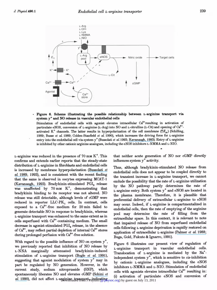
* Figure 6. Scheme illustrating the possible relationship between L-arginine transport viasystem y' and NO release in vascular endothelial cellsStimulation of endothelial cells with agonist elevates intracellular Ca2+resulting in activation ofparticulate eNOS, conversion of L-arginine (L-Arg) into NO and L-citrulline (L-Cit) and opening of Ca2+-activated K+ channels. The latter results in hyperpolarization of the cell membrane (tEm) (Schilling,1989; Busse et al. 1989; Colden-Stanfield et al. 1990), which increases the driving force for L-arginineentry into the endothelial cell via system y+ (Bussolati et al. 1993; Kavanaugh, 1993). Entry of L-arginineis inhibited by other cationic arginine analogues, including the eNOS inhibitors L-NMMA and L-NIO.
L-arginine was reduced in the presence of 70 mm K+. Thisconfirms and extends earlier reports that the steady-statedistribution of L-arginine in fibroblasts and endothelial cellsis increased by membrane hyperpolarization (Bussolati etal. 1989, 1993), and is consistent with the recent findingthat the same is observed in oocytes expressing MCAT-1(Kavanaugh, 1993). Bradykinin-stimulated PGI2 releasewas unaffected by 70 mm K+, demonstrating thatbradykinin binding to its receptor was not altered. NOrelease was still detectable, although levels of cGMP werereduced in reporter LLC-PK1 cells. In contrast, cellsexposed to a Ca2+-free medium for 20 min failed togenerate detectable NO in response to bradykinin, whereasL-arginine transport was enhanced to the same extent as incells superfused with Ca2+-containing medium. The smalldecrease in agonist-stimulated PGI2 release, in the absenceof Ca2+, may reflect partial depletion of internal Ca2+ storesduring prolonged perfusion with Ca!+-free solution.
With regard to the possible influence of NO on system y+,we previously reported that inhibition of NO release byL-NNA marginally attenuated bradykinin-inducedstimulation of L-arginine transport (Bogle et al. 1991),suggesting that agonist modulation of system y+ may inpart be regulated by NO generation. However, in thecurrent study, sodium nitroprusside (SNP), whichspontaneously liberates NO and elevates cGMP (Schini etal. 1990), did not affect L-arginine transport, indicating
that neither acute generation of NO nor cGMP directlyinfluences system y+ activity.
Thus, although bradykinin-stimulated NO release fromendothelial cells does not appear to be coupled directly tothe transient increase in L-arginine transport, we cannotexclude the possibility that the rate of L-arginine utilizationby the NO pathway partly determines the rate ofL-arginine entry. Both system y+ and eNOS are located inthe plasma membrane. Therefore, it is plausible thatpreferential delivery of extracellular L-arginine to eNOSmay occur. Indeed, if L-arginine is compartmentalized inendothelial cells, then the rate of emptying of the argininepool may determine the rate of filling from theextracellular space. In this context, it is relevant to notethat impaired release of NO from superfused endothelialcells following L-arginine deprivation is rapidly restored onapplication of extracellular L-arginine (Palmer et al. 1988;Buga, Gold, Fukuto & Ignarro, 1991).
Figure 6 illustrates our present view of regulation ofL-arginine transport in vascular endothelial cells.Translocation of L-arginine is mediated by the Nae-independent system y+, which is sensitive to cis-inhibitionby cationic L-arginine analogues, including the eNOSinhibitors L-NMMA and L-NIO. Stimulation of endothelialcells with agonists elevates intracellular CaW+ resulting in:(i) activation of particulate eNOS and conversion of
J Physiol.490.1 239
) by guest on July 13, 2011jp.physoc.orgDownloaded from J Physiol (

R. G. Bogle and others
L-arginine into NO and L-citrulline; (ii) activation ofsoluble guanylate cyclase and elevation of cGMP by NO;(iii) opening of Ca2+-activated K+ channels resulting inhyperpolarization of the cell membrane and an increaseddriving force for L-arginine entry into endothelial cells viasystem y+; and (iv) a subsequent intracellular alkalinizationwhich sustains NO release in response to bradykinin(Fleming, Hecker & Busse, 1994). Deprivation of L-arginineis associated with an enhanced transport activity of systemy+, and it is possible that a pool of L-arginine to whicheNOS has access is depleted. Whether this pool is specific toeNOS or is shared by other metabolic pathways remains tobe investigated.
AISAKA, K., GROSS, S. S., GRIFFITH, O. W. & LEVI, R. (1989).L-Arginine availability determines the duration of acetylcholine-induced systemic vasodilation in vivo. Biochemical and BiophysicalResearch Communications 163, 710-717.
AMEZCUA, J. L., PALMER, R. M. J., DE SOUZA, B. M. & MONCADA, S.(1989). Nitric oxide synthesized from L-arginine regulates vasculartone in the coronary circulation of the rabbit. British Journal ofPharmacology 97, 1119-1124.
BAYDOUN, A. R., EMERY, P. W., PEARSON, J. D. & MANN, G. E. (1990).Substrate-dependent regulation of intracellular amino acidconcentrations in cultured bovine aortic endothelial cells.Biochemical and Biophysical Research Communications 173,940-948.
BAYDOUN, A. R., BOGLE, R. G., PEARSON, J. D. & MANN, G. E. (1994).Discrimination of L-citrulline and L-arginine transport in activatedmacrophages. Inefficient synthesis of NO from recycling ofL-citrulline to L-arginine. British Journal of Pharmacology 112,487-492.
BOGLE, R. G., COADE, S. B., MONCADA, S. PEARSON, J. D. & MANN,G. E. (1991). Bradykinin and ATP stimulate L-arginine uptake andrelease of NO in vascular endothelial cells. Biochemical andBiophysical Research Communications 180, 926-932.
BOGLE, R. G., MONCADA, S., PEARSON, J. D. & MANN, G. E. (1992).Identification of inhibitors of nitric oxide synthase that do notinteract with the endothelial cell L-arginine transport. BritishJournal of Pharmacology 105, 768-770.
BUGA, G. M., GOLD, M. E., FUKUTO, J. M. & IGNARRO, L. J. (1991).Shear stress induced release of nitric oxide from endothelial cellsgrown on beads. Hypertension 17, 187-193.
BUSSE, R., FICHTNER, H., LUCKHOFF, A. & KOHLHARDT, M. (1988).Hyperpolarization and increased free calcium in acetylcholine-stimulated endothelial cells. American Journal of Physiology 255,H965-969.
BUSSOLATI, O., LARIS, P. C., NucCI, F. A., DALL'ASTA, V., FRANCHI-GAZZOLA, R., GUIDOTTI, G. G. & GAZZOLA, G. C. (1989). Influx ofL-arginine is an indicator of membrane potential in humanfibroblasts. American Journal of Physiology 256, C930-935.
BUSSOLATI, 0., SALA, R., ASTORRI, A., ROTOLI, B. M., DALL'ASTA, V.& GAZZOLA, G. C. (1993). Characterization of amino acid transportin human endothelial cells. American Journal of Physiology 265,C1006-1014.
CARTER, T. D., BOGLE, R. G. & BJAALAND, T. (1991). Spiking of intra-cellular calcium ion concentration in single cultured pig aortic
CLOSS, E. I., LYONS, C. R., KELLY, C. & CUNNINGHAM, J. M. (1993).Characterization of the third member of the MCAT family ofcationic amino acid transporters: Identification of a domain thatdetermines the transport properties of the MCAT proteins. Journalof Biological Chemistry 268, 20796-20800.
COLDEN-STANFIELD, M., SCHILLING, W. P., POSSANI, L. D. & KUNZE,D. L. (1990). Bradykinin-induced potassium current in culturedbovine aortic endothelial cells. Journal of Membrane Biology 116,227-238.
COOKE, J. P., ANDON, N. A., GIRERD, X. J., HIRSCH, A. T. &CREAGER, M. A. (1991). Arginine restores cholinergic relaxation ofhypercholesterolemic rabbit thoracic aorta. Circulation 83,1057-1062.
DEVES, R., CHAVES, P. & BOYD, C. A. R. (1992). Identification of a
new transport system (y+L) in human erythrocytes that recognizeslysine and leucine with high affinity. Journal of Physiology 454,491-501.
FLEMING, I., HECKER, M. & BUSSE, R. (1994). Intracellularalkalinization induced by bradykinin sustains activation of theconstitutive nitric oxide synthase in endothelial cells. CirculationResearch 74, 1220-1226.
GAZZOLA, G. C., DALL'ASTA, V. & GUIDOTTI, G. G. (1981). Adaptiveregulation of amino acid transport in cultured human fibroblasts.Journal of Biological Chemistry 256, 3191-3198.
GOLD, M. E., BUSH, P. A. & IGNARRO, L. J. (1989). Depletion ofarterial L-arginine causes reversible tolerance to endothelium-dependent relaxation. Biochemical and Biophysical ResearchCommunications 164, 714-721.
GREENE, B., PACITTI, A. & SOUBA, W. (1993). Characterization ofL-arginine transport by pulmonary artery endothelial cells.American Journal of Physiology 264, L351-356.
HECKER, M., MULSCH, A., BASSENGE, E., FORSTERMANN, U. & BUSSE,R. (1994). Subcellular localisation and characterization of nitricoxide synthase(s) in endothelial cells: physiological implications.Biochemical Journal 299, 247-252.
KAVANAUGH, M. M. (1993). Voltage-dependence of facilitated arginineflux mediated by the system y+ basic amino acid transporter.Biochemistry 32, 5781-5785.
KIM, J. W., CLOSS, E. I., ALBRITTON, L. M. & CUNNINGHAM, J. M.(1991). Transport of cationic amino acids by the mouse ecotropicretrovirus receptor. Nature 352, 725-728.
KLETZIEN, R. F., PARIZA, M. W., BECKER, J. E. & POTTER, V. R.(1975). A method using 3-0-methyl-D-glucose and phloretin for thedetermination of intracellular water space of cells in monolayerculture. Analytical Biochemistry 68, 537-544.
KNOWLES, R. G. & MONCADA, S. (1994). Nitric oxide synthases inmammals. Biochemical Journal 298, 249-258.
MANN, G. E., NORMAN, P. S. R. & SMITH, I. C. H. (1989a). Amino acidefflux in the isolated perfused rat pancreas: trans-stimulation byextracellular amino acids. Journal of Physiology 416,485-502.
MANN, G. E., PEARSON, J. D., SHERIFF, C.-J. & TOOTHILL, V. J.(1989b). Expression of amino acids transport systems in culturedhuman umbilical vein endothelial cells. Journal of Physiology 410,325-339.
MAYER, B., SCHMIDT, K., HUMBERT, P. & BOHME, E. (1989).Biosynthesis of endothelium-derived relaxing factor: a cytosolicenzyme in porcine aortic endothelial cells Ca2+-dependentlyconverts L-arginine into an activator of soluble guanylate cyclase.Biochemical and Biophysical Research Communications 164,678-685.
endothelial cells stimulated with ATP or bradykinin. BiochemicalJournal 278, 697-704.
J.Physiol.490.1240
) by guest on July 13, 2011jp.physoc.orgDownloaded from J Physiol (

Endothelial cell L-arginine transporter
MITCHELL, J. A., HECKER, M., ANGGARD, E. E. & VANE, J. R. (1990).Cultured endothelial cells maintain their L-arginine level despite thecontinuous release of EDRF. European Journal of Pharmacology182, 573-576.
MONCADA, S., HIGGS, E. A. & PALMER, R. M. J. (1991). Nitric oxide:physiology, pathophysiology and pharmacology. PharmacologicalReviews 43, 109-142.
NAKACHE, M. & GAUB, H. E. (1988). Hydrodynamic hyper-polarization of endothelial cells. Proceedings of the NationalAcademy of Sciences of the USA 85, 1841-1843.
NEEDHAM, L., CUSACK, N. J., PEARSON, J. D. & GORDON, J. L. (1987).Characteristics of the P2 purinoceptor that mediates prostacyclinproduction by pig aortic endothelial cells. European Journal ofPharmacology 134, 199-209.
PALMER, R. M. J., ASHTON, D. S. & MONCADA, S. (1988). Vascularendothelial cells synthesize nitric oxide from L-arginine. Nature333, 664-666.
PALMER, R. M. J., FERRIGE, A. G. & MONCADA, S. (1987). Nitric oxideaccounts for the biological activity of endothelium-derived relaxingfactor. Nature 327, 524-526.
PALMER, R. M. J. & MONCADA, S. (1989). A novel citrulline formingenzyme implicated in the formation of nitric oxide by vascularendothelial . cells. Biochemical and Biophysical ResearchCommunications 158, 348-352.
POSTON, L. & TAYLOR, P. D. (1995). Endothelium-dependent vascularfunctions in insulin-dependent diabetes. Clinical Science 88,245-255.
SCHILLING, W. P. (1989). Effect of membrane potential on cytosoliccalcium of bovine aortic endothelial cells. American Journal ofPhysiology 257, H778-784.
SCHINI, V. B., BOULANGER, C., REGOLI, D. & VANHOUTTE, P. M.(1990). Bradykinin stimulates the production of cyclic GMP viaactivation of B2 kinin receptors in cultured porcine aorticendothelial cells. Journal of Pharmacology and ExperimentalTherapeutics 252, 581-585.
SCHMIDT, H. H. H. W., NAU, H., WITTFOHT, W., GERLACH, J.,PRESCHER, K.-L., KLEIN, M. M., NIROOMAND, F. & BOHME, E.(1988). Arginine is the physiological precursor of endotheliumderived nitric oxide. European Journal of Pharmacology 154,213-216.
SCHMIDT, K., KLATT, P. & MAYER, B. (1993). Characterization ofendothelial cell amino acid transport systems involved in the actionsof nitric oxide synthase inhibitors. Molecular Pharmacology 44,615-621.
SESSA, W. C., HECKER, M., MITCHELL, J. A. & VANE, J. R. (1990). Themetabolism of L-arginine and its significance for the biosynthesis ofendothelium-derived relaxing factor: L-glutamine inhibits thegeneration of L-arginine by endothelial cells. Proceedings of theNational Academy of Sciences of the USA 87, 8607-8611.
SOBREVIA, L., CESARE, P., YUDILEVICH, D. L. & MANN, G. E. (1995).Diabetes-induced activation of system y+ and nitric oxide synthasein human endothelial cells: association with membrane hyper-polarization. Journal of Physiology 489, 183-192.
SOBREVIA, L., YUDILEVICH, D. L. & MANN, G. E. (1994). Diabetes-induced activation of the human endothelial cell L-argininetransporter is unaffected by the external glucose environment.Journal of Physiology 480, 134P.
VAN WINKLE, L. J. (1988). Amino acid transport in developinganimal oocytes and early conceptuses. Biochimica et BiophysicaActa 947,173-208.
WHITE, M. F. (1985). The transport of cationic amino acids across the
Wu, G. & MEININGER, C. J. (1993). Regulation of L-arginine synthesisfrom L-citrulline by L-glutamine in endothelial cells. AmericanJournal of Physiology 265, H1965-1977.
AcknowledgementsWe gratefully acknowledge support from the British HeartFoundation (BHF Grant 91075). R.G.B. was supported by a
SERC-CASE PhD collaborative award with Dr SalvadorMoncada FRS, Wellcome Research Laboratories, Beckenham, UK.We thank Dr Moncada for his helpful discussion of this work.
Author's present addressR. G. Bogle: Clinical Pharmacology Unit, Department ofPharmacology and Clinical Pharmacology, St. George's HospitalMedical School, London SW17 ORE, UK.
Received 17 March 1995; accepted 12 September 1995.
plasma membrane of mammalian cells. Biochimica et BiophysicaActa 822, 355-374.
J. Physiol. 490.1 241
) by guest on July 13, 2011jp.physoc.orgDownloaded from J Physiol (