regulation of gene expression in experimental autoimmune encephalomyelitis indicates early neuronal...
TRANSCRIPT

Regulation of gene expression in experimentalautoimmune encephalomyelitis indicates earlyneuronal dysfunction
Arnaud Nicot,1,2 Pillarisetty V. Ratnakar,1 Yacov Ron,3 Chiann-Chyi Chen3 and Stella Elkabes1,2
1Department of Neuroscience, University of Medicine and
Dentistry of New Jersey, New Jersey Medical School,
Newark, 2Veterans Affairs, Neurology Service,
East Orange, 3Department of Molecular Genetics and
Microbiology, University of Medicine and Dentistry of
New Jersey, Robert W. Johnson Medical School,
Piscataway, NJ, USA
Correspondence to: Stella Elkabes, PhD, Department of
Neuroscience, MSB, H-506, New Jersey Medical School/
UMDNJ, 185 South Orange Avenue, Newark, NJ 07103,
USA
E-mail: [email protected]
SummaryMultiple sclerosis is an in¯ammatory, demyelinatingdisease of the CNS. Whereas oligodendrocytes havebeen considered the primary neural cell type mostaffected, recent evidence indicates that axonal andneuronal degeneration also occurs in both multiplesclerosis and experimental autoimmune encephalomyeli-tis (EAE), an animal model reproducing many featuresof multiple sclerosis. The molecular mechanisms under-lying neuronal de®cits in multiple sclerosis and EAEremain elusive. To address this issue, we have analysedthe expression of genes encoding proteins that playcritical roles in ion homeostasis, exocytosis, mitochon-drial function and impulse conduction in the Lewis ratlumbar spinal cord during the clinical course of acuteEAE. Transcript and protein levels of plasma mem-brane Ca2+ ATPase 2 (PMCA2), an essential ion pumpexpressed exclusively in grey matter and involved inCa2+ extrusion, synapsin IIa and syntaxin 1B, important
regulators of vesicular exocytosis, were dramaticallydecreased coincident with the onset of clinical symp-toms. In contrast, changes in the expression of severalother ion pumps, vesicular proteins, mitochondrialenzymes and sodium channels occurred at moreadvanced disease stages. Moreover, exposure of spinalcord slice cultures to kainic acid signi®cantly reducedPMCA2 mRNA levels. Taken together, our ®ndings sug-gest that glutamate, which recently has been implicatedin EAE pathogenesis, suppresses neuronal PMCA2expression leading to Ca2+ dyshomeostasis at initialclinical phases. Consequently, perturbations in Ca2+ bal-ance and neurotransmitter exocytosis may partiallyunderlie aberrant neuronal function and communi-cation at onset of symptoms. Altered mitochondrialfunction and impulse conduction may exacerbate neuro-logical de®cits at subsequent disease stages.
Keywords: EAE; exitotoxicity; synaptic protein; neuronal injury; ATP2B2
Abbreviations: DIG = digoxigenin; EAE = experimental autoimmune encephalomyelitis; PMCA2 = plasma membrane
Ca2+ ATPase 2; SERCA2 = sarcoplasmic and endoplasmic reticulum Ca2+-activated ATPase 2; SNAP-25 = synaptosome-
associated protein of 25 kDa
IntroductionMultiple sclerosis is a CNS disease leading to progressive
neurological de®cits and permanent disability (Steinman,
1996; Prineas and McDonald, 1997; Antel, 1999; Hickey,
1999). Experimental autoimmune encephalomyelitis (EAE),
the animal model of multiple sclerosis, is induced by
immunization of rodents with immunogenic myelin com-
ponents or by adoptive transfer with T cells reactive
against CNS myelin antigens (Wekerle et al., 1994;
Steinman, 1999; van der Goes and Dijkstra, 2001). The
clinical symptoms and histopathology of EAE may be diverse
depending on the antigen and the animal strain employed. It
has been hypothesized that distinct EAE models reproduce
different features of multiple sclerosis, a complex disease
with variable clinical course (Wekerle et al., 1994).
Immunization of adult Lewis rats with myelin basic protein
results in acute, monophasic EAE manifested by progressive
ã Guarantors of Brain 2003
DOI: 10.1093/brain/awg041 Brain (2003), 126, 398±412
by guest on February 9, 2016http://brain.oxfordjournals.org/
Dow
nloaded from

ascending weakness leading to hindlimb paralysis and
quadriplegia.
Classically, demyelination of structurally intact axons and
the resulting impairment in saltatory nerve conduction have
been considered as the causes of functional de®cits in
multiple sclerosis and EAE (McFarlin and McFarland,
1982a, b; Raine, 1997). However, the lack of correlation
between myelin loss and symptom severity suggests that EAE
and multiple sclerosis are more complex than initially
believed (Rivera-Quinones et al., 1998; Antel, 1999).
Indeed, studies reporting axonal dysfunction and damage
(Ferguson et al., 1997; Trapp et al., 1998, 1999; De Stefano
et al., 1999; Bitsch et al., 2000; Bjartmar and Trapp, 2001;
Bjartmar et al., 2001), neuronal loss or apoptosis (Smith et al.,
2000; Peterson et al., 2001) indicate that multiple sclerosis
and EAE are not only demyelinating diseases but neuronal
disorders as well (Waxman, 2000a, b; Filippi, 2001).
The triggers that cause aberrant neuronal axonal function
and destruction of myelin are not well de®ned. A correlation
between in¯ammatory reaction and axonal injury has been
observed (Ferguson et al., 1997; Trapp et al., 1998).
Activated resident microglia and macrophages have also
been implicated in myelin and oligodendrocyte damage (for a
review see Sriram and Rodriguez, 1997). Recent studies
indicate that glutamate excitotoxicity may be a factor
contributing to axonal, neuronal and oligodendrocyte injury
during EAE (Pitt et al., 2000; Smith et al., 2000). Moreover, it
has been suggested that increased calcium in¯ux through
voltage-dependent calcium channels induces axonal degen-
eration and neuronal injury (Kornek et al., 2000, 2001).
Calcium dyshomeostasis, which has been implicated in many
pathological conditions, may also result from the abnormal
function of pumps that extrude Ca2+ from cells. This
possibility has not been investigated in EAE or multiple
sclerosis.
In summary, in contrast to the well-de®ned histopathology,
the molecular events underlying neuronal impairment con-
tributing to neurological de®cits and persistent disability in
EAE and multiple sclerosis remain elusive. To delineate
molecular mechanisms responsible for the onset and pro-
gression of symptoms in EAE, we de®ned the expression
pattern of genes encoding proteins that play essential roles in
neuronal function during the clinical course of the disease.
Our ®ndings suggest that abnormal Ca2+ extrusion and
vesicular exocytosis may initiate neuronal dysfunction. At
later clinical stages, changes in the expression of other genes
that encode proteins involved in mitochondrial function
and impulse conduction may contribute further to disease
progression.
Material and methodsInduction of EAEFemale adult Lewis rats (200±250 g body weight) were
immunized in hindlimb footpads with 100 mg of guinea pig
myelin basic protein (Sigma, St Louis, MO, USA, or
generously provided by Dr A. Ben-Nun) in 50 ml of saline
emulsi®ed in 50 ml complete Freund's adjuvant (Difco,
Detroit, MI, USA). Control rats received only saline/
adjuvant. Animals were monitored daily for clinical symp-
toms. A very stereotyped clinical onset and disease course
was observed. Neurological impairment was scored as: 0, no
neurological symptoms; 1, limp tail [stage E1, observed on
day 10 post-injection (PI)]; 2, hindlimb weakness (stage E2,
day 11 PI); 3, hindlimb paralysis (stage E3, day 12±13 PI); 4,
quadriplegia (stage E4; day 14±15 PI); 5, moribund (past day
16 PI). During the observation period, rats were given food
and water ad libitum. Some animals were sacri®ced by
exposure to CO2/O2. Others were anaesthetized with a
mixture of ketamine/xylazine and perfused with saline
followed by 4% paraformaldehyde in 0.1 M phosphate buffer
pH 7.5. Lumbar spinal cords were dissected out and frozen
immediately on dry ice. The tissue was kept at ±80°C until
further use. All animal procedures were performed according
to IACUC and institutional guidelines.
Isolation of RNATotal RNA was isolated from 100±150 mg of tissue obtained
by pooling 2±3 lumbar spinal cords. The Trizol One Step
Isolation method was utilized according to the manufacturer's
instructions (Life Technologies, Grand Island, NY, USA). All
RNA samples were treated with DNase I to remove any trace
of contaminating genomic DNA and repuri®ed by phenol/
chloroform extraction. RNA was quanti®ed by measurement
of optical density. Samples were stored at ±20°C in the
presence of RNase inhibitor.
cDNA microarray analysisThis was performed utilizing the Atlas Rat 1.2 microarrays
according to the manufacturer's instructions (Clontech, Palo
Alto, CA, USA). The microarray includes 1176 rat cDNAs
immobilized on nylon membranes, nine housekeeping
cDNAs and negative controls. The list of the cDNAs can be
found at the Clontech web site (http://www.clontech.com/
atlas/genelists/7854-1_Ra12.txt). Total RNA was puri®ed
from lumbar spinal cords of control and EAE rats (three rats
per group; EAE clinical score 3, E3). A 25 mg aliquot of
pooled RNA was reverse transcribed in the presence of
MMLV reverse transcriptase, gene-speci®c primer mix,
0.5 mM each of dCTP, dGTP and dTTP, and 35 mCi of
[a-33P]dATP (2500 Ci/mmol; Amersham, Piscataway, NJ,
USA) in a 30 ml reaction volume at 50°C for 25 min using the
Atlas pure total RNA labelling system (Clontech). The arrays
were pre-hybridized in Express Hyb hybridization buffer
(Atlas hybridization kit) containing heat-denatured salmon
testes DNA (Sigma) for 30 min at 68°C. Subsequently, the
probe mixture (2±10 3 106 c.p.m.) and 5 ml of human
Cot-1DNA were added to the pre-hybridization buffer.
Hybridization was performed overnight at 68°C. The arrays
Neuronal dysfunction in spinal cord during EAE 399
by guest on February 9, 2016http://brain.oxfordjournals.org/
Dow
nloaded from

were then washed four times in 23 SSC (standard saline
citrate), 1% SDS, and once in 0.13 SSC, 0.5% SDS for
30 min, each wash at 68°C, and exposed to phosphorimager
screens for 5 days. A Storm phosphorimager (Molecular
Dynamics, Sunnyvale, CA, USA) and ImageQuant software
were used to scan images. Gene expression was custom
analysed using Atlas Image 2.0 software by Clontech. The
intensity of each spot, quanti®ed after background correction,
re¯ected the level of expression of each gene. Experimental
variation in the overall intensity of the signal was corrected
by normalization of the gene signal with respect to the
housekeeping genes in the same array. For each gene, a ratio
was generated by dividing the intensity of the spot by the
average of the sum of housekeeping genes. Only those
housekeeping genes with signal intensity below saturation
were considered for normalization. Reproducibility of this
technique (80% concordance of signals) was veri®ed by
hybridizing probe mixtures generated from two aliquots of
the same RNA source to identical arrays. The signal levels of
genes selected for further analysis in our study were 30- to 40-
fold over the background. In addition, there was at least a
2-fold difference in the expression of the selected genes in
EAE versus control (Table 1).
RT±PCRThis was performed utilizing the Retroscript reverse tran-
scription kit and SuperTaq Polymerase (Ambion, Austin, TX,
USA) according to the manufacturer's instructions. A 1±2 mg
aliquot of DNase-treated total RNA was reverse transcribed
in a total volume of 20 ml. A 2±5 ml aliquot of RT mix was
then used for PCR, which was performed in a total volume of
50 ml by denaturation at 94°C for 30 s, annealing at 55°C for
45 s and polymerization at 68°C for 1 min. PCR was
completed by 10 min extension at 72°C. PCRs were
performed for 30±35 cycles, within the linear range.
Products were separated on a 1.5% agarose gel containing
ethidium bromide (0.5 mg/ml) and the image was captured
using an Alpha digital imaging system. The optical density in
each band was then quanti®ed utilizing the Un-Scan-It
software (Silk Scienti®c, Orem, UT, USA). Some PCR
products were analysed further by sequencing.
Preparation of riboprobes for in situhybridizationRT±PCR products generated by use of speci®c primers for
PMCA2 or a-tubulin were cloned into pCR II-TOPO vector
containing SP6 and T7 promoters (Invitrogen, Carlsbad, CA,
USA) and sequenced for further con®rmation. Antisense and
sense riboprobes were generated using the Digoxigenin (DIG)
RNA Labelling Kit according to the manufacturer's instruc-
tions (Boehringer Mannheim, Indianapolis, IN, USA). A 1 mg
aliquot of linearized plasmid was transcribed at 37°C for 2 h
in the presence of 1 mM ATP, CTP and GTP, 0.65 mM UTP,
0.35 mM DIG-UTP, 20 U of RNasin, 40 U of SP6 or T7 RNA
polymerase in 40 mM Tris buffer pH 8.0, 10 mM NaCl,
10 mM dithiothreitol and 6 mM MgCl2. The DNA template
was removed by treatment with DNase I and the probe was
precipitated with LiCl and ethanol. The concentration of
DIG-labelled riboprobe was determined by utilizing DIG
quanti®cation test strips according to the manufacturer's
instructions (Boehringer Mannheim).
Table 1 Differentially expressed genes in the lumbar spinal cord during EAE
Gene Expressionchanges
Ratio of spotintensity (E/C)
GeneBankaccession No.
Function
Plasma membrane Ca2+ ATPase 2 Decrease 0.10 J03754 Ion homeostasisNa+/K+ transporting ATPase b2 subunit Decrease <0.10 J04629 Ion homeostasisSERCA2 Decrease 0.50 J04022 Ion homeostasisSynapsin IIa Decrease 0.15 M27925 Vesicular functionSyntaxin 1B Decrease 0.40 M95735 Vesicular functionSNAP-25 Decrease 0.50 AB003991 Vesicular functionRab12 Increase 6.30 M83676 Vesicular functionRab14 Increase 8.30 M83680 Vesicular functionRab16 Increase 3.30 M83681 Vesicular functionCytochrome c oxidase subunit Vb Increase 13.0 D10952 Mitochondrial functionMitochondrial cytochrome c subunit IV Increase 4.80 X14209 Mitochondrial functionNa+ channel b1 subunit Decrease <0.10 M91808 Impulse conductionNa+ channel b2 subunit Decrease 0.30 U37026 Impulse conductionNa+ channel II Decrease 0.30 X03639 Impulse conduction
cDNA microarray analysis identi®ed differentially expressed genes in the spinal cord during EAE, including those listed above whichshowed robust changes in transcript levels (at least 2-fold difference between EAE and control) and which were highly expressed (at least30- to 40-fold over background). The genes are classi®ed according to their best characterized function. The results obtained with all thelisted genes were corroborated subsequently by RT±PCR in 2±3 independent experiments. The ratio of spot intensity re¯ects the relativetranscript levels in EAE over control.
400 A. Nicot et al.
by guest on February 9, 2016http://brain.oxfordjournals.org/
Dow
nloaded from

In situ hybridizationControls and EAE (E3) rats (six animals/group) were
sacri®ced by cardiac perfusion of saline followed by 4%
paraformaldehyde in 0.1 M phosphate buffer pH 7.5. Lumbar
spinal cords were dissected out, post-®xed in phosphate
buffer for 4 h, cryoprotected in 10 and 15% sucrose and cryo-
sectioned at 10 mm. Sections obtained from control and EAE
lumbar spinal cords were mounted on the same slides to
minimize experimental variations, ®xed in 4% paraform-
aldehyde/phosphate buffer for 15 min at room temperature,
and treated with 6% H2O2 in phosphate-buffered saline (PBS)
containing 0.1% Tween (PBST). Sections were then digested
with proteinase K (1 mg/ml) for 15 min at 37°C, followed by
incubations in glycine (2 mg/ml, 10 min) and 4% para-
formaldehyde/0.2% gluteraldehyde/PBST for 15 min. Pre-
hybridization was performed in buffer containing 50%
deionized formamide, 53 SSC pH 4.5, 1% SDS, 50 mg/ml
yeast tRNA and 50 mg/ml heparin at 65°C for 1 h.
Hybridization was performed overnight at 65°C in the same
buffer containing 1 mg/ml DIG-labelled sense or antisense
probe. The sections were then washed in 53 SSC/50%
formamide/1% SDS at 70°C, followed by 43 SSC/50%
formamide at 65°C, blocked in 10% heat-inactivated goat
serum/PBS for 1 h at room temperature and incubated in anti-
DIG±alkaline phosphatase-conjugated antibody (1 : 1000) in
PBS at 4°C overnight. Endogenous alkaline phosphatase
activity was blocked by pre-treatment with 10 mM levami-
sole. The colour reaction was developed overnight in the
presence of 250 mg/ml NBT (4-nitro blue tetrazolium
chloride) and 130 mg/ml BCIP (5-bromo-4-chloro-3-indolyl-
phosphate) as substrate.
ImmunocytochemistryLumbar spinal cord sections, prepared as described above,
were ®rst treated with 10% goat serum/0.1% Triton X-100/
PBS for 1 h at room temperature. They were then incubated in
ED-1 (1 : 500; Serotec, Oxford, UK)/0.1% Triton X-100/PBS
overnight at 4°C. The rest of the procedure was performed at
room temperature. The sections were washed in PBS,
incubated in anti-mouse IgG [1/500 (v/v)] followed by
avidin±biotin complex (Vectastain ABC kit, Vector
Laboratories, Burlingame, CA, USA). Immunopositive cells
were visualized by 3,3¢¢-diaminobenzidine reaction product.
Western blot analysisLumbar spinal cords of controls or EAE (E1±E3) rats (three
animals/group) were homogenized on ice with a motorized
pestle in 1 ml of buffer A (10 mM HEPES pH 7.0, 10%
sucrose, 0.4 mM phenylmethylsulfonyl ¯uoride, 5 mM
EDTA, 2 mg/ml leupeptin, 2 mg/ml aprotinin, 2 mg/ml
pepstatin). The homogenate was then passed ®ve times
through a syringe with a 22-gauge needle and centrifuged at
800 g for 5 min. The supernatant was removed and
centrifuged at 20 000 g for 45 min. The crude plasma
membrane preparation was suspended in buffer A and
aliquots were kept at ±70°C until use. Total protein concen-
trations were determined utilizing the CBQCA protein
quanti®cation kit according to the manufacturer's instructions
(Molecular Probes, Eugene, OR, USA). A 5 mg aliquot of
total protein was loaded on each lane of an 8% SDS±
polyacrylamide Novex, Tris-glycine gel (Invitrogen).
Electrophoresis was performed for 90 min at 125 V. The
protein was then electrotransferred onto a polyvinylidene
di¯uoride (PVDF) membrane for 45 min at 50 V, and stained
with BLOT-FastStain (Chemicon, Temecula, CA, USA)
according to the manufacturer's instructions. Immuno-
detection was performed employing a Western Breeze kit
(Invitrogen). Primary antibodies were utilized as follows:
anti-PMCA2 (1 : 5000, polyclonal; Research Diagnostic,
Flanders, NJ, USA), anti-synapsin IIa (1 : 5000, monoclonal;
BD Sciences, Palo Alto, CA, USA) and anti-a-tubulin
(1 : 20 000, monoclonal; Sigma). Signal was visualized by
exposure of blots to Hyper®lm ECL (Amersham Pharmacia
Biotech, Piscataway, NJ) for 30±120 s. Between each
immunoblot, membranes were stripped with Re-Blot
recycling kit (Chemicon). Bands were quanti®ed using the
Un-scan-it software (Silk Scienti®c).
Spinal cord slice culturesAdult rats were sacri®ced by exposure to CO2/O2. Lumbar
spinal cords were excised, sliced into 1 mm sections and
immediately placed in 1 ml of de®ned medium consisting of
L-15: neurobasal medium (2 : 1; Gibco/BRL, Rockville, MD,
USA) containing insulin (5 mg/ml), transferrin (100 mg/ml),
selenium (40 ng/ml), putrescine (16 ng/ml), progesterone
(60 ng/ml) and 1 U of penicillin/streptomycin. Kainic acid
was then introduced at the concentrations indicated in the
Results. Controls were maintained in medium containing
vehicle. The slices were incubated for 6 h at 37°C. At the end
of the incubation period, the slices were homogenized in
Trizol reagent and RNA was prepared as described above.
Some slices were ®xed in 4% paraformaldehyde, cryopro-
tected in 10 and 20% sucrose and frozen on dry ice. Cryostat
sections (10 mm) were mounted on poly-L-lysine-coated
slides and utilized for assessment of DNA fragmentation by
TUNEL assay, a hallmark of cell death.
TUNEL assay was performed utilizing the ApopTag kit
(Intergen, Purchase, NY, USA) according to the manufac-
turer's instructions. Eight control and eight kainic acid-
treated slices were analysed. At least ®ve sections from each
slice were used for TUNEL assay. Control and experimental
samples were mounted on the same slide, side by side. The
experiment was repeated twice and yielded similar results.
ResultsTo identify differentially expressed genes in the EAE lumbar
spinal cord, we ®rst performed cDNA microarray analysis at
Neuronal dysfunction in spinal cord during EAE 401
by guest on February 9, 2016http://brain.oxfordjournals.org/
Dow
nloaded from

disease stage E3. We selected a number of differentially
expressed genes for further studies because they are
expressed exclusively or mainly by neurons, they play critical
roles in important cellular functions including ion home-
ostasis, neurotransmitter exocytosis, energy metabolism or
impulse conduction, and their transcript level was altered
greatly. The genes were classi®ed according to the best
characterized function of the protein which they encode
(Table 1). Thus, cDNA microarray pro®ling provided an
initial and global insight into potential cellular mechanisms
that may be affected during a de®ned stage of the disease. We
further corroborated these ®ndings by semi-quantitative RT±
PCR utilizing the same source of RNA as that employed for
cDNA microarray analysis (stage E3). Subsequently, we
further validated our ®ndings by performing at least 2±3
additional, independent experiments employing distinct
groups of rats (2±3 rats/group) during the course of the
disease (stages E1±E3), as reported below.
PMCA2 mRNA and protein levels are decreasedat onset of symptomsOne of our observations by cDNA microarray analysis was a
pronounced change in the expression of several pumps which
modulate ion, and in particular Ca2+, homeostasis including
plasma membrane Ca2+ ATPase 2 (PMCA2), sarcoplasmic
and endoplasmic reticulum Ca2+-activated ATPase 2
(SERCA2) and Na+/K+ transporting ATPase b2 subunit
(Table 1). As Ca2+ balance plays a pivotal role in the function
and survival of cells, including neurons, we further analysed
these genes and corroborated our microarray ®ndings by
semi-quantitative RT±PCR utilizing speci®c primers for each
pump (Table 2). We examined the expression of the
aforementioned ion pumps during the course of the disease
starting at E1 (tail weakness only; clinical score 1) in order to
determine the clinical stage when the ®rst signi®cant changes
in expression occur. Transcript levels of PMCA2, a major
pump mediating Ca2+ extrusion from cells (Carafoli, 1987;
Miller, 1991; Garcia and Strehler, 1999), were signi®cantly
decreased at E1 [Fig. 1, 88% decrease, P < 0.001 by analysis
of variance (ANOVA); Scheffe's post hoc test] and con-
sistently remained low until E3, the latest clinical stage
examined. Transcript levels of SERCA2, which mediates the
sequestration of intracellular Ca2+ into the endoplasmic
reticulum, and Na+/K+ transporting ATPase b2 subunit,
which regulates Na+ homeostasis, were not signi®cantly
altered at E1 or E2 but dramatically decreased at E3 (Fig. 1;
100 and 90% decrease for SERCA2 and Na+/K+ ATPase,
respectively, P < 0.001 by ANOVA, Scheffe's post hoc test).
To ascertain that the changes in PMCA2 mRNA were also
re¯ected at the protein level, we performed western blot
analysis utilizing an antibody which previously has been
reported to be speci®c for the PMCA isoform 2 (Stauffer et al.,
1995). Indeed, we did not detect any signal when crude
membranes from kidney, a tissue which does not express
PMCA2, were tested, indicating speci®city of the antibody
(data not shown). In contrast, in spinal cord, one large band of
mol. wt ~135±140 kDa and another one of 130 kDa were
visualized (Fig. 2), in accordance with the 130, 135 and
138 kDa PMCA2 isoforms previously described (Stauffer
et al., 1995). The intensity of both bands decreased by 50% at
E1, and by 80% at subsequent stages (Fig. 2). The results by
western analysis con®rm that the reduction in PMCA2
mRNA is accompanied by a decrease in protein level. Our
®ndings, taken together, raise the possibility of ion dys-
homeostasis as one of the mechanisms leading to neural
dysfunction or injury during EAE.
Table 2 List of primers utilized in RT±PCR
Gene 5¢ primer 3¢ primer Predictedsize (bp)
Plasma membrane Ca2+ ATPase 2 667±690 1659±1681 1014Na+/K+ transporting ATPase b2 subunit 499±522 1039±1062 563SERCA2 548±570 1357±1379 823Synapsin IIa 1884±1909 2531±2556 672Syntaxin 1B 211±235 918±942 731SNAP-25 1±22 588±609 609Rab12 43±66 518±542 499Rab14 201±222 629±651 450Rab16 24±45 468±490 466Cytochrome c oxidase subunit Vb 17±38 382±403 386Mitochondrial cytochrome c subunit IV 91±122 551±572 481Na+ channel b1 subunit 333±356 858±834 525Na+ channel b2 subunit 319±339 735±755 436Na+ channel II 213±233 772±793 580a-Tubulin 68±90 687±708 640
The region selected for the design of primers and the predicted size for each RT±PCR product arepresented. GenBank accession Nos are given in Table 1.
402 A. Nicot et al.
by guest on February 9, 2016http://brain.oxfordjournals.org/
Dow
nloaded from

PMCA2 expression is restricted to grey matterTo determine the cell type-speci®c expression and modula-
tion of PMCA2, we performed in situ hybridization on lumbar
spinal cord sections utilizing DIG-labelled riboprobes spe-
ci®c for this isoform. PMCA2 mRNA was localized exclu-
sively to grey matter (Fig. 3A±D). We did not detect any
signal in white matter even when the substrate reaction was
continued for 48 h. Many cells in the grey matter, including
those in the ventral horn exhibiting motor neuron-like
morphology, expressed PMCA2 (Fig. 3C and D). The
exclusive expression of PMCA2 mRNA in spinal cord grey
matter is consistent with the ®ndings of Stauffer et al. (1997)
reporting PMCA2 immunoreactivity primarily associated
with neurons in the brain.
In situ hydridization studies further con®rmed the results
obtained by microarray analysis or RT±PCR, and indicated a
Fig. 1 Modulation of ion pump expression during the clinical course of EAE as assessed by RT±PCR.RNA obtained from rats exhibiting tail weakness only (clinical score 1, E1), hindlimb weakness (clinicalscore 2, E2), hindlimb paralysis (clinical score 3, E3) and unaffected controls (C) was subjected to RT±PCR. Each lane shows the results obtained with pooled RNA with distinct groups of rats (2±3 rats/group).The expression of a-tubulin, a housekeeping gene not modulated in EAE, was utilized as control forexperimental variations. PCRs for the various ion pumps and a-tubulin were performed in parallelutilizing the same reverse transcription mix for comparison. RNA subjected to PCR without reversetranscription con®rmed the lack of contaminating genomic DNA (negative control). PCR was performedfor 35 cycles. The ®gure shows a representative gel and the graphs present the mean 6 SEM of threeindependent experiments (n = 5±6 with 2±3 rats per group, as indicated above). ***P < 0.001,signi®cantly different from control by ANOVA, Scheffe's post hoc test.
Neuronal dysfunction in spinal cord during EAE 403
by guest on February 9, 2016http://brain.oxfordjournals.org/
Dow
nloaded from
striking reduction in PMCA2 mRNA levels in EAE spinal
cord (Fig. 3E and F) when compared with controls. The
magnitude of the change observed was variable in individual
rats (n = 6). In some animals, PMCA2 expression was
decreased dramatically to almost undetectable levels, as
illustrated in Fig. 3, whereas in others a less pronounced but
clearly discernible diminution was observed.
To ensure that the decrease in PMCA2 expression was not
due solely to a generic reduction in mRNA, we performed
in situ hybridization utilizing DIG-labelled riboprobes for
a-tubulin, a housekeeping gene not modulated during EAE.
a-Tubulin expression in experimental samples was compar-
able with that of controls (Fig. 3H and I). Adjacent sections
from control and experimental spinal cords were also stained
with cresyl violet, which con®rmed the lack of extensive cell
loss in grey matter (Fig. 3J and K). To ascertain further that
the decrease in PMCA2 mRNA levels was not due to grey
matter cell death, we investigated the distribution of TUNEL-
positive cells in the spinal cord during EAE. TUNEL-positive
cells were scarce in the grey matter even at clinical stage E3,
did not exhibit neuronal morphology and localized mostly to
white matter (results not shown). Taken together, these
®ndings indicate that cell death alone cannot account for the
88% decrease in PMCA2 mRNA levels at stages E1±E3,
although its contribution at more advanced clinical phases
cannot be ruled out. Thus, the reduction in PMCA2 mRNA
levels during EAE may be attributed to decreased gene
expression rather than loss of PMCA2-expressing cells,
especially because it occurs at the earliest clinical times,
before extensive neuronal death is evident (Smith et al.,
2000). It is noteworthy that at this stage of the disease, the
in¯ammatory reaction in the lumbar spinal cord is quite
extensive, as indicated by the distribution of ED-1-immuno-
positive activated microglia/macrophages in the grey and
white matter (Fig. 3L±N).
PMCA2 expression is modulated by kainic acidGlutamate excitotoxicity has been implicated recently in
EAE as a trigger that induces neuronal and oligodendrocyte
loss and axonal injury (Pitt et al., 2000; Smith et al., 2000). It
has also been reported that kainic acid rapidly suppresses
hippocampal PMCA expression, in vivo. Reductions in
PMCA2 levels in the dentate gyrus were already observed
4 h following the administration of kainic acid, in vivo
(Garcia et al., 1997). Taken together, these ®ndings suggested
that glutamate might be a possible modulator of PMCA2
expression in the spinal cord during EAE. To begin assessing
this possibility, we exposed lumbar spinal cord slices to
de®ned medium containing kainic acid (4±100 mM) or
medium alone for 6 h at 37°C. Kainic acid (4 mM) decreased
PMCA2 transcript levels by 86% as compared with controls
(Fig. 4A and B; P < 0.001 by t test). Exposure to 20 or 100 mM
kainic acid completely suppressed PMCA2 expression (not
shown). In contrast, a-tubulin mRNA levels were unaltered,
indicating speci®city in the changes observed (Fig. 4A).
Previous reports have shown that prolonged exposure
of neurons to kainic acid induces cell death, as indicated
by DNA fragmentation and TUNEL immunoreactivity
(Simonian et al., 1996; Cheung et al., 1998; Venero et al.,
1999; Fujikawa et al., 2000; Giardina and Beart, 2001).
Although our cultures were treated with kainic acid only for
6 h, we ascertained that the decrease in PMCA2 expression
was not due to death of grey matter cells, by performing
TUNEL assay on sections obtained from sister cultures
maintained under the same experimental conditions. In
controls, TUNEL-positive cells were infrequent (Fig. 4C)
and were found only occasionally in the white matter and in
the immediate vicinity of the dorsal root entry zone.
Following exposure to kainate, TUNEL-positive cells were
sparse in the ventral horn or intermediate zone grey matter,
regions rich in PMCA2-expressing cells (Fig. 4D), whereas
they increased in the dorsal root entry zone and white matter
(Fig. 4E). We conclude that the decrease in PMCA2 mRNA
levels in response to kainic acid is due to alterations in
expression rather than to cell death.
Synaptic protein expression is decreased atonset of symptomscDNA microarray analysis also indicated changes in the
expression of several neuronal proteins which mediate
exocytosis. The most prominent decrease occurred in
synapsin IIa, syntaxin 1B and SNAP-25 (synaptosome-
associated protein of 25 kDa) transcript levels, whereas the
expression of Rab12, 14 and 16 was increased (Table 1).
Some members of the Rab family GTPases previously have
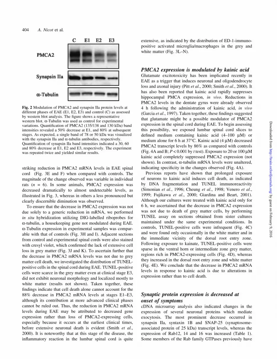
Fig. 2 Modulation of PMCA2 and synapsin IIa protein levels atdifferent phases of EAE (E1, E2, E3) and control (C) as assessedby western blot analysis. The ®gure shows a representativewestern blot. a-Tubulin was used as control for experimentalvariations. Quanti®cation of PMCA2 (135/138 and 130 kDa) bandintensities revealed a 50% decrease at E1, and 80% at subsequentstages. As expected, a single band of 78 or 50 kDa was visualizedwith the synapsin IIa and a-tubulin antibodies, respectively.Quanti®cation of synapsin IIa band intensities indicated a 30, 60and 80% decrease at E1, E2 and E3, respectively. The experimentwas repeated twice and yielded similar results.
404 A. Nicot et al.
by guest on February 9, 2016http://brain.oxfordjournals.org/
Dow
nloaded from

Fig. 3 Cellular localization and modulation of PMCA2 mRNA assessed by in situ hybridization. Bright ®eld photomicrographs of lumbarspinal cord (L4±L5) sections obtained from controls (A±D) and diseased rats (E and F) hybridized to DIG-labelled antisense riboprobe.(G) A control section hybridized to a sense riboprobe. Labelled cells were found exclusively in grey matter of control sections (A).(B) A higher magni®cation photomicrograph of the section shown in A, demonstrating labelling in cells exhibiting motor neuron-likemorphology in the ventral horn (examples shown within the square) and in smaller cells of the intermediate zone (arrows). (C and D)Higher magni®cation photomicrographs of the cells in B (indicated by arrows and enclosed within the square, respectively). Expression ofPMCA2 dramatically decreased in the spinal cord during EAE (E), con®rming results obtained by cDNA microarray analysis and RT±PCR. Note that the diseased spinal cord is usually enlarged as compared with controls. (F) A higher magni®cation picture of the sectionshown in E. Arrows point at some weakly labelled cells. To ensure that the reduction in PMCA2 expression was not due to a genericdecrease in mRNA levels, adjacent sections obtained from control (H) and experimental (I) spinal cord were hybridized to an antisense a-tubulin riboprobe. (H and I) A group of motor neuron-like cells in the ventral horn expressing the housekeeping gene at comparablelevels. The presence of cresyl violet-stained cells in sections obtained from spinal cord of control (J) and experimental (K) rats indicatedthat the decrease in PMCA2 was not due to non-speci®c cell loss. To con®rm the in¯ammatory reaction in the lumbar spinal cord,consecutive sections were stained with haematoxylin±eosin (not shown) and immunolabelled with ED-1, a marker for activated microglia/macrophages. ED-1-immunopositive cells are rare in control (L), and very abundant in EAE (M) spinal cord grey and white matter. Thegrey matter in the control section is delineated by dashed lines. (N) A higher magni®cation photomicrograph of a region in the sectionshown in M. Arrows point at examples of immunopositive cells. Sections obtained from six controls and six EAE rats (clinical score 3,E3) were analysed. The reduction in PMCA2 expression was observed in all diseased rats, although the magnitude of the change wasvariable and ranged from dramatic decreases as illustrated in this ®gure to more moderate but distinctly discernable reductions. Scalebar = 400 mm (A, E, G, L and M); 200 mm (B and F); 100 mm (C and D); 40 mm (N).
Neuronal dysfunction in spinal cord during EAE 405
by guest on February 9, 2016http://brain.oxfordjournals.org/
Dow
nloaded from

been implicated in vesicular function (for a review see
Geppert and Sudhof, 1998), although the exact role of the
aforementioned Rab isoforms has yet to be determined.
As synapsins, syntaxins and SNAP-25 are essential for
vesicular function and neurotransmitter exocytosis, we fur-
ther analysed the temporal pattern of their expression during
the course of EAE. At E1, transcript levels of synapsin IIa and
syntaxin 1B were decreased by 93 and 84%, respectively
(P < 0.001 by ANOVA, Scheffe's post hoc test) and remained
low thereafter (Fig. 5). In addition, western blot analysis
indicated 30, 60 and 80% reductions in synapsin IIa protein
levels at E1, E2 and E3, respectively (Fig. 2). SNAP-25
expression showed a tendency to decrease at E1 and E2, but
the results were more variable and did not reach statistical
signi®cance (Fig. 5). By E3, however, SNAP-25 levels had
decreased by 96% (P < 0.001 by ANOVA, Scheffe's post hoc
test), in agreement with a recent study on chronic EAE in
mice (Ibrahim et al., 2001). In contrast to synapsins, syntaxin
and SNAP-25, the transcript levels of Rab12, Rab14 and
Rab16 were 2.9-, 4.3- and 2.6-fold increased, respectively
(Fig. 6; P < 0.001 for Rab12 and 14, and P < 0.01 for Rab16,
by t test). These results raise the possibility of disturbances in
neurotransmission at onset of EAE symptoms.
Increased cytochrome c oxidase expressionsuggests deregulation of mitochondrialmetabolic activity at E2cDNA microarray analysis indicated a strong increase in
expression of cytochrome c oxidase subunits IV and Vb,
suggesting altered mitochondrial metabolic activity (Table 1).
We corroborated this ®nding by RT±PCR and found that the
most robust increases in cytochrome c oxidase IV and Vb
subunit expression (~11- and 15-fold, respectively) occur at
E2 and E3 (Fig. 7). Mitochondrial dysfunction has often been
associated with neuronal injury in CNS diseases and espe-
cially in response to intracellular Ca2+ overload or oxidative
stress (Fiskum, 2000; Hirai et al., 2001). It is conceivable that
changes in mitochondrial activity contribute to EAE path-
ology in a fashion similar to that observed in other CNS
disorders such as ischaemia.
Reduced sodium channel expression occurs atadvanced clinical phasesSodium channels are critical mediators of impulse conduction
along the axon. Therefore, the reduced expression of Na+
channel b1 and 2 subunits and brain sodium channel II, as
indicated by cDNA microarray analysis, was analysed further
by RT±PCR. We found that the expression of all the
aforementioned channels substantially decreased at E3
(Fig. 8; 80±90% decrease), con®rming the results obtained
by microarray analysis. In contrast, no changes were observed
at E1 and E2. Thus, potential abnormalities in Na+ channel
repertoires in nerve cells leading to impairment in impulse
activity may contribute to the progression of symptoms at
more advanced disease phases.
DiscussionThe present study reports alterations in the expression of
speci®c genes in the lumbar spinal cord during the clinical
course of EAE, with special emphasis on the initial phase of
symptomatic disease. In particular, we analysed the temporal
expression pattern of genes which encode proteins playing
critical roles primarily in neuronal function. Our results,
taken together, indicate that changes in neuronal gene
expression coincide with onset of symptoms. Thus, our
®ndings provide novel insights into the potential molecular
mechanisms that may underlie the recently described
neuronal dysfunction or injury during EAE (Ferguson et al.,
1997; Trapp et al., 1998, 1999; De Stefano et al., 1999; Bitsch
et al., 2000; Smith et al., 2000; Bjartmar and Trapp, 2001;
Bjartmar et al., 2001; Peterson et al., 2001). We propose that
ion dyshomeostasis due to aberrant Ca2+ extrusion and
abnormal neurotransmitter release resulting from anomalies
in the expression of vesicular proteins may promote the onset
of neural de®cits. As the disease progresses, perturbed
mitochondrial function and reduced impulse conduction due
to decreases in Na+ channel expression may exacerbate
clinical symptoms.
Glutamate-mediated changes in PMCA2expression may induce neuronal dysfunction atonset of EAE symptomsIncreases in intracellular Ca2+ often have been associated
with cell dysfunction, injury and death in many pathological
conditions of the CNS, including ischaemia, Alzheimer's and
Parkinson's disease (for reviews see Paschen, 2000; O'Neill
et al., 2001; Paschen and Frandsen, 2001). Yet, the
contribution of Ca2+ dyshomeostasis to EAE and the affected
mechanisms have not been de®ned. A loss in Ca2+ balance
may result from defects in mechanisms that regulate Ca2+
entry, extrusion or intracellular sequestration. A role for
voltage-dependent channel-mediated Ca2+ in¯ux in axonal
and neuronal degeneration during multiple sclerosis and EAE
has been reported (Kornek et al., 2000, 2001). To our
knowledge, our study is the ®rst report implicating a pivotal
role for distinct ion pumps involved in calcium extrusion and
sequestration in EAE pathogenesis.
Whereas PMCAs are major pumps modulating Ca2+
extrusion, SERCA isoforms play critical roles in sequestra-
tion of Ca2+ into the endoplasmic reticulum. Our results
indicate a profound decrease in PMCA2 expression at the
earliest manifestation of symptoms, which is then followed
by a signi®cant reduction in SERCA2 transcript levels at
subsequent clinical phases. Such temporal changes in ion
pump expression may promote accumulation of Ca2+ in the
cytoplasm, increasing the vulnerability of neurons to cal-
406 A. Nicot et al.
by guest on February 9, 2016http://brain.oxfordjournals.org/
Dow
nloaded from

cium-mediated injury. This may be one possible candidate
mechanism underlying neuronal decline, and implicates
PMCA2 and SERCA2 as potential targets for therapeutic
interventions. Importantly, our results may be relevant to the
human disease, as a recent study found a decrease in PMCA2
trancript levels by microarray analysis of three multiple
sclerosis brains (Lock et al, 2002, online supplementary
table B).
The PMCA gene family encodes four isoforms which
initially were considered to be necessary only for the
maintenance of basal Ca2+ levels. However, emerging
evidence indicates that they may play important roles in
pathological conditions (Garcia and Strehler, 1999) espe-
cially after overstimulation of glutamate receptors which
causes intracellular Ca2+ overload (Choi, 1992; Mody and
MacDonald, 1995). PMCA2 is expressed primarily in brain
and heart, in a region- and cell-type speci®c manner (Garcia
and Strehler, 1999). The highly restricted tissue distribution
of PMCA2 suggests that this isoform plays unique,
specialized and non-redundant roles in the CNS. Indeed,
PMCA2-null mice exhibit deafness, and unsteady gait and
balance, indicating that the presence of other isoforms does
not compensate for the absence of PMCA2 (Kozel et al.,
1998).
The triggers that induce changes in the expression of Ca2+
pumps during EAE have not been de®ned. Glutamate is an
appealing candidate because of its well-known effects on
Ca2+ homeostasis (Choi, 1992) and because of the recently
reported contribution of excitotoxicity to EAE pathogenesis
(Pitt et al., 2000; Smith et al., 2000). Administration of
AMPA/kainate antagonists ameliorates EAE symptoms in
mice and rats, and prevents neuronal and oligodendrocyte
loss and axonal damage. Moreover, kainate-induced decrea-
ses in PMCA1 and PMCA2 expression in hippocampal
pyramidal cells are followed by neuronal death, in vivo
(Garcia et al., 1997). These ®ndings raise the possibility of
kainate receptor-mediated alterations in PMCA2 expression
in the spinal cord during EAE. In fact, we found that exposure
of spinal cord slice cultures to relatively low concentrations
of kainate (4 mM) for 6 h dramatically decreased PMCA2
transcript levels. Thus, even low glutamate levels may be
suf®cient to affect PMCA2 expression when cells are exposed
to the neurotransmitter continuously. We propose that, at
onset of EAE, persistent stimulation of kainate receptors
following moderate but sustained elevations in glutamate
concentrations may lead to downregulation of Ca2+ pump
expression, promote Ca2+ overload and initiate the previously
described injury cascade (Shields et al., 1998).
Fig. 4 (A) Modulation of PMCA2 expression by kainic acid in spinal cord slice cultures, assessed by RT±PCR analysis. C = control, i.e. slices maintained only in de®ned medium and vehicle; KA = slicesmaintained in medium containing 4 mM kainic acid; (PMCA2) negative control, i.e. RNA subjected toPCR without reverse transcription to ascertain the lack of contaminating genomic DNA. The graph in Bshows the densitometric quanti®cation of the bands in the gel and re¯ects the mean 6 SEM of threeindependent experiments (n = 6, ***P < 0.001 by t test). To rule out the possibility that the decrease inPMCA2 expression was due to cell death, slices in sister cultures were sectioned for assessment ofTUNEL-positive cells. (C and D) Photomicrographs of a representative region in the ventral horn incontrols and kainic acid-treated slices, respectively. Note that TUNEL-positive cells are absent in controls(C) and very few in experimental samples (D, arrows). In kainic acid-treated sections, small, TUNEL-positive cells were con®ned primarily to the edge of the dorsal root entry zone (E, arrows). Scalebar = 100 mm.
Neuronal dysfunction in spinal cord during EAE 407
by guest on February 9, 2016http://brain.oxfordjournals.org/
Dow
nloaded from

Aberrant neurotransmitter exocytosis may affectneuronal communication during the earlycourse of the diseaseIn addition to PMCA2, the expression of synapsin IIa and
syntaxin 1B, vesicular proteins that are crucial for exocytosis
in presynaptic terminals, was decreased dramatically at onset
of clinical symptoms. Synapsins are a family of synaptic
vesicle-associated phosphoproteins that are required for the
tethering of vesicles to each other and to cytoskeletal proteins
such as actin. Phosphorylation of synapsins liberates vesicles
from the cytoskeleton, enabling their traf®cking in the
presynaptic terminal. Synapsins are essential for the forma-
tion, maintenance and regulation of the reserve synaptic pool
in the vicinity of the active zone. Decreases in synapsin
Fig. 5 Modulation of genes encoding proteins involved in vesicular function during the course of EAE asassessed by RT±PCR. Each lane shows the results obtained with distinct groups of rats (pooled RNAfrom 2±3 rats/group). The expression of a-tubulin, a housekeeping gene not modulated in EAE, wasutilized as control for experimental variations. PCRs for the various vesicular proteins and a-tubulin wereperformed in parallel utilizing the same reverse transcription mix. RNA subjected to PCR without reversetranscription ascertained the lack of contaminating genomic DNA (not shown). PCR was performed for35 cycles. The ®gure shows representative gels, and graphs present the mean 6 SEM of threeindependent experiments (n = 5±6 with 2±3 rats per group, as indicated above). ***P < 0.001,signi®cantly different from control by ANOVA, Scheffe's post hoc test.
408 A. Nicot et al.
by guest on February 9, 2016http://brain.oxfordjournals.org/
Dow
nloaded from

expression reduce the number of synaptic vesicles distal to
the active zone, leading to synaptic fatigue (Pieribone et al.,
1995; Rosahl et al., 1995). Moreover, perturbations in
synapsin function inhibit or slow down neurotransmitter
release.
Syntaxins belong to a class of proteins called SNAREs
(soluble N-ethylmaleimide-sensitive factor attachment
protein receptor) hypothesized to be essential for the docking
and fusion of synaptic vesicles. Vesicle-associated SNAREs,
such as synaptobrevin, and target membrane-associated
SNAREs, such as syntaxins and SNAP-25, form a high
af®nity complex. Formation of the SNARE complex draws
vesicles to the target membrane and induces membrane
fusion. In our experimental paradigm, we observed a decrease
in both syntaxin 1B and SNAP-25. However, the dramatic
reduction in syntaxin 1B occurred at onset of symptoms,
whereas SNAP-25 showed a tendency to decrease, which was
statistically signi®cant only at a later clinical stage (E3).
Hence, changes in the expression of some synapsin and
SNARE proteins can interfere with normal neuronal com-
munication during EAE.
The notion of synapse pathology is suggested further by
changes observed in Rab12, 14 and 16 expression. Rab
isoforms belong to the Ras superfamily of small GTP-binding
proteins, and have been implicated in the attachment of
vesicles to target membranes. A member of this family, Rab3,
is localized to synaptic vesicles and limits the amount of
neurotransmitter released in response to Ca2+ signal.
However, a similar function for Rab12, 14 and 16 has not
yet been de®ned. Rab12 and 14 are also expressed in
oligodendrocytes and may play additional roles (Burcelin
et al., 1997). Therefore, the exact contribution of the
aforementioned Rab isoforms to EAE pathogenesis remains
to be determined.
Perturbations in mitochondrial function maycontribute to cellular injury at advanced EAEstagesCellular injury often has been associated with disturbances in
mitochondrial function. Interestingly, our microarray analysis
indicated a pronounced increase in the expression of
mitochondrial cytochrome c oxidase subunits IV and Vb, a
®nding which was validated further by RT±PCR. Cytochrome
c oxidase has been used frequently as a marker for neuronal
metabolic activity, especially in pathological conditions
involving oxidative stress. Neurons subject to oxidative
Fig. 6 Modulation of Rab isoform expression during EAE as assessed by RT±PCR. Each lane representsresults obtained with distinct groups of control and EAE rats in three independent experiments (2±3 rats/group). The graphs show the mean 6 SEM of the densitometric quanti®cation of bands (n = 3).***P < 0.001 and **P < 0.01, signi®cantly different from control by t test.
Neuronal dysfunction in spinal cord during EAE 409
by guest on February 9, 2016http://brain.oxfordjournals.org/
Dow
nloaded from
damage show abnormalities in mitochondrial dynamics even
in the absence of any apparent indication of degeneration
(Sayre et al., 1997; Smith et al., 1997). Elevations in
cytochrome c oxidase expression or function in vulnerable
neuronal subpopulations in Alzheimer's disease, following
traumatic brain injury and preceding apoptotic death of spinal
cord motor neurons after sciatic nerve avulsion, have been
reported (Martin et al., 1999; Harris et al., 2001; Hirai et al.,
2001). Thus, an increase in cytochrome c oxidase expression
may re¯ect aberrant energy metabolism and oxidative
damage in the spinal cord during EAE.
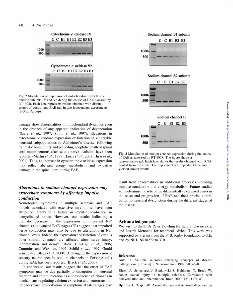
Alterations in sodium channel expression mayexacerbate symptoms by affecting impulseconductionNeurological symptoms in multiple sclerosis and EAE
models associated with extensive myelin loss have been
attributed largely to a failure in impulse conduction in
demyelinated axons. However, our results indicating a
dramatic decrease in the expression of important Na+
channels at advanced EAE stages (E3) suggest that impaired
nerve conduction may also be due to alterations in Na+
channel levels. Indeed, the expression and function of various
other sodium channels are affected after nerve injury,
in¯ammation and demyelination (Dib-Hajj et al., 1996;
Cummins and Waxman, 1997; Schild et al., 1997; Gould
et al., 1998; Black et al., 2000). A change in the expression of
sensory neuron-speci®c sodium channels in Purkinje cells
during EAE has been reported (Black et al., 2000).
In conclusion, our results suggest that the onset of EAE
symptoms may be due partially to disruption of neuronal
function and communication as a consequence of changes in
mechanisms regulating calcium extrusion and neurotransmit-
ter exocytosis. Exacerbation of symptoms at later stages may
result from abnormalities in additional processes including
impulse conduction and energy metabolism. Future studies
will determine the role of the differentially expressed genes in
the onset and progression of EAE and their precise contri-
bution to neuronal dysfunction during the different stages of
the disease.
AcknowledgementsWe wish to thank Dr Peter Dowling for helpful discussions,
and Joseph Menonna for technical advice. This work was
supported by a grant from the F. B. Kirby foundation to S.E.
and by NIH- NS38272 to Y.R.
References
Antel J. Multiple sclerosisÐemerging concepts of disease
pathogenesis. [Review]. J Neuroimmunol 1999; 98: 45±8.
Bitsch A, Schuchardt J, Bunkowski S, Kuhlmann T, Bruck W.
Acute axonal injury in multiple sclerosis. Correlation with
demyelination and in¯ammation. Brain 2000; 123: 1174±83.
Bjartmar C, Trapp BD. Axonal damage and neuronal degeneration
Fig. 7 Modulation of expression of mitochondrial cytochrome coxidase subunits IV and Vb during the course of EAE assessed byRT±PCR. Each lane represents results obtained with distinctgroups of control and EAE rats in two independent experiments(2±3 rats/group).
Fig. 8 Modulation of sodium channel expression during the courseof EAE as assessed by RT±PCR. The ®gure shows arepresentative gel. Each lane shows the results obtained with RNApooled from three rats. The experiment was repeated twice andyielded similar results.
410 A. Nicot et al.
by guest on February 9, 2016http://brain.oxfordjournals.org/
Dow
nloaded from

in multiple sclerosis: mechanisms and functional consequences.
[Review]. Curr Opin Neurol 2001; 14: 271±8.
Bjartmar C, Kinkel RP, Kidd G, Rudick RA, Trapp BD. Axonal loss
in normal-appearing white matter in a patient with acute MS.
Neurology 2001; 57: 1248±52.
Black JA, Dib-Hajj S, Baker D, Newcombe J, Cuzner ML, Waxman
SG. Sensory neuron-speci®c sodium channel SNS is abnormally
expressed in the brains of mice with experimental allergic
encephalomyelitis and humans with multiple sclerosis. Proc Natl
Acad Sci USA 2000; 97: 11598±602.
Burcelin R, Rodriguez-Gabin AG, Charron MJ, Almazan G,
Larocca JN. Molecular analysis of the monomeric GTP-binding
proteins of oligodendrocytes. Brain Res Mol Brain Res 1997; 50:
9±15.
Carafoli E. Intracellular calcium homeostasis. [Review]. Annu Rev
Biochem 1987; 56: 395±433.
Choi DW. Excitotoxic cell death. [Review]. J Neurobiol 1992; 23:
1261±76.
Cheung NS, Carroll FY, Larm JA, Beart PM, Giardina SF. Kainate-
induced apoptosis correlates with c-Jun activation in cultured
cerebellar granule cells. J Neurosci Res 1998; 52: 69±82.
Cummins TR, Waxman SG. Downregulation of tetrodotoxin±
resistant sodium currents and upregulation of a rapidly repriming
tetrodotoxin-sensitive sodium current in small spinal sensory
neurons after nerve injury. J Neurosci 1997; 17: 3503±14.
De Stefano N, Narayanan S, Matthews PM, Francis GS, Antel JP,
Arnold DL. In vivo evidence for axonal dysfunction remote from
focal cerebral demyelination of the type seen in multiple sclerosis.
Brain 1999; 122: 1933±9.
Dib-Hajj S, Black JA, Felts PA, Waxman SG. Down-regulation of
transcripts for Na channel alpha-SNS in spinal sensory neurons
following axotomy. Proc Natl Acad Sci USA 1996; 93: 14950±4.
Ferguson B, Matyszak MK, Esiri MM, Perry VH. Axonal damage
in acute multiple sclerosis lesions. Brain 1997; 120: 393±9.
Filippi M. Multiple sclerosis: a white matter disease with associated
gray matter damage. [Review]. J Neurol Sci 2001; 185: 3±4.
Fiskum G. Mitochondrial participation in ischemic and traumatic
neural cell death. [Review]. J Neurotrauma 2000; 17: 843±55.
Fujikawa DG, Shinmei SS, Cai B. Kainic acid-induced seizures
produce necrotic, not apoptotic, neurons with internucleosomal
DNA cleavage: implications for programmed cell death
mechanisms. Neuroscience 2000; 98: 41±53.
Garcia ML, Strehler EE. Plasma membrane calcium ATPases as
critical regulators of calcium homeostasis during neuronal cell
function. [Review]. Front Biosci 1999; 4: D869±82.
Garcia ML, Murray KD, Garcia VB, Strehler EE, Isackson PJ.
Seizure-induced alterations of plasma membrane calcium ATPase
isoforms 1, 2 and 3 mRNA and protein in rat hippocampus. Brain
Res Mol Brain Res 1997; 45: 230±8.
Geppert M, Sudhof TC. RAB3 and synaptotagmin: the yin and yang
of synaptic membrane fusion. [Review]. Annu Rev Neurosci 1998;
21: 75±95.
Giardina SF, Beart PM. Excitotoxic pro®les of novel, low-af®nity
kainate receptor agonists in primary cultures of murine cerebellar
granule cells. Neuropharmacology 2001; 41: 421±32.
Gould HJ 3rd, England JD, Liu ZP, Levinson SR. Rapid sodium
channel augmentation in response to in¯ammation induced by
complete Freund's adjuvant. Brain Res 1998; 802: 69±74.
Harris LK, Black RT, Golden KM, Reeves TM, Povlishock JT,
Phillips LL. Traumatic brain injury-induced changes in gene
expression and functional activity of mitochondrial cytochrome C
oxidase. J Neurotrauma 2001; 18: 993±1009.
Hickey WF. The pathology of multiple sclerosis: a historical
perspective. [Review]. J Neuroimmunol 1999; 98: 37±44.
Hirai K, Aliev G, Nunomura A, Fujioka H, Russell RL, Atwood CS,
et al. Mitochondrial abnormalities in Alzheimer's disease. J
Neurosci 2001; 21: 3017±23.
Ibrahim SM, Mix E, Bottcher T, Koczan D, Gold R, Rolfs A, et al.
Gene expression pro®ling of the nervous system in murine
experimental autoimmune encephalomyelitis. Brain 2001; 124:
1927±38.
Kornek B, Storch MK, Weissert R, Wallstroem E, Stefferl A,
Olsson T, et al. Multiple sclerosis and chronic autoimmune
encephalomyelitis: a comparative quantitative study of axonal
injury in active, inactive, and remyelinated lesions. Am J Pathol
2000; 157: 267±76.
Kornek B, Storch MK, Bauer J, Djamshidian A, Weissert R,
Wallstroem E, et al. Distribution of a calcium channel subunit in
dystrophic axons in multiple sclerosis and experimental
autoimmune encephalomyelitis. Brain 2001; 124: 1114±24.
Kozel PJ, Friedman RA, Erway LC, Yamoah EN, Liu LH, Riddle T,
et al. Balance and hearing de®cits in mice with a null mutation in
the gene encoding plasma membrane Ca2+-ATPase isoform 2. J Biol
Chem 1998; 273: 18693±6.
Lock C, Hermans G, Pedotti R, Brendolan A, Schadt E, Garren H,
et al. Gene-microarray analysis of multiple sclerosis lesions yields
new targets validated in autoimmune encephalomyelitis. Nat Med
2002; 8: 500±8.
Martin LJ, Kaiser A, Price AC. Motor neuron degeneration after
sciatic nerve avulsion in adult rat evolves with oxidative stress and
is apoptosis. J Neurobiol 1999; 40: 185±201.
McFarlin DE, McFarland HF. Multiple sclerosis (®rst of two parts).
[Review]. N Engl J Med 1982a; 307: 1183±8.
McFarlin DE, McFarland HF. Multiple sclerosis (second of two
parts). [Review]. N Engl J Med 1982b; 307: 1246±51.
Miller RJ. The control of neuronal Ca2+ homeostasis. [Review].
Prog Neurobiol 1991; 37: 255±85.
Mody I, MacDonald JF. NMDA receptor-dependent excitotoxicity:
the role of intracellular Ca2+ release. [Review]. Trends Pharmacol
Sci 1995; 16: 356±9.
O'Neill C, Cowburn RF, Bonkale WL, Ohm TG, Fastbom J,
Carmody M, et al. Dysfunctional intracellular calcium
homoeostasis: a central cause of neurodegeneration in
Alzheimer's disease. [Review]. Biochem Soc Symp 2001; 67:
177±94.
Neuronal dysfunction in spinal cord during EAE 411
by guest on February 9, 2016http://brain.oxfordjournals.org/
Dow
nloaded from

Paschen W. Role of calcium in neuronal cell injury: which
subcellular compartment is involved? [Review]. Brain Res Bull
2000; 53: 409±13.
Paschen W, Frandsen A. Endoplasmic reticulum dysfunction: a
common denominator for cell injury in acute and degenerative
diseases of the brain? [Review]. J Neurochem 2001; 79: 719±25.
Peterson JW, BoÈ L, MoÈrk S, Chang A, Trapp BD. Transected
neurites, apoptotic neurons and reduced in¯ammation in cortical
multiple sclerosis lesions. Ann Neurol 2001; 50: 389±400.
Pieribone VA, Shupliakov O, Brodin L, Hil®ker-Rothen¯uh S,
Czernik AJ, Greengard P. Distinct pools of synaptic vesicles in
neurotransmitter release. Nature 1995; 375: 493±7.
Pitt D, Werner P, Raine CS. Glutamate excitotoxicity in a model of
multiple sclerosis. Nat Med 2000; 6: 67±70.
Prineas JW, McDonald WI. Demyelinating diseases. In: Graham
DI, Lantos PL, editors. Green®eld's neuropathology. Vol. 1. 6th
edn. London: Arnold; 1997. p. 813±81
Raine CS. Demyelinating diseases. In: Davis RL, Robertson DM,
editors. Textbook of neuropathology. 3rd edn. New York: Williams
and Wilkins; 1997. p. 627±714.
Rivera-Quinones C, McGavern D, Schmelzer JD, Hunter SF, Low
PA, Rodriguez M. Absence of neurological de®cits following
extensive demyelination in a class I-de®cient murine model of
multiple sclerosis. Nat Med 1998; 4: 187±93.
Rosahl TW, Spillane D, Missler M, Herz J, Selig DK, Wolff JR,
et al. Essential functions of synapsins I and II in synaptic vesicle
regulation. Nature 1995; 375: 488±93.
Sayre LM, Zelasko DA, Harris PL, Perry G, Salomon RG, Smith
MA. 4-Hydroxynonenal-derived advanced lipid peroxidation end
products are increased in Alzheimer's disease. J Neurochem 1997;
68: 2092±7.
Schild JH, Kunze DL. Experimental and modeling study of Na+
current heterogeneity in rat nodose neurons and its impact on
neuronal discharge. J Neurophysiol 1997; 8: 3198±209.
Shields DC, Tyor WR, Deibler GE, Hogan EL, Banik NL. Increased
calpain expression in activated glia and in¯ammatory cells in
experimental allergic encephalomyelitis. Proc Natl Acad Sci USA
1998; 95: 5768±72.
Simonian NA, Getz RL, Leveque JC, Konradi C, Coyle JT. Kainate
induces apoptosis in neurons. Neuroscience 1996; 74: 675±83.
Smith T, Groom A, Zhu B, Turski L. Autoimmune
encephalomyelitis ameliorated by AMPA antagonists. Nat Med
2000; 6: 62±4.
Smith MA, Richey Harris PL, Sayre LM, Beckman JS, Perry G.
Widespread peroxynitrite-mediated damage in Alzheimer's disease.
J Neurosci 1997; 17: 2653±7.
Sriram S, Rodriguez M. Indictment of the microglia as the villain in
multiple sclerosis. [Review]. Neurology 1997; 48: 464±70.
Stauffer TP, Guerini D, Carafoli E. Tissue distribution of the four
gene products of the plasma membrane Ca2+ pump. A study using
speci®c antibodies. J Biol Chem 1995; 270: 12184±90.
Stauffer TP, Guerini D, Celio MR, Carafoli E. Immunolocalization
of the plasma membrane Ca2+ pump isoforms in the rat brain. Brain
Res 1997; 748: 21±9.
Steinman L. Multiple sclerosis: a coordinated immunological attack
against myelin in the central nervous system. [Review]. Cell 1996;
85: 299±302.
Steinman L. Assessment of animal models for MS and
demyelinating disease in the design of rational therapy. [Review].
Neuron 1999; 24: 511±4.
Trapp BD, Peterson J, Ransohoff RM, Rudick R, Mork S, Bo L.
Axonal transection in the lesions of multiple sclerosis. N Engl J
Med 1998; 338: 278±85.
Trapp BD, Ransohoff R, Rudick R. Axonal pathology in multiple
sclerosis: relationship to neurologic disability. [Review]. Curr Opin
Neurol 1999; 12: 295±302.
vander Goes A, Dijkstra CD. Models for demyelination. [Review].
Prog Brain Res 2001; 132: 149±63.
Venero JL, Revuelta M, Machado A, Cano J. Delayed apoptotic
pyramidal cell death in CA4 and CA1 hippocampal sub®elds after a
single intraseptal injection of kainate. Neuroscience 1999; 94:
1071±81.
Waxman SG. Multiple sclerosis as a neuronal disease. Arch Neurol
2000a; 57: 22±4.
Waxman SG. Do `demyelinating' diseases involve more than
myelin? Nat Med 2000b; 6: 738±9.
Wekerle H, Kojima K, Lannes-Vieira J, Lassmann H, Linington C.
Animal models. [Review]. Ann Neurol 1994; 36 Suppl: S47±53.
Received July 6, 2002. Revised September 10, 2002.
Accepted September 11, 2002
412 A. Nicot et al.
by guest on February 9, 2016http://brain.oxfordjournals.org/
Dow
nloaded from