refraction - repository of kharkiv national medical university
TRANSCRIPT
1
МІНІСТЕРСТВО ОХОРОНИ ЗДОРОВ'Я УКРАЇНИ Харківський національний медичний університет
REFRACTION
Manual for individual work in ophthalmology for English speaking foreign medical students
Рефракція
Методичні вказівки з офтальмології для індивідуальної підготовки студентів-іноземців
медичних факультетів з англійською мовою навчання
Затверджено
вченою радою ХНМУ.
Протокол № 11 від 26.12.2013.
Харків
ХНМУ
2014
2
Refraction : manual for individual work in ophthalmology for English
speaking foreign medical students / compl. P.A. Bezditko, S.F. Zubarev,
M.V. Panchenko et al. – Kharkiv : KNMU, 2014. – 16 p.
Compliers P.A. Bezditko
S.F. Zubarev
M.V. Panchenko
O.V. Zavoloka
O.P. Muzhychuk
A.Y. Savelieva
E.M. Iliuna
Рефракція : метод. вказ. з офтальмології для індивід. підготовки
студентів-іноземців мед. фак-тів з англ. мовою навчання / упор. П.А. Без-
дітко, С.Ф. Зубарев, М.В.Панченко та ін. – Харків : ХНМУ, 2014. – 16 с.
Упорядники П.А. Бездітко
С.Ф. Зубарев
М.В. Панченко
О.В. Заволока
О.П. Мужичук
А.Ю. Савельєва
Є.М. Ільїна
3
THE REQUIRED MINIMUM OF BASIS SKILLS, WHICH ARE TO BE
MASTERED BY FORAIGN STUDENTS STUDDING THE COURSE OF
OPHTHALMOLOGY
1. To be able to estimate the errors of refraction.
2. To determine the type and strength of a spectacle lens.
3. To provide a patient with prophylactic action for progressive myopia.
4. To be able for schematic drawing of light rays in an emmetropia, myopia,
hyperopia.
5. To be able to draw the range of accommodation decreasing with the age.
CONTROL QUASTIONS
1. How to determine the refractive power of the eye?
2. What does the transparent media of the eye consist of?
3. What is normal axial length of the eye?
4. What are the reasons of errors of refraction?
5. What are the main signs of severe pathologic myopia?
6. What are the myopic complications?
7. What are the treatment methods of myopia?
8. How does accommodative power of the eye decrease during human life?
9. What are the treatment methods of presbyopia?
10. What is a topic distortion of the refractive media?
Topic Relevance
The refractive power of an eye is determined by the refractive power of
transparent media and the axial length. The transparent media consist of the
cornea, the anterior chamber, the lens, and the vitreus. The axial length is nor-
mally around 24 mm.
The refractive power of an emmetropic eye is 60 diopters, of which
40 are exerted by the cornea and 20 by the lens. Aqueous and vitreous ones do
not have any refractive power of consequence.
We can determine the refraction subjectively by putting lenses in front
of each eye; objectively we can determine the refraction by retinoscopy or by a
refractometer.
The far point of distinct vision is the position of an object such that its
image falls on the retina in the relaxed eye, i.e. in the absence of accommoda-
tion. The far point of the emmetropic eye is at infinity.
The near point of distinct vision is the nearest point at which an object
can be clearly seen when maximum accommodation is used.
The range of accommodation is the distance between the far point and
the near point.
4
The amplitude of accommodation is the difference in dioptric power be-
tween the eye at rest and the fully accommodated eye.
The dioptric power of the resting eye is called its static refraction.
The dioptric power of the accommodated eye is called its dynamic re-
fraction.
The aim of the training - students should be acquainted with the fol-
lowing subjects:
- different types of clinical refraction,
- refractive errors and methods of their correction,
- presbyopia,
- clinical signs and complications of myopia.
Educational Objective
REFRACTIVE ERRORS
Indistinct vision most commonly is caused by errors of refraction. Doc-
tors do not often have to deal with this problem because patients usually are
prescribed glasses by an optometrist. However, if a patient presents complain-
ing of visual problems, it is extremely important to ask the question: ―Is this
patient’s poor vision caused by a refractive error?‖ The use of a simple ―pin-
hole‖ made in a piece of card will help to determine whether or not there is a
refractive error. In the absence of disease the vision will improve when the pin-
hole is used–unless the refractive error is extremely large.
Eye with no refractive error
In an eye with no refractive error (emmetropia) light rays from infinity
are brought to a focus on the retina by the cornea and lens when the eye is in a
―relaxed‖ state. The cornea contributes about two thirds and the lens about one
third to the eye’s refractive power. Disease affecting the cornea (for example,
keratoconus) may cause severe refractive problems.
The rays of light from closer objects, such as the printed page, are diver-
gent and have to be brought to a focus on the retina by the process of accom-
modation. The circular ciliary muscle contracts, allowing the naturally elastic
lens to assume a more globular shape that has a greater converging power.
In young people the lens is very elastic, but with age the lens gradually
hardens and even when the ciliary muscle contracts the lens no longer becomes
globular. Thus from the age of 40 onwards close work becomes gradually more
difficult (presbyopia). Objects may have to be held further away to reduce the
need for accommodation, which leads to the complaint ―my arms don’t seem to
be long enough.‖ Fine detail cannot be discerned.
Convex lenses in the form of reading glasses therefore are needed to
converge the light rays from close objects on to the retina.
5
All emmetropic people need reading glasses for close work in later life.
People who wear glasses to see clearly in the distance may find it con-
venient to change to bifocal lenses in their glasses when they become presbyop-
ic. In bifocal lenses the reading lens simply is incorporated into the lower part
of the lens. Therefore, the person does not have to change his or her glasses to
read. However, details at an intermediate distance such as the prices of items on
supermarket shelves are not clear. A third lens segment can be incorporated
between that for distance above and that for reading below, creating a trifocal
lens. However, many people cannot cope with the ―jump‖ in magnification in-
herent in the use of these lenses. This has led to the introduction of multifocal
lenses in which the lens power increases progressively from top to bottom.
People may also have problems adapting to this type of lens, as peripheral vi-
sion may be distorted.
Refractive errors do not get worse if a person reads in bad light or does
not wear their glasses. The exceptions are young children, however, who may
need a refractive error corrected to prevent amblyopia.
Myopic or shortsighted eye
In the myopic eye, light rays from infinity are brought to a focus in front
of the retina because either the eye is too long or the converging power of the
cornea and lens is too great. To achieve clear vision the rays of light must be
diverged by a concave lens so that light rays are focused on the retina.
For near vision, light rays are focused on the retina with little or no ac-
commodation depending on the degree of myopia and the distance at which the
object is held. This is the reason why shortsighted people can often read without
glasses even late in life, when those without refractive errors need reading glasses.
A certain type of cataract (nuclear sclerosis) increases the refractive
power of the lens, making the eye more myopic. Patients with these cataracts
may say their reading vision has improved. Patients with an extreme degree of
shortsightedness are more susceptible to retinal detachment, macular degenera-
tion, and primary open angle glaucoma.
Myopia has been classified in various manners. By cause:
1) Axial myopia is attributed to an increase in the eye's axial length.
2) Refractive myopia is attributed to the condition of the refractive el-
ements of the eye.
a) Curvature myopia is attributed to excessive, or increased, curvature
of one or more of the refractive surfaces of the eye, especially the corneа.
b) Index myopia is attributed to variation in the index of refraction of
one or more of the ocular media. Cataracts may lead to index myopia.
Various forms of myopia have been described by their clinical appearance:
1) Simple myopia is more common than other types of myopia and is
characterized by an eye that is too long for its optical power (which is deter-
6
mined by the cornea and crystalline lens) or optically too powerful for its axial
length. Both genetic and environmental factors, particularly significant amounts
of near work, are thought to contribute to the development of simple myopia.
2) Degenerative myopia, also known as malignant, pathological, or
progressive myopia, is characterized by marked fundus changes, such as poste-
rior staphyloma, and associated with a high refractive error and subnormal vis-
ual acuity after correction. This form of myopia gets progressively worse over
time. Degenerative myopia has been reported as one of the main causes of vis-
ual impairment.
3) Nocturnal myopia, also known as night myopia or twilight myopia,
is a condition in which the eye has a greater difficulty seeing in low illumina-
tion areas, even though its daytime vision is normal. Essentially, the eye's far
point of an individual's focus varies with the level of light. Night myopia is
believed to be caused by pupils dilating to let more light in, which adds aberra-
tions resulting in becoming more nearsighted. A stronger prescription for my-
opic night drivers is often needed. Younger people are more likely to be affect-
ed by night myopia than the elderly.
4) Pseudomyopia is the blurring of distance vision brought about by
spasm of the ciliary muscle.
5) Induced myopia, also known as acquired myopia, results from expo-
sure to various pharmaceuticals, increases in glucose levels, nuclear sclerosis,
oxygen toxicity (e.g., from diving or from oxygen and hyperbaric therapy) or
other anomalous conditions. The encircling bands used in the repair of retinal
detachments may induce myopia by increasing the axial length of the eye.
Myopia, which is measured in diopters by the strength or optical power
of a corrective lens that focuses distant images on the retina, has also been clas-
sified by degree or severity:
1) Low myopia usually describes myopia of −3.00 diopters or less.
2) Medium myopia - between −3.00 and −6.00 diopters.
3) High myopia - myopia of −6.00 or more. People with high myopia
are more likely to have retinal detachments and primary open angle glaucoma.
They are also more likely to experience floaters, shadow-like shapes which
appear singly or in clusters in the field of vision. Roughly 30% of myopes have
high myopia.
Myopia is sometimes classified by the age at onset:
1) Congenital myopia, also known as infantile myopia, is present at
birth and persists through infancy.
2) Youth onset myopia occurs prior to age 20.
3) School myopia appears during childhood, particularly the school-
age years. This form of myopia is attributed to the use of the eyes for close
work during the school years.
7
4) Early adult onset myopia occurs between ages 20 and 40.
5) Late adult onset myopia occurs after age 40.
Cause. Because in the most common, "simple" myopia, the eye length is
too long, any etiologic explanation must account for such axial elongation. To
date, no single theory has been able to satisfactorily explain this elongation.
Myopia is more common in those with higher education background and
some studies suggest that near work may exacerbate a genetic predisposition to
develop myopia. Other studies have shown that near work (reading, computer
games) may not be associated with myopic progression. If the environment
changes—as, for example, it has by the introduction of televisions and comput-
ers—the incidence of myopia can change as a result, even though heritability
remains high. From a slightly different point of view it could be concluded
that—determined by heritage—some people are at a higher risk to develop my-
opia when exposed to modern environmental conditions with a lot of extensive
near work like reading. In other words, it is often not the myopia itself which is
inherited, but the reaction to specific environmental conditions—and this reac-
tion can be the onset and the progression of myopia.
Benefits. Many people with myopia are able to read comfortably with-
out eyeglasses even in advanced age. Myopes considering refractive surgery are
advised that this may be a disadvantage after the age of 40 when the eyes be-
come presbyopic and lose their ability to accommodate or change focus.
Diagnosis. A diagnosis of myopia is typically confirmed during an eye
examination by an ophthalmologist, optometrist or orthoptist. Frequently an
autorefractor is used to give an initial objective assessment of the refractive
status of each eye, then a phoropter is used to subjectively refine the patient's
eyeglass prescription.
Prevention. There is no universally accepted method of preventing myopia.
Commonly attempted preventative methods include wearing reading glasses,
eye drops and participating in more outdoor activities are described below. Some
clinicians and researchers recommend plus power (convex) lenses in the form of
reading glasses when engaged in close work or reading instead of using single
focal concave lens glasses commonly prescribed. The reasoning behind a convex
lens's possible effectiveness in preventing myopia is simple to understand: Convex
lenses' refractive property of converging light are used in reading glasses to
help reduce the accommodation needed when reading and doing close work.
Management. Eye care professionals most commonly correct myopia
through the use of corrective lenses, such as glasses or contact lenses. It may
also be corrected by refractive surgery, but this does have many risks and side
effects. The corrective lenses have a negative optical power (concave) which
compensates for the excessive positive diopters of the myopic eye. Orthokera-
tology is the practice of using special rigid contact lenses to flatten the cornea
8
to reduce myopia. Occasionally, pinhole glasses are used by patients with low-level
myopia. These work by reducing the blur circle formed on the retina, but their
adverse effects on peripheral vision, contrast and brightness make them unsuit-
able in most situations. For people with a high degree of myopia, very strong
eyeglass prescriptions are needed to correct the focus error. However, strong
eyeglass prescriptions have a negative side effect in that off-axis viewing of
objects away from the center of the lens results in prismatic movement and sep-
aration of colors, known as chromatic aberration. Strongly nearsighted wearers
of contact lenses do not experience chromatic aberration because the lens moves
with the cornea and always stays centered in the middle of the wearer's gaze.
Hypermetropic or longsighted eye
In the hypermetropic eye, light rays from infinity are brought to a focus
behind the retina, either because the eye is too short or because the converging
power of the cornea and lens is too weak. Unlike the young shortsighted per-
son, the young longsighted person can achieve a clear retinal image by accom-
modating. Extremely good distance vision can often be achieved by this ―fine
tuning‖—for example, 6/4 on the Snellen chart—and this has given rise to the
term ―longsighted.‖ For near vision the longsighted person has to accommodate
even more. This may be possible during the first two to three decades of life,
but the need for reading glasses arises earlier than in the normal person.
As the ability to accommodate (and thus compensate for the hyperme-
tropia) fails with advancing years, the longsighted person may require glasses
for both distant and near vision when none were needed before. This may result
in the complaint of a deterioration in eyesight because the patient has gone
from not needing glasses to needing them for both distance and near vision.
Longsighted people are more susceptible to closed angle glaucoma be-cause their smaller eyes are more likely to have shallow anterior chambers and narrow angles.
Typically, the longsighted person needs reading glasses at about 30 years of age. If a high degree of hypermetropia is present, accommodation may not be adequate, and glasses may have to be worn for both distant and near vision from an earlier age.
In severe cases of hyperopia from birth the brain has difficulty to merge the images that each individual eye see. This is because the images the brain receives from each eye is always blurred. A child with severe hyperopia has never seen objects in detail and might present with amblyopia or strabismus. If the brain never learns to see objects in detail, then there is a high chance that one eye will become dominant. The result is that the brain will block the im-pulses of the non-dominant eye with resulting amblyopia or strabismus. In con-trast the child with myopia can see objects close to the eye in detail and does learn at an early age to see detail in objects.
9
The child with hyperopia will typically stand close, in front of a televi-
sion. One would have expected that the child will stand far to see, but because
the brain has never learned to see objects in detail and the child with hyperopia
from birth presents with the picture of decreased visual perception.
The parents of a child with hyperopia do not always realize that the child
has a problem at an early age. A hyperopic child might have problems with
catching a ball because of blurred vision and because of a decreased ability to
see three dimensional objects. The child will typically perform below average
at school. As soon as a child starts identifying images a parent might find that
the child cannot see small objects or pictures.
Treatment. At the conclusion of an eye examination, an eye doctor may
provide the patient with an eyeglass prescription for corrective lenses. We use
convex lenses in eyeglasses or contact lenses to correct this condition. Convex
lenses have a positive dioptric value, which causes the light to focus closer than
its normal range. Hyperopia is sometimes correctable with various refractive
surgery procedures (LASIK).
Astigmatic eye
Astigmatism occurs when the cornea does not have an even curvature. A good
analogy is that of a soccer ball (no astigmatism) and a rugby ball (astigmatism).
The curvature of a normal cornea may be likened to that of the back of a ladle
and that of the astigmatic eye to the back of a spoon. This uneven curvature
results in an uneven focus in different meridians, and the eye cannot compen-
sate by accommodating.
Astigmatism can be corrected by a lens that has power in only one me-
ridian (a cylinder). Alternatively, an evenly curved surface may be achieved by
fitting a hard contact lens. Astigmatism can be caused by any disease that af-
fects the shape the cornea; for example, a meibomian cyst may press hard
enough on the cornea to cause distortion.
Astigmatism can be measured by analyzing the image of a series of con-
centric rings reflected from the cornea.
Classification of astigmatism:
1) Regular astigmatism – principal meridians are perpendicular.
2) With-the-rule astigmatism – the vertical meridian is steepest (an
American football lying on its side).
3) Against-the-rule astigmatism – the horizontal meridian is steepest
(an American football standing on its end).
4) Oblique astigmatism – the steepest curve lies in between 120 and
150 degrees and 30 and 60 degrees.
5) Irregular astigmatism – principal meridians are not perpendicular.
10
6) Simple hyperopic astigmatism – first focal line coincides with the
retina while the second is located behind the retina.
7) Simple myopic astigmatism – first focal line is located in front of the
retina while the second focal line is located on the retina.
8) Compound hyperopic astigmatism – both focal lines are located be-
hind the retina.
9) Compound myopic astigmatism – both focal lines are located in
front of the retina.
10) Mixed astigmatism – focal lines are on both sides of the retina
(straddling the retina). In With-the-rule astigmatism, the eye sees vertical lines more sharply
than horizontal lines. Against-the-rule astigmatism reverses the situation. In
With-the-rule astigmatism a minus cylinder is placed in the horizontal axis to
correct the refractive error. Adding a minus cylinder in the horizontal axis
makes the horizontal axis "steeper" which makes both axes equally "steep." In
Against-the-rule astigmatism a plus cylinder is added in the horizontal axis.
Children tend to have With-the-rule astigmatism and adults tend to have
Against-the-rule astigmatism.
Axis is always recorded as an angle in degrees, between 0 and 180 degrees
in a counter-clockwise direction. 0 and 180 lie on a horizontal line at the level of
the centre of the pupil, and as seen by an observer, 0 lies on the right of both eyes.
CONTACT LENSES
Contact lenses have become increasingly popular in recent years. There are several types, which can be grouped into three categories.
● Hard lenses are made of polymethylmethacrylate (plastic material) and are not permeable to gases or liquids. They cannot be worn continuously because the cornea becomes hypoxic and they are the most difficult lenses to get used to. Because of their rigidity, however, they correct astigmatism well and are durable. Infection and allergy are less likely with this type of lens. They are now less commonly prescribed, but there are still many people who have been using this type of lens for a long time with no problems.
● Gas permeable lenses have properties between those of hard and soft lenses. They allow the passage of oxygen through to the tear film and cornea, and they are better tolerated than hard lenses. Being semi-rigid they correct astigmatism better than soft lenses. They are, however, more prone to the ac-cumulation of deposits and are also less durable than hard lenses. Gas permea-ble lenses usually are used as daily wear lenses.
● Soft lenses have a high water content and are permeable to both gases and liquids. They are tolerated much better than hard or gas permeable lenses and they can be worn for much longer periods. Both infection and allergy, however, are more common. The lenses are less durable, are more prone to the
11
accumulation of deposits, and do not correct astigmatism as well as the harder lenses. Nevertheless, because they are so well tolerated, they are the most commonly prescribed lenses.
Certain types of gas permeable and soft lenses can be worn continuously
for up to several months because of their high oxygen permeability, but the risk
of sight threatening complications is higher than with daily wear lenses.
Disposable lenses are soft lenses that are designed to be thrown away after
a short period of continuous use. They are popular because no cleaning is required
during this period. However, it is important that the lenses are used as recom-
mended, or the risk of complications, such as corneal infection, rise substantially.
Differences between Contact Lenses and Spectacles
Field of View A contact lens moves with the eye and therefore allows good vision in
all positions of gaze. The distortions which occur when looking through the periphery of a spectacle lens do not occur. When the pupil is dilated, a rigid contact lens may cause a halo effect because of refraction through the peripher-al zone of the lens or adjacent tear film. Hypermetropic patients reduce their field of view by wearing spectacles because the lens periphery has a prismatic effect with the base towards the visual axis. When they change to contact lenses they do not need to move their eyes so far to see the same overall field of view. The opposite applies to myopic patients whose spectacles increase the field of view because of a prismatic effect with the base away from the visual axis. As-pects of image magnification associated with contact lens wear are described elsewhere. Most anisometropia is axial, and changing from spectacles to con-tact lenses in such cases produces image magnification (and improved visual acuity) for myopic patients and image minification for those who are hyperme-tropic. Aniseikonia is reduced with contact lenses compared with spectacles.
Optical Aberration Correct contact lens fitting ensures that the lens remains almost centred
in all positions of gaze and that on blinking any lens movement is not exces-sive. This minimises the oblique aberration which occurs looking through non-axial portions of the lens and allows good visual acuity in peripheral gaze.
Accommodation and Convergence Spectacle lenses which are centred for distance induce a prismatic effect
when the eyes converge for near vision. No such effect occurs with contact lenses, which remain centred. Myopic spectacles have a base–in prismatic ef-fect which reduces the amount of convergence and accommodation required for near. A change to contact lenses therefore demands greater convergence and accommodation which may cause eye strain in presbyopic myopes. The unequal prismatic effect of anisometropic spectacles is eliminated by contact lenses.
12
Prisms It is possible to incorporate up to 3 dioptres of prism power into a corne-
al contact lens without making it too thick to be practical. The weight of the
prism rotates the contact lens so that the prism is always base down. This
makes horizontal prismatic correction impossible and limits the prism to one
lens only. Carefully fitted scleral lenses allow incorporation of vertical or hori-
zontal prism up to 6 prism dioptres divided between the two lenses.
Tint Contact lenses may incorporate a slight blue tint to make them more visible for
easier handling and retrieval. They may also have a deeper green, blue or
brown tint (sparing the centre) to make the iris appear a different colour.
Indications for prescribing contact lenses
Personal appearance and the inconvenience of spectacles are common
reasons for prescribing contact lenses. They also may considerably reduce the
optical aberrations that are associated with the wearing of glasses, particularly
those with high power that are sometimes prescribed for patients who have had
cataracts removed. The brain cannot resolve the large difference in the size of
the retinal images that occurs when the refractive power of the two eyes differs
considerably. For example, this occurs when a cataract has been removed from
one eye and a spectacle lens has been prescribed but the other eye is normal.
A contact lens brings the image size closer to ―normal,‖ permitting the
brain to fuse the two images. If a person is very myopic, the use of contact
lenses rather than spectacles may increase the image size on the retina and im-
prove the visual acuity. A contact lens can also neutralize irregularities in the
cornea and correct the effects of an irregularly shaped cornea (for example,
keratoconus or that which occurs after corneal graft surgery).
Relative contraindications to contact lens wear
Contraindications include a history of atopy, ―dry eyes‖, previous glau-
coma filtration surgery, and an inability to handle or cope with the management
of lenses. These are, however, relative contraindications; a trial of lenses may
be the only way to determine whether it is feasible for a particular patient to
wear contact lenses.
Complications of wearing contact lenses
The most serious complication of contact lens wear is a corneal abscess.
This is most common in elderly patients who have worn soft contact lenses for
an extended period. Certain bacterial pathogens such as pneumococci or Pseu-
domonas species can cause severe corneal damage and even perforation. Other
pathogens such as acanthamoebae can contaminate contact lenses or contact
13
lens cases and can produce a chronic corneal infection with severe pain. Acan-
thamoebae live in tap water and it is important to instruct all contact lens wear-
ers to avoid rinsing their lens cases with tap water. Corneal abrasions are also
fairly common. Chronic lens overuse can lead to ingrowth of blood vessels into
the normally avascular cornea.
Any contact lenses wearer with a red eye should have the contact lens
removed and the eye stained with fluorescein to show up any corneal abrasion
or abscess. As fluorescein stains soft contact lenses, the eye should be washed
out with saline before the lens is replaced. If there is an abrasion or infection
the appropriate treatment should be given, and the contact lens should not be
worn again until the condition has resolved. The wearing time may have to be
built up again, particularly if hard or gas permeable lenses are worn.
Good hygiene is essential for contact lens wearers, to minimize the risks
of infection. Lenses should never be licked and replaced in the eye. Non-sterile
solutions may contain contaminants such as amoebae, which can lead to intrac-
table ocular infection.
REFRACTIVE SURGERY
There has been much interest in operations to alter the refractive state of
the eye, particularly operations to treat myopia. The technique called radial
keratotomy entails making deep radial incisions in the peripheral cornea, which
results in flattening of the central cornea and refocusing of light rays nearer the
retina. It is only of use in short sight, and possibl disadvantages include weak-
ening of the cornea (particularly if the eye subsequently sustains trauma), infec-
tion, glare, and fluctuation of the refractive state of the eye. If contact lenses are
still required after radial keratotomy has been performed, they are much more
difficult to fit.
Surface-photorefractive keratectomy (S-PRK)
A special (excimer) laser has been used to reprofile the surface of the
cornea. This laser works by vaporising a very thin layer of the corneal stroma
after the corneal epithelium has been debrided (photoablation), which reshapes
the front surface of the cornea, changing its focusing power. This technique,
known as surface-photorefractive keratectomy (S-PRK), is safer than radial
keratotomy, as it does not involve deep cuts into the eye. Side effects include:
● pain for a few days after the laser treatment
● haze-regression reactions (a period when the vision becomes hazy,
along with a tendency for the refraction to regress back towards myopia again)
● overcorrection with a hypermetropic shift (often poorly tolerated)
● corneal opacification caused by scarring of the treated zone, which may
result in a reduction of best corrected visual acuity (usually transient) and glare.
14
Predictability of the final refractive result is poorer if the patient is very shortsighted. (This is particularly the case if the patient has more than 6 diop-tres of myopia.)
Laser assisted in situ keratomileusis (LASIK)
More recently, a technique called laser assisted in situ keratomileusis (LASIK) has been introduced. This entails cutting a superficial hinged flap in the cornea (about 160 to 200 µm thick) with an automated microkeratome, carrying out excimer laser reshaping of the underlying corneal stroma, and then replacing the flap. Advantages of the technique over surface laser treatment include more rapid stabilisation of vision, reduced corneal scarring (with a definite reduction in haze-regression reactions), and much better correction of higher degrees of myopia. Accuracy of LASIK is optimal up to - 6.00 dioptre sphere (DS), good up to - 8.00 DS, and starts to become increasingly less accurate over -10.00 DS.
Disadvantages include complications associated with the technical diffi-culties of cutting and replacing the thin surface flap, which occur in 1–5% of patients.
A recent modification of LASIK is LASEK, in which an epithelial flap is raised prior to stromal ablation and then replaced. Other methods of altering the refractive status of the eye include corneal intrastromal rings, phakic intra-ocular lenses (intraocular lenses when the natural lens remains), and small inci-sion clear lensectomy. Laser techniques can also be used to correct astigmatism and hypermetropia, although these are used much less commonly.
Possible complications of surgery
Patients who are contemplating any type of refractive surgery should be fully informed of the risks by the operating surgeon and given time to evaluate the advantages and disadvantages before undergoing a procedure that may cause irreversible change. This is especially important as many patients will have pre-operative best corrected visual acuities of 6/6 or better (although they will need glasses or contact lenses to achieve this vision). It should be empha-sised that the risk of complications is low, but complications are potentially devastating to vision. Complications that may occur include:
● infection—corneal infection is a rare problem associated with all re-fractive procedures and can substantially reduce vision.
● corneal perforation—this may very rarely occur in association with technical problems with the microkeratome in LASIK.
● corneal flap problems—there may occasionally be problems in cutting or replacing the corneal flap in LASIK. Flap irregularites, subflap foreign bod-ies, unstable flaps, and flap melts all have been reported. Epithelial ingrowth under the corneal flap is a rare complication.
● corneal ectasia—photoablative procedures all reduce the corneal thickness. If too much corneal stroma is removed then the cornea can progres-sively thin and become ectatic.
15
● regression of refractive outcome—in some patients the cornea under-goes a period of remodelling after refractive surgery, with a tendency to drift back towards the original refractive status.
● refractive under- or overcorrection—this occurs where the anticipated refractive correction does not occur. Overcorrection of myopia to produce hy-permetropia often is tolerated poorly by the patient.
● corneal stromal scarring—postoperative corneal stromal scarring produces corneal haze, which produces optical aberrations (reduced best acuity, glare, reduced contrast, and problems with night vision).
● optic neuropathy—very rarely, patients have been reported to lose vi-sion as a result of optic nerve damage after refractive procedures involving cutting a corneal flap. Optic nerve damage may be related to the transient but very high rise in intraocular pressure that occurs when the microkeratome is applied to the eye.
● retinal detachment—this serious complication may possibly be caused by tractional forces exerted on the eye when the microkeratome is used during refractive surgery.
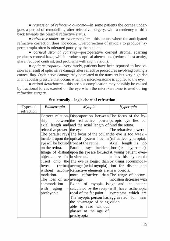
Structurally – logic chart of refraction
Types of refraction
Emmetropia Myopia Hyperopia
Correct relation-ship between axial length and refractive power. The parallel rays incident upon the eye will be focused on the retina. Image of distant objects are fo-cused onto the fovea (retina) without accom-modation. The loss of ac-commodation with aging – presbyopia
Disproportion between the refractive power and the axial length of the eye. The focus of the ocular optical system lies in front of the retina. Parallel rays incident upon the eye are focused in vitreous. The eye is longer than average (axial myopia). Refractive elements are more refractive than average. Extent of myopia is calculated by the recip-rocal of the far point. The myopic person has the advantage of being able to read without glasses at the age of presbyopia
The focus of the hy-peropic eye lies be-hind the retina. The refractive power of the eye is too weak - (refractive hyperopia). Axial length is too short (axial hyperopia). A young patient over-comes his hyperopia by using accommoda-tion for distant and near objects. The range of accom-modation decreases with age and the patient will have asthenopic symptoms which are aggravated for near vision
16
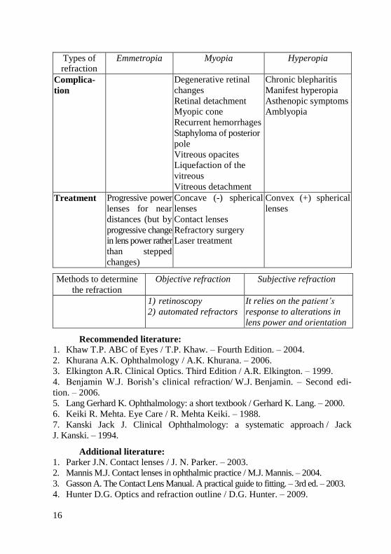
Types of refraction
Emmetropia Myopia Hyperopia
Complica-
tion
Degenerative retinal
changes
Retinal detachment
Myopic cone
Recurrent hemorrhages
Staphyloma of posterior
pole
Vitreous opacites
Liquefaction of the
vitreous
Vitreous detachment
Chronic blepharitis
Manifest hyperopia
Asthenopic symptoms
Amblyopia
Treatment Progressive power
lenses for near
distances (but by
progressive change
in lens power rather
than stepped
changes)
Concave (-) spherical
lenses
Contact lenses
Refractory surgery
Laser treatment
Convex (+) spherical
lenses
Methods to determine
the refraction
Objective refraction Subjective refraction
1) retinoscopy
2) automated refractors
It relies on the patient’s
response to alterations in
lens power and orientation
Recommended literature:
1. Khaw T.P. ABC of Eyes / T.P. Khaw. – Fourth Edition. – 2004.
2. Khurana A.K. Ophthalmology / A.K. Khurana. – 2006.
3. Elkington A.R. Clinical Optics. Third Edition / A.R. Elkington. – 1999.
4. Benjamin W.J. Borish’s clinical refraction/ W.J. Benjamin. – Second edi-
tion. – 2006.
5. Lang Gerhard K. Ophthalmology: a short textbook / Gerhard K. Lang. – 2000.
6. Keiki R. Mehta. Eye Care / R. Mehta Keiki. – 1988.
7. Kanski Jack J. Clinical Ophthalmology: a systematic approach / Jack
J. Kanski. – 1994.
Additional literature:
1. Parker J.N. Contact lenses / J. N. Parker. – 2003.
2. Mannis M.J. Contact lenses in ophthalmic practice / M.J. Mannis. – 2004.
3. Gasson A. The Contact Lens Manual. A practical guide to fitting. – 3rd ed. – 2003.
4. Hunter D.G. Optics and refraction outline / D.G. Hunter. – 2009.
17
Навчальне видання
REFRACTION
Manual for individual work in ophthalmology for English speaking foreign medical students
РЕФРАКЦІЯ
Методичні вказівки з офтальмології для індивідуальної підготовки студентів-іноземців
медичних факультетів з англійською мовою навчання
Упорядники Бездітко Павло Андрійович
Зубарев Станіслав Федорович
Панченко Микола Володимирович
Заволока Олеся Володимирівна
Мужичук Олена Павлівна
Савельєва Алла Юріївна
Ільїна Євгенія Миколаївна
Відповідальний за випуск П.А. Бездітко
Комп’ютерна верстка О.Ю. Лавриненко
План 2014, поз. 115.
Формат А5. Ризографія. Ум. друк. арк. 1,0.
Тираж 150 прим. Зам. № 14-3205.
______________________________________________________________
Редакційно-видавничий відділ
ХНМУ, пр. Леніна, 4, м. Харків, 61022
[email protected], [email protected]
Свідоцтво про внесення суб’єкта видавничої справи до Державного реєстру видавництв, виготівників і розповсюджувачів видавничої продукції серії ДК № 3242 від 18.07.2008 р.
18
REFRACTION
Manual for individual work in ophthalmology for English speaking foreign medical students