reducing perceptual interference improves visual discrimination in mild cognitive impairment:...
TRANSCRIPT
Reducing Perceptual Interference Improves Visual Discriminationin Mild Cognitive Impairment: Implications for a Model of
Perirhinal Cortex Function
Rachel N. Newsome,1* Audrey Duarte,2 and Morgan D. Barense1,3
ABSTRACT: Memory loss resulting from damage to the medial tempo-ral lobes (MTL) is traditionally considered to reflect damage to a dedi-cated, exclusive memory system. Recent work, however, has suggestedthat damage to one MTL structure, the perirhinal cortex (PRC), compro-mises complex object representations that are necessary for both mem-ory and perception. These representations are thought to be critical inshielding against the interference caused by a stream of visually similarinput. In this study, we administered a complex object discriminationtask to two memory-impaired populations thought to have brain damagethat includes the PRC [patients diagnosed with amnestic mild cognitiveimpairment (MCI), and older adults at risk for MCI], as well as age-matched controls. Importantly, we carefully manipulated the level of in-terference: in the High Interference condition, participants completed ablock of consecutive perceptually similar complex object discriminations,whereas in the Low Interference condition, we interspersed perceptuallydissimilar objects such that there was less buildup of visual interference.We found that both memory-impaired populations were impaired on theHigh Interference condition compared with controls, but critically, byreducing the degree of perceptual interference, we were largely able toimprove their performance. These findings, when taken together withconvergent evidence from animals with selective PRC lesions and amne-sic patients with focal damage to the PRC, provide support for a repre-sentational-hierarchical model of PRC function and suggest that memoryloss following PRC damage may reflect a heightened vulnerability to per-ceptual interference. VVC 2012 Wiley Periodicals, Inc.
KEY WORDS: perirhinal cortex; medial temporal lobe; Alzheimer’sdisease; mild cognitive impairment; perception; visual discrimination;interference
INTRODUCTION
Mild cognitive impairment (MCI) is a condition that often precedesdiagnosis of Alzheimer’s disease, and is usually associated with memoryimpairments. MCI patients have incipient damage to key brain struc-tures known to be vital for memory: namely, the medial temporal lobes
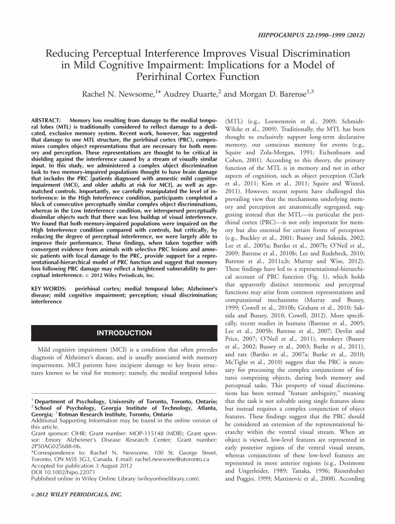
(MTL) (e.g., Loewenstein et al., 2009; Schmidt-Wilcke et al., 2009). Traditionally, the MTL has beenthought to exclusively support long-term declarativememory, our conscious memory for events (e.g.,Squire and Zola-Morgan, 1991; Eichenbaum andCohen, 2001). According to this theory, the primaryfunction of the MTL is in memory and not in otheraspects of cognition, such as object perception (Clarket al., 2011; Kim et al., 2011; Squire and Wixted,2011). However, recent reports have challenged thisprevailing view that the mechanisms underlying mem-ory and perception are anatomically segregated, sug-gesting instead that the MTL—in particular the peri-rhinal cortex (PRC)—is not only important for mem-ory but also essential for certain forms of perception(e.g., Buckley et al., 2001; Bussey and Saksida, 2002;Lee et al., 2005a; Bartko et al., 2007b; O’Neil et al.,2009; Barense et al., 2010b; Lee and Rudebeck, 2010;Barense et al., 2011a,b; Murray and Wise, 2012).These findings have led to a representational-hierarchi-cal account of PRC function (Fig. 1), which holdsthat apparently distinct mnemonic and perceptualfunctions may arise from common representations andcomputational mechanisms (Murray and Bussey,1999; Cowell et al., 2010b; Graham et al., 2010; Sak-sida and Bussey, 2010; Cowell, 2012). More specifi-cally, recent studies in humans (Barense et al., 2005;Lee et al., 2005b; Barense et al., 2007; Devlin andPrice, 2007; O’Neil et al., 2011), monkeys (Busseyet al., 2002; Bussey et al., 2003; Burke et al., 2011),and rats (Bartko et al., 2007a; Burke et al., 2010;McTighe et al., 2010) suggest that the PRC is neces-sary for processing the complex conjunctions of fea-tures comprising objects, during both memory andperceptual tasks. This property of visual discrimina-tions has been termed ‘‘feature ambiguity,’’ meaningthat the task is not solvable using single features alonebut instead requires a complex conjunction of objectfeatures. These findings suggest that the PRC shouldbe considered an extension of the representational hi-erarchy within the ventral visual stream. When anobject is viewed, low-level features are represented inearly posterior regions of the ventral visual stream,whereas conjunctions of these low-level features arerepresented in more anterior regions (e.g., Desimoneand Ungerleider, 1989; Tanaka, 1996; Riesenhuberand Poggio, 1999; Martinovic et al., 2008). According
1Department of Psychology, University of Toronto, Toronto, Ontario;2 School of Psychology, Georgia Institute of Technology, Atlanta,Georgia; 3Rotman Research Institute, Toronto, OntarioAdditional Supporting Information may be found in the online version ofthis article.Grant sponsor: CIHR; Grant number: MOP-115148 (MDB); Grant spon-sor: Emory Alzheimer’s Disease Research Center; Grant number:2P50AG025688-06.*Correspondence to: Rachel N. Newsome, 100 St. George Street,Toronto, ON M5S 3G3, Canada. E-mail: [email protected] for publication 3 August 2012DOI 10.1002/hipo.22071Published online in Wiley Online Library (wileyonlinelibrary.com).
HIPPOCAMPUS 22:1990–1999 (2012)
VVC 2012 WILEY PERIODICALS, INC.
to the representational-hierarchical account, the PRC is theapex of this representational hierarchy, representing the mostcomplex conjunctions—perhaps at the level of the whole object(Bussey and Saksida, 2002; Bussey and Saksida, 2007; Murrayet al., 2007; Cowell et al., 2010a). In this view, the PRCparticipates in both perception and memory, and impairmentsfollowing PRC damage can best be understood in terms ofdamage compromising high-level conjunctive representations(e.g., those comprising an object), leaving only lower-level rep-resentations (e.g., an object’s shape or pattern) intact (Cowellet al., 2006; Lee et al., 2012). These impoverished representa-tions will lead to impairments on both mnemonic and percep-tual tasks.
Why is it advantageous to maintain representations of com-plex feature conjunctions? A stream of visual input (such asthat encountered over a delay during a memory task) can createinterference at the level of individual features, simply becausedifferent objects tend to share lower-level features (e.g., shapes,colors). However, the ‘‘conjunctive’’ PRC representations canresolve this interference at the feature level because they areunique to each individual object. Notably, Warrington andWeiskrantz proposed over 40 yrs ago that amnesia may berelated to an increased vulnerability to interference (Warringtonand Weiskrantz, 1970). Although this theory was later rejected(Warrington and Weiskrantz, 1978), there has been a recentresurgence in the idea (Cowan et al., 2004; Loewenstein et al.,2004; Wixted, 2004; Della Sala et al., 2005; Dewar et al.,2009; Bartko et al., 2010; McTighe et al., 2010). For example,rats with PRC lesions were impaired on a minimal-delay objectrecognition task when a ‘‘perceptually similar’’ object was intro-duced either before or after the to-be-remembered object. Therats were not impaired if the interfering item was ‘‘perceptuallydissimilar’’ (Bartko et al., 2010). This suggests that in the
absence of the complex object representations contained inPRC, the animals were unable to resolve interference from inci-dental, irrelevant lower-level object features. In the perceptuallysimilar condition, many lower-level features were sharedbetween the to-be-remembered object and the interfering object.The PRC bound these features into unique objects, and thus,intact animals were able to perform the task. However, whenthe PRC was lesioned, the intact posterior regions were not suf-ficient to resolve the perceptual similarity between low-level fea-tures. Similarly, the standard object recognition memory impair-ment observed after PRC lesions was rescued in rats by using avisual restriction procedure that reduced interference (McTigheet al., 2010). This finding was replicated in aged monkeys(Burke et al., 2011), aged rats (Burke et al., 2010, 2011) and ina mouse model of Alzheimer’s disease (Romberg et al., 2012).
These findings were recently extended to humans with focaldamage to the MTL using a novel perceptual discriminationtask designed to tax complex object representations, for whichfMRI implicated the PRC (Barense et al., 2012). Whereasamnesic cases whose damage was limited to the hippocampusperformed normally, patients with MTL damage that includedthe PRC were vulnerable to object-based perceptual interfer-ence. These cases with PRC damage were impaired at successivecomplex object discriminations that contained many repeatinglow-level features, but critically, when we reduced the degree ofobject-level interference by interspersing perceptually dissimilarobjects, we recovered their performance to normal levels(Barense et al., 2012). Healthy controls, who presumably hadan intact PRC, performed as well under conditions of High In-terference as they did under conditions of Low Interference; itwas only the PRC-damaged group who were susceptible to thehigh levels of visual interference. These findings provide evi-dence to support the idea that the PRC is critical for represent-ing the complex conjunctions of features that distinguish per-ceptually similar objects (see also Baxter, 2012; Peterson et al.,2012; Ryan et al., 2012). These PRC representations becomeessential when repeated presentation of multiple, similar fea-tures causes interference at the level of the features representedby intact posterior regions in the ventral visual stream.
In this study, we sought to determine whether these findingswould generalize to another group of individuals with docu-mented memory problems—those with MCI. The MTL is oneof the earliest structures to show the neuropathological hall-marks of Alzheimer’s disease, with the PRC demonstrating sig-nificant atrophy (Juottonen et al., 1998; Du et al., 2001; Guil-lozet et al., 2003; Pennanen et al., 2004; Taylor and Probst,2008; Loewenstein et al., 2009; Schmidt-Wilcke et al., 2009).MCI patients who later convert to Alzheimer’s disease showdamage to the MTL (Bell-McGinty et al., 2005), including tothe PRC (Mitchell et al., 2002). Interestingly, evidence suggeststhat MCI and Alzheimer’s disease patients show impairmentson perceptual tasks, especially when those tasks require complexvisuoperceptual processing of objects (e.g., Alegret et al., 2009,2010). Alegret et al. found that MCI and Alzheimer’s diseasepatients have visuoperceptual deficits that may be more subtlethan standard visuoperceptual tests can pick up, yet no study
FIGURE 1. The representational-hierarchical theory. Accordingto this view, the PRC sits at the apex of the ventral visual streamprocessing pathway, and contains complex representations ofobjects (Cowell et al., 2010a). The figure shows a lateral view ofthe human ventral visual stream, and the proposed organization ofvisual object representations within this pathway. A, B, C, and Drepresent distinct visual features. Simpler features are representedin posterior regions, and complex conjunctions of features (such asthose comprising an entire object) are represented in anteriorregions, including PRC. (Figure modified from Murray and Bussey,1999 and Barense et al., 2012).
REDUCING INTERFERENCE IN MILD COGNITIVE IMPAIRMENT 1991
Hippocampus
has investigated their performance on perceptual tasks known tobe PRC dependent. To address this, here we administered thesame perceptual discrimination task from Barense et al. (2012)to MCI patients. To provide further evidence in a related partic-ipant group and to increase our sample size, we also adminis-tered the task to older adults at risk for MCI, on the assump-tion that the integrity of the PRC would already be compro-mised in the earliest stages of the disorder (e.g., Du et al.,2001; Loewenstein et al., 2009; Schmidt-Wilcke et al., 2009).We identified these individuals at risk for MCI based on theMontreal Cognitive Assessment (MoCA), a brief standardizedneuropsychological measure shown to be extremely sensitive indistinguishing controls from MCI patients (Nasreddine et al.,2005; Damian et al., 2011). Consistent with the cut-off scoresprovided by the developers of the MoCA (Nasreddine et al.,2005), we included older adults who scored below 26/30 onthe MoCA as at risk for MCI, and older adults who scored 26/30 or above as a healthy control group. We predicted that MCIpatients and individuals at risk for MCI would be impaired atobject discrimination under conditions of high perceptual inter-ference, but that we could improve their performance by reduc-ing the degree of object-based perceptual interference.
MATERIALS AND METHODS
Participants
Ten patients with clinically diagnosed amnestic MCI partici-pated in this study. These participants were recruited throughthe Emory Alzheimer’s Disease Research Center, Atlanta, GA.Of these 10, three were excluded on the basis of their near per-fect scores on the MoCA (Nasreddine et al., 2005) and Mini-Mental State Exam (MMSE, Folstein et al., 1975), leading usto believe they were ‘‘worried well’’ (Ahmed et al., 2008). Ourexclusion criteria were a perfect score on one or both of theMoCA or the MMSE, as a well as a passing score on theremaining test (i.e., a perfect score on the MoCA and a passingscore on the MMSE, or a perfect score on the MMSE and apassing score on the MoCA). The remaining seven patients(mean age 5 68.43 yrs, standard deviation 5 8.69, threefemales) completed a detailed neuropsychological battery (Table1). Patients were tested in the Memory and Aging laboratory atthe Georgia Institute of Technology. All patients providedinformed consent, and they were compensated for their timeand travel expenses. The patient testing was approved by theGeorgia Institute of Technology and the Emory UniversityInstitutional Review Boards.
In addition to MCI patients, 29 older adults with no knownneurological conditions were recruited from the University ofToronto’s Adult Volunteer Panel to participate in the study. Ofthese, 10 individuals (mean age 5 71.11, standard deviation 5
6.22, seven females) scored below 26 on the MoCA (meanscore 5 24.3, standard deviation 5 1.05) and were consideredto be at risk for MCI (Nasreddine et al., 2005). The remaining19 individuals (mean age 5 71.05 yrs, standard deviation 5
5.33, 14 females) scored above 26 on the MoCA (mean score5 27.78, standard deviation 5 1.47) and were included ascontrol participants. Participants gave informed consent, andthey were compensated for their time. These participants weretested in the Barense laboratory at the University of Toronto.The study was approved by the University of Toronto EthicsReview Board.
Thus, in total, three groups of participants participatedin this study: those with MCI, older adults at risk for MCI(‘‘At-risk’’ group), and older adults not thought to be at risk forMCI (healthy controls). Healthy controls’ MoCA scores dif-fered from those of the At-risk group [t(27) 5 6.61, p <0.001], and from those of MCI patients [t(14) 5 4.21, p <0.001]. MCI patients’ performance on the MoCA was lowerthan the At-risk group [t(15) 5 2.08, p 5 0.05]. Because ofscheduling constraints, two of the MCI patients were notadministered the MoCA. Importantly, the MoCA was givenafter the experimental task for all remaining subjects, so theexperimenter could not be biased by a participant’s MoCAperformance on the administration of the task. Age did notdiffer between controls, MCI patients, or those at risk for MCI(t < 0.21, p > 0.49).
Interference Task
Participants performed a visual discrimination task describedpreviously as Experiment 4 by Barense et al. (2012). Impor-tantly, assessment of eye movements demonstrated that this taskrequired a high degree of conjunctive processing (Experiment 1,Barense et al., 2012) and fMRI indicated that it recruited thePRC but not the hippocampus (Experiment 2, Barense et al.,2012). On each trial, participants determined if two stimuliwere identical (a match) or different (a nonmatch). Each stimu-lus was presented in one of two invisible boxes (� 98 3 98)positioned in the center of the screen, separated by 0.308. Thetask was presented using E-Prime software (Psychology SoftwareTools, Pittsburgh, PA), and participants made their responses byusing a button box connected through a USB port. The buttonswere labeled such that participants could always see which but-ton corresponded to which answer. Trials were self-paced, andstimuli were presented on screen until a response was made, fora maximum of 15 s for each trial. All participants completed apractice block with feedback after every trial until they felt com-fortable with moving on to the experimental task.
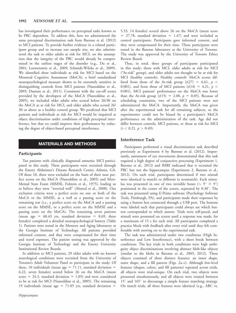
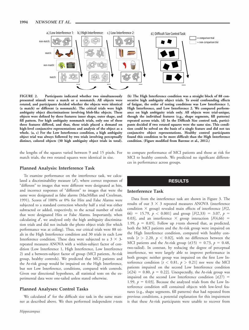
The task was administered under two conditions (High In-terference and Low Interference), with a short break betweenconditions. The key trials in both conditions were high ambi-guity object discriminations involving abstract blob-like objects(similar to the blobs in Barense et al., 2005, 2012). Theseobjects consisted of three distinct features: an inner shape,outer shape, and a fill pattern (Figs. 2a–c). Although low-levelfeatures (shapes, colors, and fill patterns) repeated across trials,all objects were trial-unique. On each trial, two objects werepresented simultaneously, and all objects were rotated between158 and 1658 to discourage a simple feature matching strategy.On match trials, all three features were identical (e.g., ABC vs.
1992 NEWSOME ET AL.
Hippocampus
ABC; letters represent individual features). Critically, for non-match trials, only one feature differed (e.g., ABC vs. ABD;whether inner shape, outer shape, or fill pattern differed wasfully counterbalanced). This high degree of feature ambiguityplaced demand on high-level conjunctive representations andanalysis of the object as a whole (Barense et al., 2012). TheHigh Interference condition contained 88 consecutive trials ofhigh ambiguity object discriminations (44 match and 44 non-match trials, intermixed in a pseudorandom sequence; Fig. 2b).Each Low Interference block also contained 88 trials, butunlike the High Interference condition, these blocks contained30 high ambiguity object trials (15 match, 15 non-match) thatwere each interspersed with two trials containing photographsof easily discriminable everyday objects (58 trials, 29 match, 29nonmatch; Figs. 2a,c). Importantly, the stimuli presented onphotograph trials shared minimal features with the blob-likeobjects (i.e., they were color photographs of everyday objects,not black and white line-drawings) and thus, the degree ofaccumulated perceptual interference was much lower in theLow Interference condition. As such, we predicted that the
MCI patient group and the At-risk group would perform betteron the Low Interference condition relative to the High Interfer-ence condition. We administered two blocks of the Low Inter-ference condition, one before and one after the High Interfer-ence condition, to ensure that any observed deficits were spe-cific to the buildup of interfering features and not fatigue orgeneric task-practice effects.
Control Task: Difficult Size
Following the completion of the interference task, partici-pants were administered a control task that did not requireconjunctive processing, but was matched in terms of difficultyto the High Interference condition (control task used previouslyin Barense et al., 2012). The procedures for the control condi-tion were identical to the interference task described aboveexcept the stimuli were squares and participants indicated ifthey were identical (match) or different (nonmatch) (Fig. 2d).For nonmatch trials, the length of each side of the square wasrandomly varied from 67 to 247 pixels. The difference between
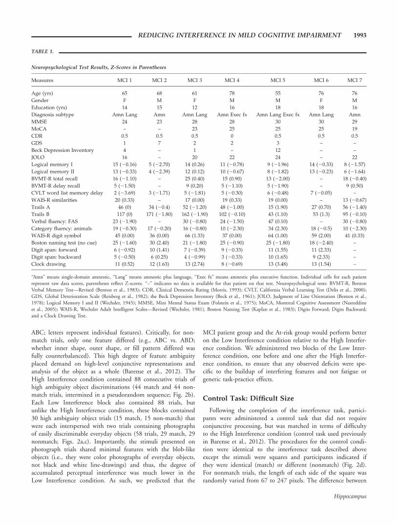
TABLE 1.
Neuropsychological Test Results, Z-Scores in Parentheses
Measures MCI 1 MCI 2 MCI 3 MCI 4 MCI 5 MCI 6 MCI 7
Age (yrs) 65 68 61 78 55 76 76
Gender F M F M M F M
Education (yrs) 14 15 12 16 18 18 16
Diagnosis subtype Amn Lang Amn Amn Lang Amn Exec fx Amn Lang Exec fx Amn Lang Amn
MMSE 24 23 28 28 30 30 29
MoCA – – 23 25 25 25 19
CDR 0.5 0.5 0.5 0 0.5 0.5 0.5
GDS 1 7 2 2 3 – –
Beck Depression Inventory 4 – 1 – 12 – –
JOLO 16 – 20 22 24 – 22
Logical memory I 15 (20.16) 5 (22.70) 14 (0.26) 11 (20.78) 9 (21.96) 14 (20.33) 8 (21.57)
Logical memory II 13 (20.33) 4 (22.39) 12 (0.12) 10 (20.67) 8 (21.82) 13 (20.23) 6 (21.64)
BVMT-R total recall 16 (21.10) – 25 (0.40) 15 (0.90) 13 (22.00) – 18 (20.40)
BVMT-R delay recall 5 (21.50) – 9 (0.20) 5 (21.10) 5 (21.90) – 9 (0.50)
CVLT word list memory delay 2 (23.69) 3 (21.71) 5 (21.81) 5 (20.30) 6 (20.48) 7 (20.05) –
WAIS-R similarities 20 (0.33) – 17 (0.00) 19 (0.33) 19 (0.00) – 13 (20.67)
Trails A 46 (0) 34 (20.4) 52 (21.20) 48 (21.00) 15 (1.90) 27 (0.70) 56 (21.40)
Trails B 117 (0) 171 (21.80) 162 (21.90) 102 (20.10) 43 (1.10) 53 (1.3) 95 (20.10)
Verbal fluency: FAS 23 (21.90) – 30 (20.80) 24 (21.50) 47 (0.10) – 30 (20.80)
Category fluency: animals 19 (20.30) 17 (20.20) 16 (20.80) 10 (22.30) 34 (2.30) 18 (20.5) 10 (22.30)
WAIS-R digit symbol 45 (0.00) 36 (0.00) 66 (1.33) 37 (0.00) 64 (1.00) 59 (2.00) 41 (0.33)
Boston naming test (no cue) 25 (21.60) 30 (2.40) 21 (21.80) 25 (20.90) 25 (21.80) 18 (22.40) –
Digit span: forward 6 (20.92) 10 (1.41) 7 (20.39) 9 (20.33) 11 (1.55) 11 (2.33) –
Digit span: backward 5 (20.50) 6 (0.25) 4 (20.99) 3 (20.33) 10 (1.65) 9 (2.33) –
Clock drawing 11 (0.52) 12 (1.63) 13 (2.74) 8 (20.69) 13 (3.48) 13 (1.54) –
‘‘Amn’’ means single-domain amnestic, ‘‘Lang’’ means amnestic plus language, ‘‘Exec fx’’ means amnestic plus executive function. Individual cells for each patientrepresent raw data scores, parentheses reflect Z-scores. ‘‘–’’ indicates no data is available for that patient on that test. Neuropsychological tests: BVMT-R, BentonVerbal Memory Test—Revised (Benton et al., 1983); CDR, Clinical Dementia Rating (Morris, 1993); CVLT, California Verbal Learning Test (Delis et al., 2000);GDS, Global Deterioration Scale (Reisberg et al., 1982); the Beck Depression Inventory (Beck et al., 1961); JOLO, Judgment of Line Orientation (Benton et al.,1978); Logical Memory I and II (Wechsler, 1945); MMSE, Mini Mental Status Exam (Folstein et al., 1975); MoCA, Montreal Cognitive Assessment (Nasreddineet al., 2005); WAIS-R, Wechsler Adult Intelligent Scales—Revised (Wechsler, 1981), Boston Naming Test (Kaplan et al., 1983); Digits Forward; Digits Backward;and a Clock Drawing Test.
REDUCING INTERFERENCE IN MILD COGNITIVE IMPAIRMENT 1993
Hippocampus
the lengths of the squares varied between 9 and 15 pixels. Formatch trials, the two rotated squares were identical in size.
Planned Analysis: Interference Task
To examine performance on the interference task, we calcu-lated a discriminability measure (d0), where correct responses of‘‘different’’ to images that were different were designated as hits,and incorrect responses of ‘‘different’’ to images that were thesame were designated as false alarms (MacMillan and Creelman,1991). Scores of 100% or 0% for Hits and False Alarms weresubjected to a standard correction whereby half a trial was eithersubtracted or added, respectively, to the actual number of trialsthat were designated Hits or False Alarms. Importantly, whencalculating d0, we analyzed only the high ambiguity discrimina-tion trials and did not include the photo object trials (for whichperformance was at ceiling). Thus, our critical trials were 88 tri-als in the High Interference condition and 30 trials in each LowInterference condition. These data were subjected to a 3 3 3-repeated measures ANOVA with a within-subject factor of con-dition (Low Interference 1, High Interference, Low Interference2) and a between-subject factor of group (MCI patients, At-riskgroup, healthy controls). We predicted that MCI patients andthe At-risk group would be impaired on the High Interference,but not Low Interference, conditions, compared with controls.Given our directional hypotheses, all statistical tests on the ex-perimental data were one-tailed unless stated otherwise.
Planned Analyses: Control Tasks
We calculated d 0 for the difficult size task in the same man-ner as described above. We then performed independent t-tests
to compare performance of MCI patients and those at risk forMCI to healthy controls. We predicted no significant differen-ces in performance across groups.
RESULTS
Interference Task
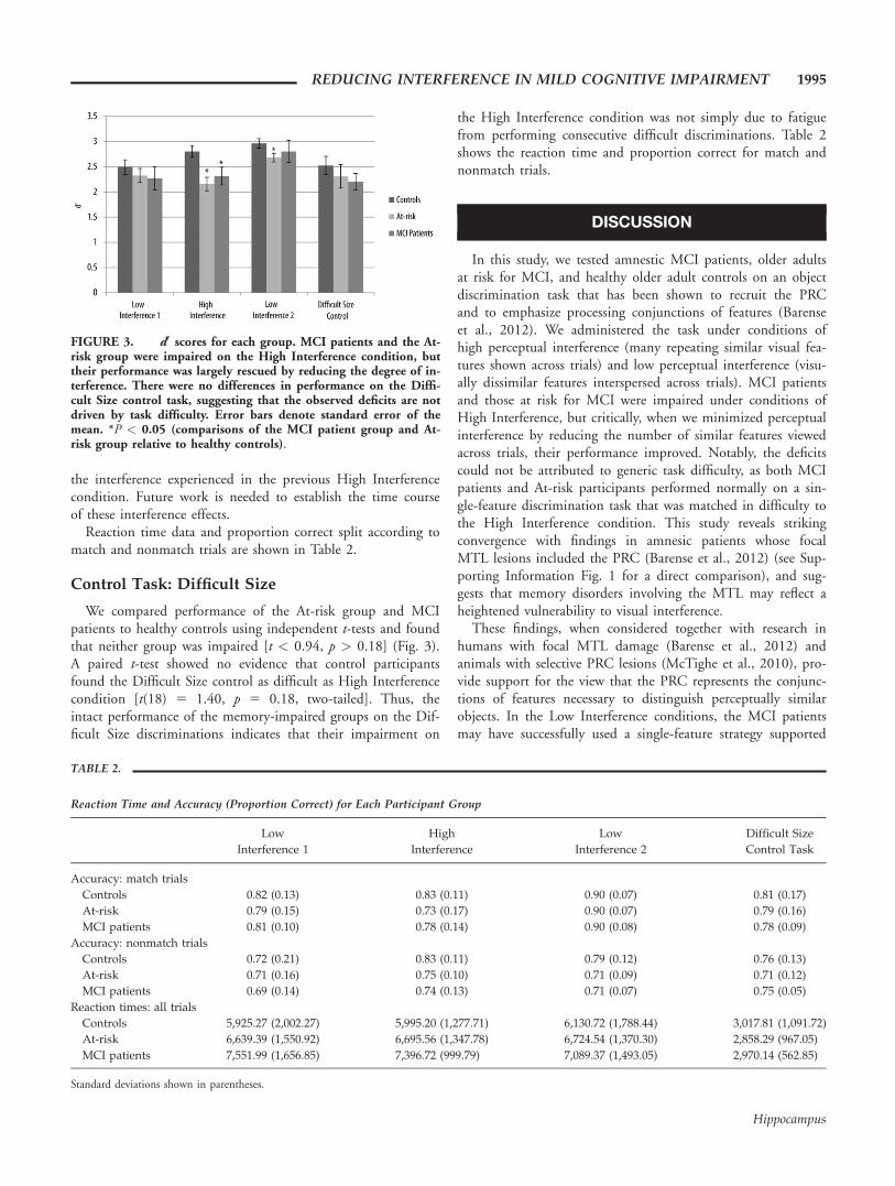
Data from the interference task are shown in Figure 3. Theresults of our 3 3 3 repeated measures ANOVA (interferencecondition 3 group) revealed main effects of interference [F(2,66) 5 15.79, p < 0.001] and group [F(2,33) 5 3.07, p 5
0.03], and an interference 3 group interaction [F(4,66) 5
1.99, p 5 0.05]. Follow up t-tests showed that, as predicted,both the MCI patients and the At-risk group were impaired onthe High Interference condition, compared with healthy con-trols [t > 2.20, p < 0.02], with no differences between theMCI patients and the At-risk group (t(15) 5 0.73, p 5 0.48,two-tailed). In contrast, by reducing the degree of perceptualinterference, we were largely able to improve performance inboth groups: neither group was impaired on the first Low In-terference condition [t < 0.81, p > 0.21] nor were the MCIpatients impaired on the second Low Interference condition[t(24) 5 0.80, p 5 0.22]. Unexpectedly, the At-risk group wasimpaired on the second Low Interference condition [t(27) 5
1.99, p 5 0.03]. Because the analyzed trials from the Low In-terference condition still contained objects with low-level fea-tures (e.g., shape segments, fill patterns) that had repeated fromprevious conditions, a potential explanation for this impairmentis that these At-risk participants were unable to recover from
FIGURE 2. Participants indicated whether two simultaneouslypresented stimuli were a match or a nonmatch. All objects wererotated, and participants decided whether the objects were identical(a match) or different (a nonmatch). The critical trials were highambiguity object discriminations involving blob-like objects. Theseobjects were defined by three features: inner shape, outer shape, andfill pattern. For high ambiguity nonmatch trials, only one of thesethree features differed, and thus, these trials placed a demand onhigh-level conjunctive representations and analysis of the object as awhole. (a, c) For the Low Interference condition, a high ambiguityobject trial was always followed by two trials involving perceptuallydistinct, colored objects (30 high ambiguity object trials in total).
(b) The High Interference condition was a straight block of 88 con-secutive high ambiguity object trials. To avoid confounding effectsof fatigue, the order of testing conditions was Low Interference 1,High Interference, and Low Interference 2. We compared perform-ance on high ambiguity trials only. All objects were trial-unique,though the individual features (e.g., shape segments, fill patterns)repeated across trials. (d) In the Difficult Size control task, partici-pants decided if two rotated squares were the same size. This condi-tion could be solved on the basis of a single feature and did not taxconjunctive object representations. Healthy control participantsfound this condition to be more difficult than the High Interferencecondition. (Figure modified from Barense et al., 2012.)
1994 NEWSOME ET AL.
Hippocampus
the interference experienced in the previous High Interferencecondition. Future work is needed to establish the time courseof these interference effects.
Reaction time data and proportion correct split according tomatch and nonmatch trials are shown in Table 2.
Control Task: Difficult Size
We compared performance of the At-risk group and MCIpatients to healthy controls using independent t-tests and foundthat neither group was impaired [t < 0.94, p > 0.18] (Fig. 3).A paired t-test showed no evidence that control participantsfound the Difficult Size control as difficult as High Interferencecondition [t(18) 5 1.40, p 5 0.18, two-tailed]. Thus, theintact performance of the memory-impaired groups on the Dif-ficult Size discriminations indicates that their impairment on
the High Interference condition was not simply due to fatiguefrom performing consecutive difficult discriminations. Table 2shows the reaction time and proportion correct for match andnonmatch trials.
DISCUSSION
In this study, we tested amnestic MCI patients, older adultsat risk for MCI, and healthy older adult controls on an objectdiscrimination task that has been shown to recruit the PRCand to emphasize processing conjunctions of features (Barenseet al., 2012). We administered the task under conditions ofhigh perceptual interference (many repeating similar visual fea-tures shown across trials) and low perceptual interference (visu-ally dissimilar features interspersed across trials). MCI patientsand those at risk for MCI were impaired under conditions ofHigh Interference, but critically, when we minimized perceptualinterference by reducing the number of similar features viewedacross trials, their performance improved. Notably, the deficitscould not be attributed to generic task difficulty, as both MCIpatients and At-risk participants performed normally on a sin-gle-feature discrimination task that was matched in difficulty tothe High Interference condition. This study reveals strikingconvergence with findings in amnesic patients whose focalMTL lesions included the PRC (Barense et al., 2012) (see Sup-porting Information Fig. 1 for a direct comparison), and sug-gests that memory disorders involving the MTL may reflect aheightened vulnerability to visual interference.
These findings, when considered together with research inhumans with focal MTL damage (Barense et al., 2012) andanimals with selective PRC lesions (McTighe et al., 2010), pro-vide support for the view that the PRC represents the conjunc-tions of features necessary to distinguish perceptually similarobjects. In the Low Interference conditions, the MCI patientsmay have successfully used a single-feature strategy supported
FIGURE 3. d0 scores for each group. MCI patients and the At-risk group were impaired on the High Interference condition, buttheir performance was largely rescued by reducing the degree of in-terference. There were no differences in performance on the Diffi-cult Size control task, suggesting that the observed deficits are notdriven by task difficulty. Error bars denote standard error of themean. *P < 0.05 (comparisons of the MCI patient group and At-risk group relative to healthy controls).
TABLE 2.
Reaction Time and Accuracy (Proportion Correct) for Each Participant Group
Low
Interference 1
High
Interference
Low
Interference 2
Difficult Size
Control Task
Accuracy: match trials
Controls 0.82 (0.13) 0.83 (0.11) 0.90 (0.07) 0.81 (0.17)
At-risk 0.79 (0.15) 0.73 (0.17) 0.90 (0.07) 0.79 (0.16)
MCI patients 0.81 (0.10) 0.78 (0.14) 0.90 (0.08) 0.78 (0.09)
Accuracy: nonmatch trials
Controls 0.72 (0.21) 0.83 (0.11) 0.79 (0.12) 0.76 (0.13)
At-risk 0.71 (0.16) 0.75 (0.10) 0.71 (0.09) 0.71 (0.12)
MCI patients 0.69 (0.14) 0.74 (0.13) 0.71 (0.07) 0.75 (0.05)
Reaction times: all trials
Controls 5,925.27 (2,002.27) 5,995.20 (1,277.71) 6,130.72 (1,788.44) 3,017.81 (1,091.72)
At-risk 6,639.39 (1,550.92) 6,695.56 (1,347.78) 6,724.54 (1,370.30) 2,858.29 (967.05)
MCI patients 7,551.99 (1,656.85) 7,396.72 (999.79) 7,089.37 (1,493.05) 2,970.14 (562.85)
Standard deviations shown in parentheses.
REDUCING INTERFERENCE IN MILD COGNITIVE IMPAIRMENT 1995
Hippocampus
by intact regions upstream in the ventral visual stream (by defi-nition, objects in the discrimination of ABC vs. ABD differedby a single feature: C vs. D, see Fig. 1). However, the repeatedadministration of multiple low-level object features (e.g.,shapes, fill patterns)—as was the case in the High Interferencecondition—created massive interference at the level of the rep-resentations for these individual features. Under these condi-tions, a more complex representation that binds the featuresinto unique objects is necessary to resolve the interference fromrepeating features. Put differently, when a constant stream ofvisual information creates large-scale feature-level interferencefrom irrelevant features processed on previous trials, a moreelaborate object-level representation will be resistant to the fea-ture-level interference. We propose this more complex object-level representation to be contained within PRC. In addition toprocessing the complex conjunction of features comprising theobject, recent research suggests that the PRC may also protectfrom visual interference by ‘‘suppressing’’ the representation ofthe lower-level features represented in earlier regions of the vis-ual stream (Peterson et al., 2012).
These results complement a growing body of evidence sug-gesting that memory impairments following MTL damage maybe due to an increased vulnerability to interference (Cowanet al., 2004; Loewenstein et al., 2004; Della Sala et al., 2005;Dewar et al., 2009). For example, one study found that recallof a story in patients with MCI was increased by a remarkable35% under conditions of Low Interference (during the delayparticipants reclined in a dark, quiet room) compared withrecall under standard interference (a 1-h delay filled with psy-chometric tasks) (Della Sala et al., 2005). A similar result wasreported for amnesics with focal brain damage (Cowan et al.,2004). The interference in these studies may have been frominterfering mental activity and memory formation (Wixted,2004), and/or it may have been due to interference from simi-lar visual information processed during the psychometric tasks.Consistent with the latter idea of visual interference from per-ceptually similar information, sensory restriction rescued anobject recognition memory deficit in rats with perirhinal lesions(McTighe et al., 2010), aged rats (Burke et al., 2010), and in amouse model of Alzheimer’s disease-typical amyloid-b pathol-ogy (the tgCRND8 mouse, Romberg et al., 2012). The presentresults extend these findings from the mnemonic domain tothe visual discrimination of trial-unique stimuli.
Although we prefer to consider these results in terms ofimpoverished object representations impairing perception, it isworth noting that there is a working memory component tothe task. Although the objects were displayed simultaneously,the discriminations required a series of eye movements andcomparisons across objects. Thus, it is possible that theobserved deficit could be related to problems with workingmemory. There has been a recent surge of research indicatingthat short-term memory may be impaired following MTLdamage (e.g., Hannula et al., 2006; Nichols et al., 2006; Olsonet al., 2006; Warren et al., 2010; although see Jeneson et al.,2010), with evidence suggesting that the MTL is important inonline processing of highly similar visual objects (Warren et al.,
2011a,b). We are certainly not opposed to the notion thatMTL structures are important for working memory. Indeed, itseems highly likely that the conjunctive representations proc-essed by PRC are essential for online maintenance of informa-tion while shifting attention from one object to the other (seealso Barense et al., 2012). We would additionally argue, how-ever, that these representations are critical for any cognitive taskthat requires them, which includes long-term memory, workingmemory, and complex object perception. In support of this,other studies have demonstrated perceptual deficits on taskswith no working memory component (i.e., perception of singleobjects and figure-ground perception of familiar object configu-ration), suggesting that the deficits observed here reflect a morefundamental deficit in representing complex objects (Lee andRudebeck, 2010; Barense et al., 2011b).
It is important to note that due to the unavailability ofstructural MRI scans of participants in the current study, wecannot conclusively localize the observed deficits to PRC pa-thology. It is very well established that the hippocampus andentorhinal cortex are affected very early, likely even first, inMCI (Wakabayashi et al., 1994; Harris et al., 2010; Devanandet al., 2012). The PRC is heavily connected to these structures(e.g., Suzuki and Amaral, 1994), and is part of the neural cir-cuit affected early in MCI (Mitchell et al., 2002; Bell-McGintyet al., 2005; Schuff and Zhu, 2007; Zhang et al., 2012). Thus,it is not surprising that PRC function would be compromised.Consistent with this, the profile of performance in both theMCI patients and those at risk for MCI is remarkably consist-ent with findings from research that allows more precise local-ization of the PRC: (1) animal studies that have demonstratedobject discrimination deficits and interference effects after pre-cise, localized PRC lesions (Buckley et al., 2001; Bussey et al.,2002, 2003; Bartko et al., 2010; McTighe et al., 2010), and(2) functional neuroimaging revealing PRC activity in healthyparticipants during the discrimination task reported here(Barense et al., 2012) and other tasks taxing complex object per-ception (Devlin and Price, 2007; Lee et al., 2008; O’Neil et al.,2009; Barense et al., 2010a, 2011a). In addition, based on theintact performance of amnesic patients with focal hippocampaldamage (Barense et al., 2012; Supporting Information Fig. 1),we can localize our deficits to nonhippocampal structures.
One notable finding of the present study is that althoughthe At-risk participants do not have diagnosed MCI, their per-formance suggests that a clinical exam might lead to an MCIdiagnosis. However, now, they appear to be ‘‘unworriedunwell.’’ In other words, these volunteers from the communitypresented with no subjective memory complaint, but had anobjective cognitive deficit. The subjective memory complaint inquestion is a vital component of diagnosis using the originalPeterson criteria (Petersen et al., 1999), and it remains to beseen whether these participants would be diagnosed with MCI.Nonetheless, the MoCA has been identified as a sensitive briefmeasure in detecting cognitive impairment (Nasreddine et al.,2005; Markwick et al., 2012). Additionally, the present studyindicates that and those who fall below norms on the MoCAare vulnerable to visual interference. In fact, an additional
1996 NEWSOME ET AL.
Hippocampus
correlational analysis investigating performance across all groupson all tasks showed that MoCA scores predicted performanceon the High Interference task (r(33) 5 0.54, p < 0.01, Bonfer-roni corrected), but not the other three conditions [r(33)< 0.29, p > 0.09, Bonferroni corrected]. We are currently fol-lowing up the At-risk participants on a number of cognitiveand physiological longitudinal assessments.
In conclusion, these data illustrate that reducing perceptualinterference improves performance on a discrimination task inMCI patients and those at risk for MCI. Furthermore, thesedata add additional support to the idea that damage to thecomplex, conjunctive representations processed by PRC causesan increased susceptibility to object-based perceptual interfer-ence, which leads to deficits in both memory and perception.
Acknowledgments
The authors thank all participants for their time, and SineadBrady and Alina Guna (University of Toronto) for help withdata collection.
REFERENCES
Ahmed S, Mitchell J, Arnold R, Dawson K, Nestor P, Hodges J.2008. Memory complaints in mild cognitive impairment, worriedwell, and semantic dementia patients. Alzheimer Dis Assoc Disord22:227–235.
Alegret M, Boada-Roviera M, Vinyes-Junque G, Valero S, Espinosa A,Hernandez I, Modinos G, Rosende-Roca M, Mauleon A, Becker J,Tarraga L. 2009. Detection of visuoperceptual deficits in preclinicaland mild Alzheimer’s Dsisease. J Clin Exp Neuropsychol 31:860–867.
Alegret M, Vinyes-Junque G, Boada M, Martinez-Lage P, Cuberas G,Espinosa A, Roca I, Hernandez I, Valero S, Rosende-Roca M,Mauleon A, Becker JT, Tarraga L. 2010. Brain perfusion correlatesof visuoperceptual deficits in mild cognitive impairment and mildAlzheimer’s disease. J Alzheimer Dis 21:557–567.
Barense M, Bussey T, Lee A, Rogers T, Davies R, Saksida L, MurrayE, Graham K. 2005. Functional specialization in the human medialtemporal lobe. J Neurosci 25:10239–10246.
Barense M, Gaffan D, Graham K. 2007. The human medial temporallobe processes online representations of complex objects. Neuropsy-chologia 45:2963–2974.
Barense M, Henson R, Lee A, Graham K. 2010a. Medial temporallobe activity during complex visual discrimination of faces, objectsand scenes: The effect of viewpoint. Hippocampus 20:389–401.
Barense M, Rogers T, Bussey T, Saksida L, Graham K. 2010b. Influenceof conceptual knowledge on visual object discrimination: Semanticdementia and MTL amnesia. Cereb Cortex 20:2568–2582.
Barense M, Henson R, Graham K. 2011a. Perception and conception:Temporal lobe activity during complex discriminations of familiarand novel faces and objects. J Cogn Neurosci 23:3052–3057.
Barense M, Ngo J, Hung L, Peterson M. 2011b. Interactions of mem-ory and perception in amnesia: The figure-ground perspective.Cereb Cortex 22:11.
Barense M, Groen I, Lee A, Yeung L, Brady S, Gregori M, Kapur N,Bussey T, Saksida L, Henson R. 2012. Intact memory for irrelevantinformation impairs perception in amnesia. Neuron 75:157–167.
Bartko S, Winters B, Cowell R, Saksida L, Bussey T. 2007a. Percep-tual functions in perirhinal cortex in rats: Zero-delay object recog-nition and simultaneous oddity discriminations. J Neurosci 27:2548–2559.
Bartko S, Winters B, Cowell R, Saksida L, Bussey T. 2007b. Perirhinalcortex resolves feature ambiguity in configural object recognitionand perceptual oddity tasks. Learn Mem 14:821–832.
Bartko S, Cowell R, Winters B, Bussey T, Saksida L. 2010. Height-ened susceptibility to interference in an animal model of amnesia:Impairment in encoding, storage, retrieval—or all three? Neuropsy-chologia 48:2987–2997.
Baxter M. 2012. It’s all coming back to me now: Perception andmemory in amnesia. Neuron 75:8–10.
Bell-McGinty S, Lopez O, Meltzer C, Scanlon J, Whyte E, DeKoskyS, Becker J. 2005. Differential cortical atrophy in subgroups ofMild Cognitive Impairment. Arch Neurol 62:1393–1397.
Buckley M, Booth M, Rolls E, Gaffan D. 2001. Selective perceptualimpairments after perirhinal cortex ablation. J Neurosci 21:9824–9836.
Burke S, Wallace J, Nematollahi S, Uprety A, Barnes C. 2010. Patternseparation deficits may contribute to age associated recognitionimpairments. Behav Neurosci 124:559–573.
Burke S, Wallace J, Hartzell A, Nematollahi S, Plange K, Barnes C.2011. Age-associated deficits in pattern separation functions of theperirhinal cortex: A cross-species consensus. Behav Neurosci125:836–47.
Bussey T, Saksida L. 2002. The organization of visual object represen-tations: A connectionist model of effects of lesions in perirhinalcortex. Eur J Neurosci 15:355–364.
Bussey T, Saksida L. 2007. Memory, perception, and the ventral visualperirhinal hippocampal stream: Thinking outside of the boxes.Hippocampus 908:898–908.
Bussey T, Saksida L, Murray E. 2002. Perirhinal cortex resolves featureambiguity in complex visual discriminations. Eur J Neurosci15:363–374.
Bussey T, Saksida L, Murray E. 2003. Impairments in visual discrimina-tion after perirhinal cortex lesions: Testing ‘declarative’ vs. ‘perceptual-mnemonic’ views of perirhinal cortex function. Eur J Neurosci 17.
Clark R, Reinagel P, Broadbent N, Flister E, Squire L. 2011. Intactperformance on feature-ambiguous discriminations in rats withlesions of the perirhinal cortex. Neuron 70:132–140.
Cowan N, Beschin N, Della Sala S. 2004. Verbal recall in amnesiacsunder conditions of diminished retroactive interference. Brain127:825–834.
Cowell R, Bussey T, Saksida L. 2006. Why does brain damage impairmemory? A connectionist model of object recognition memory inperirhinal cortex. J Neurosci 26:12186–12197.
Cowell R, Bussey T, Saksida L. 2010a. Components of recognitionmemory: Dissociable cognitive processes or just difference in repre-sentational complexity? Hippocampus 20:1245–1262.
Cowell R, Bussey T, Saksida L. 2010b. Functional dissociations withinthe ventral object processing pathway: Cognitive modules or a hier-archical continuum? J Cogn Neurosci 22:2460–2479.
Cowell RA. 2012. Computational models of perirhinal cortical func-tion. Hippocampus 22:1952–1964.
Damian A, Jacobsen S, Hentz J, Belden C, Shill H, Sabbagh M, Cavi-ness J, Adler C. 2011. The Montreal Cognitive Assessment and theMini-Mental State Examination as screening instruments for cogni-tive impairment: Item analyses and threshold scores. Dement Ger-iatr Cogn Disord 31:126–131.
Della Sala S, Cowan N, Beschin N, Perini M. 2005. Just lying there,remembering: Improving recall of prose in amnesic patients withmild cognitive impairment by minimising interference. Memory13:435–440.
Desimone R, Ungerleider L. 1989. Neural mechanisms of visual proc-essing in monkeys. In: Boller F, Grafman J, editors. Handbook ofNeuropsychology. New York: Elsevier Sciences. pp 267–299.
Devanand D, Bansal R, Liu J, Hao X, Pradhaben G, Peterson B.2012. MRI hippocampal and entorhinal cortex mapping in pre-dicting conversion to Alzheimer’s disease. Neuroimage 60:1622–1629.
REDUCING INTERFERENCE IN MILD COGNITIVE IMPAIRMENT 1997
Hippocampus
Devlin J, Price C. 2007. Perirhinal contributions to human visual per-ception. Curr Biol 17:1484–1488.
Dewar M, Garcia Y, Cowan N, Della Sala S. 2009. Delaying interfer-ence enhances memory consolidation in amnesic patients. Neuro-psychology 23:627–634.
Du A, Schuff N, Amend D, Laakso M, Hsu Y, Jagust W, Yaffe K,Kramer J, Reed B, Norman D, Chui HC, Weiner MW. 2001.Magnetic resonance imaging of the entorhinal cortex and hippo-campus in mild cognitive impairment and Alzheimer’s disease. JNeurol Neurosurg Psychiatry 71:441–447.
Eichenbaum H, Cohen N. 2001. From Conditioning to ConsciousRecollection: Memory Systems of the Brain. New York: OxfordUniversity Press.
Folstein M, Folstein S, McHugh P. 1975. ‘‘Mini-Mental State’’: Apractical method for grading the cognitive state of patients for theclinician. J Psychiatr Res 12:189–198.
Graham K, Barense M, Lee A. 2010. Going beyond LTM in theMTL: A synthesis of neuropsychological and neuroimaging findingson the role of the medial temporal lobe in memory and percep-tion. Neuropsychologia 48:831–853.
Guillozet A, Weintraub S, Mash D, Mesulam M. 2003. Neurofibril-lary tangles, amyloid, and memory in aging and mild cognitiveimpairment. Arch Neurol 60:729–736.
Harris J, Devidze N, Verret L, Ho K, Halabisky B, Thwin M, Kim D,Hamto P, Lo I, Yu G, Palop J, Masliah E, Mucke L. 2010. Transsy-naptic progression of amyloid-B-induced neuronal dysfunctionwithin the entorhinal-hippocampal network. Neuron 68:428–41.
Juottonen K, Laakso M, Insausti R, Lehtovirta M, Pitkanen A, Parta-nen K, Soininen H. 1998. Volumes of entorhinal and perirhinalcortices in Alzheimer’s disease. Neurobiol Aging 19:15–22.
Kim S, Jeneson A, van der Horst A, Frascino J, Hopkins R, Squire L.2011. Memory, visual discrimination performance, and the humanhippocampus. J Neurosci 31:2624–2630.
Lee A, Barense M, Graham K. 2005a. The contribution of the humanmedial temporal lobe to perception: Bridging the gap between ani-mal and human studies. Q J Exp Psychol 58b:300–325.
Lee A, Bussey T, Murray E, Saksida L, Epstein R, Kapur N, Hodges J,Graham K. 2005b. Perceptual deficits in amnesia: Challenging themedial temporal lobe ‘mnemonic’ view. Neuropsychologia 43:1–11.
Lee A, Rudebeck S. 2010. Human medial temporal lobe damage candisrupt the perception of single objects. J Neurosci 30:6588–6594.
Lee A, Scahill V, Graham K. 2008. Activating the medial temporallobe during oddity judgment for faces and scenes. Cereb Cortex18:683–696.
Lee A, Yeung L-K, Barense M. 2012. The hippocampus and visualperception. Front Hum Neurosci 6:91.
Loewenstein D, Acevedo A, Luis C, Crum T, Barker W, Duara R.2004. Semantic interference deficits and detection of mild Alzhei-mer’s disease and mild cognitive impairment without dementia.J Int Neuropsychol Soc 10:91–100.
Loewenstein D, Acevedo A, Potter E, Schinka J, Raj A, Greig M,Agron J, Barker W, Wu Y, Small B, Schofield E, Duara R. 2009.Severity of medial temporal lobe atrophy and amnestic mild cogni-tive impairment: Selecting type and number of memory tests.American J Geriatr Psychiatry 17:1050–1058.
MacMillan N, Creelman C. 1991. Detection Theory: A User’s Guide.New York: Cambridge University Press.
Markwick A, Zamboni G, de Jager C. 2012. Profiles of cognitive subt-est impairment in Montreal Cognitive Assessment (MoCA) in aresearch cohort with normal Mini-Mental State Examination(MMSE) scores. J Clin Exp Neuropsychol 34:750–757.
Martinovic J, Gruuber T, Muller M. 2008. Coding of visual objectfeatures and feature conjunction in the human brain. PLoS ONE3:e3781.
McTighe S, Cowell R, Winters B, Bussey T, Saksida L. 2010. Paradox-ical false memory for objects after brain damage. Science330:1408–1410.
Mitchell T, Mufson E, Schneider J, Cochran E, Nissanov J, Han L,Bienias J, Lee V, Trojanowski J, Bennett D, Arnold S. 2002.Parahippocampal tau pathology in healthy aging, mild cognitiveimpairment, and early Alzheimer’s disease. Ann Neurol 51:182–189.
Murray E, Bussey T. 1999. Perceptual-mnemonic functions of theperirhinal cortex. Trends Cogn Sci 3:142–151.
Murray E, Bussey T, Saksida L. 2007. Visual perception and memory:A new view of medial temporal lobe function in primates androdents. Annu Rev Neurosci 30:99–122.
Murray E, Wise S. 2012. Why is there a special issue on perirhinalcortex in a journal called Hippocampus?: The perirhinal cortex inhistorical perspective. Hippocampus 22:1941–1951.
Nasreddine Z, Phillips N, Bedirian V, Charbonneau S, Whitehead V,Collin I, Cummings J, Chertkow H. 2005. The Montreal Cogni-tive Assessment, MoCA: A brief screening tool for mild cognitiveimpairment. J Am Geriatr Soc 53:695–699.
O’Neil E, Cate A, Kohler S. 2009. Perirhinal cortex contributes toaccuracy in recognition memory and perceptual discriminations.J Neurosci 29:8329–8334.
O’Neil E, Protzner A, McCormick C, McLean D, Poppenk J, CateA, Kohler S. 2011. Distinct patterns of functional and effectiveconnectivity between perirhinal cortex and other cortical regionsin recognition memory and perceptual discrimination. Cereb Cor-tex 22:74–85.
Pennanen C, Kivipelto M, Tuomainen S, Hartikainen P, HanninenT, Laakso M, Hallikainen M, Vanhanen M, Nissinen A, Hel-kala E, Vainio P, Vanninen R, Partanen K, Soininen H. 2004.Hippocampus and entorhinal cortex in mild cognitive impair-ment and early AD. Neurobiol Aging 25: 303–310.
Peterson M, Cacciamani L, Barense M, Scalf P. 2012. The perirhinalcortex modulates V2 activity in response to agreement between partfamiliarity and configuration familiarity. Hippocampus 22:1965–1977.
Petersen R, Smith G, Waring S, Ivnik R, Tangalos E, Kokmen E.1999. Mild cognitive impairment: Clinical characterization andoutcome. Arch Neurol 56:303–308.
Riesenhuber M, Poggio T. 1999. Hierarchical models of object recog-nition in cortex. Nat Neurosci 2:1019–1025.
Romberg C, McTighe S, Heath C, Whitcomb D, Cho K, Bussey T,Saksida L. 2012. False recognition in a mouse model of Alzheimer’sdisease: Rescue with sensory restriction and memantine. Brain135:2103–2114.
Ryan L, Cardoza J, Barense M, Kawa K, Walletin-Flores J, AlexanderG, Barnes C. 2012. Age-related impairments in a complex objectdiscrimination task that engages perirhinal cortex. Hippocampus22:1978–1989.
Saksida L, Bussey T. 2010. The representational-hierarchical view ofamnesia: Translation from animal to human. Neuropsychologia48:2370–2384.
Schmidt-Wilcke T, Poljansky S, Hierlmeier S, Hausner J, Ibach B.2009. Memory performance correlates with gray matter density inthe ento-perirhinal cortex and posterior hippocampus in patientswith mild cognitivei mpairment and healthy controls - a voxelbased morphometry study. Neuroimage 47:1914–1920.
Schuff N, Zhu X. 2007. Imaging of mild cognitive impairment andearly dementia. Br J Radiol 80:S109–S114.
Squire L, Wixted J. 2011. The cognitive neuroscience of human mem-ory since HM. Annu Rev Neurosci 34:259–288.
Squire L, Zola-Morgan S. 1991. The medial temporal lobe memorysystem. Science 253:1380–1386.
Tanaka K. 1996. Inferotemporal cortex and object vision. Annu RevNeurosci 19:109–139.
Taylor K, Probst A. 2008. Anatomic localization of the transentorhinalregion of the perirhinal cortex. Neurobiol Aging 29:1591–1596.
Wakabayashi K, Honer W, Masliah E. 1994. Synapse alterations in thehippocampal-entorhinal formation in Alzheimer’s disease with andwithout Lewy body disease. Brain Res 19:24–32.
1998 NEWSOME ET AL.
Hippocampus
Warren D, Duff M, Jensen U, Tranel D, Cohen N. 2012. Hiding inplain view: Lesions of the medial temporal lobe impair online rep-resentation. Hippocampus 22:1577–1588.
Warren D, Duff M, Tranel D, Cohen N. 2011b. Observing degrada-tion of visual representations over short intervals when medial tem-poral lobe is damaged. J Cogn Neurosci 23:3862–3873.
Warrington E, Weiskrantz L. 1970. Amnesic syndrome: Consolidationor retrieval? Nature 228:628–630.
Warrington E, Weiskrantz L. 1978. Further analysis of the prior learn-ing effect in amnesic patients. Neuropsychologia 16:169–177.
Wixted J. 2004. The psychology and neuroscience of forgetting. AnnuRev Neurosci 55:235–269.
Zhang H, Sachdev P, Wen W, Kochan N, Crawford J, Brodaty H,Slavin M, Reppermund S, Draper B, Zhu W, Kang K, Trollor JN.2012. Gray matter atrophy patterns of mild cognitive impairmentsubtypes. J Neurol Sci 315:26–32.
REDUCING INTERFERENCE IN MILD COGNITIVE IMPAIRMENT 1999
Hippocampus