recommendations from the spanish oncology genitourinary group for the treatment of metastatic renal...
TRANSCRIPT
Cancer Chemother Pharmacol (2009) 63 (Suppl 1):S1–S13
DOI 10.1007/s00280-009-0955-3CONSENSUS STATEMENT
Recommendations from the Spanish Oncology Genitourinary Group for the treatment of metastatic renal cancer
Joaquim Bellmunt · Emiliano Calvo · Daniel Castellano · Miguel Ángel Climent · Emilio Esteban · Xavier García del Muro · José Luis González-Larriba · Pablo Maroto · José Manuel Trigo
Published online: 4 March 2009© Springer-Verlag 2009
Abstract For almost the last two decades, interleukin-2and interferon-� have been the only systemic treatmentoptions available for metastatic renal cell carcinoma.However, in recent years, Wve new targeted therapiesnamely sunitinib, sorafenib, temsirolimus, everolimusand bevacizumab have demonstrated clinical activity inthese patients. With the availability of new targetedagents that are active in this disease, there is a need tocontinuously update the treatment algorithm of the dis-ease. Due to the important advances obtained, the SpanishOncology Genitourinary Group (SOGUG) has consideredit would be useful to review the current status of the dis-ease, including the genetic and molecular biology factorsinvolved, the current predicting models for developmentof metastases as well as the role of surgery, radiotherapyand systemic therapies in the early- or late management of
the disease. Based on this previous work, a treatmentalgorithm was developed.
Introduction
Cancer of the kidney and renal pelvis constitute approxi-mately 2% of all oncological diseases of which most arerenal cell carcinomas (RCCs). The incidence of RCCranges between 12 cases per 100,000 people in some Euro-pean countries and 1.5 within India. In the United Statesalone, more than 50,000 people are diagnosed with thedisease each year and 12,000 deaths are attributable to it [1].It is the third leading cause of death amongst genitourinarymalignancies and ranks 12th amongst cancer deaths overall[1]. The median age at diagnosis is around 60–65 years andit aVects twice as many men as women.
J. Bellmunt (&)Medical Oncology Service, Hospital del Mar, Passeig Maritim 25, 08003 Barcelona, Spaine-mail: [email protected]
E. CalvoMedical Oncology Service, Hospital del Vall d’Hebron, Barcelona, Spain
D. CastellanoHospital 12 de Octubre, Madrid, Spain
M. Á. ClimentOncology Medical Service, Instituto Valenciano de Oncología, Valencia, Spain
E. EstebanMedical Oncology Service, Hospital Universitario Central de Asturias, Oviedo, Spain
X. García del MuroMedical Oncology Service, Institut Català d’Oncologia, Barcelona, Spain
J. L. González-LarribaMedical Oncology Service, Hospital Clínico San Carlos, Madrid, Spain
P. MarotoMedical Oncology Service, Hospital de la Santa Creu i San Pau, Barcelona, Spain
J. M. TrigoMedical Oncology Service, Hospital Virgen de la Victoria, Malaga, Spain
123
S2 Cancer Chemother Pharmacol (2009) 63 (Suppl 1):S1–S13
Risk factors for RCC
Several potential risk factors for RCC have been identiWedin epidemiological studies. Some of the most solid ones aresmoking, obesity, hypertension, occupational exposure andintake of certain antihypertensive drugs [2]. Heavy smokers(more than 20 cigarettes/day) have a risk, i.e. 1.5–2.0 timeshigher than that observed in people who have never smoked[3]. Also, higher body mass index and elevated blood pres-sure independently increase the long-term risk of RCC.Obese people usually have increase serum concentrationsof free estrogens, which have been linked to RCC in animalstudies [4]. Additionally, hypertension may increase a vari-ety of angiogenic and other growth factors that may beinvolved in renal carcinogenesis [4]. An increased risk ofRCC has also been reported in association with occupa-tional exposure to petroleum products or heavy metals suchas iron or steel (for exposures longer than 3 years) [5].
Genetics and molecular biology of RCC
It is estimated that RCC is inherited in »4% of patients andis sporadic in origin in 96% of patients [6]. In spite of that,diVerent genetic anomalies have been identiWed in most ofthe patients aVected. Hence, RCC is considered to comprisea group of diVerent tumours including clear-cell (75%),papillary (12%), chromophobe (4%), oncocytoma (4%),collecting duct (1%) and other unclassiWed (3–4%) tumours[6]. Each of them has a distinct phenotypic appearance andmolecular characteristics. The pathogenesis of clear-cellRCC is associated with loss of function of the von Hippel–Lindau (VHL) gene and overproduction of the hypoxia-inducible factor (HIF). Under normal oxygen conditions,the VHL gene product binds to a complex of proteins con-taining the hydroxylated � subunits of HIF (HIF-1� andHIF-2�) for HIF destruction [7]. Under hypoxic conditions,HIF-� is not hydroxylated, and therefore it is not recogni-sed by the protein pVHL. Hence, HIF accumulates andbinds to HIF-� in the nucleus, resulting in an increasedtranscription of diVerent genes, such as vascular endothelialgrowth factor (VEGF), platelet-derived growth factor(PDGF), transforming growth factor beta (TGF�) anderythropoietin. Even under normoxic conditions, the VHLgene may have mutations that avoid the binding that leadsto HIF degradation. To emphasise the key role of the VHLgene in RCC, it is important to know that the VHL gene ismutated in »70% of patients with RCC; additionally, VHLis silenced in another 20% of patients [8].
Papillary RCC is an inherited RCC characterised by apredisposition to develop multiple, bilateral papillary renaltumours. Type I papillary RCC is associated with multiplemutations of the MET gene on chromosome 7, which may
have eVects on both the VHL gene and HIF [9]. In contrast,type II papillary RCC is associated with the inhibition ofthe enzyme fumarate hydratase, which is related to HIFhydroxylation, due to a mutation in chromosome 1. It isthought that this mutation may be responsible for hereditaryleiomyomatosis and RCC [7].
One additional familial syndrome named Birt-Hogg-Dubé (BHD) has recently been described and associatedwith RCC. BHD is an autosomal, dominantly inheritedgenodermatosis that predisposes to Wbrofolliculomas, kid-ney neoplasms, lung cysts, and spontaneous pneumothorax.Patients with BHD are at risk for multiple renal tumoursthat are often malignant and which can metastasise [10].Genetic studies have led to the localisation of the BHDgene, and current studies are trying to explain how a muta-tion in the BHD gene may lead to chromophobe RCC [7].
In summary, the genetic pathways of RCC are complexand may be interconnected. However, the understanding ofthe biology and the molecular basis of RCC is of greatimportance to Wnd out how the disease develops and tooptimise the design of new disease-speciWc therapies forthese patients.
Diagnosis and staging of RCC
Symptoms of renal cancer often do not become apparentuntil the disease has progressed beyond the initial stages.The most deWning symptom is haematuria, but there maybe other less speciWc symptoms such as unexplained weightloss, fatigue, swelling of the ankles and the legs, Xank massand/or Xank pain. In spite of that, half of RCC cases are dis-covered to date purely by chance, because imaging tech-niques such as ultrasound scan or abdominal computedtomography (CT) are becoming more common [5].
When a RCC is suspected, a series of tests and studiesneed to be performed to conWrm the diagnosis of RCC andto evaluate whether the disease has spread outside the kid-ney. This process includes a complete physical examinationof the patient, speciWcally to detect supraclavicular adenop-athies, abdominal masses, lower extremity oedema or sub-cutaneous nodules. Laboratory analysis should include acomplete blood cell count, a liver function assessment, cal-cium, creatinine, a coagulation proWle and urinalysis. Inaddition, CT of the abdomen and pelvis and chest imaging(either by radiography or a CT scan) should be performed,if not already done. In contrast, bone scans should only becarried out in cases of elevated serum alkaline phosphataseor bone pain. Similarly, CT or magnetic resonance imaging(MRI) of the brain are recommended only in cases whereother symptoms suggest brain metastases [11]. However,the deWnitive diagnosis of RCC is made through biopsy or,in the case of a high risk of bleeding, through surgery.
123
Cancer Chemother Pharmacol (2009) 63 (Suppl 1):S1–S13 S3
The most commonly used staging system for RCC is theTNM (tumour, node, metastases) system of the AmericanJoint Committee of Cancer (AJCC) [12]. Once TNM cate-gories have been assigned, the information obtained is com-bined to assign an overall stage of I, II, III or IV, in order togroup together RCC patients with similar prognosis, andtherefore patients that should be treated in a similar way.Thus, stage I includes patients with tumours of less than7 cm, limited to the kidney, without lymph node involve-ment and without metastases (T1a–T1b, N0, M0). Stage IIincludes tumours larger than 7 cm, but still limited to thekidney, without spread to lymph nodes or distant sites(T2N0M0). In stage III, the main tumour is of any size andhas reached the adrenal gland, the fatty tissue around thekidney, the renal vein, and/or vena cava; however, it hasnot spread beyond the Wbrous capsule of the kidney(Gerota’s fascia), lymph nodes or distant organs (T3a–T3c,N0, M0). Also, stage III includes tumours of any size thathave spread to nearby lymph nodes, but not to distantlymph nodes, distant organs or beyond Gerota’s fascia(T1a–T3c, N1, M0). Finally, stage IV includes tumourswhich have spread beyond Gerota’s fascia, with or withoutnearby lymph node involvement and without distant metas-tases (T4, N0–N1, M0). In addition, stage IV includestumours of any size that have spread to distant lymphnodes, but not to distant organs (any T, N2, M0) or tumoursof any size, with or without nearby or distant lymph nodeinvolvement, which have spread to other distant organs(any T, any N, M1).
According to the United States National Cancer DataBase (NCDB), 48% of patients have an early-stage RCC atdiagnosis (9% of patients with stage I and 39% of patientswith stage II). Additionally, 16 and 25% are stages III andIV, respectively [13].
The role of surgery in localised and metastatic RCC
If RCC is detected in earlier stages, when surgical treat-ment may be curative, the 5-year survival rate of patientswith RCC is around 88–100%. In contrast, when diagnosisis made in more advanced stages, the 5-year survivaldecreases to 20% or less [14]. These facts point out theimportance of surgery in the treatment of RCC. Thus, radi-cal nephrectomy remains the mainstay of the initial treat-ment for patients with RCC without evidence of metastaticdisease [6].
Nephron-sparing surgery (NSS), through open surgicalpartial nephrectomy or a laparoscopic partial nephrectomy,is also an established approach for certain patients withsmall, localised RCC, and in these patients similar long-term survival rates have been observed [15]. Although opensurgical partial nephrectomy is usually the most common
approach for NSS, laparoscopic partial nephrectomy isincreasingly used in those patients with relatively small andperipheral RCC. This technique gives a minimally invasivesurgical approach with lower postoperative narcotic use,morbidity and hospital stays compared to the open surgicalapproach [15].
In contrast to the management of other solid tumours inadvanced stages such as breast or lung cancer, surgery isalso performed in advanced RCC [16]. This approach isjustiWed because cytoreductive nephrectomy not only helpsto control the symptoms but also seems to improvepatients’ survival [17, 18]. Two similar prospective, ran-domised clinical trials have been performed in Europe andin the United States to demonstrate the beneWt of cytore-ductive nephrectomy prior to immunotherapy in metastaticRCC. In a study performed by the Southwest OncologyGroup (SWOG), 241 metastatic RCC patients were ran-domised to either interferon-� (IFN-�) alone or nephrec-tomy followed by IFN-� [17]. Patients who underwentsurgery plus immunotherapy obtained a survival advantageof 3.0 months over those who received immunotherapyalone (8.1 vs. 11.1 months, p = 0.012). Similarly, in a studyperformed by the European Organization for Research andTreatment of Cancer (EORTC), 85 patients with metastaticRCC were randomised to either IFN-� alone or nephrec-tomy followed by IFN-� [18]. The median survival wasagain signiWcantly higher for patients who underwent sur-gery plus immunotherapy (17.0 vs. 7.0 months, HR: 0.54,95% CI: 0.31–0.94). As expected, the strongest beneWt wasobserved in those patients with better performance status.Although the reasons why cytoreductive nephrectomyimproves survival of patients with advanced RCC remainunknown, this approach has been widely incorporated intothe clinical practice. It is important, however, to selectpatients properly, i.e. patients with good performance sta-tus, without evidence of central nervous system metastases,aggressive extrarenal disease, and/or other important medi-cal comorbidities [19].
Metastasectomy also has an important role in advancedRCC. Thus, resection of solitary metastases after recurrenceof RCC has led to long-term survival in 30% of patients[20]. Favourable predictors of survival for these patientsincluded a single site of Wrst recurrence, curative resectiondisease and a long disease-free interval. Moreover, patientswith single or multiple resectable lesions [21], or patientswho have achieved a suYcient decrease in tumour load aftersystemic therapy such as sunitinib [22], may also beneWtfrom metastasectomy. In this regard, two clinical cases ofcytokine-refractory metastatic RCC in whom sunitinib wasadministered have been recently reported. Both patientsachieved an important decrease in tumour burden. Subse-quently, surgical resection of metastasis allowed them toachieve a long-term complete response [22].
123
S4 Cancer Chemother Pharmacol (2009) 63 (Suppl 1):S1–S13
The role of radiotherapy in localised and metastatic RCC
Many patients with large primary cancers have a signiWcantrisk of local relapse after surgery. Because of this, severalrandomised trials have assessed the role of pre- or postoper-ative radiotherapy in RCC [23–26]. Although all of thesetrials were small and probably underpowered, none of themshowed a signiWcant beneWt from radiotherapy. Moreover,in one of these trials [25, 26], postoperative radiotherapywas associated with a high rate of severe complications,and some of them led to patient death. For inoperable RCConly, stereotactic radiotherapy may have a role, but there isonly one retrospective study which supports this approach[27].
These studies suggest that the only role that radiotherapymay have in RCC is in the metastatic setting and for pallia-tive purposes, i.e. patients with brain or bone metastases. Inpatients with RCC with multiple bone metastases, radio-therapy appears to control pain in the short term and pre-vent fractures [28]. Brain metastases from RCC may causesigniWcant morbidity and mortality and, traditionally,whole-brain radiotherapy has been used in those patients.More recently, advances in radiation oncology, stereotacticradiosurgery and hypofractionated stereotactic radiother-apy, have been utilised for RCC brain metastases, deliver-ing excellent outcomes [29]. Stereotactic radiotherapy mayalso be considered as a therapeutic option to surgery inpatients with a limited number of metastases, as local treat-ment in RCC with an indolent presentation or as a methodof reducing tumour burden prior to medical treatment [30].
On the other hand, targeting tumour vasculature agentssuch as sunitinib, sorafenib, and bevacizumab, which haverecently been approved for renal cancer therapy, have astrong biological rationale in radiation therapy. Thus, pre-clinical studies have consistently showed an increase inradiosensitization with combined treatment. Nevertheless,the optimal biological doses of antiangiogenic agents withradiotherapy are still unknown, as well as its clinical safetyand eYcacy. Early clinical trials are needed to minimise thevolume of irradiated normal organs and to establish safedose–volume parameters for phase II–III clinical trials [31].
Adjuvant systemic therapies for stage I–III RCC
To date, systemic therapy after radical nephrectomy inpatients with RCC has not demonstrated an improvement insurvival [7]. RCC in the metastatic setting is highly resis-tant to systemic chemotherapy [32], and therefore it has noteven been tested in the adjuvant setting. Immunotherapywith IFN-� or interleukin-2 (IL-2) does not appear to pro-vide signiWcant beneWts to patients after complete surgical
resection [33–35]. In a previous trial, 283 patients were ran-domised either to IFN-� or to watchful waiting after radicalnephrectomy plus lymphadenectomy [34]. Patientsremained on treatment until recurrence or toxicity. After10 years of follow-up, median survival was 7.4 years in theobservation group and 5.1 years in treatment arm(p = 0.09). Relapse-free survival was similar in bothgroups. In a multivariate analysis, only performance status,nodal status and tumour stage were signiWcant prognosticfactors for survival. In another prospective trial, 68 patientswith resected locally advanced or metastatic RCC were ran-domised to observation or to one course of high-dose IL-2after surgery [33]. High-dose IL-2, although feasible, didnot result in a signiWcant beneWt. Recently, preliminaryresults of one of the biggest randomised trials have alsoindicated that adjuvant IFN-� does not seem to add anybeneWts in the treatment of patients with stage II and IIIRCC [35]. Other systemic therapies such as vaccines, anti-bodies or targeted therapies are being evaluated nowadaysin the adjuvant setting [36–38].
These results suggest that clinical observation shouldremain the standard of care in patients with RCC who haveundergone surgery. This watchful waiting approach shouldinclude several procedures such as physical examinations,chest X-ray and abdominal ultrasound, as well as blood lab-oratory analyses (including blood urea nitrogen, serum cre-atinine, calcium levels, lactate dehydrogenase [LDH] andliver function tests), which should be complemented withabdominal and chest CT scans [11]. However, there is noconsensus on which tests and studies need to be performedand at what intervals. It is assumed that a strong eVortshould be made during the Wrst 3–5 years, and subsequentlythat the frequency of the follow-ups should depend on thespeciWc risk of relapse of each patient [39].
Predicting models for development of metastasesin RCC
Between 20 and 30% of patients with localised RCC willexperience disease relapse after surgery, most of themwithin the Wrst 3 years. Thus, prognostic information isessential in the clinical management of RCC. Currently,RCC prognosis is based on standard clinical and pathologi-cal parameters such as the performance status of thepatients, tumour stage (which evaluates tumour size, nodalstatus and metastatic lesions), nuclear grade of tumour cells(such as Fuhrman Nuclear Grade) [40] and histologicaltype of the tumour (i.e. clear-cell, papillary, chromophobe,oncocytoma or collecting duct tumours). More recently, themolecular characterisation of tumours has allowed the iden-tiWcation of other more speciWc prognostic factors, such ascarbonic anhydrase IX [41], loss of phosphatase and tensin
123
Cancer Chemother Pharmacol (2009) 63 (Suppl 1):S1–S13 S5
homolog (PTEN), or tumour expression of certain mole-cules such as B7H1 and B7H4, which are known to inacti-vate local immune cells [7].
All the prognostic factors mentioned may be evaluatedseparately or together to facilitate the decision-making pro-cess. Thus, Wve prognostic models have been recentlyreported to predict the outcome of patients with localisedRCC after nephrectomy. All of them consider clinical and/or pathologic variables. The Johns Hopkins Hospital hasdeveloped a biostatistical prognostic model for postopera-tive RCC using the records of 296 patients which was basedpurely on clinical variables, allowing a non-invasive preop-erative evaluation of the risk of recurrence [42]. TheMemorial Sloan-Kettering Cancer Center (MSKCC) hasdeveloped a nomogram using variables of 601 patients suchas symptoms, histology, tumour size and pathologicalstage, which was able to predict the 5-year probability oftreatment failure [43]. The nomogram was internally vali-dated. The Mayo Clinic developed a scoring system forpatients with only clear-cell RCC which was able to associ-ate TNM stage, tumour size, nuclear grade and histologicaltumour necrosis with cancer-speciWc survival (SSIGNscore) [44]. A fourth model was developed by the Univer-sity of California Los Angeles (UCLA) using the medicalrecords of 661 patients, and was called the UCLA Inte-grated Staging System (UISS). Along with the TNM stage,it takes into account the Fuhrman grade and the EasternCooperative Oncology Group (ECOG) performance statusto stratify RCC patients into low-, intermediate- or high-risk categories of disease relapse [45]. This model was alsovalidated externally. A prospective large-scale trial with4,202 patients with localised and metastatic tumoursshowed that UISS was an accurate predictor of survival forpatients with localised RCC, but that it was less accurate inthe subset of patients with metastatic RCC due to heteroge-neity in the patients and the treatments evaluated [46].Finally, an Italian group has constructed a recurrence riskformula with two clinical variables such as asymptomatic/symptomatic presentation and tumour size [47].
In a later study, the Italian group compared the discrimi-nating accuracy of their model (Cindolo formula) withthree of the four previously mentioned prognostic models[48]. The SSIGN score could not be calculated because, inmany patients, the information regarding histologicaltumour necrosis was not available. Thus, 2,404 medicalrecords of patients from six European sites were retrospec-tively reviewed. For each patient, four prognostic scoreswere calculated. The Johns Hopkins Hospital’s score andthe Cindolo formula used exclusively clinical variablesand could be calculated preoperatively. The MSKCC scoreand the UISS score used clinical and pathologic variablesand were calculated postoperatively. All models conWrmedtheir ability to discriminate between categories with diVerent
prognoses. The conclusion was that the postoperativescores can discriminate between prognoses substantiallybetter than preoperative ones. Overall, the MSKCC wasfound to be the most accurate, although the UISS also per-formed well [48].
Systemic therapies for metastatic RCC
As mentioned before, classical systemic therapies witheither chemotherapy or radiotherapy for the advancedstages of the disease have failed to improve patient out-come. Traditionally, metastatic RCC patients have beentreated with immunotherapy agents, mainly IFN-� and IL-2, with low clinical beneWt and poor safety proWle. In thelast few years, new targeted therapies against the kinasesinvolved in the pathogenesis of RCC have been developedand rapidly incorporated into the standard clinical practice.
Immunotherapy
For patients with stage IV RCC, diVerent combinations anddosages of IFN-� and IL-2, both as single agent or in com-bination, have been tested. In a randomised trial, subcutane-ous IFN-�, in comparison with oral medroxyprogesteroneacetate, demonstrate a small beneWt in overall survival (OS)(8.5 vs. 6.0 months, p = 0.017). However, patients whoreceived IFN-� had more symptoms of toxicity and a lowerquality of life [49]. Additionally, patients treated with IFN-� rarely showed complete or long-lasting responses.
The response to high-dose IL-2 can be achieved in up to20% of patients, of which 5% of them could be consideredlong-lasting responses [50]. In spite of that, two large phaseIII trials in which high-dose IL-2 was compared with low-dose IL-2 or a combination of IFN-� plus low-dose IL-2were unable to demonstrate any beneWt regarding survival[51, 52]. Additionally, higher morbidity was observed inthe high-dose arm. In fact, the administration of high-doseIL-2 requires a careful assessment by a physician due to theincidence of capillary leak syndrome and hypotension [53].Therefore, it is considered that intensive care would beadvisable during high-dose IL-2 administration and that30–50% of patients would require treatment with vasopres-sor agents to treat hypotension. Additionally, the Pro-gramme Etude Rein Cytokines (PERCY) Quattro trial wasdesigned to evaluate the eYcacy of both cytokines, IFN-�and IL-2, in metastatic renal cancer patients with intermedi-ate prognosis in comparison with medroxyprogesterone ace-tate [54]. The results observed in 492 patients included wereIFN-� and IL-2 that did not provide any survival beneWt.Moreover, they induced a signiWcant increase of toxicity.
In a retrospective analysis of six clinical trials in which463 patients received IFN-� as Wrst-line systemic therapy
123
S6 Cancer Chemother Pharmacol (2009) 63 (Suppl 1):S1–S13
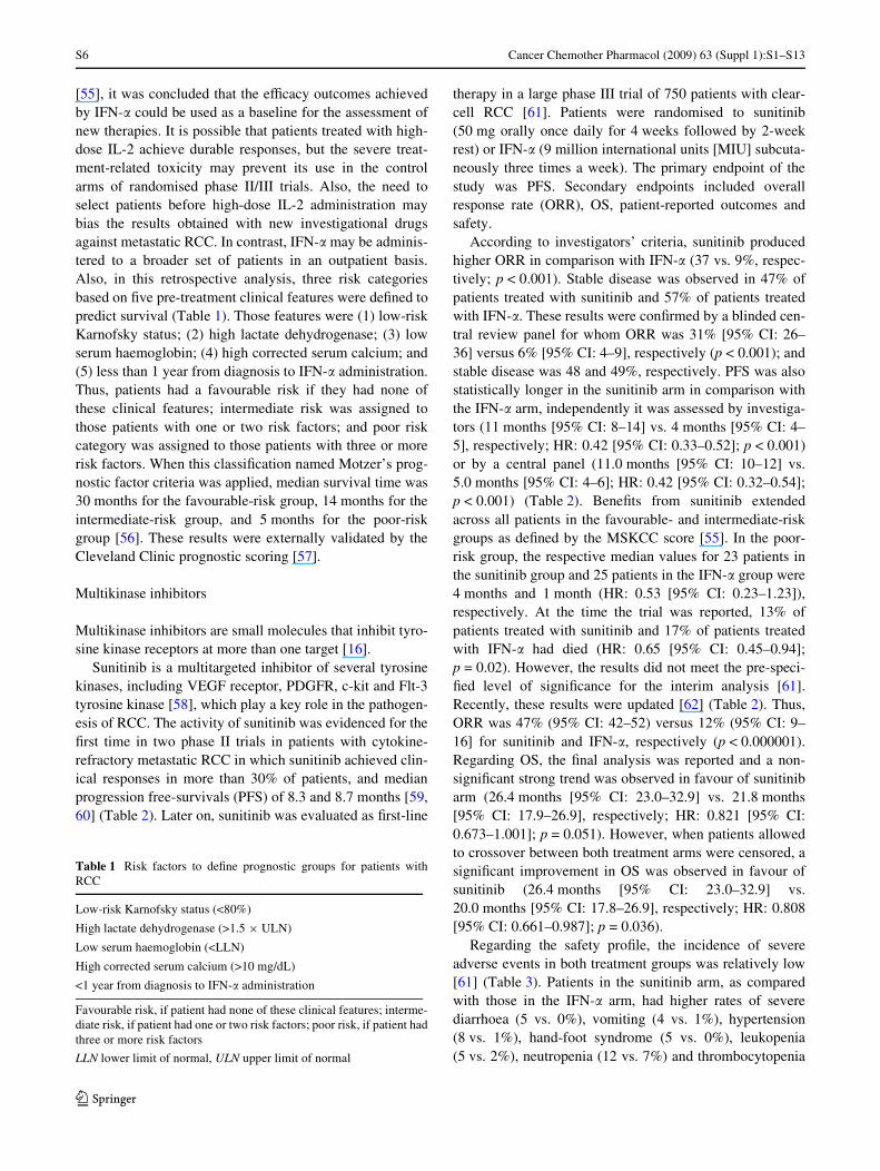
[55], it was concluded that the eYcacy outcomes achievedby IFN-� could be used as a baseline for the assessment ofnew therapies. It is possible that patients treated with high-dose IL-2 achieve durable responses, but the severe treat-ment-related toxicity may prevent its use in the controlarms of randomised phase II/III trials. Also, the need toselect patients before high-dose IL-2 administration maybias the results obtained with new investigational drugsagainst metastatic RCC. In contrast, IFN-� may be adminis-tered to a broader set of patients in an outpatient basis.Also, in this retrospective analysis, three risk categoriesbased on Wve pre-treatment clinical features were deWned topredict survival (Table 1). Those features were (1) low-riskKarnofsky status; (2) high lactate dehydrogenase; (3) lowserum haemoglobin; (4) high corrected serum calcium; and(5) less than 1 year from diagnosis to IFN-� administration.Thus, patients had a favourable risk if they had none ofthese clinical features; intermediate risk was assigned tothose patients with one or two risk factors; and poor riskcategory was assigned to those patients with three or morerisk factors. When this classiWcation named Motzer’s prog-nostic factor criteria was applied, median survival time was30 months for the favourable-risk group, 14 months for theintermediate-risk group, and 5 months for the poor-riskgroup [56]. These results were externally validated by theCleveland Clinic prognostic scoring [57].
Multikinase inhibitors
Multikinase inhibitors are small molecules that inhibit tyro-sine kinase receptors at more than one target [16].
Sunitinib is a multitargeted inhibitor of several tyrosinekinases, including VEGF receptor, PDGFR, c-kit and Flt-3tyrosine kinase [58], which play a key role in the pathogen-esis of RCC. The activity of sunitinib was evidenced for theWrst time in two phase II trials in patients with cytokine-refractory metastatic RCC in which sunitinib achieved clin-ical responses in more than 30% of patients, and medianprogression free-survivals (PFS) of 8.3 and 8.7 months [59,60] (Table 2). Later on, sunitinib was evaluated as Wrst-line
therapy in a large phase III trial of 750 patients with clear-cell RCC [61]. Patients were randomised to sunitinib(50 mg orally once daily for 4 weeks followed by 2-weekrest) or IFN-� (9 million international units [MIU] subcuta-neously three times a week). The primary endpoint of thestudy was PFS. Secondary endpoints included overallresponse rate (ORR), OS, patient-reported outcomes andsafety.
According to investigators’ criteria, sunitinib producedhigher ORR in comparison with IFN-� (37 vs. 9%, respec-tively; p < 0.001). Stable disease was observed in 47% ofpatients treated with sunitinib and 57% of patients treatedwith IFN-�. These results were conWrmed by a blinded cen-tral review panel for whom ORR was 31% [95% CI: 26–36] versus 6% [95% CI: 4–9], respectively (p < 0.001); andstable disease was 48 and 49%, respectively. PFS was alsostatistically longer in the sunitinib arm in comparison withthe IFN-� arm, independently it was assessed by investiga-tors (11 months [95% CI: 8–14] vs. 4 months [95% CI: 4–5], respectively; HR: 0.42 [95% CI: 0.33–0.52]; p < 0.001)or by a central panel (11.0 months [95% CI: 10–12] vs.5.0 months [95% CI: 4–6]; HR: 0.42 [95% CI: 0.32–0.54];p < 0.001) (Table 2). BeneWts from sunitinib extendedacross all patients in the favourable- and intermediate-riskgroups as deWned by the MSKCC score [55]. In the poor-risk group, the respective median values for 23 patients inthe sunitinib group and 25 patients in the IFN-� group were4 months and 1 month (HR: 0.53 [95% CI: 0.23–1.23]),respectively. At the time the trial was reported, 13% ofpatients treated with sunitinib and 17% of patients treatedwith IFN-� had died (HR: 0.65 [95% CI: 0.45–0.94];p = 0.02). However, the results did not meet the pre-speci-Wed level of signiWcance for the interim analysis [61].Recently, these results were updated [62] (Table 2). Thus,ORR was 47% (95% CI: 42–52) versus 12% (95% CI: 9–16] for sunitinib and IFN-�, respectively (p < 0.000001).Regarding OS, the Wnal analysis was reported and a non-signiWcant strong trend was observed in favour of sunitinibarm (26.4 months [95% CI: 23.0–32.9] vs. 21.8 months[95% CI: 17.9–26.9], respectively; HR: 0.821 [95% CI:0.673–1.001]; p = 0.051). However, when patients allowedto crossover between both treatment arms were censored, asigniWcant improvement in OS was observed in favour ofsunitinib (26.4 months [95% CI: 23.0–32.9] vs.20.0 months [95% CI: 17.8–26.9], respectively; HR: 0.808[95% CI: 0.661–0.987]; p = 0.036).
Regarding the safety proWle, the incidence of severeadverse events in both treatment groups was relatively low[61] (Table 3). Patients in the sunitinib arm, as comparedwith those in the IFN-� arm, had higher rates of severediarrhoea (5 vs. 0%), vomiting (4 vs. 1%), hypertension(8 vs. 1%), hand-foot syndrome (5 vs. 0%), leukopenia(5 vs. 2%), neutropenia (12 vs. 7%) and thrombocytopenia
Table 1 Risk factors to deWne prognostic groups for patients withRCC
Favourable risk, if patient had none of these clinical features; interme-diate risk, if patient had one or two risk factors; poor risk, if patient hadthree or more risk factors
LLN lower limit of normal, ULN upper limit of normal
Low-risk Karnofsky status (<80%)
High lactate dehydrogenase (>1.5 £ ULN)
Low serum haemoglobin (<LLN)
High corrected serum calcium (>10 mg/dL)
<1 year from diagnosis to IFN-� administration
123
Cancer Chemother Pharmacol (2009) 63 (Suppl 1):S1–S13 S7
(8 vs. 0%), with a p value of less than 0.05 for all symptoms(Table 3). In contrast, severe fatigue (7 vs. 12%) and lym-phopenia (12 vs. 22%) occurred with greater frequency inpatients treated with IFN-� (p < 0.05). The incidence of agrade 3 decrease in the left ventricular ejection fraction was2% in the sunitinib and 1% in the IFN-� arm. In the suniti-nib group, this decrease did not have any clinical sequelaeand was reversible after dose modiWcation or treatment dis-continuation. As a consequence, 38% of patients with suni-tinib and 32% of patients with IFN-� had a doseinterruption due to an adverse event, and 32 and 21%,respectively, had a dose reduction. Sunitinib may bereduced from 50 mg once daily to a minimum of 25 mg/dayat intervals of 12.5 mg each.
Sorafenib was originally developed as an inhibitor ofRaf-1, a member of the Raf/MEK/ERK signalling pathway
[63]. Soon after, it also proved to be active against B-Raf,VEGFR-2 and PDGFR [63]. Its activity was evaluatedthroughout several phase II trials as well as through theTreatment Approaches in Renal Cancer Global EvaluationTrial (TARGET) [64–67] (Table 2). In this randomised,double-blind, phase III trial, 903 patients with previouslycytokine-treated metastatic clear-cell RCC of low- or inter-mediate risk were randomised to sorafenib (400 mg orallytwice a day) or placebo [65, 66]. The primary endpoint ofthe study was OS and secondary endpoints were PFS, ORRand safety.
Treatment with sorafenib prolonged PFS in comparisonwith placebo (5.5 vs. 2.8 months, HR: 0.44; 95% CI: 0.35–0.55; p < 0.01). The diVerence of survival in favour ofsorafenib was not statistically signiWcant in the intention totreat analysis but was positive when censoring placebo
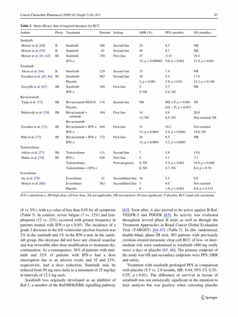
Table 2 Main eYcacy data of targeted therapies for RCC
IFN-� interferon-�, HD high dose, LD low dose, NA not applicable, NR not reported, NS non-signiWcant, P placebo, RCC renal cell carcinoma
Author Phase Treatment Patients Setting ORR (%) PFS (months) OS (months)
Sunitinib
Motzer et al. [60] II Sunitinib 106 Second line 34 8.3 NR
Motzer et al. [59] II Sunitinib 63 Second line 40 8.7 NR
Motzer et al. [61, 62] III Sunitinib 750 First line 47 11.0 26.4
IFN-� 12; p < 0.000001 5.0; p < 0.001 21.8; p = 0.051
Sorafenib
Akaza et al. [64] II Sorafenib 129 Second line 15 7.4 NR
Escudier et al. [65, 66] III Sorafenib 903 Second line 10 5.5 17.8
Placebo 2; p < 0.001 2.8; p < 0.01 15.2; p = 0.146
Szczylik et al. [67] IIb Sorafenib 189 First line 5 5.7 NR
IFN-� 9; NS 5.6; NS
Bevacizumab
Yang et al. [73] IIb Bevacizumab HD/LD 116 Second line NR HD > P; p < 0.001 NS
Placebo LD » P; p = 0.053
Bukowski et al. [70] IIb Bevacizumab +erlotinib
104 First line 14 9.9 20.0
13; NS 8.5; NS Not reached; NSBevacizumab
Escudier et al. [71] III Bevacizumab + IFN-� 649 First line 31 10.2 Not reached
IFN-� 13; p = 0.0001 5.4; p = 0.0001 19.8; NS
Rini et al. [72] III Bevacizumab + IFN-� 732 First line 26 8.5 NR
IFN-� 13; p < 0.0001 5.2; p < 0.0001
Temsirolimus
Atkins et al. [77] IIb Temsirolimus 111 Second line 7 5.8 15.0
Hudes et al. [78] III IFN-� 626 First line 5 3.1 7.3
Temsirolimus Poor prognosis 9; NS 5.5; p < 0.001 10.9; p = 0.008
Temsirolimus + IFN-� 8; NS 4.7; NS 8.4; p = 0.70
Everolimus
Jac et al. [79] Everolimus 22 Second/third line 16 5.5 8.0
Motzer et al. [80] Everolimus 362 Second/third line 3 4.0 Not reached
Placebo 0 1.9; p < 0.001 8.8; p = 0.233
123
S8 Cancer Chemother Pharmacol (2009) 63 (Suppl 1):S1–S13
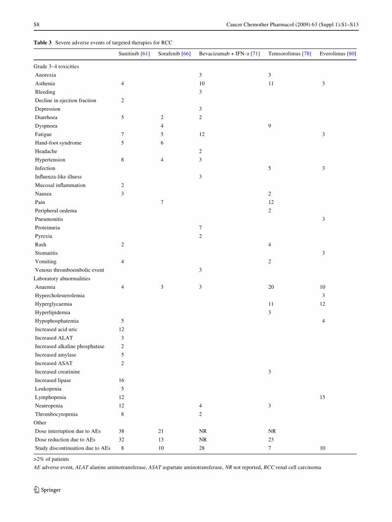
Table 3 Severe adverse events of targeted therapies for RCC
>2% of patients
AE adverse event, ALAT alanine aminotransferase, ASAT aspartate aminotransferase, NR not reported, RCC renal cell carcinoma
Sunitinib [61] Sorafenib [66] Bevacizumab + IFN-� [71] Temsorolimus [78] Everolimus [80]
Grade 3–4 toxicities
Anorexia 3 3
Asthenia 4 10 11 3
Bleeding 3
Decline in ejection fraction 2
Depression 3
Diarrhoea 5 2 2
Dyspnoea 4 9
Fatigue 7 5 12 3
Hand-foot syndrome 5 6
Headache 2
Hypertension 8 4 3
Infection 5 3
InXuenza-like illness 3
Mucosal inXammation 2
Nausea 3 2
Pain 7 12
Peripheral oedema 2
Pneumonitis 3
Proteinuria 7
Pyrexia 2
Rash 2 4
Stomatitis 3
Vomiting 4 2
Venous thromboembolic event 3
Laboratory abnormalities
Anaemia 4 3 3 20 10
Hypercholesterolemia 3
Hyperglycaemia 11 12
Hyperlipidemia 3
Hypophosphatemia 5 4
Increased acid uric 12
Increased ALAT 3
Increased alkaline phosphatase 2
Increased amylase 5
Increased ASAT 2
Increased creatinine 3
Increased lipase 16
Leukopenia 5
Lymphopenia 12 15
Neutropenia 12 4 3
Thrombocytopenia 8 2
Other
Dose interruption due to AEs 38 21 NR NR
Dose reduction due to AEs 32 13 NR 23
Study discontinuation due to AEs 8 10 28 7 10
123
Cancer Chemother Pharmacol (2009) 63 (Suppl 1):S1–S13 S9
patients that crossed over to receive sorafenib. Partialresponses were reported in 10% of patients treated withsorafenib and 2% of patients treated with placebo(p < 0.001).
The most common severe adverse events associated withsorafenib were hand-foot skin reactions (6% of patients),fatigue (5%), dyspnoea (4%), hypertension (4%) and anae-mia (3%) (Table 3). Cardiac ischaemia or infarction (3% ofpatients) was rare. Lastly, serious adverse events leading topatient hospitalisation were reported in 34% of patientsreceiving sorafenib and 24% of patients receiving placebo(p < 0.01) [66]. As a consequence, 13% of patients treatedwith sorafenib reduced the dosage compared with 3% ofpatients in the placebo arm (p < 0.001). Additionally, 21%of patients in the sorafenib arm interrupted doses opposedto 6% in the placebo arm (p < 0.001), with a median dura-tion of dose interruption of 7 and 6 days, respectively.Sorafenib may be reduced from 400 to 200 mg twice a day,or even to 200 mg once a day. Also, it is possible to alter-nate sorafenib’s dosage between 400 and 200 mg/day.
Sorafenib was also compared with IFN-� in a random-ised phase II trial of untreated RCC patients. In this trial,sorafenib did not show any advantage over IFN-� in termsof PFS (5.7 vs. 5.6 months, respectively; p = 0.504) [67].
Anti-VEGF antibodies
Anti-VEGF antibodies are used in the treatment of RCCbecause it has been demonstrated that more than 50% ofthese tumours are up-regulated by HIF-1�, HIF-2� and HIFtarget genes such as VEGF [68].
Bevacizumab is a humanised recombinant anti-VEGFantibody that binds VEGF-A isoform and neutralises itsactivity [69]. Bevacizumab in combination with IFN-� hasdemonstrated its activity in metastatic RCC in several ran-domised trials [70–73] (Table 2). In a double-blind phaseIII trial of 649 patients with previously untreated metastaticRCC, 10 mg/kg of intravenous bevacizumab every2 weeks, plus 9 MIU of IFN-� three times per week by sub-cutaneous injection, were compared with placebo plus thesame dose of IFN-� [71]. The primary endpoint of the trialwas OS. Secondary endpoints included PFS, ORR andsafety. According to the investigator’s criteria, the ORRwas signiWcantly higher with bevacizumab plus IFN-� thanwith placebo plus IFN-� (31 vs. 13%, p = 0.0001). Also, alonger PFS was observed in favour of the bevacizumab plusIFN-� arm (10.2 vs. 5.4 months, HR: 0.63; 95% CI: 0.52–0.75; p = 0.0001), regardless of the patient’s risk group(stratiWed according to MSKCC criteria) [71]. However, atthe time of data cut-oV, median OS was not reached. Seri-ous adverse events were reported in 29% of patients treatedwith bevacizumab plus IFN-� and in 16% of patientstreated with placebo plus IFN-�. Twenty-eight percent of
patients treated with bevacizumab plus IFN-� interruptedthe study drug due to an adverse event compared with 12%in the control arm (Table 3). The most frequent severeadverse events observed in patients treated with bev-acizumab plus IFN-� were fatigue (12% of patients), asthe-nia (10%), proteinuria (7%) and neutropenia (4%) [71].Importantly, thromboembolic events (3%) and severe gas-trointestinal perforations (2%) were also described. In a ret-rospective subgroup analysis, it was observed that the doseof IFN-� may be reduced to manage side eVects whilstmaintaining eYcacy of the combination [74].
In another multicenter phase III trial, previouslyuntreated metastatic RCC patients were randomised to bev-acizumab (10 mg/kg every 2 weeks) plus IFN-� (9 MIUthree times per week) or IFN-� monotherapy [72](Table 2). The median PFS was 8.5 months (95% CI: 7.5–9.7) and 5.2 months (95% CI: 3.1–5.6) in the study and thecontrol arms, respectively (p < 0.0001). Bevacizumab plusIFN-� also had a higher ORR (26% [95% CI: 21–31] vs.13% [95% CI: 10–17]; p < 0.0001, respectively). On theother hand, severe toxicity was greater in the bevacizumabarm, including severe hypertension (9 vs. 0%), anorexia (17vs. 8%), fatigue (35 vs. 28%) and proteinuria (13 vs. 0%)[72].
mTOR inhibitors
mTOR inhibitors are directed towards the inhibition of themammalian target of rapamycin (mTOR) kinase, a compo-nent of the intracellular signalling pathways involved incellular growth, proliferation and hypoxic stress response[75].
SpeciWcally, temsirolimus binds to the intracellular pro-tein FKBP-12, forming a complex that inhibits mTOR sig-nalling. This inhibition aVects cell cycle regulation andangiogenesis [76]. Temsirolimus has shown its activity inmetastatic RCC in previous phase II and III trials [77, 78](Table 2). In a large, multicentre, phase III trial, 626 previ-ously untreated RCC patients with poor prognosis wereincluded [78]. Poor prognosis was deWned slightly diVerentthan previously [55]. Thus, poor-risk patients were thosewith at least three of the following six predictors of shortsurvival: (1) a serum lactate dehydrogenase level>1.5 £ upper normal limit (UNL); (2) a haemoglobin levelbelow the lower normal limit (LNL); (3) a corrected serumcalcium level >2.5 mmol/L; (4) a time from initial diagno-sis to randomisation <1 year; (5) a Karnofsky performancescore <60–70; and (6) a new criteria based on the existenceof metastases in multiple organs. Patients received treat-ment with IFN-� (escalated from 3 to 18 MIU three timesper week if tolerated), temsirolimus (25 mg as a weeklyintravenous infusion), or a combination of both (15 mg oftemsirolimus and 3–6 MIU). The primary endpoint of the
123
S10 Cancer Chemother Pharmacol (2009) 63 (Suppl 1):S1–S13
study was OS. Secondary endpoints included PFS, ORR,clinical beneWt rate and safety. Temsirolimus as singleagent was superior to IFN-� in terms of OS (10.9 vs.7.3 months, p = 0.008), which was the main endpoint of thestudy, and PFS (5.5 vs. 3.1 months, p < 0.001). In contrast,administration of both temsirolimus and IFN-� did notimprove the results obtained with IFN-� alone [78], proba-bly due to dose reduction applied to the combination(Table 2).
Additionally, temsirolimus as single agent had a goodsafety proWle (Table 3). Thus, the highest incidence ofsevere adverse events was observed in the combination arm(87% of patients) followed by the IFN-� arm (78%) and thetemsirolimus arm (67%; p = 0.02) [78]. Asthenia was themost frequent adverse event in those patients that receivedIFN-� alone or in combination (Table 3). Severe astheniawas reported in 11% of patients treated with temsirolimus,26% of patients treated with IFN-� and in 28% of patientstreated with the combination of both. Dyspnoea, diarrhoea,nausea, or vomiting were reported similarly in the threetreatment arms. In contrast, the incidence of rash, periphe-ral oedema, stomatitis, anaemia, neutropenia and thrombo-cytopenia at any grade was higher in patients who receivedtemsirolimus, either alone or in combination, than inpatients treated with IFN-� alone (p < 0.05) [78]. Impor-tantly, hyperglycaemia, hypercholesterolemia and hyperlip-idemia were more common in patients treated withtemsirolimus, reXecting the eVect of this drug on glucoseand lipid metabolisms (Table 3). As a consequence, 23% ofpatients treated with temsirolimus as single agent had toreduce the treatment dosage. In the case of severe neutrope-nia or thrombocytopenia, temsirolimus was interrupted andrestarted with a 5 mg reduction from the recommended dos-age of 25 mg a week.
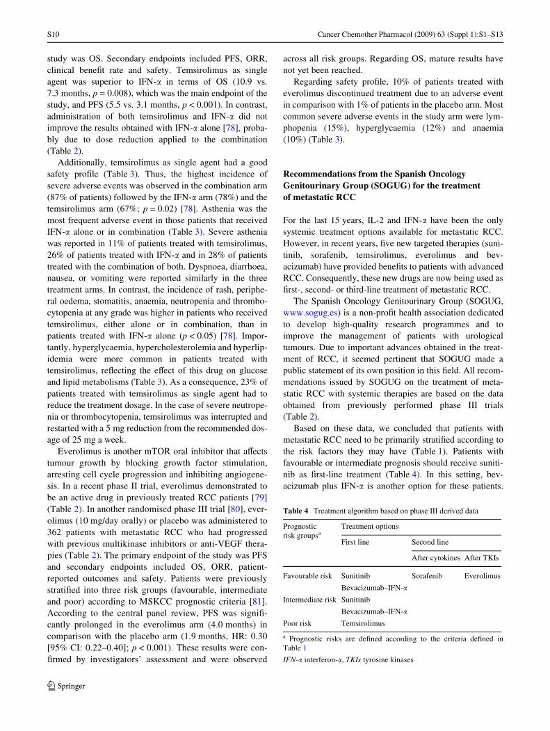
Everolimus is another mTOR oral inhibitor that aVectstumour growth by blocking growth factor stimulation,arresting cell cycle progression and inhibiting angiogene-sis. In a recent phase II trial, everolimus demonstrated tobe an active drug in previously treated RCC patients [79](Table 2). In another randomised phase III trial [80], ever-olimus (10 mg/day orally) or placebo was administered to362 patients with metastatic RCC who had progressedwith previous multikinase inhibitors or anti-VEGF thera-pies (Table 2). The primary endpoint of the study was PFSand secondary endpoints included OS, ORR, patient-reported outcomes and safety. Patients were previouslystratiWed into three risk groups (favourable, intermediateand poor) according to MSKCC prognostic criteria [81].According to the central panel review, PFS was signiW-cantly prolonged in the everolimus arm (4.0 months) incomparison with the placebo arm (1.9 months, HR: 0.30[95% CI: 0.22–0.40]; p < 0.001). These results were con-Wrmed by investigators’ assessment and were observed
across all risk groups. Regarding OS, mature results havenot yet been reached.
Regarding safety proWle, 10% of patients treated witheverolimus discontinued treatment due to an adverse eventin comparison with 1% of patients in the placebo arm. Mostcommon severe adverse events in the study arm were lym-phopenia (15%), hyperglycaemia (12%) and anaemia(10%) (Table 3).
Recommendations from the Spanish Oncology Genitourinary Group (SOGUG) for the treatment of metastatic RCC
For the last 15 years, IL-2 and IFN-� have been the onlysystemic treatment options available for metastatic RCC.However, in recent years, Wve new targeted therapies (suni-tinib, sorafenib, temsirolimus, everolimus and bev-acizumab) have provided beneWts to patients with advancedRCC. Consequently, these new drugs are now being used asWrst-, second- or third-line treatment of metastatic RCC.
The Spanish Oncology Genitourinary Group (SOGUG,www.sogug.es) is a non-proWt health association dedicatedto develop high-quality research programmes and toimprove the management of patients with urologicaltumours. Due to important advances obtained in the treat-ment of RCC, it seemed pertinent that SOGUG made apublic statement of its own position in this Weld. All recom-mendations issued by SOGUG on the treatment of meta-static RCC with systemic therapies are based on the dataobtained from previously performed phase III trials(Table 2).
Based on these data, we concluded that patients withmetastatic RCC need to be primarily stratiWed according tothe risk factors they may have (Table 1). Patients withfavourable or intermediate prognosis should receive suniti-nib as Wrst-line treatment (Table 4). In this setting, bev-acizumab plus IFN-� is another option for these patients.
Table 4 Treatment algorithm based on phase III derived data
a Prognostic risks are deWned according to the criteria deWned inTable 1
IFN-� interferon-�, TKIs tyrosine kinases
Prognostic risk groupsa
Treatment options
First line Second line
After cytokines After TKIs
Favourable risk Sunitinib Sorafenib Everolimus
Bevacizumab–IFN-�
Intermediate risk Sunitinib
Bevacizumab–IFN-�
Poor risk Temsirolimus
123
Cancer Chemother Pharmacol (2009) 63 (Suppl 1):S1–S13 S11
According to our criteria, patients with poor prognosisshould receive temsirolimus as Wrst-line treatment. Afterclinical progression, all patients previously treated withcytokines should be treated with sorafenib as second-linetreatment. In contrast, all patients previously treated withtyrosine kinases should receive everolimus.
Acknowledgments The authors acknowledge the support of PWzerSpain, which has facilitated the necessary meetings to evaluate and dis-cuss all the data presented in this review, and Dr. Beatriz Gil-Alberdifrom HealthCo SL (Madrid, Spain) for her assistance in the preparationof this manuscript.
ConXict of interest statement M. Climent is a consultant and advisorfor PWzer and Novartis. All other authors have no disclosures to be made.
References
1. Jemal A, Siegel R, Ward E et al (2008) Cancer statistics, 2008. CACancer J Clin 58:71–96
2. Dhote R, Thiounn N, Debre B et al (2004) Risk factors for adultrenal cell carcinoma. Urol Clin N Am 31:237–247
3. Vineis P, Alavanja M, BuZer P et al (2004) Tobacco and cancer:recent epidemiological evidence. J Natl Cancer Inst 96:99–106
4. Chow WH, Gridley G, Fraumeni JF Jr et al (2000) Obesity, hyper-tension, and the risk of kidney cancer in men. N Engl J Med343:1305–1311
5. Dhote R, Pellicer-Coeuret M, Thiounn N et al (2000) Risk factorsfor adult renal cell carcinoma: a systematic review and implica-tions for prevention. BJU Int 86:20–27
6. WagstaV J (2006) New horizons in the treatment of renal cellcancer. Ann Oncol 17(Suppl 10):x19–x22
7. Atkins MB, ErnstoV MS, Figlin RA et al (2007) Innovations andchallenges in renal cell carcinoma: summary statement from theSecond Cambridge Conference. Clin Cancer Res 13:667s–670s
8. Banks RE, Tirukonda P, Taylor C et al (2006) Genetic and epige-netic analysis of von Hippel–Lindau (VHL) gene alterations andrelationship with clinical variables in sporadic renal cancer.Cancer Res 66:2000–2011
9. Schmidt L, Duh FM, Chen F et al (1997) Germline and somaticmutations in the tyrosine kinase domain of the MET proto-onco-gene in papillary renal carcinomas. Nat Genet 16:68–73
10. Schmidt LS, Nickerson ML, Warren MB et al (2005) GermlineBHD-mutation spectrum and phenotype analysis of a large cohortof families with Birt-Hogg-Dube syndrome. Am J Hum Genet76:1023–1033
11. Motzer RJ, Bolger GB, Boston B et al (2006) Kidney cancer.Clinical practice guidelines in oncology. J Natl Compr CancerNetw 4:1072–1081
12. Greene FL, Page DL, Fleming ID et al (2002) AJCC cancer stag-ing handbook: TNM classiWcation of malignant tumors. Springer,New York
13. Drucker BJ (2005) Renal cell carcinoma: current status and futureprospects. Cancer Treat Rev 31:536–545
14. Guinan PD, Vogelzang NJ, Fremgen AM et al (1995) Renal cellcarcinoma: tumor size, stage and survival. Members of the CancerIncidence and End Results Committee. J Urol 153:901–903
15. Novick AC (2004) Laparoscopic and partial nephrectomy. ClinCancer Res 10:6322S–6327S
16. Costa LJ, Drabkin HA (2007) Renal cell carcinoma: new develop-ments in molecular biology and potential for targeted therapies.Oncologist 12:1404–1415
17. Flanigan RC, Salmon SE, Blumenstein BA et al (2001) Nephrec-tomy followed by interferon alfa-2b compared with interferonalfa-2b alone for metastatic renal-cell cancer. N Engl J Med345:1655–1659
18. Mickisch GH, Garin A, van Poppel H et al (2001) Radicalnephrectomy plus interferon-alfa-based immunotherapy comparedwith interferon alfa alone in metastatic renal-cell carcinoma: arandomised trial. Lancet 358:966–970
19. Garcia JA, Rini BI (2007) Recent progress in the management ofadvanced renal cell carcinoma. CA Cancer J Clin 57:112–125
20. Kavolius JP, Mastorakos DP, Pavlovich C et al (1998) Resectionof metastatic renal cell carcinoma. J Clin Oncol 16:2261–2266
21. Russo P (2004) Surgical intervention in patients with metastaticrenal cancer: current status of metastasectomy and cytoreductivenephrectomy. Nat Clin Pract Urol 1:26–30
22. Rini BI, Shaw V, Rosenberg JE et al (2006) Patients withmetastatic renal cell carcinoma with long-term disease-freesurvival after treatment with sunitinib and resection of residualmetastases. Clin Genitourin Cancer 5:232–234
23. Finney R (1973) The value of radiotherapy in the treatment ofhypernephroma—a clinical trial. Br J Urol 45:258–269
24. Juusela H, Malmio K, Alfthan O et al (1977) Preoperative irradia-tion in the treatment of renal adenocarcinoma. Scand J Urol Neph-rol 11:277–281
25. Kjaer M, Frederiksen PL, Engelholm SA (1987) Postoperativeradiotherapy in stage II and III renal adenocarcinoma. A random-ized trial by the Copenhagen Renal Cancer Study Group. Int JRadiat Oncol Biol Phys 13:665–672
26. Kjaer M, Iversen P, Hvidt V et al (1987) A randomized trial ofpostoperative radiotherapy versus observation in stage II and IIIrenal adenocarcinoma. A study by the Copenhagen Renal CancerStudy Group. Scand J Urol Nephrol 21:285–289
27. Wersall PJ, Blomgren H, Lax I et al (2005) Extracranial stereotac-tic radiotherapy for primary and metastatic renal cell carcinoma.Radiother Oncol 77:88–95
28. Reichel LM, Pohar S, Heiner J et al (2007) Radiotherapy to bonehas utility in multifocal metastatic renal carcinoma. Clin OrthopRelat Res 459:133–138
29. Doh LS, Amato RJ, Paulino AC et al (2006) Radiation therapy inthe management of brain metastases from renal cell carcinoma.Oncology (Williston Park) 20:603–613 discussion 613, 616, 619–620 passsim
30. Svedman C, Sandstrom P, Pisa P et al (2006) A prospective phaseII trial of using extracranial stereotactic radiotherapy in primaryand metastatic renal cell carcinoma. Acta Oncol 45:870–875
31. Senan S, Smit EF (2007) Design of clinical trials of radiation com-bined with antiangiogenic therapy. Oncologist 12:465–477
32. Amato RJ (2000) Chemotherapy for renal cell carcinoma. SeminOncol 27:177–186
33. Clark JI, Atkins MB, Urba WJ et al (2003) Adjuvant high-dosebolus interleukin-2 for patients with high-risk renal cell carci-noma: a cytokine working group randomized trial. J Clin Oncol21:3133–3140
34. Messing EM, Manola J, Wilding G et al (2003) Phase III study ofinterferon alfa-NL as adjuvant treatment for resectable renal cellcarcinoma: an Eastern Cooperative Oncology Group/Intergrouptrial. J Clin Oncol 21:1214–1222
35. Pizzocaro G, Piva L, Costa A, Silvestrini R (1997) Adjuvantinterferon (IFN) to radical nephrectomy in Robson’s stages II andIII renal cell cancer (RCC), a multicenter randomized study withsome biological evaluations, 1997 ASCO annual meeting, abstract1132
36. Jocham D, Richter A, HoVmann L et al (2004) Adjuvant autolo-gous renal tumour cell vaccine and risk of tumour progression inpatients with renal-cell carcinoma after radical nephrectomy:phase III, randomised controlled trial. Lancet 363:594–599
123
S12 Cancer Chemother Pharmacol (2009) 63 (Suppl 1):S1–S13
37. Jonasch E, Wood C, Tamboli P et al (2008) Vaccination ofmetastatic renal cell carcinoma patients with autologous tumour-derived vitespen vaccine: clinical Wndings. Br J Cancer 98:1336–1341
38. Wood C, Srivastava P, Bukowski R et al (2008) An adjuvant autol-ogous therapeutic vaccine (HSPPC-96; vitespen) versus observa-tion alone for patients at high risk of recurrence after nephrectomyfor renal cell carcinoma: a multicentre, open-label, randomisedphase III trial. Lancet 372:145–154
39. Skolarikos A, Alivizatos G, Laguna P et al (2007) A review onfollow-up strategies for renal cell carcinoma after nephrectomy.Eur Urol 51:1490–1500 discussion 1501
40. Fuhrman SA, Lasky LC, Limas C (1982) Prognostic signiWcanceof morphologic parameters in renal cell carcinoma. Am J SurgPathol 6:655–663
41. Sandlund J, Oosterwijk E, Grankvist K et al (2007) Prognosticimpact of carbonic anhydrase IX expression in human renal cellcarcinoma. BJU Int 100:556–560
42. Yaycioglu O, Roberts WW, Chan T et al (2001) Prognostic assess-ment of nonmetastatic renal cell carcinoma: a clinically basedmodel. Urology 58:141–145
43. Kattan MW, Reuter V, Motzer RJ et al (2001) A postoperativeprognostic nomogram for renal cell carcinoma. J Urol 166:63–67
44. Frank I, Blute ML, Cheville JC et al (2002) An outcome predictionmodel for patients with clear cell renal cell carcinoma treated withradical nephrectomy based on tumor stage, size, grade and necro-sis: the SSIGN score. J Urol 168:2395–2400
45. Zisman A, Pantuck AJ, Dorey F et al (2001) Improved prognosti-cation of renal cell carcinoma using an integrated staging system.J Clin Oncol 19:1649–1657
46. Patard JJ, Kim HL, Lam JS et al (2004) Use of the University ofCalifornia Los Angeles integrated staging system to predictsurvival in renal cell carcinoma: an international multicenterstudy. J Clin Oncol 22:3316–3322
47. Cindolo L, de la Taille A, Messina G et al (2003) A preoperativeclinical prognostic model for non-metastatic renal cell carcinoma.BJU Int 92:901–905
48. Cindolo L, Patard JJ, Chiodini P et al (2005) Comparison ofpredictive accuracy of four prognostic models for nonmetastaticrenal cell carcinoma after nephrectomy: a multicenter Europeanstudy. Cancer 104:1362–1371
49. Ritchie A, GriYths G, Parmar M, for the MRC Renal CancerCollaborators (1999) Interferon-alpha and survival in metastaticrenal carcinoma: early results of a randomised controlled trial.Medical Research Council Renal Cancer Collaborators. Lancet353:14–17
50. Fyfe G, Fisher RI, Rosenberg SA et al (1995) Results of treatmentof 255 patients with metastatic renal cell carcinoma who receivedhigh-dose recombinant interleukin-2 therapy. J Clin Oncol13:688–696
51. McDermott DF, Regan MM, Clark JI et al (2005) Randomizedphase III trial of high-dose interleukin-2 versus subcutaneousinterleukin-2 and interferon in patients with metastatic renal cellcarcinoma. J Clin Oncol 23:133–141
52. Yang JC, Sherry RM, Steinberg SM et al (2003) Randomizedstudy of high-dose and low-dose interleukin-2 in patients withmetastatic renal cancer. J Clin Oncol 21:3127–3132
53. Margolin KA, Rayner AA, Hawkins MJ et al (1989) Interleukin-2and lymphokine-activated killer cell therapy of solid tumors: anal-ysis of toxicity and management guidelines. J Clin Oncol 7:486–498
54. Negrier S, Perol D, Ravaud A et al (2007) Medroxyprogesterone,interferon alfa-2a, interleukin 2, or combination of both cytokinesin patients with metastatic renal carcinoma of intermediateprognosis: results of a randomized controlled trial. Cancer110:2468–2477
55. Motzer RJ, Bacik J, Murphy BA et al (2002) Interferon-alfa as acomparative treatment for clinical trials of new therapies againstadvanced renal cell carcinoma. J Clin Oncol 20:289–296
56. Motzer RJ, Mazumdar M, Bacik J et al (1999) Survival andprognostic stratiWcation of 670 patients with advanced renal cellcarcinoma. J Clin Oncol 17:2530–2540
57. Bukowski RM, Negrier S, Elson P (2004) Prognostic factors inpatients with advanced renal cell carcinoma: development of aninternational kidney cancer working group. Clin Cancer Res10:6310S–6314S
58. Mendel DB, Laird AD, Xin X et al (2003) In vivo antitumor activ-ity of SU11248, a novel tyrosine kinase inhibitor targeting vascu-lar endothelial growth factor and platelet-derived growth factorreceptors: determination of a pharmacokinetic/pharmacodynamicrelationship. Clin Cancer Res 9:327–337
59. Motzer RJ, Michaelson MD, Redman BG et al (2006) Activity ofSU11248, a multitargeted inhibitor of vascular endothelial growthfactor receptor and platelet-derived growth factor receptor, inpatients with metastatic renal cell carcinoma. J Clin Oncol 24:16–24
60. Motzer RJ, Rini BI, Bukowski RM et al (2006) Sunitinib inpatients with metastatic renal cell carcinoma. JAMA 295:2516–2524
61. Motzer RJ, Hutson TE, Tomczak P et al (2007) Sunitinib versusinterferon alfa in metastatic renal-cell carcinoma. N Engl J Med356:115–124
62. Figlin RA, Hutson TE, Tomczak P, Michaelson MD, BukowskiRM, Négrier S, Huang X, Kim ST, Chen I, Motzer RJ (2008)Overall survival with sunitinib versus interferon (IFN)-alfa asWrst-line treatment of metastatic renal cell carcinoma (mRCC).J Clin Oncol, vol 26 (May 20 suppl), abstr 5024
63. Hilger RA, Scheulen ME, Strumberg D (2002) The Ras-Raf-MEK-ERK pathway in the treatment of cancer. Onkologie25:511–518
64. Akaza H, Tsukamoto T, Murai M et al (2007) Phase II study toinvestigate the eYcacy, safety, and pharmacokinetics of sorafenibin Japanese patients with advanced renal cell carcinoma. Jpn J ClinOncol 37:755–762
65. Bukowski RM, Eisen T, Szczylik C, Stadler WM, Simantov R,Shan M, Elting J, Pena C, Escudier B (2007) Final results of therandomized phase III trial of sorafenib in advanced renal cell car-cinoma: survival and biomarker analysis. In: 2007 ASCO annualmeeting proceedings, part I. J Clin Oncol, vol 25, no. 18S (June 20supplement), abstr 5023
66. Escudier B, Eisen T, Stadler WM et al (2007) Sorafenib in ad-vanced clear-cell renal-cell carcinoma. N Engl J Med 356:125–134
67. Szczylik C, Demkow T, Staehler M, Rolland F, Negrier S, HutsonTE, Bukowski RM, Scheuring UJ, Burk K, Escudier B (2007)Randomized phase II trial of Wrst-line treatment with sorafenibversus interferon in patients with advanced renal cell carcinoma:Wnal results. In: 2007 ASCO annual meeting proceedings, part I.J Clin Oncol, vol 25, no. 18S (June 20 supplement), abstr 5025
68. Rini BI, Small EJ (2005) Biology and clinical development of vas-cular endothelial growth factor-targeted therapy in renal cell carci-noma. J Clin Oncol 23:1028–1043
69. Presta LG, Chen H, O’Connor SJ et al (1997) Humanization of ananti-vascular endothelial growth factor monoclonal antibody forthe therapy of solid tumors and other disorders. Cancer Res57:4593–4599
70. Bukowski RM, Kabbinavar FF, Figlin RA et al (2007) Random-ized phase II study of erlotinib combined with bevacizumabcompared with bevacizumab alone in metastatic renal cell cancer.J Clin Oncol 25:4536–4541
71. Escudier B, Pluzanska A, Koralewski P et al (2007) Bevacizumabplus interferon alfa-2a for treatment of metastatic renal cell
123
Cancer Chemother Pharmacol (2009) 63 (Suppl 1):S1–S13 S13
carcinoma: a randomised, double-blind phase III trial. Lancet370:2103–2111
72. Rini BI, Halabi S, Rosenberg JE, Stadler WM, Vaena D, Ou S,Taylor J, Tanguay S, Dutcher J, Small EJ (2008) CALGB 90206:a phase III trial of bevacizumab plus interferon-alpha versusinterferon-alpha monotherapy in metastatic renal cell carcinoma,2008 genitourinary cancers symposium, abstract no. 350
73. Yang JC, Haworth L, Sherry RM et al (2003) A randomized trialof bevacizumab, an anti-vascular endothelial growth factorantibody, for metastatic renal cancer. N Engl J Med 349:427–434
74. Melichar B, Koralewski P, Ravaud A et al (2008) First-linebevacizumab combined with reduced dose interferon-alpha2a isactive in patients with metastatic renal cell carcinoma. Ann Oncol19:1470–1476
75. Hudson CC, Liu M, Chiang GG et al (2002) Regulation ofhypoxia-inducible factor 1alpha expression and function by themammalian target of rapamycin. Mol Cell Biol 22:7004–7014
76. Del Bufalo D, CiuVreda L, Trisciuoglio D et al (2006) Antiangio-genic potential of the Mammalian target of rapamycin inhibitortemsirolimus. Cancer Res 66:5549–5554
77. Atkins MB, Hidalgo M, Stadler WM et al (2004) Randomizedphase II study of multiple dose levels of CCI-779, a novel
mammalian target of rapamycin kinase inhibitor, in patients withadvanced refractory renal cell carcinoma. J Clin Oncol 22:909–918
78. Hudes G, Carducci M, Tomczak P et al (2007) Temsirolimus,interferon alfa, or both for advanced renal-cell carcinoma. N EnglJ Med 356:2271–2281
79. Jac J, Amato RJ, Giessinger S, Saxena S, Willis JP (2008) A phaseII study with a daily regimen of the oral mTOR inhibitor RAD001(everolimus) in patients with metastatic renal cell carcinomawhich has progressed on tyrosine kinase inhibition therapy. J ClinOncol 26(May 20 suppl):abstr 5113
80. Motzer RJ, Escudier B, Oudard S, Porta C, Hutson TE, BracardaS, Hollaender N, Urbanowitz G, Kay A, Ravaud A (2008)RAD001 vs placebo in patients with metastatic renal cell carci-noma (RCC) after progression on VEGFr-TKI therapy: resultsfrom a randomized, double-blind, multicenter phase-III study.J Clin Oncol 26(May 20 suppl):abstr LBA5026
81. Motzer RJ, Bacik J, Schwartz LH et al (2004) Prognostic factorsfor survival in previously treated patients with metastatic renal cellcarcinoma. J Clin Oncol 22:454–463
123