molecular imaging of neuropsychiatry and neuro-oncology
TRANSCRIPT
Molecular Imaging of Neuropsychiatry and Neuro-oncology
Ya-Fang Chang1, Chun-Kai Fang1,2, Hui-Yen Chuang1, Jeng-Jong Hwang1
1Department of Biomedical Imaging and Radiological Sciences, National Yang-
Ming University, Taipei, Taiwan2Department of Psychiatry, Mackay Memorial Hospital, Taipei, Taiwan
Running title: Molecular imaging of brain diseases
Corresponding author:
Jeng-Jong Hwang, Ph.D., Professor, Department of Biomedical Imaging and
Radiological Sciences, National Yang-Ming University
No. 155, Sec. 2, Li-Nong St, Bei-tou, Taipei 112, TAIWAN
Tel: +886-2-28267064; Fax: +886-2-28201095; Email: [email protected]
1
Abstract
Both neuropsychiatric disorders and malignancies of central nervous system
(CNS) represent a significant health burden and life-threatening diseases
worldwide. Radiotracer-based neuroimaging is an attractive tool that permits
the in vivo detection and characterization of metabolic and molecular processes
which are fundamental elements for brain function, and improve the
theranostics of brain diseases and disorders. In this review, we outlined the
newly development of molecular imaging probes for dopamine and serotonin
systems in neuropsychiatry and boron neutron capture therapy (BNCT) for brain
tumors in neuro-oncology with positron emission tomography (PET) and single-
photon emission computed tomography (SPECT).
Keywords: Boron neutron capture therapy, Dopamine, Molecular imaging, Positron
emission tomography, Serotonin, Single-photon emission computed tomography
2
Molecular imaging is definite as the in vivo characterization and
measurement of biologic processes at the cellular and molecular levels [1].
The progress of both neuropsychiatry and neuro-oncology are tremendous in the
past decade. This review will introduce the innovation and application of
molecular imaging in the research and clinical practice for brain diseases and
disorders.
1. Molecular imaging through dopamine system for neuropsychiatry
Dopamine, one of the catecholamine neurotransmitters in the CNS, is
synthesized from amino acid tyrosine by the catalysis of tyrosine hydroxylase
(TH) to L-DOPA, and subsequent decarboxylation by aromatic amino acid
decarboxylase (AADC) to dopamine [2]. Dopaminergic neurons release dopamine
into the synapse, where it signals to post-synaptic neurons through receptors.
Dopamine is then uptake back into the pre-synaptic neuron through the dopamine
transporter (DAT) to regulate its concentration in the extracellular space.
Dopamine system plays an essential role in the states of consciousness,
affective function, and movement. Its disturbance has been implicated in many
neurological and neuropsychiatric disorders, such as schizophrenia,
Parkinson’s disease, mood disorder, and substance dependence [3-6].
1.1 Imaging of dopamine synthesis
An alteration of dopaminergic transmission is of interest to the research
of schizophrenia and Parkinson’s disease. Radiolabelled tyrosine or dopamine
precursor DOPA has been used to evaluate dopamine synthesis. Wiese et al.
reported a compromised precursor transport of 11C-tyrosine in patients with
schizophrenia, suggesting that disturbance of tyrosine utilization as a
possible cause of schizophrenia [7, 8]. However, 11C-tyrosine would be
ineffective in assessing dopamine synthesis because tyrosine is predominantly
used for protein synthesis [9]. [18F] uoro-DOPAfl (18F-DOPA) is the rst imagingfi
3
agent developed for the dopamine system [10] and its clinical use in the
diagnosis and evaluation of the progression of Parkinson's Disease and
assessment of novel treatments is well described. This radiotracer enters the
brain via an amino acid transporter and is decarboxylated by AADC to [18F]-
fluorodopamine [11, 12]. PET imaging revealed a preferential reduction of 18F-
DOPA uptake in the putamen compared with the caudate in the early stage of
Parkinson’s disease [13, 14], and progressive loss of 18F-DOPA uptake can be
observed and quantified over time [15-17]. A pilot clinical study using 18F-
DOPA to demonstrate the efficacy of ropinirole and levodopa on Parkinson’s
disease was also assessed recently [18-20]. Scanning using 18F-DOPA has also
been used in patients with schizophrenia and showed a higher uptake of 18F-
DOPA in the striatum of the patient, indicating enhanced presynaptic
dopaminergic activity in schizophrenia [21-23].
Although 18F-DOPA provides an in vivo marker of the functional integrity
of dopamine terminals, some concerns are addressed as the following: first,18F-DOPA imaging is likely to result in underestimation of the degree of
nigrostriatal damage in Parkinsonian patients because of the compensatory up-
regulation of AADC activity in the residual surviving cells [24, 25]; second,
the metabolite of 18F-DOPA via the ubiquitination of catechol-O-
methyltransferase (COMT) raises background signal in the PET image,
diminishing image contrast and complicating analysis [26, 27]; third, the
first and rate-limiting enzyme for dopamine synthesis is TH not AADC, so this
radiotracer cannot be used to measure dopamine synthesis directly [14, 28].
Despite of these limitations, 18F-DOPA is still a widely-used
radiophamaceutical for clinical examinations of psychiatric disorders
currently, except few clinical studies [21-23, 29].
Another AADC-targeting radiotracer, 6-[18F]fluoro-m-tyrosine (18F-FMT),
offers advantages over 18F-DOPA because it has higher affinity for AADC than18F-DOPA and is not a substrate for COMT [30]. PET studies in rhesus monkeys
demonstrated that 18F-FMT is a promising imaging agent to assess dopamine
synthesis [31]. The rst fi PET study comparing FMT with FDOPA in human subjects
found that 18F-FMT better re ected clinical fl symptoms of Parkinson’s disease
4
than 18F-DOPA [32].
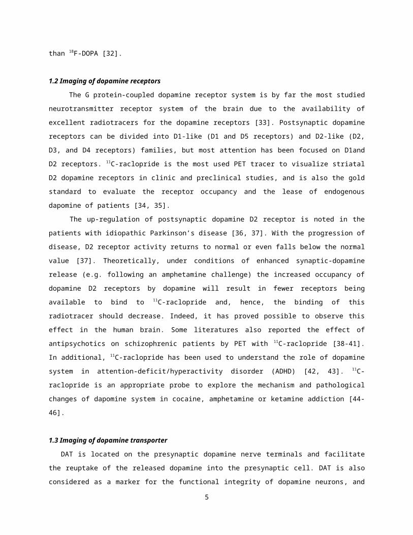
1.2 Imaging of dopamine receptors
The G protein-coupled dopamine receptor system is by far the most studied
neurotransmitter receptor system of the brain due to the availability of
excellent radiotracers for the dopamine receptors [33]. Postsynaptic dopamine
receptors can be divided into D1-like (D1 and D5 receptors) and D2-like (D2,
D3, and D4 receptors) families, but most attention has been focused on D1and
D2 receptors. 11C-raclopride is the most used PET tracer to visualize striatal
D2 dopamine receptors in clinic and preclinical studies, and is also the gold
standard to evaluate the receptor occupancy and the lease of endogenous
dapomine of patients [34, 35].
The up-regulation of postsynaptic dopamine D2 receptor is noted in the
patients with idiopathic Parkinson’s disease [36, 37]. With the progression of
disease, D2 receptor activity returns to normal or even falls below the normal
value [37]. Theoretically, under conditions of enhanced synaptic-dopamine
release (e.g. following an amphetamine challenge) the increased occupancy of
dopamine D2 receptors by dopamine will result in fewer receptors being
available to bind to 11C-raclopride and, hence, the binding of this
radiotracer should decrease. Indeed, it has proved possible to observe this
effect in the human brain. Some literatures also reported the effect of
antipsychotics on schizophrenic patients by PET with 11C-raclopride [38-41].
In additional, 11C-raclopride has been used to understand the role of dopamine
system in attention-deficit/hyperactivity disorder (ADHD) [42, 43]. 11C-
raclopride is an appropriate probe to explore the mechanism and pathological
changes of dapomine system in cocaine, amphetamine or ketamine addiction [44-
46].
1.3 Imaging of dopamine transporter
DAT is located on the presynaptic dopamine nerve terminals and facilitate
the reuptake of the released dopamine into the presynaptic cell. DAT is also
considered as a marker for the functional integrity of dopamine neurons, and
5
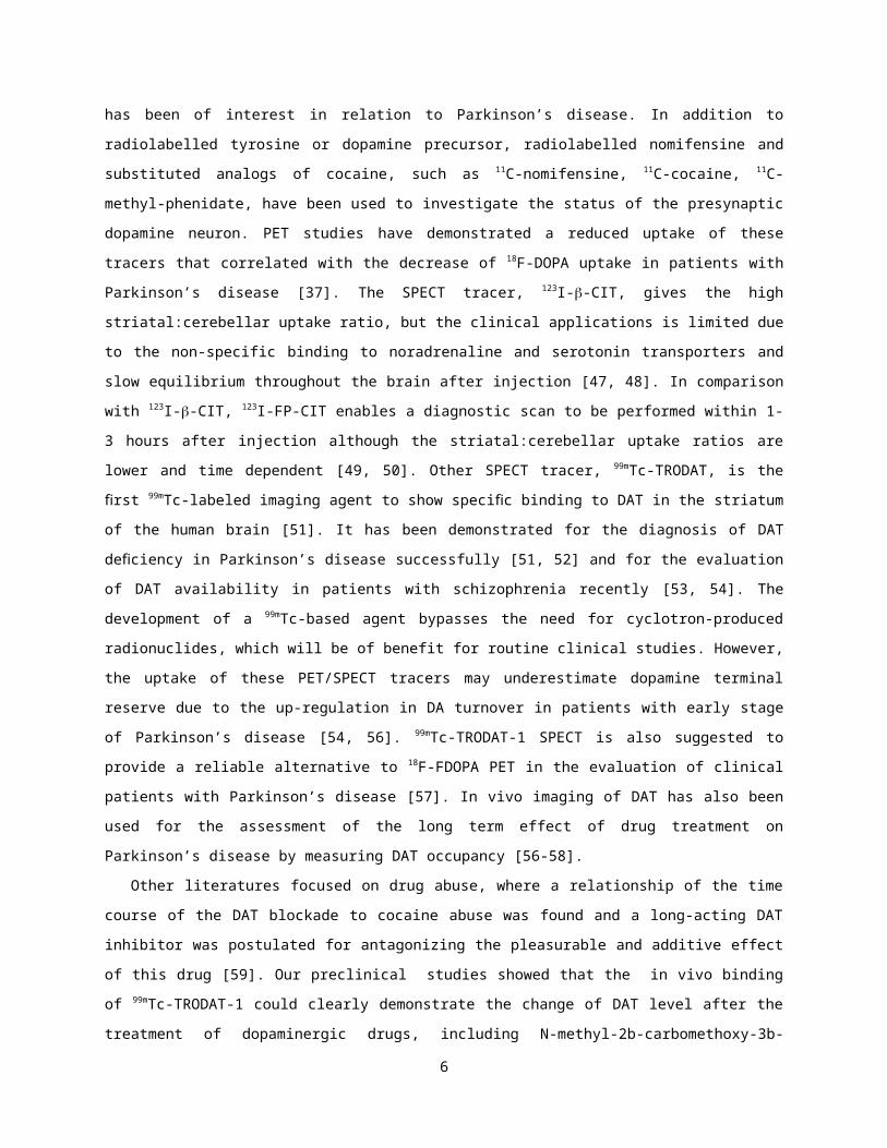
has been of interest in relation to Parkinson’s disease. In addition to
radiolabelled tyrosine or dopamine precursor, radiolabelled nomifensine and
substituted analogs of cocaine, such as 11C-nomifensine, 11C-cocaine, 11C-
methyl-phenidate, have been used to investigate the status of the presynaptic
dopamine neuron. PET studies have demonstrated a reduced uptake of these
tracers that correlated with the decrease of 18F-DOPA uptake in patients with
Parkinson’s disease [37]. The SPECT tracer, 123I--CIT, gives the high
striatal:cerebellar uptake ratio, but the clinical applications is limited due
to the non-specific binding to noradrenaline and serotonin transporters and
slow equilibrium throughout the brain after injection [47, 48]. In comparison
with 123I--CIT, 123I-FP-CIT enables a diagnostic scan to be performed within 1-
3 hours after injection although the striatal:cerebellar uptake ratios are
lower and time dependent [49, 50]. Other SPECT tracer, 99mTc-TRODAT, is the
rstfi 99mTc-labeled imaging agent to show speci c binding to DAT in thefi striatum
of the human brain [51]. It has been demonstrated for the diagnosis of DAT
de ciency in fi Parkinson’s disease successfully [51, 52] and for the evaluation
of DAT availability in patients with schizophrenia recently [53, 54]. The
development of a 99mTc-based agent bypasses the need for cyclotron-produced
radionuclides, which will be of benefit for routine clinical studies. However,
the uptake of these PET/SPECT tracers may underestimate dopamine terminal
reserve due to the up-regulation in DA turnover in patients with early stage
of Parkinson’s disease [54, 56]. 99mTc-TRODAT-1 SPECT is also suggested to
provide a reliable alternative to 18F-FDOPA PET in the evaluation of clinical
patients with Parkinson’s disease [57]. In vivo imaging of DAT has also been
used for the assessment of the long term effect of drug treatment on
Parkinson’s disease by measuring DAT occupancy [56-58].
Other literatures focused on drug abuse, where a relationship of the time
course of the DAT blockade to cocaine abuse was found and a long-acting DAT
inhibitor was postulated for antagonizing the pleasurable and additive effect
of this drug [59]. Our preclinical studies showed that the in vivo binding
of 99mTc-TRODAT-1 could clearly demonstrate the change of DAT level after the
treatment of dopaminergic drugs, including N-methyl-2b-carbomethoxy-3b-
6
(4fluorophenyl)tropane (CFT), 1-methyl-4-phenyl-1,2,3,6-tetrahydropyridine
(MPTP), l-DOPA and methylphenidate, using ICR mice [60]. Some studies used99mTc-TRODAT with SPECT to assess the changes of dopamine system among human
substance abusers [61, 62].
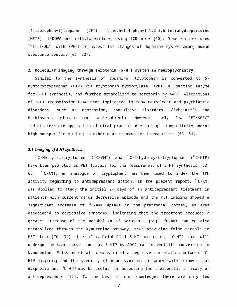
2. Molecular imaging through serotonin (5-HT) system in neuropsychiatry
Similar to the synthesis of dopamine, tryptophan is converted to 5-
hydroxytryptophan (HTP) via tryptophan hydroxylase (TPH), a limiting enzyme
for 5-HT synthesis, and further metabolized to serotonin by AADC. Alterations
of 5-HT transmission have been implicated in many neurologic and psychiatric
disorders, such as depression, compulsive disorders, Alzheimer’s and
Parkinson’s disease and schizophrenia. However, only few PET/SPECT
radiotracers are applied in clinical practice due to high lipophilicity and/or
high nonspecific binding to other neurotransmitter transporters [63, 64].
2.1 Imaging of 5-HT synthesis11C-Methyl-L-tryptophan (11C-AMT) and 11C-5-hydroxy-L-tryptophan (11C-HTP)
have been promoted as PET tracers for the measurement of 5-HT synthesis [63-
68]. 11C-AMT, an analogue of tryptophan, has been used to index the TPH
activity regarding to antidepressant action. In the present report, 11C-AMT
was applied to study the initial 24 days of an antidepressant treatment in
patients with current major depressive episode and the PET imaging showed a
significant increase of 11C-AMT uptake in the prefrontal cortex, an area
associated to depressive symptoms, indicating that the treatment produces a
greater increase of the metabolism of serotonin [69]. 11C-AMT can be also
metabolized through the kynurenine pathway, thus providing false signals in
PET data [70, 71]. Use of radiolabelled 5-HT precursor, 11C-HTP that will
undergo the same conversions as 5-HTP by ADCC can prevent the conversion to
kynurenine. Eriksson et al. demonstrated a negative correlation between 11C-
HTP trapping and the severity of mood symptoms in women with premenstrual
dysphoria and 11C-HTP may be useful for assessing the therapeutic efficacy of
antidepressants [72]. To the best of our knowledge, there are only few
7
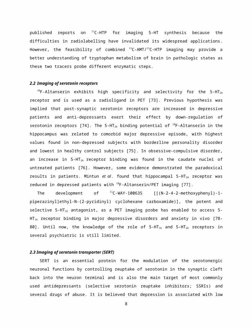
published reports on 11C-HTP for imaging 5-HT synthesis because the
difficulties in radiolabelling have invalidated its widespread applications.
However, the feasibility of combined 11C-AMT/11C-HTP imaging may provide a
better understanding of tryptophan metabolism of brain in pathologic states as
these two tracers probe different enzymatic steps.
2.2 Imaging of serotonin receptors18F-Altanserin exhibits high specificity and selectivity for the 5-HT2A
receptor and is used as a radioligand in PET [73]. Previous hypothesis was
implied that post-synaptic serotonin receptors are increased in depressive
patients and anti-depressants exert their effect by down-regulation of
serotonin receptors [74]. The 5-HT2A binding potential of 18F-Altanserin in the
hippocampus was related to comorbid major depressive episode, with highest
values found in non-depressed subjects with borderline personality disorder
and lowest in healthy control subjects [75]. In obsessive-compulsive disorder,
an increase in 5-HT2A receptor binding was found in the caudate nuclei of
untreated patients [76]. However, some evidence demonstrated the paradoxical
results in patients. Mintun et al. found that hippocampal 5-HT2A receptor was
reduced in depressed patients with 18F-Altanserin/PET imaging [77].
The development of 11C-WAY-100635 [[(N-2-4-2-methoxyphenyl)-1-
piperazinyl]ethyl-N-(2-pyridinyl) cyclohexane carboxamide)], the potent and
selective 5-HT1A antagonist, as a PET imaging probe has enabled to access 5-
HT1A receptor binding in major depressive disorders and anxiety in vivo [78-
80]. Until now, the knowledge of the role of 5-HT1A and 5-HT2A receptors in
several psychiatric is still limited.
2.3 Imaging of serotonin transporter (SERT)
SERT is an essential protein for the modulation of the serotonergic
neuronal functions by controlling reuptake of serotonin in the synaptic cleft
back into the neuron terminal and is also the main target of most commonly
used antidepressants (selective serotonin reuptake inhibitors; SSRIs) and
several drugs of abuse. It is believed that depression is associated with low
8
level of serotonin, thus blocking serotonin reuptake with SSRIs has been
applied in the treatment of depression. 11C-(+)McN5652 was the first promising
PET imaging agent for studying SERT in humans and considered as a marker for
integrity of the 5-HT terminals by assessing SERT density [81, 82]. Buchert et
al. found the distribution volume ratio (DVR) of 11C-(+)McN5652 in methylene-
dioxymethamphetamine (MDMA) users was significantly reduced in the
mesencephalon and the thalamus, indicating that 11C-(+)McN5652/PET may be
applied to investigate the long-term effect of the drug ecstasy on the
availability of 5-HT system [83, 84]. Nevertheless, high non-specific binding
has limited its clinical application. [18F]fluoromethyl analog of (+)McN5652
(18F-(+)-FMe-McN5652) has been synthesized recently, with the same
distribution pattern of serotonin uptake sites but a faster binding
equilibrium in piglets and a longer radioisotope half-life compared to 11C-
(+)McN5652 [85-87]. 18F-(+)-FMe-McN5652 may turn out to be the alternate for
SERT imaging of human brain although it still has limitations in full
quantification of PET data.
4-18F-ADAM is one of few 18F-labeled radioligands for studying SERT using
PET. The biodistribution, toxicity, and radiation dosimetry of 4-18F-ADAM has
been conducted in rats and primates, and the utilization of 4-18F-ADAM has
been validated in neurotoxin-, SSRIs-, and drug ecstasy-treated (ecstasy,
MDAM, 3, 4-Methylenedioxymethamphetamine) rat models, suggesting that 4-18F-
ADAM could be safe and suitable in human studies [88-93]. However, the major
drawback of 4-18F-ADAM is its relatively low radiochemical yield, and further
characterization of this new radioligand in humans is warranted.
Mapping the brain SERT with promising iodinated ADAM has been developed
recently [94]. The biodistribution of 123I-ADAM has been evaluated in both
animals and humans [95-101]. Our previous data further demonstrated the
heterogeneity of the SERT distribution in rat brains, with the highest uptake
of 123I-ADAM in the dorsal raphe nucleus, substantia nigra, lateral
hypothalamus, venral lateral geniculate nucleus, basolateral amygdala, and
ventromedial hypothalamus; the moderate uptake in the mediodorsal thalamus,
lateral dorsal thalamus, dorsal hypothalamus, dorsal lateral geniculate
9
nucleus, basomedial amygdala, lateral amygdala, and hippocampus; the low
uptake in the cingulum, caudate putamen, prefrontal cortex, and cerebellum.123I-ADAM uptake was dramatically decreased in the hippocampus, thalamus,
amygdala, geniculate nuclei, hypothalamus, raphe nucleus, and substantia nigra
in p-chloroamphetamine (PCA)-treated rats [99]. 2-(2'-((dimethylamino)methyl)-
4'-iodophenylthio)benzenamine (FlipADAM) was an improved SPECT radiotracer for
selective SERT imaging. Wang et al. reported that 125I-FlipADAM exhibited
faster clearance and binding equilibrium in the brain of SD rats compared to125I-ADAM [102]. 125I-FlipADAM successfully penetrated the blood brain barrier,
as evidenced by the brain uptake at 2 min (1.75% dose/g). 125I-FlipADAM also
had a good target to non-target (hypothalamus/cerebellum) ratio of 3.35 at 60
min post-injection. The value of clinical application needs to be further
validated.
3. Molecular imaging in Neuro-oncology
Brain tumors, especially high-grade gliomas, are extremely resistant to
conventional therapies, including surgery, chemotherapy, and radiotherapy. The
5-year survival rate of patients with glioblastoma multiforme (GBM) is less
than a few percent even with aggressive combinational treatments [103, 104].
Metastatic brain tumors are also a major cause of morbidity and mortality in
human being. Therefore, there is a high medical need for new effective
therapies to treat both primary and metastatic brain tumors. There is an
increasing interest in the use of BNCT as a tumor-selective treatment for
malignant brain tumors which remain incurable despite aggressive treatment
with surgery, chemotherapy, and conventional radiotherapy. Briefly, low-energy
thermal neutrons interact with nonradioactive boron (10B) accumulated in tumor
cells, and release high-LET alpha and lithium particles (7Li) with a short
path length (10-14 m) via boron neutron capture reaction, 10B (n, α) 7Li,
thereby destroying tumor cells efficiently while sparing normal tissues [105,
106]. BNCT clinical trials have recently been initiated cancer treatment in
USA, Europoe, Japan, and Taiwan. However, the lack of selective and sufficient
accumulation of 10B carriers in tumors is still the main impediment for BNCT
10
to be successful. Small-animal models with human brain tumors have played an
essential role in the better understanding of brain tumor biology, and have
made not only a significant contribution to the improvement of 10B-carriers,
but also the treatment planning and outcome of clinical BNCT while combined
with molecular imaging.
3.1 PET/ SPECT probes for boron distribution imaging
Two boron delivery agents, boronophenylalanine (BPA) and borocaptate
sodium (BSH), have been used for clinical trials. BPA, the analogs of amino
acids phenylalanine (Phe) and tyrosine (Tyr), is incorporated into tumor cells
via active transport. To optimize the efficacy of BNCT and predict the
effectiveness of the treatment, the estimated tumor-to-normal brain ratio of10B and the time window for neutron irradiation become the hinging point
before BNCT treatment. Menichetti et al. demonstrated that the use of
[18F]FBPA and PET/CT could not only provide more effective measurement of the
tumor extraction of 10BPA compared to normal tissue in patients, but obtain a
better treatment planning of BNCT for the personalized medicine [107]. To
provide the pharmacokinetics of BPA for clinical use of BNCT in Taiwan, our
research group has synthesized and characterized 18F-FBPA-Fr using a F98
glioma-bearing rat model [108, 109]. BPA conjugated with fructose (BPA-Fr) has
been proven to increase its solubility, so that the drug uptake in tumor is
enhanced [110, 111]. In biodistribution studies, the tumor-to-normal brain
ratios of 18F-FBPA-Fr were 3.45, 3.13, 2.61, and 2.02 at 0.5, 1, 2, and 4 h
post-injection, with similar uptake characteristics to BPA-Fr (2.05, 1.86,
1.24, and 1.1, respectively) estimated by inductively coupled plasma mass
spectrometry (ICP-MP) [112, 113]. PET scanning also demonstrated that the
accumulation of radioactivity in tumor peaked at first hour and then gradually
decreased after the administration of 18F-FBPA-Fr. These results indicated
that 0.5-1 h after BPA-Fr injection would be the optimal time for tumor
irradiation, and clearly, 18F-FBPA-Fr may turn out to be a prognostic and
therapeutic indicator for patients who are poor surgical candidates and are
considered for BNCT. On the contrary, BSH does not accumulate in the normal
11
brain, but target brain tumors due to the disruption of blood-brain-barrier
(BBB). 131I has been applied to study in vivo characteristics of BSH in a
melanoma-bearing animal model, demonstrating that the pharmacokinetics and
biodistribution of 131I-BSH were consistent with the data on the dynamics of
the nonlabeled BSH distribution [114]. The knowledge concerning the
bioavailability and the metabolism of BSH is nevertheless very limited.
In the past two decades, polyhedral boron compounds, including polyhedral
boron hydrides and carboranes, have been investigated as an potential boron-
delivery agents which could achieve higher tumor-to-normal brain ratios with
low chemotoxicity in preclinical studies. Boronated amino acids, nucleic
acids, peptide, and antibodies, boronated carbohydrates, and boronated
porphyrins and phthalocyanines are of interest for the development of more
efficient BNCT [115]. Radioiodination and radiobromination of polyhedral
boranes have been proposed to study their biodistribution and pharmacokinetics
in vivo [116-119]. 67Cu and 99mTc-labelled polyhedral boranes have also been
investigated as potential imaging probes to aid BNCT treatment planning [120,
121].
3.2 Nanocarrier- and antibody-mediated delivery of boron carrier
Recently, Nanocarrier, receptor ligands, and antibodies have been
intensively studied as a very promising drug delivery system. 125I- or 76Br-
polyhedral boron coupled with anti-HER2/neu humanized antibody trastuzumab was
described for the treatment of breast cancers [116, 117]. Targeting efficiency
of radioiodinated carboranes coupled with cMAb U36 on a head and neck squamous
cell carcinoma xenograft model was evaluated [118]. However, a major
limitation of boron clusters directly linked to mAb is that the modification
of mAb can reduce its immunoreactivity. Attachment of boronated dendrimers to
mAb has been designed to deliver the requisite amount of 10B without
compromising the targeting efficiency of mAb for BNCT. Yang et al. reported
that boronated polyamidoamine dendrimer linked with anti-EGFRvIII mAb (BD-
L8A4) and cetuximab (BD-C225) retained their in vitro and in vivo
immunoreactivity, and the biodistribution of 125I-BD-mAbs was determined in F98
12
glioma-bearing rats [122]. On the other hand, the EGF-conjugated boronated
dendrimers have a much smaller MW than EGFR mAb conjugates, they should be
capable of more rapid and effective tumor targeting than has been observed
with mAbs [123, 124]. Folate receptors (FR) are up-regulated in a variety of
human cancers. Many studies have shown that radiolabeled folic acid and its
derivatives are suitable for tumor-targeting imaging of SPECT or PET in small
animal models overexpressing FR. 64Cu-porphyrin-peptide-folate (64Cu-PPF) has
been evaluated as the potential of PET probe for cancer imaging [125]. 99mTc-
labeled PEGylated dendrimer PAMAM folic acid conjugate (99mTc-G5-Ac-pegFA-DTPA)
has been synthesized to study the in vitro/in vivo stability and
biodistribution in FR+ tumor bearing mice using microSPECT [126]. As such, the
development of radiolabelled polyhedral boron moiety connected to
nanoparticles, ligand/receptor of growth factors, or tumor-targeting
monoclomoal antibodies is of great potential for the medical application of
BNCT.
Boron nitride nanotubes (BNNTs), a structural analog of a carbon nanotube
where C atoms are substituted by alternating B and N atoms, have been proposed
as delivery agents able to target high boron concentration in tumors cells for
BNCT [127]. Recently, Ciofani et al. demonstrated that BNNTs functionalized
with folic acid as a tumor targeting ligand have shown the selective
accumulation in GBM cells but not normal human fibroblasts [128]. Soares et
al. reported the biodistribution of 99mTc-functionalized BNNTs in normal Swiss
mice with SPECT imaging, revealing a potential application of 99mTc-
functionalized BNNTs as a new SPECT imaging probe applied in BNCT [129].
3.3 Clinical perspective of BNCT
Several centers in USA, Europe, Japan and Taiwan are devoted to the
development of BNCT. However, over the past few decades, BNCT has progressed
relatively slowly due to the controversial outcome from clinical trials. The
design of boron carriers and drug delivery system, the evaluation of
treatments, and the source of neutron beam will need to be optimized to
enhance the therapeutic efficacy of BNCT. The use of PET/SPECT probes to
13
select patients who are likely to respond to BNCT and the combination of
multimodality imaging strategies, such as MRI and CT for anatomical
information could accelerate the development of new boron delivery agents. We
expected that BNCT for brain tumor may protect more cognitive function than
conventional cancer therapy, and it may become the personalized therapy in the
future.
Conclusions
The knowledge and the therapeutic techniques of brain diseases and
disorders are much delayed than the other diseases. With the development of
molecular imaging, neuroimaging with PET and SPECT provides the physiologic
and metabolic changes in healthy and pathologic tissues, and therefore
facilitates the diagnosis and treatment of psychiatric disorders and the
advance of brain tumor therapy. In the future, the combination of MRI/PET or
PET/CT imaging will enable to offer both anatomical and multi-functional
information of human brain and may show added value in increasing diagnostic
accuracy and better treatment planning.
14
References
[1] Weissleder R, Mahmood U. Molecular imaging. Radiology 2001; 219: 316-33.
[2] Ugrumov MV. Non-dopaminergic neurons partly expressing dopaminergic
phenotype: distribution in the brain, development and functional
significance. J Chem Neuroanat 2009; 38: 241-56.
[3] Bauer M, Praschak-Rieder N, Kasper S, Willeit M. Is dopamine
neurotransmission altered in prodromal schizophrenia? A review of the
evidence. Curr Pharm Des 2012; 18: 1568-79.
[4] Stoessl AJ, Martin WW, McKeown MJ, Sossi V. Advances in imaging in
Parkinson’s disease. Lancet Neurol 2011; 10: 987-1001.
[5] Cosgrove KP. Imaging receptor change in human drug abusers. Curr Top Behav
Neurosci 2010; 3: 199-217.
[6] Salvadore G, Quiroz JA, Machado-Vieira R, Henter ID, Manji HK, Zarate CA
Jr. The neurobiology of the switch process in bipolar disorders: a review.
J Clin Psychiatry 2010; 71: 1488-501.
[7] Wiesel FA, Blomqvist G, Halldin C, et al. The transport of tyrosine into
the human brain as determined with L-[1-11C]tyrosine and PET. J Nucl Med
1991; 32: 2043-9.
[8] Wiesel FA, Andersson JL, Westerberg G, et al. Tyrosine transport is
regulated differently in patients with schizophrenia. Schizophr Res 1999;
40: 37-42.
[9] Coenen HH, Kling P, Stocklin G. Cerebral metabolism of L-2-
[18F] uorotyrosine, a new PET tracer of protein synthesis. J Nucl Medfl
1989; 30: 1367-72.
[10] Firnau G, Nahmias C, Garnett ES. The preparation of 5-[18F] uoro-Dopafl
with reactor produced uorine-18. Int J Appl Radiat Isot 1972; 24: 182-4.fl
[11] Cumming P, Gjedde A. Compartmental analysis of dopa decarboxylation in
living brain from dynamic positron emission tomograms. Synapse 1998; 29:
37-61.
[12] Ravina B, Eidelberg D, Ahlskog JE, et al. The role of radiotracer imaging
in Parkinson disease. Neurology 2005; 64: 208-15.
15
[13] Leenders KL, Palmer AJ, Quinn N, et al. Brain dopamine metabolism in
patients with Parkinson’s disease measured with positron emission
tomography. J Neurol Neurosurg Psychiatry 1986; 49: 853-60.
[14] Grasby PM. Imaging the neurochemical brain in health and disease. Clin
Med 2002; 2: 67-73.
[15] Morrish PK, Sawle GV, Brooks DJ. An [18F]dopa-PET and clinical study of
the rate of progression in Parkinson’s disease. Brain 1996; 119: 585-91.
[16] Morrish PK, Rakshi JS, Sawle GV, et al. Measuring the rate of progression
and estimating the preclinical period of Parkinson’s disease with [18F]dopa
PET. J Neurol. Neurosurg Psychiatry 1998; 64: 314-9.
[17] Pavese N, Rivero-Bosch M, Lewis SJ, et al. Progression of monoaminergic
dysfunction in Parkinson's disease: a longitudinal 18F-dopa PET study.
Neuroimage 2011; 56: 1463-8.
[18] Brooks DJ, Rakshi JS, Pavese N, et al. Relative rates of progression of
early Parkinson’s disease patients started on either ropinirole or L-dopa:
2-year and 5-year follow-up 18F-dopa PET findings. Mov Disord 2000; 5
(suppl 2): S308.
[19] Rakshi JS, Pavese N, Uema T, et al. A comparison of the progression of
early Parkinson’s disease in patients started on ropinirole or L-dopa. An18Fdopa PET study. J Neural Transm 2002; 109: 1433-43.
[20] Rascol O, Brooks DJ, Korczyn AD, et al. A five-year study of the incidence
of dyskinesia in patients with early Parkinson’s disease who were treated
with ropinirole or levodopa. N Engl J Med 2000; 342: 1484-91.
[21] Dao-Castellana MH, Paillère-Martinot ML, Hantraye P, et al. Presynaptic
dopaminergic function in the striatum of schizophrenic patients. Schizophr
Res 1997; 23: 167-74.
[22] Bose SK, Turkheimer FE, Howes OD, et al. Classification of schizophrenic
patients and healthy controls using [18F] fluorodopa PET imaging. Schizophr
Res 2008; 106: 148-55.
[23] Shotbolt P, Stokes PR, Owens SF, et al. Striatal dopamine synthesis
capacity in twins discordant for schizophrenia. Psychol Med 2011; 41: 2331-
8.
16
[24] Lee CS, Samii A, Sossi V, et al. In vivo positron emission tomographic
evidence for compensatory changes in presynaptic dopaminergic nerve
terminals in Parkinson’s disease. Ann Neurol 2000; 47: 493-503.
[25] Brooks DJ, Frey KA, Marek KL, et al. Assessment of neuroimaging techniques
as biomarkers of the progression of Parkinson’s disease. Exp Neurol 2003;
184 Suppl 1: S68-79.
[26] Doudet DJ, Chan GL, Holden JE, et al. Effects of catechol-O-
methyltransferase inhibition on therates of uptake and reversibility of 6-
uoro-L-Dopafl trapping in MPTP-induced parkinsonism in monkeys.
Neuropharmacology 1997; 6: 363-71.
[27] DeJesus OT, Haaparanta M, Solin O, et al. 6-Fluoro-L-DOPA metabolism in
rat striatum: time course of extracellular metabolites. Brain Res 2000;
877: 31-36.
[28] DeJesus OT. Positron-labeled DOPA analogs to image dopamine terminals.
Drug Dev Res 2003; 59: 249-60.
[29] Tai YF, Hoshi R, Brignell CM, et al. Persistent nigrostriatal dopaminergic
abnormalities in ex-users of MDMA ('Ecstasy'): an 18F-dopa PET study.
Neuropsychopharmacology 2011; 36: 735-43.
[30] DeJesus OT, Holden JE, Endres C, et al. Visualization of dopamine nerve
terminals by positron tomography using [18F] uoro-beta- uoromethylene-m-fl fl
tyrosine. Brain Res 1992; 597: 151-4.
[31] DeJesus OT, Endres CJ, Shelton SE, Nickles RJ, Holden JE. Evaluation of
uorinated m-tyrosine analogs as PET imaging agents of dopamine nervefl
terminals: comparison with 6- uoroDOPA. J Nucl Med 1997; 38: 630-6.fl
[32] Gallagher CL, Christian BT, Holden JE, et al. A Within-Subject Comparison
of 6-[18F]Fluoro-m-tyrosine and 6-[18F]Fluoro-L-dopa in Parkinson’s Disease.
Mov Disord 2011; 26: 2023-38.
[33] Beaulieu JM, Gainetdinov RR. The physiology, signaling, and pharmacology
of dopamine receptors. Pharmacol Rev 2011; 63: 182-217.
[34] Volkow ND, Wang GJ, Fowler JS, et al. Imaging endogenous dopamine
competition with [11C]raclopride in the human brain. Synapse 1994; 16: 255-
62.
17
[35] Laruelle M. Imaging synaptic neurotransmission with in vivo binding
competition techniques: a critical review. J Cereb Blood Flow Metab 2000;
20: 423-51.
[36] Schreckenberger M, Hägele S, Siessmeier T, et al. The dopamine D2 receptor
ligand 18F-desmethoxyfallypride: an appropriate fluorinated PET tracer for
the differential diagnosis of parkinsonism. Eur J Nucl Med Mol Imaging
2004; 31: 1128-35.
[37] Heiss W, Herholz K. Brain receptor imaging. J Nucl Med 2006; 47: 302-12.
[38] Kim JH, Son YD, Kim HK, et al. Antipsychotic-associated mental side
effects and their relationship to dopamine D2 receptor occupancy in
striatal subdivisions: a high-resolution PET study with [11C]raclopride. J
Clin Psychopharmacol 2011; 31: 507-11.
[39] Kapur S, Remington G, Jones C, et al. High levels of dopamine D2 receptor
occupancy with low-dose haloperidol treatment: a PET study. Am J Psychiatry
1996; 153: 948-50.
[40] Arakawa R, Ito H, Takano A, et al. Dose-finding study of paliperidone ER
based on striatal and extrastriatal dopamine D2 receptor occupancy in
patients with schizophrenia. Psychopharmacology (Berl) 2008; 197: 229-35.
[41] Kodaka F, Ito H, Takano H, et al. Effect of risperidone on high-affinity
state of dopamine D2 receptors: a PET study with agonist ligand [11C](R)-2-
CH3O-N-n-propylnorapomorphine. Int J Neuropsychopharmacol 2011; 14: 83-9.
[42] Jucaite A, Fernell E, Halldin C, Forssberg H, Farde L. Reduced midbrain
dopamine transporter binding in male adolescents with attention-
deficit/hyperactivity disorder: association between striatal dopamine
markers and motor hyperactivity. Biol Psychiatry 2005; 57: 229-38.
[43] Volkow ND, Wang GJ, Newcorn J, et al. Depressed dopamine activity in
caudate and preliminary evidence of limbic in volvement in adults with
attention-deficit/hyperactivity disorder. Arch Gen Psychiatry 2007; 64:
932-40.
[44] Vollenweider FX, Vontobel P, Oye I, Hell D, Leenders KL. Effects of (S)-
ketamine on striatal dopamine: a [11C]raclopride PET study of a model
psychosis in humans. J Psychiatr Res 2000; 34: 35-43.
18
[45] Boileau I, Dagher A, Leyton M, et al. Conditioned dopamine release in
humans: a positron emission tomography [11C]raclopride study with
amphetamine. J Neurosci 2007; 27: 3998-4003.
[46] Cox SM, Benkelfat C, Dagher A, et al. Striatal dopamine responses to
intranasal cocaine self-administration in humans. Biol Psychiatry 2009; 65:
846-50.
[47] Seibyl JP, Marek KL, Quinlan D, et al. Decreased single-photon emission
computed tomographic [123I]-CIT striatal uptake correlates with symptom
severity in Parkinson’s disease. Ann Neurol 1995; 38: 589-98.
[48] Seibyl JP, Marek K, Sheff K, et al. Iodine-123-beta-CIT and iodine-123-
FPCIT SPECT measurement of dopamine transporters in healthy subjects and
Parkinson's patients. J Nucl Med 1998; 39: 1500-8.
[49] Brooks DJ. Morphological and functional imaging studies on the diagnosis
and progression of Parkinson’s disease. J Neurol 2000; 247 (Suppl 2):
II/11-8.
[50] Paweł Szymański, Magdalena Markowicz, Agnieszka Janik, Mateusz
Ciesielski, Elżbieta Mikiciuk-Olasik. Neuroimaging diagnosis in
neurodegenerative diseases. Nuclear Med Rev 2010; 13: 23-31.
[51] Kung HF, Kim HJ, Kung MP, et al. Imaging of dopamine transporters in
humans with technetium-99m TRODAT-1. Eur J Nucl Med 1996, 23, 1527-30.
[52] Mozley PD, Schneider JS, Acton PD, et al. Binding of [99mTc]TRODAT to
dopamine transporters in patients with Parkinson's disease and in healthy
volunteers. J Nucl Med 2000; 41: 584-9.
[53] Chen KC, Yang YK, Howes O, et al. Striatal dopamine transporter
availability in drug-naive patients with schizophrenia: A case-control
SPECT study with [99mTc]-TRODAT-1 and a meta-analysis. Schizophr Bull 2011,
doi: 10.1093/schbul/sbr163.
[54] Salmon E, Frackowiak RS. Functional metabolic neuroimaging by positron-
emission tomography in man. Rev Neurol (Paris) 1990; 146: 459-77.
[55] Schmitt GJ, Frodl T, Dresel S, et al. Striatal dopamine transporter
availability is associated with the productive psychotic state in first
episode, drug-naive schizophrenic patients. Eur Arch Psychiatry Clin
19
Neurosci 2006; 256: 115-21.
[56] Hartvig P, Bergström M, Antoni G, et al. Positron emission tomography and
brain monoamine neurotransmission- Entries for study of drug interactions.
Curr Pharm Des 2002; 8: 1417-34.
[57] Huang WS, Chiang YH, Lin JC, Chou YH, Cheng CY, Liu RS. Crossover study
of 99mTc-TRODAT-1 SPECT and (18)F-FDOPA PET in Parkinson's disease patients.
J Nucl Med 2003; 44: 999-1005.
[58] Tedroff J, Ekesbo A, Rydin E, et al. Regulation of dopaminergic activity
in early Parkinson's disease. Ann Neurol 1999; 46, 359-65.
[59] Volkow ND, Wang GJ, Fischman MW, et al. Relationship between subjective
effects of cocaine and dopamine transporter occupancy. Nature 1997; 386:
827-30.
[60] Hwang JJ, Liao MH, Yen TC, et al. Biodistribution study of [99mTc] TRODAT-1
alone or combined with other dopaminergic drugs in mice with
macroautoradiography. Appl Radiat Isot 2002; 57: 35-42.
[61] Szobot CM, Shih MC, Schaefer T, et al. Methylphenidate DAT binding in
adolescents with Attention-Deficit/ Hyperactivity Disorder comorbid with
Substance Use Disorder--a single photon emission computed tomography with
[Tc(99m)]TRODAT-1 study. Neuroimage 2008; 40: 1195-201.
[62] Crits-Christoph P, Newberg A, Wintering N, et al. Dopamine transporter
levels in cocaine dependent subjects. Drug Alcohol Depend. 2008; 98: 70-6.
[63] Scheffel U, Dannals RF, Suehiro M, et al. Development of PET/SPECT ligands
for the serotonin transporter. NIDA Res Monogr 1994; 138: 111-30.
[64] Visser AK, van Waarde A, Willemsen AT, et al. Measuring serotonin
synthesis: from conventional methods to PET tracers and their (pre)clinical
implications. Eur J Nucl Med Mol Imaging 2011; 38: 576-91.
[65] Reibring L, Agren H, Hartvig P, et al. Uptake and utilization of [beta-11C]5-hydroxytryptophan in human brain studied by positron emission
tomography. Psychiatry Res 1992; 45: 215-25.
[66] Muzik O, Chugani DC, Chakraborty P, et al. Analysis of [C-11]alpha-methyl-
tryptophan kinetics for the estimation of serotonin synthesis rate in vivo.
J Cereb Blood Flow Metab 1997; 17: 659-69.
20
[67] Hagberg GE, Torstenson R, Marteinsdottir I, et al. Kinetic compartment
modeling of [11C]-5-hydroxy-L-tryptophan for positron emission tomography
assessment of serotonin synthesis in human brain. J Cereb Blood Flow Metab
2002; 22: 1352-66.
[68] Nishikawa M, Kumakura Y, Young SN, et al. Increasing blood oxygen
increases an index of 5-HT synthesis in human brain asmeasured using alpha-
[(11)C]methyl-Ltryptophan and positron emission tomography. Neurochem Int
2005; 47: 556-64.
[69] Berney A, Nishikawa M, Benkelfat C, et al. An index of 5-HT synthesis
changes during early antidepressant treatment: alpha-[11C]methyl-L-
tryptophan PET study. Neurochem Int 2008; 52: 701-8.
[70] Chugani DC, Muzik O. Alpha[C-11]methyl-L-tryptophan PET maps brain
serotonin synthesis and kynurenine pathway metabolism. J Cereb Blood Flow
Metab 2000; 20: 2-9.
[71] Juhasz C, Chugani DC, Muzik O, et al. In vivo uptake and metabolism of
alpha-[11C]methyl-L-tryptophan in human brain tumors. J Cereb Blood Flow
Metab 2006; 26: 345-57.
[72] Eriksson O, Wall A, Marteinsdottir I, et al. Mood changes correlate to
changes in brain serotonin precursor trapping in women with premenstrual
dysphoria. Psychiatry Res 2006; 146: 107-16.
[73] Lemaire C, Cantineau R, Guillaume M, Plenevaux A, Christi L. Fluorine-18-
altanserin: a radioligand for the study of serotonin receptors with PET:
radiolabeling and in vivo biologic behavior in rats. J Nucl Med 1991; 32:
2266-72.
[74] Stahl SM. Regulation of neurotransmitter receptors by desipramine and
other antidepressant drugs: the neurotransmitter receptor hypothesis of
antidepressant action. J Clin Psychiatry 1984; 45: 37-45.
[75] Soloff PH, Price JC, Meltzer CC, Fabio A, Frank GK, Kaye WH. 5HT2A
receptor binding is increased in borderline personality disorder. Biol
Psychiatry 2007; 62: 580-7.
[76] Adams KH, Hansen ES, Pinborg LH, et al. Patients with obsessive-compulsive
disorder have increased 5-HT2A receptor binding in the caudate nuclei. Int
21
J Neuropsychopharmacol 2005; 8: 391-401.
[77] Mintun MA, Sheline YI, Moerlein SM, Vlassenko AG, Huang Y, Snyder, AZ.
Decreased hippocampal 5-HT2A receptor binding in major depressive disorder:
in vivo measurement with [18F]altanserin positron emission tomography. Biol
Psychiatry 2004; 55: 217-24.
[78] Mathis CA, Simpson NR, Mahmood K, Kinahan PE, Mintun, MA. [11C]WAY
100635: a radioligand for imaging 5-HT1A receptors with positron emission
tomography. Life Sci 1994; 55: PL403-7.
[79] Drevets WC, Thase ME, Moses-Kolko EL, et al. Serotonin-1A receptor imaging
in recurrent depression: replication and literature review. Nucl Med Biol.
2007; 34: 865-77.
[80] Hahn A, Lanzenberger R, Wadsak W, et al. Escitalopram enhances the
association of serotonin-1A autoreceptors to heteroreceptors in anxiety
disorders. J Neurosci 2010; 30: 14482-9.
[81] Suehiro M, Scheffel U, Ravert HT, et al. [11C](+)McN5652 as a radiotracer
for imaging serotonin uptake sites with PET. Life Sci 1993; 53: 883-92.
[82] Szabo Z, Kao PF, Scheffel U, et al. Positron emission tomography imaging
of serotonin transporters in the human brain using [11C](+)McN5652. Synapse
1995; 20: 37-43.
[83] Buchert R, Thomasius R,Nebeling B, et al. Long-term effects of "ecstasy"
use on serotonin transporters of the brain investigated by PET. J Nucl Med
2003; 44: 375-84.
[84] Buchert R, Thiel, F, Thomasius R, et al. Ecstasy-induced reduction of the
availability of the brain serotonin transporter as revealed by [11C]
(+)McN5652-PET and the multi-linear reference tissue model: loss of
transporters or artifact of tracer kinetic modelling? J Psychopharmacol
2007; 21: 628-34.
[85] Zessin J, Eskola O, Brust P, et al. Synthesis of S-([18F]fluoromethyl)-(+)-
McN5652 as a potential PET radioligand for the serotonin transporter. Nucl
Med Biol 2001; 28: 857-63.
[86] Kretzschmar M, Brust P, Zessin J, et al. Autoradiographic imaging of the
serotonin transporter in the brain of rats and pigs using S-
22
([18F]fluoromethyl)-(+)-McN5652. Eur Neuropsychopharmacol 2003; 13: 387-97.
[87] Brust P, Hinz R, Kuwabara H, et al. In vivo measurement of the serotonin
transporter with (S)-([18F]fluoromethyl)-(+)-McN5652.
Neuropsychopharmacology 2003; 28: 2010-9.
[88] Shiue GG, Choi SR, Fang P, et al. N,N-dimethyl-2-(2-amino-4-(18)F-
fluorophenylthio)-benzylamine (4-(18)F-ADAM): an improved PET radioligand
for serotonin transporters. J Nucl Med 2003; 44: 1890-7.
[89] Shiue GG, Fang P, Shiue CY. Synthesis of N,N-dimethyl-2-(2-amino-4-
[18F]fluorophenylthio)benzylamine as a serotonin transporter imaging agent.
Appl Radiat Isot 2003; 58: 183-91.
[90] Ma KH, Huang WS, Kuo YY, et al. Validation of 4-[18F]-ADAM as a SERT
imaging agent using micro-PET and autoradiography. Neuroimage 2009; 45:
687-93.
[91] Li IH, Huang WS, Shiue CY, et al. Study on the neuroprotective effect of
fluoxetine against MDMA-induced neurotoxicity on the serotonin transporter
in rat brain using micro-PET. Neuroimage 2010; 49: 1259-70.
[92] Huang YY, Ma KH, Tseng TW, et al. Biodistribution, toxicity and radiation
dosimetry studies of the serotonin transporter radioligand 4-[18F]-ADAM in
rats and monkeys. Eur J Nucl Med Mol Imaging 2010; 37: 545-55.
[93] Chen YA, Huang WS, Lin YS, et al. Characterization of 4-[(18)F]-ADAM as an
imaging agent for SERT in non-human primate brain using PET: a dynamic
study. Nucl Med Biol 2012; 39: 279-85.
[94] Oya S, Choi SR, Hou C, et al. 2-((2-((dimethylamino)methyl)phenyl)thio)-5-
iodophenylamine (ADAM): an improved serotonin transporter ligand. Nucl Med
Biol 2000; 27: 249-54.
[95] Choi SR, Hou C, Oya S, et al. Selective in vitro and in vivo binding of
[(125)I]ADAM to serotonin transporters in rat brain. Synapse 2000; 38: 403-
12.
[96] Acton PD, Choi SR, Hou C, Plossl K, Kung HF. Quanti cation of serotoninfi
transporters in nonhuman primates using 123I-ADAM and SPECT. J Nucl Med
2001; 42: 1556-62.
[97] Lin KJ, Ye XX, Yen TC, et al. Biodistribution study of 123I-ADAM in mice:
23
correlation with whole body autoradiography. Nucl Med Biol 2002; 29: 643-
50.
[98] Kauppinen TA, Bergstrom KA, Heikman P, et al. Biodistribution and
radiation dosimetry of [123I]ADAM in healthy human subjects: preliminary
results. Eur J Nucl Med Mol Imaging 2003; 30: 132-6.
[99] Lin KJ, Yen TC, Wey SP, et al. Characterization of the binding sites for123I-ADAM and the relationship to the serotonin transporter in rat and mouse
brains using quantitative autoradiography. J Nucl Med 2004; 45: 673-81.
[100] Chen YA, Huang WS, Lin YS, et al. Characterization of 4-[(18)F]-ADAM as
an imaging agent for SERT in non-human primate brain using PET: a dynamic
study. Nucl Med Biol 2012; 39: 279-85.
[101] Chou YH, Yang BH, Chung MY, et al. Imaging the serotonin transporter
using (123)I-ADAM in the human brain. Psychiatry Res 2009; 172: 38-43.
[102] Wang JL, Deutsch EC, Oya S, et al. FlipADAM: a potential new SPECT
imaging agent for the serotonin transporter. Nucl Med Biol 2010; 37: 577-
86.
[103] LacroixM, Abi-Said D, Fourney DR, et al. Amultivariate analysis of 416
patients with glioblastoma multiforme: prognosis, extent of resection, and
survival. J Neurosurg 2001; 95:190-8.
[104] Chen W, Silverman DH. Advances in evaluation of primary brain tumors.
Semin Nucl Med 2008; 38: 240-50.
[105] Barth RF, Soloway AH, Fairchild RG. Boron neutron capture therapy of
cancer. Cancer Res 1990; 50: 1061-70.
[106] Barth RF, Coderre JA, Vincente MGH, et al. Boron neutron capture therapy
of cancer: current status and future prospects. Clin Cancer Res 2005; 11:
3987-4002.
[107] Menichetti L, Cionini L, Sauerwein WA, et al. Positron emission
tomography and [18F]BPA: A perspective application to assess tumour
extraction of boron in BNCT. Appl Radiat Isot 2009; 67: S351-4.
[108] Wang HE, Liao AH, Deng WP, et al. Evaluation of 4-borono-2-18F-fluoro-L-
phenylalanine-fructose as a probe for boron neutron capture therapy in a
glioma-bearing rat model. J Nucl Med 2004; 45: 302-8.
24
[109] Chen JC, Chang SM, Hsu FY, Wang HE, Liu RS. MicroPET-based
pharmacokinetic analysis of the radiolabeled boron compound [18F]FBPA-F in
rats with F98 glioma. Appl Radiat Isot 2004; 61: 887-91.
[110] Yoshino K, Suzuki A, Mori Y, et al. Improvement of solubility of p-
boronophenylalanine by complex formation with monosaccharides. Strahlenther
Onkol 1989; 165: 127-9.
[111] Kabalka GW, Smith GT, Dyke JP, et al. Evaluation of fluorine-18-BPA-
fructose for boron neutron capture treatment planning. J Nucl Med 1997; 38:
1762-7.
[112] Barth RF, Adams DM, Soloway AH, Mechetner EB, Elam F, Anisuzzman ALM.
Determination of boron in tissues and cells using direct current plasma
atomic absorption emission spectroscopy. Anal Chem 1991; 63: 890-2.
[113] Ishiwata K, Shiono M, Kubota K, et al. A unique in vivo assessment of 4-
[10B]borono-L-phenylalanine in tumour tissues for boron neutron capture
therapy of malignant melanomas using positron emission tomography and 4-
borono-2-[18F]fluoro-L-phenylalanine. Melanoma Res 1992; 2: 171-9.
[114] Yadrovskaya VA, Ul’yanenko SE, Savina EP, Koryakin SN, Brattsev VA.
Synthesis and
pharmacokinetics of 131I-labelled [B12H10(I)SH]2-anions. Pharm Chem J
2001; 35: 408-10.
[115] Bregadze VI, Sivaev IB, Glazun SA. Polyhedral boron compounds as
potential diagnostic and therapeutic antitumor agents. Anti-cancer Agent Me
2006; 6: 75-109.
[116] Tran T, Orlova A, Sivaev I, Sandstrom M, Tolmachev V. Comparison of
benzoate- and
dodecaborate-based linkers for attachment of radioiodine to HER2-
targeting Affibody
ligand. Int J Mol Med 2007; 19: 485-93.
[117] Persson M, Sivaev I, Winberg KJ, Gedda L, Malmstrom PU, Tolmachev V. In
vitro
evaluation of two polyhedral boron anion derivatives as linkers for
attachment of
25
radioiodine to the anti-HER2 monoclonal antibody trastuzumab. Cancer
Biother
Radiopharm 2007; 22: 585-96.
[118] Orlova A, Bruskin A, Sivaev I, Sjoberg S, Lundqvist H, Tolmachev V.
Radio-iodination of monoclonal antibody using potassium [125I]-(4-
isothiocyanatobenzylammonio)-iodo-decahydro-closo-dodecaborate (iodo-DABI).
Anticancer Res 2006; 26: 1217-23.
[119] Nestor M, Persson M, Cheng J, et al. Radiobromination of monoclonal
antibody using potassium [76Br] (4isothiocyanatobenzyl-ammonio)-bromo-
decahydro-closo-dodecaborate (Bromo-DABI). Nucl Med Biol 2004; 31: 205-11.
[120] Valliant JF, Morel P, Schaffer P, Kaldis JH. Carboranes as ligands for
the preparation of organometallic Tc and Re Radiopharmaceuticals. Synthesis
of [M(CO)(3)(eta-2,3-C(2)B(9)H(11))](-) and rac-[M(CO)(3)(eta-2-R-2,3-
C(2)B(9)H(10))](-) (M = Re, (99)Tc; R = CH(2)CH(2)CO(2)H) from [M(CO)
(3)Br(3)](2-). Inorg Chem 2002; 41: 628-30.
[121] Miura M, Micca PL, Fisher CD, Gordon CR, Heinrichs JC, Slatkin DN.
Evaluation of carborane-containing porphyrins as tumour targeting agents
for boron neutron capture therapy. Br J Radiol 1998; 71: 773-81.
[122] Yang W, Wu G, Barth RF, et al. Molecular targeting and treatment of
Ccomposite EGFR and EGFRvIII-Ppositive Ggliomas using Bboronated
Mmonoclonal Aantibodies. Cli Cancer Res 2008; 14:883-891.
[123] Barth RF, Wu G, Yang W, et al. Neutron capture therapy of epidermal
growth factor (+) gliomas using boronated cetuximab (IMC-C225) as a
delivery agent. Appl Radiat Isot 2004; 61: 899-903.
[124] Yang W, Barth RF, Wu G, et al. Boronated epidermal growth factor as a
delivery agent for neutron capture therapy of EGF receptor positive
gliomas. Appl Radiat Isot 2004; 61: 981-5.
[125] Shi J, Liu TW, Chen J, et al. Transforming a Targeted Porphyrin
Theranostic Agent into a PET Imaging Probe for Cancer. Theranostics 2011;
1: 363-70.
[126] Zhang Y, Sun Y, Xu X, et al. Synthesis, biodistribution, and microsingle
photon emission computed tomography (SPECT) imaging study of technetium-99m
26
labeled PEGylated dendrimer poly(amidoamine) (PAMAM)-folic acid conjugates.
J Med Chem 2010; 53: 3262-72.
[127] Ciofani G, Ricotti L, Danti S, et al. Investigation of interactions
between poly-L-lysine-coated boron nitride nanotubes and C2C12 cells: up-
take, cytocompatibility, and differentiation. Int J Nanomedicine 2010; 5:
285-98.
[128] Ciofani G, Raffa V, Menciassi A, Cuschieri A. Folate functionalized
boron nitride nanotubes and their selective uptake by glioblastoma
multiforme cells: Implications for their use as boron carriers in clinical
boron neutron capture therapy. Nanoscale Res Lett 2008; 4: 113-21.
[129] Soares DC, Ferreira TH, Ferreira Cde A, Cardoso VN, de Sousa EM. Boron
nitride nanotubes radiolabeled with 99mTc: Preparation, physicochemical
characterization, biodistribution study, and scintigraphic imaging in Swiss
mice. Int J Pharm 2012; 423: 489-95.
27