recessive mutations in slc38a8 cause foveal hypoplasia and optic nerve misrouting without albinism
TRANSCRIPT

REPORT
Recessive Mutations in SLC38A8 Cause Foveal Hypoplasiaand Optic Nerve Misrouting without Albinism
James A. Poulter,1,14 Musallam Al-Araimi,1,14 Ivan Conte,2 Maria M. van Genderen,3
Eamonn Sheridan,1,4 Ian M. Carr,1 David A. Parry,1 Mike Shires,1 Sabrina Carrella,2 John Bradbury,5
Kamron Khan,1 Phillis Lakeman,6 Panagiotis I. Sergouniotis,7,8 Andrew R. Webster,7,8
Anthony T. Moore,7,8 Bishwanath Pal,8 Moin D. Mohamed,9 Anandula Venkataramana,10
Vedam Ramprasad,11 Rohit Shetty,10 Murugan Saktivel,11 Govindasamy Kumaramanickavel,10
Alex Tan,12 David A. Mackey,12 Alex W. Hewitt,12 Sandro Banfi,2,13 Manir Ali,1 Chris F. Inglehearn,1,*and Carmel Toomes1,*
Foveal hypoplasia and optic nerve misrouting are developmental defects of the visual pathway and only co-occur in connection with
albinism; to date, they have only been associated with defects in the melanin-biosynthesis pathway. Here, we report that these defects
can occur independently of albinism in people with recessive mutations in the putative glutamine transporter gene SLC38A8. Nine
different mutations were identified in seven Asian and European families. Using morpholino-mediated ablation of Slc38a8 in medaka
fish, we confirmed that pigmentation is unaffected by loss of SLC38A8. Furthermore, by undertaking an association study with SNPs
at the SLC38A8 locus, we showed that common variants within this gene modestly affect foveal thickness in the general population.
This study reveals a melanin-independent component underpinning the development of the visual pathway that requires a functional
role for SLC38A8.
Foveal hypoplasia is a developmental defect found in
a number of eye conditions, including aniridia (MIM
106210), achromatopsia (MIM 216900), and retinopathy
of prematurity, but to date, it has only been identified in
association with optic nerve misrouting in cases of ocular
(MIM 300500) or oculocutaneous (MIM 203100) albi-
nism.1 Mutations in multiple genes have been reported
to be responsible for the different forms of nonsyndromic
and syndromic albinism, and each of these results in a
defect in melanin biosynthesis or melanocyte differentia-
tion.2,3 However, we recently described FHONDA (foveal
hypoplasia, optic-nerve-decussation defects, and anterior
segment dysgenesis) syndrome, which combines these
features in the absence of albinism.4 Individuals with
FHONDA syndrome all have a poorly defined foveal avas-
cular zone, absent or abnormal foveal and/or macular
reflexes, and absent foveal pits, consistent with a diagnosis
of foveal hypoplasia (Figure 1). Visual-evoked potential
(VEP) analysis has shown that these individuals have optic
nerve misrouting, in which an increased number of axons
cross the optic chiasm to innervate the contralateral cortex
(Figure 1). In addition, people with FHONDA syndrome
have either posterior embryotoxon or Axenfeld anomaly,
indicating dysgenesis of the anterior segment.4,5 However,
they do not display any of the pigmentation defects asso-
1Leeds Institute ofMolecularMedicine, University of Leeds, Leeds,West Yorksh
Italy; 3Bartimeus, Institute for the Visually Impaired, Zeist 3700 BA, the Neth
Leeds, West Yorkshire LS9 7TF, UK; 5Department of Ophthalmology, Bradford R
ical Genetics, VU University Medical Center, Amsterdam NL-1081 HV, the Ne
University College London, London EC1V 9EL, UK; 8Moorfields Eye Hospital,
pital, London SE1 9RT, UK; 10Department of Ocular Genetics, Narayana Nethra
682037, India; 12Centre for Ophthalmology and Visual Science, University of
Department of Biochemistry, Biophysics, and General Pathology, Second Univ14These authors contributed equally to this work
*Correspondence: [email protected] (C.F.I.), [email protected] (C.T
http://dx.doi.org/10.1016/j.ajhg.2013.11.002. �2013 by The American Societ
The American Jou
ciated with albinism or ocular albinism; these include
hypopigmentation of the skin and hair (from careful clin-
ical examination and from inspection of baby pictures or
comparison with siblings), reduced pigmentation of the
iris and retina, and iris transillumination.4,6
Using a combinationof linkage analysis andautozygosity
mapping in two consanguineous families (F1 and F2;
Figure 2), we mapped the gene mutated in FHONDA syn-
drome to a 3.1 Mb locus in chromosomal region 16q23.3–
24.1 (chr16: 83,639,061–86,716,445; UCSC Genome
Browser, hg19).4 To identify the mutated gene, we used
Sanger sequencing to screen the coding sequence andflank-
ing splice sites of all 33 genes within the FHONDA locus in
a single affected member from both families. Primers were
designed with the ExonPrimer script accessed through
the UCSC Genome Browser (Table S1, available online).
Genomic DNAwas extracted and amplified by PCR accord-
ing to standard protocols. PCR products were processed
with ExoSAP-IT (Affymetrix USB), sequenced with BigDye
Terminator v.3.1 (Applied Biosystems), and run on an
ABI3130xl Genetic Analyzer (Applied Biosystems) accord-
ing to the manufacturers’ instructions. Informed consent
was obtained from all subjects tested, and ethical approval
was provided by the Leeds Teaching Hospitals NHS Trust
Research Ethics Committee (Ref. N. REC 03/362).
ire LS9 7TF, UK; 2Telethon Institute of Genetics andMedicine, Naples 80131,
erlands; 4Department of Clinical Genetics, St. James’s University Hospital,
oyal Infirmary, Bradford, West Yorkshire BD9 6RJ, UK; 6Department of Clin-
therlands; 7Division of Inherited Eye Disease, Institute of Ophthalmology,
London EC1V 2PD, UK; 9Department of Ophthalmology, St. Thomas’ Hos-
laya, Bangalore, Karnataka 560099, India; 11SciGenom Labs, Cochin, Kerala
Western Australia, Crawley, Perth WA 6009, Australia; 13Medical Genetics,
ersity of Naples, Naples 80138, Italy
.)
y of Human Genetics. All rights reserved.
rnal of Human Genetics 93, 1143–1150, December 5, 2013 1143

Figure 1. Foveal Hypoplasia and Chiasmal Misrouting Are Present in Individuals with Recessive SLC38A8 Mutations(A and B) Fundus photographs of the left (A) and right (B) eyes of individual IV:1 from family F5 at 12 years of age show retinal vesselswithin the normally avascular macula region, signifying foveal hypoplasia.(C) A normal control fundus is included for comparison. Note that the fundus pigmentation in (A)–(C) is normal and represents naturalvariation. An optical-coherence-tomography scan of the left eye of the same child at the age of 11 years confirmed foveal hypoplasia.(D) A fundus image showing the position of the scan (green arrow) spans the presumptive foveal region (white circle).(E) Scan results show normal retinal morphology but the absence of a foveal pit.(F) Flash VEP results of individual IV:1 from family F1 show contralateral asymmetry of VEP, demonstrating chiasmal misrouting. Thearrow shows the N2 peak, which is similar to that seen in albinos. Abbreviations are as follows: OD, right eye; and OS, left eye. Note thatthe time on the x axis begins at �15 ms.
This analysis revealed different homozygous mutations
in SLC38A8 in both families (RefSeq accession number
NM_001080442.1). We identified a homozygous missense
mutation (c.707T>A [p.Val236Asp]) in family F1 and a
homozygous 1 bp deletion resulting in a frameshift
(c.1002delG [p.Ser336Alafs*15]) in family F2 (Figure 3).
Each mutation segregated with the disease in its respective
family (Figure 2) and was excluded from ethnically
matched controls and publically available databases
(National Heart, Lung, and Blood Institute [NHLBI]
Exome Sequencing Project Exome Variant Server [EVS]
and dbSNP). Conservation analysis showed that the
substituted valine amino acid is fully conserved down
to zebrafish, with the exception of the Tasmanian devil
(Figure S1). In addition, six different pathological pre-
diction tools support the pathogenic nature of the
p.Val236Asp substitution (Table S2).
Screening of SLC38A8 in a further 12 individuals with
foveal hypoplasia in the apparent absence of albinism
identified mutations in five additional cases (Figure 3). In
a third family (family F3), we found an apparently homo-
zygous mutation (c.1234G>A [p.Gly412Arg]) in a non-
consanguineous Northern European female with foveal
hypoplasia, optic nerve misrouting, and Kartagener
syndrome (MIM 244400).6 This missense mutation is pre-
1144 The American Journal of Human Genetics 93, 1143–1150, Dece
dicted to be damaging by six prediction tools and is fully
conserved in all orthologs except gorilla, whose predicted
protein sequence deviates from all other species for the
last 40 amino acids (Figure S1). Unexpectedly, mutation
screening in the female’s parents only confirmed the
mother as a heterozygous mutation carrier and failed to
identify the mutation in her father (Figure 2). However,
we confirmed paternity by genotyping ten fluorescently
labeled microsatellite markers (Figure S2), thus indicating
that this individual inherited a large deletion from
her father and is actually hemizygous for the SLC38A8
missense mutation. Interestingly, a known gene nearby
(DNAAF1 [MIM 613190]) is mutated in Kartagener disease,
and a large 640 kb deletion encompassing both genes has
previously been reported.7 We suspect that the presence
of a similar large deletion in this individual is the most
likely explanation for the co-occurrence of these two rare
disorders in a nonconsanguineous family.
In a female simplex case with foveal hypoplasia and
consanguineous Pakistani parents (family F4), we identi-
fied a homozygous frameshift deletion (c.1029delG
[p.Leu344Cysfs*7]). Unfortunately, no additional family
DNA samples were available, so we were unable to
perform segregation analysis for this mutation. In
another female simplex case with foveal hypoplasia and
mber 5, 2013

Figure 2. Pedigrees of Families Reported in This Study and Mutation Segregation DataAffected individuals are shaded black. Detailed descriptions of members of F1, F2, F3, and F6 have been reported previously.4–6,8 Thehusband of the affected individual in F3 has oculocutaneous albinism (gray shading). In family F6, the members with a questionmark have not undergone a full clinical examination, but the fact that they have esotropia, poor vision, and nystagmus suggests thatthey have the same condition. The mutation genotypes for all tested family members are shown below each individual—M representsthe mutant allele, and þ represents the wild-type allele.
optic-nerve-decussation defects, we identified a homozy-
gous missense mutation (c.101T>G [p.Met34Arg]). This
female is from a consanguineous Turkish family (family
F5), and SNP microarray genotyping showed that her
largest region of homozygosity is ~19 Mb spanning
SLC38A8. As expected, both of her parents were found to
be heterozygous for the mutation, but DNA was unfortu-
nately not available for her unaffected sibling, so further
segregation analysis was not possible (Figures 2 and 3).
Conservation analysis showed that the methionine resi-
due substituted in this individual is not conserved and is
frequently replaced with leucine (Figure S1). However,
bothmethionine and leucine are similarly sized hydropho-
bic amino acids, whereas the substituted arginine residue
is positively charged and is therefore likely to disrupt the
hydrophobic edge of the transmembrane helix. Further-
The American Jou
more, four of the six pathogenic prediction tools class
this substitution as deleterious (Table S2).
Screening in a large highly consanguineous Indian fam-
ily (family F6) affected by foveal hypoplasia variably asso-
ciated with additional developmental eye defects8 led to
the identification of another homozygous missense muta-
tion (c.697G>A [p.Glu233Lys]). The mutated gene in this
family had previously been mapped to an ~4 Mb region
overlapping the FHONDA locus (maximum LOD score ¼2.3 for marker rs254347).9 The mutation segregates with
the disease phenotype in all available family members,
and the substituted glutamic acid residue is fully conserved
in all species. However, the pathogenicity prediction tools
gave mixed responses: three predicted benign outcomes,
and three predicted deleterious effects (Table S2). All
affected members of this family have bilateral foveal
rnal of Human Genetics 93, 1143–1150, December 5, 2013 1145
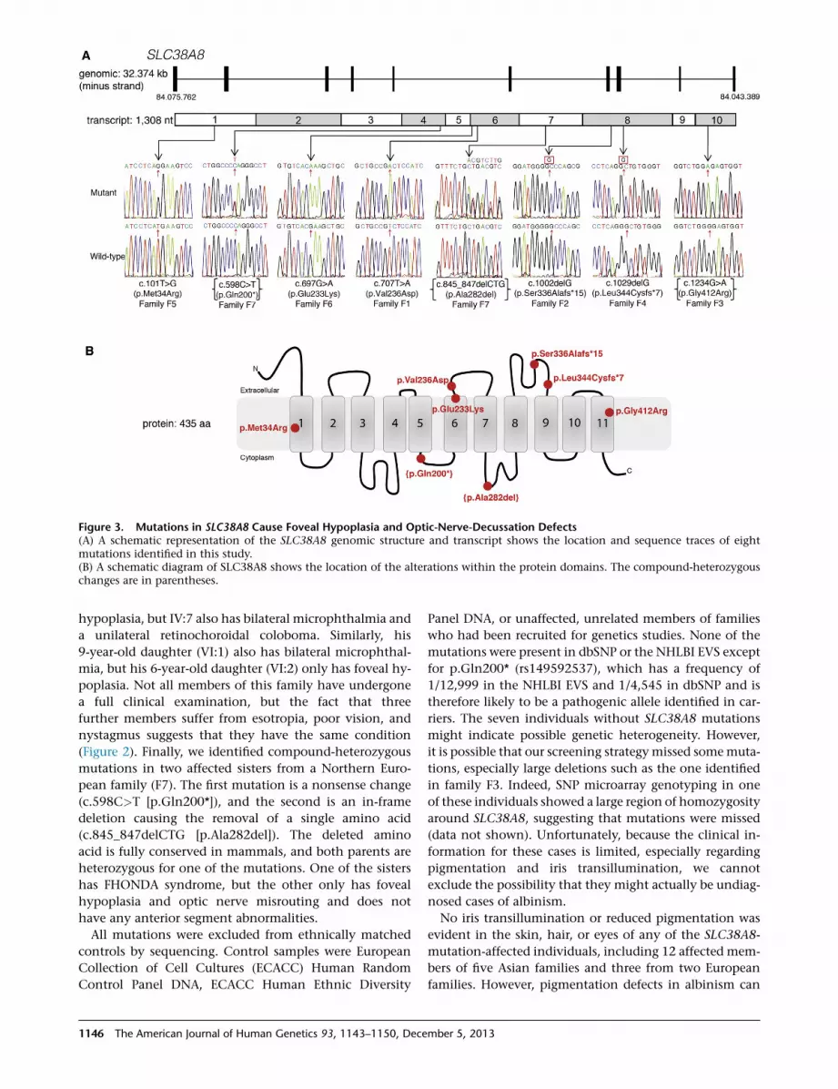
Figure 3. Mutations in SLC38A8 Cause Foveal Hypoplasia and Optic-Nerve-Decussation Defects(A) A schematic representation of the SLC38A8 genomic structure and transcript shows the location and sequence traces of eightmutations identified in this study.(B) A schematic diagram of SLC38A8 shows the location of the alterations within the protein domains. The compound-heterozygouschanges are in parentheses.
hypoplasia, but IV:7 also has bilateral microphthalmia and
a unilateral retinochoroidal coloboma. Similarly, his
9-year-old daughter (VI:1) also has bilateral microphthal-
mia, but his 6-year-old daughter (VI:2) only has foveal hy-
poplasia. Not all members of this family have undergone
a full clinical examination, but the fact that three
further members suffer from esotropia, poor vision, and
nystagmus suggests that they have the same condition
(Figure 2). Finally, we identified compound-heterozygous
mutations in two affected sisters from a Northern Euro-
pean family (F7). The first mutation is a nonsense change
(c.598C>T [p.Gln200*]), and the second is an in-frame
deletion causing the removal of a single amino acid
(c.845_847delCTG [p.Ala282del]). The deleted amino
acid is fully conserved in mammals, and both parents are
heterozygous for one of the mutations. One of the sisters
has FHONDA syndrome, but the other only has foveal
hypoplasia and optic nerve misrouting and does not
have any anterior segment abnormalities.
All mutations were excluded from ethnically matched
controls by sequencing. Control samples were European
Collection of Cell Cultures (ECACC) Human Random
Control Panel DNA, ECACC Human Ethnic Diversity
1146 The American Journal of Human Genetics 93, 1143–1150, Dece
Panel DNA, or unaffected, unrelated members of families
who had been recruited for genetics studies. None of the
mutations were present in dbSNP or the NHLBI EVS except
for p.Gln200* (rs149592537), which has a frequency of
1/12,999 in the NHLBI EVS and 1/4,545 in dbSNP and is
therefore likely to be a pathogenic allele identified in car-
riers. The seven individuals without SLC38A8 mutations
might indicate possible genetic heterogeneity. However,
it is possible that our screening strategymissed somemuta-
tions, especially large deletions such as the one identified
in family F3. Indeed, SNP microarray genotyping in one
of these individuals showed a large region of homozygosity
around SLC38A8, suggesting that mutations were missed
(data not shown). Unfortunately, because the clinical in-
formation for these cases is limited, especially regarding
pigmentation and iris transillumination, we cannot
exclude the possibility that they might actually be undiag-
nosed cases of albinism.
No iris transillumination or reduced pigmentation was
evident in the skin, hair, or eyes of any of the SLC38A8-
mutation-affected individuals, including 12 affected mem-
bers of five Asian families and three from two European
families. However, pigmentation defects in albinism can
mber 5, 2013

Figure 4. Morpholino Knockdown ofSlc38a8 in Medaka Fish Results in Micro-phthalmia, Coloboma, and Lens Defects,but No Skin- or Ocular-PigmentationAbnormalities(A) Bright-field stereomicroscope imagesshow lateral views of representative fishfor the controls and Mo-50UTR-slc38a8c3þc6 and Mo-50UTR-Slc38a8 c3þc6/Mo-p53 mutants. The red bracket highlightsthe reduced eye size in morphants, andthe arrows highlight displaced lenses.Ocular pigmentation identical to that ofthe controls can clearly be seen in themutant fish.(B) The frequency of each phenotypeobserved in the medaka fish is given as apercentage of 300.
be subtle (see GeneReviews inWeb Resources). This has led
to speculation that FHONDA syndrome is actually mild
albinism,1 especially given that anterior segment dysgen-
esis is observed in albinism.10,11 Medaka fish (Oryzias
latipes) have been shown to be a reliable model system
for investigating pigmentation defects,12 so we examined
the effect of morpholino-mediated ablation of Slc38a8 in
these fish. All experiments were conducted in strict accor-
dance with the institutional guidelines for animal research
and approved by the Department of Public Health, Animal
Health, Nutrition and Food Safety of the ItalianMinistry of
Health in accordance with the law on animal experimenta-
tion (article 7, D.L. 116/92, protocol number 00001/08/
IGB, approval date October 22, 2008). Furthermore, all
animal treatments were reviewed and approved in advance
by the ethics committee of the Institute of Genetics and
Biophysics Animal House (Naples).
Morpholinos were designed against both medaka
SLC38A8 orthologs present in the UCSC Genome
Browser (October 2005 v.1.0): ENSORLT00000016172
on chromosome 6 (19,760,744–19,770,301 bp) and
ENSORLT00000011189 on chromosome 3 (20,486,490–
20,491,975 bp), along with corresponding mismatch
controls (Table S3). The Cab strain of wild-type medaka
fish was used, and embryos were staged according to Iwa-
matsu.13 Morpholinos (Gene Tools) were injected into
1-cell fertilized embryos according to established proto-
cols.14 Optimal morpholino concentrations were deter-
mined on the basis of morphological criteria. Specificity
and inhibitory efficiency of each morpholino were deter-
mined as previously detailed.15
Both orthologs of SLC38A8 were simultaneously tar-
geted with either morpholinos targeting the 50 UTR or
The American Journal of Human Genetics
the splice donor site of exon 4. Off-
target effects of themorpholino injec-
tions were excluded by repeated
experiments with mismatched mor-
pholinos or by coinjection with a
p53 morpholino.16 None of the re-
sulting morphant embryos were
found to have any pigmentation defects of the eyes or
tegument, as determined by morphological analysis of
both the retinal pigment epithelium and tegument mela-
nophores (Figure 4 and Figure S3). Structural abnormalities
were restricted to the eye. Compared to controls, the
majority of the knockdown embryos (87%–92%) had
microphthalmia, a significant number (29%–60%) had
lens defects, and a smaller number (16%–36%) had fissure
coloboma (Figure 4). Given that medaka fish do not have a
fovea, one would not expect to see a fully overlapping
phenotype between humans and fish, but it is interesting
to note that members of family F6 have microphthalmia
and coloboma.
To investigate the effects of Slc38a8 knockdown on optic
nerve decussation, we repeated the morpholino experi-
ment on Athonal5::GFP transgenic medaka embryos,
which have GFP-labeled retinal ganglion cells.17 However,
we observed no differences between the optic chiasm
of wild-type medaka and the knockdown embryos
(Figure S4). This result was not unexpected given that the
lateral position of the eyes in medaka prevents overlap of
the visual fields so that all retinal ganglion axons cross
the midline at the optic chiasm.18 The significantly over-
lapping visual space in humans, however, makes it essen-
tial that ~40% of the axons do not cross the midline but
rather project ipsilaterally to allow binocular vision.19 Indi-
viduals with FHONDA syndrome display an increase in the
number of axons projecting contralaterally,4,6 indicating a
defect in the specification of the ipsilateral projections that
are absent in medaka.
Examination of the clinical phenotypes of the individ-
uals with SLC38A8 mutations showed that not all fulfill
the criteria for FHONDA syndrome (Table S4). Foveal
93, 1143–1150, December 5, 2013 1147

hypoplasia was present in all affected individuals, and
optic-nerve-decussation defects were present in all individ-
uals examined for this feature except one in whom the VEP
analysis was inconclusive (F4). However, anterior segment
abnormalities were only present in 3/7 families (F1, F2, and
F7), making this a variable component of the phenotype in
individuals with recessive SLC38A8 mutations. At this
time, it is not possible to determine whether the micro-
phthalmia and retinochoroidal coloboma phenotypes
observed in one of our families (and in the medaka fish)
are related to the SLC38A8 mutations, and further pheno-
type-genotype studies are thus needed. A number of cases
of isolated foveal hypoplasia have been reported in the
literature, and it would be interesting to screen SLC38A8
in these cases.20,21 Similarly, VEP analysis is often used
for diagnosing albinism in fair-skinned populations, where
pigmentation defects can be hard to identify, so it is likely
that some of these cases might actually harbor SLC38A8
mutations.22
To test the hypothesis that common variants in SLC38A8
influence fovea thickness in the general population,we car-
ried out an association study in a population cohort of
Northern European ancestry from the Western Australian
Pregnancy Cohort (Raine) 20-year follow-up eye study.23
As part of a comprehensive eye examination performed
in this study, the foveal thickness of both eyes in each
participant was imaged through dilated pupils by spec-
tral-domain optical coherence tomography (Spectralis; Hei-
delberg Engineering) by an experienced operator. Retinal
thickness at the fovea was automatically determined by
the instrument software as the distance between the inter-
nal limiting membrane and retinal pigment epithelium.
The ratio of foveal thickness to the mean parafoveal thick-
ness and the ratio of foveal thickness to the average perifo-
veal thickness for each eye were also recorded. Given the
high correlation between the right and left eyes (intraclass
correlation coefficient of 0.872 [95% confidence interval
(CI) ¼ 0.834–0.905], 0.911 [95% CI ¼ 0.847–0.943], and
0.733 [95% CI ¼ 0.576–0.819] for central foveal thickness,
fovea-parafovea ratio, and fovea-perifovea ratio, respec-
tively), the mean values for each participant were used for
analysis after inverse normal transformation.
DNA samples and consents for genome-wide association
studies (GWASs) were available from previous assessments.
Genotype data were generated with the genome-wide Illu-
mina 660 Quad Array at the Centre for Applied Genomics
(Toronto) and processed for quality control (QC). We used
the EIGENSTRAT program24 to conduct principal-compo-
nent analysis and constructed the first five principal
components for a subset of 42,888 SNPs that were not in
linkage disequilibrium with each other. Additionally, we
performed the GWAS imputation of 22 autosomes in the
MACH v.1.0.16 software by using the CEU (Utah residents
with ancestry from northern and western Europe from the
CEPH collection) samples from HapMap phase 2 (build 36,
release 22). A linear regression model in R with a PLINK
interface25 was used for determining associations between
1148 The American Journal of Human Genetics 93, 1143–1150, Dece
SNPs and foveal thickness, the ratio of foveal to parafoveal
thickness, or the ratio of foveal to perifoveal thickness. The
model was adjusted for age, sex, and the first two principal
components that accounted for the population stratifica-
tion. After QC, phenotypic and genetic data were available
from 679 individuals. We focused our analysis on SNPs sur-
rounding SLC38A8. These data suggest that variants at
this locus confer an effect in normal fovea variation and
suggest that SNP rs7200988 (allele A) is the most associated
with foveal thickness (p ¼ 5.77 3 10�4) (Figure S5 and
Table S5).
SLC38A8 encodes an orphan member of the SLC38
sodium-coupled neutral amino acid transporter (SNAT)
family of proteins, which are widely expressed and pre-
dominantly have glutamine as their preferred substrate.26
To determine the expression of SLC38A8, we performed
RT-PCR with primers spanning intron 7 on cDNA created
from a panel of mRNA from human adult and fetal tissue
(Clontech) according to standard protocols. SLC38A8
expression was found predominantly in neuronal tissue
(adult brain, fetal brain, and spinal cord), and very weak
expression was also present in the kidneys, thymus, and
testes (Figure S6).
Using a custom-made rabbit polyclonal antibody
(GenScript) raised against 14 amino acids located at the
N terminus of SLC38A8 (QTPGSRGLPEKPHP), we investi-
gated the protein localization of SLC38A8 by using human
brain and eye tissues obtained from the Leeds Tissue
Bank. SLC38A8 was shown to be located throughout the
neuronal retina but had strong staining in the inner and
outer plexiform layers and the photoreceptor layer
(Figure S6). Similarly, in the brain we showed that
SLC38A8 is localized in the cell body and axon of the
majority of neuronal cells and in a subset of glial cells
(Figure S6). These data are almost identical to those
obtained for the recently characterized glutamine trans-
porter SLC38A7,27 the closest homolog of SLC38A8,28
and suggest that both proteins have similar functions.
Glutamine is present at high concentrations in the brain
extracellular fluid and cycles between neurons and glial
cells, where it serves as an intermediate metabolite for
the formation of the neurotransmitters glutamate and
gamma-amino butyric acid. The location of SLC38A8 in
the retina is consistent with a synaptic neurotransmitter-
recycling role. However, this would not adequately explain
the developmental defect observed in SLC38A8-mutation
carriers. Active synapses are known to play a role in the
formation and remodeling of neural circuits, including
the projecting retinal ganglion cells,29 providing one
possible developmental disease mechanism. Alternatively,
the localization of SLC38A8 in the somata and axons of
brain cells might imply an additional role or roles for this
protein. Consistent with this hypothesis, there is emerging
evidence that neurotransmitters play a role in the devel-
oping retina bymodulating the proliferation of retinal pro-
genitor cells even before synapses are formed.30 In turn,
the number and spatiotemporal development of retinal
mber 5, 2013

cells are then believed to influence the extent to which
projecting axons cross at the chiasm or project ipsilaterally,
thus providing an alternative hypothesis to explain the
defects seen in individuals with defects in SLC38A8.31
In summary, we have shown that recessive mutations in
the putative glutamine transporter SLC38A8 cause foveal
hypoplasia and optic nerve misrouting. We show that
pigmentation is not affected by loss of SLC38A8 and thus
identify a melanin-independent component essential for
the development of these structures.
Supplemental Data
Supplemental Data include six figures and five tables and can be
found with this article online at http://www.cell.com/AJHG.
Acknowledgments
We thank the families who participated in this study. We thank
J. Deuchars and M. Singh for advice with immunohistochemistry
and A. Hindley and the Leeds Tissue Bank for providing and
processing samples. We thank J. Wittbrodt for providing the
ATH5::GFP medaka transgenic line and A.E. Davidson and Z. Li
for supporting genetic analysis. This work was supported by a
Royal Society University Research Fellowship (C.T.), University
of Leeds Emma and Leslie Reid Scholarship (J.A.P.), Omani
Government scholarship (M.A.-A.), Yorkshire Eye Research, the
Italian Telethon Foundation (project grant TGM11SB2), the Italian
Ministry of Research (PONA3_00311), RP Fighting Blindness,
Fight For Sight, Foundation Fighting Blindness, National Institute
for Health Research Moorfields Eye Hospital Biomedical Research
Centre, and the Sir Jules Thorn Award for Biomedical Research
(#JTA/09). We thank the Raine Study participants and staff for
cohort coordination and data collection, particularly Jenny
Mountain, Wei Ang, Craig Pennell, Hannah Forward, Charlotte
McKnight, Seyhan Yazar, Alla Soloshenko, Sandra Oates, and
Diane Wood. The core management of the Raine Study is funded
by the University of Western Australia (UWA), Telethon Institute
for Child Health Research, Raine Medical Research Foundation,
UWA Faculty of Medicine, Dentistry and Health Sciences,
Women’s and Infant’s Research Foundation, and Curtin Univer-
sity. Genotyping was funded by National Health and Medical
Research Council (NHMRC) grant 572613. The Raine Eye Health
Study was supported by NHMRC grant 1021105, the Lions Eye
Institute, the Australian Foundation for the Prevention of Blind-
ness, the Ophthalmic Research Institute of Australia, and the
Alcon Research Institute.
Received: July 31, 2013
Revised: September 5, 2013
Accepted: November 1, 2013
Published: November 27, 2013
Web Resources
The URLs for data presented herein are as follows:
dbSNP, http://www.ncbi.nlm.nih.gov/SNP/
ExonPrimer, http://ihg.gsf.de/ihg/ExonPrimer.html
GeneReviews, Lewis, R.A. (2012). Oculocutaneous Albinism Type
2, http://www.ncbi.nlm.nih.gov/books/NBK1232/
The American Jou
HapMap, http://hapmap.ncbi.nlm.nih.gov/index.html.en
Leeds Tissue Bank, http://www.gift.leeds.ac.uk/index.html
MACH 1.0.16, http://www.sph.umich.edu/csg/yli/mach/index.
html
NHLBI Exome Sequencing Project (ESP) Exome Variant Server,
http://evs.gs.washington.edu/
Online Mendelian Inheritance in Man (OMIM), http://www.
omim.org/
RefSeq, http://www.ncbi.nlm.nih.gov/RefSeq
UCSC Genome Browser, http://genome.ucsc.edu/
References
1. Michaelides, M., Jeffery, G., and Moore, A.T. (2012). Develop-
mental macular disorders: phenotypes and underlying molec-
ular genetic basis. Br. J. Ophthalmol. 96, 917–924.
2. Dessinioti, C., Stratigos, A.J., Rigopoulos, D., and Katsambas,
A.D. (2009). A review of genetic disorders of hypopigmenta-
tion: lessons learned from the biology of melanocytes. Exp.
Dermatol. 18, 741–749.
3. Schiaffino, M.V. (2010). Signaling pathways in melanosome
biogenesis and pathology. Int. J. Biochem. Cell Biol. 42,
1094–1104.
4. Al-Araimi, M., Pal, B., Poulter, J.A., van Genderen, M.M., Carr,
I., Cudrnak, T., Brown, L., Sheridan, E., Mohamed, M.D., Brad-
bury, J., et al. (2013). A new recessively inherited disorder
composed of foveal hypoplasia, optic nerve decussation de-
fects and anterior segment dysgenesis maps to chromosome
16q23.3-24.1. Mol. Vis. 19, 2165–2172.
5. Pal, B., Mohamed, M.D., Keen, T.J., Williams, G.A., Bradbury,
J.A., Sheridan, E., and Inglehearn, C.F. (2004). A new pheno-
type of recessively inherited foveal hypoplasia and anterior
segment dysgenesis maps to a locus on chromosome
16q23.2-24.2. J. Med. Genet. 41, 772–777.
6. van Genderen, M.M., Riemslag, F.C.C., Schuil, J., Hoeben, F.P.,
Stilma, J.S., and Meire, F.M. (2006). Chiasmal misrouting and
foveal hypoplasia without albinism. Br. J. Ophthalmol. 90,
1098–1102.
7. Loges, N.T., Olbrich, H., Becker-Heck, A., Haffner, K., Heer, A.,
Reinhard, C., Schmidts, M., Kispert, A., Zariwala, M.A., Leigh,
M.W., et al. (2009). Deletions and point mutations of LRRC50
cause primary ciliary dyskinesia due to dynein arm defects.
Am. J. Hum. Genet. 85, 883–889.
8. Vincent, A., Kemmanu, V., Shetty, R., Anandula, V., Madha-
varao, B., and Shetty, B. (2009). Variable expressivity of ocular
associations of foveal hypoplasia in a family. Eye (Lond.) 23,
1735–1739.
9. Anandula, V.R., Shetty, R., Vincent, A., Ramprasad, V.L., and
Ramesh, N. (2011). Gene mapping in a highly inbred consan-
guineous foveal hypoplasia family to cytogenetic region
16q24.1. JournalofMedicalGenetics andGenomics3, 122–125.
10. Charles, S.J., Green, J.S., Grant, J.W., Yates, J.R., and Moore,
A.T. (1993). Clinical features of affected males with X linked
ocular albinism. Br. J. Ophthalmol. 77, 222–227.
11. Shiono, T., Tsunoda, M., Chida, Y., Nakazawa, M., and Tamai,
M. (1995). X linked ocular albinism in Japanese patients. Br. J.
Ophthalmol. 79, 139–143.
12. Fukamachi, S., Shimada, A., and Shima, A. (2001). Mutations
in the gene encoding B, a novel transporter protein, reduce
melanin content in medaka. Nat. Genet. 28, 381–385.
13. Iwamatsu, T. (2004). Stages of normal development in the
medaka Oryzias latipes. Mech. Dev. 121, 605–618.
rnal of Human Genetics 93, 1143–1150, December 5, 2013 1149

14. Conte, I., Carrella, S., Avellino, R., Karali, M., Marco-Ferreres,
R., Bovolenta, P., and Banfi, S. (2010). miR-204 is required
for lens and retinal development via Meis2 targeting. Proc.
Natl. Acad. Sci. USA 107, 15491–15496.
15. Eisen, J.S., and Smith, J.C. (2008). Controlling morpholino
experiments: don’t stop making antisense. Development
135, 1735–1743.
16. Robu, M.E., Larson, J.D., Nasevicius, A., Beiraghi, S., Brenner,
C., Farber, S.A., and Ekker, S.C. (2007). p53 activation by
knockdown technologies. PLoS Genet. 3, e78.
17. Del Bene, F., Ettwiller, L., Skowronska-Krawczyk, D., Baier, H.,
Matter, J.M., Birney, E., and Wittbrodt, J. (2007). In vivo vali-
dation of a computationally predicted conserved Ath5 target
gene set. PLoS Genet. 3, 1661–1671.
18. Yoda, H., Hirose, Y., Yasuoka, A., Sasado, T., Morinaga, C.,
Deguchi, T., Henrich, T., Iwanami, N., Watanabe, T., Osakada,
M., et al. (2004). Mutations affecting retinotectal axonal
pathfinding in Medaka, Oryzias latipes. Mech. Dev. 121,
715–728.
19. Petros, T.J., Rebsam, A., and Mason, C.A. (2008). Retinal axon
growth at the optic chiasm: to cross or not to cross. Annu. Rev.
Neurosci. 31, 295–315.
20. Curran, R.E., and Robb, R.M. (1976). Isolated foveal hypopla-
sia. Arch. Ophthalmol. 94, 48–50.
21. Oliver, M.D., Dotan, S.A., Chemke, J., and Abraham, F.A.
(1987). Isolated foveal hypoplasia. Br. J. Ophthalmol. 71,
926–930.
22. Sjostrom, A., Kraemer, M., Ohlsson, J., and Villarreal, G.
(2001). Subnormal visual acuity syndromes (SVAS): albinism
in Swedish 12-13-year-old children. Doc. Ophthalmol. 103,
35–46.
23. Yazar, S., Forward, H., McKnight, C.M., Tan, A., Soloshenko,
A., Oates, S.K., Ang, W., Sherwin, J.C., Wood, D., Mountain,
J.A., et al. (2013). Raine Eye Health Study: Design, Methodol-
ogy and Baseline Prevalence of Ophthalmic Disease in a Birth-
1150 The American Journal of Human Genetics 93, 1143–1150, Dece
cohort Study of Young Adults. Ophthalmic Genet. Published
online January 10, 2013.
24. Price, A.L., Patterson, N.J., Plenge, R.M., Weinblatt, M.E.,
Shadick, N.A., and Reich, D. (2006). Principal components
analysis corrects for stratification in genome-wide association
studies. Nat. Genet. 38, 904–909.
25. Purcell, S., Neale, B., Todd-Brown, K., Thomas, L., Ferreira,
M.A., Bender, D., Maller, J., Sklar, P., de Bakker, P.I., Daly,
M.J., and Sham, P.C. (2007). PLINK: a tool set for whole-
genome association and population-based linkage analyses.
Am. J. Hum. Genet. 81, 559–575.
26. Mackenzie, B., and Erickson, J.D. (2004). Sodium-coupled
neutral amino acid (System N/A) transporters of the SLC38
gene family. Pflugers Arch. 447, 784–795.
27. Hagglund, M.G., Sreedharan, S., Nilsson, V.C., Shaik, J.H.,
Almkvist, I.M., Backlin, S., Wrange, O., and Fredriksson, R.
(2011). Identification of SLC38A7 (SNAT7) protein as a gluta-
mine transporter expressed in neurons. J. Biol. Chem. 286,
20500–20511.
28. Schioth, H.B., Roshanbin, S., Hagglund, M.G., and Fredriks-
son, R. (2013). Evolutionary origin of amino acid transporter
families SLC32, SLC36 and SLC38 and physiological,
pathological and therapeutic aspects. Mol. Aspects Med. 34,
571–585.
29. Koch, S.M., Dela Cruz, C.G., Hnasko, T.S., Edwards, R.H.,
Huberman, A.D., and Ullian, E.M. (2011). Pathway-specific
genetic attenuation of glutamate release alters select features
of competition-based visual circuit refinement. Neuron 71,
235–242.
30. Martins, R.A., and Pearson, R.A. (2008). Control of cell prolif-
eration by neurotransmitters in the developing vertebrate
retina. Brain Res. 1192, 37–60.
31. Jeffery, G. (2001). Architecture of the optic chiasm and the
mechanisms that sculpt its development. Physiol. Rev. 81,
1393–1414.
mber 5, 2013