reactive oxygen species, amp-activated protein kinase, and the transcription cofactor p300 regulate...
TRANSCRIPT
Codogno, Christian Pous and Daniel PerdizArnaud Bruneel, Ahmed Hamai, PatriceRatier, Najet Mejdoubi-Charef, Anita Baillet, Rafah Mackeh, Severine Lorin, Ameetha Hyperacetylation during Cell StressTAT-1/MEC-17)-Dependent Microtubule
αAcetyltransferase-1 (Cofactor p300 Regulate Alpha-TubulinProtein Kinase and the Transcription Reactive Oxygen Species, AMP-activatedCell Biology:
published online March 11, 2014J. Biol. Chem.
10.1074/jbc.M113.507400Access the most updated version of this article at doi:
.JBC Affinity SitesFind articles, minireviews, Reflections and Classics on similar topics on the
Alerts:
When a correction for this article is posted•
When this article is cited•
to choose from all of JBC's e-mail alertsClick here
http://www.jbc.org/content/early/2014/03/11/jbc.M113.507400.full.html#ref-list-1
This article cites 0 references, 0 of which can be accessed free at
at UN
IVE
RSIT
É PA
RIS-SU
D 11 on M
arch 13, 2014http://w
ww
.jbc.org/D
ownloaded from
at U
NIV
ER
SITÉ
PAR
IS-SUD
11 on March 13, 2014
http://ww
w.jbc.org/
Dow
nloaded from
Stress-induced microtubule hyperacetylation
1
Reactive Oxygen Species, AMP-activated Protein Kinase and the Transcription Cofactor p300 Regulate Alpha-Tubulin Acetyltransferase-1 (αTAT-1/MEC-17)-Dependent Microtubule
Hyperacetylation during Cell Stress*
Rafah Mackeh1, Séverine Lorin1, Ameetha Ratier1, Najet Mejdoubi-Charef1, Anita Baillet1, Arnaud Bruneel1,2, Ahmed Hamaï3, Patrice Codogno3, Christian Poüs1,4, Daniel Perdiz1
1 Université Paris Sud, EA4530, UFR de Pharmacie, Châtenay-Malabry, France 2AP-HP, service de biochimie métabolique et cellulaire, Hôpital Bichat, 75018 Paris, France.
3 INSERM U845, Université Paris Descartes, Paris, France 4Biochimie-Hormonologie, Hôpital Antoine Béclère, APHP, Clamart, France
*Running title: stress-induced microtubule hyperacetylation
Address correspondence to Pr. Christian Poüs, EA4530, Université Paris-Sud, UFR de Pharmacie, 5 rue J.B. Clément, 92296 Châtenay-Malabry, France. Tel. (33)146835477. E-mail: [email protected] Keywords: tubulin acetylation, microtubule, MEC-17/αTAT1, p300, ROS, AMPK.
Background: Tubulin acetylation is a hallmark of microtubule stabilization, which may modulate the binding of microtubule-associated proteins. Results: Microtubules are hyperacetylated due to stress-induced cellular signaling upstream of the tubulin acetyltransferase MEC-17/αTAT1. Conclusion: MEC-17/αTAT1 is regulated by p300, reactive oxygen species and AMP-activated protein kinase. Significance: Microtubule hyperacetylation is important for cell adaptation to stress through autophagy induction and for cell survival. ABSTRACT Beyond its presence in stable microtubules, tubulin acetylation can be boosted after UV exposure or after nutrient deprivation but the mechanisms of microtubule hyperacetylation are still unknown. In this study, we show that this hyperacetylation is a common response to several cellular stresses that involves the stimulation of the major tubulin acetyltransferase MEC-17. We also demonstrate that the acetyltransferase p300 negatively regulates MEC-17 expression and is sequestered on microtubules upon stress. We further show that reactive oxygen species (ROS) of mitochondrial origin are required
for microtubule hyperacetylation by activating the AMP kinase, which in turn mediates MEC-17 phosphorylation upon stress. Finally, we show that preventing microtubule hyperacetylation by knocking-down MEC-17 affects cell survival under stress conditions and starvation-induced autophagy, thereby pointing out the importance of this rapid modification as a broad cell response to stress.
Cellular adaptive response to various stresses involves the coordinated activation of several signaling pathways that may ultimately lead to cell survival or to cell death depending on the nature, intensity and duration of the insult. Cells primarily try to cope with stressful stimuli by triggering responses that may modulate gene expression and/or allow protective mechanisms such as autophagy induction. In the case their defenses are overwhelmed or in response to special stimuli, cells die by apoptosis or necrosis. Cell adaptation and survival might be compromised in pathological states such as diabetes or neurodegenerative diseases. Alternatively, cell adaptation and tolerance to death signals is an important feature of cancer (for reviews see (1, 2)).
http://www.jbc.org/cgi/doi/10.1074/jbc.M113.507400The latest version is at JBC Papers in Press. Published on March 11, 2014 as Manuscript M113.507400
Copyright 2014 by The American Society for Biochemistry and Molecular Biology, Inc.
at UN
IVE
RSIT
É PA
RIS-SU
D 11 on M
arch 13, 2014http://w
ww
.jbc.org/D
ownloaded from

Stress-induced microtubule hyperacetylation
2
Appropriate spatial and temporal coordination of cellular signaling is required for optimal cell response. In this respect, the microtubule (MT) cytoskeleton has an important role in organizing the cytoplasm by sequestering and releasing transduction factors, allowing their assembly into complexes or supporting their vectorization by molecular motors (for reviews see (3, 4)). Depending on the signaling pathway, these properties may require MT dynamics, which consists in alternations of growth and shrinking phases termed dynamic instability (5), or they may preferentially occur on a stable MT subset, in which tubulin bears numerous post-translational modifications. Some modifications act as local structural determinants that can be readily recognized by tubulin-binding proteins. This is the case for α-tubulin detyrosination, which prevents MT binding of MCAK and CLIP-170, two proteins that regulate the transitions between growth and shrinking phases termed catastrophes (towards MT disassembly) and rescues (towards a new growth phase), respectively (6, 7). Other modifications like the acetylation of the K40 residue of α-tubulin are thought to modulate MT conformation by triggering subtle changes in the way tubulin subunits interact with each other (8, 9). Although the exact molecular effect of tubulin acetylation remain largely obscure due to its localization in the MT lumen, important progress has been accomplished during the past few years in our understanding of the biological function of this modification. Even though it was recently challenged in in vitro experiments (10, 11), tubulin acetylation was found to stimulate the binding of dynein and of kinesin-1 to the surface of cellular MTs (12-15). Tubulin acetylation was also found to modulate the binding and function of signaling factors involved in cell survival in response to stress. For example, the activation of endothelial nitric oxide synthase required stable, acetylated MTs for optimal activation by phosphorylation (16). Furthermore Akt activation and p53 transport into the peri-nuclear area require tubulin acetylation (17), and this binding to MTs occurs via the chaperone Hsp90. In starvation-induced autophagy, the stress-induced MAP-kinase c-Jun N-terminal kinase (JNK) is activated via a kinesin-1-mediated recruitment on MTs that also
depends on tubulin acetylation (18). Interestingly, in these studies, tubulin ace-tylation is not a static process that may only occur on stable MTs, but that it is also highly inducible, including on the dynamic MT subset, in response to genotoxic stress or to nutrient deprivation (17, 18). Tubulin acetylation levels result from a balance between the activities of the cytoplasmic deacetylases HDAC6 and SIRT2 and that of various tubulin acetyltransferases including ARD1/NAT1, NAT10, Gcn5, ELP3 and αTAT1/MEC-17 (reviewed in (19)). Among these enzymes, MEC-17 appears as a major acetyltransferase, which can account for most of the tubulin acetylation in stable MTs (20, 21).
The level of tubulin acetylation in MTs is thus an important factor to allow cells to organize and possibly coordinate several signaling pathways along MTs. It functions in static conditions on stable MTs, but also in a dynamic manner that is probably tightly regulated. However, the cellular and molecular mechanisms that enhance tubulin acetylation in MTs (hereafter referred to as MT hyper-acetylation) have not been explored yet. In this study, we characterized MT hyperacetylation and addressed the question of its induction in response to cell stress. We show that it is a rapid and reversible process that results from an acetyltransferase induction triggered by the release of mitochondrial reactive oxygen species (ROS) and by AMPK. We further show that MEC-17 is the sole acetyltransferase responsible for MT hyperacetylation, and that AMPK stimulates its phosphorylation in response to cell stress. We also provide evidence that MT hyperacetylation is required for cell survival in response to oxidative stress and for starvation-induced autophagy stimulation.
EXPERIMENTAL PROCEDURES
Chemical products and antibodies All chemicals were purchased from Sigma-Aldrich (France). Mouse monoclonal anti-α-tubulin (DM1-A), β-tubulin I (T7816), anti-acetylated-α-tubulin (6-11B-1) and rabbit anti-LC3B (L7543) were from Sigma-Aldrich.
at UN
IVE
RSIT
É PA
RIS-SU
D 11 on M
arch 13, 2014http://w
ww
.jbc.org/D
ownloaded from

Stress-induced microtubule hyperacetylation
3
β-actin HRP-conjugated antibody (C4) was from Santa-Cruz Biotechnology (sc-47778). p300 antibody (RW-105) was from Pierce Biotechnology (MA1-16622). Rabbit anti-phospho-AMPKα (Thr172), anti-AMPKα1/2 and anti-GFP were from Cell Signaling technology (ref: 2535, 2532, 2555 respectively). PARP mAb antibody was from Clonetech (C-2-10, ref 630210). Alexa fluor 488-conjugated goat anti-mouse was purchased from Invitrogen. Cell culture and stress conditions Human HeLa cells, MEF cells and the hTERT-immortalized retinal pigment epithelial cell line (hTERT-RPE1) were cultured in Dulbecco's Modified Eagle Medium (DMEM, ATGC, France) supplemented with 1% sodium pyruvate, 10% fetal bovine serum (FBS) and antibiotics (100U/ml penicillin, 100µg/ml streptomycin) in 5% CO2 at 37ᵒC. PtK2 (Potorous tridactylis kidney cells) were purchased from ATCC and cultured in EMEM (Eagle's Minimum Essential Medium (EMEM, ATCC® 30-2003™) supplemented with 10% FBS. Cells were always used at less than 80% confluence and before passage 12. For amino acid deprivation, DMEM was replaced by 25mM HEPES-buffered EBSS (Earle's Balanced Salt Solution) after three washes. For stress induction, culture medium (or PBS for H2O2) was replaced by freshly prepared medium to which the indicated concentrations of chemical reagent were added. NaCl concentration in DMEM is 150mM (116mM for EMEM), thus the addition of 125, 250 or 500mM NaCl (as indicated in the figures) yields final NaCl concentrations of 275, 400 or 650mM for DMEM (241, 366 or 616mM for EMEM). MTT assay Cells were cultured in 12-wells plates and transfected with MEC-17 siRNA. Stress was induced with NaCl for 24 h. Cells were then incubated for 2 h with 3-(4,5-Dimethylthiazol-2-yl)-2,5-diphenyltetrazolium bromide reagent (MTT, Sigma-Aldrich) at 37ᵒC. Then medium was removed, formazan crystals were dissolved with DMSO and optical density measurements were read on a microplate reader at 540 nm. Immunofluorescence and microscopy
Cells grown on glass coverslips were washed with calcium- and magnesium-free PBS and fixed with -20ᵒC methanol for 5 min. Cells were then washed three times before staining with monoclonal anti acetyl-α-tubulin for 1 h at 37ᵒC. After three washes with PBS, cells were incubated with Alexa fluor 488-conjugated goat anti-mouse (45 min, 37ᵒC). Images were acquired using a Scion CFW1312M CCD camera, on a Leica DMLB microscope (100x 1.3 NA objective). Plasmid and siRNA transfections jetPEI™ (Polyplus™) was used for plasmid transfection of HeLa cells according to the manufacturer’s instructions. GFP-MEC-17WT and GFP-MEC17D157N were from Dr M. Nachury (Department of Molecular and Cellular Physiology, Stanford University School of Medicine; Addgene references: 27099 and 27100 respectively). mCherry-tubulin vector was kindly provided by Dr. R. Y. Tsien (Department of Pharmacology, Howard Hughes Medical Center, University of San Diego). The mCherry tubulin K40A mutated vector was a kind gift from Dr. F. Saudou (Institut Curie Orsay France). For all siRNA transfections, we used HiPerFect Transfection Reagent (Qiagen, 301705) according to the manufacturer’s instructions. To deplete p300 from HeLa cells, we used a duplex of p300 siRNA (Sigma, SASI_Hs01_00052818- 5’CUAGAGACACCU UGUAGUA[dT][dT]3’and 5’-UACUACAAGG UGUCUCUAG[dT][dT]). Cells were trans-fected after 24 h with p300 siRNA (200nM/well of a 6-well plate). To deplete MEC-17 in HeLa and RPE1 cells, we used ON-TARGETplus siRNAs from Dharmacon as a pool of four siRNAs 5’GUAGCUAGGUCCCGAUAUA-3’; 5’GAGUAUAGCUAGAUCCCUU-3'; 5’GGGAAACUCACCAGAACGA-3’; 5’CUUGUGAGAUUGUCGAGAU-3’. Transfections were repeated at days 1, 2 and 3 at a final concentration of 100nM and cells were used at day 4. AMPK siRNA was purchased from Santa-Cruz technology (AMPKα1/2 siRNA- sc-45312). Transfections were repeated at days 1 and 2 at a final concentration of 100nM.
at UN
IVE
RSIT
É PA
RIS-SU
D 11 on M
arch 13, 2014http://w
ww
.jbc.org/D
ownloaded from
Stress-induced microtubule hyperacetylation
4
Total cell lysate preparation After washing with PBS, cells were directly lysed in Laemmli sample buffer, boiled at 100ᵒC for 5 min and sonicated. 10µg to 20µg of proteins were subjected to Western blot analysis using appropriate antibodies. Preparation of soluble (cytosolic) and polymerized microtubule fractions 80% confluent cells cultured in a 75cm2 flask were washed two times with 37ᵒC-prewarmed PEM buffer (80mM PIPES, 2mM EGTA, 1mM MgCl2, pH 6.9). The cytosolic (non-microtubular) fractions were extracted after permeabilization using PEM supplemented with 0.075% Triton X-100, 10% glycerol, a protease inhibitor mixture (1mM PMSF and 20µM benzamidine/leupeptine) and phosphatase inhibitors (1mM NaF and 0.1mM ortho-vanadate) during 3 min at 37ᵒC. Cytosolic fractions were collected and permeabilized cells were gently washed to remove residual Triton. Polymerized microtubules were depolymerized on ice for one h in PM buffer (80mM PIPES 1mM MgCl2, pH 6.9) supplemented with 5mM CaCl2 and the protease and phosphatase inhibitors mixtures and then collected. Both extracts (soluble and formerly polymerized fractions) were concentrated down to 200µL using vivaspin 6 filtration units of 10kDa (Sartorius Stedim Biotech), then subjected to Western blot analysis. Cytoplasmic and nuclear extraction 80% confluent cells in a 75cm2 flask were trypsinated and cell pellet was suspended in 1mL of buffer A (10mM KCl, 1.5mM MgCl2, 10mM HEPES, 0.5mM DTT, pH 7.9) and transferred into a Dounce homogenizer for cell disruption. After centrifugation (5 min, 220g, 4°C) supernatants (containing the cytosolic fraction) were stored and pellets (containing nuclei) were resuspended in 500µL of buffer 1 (10mM MgCl2, 0.25M sucrose) and mixed with 500µL of buffer 2 (0.5mM MgCl2, 0.88M sucrose). After centrifugation, the pellets were resuspended in 100µL of lysis buffer (25mM Tris pH 8, 150mM NaCl, 1% NP-40, 1% sodium deoxycholate, 0.1% SDS) and sonicated. Finally, cytosol-containing and nuclei-containing
fractions were centrifuged (3000g, 10 min) and supernatants were stored at -20ᵒC prior to Western blot analysis. SDS-PAGE and Western blot analysis After SDS-PAGE, proteins were transferred to PVDF membranes. After blocking non-specific binding sites with 10% of non-fat dry milk, blots were probed with appropriate antibodies and revealed with HRP-conjugated secondary antibodies (Pierce ECL Plus Western Blot substrate) on Kodak Biomax MR films. Densitometry analysis was performed using the Image J software. 2-DE 6x10⁴ HeLa cells were cultured in 60mm cell culture dishes. After appropriate treatment or transfection, cells were collected in 100µL of lysis solution (50mM DTT, 4% CHAPS, proteases inhibitors (PMSF, Benzamidine / Leupeptine), sonicated and kept on ice for 30 min and centrifuged to eliminate cell debris. For phosphatase treatment, calf intestinal alkaline phosphatase (Takara Bio INC NO 2250A) was added to the lysates according to the manufacturer’s instructions and incubated for 30 min at 37ᵒC. Reactions were stopped by adding 50mM of DTT. Protein mixtures were desalted on Biospins-6 Tris columns with Bio-Gel in Tris Buffer (BioRad, Hercules, CA, USA), precipitated in cold acetone for one h, centrifuged and the pellet was solubilized in IEF rehydration solution (6M urea, 4% CHAPS, 50mM DTT, 0.5% ampholytes, orange G). IEF was performed using linear IPG strips pH 6-10 (ZOOM® STRIP Invitrogen) and the second dimension was carried out using ready-made 4-12% Bis-Tris ZOOM® mini Gel (Invitrogen). In vitro tubulin deacetylation assay Cell lysates of control cells or cells treated with NaCl were prepared using 10mM Tris buffer pH 8.0 supplemented with 1% Triton, 1mM NAD+ and the protease and phosphatase inhibitor mixtures. Lysate aliquots containing 20µg proteins were mixed with 2µg of soluble acetylated tubulin dimers purified from porcine brain (22) and adjusted to 50µL with PEM. After one h at 37ᵒC, reactions were stopped by adding 4x SDS-PAGE sample buffer
at UN
IVE
RSIT
É PA
RIS-SU
D 11 on M
arch 13, 2014http://w
ww
.jbc.org/D
ownloaded from
Stress-induced microtubule hyperacetylation
5
and immediately subjected to SDS-PAGE and Western blotting of acetylated and total α-tubulin. Reverse Transcription and quantitative PCR After siRNA transfection, total cellular RNAs were extracted with 200µL of Extract-All reagent (Eurobio) according to the manufacturer’s instructions. First-strand cDNA were generated by reverse transcription of 1µg of total RNAs using oligo (dT) 12-15 primer and SuperScriptIII ® reverse transcriptase kit (Invitrogen Life Technologies) according to the manufacturer’s instructions in a total reaction volume of 20µL. Real-time PCR was performed in a lightCycler thermal cycler (Roche Diagnostics, Meylan, France). The amplification of each cDNA (50ng/µL) was performed in a 10µL volume, using the Fast Start DNA MasterPLUS SYBER GREEN I master mix (Roche Diagnostics) with a 500nM final concentration of each primer. The primers used for MEC-17 were the following: MEC-17(f)-5’GGCGAGAACTCTTCCAGTAT-3’ and MEC-17(r) - 5’-TTGTTCACCTGTGGGACT-3’ (21). Data were normalized using the GAPDH cDNA content. Statistical analysis Quantitative data are the mean ± SEM of at least three independent experiments. Data ± SEM were compared using the non-parametric Mann-Whitney's U test for dose-dependent hyperacetylation, cell survival and autophagy induction experiments. Two-way Anova was used to evaluate the effect of chemicals on stress-induced MT hyperacetylation. Acknowledgments This work was supported by a grant from the ministère de l’enseignement supérieur et de la recherche (EA 4530) and a grant from INSERM (U845). We thank Claudine Deloménie for technical assistance in quantitative RT-PCR experiments. We also thank Béatrice Benoit and Antoine Pilon for the fruitful scientific discussions about this project. The authors also thank Dr. M.P. Murphy (MRC, Cambridge, UK) for his kind gift of, and expert advice on MitoQ.
RESULTS Microtubule hyperacetylation is a general and reversible cell stress response.
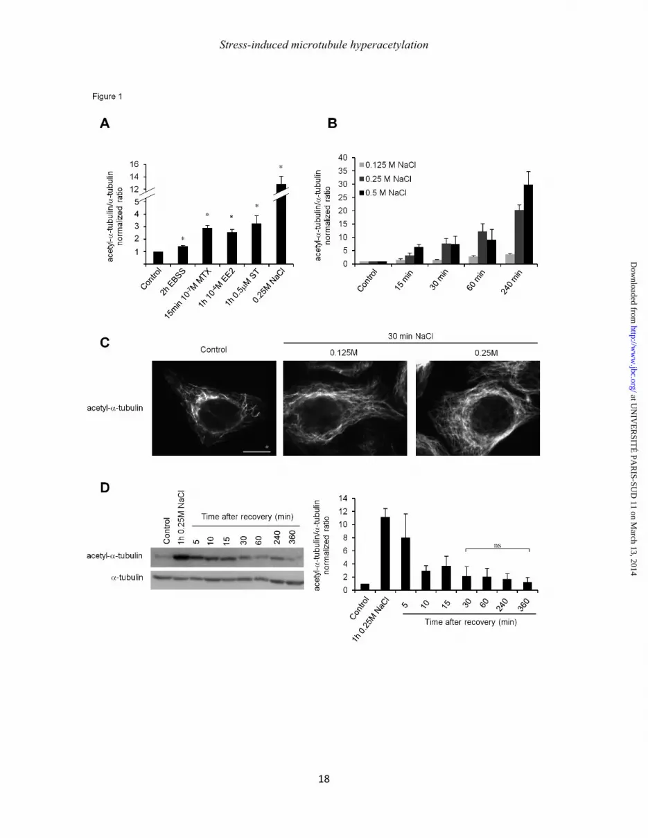
We found earlier that MT acetylation is induced during amino acid starvation (18) and after exposure to UVC radiation (17). To examine if this hyperacetylation is a general stress response, HeLa cells were starved of amino-acids for 2 h or treated with 10-4µM of the synthetic hormone ethinyl estradiol (EE2), 10-7µM methoxychlore (MTX), which is a synthetic organochlorine used as an insecticide, 0.5µM staurosporine (ST) or with the addition of 0.25M NaCl (Fig. 1A, B). All these stimuli increased tubulin acetylation with an amplitude that ranged between 1.5 fold for amino-acid starvation (EBSS) and 12.5 fold for NaCl. Particularly, exposure to NaCl caused rapid hyperacetylation of the MT network (as soluble tubulin was not affected; data not shown) in a time- and dose-dependent manner (Fig. 1B). This NaCl-induced hyperacetylation is clearly visible in immunofluorescence experiments where control cells show a few discontinuously labeled acetylated MT while stressed cells exhibit long hyperacetylated MT all over the network (Fig. 1C). Interestingly, MT hyperacetylation looks as a general cell response to stress, as we also observed it in other cell lines like RPE-1 and MEF (data not shown). We chose to use NaCl in priority to further identify the mechanisms that stimulate MT acetylation, due to its potent inductive effect. To explore the reversibility of this hyperacetylation, cells were subjected to a recovery experiment after a 1 h induction with 0.25M NaCl. As expected, tubulin acetylation level increased after 1 h of treatment, and then regularly decreased upon NaCl washout until reaching basal level after ~30 min (Fig. 1D). MEC-17 is required for stress-induced microtubule hyperacetylation.
Tubulin acetylation results from the balance between tubulin acetyltransferases (TAT) and deacetylases. To examine the
at UN
IVE
RSIT
É PA
RIS-SU
D 11 on M
arch 13, 2014http://w
ww
.jbc.org/D
ownloaded from
Stress-induced microtubule hyperacetylation
6
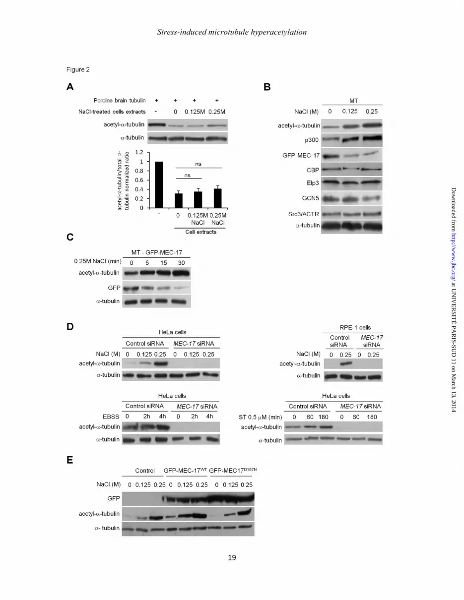
involvement of deacetylases in stress-induced MT hyperacetylation, we performed an in vitro tubulin deacetylation assay that consists in mixing brain tubulin, which is highly acetylated, with lysates of control or NaCl-treated cells. Lysates of control cells were able to deacetylate tubulin by ~70%. This activity remained unchanged in lysates of NaCl-treated cells, indicating that tubulin deacetylase inhibition is not responsible for the increase in the level of tubulin acetylation (Fig. 2A), but that it rather results from an increase in TAT activity. To identify which TATs are involved, we first monitored the recruitment of putative TATs on MT fractions prepared from control and NaCl-treated cells. MEC-17 detection required the overexpression of a GFP-tagged enzyme, since all the antibodies we tested failed to recognize endogenous MEC-17 in our hands. Among the acetyltransferases we tested, only p300 increased in the MT-containing fraction in a NaCl dose-dependent manner (Fig. 2B), suggesting that it could be involved in MT hyperacetylation. MEC-17 was recently discovered as the main TAT (20, 21) but unexpectedly, GFP-MEC-17 decreased in the MT fraction upon stress (Fig. 2B) in a time-dependent manner (Fig. 2C) and this decrease was not due to degradation as GFP-MEC-17 content remained constant in total cell lysates during NaCl stress (Fig. 2E).
To check whether MEC-17 is involved in MT hyperacetylation, we used a siRNA approach. MEC-17 knockdown dramatically reduced tubulin acetylation both in basal and upon various stress-induced conditions (amino-acid starvation, NaCl, staurosporine) (Fig 2D). MEC-17 is also required for MT hyperacetylation in other cell types such as RPE-1 (Fig. 2D). Conversely, the overexpression of MEC-17WT but not of its catalytically inactive mutant MEC-17D157N, accentuated both basal and stress-induced tubulin acetylation as shown in Fig. 2E. MEC-17 is thus required for stress-induced MT hyperacetylation.
p300 negatively regulates MEC-17-dependent tubulin acetylation.
The high level of p300 recruitment on MT upon stress was intriguing because its main
localization is expected to be nuclear. To further explore this point, we determined first in which cytoplasmic compartment p300 localizes upon stress. As shown in Fig. 3A, p300 dramatically increased in the MT fraction upon NaCl addition, in a dose-dependent manner but remained undetectable in the cytosol. Also, the global p300 level remained unchanged upon stress. Together, these two results suggest that p300 found in the MT fraction would originate from a fast and effective nuclear export. This hypothesis was readily confirmed both by a Western-blot analysis of whole cytoplasmic (thus containing MTs) and nuclear extracts from control and NaCl-treated cells and by immunofluorescence experiments (Fig. 3B). The kinetics of p300 export from the nucleus shows a rapid nuclear depletion of the enzyme, which is almost complete after 30 min of stress. Note that by 30 min, all the cytoplasmic p300 that is found in MT fraction concentrates in a peri-nuclear region (Fig. 3B). Taken together, these data show that p300 is exported from the nucleus to the cytoplasm where it is recruited on MT upon stress.
To examine the role of p300 in MT hyperacetylation, we knocked down this protein using siRNA. Surprisingly, NaCl-induced hyper-acetylation was 3 to 4 times higher after p300 knockdown compared to control siRNA conditions (Fig. 3C). The basal level of tubulin acetylation also increased after p300 inhibition (Fig. 3C and 3D). These increases in tubulin acetylation after p300 knockdown suggest that p300 negatively controls tubulin acetylation and its induction upon stress. To examine whether these regulatory effects of p300 involved MEC-17, we inhibited both enzymes by RNAi. As shown in Fig. 3D, when MEC-17 was knocked down, p300 inhibition could enhance neither basal nor stress-induced tubulin acetylation. This result indicates that increases in tubulin acetylation observed upon p300 knockdown require MEC-17 and suggest that p300 may down-regulate MEC-17. To further understand how such regulation takes place, and taking into consideration that p300 is a regulator of transcription, we tested whether it is involved in the modulation of MEC-17 mRNA levels. Quantitative RT-PCR experiments showed that
at UN
IVE
RSIT
É PA
RIS-SU
D 11 on M
arch 13, 2014http://w
ww
.jbc.org/D
ownloaded from

Stress-induced microtubule hyperacetylation
7
endogenous MEC-17 mRNA level increased by 90% when p300 was knocked down compared to control (Fig. 3E). These results show that p300 negatively modulates tubulin acetylation by a mechanism that involves a control of MEC-17 mRNA levels.
Due to its indirect role on MT hyperacetylation through regulation of MEC-17 expression, we further asked whether p300 recruitment to MT could be a consequence of hyperacetylation. Monitoring the association p300 on MTs in cells transfected with MEC-17 siRNA or with a cDNA encoding K40Aα-tubulin, (which is readily incorporated into MTs (17, 18) and is non-acetylatable) showed that upon NaCl-mediated stress, p300 was recruited to MT in controls but not in MEC-17 siRNA or in tubulinK40A transfected cells (Fig. 3F). Moreover, in NaCl-treated PtK2 cells, which naturally lack MEC-17 and tubulin acetylation, cytoplasmic p300 could not bind to MT-containing fraction upon stress (Fig. 3F, right panel). Taken together, these results indicate that p300 recruitment to MT is a consequence of hyperacetylation.
Mitochondrial ROS production participates in the induction of stress-induced microtubule hyperacetylation. An increase in ROS production is a common feature of the cellular stresses used in this project (23-25). To examine whether ROS could trigger MT hyperacetylation, HeLa cells were treated with hydrogen peroxide (H2O2), which is known to generate the hydroxyl radical OH°. H2O2 indeed induced MT hyperacetylation in a time- and dose-dependent manner (Fig. 4 A and B) and pretreatment of cells with the radical scavenger N-acetyl-L-cysteine (NAC) blocked this effect (Fig. 4C).
H2O2 being able to induce MT hyper-acetylation, we further tested if an oxidative component could participate in the induction of acetylation by other stresses. We found that NAC prevented MT hyperacetylation to a variable extent that ranged from complete inhibition in response to EE2 (data not shown) to a lesser (~50%) inhibition early after induction by NaCl as shown in Fig. 4D. ROS production may thus contribute to MT
hyperacetylation. However, other mechanism(s) may also be involved in cell response to NaCl as NAC only partially reduced MT hyperacetylation (Fig. 4D). To further explore the mechanisms of NaCl-mediated hyperacetylation involving ROS, we examined the contribution of superoxide anions O2
°- . Cells were treated with 4-Hydroxy-Tempo (OH-Tempo, a superoxide dismutase-mimetic membrane-permeable radical scavenger that generates H2O2 from O2
°-). OH-Tempo significantly decreased hyperacetylation induced by the addition of 0.25M NaCl but not by that of 0.125M NaCl (Fig. 4E), suggesting that O2
°- are only involved when cells respond to high NaCl concentrations. The measurement of O2
°- formation using a dihydroethidium assay indicated indeed that O2
°- was produced upon 0.25M but not 0.125M NaCl addition (data not shown).
To finally determine which source of ROS formation mainly contributes to MT hyperacteylation early upon a stress triggered by the addition of 0.125M NaCl, cells were pretreated with 0.5mM of the specific mitochondria-targeted antioxidant MitoQ (26) for 24h. MT hyperacetylation significantly decreased under MitoQ conditions compared to control (Fig. 4F), suggesting that mitochondria are the main organelles involved in stress-induced MT hyperacetylation, early after exposure to NaCl.
Altogether, our results indicate that ROS generation, at least from mitochondria, participates in inducing MT hyperacetylation.
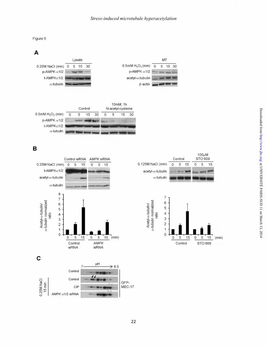
Phosphorylation of MEC-17 increases upon stress in an AMPK-dependent manner.
As ROS production stimulates AMP-kinase (AMPK) activity (27), we first examined the level of phosphorylation of AMPK on Thr172 and found it was increased after 5 minutes of NaCl induction and lasted 15 minutes after stress (Fig. 5A, left panel). This stress-induced AMPK activation was also detected in H2O2-stressed cells (data not shown). Interestingly, we found that AMPK is present and activated in MT-containing fractions (Fig.
at UN
IVE
RSIT
É PA
RIS-SU
D 11 on M
arch 13, 2014http://w
ww
.jbc.org/D
ownloaded from

Stress-induced microtubule hyperacetylation
8
5A, right panel) and that overall AMPK activation depended on ROS production. Indeed, pretreatment of cells with NAC before stress induction effectively prevented AMPK phosphorylation (Fig. 5A, bottom). To determine if such AMPK stimulation is actually involved in stress-induced MT hyperacetylation, we knocked down AMPKα1/2 by RNAi. In this condition, both basal and stress-induced MT acetylations were decreased by ~50% compared to controls (Fig. 5B, left). MT hyperacetylation was also inhibited when cells where treated with the chemical inhibitor STO609 (Fig. 5B, right). STO609 is widely used as an inhibitor of AMPK through the inhibition of the upstream activating Ca2+/Calmodulin-dependent protein kinase kinase (CamKKβ) (28).
To finally examine whether AMPK could mediate MEC-17 phosphorylation, we explored the overall level of GFP-MEC-17 posttranslational modifications by performing two-dimensional (2D) electrophoresis. The analysis revealed the presence of 5 major MEC-17 spots in basal conditions (Fig. 5C, upper lane). Interestingly, two more acidic species appeared after 15 minutes of NaCl treatment (Fig. 5C, black arrows). The treatment of cell lysates with alkaline phosphatase prior to 2D electrophoresis, prevented the occurrence of these two forms on the benefit of two more basic spots (white arrows), indicating that they corresponded to phosphorylated MEC-17. A similar 2D analysis of the MEC-17 modifications performed after AMPK RNAi showed that AMPK knockdown prevented the occurrence of stress-induced phosphorylated MEC-17 spots. These results indicate that, in response to ROS-mediated activation, AMPK is involved in MEC-17 phosphorylation upon stress.
MEC-17-dependent microtubule hyperace-tylation promotes cell survival and starvation-induced autophagy. The reversibility of MT hypera-cetylation shown in Fig. 1, suggests that it is rather an adaptative cell response to various stresses than a signal that would activate a death pathway. Among the adaptive mechanisms that occur rapidly after exposure to stress, autophagy
induction is essential to favor cell survival (29). In the case of amino-acid starvation, the breakdown of cellular components that results from autophagy induction can ensure survival by maintaining cellular energy levels. During autophagy induction, targeted cytoplasmic constituents are isolated within autophagosomes, which then fuse with lysosomes, allowing their contents to be degraded and recycled. We have previously shown that starvation-induced MT hyperacetylation is required for autophagy using the non acetylatable K40A mutant of tubulin (18). Here, we quantified autophagy induction in control and MEC-17 siRNA conditions by measuring the amount of cytosolic microtubule-associated protein light chain 3-I (LC3-I) that is converted to a lipid-conjugated form (termed LC3-II), which associates with autophagosome membranes. The amount of LC3-II being dependent on the balance between autopha-gosome formation and their clearance by lysosomes, its accumulation was measured in the presence of Balfilomycin A1 (BAF), which prevents autophagosome fusion with lysosomes. As expected, the autophagic flux (i.e. the difference between the levels of LC3-II measured in the presence and absence of BAF) was reduced by ~60% in MEC-17 siRNA conditions compared to controls (Fig. 6A).
Exposure to NaCl has been demonstrated previously to stimulate autophagy and such autophagy stimulation is important to make cell survival possible (30). To demonstrate that in our conditions, autophagy induction is the process that actually allows cell survival, we measured it by a MTT assay after autophagy was inhibited by knocking down Atg7, which is required for the conversion of LC3-I into the lipid conjugated form LC3-II. Atg7 knockdown reduced cell survival upon 24 h of mild NaCl-mediated stress by ~ 50% compared to control RNAi conditions (Fig. 6B), indicating that autophagy participates in cell survival upon NaCl treatment. To confirm that MT hyperacetylation contributes to cell survival, it was also measured in MEC-17 RNAi conditions. Fig. 6A shows that cell viability was decreased by up to ~ 20% in conditions of MEC-17 inhibition relative to controls.
at UN
IVE
RSIT
É PA
RIS-SU
D 11 on M
arch 13, 2014http://w
ww
.jbc.org/D
ownloaded from

Stress-induced microtubule hyperacetylation
9
Altogether, these data indicate that MT hyperacetylation favors cell survival through autophagy induction and point out the important physiological role of this modification and of its major effector MEC-17 in ensuring cell adaptability to stress. DISCUSSION In this study we show that, beyond the basal acetylation of stable MTs (14), the increase in the level of acetylated tubulin in the MT network i.e. MT hyperacetylation is a general, acute and reversible response that can be highly induced when cells are exposed to a variety of stresses. We also show that this induction involves the major acetyltransferase MEC-17. Furthermore, in keeping with our previous findings that tubulin K40 acetylation is required to allow starvation-induced autophagy stimulation (18) or the activation of protective signaling pathways (p53 translocation after genotoxic stress or basal Akt/PKB-dependent FoxO1 phosphorylation (17)), we found that MEC-17 is important for autophagy induction and enhances cell survival upon NaCl-mediated stress. We also evidence for the first time, the role of ROS (especially those originating from mitochondria), AMPK and p300 in regulating short-term and long-term MEC-17-mediated MT hyperacetylation (Fig. 6D). Until now, it was proposed that physiological and pathological variations in the level of tubulin acetylation would be mainly attributed to the inhibition of tubulin deacetylases and especially that of HDAC6 (19, 31). Our study shows for the first time that MT hyperacetylation, which occurs in a variety of stresses, is not due primarily to tubulin deacetylase inhibition but would rather result from enhanced tubulin acetylation. Among the multiple acetyltransferases that have been found to acetylate tubulin either in vitro or in vivo, MEC-17/αTAT-1 was shown to be the major tubulin acetyltransferase in basal conditions (20, 21). We show now that MEC-17 is also a crucial effector of stress-induced MT hyperacetylation. An approach to understand what happens in terms of MT acetylation during cell
stress was to determine if MEC-17 could be subject to modulation of expression and/or to post-translational modifications. The first appealing point from our data was the massive recruitment of p300 on MTs in response to stress. Due to the existence of multiple nuclear localization sequences, p300 is known to be a transcriptional coactivator that mainly resides in the nucleus (32). The control of its nuclear localization is critical to ensure transcriptional regulation (for review, see (33)). Here we show that p300 is redistributed to the cytoplasm and binds to acetylated MTs upon stress, which would be expected to prevent any shuttling back to the nucleus. Similar nucleo-cytoplasmic shuttling of p300 was previously observed in response to the HDAC inhibitors valproic acid or butyrate (34). In these conditions, cytoplasmic p300 is targeted to aggresomes (which bind to and need MTs for their formation) where it is ubiquitylated and subsequently degraded by the proteasome. In our models of cell stress, p300 nuclear export and cytoplasmic sequestration would favor the tubulin acetylation response by relieving negative regulation of MEC-17 gene transcription. Note that such down-regulation affects not only the extent of MT hyperacetylation, but also controls the basal acetylation level of tubulin. p300 is best known as an activator of gene transcription (35), but it was also reported to be required for down-regulation. For example, the DNA binding repressor protein PLZF (promyelocytic leukemia zinc finger gene) requires acetylation by p300 for binding to DNA, which leads to the recruitment of deacetylases and repression of PLZF-dependent HoxB2 reporter gene activity (36). In addition, recent studies show that p300 mRNA and protein levels decrease in metastatic cells (37). Also, p300/CBP mutations in some cancer cells decrease its acetylase activity (38). Given our results, such inhibition of p300 in cancer cells might presumably increase MEC-17 gene expression, which could explain why cancer cell lines like HeLa display higher levels of tubulin acetylation than non-cancer cell lines like RPE-1 (17). p300 is thus a potent regulator of MEC-17 levels and function in basal conditions and in stressed cells. However, it is expected to exert a medium- or long-term effect
at UN
IVE
RSIT
É PA
RIS-SU
D 11 on M
arch 13, 2014http://w
ww
.jbc.org/D
ownloaded from
Stress-induced microtubule hyperacetylation
10
to sustain tubulin acetylation in stressed cells. The burst in MT acetylation we observed very early after the onset of stress thus ought to involve other effectors. ROS are the major reactive atomic or molecular species produced in living organisms (39) and have been incriminated in numerous physiological adaptive reactions and diseases such as cancers, neurological disorders or cardio-vascular impairments. The superoxide anion O2
°- is generated early during ROS production. It is produced mainly in mitochondria, at the plasma membrane by NADPH oxidase, at the endoplasmic reticulum by cytochromes P450, but also in the cytoplasm by xanthine-oxidase (40). Either spontaneously or after SOD-mediated catalysis, O2
°- dismutation generates H2O2, which can subsequently produce hydroxyl radicals OH° known to be highly reactive against organic compounds. Our results suggest that ROS and especially OH° and O2
°- anions both participate in the induction of MT hyperacetylation but may be differently involved according to stress duration and/or strength. This variability may reflect the heterogeneity of ROS generation in response to NaCl treatment (41). Our experiments using MitoQ also suggest an interesting link between the release of ROS by mitochondria and the induction of MT acetylation.
ROS are effective second messengers, which may be quickly released in conditions of stress. Downstream of ROS, we focused our attention on AMPK activation. In agreement with previous studies (25, 42-45), we found that AMPK was rapidly activated by NaCl and H2O2. Upon NaCl treatment, STO609 inhibited MT hyperacetylation indicating that AMPK is at least in part activated by CamKKβ (28). CamKKβ activation is triggered both by ROS and by an increase in cytoplasmic Ca2+ (46), which both participate in cell response to NaCl treatment (40). Thus, Ca2+-mediated signaling could explain why i) hyperacetylation was higher with NaCl than with H2O2 ii) NAC was only partly effective to prevent MT hyperacetylation in response to NaCl. AMPK activation is involved in controlling MT hyperacetylation, most likely via the phosphorylation of MEC-17. To determine
whether AMPK-dependent phos-phorylation of MEC-17 actually increases the activity of the enzyme, target Ser or Thr should be identified and mutated. AMPK-mediated phosphorylation of MEC-17 could also play another role in controlling the affinity of MEC-17 for MTs. Recently, it has been shown that MEC-17 binding to MTs occurs via an interaction with the C-terminus of α-tubulin, thus on the outer surface of the MT lattice (47). However, the affinity of MEC-17 for MTs is not affected by MT acetylation (47), suggesting that the decrease of MEC-17 we observed in MT-containing fractions upon stress would be stimulated, perhaps by phosphorylation.
Interestingly, 2-dimensional gel electrophoresis of GFP-MEC-17 reveals many spots that resist phosphatase treatment, indicating that MEC-17 presents other post-translational modifications that change the enzyme’s pI and that could modulate its activity. Recently, Kalebic et al. showed that MEC-17 autoacetylation increases its catalytic activity towards α-tubulin (48).
We found that part of cytoplasmic AMPK binds to MTs and is likely to be activated in situ. This localization, together with the subcellular organization of mitochondria by MTs, suggests that MT hyperacetylation could be controlled and organized locally on MTs. Such integration of the organelles and molecules that control the level of tubulin acetylation in MTs may thus provide cells with an effective adaptive mechanism in case of exposure to stress. The differences we measured in cell survival in response to NaCl exposure show that MT hyperacetylation is not only an important hallmark of stress but also actively participates in cell adaptation and survival. The signaling mechanisms that function downstream of MT hyperacetylation are beginning to be uncovered in spite of controversial effects at the molecular level, especially regarding the effect of acetylation on the recruitment of molecular motors on MTs. Initially, acetylation was indeed proposed to enhance the recruitment of kinesin-1 and of dynein on MTs to facilitate vesicular transport in neurons (12, 13). Later on, the role of tubulin acetylation has been questioned in terms of motor velocity and run length (10, 11).
at UN
IVE
RSIT
É PA
RIS-SU
D 11 on M
arch 13, 2014http://w
ww
.jbc.org/D
ownloaded from
Stress-induced microtubule hyperacetylation
11
Even though tubulin acetylation might not directly modify the MT surface, it could operate through effects on the lateral interactions between MT protofilaments and their consequences in terms of MT bending (10). Before MEC-17 had been identified as a major tubulin acetyltransferase (20, 21), studies using non-acetylatable tubulin mutants such as the K40A mutant have led to the conclusions that tubulin acetylation favors the binding of various signaling intermediates to MTs either through the binding of kinesin-1 (as shown for JNK (18), through the binding of Hsp90 (Akt, p53) (17) or the cytoplasmic relocalization of p300 on MTs after nuclear export (this study). Interestingly such signaling pathways are activated in response to stress and participate in cell survival either directly or through the induction of autophagy (18). Autophagy induction can now be considered as a general mechanism of adaptation to stress, not only after nutrient or serum deprivation but also in response to excessive ROS production (for review, (49)) or to NaCl-mediated stress (30). It is now clear that MTs are involved at different stages of the autophagic process (50). The inhibition of MEC-17 expression by RNAi we used in this study confirms that the autophagic flux is decreased in the absence of MT hyperacetylation. Furthermore, if p300 were inactivated upon relocalization on MTs (our data), this would also be consistent with the previous findings that its inactivation allows Atg (Atg5, 7, 8 and 12) deacetylation and thus autophagy induction (51).
Beyond its enzymatic function, MEC-17 also plays a structural role in the maintenance of the MT lattice. Such structural function is indeed suspected as some phenotypes (especially touch sensitivity) in MEC-17-deficient C. elegans can be rescued using a catalytically inactive enzyme (9). In the future, it will be important to distinguish which function(s) involved in the control of cell survival actually require MTs being hyperacetylated and those which involve only the presence of MEC-17 on/inside MTs. Interestingly, the wide range of tubulin acetylation that can be covered by MTs between basal conditions and stress situations also suggests that hyperacetylation can affect highly dynamic and stable MTs to similar extents. It
will thus be important to determine precisely how boosting tubulin acetylation may affect MT dynamic instability. For example, NaCl-mediated stress seems to affect MT stability and organization at least transiently, as EB1 comets are lost during the first minutes of stress (30).
That most of the cellular MTs can be hyperacetylated very early after stress induction suggests that increased acetylated tubulin level may occur faster than the time required to breakdown and rebuild the whole dynamic MT network. Such a rapid kinetics of acetylation virtually excludes that hyperacetylation would happen only upon the elongation of new MTs. In addition, our counter-intuitive result that exogenous MEC-17 decreases upon stress from MT-containing fractions reveals that the enzyme is not statically present on MTs, but can rather undergo rapid exchange from the MT. In our observations, we only see GFP-MEC-17 loss from MTs. This suggests that all the MT-bound MEC-17 is released from binding to tubulin once the enzyme reaction is complete. Alternatively, the GFP-tagged MEC-17 isoform we followed could behave differently from the endogenous enzymes, which could be recruited to MTs and exchanged against GFP-MEC-17 during stress. Given the impossibility to detect MEC-17 using almost all the commercially available antibodies against C6ORF134/αTAT-1/MEC-17, we failed to test this hypothesis. Also, as we failed to render the MEC-17 cDNA resistant to RNAi despite all the mutations we successfully introduced in the sequences targeted by the siRNA, we could not follow the dynamics of GFP-MEC-17 alone in stressed cells. Tubulin structure shows that the K40 of α-tubulin belongs to a loop that is exposed at the opposite of the surface helices, suggesting it is located in the MT lumen (52). The K40 loop is also readily accessible from the inner of the MT as shown by the binding of anti-acetyl tubulin antibodies (10). MEC-17 was shown to directly interact with α-tubulin (48), most likely through a direct recognition of the K40 loop (53). Acetylation of tubulin in MT i.e. in polymerized rather than in a soluble form (21, 48), thus raises the question as to how MEC-17 accesses the K40 loop to bind to its substrate. MEC-17 may access the lumen through the apertures at the
at UN
IVE
RSIT
É PA
RIS-SU
D 11 on M
arch 13, 2014http://w
ww
.jbc.org/D
ownloaded from
Stress-induced microtubule hyperacetylation
12
plus and minus ends of MTs. Alternatively, the acetylase may bind the MT surface between protofilaments and access the K40 loop through the MT wall and/or through defects of the MT lattice. Such mode of interaction between MEC-17 and tubulin would more easily account for the dynamics of MEC-17 and of MT acetylation we observed during cell stress.
Although it has only been uncovered for a few years, MT hyperacetylation thus appears as a core adaptive event in cell response to a wealth of stresses. It also reinforces the notion that the MT cytoskeleton is a central structure that coordinates and organizes signaling in the cytoplasm, in relation with membrane-bound organelles like mitochondria. Our findings demonstrate that acetylation of K40 is a highly plastic post-translational modification of tubulin, which may rapidly alter MT function to promote cell survival. Other well known tubulin modifications like detyrosination are not affected by cell stress like nutrient deprivation (18), but we could not exclude that other modifications (unidentified yet) could be triggered by stress and could also participate in the control of MT functions.
at UN
IVE
RSIT
É PA
RIS-SU
D 11 on M
arch 13, 2014http://w
ww
.jbc.org/D
ownloaded from
Stress-induced microtubule hyperacetylation
13
1. Fulda, S., Gorman, A. M., Hori, O., and Samali, A. (2010) Cellular Stress Responses: Cell Survival and Cell Death. Int. J. Cell Biol. 2010, 1–23
2. Chen, F., Evans, A., Pham, J., and Plosky, B. (2010) Cellular stress responses: a balancing act. Mol. Cell 40, 175
3. Etienne-‐Manneville, S. (2009) From signaling pathways to microtubule dynamics: the key players. Curr. Opin. Cell Biol. 22, 104–111
4. Gundersen, G. G., and Cook, T. A. (1999) Microtubules and signal transduction. Curr. Opin. Cell Biol. 11, 81–94
5. Mitchison, T., and Kirschner, M. (1984) Dynamic instability of microtubule growth. Nature 312, 237–242
6. Peris, L., Thery, M., Faure, J., Saoudi, Y., Lafanechère, L., Chilton, J. K., Gordon-‐Weeks, P., Galjart, N., Bornens, M., Wordeman, L., Wehland, J., Andrieux, A., and Job, D. (2006) Tubulin tyrosination is a major factor affecting the recruitment of CAP-‐Gly proteins at microtubule plus ends. J. Cell Biol. 174, 839–849
7. Peris, L., Wagenbach, M., Lafanechère, L., Brocard, J., Moore, A. T., Kozielski, F., Job, D., Wordeman, L., and Andrieux, A. (2009) Motor-‐dependent microtubule disassembly driven by tubulin tyrosination. J. Cell Biol. 185, 1159–1166
8. Cueva, J. G., Hsin, J., Huang, K. C., and Goodman, M. B. (2012) Posttranslational acetylation of alpha-‐tubulin constrains protofilament number in native microtubules. Curr. Biol. 22, 1066–1074
9. Topalidou, I., Keller, C., Kalebic, N., Nguyen, K. C. Q., Somhegyi, H., Politi, K. A., Heppenstall, P., Hall, D. H., and Chalfie, M. (2012) Genetically Separable Functions of the MEC-‐17 Tubulin Acetyltransferase Affect Microtubule Organization. Curr. Biol. 22, 1057–1065
10. Soppina, V., Herbstman, J. F., Skiniotis, G., and Verhey, K. J. (2012) Luminal localization of alpha-‐tubulin K40 acetylation by cryo-‐EM analysis of fab-‐labeled microtubules. PLoS ONE 7, e48204
11. Walter, W. J., Beranek, V., Fischermeier, E., and Diez, S. (2012) Tubulin acetylation alone does not affect kinesin-‐1 velocity and run length in vitro. PLoS ONE 7, e42218
12. Reed, N. A., Cai, D., Blasius, T. L., Jih, G. T., Meyhofer, E., Gaertig, J., and Verhey, K. J. (2006) Microtubule acetylation promotes kinesin-‐1 binding and transport. Curr. Biol. 16, 2166–2172
13. Dompierre, J. P., Godin, J. D., Charrin, B. C., Cordelieres, F. P., King, S. J., Humbert, S., and Saudou, F. (2007) Histone deacetylase 6 inhibition compensates for the transport deficit in Huntington's disease by increasing tubulin acetylation. J Neurosci. 27, 3571–3583
14. Hammond, J. W., Huang, C. F., Kaech, S., Jacobson, C., Banker, G., and Verhey, K. J. (2010) Posttranslational modifications of tubulin and the polarized transport of kinesin-‐1 in neurons. Mol. Biol. Cell 21, 572–583
15. Cai, D., McEwen, D. P., Martens, J. R., Meyhofer, E., and Verhey, K. J. (2009) Single molecule imaging reveals differences in microtubule track selection between Kinesin motors. PLoS Biol. 7, e1000216
16. Giustiniani, J., Couloubaly, S., Pourci, M. L., Fourniat, C., Paul, J. L., Baillet, A., and Poüs, C. (2009) Basal endothelial nicric oxide synthase (eNOS) phosphorylation on Ser 1177 occurs in a stable microtubule-‐ and tubulin acetylation-‐dependent manner. Exp. Cell Res. 315, 3509–3520
17. Giustiniani, J., Daire, V., Cantaloube, I., Durand, G., Poüs, C., Perdiz, D., and Baillet, A. (2009) Tubulin acetylation favors Hsp90 recruitment to microtubules and stimulates the signaling function of the Hsp90 clients Akt/PKB and p53. Cell. Signal. 21, 529–539
18. Geeraert, C., Ratier, A., Pfisterer, S. G., Perdiz, D., Cantaloube, I., Rouault, A., Pattingre, S., Proikas-‐Cezanne, T., Codogno, P., and Poüs, C. (2010) Starvation-‐induced hyperacetylation of tubulin is required for the stimulation of autophagy by nutrient deprivation. J Biol. Chem 285, 24184–24194
19. Perdiz, D., Mackeh, R., Poüs, C., and Baillet, A. (2010) The ins and outs of tubulin acetylation:
at UN
IVE
RSIT
É PA
RIS-SU
D 11 on M
arch 13, 2014http://w
ww
.jbc.org/D
ownloaded from

Stress-induced microtubule hyperacetylation
14
more than just a post-‐translational modification? Cell. Signal. 23, 763–771 20. Akella, J. S., Wloga, D., Kim, J., Starostina, N. G., Lyons-‐Abbott, S., Morrissette, N. S., Dougan, S.
T., Kipreos, E. T., and Gaertig, J. (2010) MEC-‐17 is an alpha-‐tubulin acetyltransferase. Nature 467, 218–222
21. Shida, T., Cueva, J. G., Xu, Z., Goodman, M. B., and Nachury, M. V. (2010) The major alpha-‐tubulin K40 acetyltransferase alphaTAT1 promotes rapid ciliogenesis and efficient mechanosensation. Proc. Natl. Acad. Sci. U.S.A. 107, 21517–21522
22. Walker, R. A., O'Brien, E. T., Pryer, N. K., Soboeiro, M. F., Voter, W. A., Erickson, H. P., and Salmon, E. D. (1988) Dynamic instability of individual microtubules analyzed by video light microscopy: rate constants and transition frequencies. J. Cell Biol. 107, 1437–1448
23. Scherz-‐Shouval, R., Shvets, E., Fass, E., Shorer, H., Gil, L., and Elazar, Z. (2007) Reactive oxygen species are essential for autophagy and specifically regulate the activity of Atg4. EMBO J. 26, 1749–1760
24. Zhou, X., Matavelli, L. C., Ono, H., and Frohlich, E. D. (2005) Superiority of combination of thiazide with angiotensin-‐converting enzyme inhibitor or AT1-‐receptor blocker over thiazide alone on renoprotection in L-‐NAME/SHR. Am. J. Physiol. Renal. Physiol. 289, F871–9
25. Shimizu, T., Numata, T., and Okada, Y. (2004) A role of reactive oxygen species in apoptotic activation of volume-‐sensitive Cl(-‐) channel. Proc. Natl. Acad. Sci. U.S.A. 101, 6770–6773
26. Kelso, G. F. (2000) Selective Targeting of a Redox-‐active Ubiquinone to Mitochondria within Cells. ANTIOXIDANT AND ANTIAPOPTOTIC PROPERTIES. J. Biol. Chem. 276, 4588–4596
27. Emerling, B. M., Weinberg, F., Snyder, C., Burgess, Z., Mutlu, G. M., Viollet, B., Budinger, G. R., and Chandel, N. S. (2009) Hypoxic activation of AMPK is dependent on mitochondrial ROS but independent of an increase in AMP/ATP ratio. Free Radic. Biol. Med. 46, 1386–1391
28. Salminen, A., and Kaarniranta, K. (2012) AMP-‐activated protein kinase (AMPK) controls the aging process via an integrated signaling network. Ageing Res. Rev. 11, 230–241
29. Codogno, P., and Meijer, A. J. (2005) Autophagy and signaling: their role in cell survival and cell death. Cell Death Differ. 12 Suppl 2, 1509–1518
30. Nunes, P., Ernandez, T., Roth, I., Qiao, X., Strebel, D., Bouley, R., Charollais, A., Ramadori, P., Foti, M., Meda, P., Feraille, E., Brown, D., and Hasler, U. (2013) Hypertonic stress promotes autophagy and microtubule-‐dependent autophagosomal clusters. Autophagy 9, 550–567
31. Matthias, P., Yoshida, M., and Khochbin, S. (2008) HDAC6 a new cellular stress surveillance factor. Cell Cycle 7, 7–10
32. Yaciuk, P., and Moran, E. (1991) Analysis with specific polyclonal antiserum indicates that the E1A-‐associated 300-‐kDa product is a stable nuclear phosphoprotein that undergoes cell cycle phase-‐specific modification. Mol. Cell Biol. 11, 5389–5397
33. Chen, J., and Li, Q. (2011) Life and death of transcriptional co-‐activator p300. Epigenetics 6, 957–961
34. Chen, J., Halappanavar, S., Th' ng, J. P., and Li, Q. (2007) Ubiquitin-‐dependent distribution of the transcriptional coactivator p300 in cytoplasmic inclusion bodies. Epigenetics 2, 92–99
35. Mantelingu, K., Reddy, B. A., Swaminathan, V., Kishore, A. H., Siddappa, N. B., Kumar, G. V., Nagashankar, G., Natesh, N., Roy, S., Sadhale, P. P., Ranga, U., Narayana, C., and Kundu, T. K. (2007) Specific inhibition of p300-‐HAT alters global gene expression and represses HIV replication. Chem. Biol. 14, 645–657
36. Guidez, F., Howell, L., Isalan, M., Cebrat, M., Alani, R. M., Ivins, S., Hormaeche, I., McConnell, M. J., Pierce, S., Cole, P. A., Licht, J., and Zelent, A. (2005) Histone acetyltransferase activity of p300 is required for transcriptional repression by the promyelocytic leukemia zinc finger protein. Mol. Cell Biol. 25, 5552–5566
37. Mees, S. T., Mardin, W. A., Wendel, C., Baeumer, N., Willscher, E., Senninger, N., Schleicher, C.,
at UN
IVE
RSIT
É PA
RIS-SU
D 11 on M
arch 13, 2014http://w
ww
.jbc.org/D
ownloaded from
Stress-induced microtubule hyperacetylation
15
Colombo-‐Benkmann, M., and Haier, J. (2010) EP300-‐-‐a miRNA-‐regulated metastasis suppressor gene in ductal adenocarcinomas of the pancreas. Int. J. Cancer 126, 114–124
38. Pasqualucci, L., Dominguez-‐Sola, D., Chiarenza, A., Fabbri, G., Grunn, A., Trifonov, V., Kasper, L. H., Lerach, S., Tang, H., Ma, J., Rossi, D., Chadburn, A., Murty, V. V., Mullighan, C. G., Gaidano, G., Rabadan, R., Brindle, P. K., and Dalla-‐Favera, R. (2011) Inactivating mutations of acetyltransferase genes in B-‐cell lymphoma. Nature 471, 189–195
39. Miller, D. M., Buettner, G. R., and Aust, S. D. (1990) Transition metals as catalysts of “autoxidation” reactions. Free Radic. Biol. Med. 8, 95–108
40. Sheu, S. S., Nauduri, D., and Anders, M. W. (2006) Targeting antioxidants to mitochondria: a new therapeutic direction. Biochim. Biophys. Acta 1762, 256–265
41. Burg, M. B., Ferraris, J. D., and Dmitrieva, N. I. (2007) Cellular response to hyperosmotic stresses. Physiol. Rev. 87, 1441–1474
42. Lamberts, R. R., Onderwater, G., Hamdani, N., Vreden, M. J., Steenhuisen, J., Eringa, E. C., Loer, S. A., Stienen, G. J., and Bouwman, R. A. (2009) Reactive oxygen species-‐induced stimulation of 5'AMP-‐activated protein kinase mediates sevoflurane-‐induced cardioprotection. Circulation 120, S10–5
43. Cardaci, S., Filomeni, G., and Ciriolo, M. R. (2012) Redox implications of AMPK-‐mediated signal transduction beyond energetic clues. J. Cell. Sci. 125, 2115–2125
44. Park, I. J., Hwang, J. T., Kim, Y. M., Ha, J., and Park, O. J. (2006) Differential modulation of AMPK signaling pathways by low or high levels of exogenous reactive oxygen species in colon cancer cells. Ann. N. Y. Acad. Sci. 1091, 102–109
45. Li, L., Chen, Y., and Gibson, S. B. (2013) Starvation-‐induced autophagy is regulated by mitochondrial reactive oxygen species leading to AMPK activation. Cell. Signal. 25, 50–65
46. Sid, B., Verrax, J., and Calderon, P.B. (2013) Role of AMPK activation in oxidative cell damage: Implications for alcohol-‐induced liver disease. Biochem. Pharmacol. 86, 200-‐209
47. Howes, S. C., Alushin, G. M., Shida, T., Nachury, M. V., and Nogales, E. (2013) Effects of tubulin acetylation and tubulin acetyltransferase binding on microtubule structure. Mol. Biol. Cell 25, 257-‐266.
48. Kalebic, N., Martinez, C., Perlas, E., Hublitz, P., Bilbao-‐Cortes, D., Fiedorczuk, K., Andolfo, A., and Heppenstall, P. A. (2013) Tubulin acetyltransferase alphaTAT1 destabilizes microtubules independently of its acetylation activity. Mol. Cell Biol. 33, 1114–1123
49. Dewaele, M., Maes, H., and Agostinis, P. (2010) ROS-‐mediated mechanisms of autophagy stimulation and their relevance in cancer therapy. Autophagy 6, 838–854
50. Mackeh, R., Perdiz, D., Lorin, S., Codogno, P., and Poüs, C. (2013) Autophagy and microtubules -‐ new story, old players. J. Cell. Sci. 126, 1071–1080
51. Lee, I. H., and Finkel, T. (2009) Regulation of autophagy by the p300 acetyltransferase. J. Biol. Chem. 284, 6322–6328
52. Nogales, E., Wolf, S. G., and Downing, K. H. (1998) Structure of the alpha beta tubulin dimer by electron crystallography. Nature 391, 199–203
53. Friedmann, D. R., Aguilar, A., Fan, J., Nachury, M. V., and Marmorstein, R. (2012) Structure of the alpha-‐tubulin acetyltransferase, alphaTAT1, and implications for tubulin-‐specific acetylation. Proc. Natl. Acad. Sci. U.S.A. 109, 19655–19660
at UN
IVE
RSIT
É PA
RIS-SU
D 11 on M
arch 13, 2014http://w
ww
.jbc.org/D
ownloaded from
Stress-induced microtubule hyperacetylation
16
Figure1: Microtubule hyperacetylation is a broad and reversible cell stress response. HeLa cells were treated, for 15 min with 10-7M methoxychlore (MTX), for 1 h with 10-4M ethinylestradiol (EE2), 0.25mM NaCl or 0.5µM staurosporine, and deprived of amino acids for 2 h (EBSS) (A) or subjected to an exposure to excess NaCl as indicated (B). (C) Immuno-labeling of acetyl-tubulin after 30 min of NaCl treatment. (D) After one h of treatment with 0.25M NaCl, the medium was removed and replaced with fresh normal medium and tubulin acetylation level was monitored. The histograms show the mean normalized ratios ± SEM of acetyl-α-tubulin to total α-tubulin. *p<0.05, Scale bar = 10 µm.
Figure 2: MEC-17 is required for stress-induced microtubule hyperacetylation. (A) Tubulin deacetylation assay. After a 30-min treatment with NaCl as indicated, cell lysates were incubated in vitro with porcine brain tubulin (which is naturally acetylated) and NAD+. After Western-blotting of acetylated and total α-tubulin, the mean ± SEM ratios of acetyl-to-total α-tubulin were represented in the histogram shown (B) Western-blot of different acetyltransferases detected with a variety of specific antibodies in the MT-containing fractions of HeLa cells treated for 30 min with NaCl as indicated. (C) Western blot analysis of GFP-MEC-17 in MT-containing fraction of cells transiently overexpressing GFP-MEC-17 and treated for indicated times with 0.25M NaCl. (D) After knockdown of MEC-17 by siRNA, HeLa cells were treated with NaCl (top panels), deprived of amino acids (EBSS, bottom left panel) or treated with staurosporine (ST, bottom right panel) and analyzed for tubulin acetylation. (E) Cells transiently overexpressing GFP-MEC-17WT or the catalytically inactive mutant GFP-MEC17D157N were stressed with NaCl for 30 min and analyzed for tubulin acetylation.
Figure 3: p300 negatively regulates MEC-17-dependent tubulin acetylation. (A) Western blot analysis of cytosolic and MT-containing fractions (left) or total cell extract (right) and treated for 30 min with NaCl. (B) Subcellular distribution of p300 was analyzed by Western Blot analysis of cytoplasmic and nuclear extracts from NaCl-treated cells (left) or by immunofluorescence (right). PARP and β-tubulin were used as controls for nuclear and cytoplasmic purification, respectively. Scale bar = 10 µm. (C and D) Analysis of tubulin acetylation after p300 siRNA alone or combined with that of MEC-17. The histogram indicates the mean ± SEM normalized acetyl-to-α-tubulin ratio. *p<0.05 (E) HeLa cells were subjected or not to siRNA as indicated and MEC-17 mRNA levels were measured by RT-qPCR. Values are the mean ± SEM of the relative MEC-17 mRNA content normalized to the level measured in untreated cells. *p<0.05 (F) Loss of p300 from the MT-containing fraction prepared from cells subjected to MEC-17 RNAi (left) or from cells transiently overexpressing (72h) a non-acetylatable tubulin mutant (K40A) (middle). Cytosol and MT fractions prepared from PtK2 cells (left) treated with NaCl.
Figure 4: Mitochondrial ROS stimulate stress-induced microtubule hyperacetylation. Lysates of cells treated with H2O2 (A,B), pretreated for 1h with 10mM N-acetyl-L-cysteine prior to H2O2 (C) or NaCl treatment (D), pretreated with 2mM OH-tempo for 1h prior to a 30-min NaCl treatment (E) or pretreated for 24h with mitoQ prior to NaCl treatment (F), were subjected to western blot analysis of tubulin acetylation. Histograms indicate the mean ± SEM normalized acetyl-to-α-tubulin ratio. *p<0.05
Figure 5: Phosphorylation of MEC-17 increases upon stress in an AMPK-dependent manner. (A): Analysis of AMPK phosphorylation in total cell lysates of NaCl-treated cells (upper panel left) or in MT-containing fractions prepared from cells treated with 0.5mM H2O2 (upper panel right). Lower panel: Total cell lysate of cells treated with 0.5mM H2O2 and pretreated or not with 10mM N-acetyl-cystein (NAC) for one h were analyzed for AMPK phosphorylation and tubulin acetylation. (B) Cells processed for AMPK siRNA for 48 h (left) or pretreated with 100µM of AMPK inhibitor STO609 (right) were subjected to treatment with NaCl as indicated. The histogram shows the mean ± SEM normalized acetyl-to-α-tubulin
at UN
IVE
RSIT
É PA
RIS-SU
D 11 on M
arch 13, 2014http://w
ww
.jbc.org/D
ownloaded from
Stress-induced microtubule hyperacetylation
17
ratios. (C) Lysates prepared from control or NaCl-treated cells transiently overexpressing GFP-MEC17 and subjected or not to AMPK RNAi and treated or not with calf intestine alkaline phosphatase (CIP) were analyzed by 2D gel electrophoresis and Western blot using anti-GFP antibody.
Figure 6: MEC-17-dependent microtubule hyperacetylation prompts cell survival and starvation-induced autophagy. (A) Cells were transfected with control or MEC-17 siRNA before being starved for 2 h from amino-acid (EBSS). Autophagy induction was then assessed by Western blot using anti-LC3 antibody. Bafilomycin A1 was used 2 h prior to protein extraction to block autophagic flux. The histogram shows quantification of LC3-II/β-actin (mean± SEM). *p<0.05. (B) The histogram (left) shows the percentage of cell survival upon 0.0625M and 0.125M NaCl treatment relative to control, in Atg7 or MEC-17 RNAi conditions. On the right is shown the percentage of cell survival upon NaCl relative to untreated controls. *p<0.05 and ***p<0.001. (C) Hypothetical model for molecular mechanism of stress-induced MT hyperacetylation. In normal conditions, MEC-17 expression is inhibited by p300, which maintains low level of tubulin acetylation involved in basal MT functions. Upon stress, the accumulation of ROS leads to the activation of AMPK, which in turn mediates MEC-17 phosphorylation. This presumably activates MEC-17, leading to MT hyperacetylation, which in turn participates in autophagy induction and cell adaptation to stress.
at UN
IVE
RSIT
É PA
RIS-SU
D 11 on M
arch 13, 2014http://w
ww
.jbc.org/D
ownloaded from
Stress-induced microtubule hyperacetylation
18
at UN
IVE
RSIT
É PA
RIS-SU
D 11 on M
arch 13, 2014http://w
ww
.jbc.org/D
ownloaded from
Stress-induced microtubule hyperacetylation
19
at UN
IVE
RSIT
É PA
RIS-SU
D 11 on M
arch 13, 2014http://w
ww
.jbc.org/D
ownloaded from

Stress-induced microtubule hyperacetylation
20
at UN
IVE
RSIT
É PA
RIS-SU
D 11 on M
arch 13, 2014http://w
ww
.jbc.org/D
ownloaded from

Stress-induced microtubule hyperacetylation
21
at UN
IVE
RSIT
É PA
RIS-SU
D 11 on M
arch 13, 2014http://w
ww
.jbc.org/D
ownloaded from
Stress-induced microtubule hyperacetylation
22
at UN
IVE
RSIT
É PA
RIS-SU
D 11 on M
arch 13, 2014http://w
ww
.jbc.org/D
ownloaded from

Stress-induced microtubule hyperacetylation
23
at UN
IVE
RSIT
É PA
RIS-SU
D 11 on M
arch 13, 2014http://w
ww
.jbc.org/D
ownloaded from