raxa h.solanki associate professor biology v.p
TRANSCRIPT

RAXA H.SOLANKIASSOCIATE PROFESSOR
BIOLOGYV.P.& R.P.T.P. SCIENCE COLLEGE
RAXA H.SOLANKIASSOCIATE PROFESSOR
BIOLOGYV.P.& R.P.T.P. SCIENCE COLLEGE

SARDAR PATEL UNIVERSITY
B.Sc. FIRST SEMESTER
Core Course - Biology US01CBIO21 (T)
CELL BIOLOGY AND BIODIVERSITYEffective from June 2018 4 Credits, 4periods per week Total Marks 100,
Internal -30 Marks, External-70 Marks,exam duration: 3 hours
SARDAR PATEL UNIVERSITY
B.Sc. FIRST SEMESTER
Core Course - Biology US01CBIO21 (T)
CELL BIOLOGY AND BIODIVERSITYEffective from June 2018 4 Credits, 4periods per week Total Marks 100,
Internal -30 Marks, External-70 Marks,exam duration: 3 hours

Unit 1 Cell & Cell organelles•Cell as unit of structure and function Characteristicsand cell structure of Prokaryotic cell and Eukaryoticcell•Cell organelles – Structure and functions of : Cellwall, Plasma membrane, Nucleus, Chloroplast,Mitochondria, E.R., Golgi complex,Lysosomes,Ribosomes,•Cell division – Cell cycle , Mitosis and Meiosis andSignificance Structure of chromosome, Types basedon the position of the centromere, Giantchromosomes
Unit 1 Cell & Cell organelles•Cell as unit of structure and function Characteristicsand cell structure of Prokaryotic cell and Eukaryoticcell•Cell organelles – Structure and functions of : Cellwall, Plasma membrane, Nucleus, Chloroplast,Mitochondria, E.R., Golgi complex,Lysosomes,Ribosomes,•Cell division – Cell cycle , Mitosis and Meiosis andSignificance Structure of chromosome, Types basedon the position of the centromere, Giantchromosomes

Unit -2 Bio molecules:Carbohydrates – Nomenclature, Classification -
Monosaccharide, Disaccharides, Polysaccharides andbiological roleLipids: Definition and classification; Fatty acidsstructure ; Essential fatty acids; biological roleProteins – Definition, classification, Structure ofAmino acids, Bonds responsible for protein structure,Protein denaturation and biological role of ProteinsNucleic acids: Nitrogenous bases; Pentose sugars,Structure and function of nucleotides; Types ofnucleic acids; Structure of A, B, Z types of DNA; Typesof RNA; Structure of tRNA. Biological role.

Unit -3 Biodiversity*Viruses- General structure, Replication, TMV, Bacteriophage*General account of Mycoplasma•General characteristics of Bacteria, Reproduction andeconomic importance•*Algae –General characteristics, Range of thallus structureand reproduction, economic importance of Algae, Life cycle ofVolvox.•*Fungi - General characteristics, Range of thallus structureand reproduction, Economic importance of Fungi, Life cycle ofRhizopus.•*Introduction to Archegoniate : General characters andalternation of generation and outline lifecycles of following -A. Bryophyte – Riccia B. Pteridophyta – Nephrolepis•C. Gymnosperms – Cycas
Unit -3 Biodiversity*Viruses- General structure, Replication, TMV, Bacteriophage*General account of Mycoplasma•General characteristics of Bacteria, Reproduction andeconomic importance•*Algae –General characteristics, Range of thallus structureand reproduction, economic importance of Algae, Life cycle ofVolvox.•*Fungi - General characteristics, Range of thallus structureand reproduction, Economic importance of Fungi, Life cycle ofRhizopus.•*Introduction to Archegoniate : General characters andalternation of generation and outline lifecycles of following -A. Bryophyte – Riccia B. Pteridophyta – Nephrolepis•C. Gymnosperms – Cycas

Unit -4 General Account of Invertebrates
* General characteristics and outline classification ofMajor Invertebrate Phyla*Nutrition and reproduction in Protozoa Life cycleand pathogenicity of Plasmodium vivax andEntamoeba histolytica Life cycle and pathogenicity ofTaenia solium and Wuchereria bancrofti Metamerismand Economic importance of Annelida EconomicImportance of Arthropods Social Life of Insect*Metamorphosis in Insects Economic Importance ofMolluscs, Water vascular system in Asteroidea

SUGGESTED READINGS:
1 Cellbiology,Genetics,Molecular Biology, Evolution andEcology-P.S.Verma and V.K.agarwal2 Text book of Botany-Diversity of Microbes and Cryptogams-Singh,Pande and Jain3 Biochemistry-U.Satyanarayan4 Cell and Molecular Biology: De Robertis and De Robertis5 Lehninger Principles of Biochemistry Book by Albert L.Lehninger, David L. Nelson, and Michael M. Cox6 Modern Text Book Of Zoology Invertebrates- R. L .Kotpal7 Economic Zoology - Shukla and Upadhyay8 Medical Parasitology - Rajesh karyakarte, Ajit Damle9 Invertebrate Zoology – E.L.Jordan and P.S.Verma10 Cell Biology-P. S. Verma

VIRUSESGeneral characteristics


*A virus is a small collection of genetic code, either DNA orRNA, surrounded by a protein coat.•A virus cannot replicate alone. Viruses must infect cells anduse components of the host cell to make copies ofthemselves.•*Often, they kill the host cell in the process, and causedamage to the host organism.•*Viruses have been found everywhere on Earth. Researchersestimate that viruses outnumber bacteria by 10 to 1.•*Because viruses don’t have the same components asbacteria, they cannot be killed by antibiotics; only antiviralmedications or vaccines can eliminate or reduce the severityof viral diseases, including AIDS, COVID-19, measles andsmallpox.
*A virus is a small collection of genetic code, either DNA orRNA, surrounded by a protein coat.•A virus cannot replicate alone. Viruses must infect cells anduse components of the host cell to make copies ofthemselves.•*Often, they kill the host cell in the process, and causedamage to the host organism.•*Viruses have been found everywhere on Earth. Researchersestimate that viruses outnumber bacteria by 10 to 1.•*Because viruses don’t have the same components asbacteria, they cannot be killed by antibiotics; only antiviralmedications or vaccines can eliminate or reduce the severityof viral diseases, including AIDS, COVID-19, measles andsmallpox.

*Viruses (Latin Venum – poisonous fluid) aresimplest forms of life.*They are not cells, but their study has provideda great deal of information about cells.*Study of viruses is a branch of biology calledVirology.•Viruses are cellular parasites.•*They are smaller than bacteria and have amuch more simplified organization.
*Viruses (Latin Venum – poisonous fluid) aresimplest forms of life.*They are not cells, but their study has provideda great deal of information about cells.*Study of viruses is a branch of biology calledVirology.•Viruses are cellular parasites.•*They are smaller than bacteria and have amuch more simplified organization.

Nature of Viruses:Viruses are infective microorganisms.They show several differences from typicalbacterial cells:1. Size:On the whole viruses are much smaller than bacteria.Most animal and plant viruses are invisible under thelight microscope. Some of smaller viruses are only 200Åin diameter.
Nature of Viruses:Viruses are infective microorganisms.They show several differences from typicalbacterial cells:1. Size:On the whole viruses are much smaller than bacteria.Most animal and plant viruses are invisible under thelight microscope. Some of smaller viruses are only 200Åin diameter.

2. No independent metabolism:*Viruses cannot multiply outside a living cell.*No virus has been cultivated in a cell-freemedium.*Viruses do not have an independentmetabolism.*They are metabolically inactive outside thehost cell because they do not possess enzymesystems and protein synthesis machinery.*Thus viruses are obligatory intracellularparasites
2. No independent metabolism:*Viruses cannot multiply outside a living cell.*No virus has been cultivated in a cell-freemedium.*Viruses do not have an independentmetabolism.*They are metabolically inactive outside thehost cell because they do not possess enzymesystems and protein synthesis machinery.*Thus viruses are obligatory intracellularparasites

3. Simple structure:*Viruses have a very simple structure.*They consist of a nucleic acid core surroundedby a protein coat.*In this respect they differ from typical cellswhich are made up of proteins, carbohydrates,lipids and nucleic acids.*Myxoviruses have a membranous envelopeconsisting of proteins, carbohydrate and lipidoutside the usual protein coat, but thisenvelope is derived from the host cell.
3. Simple structure:*Viruses have a very simple structure.*They consist of a nucleic acid core surroundedby a protein coat.*In this respect they differ from typical cellswhich are made up of proteins, carbohydrates,lipids and nucleic acids.*Myxoviruses have a membranous envelopeconsisting of proteins, carbohydrate and lipidoutside the usual protein coat, but thisenvelope is derived from the host cell.

4. Absence of cellular structure:Viruses do not have any cytoplasm,and thus cytoplasmic organelles likemitochondria, Golgi complexes,ribosomes, lysosomes etc. are absent.They do not have any limiting cellmembrane.
4. Absence of cellular structure:Viruses do not have any cytoplasm,and thus cytoplasmic organelles likemitochondria, Golgi complexes,ribosomes, lysosomes etc. are absent.They do not have any limiting cellmembrane.

5. Nucleic acids:
*Viruses usually have only one nucleic acid,either DNA or RNA. Typical cells have bothDNA and RNA. Rous Sarcoma virus (RSV),producing certain cancer, is the only virushaving both DNA and RNA.6. Crystallization:Many of the smaller viruses can becrystallized, and thus behave likechemicals.
*Viruses usually have only one nucleic acid,either DNA or RNA. Typical cells have bothDNA and RNA. Rous Sarcoma virus (RSV),producing certain cancer, is the only virushaving both DNA and RNA.6. Crystallization:Many of the smaller viruses can becrystallized, and thus behave likechemicals.

7. No growth and division:*Viruses do not have the power of growth and division.*The genetic material of virus reproduces only in a hostcell.*Thus viruses do not show all the characteristics of typicalliving organisms.*They, however, possess two fundamental characteristicsof living systems.*Firstly, they contain nucleic acid as their geneticmaterial.*The nucleic acid contains all the instructions for thestructure and the function of the virus.*Secondly , they can reproduce themselves, even if only byusing the host cells’ s synthesis machinery.
7. No growth and division:*Viruses do not have the power of growth and division.*The genetic material of virus reproduces only in a hostcell.*Thus viruses do not show all the characteristics of typicalliving organisms.*They, however, possess two fundamental characteristicsof living systems.*Firstly, they contain nucleic acid as their geneticmaterial.*The nucleic acid contains all the instructions for thestructure and the function of the virus.*Secondly , they can reproduce themselves, even if only byusing the host cells’ s synthesis machinery.

Structure of Viruses:(a) Size:Variable. Most viruses are much smallerthan bacteria. The size ranges in between100A to 250 mu. Some viruses are largerthan bacteria, for example the psittacos is avirus measuring 0.75 mu in diameter.
Structure of Viruses:(a) Size:Variable. Most viruses are much smallerthan bacteria. The size ranges in between100A to 250 mu. Some viruses are largerthan bacteria, for example the psittacos is avirus measuring 0.75 mu in diameter.

(b) Symmetry:*Viruses occur in three main shapes.*They are spherical (Cubical or polyhedral), helical(Cylinderical or rod-like) and complex. Cubical virusesmay be tetrahedral (4 faces) < dodecahedral (12faces) or icosahedral (20 faces).*The Herpes virus is dodecahedral.*The Tobacco mosaic virus (TMV) and thebacteriophage are, respectively, helical and complex.1. Spherical / Cubical:Herpes virus, Tipula virus, Polyoma virus.
(b) Symmetry:*Viruses occur in three main shapes.*They are spherical (Cubical or polyhedral), helical(Cylinderical or rod-like) and complex. Cubical virusesmay be tetrahedral (4 faces) < dodecahedral (12faces) or icosahedral (20 faces).*The Herpes virus is dodecahedral.*The Tobacco mosaic virus (TMV) and thebacteriophage are, respectively, helical and complex.1. Spherical / Cubical:Herpes virus, Tipula virus, Polyoma virus.

2. Helical / Cylindrical:Tobacco Mosaic virus,Influenza virus Mumpsvirus.3. Complex:Vaccinia virus, ORF virus,Vesicular Stomatitis virus.
2. Helical / Cylindrical:Tobacco Mosaic virus,Influenza virus Mumpsvirus.3. Complex:Vaccinia virus, ORF virus,Vesicular Stomatitis virus.

(c) Morphology:*Morphologically a virus is a core of nucleic acid (DNA orRNA) surrounded by a protein shell.*An intact virus unit is known as virion. Its protein coat iscalled capsid.*The capsid is composed of a number of subunits of aparticular shape. These sub-units are known as capsomeres.*The capsid protects the nucleic acid against the action ofnuclease enzyme.*Some proteins of capsid help in binding the virus to thesurface of host cells.*Some surface proteins act as enzyme and dissolve thesurface layer of host cell and thus help in penetration of itsnucleic acid into the host cell.
(c) Morphology:*Morphologically a virus is a core of nucleic acid (DNA orRNA) surrounded by a protein shell.*An intact virus unit is known as virion. Its protein coat iscalled capsid.*The capsid is composed of a number of subunits of aparticular shape. These sub-units are known as capsomeres.*The capsid protects the nucleic acid against the action ofnuclease enzyme.*Some proteins of capsid help in binding the virus to thesurface of host cells.*Some surface proteins act as enzyme and dissolve thesurface layer of host cell and thus help in penetration of itsnucleic acid into the host cell.


*The polio virus (Poliomyelitis) is a most extensivelystudied animal virus.*It has a very simple organization. It consists of aprotein coat built up out of 60 structurally equivalent,asymmetric protein subunits of approximately 60 Å indiameter.* The spherical protein coat has a diameter about300Å. It encloses the genetic material, RNA.*The protein coat contains about 49, 600 amino acidsand RNA contains about 5200 nucleotides.*The single-stranded RNA of poliovirus, thus, hastriplet codes for 1700 amino acids.* During infection, it alters cell metabolism drasticallyand leads quick death of host cell.
*The polio virus (Poliomyelitis) is a most extensivelystudied animal virus.*It has a very simple organization. It consists of aprotein coat built up out of 60 structurally equivalent,asymmetric protein subunits of approximately 60 Å indiameter.* The spherical protein coat has a diameter about300Å. It encloses the genetic material, RNA.*The protein coat contains about 49, 600 amino acidsand RNA contains about 5200 nucleotides.*The single-stranded RNA of poliovirus, thus, hastriplet codes for 1700 amino acids.* During infection, it alters cell metabolism drasticallyand leads quick death of host cell.

*Tobacco mosaic virus is the most extensivelystudied plant virus.*It is a helically symmetrical, rod-shaped virushaving the length of 3000Å and diameter of180A.•It RNA is a single stranded spirally coiledmolecule formed of 6500 nucleotides.•* The capsid is formed of 2130 capsomeres,each with a molecular weight of 18,000.•*The capsomeres are elliptical and remainarranged helically around to form capsid.
*Tobacco mosaic virus is the most extensivelystudied plant virus.*It is a helically symmetrical, rod-shaped virushaving the length of 3000Å and diameter of180A.•It RNA is a single stranded spirally coiledmolecule formed of 6500 nucleotides.•* The capsid is formed of 2130 capsomeres,each with a molecular weight of 18,000.•*The capsomeres are elliptical and remainarranged helically around to form capsid.

Classification of Viruses:Viruses may be classified according to the type of the host,genetic material and number of strands.On the basis of type of host, viruses are:1. Animal Viruses:*They live inside animal cells including man.•On entering the cell, these disturb the metabolism of thehost cell and cause various diseases.•*The common animal viruses are small pox virus, influenzavirus, mumps virus, polio virus and herpes virus.•*In many animal viruses an extra envelope surrounds theirprotein coat.•*The membrane consists of proteins, lipids andcarbohydrates and is derived from the host plasmamembrane.
Classification of Viruses:Viruses may be classified according to the type of the host,genetic material and number of strands.On the basis of type of host, viruses are:1. Animal Viruses:*They live inside animal cells including man.•On entering the cell, these disturb the metabolism of thehost cell and cause various diseases.•*The common animal viruses are small pox virus, influenzavirus, mumps virus, polio virus and herpes virus.•*In many animal viruses an extra envelope surrounds theirprotein coat.•*The membrane consists of proteins, lipids andcarbohydrates and is derived from the host plasmamembrane.

*Animal viruses may enter cells by attaching tothe surface. Some are then engulfed by the cellthrough pinocytosis or phagocytosis.*In such cases, uncoating of the viral nucleicacid might occur within the cell.*Inside the host cell they may multiply and formnumerous new viral particles.*Usually, animal viruses release from the hostcells by the rupturing and subsequent death ofthe host cells.
*Animal viruses may enter cells by attaching tothe surface. Some are then engulfed by the cellthrough pinocytosis or phagocytosis.*In such cases, uncoating of the viral nucleicacid might occur within the cell.*Inside the host cell they may multiply and formnumerous new viral particles.*Usually, animal viruses release from the hostcells by the rupturing and subsequent death ofthe host cells.


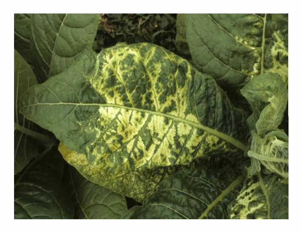
2. Plant Viruses:*They are parasites of plant cells. Their geneticmaterial is RNA which remains enclosed in theprotein coat.*The most important plant viruses are tobaccomosaic virus (TMV), tobacco rattle virus (TRV),potato virus (PV), southern bean mosaic virus(SBMV), beet yellow virus (BYV) and turnipyellow virus (TYV).
2. Plant Viruses:*They are parasites of plant cells. Their geneticmaterial is RNA which remains enclosed in theprotein coat.*The most important plant viruses are tobaccomosaic virus (TMV), tobacco rattle virus (TRV),potato virus (PV), southern bean mosaic virus(SBMV), beet yellow virus (BYV) and turnipyellow virus (TYV).

3. Bacterial virus:*They are parasitic on bacteria and so alsocalled bacteriophages.*There are many varieties of bacteriophages.*Their size and shape varies from species tospecies.*Some phages are spherical, some comma-shaped whereas majority of them havetadpole-like appearance.
3. Bacterial virus:*They are parasitic on bacteria and so alsocalled bacteriophages.*There are many varieties of bacteriophages.*Their size and shape varies from species tospecies.*Some phages are spherical, some comma-shaped whereas majority of them havetadpole-like appearance.

On the basis of nucleic acids, viruses are:1. DNA viruses:These viruses possess DNA as the genetic material. Onreplication this DNA produces new DNA. DNAtransmits information for protein synthesis throughRNA. (DNA → RNA → PROTEIN).2. RNA viruses:These viruses possess RNA as the genetic material.The RNA replicates directly to produce new RNA.Information for protein synthesis passes from RNA toprotein without involment of DNA. (RNA → RNA →PROTEIN).
On the basis of nucleic acids, viruses are:1. DNA viruses:These viruses possess DNA as the genetic material. Onreplication this DNA produces new DNA. DNAtransmits information for protein synthesis throughRNA. (DNA → RNA → PROTEIN).2. RNA viruses:These viruses possess RNA as the genetic material.The RNA replicates directly to produce new RNA.Information for protein synthesis passes from RNA toprotein without involment of DNA. (RNA → RNA →PROTEIN).

3. DNA – RNA viruses:In a group of RNA tumour viruses calledleukoviruses or rousviruses the geneticmaterial is alternately DNA and RNA. Inaddition to the normal mode of transfer foundin DNA viruses (DNA → RNA → PROTEIN) therousviruses also transfer information from RNAto DNA (RNA-DNA-RNA -PROTEIN).
3. DNA – RNA viruses:In a group of RNA tumour viruses calledleukoviruses or rousviruses the geneticmaterial is alternately DNA and RNA. Inaddition to the normal mode of transfer foundin DNA viruses (DNA → RNA → PROTEIN) therousviruses also transfer information from RNAto DNA (RNA-DNA-RNA -PROTEIN).

With respect to number of strands, four types ofnucleic acids have been found in viruses:1. Double stranded DNA:Double stranded DNA has been reported in poxviruses, the bacteriophages T 2, T 4, T 6, T 3, T 7 andlamda, herpes viruses, adeno viruses, polyoma virusSV-40 and papilloma viruses.2. Single stranded DNA:Single stranded DNA is found in the bacteriophagesph i X 174 and M-13 and is cyclic.
With respect to number of strands, four types ofnucleic acids have been found in viruses:1. Double stranded DNA:Double stranded DNA has been reported in poxviruses, the bacteriophages T 2, T 4, T 6, T 3, T 7 andlamda, herpes viruses, adeno viruses, polyoma virusSV-40 and papilloma viruses.2. Single stranded DNA:Single stranded DNA is found in the bacteriophagesph i X 174 and M-13 and is cyclic.

3. Double stranded RNA:Double stranded RNA has been found withinviral capsid in the reoviruses of animals and inthe wound tumour virus and rice dwarf virusesof plants.4. Single stranded RNA:Single stranded RNA is found in most of RNAviruses e.g. Tobacco mosaic virus, influenzavirus, poliomylitis bacteriophage MS – 2, F – 2,Coliophage R 17 and the avian leukemia virus.
3. Double stranded RNA:Double stranded RNA has been found withinviral capsid in the reoviruses of animals and inthe wound tumour virus and rice dwarf virusesof plants.4. Single stranded RNA:Single stranded RNA is found in most of RNAviruses e.g. Tobacco mosaic virus, influenzavirus, poliomylitis bacteriophage MS – 2, F – 2,Coliophage R 17 and the avian leukemia virus.

Characters of Virus:In brief the important characters of viruses are:(a) They are non-cellular, self-replicating agents.(b) They can grow and multiply intracellularly as anobligate parasite (i.e., grow only in living host) orremain inert outside the host.(c) Depending on the symmetry, they are of threetypes: cubical, helical and complex.(d) The viruses consist of two parts: the centrallyplaced nucleic acid, covered by protein coat.(e) The nucleic acid is either DNA or RNA, but both donot remain together.
Characters of Virus:In brief the important characters of viruses are:(a) They are non-cellular, self-replicating agents.(b) They can grow and multiply intracellularly as anobligate parasite (i.e., grow only in living host) orremain inert outside the host.(c) Depending on the symmetry, they are of threetypes: cubical, helical and complex.(d) The viruses consist of two parts: the centrallyplaced nucleic acid, covered by protein coat.(e) The nucleic acid is either DNA or RNA, but both donot remain together.

(f) The nucleic acid may be single or double stranded.(g) The outer covering i.e., shell or capsid is made up ofprotein units, called capsomeres; except some animalviruses which are with additional polysaccharides.(h) They have no machinery of their own for proteinsynthesis and thereby they use host machinery for thesynthesis of protein.(i) During replication their nucleic acid directs the hostcell to make different parts of virus and when these partsassemble together they form a complete infectiousparticle, the virion.(j) They are transmitted very easily from one organism toanother organism.
(f) The nucleic acid may be single or double stranded.(g) The outer covering i.e., shell or capsid is made up ofprotein units, called capsomeres; except some animalviruses which are with additional polysaccharides.(h) They have no machinery of their own for proteinsynthesis and thereby they use host machinery for thesynthesis of protein.(i) During replication their nucleic acid directs the hostcell to make different parts of virus and when these partsassemble together they form a complete infectiousparticle, the virion.(j) They are transmitted very easily from one organism toanother organism.

The viruses differ from bacteria in the followingpoints:(a) The viruses are very much smaller.(b) They lack the machinery for protein synthesis.(c) They do not have cellular organization.(d) They neither grow in artificial culture medium nordivide by binary fission.(e) They have only one kind of nucleic acid.(f) The viruses are resistant to antibiotic.
The viruses differ from bacteria in the followingpoints:(a) The viruses are very much smaller.(b) They lack the machinery for protein synthesis.(c) They do not have cellular organization.(d) They neither grow in artificial culture medium nordivide by binary fission.(e) They have only one kind of nucleic acid.(f) The viruses are resistant to antibiotic.

Viruses have both living and non-living characters.Living characters of viruses:(a) They have the nucleic acid (DNA or RNA) i.e., the geneticmaterial that can replicate.(b) Mutation is well-established by the availability of mutant formsin some viruses.(c) They are sensitive to stimulants like radiation, chemicalsubstances etc.(d) They can multiply in the living cells of the host.(e) The viruses have antigenic property.(f) They can attack specific host.Non-living characters of viruses:(a) The viruses remain as inert material outside their host.(b) They are autocatalytic in nature.(c) They are devoid of cell membrane and cell wall.(d) The viruses are devoid of cellular organelles like ribosomes,mitochondria etc.(e) The viruses can be crystallized.
Viruses have both living and non-living characters.Living characters of viruses:(a) They have the nucleic acid (DNA or RNA) i.e., the geneticmaterial that can replicate.(b) Mutation is well-established by the availability of mutant formsin some viruses.(c) They are sensitive to stimulants like radiation, chemicalsubstances etc.(d) They can multiply in the living cells of the host.(e) The viruses have antigenic property.(f) They can attack specific host.Non-living characters of viruses:(a) The viruses remain as inert material outside their host.(b) They are autocatalytic in nature.(c) They are devoid of cell membrane and cell wall.(d) The viruses are devoid of cellular organelles like ribosomes,mitochondria etc.(e) The viruses can be crystallized.

The viruses have both harmful and useful activities:A. Harmful activities:(i) Viruses are responsible for various diseases of bothplants (tobacco mosaic, yellow vein mosaic of lady’sfinger, leaf roll of potato, leaf curl of papaya etc.) andanimals (small pox, meningitis, pneumonia, mumps,bronchitis etc.). The plant viruses cause damage todifferent parts like root, leaf, fruit, seed etc. and causeeconomic losses by reducing the quality and quantityof the plant products.(ii) Phages often kill the beneficial microorganismsduring commercial production of antibiotics and milkproducts.
The viruses have both harmful and useful activities:A. Harmful activities:(i) Viruses are responsible for various diseases of bothplants (tobacco mosaic, yellow vein mosaic of lady’sfinger, leaf roll of potato, leaf curl of papaya etc.) andanimals (small pox, meningitis, pneumonia, mumps,bronchitis etc.). The plant viruses cause damage todifferent parts like root, leaf, fruit, seed etc. and causeeconomic losses by reducing the quality and quantityof the plant products.(ii) Phages often kill the beneficial microorganismsduring commercial production of antibiotics and milkproducts.

B. Useful activities:(i) In space research, lysogenic phage cultures are used asradiation detector by Russians in the space ship.(ii) Avirulent or temperate phages help in geneticrecombination (transduction) and are used widely in geneticresearch.(iii) Phages are used as scavengers to eradicate the bacteriapresent in the polluted water.(iv) To a limited extent phages are used in therapy andprophylaxis of some bacterial diseases.(v) By holding both the living and nonliving characters,viruses got the importance in determining the origin of life.(vi) Viruses are utilized in the production of vaccines, used todevelop immunity against viral infection.
B. Useful activities:(i) In space research, lysogenic phage cultures are used asradiation detector by Russians in the space ship.(ii) Avirulent or temperate phages help in geneticrecombination (transduction) and are used widely in geneticresearch.(iii) Phages are used as scavengers to eradicate the bacteriapresent in the polluted water.(iv) To a limited extent phages are used in therapy andprophylaxis of some bacterial diseases.(v) By holding both the living and nonliving characters,viruses got the importance in determining the origin of life.(vi) Viruses are utilized in the production of vaccines, used todevelop immunity against viral infection.

Structure:The virus consists of two parts:
(i) Nucleic acid (centrally placed), and(ii) Protein coat, sometimes withadditional envelope.
Structure:The virus consists of two parts:
(i) Nucleic acid (centrally placed), and(ii) Protein coat, sometimes withadditional envelope.

(i) Nucleic acid:*Viruses contain only one type of nucleic acid i.e.,either DNA or RNA.*The DNA containing viruses are called Deoxyviruses,whereas viruses having RNA are called Riboviruses.They vary in the structure of their nucleic acid.* Most of the plant viruses have RNA either single(TMV) or double stranded (Rice ragged stunt viruses),except a few have DNA either single (Gemini viruses)or double stranded (Dahlia mosaic virus).
(i) Nucleic acid:*Viruses contain only one type of nucleic acid i.e.,either DNA or RNA.*The DNA containing viruses are called Deoxyviruses,whereas viruses having RNA are called Riboviruses.They vary in the structure of their nucleic acid.* Most of the plant viruses have RNA either single(TMV) or double stranded (Rice ragged stunt viruses),except a few have DNA either single (Gemini viruses)or double stranded (Dahlia mosaic virus).

*Animal viruses have mostly double stranded DNA oreither single (Polio virus) or double (Reo virus)stranded RNA and bacteriophages contain mostlydouble stranded DNA, but they also have singlestranded RNA (f2, R17,) or single stranded DNA .*Each virion contains only one molecule of nucleicacid, called genome, consisting of nucleotide pairswhose number ranges from 1000-250,000 pairs.*The amount of nucleic acid of a virion usuallydepends on its size.*The number of genes per virion ranges from 4-8 forsmall viruses and 100-200 for the large viruses.
*Animal viruses have mostly double stranded DNA oreither single (Polio virus) or double (Reo virus)stranded RNA and bacteriophages contain mostlydouble stranded DNA, but they also have singlestranded RNA (f2, R17,) or single stranded DNA .*Each virion contains only one molecule of nucleicacid, called genome, consisting of nucleotide pairswhose number ranges from 1000-250,000 pairs.*The amount of nucleic acid of a virion usuallydepends on its size.*The number of genes per virion ranges from 4-8 forsmall viruses and 100-200 for the large viruses.

(ii) Protein coat:*The protein coat surrounding the genome iscalled capsid and the capsid together with theenclosed nucleic acid is called nucleocapsid.*The capsid is made up of a large number ofprotein subunits, called capsomeres.*Many mammalian viruses have envelope madeup of a bilayered lipoprotein, mainly of host cellorigin that surrounds the nucleocapsid
(ii) Protein coat:*The protein coat surrounding the genome iscalled capsid and the capsid together with theenclosed nucleic acid is called nucleocapsid.*The capsid is made up of a large number ofprotein subunits, called capsomeres.*Many mammalian viruses have envelope madeup of a bilayered lipoprotein, mainly of host cellorigin that surrounds the nucleocapsid

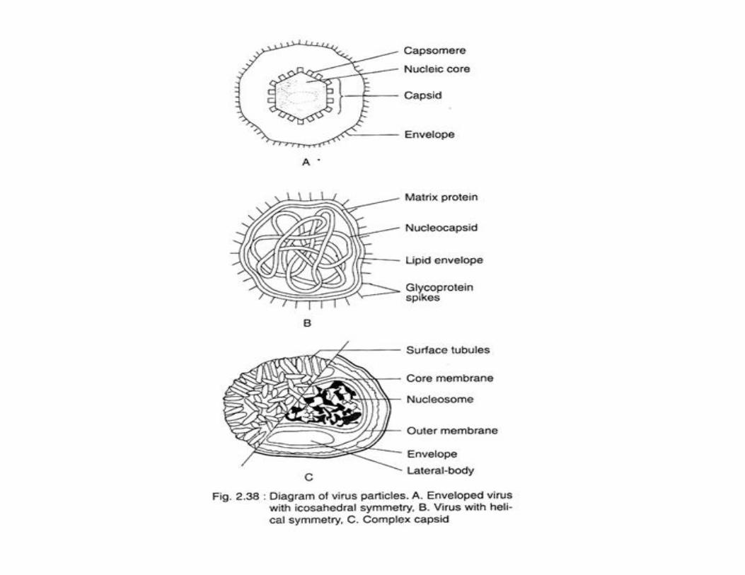
Symmetry:The capsid is symmetrically arranged around the centralnucleic acid.Based on symmetry of capsid, the viruses are grouped intothree categories:(a) Cubical (icosahedral),(b) Helical, and(c) Complex.(a) Cubical (icosahedral) capsids (Fig. 2.38A):They have a polygon with 12 corners (vertices), 20 sides(facets) and 30 edges. Each side is an equilateral triangle.They are of two types — Pentons (pentagonal capsomeres atthe corners) and Hexons (hexagonal capsomeres at thecorners), e.g., herpes and toga viruses are enveloped andpapova and adenoviruses are naked.
Symmetry:The capsid is symmetrically arranged around the centralnucleic acid.Based on symmetry of capsid, the viruses are grouped intothree categories:(a) Cubical (icosahedral),(b) Helical, and(c) Complex.(a) Cubical (icosahedral) capsids (Fig. 2.38A):They have a polygon with 12 corners (vertices), 20 sides(facets) and 30 edges. Each side is an equilateral triangle.They are of two types — Pentons (pentagonal capsomeres atthe corners) and Hexons (hexagonal capsomeres at thecorners), e.g., herpes and toga viruses are enveloped andpapova and adenoviruses are naked.

(b) Helical capsids:Both nucleic acid and capsomeres are coiled togetherand form a spiral or helical tube. All the helical typesare RNA viruses and most of them are enveloped,e.g., Tobacco mosaic virus (TMV), Influenza virus, etc.(c) Complex capsids:Viruses which do not conform to either of the abovetwo types due to complexity of their structure arecalled complex capsids, e.g., pox virus andbacteriophages like T2, T4, and T6.
(b) Helical capsids:Both nucleic acid and capsomeres are coiled togetherand form a spiral or helical tube. All the helical typesare RNA viruses and most of them are enveloped,e.g., Tobacco mosaic virus (TMV), Influenza virus, etc.(c) Complex capsids:Viruses which do not conform to either of the abovetwo types due to complexity of their structure arecalled complex capsids, e.g., pox virus andbacteriophages like T2, T4, and T6.

Types of Reproductive Cycle in VirusThe types are: 1. Lytic Cycle 2. Lysogenic Cycle.Type 1. Lytic Cycle:*It is the reproductive cycle of virulent phages, e.g.,T4 bacteriophage.*The phage attaches itself to the host cell (e.g.,Escherichia coli) through its tail fibres.*The fibres bend and bring the tip of tail in contactwith the host cell wall.*The tip of the tail produces a hole in the bacterialcell wall by means of enzyme lysozyme.
The types are: 1. Lytic Cycle 2. Lysogenic Cycle.Type 1. Lytic Cycle:*It is the reproductive cycle of virulent phages, e.g.,T4 bacteriophage.*The phage attaches itself to the host cell (e.g.,Escherichia coli) through its tail fibres.*The fibres bend and bring the tip of tail in contactwith the host cell wall.*The tip of the tail produces a hole in the bacterialcell wall by means of enzyme lysozyme.


*The tail sheath contracts and injects the viral genome intohost cell.*After entering the host cell, the viral DNA transcribes someearly mRNAs to form some enzymes over the host ribosome.Some of these are nucleases.*They degrade host DNA and mRNAs.
*Ribosomes and tRNAs remain unaffected. Phage DNA andmRNA are also protected from nucleases due to methylationof their cytosine bases.*Parent viral DNA functions as a template and replicatesrepeatedly with the help of bacterial nucleotides.*Simultaneously, host machinery (ribosomes, tRNAs, aminoacids, energy) is used by phage genes to synthesize proteinsfor vial lysozyme, internal proteins and capsid proteins.
*Ribosomes and tRNAs remain unaffected. Phage DNA andmRNA are also protected from nucleases due to methylationof their cytosine bases.*Parent viral DNA functions as a template and replicatesrepeatedly with the help of bacterial nucleotides.*Simultaneously, host machinery (ribosomes, tRNAs, aminoacids, energy) is used by phage genes to synthesize proteinsfor vial lysozyme, internal proteins and capsid proteins.

Lytic cycle
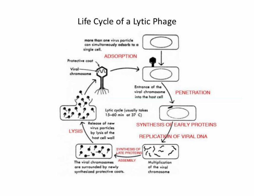


*Different components combine to form newviruses or phage particles.*The host cell ruptures by means of Lysozymereleasing the phage particles.*The period between entry of viral nucleoid intohost cell and bursting of host cell to release newviruses is called eclipse period.
*Different components combine to form newviruses or phage particles.*The host cell ruptures by means of Lysozymereleasing the phage particles.*The period between entry of viral nucleoid intohost cell and bursting of host cell to release newviruses is called eclipse period.


Type 2. Lysogenic Cycle:*Lambda phage (λ phage) has a higher degree of regulationof its genes.*The phage is parasitic over Escherichia coli. It does notpossess tail fibres for attachment to bacterial cell.*The tail directly comes in contact with bacterial cell, drills ahole in the wall and injects the phage DNA into the cell.*In lysogenic cycle, the phage DNA does not take over thecontrol of cellular machinery of the host.*Instead, it produces a repressor and undergoes reduction totemperate or non-virulent state.
*The tail directly comes in contact with bacterial cell, drills ahole in the wall and injects the phage DNA into the cell.*In lysogenic cycle, the phage DNA does not take over thecontrol of cellular machinery of the host.*Instead, it produces a repressor and undergoes reduction totemperate or non-virulent state.


Lysogenic Cycle


*With the help of enzyme integrate the viral genomebecomes integrated with the chromosomal DNA ofthe bacterium at a specific site (e.g., galactose locus inλ phage).*In this form the viral genome is called pro-phage.Pro-phage replicates along with bacterialchromosome and, therefore, gets distributed to thedaughter bacteria.*Pro-phage does not form virus particles because thegenes connected with taking over of host machineryremain repressed due to formation of a repressor.
*With the help of enzyme integrate the viral genomebecomes integrated with the chromosomal DNA ofthe bacterium at a specific site (e.g., galactose locus inλ phage).*In this form the viral genome is called pro-phage.Pro-phage replicates along with bacterialchromosome and, therefore, gets distributed to thedaughter bacteria.*Pro-phage does not form virus particles because thegenes connected with taking over of host machineryremain repressed due to formation of a repressor.

*At times the synthesis of repressor is stopped.Repressor can also be destroyed by chemicals,high energy radiations and other adverseconditions.*This converts the temperate or non-virulentvirus into virulent or lytic virus.*Therefore, the bacterial cell carrying pro-phageis called lysogenic cell and the phenomenon ofexistence of virus genome in pro-phage statealong-with host DNA is termed as lysogeny.
*At times the synthesis of repressor is stopped.Repressor can also be destroyed by chemicals,high energy radiations and other adverseconditions.*This converts the temperate or non-virulentvirus into virulent or lytic virus.*Therefore, the bacterial cell carrying pro-phageis called lysogenic cell and the phenomenon ofexistence of virus genome in pro-phage statealong-with host DNA is termed as lysogeny.

TMV (Tobacco Mosaic Virus)

*Tobacco mosaic virus (TMV) is one of thewell-characterized plant viruses.* The genome of TMV is a positive-sense,single-stranded RNA and encodes at leastthree non-structural proteins and acoat protein .
*Tobacco mosaic virus (TMV) is one of thewell-characterized plant viruses.* The genome of TMV is a positive-sense,single-stranded RNA and encodes at leastthree non-structural proteins and acoat protein .*Tobacco mosaic virus has a rod-likeappearance. Its capsid is made from 2130molecules of coat protein (see image to theleft) and one molecule of genomic singlestrand RNA, 6400 bases long.


Bacteriophage

*Like all viruses, phages are simpleorganisms that consist of a core of geneticmaterial (nucleic acid) surrounded by aprotein capsid. ...*There are three basic structural formsof phage: an icosahedral (20-sided) headwith a tail, an icosahedral head without atail, and a filamentous form.
*Like all viruses, phages are simpleorganisms that consist of a core of geneticmaterial (nucleic acid) surrounded by aprotein capsid. ...*There are three basic structural formsof phage: an icosahedral (20-sided) headwith a tail, an icosahedral head without atail, and a filamentous form.

Bacteriophage, alsocalled phage or bacterial virus, any of agroup of viruses that infect bacteria.Bacteriophages were discoveredindependently by Frederick W. Twort inGreat Britain (1915) and Felixd’Hérelle in France (1917).
Bacteriophage, alsocalled phage or bacterial virus, any of agroup of viruses that infect bacteria.Bacteriophages were discoveredindependently by Frederick W. Twort inGreat Britain (1915) and Felixd’Hérelle in France (1917).

*D’Hérelle coined theterm bacteriophage, meaning “bacteriaeater,” to describe the agent’sbactericidal ability.*Bacteriophages also infect the single-celled prokaryotic organisms knownas archaea.
*D’Hérelle coined theterm bacteriophage, meaning “bacteriaeater,” to describe the agent’sbactericidal ability.*Bacteriophages also infect the single-celled prokaryotic organisms knownas archaea.

Types of Bacteriophages:The phages have specific host. E. coli has been studiedmost extensively from this point of view. Thebacteriophage capable of destroying E. coli is called coli-phage. The types of coli-phages have been called as T-phages.They have been classified into many arbitrarygroups such as:(i) T-Even Phages (T2, T4, T6):These phages have an angular head and contractile tail.The DNA contain a unique base 5-hydroxyl methylcytosine in place of cytosine. These viruses are mostthoroughly studied viruses. These are also called virulentas they cause death of the host cells.
Types of Bacteriophages:The phages have specific host. E. coli has been studiedmost extensively from this point of view. Thebacteriophage capable of destroying E. coli is called coli-phage. The types of coli-phages have been called as T-phages.They have been classified into many arbitrarygroups such as:(i) T-Even Phages (T2, T4, T6):These phages have an angular head and contractile tail.The DNA contain a unique base 5-hydroxyl methylcytosine in place of cytosine. These viruses are mostthoroughly studied viruses. These are also called virulentas they cause death of the host cells.

(ii) T-Odd Phages (T1, T3, T7):These viruses have an angular head and a shortnon-contractile tail. The DNA contain cytosine.These are temperate viruses as their geneticmaterial becomes integrated with bacterialchromosomes and the host remains unaffected.(iii) T5 Phages:These viruses have an angular head and non-contractile tail. The DNA of these viruses alsocontains cytosine.
(ii) T-Odd Phages (T1, T3, T7):These viruses have an angular head and a shortnon-contractile tail. The DNA contain cytosine.These are temperate viruses as their geneticmaterial becomes integrated with bacterialchromosomes and the host remains unaffected.(iii) T5 Phages:These viruses have an angular head and non-contractile tail. The DNA of these viruses alsocontains cytosine.

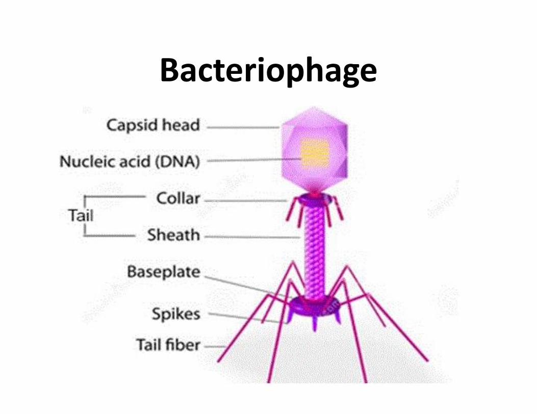
3. Structure of Bacteriophage:*With the help of electron microscope, themorphology of the bacteriophage has been studied.*The T even phages show complex symmetry. Theseviruses are generally tadpole shaped i.e., a ‘head’followed by a ‘tail’.*The head is hexagonal and like a prism in outline.•This shape is also known as elongated icosahedrons.It is 950 A° in length and 650 A° in width.•* The head has a 2-layered protein wall that enclosesthe double stranded DNA.•The wall is 35 A° thick and is composed of about2000 similar capsomeres. DNA is tightly packed in thehead and is about 50 µ long.
3. Structure of Bacteriophage:*With the help of electron microscope, themorphology of the bacteriophage has been studied.*The T even phages show complex symmetry. Theseviruses are generally tadpole shaped i.e., a ‘head’followed by a ‘tail’.*The head is hexagonal and like a prism in outline.•This shape is also known as elongated icosahedrons.It is 950 A° in length and 650 A° in width.•* The head has a 2-layered protein wall that enclosesthe double stranded DNA.•The wall is 35 A° thick and is composed of about2000 similar capsomeres. DNA is tightly packed in thehead and is about 50 µ long.

*Attached to one of the points of the head,through a neck and collar is the tail .*The tail has a complex structure andproteinaceous in nature.*It is made up of a cubical, hollow, cylindricalcore.•This core is 800 A° long, 70 A°in diameter andhas 25 A° wide central canal.•*This core is surrounded by a contractilesheath. The sheath is 165 A° in diameter.
*Attached to one of the points of the head,through a neck and collar is the tail .*The tail has a complex structure andproteinaceous in nature.*It is made up of a cubical, hollow, cylindricalcore.•This core is 800 A° long, 70 A°in diameter andhas 25 A° wide central canal.•*This core is surrounded by a contractilesheath. The sheath is 165 A° in diameter.


*The internal diameter of the tube formed by it isequal to core diameter of 70A°.*The core is terminated into a hexagonal plate whichhas six small tail fibres (tail ‘pins’) at every corner and6 tail fibres.•Each tail fiber is 1500 A° long and is composed offibrillar protein.•* The main function of the short tail fibres is to holdthe phage fast to the host during sheath contractionand DNA injection while long tail fibres helps inadsorption of the phage on the bacterial wall.
*The internal diameter of the tube formed by it isequal to core diameter of 70A°.*The core is terminated into a hexagonal plate whichhas six small tail fibres (tail ‘pins’) at every corner and6 tail fibres.•Each tail fiber is 1500 A° long and is composed offibrillar protein.•* The main function of the short tail fibres is to holdthe phage fast to the host during sheath contractionand DNA injection while long tail fibres helps inadsorption of the phage on the bacterial wall.

4. Chemistry of Bacteriophages:Bacteriophages are made up of nucleoproteins.The proteins are about 50-60% and nucleic acid40-50%. The nucleic acid is either double strandedDNA or single stranded DNA or single strandedRNA (Both never occur together).
4. Chemistry of Bacteriophages:Bacteriophages are made up of nucleoproteins.The proteins are about 50-60% and nucleic acid40-50%. The nucleic acid is either double strandedDNA or single stranded DNA or single strandedRNA (Both never occur together).


Biological Importance of Bacteriophages:*Bacteriophages have been used in prophylaxis and medicaltreatment against several pathogenic bacterial diseases e.g.,cholera, plague, dysentery, enteric fever etc.* They are also used in the diagnosis of certain infections likeplague, cholera etc. Bacteriophages feed on pathogenicbacteria present in polluted water.*So, they can also be used as scavengers. In many casesbacteriophages determine the micro-flora of the soil.*Thus, they play an important role in agriculture. In spacemicrobiology, lysogenic cultures are used as radiationdetectors and are used in USSR spaceship Vostok 2.
Biological Importance of Bacteriophages:*Bacteriophages have been used in prophylaxis and medicaltreatment against several pathogenic bacterial diseases e.g.,cholera, plague, dysentery, enteric fever etc.* They are also used in the diagnosis of certain infections likeplague, cholera etc. Bacteriophages feed on pathogenicbacteria present in polluted water.*So, they can also be used as scavengers. In many casesbacteriophages determine the micro-flora of the soil.*Thus, they play an important role in agriculture. In spacemicrobiology, lysogenic cultures are used as radiationdetectors and are used in USSR spaceship Vostok 2.

*Temperate phages serve as ‘vector intransferring the genetic material fromone bacterial cell to another(transduction).*Bacteriophages are very harmfulduring the process of manufacturing ofantibiotic and milk products becausethey kill beneficial bacteria by theirlysogenic activity.
*Temperate phages serve as ‘vector intransferring the genetic material fromone bacterial cell to another(transduction).*Bacteriophages are very harmfulduring the process of manufacturing ofantibiotic and milk products becausethey kill beneficial bacteria by theirlysogenic activity.

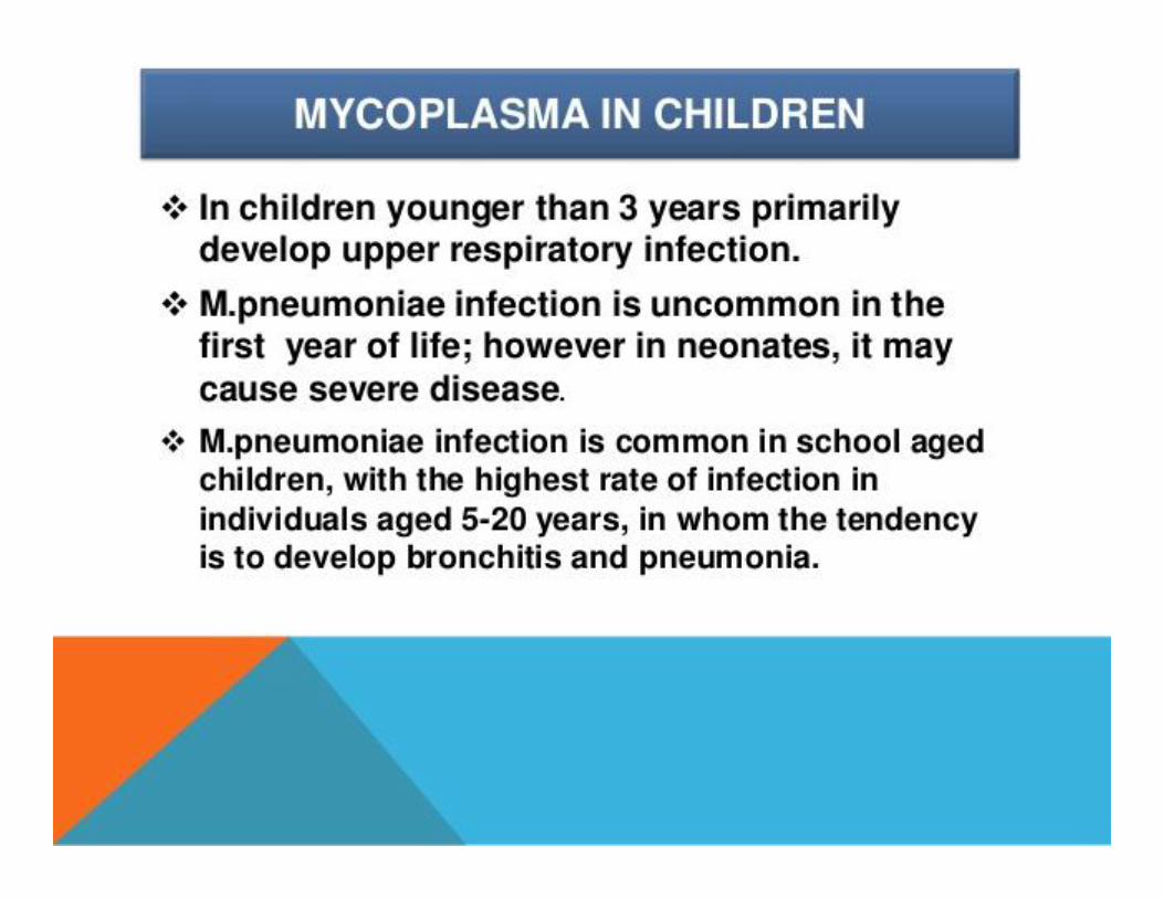
*Mycoplasmas are the “smallest, independentlyreplicating prokaryotes”.*These organisms were first discovered by Pasteur ineighteenth century when he studied the causativeagent of the “Bovine pleuropneumonia” (Apulmonary disease of cattle which appeared inGermany and Switzerland in 1713.* Due to its resemblance with pneumonia symptomsthis disease is called as Bovine Pleuropneumonia).
Mycoplasma
*Mycoplasmas are the “smallest, independentlyreplicating prokaryotes”.*These organisms were first discovered by Pasteur ineighteenth century when he studied the causativeagent of the “Bovine pleuropneumonia” (Apulmonary disease of cattle which appeared inGermany and Switzerland in 1713.* Due to its resemblance with pneumonia symptomsthis disease is called as Bovine Pleuropneumonia).


*It was believed that the causal agent wasPleuropneumonia like organisms (PPLO).*This causal agent was first isolated and cultured by E.Nocard and E. R. Roux in 1898.*They established that these causal agents ofpleuropneumonia can grow on complex nutrientmedia which do not contain cells.*They also observed that these organisms showdifferent forms, when grown on the culture media.*These organisms were named as Asterococcusmycoides by Borrel et. al (1910).*The generic name Mycoplasma was given by Nowak(1929) due to their fungi like resemblance.
*It was believed that the causal agent wasPleuropneumonia like organisms (PPLO).*This causal agent was first isolated and cultured by E.Nocard and E. R. Roux in 1898.*They established that these causal agents ofpleuropneumonia can grow on complex nutrientmedia which do not contain cells.*They also observed that these organisms showdifferent forms, when grown on the culture media.*These organisms were named as Asterococcusmycoides by Borrel et. al (1910).*The generic name Mycoplasma was given by Nowak(1929) due to their fungi like resemblance.

Habit and Habitat of Mycoplasma:*Mycoplasmas are parasitic as well assaprophytic.•More than 200 mycoplasma like bodiesare found to be associated with sewage,plants, animals, insects, humus, hot watersprings and other high temperatureenvironment.•*They have been found in phloem tissuesof diseased plants.
Habit and Habitat of Mycoplasma:*Mycoplasmas are parasitic as well assaprophytic.•More than 200 mycoplasma like bodiesare found to be associated with sewage,plants, animals, insects, humus, hot watersprings and other high temperatureenvironment.•*They have been found in phloem tissuesof diseased plants.

General Characters of Mycoplasma:1. They are unicellular, smallest, non-motile and prokaryoticorganisms forming fried egg shaped colonies.2. They are pleomorphic i.e., able to change their shapedepending upon culture media.3. They may be rod like, ring like, globoid or filamentous. Thefilaments are of uniform diameter (100-300 nm) and vary inlength from 3 nm to 150 nm.4. Some mycoplasma predominantly assume spherical shape(300-800 nm in diameter).5. They are ultra-filterable i.e., they can pass throughbacteria-proof filters.6. They do not possess rigid cell wall.
2. They are pleomorphic i.e., able to change their shapedepending upon culture media.3. They may be rod like, ring like, globoid or filamentous. Thefilaments are of uniform diameter (100-300 nm) and vary inlength from 3 nm to 150 nm.4. Some mycoplasma predominantly assume spherical shape(300-800 nm in diameter).5. They are ultra-filterable i.e., they can pass throughbacteria-proof filters.6. They do not possess rigid cell wall.


7. The cells are delimited by soft triple layeredlipo-proteinaceous membrane. It is unitmembrane about 10 nm thick.8. Within the cytoplasm ribosomes are foundscattered in the peripheral zone. These are 14nm in diameter and resemble with bacteria insedimentation characteristic of both thenucleoprotein and nucleic acid.9. The ribosomes are 72S type.10. Within the cytoplasm fine fibrillar DNA ispresent. It is double stranded helix.
7. The cells are delimited by soft triple layeredlipo-proteinaceous membrane. It is unitmembrane about 10 nm thick.8. Within the cytoplasm ribosomes are foundscattered in the peripheral zone. These are 14nm in diameter and resemble with bacteria insedimentation characteristic of both thenucleoprotein and nucleic acid.9. The ribosomes are 72S type.10. Within the cytoplasm fine fibrillar DNA ispresent. It is double stranded helix.

11. Mycoplasma generally grow more slowly thanbacteria.12. They require sterol for their nutrition.13. They are usually resistant to antibiotics likepenicillin, cephaloridine, vencomycin etc. whichaction cell wall.14. They are sensitive to tetracycline.
11. Mycoplasma generally grow more slowly thanbacteria.12. They require sterol for their nutrition.13. They are usually resistant to antibiotics likepenicillin, cephaloridine, vencomycin etc. whichaction cell wall.14. They are sensitive to tetracycline.
15. They are also killed by temperature of 40-55°C infifteen minutes.16. They do not produce spores.17. Like other prokaryotes, they usually divide bybinary fission.

Cell Structure of Mycoplasma:*In mycoplasma, the cells are small varyingfrom 300 nm to 800 nm in diameter.*Rigid cell wall is absent.*Cells are surrounded by a triple layered lipo-proteinaceous unit membrane .*It is about 10 nm thick. Unit membraneencloses the cytoplasm.
Cell Structure of Mycoplasma:*In mycoplasma, the cells are small varyingfrom 300 nm to 800 nm in diameter.*Rigid cell wall is absent.*Cells are surrounded by a triple layered lipo-proteinaceous unit membrane .*It is about 10 nm thick. Unit membraneencloses the cytoplasm.

*Within the cytoplasm RNA (ribosomes)and DNA are present.*The ribosomes are 14 nm in diameter and72 S type. DNA is double stranded helix.* It can be distinguished from bacterialDNA by its low guanine and cytosinecontent.
*Within the cytoplasm RNA (ribosomes)and DNA are present.*The ribosomes are 14 nm in diameter and72 S type. DNA is double stranded helix.* It can be distinguished from bacterialDNA by its low guanine and cytosinecontent.

*The DNA is up to four percent and RNA isabout eight percent and it is less than half thatusually occurs in other protoplasm’s. Theguanine and cytosine (G and C).*Contents in DNA range from 23-46 percent. Insome species e.g., M. gallisepticum some polarbodies protrude out from one or the other endof the cell.*These are called bleb and are considered to bethe site of enzymatic activities and attachmentduring infection.
*The DNA is up to four percent and RNA isabout eight percent and it is less than half thatusually occurs in other protoplasm’s. Theguanine and cytosine (G and C).*Contents in DNA range from 23-46 percent. Insome species e.g., M. gallisepticum some polarbodies protrude out from one or the other endof the cell.*These are called bleb and are considered to bethe site of enzymatic activities and attachmentduring infection.


Manner of transmission*Plant disease mycoplasmas are carried from a diseased plantto a healthy one by piercing and sucking insects, often of theHomopteran order such as leafhoppers.*These insects suck up the sap made by an infected plant andwith it the mycoplasmas it contains.•Mycoplasma transmission by leafhopper is circulating andpersistent.•* It requires a period of latency corresponding to itscirculation and multiplication within the vector insect, whichthen remains infectious for the rest of its life.•*The mycoplasmas are not however passed on to theoffspring.*No mycoplasma can be transmitted from plant to plant byphysical contact, nor in seed.
Manner of transmission*Plant disease mycoplasmas are carried from a diseased plantto a healthy one by piercing and sucking insects, often of theHomopteran order such as leafhoppers.*These insects suck up the sap made by an infected plant andwith it the mycoplasmas it contains.•Mycoplasma transmission by leafhopper is circulating andpersistent.•* It requires a period of latency corresponding to itscirculation and multiplication within the vector insect, whichthen remains infectious for the rest of its life.•*The mycoplasmas are not however passed on to theoffspring.*No mycoplasma can be transmitted from plant to plant byphysical contact, nor in seed.

Symptoms observedThe facts presented here are not specific to cyclamen, sincethe literature mainly enumerates the symptoms by broadcategory of plant families. The diseases caused are known as‘mycoplasmoses’, or ‘mycoplasmainfections’, and show verydiverse symptoms.In most cases one or more of the following symptoms areobserved:yellowingvarious growth disorders such as dwarfism, polyphylly,witches’ brooms (abnormal development of axillary buds).colour disorders such as variegationflower deformation: virescence (leaf-like aspect of parts offlowers) and phyllody (elongation of the gynoecium as leaf-like structures)
Symptoms observedThe facts presented here are not specific to cyclamen, sincethe literature mainly enumerates the symptoms by broadcategory of plant families. The diseases caused are known as‘mycoplasmoses’, or ‘mycoplasmainfections’, and show verydiverse symptoms.In most cases one or more of the following symptoms areobserved:yellowingvarious growth disorders such as dwarfism, polyphylly,witches’ brooms (abnormal development of axillary buds).colour disorders such as variegationflower deformation: virescence (leaf-like aspect of parts offlowers) and phyllody (elongation of the gynoecium as leaf-like structures)

*These disorders are essentially the resultof disturbance in the functioning of thephloem: the transport and transfer of theenergy-carrying molecules and mineralsalts are upset and the action of growthand development factors (plant hormones)is disturbed.
*These disorders are essentially the resultof disturbance in the functioning of thephloem: the transport and transfer of theenergy-carrying molecules and mineralsalts are upset and the action of growthand development factors (plant hormones)is disturbed.
