radiotherapy for optic nerve sheath meningioma: a case for earlier intervention?
TRANSCRIPT

at SciVerse ScienceDirect
Clinical Oncology xxx (2013) 1e6
Contents lists available
Clinical Oncology
journal homepage: www.cl in icaloncologyonl ine.net
Original Article
Radiotherapy for Optic Nerve Sheath Meningioma: A Case for EarlierIntervention?
G. Adams *, D.E. Roos *y, J.L. Crompton zx*Department of Radiation Oncology, Royal Adelaide Hospital, Adelaide, South Australia, AustraliayDepartment of Medicine, University of Adelaide, Adelaide, South Australia, AustraliazNeuro-Ophthalmology Service, Royal Adelaide Hospital, Adelaide, South Australia, Australiax Institute of Ophthalmology and Visual Science, University of Adelaide, Adelaide, South Australia, Australia
Received 29 September 2012; received in revised form 3 December 2012; accepted 24 January 2013
Abstract
Aims: To assess tumour control, visual outcomes and toxicity after radiotherapy for all patients with optic nerve sheath meningiomas (ONSM) treated by a singleradiation oncologist at a single institution over a 15 year period. To explore potential predictors of outcomes.Materials and methods: All patients underwent ophthalmological and radiological assessments before radiotherapy. These were repeated at regular intervalsafter treatment. A retrospective analysis of clinical, dosimetric and radiological data was carried out. Patients with useful vision before radiotherapy weredivided into two groups e those with maintained or improved vision and those with a deterioration in vision. The groups were compared using theManneWhitney U-test with regard to eight potential predictors of outcome.Results: Seventeen patients with 18 ONSM were treated with fractionated radiotherapy (46.8e55.8 Gy in 26e31 fractions). No evaluable tumours grew aftertreatment: control rate 100% (95% confidence interval 82e100%). Using the most common definition of visual function described in the literature, vision wasmaintained or improved in 89% (95% confidence interval 67e97%) of cases. In those with useful vision before treatment (13 evaluable eyes), visual acuity wasmaintained or improved in eight (62%, 95% confidence interval 36e82%). There was a suggestion that the time from the onset of symptoms to radiotherapy mayinfluence outcome. Those with stable or better visual acuity after radiotherapy had been observed for a shorter time compared with those who had worse visualacuity (median of 18 months versus 62 months). Acute and late toxicity from radiotherapy was manageable.Conclusion: Radiotherapy is an extremely effective modality in arresting the growth of ONSM. A longer time from symptom onset to the start of radiotherapymay predict for poorer outcomes.Crown Copyright � 2013 Published by Elsevier Ltd on behalf of The Royal College of Radiologists. All rights reserved.
Key words: Meningioma; ONSM; optic nerve; optic nerve sheath; radiotherapy
Introduction
Optic nerve sheath meningiomas (ONSM) are rare andaccount for 2% of all orbital tumours [1]. Like other me-ningiomas, they are more common in women and aretypically slow-growing benign tumours. The usual presen-tation is with progressive visual loss or proptosis (Figure 1).Historically it was felt that complete visual loss was inevi-table and that the only worthwhile intervention was to
Author for correspondence: G. Adams, NT Radiation Oncology, AlanWalker Cancer Care Centre, Rocklands Drive, Tiwi, NT 0810, Australia.Tel.: þ61 8 8944 8220; Fax þ61 8 8944 8222.
E-mail address: [email protected] (G. Adams).
0936-6555/$36.00 Crown Copyright � 2013 Published by Elsevier Ltd on behalfhttp://dx.doi.org/10.1016/j.clon.2013.02.004
Please cite this article in press as: Adams G, et al., Radiotherapy for OpticOncology (2013), http://dx.doi.org/10.1016/j.clon.2013.02.004
surgically decompress the nerve in those with useful butdeteriorating vision, with the aim of delaying complete vi-sual loss [2]. However, over the last few decades, evidencehas emerged that with radiotherapy, not only can thegrowth of the tumour be stopped or reversed, but alsovision can be maintained or improved in most cases [3].Because of the rarity of ONSM, evidence has come from caseseries with limited numbers [4e13] that have either com-bined the experience from several groups [4,5] or havepresented single institutional data [6e13]. Various tech-niques have been used, including conventional three-dimensional conformal radiotherapy (3DCRT) [4,10,12],fractionated stereotactic radiotherapy (FSRT) [4e9,11,13],stereotactic radiosurgery [11] or proton therapy [6]. It is notpossible from these small studies to determine if one
of The Royal College of Radiologists. All rights reserved.
Nerve Sheath Meningioma: A Case for Earlier Intervention? Clinical

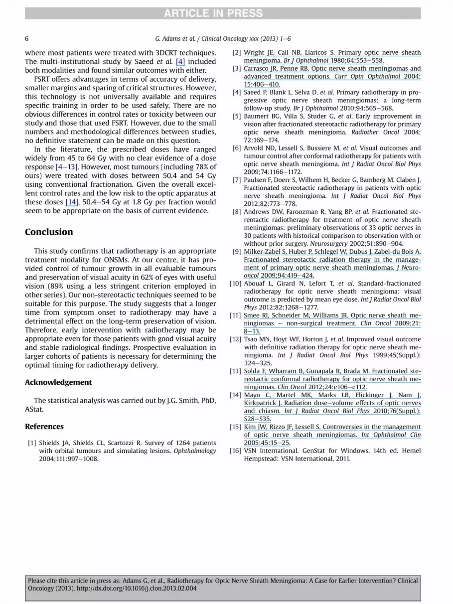
Fig 1. T1-weighted post-contrast magnetic resonance imaging of a neglected 35� 27 � 26 mm left optic nerve sheath tumour causing blindnessin a patient initially observed, then lost to follow-up before seeking treatment for cosmetic reasons. (a) Axial image showing severe left proptosisand ‘ghost’ of the atrophic optic nerve within the enhancing tumour posteriorly; (b) coronal image through the anterior aspect of the tumourshowing the compressed left optic nerve (central dark spot), and the posterior aspect of the normal right globe (dark).
G. Adams et al. / Clinical Oncology xxx (2013) 1e62
modality is superior to others. However, given that radia-tion tolerance of the optic nerve and chiasm may beimproved by using conventionally fractionated (1.8e2 Gyper fraction) radiotherapy, and the limited toxicity data onhypofractionated radiotherapy on these structures [14], it ispreferable to use conventionally fractionated radiotherapywhen attempting to maintain vision.
The timing of when radiotherapy should be administeredis less certain. Due to concern regarding late effects on theoptic apparatus, a period of observation is often advocatedwhile there is no clinical or radiological deterioration[3,4,15]. Yet, the question arises as to whether prolongedobservation may compromise preservation of vision as thetumour continues to grow-albeit slowly.
The aim of this study was to assess tumour control, vi-sual outcomes and treatment-related toxicity for all pa-tients with ONSM treated with radiotherapy at the RoyalAdelaide Hospital, South Australia, and to compare ourexperience with previously published series. In addition,we wanted to explore the influence on these outcomes offactors such as the prescribed dose of radiotherapy, thedose to critical structures, the tumour size or the timing ofradiotherapy.
Materials and Methods
All patients who received radiotherapy for ONSM be-tween 1996 and 2011 were identified using the depart-mental electronic database. The diagnosis was based on thepresence of typical clinical features and characteristicmagnetic resonance imaging (MRI) findings [3,15]. Wheretherewas sufficient doubt about the diagnosis, a biopsy wascarried out.
Relevant information was obtained from hospital re-cords, radiotherapy treatment plans and serial imaging(baseline and follow-up MRI scans). For patients referredfrom elsewhere, their ophthalmologist was contacteddirectly for further information when required.
Please cite this article in press as: Adams G, et al., Radiotherapy for OpticOncology (2013), http://dx.doi.org/10.1016/j.clon.2013.02.004
Ophthalmological Signs/Symptoms
All patients had a complete assessment at first presen-tation to an ophthalmologist. This included best correctedvisual acuity (Snellen chart), colour vision, visual fields, eyemovements, optic discs, proptosis, relative afferent pupil-lary defect and pain. At the time of referral for radio-therapy, any changes in these findings from baseline wererecorded.
After completing treatment, patients were followed longterm by their referring ophthalmologist with regular repeatassessments and (usually) yearly imaging. Cataract forma-tion as a cause of declining visual acuity was identified andcorrected where appropriate.
Radiotherapy
In all but one patient, radiotherapy was delivered us-ing conventional conformal techniques using 6 MV pho-tons with immobilisation in a thermoplastic shell. On theplanning computed tomography scan, the gross tumourvolume was defined as the visible (enhancing) meningi-oma and was marked with the aid of the diagnostic MRI(fusion images when available). A margin of 5e10 mmwas added to create the planning target volume. Organsat risk, namely retina, optic nerve, chiasm and brainstem, were delineated. An optimal conformal plan wasobtained with regard to the prescribed dose and toler-ances of organs at risk. A typical beam arrangementinvolved a three- to five-field plan sometimes including asuperioreinferior oblique field exiting via the oral cavityto help spare dose to anterior orbital structures. Imageguidance protocols followed departmental protocols atthe time.
One patient was treated using FSRT, using three non-coplanar 6 MV photon arcs with circular collimators toa total dose of 52.2 Gy in 29 fractions prescribed to the90% isodose curve (final fraction off retina). The gross
Nerve Sheath Meningioma: A Case for Earlier Intervention? Clinical

G. Adams et al. / Clinical Oncology xxx (2013) 1e6 3
tumour volume was defined the same way as in the3DCRT cases, but without a planning target volumeexpansion.
Radiology
Standard diagnostic MRI scans (typical slice thickness2e3 mm) were obtained before and after treatment. At-tempts were made to ensure that when measuring tumoursize in individual patients over time similar sequences wereused (typically T1-weighted fat suppressed with gadolin-ium). In addition to the radiology report, copies of all indi-vidual patient imaging were reviewed at the same time by asingle author. The maximum dimensions of each tumouralong the course of the optic nerve and orthogonally in thesupero-inferior and medio-lateral directions weremeasured.
Outcome Assessment
The primary outcome assessments were changes in vi-sual acuity and tumour size after radiotherapy as well astreatment toxicity.
In order to identify possible prognostic factors for visualoutcomes after radiotherapy, a strict definition for deterio-ration in acuity was used. This was any decline in visualacuity in those patients with useful vision (best correctedvisual acuity greater than 6/60) before radiotherapy. How-ever, so that a comparisonwith other published series couldbe made, we also used other common criteria quoted in theliterature, namely either a one or two line loss on theSnellen chart considering all treated eyes.
A reduction in tumour size was defined as a 2 mm orgreater sustained regression in one dimension withoutgrowth of 2 mm or greater in any other dimension. Growthof the tumour was defined as a sustained increase of 2 mmor greater in any dimension regardless of changes in otherdimensions.
Given the retrospective nature of the study, toxicity wasassessed by the descriptive reports in the case notes.Insufficient information was available to assess pituitarydysfunction related to radiotherapy.
An analysis of eight potential predictors of visualoutcome was made. These factors were the time from theonset of symptoms to radiotherapy, the size of the tumourat the start of treatment, visual acuity at the start of treat-ment, age at the start of treatment, radiotherapy dose, thedose to the retina, the dose to the chiasm and the year oftreatment.
Statistics
In order to assess the response to radiotherapy, findingsat the last assessment before treatment were comparedwith the most recent post-therapy assessment. To testwhether potential prognostic factors had a significant effecton visual acuity, tumours were divided into two groups,namely improved or stable versus worse visual acuity ac-cording to our strict definition of deterioration.
Please cite this article in press as: Adams G, et al., Radiotherapy for OpticOncology (2013), http://dx.doi.org/10.1016/j.clon.2013.02.004
Comparisons between the two groups were carried outusing the ManneWhitney U test [16]. Because eight prog-nostic factors were tested, for a single prognostic factor tobe declared significant at the 5% level, its P value wouldhave to be less than 0.0064. However, with the limitednumber of subjects, it was not considered appropriate tocarry out a multivariate analysis.
Results
Seventeen patients and 18 ONSMs were treated (one hadbilateral tumours treated simultaneously). For 16 tumoursthe diagnosis was based on clinical and radiological fea-tures. For two there was sufficient doubt about the diag-nosis towarrant biopsy. All patients were treated by a singleradiation oncologist. Outcomes for eight of these patientswere included in a previously published multi-institutionalreview of 34 ONSM patients, but their outcomes were notreported separately [4].
Table 1 summarises the baseline patient and tumourcharacteristics, with reported toxicity summarised in Table2. Prescribed doses were 46.8e55.8 Gy in 26e31 fractions(all 1.8 Gy per fraction).
One patient refused follow-up imaging, but was availablefor clinical assessments. The median follow-up from thestart of radiotherapy to the latest assessment was 6.2 years(range 0.9e13.3 years) for MRI imaging and 5.3 years (range0.6e13.4 years) for clinical assessment.
No evaluable tumours enlarged, 10 (56%) reduced in sizeand seven (39%) remained stable. Of note, the patient whorefused imaging has been followed-up for more than 5 yearswith improved visual acuity compared with base line.
Five of the 18 eyes did not have useful vision prior toradiotherapy (defined as visual acuity of 6/60 or worse). Ofthe remaining 13 eyes (all with acuity of 6/18 or betterbefore treatment), visual acuity improved in five, continuedunchanged in three and deteriorated in five. Vision wastherefore maintained or improved in 62% of the eyes withuseful vision before treatment (95% confidence interval32e86%).
For comparison with other series, improved or stablevision rates were 78% (95% confidence interval 55e91%) and89% (95% confidence interval 67e97%) for one and two linechanges on the Snellen chart, respectively.
Eight possible prognostic factors for a change in visualacuity after radiotherapy were tested in the subgroup withuseful vision before treatment. Their median follow-up forvisual assessment was 3.7 years (range 1.0e13.4 years) fromthe commencement of therapy. The results are shown inTable 3. In this small cohort with multiple statistical teststhere were no statistically significant differences betweenthe groups. However, therewas a suggestion that one factor,the time from the onset of symptoms to the start of radio-therapy was different. The median time from the onset ofsymptoms was shorter for those with maintained orimproved visual acuity compared with those where visualacuity deteriorated (18 versus 62 months, respectively,P ¼ 0.033).
Nerve Sheath Meningioma: A Case for Earlier Intervention? Clinical

Table 1Baseline characteristics of 17 patients (18 tumours)
Patient’s gender Male:female 2:15Eye with tumour Right:left:bilateral 8:8:1Age at the start of radiotherapy (years) Median (range) 47 (12e72)Onset of symptoms to radiotherapy (months) Median (range) 43 (1e149)Maximum tumour dimension (mm) Median (range) 25 (15e38)
Signs/symptoms at the time of radiotherapy Number % of 18
Visual acuity 6/6 or better 2 11%6/7.5 e 6/18 11 61%No useful vision (6/60 or worse) 5 28%
Pain No 12 67%Yes 6 33%
Relative afferent pupillary defect Present 13 72%Not recorded 5 28%
Visual fields Normal 6 33%Impaired 11 61%Not recorded 1 6%
Proptosis No 9 50%Yes 9 50%
Ocular movement Normal 13 72%Impaired 4 22%Not recorded 1 6%
Other signs Optic disc oedema 5 28%Optic disc atrophy 2 11%
Of those with functional vision: Number % of 13
Colour perception Normal 3 23%Impaired 7 54%Not recorded 3 23%
G. Adams et al. / Clinical Oncology xxx (2013) 1e64
Discussion
The results presented here represent the experience of asingle institution (and a single radiation oncologist) intreating ONSMs over a 15 year period.
Although technology has evolved, including a moverecently to intensity-modulated radiotherapy, the vastmajority of patients were treated using a 3DCRT techniquewith head shell and image guidance typical for brain orhead and neck radiotherapy in most departments. As withother groups, our results confirm that fractionated radio-therapy will probably arrest tumour growth.
Only two of 18 (11%) tumours were deemed to requirebiopsy by the referring ophthalmologists before radio-therapy. No attempt at surgical removal was made for any ofthis cohort. Therefore, the diagnosis of ONSM was
Table 2Side effects of radiotherapy in 17 patients
Acute effects (11 patients) Hair lossHeadacheOtitis externaXerostomiaConjunctivitis
Late effects (8 patients) XerophthalmiaCataractOptic disc atrophy
Please cite this article in press as: Adams G, et al., Radiotherapy for OpticOncology (2013), http://dx.doi.org/10.1016/j.clon.2013.02.004
presumptive in 89% of tumours. Although this could beinterpreted as a potential weakness of the data, the diag-nosis of ONSM can be made by the presence of classicclinical and radiological findings, and so a biopsy is seldomrequired in practice [3,15]. Other series have reported var-iable rates of tissue diagnosis. Some had low rates (less than20%) [5,12,13]; others intermediate (20e40%) [4,6,8],whereas a few had rates of around 50% [7,9]. This is partlyexplained by the fact that, in some series, there was amixture of patients treated with either primary radio-therapy or radiation after surgery (either due to only partialdebulking or relapse after attempted complete excision)[7e9,13]. Although this may represent different philoso-phies in management, it may also reflect different clinical orradiological features of tumours in the different series,which may have some influence on the response to
Number % of 17
8 47%2 12%1 6%1 6%1 6%5 29%4 24%2 12%
Nerve Sheath Meningioma: A Case for Earlier Intervention? Clinical

Table 3Possible prognostic factors for visual acuity after radiotherapy in 13 eyes with functional vision before radiotherapy (median and range ofeach variable)
Prognostic factor Improved or stable(n ¼ 8)
Worse(n ¼ 5)
P value*
Onset of symptoms to radiotherapy (months) 18 (1e89) 62 (36e149) 0.033Prescribed dose of radiotherapy (Gy) 50.4 (50.4e55.8) 50.4 (46.8e55.8) 0.97Dose to chiasm (Gy) 50.1 (5.0e52.0) 51.2 (46.8e54.0) 0.55Dose to ipsilateral retina (Gy) 50.0 (20.8e52.8) 50.5 (45.0e52.0) 0.86Maximum tumour dimension (mm) 23 (17e38) 26 (20e35) 0.35Visual acuity before radiotherapy (6/) 10.5 (5e18) 9 (5e18) 0.37Age at start of radiotherapy 49.5 (27e61) 44 (22e61) 0.91Year started radiotherapy 2006 (2002e2011) 2001 (1996e2011) 0.27
* Exact P value from two-sided Mann-Whitney U test comparing the two groups.
G. Adams et al. / Clinical Oncology xxx (2013) 1e6 5
treatment. For the two patients biopsied in our series, onehad non-functioning vision before treatment and the otherhad stable vision on follow-up (using all three criteria).Therefore, when assessing functional results after radio-therapy, previous surgical intervention is unlikely to be aconfounder in our series.
We limited our primary analysis of visual function out-comes, and possible predictors, to those with useful visionbefore radiotherapy. We feel that this is appropriate, as, foreyes with established non-useful vision, the aim of treat-ment is not to restore sight but to control other symptomssuch as pain and proptosis. Indeed, if the sight has alreadybeen lost, it is legitimate to relax or remove dose constraintsto organs at risk such as the ipsilateral retina and nerve.Inclusion of such patients in any analysis of visual outcomes(especially the effect of dose to these structures) wouldseem flawed. This exclusion has been carried out in fewother studies [5,8,13].
In addition, there are no clear criteria as to what con-stitutes a significant deterioration in vision. Most studiesused a specified objective deterioration in vision. In some,this was a one line change on the Snellen chart [6], whereasin others a two line change was used [4,5,7,10]. Others haveclassified patients according to their own subjective reportsor the non-quantified reports of their ophthalmologists[11,13].
In order to identify possible factors that may contributeto changing visual function, we chose to use a strict defi-nition of any deterioration from baseline. Although weacknowledge that in some cases, the degree of deteriorationmay not significantly affect function in patients with verygood eyesight, equally employing the other criteria mayignore an important deterioration in a patient with poorerbut functional vision before treatment. Also, as patientswere followed-up long term by their own ophthalmologistwith repeat assessments over time, any deterioration ten-ded to be noted on multiple examinations.
Our results give maintenance of function rates of 62%using our criteria, but 78 and 89% using the one and two lineSnellen chart criteria, respectively. Given the small numbersin all reports and these methodological differences inreporting visual outcomes, it is difficult to confidentlycompare results. However, a 100% tumour growth controlrate in all evaluable cases, and maintaining or improving
Please cite this article in press as: Adams G, et al., Radiotherapy for OpticOncology (2013), http://dx.doi.org/10.1016/j.clon.2013.02.004
vision in 89% using the most commonly applied measure ofvisual acuity outcomes, are consistent with previous reports(tumour control rates of 87e100% and stable or improvedvision rates of 67e98%) [4e13].
Unfortunately, due to the retrospective nature of thisstudy, no other findings (including pituitary functionassessment) were recorded consistently enoughthroughout the follow-up period to allow for a detailedanalysis. However, clinically significant toxicity (as reportedin Table 2) was manageable.
There is a suggestion from our results that the timing ofradiotherapy may influence visual outcomes, as thosewhose vision was maintained or improved tended to betreated earlier than those whose vision deteriorated (me-dian 18 versus 62 months, respectively).
The timing of radiotherapy is controversial, with rec-ommendations generally favouring delaying treatment ifvisual acuity is good and stable due to concerns abouttoxicity and the belief that a delay until signs of deteriora-tion are apparent has no long-term detrimental effect.However, specific evidence for this is scant. Saeed et al. [4]found no association between the timing of radiotherapyand outcomes, whereas Abouaf et al. [10] did show a trendtowards better acuity in those treated earlier. Unfortunately,no other studies have specifically addressed this issue.
None of the other prognostic factors analysed showedany suggestion of influencing outcome. However, as with allother reports on radiotherapy for ONSM, the small numbersin our study mean that any analysis had low statisticalpower. Although our results could be interpreted as sug-gesting that radiotherapy should be delivered earlier ratherthan later, this observation should be regarded as hypoth-esis generating and should be validated in larger cohorts.
Even if the association between timing and outcome isconfirmed, it is not necessarily causal. The time from theonset of symptoms to the delivery of radiotherapy may be asurrogate for the biology of the individual tumour. Patientswith a slower growing tumour (which may respond lesswell to radiation) may have more insidious symptoms andso present later for treatment.
As well as the timing of radiotherapy, other controversiessurround optimum modality and prescribed dose. Most re-ports have used FSRT for most of their patients [5e9,11,13].We, together with others [10,12], have reported cohorts
Nerve Sheath Meningioma: A Case for Earlier Intervention? Clinical
G. Adams et al. / Clinical Oncology xxx (2013) 1e66
where most patients were treated with 3DCRT techniques.The multi-institutional study by Saeed et al. [4] includedboth modalities and found similar outcomes with either.
FSRT offers advantages in terms of accuracy of delivery,smaller margins and sparing of critical structures. However,this technology is not universally available and requiresspecific training in order to be used safely. There are noobvious differences in control rates or toxicity between ourstudy and those that used FSRT. However, due to the smallnumbers and methodological differences between studies,no definitive statement can be made on this question.
In the literature, the prescribed doses have rangedwidely from 45 to 64 Gy with no clear evidence of a doseresponse [4e13]. However, most tumours (including 78% ofours) were treated with doses between 50.4 and 54 Gyusing conventional fractionation. Given the overall excel-lent control rates and the low risk to the optic apparatus atthese doses [14], 50.4e54 Gy at 1.8 Gy per fraction wouldseem to be appropriate on the basis of current evidence.
Conclusion
This study confirms that radiotherapy is an appropriatetreatment modality for ONSMs. At our centre, it has pro-vided control of tumour growth in all evaluable tumoursand preservation of visual acuity in 62% of eyes with usefulvision (89% using a less stringent criterion employed inother series). Our non-stereotactic techniques seemed to besuitable for this purpose. The study suggests that a longertime from symptom onset to radiotherapy may have adetrimental effect on the long-term preservation of vision.Therefore, early intervention with radiotherapy may beappropriate even for those patients with good visual acuityand stable radiological findings. Prospective evaluation inlarger cohorts of patients is necessary for determining theoptimal timing for radiotherapy delivery.
Acknowledgement
The statistical analysis was carried out by J.G. Smith, PhD,AStat.
References
[1] Shields JA, Shields CL, Scartozzi R. Survey of 1264 patientswith orbital tumours and simulating lesions. Ophthalmology2004;111:997e1008.
Please cite this article in press as: Adams G, et al., Radiotherapy for OpticOncology (2013), http://dx.doi.org/10.1016/j.clon.2013.02.004
[2] Wright JE, Call NB, Liaricos S. Primary optic nerve sheathmeningioma. Br J Ophthalmol 1980;64:553e558.
[3] Carrasco JR, Penne RB. Optic nerve sheath meningiomas andadvanced treatment options. Curr Opin Ophthalmol 2004;15:406e410.
[4] Saeed P, Blank L, Selva D, et al. Primary radiotherapy in pro-gressive optic nerve sheath meningiomas: a long-termfollow-up study. Br J Ophthalmol 2010;94:565e568.
[5] Baumert BG, Villa S, Studer G, et al. Early improvement invision after fractionated stereotactic radiotherapy for primaryoptic nerve sheath meningioma. Radiother Oncol 2004;72:169e174.
[6] Arvold ND, Lessell S, Bussiere M, et al. Visual outcomes andtumour control after conformal radiotherapy for patients withoptic nerve sheath meningioma. Int J Radiat Oncol Biol Phys2009;74:1166e1172.
[7] Paulsen F, Doerr S, Wilhem H, Becker G, Bamberg M, Claben J.Fractionated stereotactic radiotherapy in patients with opticnerve sheath meningioma. Int J Radiat Oncol Biol Phys2012;82:773e778.
[8] Andrews DW, Faroozman R, Yang BP, et al. Fractionated ste-reotactic radiotherapy for treatment of optic nerve sheathmeningiomas: preliminary observations of 33 optic nerves in30 patients with historical comparison to observation with orwithout prior surgery. Neurosurgery 2002;51:890e904.
[9] Milker-Zabel S, Huber P, Schlegel W, Dubus J, Zabel-du Bois A.Fractionated stereotactic radiation therapy in the manage-ment of primary optic nerve sheath meningiomas. J Neuro-oncol 2009;94:419e424.
[10] Abouaf L, Girard N, Lefort T, et al. Standard-fractionatedradiotherapy for optic nerve sheath meningioma: visualoutcome is predicted by mean eye dose. Int J Radiat Oncol BiolPhys 2012;82:1268e1277.
[11] Smee RI, Schneider M, Williams JR. Optic nerve sheath me-ningiomas e non-surgical treatment. Clin Oncol 2009;21:8e13.
[12] Tsao MN, Hoyt WF, Horton J, et al. Improved visual outcomewith definitive radiation therapy for optic nerve sheath me-ningioma. Int J Radiat Oncol Biol Phys 1999;45(Suppl.):324e325.
[13] Solda F, Wharram B, Gunapala R, Brada M. Fractionated ste-reotactic conformal radiotherapy for optic nerve sheath me-ningiomas. Clin Oncol 2012;24:e106ee112.
[14] Mayo C, Martel MK, Marks LB, Flickinger J, Nam J,Kirkpatrick J. Radiation doseevolume effects of optic nervesand chiasm. Int J Radiat Oncol Biol Phys 2010;76(Suppl.):S28eS35.
[15] Kim JW, Rizzo JF, Lessell S. Controversies in the managementof optic nerve sheath meningiomas. Int Ophthalmol Clin2005;45:15e25.
[16] VSN International. GenStat for Windows, 14th ed. HemelHempstead: VSN International, 2011.
Nerve Sheath Meningioma: A Case for Earlier Intervention? Clinical