radioimaging of light chain amyloid with a fibril-reactive monoclonal antibody
TRANSCRIPT

Radioimaging of Light Chain Amyloid with aFibril-Reactive Monoclonal Antibody
Jonathan S. Wall1, Stephen J. Kennel1, Mike Paulus2, Jens Gregor3, Tina Richey1, James Avenell4, Jeffrey Yap4,David Townsend4, Deborah T. Weiss1, and Alan Solomon1
1Human Immunology and Cancer Program, Department of Medicine, University of Tennessee Graduate School of Medicine, Knoxville,Tennessee; 2Engineering Science and Technology Group, Oak Ridge National Laboratory, Oak Ridge, Tennessee; 3Department ofComputer Science, University of Tennessee, Knoxville, Tennessee; and 4Cancer Imaging and Tracer Development Research Program,University of Tennessee Graduate School of Medicine, Knoxville, Tennessee
Currently, there are no available means in the United States todocument objectively the location and extent of amyloid de-posits in patients with systemic forms of amyloidosis. To addressthis limitation, we have developed a novel diagnostic strategy,namely, the use of a radiolabeled fibril-reactive murine monoclo-nal antibody (mAb) as an amyloid-specific imaging agent. Thegoal of this studywas to determine the pharmacokinetics, biodis-tribution, and ability of this reagent to target the type of amyloidthat is formed from immunoglobulin light chains, that is, AL.Methods: Subcutaneous tumors (amyloidomas) were inducedin BALB/c mice by injection of human AL fibrils. The IgG1 mAbdesignated 11-1F4 and an isotype-matched control antibodywere radioiodinated, and the pharmacokinetics and localizationof these reagents were determined from blood and tissue sam-ples. Amyloidoma-bearing animals that received 125I- or 124I-labeled antibodies were imaged by whole-body small-animalSPECT/CT or small-animal PET/CT technology, respectively.Results: Radioiodinated mAb 11-1F4 retained immunoreactiv-ity, as evidenced by its subnanomolar affinity for light chainsimmobilized on 96-well microtiter plates and for beads conju-gated with a light chain–related peptide. Additionally, after intra-venous administration, the labeled reagents had the expectedbiologic half-life of murine IgG1, with monoexponential whole-body clearance kinetics. In the amyloidoma mouse model,125I-11-1F4 was predominately localized in the tumors, as dem-onstrated in biodistribution and autoradiographic analyses. Themean uptake of this reagent, that is, the percentage injecteddose per gram of tissue, 72 h after injection was significantlyhigher for amyloid than for skeletal muscle, spleen, kidney, heart,liver, or other tissue samples. Notably, the accumulation withinthe amyloidomas of 125I- or 124I-11-1F4 was readily visible inthe fused small-animal SPECT/CT or small-animal PET/CT im-ages, respectively. Conclusion: Our studies demonstrate theamyloid-imaging capability of a radiolabeled fibril-reactive mAband provide the basis for a clinical trial designed to determineits diagnostic potential in patients with AL amyloidosis and othersystemic amyloidoses.
KeyWords: amyloid; immunoimaging; small-animal SPECT/CT;small-animal PET/CT
J Nucl Med 2006; 47:2016–2024
The ability to image a pathologic process radiograph-ically provides physicians with an objective means to deter-mine the presence and extent of disease as well as to monitora patient’s response to treatment or determine whetherrelapse has occurred. For the systemic amyloidoses, routineradiologic techniques (e.g., CT, ultrasound, or MRI) are notparticularly informative or amyloid specific. For primary(light chain amyloid [AL]) or secondary (amyloid A [AA])amyloidosis, European investigators have successfullyimaged pathologic deposits by planar scintigraphy with123I-labeled P component and with 99mTc-aprotinin (1–4);however, the U.S. Food and Drug Administration will notpermit the administration of such reagents in the UnitedStates inasmuch as the protein carriers are of human and ani-mal origins, respectively.
Given this restriction and the need to document thepresence of amyloid fibrils in affected major organs quan-titatively, especially in patients enrolled in therapeutic clin-ical trials, we have proposed another strategy, namely, theuse of a radiolabeled fibril-reactive monoclonal antibody(mAb) as an imaging agent. The rationale for this approachis based on the discovery that certain murine anti–humanlight chain mAbs recognize a conformational epitope com-mon to fibrils formed from light chains as well as otheramyloidogenic precursor molecules, such as serum AA,transthyretin, and apolipoprotein A-I (5). Further, when theprototypic IgG1 antibody designated 11-1F4 was adminis-tered to mice bearing subcutaneous human AL amyloidomas,it bound specifically to the amyloid deposits and acceleratedthe removal of this material.
On the basis of these data, we have tested whether mAb11-1F4 labeled with g- or positron-emitting isotopes of io-dine would prove to be a suitable reagent for the visuali-zation of amyloid. For these studies, we used instrumentation
Received Jun. 29, 2006; revision accepted Sep. 19, 2006.For correspondence or reprints contact: Jonathan S. Wall, PhD, University
of Tennessee Graduate School of Medicine, 1924 Alcoa Hwy., Knoxville, TN37920.E-mail: [email protected] ª 2006 by the Society of Nuclear Medicine, Inc.
2016 THE JOURNAL OF NUCLEAR MEDICINE • Vol. 47 • No. 12 • December 2006
by on August 15, 2015. For personal use only. jnm.snmjournals.org Downloaded from
designed to image small laboratory animals, that is, high-resolution small-animal SPECT and small-animal PET co-registered with small-animal CT for anatomic precision. Wenow report our experimental findings, which indicated thefeasibility of immunoimaging as a clinical means to documentthe presence and distribution of systemic amyloid deposits.
MATERIALS AND METHODS
Amyloid ProteinsAmyloid fibrils were extracted (6) from livers or spleens ob-
tained postmortem from patients with AL amyloidosis, and theirchemical compositions were determined by amino acid sequenc-ing and tandem mass spectrometry (7). Synthetic amyloid fibrilswere prepared (8) from a synthetic peptide (Keck BiotechnologyCenter) that encompassed the first 30 residues of human k4 lightchain Len (Len 1–30) (9), which was used as the immunogen togenerate mAb 11-1F4 (10).
AntibodiesThe derivation as well as the production of amyloid-reactive murine
IgG1 mAb 11-1F4 by the National Cancer Institute Biopharmaceut-ical Development Program (Science Applications International Cor-poration) was previously reported (11,12). An isotype-matched(IgG1) mouse mAb, MOPC-31C (Sigma), served as a control.
Antibody LabelingThe 11-1F4 antibody (100 mg–1 mg) was labeled with 37–74
MBq of reductant-free 125I (PerkinElmer) or 124I (kindly providedby Dr. George Kabalka, University of Tennessee, Knoxville, TN,and Dr. Ron D. Finn, Memorial Sloan-Kettering Cancer Center,New York, NY, or purchased from IBA/Eastern Isotopes) by use oflimiting amounts of N-chloro-p-toluenesulfonamide sodium salt(Chloramine-T; Sigma) (13). The labeled reagents were suspendedin phosphate-buffered saline (PBS) containing bovine serumalbumin (BSA) at 5 mg/mL (BSA/PBS), and unbound isotopeand protein aggregates were removed by size-exclusion liquidchromatography (14) through an Ultrogel AcA34 column (Amer-sham Pharmacia). Fractions containing IgG monomers were pooledfor biodistribution and imaging experiments. 125I-Labeled prepa-rations were subjected to sodium dodecyl sulfate–polyacrylamidegel electrophoresis (SDS-PAGE) (10% gels) in the presence orabsence of a reducing agent and analyzed with a CyclonePhosphorImager (Packard Instrument Co.).
Immunoreactivity AssaysTo verify the specificity and affinity of mAb 11-1F4, the
radiolabeled antibody was tested in a 96-well plate radioimmuno-assay, as well as a bead-conjugated antigen-binding assay. For theformer, Immulon 4 (Dynex Technologies) wells were coated withsynthetic AL fibrils (and, as a control, nonfibrillar protein). Afterovernight incubation at 37�C, the wells were incubated with 200mLof BSA/PBS for 2 h before the addition of 50 mL of serially dilutedradiolabeled antibody. The plates were maintained at room tem-perature on a 60� slanted, rotating disk for 2 h, after which the wellswere washed initially with PBS containing 0.1% (v/v) Tween 20and then with PBS alone. Radioactivity was measured with aPackard COBRA Quantum Gamma Spectrometer (GMI Inc.).
For the bead assay, a 1-mL volume of 0.9-mm-diameter amino-functionalized polystyrene beads (Spherotech Inc.) was washedwith PBS and activated by the addition of 4.0 mL of 0.5%glutaraldehyde in PBS for 5 min at room temperature. After another
PBS wash, a 1 mg/mL solution of the synthetic Len 1–30 peptidethat contained the epitope recognized by mAb 11-1F4 was addedto the beads, and the slurry was tumbled end over end for 18 h atroom temperature. Free glutaraldehyde sites were then blockedwith 0.5 mL of sterile glycine (1 mol/L) in PBS, and the mixturewas tumbled for 1 h. After another wash, the beads were storedas a 1:1 slurry in PBS. For the binding assay, 1–10 ng of theradioiodinated antibody was added to 5 mL of the bead suspensionin 100 mL of BSA/PBS. After 1 h of incubation, the beads werewashed twice with PBS by centrifugation at 10,000g for 2 min,and radioactivity was measured with the Gamma Spectrometer.
In Vivo StudiesAmyloidomas were induced in 8-wk-old BALB/c mice by 50-mg
subcutaneous injections between the scapulae of human AL fibrils(5). After 7 d, the animals received in the lateral tail vein 100- to200-mL volumes containing 20–50 mg of radiolabeled 11-1F4 orMOPC-31C (;12 MBq) in BSA/PBS. In some studies, a 1% KI(Lugol’s) solution was added to the animals’ drinking water 48 hbefore injection to limit radioiodine uptake by the thyroid gland.Mice were euthanized 72 h later by inhalation of an excess ofisoflurane, and imaging data were collected. To determine thebiodistribution of 125I-labeled antibodies, samples of skin, skeletalmuscle, femur, abdominal fat, stomach, small and large intestines,liver, kidneys, spleen, sternum, throat (containing the thyroid), heart,lungs, blood, and brain were harvested, placed into tared vials andweighed, and the radioactivity was measured. The data were ex-pressed as percentage injected dose per gram of tissue (%ID/g).Additionally, samples (including the amyloidomas) were fixed in10% buffered formalin for 24 h and embedded in paraffin forhistologic and autoradiographic analyses.
Single-animal in vivo whole-body clearance measurementswere obtained in BALB/c mice injected with 10 mg (5 MBq) of124I-11-1F4. An unanesthetized mouse was placed at various timeintervals into a plastic chamber and lowered into a commercial PETdose calibrator (CRC-15 PET; Capintec) that had been calibratedwith known reference standards. Multiple readings were acquiredover a 66-h period, and the data were analyzed with mono- orbiexponential kinetics. For both forms of radioiodinated mAb 11-1F4, the effective half-life (T½eff) was calculated from the mea-sured biologic half-life (T½bio) of 11-1F4 and the known physicalhalf-lives (T½rad) of 124I and 125I as follows:
T½eff 5T½bioT½rad
T½bio 1 T½rad:
For autoradiography, 6-mm-thick sections cut from formalin-fixed,paraffin-embedded blocks were placed on Probond microscope slides(Fisher Scientific), dipped in NTB-2 emulsion (Eastman Kodak),stored in the dark, and developed after a 24-h exposure. The sectionswere counterstained with hematoxylin and eosin or Congo red,placed on coverslips sealed with Permount (Fisher Scientific), andexamined by light or polarizing microscopy, respectively. Digital cam-era microscopic images were obtained and evaluated with an imageanalysis software package (Image-Pro Plus; Media Cybernetics).
Instrumentation and Image AcquisitionSPECT data were collected with a small-animal SPECT imag-
ing system (developed at the Oak Ridge National Laboratory) (15)capable of a 1.7-mm spatial resolution when equipped with a10-mm-long hexagonal parallel-hole collimator. During imaging,
RADIOIMAGING OF AMYLOID • Wall et al. 2017
by on August 15, 2015. For personal use only. jnm.snmjournals.org Downloaded from
the 2 detectors [composed of a 50-mm-diameter HamamatsuR2486-02 multianode photomultiplier tube coupled to a 1 · 1 · 8mm NaI(Tl) crystal array arranged on a 1.2-mm2 grid] werepositioned ;5 cm from the 50-mL conical tubes housing the mice.Each SPECT dataset comprised 60 projections collected over 360�over the course of 30–60 min. Images were reconstructed withan implementation of the expectation-maximization maximum-likelihood algorithm (16).
After the collection of SPECT data, high-resolution CT imageswere obtained with a MicroCAT II (Siemens Medical SolutionsMolecular Imaging, LLC) instrument (17–19) with a source anddetector configuration capable of an ;75-mm spatial resolution.The scanner had a circular-orbit cone-beam geometry, was equippedwith a 20- to 80-kVp microfocus x-ray source, and captured a 90 ·60 mm field of view with a 2,048 · 3,072 charge-coupled devicearray detector optically coupled to a minR phosphor screen(Eastman Kodak) via a fiber-optic bundle. Each CT dataset,composed of 360 projections at 1� azimuths, was acquired in 8 min.Postacquisition images were reconstructed on isotropic 100-mmvoxels by means of a recently developed modified version of theFeldkamp algorithm (20). For contrast-enhanced small-animal CT,amyloidoma-bearing mice were given 300-mL intravenous doses ofiodinated triglycerides (Fenestra VC; Advanced Research Technol-ogies) 30 min before scanning.
To facilitate coregistration of the reconstructed SPECT and CTimages, 3 capillaries filled with a 125I solution and placed on theconical tubes were used for reference purposes and providedfiducial marks in the x-, y-, and z-axes. The small-animal SPECTand CT datasets were visualized and coregistered manually with a3-dimensional image analysis software package (Amira, version3.1; Mercury Computer Systems). The volume of each amyloi-doma was determined from the small-animal CT data with theAmira tissue segmentation tool.
For PET, euthanized animals were placed on a cardboard platformthat contained 68Ge fiducial markers, and data were collected over a40-min period with the Focus 220 or P4 microPET scanner (SiemensMedical Solutions). The images were reconstructed with the ordered-subset expectation-maximization 3-dimensional maximum a post-eriori algorithm (21). After the collection of small-animal PET data,mice were placed in the MicroCAT II scanner, and the CT dataset wasacquired as described earlier. Coregistration of the PET and CT datawas performed manually with Amira software.
All animal experiments were conducted in accordance withU.S. Public Health Service guidelines and under the auspices ofUniversity of Tennessee and Oak Ridge National Laboratory Ani-mal Care and Use Committee–approved protocols.
RESULTS
Amyloidoma Model
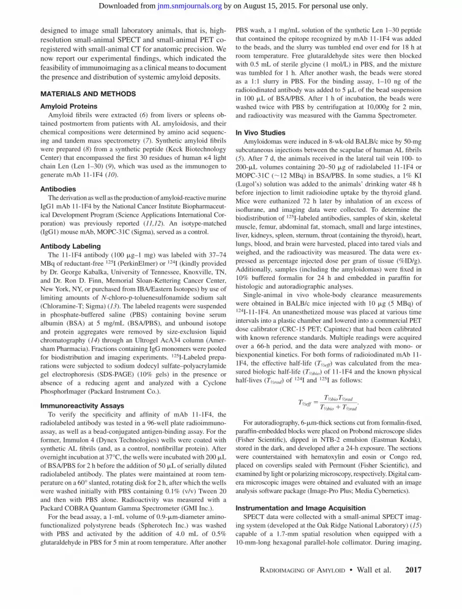
The amyloidomas induced in mice by subcutaneous in-jections between the scapulae of human AL fibril extractswere readily apparent. After 7 d, the tumors appeared semi-solid and vascularized (Fig. 1A), and their size and locationwere readily documented by small-animal CT (Figs. 1B and1C). The mean volume of the amyloid lesions, as determinedfrom CT data, was 196 mm3 (range, 130–260 mm3).
Radioiodination
The 11-1F4 and MOPC-31C antibodies were readily la-beled with 125I and 124I, with yields of up to 80%, depending
on the concentration of antibody used in the coupling reac-tion. PhosphorImager analyses of the radioiodinated proteinsafter gel filtration and SDS-PAGE indicated that greater than98% of the radioactivity was localized to the heavy and lightchains at a ratio of ;2:1. The dissociation constant (Kd) ofradiolabeled 11-1F4 for the Len 1–30 peptide, calculated byScatchard analysis of radioimmunoassay data, was deter-mined to be ;0.3 nmol/L. When the antibody was tested at arange of 1–10 ng in the bead assay, the maximum bindingwas 77%–79%. These data demonstrated the purity ofradioiodinated mAb 11-1F4 and notably that the affinity ofthe 125I-11-1F4 conjugate for the Len 1–30 peptide wasidentical to that of the unlabeled native antibody (22).
Pharmacokinetics of Radiolabeled mAb 11-1F4
Two independent (but complementary) methods wereused to determine the pharmacokinetics of mAb 11-1F4.The first method measured the clearance of 25 mg (;4.0MBq) of 125I-labeled antibody 1, 4, 24, 72, and 144 h afterintravenous injection in cohorts of 6 normal, that is,amyloidoma-free, BALB/c mice. At each time point, 1 groupof animals was euthanized, the tissues were harvested andweight normalized, and the decay-corrected specific activ-ities were determined (Table 1). For all samples, the activi-ties decreased by 4 h, with the exception of the tongue andskin, in which they more than doubled between the 1- and
FIGURE 1. Murine model of an AL amyloidoma. (A) Grossappearance of vascularized amyloidoma. (B) Axial slice throughupper abdomen of mouse 7 d after 50-mg amyloidomainduction and after administration of an intravenous dose ofcontrast medium 30 min before image acquisition. (C) Three-dimensional volumetric rendering. Arrows indicate location ofamyloidoma. Tumor volume was calculated from small-animalCT data to be 104 mm3.
2018 THE JOURNAL OF NUCLEAR MEDICINE • Vol. 47 • No. 12 • December 2006
by on August 15, 2015. For personal use only. jnm.snmjournals.org Downloaded from
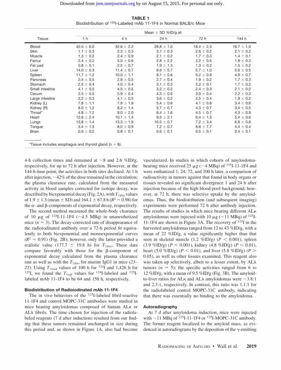
4-h collection times and remained at ;8 and 2.6 %ID/g,respectively, for up to 72 h after injection. However, at the144-h time point, the activities in both sites declined. At 1 hafter injection,;42% of the dose remained in the circulation;the plasma clearance rate, calculated from the measuredactivity in blood samples corrected for isotope decay, wasdescribed by biexponential decay (Fig. 2A), withT½biovaluesof 1.96 1.3 (mean6 SD) and 164.16 67.8 h (R2 5 0.98) forthe a- and b-components of exponential decay, respectively.
The second method measured the whole-body clearanceof 10 mg of 124I-11-1F4 (;4.5 MBq) in unanesthetizedmice (n 5 3). The decay-corrected rate of disappearance ofthe radioiodinated antibody over a 72-h period fit equiva-lently to both biexponential and monoexponential curves(R2 5 0.91) (Fig. 2B); however, only the latter provided arealistic value (177.7 6 19.8 h) for T½bio. These datacompare favorably with those for the b-component ofexponential decay calculated from the plasma clearancerate as well as with the T½bio for murine IgG1 in mice (23–25). Using T½rad values of 100 h for 124I and 1,426 h for125I, we found the T½eff values for 124I-labeled and 125I-labeled mAb 11-1F4 to be 64 and 158 h, respectively.
Biodistribution of Radioiodinated mAb 11-1F4
The in vivo behaviors of the 125I-labeled fibril-reactive11-1F4 and control MOPC-31C antibodies were studied inmice bearing amyloidomas composed of human ALk orALl fibrils. The time chosen for injection of the radiola-beled reagents (7 d after induction) resulted from our find-ing that these tumors remained unchanged in size duringthis period and, as shown in Figure 1A, also had become
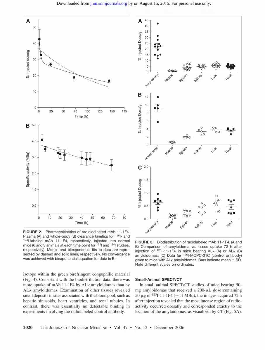
vascularized. In studies in which cohorts of amyloidoma-bearing mice received 25 mg (;4 MBq) of 125I-11-1F4 andwere euthanized 1, 24, 72, and 100 h later, a comparison ofradioactivity in tumors against that found in body organs ortissues revealed no significant divergence 1 and 24 h afterinjection because of the high blood-pool background; how-ever, at 72 h, there was selective uptake by the amyloid-omas. Thus, the biodistribution (and subsequent imaging)experiments were performed 72 h after antibody injection.The results of studies in which mice bearing different ALkamyloidomas were injected with 10 mg (;11 MBq) of 125I-11-1F4 are shown in Figure 3A. The recovery of 125I in theharvested amyloidomas ranged from 12 to 43 %ID/g, with amean of 22 %ID/g, a value significantly higher than thatseen in skeletal muscle (1.2 %ID/g) (P , 0.001), spleen(3.9 %ID/g) (P , 0.001), kidney (4.8 %ID/g) (P , 0.01),heart (5.0 %ID/g) (P , 0.01), and liver (5.8 %ID/g) (P .
0.05), as well as other tissues examined. This reagent alsowas taken up selectively, albeit to a lesser extent, by ALltumors (n 5 5); the specific activities ranged from 6 to12 %ID/g, with a mean of 9.5 %ID/g (Fig. 3B). The amyloid-to-liver ratios for ALk and ALl amyloidomas were ;3.8:1and 2.3:1, respectively. In contrast, this ratio was 1.1:1 forthe radiolabeled control MOPC-31C antibody, indicatingthat there was essentially no binding to the amyloidoma.
Autoradiography
At 7 d after amyloidoma induction, mice were injectedwith ;11 MBq of 125I-11-1F4 or 125I-MOPC-31C antibody.The former reagent localized to the amyloid mass, as evi-denced in autoradiograms by the deposition of the g-emitting
TABLE 1Biodistribution of 125I-Labeled mAb 11-1F4 in Normal BALB/c Mice
Mean 6 SD %ID/g at:
Tissue 1 h 4 h 24 h 72 h 144 h
Blood 42.5 6 6.0 32.6 6 2.2 26.8 6 1.0 18.4 6 2.3 16.7 6 1.0Skin 1.1 6 0.3 2.3 6 0.3 3.1 6 0.3 2.6 6 0.2 2.1 6 0.2
Muscle 1.3 6 0.2 2.4 6 0.9 2.1 6 0.2 1.7 6 0.3 1.4 6 0.1
Femur 2.4 6 0.2 3.0 6 0.6 2.8 6 2.2 2.2 6 0.5 1.9 6 0.3
Fat pad 0.8 6 0.1 2.5 6 0.7 1.9 6 1.3 1.3 6 0.2 1.5 6 0.2Liver 14.0 6 0.3 11.4 6 0.7 8.6 6 5.7 5.7 6 1.0 5.6 6 0.5
Spleen 11.7 6 1.2 10.0 6 1.1 8.1 6 0.6 5.2 6 0.9 4.6 6 0.7
Pancreas 2.4 6 0.5 2.8 6 0.5 2.7 6 0.4 1.9 6 0.2 1.7 6 0.3
Stomach 2.5 6 0.4 4.0 6 0.4 3.1 6 0.3 2.2 6 0.1 1.7 6 0.2Small intestine 4.1 6 0.5 4.5 6 0.5 3.2 6 0.2 2.4 6 0.3 2.1 6 0.2
Cecum 2.5 6 0.5 5.9 6 0.4 4.3 6 0.6 3.0 6 0.4 2.2 6 0.3
Large intestine 2.2 6 0.3 4.1 6 0.5 3.6 6 0.2 2.5 6 0.4 1.9 6 0.2Kidney (L) 7.8 6 1.1 7.8 6 1.9 5.4 6 0.6 4.1 6 0.8 3.4 6 0.6
Kidney (R) 8.0 6 1.2 8.2 6 1.4 5.7 6 0.7 4.3 6 0.7 3.4 6 0.5
Throat* 4.8 6 1.2 9.0 6 2.0 6.4 6 1.6 4.5 6 0.7 4.3 6 0.9
Heart 12.6 6 2.4 10.1 6 1.4 9.5 6 2.1 6.4 6 1.5 5.4 6 0.6Lungs 13.8 6 1.4 13.3 6 1.9 10.5 6 0.7 7.2 6 3.4 6.8 6 0.6
Tongue 3.4 6 1.3 8.0 6 0.9 7.2 6 0.7 8.8 6 7.7 4.4 6 0.4
Brain 0.9 6 0.2 0.8 6 0.1 0.6 6 0.1 0.5 6 0.1 0.4 6 0.1
*Tissue includes esophagus and thyroid gland (n 5 6).
RADIOIMAGING OF AMYLOID • Wall et al. 2019
by on August 15, 2015. For personal use only. jnm.snmjournals.org Downloaded from
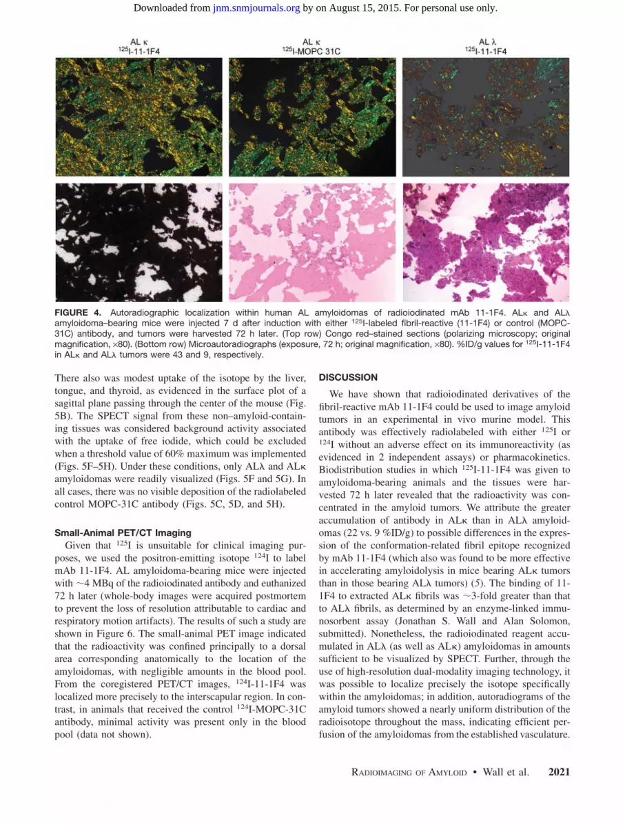
isotope within the green birefringent congophilic material(Fig. 4). Consistent with the biodistribution data, there wasmore uptake of mAb 11-1F4 by ALk amyloidomas than byALl amyloidomas. Examination of other tissues revealedsmall deposits in sites associated with the blood pool, such ashepatic sinusoids, heart ventricles, and renal tubules. Incontrast, there was essentially no detectable binding inexperiments involving the radiolabeled control antibody.
Small-Animal SPECT/CT
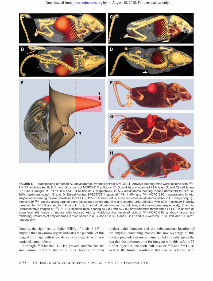
In small-animal SPECT/CT studies of mice bearing 50-mg amyloidomas that received a 200-mL dose containing50 mg of 125I-11-1F4 (;11 MBq), the images acquired 72 hafter injection revealed that the most intense region of radio-activity occurred dorsally and corresponded exactly to thelocation of the amyloidomas, as visualized by CT (Fig. 5A).
FIGURE 2. Pharmacokinetics of radioiodinated mAb 11-1F4.Plasma (A) and whole-body (B) clearance kinetics for 125I- and124I-labeled mAb 11-1F4, respectively, injected into normalmice (6 and 3 animals at each time point for 125I and 124I studies,respectively). Mono- and biexponential fits to data are repre-sented by dashed and solid lines, respectively. No convergencewas achieved with biexponential equation for data in B.
FIGURE 3. Biodistribution of radiolabeled mAb 11-1F4. (A andB) Comparison of amyloidoma vs. tissue uptake 72 h afterinjection of 125I-11-1F4 in mice bearing ALk (A) or ALl (B)amyloidomas. (C) Data for 125I-MOPC-31C (control antibody)given to mice with ALk amyloidomas. Bars indicate mean 6 SD.Note different scales on ordinates.
2020 THE JOURNAL OF NUCLEAR MEDICINE • Vol. 47 • No. 12 • December 2006
by on August 15, 2015. For personal use only. jnm.snmjournals.org Downloaded from
There also was modest uptake of the isotope by the liver,tongue, and thyroid, as evidenced in the surface plot of asagittal plane passing through the center of the mouse (Fig.5B). The SPECT signal from these non–amyloid-contain-ing tissues was considered background activity associatedwith the uptake of free iodide, which could be excludedwhen a threshold value of 60% maximum was implemented(Figs. 5F–5H). Under these conditions, only ALl and ALkamyloidomas were readily visualized (Figs. 5F and 5G). Inall cases, there was no visible deposition of the radiolabeledcontrol MOPC-31C antibody (Figs. 5C, 5D, and 5H).
Small-Animal PET/CT Imaging
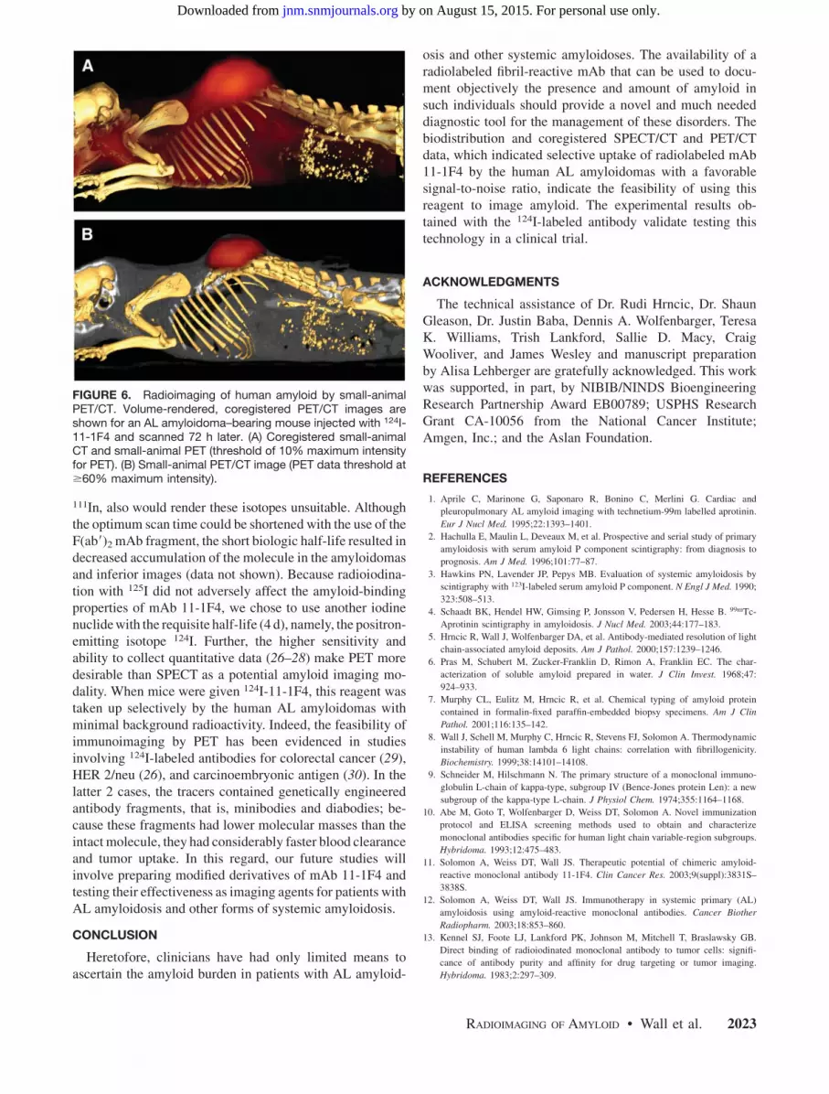
Given that 125I is unsuitable for clinical imaging pur-poses, we used the positron-emitting isotope 124I to labelmAb 11-1F4. AL amyloidoma-bearing mice were injectedwith ;4 MBq of the radioiodinated antibody and euthanized72 h later (whole-body images were acquired postmortemto prevent the loss of resolution attributable to cardiac andrespiratory motion artifacts). The results of such a study areshown in Figure 6. The small-animal PET image indicatedthat the radioactivity was confined principally to a dorsalarea corresponding anatomically to the location of theamyloidomas, with negligible amounts in the blood pool.From the coregistered PET/CT images, 124I-11-1F4 waslocalized more precisely to the interscapular region. In con-trast, in animals that received the control 124I-MOPC-31Cantibody, minimal activity was present only in the bloodpool (data not shown).
DISCUSSION
We have shown that radioiodinated derivatives of thefibril-reactive mAb 11-1F4 could be used to image amyloidtumors in an experimental in vivo murine model. Thisantibody was effectively radiolabeled with either 125I or124I without an adverse effect on its immunoreactivity (asevidenced in 2 independent assays) or pharmacokinetics.Biodistribution studies in which 125I-11-1F4 was given toamyloidoma-bearing animals and the tissues were har-vested 72 h later revealed that the radioactivity was con-centrated in the amyloid tumors. We attribute the greateraccumulation of antibody in ALk than in ALl amyloid-omas (22 vs. 9 %ID/g) to possible differences in the expres-sion of the conformation-related fibril epitope recognizedby mAb 11-1F4 (which also was found to be more effectivein accelerating amyloidolysis in mice bearing ALk tumorsthan in those bearing ALl tumors) (5). The binding of 11-1F4 to extracted ALk fibrils was ;3-fold greater than thatto ALl fibrils, as determined by an enzyme-linked immu-nosorbent assay (Jonathan S. Wall and Alan Solomon,submitted). Nonetheless, the radioiodinated reagent accu-mulated in ALl (as well as ALk) amyloidomas in amountssufficient to be visualized by SPECT. Further, through theuse of high-resolution dual-modality imaging technology, itwas possible to localize precisely the isotope specificallywithin the amyloidomas; in addition, autoradiograms of theamyloid tumors showed a nearly uniform distribution of theradioisotope throughout the mass, indicating efficient per-fusion of the amyloidomas from the established vasculature.
FIGURE 4. Autoradiographic localization within human AL amyloidomas of radioiodinated mAb 11-1F4. ALk and ALlamyloidoma–bearing mice were injected 7 d after induction with either 125I-labeled fibril-reactive (11-1F4) or control (MOPC-31C) antibody, and tumors were harvested 72 h later. (Top row) Congo red–stained sections (polarizing microscopy; originalmagnification, ·80). (Bottom row) Microautoradiographs (exposure, 72 h; original magnification, ·80). %ID/g values for 125I-11-1F4in ALk and ALl tumors were 43 and 9, respectively.
RADIOIMAGING OF AMYLOID • Wall et al. 2021
by on August 15, 2015. For personal use only. jnm.snmjournals.org Downloaded from
Notably, the significantly higher %ID/g of mAb 11-1F4 inamyloid than in various organs indicates the potential of thisreagent to image pathologic deposits in patients with sys-temic AL amyloidosis.
Although 125I-labeled 11-1F4 proved suitable for thesmall-animal SPECT studies in mice because of their
modest axial diameter and the subcutaneous location ofthe amyloid-containing masses, the low g-energy of thisnuclide precludes its use in humans. Additionally, given thefact that the optimum time for imaging with this mAb is 72h after injection, the short half-lives of 123I and 99mTc, aswell as the limited resolution that can be achieved with
FIGURE 5. Radioimaging of human AL amyloidomas by small-animal SPECT/CT. Amyloid-bearing mice were injected with 125I-11-1F4 antibody (A, B, E, F, and G) or control MOPC-31C antibody (C, D, and H) and scanned 72 h later. (A and C) Left lateralSPECT/CT images of 125I-11-1F4 and 125I-MOPC-31C, respectively, in ALk amyloidoma–bearing mouse (threshold for SPECT,10% maximum value). (B and D) Dorsal–ventral SPECT/CT images of 125I-11-1F4 and 125I-MOPC-31C, respectively, in ALkamyloidoma–bearing mouse (threshold for SPECT, 10% maximum value; arrow indicates amyloidoma visible in CT image only). (E)Intensity of 125I activity along sagittal plane bisecting amyloidoma (line and shaded area coincide with 60% maximum-intensitythreshold for SPECT applied to F, G, and H; 1, 2, 3, and 4 indicate tongue, thyroid, liver, and amyloidoma, respectively). (F and G)Representative images of 125I-11-1F4–injected mice bearing ALk (F) and ALl (G) amyloidomas; thresholded SPECT is shown asisosurface. (H) Image of mouse with induced ALk amyloidoma that received control 125I-MOPC-31C antibody (isosurfacerendering). Volumes of amyloidomas in mice shown in A, B, and F; in C, D, and H; in E; and in G were 260, 130, 120, and 196 mm3,respectively.
2022 THE JOURNAL OF NUCLEAR MEDICINE • Vol. 47 • No. 12 • December 2006
by on August 15, 2015. For personal use only. jnm.snmjournals.org Downloaded from
111In, also would render these isotopes unsuitable. Althoughthe optimum scan time could be shortened with the use of theF(ab9)2 mAb fragment, the short biologic half-life resulted indecreased accumulation of the molecule in the amyloidomasand inferior images (data not shown). Because radioiodina-tion with 125I did not adversely affect the amyloid-bindingproperties of mAb 11-1F4, we chose to use another iodinenuclide with the requisite half-life (4 d), namely, the positron-emitting isotope 124I. Further, the higher sensitivity andability to collect quantitative data (26–28) make PET moredesirable than SPECT as a potential amyloid imaging mo-dality. When mice were given 124I-11-1F4, this reagent wastaken up selectively by the human AL amyloidomas withminimal background radioactivity. Indeed, the feasibility ofimmunoimaging by PET has been evidenced in studiesinvolving 124I-labeled antibodies for colorectal cancer (29),HER 2/neu (26), and carcinoembryonic antigen (30). In thelatter 2 cases, the tracers contained genetically engineeredantibody fragments, that is, minibodies and diabodies; be-cause these fragments had lower molecular masses than theintact molecule, they had considerably faster blood clearanceand tumor uptake. In this regard, our future studies willinvolve preparing modified derivatives of mAb 11-1F4 andtesting their effectiveness as imaging agents for patients withAL amyloidosis and other forms of systemic amyloidosis.
CONCLUSION
Heretofore, clinicians have had only limited means toascertain the amyloid burden in patients with AL amyloid-
osis and other systemic amyloidoses. The availability of aradiolabeled fibril-reactive mAb that can be used to docu-ment objectively the presence and amount of amyloid insuch individuals should provide a novel and much neededdiagnostic tool for the management of these disorders. Thebiodistribution and coregistered SPECT/CT and PET/CTdata, which indicated selective uptake of radiolabeled mAb11-1F4 by the human AL amyloidomas with a favorablesignal-to-noise ratio, indicate the feasibility of using thisreagent to image amyloid. The experimental results ob-tained with the 124I-labeled antibody validate testing thistechnology in a clinical trial.
ACKNOWLEDGMENTS
The technical assistance of Dr. Rudi Hrncic, Dr. ShaunGleason, Dr. Justin Baba, Dennis A. Wolfenbarger, TeresaK. Williams, Trish Lankford, Sallie D. Macy, CraigWooliver, and James Wesley and manuscript preparationby Alisa Lehberger are gratefully acknowledged. This workwas supported, in part, by NIBIB/NINDS BioengineeringResearch Partnership Award EB00789; USPHS ResearchGrant CA-10056 from the National Cancer Institute;Amgen, Inc.; and the Aslan Foundation.
REFERENCES
1. Aprile C, Marinone G, Saponaro R, Bonino C, Merlini G. Cardiac and
pleuropulmonary AL amyloid imaging with technetium-99m labelled aprotinin.
Eur J Nucl Med. 1995;22:1393–1401.
2. Hachulla E, Maulin L, Deveaux M, et al. Prospective and serial study of primary
amyloidosis with serum amyloid P component scintigraphy: from diagnosis to
prognosis. Am J Med. 1996;101:77–87.
3. Hawkins PN, Lavender JP, Pepys MB. Evaluation of systemic amyloidosis by
scintigraphy with 123I-labeled serum amyloid P component. N Engl J Med. 1990;
323:508–513.
4. Schaadt BK, Hendel HW, Gimsing P, Jonsson V, Pedersen H, Hesse B. 99mTc-
Aprotinin scintigraphy in amyloidosis. J Nucl Med. 2003;44:177–183.
5. Hrncic R, Wall J, Wolfenbarger DA, et al. Antibody-mediated resolution of light
chain-associated amyloid deposits. Am J Pathol. 2000;157:1239–1246.
6. Pras M, Schubert M, Zucker-Franklin D, Rimon A, Franklin EC. The char-
acterization of soluble amyloid prepared in water. J Clin Invest. 1968;47:
924–933.
7. Murphy CL, Eulitz M, Hrncic R, et al. Chemical typing of amyloid protein
contained in formalin-fixed paraffin-embedded biopsy specimens. Am J Clin
Pathol. 2001;116:135–142.
8. Wall J, Schell M, Murphy C, Hrncic R, Stevens FJ, Solomon A. Thermodynamic
instability of human lambda 6 light chains: correlation with fibrillogenicity.
Biochemistry. 1999;38:14101–14108.
9. Schneider M, Hilschmann N. The primary structure of a monoclonal immuno-
globulin L-chain of kappa-type, subgroup IV (Bence-Jones protein Len): a new
subgroup of the kappa-type L-chain. J Physiol Chem. 1974;355:1164–1168.
10. Abe M, Goto T, Wolfenbarger D, Weiss DT, Solomon A. Novel immunization
protocol and ELISA screening methods used to obtain and characterize
monoclonal antibodies specific for human light chain variable-region subgroups.
Hybridoma. 1993;12:475–483.
11. Solomon A, Weiss DT, Wall JS. Therapeutic potential of chimeric amyloid-
reactive monoclonal antibody 11-1F4. Clin Cancer Res. 2003;9(suppl):3831S–
3838S.
12. Solomon A, Weiss DT, Wall JS. Immunotherapy in systemic primary (AL)
amyloidosis using amyloid-reactive monoclonal antibodies. Cancer Biother
Radiopharm. 2003;18:853–860.
13. Kennel SJ, Foote LJ, Lankford PK, Johnson M, Mitchell T, Braslawsky GB.
Direct binding of radioiodinated monoclonal antibody to tumor cells: signifi-
cance of antibody purity and affinity for drug targeting or tumor imaging.
Hybridoma. 1983;2:297–309.
FIGURE 6. Radioimaging of human amyloid by small-animalPET/CT. Volume-rendered, coregistered PET/CT images areshown for an AL amyloidoma–bearing mouse injected with 124I-11-1F4 and scanned 72 h later. (A) Coregistered small-animalCT and small-animal PET (threshold of 10% maximum intensityfor PET). (B) Small-animal PET/CT image (PET data threshold at$60% maximum intensity).
RADIOIMAGING OF AMYLOID • Wall et al. 2023
by on August 15, 2015. For personal use only. jnm.snmjournals.org Downloaded from
14. Kennel SJ, Lankford TK, Foote LJ, Shinpock SG, Stringer C. CD44 expression
on murine tissues. J Cell Sci. 1993;104:373–382.
15. Wall JS, Kennel SJ, Paulus MJ, et al. Quantitative high-resolution microradio-
graphic imaging of amyloid deposits in a novel murine model of AA amy-
loidosis. Amyloid. 2005;12:149–156.
16. Shepp LA, Vardi Y. Maximum-likelihood reconstruction for emission tomog-
raphy. IEEE Trans Med Imaging. 1982;1:113–122.
17. Paulus M, Gleason SS, Kennel SJ, Hunsicker PR, Johnson DK. High resolution
X-ray computed tomography: an emerging tool for small animal cancer research.
Neoplasia. 2000;2:62–70.
18. Paulus MJ, Gleason SS, Sari-Sarraf H, et al. High-resolution x-ray CT screening
of mutant mouse models. In: Farkas DL, Leif RC, eds. BIOS 2000 International
Biomedical Optics Symposium. San Jose, CA: SPIE-Int. Soc. Opt. Eng.; 2000:
270–279.
19. Paulus MJ, Sari-Sarraf H, Gleason SS, et al. A new x-ray computed tomography
system for laboratory mouse imaging. IEEE Trans Nucl Sci. 1999;46:558–564.
20. Gregor J, Gleason SS, Paulus MJ, Cates J. Fast Feldkamp reconstruction based
on focus of attention and distributed computing. Int J Imag Syst Tech. 2002;
12:229–234.
21. Qi J, Leahy RM. Resolution and noise properties of MAP reconstruction for fully
3-D PET. IEEE Trans Med Imaging. 2000;19:493–506.
22. O’Nuallain B, Murphy CL, Wolfenbarger DA, Kennel S, Solomon A, Wall JS.
The amyloid-reactive monoclonal antibody 11-1F4 binds a cryptic epitope on
fibrils and partially denatured immunoglobulin light chains and inhibits
fibrillogenesis. In: Grateau G, Kyle RA, Skinner M, eds. Xth International
Symposium on Amyloidosis. Tours, France: CRC Press; 2005:482-484.
23. Gurbaxani B, Dela Cruz LL, Chintalacharuvu K, Morrison SL. Analysis of a
family of antibodies with different half-lives in mice fails to find a correlation
between affinity for FcRn and serum half-life. Mol Immunol. 2006;43:1462–
1473.
24. Levy AL, Waldmann TA. The effect of hydrocortisone on immunoglobulin
metabolism. J Clin Invest. 1970;49:1679–1684.
25. Montano RF, Morrison SL. Influence of the isotype of the light chain on the
properties of IgG. J Immunol. 2002;168:224–231.
26. Gonzalez Trotter DE, Manjeshwar RM, Doss M, et al. Quantitation of small-
animal 124I activity distributions using a clinical PET/CT scanner. J Nucl Med.
2004;45:1237–1244.
27. Herzog H, Tellman L, Qaim SM, Spellerberg S, Schmid A, Coenen HH. PET
quantitation and imaging of the non-pure positron-emitting iodine isotope 124I.
Appl Radiat Isot. 2002;56:673–679.
28. Pentlow KS, Graham MC, Lambrecht RM, et al. Quantitative imaging of iodine-
124 with PET. J Nucl Med. 1996;37:1557–1562.
29. Lee FT, Hall C, Rigopoulos A, et al. Immuno-PET of human colon xenograft–
bearing BALB/c nude mice using 124I-CDR-grafted humanized A33 monoclonal
antibody. J Nucl Med. 2001;42:764–769.
30. Sundaresan G, Yazaki PJ, Shively JE, et al. 124I-Labeled engineered anti-CEA
minibodies and diabodies allow high-contrast, antigen-specific small-animal
PET imaging of xenografts in athymic mice. J Nucl Med. 2003;44:1962–1969.
2024 THE JOURNAL OF NUCLEAR MEDICINE • Vol. 47 • No. 12 • December 2006
by on August 15, 2015. For personal use only. jnm.snmjournals.org Downloaded from
2006;47:2016-2024.J Nucl Med. Townsend, Deborah T. Weiss and Alan SolomonJonathan S. Wall, Stephen J. Kennel, Mike Paulus, Jens Gregor, Tina Richey, James Avenell, Jeffrey Yap, David Radioimaging of Light Chain Amyloid with a Fibril-Reactive Monoclonal Antibody
http://jnm.snmjournals.org/content/47/12/2016This article and updated information are available at:
http://jnm.snmjournals.org/site/subscriptions/online.xhtml
Information about subscriptions to JNM can be found at:
http://jnm.snmjournals.org/site/misc/permission.xhtmlInformation about reproducing figures, tables, or other portions of this article can be found online at:
(Print ISSN: 0161-5505, Online ISSN: 2159-662X)1850 Samuel Morse Drive, Reston, VA 20190.SNMMI | Society of Nuclear Medicine and Molecular Imaging
is published monthly.The Journal of Nuclear Medicine
© Copyright 2006 SNMMI; all rights reserved.
by on August 15, 2015. For personal use only. jnm.snmjournals.org Downloaded from