monoclonal antibody imaging of human melanoma
TRANSCRIPT

Monoclonal Antibody Imagingof Human MelanomaRadioimmunodetection by Subcutaneous or Systemic Injection
MICHAEL T. LOTZE, M.D., F.A.C.S. PATRICIA PERENTESIS, R.N."1 ANDREW M. KEENAN, M.D."JORGE A. CARRASQUILLO, M.D.* JAMES C. REYNOLDS, M.D.11 INGEGERD HELLSTROM, M.D., PH.D.¶JOHN N. WEINSTEIN, M.D., PH.D.t LOUIS A. MATIS, M.D.t KARL-ERIC HELLSTROM, M.D., PH.D.¶GAIL J. BRYANT, M.D.§ RENEE R. EGER, B.S.t STEVEN M. LARSON, M.D."
Fab fragments of monoclonal antibodies (MoAb) to melanoma,radiolabeled with 3'I, were evaluated as diagnostic reagents todetermine their ability to localize systemic-MoAb injected in-travenously (IV)-or nodal metastatic disease-injected sub-cutaneously (SQ) at a site proximal to draining lymph nodes.Sixty-one scans were performed (40 IV, 21 SQ) in 59 patientswho had injections of 0.2-50 mg of 13"1 coupled (0.2-12 mCi)antibody. These included 48.7, which identifies a high molecularweight antigen (HMW), or 96.5, which identifies a transferrinlike molecule, p97. "2I coupled nonspecific Fab 1.4, reactingwith murine leukemia virus, or the whole antibody BL3, reactivewith a human B cell idiotypic determinant, was generally usedin tandem with the patients injected SQ as a nonspecific control.All patients had immunohistochemical studies performed onbiopsied lesions and demonstrated binding to the antibodies in-jected. Of the IV patients, 22/38 (58%) had (+) scans, 13 at SQor nodal sites, four at visceral sites, and five at visceral and SQsites. Patients with clinical stage II disease had SQ injection ofMoAb, including 11 additional patients injected with the wholeantibody 9.2.27 (anti-HMW) labeled with "'In (6 patients) or131I (5 patients). Nodal dissection was performed 2-4 days later.All "'In coupled antibodies demonstrated excellent nodal delin-eation without specific identification of tumor deposits. Of the21 patients injected SQ with MoAb, 17 had confirmed tumor innodes. Of patients injected with Fab fragments, 4/8 (50%) hadspecific uptake of MoAb, although only two were successfullyimaged. Increased uptake of antimelanoma antibodies was ob-served in some patients in lymph nodes not containing tumorand was possibly related to antigen shedding. Clearance of labeledantibody from the injection site occurred with a half life of 16-50 hours. Toxicity was limited to local discomfort at the site ofSQ injection. Melanoma metastases can be identified with IVor SQ injection of radiolabeled antibodies. These reagents maybe useful in the diagnosis or therapy ofhuman melanoma. Furtherevaluation will be required before they could be considered clin-ically useful.
Presented at the 106th Annual Meeting of the American Surgical As-sociation, Hot Springs, Virginia, April 24-26, 1986.
Reprint requests: Michael T. Lotze, M.D., Surgery Branch, NationalCancer Institute, Building 10, Room 2B56, Bethesda, MD 20892.
Submitted for publication: May 13, 1986.
From the Surgery, * Medicine,t and Mathematical BiologyfBranches, National Cancer Institute; Pathology§ and NuclearMedicine Departments,"1 Clinical Center, National Institutes of
Health, Bethesda, Maryland; and Departments ofMicrobiology-immunology and Pathology, University of
Washington, Seattle, WA¶
T HE ABILITY TO IDENTIFY tumor deposits usingmonoclonal antibodies has been a goal since thedevelopment of hybridoma technology.' Large
amounts ofspecific, highly purified monoclonal antibodiesreactive with melanoma cell surface associated antigensare now available and have been used in both diagnosticand therapeutic approaches to this tumor.26 In additionto intravenous administration, we have evaluated bothwhole antibodies and Fab fragments of antimelanomaantibodies as diagnostic reagents injected subcutaneously(SQ) to determine if tumor deposits could be identifiedin regional draining lymph nodes. Since the role of nodaldissection in patients with clinical stage I disease remainscontroversial,7'8 attempts to localize lymphatic drainageand spread are still being carried out.9 Based on studiesconducted in rodents,'0"' we evaluated the lymphaticroute of administration of monoclonal antibodies in pa-tients with known stage II disease to determine if thetheoretically greater sensitivity, higher target to back-ground ratio, faster localization, and lower toxicity couldbe achieved. We have contrasted our results using sub-cutaneous injection sites in 21 patients with 40 scans donein patients receiving Fab fragments of antimelanoma an-tibodies as to toxicity, clearance, and identification of tu-mor deposits.
223

LOTZE AND OTHERS
Materials and Methods
Patients
All melanoma patients considered for these evaluationswere treated on two clinical research committee approvedprotocols utilizing (1) antibodies for diagnostic and pos-
sible therapeutic intravenous use and (2) antibodies forsubcutaneous injection, coupled with surgical dissectionof stage II disease. All patients signed an informed consentindicating the possible risks and the investigative natureofthese studies. To be included, patients had to have clin-ical evidence ofstage II or III melanoma, no known allergy
to iodine or mouse antibodies, and, in patients with stageII disease, were required to have no history ofradiotherapyor prior dissection of the regional lymph nodes. All pa-
tients received complete blood counts, platelet counts,liver function and renal function studies, and thyroidfunction tests. Radiologic evaluation of cerebral, pul-monary, and hepatic disease as well as measurement ofevaluable lymph nodes and local tumor nodules was per-
formed. Plasma was stored prior to and at intervals afterimaging for detection of human antimouse antibodies.Immunohistochemical testing of a biopsy of the tumoror its metastases was carried out on frozen tissue if avail-able. When tumor was not available prior to injection(stage II patients), tumor obtained at the time of nodaldissection was submitted for confirmation ofantigen pos-itivity.
Monoclonal Antibodies and Radioactive Labeling
The MoAb 9.2.27 is a murine antibody (IgG2a,k) de-scribed by Morgan and coworkers.'2 It recognizes a highmolecular weight (240 Kd chondroitin sulfate/proteogly-can) cell surface antigen present on 90% of human mel-anoma. The 9.2.27 MoAb was obtained by immunizingmice with soluble extracts from human melanoma cells.Hybridomas were developed by fusion of Balb/C sple-nocytes with the murine myeloma p3-x63-Ag-8. Immu-noglobulin was purified from hybridoma ascites using a
double precipitation procedure with Na2SO4. The anti-body was tested as recommended by the Office of Bio-logics, U.S. Food and Drug Administration (FDA).A modification of the bifunctional chelating method
of Krejcarek was utilized to conjugate diethylenetriami-nepentaacetic acid (DTPA) to 9.2.27.'" The antibody wasreceived in kit form (Hybritech, Inc., La Jolla, CA), con-
sisting of 1 mg DTPA-conjugated 9.2.27 MoAb in 1%human serum albumin (HSA). Labeling was performedby incubating approximately 5 mCi "'In with 1 mg ofDTPA-conjugated 9.2.27. Excess DTPA was then addedto scavenge any free "'In. The antibody was administeredunder an Investigational New Drug application approvedby the FDA.
The antibody 48.7 is a murine'4 monoclonal antibody(IgGj) that also identifies the high molecular weight an-
tigen on human melanoma. This antibody was preparedby fusing SP2/0 mouse myeloma cells with spleen cellsfrom mice immunized with a cultured melanoma line.Weak staining ofsome blood vessels was noted, but othernormal cells including skin melanocytes and other tumortypes were unstained using peroxidase immunohisto-chemical stains. The antibody 96.5 is a murine'5 6 anti-body that identifies p97, a protein associated with humanmelanoma, and is present in only very small amounts insolublized normal tissues (<10 ng/mg). The antibody 1.4is an IgG, that recognizes a murine leukemia viral antigen,GP70, and is negative for all human cells tested (Brown,unpublished). The whole antibody BL-3, raised against ahuman B cell lymphoma, identifies unique idiotypic de-terminants on that patient's cells not found on cells fromother individuals (K. Foon, unpublished) and was usedas a negative control. Immunohistochemical studies wereperformed as previously reported.'4 All tumors weregraded as no staining (0), 25% staining (1+), 50% of cellsstained (2+), 75% of cells stained (3+), and very strong(100% of cells) staining (4+).
Fab fragments were obtained by first incubating theantibody with papain (150 ,g) in 4 ml of phosphate buff-ered saline (pH 7.2) containing 10 mM cysteine and 2mM EDTA under nitrogen at 37 C for 3 hours.
lodoacetamide was added to a final concentration of50 mM and the digest filtered on Sephadex G-25 (Phar-macia, Piscataway, NJ) equilibrated with saline. Fc frag-ments were removed by passage through a 5 ml columnof protein A coupled to Sepharose CL-4B (Pharmacia,Piscataway, NJ). lodination was carried out by the methodof Ferens et al.'7 The Fab fragments were incubated withNa 1251I or Na 13I' (DuPont/NEN Medical Products, N.Bilerica, MA) and 23 ,g of chloramine T per mg of Fabfragments, in phosphate buffered saline for 10 minutes at0 C. The reaction was stopped by adding 81 Mg ofsodiumthiosufate per mg protein and the labeled protein separatedon a column of Sephadex G-25.
Radiopharmaceutical Purity and Quality Control
Immunoreactivity was determined using the H 1777 or
Fem XII melanoma cell lines, which express both p97and HMW antigens. Cells (2 X 10⁶) were suspended in100 ,ul of the test sample containing 5 ng of the radiola-beled protein. Phosphate buffered saline was added, themixture was centrifuged, and cells were separated andcounted for radioactivity. Results are expressed as thepercentage of total added radioactivity bound to cells. Thepercentage of counts protein bound was determined bytrichlorocetic acid precipitation of protein. '3'i prepara-tions were analyzed for free iodine using paper chroma-tography (Whatman I) and an 85% methanol in water
224 Ann. Surg. * September 1986
ANTIMELANOMA ANTIBODY IMAGING
solvent. 'In preparations were analyzed for "In DTPAusing instant thin layer chromatography in silica gelwith methanol:water (1:1) with 5% ammonium acetateas a solvent. Pyrogen and sterility testing was routinelycarried out.
Antibody Administration and Sample Collection
Patients receiving intravenous administration of anti-body had it infused slowly over 60 minutes through a
peripheral vein. No adverse reactions during or subsequentto the infusion were noted. Some ofthe patients receivingantibody SQ were injected in 12 separate locations aroundthe primary excision site and a comparable number sym-
metrically placed on the contralateral side. Other patientswere injected in the four web spaces ofeach hand or foot.Equal quantities of specific and nonspecific antibody pro-
teins (independent of the specific activity) were alwaysinjected. Problems associated with subcutaneous injectionwere limited to local discomfort and occasional erythema(Fig. 1).
Following administration, blood samples were collectedat 5, 30, 60, 120, and 240 minutes and at daily intervalsuntil surgery was performed. Daily urine collections wereperformed up to 48 hours after infusion. Excreted urinarycounts were determined on collections obtained inclusiveof all urine at 2, 24, and 48 hours using aliquots and thenmultiplying counts by total volume for that time period.Whole body retention was measured with a 2 X 2 inchsodium iodide crystal positioned 7.1 meters from thepatient in a fixed geometric position. The immediatepostinfusion value was taken as 100%.
Surgical samples were obtained from nodal dissectionsand examined by a pathologist (G.B.). Separate lymphnodes and other tissues were isolated by dissection,weighed, counted in a gamma counter (along with stan-dards prepared from injected radiolabeled antibody), andsubmitted for histologic and immunohistochemicalstaining. Decay correction and spillover correction for "'Icounts in the 1251 channel were routinely performed.
Assayfor Human Antimouse Antibody (HAMA)
A test sample of the patients' serum (10 ,ul) was incu-bated with 10 Al of 1251-labeled mouse IgG (104 cpm) for30 minutes at 20 C. Staphylococcus aureus (10 mg) was
added in 1 ml of 20 mM Tris-HCl buffer, pH 8.0, con-
taining 100 mM NaCl, 1 mM EDTA, and 0.5% NonidetP40 (TNEN) (Bethesda Research Laboratories, Bethesda,MD). After a further 5-minute incubation, the bacteriawere separated by centrifugation and washed three timeswith 10 ml ofTNEN and counted for 125I. A comparisonwas made between the test sample and a control serum
that did not contain human antimouse antibodies(HAMA). A positive test was considered to be a valuetwice the control.
INTRAVENOUSINJECTION
SKIN
ARTER
LYMPH LIVER
NODE
VEIN
INTERSTITIALXj:. SPACE
LYMPHVESSEL
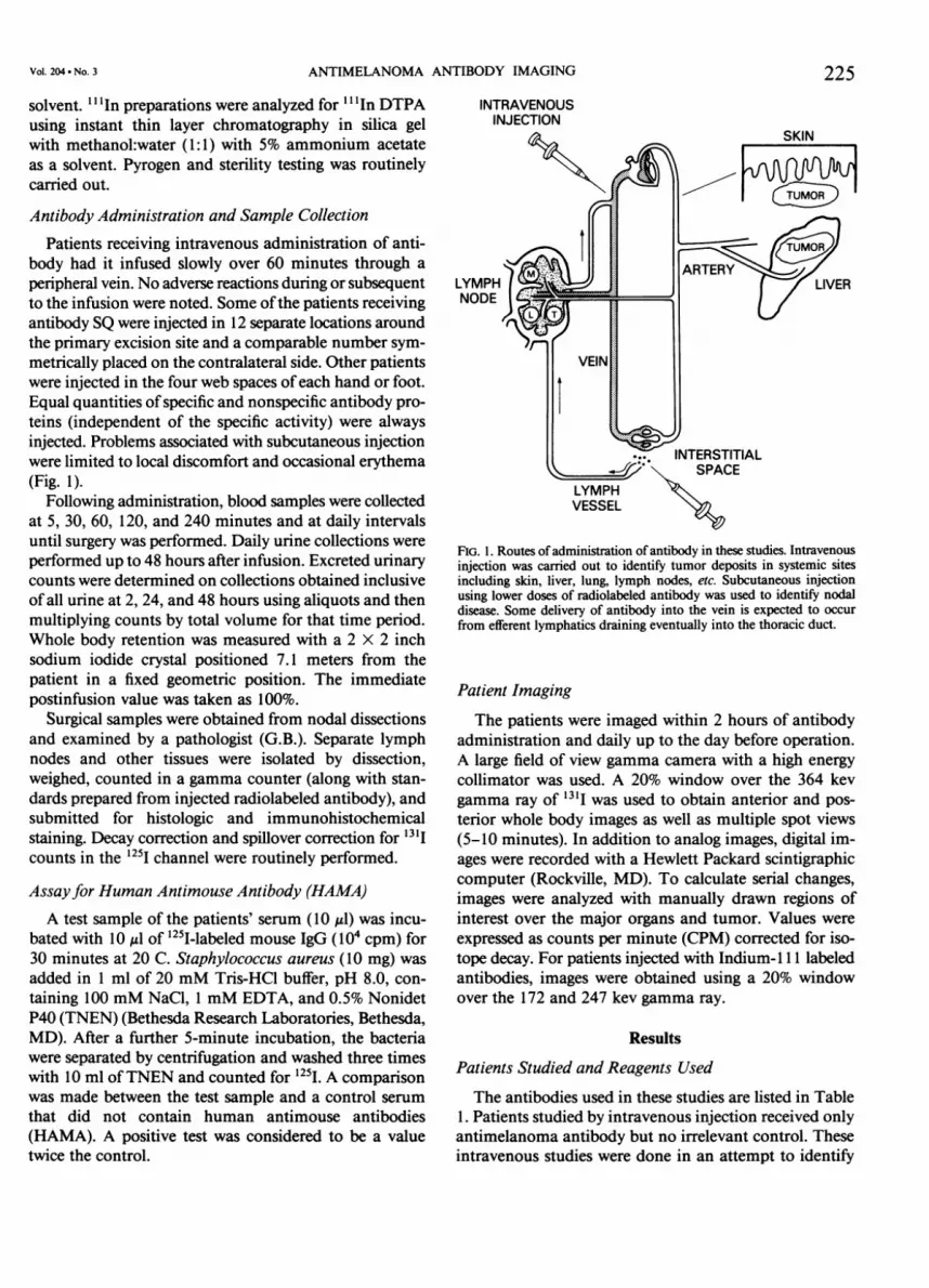
FIG. 1. Routes of administration ofantibody in these studies. Intravenousinjection was carried out to identify tumor deposits in systemic sitesincluding skin, liver, lung, lymph nodes, etc. Subcutaneous injectionusing lower doses of radiolabeled antibody was used to identify nodaldisease. Some delivery of antibody into the vein is expected to occurfrom efferent lymphatics draining eventually into the thoracic duct.
Patient Imaging
The patients were imaged within 2 hours of antibodyadministration and daily up to the day before operation.A large field of view gamma camera with a high energy
collimator was used. A 20% window over the 364 kevgamma ray of 13 I was used to obtain anterior and pos-terior whole body images as well as multiple spot views(5-10 minutes). In addition to analog images, digital im-ages were recorded with a Hewlett Packard scintigraphiccomputer (Rockville, MD). To calculate serial changes,images were analyzed with manually drawn regions ofinterest over the major organs and tumor. Values were
expressed as counts per minute (CPM) corrected for iso-tope decay. For patients injected with Indium-l 1 1 labeledantibodies, images were obtained using a 20% windowover the 172 and 247 kev gamma ray.
Results
Patients Studied and Reagents Used
The antibodies used in these studies are listed in Table1. Patients studied by intravenous injection received onlyantimelanoma antibody but no irrelevant control. Theseintravenous studies were done in an attempt to identify
225Vol. 204 * No. 3

226 LOTZE AND
TABLE 1. Antibodies Used in Diagnostic Imaging
Designation Subclass Reactivity
48.7 Fab IgG, High molecular weight (HMW)antigen on melanoma (>200kD); on 70-90% ofmelanomas
96.5 Fab IgG2a Recognizes p97, a transferrinlike molecule on 70-90% ofmelanomas: lowcrossreactivity with liver
9.2.27 whole IgG2. Recognizes different epitope onantibody same antigen as 48.7 (HMW)
1.4 Fab IgGi Control antibody recognizesmurine leukemia virus,negative on human cells
BL-3 whole IgGi Control antibody, raised againstantibody a human B cell lymphoma;
anti-idiotype antibody
'OTHERS Ann. Surg. * September 1986
patients with tumor that might be amenable to therapywith the antibody. If visualized, the patients were consid-ered for treatment with larger doses ofradiolabeled (100-200 mCi/10 mg) antibody. The patients studied are listedin Table 2 along with information regarding their specificevaluation. The last seven patients listed (#34-40) had nodiagnostic studies but were scanned while receiving ther-apy. Patients receiving subcutaneous specific and non-specific antibody are listed in Table 3. Some of these pa-tients were coadministered cold antibody (patient 56) orunlabeled human immunoglobulin preparations (patients53 to 55) to decrease uptake into negative nodes. Thiswas unsuccessful.
For patients with available tumor, immunohistochem-ical staining of tumor tissue was carried out with the an-
TABLE 2. Patients Studied with Intravenous Injection ofMonoclonal Antibodies to Melanoma
Patient Scan Sites of Antibody Mg mCi DoseNumber Age/Sex Disease Used Dose Ab 1-131 Image Notes
46/M Skin, nodes
52/F Skin, nodes
46/M Skin, lung
68/M Skin
52/M Skin
40/F
81/F
69/F65/M70/M59/M38/F
38/M34/M
47/M47/M65/M
77/M61/M
Nodes
Skin, nodes
Skin, nodesLungSkinSkinSkin
SkinMalignant
ascitesSkin, lungLiverLung, spleen
Lung, nodesLung, skin
34/M Lung, skin,spleen
48.7
48.7
48.7
48.7
48.7
48.7
48.7
48.748.748.748.748.7
48.748.7
48.796.596.5
96.596.5
48.7
10
10
10
10
10
10
7.3
10107.77.87.4
8.37.1
8.27.87.8
10.0. 50.0
50.0
5.6 - (NS) Largest lesion 5 X 4cm, others 1-2 cm
10.0 - (NS) Largest lesion 3.5 X 5cm
6.4 + (SS, SP) Largest lesion 2 X 3 cmin skin, 6 cm in lung
8.9 + (SS) Largest lesion 3 X 7 cmin skin, smaller 2 X 3cm not seen
8.9 + (SS) Largest lesion 5 X 6.5cm in skin seen; 5lesions < I cm notvisualized
8.7 - (NS) Pelvic periaortic nodesnot visualized
7.8 + (SS) Largest lesions 2.5 X2.5, 1.5 X Ivisualized; of other12 lesions <2.5 cm,only I seen
8.1 + (SS) 1/8 lesions visualized8.1 + (SP) Multiple lesions; treated8.8 + (SS) 4/22 lesions visualized7.8 + (SS) 3/4 lesions visualized8.2 - (NS) Multiple small lesions
around head andneck not visualized
9.2 + (AS) 7/7 lesions seen6.6 + (AG) Marked uptake in
peritoneal cavity7.7 + (SS) Diffuse uptake lungs
13.313.2
10.0
- (NH)(NP,NL)
+ (AP, SS)
Extensive disease
10.0 + (AP, AS) Multiple lesions,subsequently treatedX 3 with 100 mCi I-
131-Ab5.0 - (NP, NS, 7 subcutaneous lesions
NL) 1-2 cm, lung 2X 2cm, spleen 2 X 1/2cm
21 36/M Skin, lung
2
3
4
5
6
7
89101112
1314
151617
1819
20
I
48.7 42.0 10.0 + (SP, AS)

Vol. 204 * No. 3 ANTIMELANOMA ANTIBODY IMAGING 227TABLE 2. (Continued)
Patient Scan Sites of Antibody Mg mCi DoseNumber Age/Sex Disease Used Dose Ab 1-131 Image Notes
22 52/M Liver, lung, 48.7 42.0 10.0 + (AS, AP,brain, skin NH, NL)
23 45/M Skin, liver, 48.7 44.5 7.8 + (SS, NH, 1/3 lesions > 1 cmbone NB) seen, 0/11 lesions
<0.5 cm24 34/F Liver, skin 96.5 24.0 9.9 - (NS,
NH)25 69/F Skin, nodes 96.5 45.6 7.1 - (SS) Same as patient #8,
0/20 lesions26 62/F Lung, skin, 96.5 9.1 10.0 + (NP,
GI NG, SS)27 35/M Skin, lung 96.5 9.1 10.0 - (NS) Visualized after 3rd
dose of antibody ontherapy, 200 mCiX 2
28 41/F Lung, brain 96.5 11.5 9.6 + (SS) Treated with 200 mCiX 3
29 62/M Lung 96.5 11.5 9.6 - (NP)30 30/F Lung 96.5 7.8 10.0 - (NP) Later treated with IL-231 34/F Lung 96.5 7.8 10.0 - (NS) False positive at site of
recent operation leftthigh
32 49/M Brain, skin 96.5 9.0 10.0 - (NS) Multiple cutaneouslesions
33 49/M Brain, skin 96.5 44.8 10.0 - (NS) Same as patient #32
Therapy scans only34 23/F Skin, lung 96.5 10.0 100 - (NP, NS)35 30/F Skin 96.5 10.0 100 + (SS) Largest lesion 5.5 X 5.5
cm36 64/M Lung, liver, 96.5 10.0 150 - (NP,
GI, skin, NH,heart, NG, NS)kidney
37 66/M Liver 96.5 10.0 150 + (AH)38 62/F Lung, GI 96.5 10.0 150 -(NG,
NP)39 41/M Lung, skin 96.5 10.0 200 + (SP, SS)40 35/M Skin 96.5 10.0 200
S = some; A = all; N = none; S = skin and nodes; P = pulmonary;
tibody used and demonstrated to be 2+ or greater withsole exception. Some patients had determinations withother antibodies as well. The results of these assays arepresented in Table 4. Most tumors demonstrated signif-icant binding of each of the antibodies evaluated.No apparent toxicity was noted in any patient studied
with either subcutaneous or systemic injection of anti-body. No patient receiving subcutaneous or imaging(nontherapeutic) doses of antibodies developed HAMA.Some patients with repetitive therapeutic doses of anti-body developed positive HAMA.
Immunoreactivity and Radiopharmaceutical Purity
All antimelanoma monoclonal antibodies injected wereevaluated for their ability to bind to a cultured melanomatarget prior to injection (Table 5) and following radiola-beling. Fab 96.5 was judged to have excellent binding
H = liver; B = bone; C = brain (cerebrum); GI = gastrointestinal; L- spleen.
characteristics, with a mean of approximately 60% of theantibody binding to cells (for preparations used both sys-temically and SQ). Similar results were obtained for thewhole antibody 9.2.27. Although 48.7 is thought to rec-ognize a different epitope of the same antigen as 9.2.27,its radiolabeled Fab fragment routinely gave poor resultsin the cell binding assays with <25% of the antibodybinding in each preparation. Because of these difficulties,further subcutaneous studies were conducted with 96.5or 9.2.27.
Systemic Imaging
Fab fragments were used for systemic imaging ratherthan whole antibody because they are less immunogenicand more closely approximate the ideal radiopharmaceu-tical. Nonspecific binding to Fc receptors on monocytesand polymorphonuclear leucocytes is eliminated and
228TABLE 3. Patients Studied with Subcutaneous Injection ofMonoclonal Antibodies to Melanoma
Specific Ab Non-
specific AbPatientScan Site of ,g* iSCi* jLg* ,uCi* No (+) Localizing to (+)
Number Age/Sex Injection Dissection Antibody Isotopes Dose Dose Dose Dose Nodes Image Nodes/Comments
41 58/F Ant. tibia L. groin 48.7/1.4 1-131/1-125 200 200 200 133 1/9 + + (ratio 1.5)42 50/M Back L. neck 48.7/1.4 I-131/1-125 200 210 200 86 0/9 - NR43 62/F Back L. axilla 96.5/1.4 1-131/I-125 200 200 200 172 1/17 - ± (ratio 1.6)44 33/M Back R. axilla 96.5/1.4 I-131/I-125 220 157 220 200 8/21 - + (ratio 1.2-4.1)45 62/M Back L. groin 96.5/1.4 I-131/1-125 200 200 200 200 0/19 - NR46 42/F Back R. groin 96.5/1.4 I-131/I-125 620 220 620 61 3/11 - -
47 33/M Back L. axilla 96.5/1.4 I-131/1-125 220 200 220 71 1/20 - -
48 59/F Hands/webs R. neck, 96.5/1.4 I-131/I-125 100 200 100 76 14/35 - -
parotid49 37/M Hands/webs R. axilla 9.2.27/1.4 In-i 11/I-125 100 250 100 84 9/35 - -
50 71/M Feet/webs R. groin 9.2.27/ND In-Il l 100 242 ND ND 0/9 - NR51 38/M Hands/webs R. axilla 9.2.27/1.4 In-I 11/1-125 94 250 94 67 1/21 - -
52 65/M Feet/webs L. groin 9.2.27/ND In-Il l 100 250 ND ND 5/13 - -
53 38/F Hands/webs R. axilla 9.2.27/ND In-Il l 100 250 ND ND 1/32 - - Human IgG(90 mg)
54 24/M Hands/webs L. axilla 9.2.27/ND In-Il l 1000 250 ND ND 9/21 - - Human IgG(37 mg)
55 32/M Hands/webs L. axilla 9.2.27/ND I-131 100 200 ND ND 1/17 - - Human IgG(64 mg)
56 24/M Back L. neck/ 9.2.27/ND I-131 40 200 ND ND 2/23 - - Tenfoldparotid unlabeled 9.2.27
57 32/M Feet/webs L. groin 9.2.27/BL3 I-131/I-125 97 253 93 250 4/14 - -
58 64/M Head R. neck 9.2.27/BL3 1-131/I-125 100 313 82.5 200 1/17 - -
59 32/M Feet/webs L. groin 9.2.27/BL3 I-131/I-125 53 173 52 173 0/8 - NR60 40/F Feet/webs L. groin 96.5/1.4 I-131/I-125 1800 72 1400 66 15/16 + + (ratio 1.35-
8.73)61 61/M Feet/webs R. groin 96.5/1.4 I-131/I-125 2000 200 2000 200 2/17
NR = not relevant (all nodes negative).ND = not done.* Per side.
clearance is rapid. The mean clearance of the Fab frag-ments 48.7 and 96.5 is shown in Figure 2. In patientsreceiving Fab 48.7, less than 30% of the injected dose waspresent in the "whole body" at 48 hours with less than3% present in the plasma. Similar findings were noted forthe Fab 96.5.
Representative scans are shown in Figures 3 and 4. InFigure 3, scans from a patient injected intravenously (IV)
TABLE 4. Immunohistochemistry of Tumorfrom Patients Studied
Antibody 96.5 HMW GD3
0 0 1 01+ 2 1 12+ 17 20 163+ 25 21 94+ 1 3 0
Total 45 46 26
0, no cells stained; 1+, 25% cells staining; 2+, 50% cells staining; 3+,75% cells staining; 4+, 100% cells staining. Nonspecific staining of 9.2.27on some tissues without tumor gave high backgrounds.HMW = high molecular weight antigen (48.7 or 9.2.27).
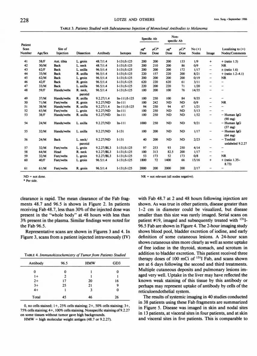
with Fab 48.7 at 2 and 48 hours following injection areshown. As was true in other patients, disease greater than1-2 cm in diameter could be visualized, but diseasesmaller than this size was rarely imaged. Serial scans onpatient #19, imaged and subsequently treated with 13I-96.5 Fab are shown in Figure 4. The 2-hour imaging studyshows blood pool, bladder excretion of iodine, and earlydefinition of some cutaneous lesions. A 24-hour scanshows cutaneous sites more clearly as well as some uptakeof free iodine in the thyroid, stomach, and scrotum inaddition to bladder excretion. This patient received threetherapy doses of 100 mCi of 131I Fab, and scans shownare at 6 days following the second and third treatments.Multiple cutaneous deposits and pulmonary lesions im-aged very well. Uptake in the liver may have reflected theknown weak staining of this tissue by this antibody orperhaps may represent uptake of antibody by cells of thereticuloendothelial system.The results of systemic imaging in 40 studies conducted
in 38 patients using these Fab fragments are summarizedin Figure 5. Disease was imaged in skin and nodal sitesin 13 patients, at visceral sites in four patients, and at skinand visceral sites in five patients. This is comparable to
LOTZE AND OTHERS Ann. Surg. - September 1986

ANTIMELANOMA ANTIBODY IMAGING 229TABLE 5. Immunoreactivity ofInjected Antibodies and Radiopharmaceutical Purity
Cell Binding % ProteinNo. of mCi, Mean mg, Mean Assay Mean Bound Mean
Route Antibody Studies (Range) (Range) (Range) (Range)
IV 96.5 20 59.2 14 59.8% 87.5(9.6-200) (7.8-50) (31.6-97.6) (77.1-95.8)
IV 48.7 20 8.0 17.9 17.1% 86.6(5.6-10.0) (7.1-50) (9.2-25.2) (68.1-95.2)
SQ 96.5 8 0.41 1.3 63.1% 92.1(0.14-0.8) (0.2-4.0) (57.0-70.8) (89-96.4)
SQ 48.7 2 0.41 0.4 20.1% 89(0.40-0.42) (0.4) (16.2-24.2) (89)
SQ 9.2.27 11 0.48 0.25 59.8% 95.8(0.34-0.62) (0.08-1.0) (48.9-74.2) (94-98.1)
our previous experience.2'3 The major limitation in theuse ofthese reagents was the lack of imaging of small (<1cm) lesions.
Subcutaneous Injection and Nodal Imaging
A total of 21 studies were conducted over a 2-year pe-riod. The initial studies were conducted using Fab frag-ments because of their favorable characteristics (notedabove). The first patient had a large 4 X 3 cm node in theleft groin on presentation. This patient had injections ofFab 48.7 into the subcutaneous tissues overlying both theright and left anterior tibia. Clearance from the injectionsites was rapid, with less than 15% remaining at 48 hoursand less than 3% at 7 days (Fig. 6). A maximum of ap-proximately 10% of the injected dose was found in theplasma at 20 hours, with subsequent clearance and lessthan 1% at 7 days. Similar clearance from other sites usingthis and other Fab fragments in other patients was noted.Excretion of radiolabel into the urine was also followed
100
\% of Injected Dosein Plsma ± SD
o % of Injected Dosein Whole Body SD
LUZ0
LU 1
0
z
zL.,
0. 3 T
(Table 6) in patients injected SQ. Most of the activityexcreted in the urine in the first 2 hours was free ofprotein(nonprotein bound) and represented rapid clearance ofthe unconjugated radionuclide. Most of the counts ex-creted in the following 24-48 hours were associated withprotein and probably represented unprocessed antibodyor breakdown products. Clearance from the whole bodyand plasma is listed in Table 7.
Sequential scans in the first patient injected with '31I-48.7 Fab are shown in Figure 7. Views of the anteriorpelvis at 24 and 48 hours are shown. Activity in the blad-der representing 13'I excretion is seen, and persistentcounts are also apparent in the left groin at the site oflocalized 4 X 3 cm inguinal lymph node metastases. Asimilar study in a patient receiving 13'1-96.5 Fab is shownin Figure 8. This patient (#60) was injected into the webspaces of the feet as a convenient route to enter the lym-phatics and more proximal lymph nodes. Views of thepelvis and abdomen at 24 hours were obtained. Excretioninto the bladder and uptake into (left) inguinal nodes arenoted. This patient had previously undergone a right in-guinal nodal dissection. The specificity ofantibody uptakewas confirmed by in vitro counting ofpositive and negativetissues (Fig. 9). The patient, site of injection, and specificantibody (48.7 or 96.5) used are shown in Figure 9 withthe ratio ofeach resected node or other tissue. All patientsreceived an equal amount of 1251 labeled Fab 1.4, whichreacts with a murine leukemia virus antigen and does notbind to human tissues. A ratio ofcounts from each tissuefor specific and nonspecific antibody was determined, andall counts were converted for decay and downscatter (131Icounts into the 1251 channel). Patient #41, whose scan isdemonstrated in Figure 7, had ratios in isolated portionsof her single positive node of 1.2-1.6 with ratios in neg-ative nodes of less than unity. The only other patient (#42)injected with Fab 48.7 had no tumor identified in nodes.
Patients injected with 131I-96.5 Fab had ratios of greaterthan one in tumor containing nodes when compared tononspecific antibody in virtually all cases. Most nonnodaltissue had ratios less than or equal to one. Increased ratios
0 8 16 24 32 40 48 0 8 16 24 32 40 48TIME (hours) TIME (hours)
FIG. 2. Mean clearance of '3'I-antimelanoma Fab fragments injectedintravenously in 24 patients. Rapid clearance and lack of nonspecificbinding via Fc receptors was the major advantage of this approach.
Vol. 204 * No. 3
230 LOTZE AND OTHERS
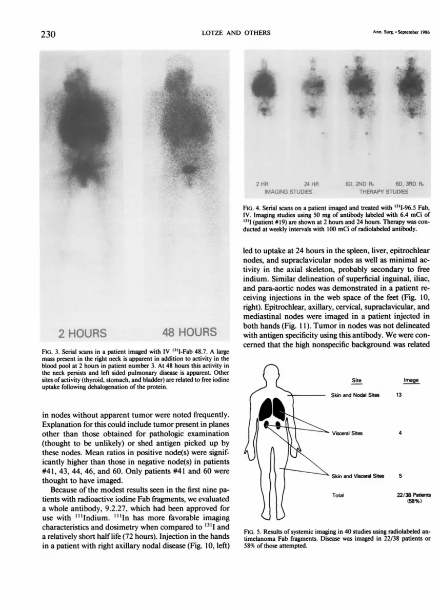
2 HOURS 48 HOURSFIG. 3. Serial scans in a patient imaged with IV '3'I-Fab 48.7. A largemass present in the right neck is apparent in addition to activity in theblood pool at 2 hours in patient number 3. At 48 hours this activity inthe neck persists and left sided pulmonary disease is apparent. Othersites of activity (thyroid, stomach, and bladder) are related to free iodineuptake following dehalogenation of the protein.
in nodes without apparent tumor were noted frequently.Explanation for this could include tumor present in planesother than those obtained for pathologic examination(thought to be unlikely) or shed antigen picked up bythese nodes. Mean ratios in positive node(s) were signif-icantly higher than those in negative node(s) in patients#41, 43, 44, 46, and 60. Only patients #41 and 60 were
thought to have imaged.Because of the modest results seen in the first nine pa-
tients with radioactive iodine Fab fragments, we evaluateda whole antibody, 9.2.27, which had been approved foruse with "'Indium. l"In has more favorable imaging
characteristics and dosimetry when compared to "''I anda relatively short half life (72 hours). Injection in the handsin a patient with right axillary nodal disease (Fig. 10, left)
Ann. Surg. * September 1986
FIG. 4. Serial scans on a patient imaged and treated with '3'I-96.5 Fab,IV. Imaging studies using 50 mg of antibody labeled with 6.4 mCi of13I1 (patient #19) are shown at 2 hours and 24 hours. Therapy was con-ducted at weekly intervals with 100 mCi of radiolabeled antibody.
led to uptake at 24 hours in the spleen, liver, epitrochlearnodes, and supraclavicular nodes as well as minimal ac-tivity in the axial skeleton, probably secondary to freeindium. Similar delineation of superficial inguinal, iliac,and para-aortic nodes was demonstrated in a patient re-ceiving injections in the web space of the feet (Fig. 10,right). Epitrochlear, axillary, cervical, supraclavicular, andmediastinal nodes were imaged in a patient injected inboth hands (Fig. 1 1). Tumor in nodes was not delineatedwith antigen specificity using this antibody. We were con-cerned that the high nonspecific background was related
Site
Skin and Nodal Sites
i Visceral Sites
' Skin and Visceral Sites
Total
Image
13
4
5
22/38 Patients(58%)
FIG. 5. Results of systemic imaging in 40 studies using radiolabeled an-timelanoma Fab fragments. Disease was imaged in 22/38 patients or58% of those attempted.

ANTIMELANOMA ANTIBODY IMAGING
to Fc binding of the whole antibody. Hence we injectedeither cold antibody or human immunoglobulin concur-rently with the "'In labeled antibody to block this binding.These attempts were unsuccessful. Results with this an-
tibody in 11 patients are summarized in Figure 12. Theradioactivity in each node or other tissue is expressed as% injected dose/gram oftissue X 10-. As expected, mostnonlymphoid tissue had few counts. Subsequent detailedimmunohistochemical studies demonstrated crossreactivebinding of 9.2.27 at low levels to normal nodal tissue,and we believe this explains the abberant results with thisantibody.The results ofall surgical procedures and subcutaneous
nodal imaging are summarized in Figure 13. No patientswith cervical disease imaged or had evidence of specificuptake in nodes. In contrast, 4/14 patients with axillaryor inguinal disease demonstrated specific uptake in theircancerous lymph nodes. Two were imaged externally.
Discussion
Melanoma is an unusual cutaneous neoplasm thatcontinues to kill almost 6000 individuals per year.'8
Treatment for clinical stage I disease clearly involves localexcision, but the role of prophylactic nodal dissection re-
mains controversial. Two prospective randomized trials,one by the World Health Organization7 and the other bythe Mayo Clinic,'9 have shown that therapeutic dissectionat the time of clinical nodal positivity gives results com-parable to those for prophylactic nodal dissection, sparing80% of patients a potentially morbid procedure. Othershave argued that long-term survival is improved by earlydissection in selected subsets of patients.8'20'2' A means
to identify those patients with occult metastatic disease,and consequently those most likely to benefit from op-
eration, would be helpful. Our study was exploratory innature, attempting to determine if patients with knownstage II disease could be imaged with radiolabeled mono-clonal antibodies. Three separate antibodies, two as Fabfragments and one as a whole antibody, were used. Re-petitive preparation of radiolabeled antibodies with ade-quate maintenance of immunoreactivity and radiophar-maceutical purity was demonstrated. We clearly dem-onstrated that the 9.2.27 whole antibody was capable ofimaging nodes after subcutaneous injection, but thatprobable crossreactivity with normal nodal tissue pre-
cluded its further use in this setting.In eight evaluable patients receiving Fab fragments (two
others had no additional tumor in lymph nodes at dis-section), four were thought to have evidence of specificantibody localization in positive lymph nodes by in vitrocounting. Only two of these imaged. The clearance ofthese Fab fragments was rapid, and thus they served asideal radiopharmaceuticals. In precinical studies in guinea
10.0 LEG LT.
cn 10.0 15-_
0
z- PLASMA
0.1
0 40 80 120 160HOURS
FIG. 6. Plasma and injection site clearance of '3'I-48.7 antimelanomaFab following subcutaneous administration. This patient was injectedaround the site of a previously excised primary overlying the left tibia.A comparable site on the left leg was injected and plasma and injectionsite clearance followed.
pigs, it appeared that 10 gCi of activity injected into theseanimals using an antitumor antibody gave good imagingresults.'1 Given that a guinea pig weighs approximately0.5 kg, a comparable dose in humans would be 1400 uCi.We used only 200 ,Ci of activity since we were limitedby the long half life of '3'I and the approximate 30 radsdelivered to local subcutaneous tissues with this dose.More recently, we have demonstrated that doses ofup to10 ,ug could be given to mice and were only partially sat-urating, using antibodies reactive with normal histocom-patability antigens.23 Higher doses (up to 35 mg) mightbe necessary to maximize imaging in humans. Futurestudies with subcutaneous injection of monoclonal anti-bodies will use larger doses of antibody (2 mg-10 mg),compared with the 0.2-2 mg dosage used in these studies.Reagents with more favorable imaging characteristics,such as 99Tc and "'In, are being evaluated. The fact thatsome tumors did image and most other tumors demon-strated selective uptake in nodes in this setting indicates
TABLE 6. Urinary Excretion ofRadiolabel FollowingSubcutaneous Injection ofAntibodies
% Injected Dose (Range)
Time Following InjectionNo. of
Antibody Patients 0-2 Hours 2-24 Hours 24-48 Hours
96.5 Fab 6 2 (1-4) 25 (19-31) 23 (10-34)48.7 Fab 2 1.7 (1.7) 7.9 (7.9) 18 (12-24)9.2.27* 11 2.3 (0.2-8) 12.2 (1-34) 8.2 (1-21)1.4 Fab 11 1.8 (0.4-6) 24 (8-60) 22 (12-35)BL3* 3 1.1 (0.3-2) 23 (18-28) 14 (7-21)
* Whole antibody.
Vol. 204 * No. 3 231

LOTZE AND OTHERS Ann. Surg. * September 1986
TABLE 7. Clearance Data Following Subcutaneous or Intravenous Antibody Injection
Whole Body, Plasma,% Dose (Range) % Dose (Range)
Route Ab N 24 Hours 48 Hours 24 Hours 48 Hours
IV 96.5 (imaging 11 50.2 20.1 4.7 1.7doses only) (31-96) (11-34) (1.7-10.0) (1.0-3.0)
IV 48.7 20 43.3 25.2 8.3 3.4(23-75) (11-64) (3.0-20) (1.6-9.0)
SQ 96.5 8 ND ND 4.8 2.5(4.0-6.8) (2.1-3.4)
SQ 48.7 2 ND ND 6.7 4.6(6.2-7.2) (4.5-4.7)
SQ 9.2.27 10 ND ND 10.6 10.9(2.9-26) (4.9-24)
SQ 1.4 12 ND ND 8.9 6.8(6.4-19) (2.3-13)
SQ BL-3 3 ND ND 36.5 33(25.1-57.3) (20-46)
IV = intravenously; SQ = subcutaneously.
that further refinements might make this a feasible tech-nique.
Previous efforts in lymphoscintigraphy have used eithercolloidal gold24 or Tc-antimony sulfate9 to identify drain-age patterns. More recently, radiolabeled polyclonal an-
tisera to carcinoembryonic antigen25 to identify breastcancer or against ferritin to identify Hodgkin's disease26have been used. The development of monoclonal anti-bodies to specific tumor antigens would seem to eliminatethe crossreactivity associated with polyclonal sera and theproblem associated with labels on irrelevant immuno-globulin molecules. Unfortunately, many of these mono-clonal reagents crossreact with a small proportion of nor-mal tissues. The ideal imaging agent may consist ofa poolof monoclonal antibodies recognizing different antigensor different epitopes present on the same antigen in a
tumor. Evidence for the use of pooled monoclonals has
FIG. 7. Anterior pelvis. Sequential scans in a patient receiving 13.I-48.7Fab subcutaneously. Activity in the bladder representing excreted anti-body as well as localized disease in the left groin at a site of a 4 X 3isolated tumor deposit is demonstrated.
been obtained in vitro using combinations offive separatemonoclonal antibodies recognizing different antigenicdeterminants on melanoma.27 Variable expression of an-tigens related to the state of differentiation28 or to heter-ogenous tumor populations29 arising from a single tumorin a patient may require a defined "cocktail" polyclonalantisera containing combinations of monoclonal anti-
FIG. 8. Scan in a patient receiving 13I-96.5 Fab subcutaneously. Specificuptake in left inguinal nodes as well as activity excreted into the bladderis noted.
232

ANTIMELANOMA ANTIBODY IMAGING
FIG. 9. Tissues from patients injected with subcutaneousFab fragments. Each of the nodes as well as other tissuesincluding skin, fat, and blood vessel was counted for bothspecific (13'I) and nonspecific ('25I) labeled antibodies anda ratio constructed. A ratio of greater than one would in-dicate some selective uptake. Two patients (#42 and 45)had nodes imaged using external scans.
00Coz
0.Vnz0zU
C-wa.(0C/OzD0
0
6.01
5.0
4.0
3.0
2.0
1.8
1.6
1.4
1.2
1.0
.8
.6
LEGEND+, Positive Node(s) or Portions-, Negative Node(s) or Portions0, Other Tissues
(Skin, Fat, Vessel)
I
-I
<.6fTISSUE + - 0
PATIENT 41
SITE OF LEGINJECTION
ANTIBODIES 48.7
I
I
I
I
S.
*0
I
I
+ - o + - + 0
42 43 44
BACK BACK BACK
I
II
I
SoSS
' I
00
O 0
:0
*
S
.0
: 1:,
I
SS
I
+ + O + + + - o + -
45 46 47 48 60 61
BACK BACK BACK HANDS FEET FEET
48.7 96.5 96.5 96.5 96.5 96.5 96.5 96.5 96.5
bodies. We have recently used Indium- I to image nodesin patients with T cell lymphomas when coupled to an
anti-T cell (T 101 ) antibody30 and injected SQ. The abilityto image nodes looking for filling defects using this or
other reagents has been considered.Previous studies of radiolabeled antibody to p97 in vitro
have shown a 10-200 times higher binding to tumor cellsthan to normal tissues. In vivo, we have demonstratedimaging of tumor in 20/33 patients with metastatic mel-anoma.2'3 Antigen specific localization with specific:non-specific ratios (in biopsies taken from patients) of 3.7 (48
0.............. ....
FIG. Scans in patients injected with the whole "In-9.2.27 monoclonal
antibody subcutaneously. The patient on the left was injected in the webspaces of the hands and the one on the right in the feet. Excellent delin-eation of nodes without specific identification of tumor is noted.
hours) and 3.4 (72 hours) was found. This is comparableto the ratios seen in our subcutaneous studies in this re-
port, and the proportion of patients imaging using sys-
temic injection of these antibodies reported here is alsocomparable to our previous experience. Uptake of specificantibody with these antibodies was strongly correlatedwith tumor antigen (p97) concentration, and blood clear-ance was significantly more rapid than for nonspecificFab. In a recent series of reports,4'5 "'In labeled wholeantibody (96.5) has imaged approximately 80% of estab-lished lesions at the optimum dose.Our future efforts will include evaluations of the role
of increased doses of antibodies used for imaging and
24 HOURS 48 HOURS
FIG. 11. Serial scans in a patient imaged with I"In-9.2.27 monoclonalantibody injected subcutaneously. Excellent delineation of epitrochlear,axillary, cervical, and mediastinal nodes is noted. A small persistent hotspot in the mid portion of the liver had no radiologic (CT scan) or ul-trasound correlate, and recent repeat studies at 1 year demonstrate no
apparent disease in that organ.
Vol. 204 * No. 3 233I
* 0
1.:
.

234>1000
600-
300
xu
D 100U,
60n
w
0
3Lu
0
a
LU 10
6z
aR
3
!
-,Il0
-I
i
.
<W' l' 11 111_1 II I' I
TISSUE + - 0 + - 0 + - 0 + - 0 + - 0 + - 0 + - 0 + - 0 + - 0PATIENT 49 50 51 52 53 54 55 56 57SITE OFINJECTION HANDS FEET HANDS FEET HANDS HANDS HANDS BACK FEETISOTOPE 111-IN 111-IN 111-IN 111-IN 111-IN 111-IN 131-I 131-I 131-I
LOTZE AND OTHERS
;
.0
sO0
i.
+ - 0 + - 0
58 59
HEAD FEET131-I 131-I
FIG. 12. Tissues from patients injected with subcutaneous 9.2.27 wholeantibody. High counts were demonstrated in both negative and positivenodes whether antibody was labeled with "I'In or 'III. Staining of cross-reactive antigens in normal lymphoid tissue was thought likely to explainthese results.
therapy as well as a new antibody, 2B2, which recognizesa disialoganglioside present on the surface of melanomacells and neuroectodermal cells.3' Recent studies usingthe antibody, R24, which recognizes the same antigen,resulted in tumor regressions in 3/8 patients.6 Based on
previous estimates ofbiodistribution, we estimate that forevery 100 mCi of 13'I-Fab given, the tumor would receive
Nodes Melanomal +) Specific UptakeRemoved Nodes in Patiems
Diesection(N) Mean (range) Mean (nge) wift (+) Nodes
*Cervical (4) 21 (9-35) 4 (0-14)
AildNry (8) 23 (17-35) 4 (1-9)
Inguinal (9) 13 (8-19) 3 (0-15)
FIG. 13. Results of surgical procedures and subcutaneous nodal imagingin 21 studies using radiolabeled antimelanoma antibodies. Every patientinjected with subcutaneous antibody had a nodal dissection 2-3 daysfollowing injection of both antimelanoma and control antibodies. Twopatients were successfully imaged.
Ann. Surg * September 1986
1040 rads (liver, 325 rads, and bone marrow, 30 rads).Further efforts to develop these reagents for therapeuticapplications using gamma emitting radionuclides such as131I or alpha emitting reagents such as 90Y or 211Bi arebeing carried out. Other plans include attempts to increaseantibody access to tumor sites using agents thought toincrease capillary permeability such as interleukin-2 (IL-2). We have recently demonstrated that IL-2 administra-tion causes significant melanoma regressions alone32 orin conjunction with transferred lymphokine activatedkiller cells.33 The coadministration of immunologic re-agents such as these and monoclonal antibodies may leadto greater numbers of tumor responses. The use of anti-body heteroconjugates with antitumor antibodies coupledto receptor molecules on lymphocyte surfaces such as CD3(T3) and CD16 (Fc receptor) are being evaluated.34 It islikely that these monoclonal reagents will prove clinicallyuseful in the diagnosis and therapy of melanoma.
AcknowledgmentsTo Steven A. Rosenberg, M.D., Ph.D., for support of these studies,
to Ray Farkes, Pharm. D., and Josephine Englert, Pharm. D., RichardFejka, M.S., and Mark Rotman, Pharm. D., for preparation of radio-labeled antibodies, to Margaret Lora and Patrick Maloney for serumand urine analyses and determinations of immunoreactivity of radio-labeled antibodies, to Louise Lorario and Warren Rumble for wholebody counting, and to Ms. Mary Ann Bodnar for careful preparation ofthis manuscript.
References1. Kohler G, Milstein C. Continuous cultures of fused cells secreting
antibody to predefined specificity. Nature 1975; 256:495-497.2. Larson SM, Brown JP, Wright PW, et al. Imaging of melanoma
with I-13 1-labeled monoclonal antibodies. J Nucl Med 1983; 24:123-129.
3. Larson SM, Carrasquillo JA, Krohn KA, et al. Localization of '31I-labeled p97-specific Fab fiagments in human melanoma as a basisfor radiotherapy. J Clin Invest 1983; 72:2101-2114.
4. Rosenblum MG, Murray JL, Haynie TP, et al. Pharmacokineticsof I'In-labeled anti-p97 monoclonal antibody in patients withmetastatic malignant melanoma. Cancer Res 1985; 45:2382-2386.
5. Murray JL, Rosenblum MG, Sobol RE, et al. Radioimmunoimagingin malignant melanoma with "'In-labeled monoclonal antibody96.5. Cancer Res 1985; 45:2376-2381.
6. Houghton AN, Mintzer DM, Corden-Cardo C, et al. Mouse mono-clonal antibody detecting GD3 ganglioside: a phase I trial in pa-tients with malignant melanoma. Proc Natl Acad Sci USA 1985;82:1242-1246.
7. Veronesi V, Adamus J, Bandiera DC, et al. Inefficacy ofimmediatenodal dissection in stage I melanoma of the liver. N Engl J Med1977; 297:627-630.
8. McCarthy WH, Shaw HM, Milton GW. Efficacy of elective lymphnode dissection in 2,347 patients with clinical stage I malignantmelanoma. Surg Gynecol Obstet 1985; 161:575-580.
9. Wanebo HJ, Harpole D, Teates C. Radionuclide lymphoscintigraphywith technetium 99m antimony sulfide colloid to identify lym-phatic drainage of cutaneous melanoma at ambiguous sites inthe head and neck and trunk. Cancer 1985; 55:1403-1413.
10. Weinstein JN, Parker RJ, Keenan AM, et al. Monoclonal antibodiesin the lymphatics: toward the diagnosis and therapy of tumormetastases. Science 1982; 218:1334-1337.
11. Weinstein JN, Parker RJ, Holton OD I1I, et al. Lymphatic delivery
0
0
0
0
I*b

Vol. 204 * No.3 ANTIMELANOMA ANTIBODY IMAGING 235
of monoclonal antibodies: potential for detection and treatmentof lymph node metastases. Cancer Invest 1985; 3:85-95.
12. Morgan AC, Galloway DR, Reisfeld RA. Production and charac-terization of monoclonal antibody to a melanoma specific gly-coprotein. Hybridoma 1981; 1:17-36.
13. Krejcarek GE, Tucker KL. Covalent attachment ofchelating groupsto macromolecules. Biochem Biophys Res Commun 1977; 77:581-585.
14. Hellstrom I, Garrigues HJ, Cabasco L, et al. Studies of a high mo-lecular weight human melanoma-associated antigen. J Immunol1983; 130:1467-1472.
15. Brown JP, Woodbury RG, Hart CE, et al. Quantitative analysis ofmelanoma-associated antigen p97 in normal and neoplastic tis-sues. Proc Natl Acad Sci USA 1981; 78:539-543.
16. Goodman GE, Beaumier P, Hellstrom I, et al. Pilot trial of murinemonoclonal antibodies in patients with advanced melanoma. JClin Oncol 1985; 3:340-352.
17. Ferens JM, Krohn KA, Beaumier PL, et al. High level iodinationofmonoclonal antibody fragments for radiotherapy. J Nucl Med1984; 25:367-370.
18. Silverberg E, Lumbera J. Cancer statistics, 1986. CA 1986; 36:9-41.
19. Sim FH, Taylor WF, Ivins JC. A prospective randomized study ofthe efficacy of routine elective lymphadenectomy in managementof malignant melanoma: preliminary results. Cancer 1978; 41:948-956.
20. Balch CM, Soong S-J, Milton GW, et al. A comparison of prognosticfactors and surgical results in 1756 patients with localized (stageI) melanoma treated in Alabama, USA, and New South Wales,Australia. Ann Surg 1982; 196:677-686.
21. Balch CM, Cascinelli N, Milton GW, Sim FH. Elective lymph nodedissection: pros and cons. In CM Balch, GW Milton, eds. Cu-taneous Melanoma: Clinical Management and Treatment ResultsWorldwide. Philadelphia: JB Lippincott Co., 1985; 131-158.
22. Weinstein JN, Steller MA, Keenan AM, et al. Monoclonal antibodiesin the lymphatics: selective delivery to lymph nodes metastasesof a solid tumor. Science 1983; 222:423-426.
23. Steller MA, Parker RJ, Covell DG, et al. Optimization ofmonoclonalantibody delivery via the lymphatics: the dose dependence. CancerRes 1986; 46:1830-1834.
DISCUSSIONDR. RICHARD E. WILSON (Boston, Massachusetts): I think it would
be inappropriate not to discuss such a dramatic presentation concerningresearch that is going to be so important for all of us.
I would like to ask the author a question about specificity for individualpatients. As I understand it, this is a monoclonal antibody raised againstan antigen (a common melanoma antigen), and in the systemic diagramI think that 58% of the patients they studied did show specific uptake.In the patients who did not have uptake, in whom they took out nodes,did they try to raise monoclonals against that tissue? If so, did they testit back in those patients to see if they could increase the specificity orthe uptake in people who did not respond to the common melanomaantibody but did show additional uptake when a more specific antibodycould be raised?One of the real questions, I think, is how good are common antigen-
raised antibodies as compared to specific antibodies in individual patients?
DR. JEROME J. DECOSSE (New York, New York): The authors havedemonstrated a phenomenology. Have they quantitated predictiveness,sensitivity, specificity?As I read the abstract, it would appear that of ten patients examined
only one demonstrated imaging. Is this correct? Could you tell us aboutthe clinical utility of this method?
DR. MICHAEL E. LOTZE (Closing discussion): First, answering Dr.Wilson's question: The problem ofspecificity, ofcourse, is a major prob-lem in any immunotherapeutic or immunodiagnostic modality. One ofthe advantages of using common melanoma antigen antibodies is that
24. Fee HJ, Robinson DS, Sample WF. The determination of lymphshed by colloidal gold scanning in patients with malignant mel-anoma: preliminary study. Surgery 1978; 84:626-632.
25. DeLand FH, Kim EE, Cirgan RL, et al. Axillary lymphoscintigraphyby radioimmunodetection ofcarcinoembryonic antigen in breastcancer. J Nucl Med 1979; 20:1243-1250.
26. Order SE, Bloomer WD, Jones AG, et al. Radionuclide immuno-globulin lymphangiography: a case report. Cancer 1975; 35:1487-1492.
27. Krizan Z, Murray JL, Hersh EM, et al. Increased labeling ofhumanmelanoma cells in vitro using combinations of monoclonal an-tibodies recognizing separate cell surface antigenic determinants.Cancer Res 1985; 45:4904-4909.
28. Ziegler-Heitbrock HWL, Munker R, Johnson J, et al. In vitro dif-ferentiation of human melanoma cells analyzed with monoclonalantibodies. Cancer Res 1985; 45:1344-1350.
29. Real FX, Houghton AN, Albino AP, et al. Surface antigens of mel-anomas and melanocytes defined by mouse monoclonal anti-bodies: specificity analysis and comparison ofantigen expressionin cultured cells and tissues. Cancer Res 1985; 45:4401-4411.
30. Keenan AM, Weinstein JN, Mulshine JL, et al. Immunolymphos-cintigraphy in patients with lymphoma after subcutaneous in-jection ofIndium-l 11-labeled T101 monoclonal antibody. J. NuclMed, in press.
31. Hellstrom I, Brankovan V, Hellstrom KE. Strong antitumor activitiesof IgG3 antibodies to a human melanoma-associated ganglioside.Proc Natl Acad Sci USA 1985; 82:1499-1502.
32. Lotze MT, Chang AE, Seipp CA, et al. High dose recombinant in-terleukin-2 in the treatment of patients with disseminated cancenresponses, treatment related morbidity and histologic findings.1986. JAMA, in press.
33. Rosenberg SA, Lotze MT, Muul LM, et al. Observations on thesystemic administration ofautologous lymphokine-activated killercells and recombinant interleukin-2 to patients with metastaticcancer. N Engl J Med 1985; 313:1485-1492.
34. Perez P, Titus JA, Lotze MT, et al. Specific lysis of human tumorcells by T cells coated with anti-T3 crosslinked to anti-tumorantibody. 1986; J Immunol, in press.
one can use the same reagent for many different patients. The problemwith trying to raise specific reagents in each patient is the time, effort,and cost associated with trying to raise them. We have not attempted todo that but are trying to undertake similar approaches and generatecellular reagents and specific antitumor T cells, primarily for therapeuticpurposes.
In terms of the value of the specific versus nonspecific antibodies, weare very concerned about many previous studies that have failed to usethese nonspecific antibodies. We felt that it was important to determinewhether the imaging oftumors occurred because of specific localizationof the antitumor antibody or was just passive and had nothing to dowith that antibody. Further trials using these monoclonals will requireefforts to develop better polyspecific antibodies that could be used inindividual patients. We are hopeful that better reagents can be identified.
Dr. DeCosse asked the central question for all of these efforts, whichhas to do with what is the clinical utility of these antibodies. Our hopeinitially was to use the systemically administered antibody and treatpatients with very highly labeled 1- 131 or other radioisotopes and anti-bodies so that specific localization could be obtained. Individual tumordeposits would be irradiated and normal tissue would be spared. So far,we have treated about half a dozen patients in this manner and havenot seen any responses. I believe that this is related to the antibodies wehave. Again, we are hoping for better antibodies to be developed.How useful is it in trying to image nodal disease? Again, our hopes
were to be able to preclude nodal dissection in the 70-80% of patientswho have clinical stage 1 disease, without occult nodal metastatic disease.It appears, at least in patients who have known stage 2 disease, that onlytwo out of ten patients had positive scans. We are hoping that higherdoses of antibody will allow us to image tumor in more patients.