r10ncd (pdf) - cms manual system
TRANSCRIPT
CMS Manual System Department of Health & Human Services (DHHS)
Pub. 100-03 Medicare National Coverage Determinations
Centers for Medicare & Medicaid Services (CMS)
Transmittal 10 Date: APRIL 6, 2004
I. SUMMARY OF CHANGES: Pub.100-03, Medicare National Coverage Determinations (NCD) Manual, is being re-released with all of the previous revisions incorporated with an implementation date of April 5, 2004 or earlier. The instructions in the NCD replaces the current instructions in the Coverage Issues Manual (CIM). It will contain information about Medicare National Coverage Determinations (NCDs).
Related instructions in the CIM are being retired with this release. This release incorporates those sections that were previously housed in the paper-based CIM. Therefore, new instructions will be published in the NCD, instead of the CIM.
NEW/REVISED MATERIAL - EFFECTIVE DATE: Not Applicable *IMPLEMENTATION DATE: Not Applicable Disclaimer for manual changes only: Revision 9 is the only red italicized material in this release. However, because this is a re-release of the initial manual, normal text font is used for this release. II. CHANGES IN MANUAL INSTRUCTIONS: (R = REVISED, N = NEW, D = DELETED R/N/D CHAPTER/SECTION/SUBSECTION/TITLE R Table of Contents R Chapters 1
III. FUNDING: *Medicare contractors only:
These instructions should be implemented within your current operating budget. IV. ATTACHMENTS: Business Requirements x Manual Instruction Confidential Requirements One-Time Notification Recurring Update Notification
Medicare National Coverage Determinations Manual
Chapter 1 - Coverage Determinations Table of Contents
(Rev. 10, 04-09-04)
Foreword - Purpose for National Coverage Determinations Manual A - Purpose
B - Organization
C - CMS Coverage Web site
10 - Anesthesia and Pain Management 10.1 - Use of Visual Tests Prior to and General Anesthesia during
Cataract Surgery
A - Presurgery Evaluations
B - General Anesthesia
10.2 - Transcutaneous Electrical Nerve Stimulation (TENS) for Acute Post-Operative Pain
10.3 - Inpatient Hospital Pain Rehabilitation Programs
10.4 - Outpatient Hospital Pain Rehabilitation Programs
10.5 - Autogenous Epidural Blood Graft 10.6 - Anesthesia in Cardiac Pacemaker Surgery
20 - Cardiovascular System
20.1 - Vertebral Artery Surgery
20.2 - Extracranial - Intracranial (EC-IC) Arterial Bypass Surgery
20.3 - Thoracic Duct Drainage (TDD) in Renal Transplants
20.4 - Implantable Automatic Defibrillators
20.5 - Extracorporeal Immunoadsorption (ECI) Using Protein A Columns
20.6 - Transmyocardial Revascularization (TMR)
20.7 - Percutaneous Transluminal Angioplasty (PTA)
20.8 - Cardiac Pacemakers
20.8.1 - Cardiac Pacemaker Evaluation Services
20.8.2 - Self-Contained Pacemaker Monitors
20.8.3 - Anesthesia in Cardiac Pacemaker Surgery
20.9 - ARTIFICIAL HEARTS AND RELATED DEVICES
3. Destination Therapy (effective for services performed on or after October 1, 2003)
B. Noncovered Indications (effective for services performed on or after May 19, 1986)
1. Artificial Heart 20.11 - Intraoperative Ventricular Mapping
20.12 - Diagnostic Endocardial Electrical Stimulation (Pacing) 20.13 - HIS Bundle Study
20.14 - Plethysmography
20.15 - Electrocardiographic Services
20.16 - CARDIAC OUTPUT MONITORING BY THORACIC ELECTRICAL BIOIMPEDANCE (TEB)
20.17 - Noninvasive Tests of Carotid Function
20.18 - Carotid Body Resection/Carotid Body Denervation
20.19 - Ambulatory Blood Pressure Monitoring
20.20 - External Counterpulsation (ECP) for Severe Angina
20.21 - Chelation Therapy for Treatment of Atherosclerosis
20.22 - Ethylenediamine-Tetra-Acetic (EDTA) Chelation Therapy for Treatment of Atherosclerosis
20.23 - Fabric Wrapping of Abdominal Aneurysms
20.24 - Displacement Cardiography
20.25 - Cardiac Catheterization Performed in Other Than a Hospital Setting
20.26 - Partial Ventriculectomy
20.27 - Cardiointegram (CIG) as an Alternative to Stress Test or Thallium Stress Test
20.28 – Therapeutic Embolization
20.29 – Hyperbaric Oxygen Therapy
30 - Complementary and Alternative Medicine
30.1 - Biofeedback Therapy
30.1.1 - Biofeedback Therapy for the Treatment of Urinary Incontinence
30.2 - Thermogenic Therapy
30.3 - Acupuncture 30.4 - Electrosleep Therapy
30.5 - Transcendental Meditation
30.6 - Intravenous Histamine Therapy
30.7 - Laetrile and Related Substances
30.8 - Cellular Therapy
30.9 - Transillumination Light Scanning, or Diaphanography
40 - Endocrine System and Metabolism
40.1 - Diabetes Outpatient Self-Management Training
40.2 - Home Blood Glucose Monitors
40.3 - Closed-Loop Blood Glucose Control Device (CBGCD) 40.4 - Insulin Syringe
40.5 - Treatment of Obesity
50 - Ear, Nose and Throat (ENT) 50.1 - Speech Generating Devices
50.2 - Electronic Speech Aids
50.3 - Cochlear Implantation
50.4 - Tracheostomy Speaking Valve
50.5 - Oxygen Treatment of Inner Ear/Carbon Therapy
50.6 - Tinnitus Masking
50.7 - Cochleostomy With Neurovascular Transplant for Meniere’s Disease
50.8 - Ultrasonic Surgery
60 - Emergency Medicine
70 - Evaluation and Management of Patients - Office/hospital/home
70.1 - Consultations With a Beneficiary’s Family and Associates
70.2 - Consultation Services Rendered by a Podiatrist in a Skilled Nursing Facility
70.2.1 - Services Provided for the Diagnosis and Treatment of Diabetic Sensory Neuropathy with Loss of Protective Sensation (aka Diabetic Peripheral Neuropathy)
70.3 - Physician’s Office Within an Institution - Coverage of Services and Supplies Incident to a Physician’s Services
70.4 - Pronouncement of Death
70.5 - Hospital and Skilled Nursing Facility Admission Diagnostic Procedures
80 - Eye
80.1 - Hydrophilic Contact Lens for Corneal Bandage
80.2 - Photodynamic Therapy
General Covered Indications
Noncovered Indications
Other
80.3 - Photosensitive Drugs
80.3.1 - Verteporfin - Effective April 1, 2004 (see also 80.2.1 Ocular Photodynamic Therapy (OPT))
Covered Indications
Other
80.4 - Hydrophilic Contact Lenses
80.5 - Scleral Shell 80.6 - Intraocular Photography
80.7 - Refractive Keratoplasty
80.7.1 - Keratoplasty
80.8 - Endothelial Cell Photography
80.9 - Computer Enhanced Perimetry
80.10 - Phaco-Emulsification Procedure - Cataract Extraction
80.11 - Vitrectomy
80.12 - Intraocular Lenses (IOLs)
90 - Genetics
100 - Gastrointestinal System
100.1 - Gastric Bypass Surgery for Obesity
100.2 - Endoscopy
100.3 - 24-Hour Ambulatory Esophegeal pH Monitoring
100.4 - Esophageal Manometry
100.5 - Diagnostic Breath Analyses
100.6 - Gastric Freezing
100.7 - Colonic Irrigation
100.8 - Intestinal Bypass Surgery
100.9 - Implantation of Anti-Gastroesophageal Reflux Device
100.10 - Injection Sclerotherapy for Esophageal Variceal Bleeding
100.11 - Gastric Balloon for Treatment of Obesity
100.12 - Gastrophotography
100.13 - Laproscopic Cholecystectomy
110 - Hematology/Immunology/Oncology
110.1 - Hyperthermia for Treatment of Cancer
110.2 - Certain Drugs Distributed by the National Cancer Institute
110.3 - Anti-Inhibitor Coagulant Complex (AICC)
110.4 - Extracorporeal Photopheresis
110.5 - Granulocyte Transfusions
110.6 - Scalp Hypothermia During Chemotherapy to Prevent Hair Loss
110.7 - Blood Transfusions
110.8 - Blood Platelet Transfusions
110.8.1 - Stem Cell Transplantation
110.9 - Antigens Prepared for Sublingual Administration
110.10 - Intravenous Iron Therapy
110.11 - Food Allergy Testing and Treatment 110.12 - Challenge Ingestion Food Testing
110.13 - Cytotoxic Food Tests
110.14 - Apheresis (Therapeutic Pheresis)
110.15 - Ultrafiltration, Hemoperfusion and Hemofiltration
110.16 - Nonselective (Random) Transfusions and Living Related Donor Specific Transfusions (DST) in Kidney Transplantation
120 - Infectious Diseases
130 - Mental Health
130.1 - Inpatient Hospital Stays for the Treatment of Alcoholism
130.2 - Outpatient Hospital Services for Treatment of Alcoholism
130.3 - Chemical Aversion Therapy for Treatment of Alcoholism
130.4 - Electrical Aversion Therapy for Treatment of Alcoholism
130.5 - Treatment of Alcoholism and Drug Abuse in a Freestanding Clinic
130.6 - Treatment of Drug Abuse (Chemical Dependency)
130.7 - Withdrawal Treatments for Narcotic Addictions
130.8 - Hemodialysis for Treatment of Schizophrenia
140 - Miscellaneous Surgical Procedures
140.1 - Abortion
140.2 - Breast Reconstruction Following Mastectomy
140.3 - Transsexual Surgery
140.4 - Plastic Surgery to Correct “Moon Face"
140.5 - Laser Procedures
150 - Musculoskeletal System
150.1 - Manipulation
150.2 - Osteogenic Stimulator
150.3 - Bone (Mineral) Density Studies
150.4 - Neuromuscular Electrical Stimulator (NMES) in the Treatment of Disuse Atrophy
150.5 - Diathermy Treatment 150.6 - Vitamin B12 Injections to Strengthen Tendons, Ligaments,
etc., of the Foot 150.7 - Prolotherapy, Joint Sclerotherapy, and Ligamentous Injections
with Sclerosing Agents
150.8 - Fluidized Therapy Dry Heat for Certain Musculoskeletal Disorders
160 - Nervous System
160.1 - Induced Lesions of Nerve Tracts
160.2 - Treatment of Motor Function Disorders with Electric Nerve Stimulation
160.3 - Assessing Patients Suitability for Electrical Nerve Stimulation
160.4 - Steroetactic Cingulotomy as a Means of Psychosurgery
160.5 - Steroetaxic Depth Electrode Implantation
160.6 - Carotid Sinus Nerve Stimulator
160.7 - Electrical Nerve Stimulators
160.7.1 - Assessing Patients Suitability for Electrical Nerve Stimulation Therapy
160.8 - Electroencephalographic Monitoring During Surgical Procedures Involving the Cerebral Vasculature
160.9 – Electroencephalographic (EEG) Monitoring During Open-Heart Surgery
160.10 - Evoked Response Tests
160.11 - Osteogenic Stimulator
160.12 - Neuromuscular Electrical Stimulator (NMES) 160.13 - Supplies Used in the Delivery of Transcutaneous Electrical
Nerve Stimulation (TENS) and Neuromuscular Electrical Stimulation (NMES)
160.14 - Invasive Intracranial Pressure Monitoring
160.15 - Electrotherapy for Treatment of Facial Nerve Palsy (Bell’s Palsy)
160.16 - Vertebral Axial Decompression (VAX-D)
160.17 - L-Dopa
160.18 - Vagus Nerve Stimulation for Treatment of Seizures
160.19 - Phrenic Nerve Stimulator
160.20 - Transfer Factor for Treatment of Multiple Sclerosis
160.21 - Telephone Transmission of EEGs
160.22 - Ambulatory EEG Monitoring
160.23 - Current Perception Threshold/Sensory Nerve Conduction Threshold Test (sNCT) - (Effective April 1, 2004)
(Rev 8, 03-19-04)
160.24 – Deep Brain Stimulation for Essential Tremor and Parkinson’s Disease
160.25 - Multiple Electroconvulsive Therapy (MECT)
170 - Nonphysician Practitioner Services (PT/OT/SLP/Audiologists/CRNA
170.1 - Institutional and Home Care Patient Education Programs
170.2 - Melodic Intonation Therapy
170.3 - Speech Pathology Services for the Treatment of Dysphagia
180 - Nutrition
180.1 - Medical Nutrition Therapy
180.2 - Enteral and Parenteral Nutritional Therapy
190 - Pathology and Laboratory
190.1 - Histocompatibility Testing
190.2 - Diagnostic Pap Smears
190.3 - Cytogenetic Studies
190.4 - Electron Microscope
190.5 - Sweat Test 190.6 - Hair Analysis
190.7 - Human Tumor Stem Cell Drug Sensitivity Assays
190.8 - Lymphocyte Mitogen Response Assays
190.9 - Serologic Testing for Acquired Immunodeficiency Syndrome (AIDS)
190.10 - Laboratory Tests - CRD Patients
190.11 - Home Prothrombin Time INR Monitoring for Anticoagulation Management
200 - Pharmacology
210 - Prevention
210.1 - Prostate Cancer Screening Tests
210.2 - Screening Pap Smears and Pelvic Examinations for Early Detection of Cervical or Vaginal Cancer
220 - Radiology
220.1 - Computerized Tomography
220.2 - Magnetic Resonance Imaging
220.3 - Magnetic Resonance Angiography
220.4 - Mammograms
220.5 - Ultrasound Diagnostic Procedures
220.6 - Positron Emission Tomography (PET) Scans
220.7 - Xenon Scan
220.8 - Nuclear Radiology Procedure
220.9 - Digital Subtraction Angiography
220.10 - Portable Hand-Held X-Ray Instrument 220.11 - Thermography
220.12 - Single Photon Emission Computed Tomograph (SPECT)
220.13 - Percutaneous Image-Guided Breast Biopsy
230 - Renal and Genitourinary System - ESRD Services
230.1 - Treatment of Kidney Stones
230.2 - Uroflowmetric Evaluations
230.3 - Sterilization
230.4 - Diagnosis and Treatment of Impotence
230.5 - Gravlee Jet Washer
230.6 - Vabra Aspirator
230.7 - Water Purification and Softening Systems Used in Conjunction With Home Dialysis
230.8 - Non-Implantable Pelvic Flood Electrical Stimulator
230.9 - Cryosurgery of Prostate
230.10 - Incontinence Control Devices
230.11 - Diagnostic Pap Smears
230.12 - Dimethyl Sulfoxide (DMSO)
230.13 - Peridex CAPD Filter Set 230.14 - Ultrafiltration Monitor 230.15 - Electrical Continence Aid
230.16 - Bladder Stimulators (Pacemakers)
230.17 - Urinary Drainage Bags
230.18 - Sacral Nerve Stimulation for Urinary Incontinence
230.19 - Levocarnitine for Use in the Treatment of Carnitine Deficiency in ESRD Patients
240 - Respiratory System
240.1 - Lung Volume Reduction Surgery (Reduction Pneumoplasty)
Covered Indications
Cardiac assessment Surgical assessment Exercise
Consent Smoking
240.2 - Home Use of Oxygen
240.3 - Heat Treatment, Including the Use of Diathermy and Ultra-Sound for Pulmonary Conditions
240.4 - Continuous Positive Airway Pressure (CPAP)
240.5 - Intrapulmonary Percussive Ventilator (IPV)
240.6 - Transvenous (Catheter) Pulmonary Embolectomy
240.7 - Postural Drainage Procedures and Pulmonary Exercises
250 - Skin
250.1 - Treatment of Psoriasis
250.2 - Hemorheograph
250.3 - Intravenous Immune Globulin for the Treatment of Autoimmune Mucutaneous Blistering Diseases
250.4 - Treatment of Actinic Keratosis
260 - Transplantation - Solid Organ Transplants
260.1 - Adult Liver Transplantation
260.2 - Pediatric Liver Transplantation
260.3 - Pancreas Transplants
260.4 - Reserved
260.5 - Intestinal and Multi-Visceral Transplantation
260.6 - Dental Examination Prior to Kidney Transplantation
260. 7 - Lymphocyte Immune Globulin, Anti-Thymocyte Globulin (Equine)
260.8 - Reserved
260.9 - Heart Transplants
270 - Wound Treatment 270.1 - Electrostimulation in the Treatment of Wounds-Not Covered
270.1.1 - Electrical Stimulation for the Treatment of Wounds
270.2 - Noncontact Normothermic Wound Therapy (NNWT)
270.3 - Platelet-Derived Wound Healing Formula
270.4 - Treatment of Decubitus Ulcers
270.5 - Porcine Skin and Gradient Pressure Dressings
280 - Medical and Surgical Supplies
280.1 - Durable Medical Equipment Reference List 280.2 - White Cane for Use by a Blind Person
280.3 - Specially Sized Wheelchairs
280.4 - Seat Lift 280.5 - Safety Roller
280.6 - Pneumatic Compression Devices
280.7 - Hospital Beds
280.8 - Air-Fluidized Bed
280.9 - Power Operated Vehicles That May Be Used as Wheelchairs
280.10 - Prosthetic Shoe
280.11 - Corset Used as Hernia Support 280.12 - Sykes Hernia Control 280.13 - Transcutaneous Electrical Nerve Stimulators (TENS) 280.14 – Infusion Pumps
290 - Nursing Services
290.1 - Home Health Visits to a Blind Diabetic
290.2 - Home Health Nurses’ Visits to Patients Requiring Heparin Injections
300 - Diagnostic Tests Not Otherwise Classified
300.1 - Obsolete or Unreliable Diagnostic Tests
310 - Clinical Trials
310.1 - Routine Costs in Clinical Trails
Foreword - Purpose for National Coverage Determinations Manual
A - Purpose
(Rev. 2, 10-17-03)
The statutory and policy framework within which National Coverage Decisions are made may be found in title XVIII of the Social Security Act (the Act), and in Medicare regulations and rulings. The National Coverage Determinations Manual describes whether specific medical items, services, treatment procedures, or technologies can be paid for under Medicare. National coverage decisions have been made on the items addressed in this manual. All decisions that items, services, etc. are not covered are based on §1862(a)(1) of the Act (the “not reasonable and necessary” exclusion) unless otherwise specifically noted. Where another statutory authority for denial is indicated, that is the sole authority for denial. Where an item, service, etc. is stated to be covered, but such coverage is explicitly limited to specified indications or specified circumstances, all limitations on coverage of the items or services because they do not meet those specified indications or circumstances are based on §1862(a)(1) of the Act. Where coverage of an item or service is provided for specified indications or circumstances but is not explicitly excluded for others, or where the item or service is not mentioned at all in the CMS Manual System the Medicare contractor is to make the coverage decision, in consultation with its medical staff, and with CMS when appropriate, based on the law, regulations, rulings and general program instructions
The coverage decisions in the manual will be kept current, based on the most recent medical and other scientific and technical advice available to CMS.
Other manuals in this system in which coverage-related instructions may be found are:
Pub 100-2 (Benefit Policy);
Pub 100-4 (Claims Processing);
Pub 100-5 (Medicare Secondary Payer); and
Pub 100-8 (Program Integrity)
These manuals usually contain more general coverage descriptions and/or processing instructions. There should be no inconsistencies among the instructions in any of these manuals and the National Coverage Determinations Manual. If any such inconsistencies are found, bring them to the attention of CMS, OSORA.
B - Organization
The NCD manual is organized by categories, e.g., Medical Procedures, Supplies, Diagnostic Services. A Table of Contents is provided at the beginning of the manual
designating coverage decision categories. Each subject discussed within the category is listed and identified by a number.
The revision transmittal sheet identifies new material and summarizes the principal changes. When a change in policy or procedure is involved, the background and effective date for the change is provided. If, at a later date, the reader wishes to refer to the background explanation given on a transmittal sheet, the reader can identify the transmittal by its number which appears on each manual page.
C - CMS Coverage Web site
The CMS Coverage Web page http://www.cms.hhs.gov/medcov contains information about pending National Coverage Determinations and also provides access to a database of National Coverage Determinations, National Coverage Analyses, and Local Medical review Policies.
10 - Anesthesia and Pain Management
(Rev. 1, 10-03-03)
10.1 - Use of Visual Tests Prior to and General Anesthesia during Cataract Surgery
(Rev. 1, 10-03-03)
CIM 35-44
A - Presurgery Evaluations
Cataract surgery with an intraocular lens (IOL) implant is a high volume Medicare procedure. Along with the surgery, a substantial number of preoperative tests are available to the surgeon. In most cases, a comprehensive eye examination (ocular history and ocular examination) and a single scan to determine the appropriate pseudophakic power of the IOL are sufficient. In most cases involving a simple cataract, a diagnostic ultrasound A-scan is used. For patients with a dense cataract, an ultrasound B-scan may be used.
Accordingly, where the only diagnosis is cataract(s), Medicare does not routinely cover testing other than one comprehensive eye examination (or a combination of a brief/intermediate examination not to exceed the charge of a comprehensive examination) and an A-scan or, if medically justified, a B-scan. Claims for additional tests are denied as not reasonable and necessary unless there is an additional diagnosis and the medical need for the additional tests is fully documented.
Because cataract surgery is an elective procedure, the patient may decide not to have the surgery until later, or to have the surgery performed by a physician other than the diagnosing physician. In these situations, it may be medically appropriate for the
operating physician to conduct another examination. To the extent the additional tests are considered reasonable and necessary by the carrier’s medical staff, they are covered.
B - General Anesthesia
The use of general anesthesia in cataract surgery may be considered reasonable and necessary if, for particular medical indications, it is the accepted procedure among ophthalmologists in the local community to use general anesthesia.
10.2 - Transcutaneous Electrical Nerve Stimulation (TENS) for Acute Post-Operative Pain
(Rev. 1, 10-03-03)
CIM 45-19
The use of transcutaneous electrical nerve stimulation (TENS) for the relief of acute post-operative pain is covered under Medicare. TENS may be covered whether used as an adjunct to the use of drugs, or as an alternative to drugs, in the treatment of acute pain resulting from surgery.
TENS devices, whether durable or disposable, may be used in furnishing this service. When used for the purpose of treating acute post-operative pain, TENS devices are considered supplies. As such they may be hospital supplies furnished inpatients covered under Part A, or supplies incident to a physician’s service when furnished in connection with surgery done on an outpatient basis, and covered under Part B.
It is expected that TENS, when used for acute post-operative pain, will be necessary for relatively short periods of time, usually 30 days or less. In cases when TENS is used for longer periods, contractors should attempt to ascertain whether TENS is no longer being used for acute pain but rather for chronic pain, in which case the TENS device may be covered as durable medical equipment as described in §280.13.
Cross-references:
Medicare Benefit Policy Manual, Chapter 1, “Inpatient Hospital Services,” §40;
Medicare Benefit Policy Manual, Chapter 2, “Hospital Services Covered Under Part B,” §§20, 20.4, and 80;
Medicare Benefit Policy Manual, Chapter 15, “Covered Medical and other Health Services, §110.”
10.3 - Inpatient Hospital Pain Rehabilitation Programs
(Rev. 1, 10-03-03)
CIM 35-21
Pain rehabilitation programs are an innovative approach to the treatment of intractable pain. The goal of such programs is to give a patient the tools to manage and control his/her pain and thereby improve his/her ability to function independently.
A hospital level pain rehabilitation program is one that employs a coordinated multi-disciplinary team to deliver, in a controlled environment, a concentrated program that is designed to modify pain behavior through the treatment of the physiological, psychological, and social aspects of pain. Such programs generally include diagnostic testing, skilled nursing, psychotherapy, structured progressive withdrawal from pain medications, physical therapy, and occupational therapy to restore physical fitness (mobility and endurance) to a maximal level within the constraints of a patient’s physical disability, and the use of mechanical devices, and/or activities to relieve pain or modify a patient’s reaction to it (e.g., nerve stimulator, hydrotherapy, massage, ice, systemic muscle relaxation training, and diversional activities). The nurse’s responsibility in such pain rehabilitation programs is to observe and assess, on a continuing basis, a patient’s condition and response to the program as reflected by his actions while in the nursing unit, and to assure that the atmosphere within the unit is not supportive of pain behavior. The day-to-day activities involved in carrying out the program are under the general supervision and, as needed, direct supervision of a physician.
Since pain rehabilitation programs of a lesser scope than that described above would raise a question as to whether the program could be provided in a less intensive setting than on an inpatient hospital basis, carefully evaluate such programs to determine whether the program does, in fact, necessitate a hospital level of care. Some pain rehabilitation programs may utilize services and devices which are excluded from coverage, e.g., acupuncture dorsal column stimulator, and family counseling services. In determining whether the scope of a pain program does necessitate inpatient hospital care, evaluate only those services and devices which are covered. Although diagnostic tests may be an appropriate part of pain rehabilitation programs, such tests would be covered in an individual case only where they can be reasonably related to a patient’s illness, complaint, symptom, or injury and where they do not represent an unnecessary duplication of tests previously performed.
An inpatient program of 4 weeks’ duration is generally required to modify pain behavior. After this period, it would be expected that any additional rehabilitation services which might be required could be effectively provided on an outpatient basis under an outpatient pain rehabilitation program (see §10.4) or other outpatient program. The first 7-10 days of such an inpatient program constitute, in effect, an evaluation period. If a patient is unable to adjust to the program within this period, it is generally concluded that it is unlikely that the program will be effective and the patient is discharged from the program. On occasions, a program longer than four weeks may be required in a
particular case. In such a case, there should be documentation to substantiate that inpatient care beyond a 4-week period was reasonable and necessary. Similarly, where it appears that a patient participating in a program is being granted frequent outside passes, a question would exist as to whether an inpatient program is reasonable and necessary for the treatment of the patient’s condition.
An inpatient hospital stay for the purpose of participating in a pain rehabilitation program would be covered as reasonable and necessary to the treatment of a patient’s condition where the pain is attributable to a physical cause, the usual methods of treatment have not been successful in alleviating it, and a significant loss of ability to function independently has resulted from the pain. Chronic pain patients often have psychological problems which accompany or stem from the physical pain, and it is appropriate to include psychological treatment in the multi-disciplinary approach. However, patients whose pain symptoms result from a mental condition, rather than from any physical cause, generally cannot be successfully treated in a pain rehabilitation program.
10.4 - Outpatient Hospital Pain Rehabilitation Programs
(Rev. 1, 10-03-03)
CIM 35-21.1
Some hospitals also provide pain rehabilitation programs for outpatients. In such programs, services frequently are provided ingroup settings even though they are being furnished pursuant to each patient’s individualized plan of treatment.
Coverage of services furnished under outpatient hospital pain rehabilitation programs, including services furnished in group settings under individualized plans of treatment, is available if the patient’s pain is attributable to a physical cause, the usual methods of treatment have not been successful in alleviating it, and a significant loss of ability by the patient to function independently has resulted from the pain. If a patient meets these conditions and the program provides services of the types discussed in §10.3, the services provided under the program may be covered. Noncovered services (e.g., vocational counseling, meals for outpatients, or acupuncture) continue to be excluded from coverage, and intermediaries would not be precluded from finding, in the case of particular patients, that the pain rehabilitation program is not reasonable and necessary under §1862(a)(1) of the Act for the treatment of their conditions.
10.5 - Autogenous Epidural Blood Graft
(Rev. 1, 10-03-03)
CIM 45-11
Autogenous epidural blood grafts are considered a safe and effective remedy for severe headaches that may occur after performance of spinal anesthesia, spinal taps or
myelograms, and are covered. In the procedure, blood is removed from the patient’s vein and injected into his epidural space, to seal the spinal fluid leak and stop the pain.
10.6 - Anesthesia in Cardiac Pacemaker Surgery
CIM 35-79
The use of general or monitored anesthesia during transvenous cardiac pacemaker surgery may be reasonable and necessary and therefore covered under Medicare only if adequate documentation of medical necessity is provided on a case-by-case basis. The contractor obtains advice from its medical consultants or from appropriate specialty physicians or groups in its locality regarding the adequacy of documentation before deciding whether a particular claim should be covered.
A second type of pacemaker surgery that is sometimes performed involves the use of the thoracic method of implantation whichrequires open surgery. Where the thoracic method is employed, general anesthesia is always used and should not require special medical documentation.
20 - Cardiovascular System
(Rev. 1, 10-03-03)
20.1 - Vertebral Artery Surgery
(Rev. 1, 10-03-03)
CIM 35-32
Obstructions which block the flow of blood through the vertebral artery can cause vertigo, visual or speech defects, ataxia, mental confusion, or stroke. These symptoms in patients result from reduction in blood flow to the brain and range from symptoms of transient basilar ischemia to mental deterioration or completed stroke.
Five types of surgical procedures are performed to relieve obstructions to vertebral artery blood flow. They are:
• Vertebral artery endarterectomy, a procedure which cleans out arteriosclerotic plaques which are inside the vertebral artery;
• Vertebral artery by-pass or resection with anastomosis or graft;
• Subclavian artery resection with or without endarterectomy;
• Removal of laterally located osteophytes anywhere in the C6(C7)-C2 course of the vertebral artery; and
• Arteriolysis which frees the artery from surrounding tissue, with or without arteriopexy (fixation of the vessel).
These procedures can be medically reasonable and necessary, but only if each of the following conditions is met:
• Symptoms of vertebral artery obstruction exist;
• Other causes have been considered and ruled out;
• There is radiographic evidence of a valid vertebral artery obstruction; and
• Contraindications to the procedure do not exist, such as coexistent obstructions of multiple cerebral vessels.
Angiograms documenting a valid obstruction should show not only the aortic arch with the vessels off the arch, but also show the vessels in the neck and head (providing biplane views of the carotid and vertebral vascular system). In addition, serial views are needed to diagnose “subclavian steal,” the condition in which subclavian artery obstruction causes the symptoms of vertebral artery obstruction. Because the symptoms are not specific for vertebral artery obstruction, other causes must be considered. In addition to vertebral artery obstruction, the differential diagnosis should include various degenerative disorders of the brain, orthostatic hypotension, acoustic neuroma, labyrinthitis, diabetes mellitus and hypoglycemia related disorders.
Obstructions which can cause symptoms of blocked vertebral artery blood flow and which can be documented by an angiogram include:
• Intravascular obstructions - arteriosclerotic lesions within the vertebral artery or in other arteries.
• Extravascular obstructions;
• Bony tissue or osteophytes, located laterally in the C6 (C7)-C2 cervical vertebral area course of the vertebral artery, most commonly at C5 -C6;
• Anatomical variations - Anomalous location of the origin of the vertebral artery, a congenital aberration, and tortuosity and kinks of the vertebral artery; to
• Fibrous tissue - Tissue changed as a result of manipulation of the neck for neck pain or injury associated with hematoma; external bands, tendinous slings, and fibrous bands.
The most controversial obstructions include vertebral artery tortuosity and kinks and connective tissue along the course of the vertebral artery, and variously called external bands, tendinous slings and fibrous bands. In the absence of symptoms of vertebral artery obstruction, vascular surgeons feel such abnormalities are insignificant. Vascular
surgery experts, however, agree that these abnormalities in very rare cases do cause symptoms of vertebral artery obstruction and do necessitate surgical correction.
Vertebral artery construction and vertebral artery surgery are phrases which most physicians interpret to include only surgical cleaning (endarterectomy) and bypass (resection) procedures. However, some physicians who use these terms mean all operative manipulations which remove vertebral artery blood flow obstructions. Also, some physicians use general terms of vascular surgery, such as endarterectomy, when vertebral artery related surgery is performed. Use of the above terminology specifies neither the surgical procedure performed nor its relationship to the vertebral artery. Therefore, in developing claims for this type of procedure, require specific identification of the obstruction in question and the surgical procedure performed. Also, in view of the specific coverage criteria given, develop all claims for vertebral artery surgery on a case-by-case basis.
Make payment for a surgical procedure listed above if: (1) it is reasonable and necessary for the individual patient to have the surgery performed to remove or relieve an obstruction to vertebral artery flow, and (2) the four conditions noted are met.
In all other cases, these procedures cannot be considered reasonable and necessary within the meaning of §1862(a)(1) of the Act and are not reimbursable under the program.
20.2 - Extracranial - Intracranial (EC-IC) Arterial Bypass Surgery
(Rev. 1, 10-03-03)
CIM 35-37
Extracranial-Intracranial (EC-IC) arterial bypass surgery is not a covered procedure when it is performed as a treatment for ischemic cerebrovascular disease of the carotid or middle cerebral arteries which includes the treatment or prevention of strokes. The premise that this procedure which bypasses narrowed arterial segments, improves the blood supply to the brain and reduces the risk of having a stroke has not been demonstrated to be any more effective than no surgical intervention. Accordingly, EC-IC arterial bypass surgery is not considered reasonable and necessary within the meaning of §1862(a)(1) of the Act when it is performed as a treatment for ischemic cerebrovascular disease of the carotid or middle cerebral arteries.
20.3 - Thoracic Duct Drainage (TDD) in Renal Transplants
(Rev. 1, 10-03-03)
CIM 35-58
Thoracic duct drainage (TDD) is an immunosuppressive technique used in renal transplantation. This procedure which removes lymph from kidney transplant recipients as a means of achieving suppression of the immune mechanism, is currently being used both pre- and post-transplant in conjunction with more conventional immunotherapy.
TDD is performed on an inpatient basis, and the inpatient stay is covered for patients admitted for treatment in advance of a kidney transplant as well as for those receiving it post-transplant.
TDD is a covered technique when furnished to a kidney transplant recipient or an individual approved to receive kidney transplantation in a hospital approved to perform kidney transplantation.
20.4 - Implantable Automatic Defibrillators
(Rev. 1, 10-03-03)
CIM 35-85
The implantable automatic defibrillator is an electronic device designed to detect and treat life-threatening tachyarrhythmias. The device consists of a pulse generator and electrodes for sensing and defibrillating.
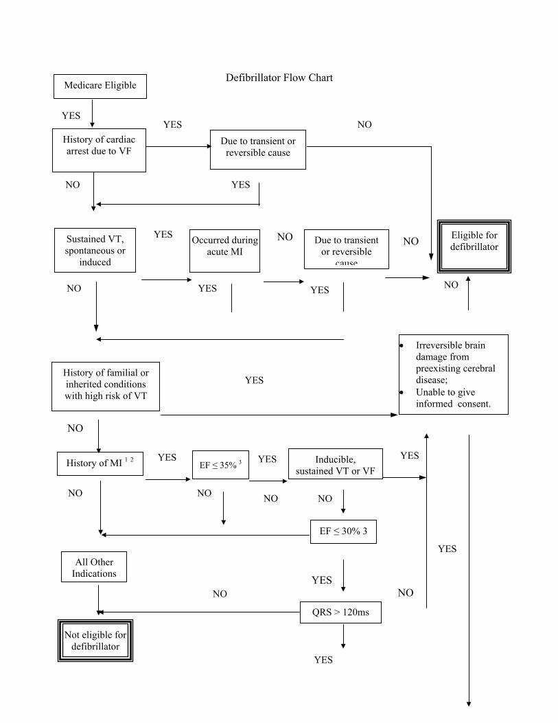
A - Covered Indications
1. Documented episode of cardiac arrest due to ventricular fibrillation (VF), not due to a transient or reversible cause (effective July 1, 1991);
2. Documented sustained ventricular tachyarrhythmia (VT), either spontaneous or induced by an electrophysiology (EP) study, not associated with an acute myocardial infarction (MI) and not due to a transient or reversible cause (effective July 1, 1999);
3. Documented familial or inherited conditions with a high risk of life-threatening VT, such as long QT syndrome or hypertrophic cardiomyopathy (effective July 1, 1999);
Additional indications effective for services performed on or after October 1, 2003:
4. Coronary artery disease with a documented prior MI, a measured left ventricular ejection fraction ≤ 0.35, and inducible, sustained VT or VF at EP study. (The MI must have occurred more than 4 weeks prior to defibrillator insertion. The EP test must be performed more than 4 weeks after the qualifying MI.);
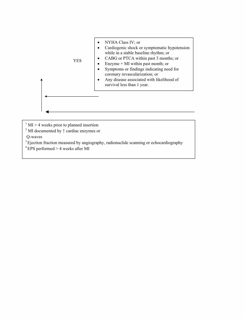
5. Documented prior MI and a measured left ventricular ejection fraction ≤ 0.30 and a QRS duration of > 120 milliseconds. Patients must not have:
• New York Heart Association classification IV;
• Cardiogenic shock or symptomatic hypotension while in a stable baseline rhythm;
• Had a coronary artery bypass graft (CABG) or percutaneous transluminal coronary angioplasty (PTCA) within past months;
• Had an enzyme-positive MI within past month;
• Clinical symptoms or findings that would make them a candidate for coronary revascularization; or
• Any disease, other than cardiac disease (e.g., cancer, uremia, liver failure), associated with a likelihood of survival less than 1 year.
B - All patients considered for implantation of a defibrillator must not have irreversible brain damage, disease or dysfunction that precludes the ability to give informed consent.
C - MIs must be documented by elevated cardiac enzymes or Q-waves on an electrocardiogram. Ejection fractions must be measured by angiography, radionuclide scanning, or echocardiography.
D - All other indications remain noncovered except in Category B IDE clinical trials (60 CFR 48417) or as a routine cost in clinical trials defined under §310.1.
Defibrillator Flow Chart
History of cardiac arrest due to VF
Due to transient or reversible cause
Occurred during acute MI
Sustained VT, spontaneous or
induced
History of familial or inherited conditions with high risk of VT
YES
NO
YES
YES
YES
YES
Y S
O
N
NO
NO
NO
NO
NO
History of MI 1 2 E sus
Eligible for defibrillator
YES
NO
YES
NO
Medicare Eligible
AIn
Not eligible for defibrillator
Due to transient or reversible
cause
E
YES
YES
Y S
NO
EF ≤ 30% 3
• Irreversible brain damage from preexisting cerebral disease;
• Unable to give informed consent.
YES
NO
E
QRS > 120ms
NO
YES
N
O
F ≤ 35% 3
Inducible, tained VT or VFll Other dications
• NYHA Class IV; or • Cardiogenic shock or symptomatic hypotension
while in a stable baseline rhythm; or • CABG or PTCA within past 3 months; or • Enzyme + MI within past month; or • Symptoms or findings indicating need for
coronary revascularization; or • Any disease associated with likelihood of
survival less than 1 year.
1 MI > 4 weeks prior to planned insertion 2 MI documented by ↑ cardiac enzymes or Q-waves 3 Ejection fraction measured by angiography, radionuclide scanning or echocardiography 4 EPS performed > 4 weeks after MI
YES
20.5 - Extracorporeal Immunoadsorption (ECI) Using Protein A Columns
(Rev. 1, 10-03-03)
CIM 35-90
Extracorporeal immunoadsorption (ECI), using Protein A columns, has been developed for the purpose of selectively removing circulating immune complexes (CIC) and immunoglobulins (IgG) from patients in whom these substances are associated with their diseases. The technique involves pumping the patient’s anticoagulated venous blood through a cell separator from which 1-3 liters of plasma are collected and perfused over adsorbent columns, after which the plasma rejoins the separated, unprocessed cells and is retransfused to the patient.
For claims with dates of service on or after January 1, 2001, Medicare covers the use of Protein A columns for the treatment of ITP. In addition, Medicare will cover Protein A columns for the treatment of rheumatoid arthritis (RA) under the following conditions:
• Patient has severe RA. Patient disease is active, having > 5 swollen joints, > 20 tender joints, and morning stiffness > 60 minutes; or
• Patient has failed an adequate course of a minimum of 3 Disease Modifying Anti-Rheumatic Drugs (DMARDs). Failure does not include intolerance.
Other uses of these columns are currently considered to be investigational and, therefore, not reasonable and necessary under the Medicare law. (See §1862(a)(1)(A) of the Act.)
20.6 - Transmyocardial Revascularization (TMR)
(Rev. 1, 10-03-03)
CIM 35-94
Transmyocardial revascularization (TMR) is a surgical technique which uses a laser to bore holes through the myocardium of the heart in an attempt to restore perfusion to areas of the heart not being reached by diseased or clogged arteries. This technique is used as a late or last resort for relief of symptoms of severe angina in patients with ischemic heart disease not amenable to direct coronary revascularization interventions, such as angioplasty, stenting or open coronary bypass.
The precise workings of this technique are not certain. The original theory upon which the technique was based, that the open channels would result in increased perfusion of the myocardium, does not appear to be the major or only action at work. Several theories have been proposed, including partial denervation of the myocardium, or the triggering of the cascade of biological reactions which encourage increased development of blood vessels.
However, research at several facilities indicates that, despite this uncertainty, the technique does offer relief of angina symptoms for a period of time in patients for whom no other medical treatment offering relief is available. Studies indicate that both reduction in pain and reduction in hospitalizations are significant for most patients treated. Consequently, CMS has concluded that, for patients with severe angina (Class III or IV, Canadian Cardiovascular Society, or similar classification system) for whom all other medical therapies have been tried or evaluated and found insufficient, such therapy offers sufficient evidence of its medical effectiveness to treat the symptomatology. It is important to note that this technique does not provide for increased life expectancy, nor is it proven to affect the underlying cause of the angina. However, it appears effective in treating the symptoms of angina, and reducing hospitalizations and allowing patients to resume some of their normal activities of daily living.
The CMS therefore covers TMR as a late or last resort for patients with severe (Canadian Cardiovascular Society classification Classes III or IV) angina (stable or unstable) which has been found refractory to standard medical therapy, including drug therapy at the maximum tolerated or maximum safe dosages. In addition, the angina symptoms must be caused by areas of the heart not amenable to surgical therapies such as percutaneous transluminal coronary angioplasty, stenting, coronary atherectomy or coronary bypass. Coverage is further limited to those uses of the laser used in performing the procedure which have been approved by the Food and Drug Administration for the purpose for which they are being used.
Patients would have to meet all of the following additional selection guidelines:
• An ejection fraction of 25 percent or greater;
• Have areas of viable ischemic myocardium (as demonstrated by diagnostic study) which are not capable of being revascularized by direct coronary intervention; and
• Have been stabilized, or have had maximal efforts to stabilize acute conditions such as severe ventricular arrhythmias, decompensated congestive heart failure or acute myocardial infarction.
Coverage is limited to physicians who have been properly trained in the procedure. Providers of this service must also document that all ancillary personnel, including physicians, nurses, operating room personnel and technicians, are trained in the procedure and the proper use of the equipment involved. Coverage is further limited to providers which have dedicated cardiac care units, including the diagnostic and support services necessary for care of patients undergoing this therapy. In addition, these providers must conform to the standards for laser safety set by the American National Standards Institute, ANSIZ1363.
20.7 - Percutaneous Transluminal Angioplasty (PTA)
(Rev. 1, 10-03-03)
CIM 50-32
This procedure involves inserting a balloon catheter into a narrow or occluded blood vessel to recanalize and dilate the vessel by inflating the balloon.
PTA is covered to treat the following indications:
• Atherosclerotic obstructive lesions:
o In the lower extremities, i.e., the iliac, femoral, and popliteal arteries, or in the upper extremities, i.e., the innominate, subclavian, axillary, and brachial arteries. The upper extremities do not include head or neck vessels.
o Of a single coronary artery for patients for whom the likely alternative treatment is coronary bypass surgery and who exhibit the following characteristics:
Angina refractory to optimal medical management;
Objective evidence of myocardial ischemia; and
Lesions amenable to angioplasty;
• Of the renal arteries for patients in whom there is an inadequate response to a thorough medical management of symptoms and for whom surgery is the likely alternative. PTA for this group of patients is an alternative to surgery, not simply an addition to medical management.
• Obstructive lesions of arteriovenous dialysis fistulas and grafts when performed through either a venous or arterial approach.
PTA is not covered to treat obstructive lesions of the carotid artery except in the following circumstance:
Effective July 1, 2001, Medicare will cover PTA of the carotid artery concurrent with carotid stent placement when furnished in accordance with the Food and Drug Administration (FDA) approved protocols governing Category B Investigational Device Exemption (IDE) clinical trials. PTA of the carotid artery, when provided solely for the purpose of carotid artery dilation concurrent with carotid stent placement, is considered to be a reasonable and necessary service only when provided in the context of such a clinical trial, and therefore is considered a covered service for the purposes of these trials. Performance of PTA in the carotid artery when used to treat obstructive lesions outside of
approved protocols governing Category B IDE clinical trials remains a noncovered service.
PTA is not covered to treat obstructive lesions of the vertebral and cerebral arteries. The safety and efficacy of these procedures have not been established.
20.8 - Cardiac Pacemakers
(Rev. 1, 10-03-03)
CIM 65-6
Cardiac pacemakers are covered as prosthetic devices under the Medicare program, subject to the conditions and limitations described in this section. While cardiac pacemakers have been covered under Medicare for many years, until recently there have been no specific guidelines for their implantation other than the general Medicare requirement that covered services be reasonable and necessary for the treatment of the condition. Services rendered for pacemaker implantations on or after the effective dates of this instruction are subject to the guidelines of this section.
These guidelines are based on certain assumptions regarding the clinical goals of pacemaker implantation. While some uses of pacemakers represent relatively certain or unambiguous usage, many others require considerable expertise and judgment.
Consequently, the medical necessity for pacemaker implantation must be viewed in the context of the overall management of the particular patient. The appropriateness of such implants may be conditional on other diagnostic or therapeutic modalities having been undertaken. Although significant complications and adverse side effects of pacemakers are relatively rare, they cannot be ignored when considering the use of pacemakers for dubious medical conditions, or marginal clinical benefit.
These guidelines represent current concepts regarding medical circumstances in which pacemaker implantation may be appropriate or necessary. As with other areas of medicine, advances in knowledge and techniques in cardiology are expected. Consequently, judgments about the medical necessity and acceptability of pacemaker implants can be expected to change, and instructions modified as more information becomes available.
It should be noted that this instruction applies only to permanent, implanted pacemakers, and does not address the use of temporary, nonimplanted pacemakers.
The two groups of conditions outlined below deal with the necessity for cardiac pacemaker implants for patients in general. These are intended as guidelines for Medicare contractors to use in assessing the medical necessity of claims for pacemaker implantation. As with other guidelines, final coverage determinations must take account of the circumstances of the particular claim, as well as factors such as the medical history of the individual patient. However, as a general rule, contractors may view the two groups of current medical concepts below as representing:
Group I: Single-Chamber Cardiac Pacemakers - A) conditions under which single-chamber pacemaker claims may be considered covered without further claims development; and B) conditions under which single-chamber pacemaker claims would be denied unless further claims development shows that they fall into the covered category, or special medical circumstances exist sufficient to convince the contractor that the claim should be paid.
Group II. Dual-Chamber Cardiac Pacemakers - A) conditions under which dual-chamber pacemaker claims may be considered covered without further claims development, and B) conditions under which dual-chamber pacemaker claims would be denied unless further claims development shows that they fall into the covered categories for single-and dual-chamber pacemakers, or special medical circumstances exist sufficient to convince the contractor that the claim should be paid.
GROUP I
Single-Chamber Cardiac Pacemakers
A - Covered
Conditions under which implantation of a cardiac pacemaker is generally considered acceptable or necessary, provided that the conditions are chronic or recurrent and not due to transient causes such as acute myocardial infarction, drug toxicity, or electrolyte imbalance. (In cases where there is a rhythm disturbance, if the rhythm disturbance is chronic or recurrent, a single episode of a symptom such as syncope or seizure is adequate to establish medical necessity.)
1 - Acquired complete (also referred to as third degree) AV heart block.
2 - Congenital complete heart block with severe bradycardia (in relation to age), or significant physiological deficits or significant symptoms due to the bradycardia.
3 - Second degree AV heart block of Type II (i.e., no progressive prolongation of P-R interval prior to each blocked beat).
4 - Second degree AV heart block of Type I (i.e., progressive prolongation of P-R interval prior to each blocked beat) with significant symptoms due to hemodynamic instability associated with the heart block.
5 - Sinus bradycardia associated with major symptoms (e.g., syncope, seizures, congestive heart failure); or substantial sinus bradycardia (heart rate less than 50) associated with dizziness or confusion. The correlation between symptoms and bradycardia must be documented, or the symptoms must be clearly attributable to the bradycardia rather than to some other cause.
6 - In selected and few patients, sinus bradycardia of lesser severity (heart rate 50-59) with dizziness or confusion. The correlation between symptoms and bradycardia
must be documented, or the symptoms must be clearly attributable to the bradycardia rather than to some other cause.
7 - Sinus bradycardia which is the consequence of long-term necessary drug treatment for which there is no acceptable alternative, when accompanied by significant symptoms (e.g., syncope, seizures, congestive heart failure, dizziness or confusion). The correlation between symptoms and bradycardia must be documented, or the symptoms must be clearly attributable to the bradycardia rather than to some other cause.
8 - Sinus node dysfunction with or without tachyarrhythmias or AV conduction block, i.e., the bradycardia-tachycardia syndrome, sino-atrial block, and sinus arrest, when accompanied by significant symptoms (e.g., syncope, seizures, congestive heart failure, dizziness or confusion).
9 - Sinus node dysfunction with or without symptoms when there are potentially life-threatening ventricular arrhythmias or tachycardia secondary to the bradycardia (e.g., numerous premature ventricular contractions, couplets, runs of premature ventricular contractions, or ventricular tachycardia).
10 - Bradycardia associated with supraventricular tachycardia (e.g., atrial fibrillation, atrial flutter, or paroxysmal atrial tachycardia) with high degree AV block which is unresponsive to appropriate pharmacological management and when the bradycardia is associated with significant symptoms (e.g., syncope, seizures, congestive heart failure, dizziness or confusion).
11 - The occasional patient with hypersensitive carotid sinus syndrome with syncope due to bradycardia and unresponsive to prophylactic medical measures.
12 - Bifascicular or trifascicular block accompanied by syncope which is attributed to transient complete heart block after other, plausible causes of syncope have been reasonably excluded.
13 - Prophylactic pacemaker use following recovery from acute myocardial infarction during which there was temporary complete (third degree) and/or Mobitz Type II second degree AV block in association with bundle branch block.
14 - In patients with recurrent and refractory ventricular tachycardia, “overdrive pacing” (pacing above the basal rate) to prevent ventricular tachycardia.
15 - Second degree AV heart block of Type I with the QRS complexes prolonged.
B - Not Covered - Additional claims development may be required
Conditions which, although used by some physicians as bases for permanent pacemaker implantation, are considered unsupported by adequate evidence of benefit and therefore should not generally be considered appropriate uses for single-chamber pacemakers in the absence of indications cited above. Contractors should review claims for pacemakers
with these indications to determine the need for further claims development prior to denying the claim. The object of such further development is to establish whether the particular claim actually meets the conditions in A. above. In claims where this is not the case or where such an event appears unlikely, the contractor may deny the claim.
1 - Syncope of undetermined cause.
2 - Sinus bradycardia without significant symptoms.
3 - Sino-atrial block or sinus arrest without significant symptoms.
4 - Prolonged R-R intervals with atrial fibrillation (without third degree AV block) or with other causes of transient ventricular pause.
5 - Bradycardia during sleep.
6 - Right bundle branch block with left axis deviation (and other forms of fascicular or bundle branch block) without syncope or other symptoms of intermittent AV block.
7 - Asymptomatic second-degree AV block of Type I unless the QRS complexes are prolonged or electrophysiological studies have demonstrated that the block is at or beyond the level of the His Bundle.
GROUP II
Dual-Chamber Cardiac Pacemakers .
A - Covered
Conditions under which implantation of a dual-chamber cardiac pacemaker is considered acceptable or necessary in the general medical community unless conditions #1 and #2, Group II.B are present:
1 - Patients in who single-chamber (ventricular pacing) at the time of pacemaker insertion elicits a definite drop in blood pressure, retrograde conduction, or discomfort.
2 - Patients in whom the pacemaker syndrome (atrial ventricular asynchrony), with significant symptoms, has already been experienced with a pacemaker that is being replaced.
3 - Patients in whom even a relatively small increase in cardiac efficiency will importantly improve the quality of life, e.g., patients with congestive heart failure despite adequate other medical measures.
4 - Patients in whom the pacemaker syndrome can be anticipated, e.g., in young and active people, etc.
Dual-chamber pacemakers may also be covered for the conditions, as listed in Group I.A. (Single-Chamber Cardiac Pacemakers), if the medical necessity is sufficiently justified through adequate claims development. Expert physicians differ in their judgments about what constitutes appropriate criteria for dual-chamber pacemaker use. The judgment that such a pacemaker is warranted in the patient meeting accepted criteria must be based upon the individual needs and characteristics of that patient, weighing the magnitude and likelihood of anticipated benefits against the magnitude and likelihood of disadvantages to the patient.
B - Not Covered
Whenever the following conditions (which represent overriding contraindications) are present, dual-chamber pacemakers are not covered:
1 - Ineffective atrial contractions, e.g., chronic atrial fibrillation or flutter, or giant left atrium.
2 - Frequent or persistent supraventricular tachycardias, except where the pacemaker is specifically for the control of the tachycardia.
3 - A clinical condition in which pacing takes place only intermittently and briefly, and which is not associated with a reasonable likelihood that pacing needs will become prolonged, e.g., the occasional patient with hypersensitive carotid sinus syndrome with syncope due to bradycardia and unresponsive to prophylactic medical measures.
4 - Prophylactic pacemaker use following recovery from acute myocardial infarction during which there was temporary complete (third degree) and/or Type II second-degree AV block in association with bundle branch block.
Cross reference:
Medicare Benefit Policy Manual, Chapter 1, Inpatient Hospital Services, §40, and Chapter 15, Covered Medical and Other Health Services, §120.
20.8.1 - Cardiac Pacemaker Evaluation Services
(Rev. 1, 10-03-03)
CIM 50-1
Medicare covers a variety of services for the post-implant follow-up and evaluation of implanted cardiac pacemakers. The following guidelines are designed to assist contractors in identifying and processing claims for such services.
NOTE: These new guidelines are limited to lithium battery-powered pacemakers, because mercury-zinc battery-powered pacemakers are no longer being manufactured and virtually all have been replaced by lithium units. Contractors still receiving claims for
monitoring such units should continue to apply the guidelines published in 1980 to those units until they are replaced.
There are two general types of pacemakers in current use - single-chamber pacemakers which sense and pace the ventricles of the heart, and dual-chamber pacemakers which sense and pace both the atria and the ventricles. These differences require different monitoring patterns over the expected life of the units involved. One fact of which contractors should be aware is that many dual-chamber units may be programmed to pace only the ventricles; this may be done either at the time the pacemaker is implanted or at some time afterward. In such cases, a dual-chamber unit, when programmed or reprogrammed for ventricular pacing, should be treated as a single-chamber pacemaker in applying screening guidelines.
The decision as to how often any patient’s pacemaker should be monitored is the responsibility of the patient’s physician who is best able to take into account the condition and circumstances of the individual patient. These may vary over time, requiring modifications of the frequency with which the patient should be monitored. In cases where monitoring is done by some entity other than the patient’s physician, such as a commercial monitoring service or hospital outpatient department, the physician’s prescription for monitoring is required and should be periodically renewed (at least annually) to assure that the frequency of monitoring is proper for the patient. Where a patient is monitored both during clinic visits and transtelephonically, the contractor should be sure to include frequency data on both types of monitoring in evaluating the reasonableness of the frequency of monitoring services received by the patient.
Since there are over 200 pacemaker models in service at any given point, and a variety of patient conditions that give rise to the need for pacemakers, the question of the appropriate frequency of monitoring is a complex one. Nevertheless, it is possible to develop guidelines within which the vast majority of pacemaker monitoring will fall and contractors should do this, using their own data and experience, as well as the frequency guidelines which follow, in order to limit extensive claims development to those cases requiring special attention.
20.8.1.1 - Transtelephonic Monitoring of Cardiac Pacemakers
(Rev. 1, 10-03-03)
CIM 50-1
A - General
Transtelephonic monitoring of pacemakers is furnished by commercial suppliers, hospital outpatient departments and physicians offices.
Telephone monitoring of cardiac pacemakers as described below is medically efficacious in identifying early signs of possible pacemaker failure, thus reducing the number of sudden pacemaker failures requiring emergency replacement. All systems that monitor
the pacemaker rate (bpm) in both the free-running and/or magnetic mode are effective in detecting subclinical pacemaker failure due to battery depletion. More sophisticated systems are also capable of detecting internal electronic problems within the pulse generator itself and other potential problems. In the case of dual chamber pacemakers in particular, such monitoring may detect failure of synchronization of the atria and ventricles, and the need for adjustment and reprogramming of the device.
NOTE: The transmitting device furnished to the patient is simply one component of the diagnostic system, and is not covered as durable medical equipment. Those engaged in transtelephonic pacemaker monitoring should reflect the costs of the transmitters in setting their charges for monitoring.
B - Definition of Transtelephonic Monitoring
In order for transtelephonic monitoring services to be covered, the services must consist of the following elements:
• A minimum 30-second readable strip of the pacemaker in the free-running mode;
• Unless contraindicated, a minimum 30-second readable strip of the pacemaker in the magnetic mode; and
• A minimum 30 seconds of readable ECG strip.
C - Frequency Guidelines for Transtelephonic Monitoring
The guidelines below constitute a system which contractors should use, in conjunction with their knowledge of local medical practices, to screen claims for transtelephonic monitoring prior to payment. It is important to note that they are not recommendations with respect to a minimum frequency for such monitorings, but rather a maximum frequency (within which payment may be made without further claims development). As with previous guidelines, more frequent monitorings may be covered in cases where contractors are satisfied that such monitorings are medically necessary; e.g., based on the condition of the patient, or with respect to pacemakers exhibiting unexpected defects or premature failure. Contractors should seek written justification for more frequent monitorings from the patient’s physician and/or any monitoring service involved.
These guidelines are divided into two broad categories - Guideline I which will apply to the majority of pacemakers now in use, and Guideline II which will apply only to pacemaker systems (pacemaker and leads) for which sufficient long-term clinical information exists to assure that they meet the standards of the Inter-Society Commission for Heart Disease Resources (ICHD) for longevity and end-of-life decay. (The ICHD standards are: (l) 90 percent cumulative survival at 5 years following implant; and (2) an end-of-life decay of less than a 50 percent drop of output voltage and less than 20 percent deviation of magnet rate, or a drop of 5 beats per minute or less, over a period of 3 months or more.) Contractors should consult with their medical advisers and other appropriate individuals and organizations (such as the North American Society of Pacing
and Electrophysiology which publishes product reliability information) should questions arise over whether a pacemaker system meets the ICHD standards.
The two groups of guidelines are then further broken down into two general categories - single chamber and dual-chamber pacemakers. Contractors should be aware that the frequency with which a patient is monitored may be changed from time to time for a number of reasons, such as a change in the patient’s overall condition, a reprogramming of the patient’s pacemaker, the development of better information on the pacemaker’s longevity or failure mode, etc. Consequently, changes in the proper set of guidelines may be required. Contractors should inform physicians and monitoring services to alert contractors to any changes in the patient’s monitoring prescription that might necessitate changes in the screening guidelines applied to that patient. (Of particular importance is the reprogramming of a dual-chamber pacemaker to a single-chamber mode of operation. Such reprogramming would shift the patient from the appropriate dual-chamber guideline to the appropriate single-chamber guideline.)
Guideline I
1 - Single-chamber pacemakers
1st month - every 2 weeks.
2nd through 36th month - every 8 weeks.
37th month to failure - every 4 weeks.
2 - Dual-chamber pacemaker
1st month - every 2 weeks.
2nd through 6th month - every 4 weeks.
7th through 36th month - every 8 weeks.
37th month to failure - every 4 weeks.
Guideline II
1 - Single-chamber pacemakers
1st month - every 2 weeks.
2nd through 48th month - every 12 weeks.
49th through 72nd month - every 8 weeks.
Thereafter - every 4 weeks.
2 - Dual-chamber pacemaker
1st month - every 2 weeks.
2nd through 30th month - every 12 weeks.
31st through 48th month - every 8 weeks.
Thereafter - every 4 weeks.
D - Pacemaker Clinic Services
1 - General
Pacemaker monitoring is also covered when done by pacemaker clinics. Clinic visits may be done in conjunction with transtelephonic monitoring or as a separate service; however, the services rendered by a pacemaker clinic are more extensive than those currently possible by telephone. They include, for example, physical examination of patients and reprogramming of pacemakers. Thus, the use of one of these types of monitoring does not preclude concurrent use of the other.
2 - Frequency Guidelines
As with transtelephonic pacemaker monitoring, the frequency of clinic visits is the decision of the patient’s physician, taking into account, among other things, the medical condition of the patient. However, contractors can develop monitoring guidelines that will prove useful in screening claims. The following are recommendations for monitoring guidelines on lithium-battery pacemakers:
• For single-chamber pacemakers - twice in the first 6 months following implant, then once every 12 months.
• For dual-chamber pacemakers - twice in the first 6 months, then once every 6 months.
20.8.2 - Self-Contained Pacemaker Monitors
(Rev. 1, 10-03-03)
CIM 60-7
Self-contained pacemaker monitors are accepted devices for monitoring cardiac pace-makers. Accordingly, program payment may be made for the rental or purchase of either of the following pacemaker monitors when a physician for a patient prescribes it with a cardiac pacemaker:
A - Digital Electronic Pacemaker Monitor
This device provides the patient with an instantaneous digital readout of his pacemaker pulse rate. Use of this device does not involve professional services until there has been
a change of five pulses (or more) per minute above or below the initial rate of the pacemaker; when such change occurs, the patient contacts his physician.
B - Audible/Visible Signal Pacemaker Monitor
This device produces an audible and visible signal which indicates the pacemaker rate. Use of this device does not involve professional services until a change occurs in these signals; at such time, the patient contacts his physician.
NOTE: The design of the self-contained pacemaker monitor makes it possible for the patient to monitor his pacemaker periodically and minimizes the need for regular visits to the outpatient department of the provider.
Therefore, documentation of the medical necessity for pacemaker evaluation in the outpatient department of the provider should be obtained where such evaluation is employed in addition to the self-contained pacemaker monitor used by the patient in his home.
Cross-referernce: §20.8.1
20.8.3 - Anesthesia in Cardiac Pacemaker Surgery
(Rev. 1, 10-03-03)
CIM 35-79
The use of general or monitored anesthesia during transvenous cardiac pacemaker surgery may be reasonable and necessary and therefore covered under Medicare only if adequate documentation of medical necessity is provided on a case-by-case basis. The contractor obtains advice from its medical consultants or from appropriate specialty physicians or groups in its locality regarding the adequacy of documentation before deciding whether a particular claim should be covered.
A second type of pacemaker surgery that is sometimes performed involves the use of the thoracic method of implantation which requires open surgery. Where the thoracic method is employed, general anesthesia is always used and should not require special medical documentation.
20.9 - ARTIFICIAL HEARTS AND RELATED DEVICES
(Rev. 2, 10-17-03)
A ventricular assist device (VAD) or left ventricular assist device (LVAD) is used to assist a damaged or weakened heart in pumping blood. These devices are used for support of blood circulation post-cardiotomy, as a bridge to a heart transplant, or as destination therapy.
A. Covered Indications
1. Postcardiotomy (effective for services performed on or after October 18, 1993)
Post-cardiotomy is the period following open-heart surgery. VADs used for support of blood circulation post-cardiotomy are covered only if they have received approval from the Food and Drug Administration (FDA) for that purpose, and the VADs are used according to the FDA- approved labeling instructions.
2. Bridge-to-Transplant (effective for services performed on or after January 22, 1996)
VADs used for bridge-to-transplant are covered only if they have received approval from the FDA for that purpose, and the VADs are used according to the FDA-approved labeling instructions. All of the following criteria must be fulfilled in order for Medicare coverage to be provided for a VAD used as a bridge-to-transplant:
a. The patient is approved and listed as a candidate for heart transplantation by a Medicare-approved heart transplant center; and,
b. The implanting site, if different than the Medicare-approved transplant center, must receive written permission from the Medicare-approved heart transplant center under which the patient is listed prior to implantation of the VAD.
The Medicare-approved heart transplant center should make every reasonable effort to transplant patients on such devices as soon as medically reasonable. Ideally, the Medicare-approved heart transplant centers should determine patient-specific timetables for transplantation, and should not maintain such patients on VADs if suitable hearts become available.
3. Destination Therapy (effective for services performed on or after October 1, 2003)
Destination therapy is for patients that require permanent mechanical cardiac support. VADs used for destination therapy are covered only if they have received approval from the FDA for that purpose, and the device is used according to the FDA-approved labeling
instructions. VADs are covered for patients who have chronic end-stage heart failure (New York Heart Association Class IV end-stage left ventricular failure for at least 90 days with a life expectancy of less than 2 years), are not candidates for heart transplantation, and meet all of the following conditions:
a. The patient’s Class IV heart failure symptoms have failed to respond to optimal medical management, including dietary salt restriction, diuretics, digitalis, beta-blockers, and ACE inhibitors (if tolerated) for at least 60 of the last 90 days;
b. The patient has a left ventricular ejection fraction (LVEF) < 25%;
c. The patient has demonstrated functional limitation with a peak oxygen consumption of < 12 ml/kg/min; or the patient has a continued need for intravenous inotropic therapy owing to symptomatic hypotension, decreasing renal function, or worsening pulmonary congestion; and,
d. The patient has the appropriate body size (≥ 1.5 m²) to support the VAD implantation.
In addition, the Centers for Medicare & Medicaid Services (CMS) has determined that VAD implantation as destination therapy is reasonable and necessary only when the procedure is performed in a Medicare-approved heart transplant facility that, between January 1, 2001, and September 30, 2003, implanted at least 15 VADs as a bridge-to-transplant or as destination therapy. These devices must have been approved by the FDA for destination therapy or as a bridge-to-transplant, or have been implanted as part of an FDA investigational device exemption (IDE) trial for one of these two indications. VADs implanted for other investigational indications or for support of blood circulation post-cardiotomy do not satisfy the volume requirement for this purpose. Since the relationship between volume and outcomes has not been well-established for VAD use, facilities that have minimal deficiencies in meeting this standard may apply and include a request for an exception based upon additional factors. Some of the factors CMS will consider are geographic location of the center, number of destination procedures performed, and patient outcomes from VAD procedures completed. Also, this facility must be an active, continuous member of a national, audited registry that requires submission of health data on all VAD destination therapy patients from the date of implantation throughout the remainder of their lives. This registry must have the ability to accommodate data related to any device approved by the FDA for destination therapy regardless of manufacturer. The registry must also provide such routine reports as may be specified by CMS, and must have standards for data quality and timeliness of data submissions such that hospitals failing to meet them will be removed from membership. CMS believes that the registry sponsored by the International Society for Heart and Lung Transplantation is an example of a registry that meets these characteristics.

Hospitals also must have in place staff and procedures that ensure that prospective VAD recipients receive all information necessary to assist them in giving appropriate informed consent for the procedure so that they and their families are fully aware of the aftercare requirements and potential limitations, as well as benefits, following VAD implantation.
CMS plans to develop accreditation standards for facilities that implant VADs and, when implemented, VAD implantation will be considered reasonable and necessary only at accredited facilities.
A list of facilities eligible for Medicare reimbursement for VADs as destination therapy will be maintained on our website and available at www.cms.hhs.gov/coverage/lvadfacility.asp. In order to be placed on this list, facilities must submit a letter to the Director, Coverage and Analysis Group, 7500 Security Blvd, Mailstop C1-09-06, Baltimore, MD 21244. This letter must be received by CMS within 90 days of the issue date on this transmittal. The letter must include the following information:
• Facility’s name and complete address;
• Facility’s Medicare provider number;
• List of all implantations between Jan. 1, 2001, and Sept. 30, 2003, with the following information:
o Date of implantation,
o Indication for implantation (only destination and bridge-to-transplant can be reported; post-cardiotomy VAD implants are not to be included),
o Device name and manufacturer, and,
o Date of device removal and reason (e.g., transplantation, recovery, device malfunction), or date and cause of patient’s death;
• Point-of-contact for questions with telephone number;
• Registry to which patient information will be submitted; and,
• Signature of a senior facility administrative official.
Facilities not meeting the minimal standards and requesting exception should, in addition to supplying the information above, include the factors that they deem critical in requesting the exception to the standards.
CMS will review the information contained in the above letters. When the review is complete, all necessary information is received, and criteria are met, CMS will include the name of the newly Medicare-approved facility on the CMS web site. No reimbursement for destination therapy will be made for implantations performed before the date the facility is added to the CMS web site. Each newly approved facility will also receive a formal letter from CMS stating the official approval date it was added to the list.
B. Noncovered Indications (effective for services performed on or after May 19, 1986)
1. Artificial Heart
Since there is no authoritative evidence substantiating the safety and effectiveness of a VAD used as a replacement for the human heart, Medicare does not cover this device when used as an artificial heart.
2. All other indications for the use of VADs not otherwise listed remain noncovered, except in the context of Category B IDE clinical trials (42 CFR 405) or as a routine cost in clinical trials defined under section 310.1 of the NCD manual (old CIM 30-1).
20.10 - Cardiac Rehabilitation Programs
(Rev. 1, 10-03-03)
CIM 35-25
A - General
Exercise programs for cardiac patients, commonly referred to as cardiac rehabilitation programs, are increasingly being conducted in specialized, free-standing, cardiac rehabilitation clinics as well as in outpatient hospital departments. Exercise programs include specific types of exercise, individually prescribed for each patient.
Medicare coverage of cardiac rehabilitation programs are considered reasonable and necessary only for patients with a clear medical need, who are referred by their attending physician and (1) have a documented diagnosis of acute myocardial infarction within the preceding 12 months; or (2) have had coronary bypass surgery; and/or (3) have stable angina pectoris.
Cardiac rehabilitation programs may be provided either by the outpatient department of a hospital or in a physician-directed clinic. Coverage for either program is subject to the following conditions:
• The facility meets the definition of a hospital outpatient department or a physician-directed clinic, i.e., a physician is on the premises available to perform medical duties at all times the facility is open, and each patient is under the care of a hospital or clinic physician;
• The facility has available for immediate use all the necessary cardio-pulmonary emergency diagnostic and therapeutic life saving equipment accepted by the medical community as medically necessary, e.g., oxygen, cardiopulmonary resuscitation equipment, or defibrillator;

• The program is conducted in an area set aside for the exclusive use of the program while it is in session;
• The program is staffed by personnel necessary to conduct the program safely and effectively, who are trained in both basic and advanced life support techniques and in exercise therapy for coronary disease. Services of nonphysician personnel must be furnished under the direct supervision of a physician. Direct supervision means that a physician must be in the exercise program area and immediately available and accessible for an emergency at all times the exercise program is conducted. It does not require that a physician be physically present in the exercise room itself, provided the contractor does not determine that the physician is too remote from the patients’ exercise area to be considered immediately available and accessible. The examples below are for illustration purposes only. They are not meant to limit the discretion of the contractor to make determinations in this regard:
o The case in which a contractor determines that the presence of a physician in an office across the hall from the exercise room who is available at all times for an emergency meets the requirement that the physician is immediately available and accessible; or
o The case in which a contractor determines that the presence of a physician in a building other than that containing the exercise room does not meet the requirement that the physician is immediately available and accessible; and
• The nonphysician personnel are employees of either the physician, hospital, or clinic conducting the program and their services are “incident-to a physician’s professional services."
Contractors need not undertake elaborate or costly monitoring activities to determine whether these requirements are met, but need only satisfy themselves to the extent that they ordinarily do in connection with, for example, the requirements for coverage of services in physician-directed clinics.
In addition to the conditions listed above, coverage for cardiac rehabilitation programs furnished by hospitals to outpatients are also subject to the rules described in the Medicare Benefit Policy Manual, Chapter 6, “Hospital Services Covered Under Part B,” §20.4.1.
B - Diagnostic Testing - Stress Testing
A prospective candidate for a cardiac rehabilitation program must be evaluated for his suitability to participate. A valuable diagnostic test for this purpose is the stress test. The program need not necessarily include a stress test, but may accept one performed by the patient’s attending physician. Stress testing performed in the outpatient department of a
hos easonable and necessary for one or more of the following:
• ion of chest pain, especially atypical chest pain;
ing
other ECG monitoring) constitute an important and necessary procedure which should be done periodically while a cardiac
cise program. See subsection E,
nic may also provide diagnostic and therapeutic services other than stress testing and ECG monitoring. Any such other
ents for the specific service, e.g., the
er
y
’s professional service - may be covered. Similarly, diagnostic testing of a cardiac rehabilitation patient for a mental problem may be covered where the patient
ssive anxiety or fear associated with the cardiac
s e
e direct supervision of a physician.) Restrictions on
pital or in a physician-directed clinic may be covered when r
Evaluat
• Development of exercise prescriptions for patients with known cardiac disease; and/or
• Pre and postoperative evaluation of patients undergoing coronary artery by-pass procedures.
Refer to subsection E, Utilization Screens, for the acceptable frequency of stress testperformed during an individual’s exercise program.
ECG Rhythm Strips. ECG rhythm strips (and
patient is engaged in a physician-controlled exer“Utilization Screens,” for allowable screens.
C - Other Diagnostic and Therapeutic Services
A freestanding or hospital based cardiac rehabilitation cli
services must meet the usual coverage requiremincident-to, and reasonable and necessary requirements.
1 - Psychotherapy and Psychological Testing
It would not normally be considered reasonable and necessary to provide psychotherapyto all cardiac rehabilitation patients, or even to test all such patients to determine wheththey may have a mental, psychoneurotic, or personality disorder. However, where a patient has a diagnosed mental, psychoneurotic, or personality disorder, psychotherapfurnished by a psychiatrist - or by a psychologist rendering such services incident to a physician
shows appropriate symptoms, e.g., excedisease.
2 - Physical and Occupational Therapy
Physical therapy and occupational therapy would not be covered when furnished in connection with cardiac rehabilitation exercise program services covered under this section unless there also is a diagnosed noncardiac condition requiring such therapy, e.g., where a patient who is just recuperating from an acute phase of heart disease may have had a stroke which would require physical and/or occupational therapy. (While somemay consider the cardiac rehabilitation exercise program a form of physical therapy, it ia specialized program conducted and/or supervised by specially trained personnel whosservices are performed under th
coverage of physical therapy and occupational therapy under this section do not affect rules regarding coverage or noncoverage of such services when furnished in a hospital
or
ardiac
se subjects are not considered reasonable and necessary as a separately identifiable service when provided as a part of a
program. In addition, where a free-standing cardiac rehabilitation clinic provides board and room for the patient (and in some cases family
ith a cardiac rehabilitation exercise program may be considered reasonable and necessary for up to 36 sessions, usually 3 sessions a week in a
ration.
Altand rehabilitation program have not been established, the following guidelines have been identified as acceptable:
• tient has achieved a stable level of exercise tolerance without ischemia or dysrhythmia;
•
• normal limits; or
Accordingly, the contractors’ medical consultants review claims for coverage of cardiac rograms beyond 12 weeks. When claims are accompanied by
inpatient or outpatient setting.
(See the Medicare Benefit Policy Manual, Chapter 1, “Inpatient Hospital Services,” §90.)
3 - Patient Education Services
Many cardiac rehabilitation programs provide health education in the form of lectures counseling, in which patients and/or family members are given information, e.g., on diet, nutrition, and sexual activity to assist them in adjusting their living habits because of the cardiac condition. However, the attending physician following the patient’s acute cepisode would have furnished the same kind of information to a patient and/or family members. Therefore, formal lectures and counseling on the
cardiac rehabilitation exercise
members), these services are not covered under Medicare.
D - Duration of the Program
Services provided in connection w
single 12-week period. Coverage for continued participation in cardiac exercise programs beyond 12 weeks would be allowed only on a case-by-case basis with exit criteria taken into conside
hough firm exit criteria for terminating the therapeutic outpatient exercise treatment
The pa
Symptoms of angina or dyspnea are stable at the patient’s maximum exercise level;
Patient’s resting blood pressure and heart rate are within
• The stress test is not positive during exercise. (A positive test in this context implies an ECG with a junctional depression of 2 mm or more associated with slowly rising, horizontal, or down sloping ST segment.)
rehabilitation exercise pacceptable documentation that the patient has not reached an exit level, coverage may be extended, but should not exceed a maximum of 24 weeks.
E - Utilization Screens
Patients who participate in cardiac rehabilitation programs will require certain services more frequently than other patients being treated on an outpatient basis. Therefore, in
age in a uniform manner, the following utilization screens should be implemented in addition to existing screens for any cardiac rehabilitation services not
Continuous ECG telemetric monitoring during exercise;
hm strip with interpretation and physician’s revision of exercise prescription; and
A visit including one or more of this range of routine services is considered as one for the visit to be reimbursable, at least one
of the Group 1 services must be performed. The same rate of reimbursement would be but not all the services need be performed at each visit.
2 - Group 2 Services
of
All attending physician, or if that performed by the patient’s attending physician is not acceptable to the program’s director.
mill or bicycle ergometer) with physician monitoring and report.
Allow one at the beginning of the program and one after three months (usually the
ian services, as needed.
order to provide cover
listed:
1 - Group 1 Services
ECG rhyt
Limited examination for physician follow-up to adjust medication or other treatment changes.
routine cardiac rehabilitation visit. In order
allowed for each visit,
Allow a maximum of three visits per week.
New patient comprehensive evaluation, including history, physical, and preparationinitial exercise prescription.
ow one at the beginning of the program if not already performed by the patient’s
• ECG stress test (tread
completion of the program).
• Other physic
20.11 - Intraoperative Ventricular Mapping
(Rev. 1, 10-03-03)
Intr directly are usually identified from an anatomical grid and may consist of epicardial, intramural, and endocardial sites. A probe with electrodes is u of electric is used by the surgeon to locate precisely the site of an operative intervention.
The int edicare only for the uses and m
• pathways associated with the Wolff-Parkinson-White (WPW)
• uence of atrial and ventricular activation for drug-resistant ular tachycardias;
eate the anatomical course of His bundle and/or bundle branches during
ias.
of
r and
enous or arterial routes may be employed as well. An intracardiac His bundle cardiogram is usually obtained during EES as are conventional electrocardiograms. No separate charge will be recognized for the His Bundle cardiogram. (See §20.16.)
CIM 35-75
aoperative ventricular mapping is the technique of recording cardiac electrical activity from the heart. The recording sites
sed to explore these surfaces and generate a map that displays the sequence al activation. This information
raoperative ventricular mapping procedure is covered under Medical conditions described below:
Localize accessoryand other preexcitation syndromes;
Map the seqsupraventric
• Delincorrective cardiac surgery for congenital heart diseases; and
• Direct the surgical treatment of patients with refractory ventricular tachyarrhythm
20.12 - Diagnostic Endocardial Electrical Stimulation (Pacing)
(Rev. 1, 10-03-03)
CIM 35-78
Diagnostic endocardial electrical stimulation (EES), also called programmed electrical stimulation of the heart, is covered under Medicare when used for patients with severe cardiac arrhythmias.
Diagnostic endocardial electrical stimulation involves the detection and stimulationcardiac electrical activity for the purpose of studying arrhythmias and abnormalities of the heart’s conduction system. Intracardiac electrode catheters, intracardiac and extracardiac recordings and a stimulator device are required. From two to six multi-polaelectrode catheters are inserted percutaneously, usually through the femoral veins, advanced to the heart under fluoroscopic control. Other v
EES is used to investigate the mechanisms, site of origin and pathways of cardiac arrhythmias as well as to select therapeutic approaches for their resolution. EES is also
patients at risk of sudden arrhythmic death. The principal use for EES is in the diagnosis and treatment of sustained ventricular tachycardia. However, it
ven to be of value in the diagnosis and management of other complex arrhythmias, conduction defects, and after cardiac arrest.
hy requiring catheterization of the right side of the heart and is a recognized diagnostic procedure.
, and those patients who have recently developed heart block secondary to a myocardial infarction. When heart catheterization
rformed at the same time, the program will cover only one catheterization and a small additional charge for the study.
When a His bundle cardiogram is obtained as part of a diagnostic endocardial electrical , no separate charge will be recognized for the His bundle study. (See §20.12
employed to identify
has also pro
20.13 - HIS Bundle Study
(Rev. 1, 10-03-03)
CIM 50-3
The HIS Bundle Study is a specialized type of electrocardiograp
Medicare coverage of the procedure would be limited to selected patients: those with complex ongoing acute arrhythmias, those with intermittent or permanent heart block in whom pacemaker implantation is being considered
and the His Bundle Study are pe
stimulation , “Diagnostic Endocardial Electrical Stimulation.”)
involves the measurement and recording (by one of several methods) of changes in the size of a body part as modified by the circulation of blood in that part.
of the
ive
he evaluation of suspected peripheral arterial disease. It is unsuitable for routine
use in the physician’s office.
20.14 - Plethysmography
(Rev. 1, 10-03-03)
CIM 50-6
Plethysmography
Plethysmography is of value as a noninvasive technique for diagnostic, preoperative and postoperative evaluation of peripheral artery disease in the internal medicine or vascular surgery practice. It is also a useful tool for the preoperative podiatric evaluationdiabetic patient or one who has intermittent claudication or other signs or symptoms indicative of peripheral vascular disease which have a bearing on the patient’s candidacy for foot surgery.
The oldest form of plethysmography is the venous occlusive pneumoplethysmography. This method is cumbersome, time consuming, and requires considerable training to guseful, reproducible results. Nonetheless, in the setting of the hospital vascular laboratory, this technique is considered a reasonable and necessary procedure for tdiagnostic
Recently, however, a number of other plethysmographic methods have been developed which make use of phenomena such as changes in electric impedance or changes in segmental blood pressure at constant volume to assess regional perfusion. Several of these methods have reached a level of development which makes them clinicallyvaluable.
xtended to those procedures listed in Category I below when used for the accepted medical indications mentioned above. The procedures in Category II are
tion of any type should be based on §1862(a)(1)
Medicare coverage is e
still considered experimental and are not covered at this time. Denial of claims because a noncovered procedure was used or because there was no medical indication for plethysmographic evalua of the Act.
performed with d a
pedance Plethysmography
plethysmographic method, this is also a useful tool in the evaluation of suspected
Category I - Covered
Segmental Plethysmography - Included under this procedure are servicesa regional plethysmograph, differential plethysmograph, recording oscillometer, anpulse volume recorder.
Electrical Im
Ultrasonic Measurement of Blood Flow (Doppler) - While not strictly a
peripheral vascular disease or preoperative screening of podiatric patients with suspected peripheral vascular compromise. (See §220.5 for the applicable coverage policy on this procedure.)
Oculoplethysmography - See §20.17, “Noninvasive Tests of Carotid Function.”
Strain Gauge Plethysmography - This test is based on recording the non-pulsatile aspects
nflation and
eir f suspected peripheral vascular disease.
not
Capacitance Plethysmography - This method is considered experimental and does not
scillometry - This is a nonstandardized method which offers poor sensitivity and is not considered superior to the simple measurement of peripheral blood
of inflowing blood at various points on an extremity by a mercury-in-silastic strain gauge sensor. The instrument consists of a chart recorder, an automatic cuff ideflation system, and a recording manometer.
Category II - Experimental
The following methods have not yet reached a level of development such as to allow throutine use in the evaluation o
Inductance Plethysmography - This method is considered experimental and doesprovide reproducible results.
provide reproducible results.
Mechanical O
pressure.
Photoelectric Plethysmography - This method is considered useful only in determiniwhether or not a pulse is present and does not provide reproducible measurements of blood flow.
Differential plethysmography, on the other hand, is a system which uses an impedance technique to compare pulse pressures at various points along a limb, with a reference pressure at the mid-brachial or wrist level. It is not clear whether this technique, as usually performed in the physician’s office, meets the de
ng
finition of plethysmography because quantitative measurements of blood flow are usually not made. It has been
thysmography system is a blood pulse recorder of undetermined value which has the potential for significant overutilization.
ement for studies done by techniques other than venous occlusive pneumoplethysmography should be denied, at least until additional data on these devices,
ntrolled clinical studies, become available.
Reimbursement may be made under Part B for electrocardiographic (EKG) services r an
icate the signs and symptoms or other clinical reason necessitating the services.
gular
supplier, ing
n. No separate bill for the services of a physician is paid unless it is clear that
he/she was the patient’s attending physician or was acting as a consulting physician. The
The documentation required in the various situations mentioned above must be furnished r its
concluded, in any event, that the differential ple
Therefore, reimburs
including co
20.15 - Electrocardiographic Services
(Rev. 1, 10-03-03)
CIM 50-15
rendered by a physician or incident to his/her services or by an approved laboratory oapproved supplier of portable x-ray services. Since there is no coverage for EKG services of any type rendered on a screening basis or as part of a routine examination, the claim must ind
A separate charge by an attending or consulting physician for EKG interpretation is allowed only when it is the normal practice to make such charge in addition to the reoffice visit charge. No payment is made for EKG interpretations by individuals other than physicians..
On a claim involving EKG services furnished by a laboratory or a portable x-ray identify the physician ordering the service and, when the charge includes both the takof the tracing and its interpretation, include the identity of the physician making theinterpretatio
taking of an EKG in an emergency, i.e., when the patient is or may be experiencing what is commonly referred to as a heart attack, is covered as a laboratory service or a diagnostic service by a portable x-ray supplier only when the evidence shows that a physician was in attendance at the time the service was performed or immediately thereafter.
not only when the laboratory or portable x-ray supplier bills the patient or carrier foservice, but also when such a facility bills the attending physician who, in turn, bills the
patient or carrier for the EKG services. (In addition to the evidence required to documthe claim, the laboratory or port
ent able x-ray supplier must maintain in its records the
referring physician’s written order and the identity of the employee taking the tracing.)
ent,
a component for either graphically recording the electrocardiographic data or for visual or computer assisted analysis of the information recorded on magnetic
ts
conventional standard EKG techniques, the
offending arrhythmias can go undetected. With the extended examination provided by
their
This modality is valuable in both inpatient and outpatient diagnosis and therapy. Long-
sive f
The long-term EKG has proven effective in detecting transient episodes of cardiac
st
also appropriate for evaluating a change of dosage and may be indicated shortly before
Long Term EKG Monitoring, also referred to as long-term EKG recording, Holterrecording, or dynamic electrocardiography, is a diagnostic procedure which provides a continuous record of the electrocardiographic activity of a patient’s heart while he is engaged in his daily activities.
The basic components of the long-term EKG monitoring systems are a sensing elemthe design of which may provide either for the recording of electrocardiographic information on magnetic tape or for detecting significant variations in rate or rhythm as they occur, and
tape. The long-term EKG permits the examination in the ambulant or potentially ambulant patient of as many as 70,000 heartbeats in a 12-hour recording while the standard EKG which is obtained in the recumbent position, yields information on only 50 to 60 cardiac cycles and provides only a limited data base on which diagnostic judgmenmay be made.
Many patients with cardiac arrhythmias are unaware of the presence of an irregularity in heart rhythm. Due to the transient nature of many arrhythmias and the short intervals inwhich the rhythm of the heart is observed by
the long-term EKG, the physician is able not only to detect but also to classify various types of rhythm disturbances and waveform abnormalities and note the frequency of occurrence. The knowledge of the reaction of the heart to daily activities with respect to rhythm, rate, conduction disturbances, and ischemic changes are of great assistance in directing proper therapy and rehabilitation.
term monitoring of ambulant or potentially ambulant inpatients provides significantpotential for reducing the length of stay for post-coronary infarct patients in the intencare setting and may result in earlier discharge from the hospital with greater assurance osafety to the patients. The indications for the use of this technique, noted below, are similar for both inpatients and outpatients.
dysrhythmia and in permitting the correlation of these episodes with cardiovascular symptomatology. It is also useful for patients who have symptoms of obscure etiologysuggestive of cardiac arrhythmia. Examples of such symptoms include palpitations, chepain, dizziness, light-headedness, near syncope, syncope, transient ischemic episodes, dyspnea, and shortness of breath.
This technique would also be appropriate at the time of institution of any arrhythmic drugtherapy and may be performed during the course of therapy to evaluate response. It is
and after the discontinuation of anti-arrhythemic medication. The therapeutic respoa drug whose duration of action and p
nse to eak of effectiveness is defined in hours cannot be
properly assessed by examining 30-40 cycles on a standard EKG rhythm strip. The
t
ain and
or
g-
cute myocardial infarction. It may also be reasonable and necessary where the high-risk patient with known cardiovascular disease
ew
knowledge that not all patients placed on anti-arrhythmic medication respond to therapyand the known toxicity of anti-arrhythmic agents clearly indicate that proper assessmenshould be made on an individual basis to determine whether medication should be continued and at what dosage level.
The long-term EKG is also valuable in the assessment of patients with coronary artery disease. It enables the documentation of etiology of such symptoms as chest pshortness of breath. Since the standard EKG is often normal during the intervals between the episodes of precordial pain, it is essential to obtain EKG information while the symptoms are occurring. The long-term EKG has enabled the correlation of chest symptoms with the objective evidence of ST-segment abnormalities. It is appropriate fpatients who are recovering from an acute myocardial infarction or coronary insufficiency before and after discharge from the hospital, since it is impossible to predictwhich of these patients is subject to ventricular arrhythmias on the basis of the presence or absence of rhythm disturbances during the period of initial coronary care. The lonterm EKG enables the physician to identify patients who are at a higher risk of dying suddenly in the period following an a
advances to a substantially higher level of activity which might trigger increased or ntypes of arrhythmias necessitating treatment. Such a high-risk case would be one in which there is documentation that acute phase arrhythmias have not totally disappeared during the period of convalescence.
The use of the long-term EKG for routine assessment of pacemaker function can no longer be justified, (see §20.8.1),. Its use for the patient with an internal pacemaker would be covered only when he has symptoms suggestive of arrhythmia not revealed bythe standard EKG or rhythm strip.
These guidelines are intended as a general outline of the circumstances under which the
ian
The performance of a long-term EKG does not necessarily require the prior performance the
is suspicion that the dysrhythmia
use of this diagnostic procedure would be warranted. Each patient receiving a long-term EKG should be evaluated completely, prior to performance of this diagnostic study. A complete history and physical examination should be obtained and the referring physicshould review the indications for use of the long-term EKG.
of a standard EKG. Nor does the demonstration of a normal standard EKG precludeneed for a long-term EKG. Finally, the demonstration of an abnormal standard EKG does not obviate the need for a long-term EKG if there is transient in nature.
A period of recording of up to 24 hours would normally be adequate to detect most transient arrhythmias and provide essential diagnostic information. The medical necessity for longer periods of monitoring must be documented.
Medical documentation for adjudicating claims for the use of the long-term EKG shoulbe similar to other EKG services, x-ray services, and laboratory procedures. Generalstatement of the diagnostic impression of the referring physician wi
d ly, a
th an indication of the patient’s relevant signs and symptoms should be sufficient for purposes of making a
is
necessary to properly adjudicate the
e
the
o
honic transmission of the recording to a physician’s office, clinic, hospital, etc., having a decoder/recorder for
as an ) for similar indications -
detecting and characterizing symptomatic arrhythmias, regulation of anti-arrhythmic drug
l for me
r,
r
tified
determination regarding the reasonableness and medical necessity for the use of thprocedure. However, the intermediaries or carriers should require whatever additional documentation their medical consultants deemindividual claim where the information submitted is not adequate.
It should be noted that the recording device furnished to the patient is simply oncomponent of the diagnostic system and a separate charge for it will not be recognized under the durable medical equipment benefit.
Patient-Activated EKG Recorders, distributed under a variety of brand names, permitpatient to record an EKG upon manifestation of symptoms, or in response to a physician’s order (e.g., immediately following strong exertion).Most such devices alspermit the patient to simultaneously voice-record in order to describe symptoms and/or activity. In addition, some of these devices permit transtelep
review and analysis, thus eliminating the need to physically transport the tape. Some of these devices also permit a “time sampling” mode of operation. However, the “time sampling” mode is not covered - only the patient-activated mode of operation, when used for the indications described below, is covered at this time.
Services in connection with patient-activated EKG recorders are covered when usedalternative to the long-term EKG monitoring (described above
therapy, etc. Like long-term EKG monitoring, use of these devices is covered for evaluating patients with symptoms of obscure etiology suggestive of cardiac arrhythmia such as palpitations, chest pain, dizziness, lightheadedness, near syncope, syncope, transient ischemic episodes, dyspnea and shortness of breath.
As with long-term EKG monitors, patient-activated EKG recorders may be usefuboth inpatient and outpatient diagnosis and therapy. While useful for assessing sopost-coronary infarct patients in the hospital setting, these devices should not, howevebe covered for outpatient monitoring of recently discharged post-infarct patients.
Computer Analyzed Electrocardiograms - Computer interpretation of EKG’s is recognized as a valid and effective technique which will improve the quality and availability of cardiology services. Reimbursement may be made for such computeservice when furnished in the setting and under the circumstances required for coverage of other electrocardiographic services. Where either a laboratory’s or a portable x-ray supplier’s charge for EKG services includes the physician review and certification of the printout as well as the computer interpretation, the certifying physician must be idenon the Form CMS-1490 before the entire charge can be considered a reimbursable charge. Where the laboratory’s (or portable x-ray supplier’s) reviewing physician is not identified, the carrier should conclude that no professional component is involved and
make its charge determination accordingly. If the supplying laboratory (or portable xsupplier when supplied by such a facility) does not include professional review and certification of the hard copy, a charge by the patient’s physician may be recognizthe service. In any case the charge for the physician component should be substantially less than that for physician interpretation of the conven
-ray
ed for
tional EKG tracing in view of markedly reduced demand on the physician’s time where computer interpretation is
for
n performed with equipment meeting the standards described below, subject to the limitations and conditions specified below. Coverage is further
unts payable with respect to the physician’s service in interpreting the ent. The device
considered durable me
1 -
The use of transtelephonic EKGs is covered for the following uses:
•
,
some uses other than those specified above ent of the contractor’s medical consultants, such a use
cket-sized, and detect and convert the norm e app and trantrantransm
involved. Considering the unit cost reduction expected of this innovation, the total charge for the complete EKG service (taking of tracing and interpretation) when computer interpretation is employed should never exceed that considered reasonable the service when physician interpretation is involved.
Transtelephonic Electrocardiographic Transmissions (Formerly Referred to as EKG Telephone Reporter Systems) coverage is extended to include the use of transtelephonic electrocardiographic (EKG) transmissions as a diagnostic service for the indications described below, whe
limited to the amoresults of such transmissions, including charges for rental of the equipmused by the beneficiary is part of a total diagnostic system and is not
dical equipment.
Covered Uses
To detect, characterize, and document symptomatic transient arrhythmias;
• To overcome problems in regulating antiarrhythmic drug dosage; or
• To carry out early post hospital monitoring of patients discharged after myocardial infarction; (only if 24-hour coverage is provided, see subsection 4“Twenty-Four Hour Coverage,” below).
Since cardiology is a rapidly changing field, may be covered if, in the judgmwas justifiable in the particular case. The enumerated uses above represent uses for which a firm coverage determination has been made, and for which contractors may make payment without extensive claims development or review.
2 - Specifications for Devices
The devices used by the patient are highly portable, usually poal EKG signal so that it can be transmitted via ordinary telephon
aratus to a receiving station. At the receiving end, the signal is decodedscribed into a conventional EKG. There are numerous devices available which smit EKG readings in this fashion. For purposes of Medicare coverage, however, the
itting devices must meet at least the following criteria:
• They must be capable of transmitting EKG Leads, I, II, or III; and
btained by
ized
situations. For example, use of these devices to diagnose and treat suspected arrhythmias as a routine substitute for more conventional methods of
hm
ve days in cases involving detection of transient arrhythmias.
Contractors may find it useful to review claims for these devices with a view toward be
4 - Twenty-Four Hour Coverage
Twenty-four hour coverage means that there must be, at the monitoring site (or sites) an technician receiving calls. Tape recording devices do not meet this
requirement. Further, such technicians should have immediate access to a physician, and atient in
Medicare Benefit Policy Manual, Chapter 6, “Hospital Services Covered Under Part B,”
Medicare Benefit Policy Manual, Chapter 15, “Covered Medical and Other Health
Medicare Services From hysicians, and Providers,” §10,
• These lead transmissions must be sufficiently comparable to readings oa conventional EKG to permit proper interpretation of abnormal cardiac rhythms.
3 - Potential for Abuse - Need for Screening Guidelines
While the use of these devices may often compare favorably with more costly alternatives, this is the case only where the information they contribute is actively utilby a knowledgeable practitioner as part of overall medical management of the patient. Consequently, it is vital that contractors be aware of the potential for abuse of these devices, and adopt necessary screening and physician education policies to detect and halt potentially abusive
diagnosis, such as a careful history, physical examination, and standard EKG and rhytstrip would not be appropriate. Moreover, contractors should require written justification for use of such devices in excess of 30 consecuti
detecting patterns of practice which may be useful in developing schedules which mayadopted for screening such claims in the future.
No payment may be made for the use of these devices to carry out early post hospital monitoring of patients discharged after myocardial infarction unless provision is made for24-hour coverage in the manner described below.
experienced EKG
have been instructed in when and how to contact available facilities to assist the pcase of emergencies.
Cross-reference:
Medicare Benefit Policy Manual, Chapter 1, “Inpatient Hospital Services,” §50,
§10 and §20.3,
Services,” §§60.1 and 250.
Claims Processing Manual, Chapter 16, “LaboratoryIndependent Labs, P
20.16 - CARDIAC OUTPUT MONITORING BY THORACIC ELECTRICAL BIOIMPEDANCE (TEB)
(Rev. 6, 01-23-04)
Thoracic electrical bioimpedance (TEB) devices, a form of plethysmography, monitor cardiac output by noninvasively measuring hemodynamic parameters, including: stroke volume, systemic vascular resistance, and thoracic fluid status. Under the previous coverage determination, effective July 1, 1999, use of TEB was covered for the “noninvasive diagnosis or monitoring of hemodynamics in patients with suspected or known cardiovascular disease.” In reconsidering this policy, CMS concluded that this use was neither sufficiently defined nor supported by available clinical literature to offer
actitioners to determine when TEB would be covered for patient management. Therefore, CMS revised its coverage policy language in response to a reque guidance and clarity for coverage of TEB based on a complete and updated literature review.
A. Covered
1. TEB
a
has riate
b. tion of atrioventricular (A/V) interval for patients with A/V sequential cardiac pacemakers when medical history, physical
patient.
minal
rt transplant.
d. ust
ailure
management of the patient.
the guidance necessary for pr
st for reconsideration to offer more explicit
Indications
is covered for the following uses:
a. Differentiation of cardiogenic from pulmonary causes of acute dyspnewhen medical history, physical examination, and standard assessmenttools provide insufficient information, and the treating physician determined that TEB hemodynamic data are necessary for appropmanagement of the patient.
Optimiza
examination, and standard assessment tools provide insufficient information, and the treating physician has determined that TEB hemodynamic data are necessary for appropriate management of the
c. Monitoring of continuous inotropic therapy for patients with ter
congestive heart failure, when those patients have chosen to die with comfort at home, or for patients waiting at home for a hea
Evaluation for rejection in patients with a heart transplant as a predetermined alternative to a myocardial biopsy. Medical necessity mbe documented should a biopsy be performed after TEB.
e. Optimization of fluid management in patients with congestive heart fwhen medical history, physical examination, and standard assessment tools provide insufficient information, and the treating physician has determined that TEB hemodynamic data are necessary for appropriate
2. ent of drug-resistant hypertension is reasonable and necessary. Drug
resistant hypertension is defined as failure to achieve goal BP in patients who ses of an appropriate three-drug regimen that includes a
diuretic.
B. Noncovered Indications
1. TEB is noncovered when used for patients:
or function pacemakers, since the
c.
ion of
2. All other uses of TEB not otherwise specified remain non-covered.
20.17 - Noninvasive Tests of Carotid Function
in studying and diagnosing carotid disease. There are varieties of these tests which measure various anatomical and
ive tests of carotid function into direct and indirect tests. The direct tests examine the anatomy and physiology of the
of
It is important to note that the names of these tests are not standardized. Following are cceptable tests, recognizing that this list is not inclusive and that local
medical consultants should make determinations:
Direct Tests
• Carotid Phonoangiography
• Direct Bruit Analysis
Contractors have discretion to determine whether the use of TEB for the managem
are adhering to full do
a. With proven or suspected disease involving severe regurgitation of theaorta;
b. With minute ventilation (MV) sensdevice may adversely affect the functioning of that type of pacemaker;
During cardiac bypass surgery; or
d. In the management of all forms of hypertension (with the exceptdrug-resistant hypertension as outlined above).
(Rev. 1, 10-03-03)
CIM 50-37
Noninvasive tests of carotid function aid physicians
physiological aspects of carotid function, including pressure (systolic, diastolic, and pulse), flow, collateral circulation, and turbulence.
For operational purposes, it is useful to classify noninvas
carotid artery, while the indirect tests examine hemodynamic changes in the distal beds the carotid artery (the orbital and cerebral circulations).
some of the a
• Spectral Bruit Analysis
• Doppler Flow Velocity
• Ultrasound Imaging including Real Time
and Doppler Devices
Ind c
oppler Ultrasonography
ody Resection/Carotid Body Denervation
-03-03)
ive-
vestigational and cannot be considered reasonable and necessary within the meaning of §1862(a)(l)
• B-Scan
ire t Tests
• Periorbital Directional D
• Oculoplethysmography
• Ophthalmodynamometry
20.18 - Carotid B
(Rev. 1, 10
CIM 35-7
Carotid body resection is occasionally used to relieve pulmonary symptoms, including asthma, but has been shown to lack general acceptance of the professional medical community. In addition, controlled clinical studies establishing the safety and effectness of this procedure are needed. Therefore, all carotid body resections to relieve pulmonary symptoms must be considered in
of the Act. No program
unity as effective. That instance is when evidence of a mass in the carotid body, with or without symptoms, indicates the need for surgery to remove the carotid
id
due to
of the carotid sinus is rarely performed, but when elected as the therapy of choice with the above indications, this procedure may be considered reasonable and necessary.
reimbursement may be made in such cases.
However, there is one instance where carotid body resection has been accepted by the medical comm
body tumor.
Denervation of a carotid sinus to treat hypersensitive carotid sinus reflex is another procedure performed in the area of the carotid body. In the case of hypersensitive carotsinus, light pressure on the upper part of the neck (such as might be experienced when turning or raising one’s head) results in symptoms such as dizziness or syncope hypotension and slowed heart rate. Failure of medical therapy and continued deteriora-tion in the condition of the patient in such cases may indicate need for surgery. Denervation
20.19 - Ambulatory Blood Pressure Monitoring
(Rev. 1, 10-03-03)
CIM 50-42
Ambulatory blood pressure monitoring (ABPM) involves the use of a noninvasive device whichis used to measure blood pressure in 24-hour cycles. These 24-hour measurements are stored in the device and are later interpreted by the physician. ABPM must be performed for at least 24 hours to meet coverage criteria.
ABPM is only covered for those patients with suspected white coat hypertension. Suspected white coat hypertension is defined as
1. Office blood pressure > 140/90 mm Hg on at least three separate clinic/office visits with two separate measurements made at each visit;
2. At least two documented blood pressure measurements taken outside the office which are < 140/90 mm Hg; and
3. No evidence of end-organ damage.
The information obtained by ABPM is necessary in order to determine the appropriate management of the patient. ABPM is not covered for any other uses. In the rare circumstance that ABPM needs to be performed more than once in a patient, the qualifying criteria described above must be met for each subsequent ABPM test.
For those patients that undergo ABPM and have an ambulatory blood pressure of < 135/85 with no evidence of end-organ damage, it is likely that their cardiovascular risk is similar to that of normotensives. They should be followed over time. Patients for which ABPM demonstrates a blood pressure of > 135/85 may be at increased cardiovascular risk, and a physician may wish to consider antihypertensive therapy.
20.20 - External Counterpulsation (ECP) for Severe Angina
(Rev. 1, 10-03-03)
CIM 35-74
Covered
External counterpulsation (ECP), commonly referred to as enhanced external counterpulsation, is a noninvasive outpatient treatment for coronary artery disease refractory to medical and/or surgical therapy. Although ECP devices are cleared by the Food and Drug Administration (FDA) for use in treating a variety of cardiac conditions, including stable or unstable angina pectoris, acute myocardial infarction and cardiogenic shock, the use of this device to treat cardiac conditions other than stable angina pectoris is not covered, since only that use has developed sufficient evidence to demonstrate its
medical effectiveness. Noncoverage of hydraulic versions of these types of devices remains in force.
Coverage is provided for the use of ECP for patients who have been diagnosed with disabling angina (Class III or Class IV, Canadian Cardiovascular Society Classification or equivalent classification) who, in the opinion of a cardiologist or cardiothoracic surgeon, are not readily amenable to surgical intervention, such as PTCA or cardiac bypass because:
1. Their condition is inoperable, or at high risk of operative complications or post-operative failure;
2. Their coronary anatomy is not readily amenable to such procedures; or
3. They have co-morbid states that create excessive risk.
A full course of therapy usually consists of 35 one-hour treatments which may be offered once or twice daily, usually five days per week. The patient is placed on a treatment table where their lower trunk and lower extremities are wrapped in a series of three compressive air cuffs which inflate and deflate in synchronization with the patient’s cardiac cycle.
During diastole, the three sets of air cuffs are inflated sequentially (distal to proximal) compressing the vascular beds within the muscles of the calves, lower thighs and upper thighs. This action results in an increase in diastolic pressure, generation of retrograde arterial blood flow and an increase in venous return. The cuffs are deflated simultaneously just prior to systole which produces a rapid drop in vascular impedance, a decrease in ventricular workload and an increase in cardiac output.
The augmented diastolic pressure and retrograde aortic flow appear to improve myocardial perfusion, while systolic unloading appears to reduce cardiac workload and oxygen requirements. The increased venous return coupled with enhanced systolic flow appears to increase cardiac output. As a result of this treatment, most patients experience increased time until onset of ischemia, increased exercise tolerance, and a reduction in the number and severity of anginal episodes. Evidence was presented that this effect lasted well beyond the immediate post-treatment phase, with patients symptom-free for several months to two years.
This procedure must be done under direct supervision of a physician.
20.21 - Chelation Therapy for Treatment of Atherosclerosis
(Rev. 1, 10-03-03)
CIM 35-64
Chelation therapy is the application of chelation techniques for the therapeutic or preventive effects of removing unwanted metal ions from the body. The application of
chelation therapy using ethylenediamine-tetra-acetic acid (EDTA) for the treatment and prevention of atherosclerosis is controversial. There is no widely accepted rationale to explain the beneficial effects attributed to this therapy. Its safety is questioned and its clinical effectiveness has never been established by well-designed, controlled clinical trials. It is not widely accepted and practiced by American physicians. EDTA chelation therapy for atherosclerosis is considered experimental. For these reasons, EDTA chelation therapy for the treatment or prevention of atherosclerosis is not covered.
Some practitioners refer to this therapy as chemoendarterectomy and may also show a diagnosis other than atherosclerosis, such as arteriosclerosis or calcinosis. Claims employing such variant terms should also be denied under this section.
Cross-reference: §20.22.
20.22 - Ethylenediamine-Tetra-Acetic (EDTA) Chelation Therapy for Treatment of Atherosclerosis
(Rev. 1, 10-03-03)
CIM 45-20
The use of EDTA as a chelating agent to treat atherosclerosis, arteriosclerosis, calcinosis, or similar generalized condition not listed by the FDA as an approved use is not covered. Any such use of EDTA is considered experimental. See §20.21 for an explanation of this conclusion.
20.23 - Fabric Wrapping of Abdominal Aneurysms
(Rev. 1, 10-03-03)
Not Covered
CIM 35-34
Fabric wrapping of abdominal aneurysms is not a covered Medicare procedure. This is a treatment for abdominal aneurysms which involves wrapping aneurysms with cellophane or fascia lata. This procedure has not been shown to prevent eventual rupture. In extremely rare instances, external wall reinforcement may be indicated when the current accepted treatment (excision of the aneurysm and reconstruction with synthetic materials) is not a viable alternative, but external wall reinforcement is not fabric wrapping. Accordingly, fabric wrapping of abdominal aneurysms is not considered reasonable and necessary within the meaning of §1862(a)(1) of the Act.
20.24 - Displacement Cardiography
(Rev. 1, 10-03-03)
CIM 50-50
Displacement cardiography, including cardiokymography and photokymography, is a noninvasive diagnostic test used in evaluating coronary artery disease.
A - Cardiokymography
Cardiokymography is covered for services rendered on or after October 12, 1988.
Cardiokymography is a covered service only when it is used as an adjunct to electrocardiographic stress testing in evaluating coronary artery disease and only when the following clinical indications are present:
• For male patients, atypical angina pectoris or nonischemic chest pain; or
• For female patients, angina, either typical or atypical.
B - Photokymography - Not Covered
Photokymography remains excluded from coverage.
20.25 - Cardiac Catheterization Performed in Other Than a Hospital Setting
(Rev. 1, 10-03-03)
CIM 35-45
Cardiac catheterization performed in a hospital setting for either inpatients or outpatients is a covered service. The procedure may also be covered when performed in a freestanding clinic when the carrier, in consultation with the appropriate Quality Improvement Organization (QIO), determines that the procedure can be performed safely in all respects in the particular facility. Prior to approving Medicare payment for cardiac catheterizations performed in freestanding clinics, carriers must request QIO review of the clinic.
20.26 - Partial Ventriculectomy
(Rev. 1, 10-03-03)
CIM 35-95
(Also Known as Ventricular Reduction, Ventricular Remodeling, or Heart Volume Reduction Surgery)
Not Covered
Partial ventriculectomy, also known as ventricular reduction, ventricular remodeling, or heart volume reduction surgery, was developed by a Brazilian surgeon and has been performed only on a limited basis in the United States. This procedure is performed on patients with enlarged hearts due to end-stage congestive heart failure. Partial ventriculectomy involves reducing the size of an enlarged heart by excising a portion of the left ventricular wall followed by repair of the defect. It is asserted that this procedure makes the failing heart pump better by improving the efficiency of the remaining left ventricle.
Since the mortality rate is high and there are no published scientific articles or clinical studies regarding partial ventriculectomy, this procedure cannot be considered reasonable and necessary within the meaning of §1862(a)(1) of the Act. Therefore, partial ventriculectomy is not covered by Medicare.
20.27 - Cardiointegram (CIG) as an Alternative to Stress Test or Thallium Stress Test
(Rev. 1, 10-03-03)
CIM 50-47
Not Covered
A cardiointegram device consists of a microcomputer which receives output from a standard electrocardiogram (EKG) and transforms it to produce a graphic representation of heart electrophysiologic signals. This procedure is used primarily as a substitute for Exercise Tolerance Testing with Thallium Imaging in patients for whom a resting EKG may be inadequate to identify changes compatible with coronary artery disease. Because this device is still considered investigational pending additional data on its clinical efficacy/sensitivity and value as a diagnostic tool, program payment may not be made for its use at this time.
20.28 – Therapeutic Embolization
(Rev. 1, 10-03-03)
CIM 35-35
Therapeutic embolization is covered when done for hemorrhage, and for other conditions amenable to treatment by the procedure, when reasonable and necessary for the individual patient. Renal embolization for the treatment of renal adenocarcinoma continues to be covered, effective December 15, 1978, as one type of therapeutic embolization, to:
• Reduce tumor vascularity preoperatively;

• Reduce tumor bulk in inoperable cases; or
• Palliate specific symptoms.
20.29 – Hyperbaric Oxygen Therapy
(Rev. 1, 10-03-03)
CIM 35-10
For purposes of coverage under Medicare, hyperbaric oxygen (HBO) therapy is a modality in which the entire body is exposed to oxygen under increased atmospheric pressure.
A - Covered Conditions
Program reimbursement for HBO therapy will be limited to that which is administered in a chamber (including the one man unit) and is limited to the following conditions:
l - Acute carbon monoxide intoxication, (ICD-9 -CM diagnosis 986).
2 - Decompression illness, (ICD-9-CM diagnosis 993.2, 993.3).
3 - Gas embolism, (ICD-9-CM diagnosis 958.0, 999.1).
4 - Gas gangrene, (ICD-9-CM diagnosis 0400).
5 - Acute traumatic peripheral ischemia. HBO therapy is a valuable adjunctive treatment to be used in combination with accepted standard therapeutic measures when loss of function, limb, or life is threatened. (ICD-9-CM diagnosis 902.53, 903.01, 903.1, 904.0, 904.41.)
6 - Crush injuries and suturing of severed limbs. As in the previous conditions, HBO therapy would be an adjunctive treatment when loss of function, limb, or life is threatened. (ICD-9-CM diagnosis 927.00- 927.03, 927.09-927.11, 927.20-927.21, 927.8-927.9, 928.00-928.01, 928.10-928.11, 928.20-928.21, 928.3, 928.8-928.9, 929.0, 929.9, 996.90- 996.99.)
7 - Progressive necrotizing infections (necrotizing fasciitis), (ICD-9-CM diagnosis 728.86).
8 - Acute peripheral arterial insufficiency, (ICD-9-CM diagnosis 444.21, 444.22, 444.81).
9 - Preparation and preservation of compromised skin grafts (not for primary management of wounds), (ICD-9CM diagnosis 996.52; excludes artificial skin graft).
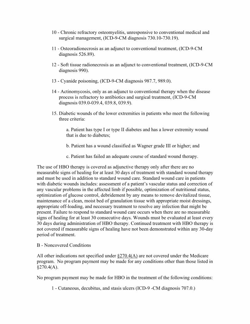
10 - Chronic refractory osteomyelitis, unresponsive to conventional medical and surgical management, (ICD-9-CM diagnosis 730.10-730.19).
11 - Osteoradionecrosis as an adjunct to conventional treatment, (ICD-9-CM diagnosis 526.89).
12 - Soft tissue radionecrosis as an adjunct to conventional treatment, (ICD-9-CM diagnosis 990).
13 - Cyanide poisoning, (ICD-9-CM diagnosis 987.7, 989.0).
14 - Actinomycosis, only as an adjunct to conventional therapy when the disease process is refractory to antibiotics and surgical treatment, (ICD-9-CM diagnosis 039.0-039.4, 039.8, 039.9).
15. Diabetic wounds of the lower extremities in patients who meet the following three criteria:
a. Patient has type I or type II diabetes and has a lower extremity wound that is due to diabetes;
b. Patient has a wound classified as Wagner grade III or higher; and
c. Patient has failed an adequate course of standard wound therapy.
The use of HBO therapy is covered as adjunctive therapy only after there are no measurable signs of healing for at least 30 days of treatment with standard wound therapy and must be used in addition to standard wound care. Standard wound care in patients with diabetic wounds includes: assessment of a patient’s vascular status and correction of any vascular problems in the affected limb if possible, optimization of nutritional status, optimization of glucose control, debridement by any means to remove devitalized tissue, maintenance of a clean, moist bed of granulation tissue with appropriate moist dressings, appropriate off-loading, and necessary treatment to resolve any infection that might be present. Failure to respond to standard wound care occurs when there are no measurable signs of healing for at least 30 consecutive days. Wounds must be evaluated at least every 30 days during administration of HBO therapy. Continued treatment with HBO therapy is not covered if measurable signs of healing have not been demonstrated within any 30-day period of treatment.
B - Noncovered Conditions
All other indications not specified under §270.4(A) are not covered under the Medicare program. No program payment may be made for any conditions other than those listed in §270.4(A).
No program payment may be made for HBO in the treatment of the following conditions:
1 - Cutaneous, decubitus, and stasis ulcers (ICD-9 -CM diagnosis 707.0.)
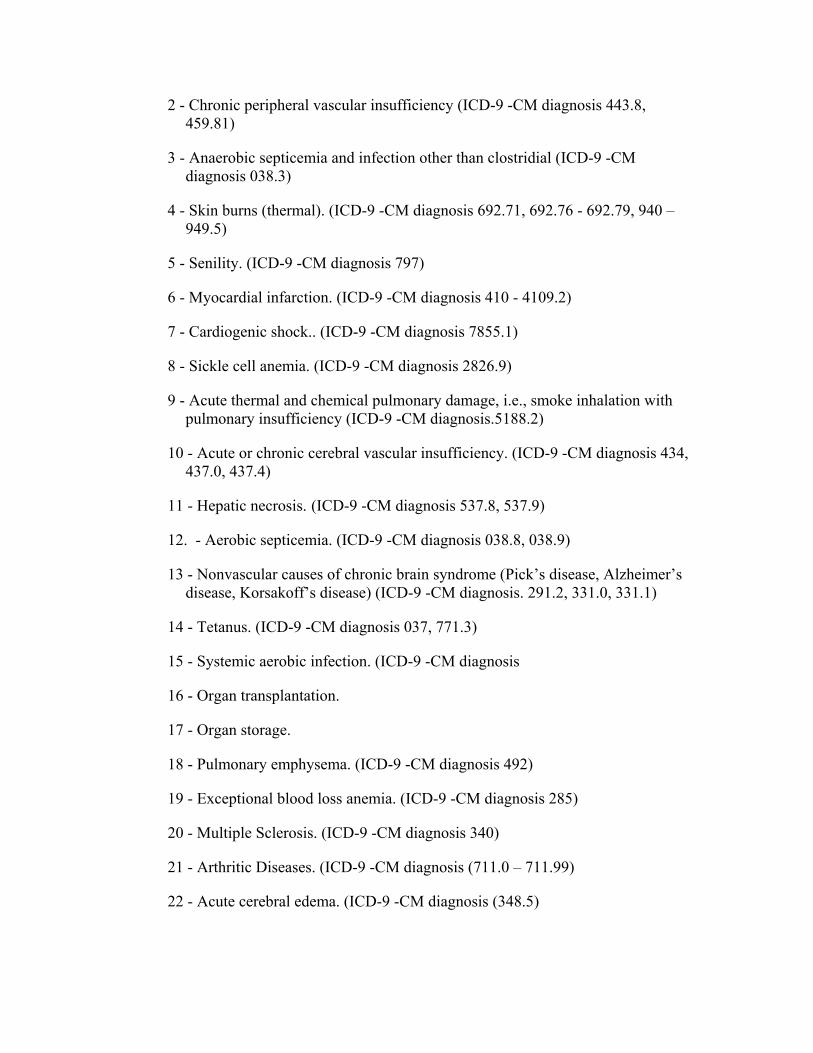
2 - Chronic peripheral vascular insufficiency (ICD-9 -CM diagnosis 443.8, 459.81)
3 - Anaerobic septicemia and infection other than clostridial (ICD-9 -CM diagnosis 038.3)
4 - Skin burns (thermal). (ICD-9 -CM diagnosis 692.71, 692.76 - 692.79, 940 – 949.5)
5 - Senility. (ICD-9 -CM diagnosis 797)
6 - Myocardial infarction. (ICD-9 -CM diagnosis 410 - 4109.2)
7 - Cardiogenic shock.. (ICD-9 -CM diagnosis 7855.1)
8 - Sickle cell anemia. (ICD-9 -CM diagnosis 2826.9)
9 - Acute thermal and chemical pulmonary damage, i.e., smoke inhalation with pulmonary insufficiency (ICD-9 -CM diagnosis.5188.2)
10 - Acute or chronic cerebral vascular insufficiency. (ICD-9 -CM diagnosis 434, 437.0, 437.4)
11 - Hepatic necrosis. (ICD-9 -CM diagnosis 537.8, 537.9)
12. - Aerobic septicemia. (ICD-9 -CM diagnosis 038.8, 038.9)
13 - Nonvascular causes of chronic brain syndrome (Pick’s disease, Alzheimer’s disease, Korsakoff’s disease) (ICD-9 -CM diagnosis. 291.2, 331.0, 331.1)
14 - Tetanus. (ICD-9 -CM diagnosis 037, 771.3)
15 - Systemic aerobic infection. (ICD-9 -CM diagnosis
16 - Organ transplantation.
17 - Organ storage.
18 - Pulmonary emphysema. (ICD-9 -CM diagnosis 492)
19 - Exceptional blood loss anemia. (ICD-9 -CM diagnosis 285)
20 - Multiple Sclerosis. (ICD-9 -CM diagnosis 340)
21 - Arthritic Diseases. (ICD-9 -CM diagnosis (711.0 – 711.99)
22 - Acute cerebral edema. (ICD-9 -CM diagnosis (348.5)
C - Reasonable Utilization Parameters
Make payment where HBO therapy is clinically practical. HBO therapy should not be a replacement for other standard successful therapeutic measures. Depending on the response of the individual patient and the severity of the original problem, treatment may range from less than 1 week to several months duration, the average being two to four weeks. Review and document the medical necessity for use of hyperbaric oxygen for more than two months, regardless of the condition of the patient, before further reimbursement is made.
D - Topical Application of Oxygen
This method of administering oxygen does not meet the definition of HBO therapy as stated above. Also, its clinical efficacy has not been established. Therefore, no Medicare reimbursement may be made for the topical application of oxygen.
Cross reference: §270.5 of this manual.
30 - Complementary and Alternative Medicine
(Rev. 1, 10-03-03)
30.1 - Biofeedback Therapy
(Rev. 1, 10-03-03)
CIM 35-27
Biofeedback therapy provides visual, auditory or other evidence of the status of certain body functions so that a person can exert voluntary control over the functions, and thereby alleviate an abnormal bodily condition. Biofeedback therapy often uses electrical devices to transform bodily signals indicative of such functions as heart rate, blood pressure, skin temperature, salivation, peripheral vasomotor activity, and gross muscle tone into a tone or light, the loudness or brightness of which shows the extent of activity in the function being measured.
Biofeedback therapy differs from electromyography which is a diagnostic procedure used to record and study the electrical properties of skeletal muscle. An electromyography device may be used to provide feedback with certain types of biofeedback.
Biofeedback therapy is covered under Medicare only when it is reasonable and necessary for the individual patient for muscle re-education of specific muscle groups or for treating pathological muscle abnormalities of spasticity, incapacitating muscle spasm, or weakness, and more conventional treatments (heat, cold, massage, exercise, support) have not been successful. This therapy is not covered for treatment of ordinary muscle tension states or for psychosomatic conditions. (See the Medicare Benefit Policy Manual, Chapter 15, for general coverage requirements about physical therapy requirements. )
30.1.1 - Biofeedback Therapy for the Treatment of Urinary Incontinence
(Rev. 1, 10-03-03)
CIM 35-27.1
Biofeedback Therapy for the Treatment of Urinary Incontinence
This policy applies to biofeedback therapy rendered by a practitioner in an office or other facility setting.
Biofeedback is covered for the treatment of stress and/or urge incontinence in cognitively intact patients who have failed a documented trial of pelvic muscle exercise (PME) training. Biofeedback is not a treatment, per se, but a tool to help patients learn how to perform PME. Biofeedback-assisted PME incorporates the use of an electronic or mechanical device to relay visual and/or auditory evidence of pelvic floor muscle tone, in order to improve awareness of pelvic floor musculature and to assist patients in the performance of PME.
A failed trial of PME training is defined as no clinically significant improvement in urinary incontinence after completing four weeks of an ordered plan of pelvic muscle exercises to increase periurethral muscle strength.
Contractors may decide whether or not to cover biofeedback as an initial treatment modality.
Home use of biofeedback therapy is not covered.
30.2 - Thermogenic Therapy
(Rev. 1, 10-03-03)
CIM 35-6
Not Covered
Thermogenic therapy which is the production of artificial fever, has been in use since 1919 in the treatment of certain types of resistant infectious diseases, rheumatoid arthritis and Sydenham’s chorea. Regardless of the medium by which the fever is induced, this modality is not scientifically accepted for the treatment of any specific disease. Since the advent of potent antibiotics, the procedure has for all practical purposes been replaced as a mode of treatment. Therefore, thermogenic therapy is not considered reasonable and necessary for the treatment of an illness or injury as required by §1862(a)(1) of the Act. (Of course, where other covered services are needed and it would be reasonable and necessary that they be furnished on an inpatient hospital basis, payment would not be excluded for the inpatient stay, notwithstanding the fact that reimbursement may not be made for thermogenic therapy furnished during the hospital stay.)
30.3 - Acupuncture
(Rev. 1, 10-03-03)
CIM 35-8
Not Covered
Although acupuncture has been used for thousands of years in China and for decades in parts of Europe, it is a new agent of unknown use and efficacy in the United States. Even in those areas of the world where it has been widely used, its mechanism is not known. Three units of the National Institutes of Health, the National Institute of General Medical Sciences, National Institute of Neurological Diseases and Stroke, and Fogarty International Center have been designed to assess and identify specific opportunities and needs for research attending the use of acupuncture for surgical anesthesia and relief of chronic pain. Until the pending scientific assessment of the technique has been completed and its efficacy has been established, Medicare reimbursement for acupuncture, as an anesthetic or as an analgesic or for other therapeutic purposes, may not be made. Accordingly, acupuncture is not considered reasonable and necessary within the meaning of §1862(a)(1) of the Act.
30.4 - Electrosleep Therapy
(Rev. 1, 10-03-03)
CIM 35-18
Not Covered
Electrosleep therapy consists of the application of short duration, low-amplitude pulses of direct current to the patient’s brain via externally placed occipital electrodes. It is commonly used in the treatment of chronic insomnia, anxiety, and depression, but has also been used for psychosomatic disorders such as asthma, spastic colitis, or tension headache, and for organic disorders including essential hypertension. Until scientific assessment of this technique has been completed and its efficacy is established, no program payment may be made for electrosleep therapy.
30.5 - Transcendental Meditation
(Rev. 1, 10-03-03)
CIM 35-92
Not Covered
Transcendental meditation (TM) is a skill that is claimed to produce a state of rest and relaxation when practiced effectively. Typically, patients are taught TM techniques over the course of several sessions by persons trained in TM. The patient then uses the TM technique on his or her own to induce the relaxed state. Proponents of TM have urged that Medicare cover the training of patients to practice TM when it is medically prescribed as treatment for mild hypertension, as adjunctive therapy in the treatment of essential hypertension, or as the sole or adjunctive treatment of anxiety and other psychological stress-related disorders.
After review of this issue, CMS has concluded that the evidence concerning the medical efficacy of TM is incomplete at best and does not demonstrate effectiveness and that a professional level of skill is not required for the training of patients to engage in TM.
Although many articles have been written about application of TM for patients with certain forms of hypertension and anxiety, there are no rigorous scientific studies that demonstrate the effectiveness of TM for use as an adjunct medical therapy for such conditions. Accordingly, neither TM nor the training of patients for its use are covered under the Medicare program.
30.6 - Intravenous Histamine Therapy
(Rev. 1, 10-03-03)
CIM 35-19
The only accepted and scientifically valid medical use of histamine is diagnostic, including tests to assess:
• The ability of the stomach to secrete acid;
• The integrity of peripheral sensory nerves (e.g., in leprosy);
• The circulatory competency in limb extremities; and
• The presence of a pheochromocytoma.
However, there is no scientifically valid clinical evidence that histamine therapy is effective for any condition regardless of the method of administration, nor is it accepted or widely used by the medical profession. Therefore, histamine therapy cannot be considered reasonable and necessary, and program payment for such therapy is not made.
30.7 - Laetrile and Related Substances
(Rev. 1, 10-03-03)
CIM 45-10
Not Covered
Laetrile (and the other drugs called by the various terms mentioned below) have been used primarily in the treatment or control of cancer. Although the terms “Laetrile,” “laetrile,” “amygdalin,” “Sarcarcinase,” “vitamin B-17,” and “nitriloside” have been used interchangeably, the chemical identity of the substances to which these terms refer has varied.
The FDA has determined that neither Laetrile nor any other drug called by the various terms mentioned above, nor any other product which might be characterized as a “nitriloside” is generally recognized (by experts qualified by scientific training and experience to evaluate the safety and effectiveness of drugs) to be safe and effective for any therapeutic use. Therefore, use of this drug cannot be considered to be reasonable and necessary within the meaning of §1862(a)(1) of the Act and program payment may not be made for its use or any services furnished in connection with its administration.
A hospital stay only for the purpose of having laetrile (or any other drug called by the terms mentioned above) administered is not covered. Also, program payment may not be made for laetrile (or other drug noted above) when it is used during the course of an otherwise covered hospital stay.
30.8 - Cellular Therapy
(Rev. 1, 10-03-03)
CIM 35-5
Not Covered
Cellular therapy involves the practice of injecting humans with foreign proteins like the placenta or lungs of unborn lambs. Cellular therapy is without scientific or statistical evidence to document its therapeutic efficacy and, in fact, is considered a potentially dangerous practice. Accordingly, cellular therapy is not considered reasonable and necessary within the meaning of §1862 (a) (1) of the Act.
30.9 - Transillumination Light Scanning, or Diaphanography
(Rev. 1, 10-03-03)
CIM 50-46
Not Covered
While transillumination light scanning, or diaphanography, for use in detection of cancer and other diseases of the breast, appears safe, the usefulness of this instrumentation, when compared to existing modes of cancer and other breast disease detection, has not clearly been established. Further study of this technology is needed to determine its role in breast cancer diagnosis. Program payment may not be made for this procedure at this time.
40 - Endocrine System and Metabolism
(Rev. 1, 10-03-03)
40.1 - Diabetes Outpatient Self-Management Training
(Rev. 1, 10-03-03)
CIM 80-2
Please refer to 42 CFR 410.140 - 410.146 for conditions that must be met for Medicare coverage.
40.2 - Home Blood Glucose Monitors
(Rev. 1, 10-03-03)
CIM 60-11
There are several different types of blood glucose monitors that use reflectance meters to determine blood glucose levels. Medicare coverage of these devices varies, with respect to both the type of device and the medical condition of the patient for whom the device is prescribed.
Reflectance colorimeter devices used for measuring blood glucose levels in clinical settings are not covered as durable medical equipment for use in the home because their need for frequent professional re-calibration makes them unsuitable for home use. However, some types of blood glucose monitors which use a reflectance meter specifically designed for home use by diabetic patients may be covered as durable medical equipment, subject to the conditions and limitations described below.
Blood glucose monitors are meter devices that read color changes produced on specially treated reagent strips by glucose concentrations in the patient’s blood. The patient, using a disposable sterile lancet, draws a drop of blood, places it on a reagent strip and,
following instructions which may vary with the device used, inserts it into the device to obtain a reading. Lancets, reagent strips, and other supplies necessary for the proper functioning of the device are also covered for patients for whom the device is indicated. Home blood glucose monitors enable certain patients to better control their blood glucose levels by frequently checking and appropriately contacting their attending physician for advice and treatment. Studies indicate that the patient’s ability to carefully follow proper procedures is critical to obtaining satisfactory results with these devices. In addition, the cost of the devices, with their supplies, limits economical use to patients who must make frequent checks of their blood glucose levels. Accordingly, coverage of home blood glucose monitors is limited to patients meeting the following conditions:
1. The patient has been diagnosed as having diabetes;
2. The patient’s physician states that the patient is capable of being trained to use the particular device prescribed in an appropriate manner. In some cases, the patient may not be able to perform this function, but a responsible individual can be trained to use the equipment and monitor the patient to assure that the intended effect is achieved. This is permissible if the record is properly documented by the patient’s physician; and
3. The device is designed for home rather than clinical use
There is also a blood glucose monitoring system designed especially for use by those with visual impairments. The monitors used in such systems are identical in terms of reliability and sensitivity to the standard blood glucose monitors described above. They differ by having such features as voice synthesizers, automatic timers, and specially designed arrangements of supplies and materials to enable the visually impaired to use the equipment without assistance.
These special blood glucose monitoring systems are covered under Medicare if the following conditions are met:
• The patient and device meet the three conditions listed above for coverage of standard home blood glucose monitors; and
• The patient’s physician certifies that he or she has a visual impairment severe enough to require use of this special monitoring system.
The additional features and equipment of these special systems justify a higher reimbursement amount than allowed for standard blood glucose monitors. Separately identify claims for such devices and establish a separate reimbursement amount for them. For those carriers using HCPCS, the procedure code and definitions are E2100 (Blood glucose monitor with integrated voice synthesizer) and E2101 (Blood glucose monitor with integrated lancing/blood sample).
40.3 - Closed-Loop Blood Glucose Control Device (CBGCD)
(Rev. 1, 10-03-03)
CIM 35-70
The closed-loop blood glucose control device (CBGCD) is a hospital bedside device designed for short-term management of patients with insulin dependent diabetes mellitus (Type I). It consists of a rapid on-line glucose analyzer; a computer with a controller for the calculation and control of the infusion of either insulin or dextrose; a multi-channel infusion system; and a printer designed to record continuous glucose values and to provide cumulative totals of the substances infused. Its primary use is for the stabilization of Type I diabetics during periods of stress, such as trauma, labor and delivery, and surgery, when there are wide fluctuations in blood sugar levels. It serves to temporarily correct abnormal blood glucose levels (hyper- or hypo-glycemia) and this correction is made by infusion of either insulin or dextrose. Its use is generally limited to a 24- to 48-hour period because of potential complications; (e.g., sepsis, thromboses, and nonportability, etc.). The CBGCD requires specialized training for use and interpretation of its diagnostic and therapeutic contribution and continuous observation by specially trained medical personnel.
Use of the CBGCD is covered for short-term management of insulin dependent diabetics in crisis situations, in a hospital inpatient setting, and only under the direction of specially trained medical personnel.
40.4 - Insulin Syringe
(Rev. 1, 10-03-03)
CIM 45-3
Medical supplies are covered under §1861(s)(2)(A) of the Act only when they are furnished incident to a physician’s professional services. To be covered under this provision an insulin syringe must have been used by the physician or under his/her direct personal supervision, and the insulin injection must have been given in an emergency situation (e.g., diabetic coma).
The use of an insulin syringe by a diabetic would not meet the requirements of §1861(s)(2)(A) of the Act. See the Medicare Benefit Policy Manual, Chapter 15, “Covered Medical and Other Health Services,” §30.
40.5 - Treatment of Obesity
(Rev. 1, 10-03-03)
CIM 35-26
Obesity itself cannot be considered an illness. The immediate cause is a caloric intake which is persistently higher than caloric output. Program payment may not be made for treatment of obesity alone since this treatment is not reasonable and necessary for the diagnosis or treatment of an illness or injury. However, although obesity is not in itself an illness, it may be caused by illnesses such as hypothyroidism, Cushing’s disease, and hypothalamic lesions. In addition, obesity can aggravate a number of cardiac and respiratory diseases as well as diabetes and hypertension. Therefore, services in connection with the treatment of obesity are covered services when such services are an integral and necessary part of a course of treatment for one of these illnesses.
Supplemented Fasting
Supplemented fasting is a type of very low calorie weight reduction regimen used to achieve rapid weight loss. The reduced calorie intake is supplemented by a mixture of protein, carbohydrates, vitamins and minerals. Serious questions exist about the safety of prolonged adherence for 2 months or more to a very low calorie weight reduction regimen as a general treatment for obesity, because of instances of cardiopathology and sudden death, as well as possible loss of body protein. Therefore, supplemented fasting is not covered as a general treatment for obesity.
In cases where weight loss is necessary before surgery in order to ameliorate the complications posed by obesity when it coexists with pathological conditions such as cardiac and respiratory diseases, diabetes, or hypertension (and other more conservative techniques to achieve this end are not regarded as appropriate), supplemented fasting with adequate monitoring of the patient are covered under Medicare on a case-by-case basis, as determined by the contractor’s medical consultant. The risks associated with the achievement of rapid weight loss must be carefully balanced against the risk posed by the condition requiring surgical treatment.
50 - Ear, Nose and Throat (ENT)
(Rev. 1, 10-03-03)
50.1 - Speech Generating Devices
(Rev. 1, 10-03-03)
CIM 60-23
Effective January 1, 2001, augmentative and alternative communication devices or communicators which are hereafter referred to as “speech generating devices” are now considered to fall within the DME benefit category established by §1861(n) of the Social
Security Act. They may be covered if the contractor’s medical staff determines that the patient suffers from a severe speech impairment and that the medical condition warrants the use of a device based on the following definitions.
Definition of Speech Generating Devices
Speech generating devices are defined as speech aids that provide an individual who has a severe speech impairment with the ability to meet his functional speaking needs. Speech generating are characterized by:
• Being a dedicated speech device, used solely by the individual who has a severe speech impairment;
• May have digitized speech output, using prerecorded messages, less than or equal to 8 minutes recording time;
• May have digitized speech output, using prerecorded messages, greater than 8 minutes recording time;
• May have synthesized speech output which requires message formulation by spelling and device access by physical contact with the device-direct selection techniques;
• May have synthesized speech output which permits multiple methods of message formulation and multiple methods of device access; or
• May be software that allows a laptop computer, desktop computer or personal digital assistant (PDA) to function as a speech generating device.
Devices that would not meet the definition of speech generating devices and therefore, do not fall within the scope of §1861(n) of the Act are characterized by:
• Devices that are not dedicated speech devices, but are devices that are capable of running software for purposes other than for speech generation, e.g., devices that can also run a word processing package, an accounting program, or perform other than non-medical function.
• Laptop computers, desktop computers, or PDA’s which may be programmed to perform the same function as a speech generating device, are noncovered since they are not primarily medical in nature and do not meet the definition of DME. For this reason, they cannot be considered speech-generating devices for Medicare coverage purposes.
• A device that is useful to someone without severe speech impairment is not considered a speech-generating device for Medicare coverage purposes.
50.2 - Electronic Speech Aids
(Rev. 1, 10-03-03)
CIM 65-5
Electronic speech aids are covered under Part B as prosthetic devices when the patient has had a laryngectomy or his larynx is permanently inoperative. There are two types of speech aids. One operates by placing a vibrating head against the throat; the other amplifies sound waves through a tube which is inserted into the user’s mouth. A patient who has had radical neck surgery and/or extensive radiation to the anterior part of the neck would generally be able to use only the “oral tube” model or one of the more sensitive and more expensive “throat contact” devices.
Cross-reference:
The Medicare Benefit Policy Manual, Chapter 15, “Covered Medical and Other Health Services,” §120.
50.3 - Cochlear Implantation
(Rev. 1, 10-03-03)
CIM 65-14
A cochlear implant device is an electronic instrument, part of which is implanted surgically to stimulate auditory nerve fibers, and part of which is worn or carried by the individual to capture, analyze and code sound. Cochlear implant devices are available in single channel and multi-channel models. The purpose of implanting the device is to provide an awareness and identification of sounds and to facilitate communication for persons who are profoundly hearing impaired.
Medicare coverage is provided only for those patients who meet all of the following selection guidelines.
A - General
• Diagnosis of bilateral severe-to-profound sensorineural hearing impairment with limited benefit from appropriate hearing (or vibrotactile) aids;
• Cognitive ability to use auditory clues and a willingness to undergo an extended program of rehabilitation;
• Freedom from middle ear infection, an accessible cochlear lumen that is structurally suited to implantation, and freedom from lesions in the auditory nerve and acoustic areas of the central nervous system;
• No contraindications to surgery; and
• The device must be used in accordance with the FDA-approved labeling.
B - Adults
Cochlear implants may be covered for adults (over age 18) for prelinguistically, perilinguistically, and postlinguistically deafened adults. Postlinguistically deafened adults must demonstrate test scores of 30 percent or less on sentence recognition scores from tape recorded tests in the patient’s best listening condition.
C - Children
Cochlear implants may be covered for prelinguistically and postlinguistically deafened children aged 2 through 17. Bilateral profound sensorineural deafness must be demonstrated by the inability to improve on age appropriate closed-set word identification tasks with amplification.
50.4 - Tracheostomy Speaking Valve
(Rev. 1, 10-03-03)
CIM 65-16
A trachea tube has been determined to satisfy the definition of a prosthetic device, and the tracheostomy speaking valve is an add on to the trachea tube which may be considered a medically necessary accessory that enhances the function of the tube. In other words, it makes the system a better prosthesis. As such, a tracheostomy speaking valve is covered as an element of the trachea tube which makes the tube more effective.
50.5 - Oxygen Treatment of Inner Ear/Carbon Therapy
(Rev. 1, 10-03-03)
CIM 35-29
Not Covered
Oxygen (95 percent) and carbon dioxide (5 percent) inhalation therapy for inner ear disease, such as endolymphatic hydrops and fluctuant hearing loss, is not reasonable and necessary. The therapeutic benefit deriving from this procedure is highly questionable.
50.6 - Tinnitus Masking
(Rev. 1, 10-03-03)
CIM 35-63
A tinnitus masker is a device designed to be worn like a behind-the-ear hearing aid by persons seeking relief from tinnitus. Tinnitus is the perception of noise in the ear and/or head area. The masker produces external sounds to distract the person from the tinnitus.
By producing an external sound a few decibels above the person’s audible threshold, tinnitus masking is thought to provide sufficient distraction from subjective idiopathic tinnitus to alleviate the discomfort and debilitation associated with endogenous sounds within the ear and/or head area.
Tinnitus masking is considered an experimental therapy at this time because of the lack of controlled clinical trials demonstrating effectiveness and the unstudied possibility of serious toxicity in the form of noise induced hearing loss. Therefore, it is not covered.
50.7 - Cochleostomy With Neurovascular Transplant for Meniere’s Disease
(Rev. 1, 10-03-03)
CIM 35-50
Not Covered
Meniere’s disease (or syndrome) is a common cause of paroxysmal vertigo. Meniere’s syndrome is usually treated medically. When medical treatment fails, surgical treatment may be required.
While there are two recognized surgical procedures used in treating Meniere’s disease (decompression of the endolymphatic hydrops and labyrinthectomy), there is no scientific evidence supporting the safety and effectiveness of cochleostomy with neurovascular transplant in treatment of Meniere’s syndrome. Accordingly, Medicare does not cover cochleostomy with neurovascular transplant for treatment of Meniere’s disease.
50.8 - Ultrasonic Surgery
(Rev. 1, 10-03-03)
CIM 35-4
Reimbursement may be made for ultrasonic surgery when required in the treatment of patients with severe and recurrent episodes of vertigo due to Meniere’s syndrome.
This procedure utilizes a machine that produces ultrasonic waves of high intensity and frequency that selectively irradiate certain portions of the inner ear thereby destroying the tissue. The procedure is usually done under local anesthesia, and requires the services of a surgeon and another individual who is responsible for calibrating the electrical equipment, and who assists in observing certain physical changes (e.g., movement of the eyes, “nystagmus") indicative of inner ear reaction to the ultrasonic destruction. Except in rare instances the desired result is achieved with one treatment. At present, there are two different approaches being used to apply the ultrasound to the inner ear: one through the lateral semicircular canal and, more recently, a simpler approach from a technical viewpoint, through the round window.
60 - Emergency Medicine
(Rev. 1, 10-03-03)
No coverage determinations.
70 - Evaluation and Management of Patients - Office/hospital/home
(Rev. 1, 10-03-03)
70.1 - Consultations With a Beneficiary’s Family and Associates
(Rev. 1, 10-03-03)
CIM 35-14
In certain types of medical conditions, including when a patient is withdrawn and uncommunicative due to a mental disorder or comatose, the physician may contact relatives and close associates to secure background information to assist in diagnosis and treatment planning. When a physician contacts his patient’s relatives or associates for this purpose, expenses of such interviews are properly chargeable as physician’s services to the patient on whose behalf the information was secured. If the beneficiary is not an inpatient of a hospital, Part B reimbursement for such an interview is subject to the special limitation on payments for physicians’ services in connection with mental, psychoneurotic, and personality disorders.
A physician may also have contacts with a patient’s family and associates for purposes other than securing background information. In some cases, the physician will provide counseling to members of the household. Family counseling services are covered only where the primary purpose of such counseling is the treatment of the patient’s condition. For example, two situations where family counseling services would be appropriate are as follows: (1) where there is a need to observe the patient’s interaction with family members; and/or (2) where there is a need to assess the capability of and assist the family members in aiding in the management of the patient. Counseling principally concerned with the effects of the patient’s condition on the individual being interviewed would not be reimbursable as part of the physician’s personal services to the patient. While to a
limited degree, the counseling described in the second situation may be used to modify the behavior of the family members, such services nevertheless are covered because they relate primarily to the management of the patient’s problems and not to the treatment of the family member’s problems.
Cross-references:
The Medicare Benefit Policy Manual, Chapter 6, “Hospital Services Covered Under Part B,”§20.
The Medicare Claims Processing Manual, Chapter 12, “Physician/Practitioner Billing,” §10.
The Medicare General Information, Eligibility, and Entitlement Manual, Chapter 3, “Deductibles, Coinsurance Amounts, and Payment Limitations,” §30.
70.2 - Consultation Services Rendered by a Podiatrist in a Skilled Nursing Facility
(Rev. 1, 10-03-03)
CIM 50-8
Consultation services rendered by a podiatrist in a skilled nursing facility are covered if the services are reasonable and necessary and do not come within any of the specific statutory exclusions. Section 1862(a)(13) of the Act excludes payment for the treatment of flat foot conditions, the treatment of subluxations of the foot, and routine foot care. To determine whether the consultation comes within the foot care exclusions, apply the same rule as for initial diagnostic examinations, i.e., where services are performed in connection with specific symptoms or complaints which suggest the need for, covered services, the services are covered regardless of the resulting diagnosis. The exclusion of routine physician examinations is also pertinent and would generally exclude podiatric consultation performed on all patients in a skilled nursing facility on a routine basis for screening purposes, except in those cases where a specific foot ailment is involved. Section 1862(a)(7) of the Act excludes payment for routine physical checkups. (See the Medicare Benefit Policy Manual, Chapter 16, “General Exclusions from Coverage,” §90 and §100.)
70.2.1 - Services Provided for the Diagnosis and Treatment of Diabetic Sensory Neuropathy with Loss of Protective Sensation (aka Diabetic Peripheral Neuropathy)
(Rev. 1, 10-03-03)
CIM 50-8.1
Presently, peripheral neuropathy, or diabetic sensory neuropathy, is the most common factor leading to amputation in people with diabetes. In diabetes, sensory neuropathy is an anatomically diffuse process primarily affecting sensory and autonomic fibers; however, distal motor findings may be present in advanced cases. Long nerves are affected first, with symptoms typically beginning insidiously in the toes and then advancing proximally. This leads to loss of protective sensation (LOPS), whereby a person is unable to feel minor trauma from mechanical, thermal, or chemical sources. When foot lesions are present, the reduction in autonomic nerve functions may also inhibit wound healing.
Diabetic sensory neuropathy with LOPS is a localized illness of the feet and falls within the regulation’s exception to the general exclusionary rule (see 42 CFR 411.15(l)(1)(i)). Foot exams for people with diabetic sensory neuropathy with LOPS are reasonable and necessary to allow for early intervention in serious complications that typically afflict diabetics with the disease.
Effective for services furnished on or after July 1, 2002, Medicare covers, as a physician service, an evaluation (examination and treatment) of the feet no more often than every six months for individuals with a documented diagnosis of diabetic sensory neuropathy and LOPS, as long as the beneficiary has not seen a foot care specialist for some other reason in the interim. LOPS shall be diagnosed through sensory testing with the 5.07 monofilament using established guidelines, such as those developed by the National Institute of Diabetes and Digestive and Kidney Diseases guidelines. Five sites should be tested on the plantar surface of each foot, according to the National Institute of Diabetes and Digestive and Kidney Diseases guidelines. The areas must be tested randomly since the loss of protective sensation may be patchy in distribution, and the patient may get clues if the test is done rhythmically. Heavily callused areas should be avoided. As suggested by the American Podiatric Medicine Association, an absence of sensation at two or more sites out of 5 tested on either foot when tested with the 5.07 Semmes-Weinstein monofilament must be present and documented to diagnose peripheral neuropathy with loss of protective sensation.
The examination includes:
1 - A patient history, and
2 - A physical examination that must consist of at least the following elements:
• Visual inspection of forefoot and hindfoot (including toe web spaces);
• Evaluation of protective sensation;
• Evaluation of foot structure and biomechanics;
• Evaluation of vascular status and skin integrity;
• Evaluation of the need for special footwear; and
3 - Patient education.
A - Treatment includes, but is not limited to:
• Local care of superficial wounds;
• Debridement of corns and calluses; and
• Trimming and debridement of nails.
The diagnosis of diabetic sensory neuropathy with LOPS should be established and documented prior to coverage of foot care. Other causes of peripheral neuropathy should be considered and investigated by the primary care physician prior to initiating or referring for foot care for persons with LOPS.
70.3 - Physician’s Office Within an Institution - Coverage of Services and Supplies Incident to a Physician’s Services
(Rev. 1, 10-03-03)
CIM 45-15
Coverage of Services and Supplies Incident to a Physician’s Services
Where a physician establishes an office within a nursing home or other institution, coverage of services and supplies furnished in the office must be determined in accordance with the “incident to a physician’s professional service” provision (see the Medicare Benefit Policy Manual, Chapter 6, “Hospital Services Covered Under Part B,” §20.4.1 or the Medicare Benefit PolicyManual, Chapter 15, “Covered Medical and Other Health Services,” §60.1) as in any physician’s office. A physician’s office within an institution must be confined to a separately identified part of the facility which is used solely as the physician’s office and cannot be construed to extend throughout the entire institution. Thus, services performed outside the “office” area would be subject to the coverage rules applicable to services furnished outside the office setting.
In order to accurately apply the criteria in the Medicare Benefit Policy Manual, Chapters 6, §20.4.1, or Chapter 15, “Covered Medical and Other Health Services,” §60.1, the contractor gives consideration to the physical proximity of the institution and physician’s office. When his office is located within a facility, a physician may not be reimbursed for services, supplies, and use of equipment which fall outside the scope of services
“commonly furnished” in physician’s offices generally, even though such services may be furnished in his institutional office. Additionally, make a distinction between the physician’s office practice and the institution, especially when the physician is administrator or owner of the facility. Thus, for their services to be covered under the criteria in the Medicare Benefit Policy Manual, Chapter 6, §20.4.1, or the Medicare Benefit Policy Manual, Chapter 15, “Covered Medical and Other Health Services,” §60.1, the auxiliary medical personnel must be members of the office staff rather than of the institution’s staff, and the cost of supplies must represent an expense to the physician’s office practice. Finally, services performed by the employees of the physician outside the “office” area must be directly supervised by the physician; his presence in the facility as a whole would not suffice to meet this requirement. (In any setting, of course, supervision of auxiliary personnel in and of itself is not considered a “physician’s professional service” to which the services of the auxiliary personnel could be an incidental part, i.e., in addition to supervision, the physician must perform or have performed a personal professional service to the patient to which the services of the auxiliary personnel could be considered an incidental part). Denials for failure to meet any of these requirements would be based on §1861(s)(2)(A) of the Act.
Establishment of an office within an institution would not modify rules otherwise applicable for determining coverage of the physician’s personal professional services within the institution. However, in view of the opportunity afforded to a physician who maintains such an office for rendering services to a sizable number of patients in a short period of time or for performing frequent services for the same patient, claims for physicians’ services rendered under such circumstances would require careful evaluation by the carrier to assure that payment is made only for services that are reasonable and necessary.
Cross-reference:
The Medicare Benefit Policy Manual, Chapter 15, “Covered Medical and Other Health Services.”
The Medicare Benefit Policy Manual, Chapter 6, “Hospital Services Covered Under Part B,” §20.4.1.
70.4 - Pronouncement of Death
(Rev. 1, 10-03-03)
CIM 50-19
According to established legal principles, an individual is not considered deceased until there has been official pronouncement of death. An individual is therefore considered to have expired as of the time he/she is pronounced dead by a person who is legally authorized to make such a pronouncement, usually a physician. Reasonable and necessary medical services rendered up to and including pronouncement of death by a physician are covered diagnostic or therapeutic services.
70.5 - Hospital and Skilled Nursing Facility Admission Diagnostic Procedures
(Rev. 1, 10-03-03)
CIM 50-28
These instructions clarify the application of the reasonable and necessary payment exclusion to diagnostic procedures, such as chest x-rays, urinalysis, etc. provided to patients upon admission to a hospital or skilled nursing facility.
The major factors which support a determination that a diagnostic procedure performed as part of the admitting procedure to a hospital or skilled nursing facility is reasonable and necessary, are:
A - The test is specifically ordered by the admitting physician (or a hospital or skilled nursing facility staff physician having responsibility for the patient where there is no admitting physician): i.e., it is not furnished under the standing orders of a physician for his patients;
B - The test is medically necessary for the diagnosis or treatment of the individual patient’s condition; and
C - The test does not unnecessarily duplicate the same test performed on an outpatient basis prior to admission or performed in connection with a recent hospital or skilled nursing facility admission.
Where the contractor has not already done so, consult with the Quality Improvement Organizations (QIOs) to obtain information gathered by the QIOs on a sample basis as to whether x-rays and diagnostic tests are being specifically ordered as described under subsection (A).
80 - Eye
(Rev. 1, 10-03-03)
80.1 - Hydrophilic Contact Lens for Corneal Bandage
(Rev. 1, 10-03-03)
CIM 45-7
Some hydrophilic contact lenses are used as moist corneal bandages for the treatment of acute or chronic corneal pathology, such as bulbous keratopathy, dry eyes, corneal ulcers and erosion, keratitis, corneal edema, descemetocele, corneal ectasis, Mooren’s ulcer, anterior corneal dystrophy, neurotrophic keratoconjunctivitis, and for other therapeutic reasons.
Payment may be made under §1861(s)(2) of the Act for a hydrophilic contact lens approved by the Food and Drug Administration (FDA) and used as a supply incident to a physician’s service. Payment for the lens is included in the payment for the physician’s service to which the lens is incident. Contractors are authorized to accept an FDA letter of approval or other FDA published material as evidence of FDA approval. (See §80.4 for coverage of a hydrophilic contact lens as a prosthetic device.) See the Medicare Benefit Policy Manual, Chapter 15, “Covered Medical and Other Health Services,” and the Medicare Benefit Policy Manual, Chapter 6, “Hospital Services Covered Under Part B,” §20.4.
80.2 - Photodynamic Therapy
(Rev. 1, 10-03-03)
CIM 35-100
Photodynamic therapy is a medical procedure which involves the infusion of a photosensitive (light-activated) drug with a very specific absorption peak. This drug is chemically designed to have a unique affinity for the diseased tissue intended for treatment. Once introduced to the body, the drug accumulates and is retained in diseased tissue to a greater degree than in normal tissue. Infusion is followed by the targeted irradiation of this tissue with a non-thermal laser, calibrated to emit light at a wavelength that corresponds to the drug’s absorption peak. The drug then becomes active and locally treats the diseased tissue.
Ocular photodynamic therapy (OPT)
OPT is used in the treatment of ophthalmologic diseases. OPT is only covered when used in conjunction with verteporfin (see §80.3, “Photosensitive Drugs”).
• Classic Subfoveal Choroidal Neovascular (CNV) Lesions - OPT is covered with a diagnosis of neovascular age-related macular degeneration (AMD) with predominately classic subfoveal choroidal neovascular (CNV) lesions (where the area of classic CNV occupies ≥ 50 percent of the area of the entire lesion) at the initial visit as determined by a fluorescein angiogram. Subsequent follow-up visits will require a fluorescein angiogram prior to treatment. There are no requirements regarding visual acuity, lesion size, and number of re-treatments.
• Occult Subfoveal Choroidal Neovascular (CNV) Lesions - OPT is noncovered for patients with a diagnosis of age-related macular degeneration (AMD) with occult and no classic CNV lesions.
• Other Conditions - Use of OPT with verteporfin for other types of AMD (e.g., patients with minimally classic CNV lesions, atrophic, or dry AMD) is noncovered. OPT with verteporfin for other ocular indications such as pathologic myopia or presumed ocular histoplasmosis syndrome, is eligible for coverage through individual contractor discretion.
80.2.1 - Ocular Photodynamic Therapy (OPT) - Effective April 1, 2004 (see also 80.3 Photosensitive Drugs)
(Rev 9, 04-01-04)
General
The OPT is used in the treatment of ophthalmologic diseases; specifically, for age-related macular degeneration (AMD), a common eye disease among the elderly. OPT involves the infusion of an intravenous photosensitizing drug called verteporfin followed by exposure to a laser. OPT is only covered when used in conjunction with verteporfin.
Effective July 1, 2001, OPT with verteporfin was approved for a diagnosis of neovascular AMD with predominately classic subfoveal choroidal neovascularization (CNV) lesions (where the area of classic CNV occupies ≥ 50% of the area of the entire lesion) at the initial visit as determined by a fluorescein angiogram.
On October 17, 2001, CMS announced its “intent to cover” OPT with verteporfin for AMD patients with occult and no classic subfoveal CNV as determined by a fluorescein angiogram. The October 17, 2001, decision was never implemented.
On March 28, 2002, after thorough review and reconsideration of the October 17, 2001, intent to cover policy, CMS determined that the current noncoverage policy for OPT for verteporfin for AMD patients with occult and no classic subfoveal CNV as determined by a fluorescein angiogram should remain in effect.
Effective August 20, 2002, CMS issued a noncovered instruction for OPT with verteporfin for AMD patients with occult and no classic subfoveal CNV as determined by a fluorescein angiogram.
Covered Indications
Effective April 1, 2004, OPT with verteporfin continues to be approved for a diagnosis of neovascular AMD with predominately classic subfoveal CNV lesions (where the area of classic CNV occupies ≥ 50% of the area of the entire lesion) at the initial visit as determined by a fluorescein angiogram. (CNV lesions are comprised of classic and/or occult components.) Subsequent follow-up visits require a fluorescein angiogram prior to treatment. There are no requirements regarding visual acuity, lesion size, and number of re-treatments when treating predominantly classic lesions.
In addition, after thorough review and reconsideration of the August 20, 2002, noncoverage policy, CMS determines that the evidence is adequate to conclude that OPT with verteporfin is reasonable and necessary for treating:
1. Subfoveal occult with no classic CNV associated with AMD; and, 2. Subfoveal minimally classic CNV (where the area of classic CNV occupies <50%
of the area of the entire lesion) associated with AMD.
The above 2 indications are considered reasonable and necessary only when:
1. The lesions are small (4 disk areas or less in size) at the time of initial treatment or within the 3 months prior to initial treatment; and,
2. The lesions have shown evidence of progression within the 3 months prior to initial treatment. Evidence of progression must be documented by deterioration of visual acuity (at least 5 letters on a standard eye examination chart), lesion growth (an increase in at least 1 disk area), or the appearance of blood associated with the lesion.
Noncovered Indications
Other uses of OPT with verteporfin to treat AMD not already addressed by CMS will continue to be noncovered. These include, but are not limited to, the following AMD indications:
• Juxtafoveal or extrafoveal CNV lesions (lesions outside the fovea),
• Inability to obtain a fluorescein angiogram,
• Atrophic or “dry” AMD.
Other
OPT with verteporfin for other ocular indications, such as pathologic myopia or presumed ocular histoplasmosis syndrome, continue to be eligible for local coverage determinations through individual contractor discretion.
(This NCD last reviewed March 2004.)
80.3 - Photosensitive Drugs
(Rev. 1, 10-03-03)
CIM 45-30
Photosensitive drugs are the light-sensitive agents used in photodynamic therapy. Once introduced into the body, these drugs selectively identify and adhere to diseased tissue. The drugs remain inactive until they are exposed to a specific wavelength of light, by means of a laser, that corresponds to their absorption peak. The activation of a photosensitive drug results in a photochemical reaction which treats the diseased tissue without affecting surrounding normal tissue.
Verteporfin
Verteporfin, a benzoporphyrin derivative, is an intravenous lipophilic photosensitive drug with an absorption peak of 690 nm. This drug was first approved by the Food and Drug Administration (FDA) on April 12, 2000, and subsequently, approved for inclusion in the
United States Pharmacopoeia on July 18, 2000, meeting Medicare’s definition of a drug when used in conjunction with ocular photodynamic therapy (see §80.2, “Photodynamic Therapy”) when furnished intravenously incident to a physician’s service. For patients with age-related macular degeneration, Verteporfin is only covered with a diagnosis of neovascular age-related macular degeneration (ICD-9-CM 362.52) with predominately classic subfoveal choroidal neovascular (CNV) lesions (where the area of classic CNV occupies ≥ 50 percent of the area of the entire lesion) at the initial visit as determined by a fluorescein angiogram (CPT code 92235). Subsequent follow-up visits will require a fluorescein angiogram prior to treatment. OPT with verteporfin is covered for the above indication and will remain noncovered for all other indications related to AMD (see §80.2). OPT with Verteporfin for use in non-AMD conditions is eligible for coverage through individual contractor discretion.
80.3.1 - Verteporfin - Effective April 1, 2004 (see also 80.2.1 Ocular Photodynamic Therapy (OPT))
(Rev 9, 04-01-04)
General
Verteporfin, a benzoporphyrin derivative, is an intravenous lipophilic photosensitive drug with an absorption peak of 690 nm. Verteporfin was first approved by the Food and Drug Administration on April 12, 2000, and subsequently approved for inclusion in the United States Pharmacopoeia on July 18, 2000, meeting Medicare’s definition of a drug as defined under §1861(t)(1) of the Social Security Act. Verteporfin is only covered when used in conjunction with ocular photodynamic therapy OPT) when furnished intravenously incident to a physician’s service.
Covered Indications
Effective April 1, 2004, OPT with verteporfin is covered for patients with a diagnosis of neovascular age-related macular degeneration (AMD) with:
• Predominately classic subfoveal choroidal neovascularization (CNV) lesions (where the area of classic CNV occupies ≥ 50% of the area of the entire lesion) at the initial visit as determined by a fluorescein angiogram. (CNV lesions are comprised of classic and/or occult components.) Subsequent follow-up visits require a fluorescein angiogram prior to treatment. There are no requirements regarding visual acuity, lesion size, and number of retreatments when treating predominantly classic lesions.
• Subfoveal occult with no classic associated with AMD.
• Subfoveal minimally classic CNV CNV (where the area of classic CNV occupies <50% of the area of the entire lesion) associated with AMD.
• The above 2 indications are considered reasonable and necessary only when: 1. The lesions are small (4 disk areas or less in size) at the time of initial treatment or within the 3 months prior to initial treatment; and, 2. The lesions have shown evidence of progression within the 3 months prior to initial treatment. Evidence of progression must be documented by deterioration of visual acuity (at least 5 letters on a standard eye examination chart), lesion growth (an increase in at least 1 disk area), or the appearance of blood associated with the lesion.
Noncovered Indications
Other uses of OPT with verteporfin to treat AMD not already addressed by CMS will continue to be noncovered. These include, but are not limited to, the following AMD indications: juxtafoveal or extrafoveal CNV lesions (lesions outside the fovea), inability to obtain a fluorescein angiogram, or atrophic or “dry” AMD.
Other
OPT with verteporfin for other ocular indications, such as pathologic myopia or presumed ocular histoplasmosis syndrome, continue to be eligible for local coverage determinations through individual contractor discretion.
(This NCD last reviewed March 2004.)
80.4 - Hydrophilic Contact Lenses
(Rev. 1, 10-03-03)
CIM 65-1
Hydrophilic contact lenses are eyeglasses within the meaning of the exclusion in §1862(a)(7) of the Act and are not covered when used in the treatment of nondiseased eyes with spherical ametrophia, refractive astigmatism, and/or corneal astigmatism. Payment may be made under the prosthetic device benefit, however, for hydrophilic contact lenses when prescribed for an aphakic patient.
Contractors are authorized to accept an FDA letter of approval or other FDA published material as evidence of FDA approval. (See §80.1 for coverage of a hydrophilic lens as a corneal bandage.)
Cross-references:
The Medicare Benefit Policy Manual, Chapter 15, “Covered Medical and Other Health Services,” §100 and §120.
The Medicare Benefit Policy Manual, Chapter 16, “General Exclusions from Coverage,” §20 and §90.
80.5 - Scleral Shell
(Rev. 1, 10-03-03)
CIM 65.3
Scleral shell (or shield) is a catchall term for different types of hard scleral contact lenses.
A scleral shell fits over the entire exposed surface of the eye as opposed to a corneal contact lens which covers only the central non-white area encompassing the pupil and iris. Where an eye has been rendered sightless and shrunken by inflammatory disease, a scleral shell may, among other things, obviate the need for surgical enucleation and prosthetic implant and act to support the surrounding orbital tissue.
In such a case, the device serves essentially as an artificial eye. In this situation, payment may be made for a scleral shell under §1861(s)(8) of the Act.
Scleral shells are occasionally used in combination with artificial tears in the treatment of “dry eye” of diverse etiology. Tears ordinarily dry at a rapid rate, and are continually replaced by the lacrimal gland. When the lacrimal gland fails, the half-life of artificial tears may be greatly prolonged by the use of the scleral contact lens as a protective barrier against the drying action of the atmosphere. Thus, the difficult and sometimes hazardous process of frequent installation of artificial tears may be avoided. The lens acts in this instance to substitute, in part, for the functioning of the diseased lacrimal gland and would be covered as a prosthetic device in the rare case when it is used in the treatment of “dry eye.”
Cross-references:
The Medicare Benefit Policy Manual, Chapter 15, “Covered Medical and Other Health Services,” §120 and §130
The Medicare Benefit Policy Manual, Chapter 1, “Inpatient Hospital Services,” §40 and §120.1.
80.6 - Intraocular Photography
(Rev. 1, 10-03-03)
CIM 35-39
Intraocular photography is covered when used for the diagnosis of such conditions as macular degeneration, retinal neoplasms, choroid disturbances and diabetic retinopathy, or to identify glaucoma, multiple sclerosis and other central nervous system abnormalities. Make Medicare payment for the use of this procedure by an opthalmologist in these situations when it is reasonable and necessary for the individual patient to receive these services.
80.7 - Refractive Keratoplasty
(Rev. 1, 10-03-03)
CIM 35-54
Not Covered
Refractive keratoplasty is surgery to reshape the cornea of the eye to correct vision problems such as myopia (nearsightedness) and hyperopia (farsightedness). Refractive keratoplasty procedures include keratomileusis, in which the front of the cornea is removed, frozen, reshaped, and stitched back on the eye to correct either near or farsightedness; keratophakia, in which a reshaped donor cornea is inserted in the eye to correct farsightedness; and radial keratotomy, in which spoke-like slits are cut in the cornea to weaken and flatten the normally curved central portion to correct nearsightedness.
The correction of common refractive errors by eyeglasses, contact lenses or other prosthetic devices is specifically excluded from coverage. The use of radial keratotomy and/or keratoplasty for the purpose of refractive error compensation is considered a substitute or alternative to eye glasses or contact lenses which are specifically excluded by §1862 (a)(7) of the Act (except in certain cases in connection with cataract surgery). In addition, many in the medical community consider such procedures cosmetic surgery which is excluded by §§1862 (a)(10) of the Act. Therefore, radial keratotomy and keratoplasty to treat refractive defects are not covered.
80.7.1 - Keratoplasty
Keratoplasty that treats specific lesions of the cornea, such as phototherapeutic keratectomy that removes scar tissue from the visual field, deals with an abnormality of the eye and is not cosmetic surgery. Such cases may be covered under §1862(a)(1)(A) of the Act.
The use of lasers to treat ophthalmic disease constitutes opthalmalogic surgery. Coverage is restricted to practitioners who have completed an approved training program in ophthalmologic surgery.
80.8 - Endothelial Cell Photography
(Rev. 1, 10-03-03)
CIM 50-38
Endothelial cell photography involves the use of a specular microscope to determine the endothelial cell count. It is used by ophthalmologists as a predictor of success of ocular surgery or certain other ocular procedures. Endothelial cell photography is a covered procedure under Medicare when reasonable and necessary for patients who meet one or more of the following criteria:
• Have slit lamp evidence of endothelial dystrophy (cornea guttata),
• Have slit lamp evidence of corneal edema (unilateral or bilateral),
• Are about to undergo a secondary intraocular lens implantation,
• Have had previous intraocular surgery and require cataract surgery,
• Are about to undergo a surgical procedure associated with a higher risk to corneal endothelium; i.e., phacoemulsification, or refractive surgery (see §80.7 for excluded refractive procedures),
• With evidence of posterior polymorphous dystrophy of the cornea or irido-corneal-endothelium syndrome, or
• Are about to be fitted with extended wear contact lenses after intraocular surgery.
When a presurgical examination for cataract surgery is performed and the conditions of this section are met, if the only visual problem is cataracts, endothelial cell photography is covered as part of the presurgical comprehensive eye examination or combination brief/intermediate examination provided prior to cataract surgery, and not in addition to it. (See §10.1)
80.9 - Computer Enhanced Perimetry
(Rev. 1, 10-03-03)
CIM 50-49
Computer enhanced perimetry involves the use of a micro-computer to measure visual sensitivity at preselected locations in the visual field. It is a covered service when used in assessing visual fields in patients with glaucoma or other neuropathologic defects.
80.10 - Phaco-Emulsification Procedure - Cataract Extraction
(Rev. 1, 10-03-03)
CIM 35-9
In view of recommendations of authoritative sources in the field of ophthalmology, the subject technique is viewed as an accepted procedure for removal of cataracts. Accordingly, program reimbursement may be made for necessary services furnished in connection with cataract extraction utilizing the phaco-emulsification procedure.
80.11 - Vitrectomy
(Rev. 1, 10-03-03)
CIM - 35-16
Vitrectomy may be considered reasonable and necessary for the following conditions: vitreous loss incident to cataract surgery, vitreous opacities due to vitreous hemorrhage or other causes, retinal detachments secondary to vitreous strands, proliferative retinopathy, and vitreous retraction. See chapter 23 of the Medicare Claims Manual for how to determine payment for physician vitrectomy services and the Medicare Claims Processing Manual, Chapter 14, “Ambulatory Surgical Centers,” §40, for how to determine payment for ASC facility vitrectomy services. Also, see the Medicare Claims Processing Manual, Chapter 23, “Fee Schedule Administration and Coding Requirements,” §20.9, to identify when, for Medicare payment purposes, certain vitrectomy codes are included in other codes or when codes for other services include vitrectomy codes.
The CPT codes for vitrectomy services are 67005, 67010, 67036, 67038, 67039, and 67040.
80.12 - Intraocular Lenses (IOLs)
(Rev. 1, 10-03-03)
CIM 65-7
An intraocular lens, or pseudophakos, is an artificial lens which may be implanted to replace the natural lens after cataract surgery. Intraocular lens implantation services, as well as the lens itself, may be covered if reasonable and necessary for the individual. Implantation services may include hospital, surgical, and other medical services, including preimplantation ultrasound (A-scan) eye measurement of one or both eyes.
Cross-reference:
The Medicare Benefit Policy Manual, Chapter 6,”Hospital Services Covered Under Part B,” §10.
The Medicare Benefit Policy Manual, Chapter 15, “Covered Medical and Other Health Services,” §120.
The Medicare Benefit Policy Manual, Chapter 16, “General Exclusions from Coverage,” §20 and §90.
90 - Genetics
(Rev. 1, 10-03-03)
No coverage determinations
100 - Gastrointestinal System
(Rev. 1, 10-03-03)
100.1 - Gastric Bypass Surgery for Obesity
(Rev. 1, 10-03-03)
CIM 35-40
Gastric bypass surgery which is a variation of the gastrojejunostomy, is performed for patients with extreme obesity. Gastric bypass surgery for extreme obesity is covered under the program if (1) it is medically appropriate for the individual to have such surgery; and (2) the surgery is to correct an illness which caused the obesity or was aggravated by the obesity.
Cross-references: §§40.5 and 100.8.
100.2 - Endoscopy
(Rev. 1, 10-03-03)
CIM 35-59
Endoscopy is a technique in which a long flexible tube-like instrument is inserted into the body orally or rectally, permitting visual inspection of the gastrointestinal tract. Although primarily a diagnostic tool, endoscopy includes certain therapeutic procedures such as removal of polyps, and endoscopic papillotomy, by which stones are removed from the bile duct.
Endoscopic procedures are covered when reasonable and necessary for the individual patient.
100.3 - 24-Hour Ambulatory Esophegeal pH Monitoring
(Rev. 1, 10-03-03)
CIM 35-83
Twenty-four hour ambulatory esophageal pH monitoring is a diagnostic procedure involving the placement of an indwelling electrode into the lower esophagus of a patient
for the purpose of determining the presence of gastric reflux and measuring abnormal esophageal acid exposure.
Twenty-four hour ambulatory pH monitoring is covered by Medicare for patients who are suspected of having gastric reflux, but only if the patient presents diagnostic problems associated with atypical symptoms or the patient’s symptoms are suggestive of reflux, but conventional tests have not confirmed the presence of reflux.
100.4 - Esophageal Manometry
(Rev. 1, 10-03-03)
CIM 50-25
Esophageal manometry is covered under Medicare where it is determined to be reasonable and necessary for the individual patient. The major use of esophageal manometry is to measure pressure within the esophagus to assist in the diagnosis of esophageal pathology including aperistalsis, spasm, achalasia, esophagitis, esophageal ulcer, esophageal congenital webs, diverticuli, scleroderma, hiatus hernia, congenital cysts, benign and malignant tumors, hypermobility, hypomobility, and extrinsic lesions. Esophageal manometry is mostly used in difficult diagnostic cases and as an adjunct to x-rays and direct visualization of the esophagus (endoscopy) through the fiberscope.
100.5 - Diagnostic Breath Analyses
(Rev. 1, 10-03-03)
CIM 50-51
Diagnostic breath analyses are tests performed to measure either the hydrogen or carbon dioxide content of the breath after the ingestion of certain compounds. The analyses are performed to diagnose certain gastrointestinal disease states.
The Following Breath Test Is Covered:
Lactose breath hydrogen to detect lactose malabsorption.
The Following Breath Tests Are Excluded From Coverage:
Lactulose breath hydrogen for diagnosing small bowel bacterial overgrowth and measuring small bowel transit time.
CO2 for diagnosing bile acid malabsorption.
CO2 for diagnosing fat malabsorption.
100.6 - Gastric Freezing
(Rev. 1, 10-03-03)
CIM 35-65
Gastric freezing for chronic peptic ulcer disease is a non-surgical treatment which was popular about 20 years ago but now is seldom done. It has been abandoned due to a high complication rate, only temporary improvement experienced by patients, and lack of effectiveness when tested by double-blind, controlled clinical trials. Since the procedure is now considered obsolete, it is not covered.
100.7 - Colonic Irrigation
CIM 35-1
Not Covered
Colonic irrigation is a procedure to wash out or lavage material on the walls of the bowel to an unlimited distance without inducing defecation. This procedure is distinguished from all types of enemas which are primarily used to induce defecation.
There are no conditions for which colonic irrigation is medically indicated and no evidence of therapeutic value. Accordingly, colonic irrigation cannot be considered reasonable and necessary within the meaning of §1862(a)(1) of the Act.
100.8 - Intestinal Bypass Surgery
(Rev. 1, 10-03-03)
CIM 35-33
Not Covered
The safety of intestinal bypass surgery for treatment of obesity has not been demonstrated. Severe adverse reactions such as steatorrhea, electrolyte depletion, liver failure, arthralgia, hypoplasia of bone marrow, and avitaminosis have sometimes occurred as a result of this procedure. It does not meet the reasonable and necessary provisions of §1862(a)(1) of the Act and is not a covered Medicare procedure.
Cross-references: §§40.5, 100.1.
100.9 - Implantation of Anti-Gastroesophageal Reflux Device
(Rev. 1, 10-03-03)
CIM 35-69
The implantation of an anti-gastroesophageal reflux device is a surgical procedure for the treatment of gastroesophageal reflux, a condition in which the caustic contents of the stomach flow back into the esophagus. The procedure involves the implantation of this special device around the esophagus under the diaphragm and above the stomach which is secured in place by a circumferential tie strap.
The implantation of this device may be considered reasonable and necessary in specific clinical situations where a conventional valvuloplasty procedure is contraindicated. The implantation of an anti-gastroesophageal reflux device is covered only for patients with documented severe or life threatening gastroesophageal reflux disease whose conditions have been resistant to medical treatment and who also:
• Have esophageal involvement with progressive systemic sclerosis; or
• Have foreshortening of the esophagus such that insufficient tissue exists to permit a valve reconstruction; or
• Are poor surgical risks for a valvuloplasty procedure; or
• Have failed previous attempts at surgical treatment with valvuloplasty procedures.
100.10 - Injection Sclerotherapy for Esophageal Variceal Bleeding
(Rev. 1, 10-03-03)
CIM 35-73
Injection sclerotherapy is a technique involving insertion of a flexible fiberoptic endoscope into the esophagus, and the injection of a sclerosing agent or solution into the varicosities to control bleeding. This procedure is covered under Medicare
100.11 - Gastric Balloon for Treatment of Obesity
(Rev. 1, 10-03-03)
CIM 35-86
Not Covered
The gastric balloon is a medical device developed for use as a temporary adjunct to diet and behavior modification to reduce the weight of patients who fail to lose weight with
those measures alone. It is inserted into the stomach to reduce the capacity of the stomach and to affect early satiety.
The use of the gastric balloon is not covered under Medicare, since the long term safety and efficacy of the device in the treatment of obesity has not been established.
100.12 - Gastrophotography
(Rev. 1, 10-03-03)
CIM 50-9
Gastrophotography is an accepted procedure for diagnosis and treatment of gastro-intestinal disorders. The photographic record provided by this procedure is often necessary for consultation and/or follow-up purposes and when required for such purposes, is more valuable than a conventional gastroscopic examination. Such a record facilitates the documentation and evaluation (healing or worsening) of lesions such as the gastric ulcer, facilitates consultation between physicians concerning difficult-to-interpret lesions, provides preoperative characterization for the surgeon, and permits better diagnosis of postoperative gastric bleeding to help determine whether there is a need for another operation. Therefore, program reimbursement may be made for this procedure.
100.13 - Laproscopic Cholecystectomy
(Rev. 1, 10-03-03)
CIM 35-91
Laparoscopic cholecystectomy is a covered surgical procedure in which a diseased gall bladder is removed through the use of instruments introduced via cannulae, with vision of the operative field maintained by use of a high-resolution television camera-monitor system (video laparoscope). For inpatient claims, use ICD-9-CM code 51.23, Laparoscopic cholecystectomy. For all other claims, use CPT codes 47562 for laparoscopy, surgical; cholecystectomy (any method), and 47563 for laparoscopy, surgical: cholecystectomy with cholangiography.
110 - Hematology/Immunology/Oncology
(Rev. 1, 10-03-03)
110.1 - Hyperthermia for Treatment of Cancer
(Rev. 1, 10-03-03)
CIM 35-49
Local hyperthermia for treatment of cancer consists of the use of heat to make tumors more susceptible to cancer therapy measures.
Local hyperthermia is covered under Medicare when used in connection with radiation therapy for the treatment of primary or metastatic cutaneous or subcutaneous superficial malignancies. It is not covered when used alone or in connection with chemotherapy.
110.2 - Certain Drugs Distributed by the National Cancer Institute
(Rev. 1, 10-03-03)
CIM 45-16
Under its Cancer Therapy Evaluation, the Division of Cancer Treatment of the National Cancer Institute (NCI), in cooperation with the Food and Drug Administration, approves and distributes certain drugs for use in treating terminally ill cancer patients. One group of these drugs, designated as Group C drugs, unlike other drugs distributed by the NCI, are not limited to use in clinical trials for the purpose of testing their efficacy. Drugs are classified as Group C drugs only if there is sufficient evidence demonstrating their efficacy within a tumor type and that they can be safely administered.
A physician is eligible to receive Group C drugs from the Division of Cancer Treatment only if the following requirements are met:
• A physician must be registered with the NCI as an investigator by having completed an FD-Form 1573;
• A written request for the drug, indicating the disease to be treated, must be submitted to the NCI;
• The use of the drug must be limited to indications outlined in the NCIs guidelines; and
• All adverse reactions must be reported to the Investigational Drug Branch of the Division of Cancer Treatment.
In view of these NCI controls on distribution and use of Group C drugs, intermediaries may assume, in the absence of evidence to the contrary, that a Group C drug and the related hospital stay are covered if all other applicable coverage requirements are satisfied.
If there is reason to question coverage in a particular case, the matter should be resolved with the assistance of the Quality Improvement Organization (QIO), or if there is none, the assistance of the contractor’s medical consultants.
Information regarding those drugs which are classified as Group C drugs may be obtained from:
Office of the Chief, Investigational Drug Branch Division of Cancer Treatment, CTEP, Landow Building Room 4C09, National Cancer Institute
Bethesda, Maryland 20205
110.3 - Anti-Inhibitor Coagulant Complex (AICC)
(Rev. 1, 10-03-03)
CIM 45-24
Anti-inhibitor coagulant complex, AICC, is a drug used to treat hemophilia in patients with factor VIII inhibitor antibodies. AICC has been shown to be safe and effective and has Medicare coverage when furnished to patients with hemophilia A and inhibitor antibodies to factor VIII who have major bleeding episodes and who fail to respond to other, less expensive therapies.
110.4 - Extracorporeal Photopheresis
(Rev. 1, 10-03-03)
CIM 35-88
Extracorporeal photopheresis is a treatment for cutaneous T-cell lymphoma (CTCL), a condition that is generally resistant to chemotherapy and radiotherapy. The treatment begins with the oral administration of the drug methoxsalen. The patient’s blood is then passed through a device that permits exposure of the blood, while it is outside the body (extracorporeal), to ultraviolet A light. The blood is then returned to the patient.
Extracorporeal photopheresis is covered by Medicare only when used in the palliative treatment of the skin manifestations of CTCL that has not responded to other therapy.
110.5 - Granulocyte Transfusions
(Rev. 1, 10-03-03)
CIM 45-18
Granulocyte transfusions to patients suffering from severe infection and granulocytopenia are a covered service under Medicare. Granulocytopenia is usually identified as fewer than 500 granulocytes/mm3 whole blood. Accepted indications for granulocyte transfusions include:
• Granulocytopenia with evidence of gram negative sepsis; and
• Granulocytopenia in febrile patients with local progressive infections unresponsive to appropriate antibiotic therapy, thought to be due to gram negative organisms.
110.6 - Scalp Hypothermia During Chemotherapy to Prevent Hair Loss
(Rev. 1, 10-03-03)
CIM 45-21
Keeping the scalp cool during chemotherapy has been noted to reduce the risk of hair loss. The cooling may be done by packing the scalp with ice-filled bags or bandages, or by specially designed devices filled with cold-producing chemicals activated during chemotherapy.
While ice-filled bags or bandages or other devices used for scalp hypothermia during chemotherapy may be covered as supplies of the kind commonly furnished without a separate charge, no separate charge for them would be recognized.
110.7 - Blood Transfusions
(Rev. 1, 10-03-03)
CIM 45-27
Blood transfusions are used to restore blood volume after hemorrhage, to improve the oxygen carrying capacity of blood in severe anemia, and to combat shock in acute hemolytic anemia.
A - Definitions
1 - Homologous Blood Transfusion
Homologous blood transfusion is the infusion of blood or blood components that have been collected from the general public.
2 - Autologous Blood Transfusion
An autologous blood transfusion is the precollection and subsequent infusion of a patient’s own blood.
3 - Donor Directed Blood Transfusion
A donor directed blood transfusion is the infusion of blood or blood components that have been precollected from a specific individual(s) other than the patient and subsequently infused into the specific patient for whom the blood is designated. For example, patient B’s brother predeposits his blood for use by patient B during upcoming surgery.
4 - Perioperative Blood Salvage
Perioperative blood salvage is the collection and reinfusion of blood lost during and immediately after surgery.
B - Policy Governing Transfusions
For Medicare coverage purposes, it is important to distinguish between a transfusion itself and preoperative blood services; e.g., collection, processing, storage. Medically necessary transfusion of blood, regardless of the type, may generally be a covered service under both Part A and Part B of Medicare. Coverage does not make a distinction between the transfusion of homologous, autologous, or donor-directed blood. With respect to the coverage of the services associated with the preoperative collection, processing, and storage of autologous and donor-directed blood, the following policies apply.
1 - Hospital Part A and B Coverage and Payment
Under §1862(a)(14) of the Act, nonphysician services furnished to hospital patients are covered and paid for as hospital services. As provided in §1886 of the Act, under the prospective payment system (PPS), the diagnosis related group (DRG) payment to the hospital includes all covered blood and blood processing expenses, whether or not the blood is eventually used.
In a situation where the hospital operates its own blood collection activities, rather than using an independent blood supplier, the costs incurred to collect autologous or donor-directed blood are recorded in the whole blood and packed red blood cells cost center. Because the blood has been replaced, Medicare does not recognize a charge for the blood itself. Under PPS, the DRG payment is intended to pay for all covered blood and blood services, whether or not the blood is eventually used.
Under its provider agreement, a hospital is required to furnish or arrange for all covered services furnished to hospital patients. Medicare payment is made to the hospital, under PPS or cost reimbursement, for covered inpatient and outpatient services, and it is intended to reflect payment for all costs of furnishing those services.
2 - Nonhospital Part B Coverage
Under Part B, to be eligible for separate coverage, a service must fit the definition of one of the services authorized by §1832 of the Act. These services are defined in 42 CFR 410.10 and do not include a separate category for a supplier’s services associated with blood donation services, either autologous or donor-directed. That is, the collection, processing, and storage of blood for later transfusion into the beneficiary is not recognized as a separate service under Part B. Therefore, there is no avenue through which a blood supplier can receive direct payment under Part B for blood donation services.
C - Perioperative Blood Salvage
When the perioperative blood salvage process is used in surgery on a hospital patient, payment made to the hospital (under PPS or through cost reimbursement) for the procedure in which that process is used is intended to encompass payment for all costs relating to that process.
110.8 - Blood Platelet Transfusions
(Rev. 1, 10-03-03)
CIM 35-30
Blood platelet transplants are safe and effective for the correction of thrombocytopenia and other blood defects. It is covered under Medicare when treatment is reasonable and necessary for the individual patient.
110.8.1 - Stem Cell Transplantation
(Rev. 1, 10-03-03)
CIM 35-30.1
Stem cell transplantation is a process in which stem cells are harvested from either a patient’s or donor’s bone marrow or peripheral blood for intravenous infusion. The transplant can be used to effect hematopoietic reconstitution following severely myelotoxic doses of chemotherapy (HDCT) and/or radiotherapy used to treat various malignancies. Allogeneic stem cell transplant may also be used to restore function in recipients having an inherited or acquired deficiency or defect.
A - Allogeneic Stem Cell Transplantation
Allogeneic stem cell transplantation (ICD-9-CM procedure codes 41.02, 41.03, 41.05, and 41.08) is a procedure in which a portion of a healthy donor’s stem cell or bone marrow is obtained and prepared for intravenous infusion.
1 - Covered Conditions - The following uses of allogeneic bone marrow transplantation are covered under Medicare:
• For the treatment of leukemia, leukemia in remission (ICD-9-CM codes 204.00 through 208.91), or aplastic anemia (ICD-9-CM codes 284.0 through 284.9) when it is reasonable and necessary; and
• For the treatment of severe combined immunodeficiency disease (SCID) (ICD-9-CM code 279.2), and for the treatment of Wiskott - Aldrich syndrome (ICD-9-CM 279.12).
2 - Noncovered Conditions - Allogeneic stem cell transplantation is not covered as treatment for multiple myeloma (ICD-9-CM codes 203.0 and 238.6).
B - Autologous Stem Cell Transplantation
Autologous stem cell transplantation (ICD-9-CM procedure codes 41.01, 41.04, 41.07, and 41.09) is a technique for restoring stem cells using the patient’s own previously stored cells.
1 - Covered Conditions - Autologous stem cell transplantation (ICD-9-CM codes 41.01, 41.04, 41.07, 41.09, CPT-4 code 38241) is considered reasonable and necessary under §1862(a)(1)(A) of the Act for the following conditions and is covered under Medicare for patients with:
• Acute leukemia in remission (ICD-9-CM codes 204.01, lymphoid; 205.01, myeloid; 206.01, monocytic; 207.01, acute erythremia and erythroleukemia; and 208.01 unspecified cell type) who have a high probability of relapse and who have no human leucocyte antigens (HLA)-matched;
• Resistant non-Hodgkin’s lymphomas (ICD-9-CM codes 200.00-200.08, 200.10-200.18, 200.20-200.28, 200.80-200.88, 202.00-202.08, 202.80-202.88, and 202.90-202.98) or those presenting with poor prognostic features following an initial response;
• Recurrent or refractory neuroblastoma (see ICD-9-CM Neoplasm by site, malignant); or
• Advanced Hodgkin’s disease (ICD-9-CM codes 201.00-201.98) who have failed conventional therapy and have no HLA-matched donor.
Effective October 1, 2000, single AuSCT is only covered for Durie-Salmon Stage II or III patients that fit the following requirement:
a. Newly diagnosed or responsive multiple myeloma. This includes those patients with previously untreated disease, those with at least a partial response to prior chemotherapy (defined as a 50 percent decrease either in measurable paraprotein [serum and/or urine] or in bone marrow infiltration, sustained for at least 1 month), and those in responsive relapse; and
b. Adequate cardiac, renal, pulmonary, and hepatic function.
NOTE: Tandem transplantation for multiple myeloma remains noncovered.
2 - Noncovered Conditions - Insufficient data exist to establish definite conclusions regarding the efficacy of autologous stem cell transplantation for the following conditions:
• Acute leukemia not in remission (ICD-9-CM codes 204.00, 205.00, 206.00, 207.00 and 208.00);
• Chronic granulocytic leukemia (ICD-9-CM codes 205.10 and 205.11);
• Solid tumors (other than neuroblastoma) (ICD-9-CM codes 140.0-l99.1);
• Up to October 1, 2000, multiple myeloma;
• Tandem transplantation (multiple rounds of autologous stem cell transplantation) for patients with multiple myeloma;
• Effective October 1, 2000, non-primary (AL) amyloidosis (ICD-9-CM 277.3); and
• Effective October 1, 2000, primary (AL) amyloidosis (ICD-9-CM 277.3) for Medicare beneficiaries age 64 or older.
In these cases, autologous stem cell transplantation is not considered reasonable and necessary within the meaning of §1862(a)(1)(A) of the Act and is not covered under Medicare.
110.9 - Antigens Prepared for Sublingual Administration
(Rev. 1, 10-03-03)
CIM 45-28
For antigens provided to patients on or after November 17, 1996, Medicare does not cover such antigens if they are to be administered sublingually, i.e., by placing drops under the patient’s tongue. This kind of allergy therapy has not been proven to be safe and effective. Antigens are covered only if they are administered by injection.
110.10 - Intravenous Iron Therapy
(Rev. 1, 10-03-03)
CIM 45-29
Iron deficiency is a common condition in end stage renal disease (ESRD) patients undergoing hemodialysis. Iron is a critical structural component of hemoglobin, a key protein found in normal red blood cells (RBCs) that transports oxygen. Without this important building block, anemic patients experience difficulty in restoring adequate, healthy RBCs that improve hematocrit levels. Clinical management of iron deficiency involves treating patients with iron replacement products while they undergo hemodialysis. Body iron stores can be supplemented with either oral or intravenous (IV) iron products. The available evidence suggests that the mode of intravenous administration is perhaps the most effective treatment for iron deficiency in hemodialysis
patients. Unlike oral iron products which must be absorbed through the GI tract, IV iron products are infused directly into the bloodstream in a form that is readily available to the bone marrow for RBC synthesis, resulting in an earlier correction of iron deficiency and anemia.
Effective December 1, 2000, Medicare covers sodium ferric gluconate complex in sucrose injection as a first line treatment of iron deficiency anemia when furnished intravenously to patients undergoing chronic hemodialysis who are receiving supplemental erythropoeitin therapy.
Effective October 1, 2001, Medicare also covers iron sucrose injection as a first line treatment of iron deficiency anemia when furnished intravenously to patients undergoing chronic hemodialysis who are receiving supplemental erythropoeitin therapy.
110.11 - Food Allergy Testing and Treatment
(Rev. 1, 10-03-03)
CIM 50-53
Not Covered
Effective October 31, 1988, sublingual intracutaneous and subcutaneous provocative and neutralization testing and neutralization therapy for food allergies are excluded from Medicare coverage because available evidence does not show that these tests and therapies are effective. This exclusion was published as a Final Notice in the “Federal Register” on September 29, 1988.
110.12 - Challenge Ingestion Food Testing
(Rev. 1, 10-03-03)
CIM 50-22
Challenge ingestion food testing is a safe and effective technique in the diagnosis of food allergies. This procedure is covered when it is used on an outpatient basis if it is reasonable and necessary for the individual patient.
Challenge ingestion food testing has not been proven to be effective in the diagnosis of rheumatoid arthritis, depression, or respiratory disorders. Accordingly, its use in the diagnosis of these conditions is not reasonable and necessary within the meaning of §1862(a)(1) of the Act, and no program payment is made for this procedure when it is so used.
110.13 - Cytotoxic Food Tests
(Rev. 1, 10-03-03)
CIM 50-2
Not Covered
Prior to August 5, 1985, Medicare covered cytotoxic food tests as an adjunct to in vivo clinical allergy tests in complex food allergy problems. Effective August 5, 1985, cytotoxic leukocyte tests for food allergies are excluded from Medicare coverage because available evidence does not show that these tests are safe and effective. This exclusion was published as a CMS Ruling in the “Federal Register” on July 5, 1985.
110.14 - Apheresis (Therapeutic Pheresis)
(Rev. 1, 10-03-03)
CIM 35-60
A - General
Apheresis (also known as pheresis or therapeutic pheresis) is a medical procedure utilizing specialized equipment to remove selected blood constituents (plasma, leukocytes, plataelets, or cells) from whole blood. The remainder is retransfused into the person from whom the blood was taken.
For purposes of Medicare coverage, apheresis is defined as an autologous procedure, i.e., blood is taken from the patient, processed, and returned to the patient as part of a continuous procedure (as distinguished from the procedure in which a patient donates blood preoperatively and is transfused with the donated blood at a later date).
B - Indications
Apheresis is covered for the following indications:
• Plasma exchange for acquired myasthenia gravis;
• Leukaphersis in the treatment of leukemia;
• Plasmapheresis in the treatment of primary macroglobulinemia (Waldenstrom);
• Treatment of hyperglobulinemias, including (but not limited to) multiple myelomas, cryoglobulinemia and hyperviscosity syndromes;
• Plasmapheresis or plasma exchange as a last resort treatment of thromobotic thrombocytopenic purpura (TTP);
• Plasmapheresis or plasma exchange in the last resort treatment of life threatening rheumatoid vasculitis;
• Plasma perfusion of charcoal filters for treatment of pruritis of cholestatic liver disease;
• Plasma exchange in the treatment of Goodpasture’s Syndrome;
• Plasma exchange in the treatment of glomerulonephritis associated with antiglomerular basement membrane antibodies and advancing renal failure or pulmonary hemorrhage;
• Treatment of chronic relapsing polyneuropathy for patients with severe or life threatening symptoms who have failed to respond to conventional therapy;
• Treatment of life threatening scleroderma and polymyositis when the patient is unresponsive to conventional therapy;
• Treatment of Guillain-Barre Syndrome; and
• Treatment of last resort for life threatening systemic lupus erythematosus (SLE) when conventional therapy has failed to prevent clinical deterioration.
C - Settings
Apheresis is covered only when performed in a hospital setting (either inpatient or outpatient); or in a nonhospital setting, e.g., a physician directed clinic when the following conditions are met:
• A physician (or a number of physicians) is present to perform medical services and to respond to medical emergencies at all times during patient care hours;
• Each patient is under the care of a physician; and
• All nonphysician services are furnished under the direct, personal supervision of a physician.
110.15 - Ultrafiltration, Hemoperfusion and Hemofiltration
(Rev. 1, 10-03-03)
CIM 35-38
A - Ultrafiltration
This is a process for removing excess fluid from the blood through the dialysis membrane by means of pressure. It is not a substitute for dialysis. Ultrafiltration is utilized in cases where excess fluid cannot be removed easily during the regular course of hemodialysis.
When it is performed, it is commonly done during the first hour or two of each hemodialysis on patients who, e.g., have refractory edema. Ultrafiltration is a coveredprocedure under the
Medicare program (effective for services performed on and after
September 1, 1979)
Predialysis Ultrafiltration
to
and
ain, the modest increase in physician involvement over routine
outpatient hemodialysis.
to why the ultrafiltration could not have been performed at the same time as the dialysis.
B - Hemoperfusion
esin ,
to be ure, as
changes may be sudden. Special staff training and equipment are required.
c
eflects only the additional care rendered over and above the care given with dialysis.
ove the
on red
sidered reasonable and necessary within the meaning of §1862(a)(1)
While this procedure requires additional staff care, the facility dialysis rate is intendedcover the full range of complicated and uncomplicated nonacute dialysis treatments. Therefore, no additional facility charge is recognized for predialysis ultrafiltration. Thephysician’s role in ultrafiltration varies with the stability of the patient’s condition. In unstable patients, the physician may need to be present at the initiation of dialysis, available either in-house or in close proximity to monitor the patient carefully. In patients who are relatively stable, but who seem to accumulate excessive weight gprocedure requires only a
Occasionally, medical complications may occur which require that ultrafiltration be performed separate from the dialysis treatment, and in these cases an additional charge can be recognized. However, the claim must be documented as
This is a process which removes substances from the blood using a charcoal or rartificial kidney. When used in the treatment of life threatening drug overdosehemoperfusion is a covered service for patients with or without renal failure. Hemoperfusion generally requires a physician to be present to initiate treatment andpresent in the hospital or an adjacent medical office during the entire proced
Develop charges for hemoperfusion in the same manner as for any new or unusual service. One or two treatments are usually all that is necessary to remove the toxicompound; document additional treatments. Hemoperfusion may be performed concurrently with dialysis, and in those cases payment for the hemoperfusion r
The effects of using hemoperfusion to improve the results of chronic hemodialysis are not known. Therefore, hemoperfusion is not a covered service when used to imprresults of hemodialysis. In addition, it has not been demonstrated that the use of hemoperfusion in conjunction with deferoxamine (DFO), in treating symptomatic patients with iron overload, is efficacious. There is also a paucity of data regarding its efficacy in treating asymptomatic patients with iron overload. Therefore, hemoperfusiused in conjunction with DFO in treating patients with iron overload is not a coveservice; i.e., it is not con
of the Act.
However, the use of hemoperfusion in conjunction with DFO for the treatment of patients with aluminum toxicity has been demonstrated to be clinically efficacious and is therefore regarded as a covered service.
C - Hemofiltration
This is a process which removes fluid, electrolytes and other low molecular weight toxic substances from the blood by filtration through hollow artificial membranes and may be routinely performed in 3 weekly sessions. Hemofiltration (which is also known as diafiltration) is a covered procedure under Medicare and is a safe and effective technique for the treatment of ESRD patients and an alternative to peritoneal dialysis and hemodialysis. In contrast to both hemodialysis and peritoneal dialysis treatments which eliminate dissolved substances via diffusion across semipermeable membranes, hemofiltration mimics the filtration process of the normal kidney. The technique requires an arteriovenous access. Hemofiltration may be performed either in facility or at home.
The procedure is most advantageous when applied to high-risk unstable patients, such as older patients with cardiovascular diseases or diabetes, because there are fewer side effects such as hypotension, hypertension or volume overload.
110.16 - Nonselective (Random) Transfusions and Living Related Donor Specific Transfusions (DST) in Kidney Transplantation
(Rev. 1, 10-03-03)
CIM 35-71
Transplant surgeons have established a definite correlation in both cadaver and living-related kidney transplantation between pretransplant transfusions of blood into the recipient and the success of graft retention.
These pretransplant transfusions are covered under Medicare without a specific limitation on the number of transfusions, subject to the normal Medicare blood deductible provisions. Where blood is given directly to the transplant patient; e.g., in the case of donor specific transfusions, the blood is considered replaced for purposes of the blood deductible provisions. (See the Medicare General Information, Eligibility, and Entitlement Manual, Chapter 3, “Deductibles, Coinsurance Amounts, and Payment Limitations,” §20.5.4.)
120 - Infectious Diseases
(Rev. 1, 10-03-03)
No coverage determinations
130 - Mental Health
(Rev. 1, 10-03-03)
130.1 - Inpatient Hospital Stays for the Treatment of Alcoholism
(Rev. 1, 10-03-03)
CIM 35-22
A - Inpatient Hospital Stay for Alcohol Detoxification
Many hospitals provide detoxification services during the more acute stages of alcoholism or alcohol withdrawal. When the high probability or occurrence of medical complications (e.g., delirium, confusion, trauma, or unconsciousness) during detoxification for acute alcoholism or alcohol withdrawal necessitates the constant availability of physicians and/or complex medical equipment found only in the hospital setting, inpatient hospital care during this period is considered reasonable and necessary and is therefore covered under the program. Generally, detoxification can be accomplished within two to three days with an occasional need for up to five days where the patient’s condition dictates. This limit (five days) may be extended in an individual case where there is a need for a longer period for detoxification for a particular patient. In such cases, however, there should be documentation by a physician which substantiates that a longer period of detoxification was reasonable and necessary. When the detoxification needs of an individual no longer require an inpatient hospital setting, coverage should be denied on the basis that inpatient hospital care is not reasonable and necessary as required by §1862(a)(l) of the Act. Following detoxification a patient may be transferred to an inpatient rehabilitation unit or discharged to a residential treatment program or outpatient treatment setting.
B - Inpatient Hospital Stay for Alcohol Rehabilitation
Hospitals may also provide structured inpatient alcohol rehabilitation programs to the chronic alcoholic. These programs are composed primarily of coordinated educational and psychotherapeutic services provided on a group basis. Depending on the subject matter, a series of lectures, discussions, films, and group therapy sessions are led by either physicians, psychologists, or alcoholism counselors from the hospital or various outside organizations. In addition, individual psychotherapy and family counseling (see §70.1) may be provided in selected cases. These programs are conducted under the supervision and direction of a physician. Patients may directly enter an inpatient hospital rehabilitation program after having undergone detoxification in the same hospital or in another hospital or may enter an inpatient hospital rehabilitation program without prior hospitalization for detoxification.
Alcohol rehabilitation can be provided in a variety of settings other than the hospital setting. In order for an inpatient hospital stay for alcohol rehabilitation to be covered under Medicare it must be medically necessary for the care to be provided in the inpatient
hospital setting rather than in a less costly facility or on an outpatient basis. Inpatient hospital care for receipt of an alcohol rehabilitation program would generally be medically necessary where either (l) there is documentation by the physician that recent alcohol rehabilitation services in a less intensive setting or on an outpatient basis have proven unsuccessful and, as a consequence, the patient requires the supervision and intensity of services which can only be found in the controlled environment of the hospital, or (2) only the hospital environment can assure the medical management or control of the patient’s concomitant conditions during the course of alcohol rehabilitation. (However, a patient’s concomitant condition may make the use of certain alcohol treatment modalities medically inappropriate.) In addition, the “active treatment” criteria (see the Medicare Benefit Policy Manual, Chapter 2, “Inpatient Psychiatric Hospital Services,” §20) should be applied to psychiatric care in the general hospital as well as to psychiatric care in a psychiatric hospital. Since alcoholism is classifiable as a psychiatric condition the “active treatment” criteria must also be met in order for alcohol rehabilitation services to be covered under Medicare. (Thus, it is the combined need for “active treatment” and for covered care which can only be provided in the inpatient hospital setting, rather than the fact that rehabilitation immediately follows a period of detoxification which provides the basis for coverage of inpatient hospital alcohol rehabilitation programs.)
Generally 16-19 days of rehabilitation services are sufficient to bring a patient to a point where care could be continued in other than an inpatient hospital setting. An inpatient hospital stay for alcohol rehabilitation may be extended beyond this limit in an individual case where a longer period of alcohol rehabilitation is medically necessary. In such cases, however, there should be documentation by a physician which substantiates the need for such care. Where the rehabilitation needs of an individual no longer require an inpatient hospital setting, coverage should be denied on the basis that inpatient hospital care is not reasonable and necessary as required by §1862 (a)(l) of the Act.
Subsequent admissions to the inpatient hospital setting for alcohol rehabilitation follow-up, reinforcement, or “recap” treatments are considered to be readmissions (rather than an extension of the original stay) and must meet the requirements of this section for coverage under Medicare. Prior admissions to the inpatient hospital setting - either in the same hospital or in a different hospital - may be an indication that the “active treatment” requirements are not met (i.e., there is no reasonable expectation of improvement) and the stay should not be covered. Accordingly, there should be documentation to establish that “readmission” to the hospital setting for alcohol rehabilitation services can reasonably be expected to result in improvement of the patient’s condition. For example, the documentation should indicate what changes in the patient’s medical condition, social or emotional status, or treatment plan make improvement likely, or why the patient’s initial hospital treatment was not sufficient.
C - Combined Alcohol Detoxification/Rehabilitation Programs
Fiscal intermediaries should apply the guidelines in A. and B. above to both phases of a combined inpatient hospital alcohol detoxification/rehabilitation program. Not all patients who require the inpatient hospital setting for detoxification also need the
inpatient hospital setting for rehabilitation. (See §130.1 for coverage of outpatient hospital alcohol rehabilitation services.) Where the inpatient hospital setting is medically necessary for both alcohol detoxification and rehabilitation, generally a 3-week period is reasonable and necessary to bring the patient to the point where care can be continued in other than an inpatient hospital setting.
Decisions regarding reasonableness and necessity of treatment, the need for an inpatient hospital level of care, and length of treatment should be made by intermediaries based on accepted medical practice with the advice of their medical consultant. (In hospitals under PSRO review, PSRO determinations of medical necessity of services and appropriateness of the level of care at which services are provided are binding on the title XVIII fiscal intermediaries for purposes of adjudicating claims for payment.)
130.2 - Outpatient Hospital Services for Treatment of Alcoholism
(Rev. 1, 10-03-03)
CIM 35-22
Some hospitals also provide services on an outpatient basis, either individually or as part of a day hospitalization program, for treatment of alcoholism. These services may include, for example, drug therapy, psychotherapy, and patient education and may be furnished by physicians, psychologists, nurses, and alcoholism counselors to individuals who have been discharged from an inpatient hospital stay for treatment of alcoholism and require continued treatment or to individuals from the community who require treatment but do not require the inpatient hospital setting.
Coverage is available for both diagnostic and therapeutic services furnished for the treatment of alcoholism by the hospital to outpatients subject to the same rules applicable to outpatient hospital services in general (see the Medicare Benefit Policy Manual, Chapter 6, “Hospital Services Covered Under Part B,” §§20). While there is no coverage for day hospitalization programs, per se, individual services which meet the requirements in the Medicare Benefit Policy Manual, Chapter 6, “Hospital Services Covered Under Part B,” §§20 may be covered. (Meals, transportation, and recreational and social activities do not fall within the scope of covered outpatient hospital services under Medicare.)
All services must be reasonable and necessary for diagnosis or treatment of the patient’s condition (see the Medicare Benefit Policy Manual, Chapter 16, “General Exclusions from Coverage,” §20). Thus, educational services and family counseling would only be covered where they are directly related to treatment of the patient’s condition. (See also §70.1.) The frequency of treatment and period of time over which it occurs must also be reasonable and necessary.
130.3 - Chemical Aversion Therapy for Treatment of Alcoholism
(Rev. 1, 10-03-03)
CIM 35-23
Chemical aversion therapy is a behavior modification technique that is used in the treatment of alcoholism. Chemical aversion therapy facilitates alcohol abstinence through the development of conditioned aversions to the taste, smell, and sight of alcohol beverages. This is accomplished by repeatedly pairing alcohol with unpleasant symptoms (e.g., nausea) which have been induced by one of several chemical agents. While a number of drugs have been employed in chemical aversion therapy, the three most commonly used are emetine, apomorphine, and lithium. None of the drugs being used, however, have yet been approved by the Food and Drug Administration specifically for use in chemical aversion therapy for alcoholism. Accordingly, when these drugs are being employed in conjunction with this therapy, patients undergoing this treatment need to be kept under medical observation.
Available evidence indicates that chemical aversion therapy may be an effective component of certain alcoholism treatment programs, particularly as part of multi-modality treatment programs which include other behavioral techniques and therapies, such as psychotherapy. Based on this evidence, CMS’s medical consultants have recommended that chemical aversion therapy be covered under Medicare. However, since chemical aversion therapy is a demanding therapy which may not be appropriate for all Medicare beneficiaries needing treatment for alcoholism, a physician should certify to the appropriateness of chemical aversion therapy in the individual case. Therefore, if chemical aversion therapy for treatment of alcoholism is determined to be reasonable and necessary for an individual patient, it is covered under Medicare.
When it is medically necessary for a patient to receive chemical aversion therapy as a hospital inpatient, coverage for care in that setting is available. (See §130.1 regarding coverage of multi-modality treatment programs.) Follow-up treatments for chemical aversion therapy can generally be provided on an outpatient basis. Thus, where a patient is admitted as an inpatient for receipt of chemical aversion therapy, there must be documentation by the physician of the need in the individual case for the inpatient hospital admission.
Decisions regarding reasonableness and necessity of treatment and the need for an inpatient hospital level of care should be made by intermediaries based on accepted medical practice with the advice of their medical consultant. (In hospitals under QIO review, QIO determinations of medical necessity of services and appropriateness of the level of care at which services are provided are binding on the title XVIII fiscal intermediaries for purposes of adjudicating claims for payment.)
130.4 - Electrical Aversion Therapy for Treatment of Alcoholism
(Rev. 1, 10-03-03)
CIM 35-23.1
Electroversion Therapy, Electro-shock Therapy, Noxious Faradic Stimulation.
Electrical aversion therapy is a behavior modification technique to foster abstinence from ingestion of alcoholic beverages by developing in a patient conditioned aversions to their taste, smell and sight through electric stimulation. Electrical aversion therapy has not been shown to be safe and effective and therefore is excluded from coverage. (See also §§130.1, 130.3, and 30.1).
130.5 - Treatment of Alcoholism and Drug Abuse in a Freestanding Clinic
(Rev. 1, 10-03-03)
CIM 35-22.3
Coverage is available for alcoholism or drug abuse treatment services (such as drug therapy, psychotherapy, and patient education) that are provided incident to a physicians professional service in a freestanding clinic to patients who, for example, have been discharged from an inpatient hospital stay for the treatment of alcoholism or drug abuse or to individuals who are not in the acute stages of alcoholism or drug abuse but require treatment. The coverage available for these services is subject to the same rules generally applicable to the coverage of clinic services. (See the Medicare Benefit Policy Manual, Chapter 15, “Covered Medical and Other Health Services,” §60.1; the Medicare Claims Processing Manual, Chapter 12, “Physician/Practitioners Billing,” §10; the Medicare General Information, Eligibility, and Entitlement Manual, Chapter 3, “Deductibles, Coinsurance Amounts, and Payment Limitations,” §30. Of course, the services also must be reasonable and necessary for the diagnosis or treatment of the individual’s alcoholism or drug abuse. The Part B psychiatric limitation (see the Medicare General Information, Eligibility, and Entitlement Manual,Chapter 3, “Deductibles, Coinsurance Amounts, and Payment Limitations,” §30) would apply to alcoholism or drug abuse treatment services furnished by physicians to individuals who are not hospital inpatients.
130.6 - Treatment of Drug Abuse (Chemical Dependency)
(Rev. 1, 10-03-03)
CIM 35-22.2
The CMS recognizes that there are similarities between the approach to treatment of drug abuse and alcohol detoxification and rehabilitation. However, the intensity and duration of treatment for drug abuse may vary (depending on the particular substance(s) of abuse,
duration of use, and the patient’s medical and emotional condition) from the duration of treatment or intensity needed to treat alcoholism. Accordingly, when it is medically necessary for a patient to receive detoxification and/or rehabilitation for drug substance abuse as a hospital inpatient, coverage for care in that setting is available. Coverage is also available for treatment services that are provided in the outpatient department of a hospital to patients who, for example, have been discharged from an inpatient stay for the treatment of drug substance abuse or who require treatment but do not require the availability and intensity of services found only in the inpatient hospital setting. The coverage available for these services is subject to the same rules generally applicable to the coverage of outpatient hospital services. (See the Medicare Benefit Policy Manual, Chapter 6, “Hospital Services Covered Under Part B,” §§20). The services must also be reasonable and necessary for treatment of the individual’s condition. (See the Medicare Benefit Policy Manual, Chapter 16, “General Exclusions From Coverage,” §90.) Decisions regarding reasonableness and necessity of treatment, the need for an inpatient hospital level of care, and length of treatment should be made by intermediaries based on accepted medical practice with the advice of their medical consultant. (In hospitals under QIO review, QIO determinations of medical necessity of services and appropriateness of the level of care at which services are provided are binding on the title XVIII fiscal intermediaries for purposes of adjudicating claims for payment.)
130.7 - Withdrawal Treatments for Narcotic Addictions
(Rev. 1, 10-03-03)
CIM 35-42
Withdrawal is an accepted treatment for narcotic addiction, and Part B payment can be made for these services if they are provided by the physician directly or under his personal supervision and if they are reasonable and necessary. In reviewing claims, reasonableness and necessity are determined with the aid of the contractor’s medical staff.
Drugs that the physician provides in connection with this treatment are also covered if they cannot be self-administered and meet all other statutory requirements.
Cross-reference:
Medicare Benefit Policy Manual, Chapter 6, “Hospital Services Covered Under Part B,” §20.4.1.
130.8 - Hemodialysis for Treatment of Schizophrenia
(Rev. 1, 10-03-03)
CIM 35-51
Not Covered
Scientific evidence supporting use of hemodialysis as a safe and effective means of treatment for schizophrenia is inconclusive at this time. Accordingly, Medicare does not cover hemodialysis for treatment of schizophrenia.
140 - Miscellaneous Surgical Procedures
(Rev. 1, 10-03-03)
140.1 - Abortion
(Rev. 1, 10-03-03)
CIM 35-99
Abortions are not covered Medicare procedures except:
1 - If the pregnancy is the result of an act of rape or incest; or
2 - In the case where a woman suffers from a physical disorder, physical injury, or physical illness, including a life-endangering physical condition caused by or arising from the pregnancy itself, that would, as certified by a physician, place the woman in danger of death unless an abortion is performed.
This restricted coverage applies to CPT codes 59840, 59841, 59850, 59851, 59852, 59855, 59856, 59857, and 59866.
140.2 - Breast Reconstruction Following Mastectomy
(Rev. 1, 10-03-03)
CIM 35-47
During recent years, there has been a considerable change in the treatment of diseases of the breast such as fibrocystic disease and cancer. While extirpation of the disease remains of primary importance, the quality of life following initial treatment is increasingly recognized as of great concern. The increased use of breast reconstruction procedures is due to several factors:
• A change in epidemiology of breast cancer, including an apparent increase in incidence;
• Improved surgical skills and techniques;
• The continuing development of better prostheses; and
• Increasing awareness by physicians of the importance of postsurgical psychological adjustment.
Reconstruction of the affected and the contralateral unaffected breast following a medically necessary mastectomy is considered a relatively safe and effective noncosmetic procedure. Accordingly, program payment may be made for breast reconstruction surgery following removal of a breast for any medical reason.
Program payment may not be made for breast reconstruction for cosmetic reasons. (Cosmetic surgery is excluded from coverage under §1862(a)(10) of the Act.)
140.3 - Transsexual Surgery
(Rev. 1, 10-03-03)
CIM 35-61
Transsexual surgery, also known as sex reassignment surgery or intersex surgery, is the culmination of a series of procedures designed to change the anatomy of transsexuals to conform to their gender identity. Transsexuals are persons with an overwhelming desire to change anatomic sex because of their fixed conviction that they are members of the opposite sex. For the male-to-female, transsexual surgery entails castration, penectomy and vulva-vaginal construction. Surgery for the female-to-male transsexual consists of bilateral mammectomy, hysterectomy and salpingo-oophorectomy which may be followed by phalloplasty and the insertion of testicular prostheses. Transsexual surgery for sex reassignment of transsexuals is controversial. Because of the lack of well controlled, long term studies of the safety and effectiveness of the surgical procedures and attendant therapies for transsexualism, the treatment is considered experimental. Moreover, there is a high rate of serious complications for these surgical procedures. For these reasons, transsexual surgery is not covered.
140.4 - Plastic Surgery to Correct “Moon Face"
(Rev. 1, 10-03-03)
CIM 35-12
Not Covered
The cosmetic surgery exclusion precludes payment for any surgical procedure directed at improving appearance. The condition giving rise to the patient’s preoperative appearance is generally not a consideration. The only exception to the exclusion is surgery for the prompt repair of an accidental injury or for the improvement of a malformed body member which coincidentally serves some cosmetic purpose. Since surgery to correct a
condition of “moon face” which developed as a side effect of cortisone therapy does not meet the exception to the exclusion, it is not covered under Medicare (§1862(a)(10) of the Act).
Cross reference: The Medicare Benefit Policy Manual, Chapter 16, “General Exclusions From Coverage,” §120
140.5 - Laser Procedures
(Rev. 1, 10-03-03)
CIM 35-52
Medicare recognizes the use of lasers for many medical indications. Procedures performed with lasers are sometimes used in place of more conventional techniques. In the absence of a specific noncoverage instruction, and where a laser has been approved for marketing by the Food and Drug Administration, contractor discretion may be used to determine whether a procedure performed with a laser is reasonable and necessary and, therefore, covered.
The determination of coverage for a procedure performed using a laser is made on the basis that the use of lasers to alter, revise, or destroy tissue is a surgical procedure. Therefore, coverage of laser procedures is restricted to practitioners with training in the surgical management of the disease or condition being treated.
150 - Musculoskeletal System
(Rev. 1, 10-03-03)
150.1 - Manipulation
(Rev. 1, 10-03-03)
CIM 35-2
A - Manipulation of the Rib Cage
Manual manipulation of the rib cage contributes to the treatment of respiratory conditions such as bronchitis, emphysema, and asthma as part of a regimen that includes other elements of therapy, and is covered only under such circumstances.
B - Manipulation of the Head
Manipulation of the occipitocervical or temporomandibular regions of the head when indicated for conditions affecting those portions of the head and neck is a covered service.
150.2 - Osteogenic Stimulator
(Rev. 1, 10-03-03)
CIM 35-48
Electrical stimulation to augment bone repair can be attained either invasively or noninvasively. Invasive devices provide electrical stimulation directly at the fracture site either through percutaneously placed cathodes or by implantation of a coiled cathode wire into the fracture site. The power pack for the latter device is implanted into soft tissue near the fracture site and subcutaneously connected to the cathode, creating a self-contained system with no external components. The power supply for the former device is externally placed and the leads connected to the inserted cathodes. With the noninvasive device, opposing pads, wired to an external power supply, are placed over the cast. An electromagnetic field is created between the pads at the fracture site.
Noninvasive Stimulator
The noninvasive stimulator device is covered only for the following indications:
• Nonunion of long bone fractures;
• Failed fusion, where a minimum of nine months has elapsed since the last surgery;
• Congenital pseudarthroses; and
As an adjunct to spinal fusion surgery for patients at high risk of pseudarthrosis due to previously failed spinal fusion at the same site or for those undergoing multiple level fusion. A multiple level fusion involves three or more vertebrae (e.g., L3-L5, L4-S1, etc).
Invasive (Implantable) Stimulator
The invasive stimulator device is covered only for the following indications:
• Nonunion of long bone fractures;
• As an adjunct to spinal fusion surgery for patients at high risk of pseudarthrosis due to previously failed spinal fusion at the same site or for those undergoing multiple level fusion. A multiple level fusion involves 3 or more vertebrae (e.g., L3-L5, L4-S1, etc).
• Nonunion of long bone fractures, for both noninvasive and invasive devices, is considered to exist only after 6 or more months have elapsed without healing of the fracture.
• Nonunion of long bone fractures, for both noninvasive and invasive devices, is considered to exist only when serial radiographs have confirmed that fracture
healing has ceased for three or more months prior to starting treatment with the electrical osteogenic stimulator. Serial radiographs must include a minimum of two sets of radiographs, each including multiple views of the fracture site, separated by a minimum of 90 days.
Ultrasonic Osteogenic Stimulators
An ultrasonic osteogenic stimulator is a noninvasive device that emits low intensity, pulsed ultrasound. The ultrasound signal is applied to the skin surface at the fracture location via ultrasound, conductive gel in order to stimulate fracture healing.
Ultrasonic osteogenic stimulators are covered as medically reasonable and necessary for the treatment of non-union fractures. In demonstrating nonunion of fractures, CMS would expect:
1 - A minimum of two sets of radiographs obtained prior to starting treatment with the osteogenic stimulator, separated by a minimum of 90 days. Each radiograph must include multiple views of the fracture site accompanied with a written interpretation by a physician stating that there has been no clinically significant evidence of fracture healing between the two sets of radiographs.
2 - Indications that the patient failed at least one surgical intervention for the treatment of the fracture.
Nonunions of the skull, vertebrae, and those that are tumor-related are excluded from coverage.
The ultrasonic osteogenic stimulator may not be used concurrently with other noninvasive osteogenic devices. The national noncoverage policy related to ultrasonic osteogenic stimulators for fresh fractures and delayed unions remains in place. This policy relates only to non-union as defined above.
150.3 - Bone (Mineral) Density Studies
(Rev. 1, 10-03-03)
CIM 50-44 NMES
Bone (mineral) density studies are used to evaluate diseases of bone and/or the responses of bone diseases to treatment. The studies assess bone mass or density associated with such diseases as osteoporosis, osteomalacia, and renal osteodystrophy. Various single or combined methods of measurement may be required to: (a) diagnose bone disease, (b) monitor the course of bone changes with disease progression, or (c) monitor the course of bone changes with therapy. Bone density is usually studied by using photodensitometry, single or dual photon absorptiometry, or bone biopsy.
The Following Bone (Mineral) Density Studies Are Covered Under Medicare
A - Single Photon Absorptiometry
A noninvasive radiological technique that measures absorption of a monochromatic photon beam by bone material. The device is placed directly on the patient, uses a low dose of radionuclide, and measures the mass absorption efficiency of the energy used. It provides a quantitative measurement of the bone mineral of cortical and trabecular bone, and is used in assessing an individual’s treatment response at appropriate intervals.
Single photon absorptiometry is covered under Medicare when used in assessing changes in bone density of patients with osteodystrophy or osteoporosis when performed on the same individual at intervals of 6 to 12 months.
B - Bone Biopsy
A physiologic test which is a surgical, invasive procedure. A small sample of bone (usually from the ilium) is removed, generally by a biopsy needle. The biopsy sample is then examined histologically, and provides a qualitative measurement of the bone mineral of trabecular bone. This procedure is used in ascertaining a differential diagnosis of bone disorders and is used primarily to differentiate osteomalacia from osteoporosis.
Bone biopsy is covered under Medicare when used for the qualitative evaluation of bone no more than four times per patient, unless there is special justification given. When used more than four times on a patient, bone biopsy leaves a defect in the pelvis and may produce some patient discomfort.
C - Photodensitometry (radiographic absorptiometry)
A noninvasive radiological procedure that attempts to assess bone mass by measuring the optical density of extremity radiographs with a photodensitometer, usually with a reference to a standard density wedge placed on the film at the time of exposure. This procedure provides a quantitative measurement of the bone mineral of cortical bone, and is used for monitoring gross bone change.
The Following Bone (Mineral) Density Study Is Not Covered Under Medicare:
D - Dual Photon Absorptiometry
A noninvasive radiological technique that measures absorption of a dichromatic beam by bone material. This procedure is not covered under Medicare because it is still considered to be in the investigational stage.
150.4 - Neuromuscular Electrical Stimulator (NMES) in the Treatment of Disuse Atrophy
(Rev. 1, 10-03-03)
CIM 35-77
Neuromuscular electrical stimulation (NMES) involves the use of a device which transmits an electrical impulse to the skin over selected muscle groups by way of electrodes. Coverage of NMES is limited to the treatment of disuse atrophy where nerve supply to the muscle is intact, including brain, spinal cord, and peripheral nerves, and other non-neurological reasons for disuse are causing atrophy. Some examples would be casting or splinting of a limb, contracture due to scarring of soft tissue as in burn lesions, and hip replacement surgery (until orthotic training begins). (See §160.13 for an explanation of coverage of medically necessary supplies for the effective use of NMES.)
150.5 - Diathermy Treatment
(Rev. 1, 10-03-03)
CIM 35-41
High energy pulsed wave diathermy machines have been found to produce some degree of therapeutic benefit for essentially the same conditions and to the same extent as standard diathermy. Accordingly, where the contractor’s medical staff has determined that the pulsed wave diathermy apparatus used is one which is considered therapeutically effective, the treatments are considered a covered service, but only for those conditions for which standard diathermy is medically indicated and only when rendered by a physician or incident to a physician’s professional services. (CPT-4 code 97024, ICD-9-CM code 93.34).
Cross-reference: §240.3.
150.6 - Vitamin B12 Injections to Strengthen Tendons, Ligaments, etc., of the Foot
(Rev. 1, 10-03-03)
CIM 45-4
Not Covered
Vitamin B12 injections to strengthen tendons, ligaments, etc., of the foot are not covered under Medicare because (1) there is no evidence that vitamin B12 injections are effective for the purpose of strengthening weakened tendons and ligaments, and (2) this is nonsurgical treatment under the subluxation exclusion. Accordingly, vitamin B12 injec-
tions are not considered reasonable and necessary within the meaning of §1862(a)(1) of the Act.
Cross reference:
The Medicare Benefit Policy Manual, Chapter 1, “Inpatient Hospital Services,” §30.
The Medicare Benefit Policy Manual, Chapter 16, “General Exclusions from Coverage,” §100.
150.7 - Prolotherapy, Joint Sclerotherapy, and Ligamentous Injections with Sclerosing Agents
(Rev. 1, 10-03-03)
CIM 35-13
Not Covered
The medical effectiveness of the above therapies has not been verified by scientifically controlled studies. Accordingly, reimbursement for these modalities should be denied on the ground that they are not reasonable and necessary as required by §1862(a)(1) of the Act.
150.8 - Fluidized Therapy Dry Heat for Certain Musculoskeletal Disorders
(Rev. 1, 10-03-03)
CIM 35-56
Fluidized therapy is a high intensity heat modality consisting of a dry whirlpool of finely divided solid particles suspended in a heated air stream, the mixture having the properties of a liquid. Use of fluidized therapy dry heat is covered as an acceptable alternative to other heat therapy modalities in the treatment of acute or subacute traumatic or nontraumatic musculoskeletal disorders of the extremities.
160 - Nervous System
(Rev. 1, 10-03-03)
160.1 - Induced Lesions of Nerve Tracts
(Rev. 1, 10-03-03)
CIM 35-17
Surgically induced lesions of nerve tracts which involve destruction of nerve tissue, are primarily indicated for controlling the chronic or acute pain arising from conditions such as terminal cancer or lumbar degenerative arthritis. Induced lesions of nerve tracts may be produced by surgical cutting of the nerve (rhizolysis), chemical destruction of the nerve, or by creation of a radio-frequency lesion (electrocautery). Accordingly, program payment may be made for these denervation procedures when used in selected cases (concurred in by contractor’s medical staff) to treat chronic pain.
Note that these procedures differ from those employing implanted electrodes and associated equipment to control pain in that the nerve fibers are ablated rather than stimulated and no electronic equipment is required by the patient after the operation.
160.2 - Treatment of Motor Function Disorders with Electric Nerve Stimulation
(Rev. 1, 10-03-03)
CIM 35-20
Not Covered
While electric nerve stimulation has been employed to control chronic intractable pain for some time, its use in the treatment of motor function disorders, such as multiple sclerosis, is a recent innovation, and the medical effectiveness of such therapy has not been verified by scientifically controlled studies. Therefore, where electric nerve stimulation is employed to treat motor function disorders, no reimbursement may be made for the stimulator or for the services related to its implantation since this treatment cannot be considered reasonable and necessary. See §§30.1 and 160.7.
NOTE: For Medicare coverage of deep brain stimulation for essential tremor and Parkinson’s disease, see §160.25.
160.3 - Assessing Patients Suitability for Electrical Nerve Stimulation
(Rev. 1, 10-03-03)
CIM 35-46
Electrical nerve stimulation is an accepted modality for assessing a patient’s suitability for ongoing treatment with a transcutaneous or an implanted nerve stimulator. Accordingly, program payment may be made for the following techniques when used to determine the potential therapeutic usefulness of an electrical nerve stimulator:
A - Transcutaneous Electrical Nerve Stimulation (TENS)
This technique involves attachment of a transcutaneous nerve stimulator to the surface of the skin over the peripheral nerve to be stimulated. It is used by the patient on a trial basis and its effectiveness in modulating pain is monitored by the physician, or physical therapist. Generally, the physician or physical therapist is able to determine whether the patient is likely to derive a significant therapeutic benefit from continuous use of a transcutaneous stimulator within a trial period of 1 month; in a few cases this determination may take longer to make. Document the medical necessity for such services that are furnished beyond the first month. (See §160.13 for an explanation of coverage of medically necessary supplies for the effective use of TENS.)
If TENS significantly alleviates pain, it may be considered as primary treatment; if it produces no relief or greater discomfort than the original pain, electrical nerve stimulation therapy is ruled out. However, where TENS produces incomplete relief, further evaluation with percutaneous electrical nerve stimulation may be considered to determine whether an implanted peripheral nerve stimulator would provide significant relief from pain.
Usually, the physician or physical therapist providing the services will furnish the equipment necessary for assessment. Where the physician or physical therapist advises the patient to rent the TENS from a supplier during the trial period rather than supplying it himself/herself, program payment may be made for rental of the TENS as well as for the services of the physician or physical therapist who is evaluating its use. However, the combined program payment which is made for the physician’s or physical therapist’s services and the rental of the stimulator from a supplier should not exceed the amount which would be payable for the total service, including the stimulator, furnished by the physician or physical therapist alone.
B - Percutaneous Electrical Nerve Stimulation (PENS)
This diagnostic procedure which involves stimulation of peripheral nerves by a needle electrode inserted through the skin is performed only in a physician’s office, clinic, or hospital outpatient department. Therefore, it is covered only when performed by a physician or incident to physician’s service. If pain is effectively controlled by percutaneous stimulation, implantation of electrodes is warranted.
As in the case of TENS (described in subsection A), generally the physician should be able to determine whether the patient is likely to derive a significant therapeutic benefit from continuing use of an implanted nerve stimulator within a trial period of one month. In a few cases, this determination may take longer to make. The medical necessity for such diagnostic services that are furnished beyond the first month must be documented.
NOTE: Electrical nerve stimulators do not prevent pain but only alleviate pain as it occurs. A patient can be taught how to employ the stimulator, and once this is done, can use it safely and effectively without direct physician supervision. Consequently, it is inappropriate for a patient to visit his/her physician, physical therapist, or an outpatient clinic on a continuing basis for treatment of pain with electrical nerve stimulation. Once it is determined that electrical nerve stimulation should be continued as therapy and the patient has been trained to use the stimulator, it is expected that a stimulator will be implanted or the patient will employ the TENS on a continual basis in his/her home. Electrical nerve stimulation treatments furnished by a physician in his/her office, by a physical therapist or outpatient clinic are excluded from coverage by §1862(a)(1) of the Act. (See §160.8 for an explanation of coverage of the therapeutic use of implanted peripheral nerve stimulators under the prosthetic devices benefit. See §230.15 for an explanation of coverage of the therapeutic use of TENS under the durable medical equipment benefit.)
160.4 - Steroetactic Cingulotomy as a Means of Psychosurgery
(Rev. 1, 10-03-03)
CIM 35-84
Not Covered
Cingulotomy is a psychosurgical procedure designed to interrupt the interconnecting neuronal pathways of the brain involved in the regulation of the emotions and certain autonomic functions. The intent of psychosurgery is to modify or alter disturbances of behavior, thought content, or mood that are not responsive to other conventional modes of therapy, or for which no organic pathological cause can be demonstrated by established methods.
The operation usually involves bilateral lesions that are placed in the anterior cingulum of the brain. Electrocautery probes are stereotactically inserted through lateral burr holes in the skull. A radio frequency pulsating current is used to ablate the tissue that connects the limbic system to the frontal lobe. Two or three repeat procedures may be performed in the same patient when a satisfactory result has not been achieved with the first cingulotomy.
Stereotactic cingulotomy is not covered under Medicare because the procedure is considered to be investigational.
160.5 - Steroetaxic Depth Electrode Implantation
(Rev. 1, 10-03-03)
CIM 50-40
Stereotaxic depth electrode implantation prior to surgical treatment of focal epilepsy for patients who are unresponsive to anticonvulsant medications has been found both safe and effective for diagnosing resectable seizure foci that may go undetected by conventional scalp electroencephalographs (EEGs).
The procedure employs thin wire electrodes which are implanted in the brain of the focal epileptic patient for EEG monitoring. By taking several readings during seizure activity, the location of the epileptic focus may be found, so that better informed decisions can be made regarding the surgical treatment of persons with intractable seizures.
160.6 - Carotid Sinus Nerve Stimulator
(Rev. 1, 10-03-03)
CIM 65-4
Implantation of the carotid sinus nerve stimulator is indicated for relief of angina pectoris in carefully selected patients who are refractory to medical therapy and who after undergoing coronary angiography study either are poor candidates for or refuse to have coronary bypass surgery. In such cases, Medicare reimbursement may be made for this device and for the related services required for its implantation.
However, the use of the carotid sinus nerve stimulator in the treatment of paroxysmal supraventricular tachycardia is considered investigational and is not in common use by the medical community. The device and related services in such cases cannot be considered as reasonable and necessary for the treatment of an illness or injury or to improve the functioning of a malformed body member as required by §1862(a)(1) of the Act.
Cross-reference:
The Medicare Benefit Policy Manual, Chapter 15, “Covered Medical and Other Services,” §120
The Medicare Benefit Policy Manual, Chapter 1, “Inpatient Hospital Services,” §40 and §120.
160.7 - Electrical Nerve Stimulators
(Rev. 1, 10-03-03)
CIM 65-8
Two general classifications of electrical nerve stimulators are employed to treat chronic intractable pain: peripheral nerve stimulators and central nervous system stimulators.
A - Implanted Peripheral Nerve Stimulators
Payment may be made under the prosthetic device benefit for implanted peripheral nerve stimulators. Use of this stimulator involves implantation of electrodes around a selected peripheral nerve. The stimulating electrode is connected by an insulated lead to a receiver unit which is implanted under the skin at a depth not greater than 1/2 inch. Stimulation is induced by a generator connected to an antenna unit which is attached to the skin surface over the receiver unit. Implantation of electrodes requires surgery and usually necessitates an operating room.
NOTE: Peripheral nerve stimulators may also be employed to assess a patient’s suitability for continued treatment with an electric nerve stimulator. As explained in §160.7.1, such use of the stimulator is covered as part of the total diagnostic service furnished to the beneficiary rather than as a prosthesis.
B - Central Nervous System Stimulators (Dorsal Column and Depth Brain Stimulators)
The implantation of central nervous system stimulators may be covered as therapies for the relief of chronic intractable pain, subject to the following conditions:
1 - Types of Implantations
There are two types of implantations covered by this instruction:
• Dorsal Column (Spinal Cord) Neurostimulation - The surgical implantation of neurostimulator electrodes within the dura mater (endodural) or the percutaneous insertion of electrodes in the epidural space is covered.
• Depth Brain Neurostimulation - The stereotactic implantation of electrodes in the deep brain (e.g., thalamus and periaqueductal gray matter) is covered.
2 - Conditions for Coverage
No payment may be made for the implantation of dorsal column or depth brain stimulators or services and supplies related to such implantation, unless all of the conditions listed below have been met:
• The implantation of the stimulator is used only as a late resort (if not a last resort) for patients with chronic intractable pain;
• With respect to item a, other treatment modalities (pharmacological, surgical, physical, or psychological therapies) have been tried and did not prove satisfactory, or are judged to be unsuitable or contraindicated for the given patient;
• Patients have undergone careful screening, evaluation and diagnosis by a multidisciplinary team prior to implantation. (Such screening must include psychological, as well as physical evaluation);
• All the facilities, equipment, and professional and support personnel required for the proper diagnosis, treatment training, and follow up of the patient (including that required to satisfy item c) must be available; and
• Demonstration of pain relief with a temporarily implanted electrode precedes permanent implantation.
Contractors may find it helpful to work with Quality Improvement Organizatins (QIOs) to obtain the information needed to apply these conditions to claims.
See the Medicare Benefit Policy Manual, Chapter 15, “Covered Medical and Other Health Services,” §120, and the following sections in this manual, §§160.2 and 30.1.
160.7.1 - Assessing Patients Suitability for Electrical Nerve Stimulation Therapy
(Rev. 1, 10-03-03)
CIM 35-46
Electrical nerve stimulation is an accepted modality for assessing a patient’s suitability for ongoing treatment with a transcutaneous or an implanted nerve stimulator. Accordingly, program payment may be made for the following techniques when used to determine the potential therapeutic usefulness of an electrical nerve stimulator:
A - Transcutaneous Electrical Nerve Stimulation (TENS)
This technique involves attachment of a transcutaneous nerve stimulator to the surface of the skin over the peripheral nerve to be stimulated. It is used by the patient on a trial basis and its effectiveness in modulating pain is monitored by the physician, or physical therapist. Generally, the physician or physical therapist is able to determine whether the patient is likely to derive a significant therapeutic benefit from continuous use of a transcutaneous stimulator within a trial period of one month; in a few cases this determination may take longer to make. Document the medical necessity for such services which are furnished beyond the first month. (See §160.13 for an explanation of coverage of medically necessary supplies for the effective use of TENS.)
If TENS significantly alleviates pain, it may be considered as primary treatment; if it produces no relief or greater discomfort than the original pain electrical nerve stimulation therapy is ruled out. However, where TENS produces incomplete relief, further evaluation with percutaneous electrical nerve stimulation may be considered to determine whether an implanted peripheral nerve stimulator would provide significant relief from pain. (See §160.3.)
Usually, the physician or physical therapist providing the services will furnish the equipment necessary for assessment. Where the physician or physical therapist advises the patient to rent the TENS from a supplier during the trial period rather than supplying it himself/herself, program payment may be made for rental of the TENS as well as for the services of the physician or physical therapist who is evaluating its use. However, the combined program payment which is made for the physician’s or physical therapist’s services and the rental of the stimulator from a supplier should not exceed the amount which would be payable for the total service, including the stimulator, furnished by the physician or physical therapist alone.
B - Percutaneous Electrical Nerve Stimulation (PENS)
This diagnostic procedure which involves stimulation of peripheral nerves by a needle electrode inserted through the skin is performed only in a physician’s office, clinic, or hospital outpatient department. Therefore, it is covered only when performed by a physician or incident to physician’s service. If pain is effectively controlled by percutaneous stimulation, implantation of electrodes is warranted.
As in the case of TENS (described in subsection A), generally the physician should be able to determine whether the patient is likely to derive a significant therapeutic benefit from continuing use of an implanted nerve stimulator within a trial period of 1 month. In a few cases, this determination may take longer to make. The medical necessity for such diagnostic services which are furnished beyond the first month must be documented.
NOTE: Electrical nerve stimulators do not prevent pain but only alleviate pain as it occurs. A patient can be taught how to employ the stimulator, and once this is done, can use it safely and effectively without direct physician supervision. Consequently, it is inappropriate for a patient to visit his/her physician, physical therapist, or an outpatient clinic on a continuing basis for treatment of pain with electrical nerve stimulation. Once it is determined that electrical nerve stimulation should be continued as therapy and the patient has been trained to use the stimulator, it is expected that a stimulator will be implanted or the patient will employ the TENS on a continual basis in his/her home. Electrical nerve stimulation treatments furnished by a physician in his/her office, by a physical therapist or outpatient clinic are excluded from coverage by §1862(a)(1) of the Act. (See §160.7 for an explanation of coverage of the therapeutic use of implanted peripheral nerve stimulators under the prosthetic devices benefit. See §280.13 for an explanation of coverage of the therapeutic use of TENS under the durable medical equipment benefit.)
160.8 - Electroencephalographic Monitoring During Surgical Procedures Involving the Cerebral Vasculature
(Rev. 1, 10-03-03)
CIM 35-57
Electroencephalographic (EEG) monitoring is a safe and reliable technique for the assessment of gross cerebral blood flow during general anesthesia and is covered under Medicare. Very characteristic changes in the EEG occur when cerebral perfusion is inadequate for cebral function. EEG monitoring as an indirect measure of cerebral perfusion requires the expertise of an electroencephalographer, a neurologist trained in EEG, or an advanced EEG technician for its proper interpretation.
The EEG monitoring may be covered routinely in carotid endarterectomies and in other neurological procedures where cerebral perfusion could be reduced. Such other procedures might include aneurysm surgery where hypotensive anesthesia is used or other cerebral vascular procedures where cerebral blood flow may be interrupted.
Electroencephalographic (EEG) monitoring during open-heart surgery - Not covered
The value of EEG monitoring during open heart surgery and in the immediate post-operative period is debatable because there is little published data, based on well designed studies, regarding its clinical effectiveness. The procedure is not frequently used and does not enjoy widespread acceptance of benefit.
Accordingly, Medicare does not cover EEG monitoring during open heart surgery and during the immediate post-operative period.
160.9 – Electroencephalographic (EEG) Monitoring During Open-Heart Surgery
(Rev. 1, 10-03-03)
CIM 35-57.1
Not Covered
The value of EEG monitoring during open heart surgery and in the immediate post-operative period is debatable because there are little published data based on well designed studies regarding its clinical effectiveness. The procedure is not frequently used and does not enjoy widespread acceptance of benefit.
Accordingly, Medicare does not cover EEG monitoring during open heart surgery and during the immediate post-operative period.
160.10 - Evoked Response Tests
(Rev. 1, 10-03-03)
CIM 50-31
Evoked response tests, including brain stem evoked response and visual evoked response tests, are generally accepted as safe and effective diagnostic tools. These tests measure brain responses to repetitive visual, click or other stimuli. Program payment may be made for these procedures.
160.11 - Osteogenic Stimulator
(Rev. 1, 10-03-03)
CIM 35-48
Electrical stimulation to augment bone repair can be attained either invasively or noninvasively. Invasive devices provide electrical stimulation directly at the fracture site either through percutaneously placed cathodes or by implantation of a coiled cathode wire into the fracture site. The power pack for the latter device is implanted into soft tissue near the fracture site and subcutaneously connected to the cathode, creating a self-contained system with no external components. The power supply for the former device is externally placed and the leads connected to the inserted cathodes. With the noninvasive device, opposing pads, wired to an external power supply, are placed over the cast. An electromagnetic field is created between the pads at the fracture site.
1 - Noninvasive Stimulator
The noninvasive stimulator device is covered only for the following indications:
• Nonunion of long bone fractures;
• Failed fusion, where a minimum of nine months has elapsed since the last surgery;
• Congenital pseudarthroses; and
• As an adjunct to spinal fusion surgery for patients at high risk of pseudarthrosis due to previously failed spinal fusion at the same site or for those undergoing multiple level fusion. A multiple level fusion involves 3 or more vertebrae (e.g., L3-L5, L4-S1, etc).
2 - Invasive (Implantable) Stimulator
The invasive stimulator device is covered only for the following indications:
• Nonunion of long bone fractures; and
• As an adjunct to spinal fusion surgery for patients at high risk of pseudarthrosis due to previously failed spinal fusion at the same site or for those undergoing multiple level fusion. A multiple level fusion involves 3 or more vertebrae (e.g., L3-L5, L4-S1, etc).
Nonunion of long bone fractures, for both noninvasive and invasive devices, is considered to exist only after six or more months have elapsed without healing of the fracture.
Nonunion of long bone fractures, for both noninvasive and invasive devices, is considered to exist only when serial radiographs have confirmed that fracture healing has ceased for three or more months prior to starting treatment with the electrical osteogenic stimulator. Serial radiographs must include a minimum of two sets of radiographs, each including multiple views of the fracture site, separated by a minimum of 90 days.
B - Ultrasonic Osteogenic Stimulators
An ultrasonic osteogenic stimulator is a noninvasive device that emits low intensity, pulsed ultrasound. The ultrasound signal is applied to the skin surface at the fracture location via ultrasound, using conductive gel, in order to stimulate fracture healing.
Ultrasonic osteogenic stimulators are covered as medically reasonable and necessary for the treatment of non-union fractures. In demonstrating nonunion of fractures, CMS would expect :
• A minimum of two sets of radiographs obtained prior to starting treatment with the osteogenic stimulator, separated by a minimum of 90 days. Each radiograph must include multiple views of the fracture site, accompanied with a written interpretation by a physician stating that there has been no clinically significant evidence of fracture healing between the two sets of radiographs.
• Indications that the patient failed at least one surgical intervention for the treatment of the fracture.
Non-unions of the skull, vertebrae, and those that are tumor-related are excluded from coverage. The ultrasonic osteogenic stimulator may not be used concurrently with other noninvasive osteogenic devices. The national noncoverage policy related to ultrasonic osteogenic stimulators for fresh fractures and delayed unions remains in place. This policy relates only to non-union as defined above.
160.12 - Neuromuscular Electrical Stimulator (NMES)
(Rev. 1, 10-03-03)
CIM 35-77
Neuromuscular electrical stimulation (NMES) involves the use of a device which transmits an electrical impulse to the skin over selected muscle groups by way of
electrodes. There are two broad categories of NMES. One type of device stimulates the muscle when the patient is in a resting state to treat muscle atrophy. The second type is used to enhance functional activity of neurologically impaired patients.
Treatment of Muscle Atrophy
Coverage of NMES to treat muscle atrophy is limited to the treatment of disuse atrophy where nerve supply to the muscle is intact, including brain, spinal cord and peripheral nerves, and other non-neurological reasons for disuse atrophy. Some examples would be casting or splinting of a limb, contracture due to scarring of soft tissue as in burn lesions, and hip replacement surgery (until orthotic training begins). (See §160.13 for an explanation of coverage of medically necessary supplies for the effective use of NMES.)
Use for Walking in Patients with Spinal Cord Injury (SCI)
The type of NMES that is use to enhance the ability to walk of SCI patients is commonly referred to as functional electrical stimulation (FES). These devices are surface units that use electrical impulses to activate paralyzed or weak muscles in precise sequence. Coverage for the use of NMES/FES is limited to SCI patients for walking, who have completed a training program which consists of at least 32 physical therapy sessions with the device over a period of three months. The trial period of physical therapy will enable the physician treating the patient for his or her spinal cord injury to properly evaluate the person’s ability to use these devices frequently and for the long term. Physical therapy necessary to perform this training must be directly performed by the physical therapist as part of a one-on-one training program. .
The goal of physical therapy must be to train SCI patients on the use of NMES/FES devices to achieve walking, not to reverse or retard muscle atrophy.
Coverage for NMES/FES for walking will be covered in SCI patients with all of the following characteristics:
1. Persons with intact lower motor unite (L1 and below) (both muscle and peripheral nerve);
2. Persons with muscle and joint stability for weight bearing at upper and lower extremities that can demonstrate balance and control to maintain an upright support posture independently;
3. Persons that demonstrate brisk muscle contraction to NMES and have sensory perception electrical stimulation sufficient for muscle contraction;
4. Persons that possess high motivation, commitment and cognitive ability to use such devices for walking;
5. Persons that can transfer independently and can demonstrate independent standing tolerance for at least 3 minutes;
6. Persons that can demonstrate hand and finger function to manipulate controls;
7. Persons with at least 6-month post recovery spinal cord injury and restorative surgery;
8. Persons with hip and knee degenerative disease and no history of long bone fracture secondary to osteoporosis; and
9. Persons who have demonstrated a willingness to use the device long-term.
NMES/FES for walking will not be covered in SCI patient with any of the following:
1. Persons with cardiac pacemakers;
2. Severe scoliosis or severe osteoporosis;
3. Skin disease or cancer at area of stimulation;
4. Irreversible contracture; or
5. Autonomic dysflexia.
The only settings where therapists with the sufficient skills to provide these services are employed, are inpatient hospitals; outpatient hospitals; comprehensive outpatient rehabilitation facilities; and outpatient rehabilitation facilities. The physical therapy necessary to perform this training must be part of a one-on-one training program.
Additional therapy after the purchase of the DME would be limited by our general policies in converge of skilled physical therapy.
(Also reference the Medicare Benefit Policy Manual, Chapter 15, “Covered Medical and Other Health Services,” §230, and the Medicare Claims Processing Manual, Chapter 5, “Part B Outpatient Rehabilitation and CORF Services,” §10.1)
160.13 - Supplies Used in the Delivery of Transcutaneous Electrical Nerve Stimulation (TENS) and Neuromuscular Electrical Stimulation (NMES)
(Rev. 1, 10-03-03)
CIM 45-25
Transcutaneous Electrical Nerve Stimulation (TENS) and/or Neuromuscular Electrical Stimulation (NMES) can ordinarily be delivered to patients through the use of conventional electrodes, adhesive tapes and lead wires. There may be times, however, where it might be medically necessary for certain patients receiving TENS or NMES treatment to use, as an alternative to conventional electrodes, adhesive tapes and lead
wires, a form-fitting conductive garment (i.e., a garment with conductive fibers which are separated from the patients’ skin by layers of fabric).
A form-fitting conductive garment (and medically necessary related supplies) may be covered under the program only when:
1 - It has received permission or approval for marketing by the Food and Drug Administration;
2 - It has been prescribed by a physician for use in delivering covered TENS or NMES treatment; and
3 - One of the medical indications outlined below is met:
• The patient cannot manage without the conductive garment because there is such a large area or so many sites to be stimulated and the stimulation would have to be delivered so frequently that it is not feasible to use conventional electrodes, adhesive tapes and lead wires;
• The patient cannot manage without the conductive garment for the treatment of chronic intractable pain because the areas or sites to be stimulated are inaccessible with the use of conventional electrodes, adhesive tapes and lead wires;
• The patient has a documented medical condition such as skin problems that preclude the application of conventional electrodes, adhesive tapes and lead wires;
• The patient requires electrical stimulation beneath a cast either to treat disuse atrophy, where the nerve supply to the muscle is intact, or to treat chronic intractable pain; or
• The patient has a medical need for rehabilitation strengthening (pursuant to a written plan of rehabilitation) following an injury where the nerve supply to the muscle is intact.
A conductive garment is not covered for use with a TENS device during the trial period specified in §160.3 unless:
4 - The patient has a documented skin problem prior to the start of the trial period; and
5 - The carrier’s medical consultants are satisfied that use of such an item is medically necessary for the patient.
(See conditions for coverage of the use of TENS in the diagnosis and treatment of chronic intractable pain in §§160.3 and 160.13 and the use of NMES in the treatment of disuse atrophy in §150.4.)
160.14 - Invasive Intracranial Pressure Monitoring
(Rev. 1, 10-03-03)
CIM 35-62
Invasive intracranial pressure monitoring is a safe and effective therapeutic tool used to monitor intracranial pressure. It is usually used for patients suffering from head injuries, subarachnoid hemorrhage, intracerebral hemorrhage, Reye’s syndrome, or posthypoxic, metabolic, and viral encephalopathies. It is usually performed in specialized intensive care units for neurosurgical and neurologic patients. It is a covered procedure when reasonable and necessary for the individual patient.
160.15 - Electrotherapy for Treatment of Facial Nerve Palsy (Bell’s Palsy)
(Rev. 1, 10-03-03)
CIM 35-72
Not Covered
Electrotherapy for the treatment of facial nerve paralysis is the application of electrical stimulation to affected facial muscles to provide muscle innervation with the intention of preventing muscle degeneration. A device that generates an electrical current with controlled frequency, intensity, wave form and type (galvanic or faradic) is used in combination with a pad electrode and a hand applicator electrode to provide electrical stimulation.
Electrotherapy for the treatment of facial nerve paralysis, commonly known as Bell’s Palsy, is not covered under Medicare because its clinical effectiveness has not been established.
160.16 - Vertebral Axial Decompression (VAX-D)
(Rev. 1, 10-03-03)
CIM 35-97
Not Covered
Vertebral axial decompression is performed for symptomatic relief of pain associated with lumbar disk problems. The treatment combines pelvic and/or cervical traction connected to a special table that permits the traction application. There is insufficient scientific data to support the benefits of this technique. Therefore, VAX-D is not covered by Medicare.
160.17 - L-Dopa
(Rev. 1, 10-03-03)
CIM 45-1
A - Part A Payment for L-Dopa and Associated Inpatient Hospital Service
A hospital stay and related ancillary services for the administration of L-Dopa are covered if medically required for this purpose. Whether a drug represents an allowable inpatient hospital cost during such stay depends on whether it meets the definition of a drug in §1861(t) of the Act; i.e., on its inclusion in the compendia named in the Act or approval by the hospital’s pharmacy and drug therapeutics (P&DT) or equivalent committee. (Levodopa (L-Dopa) has been favorably evaluated for the treatment of Parkinsonism by A.M.A. Drug Evaluations, First Edition 1971, the replacement compendia for “New Drugs.")
Inpatient hospital services are frequently not required in many cases when L-Dopa therapy is initiated. Therefore, determine the medical need for inpatient hospital services on the basis of medical facts in the individual case. It is not necessary to hospitalize the typical, well-functioning, ambulatory Parkinsonian patient who has no concurrent disease at the start of L-Dopa treatment. It is reasonable to provide inpatient hospital services for Parkinsonian patients with concurrent diseases, particularly of the cardiovascular, gastrointestinal, and neuropsychiatric systems. Although many patients require hospitalization for a period of under two weeks, a 4-week period of inpatient care is not unreasonable.
Laboratory tests in connection with the administration of L-Dopa - The tests medically warranted in connection with the achievement of optimal dosage and the control of the side effects of L-Dopa include a complete blood count, liver function tests such as SGOT, SGPT, and/or alkaline phosphatase, BUN or creatinine and urinalysis, blood sugar, and electrocardiogram.
Whether or not the patient is hospitalized, laboratory tests in certain cases are reasonable at weekly intervals although some physicians prefer to perform the tests much less frequently.
Physical therapy furnished in connection with administration of L-Dopa - Where, following administration of the drug, the patient experiences a reduction of rigidity which permits the reestablishment of a restorative goal for him/her, physical therapy services required to enable him/her to achieve this goal are payable provided they require the skills of a qualified physical therapist and are furnished by or under the supervision of such a therapist. However, once the individual’s restoration potential has been achieved, the services required to maintain him/her at this level do not generally require the skills of a qualified physical therapist. In such situations, the role of the therapist is to evaluate the patient’s needs in consultation with his/her physician and design a program of exercise appropriate to the capacity and tolerance of the patient and treatment objectives
of the physician, leaving to others the actual carrying out of the program. While the evaluative services rendered by a qualified physical therapist are payable as physical therapy, services furnished by others in connection with the carrying out of the maintenance program established by the therapist are not. (See the Medicare Benefit Policy Manual, Chapter 1, “Inpatient Hospital Services,” §30.)
B - Part A Reimbursement for L-Dopa Therapy in SNFs
Initiation of L-Dopa therapy can be appropriately carried out in the skilled nursing facility (SNF) setting, applying the same guidelines used for initiation of L-Dopa therapy in the hospital, including the types of patients who should be covered for inpatient services, the role of physical therapy, and the use of laboratory tests. (See subsection A.)
Where inpatient care is required and L-Dopa therapy is initiated in the SNF, limit the stay to a maximum of four weeks; but in many cases the need may be no longer than one or two weeks, depending upon the patient’s condition. However, where L-Dopa therapy is begun in the hospital and the patient is transferred to a SNF for continuation of the therapy, a combined length of stay in hospital and SNF of no longer than four weeks is reasonable (i.e., 1-week hospital stay followed by three weeks SNF stay; or two weeks hospital stay followed by two weeks SNF stay; etc.). Medical need must be demonstrated in cases where the combined length of stay in hospital and SNF is longer than four weeks. The choice of hospital or SNF, and the decision regarding the relative length of time spent in each, should be left to the medical judgment of the treating physician. See the Medicare Benefit Policy Manual, Chapter 8, “Coverage of Extended Care (SNF) Services Under Hospital Insurance,” §50.5.
C - L-Dopa Coverage Under Part B
Part B reimbursement may not be made for the drug L-Dopa since it is a self-administrable drug. (See the Medicare Benefit Policy Manual, Chapter 6, “Hospital Services Covered Under Part B,” §20.4.1.) However, physician services rendered in connection with its administration and control of its side effects are covered if determined to be reasonable and necessary. Initiation of L-Dopa therapy on an outpatient basis is possible in most cases. Visit frequency ranging from every week to every 2 or 3 months is acceptable. However, after half a year of therapy, visits more frequent than every month would usually not be reasonable.
160.18 - Vagus Nerve Stimulation for Treatment of Seizures
(Rev. 1, 10-03-03)
CIM 60-22
In the past 10 years, there have been significant advances in surgical treatment for epilepsy and in medical treatment of epilepsy with newly developed and approved medications. Despite these advances, 25-50 percent of patients with epilepsy experience breakthrough seizures or suffer from debilitating adverse effects of antiepileptic drugs.
The vagus nerve is a mixed nerve carrying both somatic and visceral afferent and efferent signals. The majority of vagal nerve fibers are visceral afferents with wide distribution. The basic premise of vagus nerve stimulation in the treatment of epilepsy is that vagal visceral afferents have a diffuse central nervous system projection and the activation of these pathways has a widespread effect upon neuronal excitability. Besides activation of well-defined reflexes, vagal stimulation produces evoked potentials recorded from the cerebral cortex, the hippocampus, the thalamus, and the cerebellum.
The vagus nerve stimulation system is comprised of an implantable pulse generator and lead and an external programming system used to change stimulation settings. Clinical evidence has shown that vagus nerve stimulation is safe and effective treatment for patients with medically refractory partial onset seizures, for whom surgery is not recommended or for whom surgery has failed. Vagus nerve stimulation is not covered for patients with other types of seizure disorders which are medically refractory and for whom surgery is not recommended or for whom surgery has failed.
A partial onset seizure has a focal onset in one area of the brain and may or may not involve a loss of motor control or alteration of consciousness. Partial onset seizures may be simple, complex, or complex partial seizures, secondarily generalized.
Cross reference to §160, “Electrical Nerve Stimulators.”
160.19 - Phrenic Nerve Stimulator
(Rev. 1, 10-03-03)
CIM 65-13
The implantation of a phrenic nerve stimulator is covered for selected patients with partial or complete respiratory insufficiency.
The phrenic nerve stimulator provides electrical stimulation of the patient’s phrenic nerve to contract the diaphragm rhythmically and produce breathing in patients who have hypoventilation (a state in which an abnormally low amount of air enters the lungs). The device has been used successfully to treat hypoventilation caused by a variety of conditions, including respiratory paralysis resulting from lesions of the brain stem and cervical spinal cord and chronic pulmonary disease with ventilatory insufficiency. The phrenic nerve stimulator is intended to be an alternative to management of patients with respiratory insufficiency who are dependent upon the usual therapy of intermittent or permanent use of a mechanical ventilator as well as maintenance of a permanent tracheotomy stoma.
However, an implanted phrenic nerve stimulator can be effective only if the patient has an intact phrenic nerve and diaphragm. Moreover, nerve injury may occur during the surgical procedure and if sufficient injury is incurred, the device will not prove useful to the patient. Consequently, it is possible for such a device to be indicated for a patient but,
due to injury sustained during implant, fail to assist the patient, resulting in a return to the use of mechanical ventilation.
Cross reference to §160.7, “Electrical Nerve Stimulators.”
160.20 - Transfer Factor for Treatment of Multiple Sclerosis
(Rev. 1, 10-03-03)
CIM 45-17
Transfer factor is the dialysate of an extract from sensitized leukocytes which increases cellular immune activity in the recipient. It is not covered as a treatment for multiple sclerosis because its use for the purpose is still experimental.
160.21 - Telephone Transmission of EEGs
(Rev. 1, 10-03-03)
CIM - 50-39
Telephone transmission of electroencephalograms (EEGs) is covered as a physician’s service, or as incident to a physician’s service when reasonable and necessary for the individual patient, under appropriate circumstances. The service is safe, and may save time and cost in sending EEGs from remote areas without special competence in neurology, neurosurgery, and electroencephalography, by avoiding the need to transport patients to large medical centers for standard EEG testing.
Telephone transmission of EEGs has been most helpful in the following clinical situations:
• Altered consciousness, such as stuporous, semicomatose, or comatose states;
• A typical seizure variants in patients experiencing bizarre, distressing symptoms as seen with “spike and wave stupor” or other forms of seizure disorders;
• Diagnosis of a suspected intracranial tumor;
• Head injury, where a subdural hematoma may be identified;
• Headaches during the acute phase where, for instance, in migraine syndrome, abnormal responses may be seen.
Telephonically transmitted EEGs should not be used for determining electrical inactivity (i.e., brain death), because of unavoidable signal interference.
160.22 - Ambulatory EEG Monitoring
(Rev. 1, 10-03-03)
CIM - 50-39.1
Ambulatory, or 24-hour electroencephalographic (EEG) monitoring is accomplished by a cassette recorder that continuously records brain wave patterns during 24 hours of a patient’s routine daily activities and sleep. The monitoring equipment consists of an electrode set, preamplifiers, and a cassette recorder. The electrodes attach to the scalp, and their leads are connected to a recorder, usually worn on a belt.
Ambulatory EEG monitoring is a diagnostic procedure for patients in whom a seizure diathesis is suspected but not defined by history, physical or resting EEG. Ambulatory EEG can be utilized in the differential diagnosis of syncope and transient ischemic attacks if not elucidated by conventional studies. Ambulatory EEG should always be preceded by a resting EEG.
Ambulatory EEG monitoring is considered an established technique and covered under Medicare for the above purposes.
160.23 - Current Perception Threshold/Sensory Nerve Conduction Threshold Test (sNCT) - (Effective April 1, 2004)
(Rev 8, 03-19-04)
sNCT is a psychophysical assessment of both central and peripheral nerve functions. It measures the detection threshold of accurately calibrated sensory stimuli. This procedure is entended to evaluate and quantify function in both large and small caliber fibers for the purpose of detecting neurologic disease. Sensory perception and threshold detection are dependent on the integrity of both the peripheral sensory apparatus and peripheral-central sensory pathways. In theory, an abnormality detected by this procedure may signal dysfunction anywhere in the sensory pathway from the receptors, the sensory tracts, the primary sensory cortex, to the association cortex.
This procedure is different and distinct from assessment of nerve conduction velocity, amplitude and latency. It is also different from short-latency somatosensory evoked potentials. Codes designated for eliciting nerve conduction velocity, latency or amplitude, and those designed for short latency evoked potentials are not to be used for sNCT. The sNCT has a unique code.
Effective October 1, 2002, CMS initially concluded that there was insufficient scientific or clinical evidence to consider the sNCT test and the device used in performing this test reasonable and necessary within the meaning of section 1862(a)(1)(A) of the law. Therefore, sNCT was noncovered.
Based on a reconsideration of current Medicare policy for sNCT, CMS concludes that there continues to be insufficient scientific or clinical evidence to consider the sNCT test and the device used in performing this test as reasonable and necessary within the meaning of section 1862(a)(1)(A) of the law.
A. Nationally Covered Indications
Not applicable.
B. Nationally Noncovered Indications
1. CMS reaffirms its original noncoverage determination for sNCT.
2. All other uses of sNCT not otherwise specified remain noncovered.
(This NCD last reviewed March 2004.)
160.24 – Deep Brain Stimulation for Essential Tremor and Parkinson’s Disease
(Rev. 1, 10-03-03)
CIM – 65-19
Effective for services furnished on or after April 1, 2003, Medicare will cover unilateral or bilateral thalamic ventralis intermedius nucleus (VIM) deep brain stimulation (DBS) for the treatment of essential tremor (ET) and/or Parkinsonian tremor and unilateral or bilateral subthalamic nucleus (STN) or globus pallidus interna (GPi) DBS for the treatment of Parkinson’s disease (PD) only under the following conditions:
1. Medicare will only consider DBS devices to be reasonable and necessary if they are Food and Drug Administration (FDA) approved devices for DBS or devices used in accordance with FDA approved protocols governing Category B Investigational Device Exemption (IDE) DBS clinical trials.
2. For thalamic VIM DBS to be considered reasonable and necessary, patients must meet all of the following criteria:
a. Diagnosis of ET based on postural or kinetic tremors of hand(s) without other neurologic signs, or diagnosis of idiopathic PD (presence of at least 2 cardinal PD features (tremor, rigidity or bradykinesia)) which is of a tremor- dominant form.
b. Marked disabling tremor of at least level 3 or 4 on the Fahn-Tolosa-Marin Clinical Tremor Rating Scale (or equivalent scale) in the extremity
intended for treatment, causing significant limitation in daily activities despite optimal medical therapy.
c. Willingness and ability to cooperate during conscious operative procedure, as well as during post-surgical evaluations, adjustments of medications and stimulator settings.
3. For STN or GPi DBS to be considered reasonable and necessary, patients must meet all of the following criteria:
a. Diagnosis of PD based on the presence of at least 2 cardinal PD features (tremor, rigidity or bradykinesia).
b. Advanced idiopathic PD as determined by the use of Hoehn and Yahr stage or Unified Parkinson’s Disease Rating Scale (UPDRS) part III motor subscale.
c. L-dopa responsive with clearly defined “on” periods.
d. Persistent disabling Parkinson’s symptoms or drug side effects (e.g., dyskinesias, motor fluctuations, or disabling “off” periods) despite optimal medical therapy.
e. Willingness and ability to cooperate during conscious operative procedure, as well as during post-surgical evaluations, adjustments of medications and stimulator settings.
DBS is not reasonable and necessary and is not covered for ET or PD patients with any of the following:
1. Non-idiopathic Parkinson’s disease or “Parkinson’s Plus” syndromes.
2. Cognitive impairment, dementia or depression whichwould be worsened by or would interfere with the patient’s ability to benefit from DBS.
3. Current psychosis, alcohol abuse or other drug abuse.
Structural lesions such as basal ganglionic stroke, tumor or vascular malformation as etiology of the movement disorder.
Previous movement disorder surgery within the affected basal ganglion.
Significant medical, surgical, neurologic or orthopedic co-morbidities contraindicating DBS surgery or stimulation.
Patients who undergo DBS implantation should not be exposed to diathermy (deep heat treatment including shortwave diathermy, microwave diathermy and ultrasound
diathermy) or any type of MRI whichmay adversely affect the DBS system or adversely affect the brain around the implanted electrodes.
DBS should be performed with extreme caution in patients with cardiac pacemakers or other electronically controlled implants whichmay adversely affect or be affected by the DBS system.
For DBS lead implantation to be considered reasonable and necessary, providers and facilities must meet all of the following criteria:
Neurosurgeons must:
a. Be properly trained in the procedure;
b. Have experience with the surgical management of movement disorders, including DBS therapy; and
c. Have experience performing stereotactic neurosurgical procedures.
Operative teams must have training and experience with DBS systems, including knowledge of anatomical and neurophysiological characteristics for localizing the targeted nucleus, surgical and/or implantation techniques for the DBS system, and operational and functional characteristics of the device.
Physicians specializing in movement disorders must be involved in both patient selection and post-procedure care.
Hospital medical centers must have:
a. Brain imaging equipment (MRI and/or CT) for pre-operative stereotactic localization and targeting of the surgical site(s);
b. Operating rooms with all necessary equipment for stereotactic surgery; and
c. Support services necessary for care of patients undergoing this procedure and any potential complications arising intraoperatively or postoperatively.
160.25 - Multiple Electroconvulsive Therapy (MECT)
(Rev. 1, 10-03-03)
CIM - 35-103
The clinical effectiveness of the multiple-seizure electroconvulsive therapy has not been verified by scientifically controlled studies. In addition, studies have demonstrated an increased risk of adverse effects with multiple seizures. Accordingly, MECT cannot be considered reasonable and necessary and is not covered by the Medicare program. Effective for services provided on or after April 1, 2003.
170 - Nonphysician Practitioner Services (PT/OT/SLP/Audiologists/CRNA
(Rev. 1, 10-03-03)
170.1 - Institutional and Home Care Patient Education Programs
(Rev. 1, 10-03-03)
CIM 80-1
While the Act does not specifically identify patient education programs as covered services, reimbursement may be made under Medicare for such programs furnished by providers of services (i.e., hospitals, SNFs, HHAs, and OPT providers) to the extent that the programs are appropriate, integral parts in the rendition of covered services which are reasonable and necessary for the treatment of the individual’s illness or injury. For example, educational activities carried out by nurses such as teaching patients to give themselves injections, follow prescribed diets, administer colostomy care, administer medical gases, and carry out other inpatient care activities may be reimbursable as a part of covered routine nursing care. Also, the teaching by an occupational therapist of compensatory techniques to improve a patient’s level of independence in the activities of daily living may be reimbursed as a part of covered occupational therapy. Similarly, the instruction of a patient in the carrying out of a maintenance program designed for him/her by a physical therapist may be reimbursed as part of covered physical therapy.
However, when the educational activities are not closely related to the care and treatment of the patient, such as programs directed toward instructing patients or the public generally in preventive health care activities, reimbursement cannot be made since the Act limits Medicare payment to covered care which is reasonable and necessary for the treatment of an illness or injury. For example, programs designed to prevent illness by instructing the general public in the importance of good nutritional habits, exercise regimens, and good hygiene are not reimbursable under Medicare.
170.2 - Melodic Intonation Therapy
(Rev. 1, 10-03-03)
CIM 35-67
Melodic intonation therapy is a technique used in language rehabilitation. Its purpose is to teach aphasic patients to produce useful phrases by intoning them in a melodic pattern with strong rhythmic support. Limited studies by a few institutions show some benefit for a small number of nonfluent aphasic patients otherwise unresponsive to conventional therapy.
Melodic intonation therapy is a covered service only for nonfluent aphasic patients unresponsive to conventional therapy, and only when the conditions for coverage of
speech pathology services are met. Please refer to the Medicare Benefit Policy, Chapter 15, “Covered Medical and Other Health Services,” §220; the Medicare Claims Processing Manual, Chapter 5, “Part B Outpatient Rehabilitation and CORF Services,” for these conditions of coverage.
170.3 - Speech Pathology Services for the Treatment of Dysphagia
(Rev. 1, 10-03-03)
CIM 35-89
Dysphagia is a swallowing disorder that may be due to various neurological, structural, and cognitive deficits. Dysphagia may be the result of head trauma, cerebrovascular accident, neuromuscular degenerative diseases, head and neck cancer, or encephalopathies. While dysphagia can afflict any age group, it most often appears among the elderly. Speech pathology services are covered under Medicare for the treatment of dysphagia, regardless of the presence of a communication disability.
Patients who are motivated, moderately alert, and have some degree of deglutition and swallowing functions are appropriate candidates for dysphagia therapy. Elements of the therapy program can include thermal stimulation to heighten the sensitivity of the swallowing reflex, exercises to improve oral-motor control, training in laryngeal adduction and compensatory swallowing techniques, and positioning and dietary modifications. Design all programs to ensure swallowing safety of the patient during oral feedings and maintain adequate nutrition.
Cross-reference:
The Medicare Benefit Policy, Chapter 15, “Covered Medical and Other Health Services,” §§220.1 and 230.6.
180 - Nutrition
(Rev. 1, 10-03-03)
180.1 - Medical Nutrition Therapy
(Rev. 1, 10-03-03)
CIM 80-3
Section 1861(s)(2)(V) of the Act authorizes Medicare part B coverage of medical nutrition therapy services (MNT) for certain beneficiaries who have diabetes or a renal disease. Regulations for medical nutrition therapy (MNT) were established at 42 CFR 410.130 - 410.134. This national coverage determination establishes the duration and frequency limits for the MNT benefit and coordinates MNT and diabetes outpatient self-management training (DSMT) as a national coverage determination.
Effective October 1, 2002, basic coverage of MNT, for the first year a beneficiary receives MNT, with either a diagnosis of renal disease or diabetes as defined at 42 CFR 410.130 is three hours, of administration. Also, effective October 1, 2002, basic coverage in subsequent years for renal disease or diabetes is two hours. The dietitian/nutritionist may choose how many units are administered per day as long as all of the other requirements in this NCD and 42 CFR 410.130-410.134 are met. Pursuant to the exception at 42 4CFR 410.132(b)(5), additional hours are considered to be medically necessary and covered if the treating physician determines that there is a change in medical condition, diagnosis, or treatment regimen that requires a change in MNT and orders additional hours during that episode of care.
Effective October 1, 2002, if the treating physician determines that receipt of both MNT and DSMT is medically necessary in the same episode of care, Medicare will cover both DSMT and MNT initial and subsequent years without decreasing either benefit as long as DSMT and MNT are not provided on the same date of service. The dietitian/nutritionist may choose how many units are performed per day as long as all of the other requirements in the NCD and 42 CFR 410.130-410.134 are met. Pursuant to the exception at 42 CFR 410.132(b)(5), additional hours are considered to be medically necessary and covered if the treating physician determines that there is a change in medical condition, diagnosis, or treatment regimen that requires a change in MNT and orders additional hours during that episode of care.
180.2 - Enteral and Parenteral Nutritional Therapy
(Rev. 1, 10-03-03)
CIM 65-10
Covered As Prosthetic Device
There are patients who, because of chronic illness or trauma, cannot be sustained through oral feeding. These people must rely on either enteral or parenteral nutritional therapy, depending upon the particular nature of their medical condition.
Coverage of nutritional therapy as a Part B benefit is provided under the prosthetic device benefit provision which requires that the patient must have a permanently inoperative internal body organ or function thereof. Therefore, enteral and parenteral nutritional therapy are not covered under Part B in situations involving temporary impairments. Coverage of such therapy, however, does not require a medical judgment that the impairment giving rise to the therapy will persist throughout the patient’s remaining years. If the medical record, including the judgment of the attending physician, indicates that the impairment will be of long and indefinite duration, the test of permanence is considered met.
If the coverage requirements for enteral or parenteral nutritional therapy are met under the prosthetic device benefit provision, related supplies, equipment and nutrients are also
covered under the conditions in the following paragraphs and the Medicare Benefit Policy Manual, Chapter 15, “Covered Medical and Other Health Services,” §120.
Parenteral Nutrition Therapy
Daily parenteral nutrition is considered reasonable and necessary for a patient with severe pathology of the alimentary tract which does not allow absorption of sufficient nutrients to maintain weight and strength commensurate with the patient’s general condition.
Since the alimentary tract of such a patient does not function adequately, an indwelling catheter is placed percutaneously in the subclavian vein and then advanced into the superior vena cava where intravenous infusion of nutrients is given for part of the day. The catheter is then plugged by the patient until the next infusion. Following a period of hospitalization which is required to initiate parenteral nutrition and to train the patient in catheter care, solution preparation, and infusion technique, the parenteral nutrition can be provided safely and effectively in the patient’s home by nonprofessional persons who have undergone special training. However, such persons cannot be paid for their services, nor is payment available for any services furnished by nonphysician professionals except as services furnished incident to a physician’s service.
For parenteral nutrition therapy to be covered under Part B, the claim must contain a physician’s written order or prescription and sufficient medical documentation to permit an independent conclusion that the requirements of the prosthetic device benefit are met and that parenteral nutrition therapy is medically necessary. An example of a condition that typically qualifies for coverage is a massive small bowel resection resulting in severe nutritional deficiency in spite of adequate oral intake. However, coverage of parenteral nutrition therapy for this and any other condition must be approved on an individual, case-by-case basis initially and at periodic intervals of no more than three months by the carrier’s medical consultant or specially trained staff, relying on such medical and other documentation as the carrier may require. If the claim involves an infusion pump, sufficient evidence must be provided to support a determination of medical necessity for the pump. Program payment for the pump is based on the reasonable charge for the simplest model that meets the medical needs of the patient as established by medical documentation.
Nutrient solutions for parenteral therapy are routinely covered. However, Medicare pays for no more than one month’s supply of nutrients at any one time. Payment for the nutrients is based on the reasonable charge for the solution components unless the medical record, including a signed statement from the attending physician, establishes that the beneficiary, due to his/her physical or mental state, is unable to safely or effectively mix the solution and there is no family member or other person who can do so. Payment will be on the basis of the reasonable charge for more expensive premixed solutions only under the latter circumstances.
Enteral Nutrition Therapy
Enteral nutrition is considered reasonable and necessary for a patient with a functioning gastrointestinal tract who, due to pathology to, or nonfunction of, the structures that normally permit food to reach the digestive tract, cannot maintain weight and strength commensurate with his or her general condition. Enteral therapy may be given by nasogastric, jejunostomy, or gastrostomy tubes and can be provided safely and effectively in the home by nonprofessional persons who have undergone special training. However, such persons cannot be paid for their services, nor is payment available for any services furnished by nonphysician professionals except as services furnished incident to a physician’s service.
Typical examples of conditions that qualify for coverage are head and neck cancer with reconstructive surgery and central nervous system disease leading to interference with the neuromuscular mechanisms of ingestion of such severity that the beneficiary cannot be maintained with oral feeding. However, claims for Part B coverage of enteral nutrition therapy for these and any other conditions must be approved on an individual, case-by-case basis. Each claim must contain a physician’s written order or prescription and sufficient medical documentation (e.g., hospital records, clinical findings from the attending physician) to permit an independent conclusion that the patient’s condition meets the requirements of the prosthetic device benefit and that enteral nutrition therapy is medically necessary. Allowed claims are to be reviewed at periodic intervals of no more than 3 months by the contractor’s medical consultant or specially trained staff, and additional medical documentation considered necessary is to be obtained as part of this review.
Medicare pays for no more than one month’s supply of enteral nutrients at any one time.
If the claim involves a pump, it must be supported by sufficient medical documentation to establish that the pump is medically necessary, i.e., gravity feeding is not satisfactory due to aspiration, diarrhea, dumping syndrome. Program payment for the pump is based on the reasonable charge for the simplest model that meets the medical needs of the patient as established by medical documentation.
Nutritional Supplementation
Some patients require supplementation of their daily protein and caloric intake. Nutritional supplements are often given as a medicine between meals to boost protein-caloric intake or the mainstay of a daily nutritional plan. Nutritional supplementation is not covered under Medicare Part B.
190 - Pathology and Laboratory
(Rev. 1, 10-03-03)
190.1 - Histocompatibility Testing
(Rev. 1, 10-03-03)
CIM 50-23
Histocompatibility testing involves the matching or typing of the human leucocyte antigen (HLA). This testing is safe and effective when it is performed on patients:
• In preparation for a kidney transplant;
• In preparation for bone marrow transplantation;
• In preparation for blood platelet transfusions (particularly where multiple infusions are involved); or
• Who are suspected of having ankylosing spondylitis.
This testing is covered under Medicare when used for any of the indications listed in A, B, and C and if it is reasonable and necessary for the patient.
It is covered for ankylosing spondylitis in cases where other methods of diagnosis would not be appropriate or have yielded inconclusive results. Request documentation supporting the medical necessity of the test from the physician in all cases where ankylosing spondylitis is indicated as the reason for the test.
190.2 - Diagnostic Pap Smears
(Rev. 1, 10-03-03)
CIM 50-20, CIM 50-20.1
A diagnostic pap smear and related medically necessary services are covered under Medicare Part B when ordered by a physician under one of the following conditions:
• Previous cancer of the cervix, uterus, or vagina that has been or is presently being treated;
• Previous abnormal pap smear;
• Any abnormal findings of the vagina, cervix, uterus, ovaries, or adnexa;
• Any significant complaint by the patient referable to the female reproductive system; or
• Any signs or symptoms that might in the physician’s judgment reasonably be related to a gynecologic disorder.
Screening Pap Smears and Pelvic Examinations for Early Detection of Cervical or Vaginal Cancer
(For screening pap smears, effective for services performed on or after July 1, 1990. For pelvic examinations including clinical breast examination, effective for services furnished on or after January 1, 1998.)
A screening pap smear (use HCPCS code P3000 Screening Papanicolaou smear, cervical or vaginal, up to three smears; by technician under physician supervision or P3001 Screening Papanicolaou smear, cervical or vaginal, up to three smears requiring interpretation by physician). (Use HCPCS codes G0123 Screening Cytopathology, cervical or vaginal (any reporting system), collected in preservative fluid, automated thin layer preparation, screening by cytotechnologist under physician supervision or G0124 Screening Cytopathology, cervical or vaginal (any reporting system) collected in preservative fluid, automated thin layer preparation, requiring interpretation by physician) and related medically necessary services provided to a woman for the early detection of cervical cancer (including collection of the sample of cells and a physician’s interpretation of the test results) and pelvic examination (including clinical breast examination) (use HCPCS code G0101 cervical or vaginal cancer screening; pelvic and clinical breast examination) are covered under Medicare Part B when ordered by a physician (or authorized practitioner) under one of the following conditions
She has not had such a test during the preceding three years or is a woman of childbearing age (§1861(nn) of the Act).
• There is evidence (on the basis of her medical history or other findings) that she is at high risk of developing cervical cancer and her physician (or authorized practitioner) recommends that she have the test performed more frequently than every 3 years.
High risk factors for cervical and vaginal cancer are:
• Early onset of sexual activity (under 16 years of age);
• Multiple sexual partners (five or more in a lifetime);
• History of sexually transmitted disease (including HIV infection);
• Fewer than three negative or any pap smears within the previous seven years; and
• DES (diethylstilbestrol) - exposed daughters of women who took DES during pregnancy.
NOTE: Claims for pap smears must indicate the beneficiary’s low or high risk status by including the appropriate ICD-9-CM diagnosis code as required by claims processing instructions.
Definitions
A woman as described in §1861(nn) of the Act is a woman who is of childbearing age and has had a pap smear test during any of the preceding three years that indicated the presence of cervical or vaginal cancer or other abnormality, or is at high risk of developing cervical or vaginal cancer.
A woman of childbearing age is one who is premenopausal and has been determined by a physician or other qualified practitioner to be of childbearing age, based upon the medical history or other findings.
Other “qualified practitioner,” as defined in 42 CFR 410.56(a) includes a certified nurse midwife (as defined in §1861(gg) of the Act), or a physician assistant, nurse practitioner, or clinical nurse specialist (as defined in §1861(aa) of the Act) who is authorized under State law to perform the examination.
Screening Pelvic Examination
Section 4102 of the Balanced Budget Act of 1997 provides for coverage of screening pelvic examinations (including a clinical breast examination) for all female beneficiaries, subject to certain frequency and other limitations. A screening pelvic examination (including a clinical breast examination) should include at least seven of the following eleven elements:
• Inspection and palpation of breasts for masses or lumps, tenderness, symmetry, or nipple discharge
• Digital rectal examination including sphincter tone, presence of hemorrhoids, and rectal masses. Pelvic examination (with or without specimen collection for smears and cultures) including:
o External genitalia (for example, general appearance, hair distribution, or lesions).
o Urethral meatus (for example, size, location, lesions, or prolapse).
o Urethra (for example, masses, tenderness, or scarring).
o Bladder (for example, fullness, masses, or tenderness).
o Vagina (for example, general appearance, estrogen effect, discharge lesions, pelvic support, cystocele, or rectocele).
o Cervix (for example, general appearance, lesions, or discharge).
o Uterus (for example, size, contour, position, mobility, tenderness, consistency, descent, or support).
o Adnexa/parametria (for example, masses, tenderness, organomegaly, or nodularity).
o Anus and perineum.
This description is from Documentation Guidelines for Evaluation and Management Services, published in May 1997 and was developed by the Centers for Medicare & Medicaid Services and the American Medical Association.
190.3 - Cytogenetic Studies
(Rev. 1, 10-03-03)
CIM 50-29
The term cytogenetic studies is used to describe the microscopic examination of the physical appearance of human chromosomes. Medicare covers these tests when they are reasonable and necessary for the diagnosis or treatment of the following conditions:
• Genetic disorders (e.g., mongolism) in a fetus (See the Medicare Benefit Policy Manual, Chapter 15, “Covered Medical and Other Health Services,” §20.1
• Failure of sexual development; or
• Chronic myelogenous leukemia.
• Acute leukemias lymphoid (FAB L1-L3), myeloid (FAB M0-M7), and unclassified; or
• Myelodysplasia.
190.4 - Electron Microscope
(Rev. 1, 10-03-03)
CIM 50-18
The electron microscope has been used in the examination of biopsies for years; its efficacy, and therefore its Medicare coverage, is not being questioned. However, there are less expensive methods for examining biopsies which are normally adequate. The additional expense for the electron microscope is normally warranted only when distinguishing different types of nephritis from renal needle biopsies or when there is an uncertain diagnosis from the pathologist. When an uncertain diagnosis from the pathologists results from a less expensive method of examination and an electron microscope examination is therefore necessary, both biopsy examinations are covered.
Where the additional expense for an electron microscope examination is not warranted, payment is based upon the less costly methods of examining biopsies.
190.5 - Sweat Test
CIM 50-35
The sweat test is an important diagnostic tool in cystic fibrosis and may be covered when used for that purpose. Usage of the sweat test as a predictor of efficacy of sympathectomy in peripheral vascular disease is unproven and, therefore, is not covered.
190.6 - Hair Analysis
(Rev. 1, 10-03-03)
CIM 50-24
Not Covered
Hair analysis to detect mineral traces as an aid in diagnosing human disease is not a covered service under Medicare.
The correlation of hair analysis to the chemical state of the whole body is not possible at this time, and therefore this diagnostic procedure cannot be considered to be reasonable and necessary under §1862(a)(1) of the Act.
190.7 - Human Tumor Stem Cell Drug Sensitivity Assays
(Rev. 1, 10-03-03)
CIM 50-41
Human tumor stem cell drug sensitivity assays involve exposure of human tumor stem cell colonies grown in tissue culture to anticancer drugs and observing for cytotoxic effects. Their purpose is to screen potential anticancer drugs and predict the effects of these drugs on tumors of individual patients, to allow the selection of the most effective drug or drugs for that patient. Human tumor drug sensitivity assays are considered experimental, and therefore, not covered under Medicare at this time.
The Fluorescent Cytoprint Assay, a miniaturized organ culture system for cancer chemosensitivity testing, allows for qualitative visual estimation of cell kill using low power microscopy and a noncytotoxic fluoresence probe for cell viability. The clinical application of the assay, based on testing in tumor microorgans rather than in clones derived from single cells, is considered experimental, and therefore, not covered under Medicare at this time.
190.8 - Lymphocyte Mitogen Response Assays
(Rev. 1, 10-03-03)
CIM 50-45
For Services Performed On or After May 16, 1983
The lymphocyte mitogen response assay measures the immune response of patient peripheral blood lymphocytes. It is a covered test under Medicare when it is medically necessary to assess lymphocytic function in diagnosed in munodeficiency diseases and to monitor immunotherapy.
It is not covered when it is used to monitor the treatment of cancer, because its use for that purpose is experimental.
190.9 - Serologic Testing for Acquired Immunodeficiency Syndrome (AIDS)
(Rev. 1, 10-03-03)
CIM 50-52
Serologic testing is employed to detect antibodies to the AIDS virus which is currently identified by the term “human immunodeficiency virus (HIV).” The virus originally was named “human T-cell lymphotropic virus, type III (HTLV-III),” a term that remains in common usage.
Antibodies may be detected by a variety of immunoassay techniques, the most common being an enzyme-linked immunosorbent assay (ELISA). When an assay is reactive on initial testing, it should be repeated on the same specimen. A more specific test, (Western blot, immunofluorescent assay) is usually performed following repeatedly reactive ELISA results.
These tests may be covered when performed to help determine a diagnosis for symptomatic patients. They are not covered when furnished as part of a screening program for asymptomatic persons.
NOTE: Two enzyme-linked immunosorbent assay (ELISA) tests that were conducted on the same specimen must both be positive before Medicare will cover the Western blot test.
190.10 - Laboratory Tests - CRD Patients
(Rev. 1, 10-03-03)
CIM 50-17
Laboratory tests are essential to monitor the progress of CRD patients. The following list and frequencies of tests constitute the level and types of routine laboratory tests that are covered. Bills for other types of tests are considered nonroutine. Routine tests at greater frequencies must include medical justification. Nonroutine tests generally are justified by the diagnosis. The routinely covered regimen includes the following tests:
Per Dialysis
• All hematocrit or hemoglobin and clotting time tests furnished incident to dialysis treatments.
Per Week
• Prothrombin time for patients on anticoagulant therapy, and
• Serum Creatinine
Per Week or Thirteen Per Quarter
• BUN
Monthly
• CBC,
• Serum Calcium,
• Serum Chloride,
• Serum Potassium,
• Serum Bicarbonate,
• Serum Phosphorous,
• Total Protein,
• Serum Albumin,
• Alkaline Phospatase,
• AST,
• SGOT, and
• LDH.
Guidelines for tests other than those routinely performed include:
• Serum Aluminum - one every 3 months, and
• Serum Ferritin - one every 3 months
The following tests for hepatitis B are covered when patients first enter a dialysis facility: hepatitis B surface antigen (HBsAg) and Anti-HBs. Coverage of future testing in these patients depends on their serologic status and on whether they have been successfully immunized against hepatitis B virus. The following table summarizes the frequency of serologic surveillance for hepatitis B. Tests furnished according to this table do not require additional documentation and are paid separately because payment for maintenance dialysis treatments does not take them into account.
FREQUENCY OF SCREENING
Vaccination and Serologic Status
HbsAg Patients Anti-HBs Patients
Unvaccinated Susceptible Monthly Semiannually
Unvaccinated HBsAg Carrier Annually None
Unvaccinated Anti-HBs-Positive (1) None Annually
Vaccinated Anti-HBs-Positive (1) None Annually
Vaccinated Low Level or No Anti-HBs
Monthly Semiannually
(1) At least 10 sample ration units by radioimmunoassay or positive by enzyme immunoassay.
Patients who are in the process of receiving hepatitis B vaccines, but have not received the complete series, should continue to be routinely screened as susceptible. Between one and six months after the third dose, all vaccines should be tested for anti-HBs to confirm their response to the vaccine. Patients who have a level of anti-HBs of at least 10 sample ratio units (SRUs) by radioimmunoassay (RIA) or who are positive by enzyme immunoassay (EIA) are considered adequate responders to vaccine and need only be tested for anti-HBs annually to verify their immune status. If anti-HBs drops below 10 SRUs by RIA or is negative by EIA, a booster dose of hepatitis B vaccine should be given.
Laboratory tests are subject to the normal coverage requirements. If the laboratory services are performed by a free-standing facility, the facility must meet the conditions of coverage for independent laboratories.
190.11 - Home Prothrombin Time INR Monitoring for Anticoagulation Management
(Rev. 1, 10-03-03)
CIM 50-56
Use of the International Normalized Ratio (INR) allows physicians to determine the level of anticoagulation in a patient independent of the laboratory reagents used. The INR is the ratio of the patient’s prothrombin time compared to the mean prothrombin time for a group of normal individuals. Maintaining patients within the therapeutic range minimizes adverse events associated with inadequate or excessive anticoagulation such as serious bleeding or thromboembolic events. Patient self-testing and self-management through the use of a home INR monitor may be used to improve the time in therapeutic rate (TTR) for select groups of patients. Increased TTR leads to improved clinical outcomes and reductions in thromboembolic and hemorrhagic events.
For services furnished on or after July 1, 2002, Medicare will cover the use of home prothrombin time INR monitoring for anticoagulation management for patients with mechanical heart valves on Warfarin Home prothrombin monitoring with the use of INR devices is covered only for patients with mechanical heart valves. The monitor and the home testing must be prescribed by a treating physician as provided at 42 CFR 410.32(a) and the following requirements must be met:
1. The patient must have been anticoagulated for at least three months prior to use of the home INR device;
2. The patient must undergo an educational program on anticoagulation management and the use of the device prior to its use in the home; and
3. Self-testing with the device should not occur more frequently than once a week.
200 - Pharmacology
(Rev. 1, 10-03-03)
No coverage determinations
210 - Prevention
(Rev. 1, 10-03-03)
210.1 - Prostate Cancer Screening Tests
(Rev. 1, 10-03-03)
CIM 50-55
Covered
A - General
Section 4103 of the Balanced Budget Act of 1997 provides for coverage of certain prostate cancer screening tests subject to certain coverage, frequency, and payment limitations. Medicare will cover prostate cancer screening tests/procedures for the early detection of prostate cancer. Coverage of prostate cancer screening tests includes the following procedures furnished to an individual for the early detection of prostate cancer:
• Screening digital rectal examination; and
• Screening prostate specific antigen blood test.
B - Screening Digital Rectal Examinations
Screening digital rectal examinations (HCPCS code G0102) are covered at a frequency of once every 12 months for men who have attained age 50 (at least 11 months have passed following the month in which the last Medicare-covered screening digital rectal examination was performed). Screening digital rectal examination means a clinical examination of an individual’s prostate for nodules or other abnormalities of the prostate. This screening must be performed by a doctor of medicine or osteopathy (as defined in §1861(r)(1) of the Act), or by a physician assistant, nurse practitioner, clinical nurse specialist, or certified nurse midwife (as defined in §1861(aa) and §1861(gg) of the Act) who is authorized under State law to perform the examination, fully knowledgeable about the beneficiary’s medical condition, and would be responsible for using the results of any examination performed in the overall management of the beneficiary’s specific medical problem.
C - Screening Prostate Specific Antigen Tests
Screening prostate specific antigen tests (code G0103) are covered at a frequency of once every 12 months for men who have attained age 50 (at least 11 months have passed following the month in which the last Medicare-covered screening prostate specific antigen test was performed). Screening prostate specific antigen tests (PSA) means a test to detect the marker for adenocarcinoma of prostate. PSA is a reliable immunocytochemical marker for primary and metastatic adenocarcinoma of prostate. This screening must be ordered by the beneficiary’s physician or by the beneficiary’s
physician assistant, nurse practitioner, clinical nurse specialist, or certified nurse midwife (the term “attending physician” is defined in §1861(r)(1) of the Act to mean a doctor of medicine or osteopathy and the terms “physician assistant, nurse practitioner, clinical nurse specialist, or certified nurse midwife” are defined in §1861(aa) and §1861(gg) of the Act) who is fully knowledgeable about the beneficiary’s medical condition, and who would be responsible for using the results of any examination (test) performed in the overall management of the beneficiary’s specific medical problem.
210.2 - Screening Pap Smears and Pelvic Examinations for Early Detection of Cervical or Vaginal Cancer
(Rev. 1, 10-03-03)
CIM 50-20.1
Screening Pap Smear
A screening pap smear and related medically necessary services provided to a woman for the early detection of cervical cancer (including collection of the sample of cells and a physician’s interpretation of the test results) and pelvic examination (including clinical breast examination) are covered under Medicare Part B when ordered by a physician (or authorized practitioner) under one of the following conditions:
• She has not had such a test during the preceding three years or is a woman of childbearing age (§1861(nn) of the Act).
• There is evidence (on the basis of her medical history or other findings) that she is at high risk of developing cervical cancer and her physician (or authorized practitioner) recommends that she have the test performed more frequently than every three years.
High risk factors for cervical and vaginal cancer are:
• Early onset of sexual activity (under 16 years of age)
• Multiple sexual partners (five or more in a lifetime)
• History of sexually transmitted disease (including HIV infection)
• Fewer than three negative or any pap smears within the previous seven years; and
• DES (diethylstilbestrol) - exposed daughters of women who took DES during pregnancy.
NOTE: Claims for pap smears must indicate the beneficiary’s low or high risk status by including the appropriate ICD-9-CM on the line item (Item 24E of the Form CMS-1500).
V76.2, special screening for malignant neoplasms of the cervix, indicates low risk; and
V15.89, other specified personal history presenting hazards to health, indicates high risk.
If pap smear or pelvic exam claims do not point to one of these diagnosis codes, the claim will reject in the Common Working File. Claims can contain up to four diagnosis codes, but the one pointed to on the line item must be either V76.2 or V15.89.
Definitions
• A woman as described in §1861(nn) of the Act is a woman who is of childbearing age and has had a pap smear test during any of the preceding three years that indicated the presence of cervical or vaginal cancer or other abnormality, or is at high risk of developing cervical or vaginal cancer.
• A woman of childbearing age is one who is premenopausal and has been determined by a physician or other qualified practitioner to be of childbearing age, based upon the medical history or other findings.
• Other qualified practitioner, as defined in 42 CFR 410.56(a) includes a certified nurse midwife (as defined in §1861(gg) of the Act), or a physician assistant, nurse practitioner, or clinical nurse specialist (as defined in §1861(aa) of the Act) who is authorized under State law to perform the examination.
Screening Pelvic Examination
Section 4102 of the Balanced Budget Act of 1997 provides for coverage of screening pelvic examinations (including a clinical breast examination) for all female beneficiaries, subject to certain frequency and other limitations. A screening pelvic examination (including a clinical breast examination) should include at least seven of the following eleven elements:
• Inspection and palpation of breasts for masses or lumps, tenderness, symmetry, or nipple discharge.
• Digital rectal examination including sphincter tone, presence of hemorrhoids, and rectal masses. Pelvic examination (with or without specimen collection for smears and cultures) including:
• External genitalia (for example, general appearance, hair distribution, or lesions).
• Urethral meatus (for example, size, location, lesions, or prolapse).
• Urethra (for example, masses, tenderness, or scarring).
• Bladder (for example, fullness, masses, or tenderness).
• Vagina (for example, general appearance, estrogen effect, discharge lesions, pelvic support, cystocele, or rectocele).
• Cervix (for example, general appearance, lesions, or discharge).
• Uterus (for example, size, contour, position, mobility, tenderness, consistency, descent, or support).
• Adnexa/parametria (for example, masses, tenderness, organomegaly, or nodularity).
• Anus and perineum.
This description is from Documentation Guidelines for Evaluation and Management Services, published in May 1997 and was developed by the Centers for Medicare & Medicaid Services and the American Medical Association.
210.3 – Colorectal Cancer Screening Tests
(Rev. 5, 12-19-03)
Section 4104 of the Balanced Budget Act of 1997 provides for coverage of screening colorectal cancer procedures under Medicare Part B. Medicare currently covers:
(1) annual fecal occult blood tests (FOBTs); (2) flexible sigmoidoscopy over 4 years; (3) screening colonoscopy for persons at average risk for colorectal cancer every 10 years, or for persons at high risk for colorectal cancer every 2 years; (4) barium enema every 4 years as an alternative to flexible sigmoidoscopy, or every 2 years as an alternative to colonoscopy for persons at high risk for colorectal cancer; and, (5) other procedures the Secretary finds appropriate based on consultation with appropriate experts and organizations.
Coverage of the above screening examinations was implemented in regulations through a final rule that was published on October 31, 1997 (62 FR 59079), and was effective January 1, 1998. At that time, based on consultation with appropriate experts and organizations, the definition of the term “FOBT” was defined in 42 CFR §410.37(a)(2) of the regulation to mean a “guaiac-based test for peroxidase activity, testing two samples from each of three consecutive stools.”
In the 2003 Physician Fee Schedule Final Rule (67 FR 79966) effective March 1, 2003, CMS amended the FOBT screening test regulation definition at 42 CFR §410.37(a)(2) to provide that it could include either: (1) a guaiac-based FOBT, or, (2) other tests determined by the Secretary through a national coverage determination.
Covered Indications
Fecal Occult Blood Tests (FOBT) (effective for services performed on or after January 1, 2004)
1. History
FOBTs are generally divided into two types: immunoassay and guaiac types. Immunoassay (or immunochemical) fecal occult blood tests (iFOBT) use “antibodies directed against human globin epitopes. While most iFOBTs use spatulas to collect stool samples, some use a brush to collect toilet water surrounding the stool. Most iFOBTs require laboratory processing.
Guaiac fecal occult blood tests (gFOBT) use a peroxidase reaction to indicate presence of the heme portion of hemoglobin. “Guaiac turns blue after oxidation by oxidants or peroxidases in the presence of an oxygen donor such as hydrogen peroxide. Most FOBTs use sticks to collect stool samples and may be developed in a physician’s office or a laboratory. In 1998, Medicare began reimbursement for guaiac FOBTs, but not immunoassay type tests for colorectal cancer screening. Since the fundamental process is similar for other iFOBTs, CMS evaluated colorectal cancer screening using immunoassay FOBTs in general.
2. Expanded Coverage
Medicare covers one screening FOBT per annum for the early detection of colorectal cancer. This means that Medicare will cover one guaiac-based (gFOBT) or one immunoassay-based (iFOBT) at a frequency of every 12 months; i.e., at least 11 months have passed following the month in which the last covered screening FOBT was performed, for beneficiaries aged 50 years and older. The beneficiary completes the existing gFOBT by taking samples from two different sites of three consecutive stools; the beneficiary completes the iFOBT by taking the appropriate number of stool samples according to the specific manufacturer’s instructions. This screening requires a written order from the beneficiary’s attending physician. (“Attending physician means a doctor of medicine or osteopathy (as defined in §1861(r)(1) of the Social Security Act) who is fully knowledgeable about the beneficiary’s medical condition, and who would be responsible for using the results of any examination performed in the overall management of the beneficiary’s specific medical problem.)
Noncovered Indications
All other indications for colorectal cancer screening not otherwise specified above remain noncovered.
220 - Radiology
220.1 - Computerized Tomography
(Rev. 1, 10-03-03)
CIM 50-12
A - General
Diagnostic examinations of the head (head scans) and of other parts of the body (body scans) performed by computerized tomography (CT) scanners are covered if medical and scientific literature and opinion support the effective use of a scan for the condition, and the scan is: (1) reasonable and necessary for the individual patient; and (2) performed on a model of CT equipment that meets the criteria in C below.
CT scans have become the primary diagnostic tool for many conditions and symptoms. CT scanning used as the primary diagnostic tool can be cost effective because it can eliminate the need for a series of other tests, is noninvasive and thus virtually eliminates complications, and does not require hospitalization.
B - Determining Whether a CT Scan Is Reasonable and Necessary
Sufficient information must be provided with claims to differentiate CT scans from other radiology services and to make coverage determinations. Carefully review claims to insure that a scan is reasonable and necessary for the individual patient; i.e., the use must be found to be medically appropriate considering the patient’s symptoms and preliminary diagnosis.
There is no general rule that requires other diagnostic tests to be tried before CT scanning is used. However, in an individual case the contractor’s medical staff may determine that use of a CT scan as the initial diagnostic test was not reasonable and necessary because it was not supported by the patient’s symptoms or complaints stated on the claim form; e.g., “periodic headaches."
Claims for CT scans are reviewed for evidence of abuse which might include the absence of reasonable indications for the scans, an excessive number of scans or unnecessarily expensive types of scans considering the facts in the particular cases.
C - Approved Models of CT Equipment-
1 - Criteria for Approval
In the absence of evidence to the contrary, the contractor may assume that a CT scan for which payment is requested has been performed on equipment that meets the following criteria:
a - The model must be known to the Food and Drug Administration, and
b - Must be in the full market release phase of development.
Should it be necessary to confirm that those criteria are met, ask the manufacturer to submit the information in C.2. If manufacturers inquire about obtaining Medicare approval for their equipment, inform them of the foregoing criteria.
2 - Evidence of Approval
a - The letter sent by the Bureau of Radiological Health, Food and Drug Administration (FDA), to the manufacturer acknowledging the FDA’s receipt of information on the specific CT scanner system model submitted as required under Public Law 90-602, “The Radiation Control for Health and Safety Act of 1968."
b - A letter signed by the chief executive officer or other officer acting in a similar capacity for the manufacturer which:
1 - Furnishes the CT scanner system model number, all names that hospitals and physicians’ offices may use to refer to the CT scanner system on claims, and the accession number assigned by FDA to the specific model;
2 - Specifies whether the scanner performs head scans only, body scans only (i.e., scans of parts of the body other than the head), or head and body scans;
3 - States that the company or corporation is satisfied with the results of the developmental stages that preceded the full market release phase of the equipment, that the equipment is in the full market release phase, and the date on which it was decided to put the product into the full market release phase.
D - Mobile CT Equipment
CT scans performed on mobile units are subject to the same Medicare coverage requirements applicable to scans performed on stationary units, as well as certain health and safety requirements recommended by Health Resources and Services Administration (HRSA). As with scans performed on stationary units, the scans must be determined medically necessary for the individual patient. The scans must be performed on types of CT scanning equipment that have been approved for use as stationary units (see C above), and must be in compliance with applicable State laws and regulations for control of radiation.
1 - Hospital Setting
The hospital must assume responsibility for the quality of the scan furnished to inpatients and outpatients and must assure that a radiologist or other qualified physician is in charge of the procedure. The radiologist or other physician (i.e., one who is with the mobile unit) who is responsible for the procedure must be approved by the hospital for similar privileges.
2 - Ambulatory Setting
If mobile CT scan services are furnished at an ambulatory health care facility other than a hospital-based facility, e.g., a freestanding physician-directed clinic, the diagnostic procedure must be performed by or under the direct personal supervision of a radiologist or other qualified physician. In addition, the facility must maintain a record of the attending physician’s order for a scan performed on a mobile unit.
3 - Billing for Mobile CT Scans
Hospitals, hospital-associated radiologists, ambulatory health care facilities, and physician owner/operators of mobile units may bill for mobile scans as they would for scans performed on stationary equipment.
4 - Claims Review
Evidence of compliance with applicable State laws and regulations for control of radiation should be requested from owners of mobile CT scan units upon receipt of the first claims. All mobile scan claims should be reviewed very carefully in accordance with instructions applicable to scans performed on fixed units, with particular emphasis on the medical necessity for scans performed in an ambulatory setting.
E - Multi-Planar Diagnostic Imaging (MPDI)
In usual computerized tomography (CT) scanning procedures, a series of transverse or axial images are reproduced. These transverse images are routinely translated into coronal and/or sagittal views. Multi-planar diagnostic imaging (MPDI) is a process which further translates the data produced by CT scanning by providing reconstructed oblique images which can contribute to diagnostic information. MPDI, also known as planar image reconstruction or reformatted imaging, is covered under Medicare when provided as a service to an entity performing a covered CT scan.
220.2 - Magnetic Resonance Imaging
(Rev. 1, 10-03-03)
CIM 50-13
Magnetic resonance imaging (MRI), formerly called nuclear magnetic resonance (NMR), is covered under Medicare when furnished as described below for the types of covered conditions described in this instruction.
A - General
1 - Method of Operation
Magnetic resonance imaging is a noninvasive method of graphically representing the distribution of water and other hydrogen-rich molecules in the human body. In contrast to conventional radiographs or CT scans, in which the image is produced by x-ray beam attenuation by an object, MRI is capable of producing images by several techniques. In fact, various combinations of MR image production methods may be employed to emphasize particular characteristics of the tissue or body part being examined. The basic elements by which MRI produces an image are the density of hydrogen nuclei in the object being examined, their motion, and the relaxation times, the period of time required for the nuclei to return to their original states in the main, static magnetic field after being subjected to a brief additional magnetic field. These relaxation times reflect the physical-chemical properties of tissue and the molecular environment of its hydrogen nuclei. Only hydrogen atoms are present in human tissues in sufficient concentration for current use in clinical MRI.
2 - General Clinical Utility
Overall, MRI is a useful diagnostic imaging modality that is capable of demonstrating a wide variety of soft-tissue lesions with contrast resolution equal or superior to CT scanning in various parts of the body.
Among the advantages of MRI are the absence of ionizing radiation and the ability to achieve high levels of tissue contrast resolution without injected iodinated radiological contrast agents. Recent advances in technology have resulted in development and FDA approval of new paramagnetic contrast agents for MRI which allow even better visualization in some instances. Multi-slice imaging and the ability to image in multiple planes, especially sagittal and coronal, have provided flexibility not easily available with other modalities. Because cortical (outer layer) bone and metallic prostheses do not cause distortion of MR images, it has been possible to visualize certain lesions and body regions with greater certainty than have been possible with CT. The use of MRI on certain soft tissue structures for the purpose of detecting disruptive, neoplastic, degenerative, or inflammatory lesions has now become established in medical practice.
B - Covered Clinical Applications
Although several uses of MRI are still considered investigational and some uses are clearly contraindicated (see subsection D), MRI is considered medically efficacious for a number of uses. Use the following descriptions as general guidelines or examples of what may be considered covered rather than as a restrictive list of specific coverages. Coverage is limited to MRI units that have received FDA premarket approval, and such units must be operated within the parameters specified by the approval. As with all items and services, the services must be reasonable and necessary for the diagnosis or treatment of the specific patient involved.
MRI is useful in examining the head, central nervous system, and spine. Multiple sclerosis can be diagnosed with MRI and the contents of the posterior fossa are visible. The inherent tissue contrast resolution of MRI makes it an appropriate standard diagnostic modality for general neuroradiology.
MRI can assist in the differential diagnosis of mediastinal and retroperitoneal masses, including abnormalities of the large vessels such as aneurysms and dissection. When a clinical need exists to visualize the parenchyma of solid organs to detect anatomic disruption or neoplasia, this can be accomplished in the liver, urogenital system, adrenals, and pelvic organs without the use of radiological contrast materials. When MRI is considered reasonable and necessary, the use of paramagnetic contrast materials may be covered as part of the study. MRI may also be used to detect and stage pelvic and retroperitoneal neoplasms and to evaluate disorders of cancellous bone and soft tissues. It may also be used in the detection of pericardial thickening. Primary and secondary bone neoplasm and aseptic necrosis can be detected at an early stage and monitored with MRI. Patients with metallic prostheses, especially of the hip, can be imaged in order to detect the early stages of infection of the bone to which the prothesis is attached.
MRI may also be covered to diagnose disc disease without regard to whether radiological imaging has been tried first to diagnose the problem.
C - Gating Devices and Surface Coils
Gating devices which eliminate distorted images caused by cardiac and respiratory movement cycles are now considered state of the art techniques and may be covered. Surface and other specialty coils may also be covered, as they are used routinely for high resolution imaging where small limited regions of the body are studied. They produce high signal-to-noise ratios resulting in images of enhanced anatomic detail.
D - Contraindications and Noncovered Uses
1 - Contraindications. MRI is not covered when the following patient-specific contraindications are present. It is not covered for patients with cardiac pacemakers or with metallic clips on vascular aneurysms. MRI during a viable pregnancy is also contraindicated at this time. The danger inherent in bringing ferromagnetic materials within range of MRI units generally constrains the use of MRI on acutely ill patients requiring life support systems and monitoring devices which employ ferromagnetic materials. In addition, the long imaging time and the enclosed position of the patient may result in claustrophobia, making patients who have a history of claustrophobia unsuitable candidates for MRI procedures.
2 - Noncovered Uses. Several uses of MRI have been identified as investigational and are not covered. These include measurement of blood flow and spectroscopy. In addition, MRI is not suitable for the imaging of cortical bone and calcifications and for procedures involving spatial resolution of bone or calcifications.
220.3 - Magnetic Resonance Angiography
(Rev. 1, 10-03-03)
CIM 50-14
Magnetic resonance angiography (MRA) is a non-invasive diagnostic test that is an application of magnetic resonance imaging (MRI). By analyzing the amount of energy released from tissues exposed to a strong magnetic field, MRA provides images of normal and diseased blood vessels as well as visualization and quantification of blood flow through these vessels.
Phase contrast (PC) and time-of-flight (TOF) are the available MRA techniques at the time these instructions are being issued. PC measures the difference between the phases of proton spins in tissue and blood and measures both the venous and arterial blood flow at any point in the cardiac cycle. TOF measures the difference between the amount of magnetization of tissue and blood and provides information on the structure of blood vessels, thus indirectly indicating blood flow. Two-dimensional (2D) and three-dimensional (3D) images can be obtained using each method.
Contrast-enhanced MRA (CE-MRA) involves blood flow imaging after the patient receives an intravenous injection of a contrast agent. Gadolinium, a non-ionic element, is the foundation of all contrast agents currently in use. Gadolinium affects the way in which tissues respond to magnetization, resulting in better visualization of structures when compared to un-enhanced studies. Unlike ionic (i.e. iodine-based) contrast agents used in conventional contrast angiography (CA), allergic reactions to gadolinium are extremely rare. Additionally, gadolinium does not cause the kidney failure occasionally seen with ionic contrast agents. Digital subtraction angiography (DSA) is a computer-augmented form of CA that obtains digital blood flow images as contrast agent courses through a blood vessel. The computer “subtracts” bone and other tissue from the image, thereby improving visualization of blood vessels. Physicians elect to use a specific MRA or CA technique based upon clinical information from each patient.
In a National Coverage Analysis decision memorandum (#CAG-00142N), issued on April 15, 2003, CMS reviewed scientific and clinical literature on MRA, and set forth its basis for the following coverage policy. Below are the only indications for which Medicare coverage is allowed for MRA. All other uses of MRA not listed in this manual are not covered.
A - Head and Neck
Studies have proven that MRA is effective for evaluating flow in internal carotid vessels of the head and neck. However, not all potential applications of MRA have been proven effective. As a result, all of the following criteria must apply in order for Medicare to provide coverage for MRA of the head and neck:
1 - MRA is used to evaluate the carotid arteries, the circle of Willis, the anterior, middle or posterior cerebral arteries, the vertebral or basilar arteries or the venous sinuses;
2 - MRA is performed on patients with conditions of the head and neck for which surgery is anticipated and may be found to be appropriate based on the MRA. These conditions include, but are not limited to, tumor, aneurysms, vascular malformations, vascular occlusion or thrombosis. Within this broad category of disorders, medical necessity is the underlying determinant of the need for an MRA in specific diseases. The medical records should clearly justify and demonstrate the existence of medical necessity.
3 - MRA and contrast angiography (CA) are not expected to be performed on the same patient for diagnostic purposes prior to the application of anticipated therapy. Only one of these tests will be covered routinely unless the physician can demonstrate the medical need to perform both tests.
B - Peripheral Arteries of Lower Extremities.
Studies have proven that MRA of peripheral arteries is useful in determining the presence and extent of peripheral vascular disease in lower extremities. This procedure is noninvasive and has been shown to find occult vessels in some patients for which those vessels were not apparent when CA was performed. Medicare will cover either MRA or CA to evaluate peripheral arteries of the lower extremities. However, both MRA and CA may be useful is some cases, such as:
1 - A patient has had CA and this test was unable to identify a viable run-off vessel for bypass. When exploratory surgery is not believed to be a reasonable medical course of action for this patient, MRA may be performed to identify the viable runoff vessel.
2 - A patient has had MRA, but the results are inconclusive.
C – Abdomen and Pelvis.
Effective for dates of service on or after July 1, 1999, MRA is covered for pre-operative evaluation of patients undergoing elective abdominal aortic aneurysm (AAA) repair. Scientific evidence reveals MRA is considered comparable to CA in determining the extent of AAA, as well as evaluating aortoiliac occlusion disease and renal artery pathology that may be necessary in the surgical planning of AAA repair. These studies also reveal that MRA could provide a net benefit to the patient. If preoperative CA is avoided, then patients are not exposed to the risks associated with invasive procedures, contrast media, end-organ damage, or arterial injury.
Effective for dates of service on or after July 1, 2003, MRA coverage has been expanded to include imaging the renal arteries and the aortoiliac arteries in the absence of AAA or aortic dissection. MRA should be obtained in those circumstances in which using MRA is expected to avoid obtaining CA, when physician history, physical examination, and
standard assessment tools provide insufficient information for patient management, and obtaining an MRA has a high probability of positively affecting patient management. However, CA may be ordered after obtaining the results of an MRA in those rare instances where medical necessity is demonstrated.
D - Chest
1 - Diagnosis of Pulmonary Embolism
Current scientific data has shown that diagnostic pulmonary MRAs are improving due to recent developments such as faster imaging capabilities and gadolinium-enhancement. However, these advances in MRA are not significant enough to warrant replacement of pulmonary angiography in the diagnosis of pulmonary embolism for patients who have no contraindication to receiving intravenous idodinated contrast material. Patients who are allergic to iodinated contrast material face a high risk of developing complications if they undergo pulmonary angiography or computed tomography angiography. Therefore, Medicare will cover MRA of the chest for diagnosing a suspected pulmonary embolism when it is contraindicated for the patient to receive intravascular iodinated contrast material.
2 - Evaluation of Thoracic Aortic Dissection and Aneurysm
Studies have shown that MRA of the chest has a high level of diagnostic accuracy for preoperative and post-operative evaluation of aortic dissection of aneurysm. Depending on the clinical presentation, MRA may be used as an alternative to other noninvasive imaging technologies, such as transesophageal echocardiography and CT. Generally, Medicare will provide coverage only for MRA or for CA when used as a diagnostic test. However, if both MRA and CA of the chest are used, the physician must demonstrate the medical need for performing these tests.
While the intent of this policy is to provide reimbursement for either MRA or CA, CMS is also allowing flexibility for physician to make appropriate decisions concerning the use of these tests based on the needs of individual patients. The CMS anticipates, however, low utilization of the combined use of MRA and CA. As a result, CMS encourages contractors to monitor the use of these tests and, where indicated, requires evidence of the need to perform both MRA and CA.
220.4 - Mammograms
(Rev. 1, 10-03-03)
CIM 50-21
A diagnostic mammography is a radiologic procedure furnished to a man or woman with signs and symptoms of breast disease, or a personal history of breast cancer, or a personal history of biopsy – proven benign breast disease, and includes a physician’s interpretation of the results of the procedure. A diagnostic mammography is a covered service if it is ordered by a doctor of medicine or osteopathy as defined in §1861 (r) (1) of the Act. A
screening mammography is a radiologic procedure furnished to a woman without signs or symptoms of breast disease, for the purpose of early detection of breast cancer, and includes a physician’s interpretation of the results of the procedure. A screening mammography has limitations as it must be, at a minimum a two-view exposure (cranio-caudal and a medial lateral oblique view) of each breast. Payment may not be made for a screening mammography performed on a woman under age 35. Payment may be made for only one screening mammography performed on a woman over age 34, but under age 40. For an asymptomatic woman over age 39, payment may be made for a screening mammography performed after at least 11 months have passed following the month in which the last screening mammography was performed.
A radiological mammogram is a covered diagnostic test under the following conditions:
• A patient has distinct signs and symptoms for which a mammogram is indicated;
• A patient has a history of breast cancer; or
• A patient is asymptomatic but, on the basis of the patient’s history and other factors the physician considers significant, the physician’s judgment is that a mammogram is appropriate.
Use of mammograms in routine screening of: (1) asymptomatic women aged 50 and over, and (2) asymptomatic women aged 40 or over whose mothers or sisters have had the disease, is considered medically appropriate, but would not be covered for Medicare purposes.
Cross-reference:
The Medicare Benefit Policy Manual, Chapter 15, “Covered Medical and Other Health Services,” §80.
The Medicare Benefit Policy Manual, Chapter 1, “Inpatient Hospital Services,” §50
220.5 - Ultrasound Diagnostic Procedures
(Rev. 1, 10-03-03)
CIM 50-7
Coverage
Ultrasound diagnostic procedures utilizing low energy sound waves are being widely employed to determine the composition and contours of nearly all body tissues except bone and air-filled spaces. This technique permits noninvasive visualization of even the deepest structures in the body. The use of the ultrasound technique is sufficiently developed that it can be considered essential to good patient care in diagnosing a wide variety of conditions.
Ultrasound diagnostic procedures are listed below and are divided into two categories. Medicare coverage is extended to the procedures listed in Category I. Periodic claims review by the intermediary’s medical consultants should be conducted to insure that the techniques are medically appropriate and the general indications specified in these categories are met.
Techniques in Category II are considered experimental and should not be covered at this time.
Category I - (Clinically effective, usually part of initial patient evaluation, may be an adjunct to radiologic and nuclear medicine diagnostic technique)
• Echoencephalography, (Diencephalic Midline) (A-Mode)
• Echoencephalography, Complete (Diencephalic Midline and Ventricular Size)
• Ocular and Orbital Echography (A-Mode)
• Covered procedures include efforts to determine the suitability of aphakic patients for implantation of an artificial lens (pseudophakoi) following cataract surgery.
• Ocular and Orbital Sonography (B-Mode)
• Echocardiography, Pericardial Effusion (M-Mode)
• Pericardiocentesis, by Ultrasonic Guidance
• Echocardiography, Cardiac Valve(s) (M-Mode)
• Echocardiography, Complete (M-Mode)
• Echocardiography, limited (e.g., follow-up or limited study) (M-Mode)
• Pleural Effusion Echography
• Thoracentesis, by Ultrasonic Guidance
• Abdominal Sonography, complete survey study (B-Scan)
• Abdominal Sonography, limited (e.g., follow-up or limited study) (B-Scan)
• Abdominal sonography is not synonymous with ultrasound examination of individual organs.
• Renal Cyst Aspiration, by Ultrasonic Guidance
• Renal Biopsy, by Ultrasonic Guidance
• Pancreas Sonography (B-Scan)
• Pancreatic sonography has proven effective in diagnosing pseudocysts.
• Spleen Sonography (B-Scan)
• Abdominal Aorta Echography (A-Mode)
• Abdominal Aorta Sonography (B-Scan)
• Retroperitoneal Sonography (B-Scan)
• Retroperitoneal sonography does not include planning of fields for radiation therapy.
• Urinary Bladder Sonography (B-Scan)
• Urinary bladder sonography does not include staging of bladder tumors.
• Pregnancy Diagnosis sonography (B-Scan)
• Fetal Age Determination (Biparietal Diameter) Sonography (B-Scan)
• Fetal Growth Rate Sonography (B-Scan)
• Placenta Localization Sonography (B-Scan)
• Pregnancy Sonography, Complete (B-Scan)
• Molar Pregnancy Diagnosis Sonography (B-Scan)
• Ectopic Pregnancy Diagnosis sonography (B-Scan)
• Passive Testing (Antepartum Monitoring of Fetal Heart Rate In the Resting Fetus)
• Intrauterine Contraceptive Device Sonography (B-Scan)
• Pelvic Mass Diagnosis Sonography (B-Scan)
• Amniocentesis, by Ultrasonic Guidance
• Arterial Flow Study, Peripheral (Doppler)
• Venous Flow Study, Peripheral (Doppler)
• Arterial Aneurysm, Peripheral (B-Scan)
• Radiation Therapy Planning Sonography (B-Scan)
• Thyroid Echography (A-Mode)
• Thyroid Sonography (B-Scan)
• Breast Echography (A-Mode)
• Breast Sonography (B-Scan)
• Hepatic Sonography (B-Scan)
• Gallbladder Sonography
• Renal Sonography
• Two-Dimensional Echocardiography (B-Mode)
Category II - (Clinical reliability and efficacy not proven)
• B-Scan for atherosclerotic narrowing of peripheral arteries.
• Monitoring of cardiac output (Doppler)
In view of the rapid changes in the field of ultrasound diagnosis, uses for ultrasound diagnostic procedures other than those listed under Categories I and II should be carefully reviewed before payment. Medical justification may be required. When appropriate, new uses for ultrasound diagnostic procedures should be forwarded to the Bureau of Eligibility, Reimbursement and Mammography , CMS, so that revisions may be made in the coverage policy when appropriate.
Cross refererence: §20.17.
220.6 - Positron Emission Tomography (PET) Scans
(Rev. 1, 10-03-03)
CIM 50-36
1 - General Description
Positron emission tomography (PET) is a noninvasive diagnostic imaging procedure that assesses the level of metabolic activity and perfusion in various organ systems of the [human] body. A positron camera (tomograph) is used to produce cross-sectional tomographic images which are obtained from positron emitting radioactive tracer substances (radiopharmaceuticals) such as 2-[F-18] Fluoro-D-Glucose (FDG), that are administered intravenously to the patient.
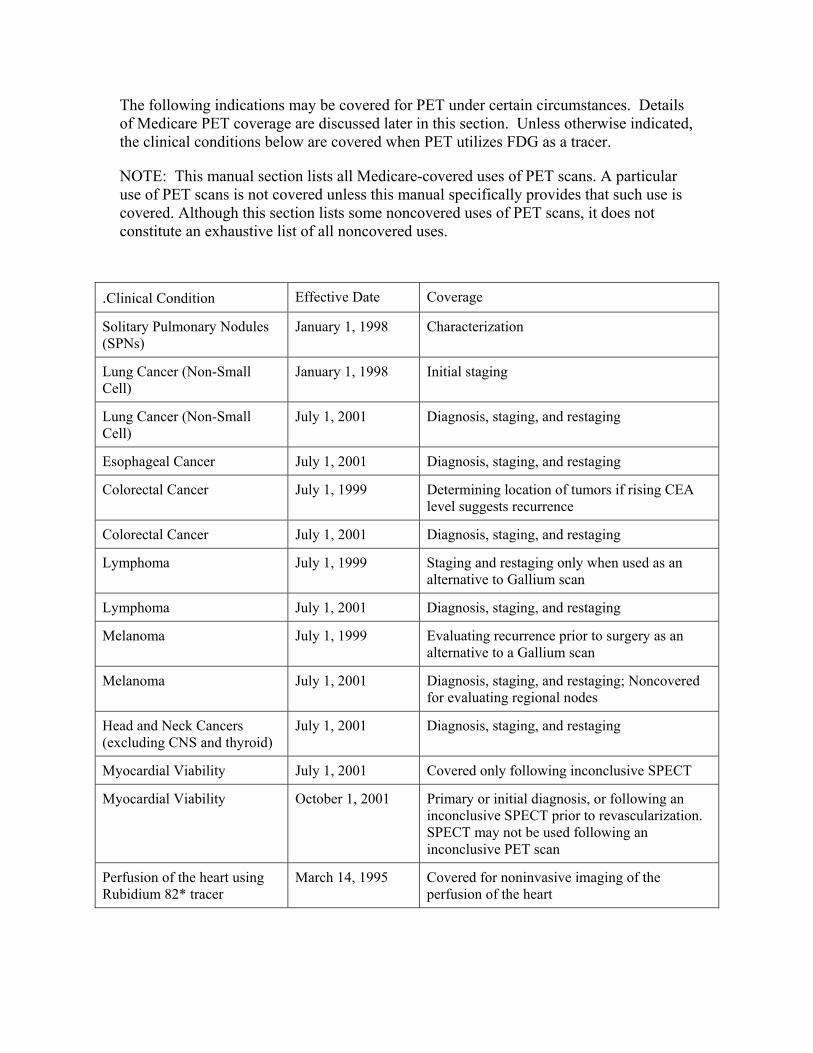
The following indications may be covered for PET under certain circumstances. Details of Medicare PET coverage are discussed later in this section. Unless otherwise indicated, the clinical conditions below are covered when PET utilizes FDG as a tracer.
NOTE: This manual section lists all Medicare-covered uses of PET scans. A particular use of PET scans is not covered unless this manual specifically provides that such use is covered. Although this section lists some noncovered uses of PET scans, it does not constitute an exhaustive list of all noncovered uses.
.Clinical Condition Effective Date Coverage
Solitary Pulmonary Nodules (SPNs)
January 1, 1998 Characterization
Lung Cancer (Non-Small Cell)
January 1, 1998 Initial staging
Lung Cancer (Non-Small Cell)
July 1, 2001 Diagnosis, staging, and restaging
Esophageal Cancer July 1, 2001 Diagnosis, staging, and restaging
Colorectal Cancer July 1, 1999 Determining location of tumors if rising CEA level suggests recurrence
Colorectal Cancer July 1, 2001 Diagnosis, staging, and restaging
Lymphoma July 1, 1999 Staging and restaging only when used as an alternative to Gallium scan
Lymphoma July 1, 2001 Diagnosis, staging, and restaging
Melanoma July 1, 1999 Evaluating recurrence prior to surgery as an alternative to a Gallium scan
Melanoma July 1, 2001 Diagnosis, staging, and restaging; Noncovered for evaluating regional nodes
Head and Neck Cancers (excluding CNS and thyroid)
July 1, 2001 Diagnosis, staging, and restaging
Myocardial Viability July 1, 2001 Covered only following inconclusive SPECT
Myocardial Viability October 1, 2001 Primary or initial diagnosis, or following an inconclusive SPECT prior to revascularization. SPECT may not be used following an inconclusive PET scan
Perfusion of the heart using Rubidium 82* tracer
March 14, 1995 Covered for noninvasive imaging of the perfusion of the heart
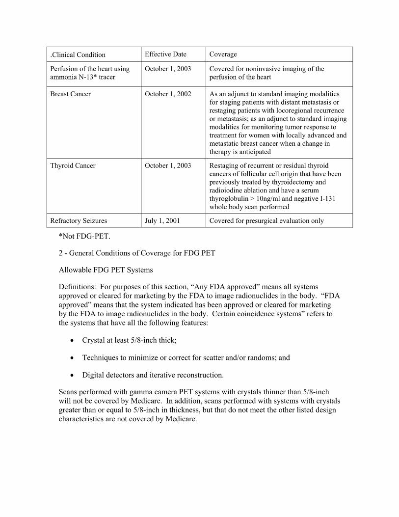
.Clinical Condition Effective Date Coverage
Perfusion of the heart using ammonia N-13* tracer
October 1, 2003 Covered for noninvasive imaging of the perfusion of the heart
Breast Cancer October 1, 2002 As an adjunct to standard imaging modalities for staging patients with distant metastasis or restaging patients with locoregional recurrence or metastasis; as an adjunct to standard imaging modalities for monitoring tumor response to treatment for women with locally advanced and metastatic breast cancer when a change in therapy is anticipated
Thyroid Cancer October 1, 2003 Restaging of recurrent or residual thyroid cancers of follicular cell origin that have been previously treated by thyroidectomy and radioiodine ablation and have a serum thyroglobulin > 10ng/ml and negative I-131 whole body scan performed
Refractory Seizures July 1, 2001 Covered for presurgical evaluation only
*Not FDG-PET.
2 - General Conditions of Coverage for FDG PET
Allowable FDG PET Systems
Definitions: For purposes of this section, “Any FDA approved” means all systems approved or cleared for marketing by the FDA to image radionuclides in the body. “FDA approved” means that the system indicated has been approved or cleared for marketing by the FDA to image radionuclides in the body. Certain coincidence systems” refers to the systems that have all the following features:
• Crystal at least 5/8-inch thick;
• Techniques to minimize or correct for scatter and/or randoms; and
• Digital detectors and iterative reconstruction.
Scans performed with gamma camera PET systems with crystals thinner than 5/8-inch will not be covered by Medicare. In addition, scans performed with systems with crystals greater than or equal to 5/8-inch in thickness, but that do not meet the other listed design characteristics are not covered by Medicare.
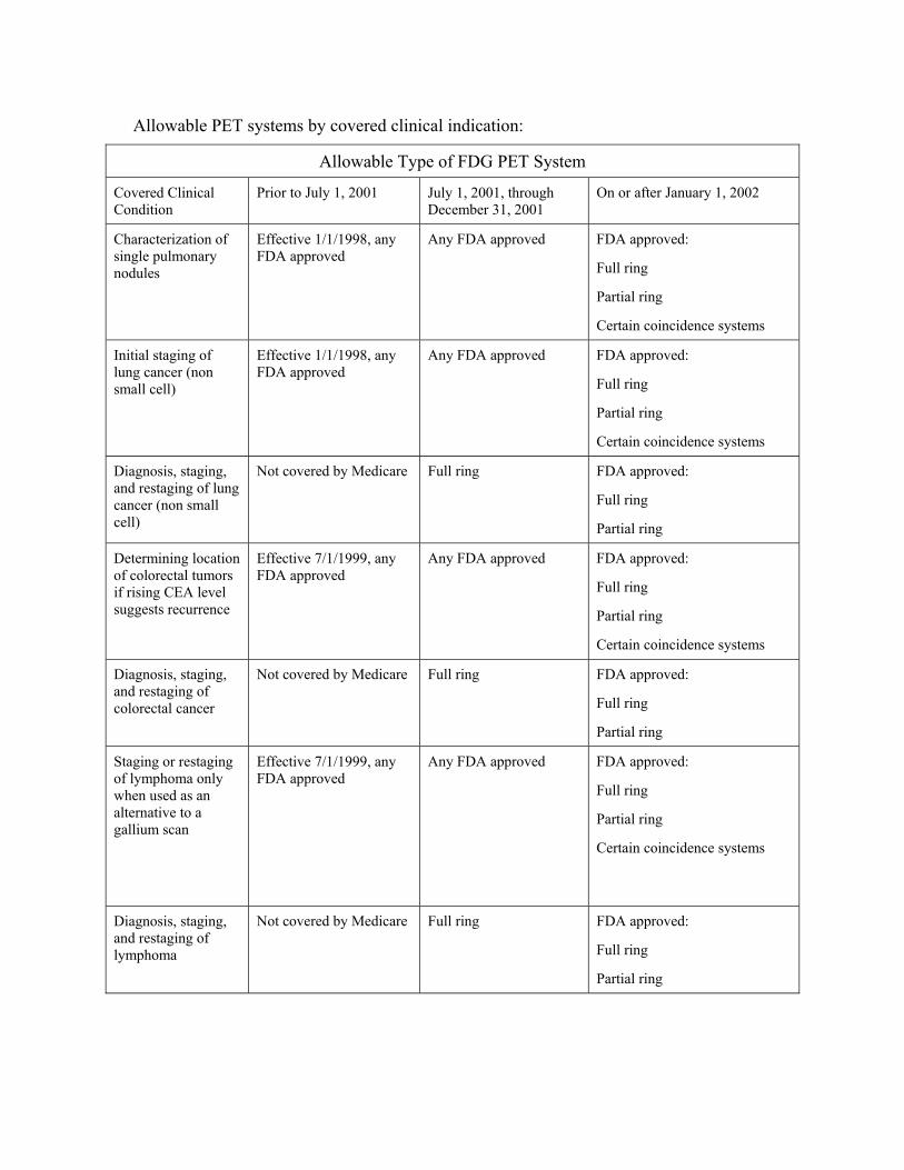
Allowable PET systems by covered clinical indication:
Allowable Type of FDG PET System
Covered Clinical Condition
Prior to July 1, 2001 July 1, 2001, through December 31, 2001
On or after January 1, 2002
Characterization of single pulmonary nodules
Effective 1/1/1998, any FDA approved
Any FDA approved FDA approved:
Full ring
Partial ring
Certain coincidence systems
Initial staging of lung cancer (non small cell)
Effective 1/1/1998, any FDA approved
Any FDA approved FDA approved:
Full ring
Partial ring
Certain coincidence systems
Diagnosis, staging, and restaging of lung cancer (non small cell)
Not covered by Medicare Full ring FDA approved:
Full ring
Partial ring
Determining location of colorectal tumors if rising CEA level suggests recurrence
Effective 7/1/1999, any FDA approved
Any FDA approved FDA approved:
Full ring
Partial ring
Certain coincidence systems
Diagnosis, staging, and restaging of colorectal cancer
Not covered by Medicare Full ring FDA approved:
Full ring
Partial ring
Staging or restaging of lymphoma only when used as an alternative to a gallium scan
Effective 7/1/1999, any FDA approved
Any FDA approved FDA approved:
Full ring
Partial ring
Certain coincidence systems
Diagnosis, staging, and restaging of lymphoma
Not covered by Medicare Full ring FDA approved:
Full ring
Partial ring
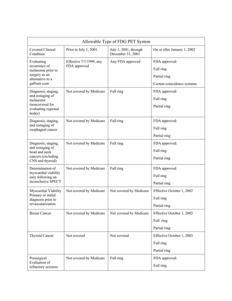
Allowable Type of FDG PET System
Covered Clinical Condition
Prior to July 1, 2001 July 1, 2001, through December 31, 2001
On or after January 1, 2002
Evaluating recurrence of melanoma prior to surgery as an alternative to a gallium scan
Effective 7/1/1999, any FDA approved
Any FDA approved FDA approved:
Full ring
Partial ring
Certain coincidence systems
Diagnosis, staging, and restaging of melanoma (noncovered for evaluating regional nodes)
Not covered by Medicare Full ring FDA approved:
Full ring
Partial ring
Diagnosis, staging, and restaging of esophageal cancer
Not covered by Medicare Full ring FDA approved:
Full ring
Partial ring
Diagnosis, staging, and restaging of head and neck cancers (excluding CNS and thyroid)
Not covered by Medicare Full ring FDA approved:
Full ring
Partial ring
Determination of myocardial viability only following an inconclusive SPECT
Not covered by Medicare Full ring FDA approved:
Full ring
Partial ring
Myocardial Viability Primary or initial diagnosis prior to revascularization
Not covered by Medicare Not covered by Medicare Effective October 1, 2002
Full ring
Partial ring
Breast Cancer Not covered by Medicare Not covered by Medicare Effective October 1, 2002
Full ring
Partial ring
Thyroid Cancer Not covered Not covered Effective October 1, 2003
Full ring
Partial ring
Presurgical Evaluation of refractory seizures
Not covered by Medicare Full ring FDA approved:
Full ring
Allowable Type of FDG PET System
Covered Clinical Condition
Prior to July 1, 2001 July 1, 2001, through December 31, 2001
On or after January 1, 2002
Partial ring
A - Regardless of any other terms or conditions, all uses of FDG PET scans, in order to be covered by the Medicare program, must meet the following general conditions prior to June 30, 2001:
• Submission of claims for payment must include any information Medicare requires to assure that the PET scans performed were: (a) medically necessary, (b) did not unnecessarily duplicate other covered diagnostic tests, and (c) did not involve investigational drugs or procedures using investigational drugs, as determined by the Food and Drug Administration (FDA).
• The PET scan entity submitting claims for payment must keep such patient records as Medicare requires on file for each patient for whom a PET scan claim is made.
B - Regardless of any other terms or conditions, all uses of FDG PET scans, in order to be covered by the Medicare program, must meet the following general conditions as of July 1,2001:
• The provider of the PET scan should maintain on file the doctor’s referral and documentation that the procedure involved only FDA approved drugs and devices, as is normal business practice.
• The ordering physician is responsible for documenting the medical necessity of the study and that it meets the conditions specified in the instructions. The physician should have documentation in the beneficiary’s medical record to support the referral to the PET scan provider.
3 - Covered Indications for PET Scans and Limitations/Requirements for Usage
For all uses of PET relating to malignancies the following conditions apply:
• Diagnosis: PET is covered only in clinical situations in which the PET results may assist in avoiding an invasive diagnostic procedure, or in which the PET results may assist in determining the optimal anatomical location to perform an invasive diagnostic procedure. In general, for most solid tumors, a tissue diagnosis is made prior to the performance of PET scanning. PET scans following a tissue diagnosis are performed for the purpose of staging, not
diagnosis. Therefore, the use of PET in the diagnosis of lymphoma, esophageal, and colorectal cancers as well as in melanoma should be rare.
PET is not covered for other diagnostic uses, and is not covered for screening (testing of patients without specific signs and symptoms of disease).
• Staging and or Restaging: PET is covered in clinical situations in which
1. a. The stage of the cancer remains in doubt after completion of a standard diagnostic workup, including conventional imaging (computed tomography, magnetic resonance imaging, or ultrasound) or
b. The use of PET would also be considered reasonable and necessary if it could potentially replace one or more conventional imaging studies when it is expected that conventional study information is insufficient for the clinical management of the patient and
2. Clinical management of the patient would differ depending on the stage of the cancer identified. PET will be covered for restaging after the completion of treatment for the purpose of detecting residual disease, for detecting suspected recurrence or to determine the extent of a known recurrence. Use of PET would also be considered reasonable and necessary if it could potentially replace one or more conventional imaging studies when it is expected that conventional study information is insufficient for the clinical management of the patient.
• Monitoring: Use of PET to monitor tumor response during the planned course of therapy (i.e. when no change in therapy is being contemplated) is not covered, except for breast cancer. Restaging only occurs after a course of treatment is completed, and this is covered, subject to the conditions above.
NOTE: In the absence of national frequency limitations, contractors should, if necessary, develop frequency requirements on any or all of the indications covered on and after July 1, 2001.
IV. Coverage of PET for Perfusion of the Heart
A. Rubidium 82
4 - Coverage of PET for Perfusion of the Heart Using Rubidium 82
PET scans For any PET scan for which performed at rest or with pharmacological stress used for noninvasive imaging of the perfusion of the heart for the diagnosis and management of patients with known or suspected coronary artery disease using the FDA-approved radiopharmaceutical Rubidium 82 (Rb 82) are covered, provided the requirements below are met.
• The PET scan, whether at rest alone, or rest with stress, is performed in place of, but not in addition to, a single photon emission computed tomography (SPECT); or
• The PET scan, whether at rest alone or rest with stress, is used following a SPECT that was found to be inconclusive. In these cases, the PET scan must have been considered necessary in order to determine what medical or surgical intervention is required to treat the patient. (For purposes of this requirement, an inconclusive test is a test(s) whose results are equivocal, technically uninterpretable, or discordant with a patient’s other clinical data and must be documented in the beneficiary’s file.)
• For any PET scan for which Medicare payment is claimed for dates of services prior to July 1, 2001, the claimant must submit additional specified information on the claim form (including proper codes and/or modifiers), to indicate the results of the PET scan. The claimant must also include information on whether the PET scan was done after an inconclusive noninvasive cardiac test. The information submitted with respect to the previous noninvasive cardiac test must specify the type of test done prior to the PET scan and whether it was inconclusive or unsatisfactory. These explanations are in the form of special G codes used for billing PET scans using Rb 82.
Beginning July 1, 2001, claims should be submitted with the appropriate codes.
B. Ammonia N-13
Effective for services performed on or after October 1, 2003, PET scans performed at rest or with pharmacological stress used for noninvasive imaging of the perfusion of the heart for the diagnosis and management of patients with known or suspected coronary artery disease using the FDA-approved radiopharmaceutical ammonia N-13 are covered, provided the requirements below are met.
Requirements:
• The PET scan, whether at rest alone, or rest with stress, is performed in place of, but not in addition to, a single photon emission computed tomography (SPECT); or
• The PET scan, whether at rest alone or rest with stress, is used following a SPECT that was found to be inconclusive. In these cases, the PET scan must have been considered necessary in order to determine what medical or surgical intervention is required to treat the patient. (For purposes of this requirement, an inconclusive test is a test whose results are equivocal, technically uninterpretable, or discordant with a patient’s other clinical data and must be documented in the beneficiary’s file.)
5 - Coverage of FDG PET for Lung Cancer
The coverage for FDG PET for lung cancer, has been expanded. Beginning July 1, 2001 usage of FDG PET for lung cancer has been expanded to include diagnosis, staging, and restaging (see section III) of the disease.
• Medicare covers regional FDG PET chest scans, on any FDA approved scanner, for the characterization of single pulmonary nodules (SPNs). The primary purpose of such characterization should be to determine the likelihood of malignancy in order to plan future management and treatment for the patient.
Beginning July 1, 2001, documentation should be maintained in the beneficiary’s medical file at the referring physician’s office to support the medical necessity of the procedure, as is normal business practice. The following are required documentation.
• There must be evidence of primary tumor. Claims for regional PET chest scans for characterizing SPNs should include evidence of the initial detection of a primary lung tumor, usually by computed tomography (CT). This should include, but is not restricted to, a report on the results of such CT or other detection method, indicating an indeterminate or possibly malignant lesion, not exceeding four centimeters (cm) in diameter.
• PET scan claims must include the results of concurrent thoracic CT (as noted above) which is necessary for anatomic information, in order to ensure that the PET scan is properly coordinated with other diagnostic modalities.
• In cases of serial evaluation of SPNs using both CT and regional PET chest scanning, such PET scans will not be covered if repeated within 90 days following a negative PET scan.
NOTE: A tissue sampling procedure (TSP) is not routinely covered in the case of a negative PET scan for characterization of SPNs, since the patient is presumed not to have a malignant lesion, based upon the PET scan results. When there has been a negative PET, the provider must submit additional information with the claim to support the necessity of a TSP, for review by the Medicare contractor.
Medicare approved coverage of FDG PET for initial staging of non-small-cell lung carcinoma (NSCLC).
Limitations: This service is covered only when the primary cancerous lung tumor has been pathologically confirmed; claims for PET must include a statement or other evidence of the detection of such primary lung tumor. The evidence should include, but is not restricted to, a surgical pathology report which documents the presence of an NSCLC. Whole body PET scan results and results of concurrent computed tomography (CT) and follow-up lymph node biopsy must be properly coordinated with other diagnostic modalities. Claims must include both:
• The results of concurrent thoracic CT, necessary for anatomic information, and
• The results of any lymph node biopsy performed to finalize whether the patient will be a surgical candidate. The ordering physician is responsible for providing this biopsy result to the PET facility.
NOTE: Where the patient is considered a surgical candidate, (given the presumed absence of metastatic NSCLC unless medical review supports a determination of medical necessity of a biopsy) a lymph node biopsy will not be covered in the case of a negative CT and negative PET. A lymph node biopsy will be covered in all other cases, i.e., positive CT + positive PET; negative CT + positive PET; positive CT + negative PET.
Beginning July 1, 2001 - Medicare covers FDG PET for diagnosis, staging, and restaging of NSCLC. Documentation should be maintained in the beneficiary’s medical file to support the medical necessity of the procedure, as is normal business practice.
Requirements: PET is covered in either/or both of the following circumstances:
• Diagnosis - PET is covered only in clinical situations in which the PET results may assist in avoiding an invasive diagnostic procedure, or in which the PET results may assist in determining the optimal anatomical location to perform an invasive diagnostic procedure. In general, for most solid tumors, a tissue diagnosis is made prior to the performance of PET scanning. PET scans following a tissue diagnosis are performed for the purpose of staging, not diagnosis. Therefore, the use of PET in the diagnosis of lymphoma, esophageal, and colorectal cancers as well as in melanoma should be rare.
• Staging and/or Restaging - PET is covered in clinical situations in which
1. Either:
a. The stage of the cancer remains in doubt after completion of a standard diagnostic workup, including conventional imaging (computed tomography, magnetic resonance imaging, or ultrasound) or
b. The use of PET would also be considered reasonable and necessary if it could potentially replace one or more conventional imaging studies when it is expected that conventional study information is insufficient for the clinical management of the patient, and
2. Clinical management of the patient would differ depending on the stage of the cancer identified. PET will be covered for restaging after the completion of treatment for the purpose of detecting residual disease, for detecting suspected recurrence or to determine the extent of a known recurrence. Use of PET would also be considered reasonable and necessary if it could potentially replace one or more conventional imaging studies when it is expected that conventional study information is insufficient for the clinical management of the patient.
Documentation should be maintained in the beneficiary’s medical record at the referring physician’s office to support the medical necessity of the procedure, as is normal business practice.
6 - Coverage of FDG PET for Esophageal Cancer
Beginning July 1, 2001, Medicare covers FDG PET for the diagnosis, staging, and restaging of esophageal cancer. Medical evidence is present to support the use of FDG PET in presurgical staging of esophageal cancer.
Requirements: PET is covered in either/or both of the following circumstances:
• Diagnosis - PET is covered only in clinical situations in which the PET results may assist in avoiding an invasive diagnostic procedure, or in which the PET results may assist in determining the optimal anatomical location to perform an invasive diagnostic procedure. In general, for most solid tumors, a tissue diagnosis is made prior to the performance of PET scanning. PET scans following a tissue diagnosis are performed for the purpose of staging, not diagnosis. Therefore, the use of PET in the diagnosis of lymphoma, esophageal and colorectal cancers as well as in melanoma should be rare.
• Staging and/or Restaging - PET is covered in clinical situations in which
1. a. The stage of the cancer remains in doubt after completion of a standard diagnostic workup, including conventional imaging (computed tomography, magnetic resonance imaging, or ultrasound) or
b. The use of PET would also be considered reasonable and necessary if it could potentially replace one or more conventional imaging studies when it is expected that conventional study information is insufficient for the clinical management of the patient, and
2. Clinical management of the patient would differ depending on the stage of the cancer identified. PET will be covered for restaging after the completion of treatment for the purpose of detecting residual disease, for detecting suspected recurrence, or to determine the extent of a known recurrence. Use of PET would also be considered reasonable and necessary if it could potentially replace one or more conventional imaging studies when it is expected that conventional study information is insufficient for the clinical management of the patient.
Documentation is required in the beneficiary’s medical record at the referring physician’s office to support the medical necessity of the procedure, as is normal business practice.
7 - Coverage of FDG PET for Colorectal Cancer
Medicare coverage of FDG PET for colorectal cancer where there is a rising level of carcinoembryonic antigen (CEA) was effective July 1, 1999, through June 30, 2001.
Beginning July 1, 2001, usage of FDG PET for colorectal cancer has been expanded to include diagnosis, staging, and restaging of the disease(see part III).
A - Effective July 1, 1999 - Medicare covers FDG PET for patients with recurrent colorectal carcinomas which are suggested by rising levels of the biochemical tumor marker CEA.
1 - Frequency Limitations: Whole body PET scans for assessment of recurrence of colorectal cancer cannot be ordered more frequently than once every 12 months unless medical necessity documentation supports a separate re-elevation of CEA within this period.
2 - Limitations: Because this service is covered only in those cases in which there has been a recurrence of colorectal tumor, claims for PET should include a statement or other evidence of previous colorectal tumor, through June 30, 2001.
B - Beginning July 1, 2001 - Medicare coverage has been expanded for colorectal carcinomas for diagnosis, staging and re-staging. New medical evidence supports the use of FDG PET as a useful tool in determining the presence of hepatic/extrahepatic metastases in the primary staging of colorectal carcinoma, prior to selecting a treatment regimen. Use of FDG PET is also supported in evaluating recurrent colorectal cancer beyond the limited presentation of a rising CEA level where the patient presents clinical signs or symptoms of recurrence.
Requirements: PET is covered in either/both of the following circumstances:
• Diagnosis - PET is covered only in clinical situations in which the PET results may assist in avoiding an invasive diagnostic procedure, or in which the PET results may assist in determining the optimal anatomical location to perform an invasive diagnostic procedure. In general, for most solid tumors, a tissue diagnosis is made prior to the performance of PET scanning. PET scans following a tissue diagnosis are performed for the purpose of staging, not diagnosis. Therefore, the use of PET in the diagnosis of lymphoma, esophageal, and colorectal cancers as well as in melanoma should be rare.
• Staging and/or Restaging - PET is covered in clinical situations in which
1. a. The stage of the cancer remains in doubt after completion of a standard diagnostic workup, including conventional imaging (computed tomography, magnetic resonance imaging, or ultrasound) or
b. The use of PET would also be considered reasonable and necessary if it could potentially replace one or more conventional imaging studies when it is expected that conventional study information is insufficient for the clinical management of the patient, and
2. Clinical management of the patient would differ depending on the stage of the cancer identified. PET will be covered for restaging after the
completion of treatment for the purpose of detecting residual disease, for detecting suspected recurrence, or to determine the extent of a known recurrence. Use of PET would also be considered reasonable and necessary if it could potentially replace one or more conventional imaging studies when it is expected that conventional study information is insufficient for the clinical management of the patient.
• Documentation that these conditions are met should be maintained by the referring physician in the beneficiary’s medical record, as is normal business practice.
8 - Coverage of FDG PET for Lymphoma
Medicare coverage of FDG PET to stage and re-stage lymphoma as alternative to a Gallium scan, was effective July 1, 1999. Beginning July 1, 2001, usage of FDG PET for lymphoma has been expanded to include Diagnosis, staging, and restaging (see section III) of the disease.
A - Effective July 1, 1999 - FDG PET is covered for the staging and restaging of lymphoma a follows:
• FDG PET is covered only for staging or follow-up restaging of lymphoma. Claims must include a statement or other evidence of previous diagnosis of lymphoma when used as an alternative to a Gallium scan.
• To ensure that the PET scan is properly coordinated with other diagnostic modalities, claims must include the results of concurrent computed tomography (CT) and/or other diagnostic modalities when they are necessary for additional anatomic information.
• In order to ensure that the PET scan is covered only as an alternative to a Gallium scan, no PET scan may be covered in cases where it is done within 50 days of a Gallium scan done by the same facility where the patient has remained during the 50-day period. Gallium scans done by another facility less than 50 days prior to the PET scan will not be counted against this screen. The purpose of this screen is to assure that PET scans are covered only when done as an alternative to a Gallium scan within the same facility. The CMS is aware that, in order to assure proper patient care, the treating physician may conclude that previously performed Gallium scans are either inconclusive or not sufficiently reliable.
• Frequency Limitation for Restaging: PET scans will be allowed for restaging no sooner than 50 days following the last staging PET scan or Gallium scan, unless sufficient evidence is presented to convince the Medicare contractor that the restaging at an earlier date is medically necessary. Since PET scans for restaging are generally done following cycles of chemotherapy, and since such cycles usually take at least 8 weeks, CMS believes this screen will adequately prevent medically unnecessary scans while allowing some adjustments for unusual cases.
In all cases, the determination of the medical necessity for a PET scan for re-staging lymphoma is the responsibility of the local Medicare contractor..
B - The Medicare program has broadened coverage of FDG PET for the Diagnosis, staging, and restaging of lymphoma.
Requirements: PET is covered in either/both of the following circumstances:
• Diagnosis - PET is covered only in clinical situations in which the PET results may assist in avoiding an invasive diagnostic procedure, or in which the PET results may assist in determining the optimal anatomical location to perform an invasive diagnostic procedure. In general, for most solid tumors, a tissue diagnosis is made prior to the performance of PET scanning. PET scans following a tissue diagnosis are performed for the purpose of staging, not diagnosis. Therefore, the use of PET in the diagnosis of lymphoma, esophageal, and colorectal cancers as well as in melanoma should be rare.
• Staging and/or Restaging - PET is covered in clinical situations in which (
1. a. The stage of the cancer remains in doubt after completion of a standard diagnostic workup, including conventional imaging (computed tomography, magnetic resonance imaging, or ultrasound) or
b. The use of PET would also be considered reasonable and necessary if it could potentially replace one or more conventional imaging studies when it is expected that conventional study information is insufficient for the clinical management of the patient, and
2. Clinical management of the patient would differ depending on the stage of the cancer identified. PET will be covered for restaging after the completion of treatment for the purpose of detecting residual disease, for detecting suspected recurrence, or to determine the extent of a known recurrence. Use of PET would also be considered reasonable and necessary if it could potentially replace one or more conventional imaging studies when it is expected that conventional study information is insufficient for the clinical management of the patient.
• Documentation that these conditions are met should be maintained by the referring physician in the beneficiary’s medical record, as is normal business practice.
9 - Coverage of FDG PET for Melanoma
Medicare covered the evaluation of recurrent melanoma prior to surgery when used as an alternative to a Gallium scan, effective July 1, 1999. For services furnished on or after July 1, 2001, FDG PET is covered for the diagnosis, staging, and restaging of malignant melanoma (see part III). FDG PET is not covered for the use of evaluating regional nodes in melanoma patients.
A - In the case of patients with recurrent melanoma prior to surgery, FDG PET (when used as an alternative to a Gallium scan) is covered for tumor evaluation.
Frequency Limitations: Whole body PET scans cannot be ordered more frequently than once every 12 months, unless medical necessity documentation, maintained in the beneficiaries medical record, supports the specific need for anatomic localization of possible recurrent tumor within this period.
Limitations: The FDG PET scan is covered only as an alternative to a Gallium scan. PET scans can not be covered in cases where it is done within 50 days of a Gallium scan done by the same PET facility where the patient has remained under the care of the same facility during the 50-day period. Gallium scans done by another facility less than 50 days prior to the PET scan will not be counted against this screen. The purpose of this screen is to assure that PET scans are covered only when done as an alternative to a Gallium scan within the same facility. The CMS is aware that, in order to assure proper patient care, the treating physician may conclude that previously performed Gallium scans are either inconclusive or not sufficiently reliable to make the determination covered by this provision. Therefore, CMS will apply this 50-day rule only to PET scans done by the same facility that performed the Gallium scan.
Beginning July 1, 2001 - documentation should be maintained in the beneficiary’s medical file at the referring physician’s office to support the medical necessity of the procedure, as is normal business practice.
B - FDG PET scan coverage for the Diagnosis, staging, and restaging of melanoma (not the evaluation regional nodes) has been broadened.
Limitations: PET scans are not covered for the evaluation of regional nodes.
Requirements: PET is covered in either/both of the following circumstances:
Diagnosis: PET is covered only in clinical situations in which the PET results may assist in avoiding an invasive diagnostic procedure, or in which the PET results may assist in determining the optimal anatomical location to perform an invasive diagnostic procedure. In general, for most solid tumors, a tissue diagnosis is made prior to the performance of PET scanning. PET scans following a tissue diagnosis are performed for the purpose of staging, not diagnosis. Therefore, the use of PET in the diagnosis of lymphoma, esophageal, and colorectal cancers as well as in melanoma should be rare.
Staging and/or Restaging - PET is covered in clinical situations in which
1. a. The stage of the cancer remains in doubt after completion of a standard diagnostic workup, including conventional imaging (computed tomography, magnetic resonance imaging, or ultrasound) or
b. The use of PET would also be considered reasonable and necessary if it could potentially replace one or more conventional imaging studies when it is expected that conventional study information is insufficient for the clinical management of the patient, and
2. Clinical management of the patient would differ depending on the stage of the cancer identified.
PET will be covered for restaging after the completion of treatment for the purpose of detecting residual disease, for detecting suspected recurrence, or to determine the extent of a known recurrence. Use of PET would also be considered reasonable and necessary if it could potentially replace one or more conventional imaging studies when it is expected that conventional study information is insufficient for the clinical management of the patient.
Documentation that these conditions are met should be maintained by the referring physician in the beneficiary’s medical file, as is normal business practice.
10 - Coverage of FDG PET for Head and Neck Cancers (Cancers of the Central Nervous System (CNS) and thyroid are not covered)
Medicare will provide coverage for cancer of the head and neck, excluding the central nervous system (CNS) and thyroid. The head and neck cancers encompass a diverse set of malignancies of which the majority is squamous cell carcinomas. Patients may present with metastases to cervical lymph nodes but conventional forms of diagnostic imaging fail to identify the primary tumor. Patients that present with cancer of the head and neck are left with two options either to have a neck dissection or to have radiation of both sides of the neck with random biopsies. PET scanning attempts to reveal the site of primary tumor to prevent the adverse effects of random biopsies or unneeded radiation.
Limitations: PET scans for head and neck cancers are not covered for CNS or thyroid cancers.
PET is covered in either/or both of the following circumstances:
• Diagnosis - PET is covered only in clinical situations in which the PET results may assist in avoiding an invasive diagnostic procedure, or in which the PET results may assist in determining the optimal anatomical location to perform an invasive diagnostic procedure. In general, for most solid tumors, a tissue diagnosis is made prior to the performance of PET scanning. PET scans following a tissue diagnosis are performed for the purpose of staging, not diagnosis. Therefore, the use of PET in the diagnosis of lymphoma, esophageal, and colorectal cancers as well as in melanoma should be rare.
• Staging and/or Restaging - PET is covered in clinical situations in which
1. a. The stage of the cancer remains in doubt after completion of a standard diagnostic workup, including conventional imaging (computed tomography, magnetic resonance imaging, or ultrasound) or
b. The use of PET would also be considered reasonable and necessary if it could potentially replace one or more conventional imaging studies when it is expected that conventional study information is insufficient for the clinical management of the patient, and
2. Clinical management of the patient would differ depending on the stage of the cancer identified.
PET will be covered for restaging after the completion of treatment for the purpose of detecting residual disease, for detecting suspected recurrence, or to determine the extent of a known recurrence. Use of PET would also be considered reasonable and necessary if it could potentially replace one or more conventional imaging studies when it is expected that conventional study information is insufficient for the clinical management of the patient.
Documentation that these conditions are met should be maintained by the referring physician in the beneficiary’s medical record, as is normal business practice.
11 - Coverage of FDG PET for Myocardial Viability
The identification of patients with partial loss of heart muscle movement or hibernating myocardium is important in selecting candidates with compromised ventricular function to determine appropriateness for revascularization. Diagnostic tests such as FDG PET distinguish between dysfunctional but viable myocardial tissue and scar tissue in order to affect management decisions in patients with ischemic cardiomyopathy and left ventricular dysfunction.
FDG PET is covered for the determination of myocardial viability following an inconclusive SPECT from July 1, 2001, through September 30, 2002. Only full ring PET scanners are covered from July 1, 2001, through December 31, 2001. However, as of January 1, 2002, full and partial ring scanners are covered.
Beginning October 1, 2002, Medicare covers FDG PET for the determination of myocardial viability as a primary or initial diagnostic study prior to revascularization, or following an inconclusive SPECT. Studies performed by full and partial ring scanners are covered.
Limitations: In the event that a patient has received a single photon computed tomography test (SPECT) with inconclusive results, a PET scan may be covered. However, if a patient received a FDG PET study with inconclusive results, a follow up SPECT is not covered.
Documentation that these conditions are met should be maintained by the referring physician in the beneficiary’s medical record, as is normal business practice
12 - Coverage of FDG-PET for Refractory Seizures
Beginning July 1, 2001 - Medicare will cover FDG-PET for presurgical evaluation for the purpose of localization of a focus of refractory seizure activity.
Limitations: Covered only for presurgical evaluation.
Documentation that these conditions are met should be maintained by the referring physician in the beneficiary’s medical record, as is normal business practice.
13 - Breast Cancer
Beginning October 1, 2002, Medicare covers FDG PET as an adjunct to other imaging modalities for staging patients with distant metastasis, or restaging patients with locoregional recurrence or metastasis. Monitoring treatment of a breast cancer tumor when a change in therapy is contemplated is also covered as an adjunct to other imaging modalities.
Limitations: Effective October 1, 2002, Medicare continues to have a national noncoverage determination for initial diagnosis of breast cancer and staging of axillary lymph nodes. Medicare coverage for staging patients with distant metastasis or restaging patients with locoregional recurrence or metastasis; and for monitoring tumor response to treatment for women with locally advanced and metastatic breast cancer when a change in therapy is anticipated, is only covered as an adjunct to other imaging modalities.
Documentation that these conditions are met should be maintained by the referring physician in the beneficiary’s medical record, as is normal business practice.
14 - Thyroid Cancer
1. Effective for services furnished on or after October 1, 2003, Medicare covers the use of FDG PET for thyroid cancer only for restaging of recurrent or residual thyroid cancers of follicular cell origin that have been previously treated by thyroidectomy and radioiodine ablation and have a serum thyroglobulin > 10ng/ml and negative I-131 whole body scan performed.
2. All other uses of FDG PET in the diagnosis and treatment of thyroid cancer remain noncovered.
15 - Soft Tissue Sarcoma – NOT COVERED
Following a thorough review of the scientific literature, including a technology assessment on the topic, Medicare maintains its national noncoverage determination for all uses of FDG PET for soft tissue sarcoma.
16 - Dementia and Neurogenerative Diseases – NOT COVERED
Following a thorough review of the scientific literature, including a technology assessment on the topic and consideration by the Medicare Coverage Advisory
Committee, Medicare maintains its national noncoverage determination for all uses of FDG-PET for the diagnosis and management of dementia or other neurogenerative diseases
220.7 - Xenon Scan
(Rev. 1, 10-03-03)
CIM 50-27
Program payment may be made for this diagnostic procedure which involves perfusion lung imaging with 133 xenon. However, review for evidence of abuse which might include absence of reasonable indications, inappropriate sequence, or excessive number or kinds of procedures used in the care of individual patients.
220.8 - Nuclear Radiology Procedure
(Rev. 1, 10-03-03)
CIM 50-30
Nuclear radiology procedures, including nuclear examinations performed with mobile radiological equipment, are covered if reasonable and necessary for the individual patient. Although these procedures may not be widely used, they are generally accepted. Review claims for these procedures for evidence of abuse that might include absence of reasonable indications, inappropriate sequence, or excessive number or kinds of procedures used in the care of individual patients.
220.9 - Digital Subtraction Angiography
(Rev. 1, 10-03-03)
CIM 50-43
Digital subtraction angiography (DSA) is a diagnostic imaging technique that applies computer technology to fluoroscopy for the purpose of visualizing the same vascular structures observable with conventional angiography. Since the radiographic contrast material can be injected into a vein rather than an artery, the procedure reduces the risk to patients, and can be done on an outpatient basis.
Contractors should be alert to possible increases in utilization of DSA over conventional angiographic procedures, as well as to the fact that ordinarily patients should not require inpatient hospitalization solely to perform the procedure.
Payment for DSA should not exceed, and may be less than, that being paid for conventional angiographic techniques.
220.10 - Portable Hand-Held X-Ray Instrument
(Rev. 1, 10-03-03)
CIM 50-48
This low intensity x-ray imaging device is a light weight portable hand-held instrument using a low level isotope as its penetrating energy source. It can picture any part of the human anatomy that can be inserted in the space between the energy source and the viewing mechanism. The device can be useful in making an immediate diagnosis in the following settings: isolated areas, accident scenes, sports events and emergency rooms. It is also useful in the following instances where fluoroscopy would ordinarily be used: localization of foreign bodies, selected surgical procedures and the evaluation of premature or low birth weight infants. The use of the portable hand-held x-ray instrument as an imaging device is covered under Medicare. It should be reimbursed as part of the physicians’ professional service, and no additional charge should be allowed.
220.11 - Thermography
(Rev. 1, 10-03-03)
CIM 50-5
Thermography is the measurement of self-emanating infrared radiation that reveals temperature variations at the surface of the body. The thermographic device senses body temperature and demonstrates areas of differing heat emission by producing brightly colored patterns. Each color represents a specific temperature level. Interpretation of these color patterns according to designated anatomic distribution is thought to aid in diagnosing a vast array of diseases.
Thermography for any indication (including breast lesions which were excluded from Medicare coverage on July 20, 1984) is excluded from Medicare coverage because the available evidence does not support this test as a useful aid in the diagnosis or treatment of illness or injury. Therefore, it is not considered effective. This exclusion was published as a CMS Final Notice in the “Federal Register” on November 20, 1992.
220.12 - Single Photon Emission Computed Tomograph (SPECT)
(Rev. 1, 10-03-03)
CIM 50-58
SPECT acquires information on the concentration of radionuclides introduced into the patient’s body. It is useful in the diagnosis of several clinical conditions including:
• Stress fracture
• Spondylosis
• Infection (e.g., discitis)
• Tumor (e.g., osteoid osteoma)
• Analyze blood flow to an organ, as in the case of myocardial viability
• Differentiate ischemic heart disease from dilated cardiomyopathy.
Frequency limitations: Contractor discretion.
In the case of myocardial viability, FDG PET may be used following a SPECT that was found to be inconclusive. However, SPECT may not be used following an inconclusive FDG PET performed to evaluate myocardial viability.
220.13 - Percutaneous Image-Guided Breast Biopsy
(Rev. 1, 10-03-03)
CIM 50-59
Percutaneous image-guided breast biopsy is a method of obtaining a breast biopsy through a percutaneous incision by employing image guidance systems. Image guidance systems may be either ultrasound or stereotactic.
The Breast Imaging Reporting and Data System (or BIRADS system) employed by the American College of Radiology provides a standardized lexicon with which radiologists may report their interpretation of a mammogram. The BIRADS grading of mammograms is as follows: Grade I-Negative, Grade II-Benign finding, Grade III-Probably benign, Grade IV-Suspicious abnormality, and Grade V-Highly suggestive of malignant neoplasm.
A - Nonpalpable Breast Lesions
Effective January 1, 2003, Medicare covers percutaneous image-guided breast biopsy using stereotactic or ultrasound imaging for a radiographic abnormality that is nonpalpable and is graded as a BIRADS III, IV, or V.
B - Palpable Breast Lesions
Effective January 1, 2003, Medicare covers percutaneous image guided breast biopsy using stereotactic or ultrasound imaging for palpable lesions that are difficult to biopsy using palpation alone. Contractors have the discretion to decide what types of palpable lesions are difficult to biopsy using palpation.
230 - Renal and Genitourinary System - ESRD Services
230.1 - Treatment of Kidney Stones
(Rev. 1, 10-03-03)
CIM 35-81
Traditional approaches for the treatment of kidney stones are the surgical technique nephrectomy (or nephrotomy) and endoscopic treatments via the urethra. In the last few years, several new approaches in the surgical management of upper urinary tract kidney stones have been developed, among them invasive and noninvasive lithotripsy techniques.
In addition to the traditional surgical/endoscopic techniques for the treatment of kidney stones, the following lithotripsy techniques are also covered for services rendered on or after March 15, 1985.
A - Extracorporeal Shock Wave Lithotripsy
Extracorporeal Shock Wave Lithotripsy (ESWL) is a noninvasive method of treating kidney stones using a device called a lithotriptor. The lithotriptor uses shock waves generated outside of the body to break up upper urinary tract stones. It focuses the shock waves specifically on stones under x-ray visualization, pulverizing them by repeated shocks. ESWL is covered under Medicare for use in the treatment of upper urinary tract kidney stones.
B - Percutaneous Lithotripsy
Percutaneous lithotripsy (or nephrolithotomy) is an invasive method of treating kidney stones by using ultrasound, electrohydraulic or mechanical lithotripsy. A probe is inserted through an incision in the skin directly over the kidney and applied to the stone. A form of lithotripsy is then used to fragment the stone. Mechanical or electrohydraulic lithotripsy may be used as an alternative or adjunct to ultrasonic lithotripsy. Percutaneous lithotripsy of kidney stones by ultrasound or by the related techniques of electrohydraulic or mechanical lithotripsy is covered under Medicare.
The following is covered for services rendered on or after January 16, 1988.
C - Transurethral Ureteroscopic Lithotripsy
Transurethral ureteroscopic lithotripsy is a method of fragmenting and removing ureteral and renal stones through a cystoscope. The cystoscope is inserted through the urethra into the bladder. Catheters are passed through the scope into the opening where the ureters enter the bladder. Instruments passed through this opening into the ureters are used to manipulate and ultimately disintegrate stones, using either mechanical crushing, transcystoscopic electrohydraulic shock waves, ultrasound, or laser. Transurethral
ureteroscopic lithotripsy for the treatment of urinary tract stones of the kidney or ureter is covered under Medicare.
230.2 - Uroflowmetric Evaluations
(Rev. 1, 10-03-03)
CIM 50-33
Uroflowmetric evaluations (also referred to as urodynamic voiding or urodynamic flow studies) are covered under Medicare for diagnosing various urological dysfunctions, including bladder outlet obstructions.
230.3 - Sterilization
(Rev. 1, 10-03-03)
CIM 35-11
A - Covered Conditions
• Payment may be made only where sterilization is a necessary part of the treatment of an illness or injury, e.g., removal of a uterus because of a tumor or removal of diseased ovaries.
• Sterilization of a mentally retarded beneficiary is covered if it is a necessary part of the treatment of an illness or injury, (bilateral oophorectomy), or bilateral orchidectomy in a case of cancer of the prostate. The contractor denies claims when the pathological evidence of the necessity to perform any such procedures to treat an illness or injury is absent; and .
• Monitor such surgeries closely and obtain the information needed to determine whether in fact the surgery was performed as a means of treating an illness or injury or only to achieve sterilization.
B - Noncovered Conditions
• Elective hysterectomy, tubal ligation, and vasectomy, if the stated reason for these procedures is sterilization;
• A sterilization that is performed because a physician believes another pregnancy would endanger the overall general health of the woman is not considered to be reasonable and necessary for the diagnosis or treatment of illness or injury within the meaning of §1862(a)(1) of the Act. The same conclusion would apply where the sterilization is performed only as a measure to prevent the possible development of, or effect on, a mental condition should the individual become pregnant; and sterilization of a mentally retarded person where the purpose is to prevent conception, rather than the treatment of an illness or injury.
230.4 - Diagnosis and Treatment of Impotence
(Rev. 1, 10-03-03)
CIM 35-24
Program payment may be made for diagnosis and treatment of sexual impotence. Impotence is a failure of a body part for which the diagnosis, and frequently the treatment, require medical expertise. Depending on the cause of the condition, treatment may be surgical; e.g., implantation of a penile prosthesis, or nonsurgical; e.g., medical or psychotherapeutic treatment. Since causes and, therefore, appropriate treatment vary, if abuse is suspected it may be necessary to request documentation of appropriateness in individual cases. If treatment is furnished to patients (other than hospital inpatients) in connection with a mental condition, apply the psychiatric service limitation described in the Medicare General Information, Eligibility, and Entitlement Manual, Chapter 3.
230.5 - Gravlee Jet Washer
(Rev. 1, 10-03-03)
CIM 50-4
The Gravlee Jet Washer is a sterile, disposable, diagnostic device for detecting endometrial cancer. The use of this device is indicated where the patient exhibits clinical symptoms or signs suggestive of endometrial disease, such as irregular or heavy vaginal bleeding.
Program payment cannot be made for the washer or the related diagnostic services when furnished in connection with the examination of an asymptomatic patient. Payment for routine physical checkups is precluded under the statute. (See §1862(a)(7) of the Act.) (See the Medicare Benefit Policy Manual, Chapter 16, “General Exclusions From Coverage,” §90).
230.6 - Vabra Aspirator
(Rev. 1, 10-03-03)
CIM 50-10
The VABRA aspirator is a sterile, disposable, vacuum aspirator which is used to collect uterine tissue for study to detect endometrial carcinoma. The use of this device is indicated where the patient exhibits clinical symptoms or signs suggestive of endometrial disease, such as irregular or heavy vaginal bleeding.
Program payment cannot be made for the aspirator or the related diagnostic services when furnished in connection with the examination of an asymptomatic patient. Payment for routine physical checkups is precluded under the statute (§1862(a)(7) of the Act).
Cross-reference:
See the Medicare Benefit Policy Manual, Chapter 16, “General Exclusions From Coverage,” §90, and §230.5 of this manual.
230.7 - Water Purification and Softening Systems Used in Conjunction With Home Dialysis
(Rev. 1, 10-03-03)
CIM 55-1
A - Water Purification Systems
Water used for home dialysis should be chemically free of heavy trace metals and/or organic contaminants that could be hazardous to the patient. It should also be as free of bacteria as possible but need not be biologically sterile. Since the characteristics of natural water supplies in most areas of the country are such that some type of water purification system is needed, such a system used in conjunction with a home dialysis (either peritoneal or hemodialysis) unit is covered under Medicare.
There are two types of water purification systems that will satisfy these requirements:
• Deionization - The removal of organic substances, mineral salts of magnesium and calcium (causing hardness), compounds of fluoride and chloride from tap water using the process of filtration and ion exchange; or
• Reverse Osmosis - The process used to remove impurities from tap water utilizing pressure to force water through a porous membrane.
Use of both a deionization unit and reverse osmosis unit in series, theoretically to provide the advantages of both systems, has been determined medically unnecessary since either system can provide water which is both chemically and bacteriologically pure enough for acceptable use in home dialysis. In addition, spare deionization tanks are not covered since they are essentially a precautionary supply rather than a current requirement for treatment of the patient.
Activated carbon filters used as a component of water purification systems to remove unsafe concentrations of chlorine and chloramines are covered when prescribed by a physician.
B - Water Softening System
Except as indicated below, a water softening system used in conjunction with home dialysis is excluded from coverage under Medicare as not being reasonable and necessary within the meaning of §1862(a)(1) of the Act. Such a system, in conjunction with a home dialysis unit, does not adequately remove the hazardous heavy metal contaminants (such as arsenic) which may be present in trace amounts.
A water softening system may be covered when used to pretreat water to be purified by a reverse osmosis (RO) unit for home dialysis where:
The manufacturer of the RO unit has set standards for the quality of water entering the RO (e.g., the water to be purified by the RO must be of a certain quality if the unit is to perform as intended);
The patients water is demonstrated to be of a lesser quality than required; and
The softener is used only to soften water entering the RO unit, and thus, used only for dialysis. (The softener need not actually be built into the RO unit, but must be an integral part of the dialysis system.)
C - Developing Need When a Water Softening System is Replaced with a Water Purification Unit in an Existing Home Dialysis System
The medical necessity of water purification units must be care fully developed when they replace water softening systems in existing home dialysis systems. A purification system may be ordered under these circumstances for a number of reasons. For example, changes in the medical community’s opinions regarding the quality of water necessary for safe dialysis may lead the physician to decide the quality of water previously used should be improved, or the water quality itself may have deteriorated. Patients may have dialyzed using only an existing water softener previous to Medicare ESRD coverage because of inability to pay for a purification system. On the other hand, in some cases, the installation of a purification system is not medically necessary. Thus, when such a case comes to the contractor’s attention, the contractor asks the physician to furnish the reason for the changes. Supporting documentation, such as the suppliers recommendations or water analysis, may be required. All such cases should be reviewed by the contractor’s medical consultants.
Cross reference:
The Medicare Benefit Policy Manual, Chapter 15, “Covered Medical and Other Health Services,” §110.
230.8 - Non-Implantable Pelvic Flood Electrical Stimulator
(Rev. 1, 10-03-03)
CIM 60-24
Non-implantable pelvic floor electrical stimulators provide neuromuscular electrical stimulation through the pelvic floor with the intent of strengthening and exercising pelvic floor musculature. Stimulation is generally delivered by vaginal or anal probes connected to an external pulse generator.
The methods of pelvic floor electrical stimulation vary in location, stimulus frequency (Hz), stimulus intensity or amplitude (mA), pulse duration (duty cycle), treatments per
day, number of treatment days per week, length of time for each treatment session, overall time period for device use and between clinic and home settings. In general, the stimulus frequency and other parameters are chosen based on the patient’s clinical diagnosis.
Pelvic floor electrical stimulation with a non-implantable stimulator is covered for the treatment of stress and/or urge urinary incontinence in cognitively intact patients who have failed a documented trial of pelvic muscle exercise (PME) training.
A failed trial of PME training is defined as no clinically significant improvement in urinary continence after completing 4 weeks of an ordered plan of pelvic muscle exercises designed to increase periurethral muscle strength.
230.9 - Cryosurgery of Prostate
(Rev. 1, 10-03-03)
CIM 35-96
Cryosurgery of the prostate gland, also known as cryosurgical ablation of the prostate (CSAP), destroys prostate tissue by applying extremely cold temperatures in order to reduce the size of the prostate gland. It is safe and effective, as well as medically necessary and appropriate, as primary treatment for patients with clinically localized prostate cancer, Stages T1-T3.
Cryosurgery of the prostate as a salvage therapy is not covered for any services performed prior to June 30, 2001.
Salvage Cryosurgery Of Prostate After Radiation Failure. Salvage cryosurgery of the prostate for recurrent cancer is medically necessary and appropriate only for those patients with localized disease who:
1 - Have failed a trial of radiation therapy as their primary treatment; and
2 - Meet one of the following conditions: Stage T2B or below, Gleason score < 9, PSA < 8 ng/mL.
Cryosurgery as salvage therapy is therefore not covered under Medicare after failure of other therapies as the primary treatment. Cryosurgery as salvage is only covered after the failure of a trial of radiation therapy, under the conditions noted above.
230.10 - Incontinence Control Devices
(Rev. 1, 10-03-03)
CIM 65-9
A - Mechanical/Hydraulic Incontinence Control Devices
Mechanical/hydraulic incontinence control devices are accepted as safe and effective in the management of urinary incontinence in patients with permanent anatomic and neurologic dysfunctions of the bladder. This class of devices achieves control of urination by compression of the urethra. The materials used and the success rate may vary somewhat from device to device. Such a device is covered when its use is reasonable and necessary for the individual patient.
B - Collagen Implant
A collagen implant which is injected into the submucosal tissues of the urethra and/or the bladder neck and into tissues adjacent to the urethra, is a prosthetic device used in the treatment of stress urinary incontinence resulting from intrinsic sphincter deficiency (ISD). ISD is a cause of stress urinary incontinence in which the urethral sphincter is unable to contract and generate sufficient resistance in the bladder, especially during stress maneuvers.
Prior to collagen implant therapy, a skin test for collagen sensitivity must be administered and evaluated over a 4-week period.
In male patients, the evaluation must include a complete history and physical examination and a simple cystometrogram to determine that the bladder fills and stores properly. The patient then is asked to stand upright with a full bladder and to cough or otherwise exert abdominal pressure on his bladder. If the patient leaks, the diagnosis of ISD is established.
In female patients, the evaluation must include a complete history and physical examination (including a pelvic exam) and a simple cystometrogram to rule out abnormalities of bladder compliance and abnormalities of urethral support. Following that determination, an abdominal leak point pressure (ALLP) test is performed. Leak point pressure, stated in cm H2O, is defined as the intra-abdominal pressure at which leakage occurs from the bladder (around a catheter) when the bladder has been filled with a minimum of 150 cc fluid. If the patient has an ALLP of less than 100 cm H2O, the diagnosis of ISD is established.
To use a collagen implant, physicians must have urology training in the use of a cystoscope and must complete a collagen implant training program.
Coverage of a collagen implant, and the procedure to inject it, is limited to the following types of patients with stress urinary incontinence due to ISD:
• Male or female patients with congenital sphincter weakness secondary to conditions such as myelomeningocele or epispadias;
• Male or female patients with acquired sphincter weakness secondary to spinal cord lesions;
• Male patients following trauma, including prostatectomy and/or radiation; and
• Female patients without urethral hypermobility and with abdominal leak point pressures of 100 cm H2O or less.
Patients whose incontinence does not improve with five injection procedures (five separate treatment sessions) are considered treatment failures, and no further treatment of urinary incontinence by collagen implant is covered. Patients who have a reoccurrence of incontinence following successful treatment with collagen implants in the past (e.g., 6-12 months previously) may benefit from additional treatment sessions. Coverage of additional sessions may be allowed but must be supported by medical justification. See the Medicare Benefit Policy Manual, Chapter 15, “Covered Medical and Other Health Services,” §120. (See §230.8.)
230.11 - Diagnostic Pap Smears
(Rev. 1, 10-03-03)
CIM 50-20
The guide in §190.2 applies.
230.12 - Dimethyl Sulfoxide (DMSO)
(Rev. 1, 10-03-03)
CIM 45-23
DMSO is an industrial solvent produced as a chemical byproduct of paper production from wood pulp. The Food and Drug Administration has determined that the only purpose for which DMSO is safe and effective for humans is in the treatment of the bladder condition, interstitial cystitis. Therefore, the use of DMSO for all other indications is not considered to be reasonable and necessary. Payment may be made for its use only when reasonable and necessary for a patient in the treatment of interstitial cystitis.
230.13 - Peridex CAPD Filter Set
(Rev. 1, 10-03-03)
CIM 55-2
The Peridex Filter Set is used by home continuous ambulatory peritoneal dialysis (CAPD) patients. The Peridex Filter Set is designed to provide sterile filtration during infusion of the dialysis solution in a beneficiary’s peritoneal cavity; included in the filter set is a bacterial filter designed to block peritonitis-causing organisms and thus reduce the incidence of peritonitis.
Based upon advice of our medical consultants, CMS has determined that the Peridex CAPD Filter Set cannot be covered at this time by Medicare because it has not yet been shown to be safe and effective in preventing peritonitis.
230.14 - Ultrafiltration Monitor
(Rev. 1, 10-03-03)
CIM 55-3
The Ultrafiltration Monitor is designed to reduce the clinical risks of overfiltration and underfiltration during hemodialysis. Overfiltration is the removal of too much fluid from body tissues and underfiltration is removal of too little fluid.
Covered
Ultrafiltration and ultrafiltration monitoring as a component of hemodialysis has an established and critical role in maintaining the well-being of ESRD patients and is a covered service. The Ultrafiltration Monitor is covered under the Medicare program when it is used to calculate fluid rates for those recipients who present difficult fluid management problems. Determine the medical necessity of this device on a case-by-case basis.
Not Covered
Ultrafiltration, independent of conventional dialysis, is considered experimental, and technology exclusively designed for this purpose is not covered under Medicare.
230.15 - Electrical Continence Aid
(Rev. 1, 10-03-03)
CIM 65-2
Not Covered
An electrical continence aid is a device consisting of a plastic plug, molded into the shape of the patient’s anal canal which contains two implanted electrodes that are connected by a wire to a small portable generator. An electrical current is produced which stimulates the anal musculature to cause a contraction sufficient to hold the plug in while allowing the patient to ambulate without incontinence.
Electrical continence aids are in the experimental stage of development and there is no valid scientific documentation of their effectiveness and safety. Therefore, they are not covered under Medicare since they cannot be considered to be reasonable and necessary for the treatment of an illness or injury or to improve the functioning of a malformed body member as required by §1862(a)(1) of the Act.
230.16 - Bladder Stimulators (Pacemakers)
(Rev. 1, 10-03-03)
CIM 65-11
Not Covered
There are a number of devices available to induce emptying of the urinary bladder by using electrical current which forces the muscles of the bladder to contract. These devices (commonly known as bladder stimulators or pacemakers) are characterized by the implantation of electrodes in the wall of the bladder, the rectal cones, or the spinal cord. While these treatments may effectively empty the bladder, the issue of safety involving the initiation of infection, erosion, placement, and material selection has not been resolved. Further, some facilities previously using electronic emptying have stopped using this method due to the pain experienced by the patient.
The use of spinal cord electrical stimulators, rectal electrical stimulators, and bladder wall stimulators is not considered reasonable and necessary. Therefore, no program payment may be made for these devices or for their implant.
230.17 - Urinary Drainage Bags
(Rev. 1, 10-03-03)
CIM 65-17
Urinary collection and retention system are covered as prosthetic devices that replace bladder function in the case of permanent urinary incontinence. Urinary drainage bags that can be used either as bedside or leg drainage bags may be either multi-use or single use systems. Both the multi-use and the single use bags have a system that prevents urine backflow. However, the single use system is non-drainable. There is insufficient evidence to support the medical necessity of a single use system bag rather than the multi-use bag. Therefore, a single use drainage system is subject to the same coverage parameters as the multi-use drainage bags.
230.18 - Sacral Nerve Stimulation for Urinary Incontinence
(Rev. 1, 10-03-03)
CIM 65-18
Effective January 1, 2002, sacral nerve stimulation is covered for the treatment of urinary urge incontinence, urgency-frequency syndrome, and urinary retention. Sacral nerve stimulation involves both a temporary test stimulation to determine if an implantable stimulator would be effective and a permanent implantation in appropriate candidates. Both the test and the permanent implantation are covered.
The following limitations for coverage apply to all three indications:
• Patient must be refractory to conventional therapy (documented behavioral, pharmacologic and/or surgical corrective therapy) and be an appropriate surgical candidate such that implantation with anesthesia can occur.
• Patients with stress incontinence, urinary obstruction, and specific neurologic diseases (e.g., diabetes with peripheral nerve involvement) which are associated with secondary manifestations of the above three indications are excluded.
• Patient must have had a successful test stimulation in order to support subsequent implantation. Before a patient is eligible for permanent implantation, he/she must demonstrate a 50 percent or greater improvement through test stimulation. Improvement is measured through voiding diaries.
• Patient must be able to demonstrate adequate ability to record voiding diary data such that clinical results of the implant procedure can be properly evaluated.
230.19 - Levocarnitine for Use in the Treatment of Carnitine Deficiency in ESRD Patients
(Rev. 1, 10-03-03)
CIM 45-32
Carnitine is a naturally occurring substance that functions in the transport of the long-chain fatty acids for energy production by the body. Deficiency can occur due to a congenital defect in synthesis or utilization, or from dialysis. The causes of carnitine deficiency in hemodialysis patients include dialytic loss, reduced renal synthesis and reduced dietary intake.
Intravenous levocarnitine, for one of the following indications, will only be covered for those ESRD patients who have been on dialysis for a minimum of three months.
Patients must have documented carnitine deficiency, defined as a plasma free carnitine level < 40 micromol/L (determined by a professionally accepted method as recognized in current literature), along with signs and symptoms of:
• Erthropoietin-resistant anemia (persistent hermatocrit < 30 percent with treatment) that has not responded to standard erthropoietin dosage (that which is considered clinically appropriate to treat the particular patient) with iron replacement, and for which other causes have been investigated and adequately treated, or
• Hypertension on hemodialysis that interferes with delivery of the intended dialysis despite application of usual measures deemed appropriate (e.g., fluid management). Such episodes of hypotension must have occurred during at least 2 dialysis treatments in a 30-day period.
Continued use of levocarnitine will not be covered if improvement has not been demonstrated within six months of initiation of treatment. All other indications for levocarnitine are noncovered in the ESRD population.
For a patient currently receiving intravenous levocarnitine, Medicare will cover continued treatment if:
• Levocarnitine has been administered to treat erythropoietin-resistent anemia (persistent hematocrit < 30 percent with treatment) that has not responded to standard erythropoietin dosage (that which is considered clinically appropriate to treat the particular patient) with iron replacement, and for which other causes have been investigated and adequately treated, or hypotension on hemodialysis that interferes with delivery of the intended dialysis despite application of usual measures deemed appropriate (e.g., fluid management) and such episodes of hypotension occur during at least 2 dialysis treatments in a 30-day period; and
• The patient’s medical record documents a pre-dialysis plasma free carnitine level < 40 micromol/L prior to the initiation of treatment; or
• The treating physician certifies (documents in the medical record) that in his/her judgment, if treatment with the levocarnitine is discontinued, the patient’s pre-dialysis carnitine level would fall below 40 micromol/L and the patient would have recurrent erythropoietin-resistant-anemia or intradialytic hypotension.
240 - Respiratory System
(Rev. 1, 10-03-03)
240.1 - Lung Volume Reduction Surgery (Reduction Pneumoplasty)
(Rev. 3, 11-04-03) Lung volume reduction surgery (LVRS) or reduction pneumoplasty, also referred to as lung shaving or lung contouring, is performed on patients with severe emphysema in order to allow the remaining compressed lung to expand, and thus, improve respiratory function.
Covered Indications
Medicare-covered LVRS approaches are limited to bilateral excision of a damaged lung with stapling performed via median sternotomy or video-assisted thoracoscopic surgery.
1. National Emphysema Treatment Trial (NETT) participants (effective for services performed on or after August 11, 1997): Medicare provides coverage to those beneficiaries who are participating in the NETT trial for all services integral to the study and for which the Medicare statute does not prohibit coverage.
2. Medicare will only consider LVRS reasonable and necessary when all of the following requirements are met (effective for services performed on or after January 1, 2004):
a. The patient satisfies all the criteria outlined below:
Assessment Criteria
Consistent with emphysema
BMI, ≤ 31.1 kg/m2 (men) or ≤ 32.3 kg/m2 (women)
History and physical examination
Stable with ≤20 mg prednisone (or equivalent) qd
Radiographic High Resolution Computer Tomography (HRCT) scan evidence of bilateral emphysema
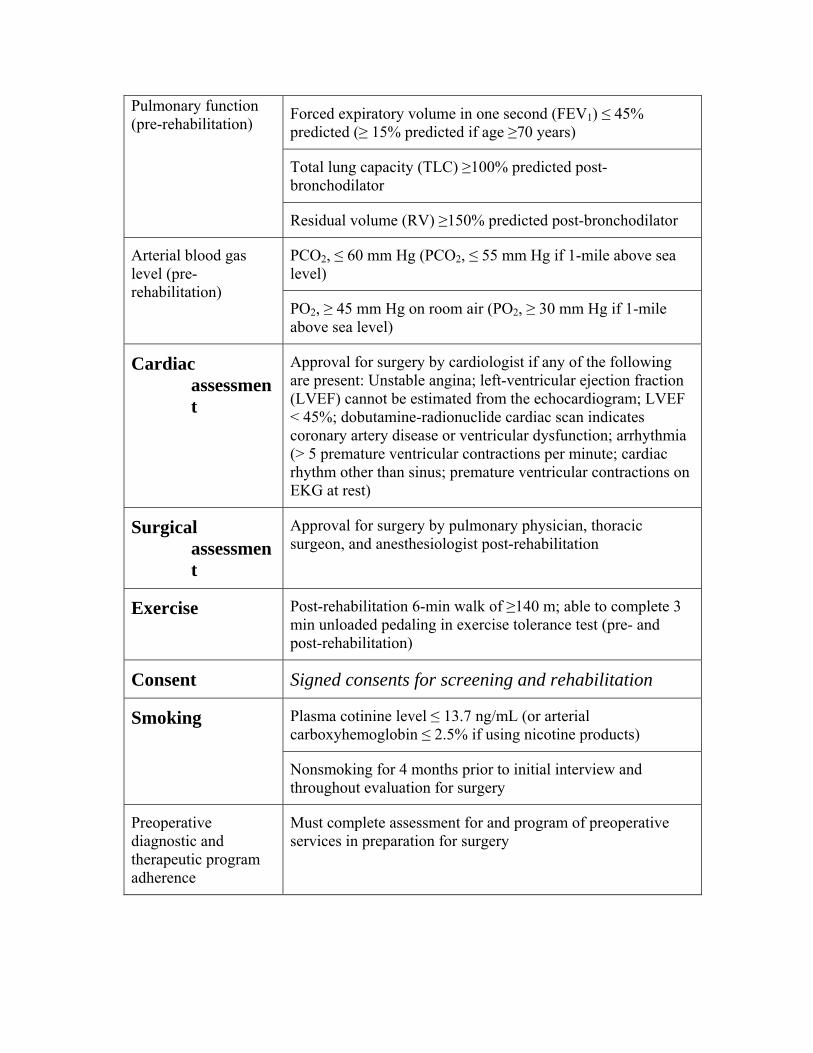
Forced expiratory volume in one second (FEV1) ≤ 45% predicted (≥ 15% predicted if age ≥70 years)
Total lung capacity (TLC) ≥100% predicted post-bronchodilator
Pulmonary function (pre-rehabilitation)
Residual volume (RV) ≥150% predicted post-bronchodilator
PCO2, ≤ 60 mm Hg (PCO2, ≤ 55 mm Hg if 1-mile above sea level)
Arterial blood gas level (pre-rehabilitation)
PO2, ≥ 45 mm Hg on room air (PO2, ≥ 30 mm Hg if 1-mile above sea level)
Cardiac assessment
Approval for surgery by cardiologist if any of the following are present: Unstable angina; left-ventricular ejection fraction (LVEF) cannot be estimated from the echocardiogram; LVEF < 45%; dobutamine-radionuclide cardiac scan indicates coronary artery disease or ventricular dysfunction; arrhythmia (> 5 premature ventricular contractions per minute; cardiac rhythm other than sinus; premature ventricular contractions on EKG at rest)
Surgical assessment
Approval for surgery by pulmonary physician, thoracic surgeon, and anesthesiologist post-rehabilitation
Exercise Post-rehabilitation 6-min walk of ≥140 m; able to complete 3 min unloaded pedaling in exercise tolerance test (pre- and post-rehabilitation)
Consent Signed consents for screening and rehabilitation
Plasma cotinine level ≤ 13.7 ng/mL (or arterial carboxyhemoglobin ≤ 2.5% if using nicotine products)
Smoking
Nonsmoking for 4 months prior to initial interview and throughout evaluation for surgery
Preoperative diagnostic and therapeutic program adherence
Must complete assessment for and program of preoperative services in preparation for surgery
b. In addition, the patient must have:
o Severe upper lobe predominant emphysema (as defined by radiologist assessment of upper lobe predominance on CT scan), or
o Severe non-upper lobe emphysema with low exercise capacity.
Patients with low exercise capacity are those whose maximal exercise capacity is at or below 25 watts for women and 40 watts (w) for men after completion of the preoperative therapeutic program in preparation for LVRS. Exercise capacity is measured by incremental, maximal, symptom-limited exercise with a cycle ergometer utilizing 5 or 10 watt/minute ramp on 30% oxygen after 3 minutes of unloaded pedaling.
c. The surgery must be performed at facilities that were identified by the National Heart, Lung, and Blood Institute to meet the thresholds for participation in the NETT, and at sites that have been approved by Medicare as lung transplant facilities. These facilities are listed on our Web site at www.cms.hhs.gov/coverage/lvrsfacility.pdf. The CMS is currently working to develop accreditation standards for facilities to perform LVRS and when implemented, will consider LVRS to be reasonable and necessary only at accredited facilities.
d. The surgery must be preceded and followed by a program of diagnostic and therapeutic services consistent with those provided in the NETT and designed to maximize the patient's potential to successfully undergo and recover from surgery. The program must include a 6- to 10-week series of at least 16, and no more than 20, preoperative sessions, each lasting a minimum of 2 hours. It must also include at least 6, and no more than 10, postoperative sessions, each lasting a minimum of 2 hours, within 8 to 9 weeks of the LVRS. This program must be consistent with the care plan developed by the treating physician following performance of a comprehensive evaluation of the patient's medical, psychosocial and nutritional needs, be consistent with the preoperative and postoperative services provided in the NETT, and arranged, monitored, and performed under the coordination of the facility where the surgery takes place.
Noncovered Indications
1. LVRS is not covered in any of the following clinical circumstances:
a. Patient characteristics carry a high risk for perioperative morbidity and/or mortality;
b. The disease is unsuitable for LVRS;
c. Medical conditions or other circumstances make it likely that the patient will be unable to complete the preoperative and postoperative pulmonary diagnostic and therapeutic program required for surgery;
d. The patient presents with FEVı ≤ 20% of predicted value, and either homogeneous distribution of emphysema on CT scan, or carbon monoxide
diffusing capacity of ≤ 20% of predicted value (high-risk group identified October 2001 by the NETT); or
e. The patient satisfies the criteria outlined above in section 2(a), and has severe, non-upper lobe emphysema with high exercise capacity. High exercise capacity is defined as a maximal workload at the completion of the preoperative diagnostic and therapeutic program that is above 25 w for women and 40 w for men (under the measurement conditions for cycle ergometry specified above).
2. All other indications for LVRS not otherwise specified remain noncovered.
240.2 - Home Use of Oxygen
(Rev. 1, 10-03-03)
CIM 60-4
A - General
Medicare coverage of home oxygen and oxygen equipment under the durable medical equipment (DME) benefit (see §1861(s)(6)of the Act) is considered reasonable and necessary only for patients with significant hypoxemia who meet the medical documentation, laboratory evidence, and health conditions specified in subsections B, C, and D. This section also includes special coverage criteria for portable oxygen systems. Finally, a statement on the absence of coverage of the professional services of a respiratory therapist under the DME benefit is included in subsection F.
B - Medical Documentation
Initial claims for oxygen services must include a completed Form CMS-484 (Certificate of Medical Necessity: Oxygen) to establish whether coverage criteria are met and to ensure that the oxygen services provided are consistent with the physician’s prescription or other medical documentation. The treating physician’s prescription or other medical documentation must indicate that other forms of treatment (e.g., medical and physical therapy directed at secretions, bronchospasm and infection) have been tried, have not been sufficiently successful, and oxygen therapy is still required. While there is no substitute for oxygen therapy, each patient must receive optimum therapy before long-term home oxygen therapy is ordered. Use Form CMS-484 for recertifications. (See the Medicare Program Integrity Manual, Chapter 5, for completion of Form CMS-484.)
The medical and prescription information in section B of Form CMS-484 can be completed only by the treating physician, the physician’s employee, or another clinician (e.g., nurse, respiratory therapist, etc.) as long as that person is not the DME supplier. Although hospital discharge coordinators and medical social workers may assist in arranging for physician-prescribed home oxygen, they do not have the authority to prescribe the services. Suppliers may not enter this information. While this section may be completed by non-physician clinician or a physician employee, it must be reviewed and the Form CMS-484 signed by the attending physician.
A physician’s certification of medical necessity for oxygen equipment must include the results of specific testing before coverage can be determined.
Claims for oxygen must also be supported by medical documentation in the patient’s record. Separate documentation is used with electronic billing. This documentation may be in the form of a prescription written by the patient’s attending physician who has recently examined the patient (normally within a month of the start of therapy) and must specify:
• A diagnosis of the disease requiring home use of oxygen;
• The oxygen flow rate; and
• An estimate of the frequency, duration of use (e.g., 2 liters per minute, 10 minutes per hour, 12 hours per day), and duration of need (e.g., 6 months or lifetime).
NOTE: A prescription for “Oxygen PRN” or “Oxygen as needed” does not meet this last requirement. Neither provides any basis for determining if the amount of oxygen is reasonable and necessary for the patient.
A member of the carrier’s medical staff should review all claims with oxygen flow rates of more than four liters per minute before payment can be made.
The attending physician specifies the type of oxygen delivery system to be used (i.e., gas, liquid, or concentrator) by signing the completed Form CMS-484. In addition, the supplier or physician may use the space in section C for written confirmation of additional details of the physician’s order. The additional order information contained in section C may include the means of oxygen delivery (mask, nasal, cannula, etc.), the specifics of varying flow rates, and/or the noncontinuous use of oxygen as appropriate. The physician confirms this order information with their signature in section D.
New medical documentation written by the patient’s attending physician must be submitted to the carrier in support of revised oxygen requirements when there has been a change in the patient’s condition and need for oxygen therapy.
Carriers are required to conduct periodic, continuing medical necessity reviews on patients whose conditions warrant these reviews and on patients with indefinite or extended periods of necessity as described in the Medicare Program Integrity Manual, Chapter 5, “Items and Services Having Special DMERC Review Considerations.” When indicated, carriers may also request documentation of the results of a repeat arterial blood gas or oximetry study.
NOTE: Section 4152 of OBRA 1990 requires earlier recertification and retesting of oxygen patients who begin coverage with an arterial blood gas result at or above a partial pressure of 55 or an arterial oxygen saturation percentage at or above 89. (See the Medicare Claims Processing Manual, Chapter 20, “Durable Medical Equipment, Prosthetics and Orthotics, and Supplies (DMEPOS),” §100.2.3, for certification and retesting schedules.)
C - Laboratory Evidence
Initial claims for oxygen therapy must also include the results of a blood gas study that has been ordered and evaluated by the attending physician. This is usually in the form of a measurement of the partial pressure of oxygen (PO2) in arterial blood. A measurement of arterial oxygen saturation obtained by ear or pulse oximetry, however, is also acceptable when ordered and evaluated by the attending and performed under his or her supervision or when performed by a qualified provider or supplier of laboratory services. When the arterial blood gas and the oximetry studies are both used to document the need for home oxygen therapy and the results are conflicting, the arterial blood gas study is the preferred source of documenting medical need. A DME supplier is not considered a qualified provider or supplier of laboratory services for purposes of these guidelines. This prohibition does not extend to the results of blood gas test conducted by a hospital certified to do such tests. The conditions under which the laboratory tests are performed must be specified in writing and submitted with the initial claim, i.e., at rest, during exercise, or during sleep.
The preferred sources of laboratory evidence are, existing physician and/or hospital records that reflect the patient’s medical condition. Since it is expected that virtually all patients who qualify for home oxygen coverage for the first time under these guidelines have recently been discharged from a hospital where they submitted to arterial blood gas tests, the carrier needs to request that such test results be submitted in support of their initial claims for home oxygen. If more than one arterial blood gas test is performed during the patient’s hospital stay, the test result obtained closest to, but no earlier than two days prior to the hospital discharge date is required as evidence of the need for home oxygen therapy.
For those patients whose initial oxygen prescription did not originate during a hospital stay, blood gas studies should be done while the patient is in the chronic stable state, i.e., not during a period of an acute illness or an exacerbation of their underlying disease.
Carriers may accept an attending physician’s statement of recent hospital test results for a particular patient, when appropriate, in lieu of copies of actual hospital records.
A repeat arterial blood gas study is appropriate when evidence indicates that an oxygen recipient has undergone a major change in their condition relevant to home use of oxygen. If the carrier has reason to believe that there has been a major change in the patient’s physical condition, it may ask for documentation of the results of another blood gas or oximetry study.
D - Health Conditions
Coverage is available for patients with significant hypoxemia in the chronic stable state, i.e, not during a period of acute illness or an exacerbation of their underlying disease, if:
1. The attending physician has determined that the patient has a health condition outlined in subsection D.1,
2. The patient meets the blood gas evidence requirements specified in subsection D.3, and
3. The patient has appropriately tried other treatment without complete success. (See subsection B.)
1 - Conditions for Which Oxygen Therapy May Be Covered
• A severe lung disease, such as chronic obstructive pulmonary disease, diffuse interstitial lung disease, cystic fibrosis, bronchiectasis, widespread pulmonary neoplasm, or
• Hypoxia-related symptoms or findings that might be expected to improve with oxygen therapy. Examples of these symptoms and findings are pulmonary hypertension, recurring congestive heart failure due to chronic cor pulmonale, erythrocytosis, impairment of the cognitive process, nocturnal restlessness, and morning headache.
2 - Conditions for Which Oxygen Therapy Is Not Covered
• Angina pectoris in the absence of hypoxemia. This condition is generally not the result of a low oxygen level in the blood, and there are other preferred treatments;
• Breathlessness without cor pulmonale or evidence of hypoxemia. Although intermittent oxygen use is sometimes prescribed to relieve this condition, it is potentially harmful and psychologically addicting;
• Severe peripheral vascular disease resulting in clinically evident desaturation in one or more extremities. There is no evidence that increased PO2 improves the oxygenation of tissues with impaired circulation; or
• Terminal illnesses that do not affect the lungs.
3 - Covered Blood Gas Values
If the patient has a condition specified in subsection D.1, the carrier must review the medical documentation and laboratory evidence that has been submitted for a particular patient (see subsections B and C) and determine if coverage is available under one of the three group categories outlined below.
(a) - Group I - Except as modified in subsection d, coverage is provided for patients with significant hypoxemia evidenced by any of the following:
• An arterial PO2 at or below 55 mm Hg, or an arterial oxygen saturation at or below 88 percent, taken at rest, breathing room air.
• An arterial PO2 at or below 55 mm Hg, or an arterial oxygen saturation at or below 88 percent, taken during sleep for a patient who demonstrates an arterial PO2 at or above 56 mm Hg, or an arterial oxygen saturation at or
above 89 percent, while awake; or a greater than normal fall in oxygen level during sleep (a decrease in arterial PO2 more than 10 mm Hg, or decrease in arterial oxygen saturation more than 5 percent) associated with symptoms or signs reasonably attributable to hypoxemia (e.g., impairment of cognitive processes and nocturnal restlessness or insomnia). In either of these cases, coverage is provided only for use of oxygen during sleep, and then only one type of unit will be covered. Portable oxygen ,therefore, would not be covered in this situation.
• An arterial PO2 at or below 55 mm Hg or an arterial oxygen saturation at or below 88 percent, taken during exercise for a patient who demonstrates an arterial PO2 at or above 56 mm Hg, or an arterial oxygen saturation at or above 89 percent, during the day while at rest. In this case, supplemental oxygen is provided for during exercise if there is evidence the use of oxygen improves the hypoxemia that was demonstrated during exercise when the patient was breathing room air.
(b) - Group II - Except as modified in subsection d, coverage is available for patients whose arterial PO2 is 56-59 mm Hg or whose arterial blood oxygen saturation is 89 percent, if there is evidence of:
• Dependent edema suggesting congestive heart failure;
• Pulmonary hypertension or cor pulmonale, determined by measurement of pulmonary artery pressure, gated blood pool scan, echocardiogram, or “P” pulmonale on EKG (P wave greater than 3 mm in standard leads II, III, or AVF); or
• Erythrocythemia with a hematocrit greater than 56 percent.
(c) - Group III - Except as modified in subsection d, carriers must apply a rebuttable presumption that a home program of oxygen use is not medically necessary for patients with arterial PO2 levels at or above 60 mm Hg, or arterial blood oxygen saturation at or above 90 percent. In order for claims in this category to be reimbursed, the carrier’s reviewing physician needs to review any documentation submitted in rebuttal of this presumption and grant specific approval of the claims. The CMS expects few claims to be approved for coverage in this category.
(d) - Variable Factors That May Affect Blood Gas Values - In reviewing the arterial PO2 levels and the arterial oxygen saturation percentages specified in subsections D. 3.a, b and c, the carrier’s medical staff must take into account variations in oxygen measurements that may result from such factors as the patient’s age, the altitude level, or the patient’s decreased oxygen carrying capacity.
E - Portable Oxygen Systems
A patient meeting the requirements specified below may qualify for coverage of a portable oxygen system either (1) by itself or (2) to use in addition to a stationary oxygen system. Portable oxygen is not covered when it is provided only as a backup to a stationary oxygen system. A portable oxygen system is covered for a particular patient if:
• The claim meets the requirements specified in subsections A-D, as appropriate; and
• The medical documentation indicates that the patient is mobile in the home and would benefit from the use of a portable oxygen system in the home. Portable oxygen systems are not covered for patients who qualify for oxygen solely based on blood gas studies obtained during sleep
F - Respiratory Therapists
Respiratory therapists’ services are not covered under the provisions for coverage of oxygen services under the Part B durable medical equipment benefit as outlined above. This benefit provides for coverage of home use of oxygen and oxygen equipment, but does not include a professional component in the delivery of such services.
(See §280.1, and the Medicare Benefit Policy Manual, Chapter 15, “Covered Medical and Other Health Services,” §110)
240.3 - Heat Treatment, Including the Use of Diathermy and Ultra-Sound for Pulmonary Conditions
(Rev. 1, 10-03-03)
CIM 35-3
Not Covered
There is no physiological rationale or valid scientific documentation of effectiveness of diathermy or ultrasound heat treatments for asthma, bronchitis, or any other pulmonary condition and for such purpose this treatment cannot be considered reasonable and necessary within the meaning of §1862(a)(1) of the Act.
Cross-reference: §150.5.
240.4 - Continuous Positive Airway Pressure (CPAP)
(Rev. 1, 10-03-03)
CIM 60-17
CPAP is a noninvasive technique for providing single levels of air pressure from a flow generator, via a nose mask, through the nares. The purpose is to prevent the collapse of the oropharyngeal walls and the obstruction of airflow during sleep which occurs in obstructive sleep apnea (OSA).
The diagnosis of OSA requires documentation of at least 30 episodes of apnea, each lasting a minimum of 10 seconds, during 6-7 hours of recorded sleep. The use of CPAP is covered under Medicare when used in adult patients with moderate or severe OSA for whom surgery is a likely alternative to CPAP.
Initial claims must be supported by medical documentation (separate documentation where electronic billing is used), such as a prescription written by the patient’s attending physician, that specifies:
• A diagnosis of moderate or severe obstructive sleep apnea, and
• Surgery is a likely alternative.
• The claim must also certify that the documentation supporting a diagnosis of OSA (described above) is available.
The use of CPAP devices are covered under Medicare when ordered and prescribed by the licensed treating physician to be used in adult patients with OSA if either of the following criteria using the Apnea-Hyopopnea Index (AHI) are met:
• AHI ≥ 15 events per hour, or
• AHI ≥ 5 and ≤ 14 events per hour with documented symptoms of excessive daytime sleepiness, impaired cognition, mood disorders or insomnia, or documented hypertension, ischemic heart disease or history of stroke.
The AHI is equal to the average number of episodes of apnea and hyponea per hour and must be based on a minimum of two hours of sleep recorded by polysomnography using actual recorded hours of sleep (i.e., the AHI may not be extrapolated or projected).
Apnea is defined as a cessation of airflow for at least 10 seconds. Hypopnea is defined as an abnormal respiratory event lasting at least 10 seconds with at least a 30 percent reduction in thoracoabdominal movement or airflow as compared to baseline, and with at least a 4 percent oxygen desaturation.
The polysomnography must be performed in a facility - based sleep study laboratory, and not in the home or in a mobile facility.
Initial claims for CPAP devices must be supported by information contained in the medical record indicating that the patient meets Medicare’s stated coverage criteria.
Cross References: §280.1.
240.5 - Intrapulmonary Percussive Ventilator (IPV)
(Rev. 1, 10-03-03)
CIM 60-21
Not Covered
IPV is a mechanized form of chest physical therapy. Instead of a therapist clapping or slapping the patient’s chest wall, the IPV delivers mini-bursts (more than 200 per minute) of respiratory gasses to the lungs via a mouthpiece. Its intended purpose is to mobilize endobronchial secretions and diffuse patchy atelectasis. The patient controls variables such as inspiratory time, peak pressure and delivery rates.
Studies do not demonstrate any advantage of IPV over that achieved with good pulmonary care in the hospital environment and there are no studies in the home setting. There are no data to support the effectiveness of the device. Therefore, IPV in the home setting is not covered.
240.6 - Transvenous (Catheter) Pulmonary Embolectomy
(Rev. 1, 10-03-03)
CIM 35-55
Not Covered
Transvenous (catheter) pulmonary embolectomy is a procedure for removing pulmonary emboli by passing a catheter through the femoral vein. It is not covered under Medicare because it is still experimental.
240.7 - Postural Drainage Procedures and Pulmonary Exercises
(Rev. 1, 10-03-03)
CIM 35-15
In most cases, postural drainage procedures and pulmonary exercises can be carried out safely and effectively by nursing personnel. However, in some cases patients may have acute or severe pulmonary conditions involving complex situations in which these procedures or exercises require the knowledge and skills of a physical therapist or a respiratory therapist. Therefore, if the attending physician determines as part of his/her plan of treatment that for the safe and effective administration of such services the
procedures or exercises in question need to be performed by a physical therapist, the services of such a therapist constitute covered physical therapy when provided as an inpatient hospital service, extended care service, home health service, or outpatient physical therapy service.
NOTE: Physical therapy furnished in the outpatient department of a hospital is covered under the outpatient physical therapy benefit.
If the attending physician determines that the services should be performed by a respiratory therapist, the services of such a therapist constitute covered respiratory therapy when provided as an inpatient hospital service, outpatient hospital service, or extended care service, assuming that such services are furnished to the skilled nursing facility by a hospital with which the facility has a transfer agreement. Since the services of a respiratory therapist are not covered under the home health benefit, payment may not be made under the home health benefit for visits by a respiratory therapist to a patient’s home to provide such services. Postural drainage procedures and pulmonary exercises are also covered when furnished by a physical therapist or a respiratory therapist as incident to a physician’s professional service.
Cross references:
The Medicare Benefit Policy Manual, Chapter 6, “Hospital Services Covered Under Part B,” §§20.
The Medicare Benefit Policy Manual, Chapter 7, “Home Health Services,” §20.
The Medicare Benefit Policy Manual, Chapter 8, “Coverage of Extended Care (SNF) Services Under Health Insurance,” §50.
The Medicare Benefit Policy Manual, Chapter 15, “Covered Medical and Other Health Services,” §60.2.
250 - Skin
(Rev. 1, 10-03-03)
250.1 - Treatment of Psoriasis
(Rev. 1, 10-03-03)
CIM 35-66
Psoriasis is a chronic skin disease, for which several conventional methods of treatment have been recognized as covered. These include topical application of steroids or other drugs; ultraviolet light (actinotherapy); and coal tar alone or in combination with ultraviolet B light (Goeckerman treatment).
A newer treatment for psoriasis uses a psoralen derivative drug in combination with ultraviolet A light, known as PUVA. PUVA therapy is covered for treatment of intractable, disabling psoriasis, but only after the psoriasis has not responded to more conventional treatment. The contractor should document this before paying for PUVA therapy.
In addition, reimbursement for PUVA therapy should be limited to amounts paid for other types of photochemotherapy; ordinarily, payment should not be allowed for more than 30 days of treatment, unless improvement is documented.
250.2 - Hemorheograph
(Rev. 1, 10-03-03)
CIM 50-16
The hemorheograph is a diagnostic instrument which is safe and effective for determining the adequacy of skin perfusion prior to the performance of minor surgical procedures on the extremities, including minor podiatric procedures, and as an adjunct to the evaluation of patients suspected of having peripheral vascular disease.
Program payment may be made only for those services employing the hemorheograph which are performed for preoperative and postoperative diagnostic evaluation of suspected peripheral artery disease.
NOTE: This instrument is not a plethysmograph and is not considered as such. A plethysmograph measures and records changes in the size of a body part as modified by the circulation of blood in that part. The hemorheograph, on the other hand, measures surface blood flow in the skin; it does not measure total blood flow in a digit or limb.
250.3 - Intravenous Immune Globulin for the Treatment of Autoimmune Mucutaneous Blistering Diseases
(Rev. 1, 10-03-03)
CIM 45-31
Intravenous immune globulin (IVIg) is a blood product prepared from the pooled plasma of donors. It has been used to treat a variety of autoimmune diseases, including mucocutaneous blistering diseases. It has fewer side effects than steroids or immunosuppressive agents.
Effective October 1, 2002, IVIg is covered for the treatment of biopsy-proven: (1) Pemphigus Vulgaris, (2) Pemphigus Foliaceus, (3) Bullous Pemphigoid, (4) Mucous Membrane Pemphigoid (a.k.a., Cicatricial Pemphigoid), and (5) Epidermolysis Bullosa Acquisita for the following patient subpopulations:
• Patients who have failed conventional therapy. Contractors have the discretion to define what constitutes failure of conventional therapy;
• Patients in whom conventional therapy is otherwise contraindicated. conventional therapy; or
• Patients with rapidly progressive disease in whom a clinical response could not be affected quickly enough using conventional agents. In such situations IVIg therapy would be given along with conventional treatment(s) and the IVIg would be used only until the conventional therapy could take effect.
In addition, IVIg for the treatment of autoimmune mucocutaneous blistering diseases must be used only for short-term therapy and not as a maintenance therapy. Contractors have the discretion to decide what constitutes short-term therapy.
HCPCS code pending Long description pending
250.4 - Treatment of Actinic Keratosis
(Rev. 1, 10-03-03)
CIM 35-101
Actinic keratoses (AKs), also known as solar keratoses, are common, sun-induced skin lesions that are confined to the epidermis and have the potential to become a skin cancer.
Various options exist for treating AKs. Clinicians should select an appropriate treatment based on the patient’s medical history, the lesion’s characteristics, and on the patient’s preference for a specific treatment. Commonly performed treatments for AKs include cryosurgery with liquid nitrogen, topical drug therapy, and curettage. Less commonly performed treatments for AK include dermabrasion, excision, chemical peels, laser therapy, and photodynamic therapy (PDT). An alternative approach to treating AKs is to observe the lesions over time and remove them only if they exhibit specific clinical features suggesting possible transformation to invasive squamous cell carcinoma (SCC).
Effective for services performed on and after November 26, 2001, Medicare covers the destruction of actinic keratoses without restrictions based on lesion or patient characteristics.
260 - Transplantation - Solid Organ Transplants
(Rev. 1, 10-03-03)
260.1 - Adult Liver Transplantation
(Rev. 1, 10-03-03)
CIM 35-53
A - General
Effective July 15, 1996, adult liver transplantation when performed on beneficiaries with end stage liver disease other than hepatitis B or malignancies is covered under Medicare when performed in a facility which is approved by CMS as meeting institutional coverage criteria.
Effective December 10, 1999, adult liver transplantation when performed on beneficiaries with end stage liver disease other than malignancies is covered under Medicare when performed in a facility which is approved by CMS as meeting institutional coverage criteria.
Effective September 1, 2001, Medicare covers adult liver transplantation for hepatocellular carcinoma when the following conditions are met:
• The patient is not a candidate for subtotal liver resection;
• The patient’s tumor(s) is less than or equal to 5 cm in diameter;
• There is no macrovascular involvement;
• There is no identifiable extrahepatic spread of tumor to surrounding lymph nodes, lungs, abdominal organs or bone; and
• The transplant is furnished in a facility that is approved by CMS as meeting institutional coverage criteria for liver transplants (See 65 FR 15006).
Adult liver transplantation for other malignancies remains excluded from coverage.
Coverage of adult liver transplantation is effective as of the date of the facility’s approval, but for applications received before July 13, 1991, can be effective as early as March 8, 1990. (See “Federal Register” 56 FR 15006 dated April 12, 1991.)
B - Follow-Up Care
Follow-up care or retransplantation (ICD-9-M 996.82, Complications of Transplanted Organ, Liver required as a result of a covered liver transplant is covered, provided such services are otherwise reasonable and necessary. Follow-up care is also covered for
patients who have been discharged from a hospital after receiving noncovered liver transplant. Coverage for follow-up care is for items and services that are reasonable and necessary as determined by Medicare guidelines.
C - Immunosuppressive Drugs
See the Medicare Benefit Policy Manual, Chapter 15, “Covered Medical and Other Health Services,” §50.5.1 and the Medicare Claims Processing Manual, Chapter 17, “Drugs and Biologicals,” §80.3.
260.2 - Pediatric Liver Transplantation
(Rev. 1, 10-03-03)
CIM 35-53.1
Liver transplantation is covered for children (under age 18) with extrahepatic biliary atresia or any other form of end stage liver disease, except that coverage is not provided for children with a malignancy extending beyond the margins of the liver or those with persistent viremia.
Liver transplantation is covered for Medicare beneficiaries when performed in a pediatric hospital that performs pediatric liver transplants if the hospital submits an application which CMS approves documenting that:
The hospital’s pediatric liver transplant program is operated jointly by the hospital and another facility that has been found by CMS to meet the institutional coverage criteria in the “Federal Register” notice of April 12, 1991;
• The unified program shares the same transplant surgeons and quality assurance program (including oversight committee, patient protocol, and patient selection criteria); and
• The hospital is able to provide the specialized facilities, services, and personnel that are required by pediatric liver transplant patients.
260.3 - Pancreas Transplants
(Rev. 1, 10-03-03)
CIM 35-82
Pancreas transplantation is performed to induce an insulin independent, euglycemic state in diabetic patients. The procedure is generally limited to those patients with severe secondary complications of diabetes, including kidney failure. However, pancreas transplantation is sometimes performed on patients with labile diabetes and hypoglycemic unawareness.
Medicare has had a policy of not covering pancreas transplantation for many years as the safety and effectiveness of the procedure had not been demonstrated. The Office of Health Technology Assessment performed an assessment on pancreas-kidney transplantation in 1994. They found reasonable graft survival outcomes for patients receiving either simultaneous pancreas-kidney transplantation and pancreas after kidney transplantation.
Effective July 1, 1999, Medicare will cover whole organ pancreas transplantation (ICD-9-CM code 52.80, or 52.82, CPT code 48554) only when it is performed simultaneous with or after a kidney transplant (ICD-9-CM code 55.69, CPT code 50360, or 50365). If the pancreas transplant occurs after the kidney transplant, immunosuppressive therapy will begin with the date of discharge from the inpatient stay for the pancreas transplant.
Pancreas transplantation for diabetic patients who have not experienced end stage renal failure secondary to diabetes continues to be excluded from Medicare coverage. Medicare also excludes coverage of transplantation of partial pancreatic tissue or islet cells. There is not sufficient evidence at this time to support a determination that these procedures are reasonable and necessary.
260.4 - Reserved
(Rev. 1, 10-03-03)
260.5 - Intestinal and Multi-Visceral Transplantation
(Rev. 1, 10-03-03)
CIM 35-104
Intestinal and Multi-Visceral Transplantation
Effective for services performed on and after April 1, 2001, Medicare covers intestinal and multi-visceral transplantation for the purpose of restoring intestinal function in patients with irreversible intestinal failure. Intestinal failure is defined as the loss of absorptive capacity of the small bowel secondary to severe primary gastrointestinal disease or surgically induced short bowel syndrome. It may be associated with both mortality and profound morbidity. Multi-visceral transplantation includes organs in the digestive system (stomach, duodenum, pancreas, liver and intestine).
The evidence supports the fact that aged patients generally do not survive as well as younger patients receiving intestinal transplantation. Nonetheless, some older patients who are free from other contraindications have received the procedure and are progressing well, as evidenced by the United Network for Organ Sharing (UNOS) data. Thus, it is not appropriate to include specific exclusions from coverage, such as an age limitation, in the national coverage policy.
This procedure is covered only when performed for patients who have failed total parenteral nutrition (TPN) and only when performed in centers that meet approval criteria.
Failed TPN
TPN delivers nutrients intravenously, avoiding the need for absorption through the small bowel. TPN failure includes the following:
• Impending or overt liver failure due to TPN induced liver injury. The clinical manifestations include elevated serum bilirubin and/or liver enzymes, splenomegaly, thrombocytopenia, gastroesophageal varices, coagulopathy, stomal bleeding or hepatic fibrosis/cirrhosis.
• Thrombosis of the major central venous channels; jugular, subclavian, and femoral veins. Thrombosis of two or more of these vessels is considered a life threatening complication and failure of TPN therapy. The sequelae of central venous thrombosis are lack of access for TPN infusion, fatal sepsis due to infected thrombi, pulmonary embolism, Superior Vena Cava syndrome, or chronic venous insufficiency.
• Frequent line infection and sepsis. The development of two or more episodes of systemic sepsis secondary to line infection per year that requires hospitalization indicates failure of TPN therapy. A single episode of line related fungemia, septic shock and/or Acute Respiratory Distress Syndrome are considered indicators of TPN failure.
• Frequent episodes of severe dehydration despite intravenous fluid supplement in addition to TPN. Under certain medical conditions such as secretory diarrhea and non-constructable gastrointestinal tract, the loss of the gastrointestinal and pancreatobiliary secretions exceeds the maximum intravenous infusion rates that can be tolerated by the cardiopulmonary system. Frequent episodes of dehydration are deleterious to all body organs particularly kidneys and the central nervous system with the development of multiple kidney stones, renal failure, and permanent brain damage.
Approved Transplant Facilities
Intestinal transplantation is covered by Medicare if performed in an approved facility. The criteria for approval of centers will be based on a volume of 10 intestinal transplants per year with a 1-year actuarial survival of 65 percent using the Kaplan-Meier technique. More specific criteria can be found at: http://cms.hhs.gov/providers/transplant/default.asp.
260.6 - Dental Examination Prior to Kidney Transplantation
(Rev. 1, 10-03-03)
CIM 50-26
Despite the “dental services exclusion” in §1862(a)(12) of the Act (see the Medicare Benefit Policy Manual, Chapter 16, “General Exclusions From Coverage,” §140;), an oral or dental examination performed on an inpatient basis as part of a comprehensive workup prior to renal transplant surgery is a covered service. This is because the purpose of the examination is not for the care of the teeth or structures directly supporting the teeth. Rather, the examination is for the identification, prior to a complex surgical procedure, of existing medical problems where the increased possibility of infection would not only reduce the chances for successful surgery but would also expose the patient to additional risks in undergoing such surgery.
Such a dental or oral examination would be covered under Part A of the program if performed by a dentist on the hospital’s staff, or under Part B if performed by a physician. (When performing a dental or oral examination, a dentist is not recognized as a physician under §1861(r) of the Act.) (See the Medicare General Information, Eligibility, and Entitlement Manual, Chapter 5, “Definitions,” §70.2, and the Medicare Benefit Policy Manual, Chapter 15, “Covered Medical and Other Health Services,” §150.)
260. 7 - Lymphocyte Immune Globulin, Anti-Thymocyte Globulin (Equine)
(Rev. 1, 10-03-03)
CIM 45-22
The lymphocyte immune globulin preparations are biologic drugs not previously approved or licensed for use in the management of renal allograft rejection. A number of other lymphocyte immune globulin products of equine, lapine, and murine origin are currently under investigation for their potential usefulness in controlling allograft rejections in human transplantation. These biologic drugs are viewed as adjunctive to traditional immunosuppressive products such as steroids and anti-metabolic drugs. At present, lymphocyte immune globulin preparations are not recommended to replace conventional immunosuppressive drugs, but to supplement them and to be used as alternatives to elevated or accelerated dosing with conventional immunosuppressive agents.
The FDA has approved one lymphocyte immune globulin preparation for marketing, lymphocyte immune globulin, anti-thymocyte globulin (equine). This drug is indicated for the management of allograft rejection episodes in renal transplantation. It is covered under Medicare when used for this purpose. Other forms of lymphocyte globulin preparation which the FDA approves for this indication in the future may be covered under Medicare.
260.8 - Reserved
(Rev. 1, 10-03-03)
Reserved
260.9 - Heart Transplants
(Rev. 1, 10-03-03)
CIM 35-87
A - General
Cardiac transplantation is covered under Medicare when performed in a facility which is approved by Medicare as meeting institutional coverage criteria. (See CMS Ruling 87-1.)
B - Exceptions
In certain limited cases, exceptions to the criteria may be warranted if there is justification and if the facility ensures our objectives of safety and efficacy. Under no circumstances will exceptions be made for facilities whose transplant programs have been in existence for less than two years, and applications from consortia will not be approved.
Although consortium arrangements will not be approved for payment of Medicare heart transplants, consideration will be given to applications from heart transplant facilities that consist of more than one hospital where all of the following conditions exist:
The hospitals are under the common control or have a formal affiliation arrangement with each other under the auspices of an organization such as a university or a legally-constituted medical research institute; and
The hospitals share resources by routinely using the same personnel or services in their transplant programs. The sharing of resources must be supported by the submission of operative notes or other information that documents the routine use of the same personnel and services in all of the individual hospitals. At a minimum, shared resources means:
• The individual members of the transplant team, consisting of the cardiac transplant surgeons, cardiologists and pathologists, must practice in all the hospitals and it can be documented that they otherwise function as members of the transplant team; and
• The same organ procurement organization, immunology, and tissue-typing services must be used by all the hospitals; and
• The hospitals submit, in the manner required (Kaplan-Meier method) their individual and pooled experience and survival data; and
• The hospitals otherwise meet the remaining Medicare criteria for heart transplant facilities; that is, the criteria regarding patient selection, patient management, program commitment, etc.
C - Pediatric Hospitals
Cardiac transplantation is covered for Medicare beneficiaries when performed in a pediatric hospital that performs pediatric heart transplants if the hospital submits an application whichCMS approves as documenting that:
• The hospital’s pediatric heart transplant program is operated jointly by the hospital and another facility that has been found by CMS to meet the institutional coverage criteria in CMS Ruling 87-1;
• The unified program shares the same transplant surgeons and quality assurance program (including oversight committee, patient protocol, and patient selection criteria); and
• The hospital is able to provide the specialized facilities, services, and personnel that are required by pediatric heart transplant patients.
D - Follow-Up Care
Follow-up care required as a result of a covered heart transplant is covered, provided such services are otherwise reasonable and necessary. Follow-up care is also covered for patients who have been discharged from a hospital after receiving a noncovered heart transplant. Coverage for follow-up care would be for items and services that are reasonable and necessary, as determined by Medicare guidelines. (See the Medicare Benefit Policy Manual, Chapter 16, “General Exclusions From Coverage,” §180.)
E - Immunosuppressive Drugs
See the Medicare Claims Processing Manuals, Chapter 17, “Drugs and Biologicals,” §§80.3.1 and, Chapter 8, “Outpatient ESRD Hospital, Independent Facility, and Physician/Supplier Claims,” §120.1.
F - Artificial Hearts
Medicare does not cover the use of artificial hearts as a permanent replacement for a human heart or as a temporary life-support system until a human heart becomes available for transplant (often referred to as a “bridge to transplant"). Medicare does cover a ventricular assist device (VAD) when used in conjunction with specific criteria listed in §20.9.
270 - Wound Treatment
(Rev. 1, 10-03-03)
270.1 - Electrostimulation in the Treatment of Wounds-Not Covered
(Rev. 1, 10-03-03)
CIM 35-98
Electrical stimulation (ES) has been used or studied for many different applications, one of which is accelerating wound healing. The types of ES used for healing chronic venous and arterial wound and pressure ulcers are direct current (DC), alternating current (AC), pulsed current (PC), pulsed electromagnetic induction (PEMI), and spinal cord stimulation (SCS). An example of AC is transcutaneous electrical stimulation (TENS). The PEMI includes Pulsed Electromagnetic Field (PEMF) and Pulsed Electromagnetic Energy (PEE) using pulsed radio frequency energy, both of which are nonthermal i.e., they do not produce heat. Some ES use generators to create energy in the radio frequency band, delivered in megahertz (MHz). They typically deliver energy by contacting means such as coils, rather than by leads or surface electrodes.
There is insufficient evidence to determine any clinically significant differences in healing rates. Therefore, ES cannot be covered by Medicare because its effectiveness has not been adequately demonstrated, accept as indicated below.
270.1.1 - Electrical Stimulation for the Treatment of Wounds
(Rev. 1, 10-03-03)
CIM 35-102
Covered
Electrical stimulation (ES) for the treatment of wounds is the application of electrical current through electrodes placed directly on the skin in close proximity to the wound. ES for the treatment of wounds will only be covered for chronic Stage III or Stage IV pressure ulcers, arterial ulcers, diabetic ulcers and venous stasis ulcers, Effective for services on and after April 1, 2003. All other uses of electrical stimulation for treatment of wounds are noncovered. Chronic ulcers are defined as ulcers that have not healed within 30 days of occurrence. ES will not be covered as an initial treatment modality.
The use of ES for the treatment of wounds is considered an adjuctive therapy. ES will be covered only after appropriate standard wound therapy has been tried for at least 30-days and there are no measurable signs of healing. This 30-day period can begin while the wound is acute. Measurable signs of improved healing include a decrease in wound size, either surface area or volume, decrease in amount of exudates and decrease in amount of necrotic tissue. Standard wound care includes; optimization of nutritional status; debridement by any means to remove devitalized tissue; maintenance of a clean, moist
bed of granulation tissue with appropriate moist dressings; and necessary treatment to resolve any infection that may be present. Standard wound care based on the specific type of wound includes: frequent repositioning of a patient with pressure ulcers (usually every two hours); off-loading of pressure and good glucose control for diabetic ulcers; establishment of adequate circulation for arterial ulcers; and the use of a compression system for patients with venous ulcers.
Continued treatment with ES is not covered if measurable signs of healing have not been demonstrated within any 30-day period of treatment. ES must be discontinued when the wound demonstrated 100 percent epithelialized wound bed.
Any form of electromagnetic therapy for the treatment of chronic wounds will not be covered.
This service can only be covered when performed by a physician, physical therapist, or incident to a physician service. Evaluation of the wound is an integral part of wound therapy. When physician, physical therapist, or a clinician incident to a physician, performs ES, that practitioner must evaluate the wound and contact the treating physician if the wound worsens. If ES is being used, wounds must be evaluated at least monthly by the treating physician.
Unsupervised use of ES for wound therapy will not be covered, as this use has not been found to be medically reasonable and necessary.
270.2 - Noncontact Normothermic Wound Therapy (NNWT)
(Rev. 1, 10-03-03)
CIM 60-25
NNWT is a device reported to promote wound healing by warming a wound to a predetermined temperature. The device consists of a noncontact wound cover into which a flexible, battery powered, infrared heating card is inserted. There is insufficient scientific or clinical evidence to consider this device as reasonable and necessary for the treatment of wounds within the meaning of §1862(a)(1)(A) of the Act and will not be covered by Medicare.
270.3 - Platelet-Derived Wound Healing Formula
(Rev. 1, 10-03-03)
CIM 45-26
Not Covered
A platelet-derived formula containing growth factors intended to treat nonhealing wounds (e.g., Procuren) is provided through hospital-based outpatient facilities as part of comprehensive wound-care programs designed to treat patients with chronic nonhealing
wounds. It is usually applied at first in the presence of a physician, with the patient continuing applications at home. There is a lack of sufficient published data to determine the safety and efficacy of the platelet-derived wound healing formula (based on a technology review by the Public Health Service). Therefore, it is not covered under Medicare because it is not considered reasonable and necessary within the meaning of §1862(a)(1) of the Act.
270.4 - Treatment of Decubitus Ulcers
(Rev. 1, 10-03-03)
CIM 35-31
An accepted procedure for healing decubitus ulcers is to remove dead tissue from the lesions and to keep them clean to promote the growth of new tissue. This may be accomplished by hydrotherapy (whirlpool) treatments. Hydrotherapy (whirlpool) treatment for decubitus ulcers is a covered service under Medicare for patients when treatment is reasonable and necessary. Some other methods of treating decubitus ulcers, the safety and effectiveness of which have not been established, are not covered under the Medicare program. Some examples of these types of treatments are: ultraviolet light, low intensity direct current, topical application of oxygen, and topical dressings with Balsam of Peru in castor oil.
270.5 - Porcine Skin and Gradient Pressure Dressings
(Rev. 1, 10-03-03)
CIM 45-12
Porcine (pig) skin dressings are covered, if reasonable and necessary for the individual patient as an occlusive dressing for burns, donor sites of a homograft, and decubiti and other ulcers.
Gradient pressure dressings are Jobst elasticized heavy duty dressings used to reduce hypertrophic scarring and joint contractures following burn injury. They are covered when used for that purpose.

280 - Medical and Surgical Supplies
(Rev. 1, 10-03-03)
280.1 - Durable Medical Equipment Reference List
(Rev. 1, 10-03-03)
CIM 60-9
The durable medical equipment (DME) list which follows is designed to facilitate the contractor’s processing of DME claims. This section is designed to be used as a quick reference tool for determining the coverage status of certain pieces of DME and especially for those items which are commonly referred to by both brand and generic names. The information contained herein is applicable (where appropriate) to all DME coverage determinations discussed in the DME portion of this manual. The list is organized into two columns. The first column lists alphabetically various generic categories of equipment on which national coverage decisions have been made by CMS; and the second column notes the coverage status of each equipment category.
In the case of equipment categories that have been determined by CMS to be covered under the DME benefit, the list outlines the conditions of coverage that must be met if payment is to be allowed for the rental or purchase of the DME by a particular patient, or cross-refers to another section of the manual where the applicable coverage criteria are described in more detail. With respect to equipment categories that cannot be covered as DME, the list includes a brief explanation of why the equipment is not covered. This DME list will be updated periodically to reflect any additional national coverage decisions that CMS may make with regard to other categories of equipment.
When the contractor receives a claim for an item of equipment which does not appear to fall logically into any of the generic categories listed, the contractor has the authority and responsibility for deciding whether those items are covered under the DME benefit. These decisions must be made by each contractor based on the advice of its medical consultants, taking into account:
The Medicare Claims Processing Manual, Chapter 20, “Durable Medical Equipment, Prosthetics and Orthotics, and Supplies (DMEPOS).”
• Whether the item has been approved for marketing by the Food and Drug Administration (FDA) and is otherwise generally considered to be safe and effective for the purpose intended; and
• Whether the item is reasonable and necessary for the individual patient.
The term durable medical equipment (DME) is defined as equipment which:
• Can withstand repeated use; i.e., could normally be rented, and used by successive patients;

• Is primarily and customarily used to serve a medical purpose;
• Generally is not useful to a person in the absence of illness or injury; and
• Is appropriate for use in a patient’s home.
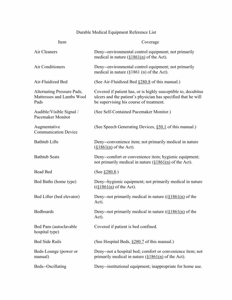
Durable Medical Equipment Reference List
Item Coverage
Air Cleaners Deny--environmental control equipment; not primarily medical in nature (§1861(n) of the Act).
Air Conditioners Deny--environmental control equipment; not primarily medical in nature (§1861 (n) of the Act).
Air-Fluidized Bed (See Air-Fluidized Bed §280.8 of this manual.)
Alternating Pressure Pads, Mattresses and Lambs Wool Pads
Covered if patient has, or is highly susceptible to, decubitus ulcers and the patient’s physician has specified that he will be supervising his course of treatment.
Audible/Visible Signal / Pacemaker Monitor
(See Self-Contained Pacemaker Monitor.)
Augmentative Communication Device
(See Speech Generating Devices, §50.1 of this manual.)
Bathtub Lifts Deny--convenience item; not primarily medical in nature (§1861(n) of the Act).
Bathtub Seats Deny--comfort or convenience item; hygienic equipment; not primarily medical in nature (§1861(n) of the Act).
Bead Bed (See §280.8.)
Bed Baths (home type) Deny--hygienic equipment; not primarily medical in nature ((§1861(n) of the Act).
Bed Lifter (bed elevator) Deny--not primarily medical in nature ((§1861(n) of the Act).
Bedboards Deny--not primarily medical in nature ((§1861(n) of the Act).
Bed Pans (autoclavable hospital type)
Covered if patient is bed confined.
Bed Side Rails (See Hospital Beds, §280.7 of this manual.)
Beds-Lounge (power or manual)
Deny--not a hospital bed; comfort or convenience item; not primarily medical in nature (§1861(n) of the Act).
Beds--Oscillating Deny--institutional equipment; inappropriate for home use.
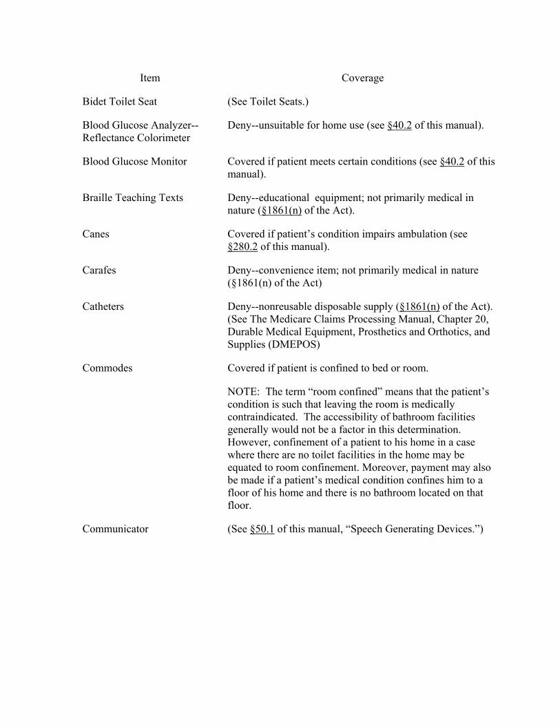
Item Coverage
Bidet Toilet Seat (See Toilet Seats.)
Blood Glucose Analyzer-- Reflectance Colorimeter
Deny--unsuitable for home use (see §40.2 of this manual).
Blood Glucose Monitor Covered if patient meets certain conditions (see §40.2 of this manual).
Braille Teaching Texts Deny--educational equipment; not primarily medical in nature (§1861(n) of the Act).
Canes Covered if patient’s condition impairs ambulation (see §280.2 of this manual).
Carafes Deny--convenience item; not primarily medical in nature (§1861(n) of the Act)
Catheters Deny--nonreusable disposable supply (§1861(n) of the Act). (See The Medicare Claims Processing Manual, Chapter 20, Durable Medical Equipment, Prosthetics and Orthotics, and Supplies (DMEPOS)
Commodes Covered if patient is confined to bed or room.
NOTE: The term “room confined” means that the patient’s condition is such that leaving the room is medically contraindicated. The accessibility of bathroom facilities generally would not be a factor in this determination. However, confinement of a patient to his home in a case where there are no toilet facilities in the home may be equated to room confinement. Moreover, payment may also be made if a patient’s medical condition confines him to a floor of his home and there is no bathroom located on that floor.
Communicator (See §50.1 of this manual, “Speech Generating Devices.”)
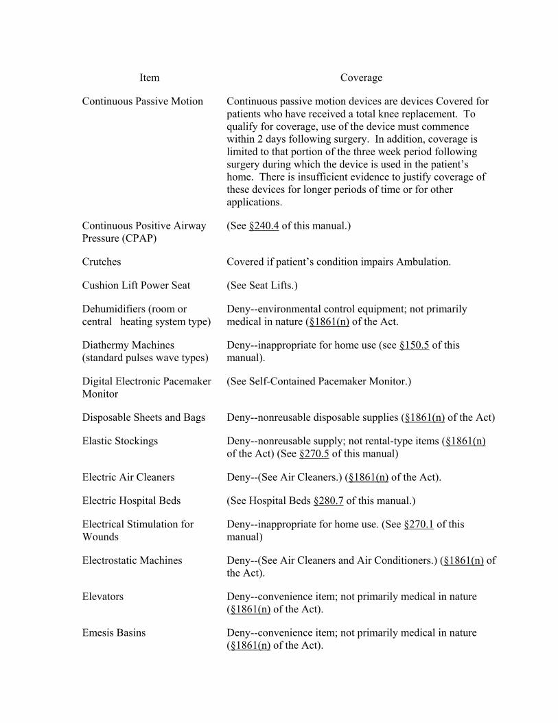
Item Coverage
Continuous Passive Motion Continuous passive motion devices are devices Covered for patients who have received a total knee replacement. To qualify for coverage, use of the device must commence within 2 days following surgery. In addition, coverage is limited to that portion of the three week period following surgery during which the device is used in the patient’s home. There is insufficient evidence to justify coverage of these devices for longer periods of time or for other applications.
Continuous Positive Airway Pressure (CPAP)
(See §240.4 of this manual.)
Crutches Covered if patient’s condition impairs Ambulation.
Cushion Lift Power Seat (See Seat Lifts.)
Dehumidifiers (room or central heating system type)
Deny--environmental control equipment; not primarily medical in nature (§1861(n) of the Act.
Diathermy Machines (standard pulses wave types)
Deny--inappropriate for home use (see §150.5 of this manual).
Digital Electronic Pacemaker Monitor
(See Self-Contained Pacemaker Monitor.)
Disposable Sheets and Bags Deny--nonreusable disposable supplies (§1861(n) of the Act)
Elastic Stockings Deny--nonreusable supply; not rental-type items (§1861(n) of the Act) (See §270.5 of this manual)
Electric Air Cleaners Deny--(See Air Cleaners.) (§1861(n) of the Act).
Electric Hospital Beds (See Hospital Beds §280.7 of this manual.)
Electrical Stimulation for Wounds
Deny--inappropriate for home use. (See §270.1 of this manual)
Electrostatic Machines Deny--(See Air Cleaners and Air Conditioners.) (§1861(n) of the Act).
Elevators Deny--convenience item; not primarily medical in nature (§1861(n) of the Act).
Emesis Basins Deny--convenience item; not primarily medical in nature (§1861(n) of the Act).
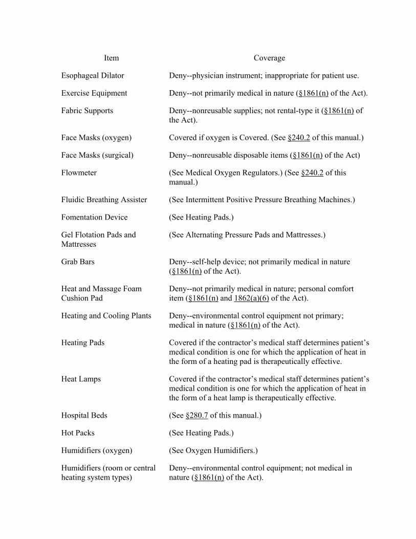
Item Coverage
Esophageal Dilator Deny--physician instrument; inappropriate for patient use.
Exercise Equipment Deny--not primarily medical in nature (§1861(n) of the Act).
Fabric Supports Deny--nonreusable supplies; not rental-type it (§1861(n) of the Act).
Face Masks (oxygen) Covered if oxygen is Covered. (See §240.2 of this manual.)
Face Masks (surgical) Deny--nonreusable disposable items (§1861(n) of the Act)
Flowmeter (See Medical Oxygen Regulators.) (See §240.2 of this manual.)
Fluidic Breathing Assister (See Intermittent Positive Pressure Breathing Machines.)
Fomentation Device (See Heating Pads.)
Gel Flotation Pads and Mattresses
(See Alternating Pressure Pads and Mattresses.)
Grab Bars Deny--self-help device; not primarily medical in nature (§1861(n) of the Act).
Heat and Massage Foam Cushion Pad
Deny--not primarily medical in nature; personal comfort item (§1861(n) and 1862(a)(6) of the Act).
Heating and Cooling Plants Deny--environmental control equipment not primary; medical in nature (§1861(n) of the Act).
Heating Pads Covered if the contractor’s medical staff determines patient’s medical condition is one for which the application of heat in the form of a heating pad is therapeutically effective.
Heat Lamps Covered if the contractor’s medical staff determines patient’s medical condition is one for which the application of heat in the form of a heat lamp is therapeutically effective.
Hospital Beds (See §280.7 of this manual.)
Hot Packs (See Heating Pads.)
Humidifiers (oxygen) (See Oxygen Humidifiers.)
Humidifiers (room or central heating system types)
Deny--environmental control equipment; not medical in nature (§1861(n) of the Act).
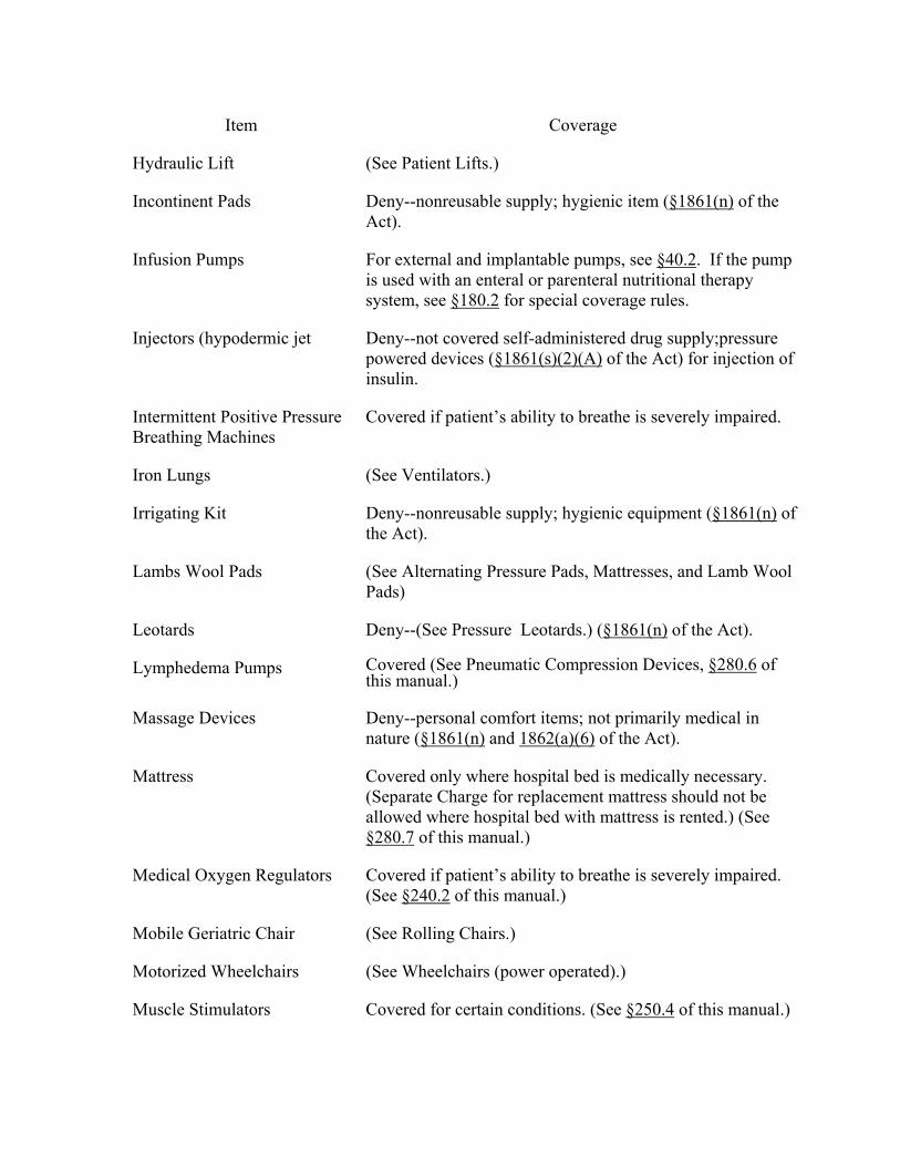
Item Coverage
Hydraulic Lift (See Patient Lifts.)
Incontinent Pads Deny--nonreusable supply; hygienic item (§1861(n) of the Act).
Infusion Pumps For external and implantable pumps, see §40.2. If the pump is used with an enteral or parenteral nutritional therapy system, see §180.2 for special coverage rules.
Injectors (hypodermic jet Deny--not covered self-administered drug supply;pressure powered devices (§1861(s)(2)(A) of the Act) for injection of insulin.
Intermittent Positive Pressure Breathing Machines
Covered if patient’s ability to breathe is severely impaired.
Iron Lungs (See Ventilators.)
Irrigating Kit Deny--nonreusable supply; hygienic equipment (§1861(n) of the Act).
Lambs Wool Pads (See Alternating Pressure Pads, Mattresses, and Lamb Wool Pads)
Leotards Deny--(See Pressure Leotards.) (§1861(n) of the Act).
Lymphedema Pumps Covered (See Pneumatic Compression Devices, §280.6 of this manual.)
Massage Devices Deny--personal comfort items; not primarily medical in nature (§1861(n) and 1862(a)(6) of the Act).
Mattress Covered only where hospital bed is medically necessary. (Separate Charge for replacement mattress should not be allowed where hospital bed with mattress is rented.) (See §280.7 of this manual.)
Medical Oxygen Regulators Covered if patient’s ability to breathe is severely impaired. (See §240.2 of this manual.)
Mobile Geriatric Chair (See Rolling Chairs.)
Motorized Wheelchairs (See Wheelchairs (power operated).)
Muscle Stimulators Covered for certain conditions. (See §250.4 of this manual.)
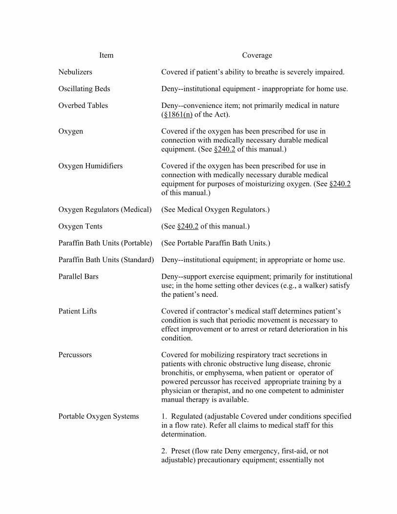
Item Coverage
Nebulizers Covered if patient’s ability to breathe is severely impaired.
Oscillating Beds Deny--institutional equipment - inappropriate for home use.
Overbed Tables Deny--convenience item; not primarily medical in nature (§1861(n) of the Act).
Oxygen Covered if the oxygen has been prescribed for use in connection with medically necessary durable medical equipment. (See §240.2 of this manual.)
Oxygen Humidifiers Covered if the oxygen has been prescribed for use in connection with medically necessary durable medical equipment for purposes of moisturizing oxygen. (See §240.2 of this manual.)
Oxygen Regulators (Medical) (See Medical Oxygen Regulators.)
Oxygen Tents (See §240.2 of this manual.)
Paraffin Bath Units (Portable) (See Portable Paraffin Bath Units.)
Paraffin Bath Units (Standard) Deny--institutional equipment; in appropriate or home use.
Parallel Bars Deny--support exercise equipment; primarily for institutional use; in the home setting other devices (e.g., a walker) satisfy the patient’s need.
Patient Lifts Covered if contractor’s medical staff determines patient’s condition is such that periodic movement is necessary to effect improvement or to arrest or retard deterioration in his condition.
Percussors Covered for mobilizing respiratory tract secretions in patients with chronic obstructive lung disease, chronic bronchitis, or emphysema, when patient or operator of powered percussor has received appropriate training by a physician or therapist, and no one competent to administer manual therapy is available.
Portable Oxygen Systems 1. Regulated (adjustable Covered under conditions specified in a flow rate). Refer all claims to medical staff for this determination.
2. Preset (flow rate Deny emergency, first-aid, or not adjustable) precautionary equipment; essentially not
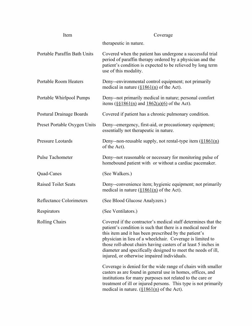
Item Coverage
therapeutic in nature.
Portable Paraffin Bath Units Covered when the patient has undergone a successful trial period of paraffin therapy ordered by a physician and the patient’s condition is expected to be relieved by long term use of this modality.
Portable Room Heaters Deny--environmental control equipment; not primarily medical in nature (§1861(n) of the Act).
Portable Whirlpool Pumps Deny--not primarily medical in nature; personal comfort items (§§1861(n) and 1862(a)(6) of the Act).
Postural Drainage Boards Covered if patient has a chronic pulmonary condition.
Preset Portable Oxygen Units Deny--emergency, first-aid, or precautionary equipment; essentially not therapeutic in nature.
Pressure Leotards Deny--non-reusable supply, not rental-type item (§1861(n) of the Act).
Pulse Tachometer Deny--not reasonable or necessary for monitoring pulse of homebound patient with or without a cardiac pacemaker.
Quad-Canes (See Walkers.)
Raised Toilet Seats Deny--convenience item; hygienic equipment; not primarily medical in nature (§1861(n) of the Act).
Reflectance Colorimeters (See Blood Glucose Analyzers.)
Respirators (See Ventilators.)
Rolling Chairs Covered if the contractor’s medical staff determines that the patient’s condition is such that there is a medical need for this item and it has been prescribed by the patient’s physician in lieu of a wheelchair. Coverage is limited to those roll-about chairs having casters of at least 5 inches in diameter and specifically designed to meet the needs of ill, injured, or otherwise impaired individuals.
Coverage is denied for the wide range of chairs with smaller casters as are found in general use in homes, offices, and institutions for many purposes not related to the care or treatment of ill or injured persons. This type is not primarily medical in nature. (§1861(n) of the Act).
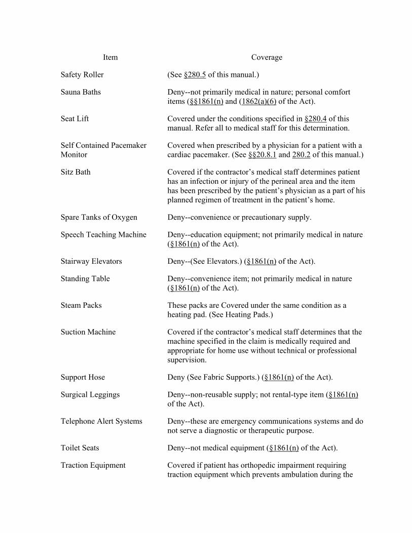
Item Coverage
Safety Roller (See §280.5 of this manual.)
Sauna Baths Deny--not primarily medical in nature; personal comfort items (§§1861(n) and (1862(a)(6) of the Act).
Seat Lift Covered under the conditions specified in §280.4 of this manual. Refer all to medical staff for this determination.
Self Contained Pacemaker Monitor
Covered when prescribed by a physician for a patient with a cardiac pacemaker. (See §§20.8.1 and 280.2 of this manual.)
Sitz Bath Covered if the contractor’s medical staff determines patient has an infection or injury of the perineal area and the item has been prescribed by the patient’s physician as a part of his planned regimen of treatment in the patient’s home.
Spare Tanks of Oxygen Deny--convenience or precautionary supply.
Speech Teaching Machine Deny--education equipment; not primarily medical in nature (§1861(n) of the Act).
Stairway Elevators Deny--(See Elevators.) (§1861(n) of the Act).
Standing Table Deny--convenience item; not primarily medical in nature (§1861(n) of the Act).
Steam Packs These packs are Covered under the same condition as a heating pad. (See Heating Pads.)
Suction Machine Covered if the contractor’s medical staff determines that the machine specified in the claim is medically required and appropriate for home use without technical or professional supervision.
Support Hose Deny (See Fabric Supports.) (§1861(n) of the Act).
Surgical Leggings Deny--non-reusable supply; not rental-type item (§1861(n) of the Act).
Telephone Alert Systems Deny--these are emergency communications systems and do not serve a diagnostic or therapeutic purpose.
Toilet Seats Deny--not medical equipment (§1861(n) of the Act).
Traction Equipment Covered if patient has orthopedic impairment requiring traction equipment which prevents ambulation during the
Item Coverage
period of use (Consider covering devices usable during ambulation; e.g., cervical traction collar, under the brace provision).
Trapeze Bars Covered if patient is bed confined and the patient needs a trapeze bar to sit up because of respiratory condition, to change body position for other medical reasons, or to get in and out of bed.
Treadmill Exerciser Deny--exercise equipment; not primarily medical in nature (§1861(n) of the Act).
Ultraviolet Cabinet Covered for selected patients with generalized intractable psoriasis. Using appropriate consultation, the contractor should determine whether medical and other factors justify treatment at home rather than at alternative sites, e.g., outpatient department of a hospital.
Urinals autoclavable Covered if patient is bed confined hospital type.
Vaporizers Covered if patient has a respiratory illness.
Ventilators Covered for treatment of neuromuscular diseases, thoracic restrictive diseases, and chronic respiratory failure consequent to chronic obstructive pulmonary disease. Includes both positive and negative pressure types. (See also §240.5 of this manual.)
Walkers Covered if patient’s condition impairs ambulation (See also §280.5 of this manual.)
Water and Pressure Pads and Mattresses
(See Alternating Pressure Pads, Mattresses and Lamb Wool Pads.)
Wheelchairs (power operated) Covered if patient’s condition is such and wheelchairs with other that a wheelchair is medically necessary special features and the patient is unable to operate the wheelchair manually. Any claim involving a power wheelchair or a wheelchair with other special features should be referred for medical consultation since payment for the special features is limited to those which are medically required because of the patient’s condition. (See §280.9 for power operated and §280.3 for specially sized wheelchairs.)
NOTE:A power-operated vehicle that may appropriately be used as a wheelchair can be Covered. (See §280.9 of this
Item Coverage
manual for coverage details.)
Whirlpool Bath Equipment Covered if patient is homebound and has a (standard)condition for which the whirlpool bath can be expected to provide substantial therapeutic benefit justifying its cost. Where patient is not homebound but has such a condition, payment is restricted to the cost of providing the services elsewhere; e.g., an outpatient department of a participating hospital, if that alternative is less costly. In all cases, refer claim to medical staff for a determination.
Whirlpool Pumps Deny--(See Portable Whirlpool Pumps.) (§1861(n) of the Act).
White Cane Deny--(See §280.2 of this manual.)
Cross-references:
The Medicare Benefit Policy Manual, Chapter 15, “Covered Medical and Other Health Services.”
The Medicare Claims Processing Manual, Chapter 20, “Durable Medical Equipment, Prosthetics and Orthotics, and Supplies (DMEPOS).”
280.2 - White Cane for Use by a Blind Person
(Rev. 1, 10-03-03)
CIM 60-3
Not Covered
A white cane for use by a blind person is more an identifying and self-help device than an item which makes a meaningful contribution in the treatment of an illness or injury.
280.3 - Specially Sized Wheelchairs
(Rev. 1, 10-03-03)
CIM 60-6
Payment may be made for a specially sized wheelchair even though it is more expensive than a standard wheelchair. For example, a narrow wheelchair may be required because of the narrow doorways of a patient’s home or because of a patient’s slender build. Such
difference in the size of the wheelchair from the standard model is not considered a deluxe feature.
A physician’s certification or prescription that a special size is needed is not required where the contractor can determine from the information in file or other sources that a specially sized wheelchair (rather than a standard one) is needed to accommodate the wheelchair to the place of use or the physical size of the patient.
To determine the reasonable charge in these cases, the contractor uses the criteria set out in the Medicare Claims Processing Manuals, Chapter 23, “Fee Schedule Administration and Coding Requirements,” and Chapter 12, “Physician/ Practitioner Billing,” as necessary.
Cross-references:
The Medicare Benefit Policy Manual, Chapter 15, “Covered Medical and Other Health Services,” §110.
The Medicare Claims Processing Manual, Chapter 20, “Durable Medical Equipment, Prosthetics and Orthotics, and Supplies (DMEPOS),” §§30.5.3 and 20.2
The Medicare Benefit Policy Manual, Chapter 13, “Rural Health Clinic (RHC) and Federally Qualified Health Center (FQHC) Services,” §30.1.
280.4 - Seat Lift
(Rev. 1, 10-03-03)
CIM 60-8
Reimbursement may be made for the rental or purchase of a medically necessary seat lift when prescribed by a physician for a patient with severe arthritis of the hip or knee and patients with muscular dystrophy or other neuromuscular disease when it has been determined the patient can benefit therapeutically from use of the device. In establishing medical necessity for the seat lift, the evidence must show that the item is included in the physician’s course of treatment, that it is likely to effect improvement, or arrest or retard deterioration in the patient’s condition, and that the severity of the condition is such that the alternative would be chair or bed confinement.
Coverage of seat lifts is limited to those types which operate smoothly, can be controlled by the patient, and effectively assist a patient in standing up and sitting down without other assistance. Excluded from coverage is the type of lift which operates by a spring release mechanism with a sudden, catapult-like motion and jolts the patient from a seated to a standing position. Limit the payment for units which incorporate a recliner feature along with the seat lift to the amount payable for a seat lift without this feature.
Cross Reference:
The Medicare Claims Processing Manual, Chapter 20, “Durable Medical Equipment, Prosthetics and Orthotics, and Supplies (DMEPOS),” §90.
280.5 - Safety Roller
(Rev. 1, 10-03-03)
CIM 60-15
Effective for Claims Adjudicated On or After June 3, 1985
“Safety roller” is the generic name applied to devices for patients who cannot use standard wheeled walkers. They may be appropriate, and therefore covered, for some patients who are obese, have severe neurological disorders, or restricted use of one hand which makes it impossible to use a wheeled walker that does not have the sophisticated braking system found on safety rollers.
In order to assure that payment is not made for a safety roller when a less expensive standard wheeled walker would satisfy the patient’s medical needs, carriers should refer safety roller claims to their medical consultants. The medical consultant determines whether some or all of the features provided in a safety roller are necessary, and therefore covered and reimbursable. If it is determined that the patient could use a standard wheeled walker, the charge for the safety roller is reduced to the charge of a standard wheeled walker.
Some obese patients who could use a standard wheeled walker if their weight did not exceed the walker’s strength and stability limits can have it reinforced and its wheel base expanded. Such modifications are routine mechanical adjustments and justify a moderate surcharge. In these cases the carrier reduces the charge for the safety roller to the charge for the standard wheeled walker plus the surcharge for modifications.
In the case of patients with medical documentation showing severe neurological disorders or restricted use of one hand which makes it impossible for them to use a wheeled walker that does not have a sophisticated braking system, a reasonable charge for the safety roller may be determined without relating it to the reasonable charge for a standard wheeled walker. (Such reasonable charge should be developed in accordance with the instructions in the Medicare Claims Processing Manual, Chapter 23, “Fee Schedule Administration and Coding Requirements.”)
Cross Reference:
The Medicare Benefit Policy Manual, Chapter 15, “Covered Medical and Other Health Services,” §120, and §280.1 of this manual.
280.6 - Pneumatic Compression Devices
(Rev. 1, 10-03-03)
CIM 60-16
Pneumatic compression devices consist of an inflatable garment for the arm or leg and an electrical pneumatic pump that fills the garment with compressed air. The garment is intermittently inflated and deflated with cycle times and pressures that vary between devices. Pneumatic devices are covered for the treatment of lymphedema or for the treatment of chronic venous insufficiency with venous stasis ulcers.
Lymphedema
Lymphedema is the swelling of subcutaneous tissues due to the accumulation of excessive lymph fluid. The accumulation of lymph fluid results from impairment to the normal clearing function of the lymphatic system and/or from an excessive production of lymph. Lymphedema is divided into two broad classes according to etiology. Primary lymphedema is a relatively uncommon, chronic condition which may be due to such causes as Milroy’s Disease or congenital anomalies. Secondary lymphedema which is much more common, results from the destruction of or damage to formerly functioning lymphatic channels, such as surgical removal of lymph nodes or post radiation fibrosis, among other causes.
Pneumatic compression devices are covered in the home setting for the treatment of lymphedema if the patient has undergone a four-week trial of conservative therapy and the treating physician determines that there has been no significant improvement or if significant symptoms remain after the trial. The trial of conservative therapy must include use of an appropriate compression bandage system or compression garment, exercise, and elevation of the limb. The garment may be prefabricated or custom-fabricated but must provide adequate graduated compression.
Chronic Venous Insufficiency With Venous Stasis Ulcers
Chronic venous insufficiency (CVI) of the lower extremities is a condition caused by abnormalities of the venous wall and valves, leading to obstruction or reflux of blood flow in the veins. Signs of CVI include hyperpigmentation, stasis dermatitis, chronic edema, and venous ulcers.
Pneumatic compression devices are covered in the home setting for the treatment of CVI of the lower extremities only if the patient has one or more venous stasis ulcer(s) which have failed to heal after a six month trial of conservative therapy directed by the treating physician. The trial of conservative therapy must include a compression bandage system or compression garment, appropriate dressings for the wound, exercise, and elevation of the limb.
General Coverage Criteria
Pneumatic compression devices are covered only when prescribed by a physician and when they are used with appropriate physician oversight, i.e., physician evaluation of the patient’s condition to determine medical necessity of the device, assuring suitable instruction in the operation of the machine, a treatment plan defining the pressure to be used and the frequency and duration of use, and ongoing monitoring of use and response to treatment.
The determination by the physician of the medical necessity of a pneumatic compression device must include:
1. The patient’s diagnosis and prognosis;
2. Symptoms and objective findings, including measurements which establish the severity of the condition;
3. The reason the device is required, including the treatments which have been tried and failed; and
4. The clinical response to an initial treatment with the device.
The clinical response includes the change in pretreatment measurements, ability to tolerate the treatment session and parameters, and ability of the patient (or caregiver) to apply the device for continued use in the home.
The only time that a segmented, calibrated gradient pneumatic compression device (HCPCS code E0652) would be covered is when the individual has unique characteristics that prevent them from receiving satisfactory pneumatic compression treatment using a nonsegmented device in conjunction with a segmented appliance or a segmented compression device without manual control of pressure in each chamber.
Cross Reference: §280.1.
280.7 - Hospital Beds
(Rev. 1, 10-03-03)
CIM 60-18
A - General Requirements for Coverage of Hospital Beds
A physician’s prescription, and such additional documentation as the contractors’ medical staffs may consider necessary, including medical records and physicians’ reports, must establish the medical necessity for a hospital bed due to one of the following reasons:
• The patient’s condition requires positioning of the body; e.g., to alleviate pain, promote good body alignment, prevent contractures, avoid respiratory infections, in ways not feasible in an ordinary bed; or
• The patient’s condition requires special attachments that cannot be fixed and used on an ordinary bed.
B - Physician’s Prescription
The physician’s prescription which must accompany the initial claim, and supplementing documentation when required, must establish that a hospital bed is medically necessary. If the stated reason for the need for a hospital bed is the patient’s condition requires positioning, the prescription or other documentation must describe the medical condition, e.g., cardiac disease, chronic obstructive pulmonary disease, quadriplegia or paraplegia, and also the severity and frequency of the symptoms of the condition, that necessitates a hospital bed for positioning.
If the stated reason for requiring a hospital bed is the patient’s condition requires special attachments, the prescription must describe the patient’s condition and specify the attachments that require a hospital bed.
C - Variable Height Feature
In well documented cases, the contractors’ medical staffs may determine that a variable height feature of a hospital bed, approved for coverage under subsection A above, is medically necessary and, therefore, covered, for one of the following conditions:
• Severe arthritis and other injuries to lower extremities; e.g., fractured hip - The condition requires the variable height feature to assist the patient to ambulate by enabling the patient to place his or her feet on the floor while sitting on the edge of the bed;
• Severe cardiac conditions - For those cardiac patients who are able to leave bed, but who must avoid the strain of “jumping” up or down;
• Spinal cord injuries, including quadriplegic and paraplegic patients, multiple limb amputee and stroke patients. For those patients who are able to transfer from bed to a wheelchair, with or without help; or
• Other severely debilitating diseases and conditions, if the variable height feature is required to assist the patient to ambulate.
D - Electric Powered Hospital Bed Adjustments
Electric powered adjustments to lower and raise head and foot may be covered when the contractor’s medical staff determines that the patient’s condition requires frequent change in body position and/or there may be an immediate need for a change in body position (i.e., no delay can be tolerated) and the patient can operate the controls and cause the
adjustments. Exceptions may be made to this last requirement in cases of spinal cord injury and brain damaged patients.
E - Side Rails
If the patient’s condition requires bed side rails, they can be covered when an integral part of, or an accessory to, a hospital bed.
280.8 - Air-Fluidized Bed
(Rev. 1, 10-03-03)
CIM 60-19
Air fluidized beds are covered for services rendered on or after: July 30, 1990.
An air-fluidized bed uses warm air under pressure to set small ceramic beads in motion which simulate the movement of fluid. When the patient is placed in the bed, his body weight is evenly distributed over a large surface area which creates a sensation of “floating.” Medicare payment for home use of the air-fluidized bed for treatment of pressure sores can be made if such use is reasonable and necessary for the individual patient.
A decision that use of an air-fluidized bed is reasonable and necessary requires that:
• The patient has a stage 3 (full thickness tissue loss) or stage 4 (deep tissue destruction) pressure sore;
• The patient is bedridden or chair bound as a result of severely limited mobility;
• In the absence of an air-fluidized bed, the patient would require institutionalization;
• The air-fluidized bed is ordered in writing by the patient’s attending physician based upon a comprehensive assessment and evaluation of the patient after completion of a course of conservative treatment designed to optimize conditions that promote wound healing. This course of treatment must have been at least one month in duration without progression toward wound healing. This month of prerequisite conservative treatment may include some period in an institution as long as there is documentation available to verify that the necessary conservative treatment has been rendered.
• Use of wet-to-dry dressings for wound debridement, begun during the period of conservative treatment and which continue beyond 30 days, will not preclude coverage of air-fluidized bed. Should additional debridement again become necessary, while a patient is using an air-fluidized bed (after the first 30-day course of conservative treatment) that will not cause the air-fluidized bed to
become noncovered. In all instances documentation verifying the continued need for the bed must be available.
• A trained adult caregiver is available to assist the patient with activities of daily living, fluid balance, dry skin care, repositioning, recognition and management of altered mental status, dietary needs, prescribed treatments, and management and support of the air-fluidized bed system and its problems such as leakage;
• A physician directs the home treatment regimen, and reevaluates and recertifies the need for the air-fluidized bed on a monthly basis; and
• All other alternative equipment has been considered and ruled out.
Conservative treatment must include:
• Frequent repositioning of the patient with particular attention to relief of pressure over bony prominences (usually every 2 hours);
• Use of a specialized support surface (Group II) designed to reduce pressure and shear forces on healing ulcers and to prevent new ulcer formation;
• Necessary treatment to resolve any wound infection;
• Optimization of nutrition status to promote wound healing;
• Debridement by any means (including wet to dry dressings-which does not require an occlusive covering) to remove devitalized tissue from the wound bed;
• Maintenance of a clean, moist bed of granulation tissue with appropriate moist dressings protected by an occlusive covering, while the wound heals.
Home use of the air-fluidized bed is not covered under any of the following circumstances:
• The patient has coexisting pulmonary disease (the lack of firm back support makes coughing ineffective and dry air inhalation thickens pulmonary secretions);
• The patient requires treatment with wet soaks or moist wound dressings that are not protected with an impervious covering such as plastic wrap or other occlusive material;
• The caregiver is unwilling or unable to provide the type of care required by the patient on an air-fluidized bed;
• Structural support is inadequate to support the weight of the air-fluidized bed system (it generally weighs 1600 pounds or more);
• Electrical system is insufficient for the anticipated increase in energy consumption; or
• Other known contraindications exist.
Coverage of an air-fluidized bed is limited to the equipment itself. Payment for this covered item may only be made if the written order from the attending physician is furnished to the supplier prior to the delivery of the equipment. Payment is not included for the caregiver or for architectural adjustments such as electrical or structural improvement.
Cross reference:
The Medicare Claims Processing Manual, Chapter 23, “Fee Schedule Administration and Coding Requirements,” §§60.
280.9 - Power Operated Vehicles That May Be Used as Wheelchairs
(Rev. 1, 10-03-03)
CIM 60-5
Power-operated vehicles that may be appropriately used as wheelchairs are covered under the durable medical equipment provision.
These vehicles have been appropriately used in the home setting for vocational rehabilita-tion and to improve the ability of chronically disabled persons to cope with normal domestic, vocational and social activities. They may be covered if a wheelchair is medically necessary and the patient is unable to operate a wheelchair manually.
A specialist in physical medicine, orthopedic surgery, neurology, or rheumatology must provide an evaluation of the patient’s medical and physical condition and a prescription for the vehicle to assure that the patient requires the vehicle and is capable of using it safely. When a Durable Medical Equipment Regional Carrier (DMERC) determines that such a specialist is not reasonably accessible, e.g., more than one day’s round trip from the beneficiary’s home, or the patient’s condition precludes such travel, a prescription from the beneficiary’s physician is acceptable.
The DMERC’s medical staff reviews all claims for a power-operated vehicle, including the specialists’ or other physicians’ prescriptions and evaluations of the patient’s medical and physical conditions, to insure that all coverage requirements are met. (See §280.1 and the Medicare Claims Processing Manual, Chapter 20,”Durable Medical Equipment, Prosthetics and Orthotics, and Supplies (DMEPOS),” §§110, 120, 130, 140, 150, 160, and 170.)
280.10 - Prosthetic Shoe
(Rev. 1, 10-03-03)
CIM 70-3
A prosthetic shoe (a device used when all or a substantial portion of the front part of the foot is missing) can be covered as a terminal device; i.e., a structural supplement replacing a totally or substantially absent hand or foot. The coverage of artificial arms and legs includes payment for terminal devices such as hands or hooks even though the patient may not require an artificial limb. The function of the prosthetic shoe is quite distinct from that of excluded orthopedic shoe and supportive foot devices which are used by individuals whose feet, although impaired, are essentially intact. (Section 1862(a)(8) of the Act excludes payment for orthopedic shoes or other supportive devices for the feet.) See the Medicare Benefit Policy Manual, Chapter 15, “Covered Medical and Other Services,” §130.
280.11 - Corset Used as Hernia Support
(Rev. 1, 10-03-03)
CIM 70-1
A hernia support (whether in the form of a corset or truss) which meets the definition of a brace is covered under Part B under §1861(s)(9) of the Act. See the Medicare Benefit Policy Manual, Chapter 15, “Covered Medical and Other Services,” §130.
280.12 - Sykes Hernia Control
(Rev. 1, 10-03-03)
CIM 70-2
Based on professional advice, it has been determined that the sykes hernia control (a spring-type, U-shaped, strapless truss) is not functionally more beneficial than a conventional truss. Make program reimbursement for this device only when an ordinary truss would be covered. (Like all trusses, it is only of benefit when dealing with a reducible hernia). Thus, when a charge for this item is substantially in excess of that which would be reasonable for a conventional truss used for the same condition, base reimbursement on the reasonable charges for the conventional truss. See the Medicare Benefit Policy Manual, Chapter 15, “Covered Medical and Other Services,” §130.
280.13 - Transcutaneous Electrical Nerve Stimulators (TENS)
(Rev. 1, 10-03-03)
CIM 60-20
TENS is a type of electrical nerve stimulator that is employed to treat chronic intractable pain. This stimulator is attached to the surface of the patient’s skin over the peripheral nerve to be stimulated. It may be applied in a variety of settings (in the patient’s home, a physician’s office, or in an outpatient clinic). Payment for TENS may be made under the durable medical equipment benefit. (See §160.13 for an explanation of coverage of medically necessary supplies for the effective use of TENS and §10.2 for an explanation of coverage of TENS for acute post-operative pain.)
280.14 – Infusion Pumps
(Rev. 1, 10-03-03)
CIM 60-14
The following indications for treatment using infusion pumps are covered under Medicare:
A - External Infusion Pumps
1 - Iron Poisoning - Effective for Services Performed On or After 9/26/84.
When used in the administration of deferoxamine for the treatment of acute iron poisoning and iron overload, only external infusion pumps are covered.
2 - Thromboembolic Disease - Effective for Services Performed On or After 9/26/84
When used in the administration of heparin for the treatment of thromboembolic disease and/or pulmonary embolism, only external infusion pumps used in an institutional setting are covered.
3 - Chemotherapy for Liver Cancer - Effective for Services Performed On or After 1/29/85.
The external chemotherapy infusion pump is covered when used in the treatment of primary hepatocellular carcinoma or colorectal cancer where this disease is unresectable or where the patient refuses surgical excision of the tumor.
4 - Morphine for Intractable Cancer Pain - Effective for Services Performed On or After 4/22/85.
Morphine infusion via an external infusion pump is covered when used in the treatment of intractable pain caused by cancer (in either an inpatient or outpatient setting, including a hospice).
5 - Continuous subcutaneous insulin infusion pumps (CSII) - Effective for Services Performed On or after 4/1/2000.
An external infusion pump and related drugs/supplies are covered as medically necessary in the home setting in the following situation:
Treatment of diabetes
In order to be covered, patients must meet criterion a or b:
Criterion a
The patient has completed a comprehensive diabetes education program, and has been on a program of multiple daily injections of insulin (i.e. at least 3 injections per day), with frequent self-adjustments of insulin dose for at least 6 months prior to initiation of the insulin pump, and has documented frequency of glucose self-testing an average of at least 4 times per day during the 2 months prior to initiation of the insulin pump, and meets one or more of the following criteria while on the multiple daily injection regimen:
1. Glycosylated hemoglobin level (HbAlc) > 7.0 percent
2. History of recurring hypoglycemia
3. Wide fluctuations in blood glucose before mealtime
4. Dawn phenomenon with fasting blood sugars frequently exceeding 200 mg/dl
5. History of severe glycemic excursions
Criterion b
1. The patient with diabetes has been on a pump prior to enrollment in Medicare and has documented frequency of glucose self-testing an average of at least 4 times per day during the month prior to Medicare enrollment.
Diabetes needs to be documented by a fasting C-peptide level that is less than or equal to 110 percent of the lower limit of normal of the laboratory’s measurement method. Effective for Services Performed on or after January 1, 2002.
Continued coverage of the insulin pump would require that the patient has been seen and evaluated by the treating physician at least every three months.
The pump must be ordered by and follow-up care of the patient must be managed by a physician who manages multiple patients with CSII and who works closely with a team including nurses, diabetes educators, and dietitians who are knowledgeable in the use of CSII.
6 - Other Uses
Other uses of external infusion pumps are covered if the contractor’s medical staff verifies the appropriateness of the therapy and of the prescribed pump for the individual patient.
NOTE: Payment may also be made for drugs necessary for the effective use of an external infusion pump as long as the drug being used with the pump is itself reasonable and necessary for the patient’s treatment.
B - Implantable Infusion Pumps
1 - Chemotherapy for Liver Cancer
Effective for Services Performed On or After 9/26/84.
The implantable infusion pump is covered for intra-arterial infusion of 5-FUdR for the treatment of liver cancer for patients with primary hepatocellular carcinoma or Duke’s Class D colorectal cancer, in whom the metastases are limited to the liver, and where (1) the disease is unresectable or (2) where the patient refuses surgical excision of the tumor.
2 - Anti-Spasmodic Drugs for Severe Spasticity
An implantable infusion pump is covered when used to administer anti-spasmodic drugs intrathecally (e.g., baclofen) to treat chronic intractable spasticity in patients who have proven unresponsive to less invasive medical therapy as determined by the following criteria:
As indicated by at least a 6-week trial, the patient cannot be maintained on noninvasive methods of spasm control, such as oral anti-spasmodic drugs, either because these methods fail to control adequately the spasticity or produce intolerable side effects, and
Prior to pump implantation, the patient must have responded favorably to a trial intrathecal dose of the anti-spasmodic drug.
3 - Opioid Drugs for Treatment of Chronic Intractable Pain
An implantable infusion pump is covered when used to administer opioid drugs (e.g., morphine) intrathecally or epidurally for treatment of severe chronic intractable pain of malignant or nonmalignant origin in patients who have a life expectancy of at least
three months and who have proven unresponsive to less invasive medical therapy as determined by the following criteria:
The patient’s history must indicate that he/she would not respond adequately to noninvasive methods of pain control, such as systemic opioids (including attempts to eliminate physical and behavioral abnormalities which may cause an exaggerated reaction to pain); and
A preliminary trial of intraspinal opioid drug administration must be undertaken with a temporary intrathecal/epidural catheter to substantiate adequately acceptable pain relief and degree of side effects (including effects on the activities of daily living) and patient acceptance.
4 - Coverage of Other Uses of Implanted Infusion Pumps
Determinations may be made on coverage of other uses of implanted infusion pumps if the contractor’s medical staff verifies that:
• The drug is reasonable and necessary for the treatment of the individual patient;
• It is medically necessary that the drug be administered by an implanted infusion pump; and
• The FDA approved labeling for the pump must specify that the drug being administered and the purpose for which it is administered is an indicated use for the pump.
5 - Implantation of Infusion Pump Is Contraindicated
The implantation of an infusion pump is contraindicated in the following patients:
• Patients with a known allergy or hypersensitivity to the drug being used (e.g., oral baclofen, morphine, etc.);
• Patients who have an infection;
• Patients whose body size is insufficient to support the weight and bulk of the device; and
• Patients with other implanted programmable devices since crosstalk between devices may inadvertently change the prescription.
NOTE: Payment may also be made for drugs necessary for the effective use of an implantable infusion pump as long as the drug being used with the pump is itself reasonable and necessary for the patient’s treatment.
Infusion Pumps Not Covered
The following indications for treatment using infusion pumps are not covered under Medicare:
External Infusion Pumps
Vancomycin
Effective for Services Beginning On or After September 1, 1996.
Medicare coverage of vancomycin as a durable medical equipment infusion pump benefit is not covered. There is insufficient evidence to support the necessity of using an external infusion pump, instead of a disposable elastomeric pump or the gravity drip method, to administer vancomycin in a safe and appropriate manner.
Implantable Infusion Pump
A - Thromboembolic Disease
Effective for Services Performed On or After 9/26/84.
According to the Public Health Service, there is insufficient published clinical data to support the safety and effectiveness of the heparin implantable pump. Therefore, the use of an implantable infusion pump for infusion of heparin in the treatment of recurrent thromboembolic disease is not covered.
B - Diabetes
An implanted infusion pump for the infusion of insulin to treat diabetes is not covered. The data does not demonstrate that the pump provides effective administration of insulin.
290 - Nursing Services
(Rev. 1, 10-03-03)
290.1 - Home Health Visits to a Blind Diabetic
(Rev. 1, 10-03-03)
CIM 90-1
Many individuals who are blind and require daily insulin for the control of a diabetic condition are able to administer their injections without assistance (other than possibly that which may be furnished by family members or friends). There are organizations which encourage and train blind diabetics, both to fill their own syringes and to inject themselves. There are also a number of devices available for blind individuals to fill their syringes accurately. However, the individuals who may need assistance with prefilling
their syringes may also require periodic observation and evaluation, even though their diabetes is fairly stabilized. In such cases, probably few in number, home health services may be required for this purpose.
To qualify for home health benefits, a blind diabetic must be confined to his home, under the care of a physician, and in need of either skilled nursing services on an intermittent basis or physical therapy or speech therapy. Effective July 1, 1981, a person may qualify for home health benefits based on his or her need for skilled nursing services on an intermittent basis, physical therapy, speech therapy, or occupational therapy. Effective December 1, 1981, occupational therapy is eliminated as a basis for entitlement to home health services. However, if a person has otherwise qualified for home health services because of the need for skilled nursing care, physical therapy or speech therapy, the patient’s eligibility for home health services may be extended solely on the basis of the continuing need for occupational therapy. (See the Medicare Benefit Policy Manual, Chapter 7, “Home Health Services,” §20.) There must be a plan of treatment, established and periodically reviewed by a physician which indicates that there is a recurring need for home health services to supplement the physician’s contacts with the patient; e.g., skilled nursing visits for observing and determining the need for changes in the level and type of care which has been prescribed. (See the Medicare Benefit Policy Manual, Chapter 7, “Home Health Services,” §§30.) Once an initial regimen has been established, the frequency of need for further home health services can vary greatly from patient to patient, depending on their condition and the likelihood of its changing. Some may need visits only every 90 days, for example, while others may require them much more frequently. If a nurse makes a visit to provide skilled services, and also prefills syringes, the purpose of the visit which was to provide skilled services, does not change. However, if the sole purpose of the nurse’s visit is to prefill insulin syringes for a blind diabetic, it is not a skilled nursing visit although it may be reimbursed as such as indicated below.
Filling a syringe can be safely and effectively performed by the average nonmedical person without the direct supervision of a licensed nurse. Consequently, it would not constitute a skilled nursing service even if it is performed by a nurse. (See the Medicare Benefit Policy Manual, Chapter 7, “Home Health Services,” §30.2.2.) The personal care duties normally performed by home health aides include assisting the patient with medications ordered by a physician which are ordinarily self-administered. (See the Medicare Benefit Policy Manual, Chapter 7, “Home Health Services,” §50.2.) Performance of such a service by an aide is consistent with the Medicare conditions of participation for home health agencies. Therefore, home health aide services would be appropriate for those blind diabetics who are qualified for home health benefits and who cannot fill their syringes. An adequately trained home health aide could make intermittent visits, usually on a weekly basis, to the home for the purpose of filling that supply of insulin ordered by the physician.
If State law, however, precludes a home health aide from prefilling insulin syringes, payment may be made for this service as part of the cost of skilled nursing services when performed by a nurse for a blind diabetic who is otherwise unable to prefill his or her syringes. There are no adverse consequences with respect to reimbursement to the home health agency for providing the service in this manner.
If State law does not preclude a home health aide from prefilling insulin syringes, but the home health agency chooses to send a nurse to perform only this task, the visit is reimbursed as if made by a home health aide.
NOTE: As indicated, to qualify for home health benefits, a patient must require skilled nursing services on an intermittent basis or physical therapy or speech therapy. If a beneficiary does not qualify for home health benefits but only needs someone to prefill syringes with the correct dosage of insulin, then no program payment can be made.
Cross-reference:
The Medicare Benefit Policy Manual, Chapter 7, “Home Health Services,” §§20, §§30, §30.2.2 and, §§50.
290.2 - Home Health Nurses’ Visits to Patients Requiring Heparin Injections
(Rev. 1, 10-03-03)
CIM 90-2
Professional medical advice indicates that subcutaneous injections of low dose heparin can be, under certain circumstances, medically accepted therapy for the treatment of recurrent deep venous thrombosis, recurrent pulmonary emboli, and other conditions requiring long term anticoagulation. The usual drug of choice for these conditions is warfarin. Heparin may be substituted for warfarin in circumstances such as demonstrated warfarin sensitivity. Heparin is now the drug of choice for anticoagulation during pregnancy.
Medicare payment may be made for serial visits by the home health nurse to teach the patient or the caring person to give subcutaneous injections of low dose heparin if it is prescribed by a physician for a homebound patient who:
• Is pregnant and requires anticoagulant therapy, or
• Requires treatment for deep venous thrombosis or pulmonary emboli or for another condition requiring anticoagulation and documentation justifies that the patient cannot tolerate warfarin.
If the patient or caring person is unable to administer the injection, nursing visits to give the injections on a daily basis, seven days a week, for a period of up to six months (in the case of pregnancy, visits may be made for a period beyond six months if reasonable and necessary) would be reimbursed by Medicare. Coverage for these services after six months of treatment would be provided only if the prescribing physician can justify and document the need for such an extended course of treatment. Documentation of need for heparin injections beyond six months would not be required for pregnant patients who meet the homebound criteria.
Cross-reference:
The Medicare Benefit Policy Manual, Chapter 7, “Home Health Services,” §§30.4,
300 - Diagnostic Tests Not Otherwise Classified
300.1 - Obsolete or Unreliable Diagnostic Tests
(Rev. 1, 10-03-03)
CIM 50-34
A - Diagnostic Tests
Do not routinely pay for the following diagnostic tests because they are obsolete and have been replaced by more advanced procedures. The listed tests may be paid for only if the medical need for the procedure is satisfactorily justified by the physician who performs it. When the services are subject to the Quality Improvement Organization (QIO) review, the QIO is responsible for determining that satisfactory medical justification exists. When the services are not subject to QIO review, the intermediary or carrier is responsible for determining that satisfactory medical justification exists. This includes:
• Amylase, blood isoenzymes, electrophoretic,
• Chromium, blood,
• Guanase, blood,
• Zinc sulphate turbidity, blood,
• Skin test, cat scratch fever,
• Skin test, lymphopathia venereum,
• Circulation time, one test,
• Cephalin flocculation,
• Congo red, blood,
• Hormones, adrenocorticotropin quantitative animal tests,
• Hormones, adrenocorticotropin quantitative bioassay,
• Thymol turbidity, blood,
• Skin test, actinomycosis,
• Skin test, brucellosis,
• Skin test, psittacosis,
• Skin test, trichinosis,
• Calcium, feces, 24-hour quantitative,
• Starch, feces, screening,
• Chymotrypsin, duodenal contents,
• Gastric analysis, pepsin,
• Gastric analysis, tubeless,
• Calcium saturation clotting time,
• Capillary fragility test (Rumpel-Leede),
• Colloidal gold,
• Bendien’s test for cancer and tuberculosis,
• Bolen’s test for cancer,
• Rehfuss test for gastric acidity, and
• Serum seromucoid assay for cancer and other diseases.
B - Cardiovascular Tests
Do not pay for the following phonocardiography and vectorcardiography diagnostic tests because they have been determined to be outmoded and of little clinical value. They include:
• CPT code 93201, Phonocardiogram with or without ECG lead; with supervision during recording with interpretation and report (when equipment is supplied by the physician),
• CPT code 93202, Phonocardiogram; tracing only, without interpretation and report (e.g., when equipment is supplied by the hospital, clinic),
• CPT code 93204, Phonocardiogram; interpretation and report,
• CPT code 93205, Phonocardiogram with ECG lead, with indirect carotid artery and/or jugular vein tracing, and/or apex cardiogram; with interpretation and report,
• CPT code 93208, Phonocardiogram; without interpretation and report,
• CPT code 93209, Phonocardiogram; interpretation and report only,
• CPT code 93210, Intracardiac,
• CPT code 93220, Vectorcardiogram (VCG), with or without ECG; with interpretation and report,
• CPT code 93221, Vectorcardiogram; tracing only, without interpretation and report, and
• CPT code 93222, Vectorcardiogram; interpretation and report only.
CPT codes 93201, 93202, 93204, 93205, 93208, 93209, 93210, 93220, 93221, and 93222 have been deleted, to report, use 93799 cardiovascular procedure.
310 - Clinical Trials
(Rev. 1, 10-03-03)
310.1 - Routine Costs in Clinical Trails
(Rev. 1, 10-03-03)
CIM 30-1
Effective for items and services furnished on or after September 19, 2000, Medicare covers the routine costs of qualifying clinical trials, as such costs are defined below, as well as reasonable and necessary items and services used to diagnose and treat complications arising from participation in all clinical trials. All other Medicare rules apply.
Routine costs of a clinical trial include all items and services that are otherwise generally available to Medicare beneficiaries (i.e., there exists a benefit category, it is not statutorily excluded, and there is not a national noncoverage decision) that are provided in either the experimental or the control arms of a clinical trial except:
• The investigational item or service, itself;
• Items and services provided solely to satisfy data collection and analysis needs and that are not used in the direct clinical management of the patient (e.g., monthly CT scans for a condition usually requiring only a single scan); and
• Items and services customarily provided by the research sponsors free of charge for any enrollee in the trial.
Routine costs in clinical trials include:
• Items or services that are typically provided absent a clinical trial (e.g., conventional care);
• Items or services required solely for the provision of the investigational item or service (e.g., administration of a noncovered chemotherapeutic agent), the clinically appropriate monitoring of the effects of the item or service, or the prevention of complications; and
• Items or services needed for reasonable and necessary care arising from the provision of an investigational item or service - in particular, for the diagnosis or treatment of complications.
This policy does not withdraw Medicare coverage for items and services that may be covered according to local medical review policies or the regulations on category B investigational device exemptions (IDE) found in 42 CFR 405.201 - 405.215, 411.15, and 411.406. For information about local medical review policies (LMRPs), refer to http://www.lmrp.net/, a searchable database of Medicare contractors’ local policies.