purification and characterization of fetal hematopoietic cells that express the common acute...
TRANSCRIPT

P U R I F I C A T I O N A N D C H A R A C T E R I Z A T I O N O F F E T A L
H E M A T O P O I E T I C C E L L S T H A T E X P R E S S T H E C O M M O N
A C U T E L Y M P H O B L A S T I C L E U K E M I A A N T I G E N ( C A L L A ) *
BY PETER HOKLAND,:~ PAUL ROSENTHAL, JAMES D. GRIFFIN,§ LEE M. NADLER, H JOHN DALEY, MARIANNE HOKLAND,¶
STUART F. SCHLOSSMAN, AND JEROME RITZ**
From the Division of Tumor Immunology, Sidney Farber Cancer Institute, and Department of Medicine, Harvard Medical School, Boston, Massachusetts 02l 15
Previous studies using convent ional heteroantisera and monoclonal antibodies (1, 2) have characterized the expression of a common acute lymphoblast ic leukemia ant igen (CALLA) 1 in various hematopoiet ic malignancies and normal tissues, CALLA
has been demonstrated to be a 100,000-dahon glycoprotein that is expressed by leukemic cells from ~80% of patients with non - T acute lymphoblast ic leukemia (ALL), 10% of patients with T-ALL, and 40% of patients with chronic myelocytic leukemia (CML) in blast crisis, as well as lymphoma cells from ~40% of patients with T cell lymphoblast ic lymphomas and almost all Burkitt 's lymphomas and nodular lymphomas (3). Wi th in the hematopoiet ic system, CALLA is also detected on a small populat ion of mononuclear cells (1-3%) in either normal or regenerat ing bone marrow. Fetal hematopoietic organs contain a larger proport ion of C A L L A + cells and mononuc lear cell suspensions from fetal liver and fetal bone marrow have been found to conta in 2-10% C A L L A + cells (2-5).
Although much is known about the dis tr ibut ion of CALLA, very little is known about its functional role on the cell surface or its appearance dur ing ontogeny. Recent studies have demonstra ted that CALLA belongs to a family of 100,000-dahon cell surface proteins that are widely expressed by hematopoiet ic cells (6) and is also present in some nonhematopoiet ic tissues (7). In addit ion, b ind ing of j 5 monoclonal an t ibody to cell surface ant igen results in the modula t ion of the expression of CALLA on
* Supported in part by grant CA-28704 from the National Institutes of I ~cal~h. On leave from the University Department of Medicine and ttaematology, University of Aarhus.
Denmark. Research Fellow of the Danish Medical Research Council. § Research Fellow of the Medical Foundation, Boston, MA and tile Joanna C. \~,~ood Foundation,
Philadelphia, PA. II Research Fellow of the Medical Foundation, Boston, MA. ¶ Research Fellow of the Danish Cancer Society. ** Special Fellow of the Leukemia Society of America. 1 Abbreviations u~ed in thi~" paper: ALL, acute lymphoblastic leukemia: BM, bone marrow; C, complement;
CALl,A, common acute lymphoblastic leukemia antigen; CFU~G/M, granulocytc/monocyte cotol~y forming unit; CML, chronic myelocytic leukemia; tyro-p,, intracytoplasmic g chain; FACS, fluorescence activated cell sorter: FCS, fetal calf serum: C,/Md"ITC, fluorescein-conjugated goat anti-mouse immuno- globulin; G/M-TRITC, rhodamine-col~iugated goat anti-mouse immunoglobulin; ItBSS, t tanks' balanced salts solution; MoAb, monoclonal antibodies; R/M-SRBC, rabbit anti-mouse Ig-coated sheep erythrocytes; SMEM + 2.5% AB, supplemented minimum cxsential medium with 2.5+;4, human AB: SRBC, sheep erytbrocytes; TdT, terminal transferase enzyme.
114 J. Exp. MEO. © The Rockefeller University Press • 0022-1007/83/01/0114/16 $1.00 Volmne 157 January 1983 114-129
on August 13, 2015
jem.rupress.org
Dow
nloaded from
Published January 1, 1983

HOKLAND ET AL. 1 15
leukemic cells and cell lines. J5 modu la t i on occurs in vitro (8) and in vivo (9) and
results in a reversible and specific loss and in te rna l iza t ion of cell surface C A L L A (10). Despi te the analogous modu la to ry behav io r of C A L L A and known m e m b r a n e
receptors for various hormones, no funct ional role for C A L L A has yet been defined. Given the compel l ing d a t a demons t r a t i ng that most tumors of myelo id and
lympho id systems reflect phenotypes o f frozen stages o f d i f ferent ia t ion cor responding to their no rma l cel lular counterpar ts , in this s tudy we have pur i f ied no rma l fetal hematopo ie t i c cells that express C A L L A to fur ther de l inea te the role of this cell
surface glycoprote in in ear ly l y m p h o i d different iat ion. Pure popu la t ions of C A L L A +
cells were ob ta ined by ei ther i m m u n e roset t ing or f luorescence-act ivated cell sorting, and these cells were subsequent ly charac te r ized using dual fluorescence label ing
techniques for the presence of specific cy top lasmic and surface markers. These d a t a suggest tha t C A L L A + cells are c o m m i t t e d lympho id cells tha t are in the ear ly stages
of l ympho id di f ferent ia t ion and that , fur thermore , no rma l C A L L A + cells a p p e a r to be the no rma l counte rpar t s of C A L L A + lymphob las t i c l eukemia cells.
M a t e r i a l s a n d M e t h o d s Preparation of Mononuclear Single Cell Suspensions from Fetal Tissues. Fetal tissue specimens were
obtained immediately after prostaglandin- or saline- induced abortions from fetuses with no apparent abnormalities. The age of the fetus was determined by measurements of crown-rump and foot lengths. Procurance of all specimens was approved by the Brigham and Women's Hospital Committee on the Use of Human Subjects in Research, Boston, MA.
Fetal bone marrow was obtained from femoral bones that were removed aseptically. Single cell suspensions were produced by flushing the intramedullary cavities with Hanks' balanced saline solution (HBSS). Fetal livers were minced into 1-cm 3 pieces and passed repeatedly in and out of a 20-cm ~ syringe until large clumps of cells were no longer visible. Subsequently, cells were passed through a cone-shaped nylon mesh to obtain single cell suspensions.
Single cell suspensions from both organs were then layered onto Ficoll-Hypaque (F/H) density gradients and after centrifugation the resulting interface cells were washed twice and resuspended in supplemented minimum essential medium with 2.5% pooled human AB serum (SMEM + 2.5% AB). For preparation of fetal liver mononuclear cell suspensions, this step was repeated to maximize elimination of erythrocytes.
Monoclonal Antibodies. The production and reactivities of the monoclonal antibodies (MoAb) used in this study have been described previously. A summary of the specificity of these reagents is presented in Table I.
Preparation of Rabbit Anti-Mouse tg-coated Sheep E~throcytes (R/M-SRBC). As described previ- ously (23), New Zealand white rabbits were immunized repeatedly with a mixture of MoAb including J2, J5, J13, MY7, and MYS. The immune serum was first passed through a column of human Ig coupled to Sepharose 4B (Pharmacia) and then through a column of mouse Ig- Sepharose. Purified rabbit anti-mouse Ig was eluted from the second column with 1 M glycine buffer, pH 2.0, extensively dialyzed against 0.9% saline solution, adjusted to a concentration of 1 mg protein/ml, and stored at -20°C.
Sheep erythrocytes (SRBC), washed three times in saline, were coupled with the rabbit anti- mouse antiserum with CrCla as follows: 0.5 ml packed SRBC were mixed with 0.5 ml rabbit anti-mouse antiserum and 0.5 mI saline, whereupon 0.5 mt of freshly prepared CrCla solution (1 mg/ml) was added dropwise under vigorous stirring. After a 6-7-min incubation at room temperature, the reaction was stopped with cold phosphate-buffered saline and the coupled SRBC were washed three times in saline and resuspended to a 10% solution in SMEM + 2.5% AB.
Immune Rosette Depletion of Myeloid and E~ythroid Cells. To deplete the mononuclear cell suspensions of erythroid and myeloid precursor cells, mononuclear cells from fetal bone marrow
on August 13, 2015
jem.rupress.org
Dow
nloaded from
Published January 1, 1983

116 CHARACTERIZATION OF FETAL HEMATOPOIETIC CELLS EXPRESSING CALLA
TABLE I
Summary of Monoclonal Antibodies Used in the Purification and Characterization of Fetal Hematopoietic Cells
Monoclonal Molecular antibody weight* ttematopoietic cellular reactivily Reference
J5/J13 100,000 Early lymphoid cells, common ALL cells, 2, 32 blast-crisis lymphoid CML, Burkitt's lymphoma, nodular lymphoma
26,000 Activated T cells, some thymocytes, plate- 11 lets, early lymphoid cells, ALL cells
29,000 B cells, monocytes, activated T cells, early 12 34,000 hematopoietic cells 30,000 All B cells except for plasma cells, B cell 13, 14
tumors except for myeloma, some non-T ALL cells
94,000 Monocytes, null cells, granulocytes, acute 15, 16 155,000 monocytic leukemia 55,000 Monocytes, acute monocytic leukemia I5, 16
160,000 Differentiated granulocytic and mono- 17 - - cytic cells, acute myelocytic leukemia
cells 20,000 Mature T cells 18, 19 94,000 Activated T cells, rapidly proliferating 19, 20, 21
cells, early erythroid cells 45,000 Activated T cells, rapidly proliferating 19, 21
cells, early lymphoid cells, plasma cells 55,000 T cells, thymocytes 22
j2
I-2 (Ia antigen)
BI
Mol
Mo2 MY7 MY8
T3 T9 (transferrin receptor)
TIO
T1 l (E rosette receptor)
* Apparent molecular weight of antigen under reducing conditions in sodium dodecyl sulfide-polyacryl- amide gel electrophoresis.
and fetal liver were incuba ted at 4°C for 30 min with a mixture of MoAb including: T9 (dilution 1:2,000), M o l (1:100), Mo2 (I:100), MY7 (1:100), and MY8 (1:100). After three washes in S M E M + 2.5% AB, the cells were resuspended to 1.0 ml and mixed with 1.0 ml of the 10% R / M - S R B C solution. W h e n leukocyte cell numbers exceeded 200 X 10 ~, 1 3 ml of R / M - S R B C was used_ Using this method, up to 600 X 10 ~ cells can be processed in one tube. After a centr i fugat ion at 180 g for 5 min, the rosette mixture was resuspended with a Pasteur pipette unti l c lumps were no longer visible. The quali ty of the rosettes was de termined under fluorescence microscopy in an acridine orange-stained aliquot; subsequently, the rosettes were layered on F / H and spun for 15 rain at 1,140 g. The result ing interface suspension (subsequently referred to as MoAb-roset te-negat ive cells) was harvested and washed, whereas the pellet (MoAb-rosette-positive cells) was recovered after lysis of SRBC with a m m o n i u m chloride buffer. T he viabili ty in each fraction was >90% and total recovery of cells was >60%.
Purification of CALLA + Cells by Rosette Sedimentation. C A L L A + cells were purified by a second rosette sedimenta t ion after label ing of the MoAb-roset te-negat ive cell fractions with an t i -CALLA antibodies. Pilot experiments with C A L L A + ALL lines (ba lm-1 and Laz 221) had shown that the rosett ing efficiency could be increased by add ing an IgM ant ibody, J13, which is also specific for CALLA, to J5 dur ing the ant ibody-label ing procedure. Consequent ly, an equal mixture of J5 and J 13 (final di lut ion of each, 1 : 100) was used when the MoAb-roset re- negative cell fraction was to be rosetted. Otherwise, the rosetting was performed as described above.
Purification of CALLA+ Cells by Fluorescence-activated Cell Sorting. In some experiments, C A L L A + cells were purified from MoAb-roset te-negat ive fractions by fluorescence-activated
Greaves, M. F., G. Hariri, R. A. Newman, D. R. Sutherland, M. A. Ritter, and J. Ritz. Selective expression of the common acute [ymphoblastic leukemia (gpl00) antigen on immature lymphoid cells and their malignant counterparts. Blood. In press..
on August 13, 2015
jem.rupress.org
Dow
nloaded from
Published January 1, 1983
HOKLAND ET AL. 117
cell sorting after labeling with J5 antibody and a 1:50 dilution of fluorescein-conjugated goat anti-mouse antiserum (G/M-FITC; Tago Inc., Burlingame, CA) for 30 min at 4 C. After two washes the cells were separated into J5+ and J 5 - (CALLA+ and CALLA-) subpopulations using an EPICS V (Coulter Electronics Inc., Hialeah, FL). Post-sort viability in both fractions was >95%, and the purity of the subsets was >97% by reanalysis on the EPICS V.
Phenotypic Analysis of CALLA + and CALLA- Cell Fractions. Dual fluorescence techniques were used to obtain direct evidence for the coexpression of other antigens on the CALLA+ cells. With sorted CALLA+ cells that were labeled with J5 and G/M-FITC, capping of CALLA-J5-G/M-FITC complexes was observed after an overnight incubation at 37°C. CALLA+ cells purified by J5 rosette sedimentation were labeled with G/M-FITC before overnight incubation to induce capping of J5-CALLA complexes.
After overnight incubation, the CALLA+ cells were incubated with appropriate dilutions of a second monoclonal antibody followed by addition of rhodamine-conjugated goat anti-mouse antisera (G/M-TRITC, 1:40 dilution) (Sigma Chemical Co., St. Louis, MO). For detection of the B 1 antigen, biotin-labeled anti-B 1 (1:20) and rhodamine-conjugated avidin (1:80; Sigma Chemical Co., St. Louis, MO) was used. A minimmn of 200 cells were counted in a Zeiss fluorescence microscope (Carl Zeiss, Inc., New York) equipped with filter combinations that allowed for the clear distinction between fluorescein caps (J5-CALLA) and rhodamine mem- brane staining (second antibodies).
The CALLA-negative cells, which contained no interfering antibodies, were analyzed for reactivity with specific monoclonal antibodies by indirect immunofluorescence. Cells were incubated with monoclonal antibody at 4°C for 30 min, washed twice, and then incubated with G/M-FITC for 30 min at 4°C. 10,000 cells were then analyzed for each reagent on a fluorescence-activated cell sorter (FACS-I; B-D FACS Systems, Mountain View, CA). Back- ground fluorescence was determined by incubating identical cells with ascites fluid from a nonreactive hybridoma (J0).
Detection of Intracellular Antigens. The terminal transferase enzyme (TdT) was detected by a two-layer immunofluorescence assay on cytocentrifuged, methanol-fixed cells as described by Bollum (24) using a kit from Bethesda Research Laboratories (Rockville, MD). Intracytoplasmic /z (cyto-/t) was detected on cytocentrifuged, methanol-fixed smears using directly fluorescein- or rhodamine-conjugated rabbit anti-p chain antisera (kindly provided by Dr. M. D. Cooper, University of Alabama, Birmingham, AL). Nalm- 1 cells were used as positive controls for the intracellular markers in the TdT assay and Raji cells in the cyto-# assay.
Modulation In Vitro of the CALLA+ Cells. Antigenic modulation in vitro was induced in MoAb-rosette-negative cell fractions by addition of monoclonal antibody and subsequent incubation for different periods of time at 37°C. Either J5 antibody, J0, or anti-Ia was added to identical cultures. To ensure antibody excess, 0.1 mg antibody per l0 s cells was used. Cell culture media consisted of RPMI 1640 supplemented with 10% fetal calf serum (FCS). After incubation, cells were washed twice and phenotyped as described above for the CALLA- cells.
Myeloid Precursor Cell (CFU-G/M) Assays. CFU-G/M were assayed as described by Griffin et al. (23) by plating l0 s mononuclear cells/ml in Iscove's modified Dulbecco's minimal essential medium containing 20% FCS and 0.3% agar (Agar Noble; Difco, Detroit, MI), over a feeder layer of 1 × 10⁶ peripheral blood leukocytes/ml in 0.5% agar. After 10 d of culture at 37°C in an atmosphere of 5% CO2 in humidified air, colonies (>40 cells) were enumerated using an inverted microscope.
Resu l t s
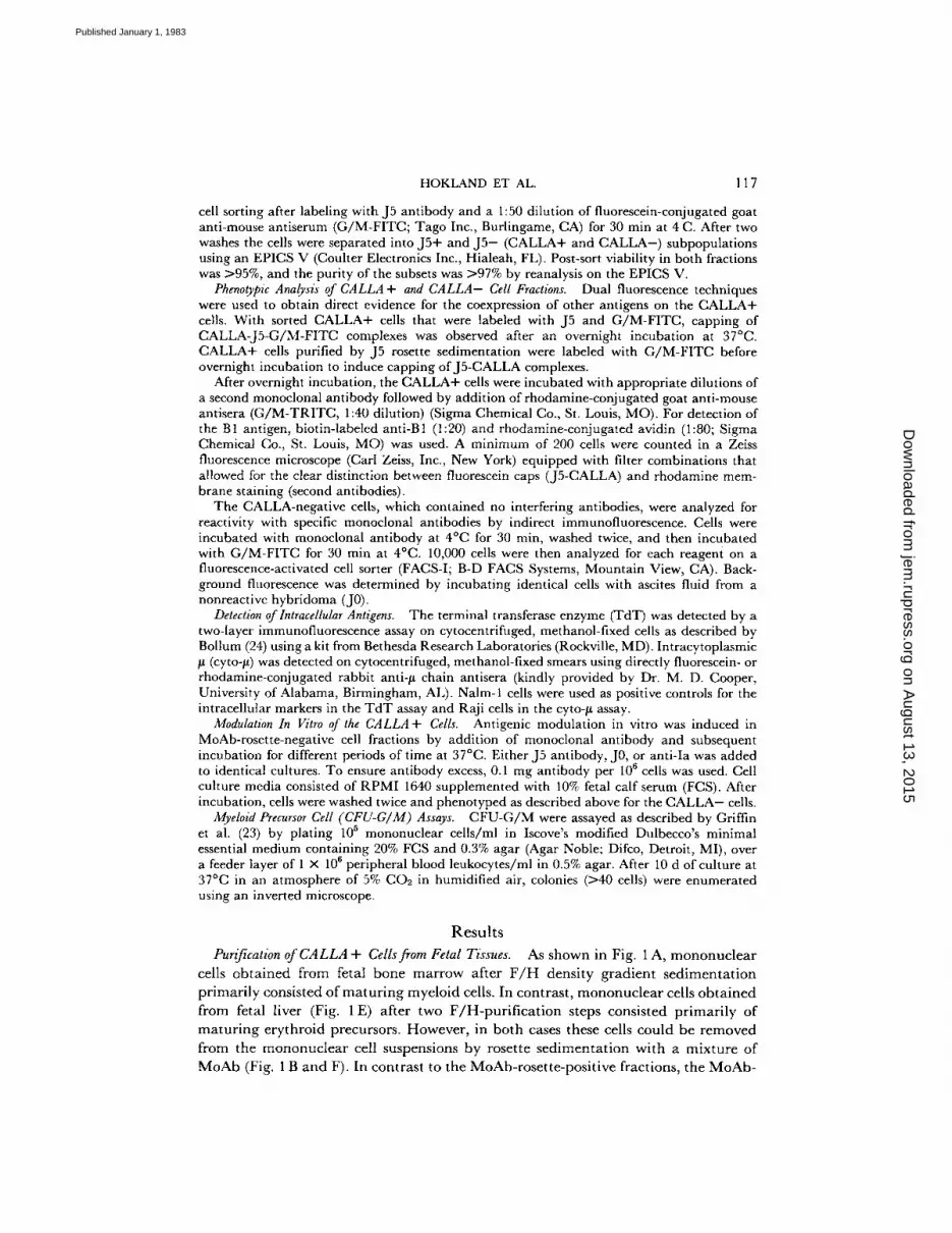
Purification of CALLA + Cells from Fetal Tissues. As shown in Fig. 1 A, mononuc lea r cells ob ta ined from fetal bone marrow after F / H density gradient sedimenta t ion pr imari ly consisted of ma tu r ing myeloid cells. In contrast, mononuc lea r cells ob ta ined from fetal liver (Fig. 1 E) after two F /H-pur i f i ca t ion steps consisted pr imari ly of ma tu r ing erythroid precursors. However, in both cases these cells could be removed from the mononuc lear cell suspensions by rosette sedimenta t ion with a mixture of MoAb (Fig. 1 B and F). In contrast to the MoAb-rosette-positive fractions, the MoAb-
on August 13, 2015
jem.rupress.org
Dow
nloaded from
Published January 1, 1983
118 CHARACTERIZATION OF FETAL HEMATOPOIETIC CELLS EXPRESSING CALLA
on August 13, 2015
jem.rupress.org
Dow
nloaded from
Published January 1, 1983

HOKLAND ET AL. 119
.~X
<
x ~.>
~ .
> o
e-
~< o .-.2 M N x <
X " " ~ . ,
X.~
o
• r " o
~ x ~ ~-x ~ 8 , b ;
m
e
< d
on August 13, 2015
jem.rupress.org
Dow
nloaded from
Published January 1, 1983
120 CHARACTERIZATION OF FETAL HEMATOPOIETIC CELLS EXPRESSING CALLA
rosette-negative suspensions from both bone marrow (Fig. 1 C) and liver (Fig, 1 G) were remarkably similar, consisting of a mixture of small to medium-sized lymphocytes and large blast cells. When separating the MoAb-rosette-negative fraction further into C A L L A + and C A L L A - subsets, the large blast cells were found exclusively in the C A L L A - fraction (Fig. 1 D), whereas the C A L L A + fraction consisted solely of lymphoid-appear ing cells (Fig. 1 H).
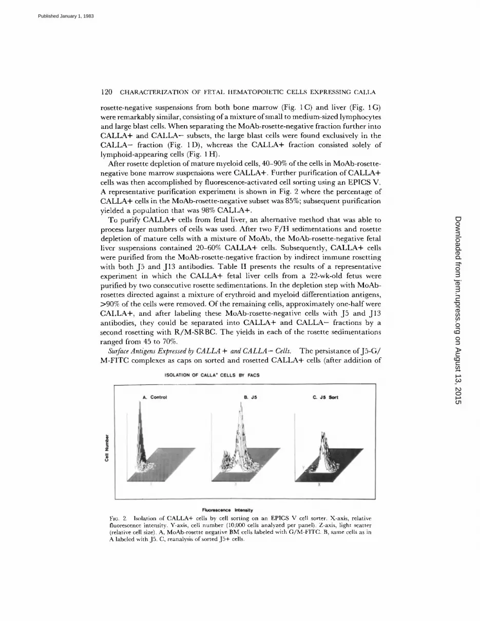
After rosette depletion of mature myeloid cells, 40-90% of the cells in MoAb-rosette- negative bone marrow suspensions were C A L L A + . Further purification of C A L L A + cells was then accomplished by fluorescence-activated cell sorting using an EPICS V. A representative purification experiment is shown in Fig. 2 where the percentage of C A L L A + cells in the MoAb-rosette-negative subset was 85%; subsequent purification yieIded a populat ion that was 98% C A L L A + .
To purify C A L L A + cells from fetal liver, an alternative method that was able to process larger numbers of cells was used. After two F / H sedimentations and rosette depletion of mature cells with a mixture of MoAb, the MoAb-rosette-negative fetal liver suspensions contained 20-60% C A L L A + cells. Subsequently, C A L L A + cells were purified from the MoAb-rosette-negative fraction by indirect immune rosetting with both J5 and J13 antibodies. Table II presents the results of a representative experiment in which the C A L L A + fetal liver cells from a 22-wk-old fetus were purified by two consecutive rosette sedimentations. In the depletion step with MoAb- rosettes directed against a mixture of erythroid and myeloid differentiation antigens, >90% of the cells were removed. Of the remaining cells, approximately one-half were C A L L A + , and after labeling these MoAb-rosette-negative cells with J5 and J13 antibodies, they could be separated into C A L L A + and C A L L A - fractions by a second rosetting with R / M - S R B C . The yields in each of the rosette sedimentations ranged from 45 to 70%.
Surface Antigens Expressed by CALLA + and CALLA- Cells. The persistance of J 5 - G / M - F I T C complexes as caps on sorted and rosetted C A L L A + cells (after addit ion of
FIc. 2. Isolation of CALLA+ cells by cell sorting on an EPICS V cell sorter. X-axis, relative fluorescence intensity. Y-axis, cell number (10,000 cells analyzed per panel). Z-axis, light scatter (relative cell size). A, MoAb-rosette negative BM cells labeled with G/M-FITC. B, same cells as in A labeled with J5. C, reanalysis of sorted JS+ cells.
on August 13, 2015
jem.rupress.org
Dow
nloaded from
Published January 1, 1983
HOKLAND ET AL. 121
TABLE II
Purification of CALLA + Gel#from Fetal Liver by Immune Rosette Sedimentations
Total J5+ Percent J5+ Cell fraction Total cells cells ceils*
× 10 ° × lOa Unseparated~: 510 35.7 7 MoAb-rosette positive§ 320 6.4 2 MoAb-rosette negative§ 38 20.1 53 MoAb-rosette negative, J5-[I l 2 0.4 3 MoAb-rosette negative, Js+ll 11 10.5 >95
* Determined by indirect immunofluorescence assay and FAGS analysis. :~ Mononuclear cell suspensions purified two times by F/H density-gradient
sedimentation. § Cells obtained from interfaces (rosette negative) or pellets (rosette positive)
after R/M-SRBC immune rosette sedimentation with cells labeled simulta- neously with Mol, Mo2, MY7, MY8, and T9 monoclonal antibodies.
[I Rosette-negative cell suspensions obtained after depletion with Mol, Mo2, MY7, MY8, and T9 monoclonal antibodies and R/M-SRBC were incubated with J5 antibody and R/M-SRBC. J 5 - (interface cells) and J5+ (pellet ceils) populations were then separated by an additional immune rosette sedimentation.
TABLE III
Phenotypic Analysis of CALLA + Cells from Fetal Hematopoietic Tissues with Monoclonal Antibodies
Percent positive cells*
Cell source Age MY7/ T3/ J2 J5 j13 Ia TI0 BI~: MY8§ T9 T l l §
Bone marrow
Liver
wk
2211 >95 >95 NT** >95 >95 45 NT NT NT 2211 >95 >95 >95 >95 >95 41 <5 NT NT 2311 NT >95 NT >95 >95 52 NT NT NT
21 ¶ >95 >90 >95 >95 >95 31 <5 <5 <5 2211 >95 >95 >95 >95 >95 39 <5 <5 <5 23¶ >95 >95 >95 >95 >95 30 <5 <5 <5 24¶ NT >90 NT >95 >95 27 NT NT NT
* Determined by fluorescence microscopy of J5+ cells incubated overnight at 37°C with G/M-FITC to obtain capping and subsequently stained with a second monoclonal antibody and G/M-TRITC.
:~ Labeled with biotin-conjugated B 1 followed by rhodamine-labeled avidin. § Labeled simultaneously with both monoclonal antibodies noted. 11 Purified from MoAb-rosette-negative cell fractions by cell sorting. ¶ Purified from MoAb-rosette-negative ceil fractions by J5 rosette sedimentation.
** Not tested.
G / M - F I T C to t h e l a t t e r ) a l l o w e d for t h e d e f i n i t i o n o f o t h e r cell su r f ace a n t i g e n s o n
these cells b y a s e c o n d m o n o c l o n a l a n t i b o d y a n d G / M - T R I T C . T h e resu l t s f r o m a
series o f e x p e r i m e n t s w i t h b o t h fe ta l b o n e m a r r o w a n d fe ta l l i ve r o f d i f f e r e n t ages a re
g i v e n in T a b l e III . A l t h o u g h these cells h a v e b e e n i so l a t ed f r o m d i f f e r e n t t issues b y
t w o d i f f e r en t m e t h o d s , t h e cells d i s p l a y e d s i m i l a r cell su r f ace a n t i g e n s . T h e p u r i t y o f
e a c h s u s p e n s i o n , as j u d g e d b y t h e p r e s e n c e o f J 5 - F I T C caps , e x c e e d e d 90% in eve ry
i n s t ance . I t s h o u l d also b e n o t e d t h a t a d d i t i o n o f m o r e J 5 a n t i b o d y a n d G / M - T R I T C
on August 13, 2015
jem.rupress.org
Dow
nloaded from
Published January 1, 1983

122 CHARACTERIZATION OF FETAL ItEMATOPOIETIC CELLS EXPRESSING CALI,A
did not give any addi t iona l fluorescence, ind ica t ing that all of the J5 surface ant igen was located wi thin the G / M - F I T C cap and that these cells had not synthesized more C A L L A dur ing the overnight culture. Fur ther analysis with J13 an t ibody , which is also specific for C A L L A , showed that near ly all the cells bound J13 within the J5- F I T C caps. There was also no apprec iab le cell surface s ta ining with J13 an t ibody other than tha t found in the J5 caps.
Three antigens, Ia, J2, and T10, were found on vi r tua l ly all the C A L L A + cells. In contrast to the react ivi ty pa t te rn seen with J13, these ant igens were uni formly d is t r ibuted a long the cell m e m b r a n e and were dist inct from C A L L A . The B 1 ant igen, in contrast , was only found on a fraction of the C A L L A + cells, and, when present, was uni formly d is t r ibu ted on the cell surface. Very few C A L L A + cells were reactive with an t i -myelo id or T cell ant ibodies. Thus, v i r tual ly all C A L L A + fetal bone marrow and liver cells were J 2 + , I a+ , T10+ , M Y 7 / 8 - , T 3 / 9 / 1 1 - , whereas 30-50% expressed B 1.
The pheno type of the C A L L A - cells ob ta ined after J 5 / J 1 3 rosette sed imenta t ion of MoAb-rose t te -nega t ive fractions was de te rmined by indirect immunof luorescence with subsequent analysis on a FACS-I . As shown in T a b l e IV, these cells have been effectively deple ted of C A L L A + cells, as j u d g e d by the low numbers of cells reactive with the J5 and J13 ant ibodies. Likewise, very few myeloid or ma tu re T cells were found. On the other hand , significant percentages of these cells expressed T l 0 . In contrast to the analysis of C A L L A + cells, only a minor i ty of the C A L L A - cells were reactive with ei ther J2 or anti-B1.
The C A L L A + and C A L L A - popula t ions were also ana lyzed for the presence of nuclear T d T and cyto-~ after me thano l fixation. As shown in T a b l e V, 10-30°7c of the C A L L A + cells express cyto-/t chains, but this marker is not restr icted to the C A L L A + subset because 10-15% of C A L L A - cells also conta in cyto-/*. Fur the rmore , the f inding of cyto-/~+ cells wi th in the C A L L A - subset correlates ra ther closely with the den> onst ra t ion of 11-17% B 1+ cells wi th in this popu la t ion (Table IV). W h e n expression of T d T was examined , it was found that 5-25% of C A L L A + cells exhib i ted a nuclear T d T staining pat tern . In the C A L L A - popula t ion , a somewhat higher percentage of cells (35-50%) was T d T positive. In fur ther exper iments , b io t in - labe led B1 and rhodamine-av id in were combined with the in t racel lu lar markers in C A L L A + cells. As summar ized in T a b l e VI, it was de te rmined that near ly all of the B 1+ cells were
TABLE IV Phenotypic Analysis of MoAb-Rosette-negative, C A L L A - Cells" Isolated from Fetal Liver
Percent positive cells* Age of
MY7/ fetus J2 J5 J13 Ia T10 BI MY8$ T9 T3/Tll}
wk 22 23 3 5 56 74 13 <1 0 2 22 16 <1 6 47 57 11 <1 4 <1 23 33 2 2 63 49 17 6 NT§ 6 27 13 3 2 NT 54 16 5 NT NT
* Determined by indirect immunofluorescence assay and FACS analysis. Labeled simultaneously with both monoclonal antibodies noted.
§ Not tested.
on August 13, 2015
jem.rupress.org
Dow
nloaded from
Published January 1, 1983
HOKLAND ET AL.
TABLE V
Intracellular Markers of Purified CALLA + and C A L L A - Fetal Cells
123
Cell source Age CALLA Cyto-/.t* TdT*
wk Bone marrow 22 +:]: 17 7
Bone marrow 23 + 23 16
Liver 21 + 14 7 Liver 22 + 27 19 Liver 23 + 21 21
Liver 22 - § 12 43 Liver 23 - 10 39 Liver 27 - 15 47
* Percent positive cells determined by immunofluorescence microscopy. 200 cells were scored in each experiment.
:1: +, MoAb-rosette negative, CALLA+. § - , MoAb-rosette negative, CALLA-.
TABLE V I
Double Marker Analysis of CALLA + Cells with B1 Antibody and lntracellular
Antigens
Subset Tissue Age T d T + T d T - Cyto-~+ Cyto-#-
wk B 1 positive Bone marrow 23 6 94* 44 56
Liver 23 13 87 28 72
B1 negative Bone marrow 23 28 72 2 98 Liver 23 32 68 1 99
* At least 200 cells were scored by fluorescence microscopy and the percentage of cells positive and negative for a specific marker was subsequently calculated.
A. Control Modulolion
z
IQ
a J$ Moduiolion
"~G/M FITC I
Fluorescence Intensity
C. Io Modulation
G/M FITC
...u~ ~r---7 -[a
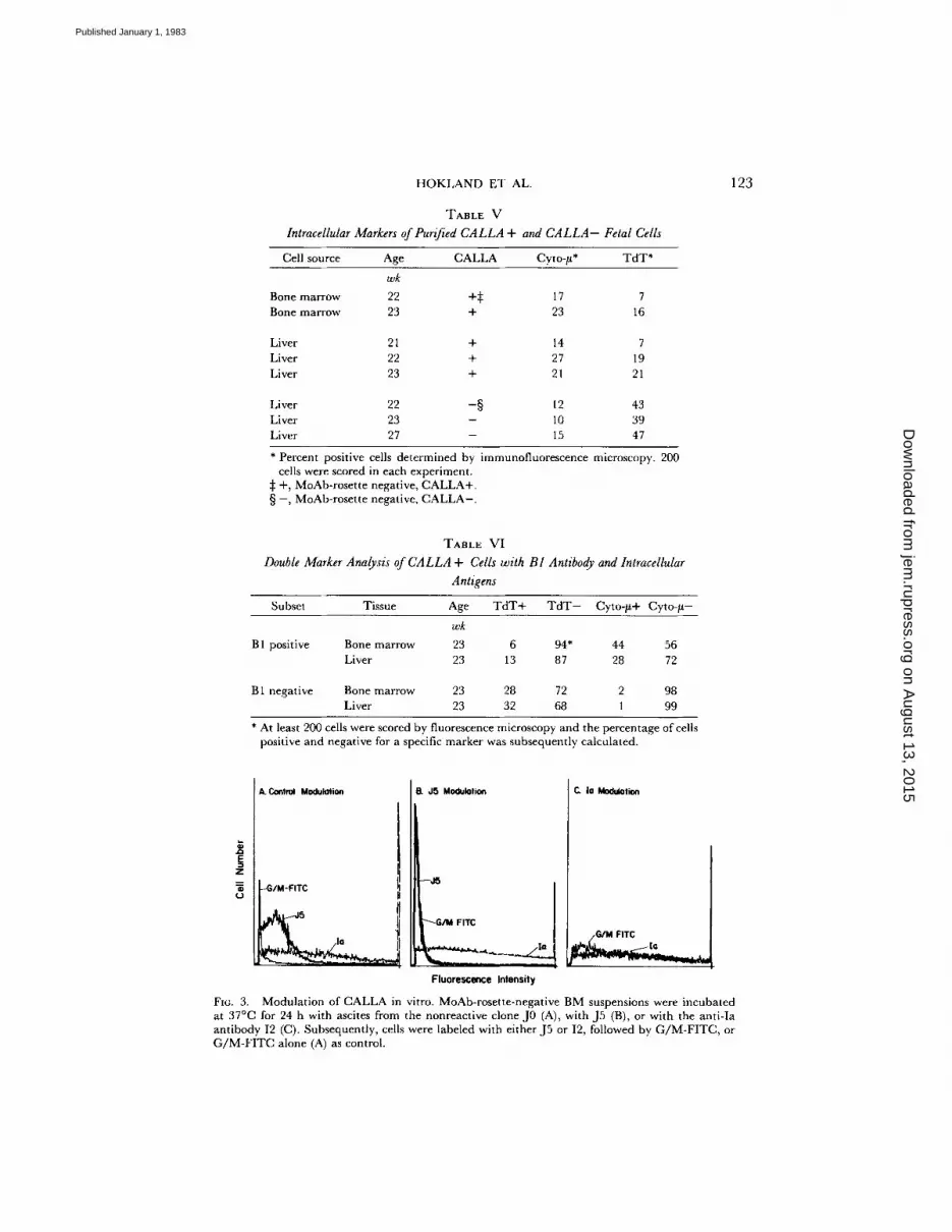
FIG. 3. Modulation of CALLA in vitro. MoAb-rosette-negative BM suspensions were incubated at 37°C for 24 h with ascites from the nonreactive clone J0 (A), with J5 (B), or with the anti-Ia antibody 12 (C). Subsequently, cells were labeled with either J5 or I2, followed by G/M-FITC, or G/M-FITC alone (A) as control.
on August 13, 2015
jem.rupress.org
Dow
nloaded from
Published January 1, 1983
124 CHARACTERIZATION OF FETAL HEMATOPOIETIC CELLS EXPRESSING CALLA
TABLE VII
Effect of CALLA Modulation by J5 Anttbody In Vitro on Expression of Unrelated Surface Antzgens
Incubation with J5
Percentage posilive cells*
J2 J5 J13 T10 B1 Ia
h
0 NT$ 64 NT NT 19 82 24 87 6 4 91 21 92 48 81 2 4 95 19 96
* Determined by indirect irnmunofluorescence assay and FACS analysis. :~ Not tested.
TABLE VIII
Analysis of CFU-G/M Cells
Colonies/105 Percent Specimen Subset * cells:l: yield§
Liver Unseparated cells 980 - - MoAb-rosette negative, CALLA- 1,600 56.0 MoAb-rosette negative, CALLA+ 50 0.4
Bone marrow
Liver
Unseparated cells 220 - - MoAb-rosette positive 70 18.5 MoAb-rosette negative 720 78.5 MoAb-rosette negative, CALLA- 2,100 76.3 MoAb-rosette negative, CALLA+ 50 0.1
Unseparated cells 650 - - MoAb-rosette positive 53 5.8 MoAb-roset te negative 1,600 45.7 MoAb-rosette negative, CALLA+ 16 0.0
* For explanation, see legend to Table II. ~: Scored day tO. § Based on the number of colonies compared with unseparated cells.
found in the T d T - subset and tha t the cyto-/x+ cells were a subset o f t he B 1 + cells.
Th i s suggests tha t there is very l i t t le o v e r l a p be tween subsets express ing e i the r T d T
or cy to-# w i th in C A L L A + fetal cells.
Modulation of CALLA Expression. Prev ious s tudies us ing l eukemic cells a n d cell l ines
h a v e d e m o n s t r a t e d tha t b i n d i n g of J 5 m o n o c l o n a l a n t i b o d y to cell sur face C A L L A
results in the specific m o d u l a t i o n o f this cell surface an t igen . W e were the re fore
in te res ted in e v a l u a t i n g the re levance o f this p h e n o m e n o n us ing n o n m a l i g n a n t
C A L L A + ceils. M o A b - r o s e t t e - n e g a t i v e bone m a r r o w (BM) suspensions h igh ly en-
r i ched for C A L L A + cells were i n c u b a t e d for var ious per iods o f t i m e wi th e i the r J 0
(ascites f rom a n o n r e a c t i v e h y b r i d o m a ) , J5 , or an t i - Ia , w h i c h has p rev ious ly been
shown not to m o d u l a t e Ia an t i gen express ion (8). As can be seen in Fig. 3 A, 65% of
the u n m o d u l a t e d cells were C A L L A + and 85% bore Ia ant igens . Af t e r i n c u b a t i o n
wi th J 5 at 37°C, 80% of the C A L L A + cells no longer expressed e i the r surface a n t i g e n or J 5 an t ibody . H o w e v e r , these cells c o n t i n u e d to express Ia an t igen . In cont ras t , the
Ia an t ibod ies d id not affect the express ion o f Ia a n t i g e n (Fig. 3C) . U s i n g a la rger
on August 13, 2015
jem.rupress.org
Dow
nloaded from
Published January 1, 1983
HOKLAND ET AL. 125
panel of MoAb to analyze the J5-modulated cells (Table VII), it was seen that the decrease in J5 reactivity was accompanied by a similar decrease in J13 expression. In contrast, only minor changes were seen in the expression of the non-CALLA-related antigens B1, J2, T10, and Ia even when the incubation period was extended to 48 h.
CFU-G/M Assays. Previous studies using adult bone marrow or peripheral blood depleted of CALLA+ cells by J5 and complement (C) treatment have shown that such procedures did not selectively deplete this population of committed myeloid precursor cells (GFU-C, BFU-E, CFU-E, CFU-G/E) (25). In Table VIII, data from experiments where different purified fractions of fetal liver and fetal bone marrow were tested for activity in CFU-G/M assays are shown. Irrespective of the tissue source and the method of purification, the CALLA+ cell fractions contained virtually no CFU-G/M. In contrast, most of the CFU-G/M could be recovered from the MoAb-rosette-negative and the C A L L A - fractions. Since none of the antibodies used during the rosette depletion reacted with CFU-G/M, the low number of colonies in the MoAb-rosette positive fractions strongly indicate that very little nonspecific loss of CFU-G/M occurred during rosette depletion.
Discussion
The series of experiments described in this study was designed to purify and characterize normal hematopoietic cells that express CALLA from fetal liver and fetal bone marrow, where they represent a relatively small fraction of the total rnononuclear cell population. A major reason for the success of the purification procedure was the initial MoAb-rosette depletion step, where R/M-SRBG were used to rosette leukocytes labeled with a mixture of MoAb against erythroid and myeloid differentiation antigens. In the subsequent rosette sedimentation, large numbers of erythroid and myeloid cells could be removed quickly and effectively. In this context, T9 antibody, which defines the transferrin receptor (20) was particularly helpful, because the majority of mononuclear cells in fetal liver suspensions, even after two F / H centrifu- gations, were normoblasts. In fetal bone marrow, the resulting rosette-negative cell suspensions were so enriched for CALLA+ cells that frequently these cells were >80% pure. Subsequently, further purification of CALLA+ cells was obtained either by cell sorting or by additional immune rosetting with J5 and J13 antibodies.
After purification, CALLA+ cells from both fetal liver and fetal bone marrow appeared to be a homogeneous population of medium-sized lymphocytes. However, with a series of monoclonal antibodies and immunofluorescence assays for intracyto- plasmic-/~ and TdT, we have been able to demonstrate that the phenotype of CALLA+ cells is strikingly heterogeneous. This was found particularly when markers for immature B cells were used, because only ~40% of the cells were positive for the B1 differentiation antigen and 15-30% expressed cyto-# (Tables III and V). Further- more, only a minority (5-25%) of the CALLA+ cells contained T d T (Table V). In contrast, virtually all CALLA+ cells expressed Ia, T10, and J2 antigens, and there were no major differences between cells isolated from fetal liver and fetal bone marrow. It should be noted, however, that some CALLA+ cells were also lost at each step of the purification procedure. For example, as shown in Table II, the MoAb- rosette-positive population contained 2% J5+ cells, and the MoAb-rosette-negative, J5-rosette-negative population contained 3% J5+ cells. As these CALLA+ cells comprised very small fractions of these populations, they could not be accurately
on August 13, 2015
jem.rupress.org
Dow
nloaded from
Published January 1, 1983
126 CHARACTERIZATION OF FETAL HEMATOPOIETIC CELLS EXPRESSING CALLA
characterized. However, because these losses appeared to be nonspecific, we have assumed that the J 5 + cells that were enriched and purified at each step are representative of the entire CALLA-posit ive population.
Earlier studies have demonstrated that leukemic cells from ~50% of patients with non-T ALL were reactive with B1 ant ibody (14). When expression of B1 antigen was correlated with expression of CALLA, it was found that expression of B 1 was restricted to those tumor cells that were also C A L L A + . Furthermore, recent studies using in vitro differentiation of AL L cells by phorbol diester showed that ALL cells which were C A L L A + , B 1 - could be induced to express B1 surface antigen, whereas C A L L A - AL L cells could not be induced to express B1 (26). Taken together, this analysis of leukemic cells suggested that the majority of C A L L A + non-T ALL cells were derived from the B cell lineage and Ied us to predict that C A L L A would be expressed at an earlier point in B cell development than B t. The analysis of C A L L A + fetal hematopoietic cells indicates that a large fraction of these cells are indeed in early phases of B cell development. In fact, the phenotype of normal C A L L A + cells is almost identical to that of C A L L A + leukemic cells, and, as has been suggested previously (4, 5), these normal cells appear to be the normal counterpar t of ALL cells. One major difference, however, is that only a small fraction of normal C A L L A + fetal lymphoid cells are T d T + , whereas almost all ALL cells contain this nuclear enzyme.
In addit ion to allowing a direct comparison of the surface markers of normal C A L L A + cells with leukemic cells, our purification of C A L L A + and C A L L A - populations has allowed the identification of fetal lymphoid cells at different stages of maturat ion. It is evident that within the C A L L A + positive populat ion at least three subsets can be identified. The first, and most differentiated, subset expresses both surface B 1 and cyto-bt , and therefore corresponds to a populat ion that has previously been classified as "pre-B" cells (27, 28). In addition, these cells express Ia, J2, and T10 antigens (as do all C A L L A + cells), but few of these cells appear to be T d T + . The second, less mature, subset has an identical surface phenotype (CALLA+, Ia+ , J 2 + , T10+, B I + ) but does not express cyto-~ or TdT. These cells appear to be at an earlier stage of lymphoid differentiation, but the expression of B1 suggests that they have already been commit ted to the B cell lineage. In studies noted previously (26), ALL cells with this phenotype can be induced to express cyto-/~ with phorbol diester. The third subset of C A L L A + cells expresses Ia, J2, and T 10 but does not express B 1. The lineage of these cells is not clearly established by their surface phenotype because none of these antigens are lineage specific. Although a significant fraction of these cells is T d T + , most are T d T - . It is likely that some cells within this group are very early B cells that will subsequently express B 1 and then cyto-~ with further differen- tiation. However, the expression of C A L L A is not restricted to B cells or B cell tumors because some T cell malignancies (T-ALL and lymphoblast ic lymphoma) also express this antigen. It is therefore possible that some ceils within this group may be T cell precursors, with yet-undefined surface markers, that will later migrate to the thymus where further differentiation will occur.
Within the C A L L A - cell fraction that was first depleted of mature erythroid and myeloid cells by immune rosetting and then further purified by removal of C A L L A + cells, our results also indicate considerable heterogeneity. Firstly, Giemsa-stained smears demonstrated that this fraction consisted of both large blast cells and small- to medium-sized lymphocytes (Fig. 1). Approximately 60% were T10+ , 55% were Ia+ ,
on August 13, 2015
jem.rupress.org
Dow
nloaded from
Published January 1, 1983

HOKLAND ET AL. 127
20% were J2+ , and 15% were B 1 + (Table IV). In addition, 35-50% of the C A L L A - cells were T d T + . It therefore seems likely that this fraction contains very early lymphoid precursor cells in addition to almost all myeloid precursor cells (here, CFU- G/M). Phenotypically, these cells appear to represent the nonmalignant counterparts of undifferentiated CALLA-- ALL cells. The heterogeneity of the C A L L A - fraction is further emphasized by the small but significant number of B I + cells within this population. Because we did not test for expression of surface Ig, it is possible that some of these C A L L A - , B 1 + cells are mature B cells. However, a small but significant percentage of C A L L A - cells also express cyto-#, and it is therefore evident that not all pre-B are contained within the CALLA+ population. Recent clinical studies also support the conclusion that the most immature stem cells (both myeloid and lymphoid) do not express CALLA. In these studies, patients with CALLA+ ALL in second remission received ablative chemotherapy and total body irradiation followed by infusion of autologous bone marrow that had been depleted of CALLA+ cells (both normal and leukemic) by in vitro treatment with J5 antibody and C (29). All patients thus far have been engrafted with antibody-treated bone marrow, and two patients who have been followed for >1 yr have demonstrated complete reconstitution of all myeloid and lymphoid populations.
Earlier studies using CALLA+ leukemia cells and cell lines have shown that J5 antibody induces antigenic modulation of cell surface antigen (8). This phenomenon has had important implications for the passive serotherapy of patients with ALL because J5 antibody infusions lead to loss of CALLA expression on malignant cells without impeding their growth (9). The data presented in Fig. 3 and T a b l e VII demonstrate that modulation of CALLA by J5 antibody is a physiologic phenomenon that occurs on normal CALLA+ ceils as well. Thus, whereas CALLA modulation may be a mechanism whereby leukemic cells may become resistant to antibody- mediated lysis, the modulation of CALLA on fetal cells may be indicative of a normal functional property of this cell surface glycoprotein. Other surface molecules that undergo antigenic modulation such as surface Ig (30), C3b receptor (31); and T3 antigen (32) appear to have specific membrane receptor-like functions; CALLA modulation may therefore be analogous to the phenomenon of "down-regulation" in which expression of cell surface receptor is decreased in the presence of excess ligand or hormone (33, 34).
S u m m a r y
Fetal hematopoietic cells that express the common acute lymphoblastic leukemia antigen (CALLA) were purified from both fe ta l liver and fetal bone marrow by immune rosetting with sheep erythrocytes coated with rabbit anti-mouse immuno- globulin and by fluorescence-activated cell sorting. Dual fluorescence techniques disclosed that these cells were heterogenous with respect to the expression of a series of differentiation and activation antigens defined by monoclonal antibodies. Thus, whereas all CALLA+ cells were Ia+ and expressed two activation antigens, J2 and T10, only 30-50% expressed B 1 antigen. Furthermore, using methanol-fixed cells, it could be shown that ~20% contained intracytoplasmic ~ chains (cyto-/l) and that
15% were positive for the terminal transferase enzyme (TdT) marker. The CALLA+ fetal cells thus closely resemble the childhood acute lymphoblastic leukemia cell with respect to surface marker phenotype. A population of CALLA-- cells devoid of mature erythroid and myeloid surface markers was found to contain higher numbers of T d T + cells but lower numbers of cyto-#, B1, and Ia+ cells than the CALLA+ subset. In
on August 13, 2015
jem.rupress.org
Dow
nloaded from
Published January 1, 1983
128 CHARACTERIZATION OF FETAL HEMATOPOIETIC CELLS EXPRESSING CALLA
vitro analysis of normal , pur i f ied C A L L A + cells demons t ra t ed tha t incuba t ion at 37°C with J5 monoclona l an t ibody specific for C A L L A resulted in the specific modu la t ion of surface antigen. S imi lar results have previously been ob ta ined with C A L L A + tumor cells. Al though pheno typ ic analysis of C A L L A + cells suggests tha t these cells are relat ively i m m a t u r e l ympho id cells, C A L L A + cells do not a p p e a r to conta in ei ther myelo id precursor cells ( C F U - G / M ) or the earliest l ympho id stem cells.
We thank Lori Palley for technical assistance and Pearl Leavitt for help in preparing the photomicrographs.
Received for publication 7July 1982 and in revised form 27 August 1982.
R e f e r e n c e s 1. Greaves, M. F., G. Brown, N. T. Rapson, and T. A. Lister. 1975. Antisera to acute
lymphoblastic leukemia cells. Clin. Immunol. Immunopathol. 4:67, 2. Ritz, J., J. M. Pesando, J. Notis-McConarty, H. Lazarus, and S. F. Schlossman. 1980. A
monoclonal antibody to human acute lymphoblastic leukemia antigen. Nature (Lond.). 283:583.
3. Ritz, J., L. M. Nadler, A. K. Bhan, J. Notis-McConarty, J. M. Pesando, and S. F. Schlossman. 1981. Expression of common acute lymphoblastic leukemia antigen (CALLA) by lymphomas of B cell and T cell lineage. Blood. 58:648.
4. Janossy, G., F. J. Bollum, K. F. Bradstock, A. McMichael, N. Rapson, and M Greaves. 1979. Terminal transferase bone marrow cells exhibit the antigen phenotype of common acute lymphoblastic leukemia. J. Immunol. 123:1525.
5. Greaves, M., D. Delia, G. Janossy, N. Rapson, J. Chessels, M. Woods, and G. Prentice. 1980. Acute lymphoblastic leukemia associated antigen. IV. Expression on nonleukemic "lymphoid" cells. Leukemia Res. 4:15.
6. Pesando, J. M., J. Ritz, H. Levine, C. Terhorst, H. Lazarus, and S. F. Schlossman. 1980. Human leukemia-associated antigen: relation to a family of surface glycoproteins. J. Immunol. 124:2794.
7. Metzgar, R. S., M. J. Borowitz, N. H. Jones, and B. L. Dowell. 1981. Distribution of common acute lymphoblastic leukemia antigen in nonhematopoietic tissues. J. Exp. Meal. 154:1249.
8. Ritz, J., j . M. Pesando, J. Notis-McConarty, and S. F. Schlossman. 1980. Modulation of human acute lymphoblastic leukemia antigen induced by monoclonal antibody in vitro.J. Immunol. 125:1506.
9. Ritz, J., J. M. Pesando, S. E. Sallan, L. A. Clavell, J. Notis-McConarty, P. Rosenthal, and S. F. Schlossman. 1981. Serotherapy of acute lymphoblastic leukemia with monoclonal antibody. Blood. 58:141.
10. Pesando, J. M., J. Ritz, H. Lazarus, K. J. Tomaselli, and S. F Schlossman. 1981. Fate of common acute lymphoblastic leukemia antigen during modulation by specific antibody.J. Immunol. 126:540.
11. Hercend, T., L. M. Nadler, J. M. Pesando, E. L. Reinherz, S. F. Schlossman, and J. Ritz. 1981. Expression of a 26,000 dalton glycoprotein on activated human T cells. Cell. lmmunol. 64:192.
12. Nadler, L. M., P. Stashenko, R. Hardy, J. M. Pesando, E. J. Yunis, and S. F. Schlossman. 1981. Monoclonal antibodies defining serologically distinct HLA-D/DR related Ia antigens in man. Human ImmunoL 1:77.
13. Stashenko, P., L. M. Nadler, R. Hardy, and S. F. Schlossman. 1980. Characterization of a human B lymphocyte specific antigen.J. Immunol. 125:1678.
14. Nadler, L. M., P. Stashenko, J. Ritz, R. Hardy, J. M. Pesando, and S. F. Schlossman. 1981. A unique cell surface antigen identifying lymphoid malignancies of B cell origin. J. Clin. Invest. 67:134.
on August 13, 2015
jem.rupress.org
Dow
nloaded from
Published January 1, 1983

HOKLAND ET Air. 129
15. Todd, R. F., L. M. Nadler, and S. F. Schlossman. 1981. Antigens on human monocytes identified by monoclonal antibodies. J. Immunol. 126:1435.
16. Todd, R. F., A. van Agthoven, S. F. Schlossman, and C. Terhorst. 1982. Structural analysis of differentiation antigens Mol and Mo2 on human monocytes. Hybridoma. 3:329.
17. Griffin, J. D., J. Ritz, L. M. Nadler, and S. F. Schlossman. 1981. Expression of myeloid differentiation antigens on normal and malignant myeloid cells. J. Clin. Invest. 68:932.
18. Reinherz, E. L., R. E. Hussey, and S. F. Schlossman. 1980. A monoclonal antibody blocking human T cell function. Eur. J. Immunol. 10:758.
19. Reinherz, E. L., P. C. Kung, G. Goldstein, R. H. Levy, and S. F. Schlossman. 1980. Discrete stages of human intrathymic differentiation: analysis of normal thymocytes and leukemic lymphoblasts of T lineage. Proc. NatL Acad. Sci. U. S. A. 58:954.
20. Sutherland, R., D. Delia, C. Schneider, R. Newman, J. Kemshead, and M. Greaves. 1981. Ubiquitous cell surface glycoprotein on tumor cells is proliferation-associated receptor for transferrin. Proc. Natl. Acad. Sci. U S. A. 78:4515.
21. Hercend, T., J. Ritz, S. F. Schlossman, and E. L. Reinherz. 1981. Comparative expression of T9, T10 and Ia antigens on activated human T cell subsets. Human Immunol. 3:247.
22. Meuer, S. C., S. F. Schlossman, and E. L. Reinherz. 1982. Clonal analysis of human cytotoxic T lymphocytes: T4+ and T8+ effector T cells recognize products of different major histocompatibility complex regions. Proc. Natl. Acad. Sci. U. S. A. 79:4395.
23. Griffin, J. D., R. P. Beveridge, and S. F. Schlossman. 1982. Isolation of myeloid progenitor cells from peripheral blood of chronic myelogenous leukemia patients. Blood. 60:30.
24. Bollum, F. J. 1979. Terminal deoxynucleotidyl transferase as a hematopoietic cell marker. Blood. 54:1203.
25. Clavell, L. A., J. M. Lipton, R. C. Bast, M. Kudisch, J. M. Pesando, S. F.Schlossman, and J. Ritz. 1981. Absence of common ALL antigen on bi-potent myeloid, erythroid, and granulocyte progenitors. Blood. 58:333.
26. Nadler, L. M., J. Ritz, M. P. Bates, E. K. Park, K. C. Anderson, S. E. Sallan, and S. F. Schlossman. 1982. Induction of human B cell antigens in non-T cell acute lymphoblastic leukemia. J. Clin. Invest. 70:433.
27. Pearl, E. R., L. B. Vogler, A. J. Okos, W. M. Christ, A. R. Lawton, and M. D. Cooper. 1978. B lymphocytic precursors in human bone marrow: an analysis of normal individuals and patients with antibody deficiency states. J. Immunol. 120:1169.
28. Owen, J. J. T. 1980. B cell development. Prog. Immunol. 4:303. 29. Ritz, J., S. E. Sallan, R. C. Bast, J. M. Lipton, L. A. Clavell, M. Feeney, T. Hercend, D.
G. Nathan, and S. F. Schlossman. 1982. Autologous bone marrow transplantation in CALLA positive acute lymphoblastic leukemia following in vitro treatment with J5 monoclonal antibody and complement. Lancet. II:60.
30. Knopf, P. M., A. Destree, and R. Hyman. 1973. Antibody-induced changes in expression of an immunoglobulin surface antigen. Eur. J. Immunol. 3:251.
31. Fearon, D. T., I. Kaneko, and G. G. Thomson. 1981. Membrane distribution and adsorptive endocytosis by C3b receptors on human polymorphonuclear leukocytes. J. Exp. Med. 153:1615.
32. Reinherz, E. L., S. C. Meuer, K. A. Fitzgerald, R. E. Hussey, H. Levine, and S. F. Schlossman. 1982. Antigen recognition by human T lymphocytes is linked to surface expression of the T3 molecular complex. Cell' 30:735.
33. Baldwin, D., M. Prince, S. Marshall, P. Davies, and J. M. Olefsky. 1980. Regulation of insulin receptors: evidence for involvement of an endocytic internalization pathway. Proc. Natl. Acad. Sci. U. S. A. 77:5975.
34. Pastan, I. H., and M. C. Willingham. 1981. Journey to the center of the cell: role of the receptosome. Science (Wash. D. C.). 214:504.
on August 13, 2015
jem.rupress.org
Dow
nloaded from
Published January 1, 1983