proteogenomic analysis of bradyrhizobium japonicum usda110 using genosuite, an automated...
TRANSCRIPT
1
Proteogenomic analysis of Bradyrhizobium japonicum USDA110 using Genosuite, an
automated multi-algorithmic pipeline
Dhirendra Kumar1, Amit Kumar Yadav
1, Puneet Kumar Kadimi
1, Shivashankar H. Nagaraj
2, Sean M
Grimmond2, Debasis Dash
1, *
1 G.N. Ramachandran Knowledge Center for Genome Informatics, CSIR-Institute of Genomics and
Integrative Biology, South Campus, Sukhdev Vihar, Mathura Road, Delhi-110025, India
2 Queensland Centre for Medical Genomics, Institute for Molecular Bioscience, The University of
Queensland, St Lucia, QLD, 4072, Australia
* To whom correspondence should be addressed.
Dr. Debasis Dash, G.N. Ramachandran Knowledge Center for Genome Informatics, CSIR-Institute of
Genomics and Integrative Biology, South Campus, Sukhdev Vihar, Mathura Road, Delhi-110025,
India
Tel: +91-11-29879301; Fax: +91-11-29879301; Email: [email protected]
MCP Papers in Press. Published on July 23, 2013 as Manuscript M112.027169
Copyright 2013 by The American Society for Biochemistry and Molecular Biology, Inc.

2
Running Title
Discovery of novel genes in B. japonicum by GenoSuite
Keywords
Proteomics, Genome annotation, Translation initiation site, Operon, N-Acetylation

3
List of Abbreviations
TIS- Translation Initiation Site
FDR- False Discovery Rate
PSM- Peptide Spectrum Match
NPCRs- Novel Protein Coding Regions
ORF- Open Reading Frame
4
SUMMARY
We present GenoSuite, an integrated proteogenomic pipeline to validate, refine and discover protein
coding genes using high-throughput mass spectrometry (MS) data from prokaryotes. To demonstrate
the effectiveness of GenoSuite, we analysed proteomics data of Bradyrhizobium japonicum
(USDA110), a model organism to study agriculturally important rhizobium-legume symbiosis. Our
analysis confirmed 31% of known genes, refined 49 gene models for their translation initiation site
(TIS) and discovered 59 novel protein coding genes. Notably, a novel protein which redefined the
boundary of a crucial cytochrome P450 system related operon was discovered, known to be highly
expressed in the anaerobic symbiotic bacteroids. A focussed analysis on N-terminally acetylated
peptides indicated downstream TIS for gene blr0594. Finally, ortho-proteogenomic analysis revealed
three novel genes in recently sequenced B. japonicum USDA6T genome. The discovery of large
number of missing genes and correction of gene models have expanded the proteomic landscape of
B. japonicum and presents an unparalleled utility of proteogenomic analyses and versatility of
GenoSuite for annotating prokaryotic genomes including pathogens.
INTRODUCTION
Rapid advances in massively parallel sequencing technologies have enabled the sequencing of
thousands of prokaryote genomes. However, to understand the functional elements of the genome,
annotation of the protein coding genes is the first and usually the most challenging task. Protein
coding genes in a genome are annotated by in silico predictions and homology searches against
known proteins. In silico gene annotations are prone to errors such as missing genes, incorrect
translation initiation sites (TIS) and pseudogenes. For instance, TIS mis-annotations have been
estimated to be around 10-40% in several bacterial and archaeal genomes (1, 2). Some genes are
wrongly annotated as pseudogenes in bacterial genomes (3). Several proteogenomic studies have
reported new protein coding genes which were previously missed by in silico gene annotations (4-6).
Mass spectrometry has emerged as a sensitive high-throughput method in which thousands of
proteins can be identified in a single experiment (7). Peptides identified from mass spectrometry
based proteomics data can be utilized as an experimental evidence to identify protein coding genes
and correct such annotation errors (7). The method of harnessing mass spectrometry proteomic data
5
to annotate genomes is generally referred as proteogenomics(8). This approach has been
successfully applied to re-annotate several genomes (1, 9-12) and to improve annotations of larger
taxonomic groups than a single bacterium (1, 13). Additionally, identification of peptides with protein
N-terminal modifications like formylated methionine or N-acetylation has been used to annotate TIS
for genes (5). Despite its direct benefits, proteogenomic analyses is computationally challenging,
multi-step process and tends to have high rates of false positive identification (14). Multi-algorithmic
search approaches have been shown to increase sensitivity and specificity in large scale proteomic
studies (15, 16) but are difficult to carry out in a proteogenomic context. This is due to the lack of
automated software for proteogenomic analyses that incorporates multiple search engines without
compromising on the statistical robustness of individual algorithms.
In order to fill this gap, we developed an automated pipeline, GenoSuite to carry out genome
translations, database searches using multiple search engines, result integration based on statistical
significance of PSMs, FDR calculations, coordinate mapping and finding completely novel genes. To
demonstrate the effectiveness of GenoSuite, we analysed proteomics data of Bradyrhizobium
japonicum (USDA110), a model organism to study agriculturally important rhizobium-legume
symbiosis. We selected B. japonicum for proteogenomic re-annotation because of its large genome
size, high GC content and non-availability of many closely related genome sequences. B. japonicum
is a symbiont to legumes and is important for nitrogen fixation in root nodules which helps these
plants to grow without any nitrogenous fertilizers. Its primary host is Soybean, an economically
important crop and model system to study rhizobia-legume symbiosis. This bacterium has a 9.1 Mb
genome, one of the longest among bacteria, with 64.1 % GC content (17). Kaneko et al. annotated
8,317 protein coding regions by in silico approach based on Glimmer (18) gene predictions and
sequence similarity with known proteins. A large number of transcriptomics and proteomics studies
have also been performed to understand the mechanisms and genes involved in symbiosis and
nitrogen fixing process. However, a high quality annotation of protein coding genes is still not
achieved for this class of bacteria. To improve on the existing annotation of B. japonicum, we carried
out a comprehensive proteogenomic analysis using publicly available proteomics data generated from
bacteroids of three host systems (19, 20). We used GenoSuite to search spectral data against
genome translated database of B. japonicum and selected peptide identifications at ≤1%FDR to re-

6
annotate B. japonicum genome. We identified 59 novel protein coding regions (NPCRs) and corrected
annotations for 49 genes.
EXPERIMENTAL PROCEDURES
Data
The mass spectral data for proteogenomic analysis on B. japonicum were obtained from PRIDE
repository (21). A total of nine data sets with PRIDE accessions 10099-10104 and 10114-10116 were
used. These mass spectra represent B. japonicum proteomes from three different host systems in
triplicates for each host. In total, these data sets had 621,176 MS/MS spectra.
Development of GenoSuite, an automated proteogenomic pipeline
We developed GenoSuite, a standalone pipeline for automated proteogenomic analysis. GenoSuite
is a suite of three tools: PPT (Prokaryotic Proteogenomic Tool), ORFmapper and PSMplotter.
GenoSuite is an easily configurable tool and is ready for use after downloading and unzipping the
archive. Integration with the search algorithms OMSSA, X!Tandem and InsPecT is also easy since
only the paths need to be added to GenoSuite configuration file. MassWiz comes integrated as a part
of the standard distribution.
PPT searches spectral data against a genome database with 4 peptide identification algorithms
namely MassWiz (22), OMSSA (23), X!Tandem (24) and InsPecT (25). It is highly configurable and
any combination of these algorithms can be used for search. All inputs for PPT are defined in a
configuration file. GenoSuite uses common file formats e.g. FASTA, Genbank, MGF, GFF etc., so that
the pipeline can be easily integrated with existing frameworks. We employed Combined
FDRScore(26) based approach to integrate results from multiple algorithms, each of which has a non-
comparable scoring metric. In brief, GenoSuite calculates FDRScore for all employed algorithms’
results. Average FDRScore is calculated for each PSM by calculating geometric mean of FDRScores
from individual algorithms identifying peptide spectrum pair. All PSMs are divided into subsets based
on the combination of algorithms identifying PSMs. Combined FDRScores are again calculated from
Average FDRScores separately for each subset of PSMs and significant PSMs below a user defined

7
FDR threshold are selected for further analyses. False Discovery Rate (FDR) (in %) is estimated by
Kall method(27)
100T
DFDR
D=Decoy PSMs passing the threshold
T=Target PSMs passing the threshold
Filtered peptides are then mapped onto the genome and also onto the known proteins. Identified
peptides mapping exclusively to the genome translated database are classified as novel peptides. A
complete outline of the analysis pipeline GenoSuite is shown in Figure 1. List of novel peptides are
exported as GFF files which can be integrated with any DAS annotation server. For spectral quality
assessment, XML files are created for peptide identifications from each algorithm and for novel
peptides.
ORFmapper compares novel peptides to existing annotations and to ab initio predictions. It requires
genbank file, ORF prediction file (GeneMark or GFF format) and novel peptide GFF as inputs. Novel
peptide coordinates in input GFF file are compared with the gene coordinates in genbank file and
peptides are further classified into (i) novel proteins coding region (NPCR) or (ii) gene model changes.
ORFmapper creates separate files for peptides leading to novel proteins, peptides suggesting gene
model changes and ORFs mapped to novel peptides. It also creates genomic map for each peptide
to provide a visual of its genomic context. These images are linked to an HTML file for ease of
analysis.
PSMplotter program is a utility to visualize peptide spectrum matches. It takes the XML file created by
PPT as its Input and generates an HTML file where all spectrum matches from the XML file are
hyperlinked with their PSM images. This can be used for manual validation of peptide spectral
matches.
It is written in Perl and the executables are distributed for Windows and Linux platforms. It is freely
available for download at (https://sourceforge.net/projects/proteogenomic/files/). The code is open-
source and freely available for academic purpose on request.

8
Proteogenomic analysis
Spectral data were searched by GenoSuite against a six frame translated database of B. japonicum
USDA110 genome (NC_004463.1). GenoSuite translates a genome from stop to stop codon in all six
reading frames and translation products of length 50 aa or more are kept in database. In total, the B.
japonicum database had 98,716 sequence entries. All four algorithms in the GenoSuite were
employed for spectral data search with 20 ppm precursor ion tolerance, 0.6 Da product ion tolerance,
trypsin as the protease with one missed cleavage, carbamidomethylation of cysteine as a fixed
modification and methionine oxidation as variable modification. For parameters not common across
algorithms, we used the algorithm defaults. Separate target-decoy database searches were carried
out. Stringent FDR threshold of ≤1% was applied to the resulting PSMs. Leucine and Isoleucine were
considered identical during FDR calculations and mapping onto the genome or proteome. Peptides
mapping to two different genomic loci were not considered for further analysis.
Translation Initiation Site search
A different search for estimating translation initiation sites for the gene models was performed using
OMSSA and X!Tandem. Search parameters were same as above except semitryptic enzyme
specificity, peptide N-terminal acetylation and peptide N-terminal formylated methionine as variable
modifications. Only N-terminally modified peptides identified at ≤ 1% FDR were selected for further
analysis.
Gene prediction and ORF comparison
To define boundaries of maximum number of novel translations in the B. japonicum USDA110
genome we used ORF predictions by four different algorithms namely GeneMark.hmm prokaryotic
(Version 2.6r) (28), Glimmer3 (18), Prodigal.v2.50 (29) and FGENESB (30). GeneMark, Glimmer and
Prodigal predictions were downloaded from NCBI
(ftp://ftp.ncbi.nih.gov/genomes/Bacteria/Bradyrhizobium_japonicum_USDA_110_uid57599/).
FGENESB gene predictions were downloaded from
(http://linux1.softberry.com/data/annotation/bact/NC_004463.fna.ann.gz). ORF comparison across
algorithms was performed using in-house scripts.
9
Protein functional annotation
In order to annotate proteins for their probable functions, we used domain assignment method Pfam
(31), Gene ontology assignments based on sequence similarity by GOANNA (32) and annotations by
RAST (33). Pfam-A profile database and HMMER-3.0 were used to assign profiles. GOANNA tool
was used for gene ontology assignment and only molecular function (F) category was used for
annotation.
RESULTS
Multi-algorithmic proteogenomic analysis pipeline development
GenoSuite was developed to automate spectral searches, statistical integration of PSMs and
downstream analysis. It is configured with four open source peptide identification algorithms namely
OMSSA, X!Tandem, InsPecT and MassWiz. The choice of algorithms was empirical and these
algorithms have been benchmarked in our previous studies (16, 22). These algorithms differ in their
basic scoring and have their own advantages. For instance, OMSSA uses a Poisson distribution for
separating significant matches from random hits(23) while X!Tandem uses a hyper-geometric model
(24). MassWiz is an empirical scorer based on continuity, mass error and intensity of matched
fragment ions (22). InsPecT is a tag based peptide identification method (25). Although there are
rescoring algorithms to improve database search results (34-36), a diverse set of algorithms provides
an increased sensitivity for peptide identifications than any single algorithm. GenoSuite incorporates a
reversed protein decoy database strategy to calculate FDR. For result integration from these
algorithms, GenoSuite calculates Combined FDRScore which is reported to give ≈35% more
identifications than any single algorithm(26). To check the consistency of Combined FDRScore
calculation implemented in GenoSuite, we compared the combined FDRScores calculated by
GenoSuite and FDRapp(37) from OMSSA and X!Tandem search results. We get highly correlated
CFS values from these two tools (Supplementary figure 1). FDRapp implements the same approach
on OMSSA, X!Tandem and Mascot algorithms. We extended this approach to Inspect and MassWiz
and implemented in GenoSuite (Supplementary figure 2). Peptides from FDR filtered PSMs are then
mapped onto the genome to report NPCR and gene model changes. Figure 1 depicts a schematic
10
chart of GenoSuite. Additional functionality for ORF prediction mappings and peptide spectral match
visualisation is also added to the pipeline.
Genome Search specific peptide identification
Total 621,176 spectra from nine different datasets were searched against B. japonicum genome. A
complete list of significant PSM identifications from each of nine datasets is provided in
Supplementary file 1. Total 26,592 peptides were identified of which 511 were shared across multiple
genomic loci. As the primary aim of this study was to annotate genomic loci, shared peptides were not
considered for further analysis. Figure 2.A depicts all unique peptides (26,081) identified in this study
mapped to their genomic loci. After mapping unique peptides onto the genome translated database,
GenoSuite reports proteins identified by at least two unique peptides or single peptide with ≥5
significant PSMs above desired FDR threshold. 2,654 proteins were identified in an automated
manner at a global protein level FDR of 0.0018. Supplementary file 2 provides a full list of proteins
identified in this analysis and their sample wise identification. Four out of ten proteins, which were
identified with only a single unique peptide, had dubious peptide spectral matches and hence were
not considered for further analysis. Supplementary file 3 provides annotated peptide spectral matches
for six accepted single peptide hits. GenoSuite further maps all peptides from significant protein
identifications to the annotated proteome and lists novel peptides. 283 peptides were reported as
novel. GenoSuite then categorizes novel peptides into (i) NPCR or (ii) changes in the annotated gene
models. A total of 59 new protein coding regions and 49 wrongly annotated genes were reported in
this automated analysis by GenoSuite. A potential contamination of host proteins or any other
contamination may also result in observation of novel peptides. To eliminate this possibility, we
mapped all the novel peptides on a sequence database of Glycine max and common contaminants
(cRAP) allowing one amino acid mismatch. 18 of these novel peptides, all of length 8 aa or below
were found similar with host or contaminant proteins. This did not affect the results at protein level as
novel proteins had other unique peptides. One gene model change suggested exclusively by a
peptide having similarity with host or contaminant proteins was not considered further.
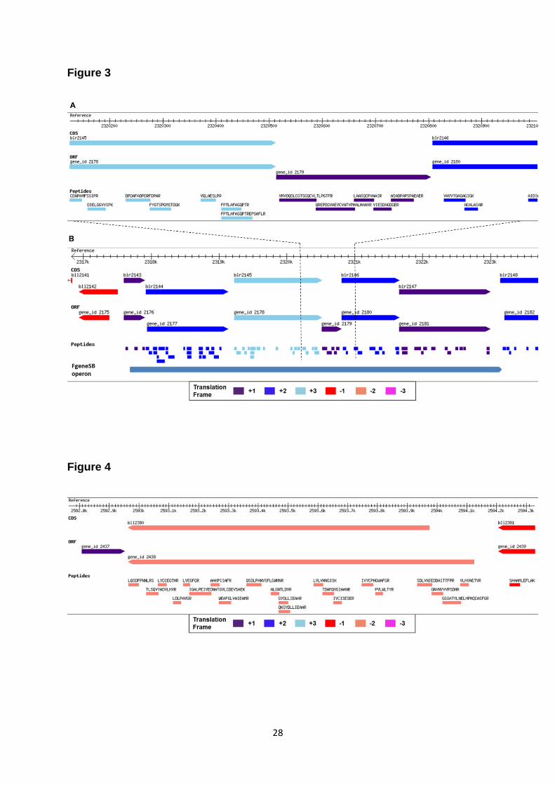
Novel protein coding regions in B. japonicum genome
We discovered 59 confident NPCRs in B. japonicum USDA110 genome. Supplementary file 4 lists all
NPCRs identified in this study with information about their function, sample-wise and host-wise

11
expression. Ab initio gene predictions were used to determine the boundary of un-annotated genes.
ORF predictions provide information about conservation of transcription and translational features
beyond coding region and add confidence to the peptide identifications. 51 out of 59 NPCRs are
supported by ORF predictions on the same frame and strand. Based on gene predictions, we
determined the translation initiation site (TIS) of these proteins. We also analysed the length
distribution of these 51 NPCRs. The average length of these proteins is 113 aa. Interestingly, 92 % of
these 51 novel proteins were below 200 aa length suggesting that most novel proteins are encoded
by short ORFs (Figure 2.B). Supplementary Table 1 lists all NPCRs with their genomic coordinates
and identified peptides. As our proteogenomic analysis is based on the proteomic data from different
hosts of B. japonicum, we analysed the NPCRs for their host-specific expression. Following the
criterion of Koch et al. for this analysis, we considered only those NPCRs which are identified in at
least two replicates for a host. Collectively, 34 NPCRs are identified in at least two replicates. 13 of
these are specifically expressed in bacteroids from soybean, two from siratro and three are specific
for cowpea bacteroids (Figure 2.C). Host specific expression of NPCRs suggests their putative role in
host adaptation and survival.
We also probed the genomic context of the NPCRs. Among all NPCRs, 36 were intergenic,
21 were on the opposite strand to existing gene annotation and two were on different frame. Figure
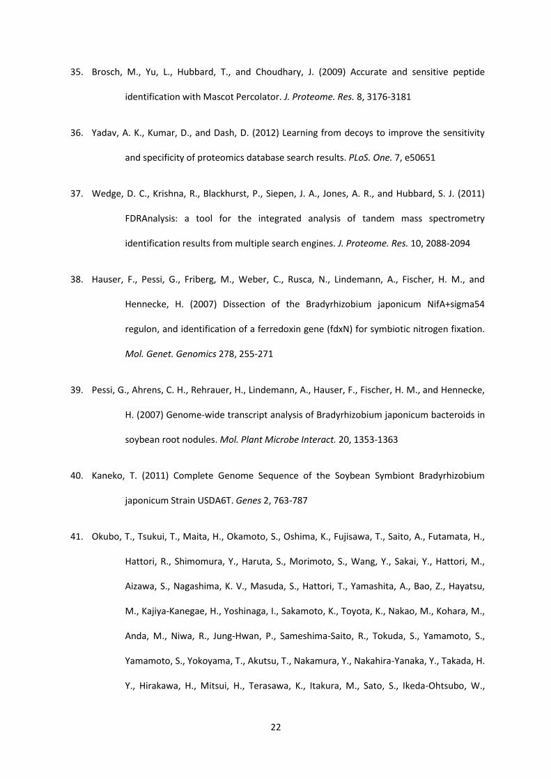
3.A shows a NPCR identified with five peptides mapping to an intergenic region of the genome. The
protein is supported by ORF prediction by all four gene prediction algorithms and is identified in five
out of nine samples. Function prediction suggests that this protein is a putative cytochrome P450
hydroxylase. Interestingly, Inclusion of this NPCR suggests a continuous operon in this genomic
region (Figure 3.B). Earlier this operon was considered with only three members (blr2143-blr2145) but
now with NPCR bridging the gap, this operon can be extended to six members. FGENESB also
predicts an operon of 6 proteins which includes the NPCR identified in this genomic locus. Sequence
similarity shows the NPCR and operon to be specific to the rhizobia. The operon has been shown to
be under regulation of oxygen-responsive transcriptional activator protein NifA (38) and is highly
expressed in anaerobic conditions of bacteroids within the host(39). Four proteins of the operon are
annotated as cytochrome P450 proteins and remaining two have cytochrome P450 hydroxylase
domain. In our analysis, all six proteins of this operon are identified with multiple peptide hits.

12
Identification of all proteins of this operon and their related functions further strengthens coordinated
gene expression as in operonic structures.
Novel proteins and comparative genomics
We extended newly identified annotations of B. japonicum in related rhizobial genomes based on
orthology of NPCRs. Ortho-proteogenomic strategy was first applied on genus Mycobacterium by
Gallien et al. (1) and recently adopted by Christie-Oleza et al. for annotating Roseobacter clad(10).
For this analysis we included all the sequenced genomes in Bradyrhizobiaceae family along with
Mesorhizobium loti, Sinorhizobium meliloti and Rhizobium leguminosarum since these are widely
studied rhizobial genomes. These genomes are of diverse sizes ranging from ≈3 Mb to ≈9 Mb with
3,122 to 8,826 protein coding genes. Genomic level identity between these genomes and B.
japonicum USDA110 genome ranges from 1% to 75% (Supplementary Table 2). A full list of NPCRs
with presence/absence of orthologs in related genomes is tabulated in Supplementary file 5.
Orthologous sequences were found for 43 NPCRs. Interestingly, orthologs of 35 NPCRs identified in
this study in B. japonicum USDA110 are annotated as protein coding genes in the recently sequenced
and annotated B. japonicum USDA6T (40) and/or B. sp.S23321 (41) genomes. After comparing the
orthologous loci with genome annotations and ORF predictions of the respective genomes, we added
three novel proteins in B. japonicum USDA6T genome and found three protein coding genes with N-
terminal changes.
N terminal changes in the annotated genes
Sixty two novel peptides mapped upstream of annotated gene models on the same frame and strand.
Since there was no stop codon between identified peptide and annotated gene, these peptides
suggested change in the currently annotated translation initiation site (TIS). We excluded one peptide
from the list because of its similarity with host proteins. The remaining 61 novel peptides indicated
changes in 48 annotated gene models. A detailed list of peptides suggesting gene model changes is
provided as Supplementary file 6. The ORF predictions were checked for any upstream TIS. 34 of
these 48 gene model changes are also supported by gene predictions. We could assign new TIS to
34 genes, 21 of which had TTG as the start codon. We considered maximum voted gene start of the

13
ORF predictions and the longest ORF in cases where votes tied. Figure 4 highlights a case of gene
model change. Four peptides map upstream to the currently annotated gene model for locus bll2380,
a gene coding for a glycosyl transferase enzyme. ORF prediction by GeneMark also agrees with the
upstream TIS. A longer bll2380 protein is also annotated in B. japonicum USDA6T and B. sp.23321.
In cases where ORF predictions did not support gene model changes, we applied sequence
similarity searches. One striking case is identified for NolA (bll2019), a transcriptional regulator protein
which is a key member involved in nodulation in the host plant during symbiosis. The NolA protein is
identified with six peptides. One of the peptides, IGELAEATGVTVR is identified in all nine samples
and it uniquely maps upstream to the current annotation of the NolA TIS. All four gene predictions
support the existing annotation of translational start site but do not cover the novel peptide. However,
a longer protein is reported for some close strains by sequence similarity analysis (Figure 5). Even for
B. japonicum USDA110, a longer NolA protein is suggested in few studies (42, 43). Interestingly, the
NolA locus of B. japonicum has been shown to code for 3 distinct proteins, varying in their TIS (44).
The peptide is covered in the longest protein isoform from NolA locus and is a part of DNA binding
domain in the protein’s N terminus. The observation of the novel peptide in all nine samples suggests
the longest protein isoform from NolA locus to be expressed during symbiosis with legume plants.
Translation initiation site (TIS) confirmation
We analysed protein translation initiation site by probing N terminal specific modifications. Formylation
happens on the initiator methionine and N-terminal acetylation on second amino acid after initiator
methionine is cleaved. These modifications directly mark the TIS for a protein coding gene. Based on
peptides identified at ≤1% FDR, and their upstream codon in the genome we could confirm the
annotated TIS for 15 genes. Annotated peptide spectrum matches of selected N-acetylated peptides
are provided in Supplementary file 7. Additionally, we corrected TIS for one gene where N-terminally
modified peptides did not agree with the currently annotated start site. Figure 6 depicts an N-
terminally acetylated peptide and a representative PSM which helped in correcting TIS for the locus
blr0594 (NP_767234.1), a thioredoxin protein. Peptide TIIDQGNGAAGPAAADLIK is identified with N-
terminal acetylation. The codon preceding the N-terminally acetylated Threonine is GTG which is
known to code for initiator methionine in genomes with high GC content. Based on the acetylated
peptide we corrected the TIS for blr0594 locus. The newly assigned TIS is downstream to the

14
annotated TIS for this locus. The orthologous protein sequences in related Bradyrhizobium genomes
also agree with the newly assigned TIS for blr0594.
Proteogenomic analysis of Shigella flexneri
We also applied GenoSuite on Shigella flexneri 2a str. 2457T data (Pride Accession 18992-18999). S.
flexneri is a human pathogen which causes dysentery and diarrhoea. We discovered 28 previously
un-annotated protein coding genes and corrected annotation for 22 genes which shows rapid
proteogenomic analyses of another prokaryotic genome (Supplementary file 8). No proteogenomic
study for this bacterium has been reported so far. Novel and better annotations can lead to better
understanding of pathogenesis by Shigella flexneri.
DISCUSSION
GenoSuite tool described here provides a simple and effective informatics framework for
proteogenomic analyses from prokaryotes. To the best of our knowledge, there are no readily
available tools dedicated for prokaryotic proteogenomic analyses. For instance, GAPP (45) is specific
to human annotation and no longer actively developed. PepLine (46) uses a de novo tag based
approach to detect peptides in a genome which is usually more error prone than database searches.
Integration of multiple algorithms in GenoSuite improves both sensitivity and specificity. Implementing
FDScore method on different algorithms before integrating their outputs and calculating FDR at PSM
and protein levels makes GenoSuite a robust statistical pipeline. It is currently designed to discover
simple gene models found in prokaryotes.
As a proof of principle, we have used GenoSuite to analyse Bradyrhizobium japonicum USDA110
genome for discovering novel protein coding genes. B. japonicum USDA110 is an agriculturally
important model organism to study symbiotic nitrogen fixation in legumes. A comprehensive
annotation is a key prerequisite for such studies but its genome annotation is far from complete. Since
only a handful of related genomes are sequenced, the annotation of protein coding genes could not
take advantage of comparative genomics. GenoSuite identified large number of new proteins coding
genes in B. japonicum USDA110. A significant number of these NPCRs are unique to this bacterium
while others have orthology in two closely related genomes, namely B. japonicum USDA6T and B.
15
sp.S23321. The exclusivity of these genes to one organism or to a small group of organisms makes
them difficult to annotate by methods other than proteogenomics. Most of the NPCRs in our analysis
are short in length (<200 aa). Previous proteogenomic studies in other organisms have also identified
mostly short novel proteins (5, 6) and have speculated that short length of proteins could be a reason
for missed annotation. Although Kaneko et al. (17) considered all ORF predictions of length 150 bp or
above for gene prediction, many short genes within the acceptable length range (>150bp) were left
un-annotated in B. japonicum USDA110 genome. It is generally believed that genomes with high
GC% suffer with over-prediction of short proteins (29, 47); our data suggests the opposite for B.
japonicum genome which also has high GC (64.1%). As NPCRs identified in proteogenomics studies
are experimentally discovered, these can be a valuable resource for improving the existing gene
prediction algorithms or development of new algorithms for predicting short length proteins.
Additionally, by applying ortho-proteogenomics, we identified three new genes in the B. japonicum
USDA6T genome. This indicates that although the computational gene prediction methods have
improved, they still miss some protein coding genes and it emphasizes the use of proteogenomics as
a powerful solution to such issues in genome annotation.
We also utilized novel peptides in the discovery of novel operons or correction of known operons. An
operon is predicted when genes are on the same strand and the gap between them is not more than
50-60 bp. Many novel genes or N-terminal extension to existing genes reduce this gap and can
correct operon models. In addition, it is a prerequisite that all proteins in new operon are identified.
We discovered 11 operon models which could not be annotated based on initial annotations of protein
coding genes. An example is an operon with six proteins related to Cytochrome P540 system. This
operon is highly active in anaerobic conditions within symbiotic bacteroids and is crucial for normal
nitrogen reduction within legume host. Christie-Oleza et al. also reported operons in Ruegeria
pomeroyi by proteogenomic analysis (10). Correction and unveiling of operonic structures can aid in
quick annotation as well as provide functional insights into the underlying biology of the organism.
Incorrect assignment of TIS is probably the most frequent error of genome annotation. We used fully
tryptic peptides to correct such errors. We found changes in 48 existing gene models in B. japonicum
genome. Most of the errors in TIS assignment were associated with TTG start codon. ATG and GTG
start codons are preferred over TTG by gene prediction algorithms which may have resulted in

16
incorrect assignment of TIS in observed cases. Protein sequences from these loci are significantly
changed and may contribute to newer structural and functional features as shown in NolA gene where
N-terminal extension provides additional DNA binding domain probably involved in a regulatory role.
Protein N-terminal modifications can also be used to probe TIS. All TIS confirmations in our study are
based on N-terminally acetylated peptides. N-Acetylation for blr0594 gene revealed that TIS is located
downstream to the currently annotated site. N-terminal acetylation is believed to be rare in bacteria
and only few proteins, mostly ribosomal, are known to be acetylated (48, 49). However, we observed
diverse classes of proteins as N-acetylated in B. japonicum USDA110 which agrees well with a recent
study by Bonissone S. et al.(50) , which showed N-acetylation as a widespread protein modification
among bacteria.
To establish GenoSuite’s versatility and applicability in discovering novel translations, it was also
applied on a recent Shigella flexneri 2a str. 2457T data where we discovered previously un-annotated
genes and corrected annotation for several genes. This demonstrates rapid and automated
proteogenomic analyses of prokaryotic genomes using GenoSuite. GenoSuite is a highly effective,
easily configurable analysis suite for prokaryotic proteogenomics.
REFERENCES
Reference List
1. Gallien, S., Perrodou, E., Carapito, C., Deshayes, C., Reyrat, J. M., Van, D. A., Poch, O.,
Schaeffer, C., and Lecompte, O. (2009) Ortho-proteogenomics: multiple proteomes
investigation through orthology and a new MS-based protocol. Genome Res. 19, 128-
135
2. Aivaliotis, M., Gevaert, K., Falb, M., Tebbe, A., Konstantinidis, K., Bisle, B., Klein, C., Martens, L.,
Staes, A., Timmerman, E., Van, D. J., Siedler, F., Pfeiffer, F., Vandekerckhove, J., and
Oesterhelt, D. (2007) Large-scale identification of N-terminal peptides in the halophilic

17
archaea Halobacterium salinarum and Natronomonas pharaonis. J. Proteome. Res. 6,
2195-2204
3. Lamontagne, J., Beland, M., Forest, A., Cote-Martin, A., Nassif, N., Tomaki, F., Moriyon, I.,
Moreno, E., and Paramithiotis, E. (2010) Proteomics-based confirmation of protein
expression and correction of annotation errors in the Brucella abortus genome. BMC.
Genomics 11, 300
4. Gupta, N., Benhamida, J., Bhargava, V., Goodman, D., Kain, E., Kerman, I., Nguyen, N.,
Ollikainen, N., Rodriguez, J., Wang, J., Lipton, M. S., Romine, M., Bafna, V., Smith, R. D.,
and Pevzner, P. A. (2008) Comparative proteogenomics: combining mass spectrometry
and comparative genomics to analyze multiple genomes. Genome Res. 18, 1133-1142
5. Kelkar, D. S., Kumar, D., Kumar, P., Balakrishnan, L., Muthusamy, B., Yadav, A. K., Shrivastava,
P., Marimuthu, A., Anand, S., Sundaram, H., Kingsbury, R., Harsha, H. C., Nair, B.,
Prasad, T. S., Chauhan, D. S., Katoch, K., Katoch, V. M., Kumar, P., Chaerkady, R.,
Ramachandran, S., Dash, D., and Pandey, A. (2011) Proteogenomic analysis of
Mycobacterium tuberculosis by high resolution mass spectrometry. Mol. Cell
Proteomics. 10, M111
6. Venter, E., Smith, R. D., and Payne, S. H. (2011) Proteogenomic analysis of bacteria and
archaea: a 46 organism case study. PLoS. One. 6, e27587
7. Yates, J. R., III, Eng, J. K., and McCormack, A. L. (1995) Mining genomes: correlating tandem
mass spectra of modified and unmodified peptides to sequences in nucleotide
databases. Anal. Chem. 67, 3202-3210
8. Jaffe, J. D., Berg, H. C., and Church, G. M. (2004) Proteogenomic mapping as a complementary
method to perform genome annotation. Proteomics. 4, 59-77

18
9. Baudet, M., Ortet, P., Gaillard, J. C., Fernandez, B., Guerin, P., Enjalbal, C., Subra, G., de, G. A.,
Barakat, M., Dedieu, A., and Armengaud, J. (2010) Proteomics-based refinement of
Deinococcus deserti genome annotation reveals an unwonted use of non-canonical
translation initiation codons. Mol. Cell Proteomics. 9, 415-426
10. Christie-Oleza, J. A., Miotello, G., and Armengaud, J. (2012) High-throughput proteogenomics
of Ruegeria pomeroyi: seeding a better genomic annotation for the whole marine
Roseobacter clade. BMC. Genomics 13, 73
11. Castellana, N. E., Payne, S. H., Shen, Z., Stanke, M., Bafna, V., and Briggs, S. P. (2008) Discovery
and revision of Arabidopsis genes by proteogenomics. Proc. Natl. Acad. Sci. U. S. A
105, 21034-21038
12. Chaerkady, R., Kelkar, D. S., Muthusamy, B., Kandasamy, K., Dwivedi, S. B., Sahasrabuddhe, N.
A., Kim, M. S., Renuse, S., Pinto, S. M., Sharma, R., Pawar, H., Sekhar, N. R., Mohanty,
A. K., Getnet, D., Yang, Y., Zhong, J., Dash, A. P., MacCallum, R. M., Delanghe, B.,
Mlambo, G., Kumar, A., Keshava Prasad, T. S., Okulate, M., Kumar, N., and Pandey, A.
(2011) A proteogenomic analysis of Anopheles gambiae using high-resolution Fourier
transform mass spectrometry. Genome Res. 21, 1872-1881
13. Zhong, Y., Chang, X., Cao, X. J., Zhang, Y., Zheng, H., Zhu, Y., Cai, C., Cui, Z., Zhang, Y., Li, Y. Y.,
Jiang, X. G., Zhao, G. P., Wang, S., Li, Y., Zeng, R., Li, X., and Guo, X. K. (2011)
Comparative proteogenomic analysis of the Leptospira interrogans virulence-
attenuated strain IPAV against the pathogenic strain 56601. Cell Res. 21, 1210-1229
14. Castellana, N., and Bafna, V. (2010) Proteogenomics to discover the full coding content of
genomes: a computational perspective. J. Proteomics. 73, 2124-2135

19
15. Yu, W., Taylor, J. A., Davis, M. T., Bonilla, L. E., Lee, K. A., Auger, P. L., Farnsworth, C. C.,
Welcher, A. A., and Patterson, S. D. (2010) Maximizing the sensitivity and reliability of
peptide identification in large-scale proteomic experiments by harnessing multiple
search engines. Proteomics. 10, 1172-1189
16. Yadav, A. K., Bhardwaj, G., Basak, T., Kumar, D., Ahmad, S., Priyadarshini, R., Singh, A. K., Dash,
D., and Sengupta, S. (2011) A systematic analysis of eluted fraction of plasma post
immunoaffinity depletion: implications in biomarker discovery. PLoS. One. 6, e24442
17. Kaneko, T., Nakamura, Y., Sato, S., Minamisawa, K., Uchiumi, T., Sasamoto, S., Watanabe, A.,
Idesawa, K., Iriguchi, M., Kawashima, K., Kohara, M., Matsumoto, M., Shimpo, S.,
Tsuruoka, H., Wada, T., Yamada, M., and Tabata, S. (2002) Complete genomic
sequence of nitrogen-fixing symbiotic bacterium Bradyrhizobium japonicum USDA110.
DNA Res. 9, 189-197
18. Delcher, A. L., Bratke, K. A., Powers, E. C., and Salzberg, S. L. (2007) Identifying bacterial genes
and endosymbiont DNA with Glimmer. Bioinformatics. 23, 673-679
19. Koch, M., Delmotte, N., Rehrauer, H., Vorholt, J. A., Pessi, G., and Hennecke, H. (2010)
Rhizobial adaptation to hosts, a new facet in the legume root-nodule symbiosis. Mol.
Plant Microbe Interact. 23, 784-790
20. Delmotte, N., Ahrens, C. H., Knief, C., Qeli, E., Koch, M., Fischer, H. M., Vorholt, J. A., Hennecke,
H., and Pessi, G. (2010) An integrated proteomics and transcriptomics reference data
set provides new insights into the Bradyrhizobium japonicum bacteroid metabolism in
soybean root nodules. Proteomics. 10, 1391-1400
20
21. Vizcaino, J. A., Cote, R., Reisinger, F., Barsnes, H., Foster, J. M., Rameseder, J., Hermjakob, H.,
and Martens, L. (2010) The Proteomics Identifications database: 2010 update. Nucleic
Acids Res. 38, D736-D742
22. Yadav, A. K., Kumar, D., and Dash, D. (2011) MassWiz: a novel scoring algorithm with target-
decoy based analysis pipeline for tandem mass spectrometry. J. Proteome. Res. 10,
2154-2160
23. Geer, L. Y., Markey, S. P., Kowalak, J. A., Wagner, L., Xu, M., Maynard, D. M., Yang, X., Shi, W.,
and Bryant, S. H. (2004) Open mass spectrometry search algorithm. J. Proteome. Res.
3, 958-964
24. Craig, R., and Beavis, R. C. (2004) TANDEM: matching proteins with tandem mass spectra.
Bioinformatics. 20, 1466-1467
25. Tanner, S., Shu, H., Frank, A., Wang, L. C., Zandi, E., Mumby, M., Pevzner, P. A., and Bafna, V.
(2005) InsPecT: identification of posttranslationally modified peptides from tandem
mass spectra. Anal. Chem. 77, 4626-4639
26. Jones, A. R., Siepen, J. A., Hubbard, S. J., and Paton, N. W. (2009) Improving sensitivity in
proteome studies by analysis of false discovery rates for multiple search engines.
PROTEOMICS 9, 1220-1229
27. Kall, L., Storey, J. D., MacCoss, M. J., and Noble, W. S. (2008) Assigning significance to peptides
identified by tandem mass spectrometry using decoy databases. J. Proteome. Res. 7,
29-34
28. Lukashin, A. V., and Borodovsky, M. (1998) GeneMark.hmm: new solutions for gene finding.
Nucleic Acids Res. 26, 1107-1115
21
29. Hyatt, D., Chen, G. L., Locascio, P. F., Land, M. L., Larimer, F. W., and Hauser, L. J. (2010)
Prodigal: prokaryotic gene recognition and translation initiation site identification.
BMC. Bioinformatics. 11, 119
30. (2010) FGENESB: Bacterial Operon and Gene Prediction, In:
http://linux1.softberry.com/berry.phtml?topic=fgenesb&group=programs&subgroup=
gfindb
31. Punta, M., Coggill, P. C., Eberhardt, R. Y., Mistry, J., Tate, J., Boursnell, C., Pang, N., Forslund, K.,
Ceric, G., Clements, J., Heger, A., Holm, L., Sonnhammer, E. L., Eddy, S. R., Bateman, A.,
and Finn, R. D. (2012) The Pfam protein families database. Nucleic Acids Res. 40, D290-
D301
32. McCarthy, F. M., Wang, N., Magee, G. B., Nanduri, B., Lawrence, M. L., Camon, E. B., Barrell, D.
G., Hill, D. P., Dolan, M. E., Williams, W. P., Luthe, D. S., Bridges, S. M., and Burgess, S.
C. (2006) AgBase: a functional genomics resource for agriculture. BMC. Genomics 7,
229
33. Aziz, R. K., Bartels, D., Best, A. A., DeJongh, M., Disz, T., Edwards, R. A., Formsma, K., Gerdes,
S., Glass, E. M., Kubal, M., Meyer, F., Olsen, G. J., Olson, R., Osterman, A. L., Overbeek,
R. A., McNeil, L. K., Paarmann, D., Paczian, T., Parrello, B., Pusch, G. D., Reich, C.,
Stevens, R., Vassieva, O., Vonstein, V., Wilke, A., and Zagnitko, O. (2008) The RAST
Server: rapid annotations using subsystems technology. BMC. Genomics 9, 75
34. Kall, L., Canterbury, J. D., Weston, J., Noble, W. S., and MacCoss, M. J. (2007) Semi-supervised
learning for peptide identification from shotgun proteomics datasets. Nat. Methods 4,
923-925
22
35. Brosch, M., Yu, L., Hubbard, T., and Choudhary, J. (2009) Accurate and sensitive peptide
identification with Mascot Percolator. J. Proteome. Res. 8, 3176-3181
36. Yadav, A. K., Kumar, D., and Dash, D. (2012) Learning from decoys to improve the sensitivity
and specificity of proteomics database search results. PLoS. One. 7, e50651
37. Wedge, D. C., Krishna, R., Blackhurst, P., Siepen, J. A., Jones, A. R., and Hubbard, S. J. (2011)
FDRAnalysis: a tool for the integrated analysis of tandem mass spectrometry
identification results from multiple search engines. J. Proteome. Res. 10, 2088-2094
38. Hauser, F., Pessi, G., Friberg, M., Weber, C., Rusca, N., Lindemann, A., Fischer, H. M., and
Hennecke, H. (2007) Dissection of the Bradyrhizobium japonicum NifA+sigma54
regulon, and identification of a ferredoxin gene (fdxN) for symbiotic nitrogen fixation.
Mol. Genet. Genomics 278, 255-271
39. Pessi, G., Ahrens, C. H., Rehrauer, H., Lindemann, A., Hauser, F., Fischer, H. M., and Hennecke,
H. (2007) Genome-wide transcript analysis of Bradyrhizobium japonicum bacteroids in
soybean root nodules. Mol. Plant Microbe Interact. 20, 1353-1363
40. Kaneko, T. (2011) Complete Genome Sequence of the Soybean Symbiont Bradyrhizobium
japonicum Strain USDA6T. Genes 2, 763-787
41. Okubo, T., Tsukui, T., Maita, H., Okamoto, S., Oshima, K., Fujisawa, T., Saito, A., Futamata, H.,
Hattori, R., Shimomura, Y., Haruta, S., Morimoto, S., Wang, Y., Sakai, Y., Hattori, M.,
Aizawa, S., Nagashima, K. V., Masuda, S., Hattori, T., Yamashita, A., Bao, Z., Hayatsu,
M., Kajiya-Kanegae, H., Yoshinaga, I., Sakamoto, K., Toyota, K., Nakao, M., Kohara, M.,
Anda, M., Niwa, R., Jung-Hwan, P., Sameshima-Saito, R., Tokuda, S., Yamamoto, S.,
Yamamoto, S., Yokoyama, T., Akutsu, T., Nakamura, Y., Nakahira-Yanaka, Y., Takada, H.
Y., Hirakawa, H., Mitsui, H., Terasawa, K., Itakura, M., Sato, S., Ikeda-Ohtsubo, W.,
23
Sakakura, N., Kaminuma, E., and Minamisawa, K. (2012) Complete Genome Sequence
of Bradyrhizobium sp. S23321: Insights into Symbiosis Evolution in Soil Oligotrophs.
Microbes. Environ. 27, 306-315
42. Gottfert, M., Rothlisberger, S., Kundig, C., Beck, C., Marty, R., and Hennecke, H. (2001)
Potential symbiosis-specific genes uncovered by sequencing a 410-kilobase DNA
region of the Bradyrhizobium japonicum chromosome. J. Bacteriol. 183, 1405-1412
43. Sadowsky, M. J., Cregan, P. B., Gottfert, M., Sharma, A., Gerhold, D., Rodriguez-Quinones, F.,
Keyser, H. H., Hennecke, H., and Stacey, G. (1991) The Bradyrhizobium japonicum nolA
gene and its involvement in the genotype-specific nodulation of soybeans. Proc. Natl.
Acad. Sci. U. S. A 88, 637-641
44. Loh, J., Stacey, M. G., Sadowsky, M. J., and Stacey, G. (1999) The Bradyrhizobium japonicum
nolA gene encodes three functionally distinct proteins. J. Bacteriol. 181, 1544-1554
45. Shadforth, I., Xu, W., Crowther, D., and Bessant, C. (2006) GAPP: a fully automated software
for the confident identification of human peptides from tandem mass spectra. J.
Proteome. Res. 5, 2849-2852
46. Ferro, M., Tardif, M., Reguer, E., Cahuzac, R., Bruley, C., Vermat, T., Nugues, E., Vigouroux, M.,
Vandenbrouck, Y., Garin, J., and Viari, A. (2008) PepLine: a software pipeline for high-
throughput direct mapping of tandem mass spectrometry data on genomic sequences.
J. Proteome. Res. 7, 1873-1883
47. Fukuchi, S., and Nishikawa, K. (2004) Estimation of the number of authentic orphan genes in
bacterial genomes. DNA Res. 11, 219-313
48. Soppa, J. (2010) Protein acetylation in archaea, bacteria, and eukaryotes. Archaea. 2010

24
49. Hu, L. I., Lima, B. P., and Wolfe, A. J. (2010) Bacterial protein acetylation: the dawning of a new
age. Mol. Microbiol. 77, 15-21
50. Bonissone, S., Gupta, N., Romine, M., Bradshaw, R. A., and Pevzner, P. A. (2012) N-terminal
protein processing: A comparative proteogenomic analysis. Mol. Cell Proteomics
ACKNOWLEDGEMENT
The authors thank Anupam Kumar Mondal for helpful discussions and insightful comments while
proof-reading the manuscript. DK is supported by Department of Science and Technology -INSPIRE
Senior Research Fellowship. AKY is supported by Council of Scientific and Industrial Research
(India), Senior Research Fellowship. SHN is supported by UQ Postdoctoral Research Fellowship.
SMG is supported by National Health and Medical Research Council (NHMRC) Principal Research
Fellowship. DD acknowledges CSIR In silico Biology project (CMM-0017) and Genesis project (BSC-
0121) for compute infrastructure and publication charges.
FIGURE LEGENDS
Figure 1: Schematic representation of GenoSuite workflow.
Figure 2: Proteogenomic identifications of peptides and proteins. (A) Genomic view of peptides
identified (B) Length distribution of novel proteins (C) Host specific expression of novel proteins of B
.japonicum.
Figure 3: Novel protein coding gene discovered in its genomic context. Reference track shows
genomic co-ordinates. CDS track represent annotated protein coding genes. ORF track shows ORF
predictions by GeneMark algorithm. Peptide track shows all identified peptides with their respective
sequences. Different bar colours represent different reading frames. (A) Five peptides map to a
genomic region where no protein coding gene is annotated. GeneMark predicts a putative gene in the
same translation frame of the identified peptide. (B) The identified novel protein suggests a

25
continuous operon. FGENESB also predicts an operon of six proteins including the novel protein.
Peptides are identified for all proteins in the operon.
Figure 4: Gene model correction discovered by proteogenomics. Four novel peptides map upstream
to annotated gene suggesting an upstream TIS. Four different tracks are shown. Reference track
shows genomic co-ordinates. CDS track represent annotated protein coding genes. ORF track shows
ORF predictions by GeneMark algorithm. Peptide track shows all the identified peptides with their
respective sequences. Different bar colours represent different reading frames.
Figure 5: Gene model change for NolA gene (bll2019). Peptide IGELAEATGVTVR is identified
upstream to current annotation of NolA gene on complement strand. GeneMark ORF prediction does
not include the novel peptide. However, longer ortholog proteins are annotated for some closely
related strains.
Figure 6: TIS correction discovered by N-acetylated peptides. Peptide TIIDQGNGAAGPAAADLIK
and its peptide spectral match. Blue coloured peaks are matched y-ion series. Red coloured peaks
are matched b-ion series. Black coloured peaks are unassigned experimental peaks.

26
Figure 1

27
Figure 2
28
Figure 3
Figure 4

29
Figure 5
Figure 6