protein deiminases: new players in the developmentally regulated loss of neural regenerative ability
TRANSCRIPT
Developmental Biology 355 (2011) 205–214
Contents lists available at ScienceDirect
Developmental Biology
j ourna l homepage: www.e lsev ie r.com/deve lopmenta lb io logy
Protein deiminases: New players in the developmentally regulated loss of neuralregenerative ability
Sigrun Lange a, Stefanie Gögel a, Kit-Yi Leung b, Bertrand Vernay a, Anthony P. Nicholas c, Corey P. Causey d,Paul R. Thompson e, Nicholas D.E. Greene b, Patrizia Ferretti a,⁎a Developmental Biology Unit, UCL Institute of Child Health, London WC1N 1EH, UKb Neural Development Unit, UCL Institute of Child Health, London WC1N 1EH, UKc Department of Neurology, University of Alabama at Birmingham and Birmingham VA Medical Center, Birmingham, Alabama 35294, USAd University of South Carolina, Department of Chemistry & Biochemistry, Columbia, 29208, USAe Department of Chemistry, TSRI, Scripps Florida, Florida 33458, USA
⁎ Corresponding author at: Developmental BiologyHealth, 30 Guilford Street, London WC1N 1EH, UK. Fax:
E-mail address: [email protected] (P. Ferretti).
0012-1606/$ – see front matter © 2011 Elsevier Inc. Aldoi:10.1016/j.ydbio.2011.04.015
a b s t r a c t
a r t i c l e i n f oArticle history:Received for publication 28 November 2010Revised 6 April 2011Accepted 14 April 2011Available online 22 April 2011
Keywords:ApoptosisDeimination/citrullinationDevelopmentPeptidyl arginine deiminaseRegenerationSpinal cord
Spinal cord regenerative ability is lost with development, but the mechanisms underlying this loss are stillpoorly understood. In chick embryos, effective regeneration does not occur after E13, when spinal cord injuryinduces extensive apoptotic response and tissue damage. As initial experiments showed that treatment with acalcium chelator after spinal cord injury reduced apoptosis and cavitation, we hypothesized thatdevelopmentally regulated mediators of calcium-dependent processes in secondary injury response maycontribute to loss of regenerative ability. To this purpose we screened for such changes in chick spinal cords atstages of development permissive (E11) and non-permissive (E15) for regeneration. Among thedevelopmentally regulated calcium-dependent proteins identified was PAD3, a member of the peptidylargi-nine deiminase (PAD) enzyme family that converts protein arginine residues to citrulline, a process known asdeimination or citrullination. This post-translational modification has not been previously associated withresponse to injury. Following injury, PAD3 up-regulation was greater in spinal cords injured at E15 than atE11. Consistent with these differences in gene expression, deimination was more extensive at the non-regenerating stage, E15, both in the gray and white matter. As deimination paralleled the extent of apoptosis,we investigated the effect of blocking PAD activity on cell death and deiminated-histone 3, one of the PADtargets we identified by mass-spectrometry analysis of spinal cord deiminated proteins. Treatment with thePAD inhibitor, Cl-amidine, reduced the abundance of deiminated-histone 3, consistent with inhibition of PADactivity, and significantly reduced apoptosis and tissue loss following injury at E15. Altogether, our findingsidentify PADs and deimination as developmentally regulated modulators of secondary injury response, andsuggest that PADs might be valuable therapeutic targets for spinal cord injury.
Unit, UCL Institute of Child+44 20 7831 4366.
l rights reserved.
© 2011 Elsevier Inc. All rights reserved.
Introduction
In amniotes, such as birds andmammals, the ability to significantlyregenerate the central nervous system (CNS) is lost with develop-ment. Comparative analysis of injury responses at permissive andnon-permissive stages of regeneration can therefore shed light on thecellular and molecular basis underlying such loss. The chick spinalcord provides an excellent model for these comparative studies, givenits accessibility in ovo and the fact that at non-regenerating stages ofdevelopment it appears to respond to traumatic injury in a similarfashion to the human spinal cord, by forming a large fluid-filled cavity.In contrast, the injury site in the mouse spinal cord is filled in withcells and connective tissue (Inman et al., 2002; Zhang et al., 1996).
The chick spinal cord displays remarkable regenerative ability,even at rather advanced stages of development, but this is eventuallylost around embryonic day 13 (E13) (Ferretti and Whalley, 2008;Hasan et al., 1991; Shimizu et al., 1990). The ability to regenerate thespinal cord, as well as the loss of it with development, is most likelydue to a combination of several factors, including changes in earlyresponse to injury (involving events occurring within hours ratherthan days after injury), progression of myelination and possiblyintrinsic properties of neurons and of neural stem/progenitor cells(Blackmore and Letourneau, 2006; Keirstead et al., 1992; Whalleyet al., 2006, 2009). Glial scar formation does not seem to play a crucialrole in the loss of regenerative ability, as no obvious scarring isobserved in the chick spinal cord either at regeneration competent orincompetent stages of development ((Shimizu et al., 1990) andFerretti et al., personal communication).
Significantly, early response to injury in the chick spinal cord isstrikingly different at regeneration permissive and non-permissive
206 S. Lange et al. / Developmental Biology 355 (2011) 205–214
stages of development, with the latter showing much more extensiveapoptosis and cavitation by 24 h after spinal cord injury (McBrideet al., 2003; Whalley et al., 2006). Changes in calcium homeostasisfollowing neural trauma play a critical role in the progression ofsecondary injury in mammals, including triggering an apoptoticresponse (Norberg et al., 2008; Orrenius et al., 2003; Velardo et al.,2000; Wang et al., 1999). Therefore, we wished to assess whetherdevelopmentally regulated calcium-dependent processes might alsoplay a role in the changes in secondary injury response, and con-sequently in the regenerative ability of the chick spinal cord, and useda microarray-based analysis to identify such genes.
Among differentially regulated calcium-dependent proteins, weidentified a member of the rather neglected calcium-dependentprotein peptidylarginine deiminase (PAD) family. Enzymes of the PADfamily, of which 5 members (PAD1–4 and PAD6) are known inmammals and 3 (PAD 1–3) in chick, convert protein arginine residuesto citrulline (deimination/citrullination) (Balandraud et al., 2005;Chavanas et al., 2004). It has been suggested that deimination plays arole in a number of diseases including skin diseases, rheumatoidarthritis and allergic encephalomyelitis (Cao et al., 1998; Doyle andMamula, 2005; Gyorgy et al., 2006; Mastronardi et al., 2006; Nicholaset al., 2005; Raijmakers et al., 2006; Vossenaar et al., 2003;Wood et al.,2008; Ying et al., 2009). Deimination is a post-translational modifi-cation that leads to a charge loss that can alter protein conformationand consequently their structure, function and interaction with otherproteins. We show here that PAD3 is developmentally regulated inresponse to injury and that PAD inhibition results in a reduction inapoptosis and cavitation, supporting a role for PAD and deimination inmodulating secondary injury response with development followingspinal cord damage. This study also points at PADs as potential ther-apeutic targets.
Materials and methods
Animals, surgery and pharmacological treatments
Fertilized Brown Leghorn eggs (Needle Farm, Cambridge, UK)wereincubated at 37 °C in a humidified forced flow incubator. All pro-cedures were approved under the Animals Scientific Procedures Act1986. Surgery and tissue collection was carried out as previouslyreported (Shimizu et al., 1990; Whalley et al., 2006).
Different concentrations of the Ca++ chelator BAPTA-AM solubi-lized in DMSO or of the PAD inhibitor, Cl-amidine (Knuckley et al.,2008; Luo et al., 2006), solubilized in phosphate buffer saline (PBS), orof the carrier alone were applied in ovo immediately after spinal cordinjury at E15 and their effect on survival assessed at 24 h (Whalleyet al., 2006). Selected concentrations were tested for their effect onapoptosis and deimination. The extent of cavity formation wasassessed in hematoxylin and eosin stained longitudinal spinal cordsections and apoptosis detected by TUNEL as previously described(Whalley et al., 2006). Briefly, montages of images of longitudinalsections of spinal cord stained with H&E or TUNEL were created at alevel close to the center of the injury and the size of visible cavitation.Tissue damage extending cranially and caudally from the site of injurywasmeasured at 24 h using ImageJ; apoptosis was assessed by scoringthe density of apoptotic cells on a scale of 1–4 using the criteriaexemplified in Fig. 6F at different distances from the injury site (1, 2, 3,4 mm). Statistical significance was evaluated by ANOVA and Student'st-test and pb0.05 was taken to be significant.
RNA analysis
Total mRNA was extracted from the cervical peri-injury region orthe equivalent region from sham-operated embryos of five individualspinal cords for each treatment group (E11 and E15 24 h post-surgery)following the protocol of ARK genomics (www.ark-genomics.org/
protocols). mRNA quality was checked using Agilent 2100 Bioanalyzerand mRNA expression assessed using chick GeneChip according tomanufacturer's protocol (Affymetrix). RNA (1 μg) from individualspinal cordswas used to provide independent replicates for each array.GeneSpring software (Agilent Technologies) was used for expressionanalysis and filteringwas performed byVolcano plot (1.5 fold changes,pb0.05) and verified using functional annotation clustering in David6.7.
PAD3 and Gapdh gene expression was determined by quantitativereal time PCR (7500 Fast Real Time PCR System, Applied Biosystems)using the SYBR GreenER reagent kit (Invitrogen) (Gögel et al., 2010).Each sample was run in duplicate. Primers were designed to amplifyspecific regions to the chickPAD3flanking region987–1123: PAD3chickforward: 5′ CGTGAAGGACAATGAGGACT 3′; PAD3 chick reverse: 5′GCACATAGCCAAACTCCACT3′. All results were normalized with respectto Gapdh expression; GAPDH chick forward 5'CCAGGTTGTCTCCTGT-GACT3′; GAPDH chick reverse 5'CACAACACGGTTGCTGTATC3′.
Protein analysis
Immunohistochemical staining and Western blotting were carriedout as previously described (Whalley et al., 2009). Sections wereimaged either under a Zeiss Axioplan or by confocal laser scanningmicroscopy (LSM 710, Zeiss, Germany). Complete “z” series opticalsections were collected and projected onto a single plane using Zeisssoftware and ImageJ. Nuclear and cytoplasmic protein fractions wereobtained by homogenizing pools of three cervical spinal cords in ice-cold hypotonic buffer (10 mM Hepes, pH 7.9, 1.5 mM MgCl2, 10 mMKCl, 0.5 mM DTT) supplemented with complete protease inhibitorcocktail (Sigma). Lysis was achieved using QIAshredder spin columns(QIAGEN) at 500 g. Cytoplasmic fractions were stored for furtheranalysis. Nuclear pellets resuspended in 0.5 ml of ice-cold high saltbuffer (20 mM Hepes pH 7.9, 25% glycerol, 1.5 mM MgCl2, 0.2 mMEDTA, 350 mM NaCl, 0.5 mM DTT) containing protease inhibitors andTritonX100 were kept on ice for 30 min. After centrifugation at13,000 g (30 min, 4 °C), supernatant containing nuclear proteins werecollected. Proteins (2–4 μg/lane) resolved by 12% SDS-PAGE wereanalyzed by Western blotting.
A pool of 5 spinal cords per treatment group was used for immu-noprecipitation of deiminated proteins, using the Catch and Release®v2.0 Kit according to the manufacturer's instructions (Upstate). Anidentical amount of proteins (5 μg) from each treatment group poolwas immunoprecipitated. Immunoprecipitated protein fractions wereanalyzed by 12% SDS-PAGE and Western blotting. Candidate bandswere excised from silver stained gels, and analyzed by liquid chro-matography electrospray tandemmass spectrometry (LC-ESI-MS/MS)as previously described (Gögel et al., 2010).
Primary antibodies were: F95 monoclonal mouse IgM antibody(Nicholas and Whitaker, 2002) (2 μg for immunoprecipitation; 1/200for immunohistochemistry; 1/5000 for Western blotting); rabbit anti-PAD3 (Chemicon, 1:200 for immunohistochemistry; 1:1000 for West-ern blotting), mouse anti-NeuN (1:50, Millipore); rabbit anti-GFAP(1:1000, Sigma); rat anti-MBP, and rabbit anti-deiminated histone 3(citH3; 1:100 and 1:250, Abcam). Secondary antibodies were: HRP-conjugated goat anti-mouse IgM (Serotec, USA 1:2000), HRP-conjugat-ed goat anti-rabbit (DAKO, 1:2000); Alexa 568-conjugated goat anti-mouse IgM and Alexa 488-conjugated goat anti-rabbit (1:400;Molecular Probes), Cy5-conjugated anti-rat (Invitrogen, 1:50).
Results
To establish whether inhibition of calcium influx at the time ofinjury reduces injury response in E15 spinal cords, we administered25 mg/kg of the calcium chelator BAPTA in ovo either in sham-operated or injured embryos; this dose did not impair embryonicsurvival, and slightly increased it (not shown). By 24 h after injury
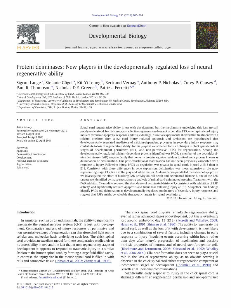
Fig. 1. Detection of apoptosis by TUNEL in control and BAPTA-treated E15 spinal cords 24 h after injury and neurofilament staining after 4 days. A) TUNEL staining of control spinalcord; note extensive apoptosis far from the injury site; asterisks indicate the extensive cavity. B) TUNEL staining of spinal cord treated with the Ca++ ionophore BAPTA at the time ofinjury; note the reduction in apoptosis. C) Neurofilament staining (red) in E19 control spinal cord injured at E15. Note extensive tissue disruption and overall failure of axons to crossthe injury site. D) Neurofilament staining (red) in E19 BAPTA-treated spinal cord injured at E15; though the tissue at the site of injury appears rather disorganized, there is no largecavity and extensive neurofilament staining is observed at the site of injury. E–F) H&E stained sections from the E19 control and BAPTA-treated spinal cords respectively. Scalebars=130 μm (A), 65 μm (B), 200 μm (C-F).
207S. Lange et al. / Developmental Biology 355 (2011) 205–214
E15 control embryos displayed an extensive apoptotic response, asindicated by TUNEL, and significant tissue loss was already evident(Fig. 1A). In contrast, in BAPTA-treated embryos little tissue damagewas observed and apoptotic cells were restricted to the region closeto the injury site (Fig. 1B). Furthermore, at 4 days after injury onlysmall cavities were observed and axonal tracts were present in the
Table 1Calcium-dependent molecules differentially regulated following spinal cord injury with dev
Gene (GenBank)
Peptidyl arginine deiminase, type III (NM_205043)S100 calcium binding protein A6 (calcyclin) (NM_204148)S100 calcium binding protein A9 (calgranulin B) (X61200)S100 calcium binding protein A11 (calgizzarin) (NM_205166)Cellular ligand of annexin 2(p11) (NM_205506)Annexin A1 (NM_206906)Annexin A11 (CR390633)Myosin light chain 2 (LC2f) (M11030)Myosin IF (MYO1F; CBBMIB) (NM_205254)Myosin Ie (Myosin heavy chain myr 3) (ENSGALT00000006599)Myosin, light polypeptide kinase (MYLK) (M96655)Phospholipase A2, group IVA (cytosolic, calcium-dependent) (NM_205423)Janus kinase 1 (JAK1) (NM_204870)Plastin 3 (T isoform) (PLS3; fimbrin) (AJ720945)Similar to calcium-regulated heat-stable protein (24 kD); calcineurinsubstrate CRHSP-24 (ENSGALT00000011834)
Calbindin 1, 28 kDa (NM_205513)Kv channel interacting protein 4 (KCNIP4) (NM_204555)Follistatin-like 4 (FSTL4) (NM_204502)
injured region though they did not display normal organization(Figs. 1C–F).
To identify possible modulators of this response, we carried outgene profiling experiments aimed at discovering molecules withcalcium-dependent activity differentially regulated in the chick spinalcord in response to injury at permissive (E11) and non-permissive
elopment.
mRNA fold changes
E11 inj/sh E15 inj/sh E15/E11 sh
1.8 6.0 2.14.4 22 ncnc 4.1 2.4nc 2.5 nc1.7 4.3 nc14.7 3.5 ncnc 1.7 ncnc 3.7 31.7 3.1 1.8nc 2.1 ncnc 1.5 ncnc 1.9 ncnc 1.6 ncnc 2.1 ncnc 2.2 nc
nc 0.4 ncnc 0.6 0.7nc 0.6 nc
208 S. Lange et al. / Developmental Biology 355 (2011) 205–214
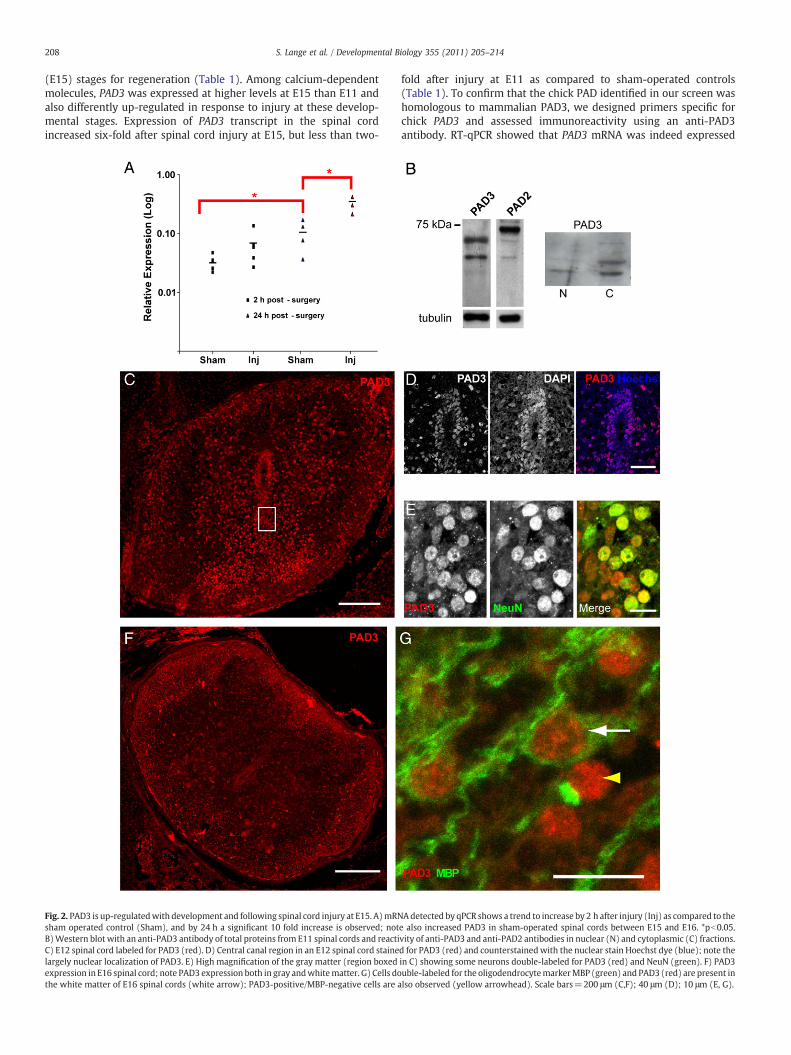
(E15) stages for regeneration (Table 1). Among calcium-dependentmolecules, PAD3 was expressed at higher levels at E15 than E11 andalso differently up-regulated in response to injury at these develop-mental stages. Expression of PAD3 transcript in the spinal cordincreased six-fold after spinal cord injury at E15, but less than two-
Fig. 2. PAD3 is up-regulatedwith development and following spinal cord injury at E15. A)mRNsham operated control (Sham), and by 24 h a significant 10 fold increase is observed; notB)Western blot with an anti-PAD3 antibody of total proteins from E11 spinal cords and reactiC) E12 spinal cord labeled for PAD3 (red). D) Central canal region in an E12 spinal cord stainelargely nuclear localization of PAD3. E) High magnification of the gray matter (region boxedexpression in E16 spinal cord; note PAD3 expression both in gray andwhitematter. G) Cells dothe white matter of E16 spinal cords (white arrow); PAD3-positive/MBP-negative cells are a
fold after injury at E11 as compared to sham-operated controls(Table 1). To confirm that the chick PAD identified in our screen washomologous to mammalian PAD3, we designed primers specific forchick PAD3 and assessed immunoreactivity using an anti-PAD3antibody. RT-qPCR showed that PAD3 mRNA was indeed expressed
Adetected by qPCR shows a trend to increase by 2 h after injury (Inj) as compared to thee also increased PAD3 in sham-operated spinal cords between E15 and E16. *pb0.05.vity of anti-PAD3 and anti-PAD2 antibodies in nuclear (N) and cytoplasmic (C) fractions.d for PAD3 (red) and counterstainedwith the nuclear stain Hoechst dye (blue); note thein C) showing some neurons double-labeled for PAD3 (red) and NeuN (green). F) PAD3uble-labeled for the oligodendrocytemarkerMBP (green) and PAD3 (red) are present inlso observed (yellow arrowhead). Scale bars=200 μm (C,F); 40 μm (D); 10 μm (E, G).
209S. Lange et al. / Developmental Biology 355 (2011) 205–214
in the chick spinal cord, increased significantly from E15 to E16 andwas greatly up-regulated after injury at E15 (Fig. 2A). As observed inthe microarray study, only a small PAD3 mRNA up-regulation wasobserved in response to injury at E11 by qRT-PCR (not shown).Expression of PAD3 proteinwas also assessed byWestern blotting andimmunohistochemistry. The anti-PAD3 antibody reacted with a chickprotein of molecular weight consistent with that of human PAD3(Nachat et al., 2005) and reactivity was detected both in the nucleusand in the cytoplasm, with a main band of approximately 70 kDabeing detected (Fig. 2B). Consistent with both nuclear and cytoplas-mic localization of PAD3 was the identity of proteins immunopreci-pitated by the PAD3 antibody identified by mass spectrometry, whichincluded histones and tubulin (Supplementary Table 1). Given thehigh sequence similarity between PADs and the fact that both PAD2and PAD3 are expressed in the spinal cord, we cannot fully rule outthat the antibody we used might to some extent react also with otherPADs. However, the difference in staining pattern observed with an-tibodies to PAD3 and PAD2 (Fig. 2B), with the latter detecting a highermolecular weight band, suggests specificity of the PAD3 stainingobserved in the chick spinal cord. In spinal cord sections (Figs. 2C–G),PAD3 reactivity in cells surrounding the central canal was mainlynuclear (Figs. 2C and D) and appeared to be stronger at E12 (Fig. 2C)than at E15 (Fig. 2F). At E11, PAD3 was also detected in a subset ofneurons, as indicated by co-labeling with the neuronal marker NeuN,and in the white matter (Fig. 2E). At E15 PAD staining was stillobserved in the gray matter and had increased in the white matter(Fig. 2F). Double-staining for MBP and PAD3 showed PAD3 expressionin developing oligodendrocytes, but some MBP-negative/PAD-posi-tive cells, presumably astrocytes, were also observed in the whitematter (Fig. 2G).
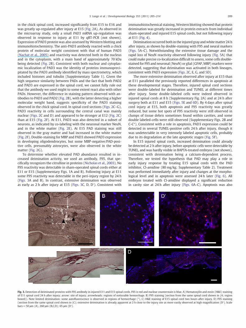
To determine whether elevated PAD abundance resulted in in-creased deimination activity, we used an antibody, F95, that spe-cifically recognizes the citrulline in proteins (Nicholas et al., 2003). NoF95 reactivity was detectable in sham-operated spinal cords either atE11 or E15 (Supplementary Figs. 1A and B). Following injury at E11some F95 reactivity was detectable in the peri-injury region by 24 h(Figs. 3A and B). In contrast, extensive deimination was observedas early as 2 h after injury at E15 (Figs. 3C, D, D′). Consistent with
Fig. 3. Detection of deiminated proteins with F95 antibody in injured E11 and E15 spinal cordof E11 spinal cord 24 h after injury; arrow: site of injury; arrowheads: regions of noticeableboxed)). Note limited deimination; some autofluorescence is observed in regions of hemo(section from the same spinal cord shown in (C); extensive deimination is already apparentbars=50 μm (A), 260 μm (B,C,D); 65 μm (D′).
immunohistochemical staining,Western blotting showed that proteindeimination was greatly increased in protein extracts from individualsham-operated and injured E15 spinal cords, but not following injuryat E11 (Fig. 4).
Deiminationoccurred both in the injuredgray andwhitematter 24 hafter injury, as shown by double-staining with F95 and neural markers(Figs. 5A–C). Notwithstanding the extensive tissue damage and therapid loss of NeuN reactivity observed following injury (Fig. 5A) thatcouldmake precise co-localization difficult to assess, some cells double-stained for F95 and neuronal (NeuN) or glial (GFAP,MBP)markersweredetected, suggesting that deimination was activated in both lineages,consistent with PAD3 expression (Figs. 2C, E, G, and 5D).
The more extensive deimination observed after injury at E15 thanat E11 paralleled the previously reported differences in apoptosis atthese developmental stages. Therefore, injured spinal cord sectionswere double-labeled for deimination and TUNEL at different timesafter injury. Some double-labeled cells were indeed observed ininjured spinal cords at 8 h (Supplementary Fig. 2A) and at 24 h aftersurgery both at E11 and E15 (Figs. 5E and 6D). By 4 days after spinalcord injury at E15, both apoptosis and F95 reactivity was greatlyreduced, but some hot spots of F95 reactivity were still observed inclumps of tissue debris sometimes found within cavities, and somedouble labeled-cells were still observed (Supplementary Figs. 2B andC-C″). Consistent with a role in apoptosis, PAD3 expression could bedetected in several TUNEL-positive cells 24 h after injury, though itwas undetectable in very intensely labeled apoptotic cells, probablydue to its degradation at the late apoptotic stages (Fig. 5F).
In E15 injured spinal cords, increased deimination could alreadybe detected at 2 h after injury, before apoptotic cells were detectable byTUNEL, and was hardly visible in BAPTA-treated embryos (not shown),consistent with deimination being a calcium-dependent process.Therefore, we tested the hypothesis that PAD may play a role inearly injury response by treating E15 spinal cords with the PADinhibitor, Cl-amidine (80 mg/kg; Supplementary Table 2). Treatmentwas performed immediately after injury and changes at the morpho-logical level and in apoptosis were assessed 24 h later (Fig. 6). Allembryos treated with Cl-amidine displayed a significant reductionin cavity size at 24 h after injury (Figs. 6A–C). Apoptosis was also
s. F95 is red and nuclear counterstain is blue. A) Hematoxylin and eosin (H&E) staininghemorrhage. B) F95 staining (section from the same spinal cord shown in (A; region
rrhage (*). C) H&E staining of E15 spinal cord two hours after injury. D) F95 stainingat 2 h close to the injury site as more easily observed at high magnification (D′). Scale
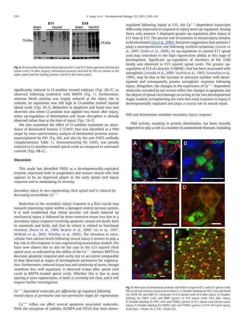
Fig. 5. Detection of deiminated proteins and PAD3 in injured E11 and E15 spinal cords.F95 is red and nuclear counterstain is blue A–C) Double-labeling for F95 (red) and NeuN(A), GFAP (B) and MBP (C) (all green) in E15 spinal cords 24 h after injury. D) Double-labeling for PAD3 (red) and MBP (green) in E15 spinal cords 24 h after injury.E) Double-labeling for F95 (red) and TUNEL (green) in E11 spinal cord 24 hors post-injury. F) Double-labeling for PAD3 (red) and TUNEL (green) in E15 24 h post-injury.Scale bars=20 μm (A–C, E,F); 10 μm (D).
Fig. 4.Western blot of proteins from individual E11 and E15 sham-operated and injuredspinal cords 2 h after surgery. Deiminated proteins detected by F95 are shown in theupper panel and the loading protein control in the lower panel.
210 S. Lange et al. / Developmental Biology 355 (2011) 205–214
significantly reduced in Cl-amidine treated embryos (Figs. 6D–F), asobserved following treatment with BAPTA (Fig. 1). Furthermore,whereas NeuN staining was largely reduced at the injury site incontrols, its expression was still high in Cl-amidine treated injuredspinal cords (Figs. 6G-I). Reduction in apoptosis and tissue loss wasobserved also when Cl-amidine was applied two hours after injury,when up-regulation of deimination and tissue disruption is alreadyobserved rather than at the time of injury (Figs. 7A–F).
We also examined the effect of Cl-amidine treatment on abun-dance of deiminated histone 3 (CitH3) that was identified as a PADtarget by mass-spectrometry analysis of deiminated proteins immu-noprecipitated by F95 (Fig. 8A) and also by the anti-PAD3 antibody(Supplementary Table 1). Immunostaining for CitH3, was greatlyreduced in Cl-amidine-treated spinal cords as compared to untreatedcontrols (Figs. 8B–C).
Discussion
This study has identified PAD3 as a developmentally-regulatedenzyme, expressed both in progenitors and mature neural cells thatappears to be an important player in the early spinal cord injuryresponse and in modulating its severity.
Secondary injury in non-regenerating chick spinal cord is reduced bydecreasing intracellular Ca++
Reduction in the secondary injury response is a first crucial steptowards improving repair within a damaged central nervous system.It is well established that initial necrotic cell death induced bymechanical injury is followed by more extensive tissue loss due to asecondary injury response involving apoptotic neural cell death, bothin mammals and birds, and that its extent is related to functionalrecovery (Basso et al., 1996; Beattie et al., 2000; Liu et al., 1997;McBride et al., 2003; Whalley et al., 2006). The elevation in intra-cellular free calcium levels following neural injury is known to play akey role in this response in non-regenerating mammalian models. Wehave now shown this to also be the case in the E15 injured chickspinal cord, as indicated by the ability of the Ca++ chelator BAPTA todecrease apoptotic response and cavity size to an extent comparableto that observed at stages of development permissive for regenera-tion. Furthermore, reduced tissue loss and continuity of axons, thoughsomehow less well organized, is observed 4 days after spinal cordcrush in BAPTA-treated spinal cords. Whether this is due to axonsparing or axon regeneration, or both, is currently not clear, and it willrequire further investigation.
Ca++-dependent molecules are differently up-regulated followingneural injury at permissive and non-permissive stages for regeneration
Ca++ influx can affect several apoptosis associated molecules.With the exception of calbidin, KCNIP4 and FSTL4 that were down-
regulated following injury at E15, the Ca++-dependent transcriptsdifferently expressed in response to injury were up-regulated. Amongthem, only annexin 1 displayed greater up-regulation after injury atE11 than at E15. The precise role of annexins in neural injury remainsto be elucidated (Liu et al., 2004), but given suggestions that annexin 1plays a neuroprotective role following cerebral ischaemia (Gavins etal., 2007; Solito et al., 2008), its up-regulation in injured E11 spinalcord may contribute to the high regenerative ability at this stage ofdevelopment. Significant up-regulation of members of the S100family was observed in E15 injured spinal cords. The greater up-regulation at E15 of calcyclin (S100A6), that has been associated withastrogliosis (Lesniak et al., 2009; Yachnis et al., 1993; Yamashita et al.,1999), may be due to the increase in astrocyte number with devel-opment and consequently greater astrogliotic response followinginjury. Altogether, the changes in the expression of Ca++-dependentmolecules revealed by our screen reflect the changes in apoptosis andthe degree of spinal cord damage occurring at the two developmentalstages studied, strengthening the view that early response to injury isdevelopmentally regulated and plays a crucial role in neural repair.
PAD and deimination modulate secondary injury response
PAD activity, resulting in protein deimination, has been recentlysuggested to play a role in a number of autoimmune diseases, including
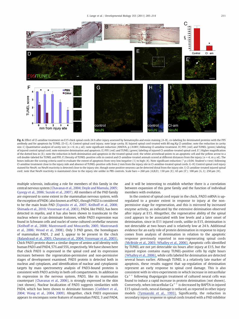
Fig. 6. Effect of Cl-amidine treatment on E15 chick spinal cords 24 h after injury assessed by hematoxylin and eosin staining (A–B), co-labeling for deiminated proteins with the F95antibody and for apoptosis by TUNEL (D–E). A) Control spinal cord injury; note large cavity. B) Injured spinal cord treated with 80 mg/Kg Cl-amidine; note the reduction in cavitysize. C) Quantitative analysis of cavity size (n=6; m±sd); note significant reduction (ANOVA, p≤0.001) following Cl-amidine treatment. D) F95 (red) and TUNEL (green) labelingof injured control spinal cord; note extensive deimination and apoptosis. E) F95 (red) and TUNEL (green) labeling of injured Cl-amidine-treated spinal cord. E′) Higher magnificationof the dotted box in (E); note the reduction in both deimination and apoptosis in the treated spinal cord; the white arrowhead points to an apoptotic cell and the yellow arrow to acell double-labeled for TUNEL and F95. F) Density of TUNEL-positive cells in control and Cl-amidine-treated animals scored at different distances from the injury (n=4; m±sd). Theboxes indicate the scoring criteria used to evaluate the extent of apoptosis from very low/negative (1) to high (4). Note significant reduction (* p≤0.04; Student's t-test) followingCl-amidine treatment close to the injury side and absence of TUNEL-positive cells from 2 mm from the injury site in Cl-amidine-treated spinal cords. G–H) Control spinal cord injurystained for NeuN; no NeuN reactivity is detected close to the injury site, though some positive neurons can be detected distal from the injury site. I) Cl-amidine-treated injured spinalcord; note that NeuN reactivity is maintained close to the injury site unlike in PBS controls. Scale bars=260 µm (A,B,D); 130 µm (E); 65 μm (E’); 100 μm (G, I); 250 μm (H).
211S. Lange et al. / Developmental Biology 355 (2011) 205–214
multiple sclerosis, indicating a role for members of this family in thecentral nervous system(Chavanas et al., 2004;Doyle andMamula, 2005;Gyorgy et al., 2006; Suzuki et al., 2007). All members of the PAD familyare expressed to some extent in the mammalian nervous system, withtheexceptionof PAD6 (also knownasePAD), thoughPAD2 is consideredto be the main brain PAD (Esposito et al., 2007; Keilhoff et al., 2008;Shimada et al., 2010; Vossenaar et al., 2003). PAD4, like PAD2, has beendetected in myelin, and it has also been shown to translocate to thenucleus where it can deiminate histones, while PAD3 expression wasfound in Schwann cells and nuclei of some cultured cerebellar neurons(Keilhoff et al., 2008; Mastronardi and Moscarello, 2005; Mastronardiet al., 2006; Wood et al., 2008). Only 3 PAD genes, the homologuesof mammalian PAD1, 2 and 3, appear to be present in the chick(Balandraud et al., 2005; Chavanas et al., 2004; Vossenaar et al., 2003).Chick PAD3 protein shares a similar degree of amino acid identity withhumanPAD3andPAD4, 57%and55%, respectively.Wehave shownherethat chick PAD3 is expressed in the spinal cord and its expressionincreases between the regeneration-permissive and non-permissivestages of development examined. PAD3 protein is detected both innucleus and cytoplasm, and identification of nuclear and cytoplasmictargets by mass spectrometry analysis of PAD3-bound proteins isconsistent with PAD3 activity in both cell compartments. In addition toits expression in the nervous system, PAD3, like its mammaliancounterpart (Chavanas et al., 2006), is strongly expressed in the skin(not shown). Nuclear localization of PAD3 suggests similarities withPAD4, which has been shown to deiminate histones (Cuthbert et al.,2004; Wang et al., 2004, 2009). Altogether, chick PAD3 expressionappears to encompass some features of mammalian PAD2, 3 and PAD4,
and it will be interesting to establish whether there is a correlationbetween expansion of this gene family and the function of individualmembers with evolution.
In the context of spinal cord repair in the chick, PAD3 mRNA is up-regulated to a greater extent in response to injury at the non-permissive stage for regeneration, and this is mirrored by increasedenzyme activity, as indicated by the extensive deimination observedafter injury at E15. Altogether, the regenerative ability of the spinalcord appears to be associated with low levels and a later onset ofdeimination, since in E11 injured cords, unlike in E15, deimination isnot detectable at two hours and is relatively low at 24 h. Additionalevidence for an early role of protein deimination in response to injurycomes from analysis of deimination in relation to the apoptoticresponse previously reported in non-regenerating spinal cords(McBride et al., 2003; Whalley et al., 2006). Apoptotic cells identifiedby TUNEL are not yet detectable six hours after injury at E15, but theinjured region contains many TUNEL-positive cells by eight hours(Whalley et al., 2006), while cells labeled for deimination are detectedseveral hours earlier. Although TUNEL is a relatively late marker ofapoptosis, these results suggest that up-regulation of deiminationrepresent an early response to spinal cord damage. This is alsoconsistent with in vitro experiments in which increase in intracellularCa++ following thapsigargin treatment of cultured neural cells wasfound to induce a rapid increase in protein deimination (not shown).Conversely, when intracellular Ca++ is decreased by BAPTA in injuredE15 spinal cords, neural damage is reduced, as reported in other injurymodels (Tymianski et al., 1993). Significantly, the reduction insecondary injury response in spinal cords treated with a PAD inhibitor
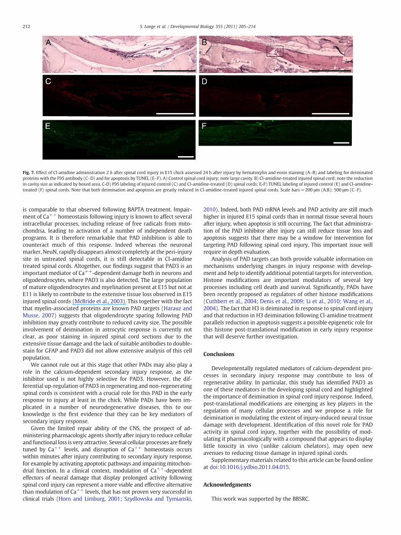
Fig. 7. Effect of Cl-amidine administration 2 h after spinal cord injury in E15 chick assessed 24 h after injury by hematoxylin and eosin staining (A–B) and labeling for deiminatedproteins with the F95 antibody (C–D) and for apoptosis by TUNEL (E–F). A) Control spinal cord injury; note large cavity. B) Cl-amidine-treated injured spinal cord; note the reductionin cavity size as indicated by boxed area. C-D) F95 labeling of injured control (C) and Cl-amidine-treated (D) spinal cords; E-F) TUNEL labeling of injured control (E) and Cl-amidine-treated (F) spinal cords. Note that both deimination and apoptosis are greatly reduced in Cl-amidine-treated injured spinal cords. Scale bars=200 μm (A,B); 500 μm (C–F).
212 S. Lange et al. / Developmental Biology 355 (2011) 205–214
is comparable to that observed following BAPTA treatment. Impair-ment of Ca++ homeostasis following injury is known to affect severalintracellular processes, including release of free radicals from mito-chondria, leading to activation of a number of independent deathprograms. It is therefore remarkable that PAD inhibition is able tocounteract much of this response. Indeed whereas the neuronalmarker, NeuN, rapidly disappears almost completely at the peri-injurysite in untreated spinal cords, it is still detectable in Cl-amidinetreated spinal cords. Altogether, our findings suggest that PAD3 is animportant mediator of Ca++-dependent damage both in neurons andoligodendrocytes, where PAD3 is also detected. The large populationof mature oligodendrocytes and myelination present at E15 but not atE11 is likely to contribute to the extensive tissue loss observed in E15injured spinal cords (McBride et al., 2003). This together with the factthat myelin-associated proteins are known PAD targets (Harauz andMusse, 2007) suggests that oligodendrocyte sparing following PADinhibition may greatly contribute to reduced cavity size. The possibleinvolvement of deimination in astrocytic response is currently notclear, as poor staining in injured spinal cord sections due to theextensive tissue damage and the lack of suitable antibodies to double-stain for GFAP and PAD3 did not allow extensive analysis of this cellpopulation.
We cannot rule out at this stage that other PADs may also play arole in the calcium-dependent secondary injury response, as theinhibitor used is not highly selective for PAD3. However, the dif-ferential up-regulation of PAD3 in regenerating and non-regeneratingspinal cords is consistent with a crucial role for this PAD in the earlyresponse to injury at least in the chick. While PADs have been im-plicated in a number of neurodegenerative diseases, this to ourknowledge is the first evidence that they can be key mediators ofsecondary injury response.
Given the limited repair ability of the CNS, the prospect of ad-ministering pharmacologic agents shortly after injury to reduce cellularand functional loss is very attractive. Several cellular processes arefinelytuned by Ca++ levels, and disruption of Ca++ homeostasis occurswithin minutes after injury contributing to secondary injury response,for example by activating apoptotic pathways and impairingmitochon-drial function. In a clinical context, modulation of Ca++-dependenteffectors of neural damage that display prolonged activity followingspinal cord injury can represent a more viable and effective alternativethan modulation of Ca++ levels, that has not proven very successful inclinical trials (Horn and Limburg, 2001; Szydlowska and Tymianski,
2010). Indeed, both PAD mRNA levels and PAD activity are still muchhigher in injured E15 spinal cords than in normal tissue several hoursafter injury, when apoptosis is still occurring. The fact that administra-tion of the PAD inhibitor after injury can still reduce tissue loss andapoptosis suggests that there may be a window for intervention fortargeting PAD following spinal cord injury. This important issue willrequire in depth evaluation.
Analysis of PAD targets can both provide valuable information onmechanisms underlying changes in injury response with develop-ment and help to identify additional potential targets for intervention.Histone modifications are important modulators of several keyprocesses including cell death and survival. Significantly, PADs havebeen recently proposed as regulators of other histone modifications(Cuthbert et al., 2004; Denis et al., 2009; Li et al., 2010; Wang et al.,2004). The fact that H3 is deiminated in response to spinal cord injuryand that reduction in H3 deimination following Cl-amidine treatmentparallels reduction in apoptosis suggests a possible epigenetic role forthis histone post-translational modification in early injury responsethat will deserve further investigation.
Conclusions
Developmentally regulated mediators of calcium-dependent pro-cesses in secondary injury response may contribute to loss ofregenerative ability. In particular, this study has identified PAD3 asone of these mediators in the developing spinal cord and highlightedthe importance of deimination in spinal cord injury response. Indeed,post-translational modifications are emerging as key players in theregulation of many cellular processes and we propose a role fordeimination in modulating the extent of injury-induced neural tissuedamage with development. Identification of this novel role for PADactivity in spinal cord injury, together with the possibility of mod-ulating it pharmacologically with a compound that appears to displaylittle toxicity in vivo (unlike calcium chelators), may open newavenues to reducing tissue damage in injured spinal cords.
Supplementarymaterials related to this article can be found onlineat doi:10.1016/j.ydbio.2011.04.015.
Acknowledgments
This work was supported by the BBSRC.
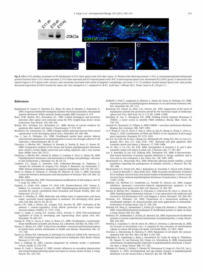
Fig. 8. Effect of Cl-amidine treatment on H3 deimination in E15 chick spinal cords 24 h after injury. A) Western blot detecting histone 3 (H3) in immunoprecipitated deiminatedprotein fractions from 1) E11 sham-operated, 2) E15 sham-operated and E15 injured spinal cords. B-B′) Control injured spinal cord; deiminated H3 (CitH3, green) is detected in theinjured region in E15 spinal cords (arrows) and commonly associated with nuclei of apoptotic morphology (see inset). C-C′) Cl-amidine-treated injured spinal cord; note greatlydecreased expression of CitH3 around the injury site (box enlarged in C′) compared to (B-B′). Scale bars=260 μm (B,C), 20 μm (inset in B′), 65 μm (C′).
213S. Lange et al. / Developmental Biology 355 (2011) 205–214
References
Balandraud, N., Gouret, P., Danchin, E.G., Blanc, M., Zinn, D., Roudier, J., Pontarotti, P.,2005. A rigorousmethod formultigenic families' functional annotation: the peptidylarginine deiminase (PADs) proteins family example. BMC Genomics 6, 153.
Basso, D.M., Beattie, M.S., Bresnahan, J.C., 1996. Graded histological and locomotoroutcomes after spinal cord contusion using the NYU weight-drop device versustransection. Exp. Neurol. 139, 244–256.
Beattie, M.S., Farooqui, A.A., Bresnahan, J.C., 2000. Review of current evidence forapoptosis after spinal cord injury. J. Neurotrauma 17, 915–925.
Blackmore, M., Letourneau, P.C., 2006. Changes within maturing neurons limit axonalregeneration in the developing spinal cord. J. Neurobiol. 66, 348–360.
Cao, L., Sun, D., Whitaker, J.N., 1998. Citrullinated myelin basic protein inducesexperimental autoimmune encephalomyelitis in Lewis rats through a diverse T cellrepertoire. J. Neuroimmunol. 88, 21–29.
Chavanas, S., Mechin, M.C., Takahara, H., Kawada, A., Nachat, R., Serre, G., Simon, M.,2004. Comparative analysis of the mouse and human peptidylarginine deiminasegene clusters reveals highly conserved non-coding segments and a new humangene, PADI6. Gene 330, 19–27.
Chavanas, S., Mechin, M.C., Nachat, R., Adoue, V., Coudane, F., Serre, G., Simon, M., 2006.Peptidylarginine deiminases and deimination in biology and pathology: relevanceto skin homeostasis. J. Dermatol. Sci. 44, 63–72.
Cuthbert, G.L., Daujat, S., Snowden, A.W., Erdjument-Bromage, H., Hagiwara, T.,Yamada, M., Schneider, R., Gregory, P.D., Tempst, P., Bannister, A.J., Kouzarides, T.,2004. Histone deimination antagonizes arginine methylation. Cell 118, 545–553.
Denis, H., Deplus, R., Putmans, P., Yamada, M., Metivier, R., Fuks, F., 2009. Functionalconnection between deimination and deacetylation of histones. Mol. Cell. Biol. 29,4982–4993.
Doyle, H.A., Mamula, M.J., 2005. Posttranslational modifications of self-antigens. Ann. N.Y. Acad. Sci. 1050, 1–9.
Esposito, G., Vitale, A.M., Leijten, F.P., Strik, A.M., Koonen-Reemst, A.M., Yurttas, P.,Robben, T.J., Coonrod, S., Gossen, J.A., 2007. Peptidylarginine deiminase (PAD) 6 isessential for oocyte cytoskeletal sheet formation and female fertility. Mol. Cell.Endocrinol. 273, 25–31.
Ferretti, P., Whalley, K., 2008. Molecular and cellular basis of regeneration and tissuerepair: successful neural regeneration in amniotes: the developing chick spinalcord. Cell. Mol. Life Sci. 65, 45–53.
Gavins, F.N., Dalli, J., Flower, R.J., Granger, D.N., Perretti, M., 2007. Activation of theannexin 1 counter-regulatory circuit affords protection in the mouse brainmicrocirculation. FASEB J. 21, 1751–1758.
Gögel, S., Lange, S., Leung, K.Y., Greene, N.D.E., Ferretti, P., 2010. Post-translationalregulation of Crmp in developing and regenerating chick spinal cord. Dev.Neurobiol. 70, 456–471.
Gyorgy, B., Toth, E., Tarcsa, E., Falus, A., Buzas, E.I., 2006. Citrullination: a posttransla-tional modification in health and disease. Int. J. Biochem. Cell Biol. 38, 1662–1677.
Harauz, G., Musse, A.A., 2007. A tale of two citrullines—structural and functional aspectsof myelin basic protein deimination in health and disease. Neurochem. Res. 32,137–158.
Hasan, S.J., Nelson, B.H., Valenzuela, J.I., Keirstead, H.S., Shull, S.E., Ethell, D.W., Steeves, J.D.,1991. Functional repair of transected spinal cord in embryonic chick. Restor. Neurol.Neurosci. 2, 137–154.
Horn, J., Limburg, M., 2001. Calcium antagonists for ischemic stroke: a systematicreview. Stroke 32, 570–576.
Inman, D., Guth, L., Steward, O., 2002. Genetic influences on secondary degenerationand wound healing following spinal cord injury in various strains of mice. J. Comp.Neurol. 451, 225–235.
Keilhoff, G., Prell, T., Langnaese, K., Mawrin, C., Simon, M., Fansa, H., Nicholas, A.P., 2008.Expression pattern of peptidylarginine deiminase in rat and human Schwann cells.Dev. Neurobiol. 68, 101–114.
Keirstead, H.S., Hasan, S.J., Muir, G.D., Steeves, J.D., 1992. Suppression of the onset ofmyelination extends the permissive period for the functional repair of embryonicspinal cord. Proc. Natl Acad. Sci. U. S. A. 89, 11664–11668.
Knuckley, B., Luo, Y., Thompson, P.R., 2008. Profiling Protein Arginine Deiminase 4(PAD4): a novel screen to identify PAD4 inhibitors. Bioorg. Med. Chem. 16,739–745.
Lesniak,W., Slomnicki, L.P., Filipek, A., 2009. S100A6— new facts and features. Biochem.Biophys. Res. Commun. 390, 1087–1092.
Li, P., Wang, D., Yao, H., Doret, P., Hao, G., Shen, Q., Qiu, H., Zhang, X., Wang, Y., Chen, G.,Wang, Y., 2010. Coordination of PAD4 and HDAC2 in the regulation of p53-targetgene expression. Oncogene 29, 3153–3162.
Liu, X.Z., Xu, X.M., Hu, R., Du, C., Zhang, S.X.,McDonald, J.W., Dong, H.X.,Wu, Y.J., Fan, G.S.,Jacquin, M.F., Hsu, C.Y., Choi, D.W., 1997. Neuronal and glial apoptosis aftertraumatic spinal cord injury. J. Neurosci. 17, 5395–5406.
Liu, N., Han, S., Lu, P.H., Xu, X.M., 2004. Upregulation of annexins I, II, and V aftertraumatic spinal cord injury in adult rats. J. Neurosci. Res. 77, 391–401.
Luo, Y., Knuckley, B., Lee, Y.H., Stallcup, M.R., Thompson, P.R., 2006. A fluoroacetami-dine-based inactivator of protein arginine deiminase 4: design, synthesis, and invitro and in vivo evaluation. J. Am. Chem. Soc. 128, 1092–1093.
Mastronardi, F.G., Moscarello, M.A., 2005. Molecules affecting myelin stability: a novelhypothesis regarding the pathogenesis of multiple sclerosis. J. Neurosci. Res. 80,301–308.
Mastronardi, F.G., Wood, D.D., Mei, J., Raijmakers, R., Tseveleki, V., Dosch, H.M., Probert,L., Casaccia-Bonnefil, P., Moscarello, M.A., 2006. Increased citrullination of histoneH3 in multiple sclerosis brain and animal models of demyelination: a role for tumornecrosis factor-induced peptidylarginine deiminase 4 translocation. J. Neurosci. 26,11387–11396.
McBride, C.B., McPhail, L.T., Vanderluit, J.L., Tetzlaff, W., Steeves, J.D., 2003. Caspaseinhibition attenuates transection-induced oligodendrocyte apoptosis in thedeveloping chick spinal cord. Mol. Cell. Neurosci. 23, 383–397.
Nachat, R., Mechin, M.C., Takahara, H., Chavanas, S., Charveron, M., Serre, G., Simon, M.,2005. Peptidylarginine deiminase isoforms 1–3 are expressed in the epidermis andinvolved in the deimination of K1 and filaggrin. J. Invest. Dermatol. 124, 384–393.
Nicholas, A.P., Whitaker, J.N., 2002. Preparation of a monoclonal antibody tocitrullinated epitopes: its characterization and some applications to immunohis-tochemistry in human brain. Glia 37, 328–336.
Nicholas, A.P., King, J.L., Sambandam, T., Echols, J.D., Gupta, K.B., McInnis, C., Whitaker, J.N.,2003. Immunohistochemical localization of citrullinated proteins in adult rat brain.J. Comp. Neurol. 459, 251–266.
Nicholas, A.P., Sambandam, T., Echols, J.D., Barnum, S.R., 2005. Expression of citrullinatedproteins in murine experimental autoimmune encephalomyelitis. J. Comp. Neurol.486, 254–266.
Norberg, E., Gogvadze, V., Ott, M., Horn, M., Uhlen, P., Orrenius, S., Zhivotovsky, B., 2008.An increase in intracellular Ca2+ is required for the activation of mitochondrialcalpain to release AIF during cell death. Cell Death Differ. 15, 1857–1864.
Orrenius, S., Zhivotovsky, B., Nicotera, P., 2003. Regulation of cell death: the calcium-apoptosis link. Nat. Rev. Mol. Cell Biol. 4, 552–565.
Raijmakers, R., Vogelzangs, J., Raats, J., Panzenbeck, M., Corby, M., Jiang, H., Thibodeau,M., Haynes, N., van Venrooij, W.J., Pruijn, G.J., Werneburg, B., 2006. Experimentalautoimmune encephalomyelitis induction in peptidylarginine deiminase 2 knock-out mice. J. Comp. Neurol. 498, 217–226.
Shimada, N., Handa, S., Uchida, Y., Fukuda, M., Maruyama, N., Asaga, H., Choi, E.K., Lee, J.,Ishigami, A., 2010. Developmental and age-related changes of peptidylargininedeiminase 2 in the mouse brain. J. Neurosci. Res. 88, 798–806.
214 S. Lange et al. / Developmental Biology 355 (2011) 205–214
Shimizu, I., Oppenheim, R.W., Obrien, M., Shneiderman, A., 1990. Anatomical andfunctional recovery following spinal cord transection in thechick embryo. J. Neurobiol.21, 918–937.
Solito, E., McArthur, S., Christian, H., Gavins, F., Buckingham, J.C., Gillies, G.E., 2008.Annexin A1 in the brain—undiscovered roles? Trends Pharmacol. Sci. 29, 135–142.
Suzuki, A., Yamada, R., Yamamoto, K., 2007. Citrullination by peptidylargininedeiminase in rheumatoid arthritis. Ann. N. Y. Acad. Sci. 1108, 323–339.
Szydlowska, K., Tymianski, M., 2010. Calcium, ischemia and excitotoxicity. Cell Calcium47, 122–129.
Tymianski, M., Wallace, M.C., Spigelman, I., Uno, M., Carlen, P.L., Tator, C.H., Charlton, M.P., 1993. Cell-permeant Ca2+ chelators reduce early excitotoxic and ischemicneuronal injury in vitro and in vivo. Neuron 11, 221–235.
Velardo, M.J., Reier, P.J., Anderson, D.K., 2000. Spinal cord injury. In: Crockard, A., et al.(Ed.), Neurosurgery. : The scientific basis of clinical practice, vol. 1. BlackwellScientific, Oxford, pp. 499–515.
Vossenaar, E.R., Zendman, A.J., van Venrooij, W.J., Pruijn, G.J., 2003. PAD, a growingfamily of citrullinating enzymes: genes, features and involvement in disease.Bioessays 25, 1106–1118.
Wang, H.G., Pathan, N., Ethell, I.M., Krajewski, S., Yamaguchi, Y., Shibasaki, F., McKeon,F., Bobo, T., Franke, T.F., Reed, J.C., 1999. Ca2+−induced apoptosis throughcalcineurin dephosphorylation of BAD. Science 284, 339–343.
Wang, Y., Wysocka, J., Sayegh, J., Lee, Y.H., Perlin, J.R., Leonelli, L., Sonbuchner, L.S.,McDonald, C.H., Cook, R.G., Dou, Y., Roeder, R.G., Clarke, S., Stallcup, M.R., Allis, C.D.,Coonrod, S.A., 2004. Human PAD4 regulates histone arginine methylation levels viademethylimination. Science 306, 279–283.
Wang, Y., Li, M., Stadler, S., Correll, S., Li, P., Wang, D., Hayama, R., Leonelli, L., Han, H.,Grigoryev, S.A., Allis, C.D., Coonrod, S.A., 2009. Histone hypercitrullinationmediateschromatin decondensation and neutrophil extracellular trap formation. J. Cell Biol.184, 205–213.
Whalley, K., O'Neill, P., Ferretti, P., 2006. Changes in response to spinal cord injury withdevelopment: vascularization, haemorrhage and apoptosis. Neuroscience 137, 821–832.
Whalley, K., Gogel, S., Lange, S., Ferretti, P., 2009. Changes in progenitor populations andongoing neurogenesis in the regenerating chick spinal cord. Dev. Biol. 332, 234–245.
Wood, D.D., Ackerley, C.A., Brand, B., Zhang, L., Raijmakers, R., Mastronardi, F.G.,Moscarello, M.A., 2008. Myelin localization of peptidylarginine deiminases 2 and 4:comparison of PAD2 and PAD4 activities. Lab. Invest. 88, 354–364.
Yachnis, A.T., Rorke, L.B., Lee, V.M., Trojanowski, J.Q., 1993. Expression of neuronal andglial polypeptides during histogenesis of the human cerebellar cortex includingobservations on the dentate nucleus. J. Comp. Neurol. 334, 356–369.
Yamashita, N., Ilg, E.C., Schafer, B.W., Heizmann, C.W., Kosaka, T., 1999. Distribution of aspecific calcium-binding protein of the S100 protein family, S100A6 (calcyclin), insubpopulations of neurons and glial cells of the adult rat nervous system. J. Comp.Neurol. 404, 235–257.
Ying, S., Dong, S., Kawada, A., Kojima, T., Chavanas, S., Mechin, M.C., Adoue, V., Serre, G.,Simon, M., Takahara, H., 2009. Transcriptional regulation of peptidylargininedeiminase expression in human keratinocytes. J. Dermatol. Sci. 53, 2–9.
Zhang, Z., Fujiki, M., Guth, L., Steward, O., 1996. Genetic influences on cellular reactionsto spinal cord injury: a wound-healing response present in normal mice is impairedin mice carrying a mutation (WldS) that causes delayed Wallerian degeneration.J. Comp. Neurol. 371, 485–495.