proline to arginine mutations in fgf receptors 1 and 3 result in pfeiffer and muenke...
TRANSCRIPT

Proline to arginine mutations in FGFreceptors 1 and 3 result in Pfeiffer and Muenkecraniosynostosis syndromes throughenhancement of FGF binding affinity
Omar A. Ibrahimi1, Fuming Zhang2, Anna V. Eliseenkova1, Robert J. Linhardt2 and
Moosa Mohammadi1,*
1Department of Pharmacology, New York University School of Medicine, New York, NY 10016, USA and2Department of Chemistry, Biology, and Chemical and Biological Engineering, Rensselaer Polytechnic Institute,
Troy, NY 12180, USA
Received August 18, 2003; Revised October 22, 2003; Accepted November 3, 2003
Identical proline!arginine gain-of-function mutations in fibroblast growth factor receptor (FGFR) 1(Pro252Arg), FGFR2 (Pro253Arg) and FGFR3 (Pro250Arg), result in type I Pfeiffer, Apert and Muenkecraniosynostosis syndromes, respectively. Here, we characterize the effects of proline!arginine mutationsin FGFR1c and FGFR3c on ligand binding using surface plasmon resonance and X-ray crystallography. BothPro252Arg FGFR1c and Pro250Arg FGFR3c exhibit an enhancement in ligand binding in comparison to theirrespective wild-type receptors. Interestingly, binding of both mutant receptors to FGF9 was notably enhancedand implicates FGF9 as a potential pathophysiological ligand for mutant FGFRs in mediating craniosynostosis.The crystal structure, of Pro252Arg FGFR1c in complex with FGF2, demonstrates that the enhanced ligandbinding is due to an additional set of receptor-ligand hydrogen bonds, similar to those gain-of-functioninteractions that occur in the Apert syndrome Pro253Arg FGFR2c-FGF2 crystal structure. However, unlike theApert syndrome Pro253Arg FGFR2c mutant, neither the Pfeiffer syndrome Pro250Arg FGFR1c mutant nor theMuenke syndrome Pro250Arg FGFR3c mutant bound appreciably to FGF7 or FGF10. This observation providesa potential explanation for why the limb phenotypes, observed in type I Pfeiffer and Muenke syndromes, areless severe than the limb abnormalities observed in Apert syndrome. Hence, although analogous proline!arginine mutations in FGFR1-3 act through a common structural mechanism to result in gain-of-function,differences in the primary sequence among FGFRs result in varying effects on ligand binding specificity.
INTRODUCTION
The fibroblast growth factor (FGF) receptor (FGFR) familyconsists of four single-pass transmembrane receptor tyrosinekinases, FGFR1-4 (1,2). The extracellular portion of FGFRsmediates ligand binding and consists of three immunoglobulin(Ig)-like domains (D1–D3). Tissue specific alternative splicingof the second half of D3 in FGFR1-3 creates ‘b’ and ‘c’isoforms and plays a major role in determining the bindingprofile of each receptor to the 18 known FGF ligands(excluding FHFs) (1–3). Heparin or heparan sulfate proteo-glycan is an obligatory cofactor for receptor binding anddimerization, the initial steps in FGFR activation (4–7).
Craniosynostosis, the premature fusion of one or morecranial sutures, is a common clinical finding that occurs in�1/2500 births (8). Mutations in FGFR1-3 are responsible forseveral craniosynostosis syndromes including Apert syndrome(AS), Beare-Stevenson syndrome, Crouzon syndrome,Jackson–Weiss syndrome, Muenke syndrome (MS) andPfeiffer syndrome (PS) (9,10). The etiology of FGFR relatedskeletal disorders stems from receptor gain-of-function, eitherthrough a ligand-independent or ligand-dependent manner.Ligand-independent gain-of-function may arise from mutationsresulting in: (i) covalent receptor dimerization (11–13); (ii)non-covalent receptor dimerization (14,15); or (iii) stabilizationof the active conformation of the kinase domain (15,16). In
*To whom correspondence should be addressed. Tel: þ1 2122632907; Fax: þ1 2122637133; Email: [email protected]
Human Molecular Genetics, 2004, Vol. 13, No. 1 69–78DOI: 10.1093/hmg/ddh011Advance Access published on November 12, 2003
Human Molecular Genetics, Vol. 13, No. 1 # Oxford University Press 2004; all rights reserved
by guest on March 16, 2016
http://hmg.oxfordjournals.org/
Dow
nloaded from

contrast, ligand-dependent gain-of-function may result frommutations causing: (i) enhanced ligand binding affinity (17,18);(ii) loss of ligand binding specificity (19); or (iii) ectopicexpression of inappropriate splice isoforms (20).Mutations of a highly conserved Ser-Pro dipeptide motif in
the D2–D3 linker region account for the most common of thecraniosynostosis syndromes. Interestingly, these mutationshave the highest rates for transversions known in the humangenome (8). AS results almost exclusively from one or theother of two missense mutations, Ser252Trp or Pro253Arg, inFGFR2 (21). It is the most severe of the craniosynostosissyndromes and is additionally characterized by syndactyly ofthe hands and feet. Type I PS and MS result from Pro252Argand Pro250Arg mutations in FGFR1 and FGFR3, respectively(22,23). In contrast to AS, type I PS and MS are characterizedby mild limb phenotypes (8,24).The AS mutations, Ser252Trp and Pro253Arg in FGFR2, are
the most well-characterized of the D2–D3 linker regioncraniosynostosis mutations. Because these mutations occurprior to the alternatively spliced D3 region, both the ‘b’ and ‘c’splice isoforms are affected. However, most studies havefocused on the ‘c’ splice isoform since histological analysis ofAS tissues suggests that the majority of pathology occurs inmesenchymal tissue that expresses the ‘c’ splice isoform ofFGFRs (25). Initial evidence suggesting how these mutationsmay result in receptor gain-of-function came from ligandbinding studies of wild type and AS mutant FGFR2c in whichthe mutant receptors displayed enhanced binding to FGF2 (17).A later report demonstrated that each AS mutant FGFR2c doesnot bind FGF2 better than wild-type FGFR2c (19). Instead, itwas shown that AS mutations result in the loss of FGFR2ligand specificity (19). Normally, mesenchymally expressedFGFR2c binds to FGF2 but does not bind to FGF7 or FGF10.Conversely, epithelial FGFR2b binds to FGF7 and FGF10 buthas weak affinity for FGF2 (26–28). This specificity permitsdirectional epithelial-mesenchymal signaling to occur duringorganogenesis and limb development. However, AS Ser252Trpand Pro253Arg mutations in FGFR2c enabled FGF7 binding,thus suggesting that some of the severe and unique phenotypesin AS may result from pathophysiological disruption of normalsignaling networks by inappropriate autocrine activation (19).We recently reported the crystal structures of each AS mutant
FGFR2c in complex with FGF2 (18). Both Ser252Trp FGFR2cand Pro253Arg FGFR2c engage FGF2 with distinct additionalcontacts, thereby providing a structural basis for receptor gain-of-function. These structures and sequence alignment of theFGF family suggests that the Pro253Arg mutation, and to alesser extent the Ser252Trp mutation, result in generalizedenhancement of ligand binding and hence may even enable thebinding of FGFs that are outside of the normal receptor bindingspectrum.To date, the molecular mechanism by which the correspond-
ing proline!arginine mutations in FGFR1 and FGFR3 resultin PS and MS, respectively, have not been characterized,although a mouse model for PS has provided direct geneticevidence that the Pro252Arg mutation in FGFR1 is causativefor PS (29). In this report, we examine the mechanisms bywhich Pro252Arg and Pro250Arg mutations activate FGFR1cand FGFR3c, respectively, using surface plasmon resonance(SPR) and X-ray crystallography. Here, we show that these
mutations result in the enhancement of FGF binding affinity,but are not sufficient to override FGFR1c and FGFR3c ligandbinding specificity.
RESULTS AND DISCUSSION
Several FGFR ligand binding studies have been reported thatemploy a variety of techniques including radiolabeled ligandcompetition binding, SPR, and isothermal titration calorimetryassays (17,30–33). However, these studies are limited in thatthey focus on only a few interactions, and since they utilizedifferent techniques, comparisons between studies are difficult.Hence, in order to quantify mutant FGFR–FGF interactions, wemeasured the binding of wild-type and Pro252Arg FGFR1cand wild-type and Pro250Arg FGFR3c to FGF1-10 using SPR.FGF8 was found to aggregate on the biosensor surface andtherefore FGFR–FGF8 interactions could not be analyzedusing SPR. The kinetic data are summarized in Table 1 and, toour knowledge, are the most comprehensive ligand bindingstudies reported for FGFR1c and FGFR3c.Wild-type FGFR1c bound with high affinity to FGF1, FGF2,
FGF4 and FGF6 but with poor affinity to FGF9 (Fig. 1A, C, Eand I, Table 1). These findings are in agreement withprior binding and mitogenic response assays for FGFR1c(3,17,30–34). In contrast, wild-type FGFR1c did not bind toFGF3, FGF7 and FGF10, ligands which preferentially signalthrough FGFR2b (Fig. 1G; Table 1) (3). We did not detectbinding of FGF5 to wild-type FGFR1c (Table 1). Wild-typeFGFR1c–FGF5 binding has not been previously demonstratedalthough FGF5 is moderately mitogenic for FGFR1c BaF3cells (3). Interestingly, compared to wild-type FGFR1c, thePS Pro252Arg FGFR1c mutant displayed a generalized 2- to5-fold increase in binding affinity for FGF1, FGF2, FGF4, andFGF6 (Fig. 1A, B, C, D, E and F, Table 1). A notable exceptionwas FGF9, which had a striking 30-fold increase in bindingaffinity for the mutant FGFR1c (Fig. 1I and J Table 1). Theenhanced binding in almost all cases could be attributed to bothfaster association rates (Kon) and slower dissociation kinetics(Koff) (Table 1).In the case of wild-type FGFR3c, high affinity binding was
observed only to FGF1 (Fig. 2A; Table 1), but not to otherFGFs examined. The lack of binding of FGF3, FGF7 andFGF10 to wild-type FGFR3c is consistent with the preferentialsignaling of these FGFs through FGFR2b (3). Other studieshave also detected poor FGFR3c binding to FGF2 (35–37),FGF4 and FGF6 (37). Additionally, studies of BaF3 cellsexpressing FGFR3 show negligible to modest mitogenicresponses to FGF2, FGF4, FGF6 and FGF9 (38), althoughFGF2, FGF4 and FGF9 strongly activate BaF3 cells expressinga chimeric FGFR3c-FGFR1c construct (3). It is possible thatthe chimeric FGFR3c–FGFR1c construct used by Ornitz et al.(3) masks differences in FGFR3c ligand binding due to signaloveramplification by the inherently more active FGFR1c kinasedomain.The MS Pro250Arg FGFR3c mutant also displayed an
increase in ligand binding affinity (Fig. 2; Table 1). FGF1displays a nearly 2-fold increase in binding affinity forPro250Arg FGFR3c over wild-type FGFR3c (Fig. 2A and B,Table 1). Additionally, the Pro250Arg mutation permits
70 Human Molecular Genetics, 2004, Vol. 13, No. 1
by guest on March 16, 2016
http://hmg.oxfordjournals.org/
Dow
nloaded from

FGFR3c to weakly bind FGF2, whereas wild-type FGFR3cbinds FGF2 negligibly (Fig. 2C and D; Table 1). Interestingly,as in the case of FGFR1c, the proline!arginine mutationenables FGFR3c to bind FGF9 with high affinity while FGF9binding to wild-type FGFR3c is negligible (Fig. 2I and J; Table1). This robust increase in FGF9 binding has also beenqualitatively observed by size exclusion chromatography (datanot shown).To ascertain the structural basis for the enhanced affinity of
Pro252Arg FGFR1c and Pro250Arg FGFR3c mutants towardsFGFs, we chose to crystallize each mutant receptor with FGFligand. We were able to successfully generate diffractingcrystals of Pro252Arg FGFR1c–FGF2. The crystal structure ofPro252Arg FGFR1c–FGF2 was solved using molecularreplacement. Data collection and refinement statistics are givenin Tables 2 and 3. The overall structure of the Pro252ArgFGFR1c–FGF2 complex is identical to the structure of thewild-type FGFR1c–FGF2 complex. The relative orientationbetween D2 and D3 is unaffected by the proline!argininemutation. Two 1:1 FGFR1c–FGF2 complexes compose theasymmetric unit and form a symmetric dimer.In both copies of Pro252Arg FGFR1c–FGF2, the guanidinium
group of Arg-252 in FGFR1c makes three hydrogen bonds withthe b8–b9 turn of FGF2 (Fig. 3). Two hydrogen bonds are madewith the backbone carbonyl oxygens of Leu-107 and Glu-108of FGF2 and a third with the side chain amide group of Asn-111of FGF2. Thus, the structural basis for the enhanced binding ofPro252Arg FGFR1c to FGF2 is the presence of additional
receptor–ligand interactions that are not present in the wild-typeFGFR1c–FGF2 structure. Interestingly, these gain-of-functioncontacts are similar to the additional contacts made between theAS Pro253Arg FGFR2c mutant and FGF2 (18). Since two ofthe three additional hydrogen bonds involve backbone carbonyloxygens of FGF2, these interactions should also occur withother FGFs and explains the generalized enhancement of FGFbinding affinity by proline!arginine mutant FGFRs (Table 1).We predict that these proline!arginine mutant FGFRs shouldalso display enhanced binding to FGF16-23, which have notyet been analyzed for binding.Interestingly, the exceptional increase in binding affinity of
FGF9 for Pro252Arg FGFR1c and Pro253Arg FGFR3cimplicates FGF9 as a potential pathophysiological ligandfor craniosynostosis mediated by D2–D3 linker regionmutants. Indeed, FGF9 is expressed at extremely high levelsin the cranial suture and has been shown to stimulate themesenchymally expressed ‘c’ isoform of FGFR1-3 (3,39,40).
Table 1. Summary of kinetic data
FGF FGFR1c FGFR3c
Wild-type Pro252Arg Wild-type Pro250Arg
FGF1 Kon (/M/s)a 2.24� 105 5.6� 105 8.79� 105 8.92� 105
Koff (/s)a 3.05� 10�2 1.71� 10�2 2.02� 10�1 1.15� 10�1
KD (M)b 1.36� 10�7 3.05� 10�8 2.30� 10�7 1.28� 10�7
FGF2 Kon (/M/s) 9.64� 104 1.77� 105 NB 4.01� 105
Koff (/s) 5.96� 10�3 4.30� 10�3 NB 4.10� 10�1
KD (M) 6.19� 10�8 2.44� 10�8 NB 1.02� 10�6
FGF3 Kon (/M/s) NBc NB NB NBKoff (/s) NB NB NB NBKD (M) NB NB NB NB
FGF4 Kon (/M/s) 2.62� 105 5.97� 105 NB NBKoff (/s) 4.33� 10�2 4.41� 10�2 NB NBKD (M) 1.65� 10�7 7.39� 10�8 NB NB
FGF5 Kon (/M/s) NB NB NB NBKoff (/s) NB NB NB NBKD (M) NB NB NB NB
FGF6 Kon (/M/s) 2.93� 105 5.07� 105 NB NBKoff (/s) 3.05� 10�2 1.88� 10�2 NB NBKD (M) 1.04� 10�7 3.71� 10�8 NB NB
FGF7 Kon (/M/s) NB NB NB NBKoff (/s) NB NB NB NBKD (M) NB NB NB NB
FGF9 Kon (/M/s) 3.44� 104 5.58� 105 NB 5.63� 104
Koff (/s) 4.15� 10�2 2.26� 10�2 NB 4.58� 10�3
KD (M) 1.21� 10�6 4.05� 10�8 NB 8.14� 10�8
FGF10 Kon (/M/s) NB NB NB NBKoff (/s) NB NB NB NBKD (M) NB NB NB NB
aKon and Koff were derived as described in Materials and Methods. w2 was less than 10% of Rmax in all cases.bThe apparent affinity, KD, is equal to Koff/Kon.cNB, negligible binding.
Table 2. Summary of crystallographic analysis: data collection statistics
Resolution(A)
Reflections(total/unique)
Completeness(%)
Rsyma (%) Signal
(hI/sIi)
30.0–2.9 134842/22295 99.7 (99.9)b 11.7 (37.4)b 13.1
aRsym¼ 100�ShklSijIi(hkl)7 hI(hkl)ij/ShklSiIi(hkl).bValue in parentheses is for the highest resolution shell: 3.0–2.9 A.
Human Molecular Genetics, 2004, Vol. 13, No. 1 71
by guest on March 16, 2016
http://hmg.oxfordjournals.org/
Dow
nloaded from

However, since our SPR analysis did not include every FGF,the possibility of another FGF that exhibits a robustenhancement in binding mutant FGFRs cannot be ruled out.Nonetheless, in order for any FGF to be pathophysiologicallyrelevant in mediating craniosynostosis, it must also beexpressed in the cranial sutures and capable of activatingFGFR1-3.The crystal structure of FGF9 provides an explanation as to
why the increase in binding affinity of FGF9 for mutant receptorsis so marked in comparison to other FGFs examined (41). FGF9activity is proposed to be regulated through an autoinhibitorymechanism that stems from its ability to dimerize. Dimerizationof FGF9 involves the N- and C-terminal regions of FGF9, as wellas the b-trefoil core region, thereby occluding a major portion ofthe receptor binding interface. Notably, the b8–b9 turn of FGF9,part of the core region involved in FGF9 dimerization, is also theregion of FGF ligand that is predicted to engage in additionalcontacts with proline!arginine mutant FGFRs. It is likely thatthe balance between FGF9 auto-inhibition and receptor bindingis shifted to favor the latter due to enhanced interaction ofmutant FGFRs with the b8–b9 turn of FGF9. Such an effect isconsistent with the binding kinetics of FGF9 to wild-type andmutant FGFR1c (Table 1). The large increase in association rate(Kon) of the Pro252Arg FGFR1c–FGF9 interaction, relative to thewild-type FGFR1c–FGF9 interaction, is not observed for otherFGFs and is harmonious with the above hypothesis. Similarly, thereduction in dissociation rate (Koff) for the Pro252Arg FGFR1c–FGF9 interaction, relative to the wild-type FGFR1c–FGF9interaction, is similar for other FGFs and suggests that mutantreceptor–FGF9 complexes are stabilized through similar addi-tional contacts. However, validation of this hypothesis requiresadditional experimental analysis.FGF9 knockout mice have been recently reported (42), but
since suture closure normally occurs between P25 and P45 inmice (43) and FGF9 knockout mice die at birth, little can beinferred about the role of FGF9 in cranial suture signaling.However, these mice are notable in that they display male-to-female sex reversal, thereby implicating FGF9 as a centralplayer in testicular development (42). Paradoxically, whileFGFR mutations are deleterious for skeletal development,several recent studies provide evidence that certain mutations inFGFR2-3 confer a selective advantage to male sperm cells (44–46). FGFs, perhaps FGF9, may mediate this selectiveadvantage in the case of ligand-dependent FGFR mutationsby enhancing FGF–FGFR binding.Interestingly, craniosynostosis syndromes resulting from
proline!arginine mutations in FGFR1 and FGFR3, type IPS and MS, respectively, are characterized by very mild limb
pathology in contrast to AS (8,24). Both AS mutations havebeen shown to enable FGFR2c to bind FGF7 (19), with thePro253Arg substitution demonstrating a stronger effect andcorrelating with the more severe syndactyly observed inPro253Arg AS patients (47–49). Hence, it is currently thoughtthat limb abnormalities in AS arise from autocrine activation ofFGFR2c by FGF7 or a similar FGF, although mutant FGFR2c–FGF7 binding has not yet been examined using SPR. Based onthe data of Ornitz et al. (3), our inability to detect binding ofcorresponding proline!arginine mutations in FGFR1c andFGFR3c to bind FGF7 or FGF10 (Table 1; Figs 1H and 2H)seems to be consistent with the absence of severe limbphenotypes in type I PS and MS.The recent crystal structure of FGFR2b–FGF10 provides a
structural basis for why these mutations in FGFR1c andFGFR3c do not result in FGF7 binding. In this structure,several D2 residues that are conserved only in FGFR2 arecritical for D2-FGF10 binding (50). The lack of conservation ofthese residue in FGFR1 and FGFR3 suggests that even ifcorresponding proline!arginine mutations were to introduceadditional receptor-ligand contacts they may not be sufficient toenable either mutant FGFR1c or FGFR3c to bind to FGF7.Alternatively, the differences in limb phenotype between thesethree craniosynostosis syndromes may be due to the distinctroles of FGFR1-3 in limb development. Nevertheless, addi-tional factors must play a role, since some FGFR2 mutationsresult in craniosynostosis with mild limb phenotypes.
CONCLUDING REMARKS
The results presented here indicate that analogous proline!arginine mutations in the D2–D3 linker region of FGFR1c andFGFR3c result in PS and MS, respectively, through anenhancement of FGF binding. The structural basis for thisenhanced binding is the additional interactions that occurbetween the b8–b9 turn of FGF ligand and the mutatedproline!arginine residue of FGFR. The striking enhancementof FGF9 binding in the case of Pro252Arg FGFR1c andPro250Arg FGFR3c, in comparison to respective wild typeFGFRs, points to FGF9 as a potential pathophysiologicalligand in mediating craniosynostosis. Importantly, the lack ofbinding of Pro252Arg FGFR1c and Pro250Arg FGFR3c toFGF7 or FGF10 provides an explanation for the mild limbphenotypes in type I PS and MS. Moreover, the specificinvolvement of these mutations with the b8–b9 turn of FGFallows for the design of antagonist and agonist of FGFsignaling that may hold therapeutic value for the treatment
Table 3. Summary of crystallographic analysis: refinement statisticsa
Resolution (A) Reflections Rcryst/Rfreeb(%) Root-mean-square deviations B-factorsc (A2)
Bonds (A) Angles (�)
25.0–2.9 21918 26.7–31.8 0.009 1.5 0.64
aAtomic model: 5161 protein atoms and 4 SO4 ions.bRcyst/free¼ 100�Shkl kFo(hkl)j7 jFc(hkl) k /ShkljFo(hkl)j, where Fo (>0s) and Fc are the observed and calculated structure factors, respectively, 10% of thereflections were used for calculation of Rfree.cFor bonded protein atoms.
72 Human Molecular Genetics, 2004, Vol. 13, No. 1
by guest on March 16, 2016
http://hmg.oxfordjournals.org/
Dow
nloaded from

Figure 1. Surface plasmon resonance analysis of FGFR1c–FGF interactions. Sensorgrams of wild-type and Pro252Arg FGFR1c, respectively, binding to FGF1(A and B), FGF2 (C and D), FGF4 (E and F), FGF7 (G and H) and FGF9 (I and J). Analyte concentrations are colored as follows: 25 nM in gray, 50 nM in blue,100 nM in red, 200 nM in green, 400 nM in violet and 800 nM in brown. The biosensor chip response is indicated on the y-axis (DRU) as a function of time (x-axis)at 25�C.
Human Molecular Genetics, 2004, Vol. 13, No. 1 73
by guest on March 16, 2016
http://hmg.oxfordjournals.org/
Dow
nloaded from
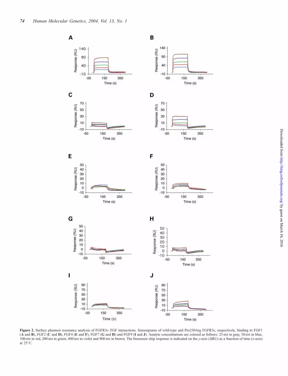
Figure 2. Surface plasmon resonance analysis of FGFR3c–FGF interactions. Sensorgrams of wild-type and Pro250Arg FGFR3c, respectively, binding to FGF1(A and B), FGF2 (C and D), FGF4 (E and F), FGF7 (G and H) and FGF9 (I and J). Analyte concentrations are colored as follows: 25 nM in gray, 50 nM in blue,100 nM in red, 200 nM in green, 400 nM in violet and 800 nM in brown. The biosensor chip response is indicated on the y-axis (DRU) as a function of time (x-axis)at 25�C.
74 Human Molecular Genetics, 2004, Vol. 13, No. 1
by guest on March 16, 2016
http://hmg.oxfordjournals.org/
Dow
nloaded from

of FGFR-related craniosynostosis syndromes or other patho-logical conditions.
MATERIALS AND METHODS
Protein expression and purification
Recombinant human FGFs, FGF1–FGF10 and FGF12b(FHF1b) were expressed in Escherichia coli. Soluble FGFs(FGF1, FGF2, FGF10 and FGF12) were purified by heparinaffinity, ion exchange and size exclusion chromatography asdescribed elsewhere (18,51–53). FGF7 was generously pro-vided by Amgen (Amgen Inc.). Insoluble FGFs (FGF3, FGF5,FGF6 and FGF8) were refolded according to previouslyreported protocols and subsequently purified in a similarmanner to soluble FGFs (54–56). FGF4 and FGF9 werepurified through salt extraction and ammonium sulfateprecipitation, respectively, and then purified as soluble FGFs(41,57).The point mutations Pro252Arg and Pro250Arg were
introduced into the minimal ligand-binding portion (D2–D3)of human FGFR1c (residues 142–365) and FGFR3c (residues143–365), respectively, by using the Quik Change site-directedmutagenesis kit (Stratagene). Wild type and mutant receptorswere expressed in E. coli, refolded in vitro and purified
according to a previously reported protocol (51). Additionalpurification of wild type and mutant receptors was achievedthrough size-exclusion chromatography. Protein concentrationwas determined by mixing the protein solution with denaturingbuffer (6.0 M guanidinium hydrochloride, 0.02M sodiumphosphate buffer, pH 6.5) using a Beckman DU640B spectro-photometer (Beckman Coulter) to measure absorbance at280 nm along with the extinction coefficient, obtained fromthe ProtParam tool (www.expasy.ch), to calculate correctedprotein concentrations.
Surface plasmon resonance analysis ofFGF–FGFR interactions
Wild type and mutant FGFR–FGF interactions were character-ized using a BIAcore 3000 instrument (Biacore AB, Uppsala,Sweden). To obtain kinetic data for wild-type and mutantFGFR–FGF interactions, FGF ligands were immobilized onresearch grade CM 5 chips according to standard aminecoupling protocol (Biacore AB, Uppsala, Sweden). Briefly,carboxymethyl groups on the CM5 chip surface were firstactivated using an injection pulse of 50 ml (flow rate, 5 ml/min)of an equimolar mix of N-ethyl-N-(dimethyaminopropyl)carbodiimide and N-hydroxysuccinimide (final concentration0.05M, mixed immediately prior to injection). Followingactivation, FGF ligand was diluted to 25 mg/ml in HBS-EP
Figure 3. Gain-of-function hydrogen bonds in the Pro252Arg FGFR1c–FGF2 dimer. D2 and D3 of FGFR1c are shown in green and cyan, respectively. The D2–D3linker is colored gray. FGF2 is shown in orange. Dotted lines represent hydrogen bonds. The hydrogen-bonding distances are indicated. On the right is a view ofwhole structure in the exact orientation as the detailed view shows, with the region of interest boxed.
Human Molecular Genetics, 2004, Vol. 13, No. 1 75
by guest on March 16, 2016
http://hmg.oxfordjournals.org/
Dow
nloaded from

buffer [0.01 M HEPES, 0.15M NaCl, 3mM EDTA, 0.005%polysorbate 20 (v/v), pH 7.4] or 100mM sodium acetate(pH 4.5) buffer and injected over the activated biosensorsurface. Excess unreacted sites on the sensor surface weredeactivated with a 40 ml injection of 1M ethanolamine. FGF1was immobilized to approximately 1000 response units (RU).FGF8 was found to aggregate on the sensor chip surface andtherefore could not be immobilized. The remaining FGFs weresuccessfully immobilized to comparable RU levels afteraccounting for relative molecular weight differences. FGF12(FHF1b), an FGF homologous factor (FHF) that does not bindFGFRs, was immobilized on reference flow cells as a control.To obtain kinetic data, different concentrations of analytes(wild-type and mutant FGFR) in HBS-EP buffer [0.01M
HEPES, 0.15M NaCl, 3mM EDTA, 0.005% polysorbate 20(v/v), pH 7.4] were injected over the FGF sensor chips at a flowrate of 50 ml/min. At the end of each sample injection (180 s),HBS-EP buffer was passed over the sensor surface to monitorthe dissociation phase. Following 180 s of dissociation, thesensor surface was fully regenerated by injection of 50 ml of 2M
NaCl in 100mM sodium acetate buffer (pH 4.5).
SPR data analysis
Reference responses from the control flow cell, containingimmobilized FGF12 (FHF1b), were subtracted from FGF flowcells for each analyte injection using BiaEvaluation software(Biacore AB, Uppsala, Sweden). The resulting sensorgramswere used for kinetic parameter determination by globallyfitting the entire association and dissociation phases to a 1:1interaction using BiaEvaluation software (Biacore AB,Uppsala, Sweden). Disturbances, at the beginning and end, ofeach analyte injection were excluded. A minimum of fourdifferent analyte concentrations was used to determine thekinetic parameters for each interaction. Following curve fitting,each sensorgram was manually examined for data quality. w2
was less than 10% of Rmax for each fit.
Crystalization and data collection
Crystals of Pro252Arg FGFR1c in complex with FGF2 wereobtained using similar conditions to the previously reportedcrystallization conditions for wild type FGFR1c in complexwith FGF2 (51). Two microliters of protein solution [10mg/mlin 25mM HEPES-NaOH (pH 7.5) and 150mM NaCl] weremixed with 2 ml of the crystallization buffer consisting of 1.4M
ammonium sulfate, 22% glycerol in 0.1M Tris–HCl (pH 8.5) at23�C. Pro252Arg FGFR1c–FGF2 crystals belong to thetetragonal space group P41212 and contain two FGFR1c–FGF2 complexes in the asymmetric unit. The unit celldimensions are as follows: a¼ b¼ 98.202 A, c¼ 197.174 A,a¼ b¼ g¼ 90�. A 2.9 A data set for the Pro252Arg mutantFGFR1c–FGF2 complex was collected from a flash frozencrystal (in a dry nitrogen stream) on a CCD detector atbeamline X-4A at the National Synchrotron Light Source,Brookhaven National Laboratory. Data were processed usingDENZO and SCALEPACK (58).
Structure determination and refinement of Pro252ArgFGFR1c-FGF2 complex
Molecular replacement solutions for the two copies ofFGFR1c–FGF2 complex in the unit cells of Pro252ArgFGFR1c–FGF2 crystals were found with AmoRe (59) usingthe structure of wild-type FGFR1c complexed with FGF2 (51)(Protein Data Bank identification code 1CVS) as the searchmodel. Tight non-crystallographic symmetry restraints wereimposed throughout the refinement for the backbone atoms ofFGF2, D2 and D3. Simulated annealing and positional/B factorrefinement were performed using CNS (60). Model buildinginto 2Fo – Fc and Fo – Fc electron density maps was performedwith program O (61). The average B factor for all the proteinatoms is 32.5 A2.
ACKNOWLEDGEMENTS
The authors acknowledge C. Basilico and S. Hubbard forcomments and helpful discussions. Beamline X4A at theNational Synchrotron Light Source, a DOE facility, issupported by the Howard Hughes Medical Institute. This workwas sponsored in part by National Institute of Health grantsDE13686 (M.M.) and HL52622 (R.J.L.).
REFERENCES
1. Jaye, M., Schlessinger, J. and Dionne, C.A. (1992) Fibroblast growth factorreceptor tyrosine kinases: molecular analysis and signal transduction.Biochim. Biophys. Acta., 1135, 185–199.
2. Johnson, D.E., Williams, L.T., Gritli-Linde, A., Lewis, P., McMahon, A.P.and Linde, A. (1993) Structural and functional diversity in the FGF receptormultigene family. Adv. Cancer Res., 60, 1–41.
3. Ornitz, D.M., Xu, J., Colvin, J.S., McEwen, D.G., MacArthur, C.A.,Coulier, F., Gao, G. and Goldfarb, M. (1996) Receptor specificity of thefibroblast growth factor family. J. Biol. Chem., 271, 15292–15297.
4. Yayon, A., Klagsbrun, M., Esko, J.D., Leder, P. and Ornitz, D.M. (1991)Cell surface, heparin-like molecules are required for binding of basicfibroblast growth factor to its high affinity receptor. Cell, 64, 841–848.
5. Rapraeger, A.C., Krufka, A. and Olwin, B.B. (1991) Requirement ofheparan sulfate for bFGF-mediated fibroblast growth and myoblastdifferentiation. Science, 252, 1705–1708.
6. Ornitz, D.M., Yayon, A., Flanagan, J.G., Svahn, C.M., Levi, E. and Leder, P.(1992) Heparin is required for cell-free binding of basic fibroblast growthfactor to a soluble receptor and for mitogenesis in whole cells. Mol. Cell.Biol., 12, 240–247.
7. Schlessinger, J., Plotnikov, A.N., Ibrahimi, O.A., Eliseenkova, A.V.,Yeh, B.K., Yayon, A., Linhardt, R.J. and Mohammadi, M. (2000) Crystalstructure of a ternary FGF-FGFR-heparin complex reveals a dual role forheparin in FGFR binding and dimerization. Mol. Cell, 6, 743–750.
8. Muenke, M. and Wilkie, A.O. (2001) Craniosynostosis Syndromes. InScriver, C.R., Beaudet, A.L., Valle, D., Sly, W.S., Childs, Kinzler, K. andVogelstein, B. (eds.), The Metabolic and Molecular Bases of InheritedDisease. McGraw-Hill, New York, NY, Vol. IV, pp. 6117–6146.
9. Passos-Bueno, M.R., Wilcox, W.R., Jabs, E.W., Sertie, A.L., Alonso, L.G.and Kitoh, H. (1999) Clinical spectrum of fibroblast growth factor receptormutations. Hum. Mutat., 14, 115–125.
10. Kan, S.H., Elanko, N., Johnson, D., Cornejo-Roldan, L., Cook, J.,Reich, E.W., Tomkins, S., Verloes, A., Twigg, S.R., Rannan-Eliya, S. et al.(2002) Genomic screening of fibroblast growth-factor receptor 2 reveals awide spectrum of mutations in patients with syndromic craniosynostosis.Am. J. Hum. Genet., 70, 472–486.
11. Neilson, K.M. and Friesel, R. (1996) Ligand-independent activation offibroblast growth factor receptors by point mutations in the extracellular,transmembrane, and kinase domains. J. Biol. Chem., 271, 25049–25057.
76 Human Molecular Genetics, 2004, Vol. 13, No. 1
by guest on March 16, 2016
http://hmg.oxfordjournals.org/
Dow
nloaded from

12. Neilson, K.M. and Friesel, R.E. (1995) Constitutive activation of fibroblastgrowth factor receptor-2 by a point mutation associated with Crouzonsyndrome. J. Biol. Chem., 270, 26037–26040.
13. Galvin, B.D., Hart, K.C., Meyer, A.N., Webster, M.K. and Donoghue, D.J.(1996) Constitutive receptor activation by Crouzon syndrome mutations infibroblast growth factor receptor (FGFR)2 and FGFR2/Neu chimeras. Proc.Natl Acad. Sci. USA, 93, 7894–7899.
14. Li, Y., Mangasarian, K., Mansukhani, A. and Basilico, C. (1997) Activationof FGF receptors by mutations in the transmembrane domain. Oncogene,14, 1397–1406.
15. Webster, M.K. and Donoghue, D.J. (1997) FGFR activation in skeletaldisorders: too much of a good thing. Trends Genet., 13, 178–182.
16. Mohammadi, M., Schlessinger, J. and Hubbard, S.R. (1996) Structure of theFGF receptor tyrosine kinase domain reveals a novel autoinhibitorymechanism. Cell, 86, 577–587.
17. Anderson, J., Burns, H.D., Enriquez-Harris, P., Wilkie, A.O. andHeath, J.K. (1998) Apert syndrome mutations in fibroblast growth factorreceptor 2 exhibit increased affinity for FGF ligand. Hum. Mol. Genet.,7, 1475–1483.
18. Ibrahimi, O.A., Eliseenkova, A.V., Plotnikov, A.N., Yu, K., Ornitz, D.M.and Mohammadi, M. (2001) Structural basis for fibroblast growth factorreceptor 2 activation in Apert syndrome. Proc. Natl Acad. Sci. USA,98, 7182–7187.
19. Yu, K., Herr, A.B., Waksman, G. and Ornitz, D.M. (2000) Loss offibroblast growth factor receptor 2 ligand-binding specificity in Apertsyndrome. Proc. Natl Acad. Sci. USA, 97, 14536–14541.
20. Oldridge, M., Zackai, E.H., McDonald-McGinn, D.M., Iseki, S.,Morriss-Kay, G.M., Twigg, S.R., Johnson, D., Wall, S.A., Jiang, W.,Theda, C. et al. (1999) De novo alu-element insertions in FGFR2 identifya distinct pathological basis for Apert syndrome. Am. J. Hum. Genet.,64, 446–461.
21. Wilkie, A.O., Slaney, S.F., Oldridge, M., Poole, M.D., Ashworth, G.J.,Hockley, A.D., Hayward, R.D., David, D.J., Pulleyn, L.J., Rutland, P. et al.(1995) Apert syndrome results from localized mutations of FGFR2 and isallelic with Crouzon syndrome. Nat. Genet., 9, 165–172.
22. Muenke, M., Schell, U., Hehr, A., Robin, N.H., Losken, H.W., Schinzel, A.,Pulleyn, L.J., Rutland, P., Reardon, W., Malcolm, S. et al. (1994) Acommon mutation in the fibroblast growth factor receptor 1 gene in Pfeiffersyndrome. Nat. Genet., 8, 269–274.
23. Bellus, G.A., Gaudenz, K., Zackai, E.H., Clarke, L.A., Szabo, J.,Francomano, C.A. and Muenke, M. (1996) Identical mutations in threedifferent fibroblast growth factor receptor genes in autosomal dominantcraniosynostosis syndromes. Nat. Genet., 14, 174–176.
24. Roscioli, T., Flanagan, S., Kumar, P., Masel, J., Gattas, M., Hyland, V.J.and Glass, I.A. (2000) Clinical findings in a patient with FGFR1P252R mutation and comparison with the literature. Am. J. Med. Genet.,93, 22–28.
25. Lomri, A., Lemonnier, J., Hott, M., de Parseval, N., Lajeunie, E.,Munnich, A., Renier, D. and Marie, P.J. (1998) Increased calvaria celldifferentiation and bone matrix formation induced by fibroblastgrowth factor receptor 2 mutations in Apert syndrome. J. Clin. Invest.,101, 1310–1317.
26. Dell, K.R. and Williams, L.T. (1992) A novel form of fibroblast growthfactor receptor 2. Alternative splicing of the third immunoglobulin-likedomain confers ligand binding specificity. J. Biol. Chem., 267,21225–21229.
27. Miki, T., Bottaro, D.P., Fleming, T.P., Smith, C.L., Burgess, W.H.,Chan, A.M. and Aaronson, S.A. (1992) Determination of ligand-binding specificity by alternative splicing: two distinct growth factorreceptors encoded by a single gene. Proc. Natl Acad. Sci. USA, 89,246–250.
28. Yayon, A., Zimmer, Y., Shen, G.H., Avivi, A., Yarden, Y. and Givol, D.(1992) A confined variable region confers ligand specificity on fibroblastgrowth factor receptors: implications for the origin of the immunoglobulinfold. EMBO J., 11, 1885–1890.
29. Zhou, Y.X., Xu, X., Chen, L., Li, C., Brodie, S.G. and Deng, C.X. (2000) APro250Arg substitution in mouse Fgfr1 causes increased expression ofCbfa1 and premature fusion of calvarial sutures. Hum. Mol. Genet.,9, 2001–2008.
30. Chellaiah, A., Yuan, W., Chellaiah, M. and Ornitz, D.M. (1999) Mappingligand binding domains in chimeric fibroblast growth factor receptormolecules. Multiple regions determine ligand binding specificity. J. Biol.Chem., 274, 34785–34794.
31. Pantoliano, M.W., Horlick, R.A., Springer, B.A., Van Dyk, D.E.,Tobery, T., Wetmore, D.R., Lear, J.D., Nahapetian, A.T., Bradley, J.D. andSisk, W.P. (1994) Multivalent ligand-receptor binding interactions in thefibroblast growth factor system produce a cooperative growth factor andheparin mechanism for receptor dimerization. Biochemistry, 33,10229–10248.
32. Hoshikawa, M., Yonamine, A., Konishi, M. and Itoh, N. (2002) FGF-18is a neuron-derived glial cell growth factor expressed in the rat brainduring early postnatal development. Brain Res. Mol. Brain Res., 105,60–66.
33. Ohmachi, S., Mikami, T., Konishi, M., Miyake, A. and Itoh, N. (2003)Preferential neurotrophic activity of fibroblast growth factor-20 fordopaminergic neurons through fibroblast growth factor receptor-1c.J. Neurosci. Res., 72, 436–443.
34. Santos-Ocampo, S., Colvin, J.S., Chellaiah, A. and Ornitz, D.M. (1996)Expression and biological activity of mouse fibroblast growth factor-9.J. Biol. Chem., 271, 1726–1731.
35. Ornitz, D.M. and Leder, P. (1992) Ligand specificity and heparindependence of fibroblast growth factor receptors 1 and 3. J. Biol. Chem.,267, 16305–16311.
36. Chellaiah, A.T., McEwen, D.G., Werner, S., Xu, J. and Ornitz, D.M. (1994)Fibroblast growth factor receptor (FGFR) 3. Alternative splicing inimmunoglobulin-like domain III creates a receptor highly specific for acidicFGF/FGF-1. J. Biol. Chem., 269, 11620–11627.
37. Lin, H.Y., Kaplow, J., Jaye, M. and Hayman, M.J. (1997) Ligand-bindingspecificity of human fibroblast growth factor receptor-3 IIIc. FEBS Lett.,411, 389–392.
38. Shimizu, A., Tada, K., Shukunami, C., Hiraki, Y., Kurokawa, T.,Magane, N. and Kurokawa-Seo, M. (2001) A novel alternatively splicedfibroblast growth factor receptor 3 isoform lacking the acid box domainis expressed during chondrogenic differentiation of ATDC5 cells. J. Biol.Chem., 276, 11031–11040.
39. Kim, H.J., Rice, D.P., Kettunen, P.J. and Thesleff, I. (1998) FGF-,BMP- and Shh-mediated signalling pathways in the regulation of cranialsuture morphogenesis and calvarial bone development. Development,125, 1241–1251.
40. Rice, D.P., Aberg, T., Chan, Y., Tang, Z., Kettunen, P.J., Pakarinen, L.,Maxson, R.E. and Thesleff, I. (2000) Integration of FGF andTWIST in calvarial bone and suture development. Development,127, 1845–1855.
41. Plotnikov, A.N., Eliseenkova, A.V., Ibrahimi, O.A., Shriver, Z.,Sasisekharan, R., Lemmon, M.A. and Mohammadi, M. (2001) Crystalstructure of fibroblast growth factor 9 reveals regions implicated indimerization and autoinhibition. J. Biol. Chem., 276, 4322–4329.
42. Colvin, J.S., Green, R.P., Schmahl, J., Capel, B. and Ornitz, D.M. (2001)Male-to-female sex reversal in mice lacking fibroblast growth factor 9.Cell, 104, 875–889.
43. Warren, S.M., Greenwald, J.A., Spector, J.A., Bouletreau, P., Mehrara, B.J.and Longaker, M.T. (2001) New developments in cranial suture research.Plast. Reconstr. Surg., 107, 523–540.
44. Glaser, R.L., Broman, K.W., Schulman, R.L., Eskenazi, B., Wyrobek, A.J.and Jab, E.W. (2003) The paternal-age effect in Apert syndrome is due, inpart, to the increased frequency of mutations in sperm. Am. J. Hum. Genet.,73, 939–947.
45. Goriely, A., McVean, G.A.T., Rojmyr, M., Ingemarsson, B. andWilkie, A.O.M. (2003) Evidence for selective advantage of pathogenicFGFR2 mutations in the male germ line. Science, 301, 643–646.
46. Tiemann-Boege, I., Navidi, W., Grewal, R., Cohn, D., Eskenazi, B.,Wyrobek, A.J. and Arnheim, N. (2002) The observed human spermmutation frequency cannot explain the achondroplasia paternal age effect.Proc. Natl Acad. Sci. USA, 99, 14952–14957.
47. Slaney, S.F., Oldridge, M., Hurst, J.A., Moriss-Kay, G.M., Hall, C.M.,Poole, M.D. and Wilkie, A.O. (1996) Differential effects of FGFR2mutations on syndactyly and cleft palate in Apert syndrome. Am. J. Hum.Genet., 58, 923–932.
48. Lajeunie, E., Cameron, R., El Ghouzzi, V., de Parseval, N., Journeau, P.,Gonzales, M., Delezoide, A.L., Bonaventure, J., Le Merrer, M. andRenier, D. (1999) Clinical variability in patients with Apert’s syndrome.J. Neurosurg., 90, 443–447.
49. von Gernet, S., Golla, A., Ehrenfels, Y., Schuffenhauer, S. and Fairley, J.D.(2000) Genotype-phenotype analysis in Apert syndrome suggests oppositeeffects of the two recurrent mutations on syndactyly and outcome ofcraniofacial surgery. Clin. Genet., 57, 137–139.
Human Molecular Genetics, 2004, Vol. 13, No. 1 77
by guest on March 16, 2016
http://hmg.oxfordjournals.org/
Dow
nloaded from

50. Yeh, B.K., Igarashi, M., Eliseenkova, A.V., Plotnikov, A.N., Sher, I.,Ron, D., Aaronson, S.A. and Mohammadi, M. (2003) Structural basis bywhich alternative splicing confers specificity in fibroblast growth factorreceptors. Proc. Natl Acad. Sci. USA, 100, 2266–2271.
51. Plotnikov, A.N., Schlessinger, J., Hubbard, S.R. and Mohammadi, M.(1999) Structural basis for FGF receptor dimerization and activation.Cell, 98, 641–650.
52. Igarashi, M., Finch, P.W. and Aaronson, S.A. (1998) Characterization ofrecombinant human fibroblast growth factor (FGF)-10 reveals functionalsimilarities with keratinocyte growth factor (FGF-7). J. Biol. Chem.,273, 13230–13235.
53. Olsen, S.K., Garbi, M., Zampieri, N., Eliseenkova, A.V., Ornitz, D.M.,Goldfarb, M. and Mohammadi, M. (2003) FHFs share structural but notfunctional homology to FGFs. J. Biol. Chem., 278, 34226–34236.
54. MacArthur, C.A., Lawshe, A., Xu, J., Santos-Ocampo, S., Heikinheimo,M., Chellaiah, A.T. and Ornitz, D.M. (1995) FGF-8 isoforms activatereceptor splice forms that are expressed in mesenchymal regions ofmouse development. Development, 121, 3603–3613.
55. Pizette, S., Batoz, M., Prats, H., Birnbaum, D. and Coulier, F. (1991)Production and functional characterization of human recombinant FGF-6protein. Cell. Growth Differ., 2, 561–566.
56. Clements, D.A., Wang, J.K., Dionne, C.A. and Goldfarb, M. (1993)Activation of fibroblast growth factor (FGF) receptors by recombinanthuman FGF-5. Oncogene, 8, 1311–1316.
57. Bellosta, P., Iwahori, A., Plotnikov, A.N., Eliseenkova, A.V., Basilico, C.and Mohammadi, M. (2001) Identification of receptor and heparin bindingsites in fibroblast growth factor 4 by structure-based mutagenesis. Mol.Cell. Biol., 21, 5946–5957.
58. Otwinowski, Z. and Minor, W. (1997) Processing of x-raydiffraction data collected in oscillation mode. Methods Enzymol., 276,307–326.
59. Navaza, J. (1994) AMoRe: an automated package for molecularreplacement. Acta Crystallogr. A, 50, 157–163.
60. Brunger, A.T., Adams, P.D., Clore, G.M., DeLano, W.L., Gros, P.,Grosse-Kunstleve, R.W., Jiang, J.S., Kuszewski, J., Nilges, M., Pannu, N.S.et al. (1998) Crystallography and NMR system: A new software suite formacromolecular structure determination. Acta Crystallogr. D. Biol.Crystallogr., 54, 905–921.
61. Jones, T.A., Zou, J.Y., Cowan, S.W. and Kjeldgaard, G. (1991)Improved methods for binding protein models in electron density mapsand the location of errors in these models. Acta Crystallogr. A., 47,110–119.
78 Human Molecular Genetics, 2004, Vol. 13, No. 1
by guest on March 16, 2016
http://hmg.oxfordjournals.org/
Dow
nloaded from