prognostic factors affecting the - iium student repository
TRANSCRIPT

PROGNOSTIC FACTORS AFFECTING THE
FUNCTIONAL RECOVERY IN SPINAL
TUBERCULOSIS PATIENTS WITH NEUROLOGICAL
DEFICIT
BY
HARKEERAT SINGH SUKHDARSHAN SINGH
A dissertation submitted in fulfillment of the requirement for
the degree of Master of Orthopaedic Surgery
Kulliyyah of Medicine
International Islamic University Malaysia
OCTOBER 2019

ii
ABSTRACT
This study is aimed to determine the factors associated with the functional recovery in
spinal tuberculosis patients with neurology deficits. To assess clinical, biochemical and
radiological parametersin relation to the prognostic factors. This cross-sectional study
from July 2017 to June 2018, patients presenting to 3 hospitals in Sabah, with significant
neurology fulfilling the inclusion criteria were enrolled. Diagnosis of spinal
tuberculosis was determined either by clinical radiological factors and confirmed with
histopathology, microbacteriology and immunoassay. Patients with significant
neurology received standard treatment protocol according to the middle pathway
regime, receiving anti tuberculosis for the first 3 weeks and were subjected to surgery
if there were no improvement. Modified Barthel Index (MBI) was used to asses
functional outcome, scores were taken before and after treatment and with improvement
of 4 points or a total of more than 12 were considered as good outcome. After a year,
146 patients (65.1%) had good functional recovery and 78 patients had no significant
improvement. On univariate analysis, there was no significant correlation with age
(p>0.45), duration of symptoms (p>0.68), CRP (p>0.76), WCC (p>0.99), and presence
of soft tissue collection (p>0.21) with functional outcome. High ESR (p<0.008) and
thoracolumbar lesions are associated with a poor functional recovery (p<0.033). In
conclusion, higher ESR values, numbers of vertebrae involved, and thoracolumbar
lesions are associated with poor functional outcome in spinal tuberculosis patients.

iii
APPROVAL PAGE
I certify that I have supervised and read this study and that in my opinion, it conforms
to acceptable standards of scholarly presentation and is fully adequate, in scope and
quality, as a dissertation for the degree of Master of Orthopaedic Surgery
……………………………….
Zamzuri bin Zakaria
Supervisor
I certify that I have read this study and that in my opinion, it conforms to acceptable
standards of scholarly presentation and is fully adequate, in scope and quality, as a
thesis for the degree of Master of Orthopaedic Surgery.
……………………………….
Goh Kian Liang
Internal Examiner
This dissertation was submitted to the Department of Orthopaedic and is accepted as
a fulfillment of the requirements for the degree of Master of Orthopaedic Surgery
……………………………...
Mohd Shukrimi bin Awang
Head, Department of
Orthopaedics, Traumatology and
Rehabilitation
This dissertation was submitted to the Kulliyyah of Medicine and is accepted as a
fulfillment of the requirements for the degree of Master of Orthopaedic Surgery
………………………………
Azmi bin Md Nor
Dean, Kulliyyah of Medicine

iv
DECLARATION
I hereby declare that this thesis is the result of my own investigation, except where
otherwise stated. I also declare that it has not been previously or concurrently submitted
as a whole for any other degrees at IIUM or other institutions.
Harkeerat Singh Sukhdarshan Singh
Signature……………………. Date ……………………..

v
INTERNATIONAL ISLAMIC UNIVERSITY MALAYSIA
DECLARATION OF COPYRIGHT AND AFFIRMATION OF
FAIR USE OF UNPUBLISHED RESEARCH
PROGNOSTIC FACTORS AFFECTING THE FUNCTIONAL
RECOVERY IN SPINAL TUBERCULOSIS PATIENTS WITH
NEUROLOGICAL DEFICIT
I declare that the copyright holders of this dissertation are jointly owned by the
student and IIUM.
Copyright ©2019 Harkeerat Singh Sukhdarshan Singh and International Islamic University
Malaysia. All rights reserved.
No part of this unpublished research may be reproduced, stored in a retrieval system,
or transmitted, or transmitted, in any form or by any means, electronic, mechanical,
photocopying, recording or otherwise without prior written permission of the
copyright holder except as provided below.
1. Any material contained in or derived from this unpublished research may
be used by others in their writing with due acknowledgement.
2. IIUM or its library will have the right to make and transmit copies (print
or electronic) for institutional and academic purposes.
3. The IIUM library will have the right to make, store in a retrieval system
and supply copies of this unpublished research if requested by other
universities and research libraries.
By signing this form, I acknowledged that I have read and understand the IIUM
Intellectual Property Right and Commercialization policy.
Affirmed by Harkeerat Singh Sukhdarshan Singh
……..……..…………… …………………..
Signature Date

vi
ACKNOWLEDGEMENTS
First and foremost, I would like to thank my supervisor Associate Professor Dr Zamzuri
Bin Zakaria for accepting me as his mentee. He has been great role model and educators
who have been helping me throughout the completion of this thesis. This thesis would
not be completed without his guidance.
I am grateful to have excellent lecturers and specialists who are dedicated in
teaching and providing guidance throughout the completion of this thesis. I would like
to take this opportunity to thank all specialists and colleagues as well as all the staffs in
the Department of Orthopedic, Traumatology and Rehabilitation, in Queen Elizabeth
Hospital, Sabah and IIUM Medical Center, for their contribution, participation and
helpful assistance. My appreciation is also for all the patients who participated in this
study.
Special thanks to my family members for their support and encouragement.
Most importantly, thanks to my colleagues and supporting staffs of the Department of
Orthopedics and Traumatology, IIUM.
Last but not least, I would like to send my regards and dedications to those who
have given me support and help, either directly or indirectly while I struggle to complete
this thesis.

vii
TABLE OF CONTENTS
Abstract .......................................................................................................................... ii Approval page ............................................................................................................... iii Declaration .................................................................................................................... iv Copyright Page ............................................................................................................... v
Acknowledgements ....................................................................................................... vi List of Tables ................................................................................................................ ix List of Figures ................................................................................................................ x
List of Abbreviations .................................................................................................... xi
CHAPTER ONE: INTRODUCTION ........................................................................ 1
1.1 Background of Study .................................................................................... 1 1.2 Objectives and Hypothesis ........................................................................... 3
1.2.1 General Objective............................................................................... 3
1.2.2 Specific Objective .............................................................................. 3
1.2.3 Hypothesis .......................................................................................... 4
CHAPTER TWO: LITERATURE REVIEW ........................................................... 4 2.1 Anatomy of The Spine ................................................................................. 4
2.1.1 Cervical Spine .................................................................................... 4
2.1.2 Thoracic Spine ................................................................................... 6
2.1.3 Lumbar Spine ..................................................................................... 7
2.1.4 Blood Supply of The Spine ................................................................ 8
2.2 Spinal Tuberculosis .................................................................................... 10
2.2.1 Pathophysiology ............................................................................... 10
2.2.2 Clinical Features............................................................................... 12
2.2.3 Investigations ................................................................................... 13
2.2.3.1 Blood Investigations .............................................................. 13
2.2.3.2 Tissue Sampling ..................................................................... 14
2.2.3.3 Imaging .................................................................................. 14
2.2.4 Management ..................................................................................... 16
2.2.4.1 Conservative Management ..................................................... 16
2.2.4.2 Surgical Management ............................................................ 17
2.2.5 Prognostic Factors ............................................................................ 19
2.2.6 Functional Outcome ......................................................................... 20
CHAPTER THREE: MATERIALS AND METHODS ......................................... 21 3.1 Study Design .............................................................................................. 21
3.2 Statistical Analysis ..................................................................................... 24
CHAPTER FOUR: RESULTS AND FINDINGS ................................................... 25 4.1 Sample Population .................................................................................... 25
4.2 Dermographic Factors ................................................................................ 25
4.3 Duration of Symptoms and Functional Recovery of Spinal Tuberculosis
Patients ...................................................................................................... 28
4.4 Patients Age and Functional Recovery of Spinal Tuberculosis Patients ... 28

viii
4.5 Inflammatory Markers and Functional Recovery of Spinal Tuberculosis
Patients ...................................................................................................... 29
4.6 Radiological Findings And Functional Outcome Of Spinal Tuberculosis
Patients ...................................................................................................... 30
4.6 Multivariate Analysis ................................................................................. 33
CHAPTER FIVE: DISCUSSION AND CONCLUSION ....................................... 34
5.1 Discussion .................................................................................................. 34
5.2 Limitation .................................................................................................. 39
5.3 Conclusion .................................................................................................. 39
REFERENCES ........................................................................................................... 40
APPENDIX I: PATIENT INFORMATION SHEET AND INFORMED
CONSENT FORM (ENGLISH) ............................................................................... 44
APPENDIX II: PATIENT INFORMATION SHEET AND INFORMED
CONSENT FORM (MALAY VERSION) ............................................................... 49
APPENDIX III: DATA COLLECTION FORM .................................................... 54
ix
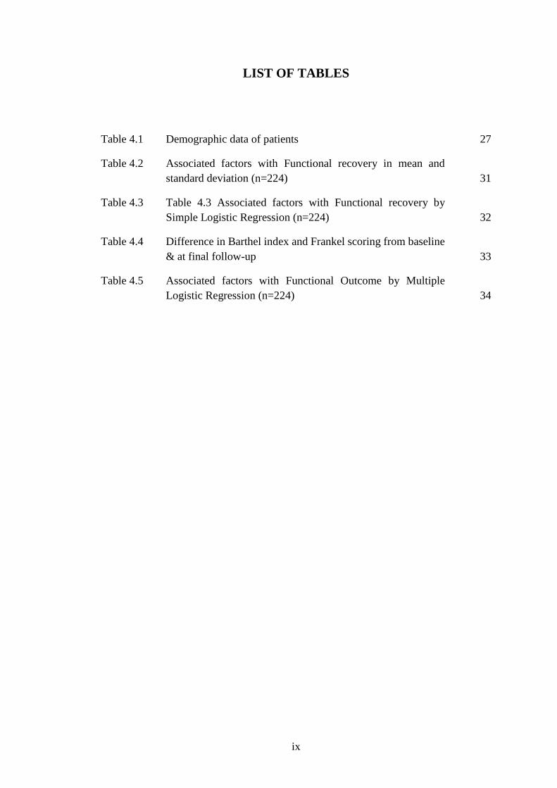
LIST OF TABLES
Table 4.1 Demographic data of patients 27
Table 4.2 Associated factors with Functional recovery in mean and
standard deviation (n=224) 31
Table 4.3 Table 4.3 Associated factors with Functional recovery by
Simple Logistic Regression (n=224) 32
Table 4.4 Difference in Barthel index and Frankel scoring from baseline
& at final follow-up 33
Table 4.5 Associated factors with Functional Outcome by Multiple
Logistic Regression (n=224) 34

x
LIST OF FIGURES
Figure 2.1 Diagrammatic presentation of the typical cervical
vertebra 6
Figure 2.2 Diagrammatic presentation of the typical thoracic
vertebra 7
Figure 2.3 Diagrammatic presentation of the typical lumbar
vertebra 8
Figure 2.4 Diagrammatic presentation of the blood supply to the
spine 10
Figure 2.5 Diagrammatic presentation of the sites of vertebral
tuberculosis 12
Figure 2.6 Spine at risk as described by (Rajasekaran et al, 2001) 18
Figure 4.1 Flow chart of study protocol 25
Figure 4.2 Distribution of patients based on sex 26
Figure 4.3 Number of patients and age grouping 27

xi
LIST OF ABBREVIATIONS
ADL Activities of daily living
CRP C-Reactive Protein
CT Computerized Tomography
DNA Deoxyribonucleic Acid
ESR Erythrocyte Sedimentation Rate
HQE Hospital Queen Elizabeth
MDI Modified Barthel Index
MRI Magnetic Resonance Imaging
PCR Polymerase Chain Reaction
ROM Range of Movement
TB Tuberculosis
WCC White Cell Count

1
CHAPTER ONE
INTRODUCTION
1.1 BACKGROUND OF STUDY
Spinal tuberculosis has been on the rise due to reemergence of immunocompromised
populations. Majority of the countries involved are developing countries especially
patients with postive HIV (with CD4+ of 50 to 200). Extrapulmonary tuberculosis
accounts for 15% of the total tuberculosis patients and spinal tuberculosis represents
5% of the total number of tuberculosis patient. The most common site of involvement
in the spine is the thoracic spine.
The causative organism for tuberculosis is Mycobacterium Tuberculosis, it is an
acid-fast bacilli which thrives in an aerobic environment. It has a very low tendency to
adhere to implants and does not form biofilms, thus allowing implants to be used safely
in spinal tuberculosis.
The pathogenesis of spinal tuberculosis is commonly due to a secondary
infection, the primary lesion is normally seen in the lung, gastrointestinal tract or the
genitourinary tract. The bacilli then travel via hematogenous spread to the vertebrae
(presence of valveless venous plexus) and seed over the narrow hair pin metaphyseal
vessels of the anterior vertebral body. They remain dormant and express an delayed
hypersensitivity immune response which will trigger the inflammatory cascade and for
a granulomatous lesion with bone destruction and central casseation. It then spreads
beneath the anterior longitudinal ligament as it is the pathway with the least resistance
and causes contiguous and non-contiguous multilevel destruction. Spinal tuberculosis
is also notorious in forming huge psoas abscesses. Intervertebral discs being relatively

2
avascular are preserved until late disease. The bony destruction will result in collapse
and kyphosis, anterior vertebral collapse results in sharp kyphosis aka “gibbus”
increasing the risk of cord compression.
Potts disease introduced by Sir Percival Pott in 1979 is defined as paraplegia
secondary to spinal tuberculosis. Its etiology can be further divided into early and late
onset of paralysis. In early paralysis it can be due to the mechanical pressure exerted by
the abscess, caseous granuloma or the sequestra, or spinal artery thrombosis or
tuberculous arachnoiditis. Late onset paralysis is commonly due to increasing
deformity, reactivation of tuberculosis or the fibrosis of the dura.
Tuberculosis may affect the vertebrae in many locations, however three
common patterns have been described being, peridiscal, central and anterior. Peridiscal
being the most common form occurs adjacent to the vertebral endplate and spreads
around a single intervertebral disc as the subligamentous spread occurs. Central occurs
in the middle of the vertebral body which leads to vertebral collapse and kyphotic
deformity. Anterior lesions begin as tuberculosis spreads beneath the anterior
longitudinal ligament which may be seen a scalloping of the anterior vertebral bodies.
Common investigations performed are the Mantoux/ Tuberculin skin test, ESR, CRP,
chest Xrays, whole spine Xrays, CT and MRI scans. Mantoux test is an unreliable test
as patients from endemic areas with subclinical exposures or previous BCG
vaccinations may elicit a false positive result. Two thirds of patients with spinal
tuberculosis may present with chest xray findings. Literature has shown ESR to be
mildly elevated with 25% of patients presenting within normal levels. The gold standard
investigation for spinal tuberculosis is tissue biopsy which may be performed
percutaneously, CT guided or via the open technique.

3
Biopsy samples are sent for histopathological analysis which reveal a
granulomatous lesion with surrounding multinucleated giant cells and central necrosis.
Ziehl Neelson staining for acid fast bacilli may also be performed but may only be
positive in 50% of patients. Another investigation is to perform a Tuberculosis PCR
(polymerase chain reaction) which amplifies DNA of the Mycobacterium for
identification, this test is quick and highly sensitive with a PPV of 95-98% in smear
positive patients. Mycobacterium tuberculosis is cultured in Lowenstine-Jensen
medium typically yielding positive growth in 6-10-week period.
Treatment of spinal tuberculosis comprise of three main goals, to eradicated
disease, to prevent and correct deformity and to prevent neurological insult. Anti-
tuberculous therapy remains the mainstay of treatment in tuberculosis supplemented
with surgery and/or orthosis. Recovery is not easily predictable and very variable, some
recover completely but some are left with debilitating weakness. We aim to investigate
and look into potentially identify clinical, biochemical and radiological factors that
might affect functional outcome recovery in spinal tuberculosis patients.
1.2 OBJECTIVES AND HYPOTHESIS
1.2.1 General Objectives
To evaluate the relationship of clinical parameters and functional recovery in patients
treated as spinal tuberculosis patients with neurology.
1.2.2 Specific Objectives
Specific objectives of this study are:
1. To compare the duration of symptoms with the functional recovery in patients
treated as spine tuberculosis with neurology.

4
2. To identify correlations of patients age and functional recovery in patients
treated as spine tuberculosis with neurology.
3. To analyze the correlation of inflammatory markers (white cell count, ESR &
CRP) with the functional recovery in patients treated as spine tuberculosis with
neurology.
4. To study the relationship of radiological findings (Xray & MRI determining
levels of involved vetebrae, soft tissue or spinal extension of the tuberculous
abscess) with the functional recovery in patients treated as spine tuberculosis
with neurology.
1.2.3 Hypothesis
There is a positive correlation between the clinical presentation of spinal tuberculosis
patients and improvement in functional recovery in patients treated as spine tuberculosis
with neurology.

5
CHAPTER TWO
LITERATURE REVIEW
2.1 ANATOMY OF THE SPINE
2.1.1 Cervical spine
The cervical spine consists of seven vertebrae, among which there are typical vertebrae
(C3 - C6) and the atypical vertebrae (C1, C2, and C7). The typical cervical vertebrae
have a broad kidney shaped body that is similar or smaller than the size of its vertebral
foramen and is concave in its anterior surface. There are also the presences of uncus
process a posterolateral lip projection of the lateral edge. Typical cervical spine pedicles
are attached to the body and has a foramen from its expansion with the cervical
transverse process which have tubercles over their far lateral lips. The transverse
process foramen is perforated by the vertebral artery with its accompanying sympathetic
nerve fibers. The vertebral foramen is triangular in cross section bounded by the lamina,
the lamina from its articular processes at its junction with the pedicle. The facets of the
upper cervical spine face upwards and backwards, and the lower ones face downwards
and forwards. Lastly the spinous process in the cervical spine is bifid. The intervetebral
discs are also thick compared to the size of the cervical body.

6
Figure 2.1 Diagrammatic presentation of the typical cervical vertebra
2.1.2 Thoracic Spine
Thoracic vertebra presents with two costal facets for articulation with the ribs. The body
of the thoracic vertebra is heart shaped with the spinal canal circular in shape. The
pedicles project from the upper half of the body and its upper border in line with the
upper surface of the body. The lamina is flat and slope downwards and backwards, the
facets are facing laterally and backwards, with circular articular surfaces to allow some
amount of rotation. The spinous processes of thoracic vertebrae slope downwards up to
T7 and begin to progressively level out by T12. Thoracic transverse processes project
backwards and laterally and articulate with the adjacent ribs.

7
Figure 2.2 Diagrammatic presentation of the typical thoracic vertebra
2.1.3 Lumbar Spine
Lumbar spine bodies are kidney shaped and increase in width as they progress distally.
The spinal canal is triangular in cross section. Pedicles are attached to the upper half of
the body simmilar to the thoracic spine. The articular processes face downwards and
laterally the transverse process of L4 is the longest among the lumbar spine with a
quadrangular spinous process. The presence of mamillary bodies as posterior
projections of the superior articular process. Lumbar spine has massive intervetebral
discs among the other regions.

8
Figure 2.3 Diagrammatic presentation of the typical lumbar vertebra
2.1.4 Blood Supply of The Spine
Spinal blood supply was first studied by Albert Wojciech Adamkiewicz a Polish
pathologist, in 1881. Blood supply to the spinal cord is mainly via one anterior median
spinal artery and two posterior spinal arteries. The anterior spinal artery is formed by
the two vertebral arteries at the foramen magnum supplies 85% of blood supply to the
cord throughout its length in the anterior median fissure. The two posterior vertebral
arteries originate from the two posterior inferior cerebellar arteries at the level of
foramen magnum and lie on the posterolateral sulcus where it divides into two collateral
arteries which travel with the nerve roots.
In the cervical spine majority of the radicular arteries arise from the vertebral
artery. In this region basilar artery also provides contribution via anastomoses with the
anterior spinal artery
In the thoracic spine, the radicular arteries branch off intercostal arteries at the
level of costotransverse joint. Here there is the radicular artery of Adamkiewicz, which

9
is the largest segmental artery and is the major blood supply to the lower cord. In 80%
of the population it arises from the left side of the body and usually accompanies ventral
root of the thoracic nerves 9-10 but may originate anywhere from T5 to L5 verterbrae.
(ACM Amato, 2015) Reported that the watershed region of the spinal cord is said to be
at the thoracic region as it has a limited number of segmental arteries. Branches of
anterior spinal artery supply the ventral 2/3 of the cord and the posterior artery branches
supply the dorsal 1/3 of the cord. The watershed region is said to be at the junction of
these two zones as its vascularity is the poorest. Based on the cross-sectional anatomy
of the spinal cord it can be understood that injuries to the anterior spinal artery results
in mainly motor deficits and posterior spinal artery injuries usually result in sensory
deficits. Another region of the thoracic spine that is said to have a critical vascular zone
is the zone between the 4th to 9th thoracic vertebrae, as it has the least profuse blood
supply and clinically is the narrowest region of the spinal canal.
In the lumbar spine blood supply to each vertebral body is via the lumbar
segmental arteries. The segmental arteries arise from the aorta for the levels L1-L4 and
from the iliolumbar artery from the L5 level. These arteries than traverse towards the
intervetebral foramina where it has three divisions, an anterior branch to the anterior
abdominal wall, a posterior branch to the paraspinal muscles & facet joints and a
foraminal branch to the spinal canal and its contents. In the lumbar spine the venous
supply parallels the arterial supply.
Batson’s venous plexus is a system of valveless veins within the spinal canal
and around the vertebral body and is in continuity with pelvic plexus. It provides an
alternative route of venous drainage to IVC in instances of an increase in abdominal

10
pressure where the venous return would be preferred via the spinal canal. This makes
this system a potent pathway for metastasis and infection to invade the lumbar spine.
Figure 2.4 Diagrammatic presentation of the blood supply to the spine
2.2 SPINAL TUBERCULOSIS
2.2.1 Pathophysiology
The causative organism of tuberculosis is the mycobacterium tuberculosis complex
which is a bacillus with a thick mycolate-rick outer covering which functions as an
excellent barrier. It is an aerobic organism and fortunately does not form biofilm, thus
allowing the usage of spinal implants to provide stability. The mycobacterium makes
its way to the vertebrae via haematogenous spread from a primary foci commonly seen
in the lungs, the gastrointestinal and genitourinary systems. The oxygen hungry bacilli
usally disseminates into the vertebra through the anterior spinal artery, the segmental
arteries that branch from the abdominal aorta and pelvic organs may assist in
dissemination. The fact that the Adankiewicsz Artery is in the thoracic region explains
why the commonest site of spinal tuberculosis to be in the thoracic region. The bacilli
usually diseminates after a prolonged latent phase slowly progressing to from a foci of

11
chronic inflammation with epitheloid cells, followed by Langerhan giant cells,
lyphocytes. This causes progressive bony destruction resulting in central caseous
necrosis, these foci of chronic inflammation are the pathognomonic finding in
histopathological analysis of suspected cases. There are two gross types of tuberculous
infections, the caseous exudative type which is commonly seen in children with massive
destruction, exudation and gross abscess formation. The other is a granular type more
commonly seen in adults, it is a slow and latent type of infection which is less
destructive and has minimal abscess formation. The types of tuberculous lesions in the
spine can present in five variants, the commonest, paradiscal type via arterial spread,
where the bacilli lodge in the subcondral marrow on either side of the disc. The
avascular disc is usually spared in early spinal tuberculosis and is only affected in late
disease. The central type due to venous spread causes vertebral body destruction, the
posterior or appendicular type which involves the posterior elements only is nortorius
for destabalizing the spine. The fourth of anterior type is due to subligamentous spread
of tuberculosis under the anterior longitudinal ligament, this group usually present with
anterior scalloping of the vertebral bodies. The final type is the least understood type
and affects the articular joints of the spine only.

12
Figure 2.5 Diagrammatic presentation of the sites of vertebral tuberculosis
2.2.2 Clinical Features
Clinical features of spinal tuberculosis are very variable, it is a chronic spine disease
commonly with an acute onset of symptoms. Pain, neurological deficits, cold abscess,
kyphotic deformities are characteristic findings. 100% of patients in (Azzam and
Tammawy, 1988) study reported back pain as their presenting symptom. Pott’s disease
or Potts paraplegia is paraplegia secondary to spinal tuberculosis. Neurology in spinal
tuberculosis may be present in up to 40-50% of cases and can be due to acute causes
such as soft compression from the abscess, caseous granuloma or the sequestra,
infective thrombosis of the vasculature leading to ischaemia or arachdonitis which is
meningeal inflammation and fibrosis. Late causes of neurology are, dura fibrosis,
progression of deformity and reactivation. Kyphosis in spinal tuberculosis is commonly
acute and presents as a gibbus deformity, (Rajasekar at al, 1998) divided spinal
tuberculosis deformities as knuckle deformity (single vertebra deformity), gibbus

13
deformmity (collapse of two or three vertebra) or global rounded kyphosis (involvement
of multiple adjacent vertebra). Constitutional symptoms fever, profuse night sweats and
malaise may be reported by patients, but are more commonly seen in patients with
pulmonary tuberculosis
2.2.3 Investigations
2.2.3.1 Blood Investigations
Investigations can be divided into routine blood investigations, tissue sampling, and
imaging investigations. Routine blood works include erythrocyte sedimentation rate
(ESR), C-reactive protien (CRP), and white cell counts. All blood parameters lack
specificity to spinal tuberculosis as they have many false positive triggers. Among them
the ESR has been seen to be elevated in more than 60% of patients across different
studies, with mean values of 61mm/hr, keep in mind that ESR levels can be raised by
non-infectious causes, tumors and even trauma. In Guo et al’s study, it was noted that
69% of spinal tuberculosis patients had elevated CRP, it was also noted that this
elevation was more specific for acute infective lesions compared to disseminated ones.
ESR and CRP levels may even be normal in approximately 20% of patients. WCC has
not been shown to provide a role in diagnosing spinal tuberculosis. Tuberculin skin test
also displays high false positive cases due to previous exposure or in patients with
Bacilus Calmette-Guerin (BCG) vaccination. The QuantiFERON-TB that measures
interferon-gamma levels from suspected patients has been reported to have sensitivity
of 84% and spcificity of 95% by Kumar et al. However, its cost and availability are
some of the reasons it is not routinely done.