factors affecting surfactant responsiveness
TRANSCRIPT
Factors Affecting Surfactant Responsiveness Influence of mode of administration and ventilation, disease stage aud type of surfactant
ISBN 90-9011902-7
Gammers, Diederik
Factors affecting surfactant responsiveness: influence of mode of administration and
ventilation, disease stage and type of surfactant I Diederik Gammers.
Thesis Rotterdam. - With ref. - With summary in Dutch.
No part of this book may be reproduced without permission in writing from the author.
FACTORS AFFECTING SURFACTANT RESPONSIVENESS Influence of mode of administration and ventilation, disease stage and type of surfactant
FAKTOREN DIE DE WERKING V AN SURFACTANT BEINVLOEDEN
Invloed van de manier van toediening en beademing, ziekte toestand en soort surfactant
PROEFSCHRIFT
ter verkrijging van de graad van doctor
aan de Erasmus Universiteit Rotterdam
op gezag van de rector magnificus
Prof. dr P.W.C. Akkermans M.A.
en volgens besluit van het College van Promoties.
De openbare verdediging zal plaatsvinden op
woensdag 9 december 1998 om 11.45 uur
door
Dicdcrik Antonius Maria Paulus Johannes Gommers
geboren te Gorinchem
PROMOTIECOMMISSIE:
Promotor:
Overige leden:
Prof. dr B. Lachmann
Prof. dr W. Erdmann
Prof. dr L.M.G. van Golde
Prof. dr D. Tibboel
Dit proefschrift werd bewerkt binnen de afdeling Anesthesiologie van de Erasmus Universiteit
Rotterdam.
The studies presented in this thesis were fmancially supported by the International Foundation
for Clinically Oriented Research (IFCOR).
/~t proefschrift werd gedrukt door Haveka B.V. te Alblasserdam.

Aall Arie Kokt

Contents
Introduction Chapter I Surfactant therapy
Adaptedjrom: Clill III/ellsive Care 1993; 4: 284-295
Origiual studies Chapter 2
Chapter 3
Chapter 4
Chapter 5
Chapter 6
Chapter 7
Chapter 8
Chapter 9
Summary
Exogenous surfactant therapy increases static lung compliance, and cannot be assessed by measurements of dynamic compliance alone Published ill: Crit Care Med 1993; 21: 567-574
Comparison of eight different surfactant preparations on
improvement of blood gases in lung-Iavaged rats
III press: Appl Cardiopulm Pathophysiol Prevention of respiratory failure after hydrochloric acid aspiration
by intratracheal surfactant instillation in rats Published iu: Allesth Allalg 1993; 76: 472-477
In-vivo evalution of the inhibitory capacity of human plasma on
exogenous surfactant function Published ill: I11Iellsive Care Med 1994; 20: 6-11
Bronchoalveolar lavage with a diluted surfactaut suspension prior
to surfactant instillation improves the effectiveness of surfactant therapy in experimental acute respiratory distress syndrome (ARDS)
Published ill: l111ellsive Care Med 1998; 24: 494-500
Surfactant therapy in combination with high-frequency oscillatory
ventilation is not superior to conventional mechanical ventilation in reducing lung injury in lung-Iavaged rabbits
Submitted jar publieatioll
Conventional ventilation modes with small pressure amplitudes and
high end-expiratory pressure levels optimize surfactant therapy
111 press: Crit Care Med Improved oxygenation by nitric oxide is enhanced by prior lung
reaeration with surfactant, rather than positive end-expiratory pressure, in lung-Iavaged rabbits
Published ill: Crit Care Med 1997; 25: 1868-1873
Samenvatting (sununary in Dutch)
Abbreviations
Dankwoord (Acknowledgement)
List of publications
Curriculum vitae
9
39
55
75
91
107
121
137
149
163 168 173 175
177
184

Chapter 1
Surfactant therapy
Adapted from: Clinlm€lISive Care 1993; 4: 284-295
D. Gommers, B. weilli/O/ill. Surfactant therapy: does it hm'€ a role ill adults? Repri1lled with permission (copyright holder: Caslle HOllse Publications Ltd)
-9-

Chapter 1
Introduction
Historically, Kurt von Neergaard [1) was the fIrst to suggest that surface tension plays
an important role in lung elasticity. He showed, in 1929, that the pressure necessary
for fIlling the lung with liquid was less than half the pressure necessary for fIlling the
lung WiOl air, and concluded that two-thirds to three-fourths of the elasticity of the lung
was derived from interfacial forces [1). The problem with his discovery was that this
paper was published in Gemlan and that, for 25 years, no scientists in the evolving fIeld
really took note of this pnblication. In 1954, Macklin [2) described the presence of a
thin aqueous mucoid microfIlm, formed from secretion of the granular pneumocytes,
on the pnlmonary alveolar walls and which is in constant slow movement toward the
phagocytic pneumocytes and bronchioles. One year later, Pattie [3) noticed the
remarkable stability of foam and bubbles from lung edema and healthy lung cut. He
assumed that the walls of these bubbles consists of surface-active material which must
lower the surface tension to nearly zero. In 1957, Clements [4) was the fIrst to prove
the direct evidence of surface active material in the lungs. He measured surface tension
of a surface fIlm derived from the lung by using a Wilhelmy balance and demonstrated
that the surface tension was not a constant value; when the surface was stretched the
tension was relatively high (40 dynes/cm), but when the surface area was decreased the
tension fell to 10 dynes/cm. He pointed out that such a reduction in surface tension
during deflation in the lung would tend to stabilize the air spaces by permitting them
to remain open at low lung volumes. Two years later, Avery and Mead [5)
demonstrated that lung extracts of very small premature infants and infants dying with
hyaline membrane disease had much higher surface tension than normal lung extracts,
due to a defIciency in surface active material. This was the first step towards extensive
research on the surfactant system, and Fujiwara and colleagues, in 1980 [6), were the
fIrst to treat premature babies suffering from respiratory insufficiency with exogenous
surfactant.
Pulmonary surfactant is a complex of phospholipids (80-90%), neutral lipids (5-
10%) and at least four specifIc surfactant-proteins (5-10%) (SP-A, SP-B, SP-C and SP
D), lying as a monolayer at the air-liquid interface in the lung [7,8). Surfactant is
synthesized by the alveolar type II cells and secreted into alveolar spaces [7). The
surfactant lipids are lying in a thin aqueous fIlm which coats the pulmonary alveolar
walls and small airways. At the surface of this aqueous fIlm, the phospholipid
molecules are lying as a monolayer and lower its surface tension; this reduce the
-11-
Introduction
muscular effort uecessary to breathe and prevents collapse of the alveoli at the end of
expiration [9).
Since 1980, more than 100,000 premature infants suffering from respiratory
distress syndrome (RDS) due to a surfactant deficiency are successfully treated with
exogenous surfactant almost without any side-effects [10,11). Biochemical and
biophysical abnormalities of the pulmonary surfactant system is also seen in other
diseases such as the adult respiratory distress syndrome (ARDS) [9,12,13], infectious
lung disease [14) and after cardiopulmonary bypass surgery [15). Furthennore, it could
be demonstrated that nou-optimal ventilation may lead to disturbance of alveolar
surfactant [16).
However, only a few case reports and results of limited clinical pilot studies are
available, in which patients other than neonates with RDS are treated with exogenous
surfactant [17). In this chapter, we describe the rationale for exogenous surfactant
therapy in the different lung diseases by reviewing experimental and clinical findings.
Function of the pulmonary surfactant
The normal physiological functions of the pulmonary surfactant system include:
1) Mechanical stabilization of lung alveoli.
The force required to open alveoli is determined by surface tension at the air
liquid interface and by the radius of the terminal units of the lung in accordance with
the LaPlace law (P = 2y Ir; P = pressure in the bubble, y = surface tension, r = radius of the bubble). Pulmonary surfactant decreases the surface tension of the
interface and thereby allows normal breathing with the least possible effort.
During deflation of the lung, a static high surface tension would tend to promote
alveolar collapse. However, as alveolar size decreases, pulmonary surfactant ensures
that surface tension falls approximately to zero. Thus, at small alveolar volumes,
surface tension becomes a negligible force and thereby tends to promote alveolar
stability [18).
2) Stabilization of small airways.
Pulmonary surfactant also ensures stabilization of the peripheral airways and Oms
its lack might cause airway obstruction or collapse of the small bronchioli with air
trapping [19). Besides its role in mechanical stabilization, bronchial surfactant also has
a transport function for mucus and inhaled particles [20). Furthermore, bronchial
surfactant acts as an antiglue factor, preventing the development of large adhesive
-12-

forces between mucus and the bronchial wall [21].
3) Protection against lung edema.
Chapter 1
Another function of the pulmonary surfactant system is stabilization of the fluid
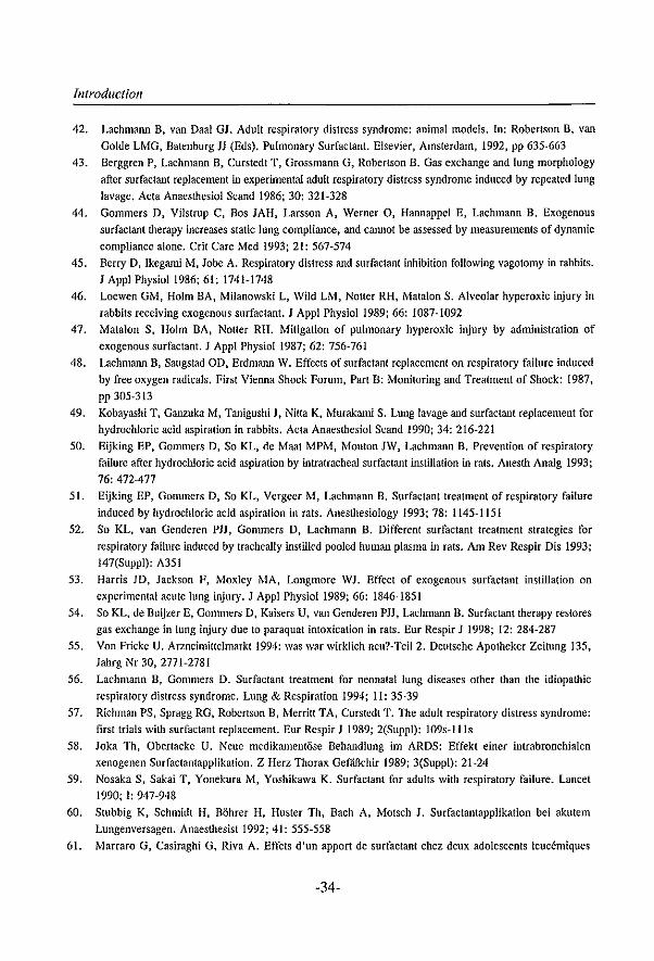
balance in the lung and protection against lung edema [22]. In general, the forces that
influence the circulation of liquid at the alveolar-capillary level in the lungs include:
plasma colloid osmotic pressure on one side and capillary hydrostatic pressure,
interstitial colloid osmotic pressure and alveolar surface tension on the other side
(Figure 1). This means that a surfactant deficiency will increase the surface tension at
the air-liquid interface and thereby the suction forces will increase, resulting in lung
edema (Figure 1).
4) Surfactant and local defense mechanism.
It has also been demonstrated that surfactant plays a role in the lung's defense
against infection [23]. Surfactant, and in particular SP-A, enhances Ule antibacterial and
antiviral defense of alveolar macrophages [23].
We have demonstrated that the pulmonary surfactant system may also be
involved in protecting the lung against its own mediators (e.g. angiotensin II) and in
protecting the cardiocirculatory system against mediators produced by the lung [24,25].
SCHEMATIC DIAGRAM OF WATER BALANCE IN THE LUNG
-PLASMA ONCOTIC PRESSURE
.. NORM4l- VALUES 37
RESPIRATORY DIStRESS 37 SYHORO~E (I)
.--PULMONARY CAPILLARY
BASE LAYER
SUPERFICIAL LAYER
AIRSPACE
-----+ --+ CAPILLARY TISSUE BLOOD FLUID PRESSURE ONCOIIC
PRE SURE 15 + 18 +
< 15 + 18 + II) 11)
--+ SURFACE TENSION PRESSURE IRadiuI.hnaion) ,
" to 30
Figure 1. Simplified schematic diagram representing the factors influencing fluid balance in the lung (from
reference [28]).
-13-
introduction
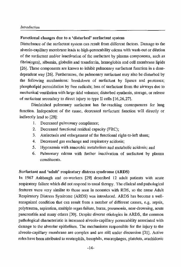
Functional changes due to a 'disturbed' surfactant system
Disturbance of the surfactant system can result from different factors. Damage to the
alveolo-capillary membrane leads to high-permeability edema with wash-out or dilution
of the surfactant and/or inactivation of the surfactant by plasma components, such as
fibrin(ogen), albumin, globulin and transferrin, hemoglobin and cell membrane lipids
[26]. These components are known to inhibit pulmonary surfactant function in a dose
dependent way [26]. Furthermore, the pulmonary surfactant may also be disturbed by
the following mechanisms: breakdown of surfactant by lipases and proteases;
phospholipid peroxidation by free radicals; loss of surfactant from the airways due to
mechanical ventilation with large tidal volumes; disturbed synthesis, storage, or release
of surfactant secondary to direct injury to type II cells [16,26,27].
Diminished pulmonary surfactant has far-reaching consequences for lung
function. Independent of the cause, decreased surfactant function will directly or
indirectly lead to [28]:
I. Decreased pulmonary compliance;
2. Decreased functional residual capacity (FRC);
3. Atelectasis and enlargement of the functional right-to-Ieft shunt;
4. Decreased gas exchange and respiratory acidosis;
5. Hypoxemia with anaerobic metabolism and metabolic acidosis; and
6. Pulmonary edema with further inactivation of surfactant by plasma
constituents.
Snrfactant and 'adult' respiratory distress syndrome (ARDS)
In 1967 Ashbaugh and co-workers [29] described 12 adult patients with acute
respiratory failure which did not respond to usual therapy. The clinical and pathological
features were very similar to those seen in neonates with RDS, so the name Adult
Respiratory Distress Syndrome (ARDS) was introduced. ARDS has become a well
recognized condition that can result from a number of different causes, e.g. sepsis,
poly trauma, aspiration, multiple organ failure, burns, pneumonia, near-drowning, acute
pancreatitis and many others [30]. Despite diverse etiologies in ARDS, the common
pathological characteristic is increased alveolo-capillary permeability associated with
damage to the alveolar epithelium. The mechanisms responsible for the injury to the
alveolo-capillary membrane are complex and are still under discussion [31]. Active
roles have been attributed to neutrophils, basophils, macrophages, platelets, arachidonic
-14-
Chapter 1
acid metabolites, oxygen-derived free radicals, complement, proteases, interleukins,
serotonin, platelet activating factor (PAF), tumor necrosis factor (TNF), surfactant
inhibiting plasma-proteins, drugs, and many other substances [32]. But all these
individual factors which can lead to a pulmonary edema do not, however, necessarily
lead to ARDS. Therefore, another system must be involved to explain the functional
changes as seen in ARDS. It is established that the capillary leakage combined with
damage to the alveolar epithelium leads to an immediate, or moderately slow, loss of
active surfactant by inactivation or depletion from the alveoli and small airways which
is, however, compensated by a release of stored surfactant from type II cells [26].
Thus, the progress of the disease depends on the balance between new production and
release of surfactant into the alveoli and its inactivation/loss from the alveoli and
airways. If the synthesis is reduced e.g. by influenza virus, hypoxia or hyperoxia, etc.,
an imbalance between new synthesis and demand will result. This will finally lead to
a total loss of functional active surfactant, resulting in failure of the lung as a gas
exchange organ [28,33]. Thus in ARDS the surfactant deficiency is a complication of
lung iI\iury rather than, as in neonatal RDS, a primary etiological factor. In spite
increased sophistication in methods of respiratory support, mortality associated with
ARDS currently remains between 48 and 75%, depending on the etiology [34,35].
Nowadays, it is more appropriate to speak about tile Acute, rather than Adult,
Respiratory Distress Syndrome (AROS), since ARDS is not limited to adults [36].
Analyses of lung surfactant recovered iII BAL from patients with AROS, or
from animal models of acute respiratory failure, demonstrate disturbances of the lung
surfactant system [37]. Reduction of surfactant activity is associated with increased
minimal surface tension of lung extracts or lung homogenates, and compositional
changes of surfactant andlor decreased surfactant content of the lungs [7,17,33].
Ashbaugh and colleagues [29] were the first to demonstrate decreased lung compliance
and iIlcreased miniInal surface tension in lung extracts from tlVO ARDS patients. Since
then, several studies have demonstrated qualitative and quantitative changes of
surfactant in BAL fluid from ARDS patients [38-41]. Recently, Gregory and colleagues
[41] demonstrated that several of these alterations already occur in patients at risk of
developing AROS, suggestiIlg that these abnormalities of surfactant occur early in the
disease process.
The central role of surfactant deficiency can further be illustrated by recent
studies in aninlal models of ARDS which demonstrated that exogenous surfactant
-15-
Introduction
instillation dramatically improved blood gases and lung mechanics [9,42]. The models
of surfactant deficiency in which these inlprovements could be demonstrated include
acute respiratory failure due to in-vivo whole-lung lavage [43,44], neurogenic ARDS
[45], respiratory failure as a result of oxygen toxicity [46,47] or oxidant-producing
enzymes [48], acute respiratory failure after instillation of hydrochloric acid [49-51] or
plasma instillation [52], and respiratory failure after intoxication with N-nitroso-N
methylurethane (NNNMU) [53] or paraquat [54].
Evidently, it is rational to administer exogenous surfactant in ARDS patients,
but the question then arises why is this not yet a reality. Surfactant has been
commercially available for neonates since 1987 [55]. Surfactant therapy in patients
other than neonates with RDS is almost inlpossible due to the fact that there is not
enough surfactant available and tilat current prices are too high (l g of surfactant costs
about US$ 3,000-5,000) [55]. Therefore, only a few case-reports and pilot studies have
been performed up to now.
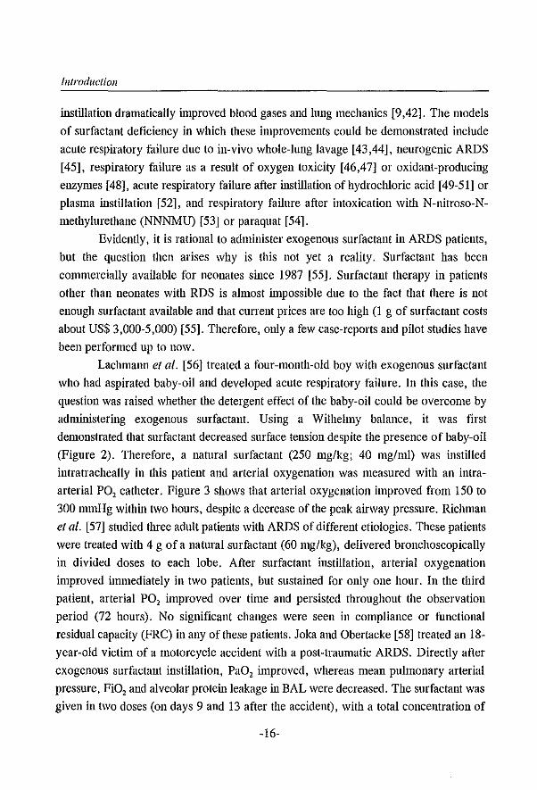
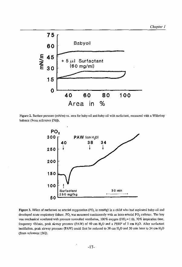
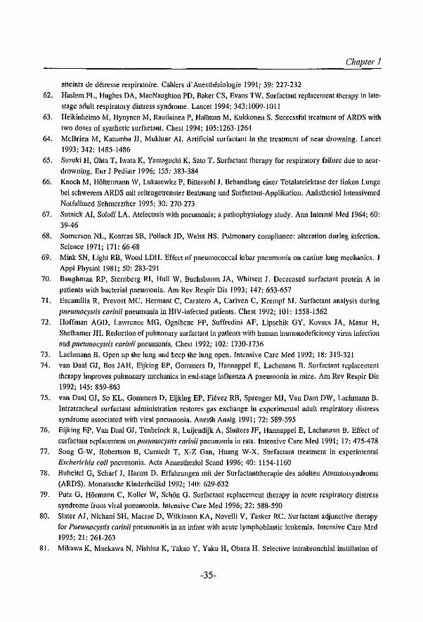
Laclmmm el al. [56] treated a four-month-old boy with exogenous surfactant
who had aspirated baby-oil and developed acute respiratory failure. In this case, the
question was raised whetiler the detergent effect of the baby-oil could be overcome by
administering exogenous surfactant. Using a Wilhelmy balance, it was first
demonstrated that surfactant decreased surface tension despite the presence of baby-oil
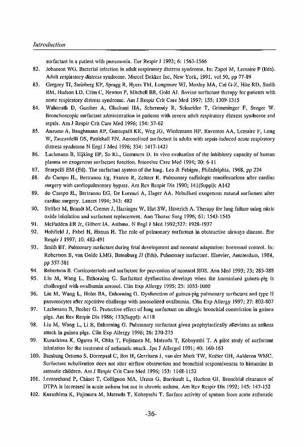
(Figure 2). Therefore, a natural surfactant (250 mg/kg; 40 mg/ml) was instilled
intratracheally in this patient and arterial oxygenation was measured with an intra
arterial PO, catheter. Figure 3 shows that arterial oxygenation improved from 150 to
300 mmHg within two hours, despite a decrease of the peak airway pressure. Richman
el al. [57] studied three adult patients with ARDS of different etiologies. These patients
were treated with 4 g of a natural surfactant (60 mg/kg), delivered bronchoscopically
in divided doses to each lobe. After surfactant instillation, arterial oxygenation
improved immediately in two patients, but sustained for only one hour. In the third
patient, arterial PO, improved over time and persisted throughout the observation
period (72 hours). No significant changes were seen in compliance or functional
residual capacity (FRC) in any of these patients. Joka and Obertacke [58] treated an 18-
year-old victim of a motorcycle accident with a post-traumatic ARDS. Directly after
exogenous surfactant instillation, PaO, improved, whereas mean pulmonary arterial
pressure, FiO, and alveolar proteillieakage in BAL were decreased. The surfactant was
given in two doses (on days 9 and 13 after the accident), with a total concentration of
-16-
Chapter 1
75
60 Babyoil
E 45 ..... + 5 ,u I Surfactant Z E 30 (60 mg/ml)
15
0 40 60 80 100 Area In %
Figure 2. Surface pressure (mN/m) vs. area for baby~oil and baby~oil with surfactant, measured with a Wilhelmy
balance (from reference (56)).
250
200
150
100
50
40 I
PAW (omH,o)
38 I
Surfactant
34 I
30 min
l~2~5~O!m~g~/~kg~ __________ ~::::::::~ __ _
Figure 3. Effect of surfactant on arterial oxygenation (POl in mmHg) in a child who had aspirated baby-oil and
developed acute respiratory failure. P02 was measured continuously with an intra-arterial POl catheter. The boy was mechanical ventilated with pressure controlled ventilation, 100% oxygen (Fi01= 1.0), 70% inspiration time.
frequency 45/min, peak airway pressure (PAW) of 40 em H20 and a PEEP of 5 em Hp. After surfactant
instillation, peak airway pressure (PAW) could first be reduced to 38 em HP and 30 min later to 34 em HP
(from reference [56]),
-17-
Introduction
about 50 mg/kg body weight. Nosoka and co-workers [59] demonstrated improvement
in PaO, and chest X-ray after multiple instillations of surfactant in two adult patients
with ARDS. The fIrst patient received surfactant 20 times (240 mg each) during 38
days, whereas dIe second patient received dlree doses of surfactant (also 240 mg each)
on three consecutive days (antibodies to the natural surfactant were not detected in
either patient). Stubbig el al. [60] reported a case of surfactant therapy in a 21-year-old
man who developed ARDS after severe lung contusion due to a car accident. No
improvement occurred during conventional ventilatory treatment, including inverse
ratio ventilation and high-frequency ventilation. Immediately after instillation of a
natural surfactant (on day 15 after the accident; 38 mg/kg), they observed the following
changes: deterioration of the pulmonary function probably due to crusts in the lung;
after aspiration of the crusts at bronchoscopy, there was a progressive improvement in
respiratory parameters. The PaO, and chest X-ray improved, whereas FiO" inspiration
time and PEEP level could be reduced. Marraro el al. [61] treated two adolescents who
developed ARDS which appeared during leukemia treatment with surfactant (patient
one, 60 mg/kg and patient two, 40 mg/kg). Arterial oxygenation improved within dlree
hours (patient one: 60 to 350 mmHg; patient two: 160 to 300 nunHg) during
mechanical ventilation with 100% oxygen. Haslem and colleagues [62] treated four
adult patients with late stage of ARDS with a single bolus of synthetic surfactant (75
mg/kg) and found no sustained clinical inlprovement. In contrast to the results of
Haslem and co-workers [62], Heikinheimo el al. [63] reported successful treatment of
a 50-year-old patient SUffering from ARDS with two doses of synthetic surfactant (total
amount 104 mg/kg). McBrien el al. [64] treated a nearly drowned 9-year-old boy with
synthetic surfactant. PaO,/FiO, was increased from 57 to 293 mmHg while PIP was
reduced from 40 to 25 cm H,o and the patient was discharged successfully from the
hospital two days later. Suzuki el al. [65] confIrmed the rapid and dramatic effect of
surfactant therapy on lung compliance, oxygenation and ventilation in a 3-year-old boy
with refractory respiratory failure due to near-drowning. Knoch el al. [66] reported a
case of surfactant therapy in a 48-year-old patient who developed respiratory
insufficiency nine days after a bicycle accident. The left lung could not be ventilated
even after separate artificial ventilation of each lung. After administration of a bolus
of synthetic surfactant (50 mg/kg) and continued separate artificial ventilation on each
side, there was a complete re-expansion of the left lung with an increase of arterial PO,
values from 65 to 416 nnnHg within a few hours (FiO,=1.0). The results of first
-18-
Chapter 1
clinical studies of surfactant therapy in ARDS patients are described in the next
paragraph.
Snrfactant and infectious lung diseases
Pneumonia is an important cause of respiratory failure and is associated with increased
alveolar permeability leading to pulmonary edema, hemorrhage and atelectasis [67,68].
The pathophysiological changes in pneumonia include hypoxemia, decreased functional
residual capacity (FRC) , decreased total lung capacity (TLC) , decreased lung
compliance, and a diminished surfactant system [14,68-70].
As far back as 1964, Sutnick and Soloff [67] demonstrated that the surface
tension of BAL fluid from lung tissue with pneumonia was increased; they suggested
that the pulmonary surfactant became inactivated and was responsible for atelectasis.
It has since been demonstrated that the surfactant system is also impaired in bacterial
[14,70] and viral pneumonia [27], as well as in Pllelllllocystis carillii pneumonia
[71,72]. In bacterial pneumonia surface tension of BAL fluid is increased, whereas SP
A content and total surfactant lipids are all significantly decreased [14]. In viral
pneumonia, Stinson et al. [27] demonstrated that pulmonary surfactant activity is
decreased; these workers suggested that injury and destruction of type II pneumocytes
by the ~irus was the cause of reduced surfactant activity. Recently, two studies have
demonstrated surfactant abnormalities in HIV positive patients with Pllel/lllocystis carillii pneumonia [71,72]. In these patients qualitative and quantitative changes were
seen in the surfactant composition, as well as increased phospholipase A, activity [72].
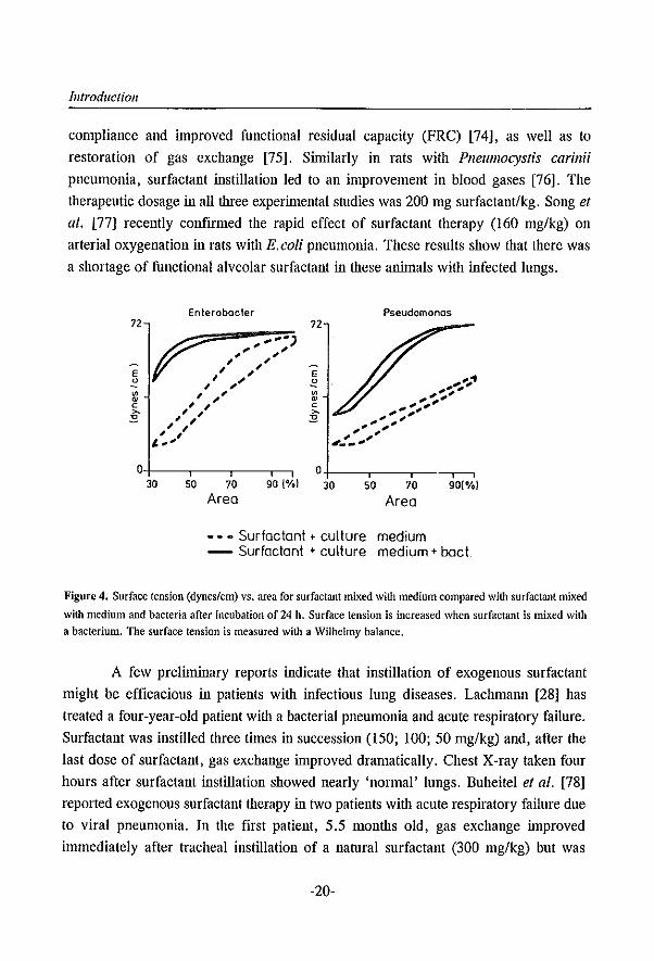
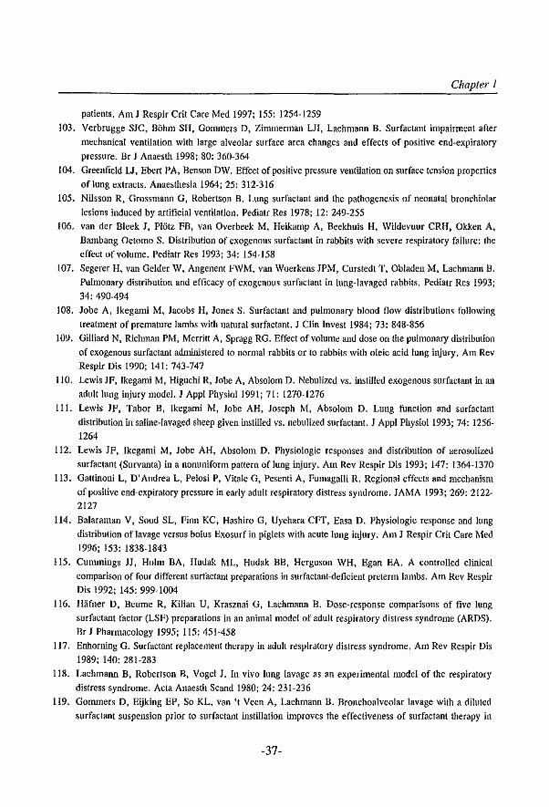
Bacteria, bacterial toxins, viruses, phospholipases, and proteinases released
from inflannnatory cells interact either directly with the surfactant film (Figure 4), or
damage tile endotllelial and epitllelial cells leading to high permeability edema [14]. It
is well established that plasma proteins of the edema fluid inactivate the surfactant [26].
Due to the decreased surfactant activity, surface tension at the alveolar walls increases,
leading to increased suction forces across the alveolo-capillary membrane [22]. This
finally results in a vicious circle [13,73].
Evidence thus exists of a deficiency of active pulmonary surfactant in patients
Witll pneumonia and this would be the rationale for exogenous surfactant therapy. We
recently demonstrated the effectiveness of surfactant tllCrapy in different animal models
suffering from viral pueumonia or Pllel/lllocystis carillii pneumonia [74-76]. In viral
pneumonia, tracheal administration of exogenous surfactant led to inlproved lung
-19-
Introduction
compliance and improved functional residual capacity (FRC) [74], as well as to
restoration of gas exchange [75]. Similarly in rats with Pllelllllocystis carillii pneumonia, surfactant instillation led to an improvement in blood gases [76]. The
therapeutic dosage in all three experin,ental studies was 200 mg surfactant/kg. Song et al. [77] recently confirmed the rapid effect of surfactant therapy (160 mg/kg) on
arterial oxygenation in rats with E.coli pneumonia. These results show that there was
a shortage of functional alveolar surfactant in these animals with infected lungs.
72
E o
~ c §
Enlerobacler
.... .,. ... , ...... ... , " ... ... ... ...
" ,~ ... ... , ... , ... , ... ... ... , " ,.'
Pseudomonas
O+---~---.---,-, 0+-__ -. __ -. __ -.-. 30 50 70 90 1%) 30 50 70 90(010)
Area Area
- - - Surfactant + culture medium -- Surfactant + culture medium + bact.
Figure 4. Surface tension (dynes/cm) vs. area for surfactant mixed with medium compared with surfactant mixed
with medium and bacteria after incubation of 24 h. Surface tension is increased when surfactant is mixed with a bacterium. The surface tension is measured with a Wilhelmy balance.
A few preliminary reports indicate that instillation of exogenous surfactant
might be efficacious in patients with infectious lung diseases. Lachmaml [28] has
treated a four-year-old patient wiOl a bacterial pneumonia and acute respiratory failure.
Surfactant was instilled three times in succession (150; 100; 50 mg/kg) and, after the
last dose of surfactant, gas exchange inlproved dramatically. Chest X-ray taken four
hours after surfactant instillation showed nearly 'normal' lungs. Buheitel et al. [78]
reported exogenous surfactant therapy in two patients with acute respiratory failure due
to viral pneumonia. In the first patient, 5.5 months old, gas exchange improved
immediately after tracheal instillation of a natural surfactant (300 mg/kg) but was
-20-
Chapter 1
sustained only for three hours. A second dose was given (215 mg/kg) 12 hours after the
fIrst one, arterial oxygenation improved slowly over time and after fIve weeks the boy
could be extubated and discharged in good health. The second patient was almost four
years old and was pressure-controlled ventilated as follows: peak airway pressure of
41 cm H,O, PEEP of 12 cm H,O, I/E ratio of2:l and FiO, of 0.95. Blood pressure
decreased several times before surfactant instillation, probably as a result of the high
ventilator pressures. After a natural surfactant (50 mg/kg) was instilled, arterial
oxygenation inlproved inmlediately and peak pressure could be reduced from 41 to 30
cm H,O. However, six hours after surfactant instillation the patient died, probably as
a result of cardiovascular failure. Putz et al. [79] confIrmed the successful treatment
of ARDS caused by viral pneumonia in a 3-year-old boy with a bolus of a natural
surfactant (200 mg/kg). Slater et al. [80] reported an infant with Pllel/Illocystis carillii pneumonia associated ARDS who failed to respond to standard therapy, including
corticosteroids, but improved dramatically with artifIcial surfactant (40 mg/kg).
Mikawa et al. [81] showed an improvement of oxygenation after selective instillation of exogenous surfactant in a 71-year-old man who developed lobar bacterial
pneumonia and unsatisfactory oxygenation following abdominal surgery. On post
operative day fIve, surfactant was instilled via a bronchofIberscope which enabled
deposition of a small amount of surfactant in the infected lobe only. This method of
instillation was probably chosen due to the prohibitive price of surfactant and the non
availability of suffIciently large amounts of surfactant for use in adults. Inllilediately
after surfactant application, oxygenation increased; this improvement was not dramatic
but this may be attributed to the low dose of surfactant (240 mg) given. One may
speculate that if surfactant had been administered to the whole right lung, the increase
in oxygenation would be more striking.
The reported experimental and clinical fIndings support the role of exogenous
surfactant therapy in bacterial, viral and Pllel/Illocytis carillii pneumonia. Pneumonia
and ARDS are closely associated. Not only is ARDS often complicated by nosocomial
infections, but infection can also lead to ARDS [82].
Although these case-reports of surfactant therapy in ARDS and infectious lung
diseases showed that some patients had only a transient improvement after a single dose
of surfactant, better results are seen with higher or multiple surfactant doses. This was
recently confIrmed by two pilot studies [83,84]. Gregory et al. [83] studied four
different dosing strategies in 48 adults with ARDS and the results showed that
-21-
introduction
maximum improvement in oxygenation, minimum ventilatory requirements, and the
lowest mortality rate were obtained by using four doses of 100 mg/kg of a natural
surfactant (total amount of 400 mg/kg). Walmrath and colleagues [84] reported an
impressive acute improvement of arterial oxygenation in response to bronchoscopic
application of a large quantity of natural surfactant (300 mg/kg) in 10 adult patients
with severe ARDS and sepsis. In half of their patients, a second dose (200 mg/kg) was
required within 24 h to achieve a prolonged effect on gas exchange. In contrast to these
results, Anzueto et al. [85] demonstrated that administration of aerosolized artificial
surfactant had no effect on mortality and lung function in a multicenter, randomized
placebo-controlled trial in 725 patients with sepsis-induced ARDS. The authors
speculated that one of the reasons for the lack of response could be that less than 25 mg
surfactant per kg body weight was actually delivered into the lungs due to the method
of administration, which is only one-sixteenth of the dosage used by Gregory and
colleagues [83].
Thus, the reason for lack of response or only transient improvement after
exogenous surfactant application in patients with ARDS has been attributed to the
hthibition of the instilled surfactant by plasma components filling the alveolar space
[26]. Therefore, the therapeutic goal must be to overcome the inhibitor capacity by
large amounts of exogenous surfactant. This inlplies that if after surfactant instillation
there is no, or only transient, inlprovement of blood gases in these patients (fibrotic
lungs excluded), this does not mean that surfactant treatment does not work but only
that the concentration of the exogenous surfactant used is too low in relation to the
amount of surfactant inhibitors in the lung. We [86] have demonstrated in rats that
approxin13tely 1 mg surfactant phospholipids is required to overcome the inhibitory
effect of 1 mg plasma proteins (see Chapter 5).
Surfactant and patients following cardiopulmonary by-pass
It has been seen that cardiopulmonary by-pass (CPBP) causes atelectasis, low
pulmonary compliance, decreased diffusing capacity, and pulmonary hemorrhage [87].
Various explanations have been proposed including a loss or inl1ibition of surfactant.
do Campo and colleagues [15] measured the total phospholipid concentration of BAL
fluid of patients immediately after cardiac surgery with CPBP and found a significantly
lower concentration of total phospolipids than in normal patients. Furthermore,
radiological pictures taken itl1l11ediately post-operatively appeared similar to ARDS
-22-
Chapter I
[88].
In this respect, the same investigators treated these patients with nebulised
exogenous surfactant and found improved arterial oxygenation [89]. The magnitude of
improvement was higher in the group treated Witll 30 mg/kg than in the group receiving
to mg/kg. Striiber et al. [90] reported good result of surfactant tllerapy in a 38-year-old
patient with extensive coronary disease in whom reperfusion injury of the lung
developed after CPBP. Treatment was first started with inhaled nitric oxide at a
concentration of30 ppm and arterial PO, increased from 50 to 160 mmHg with 100%
oxygen. However, lung compliance continued to drop and two days later, gas exchange
deteriorated in spite of nitric oxide inhalation. Then, a bolus of exogenous synthetic
surfactant was applied, resulting in an increase of compliance from 18 to 35 ml/nmlHg
and gas exchange inlproved from 70 to 220 mmHg. Because lung function had
improved but heart failure remained, the patient was registered for heart
transplantation. Finally, the patient was discharged from the hospital with excellent
cardiopulmonary function.
Surfactant and asthmatic aUack
In an asthmatic attack, increased mucus secretion, transudation of proteinaceous fluid
and mucociliary disturbance causes mucus plug formation [91]. Even 23 years ago,
Macklem and co-workers [19] concluded that the existence of bronchial surfactant is
a prerequisite for normal lung function and tlmt disturbance of the bronchial surfactant
leads to airway obstruction and impaired bronchial clearance. It is tempting, therefore,
to speculate on the possible role of surfactant in reversing airway obstruction in asthma
attack [92]. Furthermore, it has been demonstrated that beta-adrenergic agents and
glucocorticoids, which are two of the most widely used medications for the treatment
of asthma, stimulate the release of surfactant and/or the production of surfactant
[93,94].
Liu et al. [95] showed that surfactant dysfunction developed in sensitized
guinea-pigs challenged with aerolized ovalbumin. Surface activity of BAL from
immunized, challenged animals was significantly reduced, but there was no change in
the concentration nor in the composition of the surfactant phospholipids. However, the
BAL fluid showed a substantial increase in tlle concentration of proteins, and that was
the likely reason for tlle increased surface tension. In addition, Liu et al. [96] recently
reported that repeated challenges of immunized guinea-pigs resulted in decreased
-23-
Introduction
synthesis and storage of pulmonary surfactant in type II cells. Further, the possibility
that surfactant might play an important role in the pathogenesis of asthma was
substantiated fIrst by Lachmann et al. [97] and later confIrmed by Liu et al. [98] who
showed that the increase in airway resistance, following the challenge of in1ll1unized
guinea-pigs, was less prominent when surfactant had been instilled into Ole airway prior
to the challenge.
At this moment, human data are rare. Kurishima and co-workers [99] showed
in a pilot study of 11 patients that surfactant inhalation had a therapeutic effect in
asthmatic attack. Adult patients were assigned to either placebo (=saline) or surfactant
inhalation (1 nil; 10 mg/ml). After surfactant inhalation (by a hand jet nebulizer),
respiratory functions markedly inlproved; FVC, FEV, and PaO, increased with 11.7
± 1.3%, 27.3±4.4% and 13.4±0.8%, respectively. However, Bambang Oetomo et al.
[100] found Olat inhalation of 100 mg nebulized natural surfactant did not alter airflow
obstruction and bronchial responsiveness to histamine in 12 asthmatic children with
severe airflow obstruction. In that study [100] exogenous surfactant was applied during
a stable phase of the disease process and not during an astlnna attack. Lemarchand and
colleagues [101] found that the bronchial clearance of DTPA is Olily increased in
asthmatic patients during attacks but not increased in asthmatic patients with chronic
airflow lintitation (Le. stable phase), or asthmatics without airflow linlitation but with
bronchial hyperresponsiveness to meta choline. Further, it was shown that the bronchial
clearance of DTPA decreased toward normal levels after recovery from the acute
attack. Kurashirna et al. [102] recently reported that in the acute phase of an asthma
attack mininlal surface tension and total protein of airway fluid increased signifIcantly
in Olese patients, and decreased again in the recovery phase of the attack. Further, these
authors demonstrated no difference in surface activity of sputum between patients with
stable astlnna and healthy controls. In addition, it was shown that total phospholipids
of airway fluid increased signifIcantly in the recovery phase of an attack, indicating an
increased secretion of surfactant from type II cells. In conclusion, these results suggest
that the many different facets of surfactant function in the airways need to be
considered in the interpretation of the pathogenesis of bronchial asthma.
Snrfactant and artificial ventilation
Studies have shown that ventilator modes with large tidal volumes and high peak
inspiratory pressures during artifIcial ventilation affect the puinlOnary surfactant system
-24-
Chapter 1
[16,103]. The exact mechanism by which the surfactant system is affected by artificial
ventilation is not yet entirely clear. One factor is that the surfactant in the alveolar
lining is actively removed from the alveolus towards the larger airways; this can lead
to a shortage of surfactant at alveolar level causing the changes in surface tension
characteristics in the lung seen during or after prolonged periods of mechanical
ventilation [104].
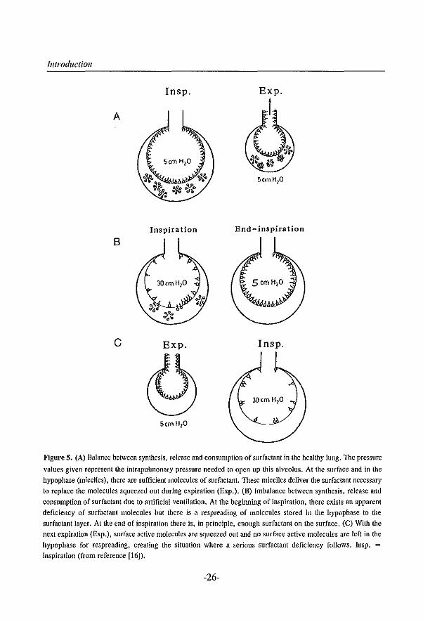
During end-expiration the surfactant molecules covering the alveolar epithelium
are compressed on the small alveolar area (leading to low surface tension or a high
surface pressure) thus preventing the alveoli from collapse [16]. If the surface of the
alveolus is smaller than the surface occupied by the surfactant molecules, the molecules
are squeezed out of the surface of the alveolus and forced towards the airways. These
surfactant molecules are then 'lost' for the alveoli and are eventually cleared from these
alveoli. During the following inflation of the alveoli, the surface is replenished with
surfactant molecules coming from the underlying hypophase where surfactant molecules
in micelles are' stored' for later use. During the next expiration, the mechanism repeats
itself and again surfactant molecules are forced out of the alveolus and subsequently
replenished from the hypophase; this is a continuing cycle (Figure 5) [16].
The amount of surfactant that must be produced and subsequently secreted by
the alveolar type II cells is proportional to the loss of surface active molecules during
the breathing cycle. When production and secretion of new surfactant molecules keep
pace with consumption, no surfactant deficiency can occur, as in a normal healthy lung.
Thus, artificial ventilation should take place at a lung volume equal or higher
as the functional residual capacity level witl! the smallest possible volume and pressure
changes. Another factor that might be of some importance is that mechanical ventilation
especially in non-homogeneous lungs, creates severe shear forces between open and
closed airways and possible overstretch of the epithelium during the breathing cycle,
resulting in necrosis and desquamation of bronchiolar and alveolar epithelium [105].
The overstretch of the intercellular junctions of the epithelium leads to an increased
permeability with the result of surfactant inhibition (as described earlier).
-25-
Introduction
Insp.
A
Inspiration End-inspiration
B
c Exp. Insp.
Figure 5. (A) Balance between synthesis, release and consumption of surfactant in the healthy lung. The pressure
values given represent the intrapulmonary pressure needed to open up this alveolus. At the surface and in the
hypophase (micelles), there are sufficient molecules of surfactant. These micelles deliver the surfactant necessary
to replace the molecules squeezed out during expiration (Exp.). (B) Imbalance between synthesis, release and
consumption of surfactant due to artificial ventilation. At the beginning of inspiration, there exists an apparent
deficiency of surfactant molecules but there is a respreading of molecules stored in the hypophase to the
surfactant layer. At the end of inspiration there is, in principle, enough surfactant on the surface. (C) With the
next expiration (Exp.), surface active molecules are squeezed out and no surface active molecules are left in the
hypophase for respreading, creating the situation where a serious surfactant deficiency follows. Insp. = inspiration (from reference [16]).
-26-
Chapter 1
Delivery techniqnes, timing, type of snrfactant, and ventilatory snpport
The optimal method to deliver exogenous surfactant is not yet known. Possibilities
include aerosol delivery, continuous infusion, lung lavage or bolus administration. The
latter method has been used in most auinlal studies, clinical case reports, as well as in
neonates with RDS [9,11]. The advantage of this method of instillation is that it is rapid
and capable of delivering large quantities of surfactant that are necessary, especially in
ARDS, to overcome the inhibitory effects of the serum proteins present in the alveoli.
Van der Bleek el al. [106] also demonstrated that the distribution of endotracheal
instilled surfactant is more homogeneous after a large bolus than after a smaller one.
This could be confirmed by Segerer and colleagues [107] who demonstrated a
homogeneous pulmonary surfactant distribution after bolus instillation, whereas
distribution after slow tracheal infusion of exogenous surfactant was extremely uneven.
In this study, it was shown that the distribution of surfactant was closely related to its
effect on pulmonary gas exchange [107]. Results from studies in premature animals
showed that administration of surfactant directly after birth gives a more homogeneous
distribution than in auinlals ventilated before treatment [108]. Instillation of surfactant
into lungs which are filled with intrapulmonary fluid could be compared with
instillation of a very large bolus of surfactant in sick lungs. Therefore, one may
speculate that one has to fIll up at least the total dead space of the lungs with surfactant
suspension for a more even distribution. However, the disadvantage of bolus instillation
technique is the relatively large amount of fluid which has to be instilled. However,
Gilliard el al. [109] demonstrated that the volume of fluid in which surfactant is
administered is rapidly absorbed; 30 minutes after surfactant instillation, there was no
significant difference between the lung weights of aninlals with lung injury receiving
5 ml and those of aninlals receiving 50 ml of surfactant suspension. Thus, studies in
which exogenous bolus instillation show heterogeneous distribution may be explained
by too small an amouut of fluid of each single bolus [109-111].
In addition, exogenous surfactant delivered as an aerosol has beeu investigated
[110 ,Ill]. The rationale was that by this method of instillation less volume of liquid
will be instilled into the lungs at one tinle and that the distribution will be more
homogeneous. In two different auinlal models, Lewis el al. [110,111] could
demonstrate that the distribution pattern was more homogeneous after aerosolized
surfactant administration. However, they found that tracheally instilled surfactaut was
superior to aerosolized surfactant in inlproVing blood gases, whereas there was no
-27-
Introduction
difference concerning improvement of lung mechanics. They suggested that 'the low
quautities of aerosolized surfactant deposited in the lungs limited the physiological
responses'. In this study, only 6.1 ±2.2 % of the total aerosolized surfactant was
recovered in the peripheral lung tissue whereas after bolus instillation 51 ±2 % of the
instilled surfactant was recovered [111]. They concluded that: 'a disadvantage of
aerosolized surfactant administration is the relatively long time period required to
administer significant quantities of exogenous surfactant due to the inefficiencies of
aerosol deposition'. Also, aerosolized surfactant could not be considered cost-effective
because large quantities of surfactant are required. Recently, the same group of
investigators also showed that it was impossible to inlprove gas exchange after
aerosolized surfactant in a non-uniform pattern of lung injury [112]. In this study the
less injured areas of the lung received relatively more surfactant than the severely
injured areas. They conclude that 'one should be cautious in administering aerosolized
surfactant to patients Witil ARDS who have non-uniform infiltrates on chest radiograph'
but this a contradiction because ARDS lungs are always injured in a non-uniform way
[113].
An alternative approach to administer surfactant is by lung lavage. We [51]
have shown that lavaging the lung with a diluted surfactant suspension (3.3 mg/mI, 30
mI/kg) was as effective as high-bolus administration (200 mg/kg) to improve gas
exchange in a model of acid aspiration. In lung lavaged rabbits, Balaraman et al. [114]
demonstrated that the effectiveness of a syntlletic surfactant, which has not been shown
to be highly effective in various animal models [115,116]. was enl1anced by
administering the surfactant by means of a lavage procedure compared to normal bolus
administration. Further, Enhorning [117] has suggested that saline lavage can also be
used to reduce the protein content intra-alveolar, which would be beneficial in the
treatment of ARDS. This idea has been investigated by Kobayashi et al. [49] and these
workers demonstrated that a relative low dose of exogenous surfactant (75 mg/kg)
could only improve gas exchange after intra-alveolar edema was removed by lung
lavage with saline in rabbits with severe respiratory failure dne to acid aspiration.
However, lung lavage with saline will also remove the endogenous surfactant, leading
to further deterioration of the pulmonary function [118]. Therefore, we [119]
performed lavage with a diluted surfactant suspension prior to surfactant therapy and
showed that this combination was the optimal treatment regime compared to the
alternatives in a model of severe respiratory failure (see Chapter 6).
-28-
Chapter 1
Another important aspect of surfactant response is the time at which surfactant
is given. In a model of acid aspiration, we [50] showed that respiratory failure can be
prevented when exogenous surfactant was given before deterioration of lung function
(i.e. within 10 min after acid aspiration), whereas after development of respiratory
failure exogenous surfactant served only to prevent further deterioration of lung
function but did not restore gas exchange (see Chapter 4). In the model of repeated
saline lavage, Ito et al. [120], and later confirmed by our group [119], demonstrated
that exogenous surfactant at an early stage of lung injury resulted in a sustained
improvement of lung function whereas lung function deteriorated when surfactant was
given at a relative late time point iu lung iujury, due to increased amount of proteins
(see also Chapter 6). Therefore, it is expected that early treatment of patients with
ARDS may require smaller amounts of exogenous surfactant and the outcome results
will probably be better.
Various surfactant preparations are already available on the market and have
been used successfully iu worldwide clinical trials in neonates with RDS [9]. In lung
lavaged rats, we [121] compared eight clinically used surfactants under standardized
conditions and confumed previous results of several animal studies [115,116] and
studies in neonates WiOl RDS [122,123] that the natural surfactant preparations, which
contain the hydrophobic peptides SP-B and SP-C, are more effective in iulproviug lung
function immediately after instillation than the artificial surfactant preparations without
surfactant proteius. In the same study [121], we showed that the effect of surfactants
on oxygenation was, iu general, dose-dependent and we found even marked differences
iu response pattern between the natural surfactants, especially when PEEP was reduced
at the end of the study protocol (see Chapter 3).
Several experiulental studies have shown that ventilator pattern strongly
iufluences exogenous surfactant efficacy [124-129]. Recently, Froese et al. [126] have
demonstrated that in lung lavaged rabbits the effect of exogenous surfactant on arterial
oxygenation remained stable only in combiuation with high-frequency oscillatory
ventilation (HFOV) at high-lung volume, whereas not with HFOV at low-lung volume
or conventional mechanical ventilation (CMV) at high or low lung volume. High-lung
volume means that lungs are actively opened (re-expanded) and then kept expanded by
using relative high mean airway pressures [128]. These results are in contrast to our
findiugs [129] iu which surfactant therapy iu combiuation with HFOV was not superior
to CMV iu iucreasiug lung function and/or reduciug lung iujury iu lung lavaged rabbits
-29-
Introduction
(see Chapter 7). Further, it has been shown that HFOV has also a beneficial effect on
exogenous surfactant composition by reducing the conversion of exogenously
administered surfactant into small aggregates (non-active) forms [126]. This has been
attributed to the small alveolar volume changes with HFOV. However, we have shown
in another study [130] that this can also be obtained with CMV by using small pressure
amplitudes and high end-expiratory pressures (see Chapter 8).
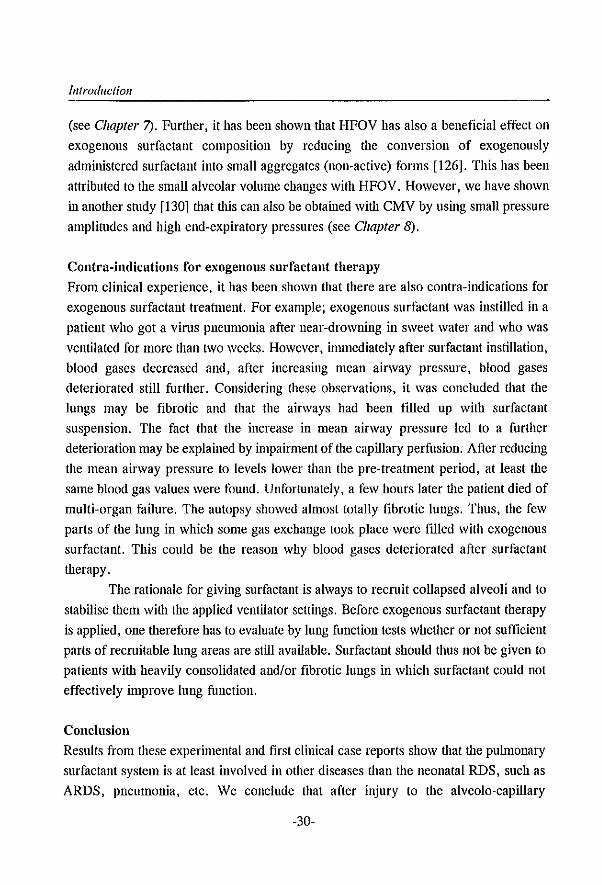
Contra-indications for exogenous surfactant therapy
From clinical experience, it has been shown that there are also contra-indications for
exogenous surfactant treatment. For example; exogenous surfactant was instilled in a
patient who got a virus pneumonia after near-drowning in sweet water and who was
ventilated for more than two weeks. However, in1ll1ediately after surfactant instillation,
blood gases decreased and, after increasing mean airway pressure, blood gases
deteriorated still further. Considering these observations, it was concluded that the
lungs may be fibrotic and that the airways had been filled up with surfactant
suspension. The fact that the increase in mean airway pressure led to a further
deterioration may be explained by impairment of the capillary perfusion. After reducing
the mean airway pressure to levels lower than the pre-treatment period, at least the
same blood gas values were found. Unfortunately, a few hours later tlle patient died of
multi-organ failure. The autopsy showed almost totally fibrotic lungs. Thus, the few
parts of the lung in which some gas exchange took place were fllied with exogenous
surfactant. This could be the reason why blood gases deteriorated after surfactant
therapy.
The rationale for giving surfactant is always to recruit collapsed alveoli and to
stabilise them with the applied ventilator settings. Before exogenous surfactant therapy
is applied, one tllerefore has to evaluate by lung function tests whetller or not sufficient
parts of recruitable lung areas are still available. Surfactant should thus not be given to
patients with heavily cousolidated and/or fibrotic lungs in which surfactant could not
effectively improve lung function.
Conclusion
Results from these experinlental and first clinical case reports show tlmt tlle puhnonary
surfactant system is at least involved in other diseases than the neonatal RDS, such as
ARDS, pneumonia, etc. We conclude that after injury to the alveolo-capillary
-30-
Chapter 1
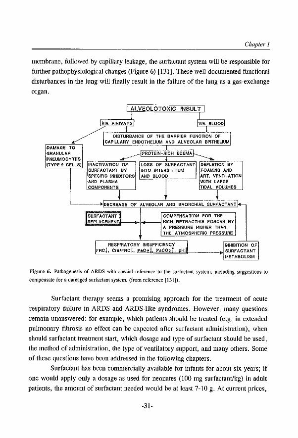
membrane, followed by capillary leakage, the surfactant system will be responsible for
further pathophysiological changes (Figure 6) [131]. These well-documented functional
disturbances in the lung will finally result in the failure of the lung as a gas-exchange
organ.
I ALVEOLOTOXIC INSULT I I I
IVIA AIAWA VS I IVIA BLOOD
! !
k
/1 DISTURBANCE OF THE BARRIER FUNCTION OF ~I CAPILLARY ENDOTHELIUM AND ALVEOLAR EPITHELIUM
DAMAGE TO GRANULAR ......--- PROTEIN-RICH EOEMAh ----... PNEUMOCYTES (TYPE 2 CEllS) INACTIVATION OF LOSS OF SURFACTANT I DEPLETION BY
SURFACTANT BY INTO INTERSTITIUM FOAMING AND SPECIFIC INHIBITORS AND BLOOD ART. VENTILATION AND PLASMA WITH LARGE COMPONENTS TIDAL VOLUMES
+ + DECREASE OF ALVEOLAR AND BRONCHIAL SURFACTANTl..-
SURF ACT ANT...I COMPENSATION FOR THE REPLACEMENT HIGH RETRACTIVE FORCES BY
A PRESSURE HIGHER THAN THE ATMOSPHERIC PRESSURE
II RESPIRATORY INSUFFICIENCY i ,I INHIBITION OF FRO," Crs/FRCJ, paC;r. PaCO;i, pH SURFACTANT
METABOLISM
Figure 6. Pathogenesis of ARDS with special reference to the surfactant system, including suggestions to
compensate for a damaged surfactant system. (from reference [1311).
Surfactant therapy seems a promising approach for the treatment of acute
respiratory failure in ARDS and ARDS-like syndromes. However, many questions
remain unanswered: for example, which patients should be treated (e.g. in extended
pulmonary fibrosis no effect can be expected after surfactant administration), when
should surfactant treannent start, which dosage and type of surfactant should be used,
the method of administration, the type of ventilatory support, and many others. Some
of these questions have been addressed in the following chapters.
Surfactant has been conunercially available for infants for about six years; if
one would apply only a dosage as used for neonates (100 mg surfactant/kg) in adult
patients, the amount of surfactant needed would be at least 7-10 g. At current prices,
-31-
Introduction
the costs of one treatment for one adult would then be US$ 30,000-50,000. So the price
of exogenous surfactant preparations has ftrst to be lowered before exogenous
surfactant therapy in adults can become a reality.
References 1. Von Neergaard K. Neue Auffassungen uber einen Grund-begriff der Atemme<:hanik. Z Ges Exp Med
1929; 66: 373-394
2. Macklin CC. The pulmonary alveolar mucoid film and the pneumocytes. Lancet 1954; 2: 1099-1104
3. Pattie RE. Properties. function, and origin of the alveolar lining layer. Nature 1955; 175: 1125-1126
4. Clements JA. Surface tension of lung extracts. Proc Soc Exp Bioi Med 1957; 95: 170-172
5. A very ME, Mead J. Surface properties in relation to atelectasis and hyaline membrane disease. Am J Dis
Child 1959; 97: 517-523
6. Fujiwara T, Maela H, Chida S, Morita T, Watabe Y, Abe T. Artificial surfactant therapy in hyaline
membrane disease. Lancet 1980, I: 55-59
7. van Golde LMG, Batenburg JJ, Robertson B. The pulmonary surfactant system: biochemical aspects and
functional significance. Physiol Rev 1988; 68: 374-455
8. Johansson J, Curstedt T, Robertson B. The proteins of the surfactant system. Eur Respir J 1994; 7: 372-
391
9. Lewis JF, Jobe AH. Surfactant and the adult respiratory distress syndrome. Am Rev Respir Dis 1993; 147:
218-233
to. Segerer H, Obladen M. Surfactant substitution treatment of neonatal respiratory distress syndrome.
Pediatric Rev Commun 1990; 5: 67-82
11. Jobe AH. Pulmonary surfactant therapy. N EngJ J Med 1993; 328: 861-868
12. Holm BA, Matalon S. Role of pulmonary surfactant in the development and treatment of adult respiratory
distress syndrome. Anesth Analg 1989; 69: 805-818
13. Lachmann B, Danzmann E. Acute respiratory distress syndrome. In: Robertson B. van Golde LMG,
Batenburg JJ (Eds). Pulmonary surfactant. Elsevier, Amsterdam, 1984. pp 505-548
14. Brogden KA. Changes in pulmonary surfactant during bacterial pneulllonia. Antonie van Leeuwenhoek
1991; 59: 215·223
15. do Campo JL. Bertranou EG, Casellas A, Donatto p. Bauellini R. Pulmonary surfactant post cardiac
surgery with cardiopulmonary bypass. Am Rev Respir Dis 1990; 141(Suppl): A512
16. Bos JAH, Lachmann B. Effects of artificial ventilation on surfactant function. In: Riigheimer E (Ed). New
aspects on respiratory failure. Springer-Verlag. Berlin. 1992, pp 194-208
17. Gammers D, Lachmann B. Surfactant therapy perspectives in adult patients. Curr Opinion Crit Care 1995;
1: 57-61
18. Lachmann B, Winsel K, Reutgen H. Der Anti-Atelektase-Faktor der Lunge I. Z Erkr Atm 1972; 137: 267-
287
19. Macklem PT, Proctor DF. Hogg JC. The stability of peripheral airways. Respir PhysioI1970; 8: 191-203
20. Lachmann B. Possible function of bronchial surfactant. Eur J Respir Dis 1985;67:46-61
21. Reinfenrath R. Surfactant action in bronchial mucus. In: Cosmi Ev, Scarpelli EM (Eds). Pulmonary
surfactant system. Elsevier. Amsterdam, 1983, pp 339-347
22. Guyton AC, Moffatt DS, Adair TA. Role of alveolar surface tension in transepitheliai movement of fluid.
In: Robertson B, van Golde LMG, Datenburg JJ (Eds). Pulmonary surfactant. Elsevier. Amsterdam, 1984,
-32-
Chapler 1
pp 171-185
23. van Iwaarden F. Surfactant and the pulmonary defense system. In: Robertson B. Van Golde LMG, Balenburg JJ (Eds), Pulmonary surfactant. Elsevier, Amsterdam, 1992, pp 215-253
24. Hein T, Lachmann B, Annbruster S, Smit JM, Voelkel N. Erdmann \y, Pulmonary surfactant inhibits the
cardiovascular effects of platelet-activating factor (PAF), 5-hydroxytryptamine (5-HT) and angiotensin II.
Am Rev Respir Dis 1987; 135(SuppJ): A506
25. So KL, Gammers D, Lachmann B. Bronchoal\'colar surfactant system and intratracheal adrenaline. Lancet 1993; 341: 120-121
26. Seeger W. Gunther At Walmrath HD t Grimmingcr F, Lasch HG. Alveolar surfactant and adult respiratory distress syndrome. Clin Investigator 1993; 71: 177-190
27. Stinson SF. Ryan DP, Hertweck MS, Hardy JD, Hwang-Kow SY, LoasH CO. Epithelial and surfactant
changes in influenza pulmonary lesions. Arch Patho! Lab Med 1976; 100: 147-153
28. Lachmann B. The role of pulmonary surfactant in the pathogenesis and therapy of ARDS. In: Vincent JL
(Ed). Update in intensive care and emergency medicine. Springer-Verlag, Berlin. 1987. pp 123-134
29. Ashbaugh DO. Bigelow DB, Petty TL, Levine BE. Acute respiratory distress in adults. Lancet 1967; 2:
319-323
30. Bernard GR. Artigas A, Brigham KL. CarIet J, Falke K, Hudson L. Lamy M. LeGalJ JR, Morris A,
Spragg R, The consensus committee. Report of the American-European consensus conference on ARDS:
defmitions, mechanisms, relevant outcomes and clinical trial coordination. Intensive Care Med 1994; 20:
225-232
31. Repine JE. Scientific perpectives on adult respiratory distress syndrome. Lancet 1992; 339: 466-469
32. Rinaldo JE. Rogers RM. Adult respiratory distress syndrome. N Engl J Med 1982; 306: 900·Cl09
33. Veldhuizen RAW, McCaig LA, Akino T, Lewis JF. Pulmonary surfactant sufractions in patients with the
acute respiratory distress syndrome. Am J Respir Ceit Care Med 1995; 152: 1867-1871
34. Doyle RL, Szaflarski N. Modin GW, Wiener-Kronish JP, Mauay MA. Identification of patients with acute
lung injury; predictors of mortality. Am J Respir Crit Care Med 1995; 152: 1818·1824
35. Kraft P, Fridrich P, Pernerstorfer T. et al. The acute respiratory distress syndrome: definitions, severity
and clinical outcome. Intensive Care Med 1996; 22:519-529
36. Royall JA, Levin DL. Adult respiratory distress syndrome in pediatric patients. I: clinical aspects,
pathophysiology, pathology, and mechanisms of lung injury. J Pediatr 1988; 112: 169-180
37. Spragg RO, Gilliard N, Richman p. Smith RM, Hite D, Pappert D, Heldt GP, Merritt TA. The adult
respiratory distress syndrome: clinical aspects relevant to surfactant supplementation. In: Robertson B, Van
Golde LMG, Batenburg JJ (Eds). Pulmonary surfactant. Elsevier, Amsterdam 1992, pp 685-703
38. Hallman M, Spragg R. Harrell JH, Moser KM, Gluck L. Evidence of lung function abnormality in
respiratory failure. Study of bronchoalveolar lavage phospholipids, surface activity phospholipase activity,
and plasma myoinositol. J Clin Invest 1982; 70: 673-683
39. Pison U, Seeger W, Buchom R. loka T, Brand M. Obertacke U, Neuhof H, Schmit-Neuerburg KP.
Surfactant abnormalities in patients with respiratory failure after multiple trauma. Am Rev Respir Dis
1989; 140: 1033-1039
40. Veldhuizen RAW, McCaig LA, Akino T, Lewis JF. Pulmonary surfactant subfractions in patients with
the acute respiratory distress syndrome. Am J Respir Crit Care Med 1995; 152: 1867-1871
41. Gregory TJ, Longmore WJ, Moxley MA, Whitsett JA, Reed CR, Fowler AA, Hudson LD, Maunder RJ,
Crim C, Hyers TM. Surfactant chemical composition and biophysical activity in acute respiratory distress
syndrome. J Clin Invest 1991; 88: 1976-1981
-33-
IntroductiOll
42. Lachmann B, van Daal GJ. Adult respiratory distress syndrome: animal models. In: Robertson B, van
Golde LMG, Batenburg JJ (Eds). Pulmonary Surfactant. Elsevier, Amsterdam, 1992, pp 635-663
43. Berggren P, Lachmann B, Curstedt T, Grossmann G, Robertson B. Gas exchange and lung morphology
after surfactant replacement in experimental adult respiratory distress syndrome induced by repeated lung
lavage. Acta Anaesthesiol Scand 1986; 30: 321-328
44. Gommers D, Vilstrup C, Bos JAH, Larsson A, Werner 0, Hannappel E, Lachmann B. Exogenous
surfactant therapy increases static lung compliance, and cannot be assessed by measurements of dynamic
compliance alone. Crit Care Med 1993; 21: 567-574
45. Berry D, Ikegami M, lobe A. Respiratory distress and surfactant inhibition following vagotomy in rabbits.
J Appl Physiol1986; 61: 1741-1748
46. Loewen GM, Holm BA, Milanowski L, Wild LM, Notter RH, Matalon S. Alveolar hyperoxic injury in
rabbits receiving exogenous surfactant. 1 Appl Physiol 1989; 66: 1087-lO92
47. Matalon S, Holm BA, Notter RH. Mitigation of pulmonary hyperoxic injury by administration of
exogenous surfactant. 1 Appl Physiol 1987; 62: 756-761
48. Lachmann B, Saugstad 00, Erdmann W. Effects of surfactant replacement on respiratory failure induced
by free oxygen radicals. First Vienna Shock Forum, Part B: Monitoring and Treatment of Shock: 1987,
pp 305-313
49. Kobayashi T, Ganzuka M, Tanigushi J, Nitta K, Murakami S. Lung lavage and surfactant replacement for
hydrochloric acid aspiration in rabbits. Acta Anaesthesiol Scand 1990; 34: 216-221
50. Eijking EP, Gommers D, So KL, de Maat MPM, Mouton JW, Lachmann B. Prevention of respiratory
failure after hydrochloric acid aspiration by intratracheal surfactant instillation in rats. Anesth Analg 1993;
76: 472-477
51. Eijking EP, Gommers D, So KL, Vcrgeer M, Lachmann B. Surfactant treatment of respiratory failure
induced by hydrochloric acid aspiration in rats. Anesthesiology 1993; 78: 1145-1151
52. So KL, van Genderen PH, Gommers 0, Lachmann B. Different surfactant treatment strategies for
respiratory failure induced by tracheally instilled pooled human plasma in rats. Am Rev Respir Dis 1993;
147(Suppl): A351
53. Harris ID, Jackson F, Moxley MA, Longmore WJ. Effect of exogenous surfactant instillation on
experimental acute lung injury. 1 Appl Physiol1989; 66: 1846~1851
54. So KL, de Buijzer E, Gammers 0, Kaisers U, van Genderen PJJ, Lachmann B. Surfactant therapy restores
gas exchange in lung injury due to paraquat intoxication in rats. Eur Respir 1 1998; 12: 284-287
55. Von Fricke U. Arzneimittelmarkt 1994: was war wirklich neu?-TeiI2. Deutsche Apotheker Zeitung 135,
Jahrg Nr 30, 2771-2781
56. Lachmann B, Gommers D. Surfactant treatment for neonatal lung diseases other than the idiopathic
respiratory distress syndrome. Lung & Respiration 1994; 11: 35-39
57. Richman PS, Spragg RG, Robertson B, Merritt TA, Curstedt T. The adult respiratory distress syndrome:
first trials with surfactant replacement. Eur Respir J 1989; 2(Suppl): 109s-111s
58. loka Th, Obertacke U. Neue mcdikamentosc Behandlung im ARDS: Effekt einer intrabronchialen
xenogenen Surfactantapplikation. Z Herz Thorax Gefrukhir 1989; 3(Suppl): 21-24
59. Nosaka S, Sakai T, Yonekura M, Yoshikawa K. Surfactant for adults with respiratory failure. Lancet
1990; I: 947-948
60. Stubbig K, Schmidt H, Bohrer H, Huster Th, Bach A, Motsch L Surfactantapplikation bei akutem
Lungenversagen. Anaesthesist 1992; 41: 555-558
61. Marraro G, Casiraghi G, Riva A. Effets d'un apport de surfactant chez deux adolescents Jeucemiques
-34-
Chapter 1
atteinls de detresse respiratoire. Cahiers d'Anesthesioiogie 1991; 39: 227-232
62. Haslem PL, Hughes DA, MacNaughton PO, Baker CSt Evans TW. Surfactant replacement therapy in latestage adult respiratory distress syndrome. Lancet 1994; 343:1009-1011
63. Heikinheimo M, Hynynen M, Rauliainen P, Hallman M, Kukkonen S. Successful treatment of ARDS with two doses of synthetic surfactant. Chest 1994; 105: 1263-1264
64. McBrien M. Katumba JJ, Mukhtar AI. Artificial surfactant in the treatment of near drowning. Lancet
1993: 342: 1485-1486
65. Suzuki H, Ohla T, Iwata K, Yamaguchi K, Salo T, Surfactant therapy for respiratory failure due to ncar
drowning. Eur J Pediatr 1996; 155: 383-384
66. Knoch M, H6itennann W, Lukasewitz P, Bitlersohl 1. Behandlung einer Totalatclektase der Iinken Lunge
bei schwerem ARDS mit seitengetrennter Beaunung und Surfactant-Applikation. Anasthesiol lntensivmed
Notfallmed Schmerzther 1995; 30: 270-273
67. Sutnick AI, Soloff LA. Atelectasis with pneumonia; a pathophysiology study. Ann Internal Med 1964; 60:
39-46
68. Somerson NL, Kontras SB, Pollack lD, Weiss HS. Pulmonary compliance: alteration during infection.
Science 1971; 171: 66-68
69. Mink SN, Light RB, Wood LDH. Effect of pneumococcal lobar pneumonia on canine lung mechanics. 1
Appl Physiol1981: 50: 283-291
70. Baughman RP, Sternberg RI, Hull W, Buchsbaum JA, Whitsett 1. Decreased surfactant protein A in
patients with bacterial pneumonia. Am Rev Respir Dis 1993; 147: 653-657
71. Escamilla R, Prevost MC, Hermant C, Caratero A, Cariven C, Krempf M. Surfactant analysis during
plleulJlocystis carinii pneumonia in HIV-infected patients. Chest 1992; 101: 1558-1562
72. Hoffman AGD, Lawrence MG, Ognibene FP, Suffredini AF, Lipschik GY, Kovacs JA, Masur H,
Shelhamer JH. Reduction of pulmonary surfactant in patients with human inmmnodeficiency virus infection
and pllelllJlocystis carilli; pneumonia. Chest 1992; 102: 1730-1736
73. Lachmann B. Open up the lung and keep the lung open. Intensive Care Med 1992; 18: 319-321
74. van Daal GJ, Bos JAH, Eijking EP, Gommers D, Hannappcl E, Lachmann B. Surfactant replacement
therapy improves pulmonary mechanics in end-stage influenza A pneumonia in mice. Am Rev Respir Dis
1992: 145: 859-863
75. van Daal GJ, So KL, Gommers D, Eijking EP, Fievez RB, Sprenger MJ, Van Dam DW, Laclunann B.
Intratracheal surfactant administration restores gas exchange in experimental adult respiratory distress
syndrome associated with viral pneumonia. Anesth Analg 1991; 72: 589-595
76. Eijkiog EP, Van Daal GJ, Tenbrinck R. Luijendijk A, Sluiters JF, Hannappel E, Lachmann B. Eftcct of
surfactant replacement on pJlelllllocystis carill;; pneumonia in rats. Intensive Care Med 1991; 17: 475-478
77. Song G-W, Robertson B, Curstedt T, X-Z Gao, Huang W-X. Surfactant treatment in experimental
Escherichia coli pneumonia. Acta Anaesthesiol Scand 1996; 40: 1154-1160
78. Buheitel G, Scharf J, Harms D. Erfahrungen mit der Surfactanttherapie des adulten Atenmotsyndroms
(ARDS). Monatsschr Kinderheilkd 1992; 140: 629-632
79. Putz G, Hormann C, Koller W, Schon G. Surfactant replacement therapy in acute respiratory distress
syndrome from viral pneumonia. Intensive Care Med 1996; 22: 588-590
80. Slater AJ, Nichani SH, Macrae D, Wilkinson KA, Novelli V, Tasker RC. Surfactant adjunctive therapy
for PlleulJ/ocystis carill;; pneumonitis in an infant with acute lymphoblastic leukemia. Intensive Care Med
1995: 21: 261-263
81. Mikawa K, Maekawa N, Nishina K, Takao Y, Yaku H, Obara H. Selective intrabronchial instillation of
-35-
Introduction
surfactant in a patient with pneumonia. Eur Respir J 1993; 6: 1563-1566
82. Johanson WG. Bacterial infection in adult respiratory distress syndrome. In: Zapol M, Lemaire F (Eds).
Adult respiratory distress syndrome. Marcel Dekker Inc, New York, 1991. vo150, pp 77-89
83. Gregory TI, Steinberg KP, Spragg R, Hyers TM, Longmore WJ. Moxley MA. Cai G-Z. Hite RD, Smith
RM, Hudson LD, Crim C, Newton p. Mitchell BR, Gold AJ. Bovine surfactant therapy for patients with
acute respiratory distress syndrome. Am J Respir Cri! Care Med 1997; 155; 1309-1315
84. Walmrath D, Gunther A, Ghofrani HA, Schermuly R, Sclmcider T. Grimminger F, Sceger W.
Bronchoscopic surfactant administration in patients with severe adult respiratory distress syndrome and
sepsis. Am J Respir Crit Care Med 1996; 154: 57-62
85. Anzueto A, Baughmann RP, Guntupalli KK, Weg JG, Wiedemann HP. Raventos AA. Lemaire F, Long
W. Zaccardelli OS, Pattishall EN. Aerosolized surfactant in adults with sepsis-induced acute respiratory
distress syndrome N Engl J Med 1996; 334: 1417-1421
86. Lachmann B, Eijking EP, So KL, Gommers D. In vivo evaluation of the inhibitory capacity of human
plasma on cxogenous surfactant function. Intensive Care Med 1994; 20: 6-11
87. Scarpelli EM (Ed). The surfactant system of the lung. Lea & Febiger, Philadelphia, 1968. pp 224
88. do Campo JL. Bertranou Eg, Franco R, Zeltzer R. Pulmonary radiologic manifestations after cardiac
surgery with cardiopulmonary bypass. Am Rev Respir Dis 1990; 141(Suppl): AI42
89. do Campo JL. Bertranou EG, De Lorenzi A, Hager AA. Nebulised exogenous natural surfactant after
cardiac surgery. Lancet 1994; 343: 482
90. Struber M, Brandt M, Cremcr J. Harringer W. Hirt SW. Havcrich A. Therapy for lung failure using nitric
oxide inhalation and surfactant replacemcnt. Ann Thorac Surg 1996; 61: 1543-1545
91. McFadden ER Jr, Gilbert IA. Asthma. N Engl J Mcd 1992;327: 1928-1937
92. Hohfield J, Fabel H, Hamm H. The role of pulmonary surfactant in obstructivc airways discase. Eur
Respir J 1997; 10: 482-491
93. Smith BT. Pulmonary surfactant during fetal development and neonatal adaptation: hormonal control. In:
Robertson B, van Golde LMG, Datcnburg JJ (Eds). Pulmonary surfactant. Elsevier. Amsterdam, 1984,
pp 357-381
94. Robertson B. Corticostcriods and surfactant for prevention of neonataJ RDS. Ann Med 1993; 25: 285-288
95. Liu M, Wang L, Enhorning G. Surfactant dysfunction develops when the immunized guinea-pig is
challenged with ovalbumin aerosol. Clin Exp Allergy 1995; 25: 1053-1060
96. Liu M. Wang L, Holm BA. Enhorning G. Dysfunction of guinea-pig pulmonary surfactant and typc II
pneumocytes after repetitivc challcngc with acrosolized ovalbumin. Clin Exp Allcrgy 1997; 27: 802-807
97. Lachmann B. Becher G. Protective effect of lung surfactant on allcrgic bronchial constriction in guinea
pigs. Am Rev Respir Dis 1986; 133(Suppl): A1I8
98. Liu M. Wang L, Li E. Enhorning G. Pulmonary surfactant givcn prophylactically allcviatcs an astlmla
attack in guinea pigs. Clin Exp Allergy 1996; 26: 270-275
99. Kurashima K, Ogawa H, Ohka T, Fujimura M, Matsuda T, Kobayashi T. A pilot study of surfactant
inhalation for the treatment of asthmatic attack. Jpn J Allergol 1991; 40: 160-163
100. Bambang Oetomo S, Dorrepaal C, Bos H, Gerritsen J, van der Mark TW. Koeter GH. Aalderen WMC.
Surfactant nebulization does not alter airflow obstruction and bronchial responsivcness to histamine in
astmatic children, Am J Rcspir Cdt Care Med 1996; 153: 1148-1152
101. Lemarchand P, Chinet T, Collignon MA, Urzua G, Barritault L, Huchon GJ. Bronchial clearance of
DTPA is increased in acute asthma but not in chronic asthma. Am Rev Respir Dis 1992; 145: 147-152
102. Kurashima K. Fujinmra M, Matsuda T, Kobayashi T. Surface activity of sputum from acute asthmatic
-36-
Chapter 1
patients. Am J Respir edt Care Med 1997; 155: 1254-1259 103. Verbrugge SIC, B6hm SH, Gammers D. Zinilllcrman UI, Lachmann B. Surfactant impairment after
mechanical ventilation with large alveolar surface area changes and effects of positive end-expiratory pressure. Br J Anaesth 1998; 80: 360-364
104. Greenfield U, Ebert PA, Benson DW. Effect of positive pressure ventilation on surface tension properties of lung extracts. Anaesthesia 1964; 25: 312-316
105, Nilsson R. Grossmann G, Robertson B. Lung surfactant and the pathogenesis of neonatal bronchiolar
lesions induced by artificial ventilation. Pedialr Res 1978; 12: 249-255
106. van def Bleek I, Plotz FB. van Overbeck M, Heikamp A, Beekhuis H. WiJdevuur CRH, Okken A,
Bambang Oetamo S. Distribution of exogenous surfactant in rabbits with severe respiratory failure: the
effect of volume. Pediatr Res 1993; 34: 154~158
107. Segerer H, van Gelder W, Angenent FWM, van Woerkcns JPM, Curstedt T, Obladen M, Lachmann B. Pulmonary distribution and efficacy of exogenous surfactant in lung-Iavaged rabbits. Pediatr Res 1993;
34: 490-494
108. Jobe A, Jkegami M, Jacobs H, Jones S. Surfactant and pulmonary blood flow distributions following
treatment of premature lambs with natural surfactant. J Clin Invest 1984; 73: 848-856
109. Gilliard N, Richman PM, Merritt A, Spragg RG. Effect of volume and dose on the pulmonary distribution
of exogenous surfactant administered to normal rabbits or to rabbits with oleic acid lung injury. Am Rev
Respir Dis 1990; 141: 743~747
1 to. Lewis JF, Ikegami lvi, Higuchi R, Jobe A, Absolom D. Nebulized vs. instilled exogenous surfactant in an
adult lung injury model. J AppJ Physiol1991; 71: 1270-1276
111. Lewis JF, Tabor B, Ikeganli M, Jobe AH, Joseph M, Absolom D. Lung function and surfactant
distribution in saline-Iavaged sheep given instilled vs. nebulized surfactant. J Appl Physiol1993; 74: 1256~
1264
112. Lewis JF, Ikegami M, Jobe AH, Absolom D. Physiologic responses and distribution of aerosolized
surfactant (Survanta) in a nonuniform pattern of lung injury. Am Rev Respir Dis 1993; 147: 1364~1370
113. Gallinoni L, D'Andrea L, Pelosi p. Vitale G. Pesenti A. Fumagalli R. Regional effects and mechanism
of positive end-expiratory pressure in early adult respiratory distress syndrome. JAMA 1993; 269: 2122~
2127
114. Balaraman V. Sood SL, Finn KC. Hashiro G. Uyehara CFf, Easa D. Physiologic response and lung
distribution of lavage versus bolus Exosurf in piglets with acute lung injury. Am J Respir Crit Care Med
1996: 153: 1838-1843
115. Cummings JJ, Holm BA, Hudak ML. Hudak BB, Herguson WH, Egan EA. A controlled clinical
comparison of four different surfactant preparations in surfaclant~deficient preterm lanlbs. Am Rev Respir
Dis 1992: 145: 999-1004
116. Hafner D. Beumc R, Kilian V, Krasznai G, Lachmann B. Dose~response comparisons of five lung
surfactant factor (LSF) preparations in an animal model of adult respiratory distress syndrome (ARDS).
Dr J Pharmacology 1995; 115: 451-458
117. Enhoming G. Surfactant replacement therapy in adult respiratory distress syndrome. Am Rev Respir Dis
1989: 140: 281-283
118. Lachmann B. Robertson B, Vogel J. In vivo lung lavage as an experimental model of the respiratory
distress syndrome. Acta Anaesth Scand 1980; 24: 231~236
119. Gommers D, Eijking EP, So KL, van 't Veen A, Lachmann B. Bronchoalveolar lavage with a diluted
surfactant suspension prior to surfactant instillation improves the effectiveness of surfactant therapy in
-37-
Introduction
experimental acute respirtatory distress syndrome (ARDS). Intensive Care Med 1998; 24: 494-500
120. Ito Y, Goffin 1, Veldhuizen R, Joseph M, Bjarneson D, McCaig L, Yao L-I, Marcou 1, Lewis J. Timing
of exogenous surfactant administration in a rabbit model of acute lung injury. 1 Appl Physiol 1996; 80:
1357-1364
121. Gammers D, van 't Veen A, Verbrugge SIC, Lachmann B. Comparison of eight surfactant preparations
on improvement of blood gases in lung-Iavaged rats. Appl Cardiopulm Pathophysiol (In press)
122. Soli RF and Vermont-Oxford Neonatal Network. A multicenter, randomized trial comparing synthetic
surfactant with modified bovine surfactant extract in the treatment of neonatal respiratory distress
syndrome. Pediatrics 1996; 97: 1-6
123. Hudak ML, Farrell EE, Rosenberg AA, lung AL, Auten RL, Durand DI, Horgan MI, Buckwald S,
Belcastro MR, Donohue PK, Carrion V. Maniscalco WW, Balsan MI, Tones BA, Miller RR, Iansen RD.
Graeber IE, Laskay KM. Matteson El, Egan EA, Brody AS, Martin DI, Riddlesberger MM, Montgomery
P. A multicenter randomized. masked comparison trial of natural versus synthetic surfactant for the
treatment of respiratory distress syndrome. 1 Pediatr 1996; 128: 396-406
124. Kobayashi T, Kataoka H. Ueda T, Murakami S, Takada Y, Kokubo M. Effects of surfactant supplement
and end-expiratory pressure in lung-lavaged rabbits. 1 Appl Physiol1984; 57: 995-1001
125. Rider ED, lobe AH, Ikegami M, Sun B. Different ventilation strategies alter surfactant responses in
preterm rabbits. 1 AppJ PhysiolI992; 73: 2089-2096
126. Froese AB, McCulloch PR, Sugiura M, Vaclavik S, Possmayer F, Moller F. Optimizing alveolar
expansion prolongs the effectiveness of exogenous surfactant therapy in the adult rabbit. Am Rev Respir
Dis 1993; 148: 569-577
127. Ito Y, Manwell SEE, Kerr CL, Veldhuizen RAW. Yao L-I, Bjameson D, McCaig LA, Barlett AI, Lewis
IF. Effects of ventilation strategies on the efficacy of exogenous surfactant therapy in a rabbit model of
acute lung injury. Am J Respir Crit Care Med 1998; 157: 149-155
128. McCulloch PR, Forkert PG, Froese AB. Lung volume maintenance prevents lung injury during high
frequency oscillatory ventilation in surfactant-deficient rabbits. Am Rev Respir Dis 1988; 137: 1185-1192
129. Gonmlers D, Hartog A, Schnabel R, De Jaegere A. Lachmann B. Surfactant therapy in combination with
high frequency oscillatory ventilation is not superior to conventional mechanical ventilation in reducing
lung injury in lung-Iavaged rabbits. (Submitted)
130. Verbrugge SIC. Gommers D, Lachrnann B. Conventional ventilation modes with small pressure amplitudes
and high end-expiratory pressure levels optimize surfactant therapy. Crit Care Med (In press)
131. Lachmann B. Surfactant replacement in acute respiratory failure: animal studies and first clinical trials.
In: Lachmann B (Ed). Surfactant replacement therapy. Springer-Verlag, Berlin, 1987, pp 212-223
-38-
Chapter 2
Exogenous surfactant therapy increases static lung
compliance, and cannot be assessed by
measurements of dynamic compliance alone
D. Gommersi, C. Vilstrup', J.A.H. Bos\ A. Larsson', O. Werner',
E. Hallllappel', B. Lachmanni
iDept. of Anesthesiology, Erasmus University Rotterdam, The Netherlands
'Dept. of Anesthesiology, University of Lund, Sweden
'Dept. of Biochemistry, University of Erlangen, Germany
Published ill: Crit Care Med 1993; 21.' 567-574 Reprinted with permission (copyright holder: Williams & lVilkens)
-39-
Swfactall! and lung mechanics
Summary
Objective: To study the immediate effects of exogenous surfactant therapy on blood
gases, lung volumes and lung mechanics in adult rabbits with experimentally induced
respiratory distress syndrome.
Desigll: Prospective randomised, controlled study.
Settillg: Laboratory and animal facility of a large university.
Subjects: Twelve adult New Zealand white rabbits.
IlIIervellliolls: Respiratory failure was induced by repeated bilateral whole-lung lavage
with saline (30 ml/kg body weight). After the last lavage, the animals were randomly
assigned to two groups. Group 1 received surfactant (120 mg/kg body weight) that was
suspended in a 0.6% sodium chloride solution. Group 2 received comparable volumes
of the same hypotonic solution and served as controls.
Measuremellts alld maill results: Before and after endotracheal surfactant instillation,
blood gases and functional residual capacity were measured, and lung mechanics from
tidal volumes and pressure-volume curves were calculated. Functional residual capacity
was measured with a computerized, multiple-breath, washin-washout method using
sulfur hexafluoride (SF.) as tracer gas. The pressure-volume curves were obtained by
an occlusion technique originally described for measuring static breath-by-breath
compliance. The technique was modified for present use and fully computerized.
Within 60 min after surfactant instillation, there was a marked improvement in PaO,
(61±7 nnnHg [S.2±0.9 kPa) to 470±47 nnnHg (62.6±6.2 kPa)) and in functional
residual capacity (7.6±1.4 to 17.7±1.6 ml/kg body weight) at unchanged ventilator
settings. The pressure-volume curves became steeper over tinle and the pressure
volume curves for total lung volume were restored to an almost normal state. Maximum
compliance calculated from the pressure-volume curves increased by 92 % but there was
no significant change in dynamic compliance. In the control group, no inIprovements
in any measured or calculated lung parameters were seen.
COllclusiolls: The findings indicate that during mechanical ventilation, the effects of
surfactant therapy on lung mechanics are best characterized by changes in functional
residual capacity and maximum compliance obtained from static pressure-volume
curves and not by dynamic compliance.
-40-
Chapter 2
Introduction
Patients with respiratory distress syndrome have impaired surfactant function; the
physiologic abnormalities associated with respiratory distress syndrome may be related
to this diminished function [1]. Exogenous surfactant therapy should, therefore, result
in improved lung function, which has already been demonstrated by the improvement
of arterial oxygenation in clinical trials [2] with premature babies and a few studies
[3,4] on patients with adult respiratory distress syndrome. It seems reasonable to
assume that such improvement ill gas exchange is caused by an immediate improvement
in lung volumes and lung mechanics. However, studies [5-8] in which respiratory
compliance was calculated from tidal volumes (dynamic compliance) during mechanical
ventilation in surfactant-treated infants did not show any inunediate changes. These
seemingly paradoxical results may be explained by a surfactant-induced left-shift and
increase of the pressure-volume curve.
Thus, after exogenous surfactant therapy with unchanged positive end-expiratory
pressure levels and unchanged ventilatory settings, the lung would operate on the
upper, more flattened part of the pressure-volume curve. We investigated this
hypothesis by measuring blood gases and functional residual capacity, and by
calculating lung mechanics from tidal volumes and pressure-volume curves both before
and after endotracheal surfactant instillation in adult rabbits with acute respiratory
distress syndrome.
Materials and methods
Preparatiol/ of al/illla/s. This study was approved by the local Animal Committee at the
Erasmus University, Rotterdam, and the care and handling of the animals were in
accord with the European Community guidelines (86/609/EEG). In 12 adult New
Zealand white rabbits (CPB, Zeist, The Netherlands) (body weight 2.7 ±0.3 kg),
anesthesia was induced with sodium pentobarbital (50 mg/kg intravenously) and
maintained by intermittent intraperitoneal injection of the same drug (5 mg/kg/h). For
muscle relaxation, pancuronium bromide (0.3 mg/kg/h intramuscularly) was given.
After tracheostomy was performed, the aninIals were pressure-control ventilated by a
Servo Ventilator (900C, Siemens-Elema, Soll1a, Sweden) at the following settings:
frequency of 20 cycles/min; inspiratory/expiratory ratio of 1 :2; FiO, of 1.0; peak
pressure of 9 to 12 cm H,o and zero end-expiratory pressure. The left carotid artery
was cannulated for continuous monitoring of arterial pressure and to allow for blood
-41-
Swiac/ant alld lung mechanics
sampling. Maintenance fluid consisted of 5 mllkg/h of 2.5 % glucose. Five millilitres
of sodium bicarbonate (0.8 %) was given when base excess was < -5 mmolli. Body
temperature was maintained within the normal range by means of a heating pad.
Lung lavage. Respiratory failure was induced by repeated lung lavage [9]. Each
lavage was performed with saline (30 mllkg body weight) heated to 37°C. During
lavage, tracheal pressure was measured and not allowed to increase to > 40 cm H,o.
Lung lavage was repeated five to eight times at 5-min intervals until Pa02 was < 80
mmHg (10.6 kPa) at a peak pressure of 26 cm H,o and a positive end-expiratory
pressure (PEEP) of 6 cm H20 with 100 % oxygen; inspiratory-expiratory ratio and
frequency were not changed. The animals were randomly assigned to two groups 10
min after tlle last lavage. Group I (n=6) received surfactant 120 mg/kg body weight,
which was suspended in a 0.6% sodium chloride solution. Group 2 (n=6) received
comparable volumes of tlle same hypotonic solution (0.6% sodium chloride) and served
as controls. Immediately after discomlection from the ventilator, the animals received
via a syringe eitller 3 ml surfactant and 10 m1 air per kg body weight, or 3 ml
hypotonic solution and 10 m1 air per kg body weight. Peak pressure was varied to keep
the PaC02 at 30 to 45 IIlIllHg (4.0 to 6.0 kPa) but peak pressure was never increased
to > 35 cm H,o. PEEP was increased when Pa02 was <60 nunHg «8.0 kPa).
Sw!actallt. Natural surfactant was isolated from tlle lungs of recently slaughtered
cows. The lungs were minced and tlle tissue fragments were washed with normal
saline. The mixture of saline and tissue fragments was ftltered, and the ftltrate was
centrifuged to remove cell debris. The supernatant was then ultra-centrifuged to obtain
pellets of 'crude' surfactant. This 'crude' surfactant was extracted by the method of
Bligh-Dyer [10]. The chloroform phase was evaporated and suspended in acetone to
remove the neutral lipids. After centrifugation, the pellet was suspended in water and
finally freeze-dried.
The surfactant consists of approximately 90 to 95 % phospholipids and 1%
hydrophobic proteins (surfactant-protein B and C), with the remainder being other
lipids such as cholesterol, glyceride and free fatty acids. There is no surfactant-protein
A in this surfactant preparation. The freeze-dried preparation was resuspended in a
0.6% sodium chloride solution to a total concentration of 40 mg phospholipids/m!.
Measurements. Data were collected at the following tinles: before lavage at
PEEP of 0, 4, 6 and 8 cm H,o; 5 min after lavage; 5, 15 and 30 min after surfactant
or hypotonic solution instillation; and every 30 min for 3 h. At each data collection
-42-
Chapter 2
point, PaO" PaCO, base excess, pH, functional residual capacity and dynamic
compliance measurements were obtained. Pressure-volume curves were recorded before
and after lavage and at 15 min, 1, 2, and 3 h after surfactant or hypotonic solution
instillation. PaO" PaCO" base excess and pH of the samples were measured by
conventional methods (ABL 330, Radiometer, Copenhagen, Denmark).
Functional residual capacity was measured using a computerized, multiple
breaUl, washin-washout method, with sulfur hexafluoride (SF.) as the tracer gas. The
method has been described previously for studies of functional residual capacity and gas
washout in adults, children and small neonates [11-13]. The components of the system
(Figure 1) included a computer (pDP 11/23, Digital Equipment, Maynard, Marlboro,
MA), a ventilator (Servo 900C, Siemens-Elema), electromagnetic inspiratory and
expiratory auxiliary valve (360POI21, Neptune Research, Maplewood, NS), and
inspiratory and expiratory heated Fleisch pneumotachograph no. "00" (Gould,
Lausanne, Switzerland; linearity flow range of 0 to 100 mllsec) with differential
pressure transducers (MP 45-1-871, Validyne, Northridge, CAl.
The tracer gas concentration was measured using an infrared analyser(Siemens)
[14]. The transducer of the analyser was placed over a cuvette with windows near the
T-piece in the expiratory part of the ventilatory tubings. Sulfur hexafluoride was
washed in until the alveolar concentration was about 1.5 %. Sulfur hexafluoride washout
was continued until Ule concentration was < 0.01 %. Signals representing expired flow
and sulfur hexafluoride concentration were fed into the computer, which calculated
functional residual capacity as the value of sulfur hexafluoride washed out plus the
amount left in the lungs at the end of washout divided by the alveolar concentration at
the end of washin. The amonnt of sulfur hexafluoride remaining in the lungs was
estimated, assuming that the final part of washout was monoexponentia!. The volume
of the tubing between the auxiliary valves, including the bar of the T-piece, was 0.8 m!.
All volumes were converted to body temperature and atmospheric pressure, completely
saturated with water vapour at body temperature, by multiplying by 1.09. Apparatus
dead space was subtracted from the obtained functional residual capacity values.
Duplicate measurements were made on each occasion.
Dynamic thorax-lung compliance (Cd,.) was calculated as tidal volume/(peak
pressure minus PEEP) at mechanical ventilation. Since the air flow at end-inspiration
and end- expiration are zero, peak pressure and PEEP were assumed to be the alveolar
pressure. The pressure-volume curves were obtained by an occlusion technique that was
-43-
Swfactant and lung mechanics
originally described for the purpose of measuring static breath-by-breath compliance
[15]. The technique was modified for present use and was fully computerized. After
switching to the pressure-volume system (Figure 1), which was done during a
prolonged inspiratory pause to prevent lung collapse, the lungs were inflated by a
syringe to an airway pressure of about 30 cm H,o. During the following deflation to
o cm H20 (equal to functional residual capacity), the expiratory flow was interrupted
for 80 msec in a 160 msec cycle by a computer-governed electromagnetic occluder
placed close to the tracheal tube. The airway pressure was measured at the end of each
occlusion. The computer integrated the flow signal and calculated the volume expired
between occlusion. After completion of the manoeuvre, the pressure-volume curve was
plotted by an X-Y writer (Hewlett-Packard, Palo Alto, CAl and the data were stored
on a computer disk. Total-lung-volume curves were also presented by the computer
using the measured functional residual capacity value to calculate the total lung volumes
at different airway pressures.
Static compliance (C,a6/6) was calculated from the pressure-volume curve above
functional residual capacity level by dividing the difference between Ole volumes at Ole
used peak pressure and PEEP. Compliance was also calculated from the pressure
volume curve above functional residual capacity level by dividing the difference
between the volumes at pressures 10 (C"IOIO)' 20 (C,ao/o) and 30 (C'dOIO) cm H20
respectively, and 0 cm H20. Maximum compliance was defmed as the steepest slope
of the pressure-volume curve. The specific compliance (C,,,,J was calculated by
dividing static compliance (C,a6/6) by functional residual capacity. The lung stability
index (L), according to Gruenwald [16], was calculated from the pressure-volume
deflation curve as L= (2V, + VIO)I2Vm, where V" VIO and Vm are the volumes at 5,
10 and 30 cm H20, respectively. Thoracic-lung compliance was also calculated for total
lung volumes (inclusive functional residual capacity) at pressures of 10 (CTLVIO/O)' 20
(CTLV20/0) and 30 (CTLV301O) cm H20. Statistical analysis. Analysis of variances (ANOVA) was used to assess wheOler
Owre was an overall difference within or between the two groups. If a difference was
found, a post hoc test was used (Student Newman-Keuls multiple comparison
procedure). Statistical significance was accepted at p~0.05. All data are reported as
mean ± SD.
-44-
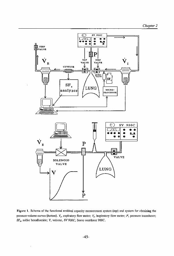
PEBP
CUVETTE
}( SOLENOID {1 VALVE
p
•• ....... •
E) SV 900C
[2][1:J • • • •••• • L2
••• •
Chapter 2
Figure 1. Schema of the functional residual capacity measurement system (top) and system for obtaining the
pressure-volume curves (bOllum). VI' expiratory flow meter; \Jj. inspiratory flow meter; P, pressure transducer;
SF6• sulfur hexafluoride; V. volume, SV 900C. Servo ventilator 900C.
-45-
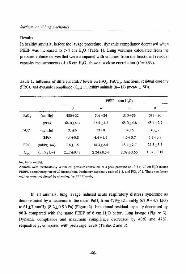
SIII!actan( and lung mecha1lics
Results
In healthy animals, before the lavage procedure, dynamic compliance decreased when
PEEP was increased to >4 cm H,O (Table I). Lung volumes calculated from the
pressure-volume curves that were compared with volumes from the functional residual
capacity measurements of ~S cm H,O, showed a close correlation (r'=0.99).
Table 1. Influence of different PEEP levels on PaO" PaCO" functional residual capacity (FRC), and dynamic compliance (Cd,,) in healthy animals (n= 12) (mean ± SD).
PEEP (em HP)
0 4 6 S
Pa02 (mmHg) 4S0±32 506±24 51O±36 513±20
(kPa) 64.0±4.3 67.5±3.2 6S.0±4.S 6S.4±2.7
PaCO, (mmHg) 31H 33±8 34±5 40±7
(kPa) 4.J±0.S 4.4± l.l 4.5±0.7 5.3±0.9
PRC (ml/kg bw) 7.6± 1.5 14.3±2.5 IS.4±2.7 22.5±3.3
Cd)n (ml/kg bw) 2.17+0.47 2.24+0.54 2.02±0.56 1.32+0.IS
bw, body weight. Animals were mechanically ventilated, pressure controlled, at a peak pressure of 1O.4± 1.7 cm HP (above PEEP), a respiratory rate of 20 breaths/min, inspiratory-expiratory ratio of 1 :2, and Fi02 of I. These ventilatory settings were not altered by changing the PEEP levels.
In all animals, lung lavage induced acute respiratory distress syndrome as
demonstrated by a decrease in the mean PaO, from 479±32 mmHg (63.9±4.3 kPa)
to 61 ± 7 mmHg (S.2±0.9 kPa) (Figure 2). Functional residual capacity decreased by
66% compared with the same PEEP of 6 cm H,O before lung lavage (Figure 3).
Dynamic compliance and maxinmm compliance decreased by 43 % and 47 %,
respectively, compared with prelavage levels (Tables 2 and 3).
-46-
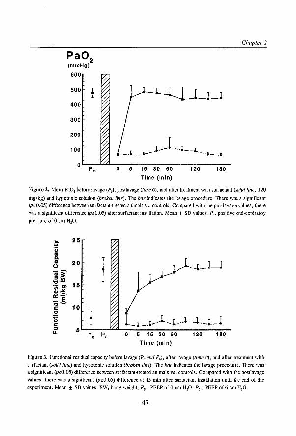
Pa02
(mmHg)
600
500 • 1
400
300
200
100
0 Po o 5 15 30 60
Time (min)
120
Chapter 2
180
Figure 2. Mean Pa02 before lavage (Pa), posllavage (time 0), and after treatment with surfactant (solid /il/e, 120
mg/kg) and hypotonic solution (broken line). The bar indicates the lavage procedure. There was a significant (p5:0.05) difference between surfactant-treated animals vs. controls. Compared with the postlavage values, there was a significant difference (p5:0.05) after surfactant instillation. Mean ± SD values. Po> positive end-expiratoy pressure of 0 em H20.
25
20 I 15
10
_~_~~)l~~_J-~_L_l 5L-p-O---P-6~~0~-5~~1-5--3-0~6~0----1~2~0----1~8~0-
Time (min)
Figure 3. Functional residual capacity before lavage (Po and P6)' after lavage (time 0), and after treatment with surfactant (solid line) and hypotonic solution (broken line). The bar indicates the lavage procedure. There was
a significant (p.5.0.05) difference between surfactant-treated animals vs. controls. Compared with the postlavage
values, there was a significant (p:>O.05) difference at 15 min after surfactant instillation until the end of the
experiment. Mean ± SO values. BW, body weight; Po , PEEP of 0 em H20; P6 , PEEP of 6 em HzO.
-47-
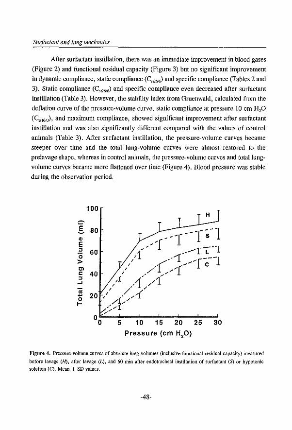
Sw/actallt and lling mechallics
After surfactant instillation, there was an innnediate improvement in blood gases
(Figure 2) and functional residual capacity (Figure 3) but no significant improvement
in dynamic compliance, static compliance (C,Q6!6) and specific compliance (Tables 2 and
3). Static compliance (C,Q616) and specific compliance even decreased after surfactant
iru;tillation (Table 3). However, the stability index from Gruenwald, calculated from the
deflation curve of the pressure-volume curve, static compliance at pressure 10 cm H20
(C"IOIO)' and maximum compliance, showed significant improvement after surfactant
instillation and was also significantly different compared with the values of control
animals (Table 3). After surfactant instillation, the pressure-volume curves became
steeper over time and the total lung-volume curves were almost restored to the
prelavage shape, whereas in control animals, the pressure-volume curves and total lung
volume curves became more flattened over tinIe (Figure 4). Blood pressure was stable
during the observation period.
100
.§, 80 Q)
E :J
o > Cl C :J
..J
<U .... o I-
60
40
20
5
H
10 15 20 25 30
Pressure (em H20)
Figure 4. Pressure-volume curves of absolute lung volumes (inclusive functional residual capacity) measured
before lavage (H), after lavage (L), and 60 min after endotracheal instillation of surfactant (S) or hypotonic solution (C). Mean ± so values.
-48-
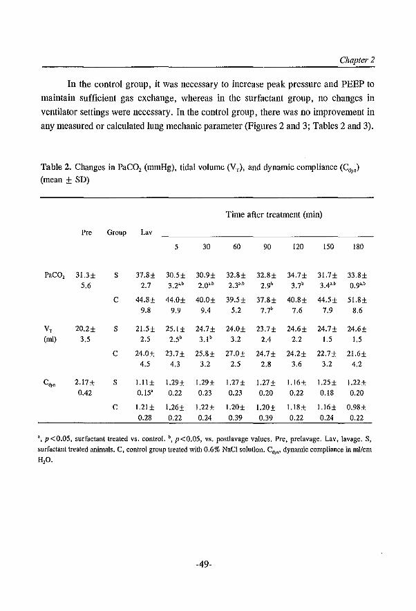
Chapter 2
In the control group, it was necessary to increase peak pressure and PEEP to
maintain sufficient gas exchange, whereas in the surfactant group, no changes in
ventilator settings were necessary. In the control group, there was no improvement in
any measured or calculated lung mechanic parameter (Figures 2 and 3; Tables 2 and 3).
Table 2. Changes in PaCO, (nunHg), tidal volume (VT), and dynamic compliance (Cd)') (mean ± SD)
Time after treatment (min)
Pre Group Lay
5 30 60 90 120 150 180
PaC02 3l.3± S 37.8± 30.5± 30.9± 32.8± 32.8± 34.7± 31.7± 33.8± 5.6 2.7 3.21,b 2.0··b 2.33.b 2.9b 3.7b 3.4"b 0.9"1:>
C 44.8± 44.0± 40.0± 39.5± 37.8± 40.8± 44.5± 51.8± 9.8 9.9 9.4 5.2 7.7b. 7.6 7.9 8.6
V, 20.2± S 21.5± 25.1± 24.7± 24.0± 23.7± 24.6± 24.7± 24.6± (ml) 3.5 2.5 2.5b 3.1 b 3.2 2.4 2.2 1.5 1.5
C 24.0± 23.7± 25.8± 27.0± 24.7± 24.2± 22.7± 21.6± 4.5 4.3 3.2 2.5 2.8 3.6 3.2 4.2
C~, 2.17± S l.ll± 1.29± 1.29± l.27± 1.27± l.l6± l.25± l.22± 0.42 0.15' 0.22 0.23 0.23 0.20 0.22 O.IS 0.20
C 1.21± 1.26± l.22± l.20± l.20± l.lS± l.l6± 0.98± 0.28 0.22 0.24 0.39 0.39 0.22 0.24 0.22
a, p <0.05, surfactant treated vs. control. \ p <0.05. VS, postlavage values. Pre. prelavage. Lav. lavage. S, surfactant treated animals. C, control group treated with 0.6% NaCI solution. Cd}n' dynamic compliance in ml/cm H2O.
-49-
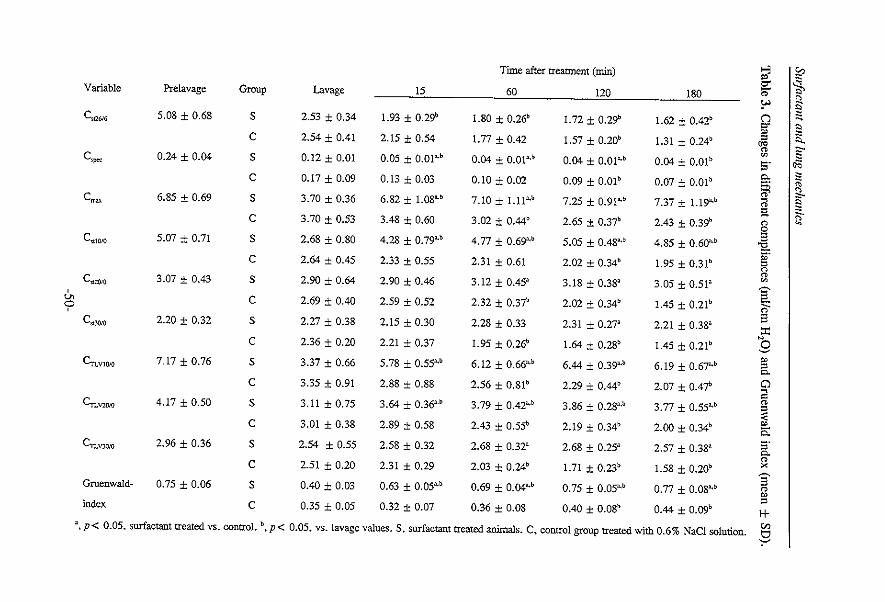
Time after treatment (min) ..., ~ " ~ Si, Variable Prelavage Group Lavage 15 60 120 180 " !" ~ C"'" 5.08 ± 0.68 S 2.53 ± 0.34 1.93 ± 0.29b 1.80 ± 0.26b 1.72 ± 0.29b 1.62 ± 0.42b (")
"" ~ " C 2.54 ± 0.41 2.15 ± 0.54 1.77 ± 0.42 1.57 ± 0.20b 1.31 ± 0.24b " .. " ~ 0.05 ± O.Ol,·b 0.04 ± 0.01",1) 0.04 ± O.Ol~,b 0.04 ± O.Olb ~
C"" 0.24 ± 0.04 S 0.12 ± 0.01 S· C 0.17 ± 0.09 0.13 ± 0.03 0.10 ± 0.02 0.09 ± O.01b 0.07 ± O.Ol b e. " § " <)
" "'" C~ 6.85 ± 0.69 S 3.70 ± 0.36 6.82 ± 1.08"'b 7.10 ± 1.11"'b 7.25 ± 0.91"'b 7.37 ± 1.19"'b ~ " " " g ~. C 3.70 ± 0.53 3.48 ± 0.60 3.02 ± O.44b 2.65 ± 0.37b 2.43 ± 0.39b
" 0 Cst1OiO 5.07 ± 0.71 S 2.68 ± 0.80 4.28 ± 0.79",1) 4.77 ± 0.69'"b 5.05 ± 0.48",b 4.85 ± 0.60'"b " 'i2.
C 2.64 ± 0.45 2.33 ± 0.55 2.31 ± 0.61 2.02 ± 0.34b 1.95 ± 0.31b ". " n
Cl't,.."'I)IO 3.07 ± 0.43 S 2.90 ± 0.64 2.90 ± 0.46 3.12 ± 0.45' 3.18 ± 0.38' 3.05 ± 0.51' Vi V. C 2.69 ± 0.40 2.59 ± 0.52 2.32 ± 0.37b 2.02 ± 0.34b 1.45 ± 0.21b
§: '? ;,-
3 Cst3()/o 2.20 ± 0.32 S 2.27 ± 0.38 2.15 ± 0.30 2.28 ± 0.33 2.31 ± 0.27' 2.21 ± 0.38' ::r: C 2.36 ± 0.20 2.21 ± 0.37 1.95 ± 0.26b 1.64 ± 0.28b 1.45 ±0.21b NO
~
7.17 ± 0.76 3.37 ± 0.66 5.78 ± 0.55" 6.12 ± 0.66'b 6.44 ± O.39'·b 6.19 ± 0.67'b " c".V1OIO S " e.
C 3.35 ± 0.91 2.88 ± 0.88 2.56 ± 0.81b 2.29 ± O.44b 2.07 ± 0.47b Cl ~
'" Cn.""m 4.17 ± 0.50 S 3.11 ± 0.75 3.64 ± 0.363.b 3.79 ± 0.42",1) 3.86 ± 0.28'·b 3.77 ± 0.55'b " " C 3.01 ± 0.38 2.89 ± 0.58 2.43 ± 0.55b ~ 2.19 ± 0.34b 2.00 ± 0.34b " ;:;:
CTLVJOiO 2.96 ± 0.36 S 2.54 ± 0.55 2.58 ± 0.32 2.68 ± 0.32' 2.68 ± 0.25' 2.57 ± 0.38' [ " C 2.51 ± 0.20 2.31 ± 0.29 2.03 ± 0.24b 1.71 ± O.23b 1.58 ± 0.20b x ~
Gruenwald- 0.75 ± 0.06 S 0.40 ± 0.03 0.63 ± 0.05" 0.69 ± O.04"'b 0.75 ± 0.05"'b 0.77 ± 0.08" 3 " " " index C 0.35 ± 0.05 0.32 ± 0.07 0.36 ± 0.08 0.40 ± 0.08b 0.44 ± 0.09b 1+
3, p< 0.05. surfactant treated VS. control. 1), p < 0.05. vs. lavage values. S. surfactant treated animals. C. control group treated with 0.6% NaC} solution. en 51
Chapter 2
Discussion
It is well known that surfactant function is impaired in respiratory distress syndrome
patients, and associated abnormalities may be related to this diminished function [1].
Therefore, exogenous surfactant therapy should result in improved lung expansion,
increased functional residual capacity and decreased shunt [17]. Benefits of these
changes include: a) enhanced blood oxygenation at lower fractions of inspired oxygen;
b) avoidance of toxic levels of oxygen; c) use of lower PEEP with reduced cardio
circulatory depression; and d) lower peak airway pressures with reduced barotrauma
[2,17]. The model used in this study indicates a condition similar to respiratory distress
syndrome by repeated bilateral whole-lung lavage with saline [9]. This model is widely
used for testing different ventilatory settings in respiratory distress syndrome and for
surfactant replacement therapy because it remains stable for hours and presents
histologic changes sinlilar to those changes in respiratory distress syndrome [18-21].
In the present study, the ventilatory settings were kept constant because we were only
interested in the effect of surfactant on lung mechanics; the influence of different
ventilatory settings in this model is already known [18].
The method of measuring functional residual capacity gives accurate and
reproducible results and does not require alteration of the breathing circuit or any
interruption of ventilatory support. Also, due to the small amount of tracer gas needed
(1.5 % of sulfur hexafluoride), the method can be used at all inspired oxygen
concentrations. Modifications of the functional residual capacity measurement system
have been used in adults, children and neonates [11-13].
The method employed for measuring pressure-volume curves is adapted from an
occlusion technique for static breath-by-breath compliance [14]. Although the occlusiou
period, 80 msec, appears short, a well-defined pressure plateau at the end of each
occlusion was observed. Also, Carlo [22] found that pressure equilibrium was reached
within 50-100 msec after an occlusion in a rabbit model of healthy and surfactant
depleted lungs. Another indication of the validity of the method is that increases in lung
volume from 0 cm H20 to the different levels of PEEP calculated from the pressure
volume curves were almost equal to the increases in lung volume obtained by functional
residual capacity measurement (1"=0.99).
This study demonstrates that treatment with exogenous surfactant llml1ediately
reverses hypoxemia and improves functional residual capacity, maxllnum compliance
and static compliance related to total lung volumes (CTLy). However, no significant
-51-
SIll/actant and hmg mechanics
improvement in dynamic compliance was found. These results confIrm those of other
studies [5-8] in which there were signifIcant inlprovements in oxygenation only, but no
improvement in dynamic compliance, directly after surfactant instillation in neonates
wifu respiratory distress syndrome. This discrepancy can be attributed to the increase
of functional residual capacity and the change of the shape of the pressure-volume curve
directly after surfactant instillation. The pressure-volume curve became steeper over
time, the increase of maximum compliance and static compliance at pressure 10 cm
H,o (C'UOIO) are an expression of this shift (Table 3).
These two changes are best visualized by the total-lung-volume curves (Figure
4), which were almost restored to the healthy state after surfactant instillation. Thus,
mechanically ventilating the animals that had been pressure-controlled with a peak
pressure of 26 cm H20 and a PEEP of 6 cm H20 meant that the animals were ventilated
at the upper, flatter part of the pressure-volume curve, resulting in a low value of
dynamic compliance. Therefore, if a lower PEEP and tidal volume had been used, the
lung would likely have operated on a steeper part of the pressure-volume curve after
surfactant therapy and the dynamic compliance would have been increased. This
possibility could explain the results of Davis and co-workers [6] who reported that
during spontaneous breathing, dynamic compliance increased after surfactant therapy.
In tlleir study [6], the infants may not have generated the same transpulmonary pressure
as in the mechanically ventilated group and were probably breatlling on the steeper part
of the pressure-volume curve. This observation remains speculative because no data on
pressures (peak pressure, PEEP, continuous positive airway pressure) were presented.
Dynamic compliance was measured within the tidal volume at the used PEEP
level. Changes in both tidal volume and PEEP markedly influence fue value of dynamic
compliance [23,24]. Schaffer and colleagues [24] showed that dynamic compliance
values obtained at > 4 cm H,o of PEEP are signifIcantly less than fuose values obtained
with a lower PEEP. These results are confIrmed in the present study (Table I). Thus,
to compare values of dynamic compliance, it is essential to keep tidal volume and PEEP
constant. However, since lung function improves after surfactant treatment, PEEP is
usually decreased [2]. Therefore, it is difficult to interpret results from studies that have
shown an improyement in dynamic compliance 24 h after surfactant therapy in
mechanically ventilated neonates [5,7]. In these studies, the ventilatory settings were
changed and mean airway pressure was decreased. Moreover, in surfactant-treated vs.
nontreated groups, comparisons of dynamic compliance are not appropriate if tidal
-52-
Chapter 2
volume and PEEP levels are not considered.
We conclude that dynamic compliance and static compliance (C,Q616) do not
accurately reflect changes in lung function after surfactant replacement therapy in
surfactant-depleted, mechanically ventilated rabbits, and that the effects of surfactant
therapy on lung mechanics are best characterized by changes in functional residual
capacity and maximum compliance obtained from static pressure-volume curves.
References 1. Van Golde LMG, Batenburg JJ, Robertson B. The pulmonary surfactant system: Biochemical aspects and
functional significance. Physiological Rev 1988;68: 374-455
2. Merritt TA, Hallman M, Spragg R, Heldt GP, Gilliard N. Exogenous surfactant treatments for neonatal
respiratory distress syndrome and their potential role in the adult respiratory distress syndrome. Drugs
1989; 38: 591-611
3. Lachmann B. The role of pulmonary surfactant in the pathogenesis and therapy of ARDS. In: Vincent JL (Ed). Update in Intensive Care and Emergency Medicine. Springer-Verlag, Berlin. 1987, pp 123-134
4. Richman PS, Spragg RO. Robertson B, Merritt TA, Curstedt T. The adult respiratory distress syndrome:
first trials with surfactant replacement. Eur Respir J 1989; 2: 109s-Ills
5. Bhat R, Dziedzic K, Bhatani V, Vidyasagar D. Effect of single dose surfactant on pulmonary function. Crit
Care Med 1990; 18: 590-595
6. Davis JM, Veness-Meehan K, Notter RH, Bhutani VK, Kendig JW, Shapiro DL. Changes in pulmonary
mechanics after the administration of surfactant to infants with respiratory distress syndrome. N Eng J Med
1988; 319: 476-479
7. Couser RJ, Ferrara TB, Ebert J. Hoekstra RE, Fangman JJ. Effects of exogenous surfactant therapy on
dynamic compliance during mechanical breathing in preterm infants with hyaline membrane disease. J
Pedialr 1990; 116: 119-124
8. Edberg KE. Ekstrom-Jodal B, Hallman M, Hjaimarson 0, Sandberg K, Silberberg A. Immediate effects
on lung function of instilled human surfactant in mechanically ventilated newborn infants with IRDS. Acta
Paediatr Scand 1990; 79: 750-755
9. Lachmann B, Robertson B, Vogel J. In vivo lung lavage as an experimental model of the respiratory
distress syndrome. Acta Anaesthesiol Scand 1980; 24: 231-236
10. Bligh EG, Dyer WJ. A rapid method of total lipid extraction and purification. Can J Biochem Physiol1959;
37: 911-917
II. Jonmarker C, Jansson L, Jonson B, Larsson A, Werner O. Measurement of functional residual capacity
by sulfur hexafluoride washout. Anesthesiology 1985; 63: 89-95
12. Jonmarker C, Larsson A, Werner O. Changes in lung volume and lung-thorax compliance during cardiac
surgery in children 11 days - 4 years of age. Anesthesiology 1986; 65: 259-265
13. Vilstrup CT, Bjorklund U, Larsson A. Lachmann B. Werner O. Measurement of FRC by sulfur
hexafluoride (SF6) washout in ventilated newborn infants. J Appl Physiol 1992; 73: 276·283
14. Jonmarker C, Castor R, Drefeldt B, Werner O. An analyzer for on-line measurement of expiratory sulfur
hexafluoride concentration. Anesthesiology 1985; 63: 84-88
15. Gottfried SB, Rossi A. Calverley PMA, Zocchi L, Milic-Emili 1. Interrupter technique for measurement
of respiratory mechanics in anesthetized cats. J AppJ Physiol1984; 56: 681-690
-53-
Smfactallt (lnd hmg mechanics
16. Gruenwald P. A numerical index of the stability of lung expansion. J Appl Physiol 1963; 18: 665-667
17. Jobe A, Ikegami M. Surfactant for treatment of respiratory distress syndrome_ Am Rev Respir Dis 1987;
136: 1256·1275
18_ Lachmann B, Ionson B, Lindroth M, Robertson B. Modes of artificial ventilation in severe respiratory
distress syndrome. Crit Care Med 1982; 10: 724-732
19. Kobayashi T, Kataoka H, Veda T, Murakami S, Takada Y, Kok:ubo M. Effects of surfactant supplement
and end-expiratory pressure in lung-Iavaged rabbits_ J Appl Physiol1984; 57: 995-1001
20. Hamilton PP, Onayemi A, Smyth JA, Gillan IE, Cutz E, Froese AB, Bryan AC. Comparison of
conventional and high-frequency ventilation: oxygenation and lung pathology. I Appl Physiol1983; 55:
131·138
21. Kolton M. Cattran CB, Kent G, Volgyesi G, Froese AB, Bryan AC. Oxygenation during high-frequency
ventilation compared with conventional mechanical ventilation in two models of lung injury. Anesth Analg
1982; 61: 323·332
22. Carlo WA. Validation of the interrupter technique in nomlal and surfactant deficient lungs_ In: Bhutani VK,
Shaffer TH. Vidysagar D (Eds). Neonatal pulmonary function testing_ Perinatology Press, New York.
1988, pp 35·45
23_ Suter PM, Fairley HB. Isenberg MD. Effect of tidal volume and positive end-expiratory pressure on
compliance during mechanical ventilation_ Chest 1978; 73: 158-162
24_ Schaffer TH. Kocn PA, Moskowitz GO. Ferguson 10. DeJivoria-Papadopoulos M. Positive End Expiratory
Pressure: Effects on lung mechanics of premature lambs. Bioi Neonate 1978; 34: 1-10
-54-
Chapter 3
Comparison of eight different surfactant
preparations on improvement of blood gases
in lung-Iavaged rats
D. Gommers, A. van 't Veen, S.J.C. Verbrugge, and B. Lachmann
Dept. of Anesthesiology, Erasmus University Rotterdam, The Netherlands
III press: Appl Cardiopulm Patllophysiol Reprinted with penllissioll (copyright holder: Kluwer Academic Publishers)
-55-
Type of surfactant
Summary
A variety of surfactant preparations has been evaluated in different clinical stndies but
these differ too widely in their study design to allow conclusions about differences of
efficacy. Therefore, we compared six natnral (Alveofact', BLES, Curosur!", Infasur!",
Surfacten', and Survanta"l) and two synthetic (AlecT>! and Exosurt') surfactant
preparations in-vivo under standardized conditions. Adult rats were anesthetized,
tracheotomized, paralysed and mechanically ventilated. Surfactant deficiency was
induced by repeated whole-lung lavage. Ten min later, the animals were randomly
divided and received one 011 of a surfactant suspension or served as controls. For each
surfactant preparation, three different surfactant doses (25, 50 and 100 mg/kg) were
tested. After surfactant administration, ventilator settings were not changed for 2 hand
blood gases were measured. After 2 h, positive end-expiratory pressure (PEEP) and in
parallel peak inspiratory pressure (PIP) were twice reduced by steps of 2 Col H,o (to
PIP/PEEP: 24/4 and 22/2 cm H20, respectively) and the effect on blood gases was
stndied. In the control group, arterial oxygenation did not improve and all animals died
within the observation period due to hypoxemia, whereas the induced acute respiratory
failure reversed rapidly after all exogenous surfactants. The effect of exogenous
surfactant on arterial oxygenation was, in general, dose-dependent. The natural
surfactants were more effective in increasing arterial oxygenation to prelavage values
than the synthetic surfactants. However, the natural surfactants differed in efficacy to
maintain arterial oxygenation at prelavage values, especially when PEEP was reduced.
-56-
Chapter 3
Introduction
Both natural and synthetic surfactant preparations have been successfully used in
clinical trials in neonates suffering from respiratory distress syndrome (RDS), but
differences in efficacy were noted [I]. The natural surfactants, which are obtained by
organic solvent extraction of bovine or calf lung lavage (Alveofact", BLES, and
Infasurf) or minced bovine lungs (Surfacten' and Survanta TM) or minced pig lungs
(Curosurf), contain 1-2% of the surfactant proteins Band C, whereas the available
synthetic surfactants (AlecT" and Exosurf) are protein free [2] (Table 1). From
experimental studies it is known that in contrast to the synthetic surfactants, Ole natural
surfactants are characterized by rapid adsorption to the air-liqnid interface, less
sensitivity to inactivation by serum proteins, and almost immediate inlprovement of gas
exchange [3-6]. It is assumed Olat the presence of the surfactant proteins in the natural
surfactant preparations accounts for these differences.
Data on direct comparison of surfactants are limited and mostly generated by
means of in-vitro tests [2]. It has been established that these in-vitro tests do not
accurately predict the performance of a surfactant in in-vivo conditions and, therefore,
pharmacological tests of surfactant preparations must be undertaken under well
controlled experimental conditions [7]. In preterm lambs, Cummings and colleagues [4]
compared three clinically used surfactants and showed that arterial oxygenation
improved in1lllediately after both natural surfactants (SurvantaTh• vs. Infasurf) whereas
no improvement in gas exchange was observed after synthetic surfactant application
(Exosurf). Based on the different amounts of surfactant giveu and different application
teclmiques used, this latter study does not, however, allow a flnn conclusion about the
activity of the tested materials. In a rat model of surfactant deflciency, originally
described by Lachmallll el al. [7,8], Hafner and colleagues [6] compared three
clinically used surfactants (Alveofact', Exosurt' and Survanta™) under standardized
ventilator conditions. Furthermore, they kept volume, dose and mode of surfactant
administration constant. They conflrmed that both natural surfactants (Alveofact' and
Survanta TM) were more effective in improving gas exchange than the synthetic
surfactant (Exosurf) and demonstrated that Survanta ™ was superior to Alveofact' to
maintain arterial oxygenation at prelavage values at the used ventilatory settings [6].
In the present study, we used the same surfactant deflciency model under well
standardized experimental conditions and evaluated eight surfactant preparations
(AlecTh', Alveofact, BLES, Curosurt', Exosurt', Infasurf, Surfacten' and SurvantaTh
')
-57-
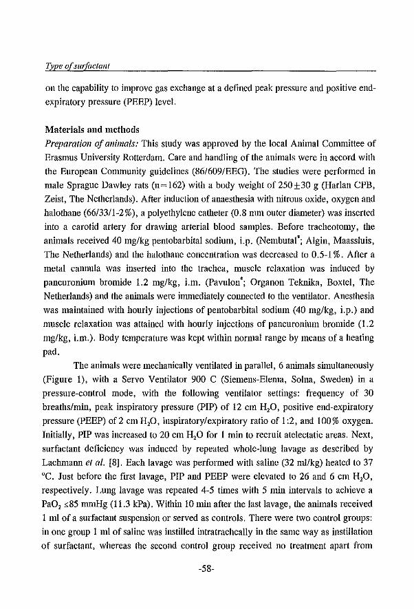
Type of surfactant
on the capability to improve gas exchange at a defined peak pressure and positive end
expiratory pressure (PEEP) level.
Materials and methods
Preparation of anilllals: This study was approved by the local Animal Committee of
Erasmus University Rotterdam. Care and handling of the animals were in accord with
the European Conll11unity guidelines (86/609/EEG). The studies were performed in
male Sprague Dawley rats (n= 162) with a body weight of 250±30 g (Harlan CPB,
Zeist, The Netherlands). After induction of anaesthesia with nitrous oxide, oxygen and
halothane (66/33/1-2%), a polyethylene catheter (0.8 mm outer diameter) was inserted
into a carotid artery for drawing arterial blood samples. Before tracheotomy, the
animals received 40 mg/kg pentobarbital sodium, Lp. (Nembutal'; Algin, Maassluis,
The Netherlands) and the halothane concentration was decreased to 0.5-1 %. After a
metal cannula was inserted into the trachea, muscle relaxation was induced by
pancuronium bromide 1.2 mg/kg, Lm. (Pavulon'; Organon Teknika, Boxtel, The
Netherlands) and the animals were immediately connected to the ventilator. Anestilesia
was maintained with hourly injections of pentobarbital sodium (40 mg/kg, Lp.) and
muscle relaxation was attained with hourly injections of pancuronium bromide (1.2
mg/kg, i.m.). Body temperature was kept within normal range by means of a heating
pad.
The animals were mechanically ventilated in parallel, 6 animals sinmltaneously
(Figure I), with a Servo Ventilator 900 C (Siemens-Elema, Solna, Sweden) in a
pressure-control mode, with the following ventilator settings: frequency of 30
breaths/min, peak inspiratory pressure (PIP) of 12 cm H,O, positive end-expiratory
pressure (PEEP) of2 cm H,o, inspiratory/expiratory ratio of I :2, and 100% oxygen.
Initially, PIP was increased to 20 cm H,O for 1 min to recruit atelectatic areas. Next,
surfactant deficiency was induced by repeated whole-lung lavage as described by
Lachmann et al. [8]. Each lavage was performed with saline (32 nil/kg) heated to 37
·C. Just before the first lavage, PIP and PEEP were elevated to 26 and 6 cm H,O,
respectively. Lung lavage was repeated 4-5 times with 5 min intervals to achieve a
PaO, ,,85 mmHg (11.3 kPa). Within 10 min after tile last lavage, the animals received
I ml of a surfactant suspension or served as controls. There were two control groups:
in one group I ml of saline was instilled intratracheally in the same way as instillation
of surfactant, whereas the second control group received no treatment apart from
-58-
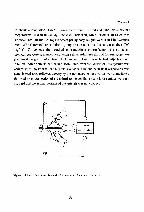
Chapter 3
mechanical ventilation. Table I shows the different natural and synthetic surfactant
preparations used in this study. For each surfactant, three different doses of each
surfactant (25, 50 and 100 mg surfactant per kg body weight) were tested in 6 animals
each. With Curosurf', an additional group was tested at the clinically used dose (200
mg/kg). To achieve the required concentrations of surfactant, the surfactant
preparations were suspended with warm saline. Administration of the surfactant was
performed using a 10 ml syringe which contained 1 mI of a surfactant suspension and
7 ml air. After anin1als had been disconnected from the ventilator, the syringe was
cOlmected to the tracheal calmula via a silicone tube and surfactant suspension was
administered fIrst, followed directly by the administration of air, this was inm1ediately
followed by re-connection of the animal to the ventilator (ventilator settings were not
changed and the supine position of the animals was not changed).
v,
SERVO
VENTILATOR
v,
Figure 1. Schema of the device for the simultaneous ventilation of several animals.
-59-
Type of surfactant
In all animals, ventilator settings were not changed for 2 h and arterial blood
samples were taken at: 5, 30, 60, 90, 120 min after treatment. After 2 h, PEEP and in
parallel PIP were twice reduced by steps of2 cm H,O (to PIP/PEEP: 24/4 and 22/2 cm
H,O, respectively) and arterial blood samples were taken at each PIP/PEEP level 5-10
min later. PaO" PaCO, and pH were measured by conventional methods (ABL 505,
Radiometer, Copenhagen, Demnark). At the end of each experiment, all animals were
killed with an overdose of pentobarbital sodium.
Statistical analysis: All data are expressed as mean ± standard deviation (SD).
Statistical analysis of the data was performed with analysis of variance (ANOYA) for
repeated time measurements by use of the General Linear Models (GLM) procedure of
the SAS statistical package (SAS Users Guide, 1990, SAS Institute Inc., Cary NC).
Tests performed were: (I) within a treatment group, the effect of time on changes in
PaO, and PaCO" (2) with a surfactant preparation, the effect of doses on PaO" (3) the
difference in PaO, values between groups, using both control groups as negative
controls. Tests were performed from t=O' to t= 120' to evaluate dose-dependency and
overall differences between the groups. Within groups, tests were performed from t=O'
to t=5' to evaluate the immediate response of the instilled surfactant, t=5' to t= 120'
to evaluate stability of PaO, over time. To evaluate stability of PaO, after PEEP
reduction within groups in which PaO, kept stable during the first 2 h, additional tests
were performed from t= 120' to t= 130' (=PIP/PEEP of 24/4 cm H,O) and t= 120' to
t= 140' (=PIP/PEEP of 22/2 cm HP). For evaluation of survival rates, the Kaplan
Meier method (product-linlit survival estimates) was performed. Statistical significance
was accepted at a p-value of ~0.05.
-60-
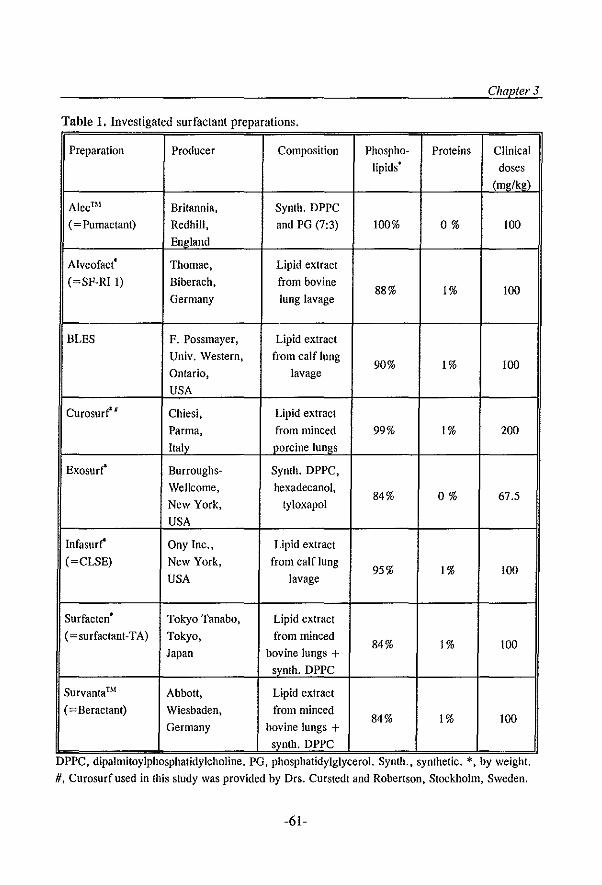
Chapter 3
Table 1. Investigated surfactant preparations.
Preparation Producer Composition Phospho- Proteins Clinical lipids' doses
(mg/kg)
Alec™ Britannia, Synth. DPPC
(~ Pumactant) Redhill, and PG (7:3) 100% 0% 100
England
AlveofactC Thomae, Lipid extract (~SF-R[ I) Biberach. from bovine
88% 1% 100 Germany lung lavage
BLES F. Possmayer, Lipid extract
Univ. Western. from calf lung 90% 1% 100
Ontario, lavage
USA
Curosur~K Chiesi, Lipid extract Parma, from minced 99% 1% 200
Italv porcine lungs
Exosurr Burroughs- Synth. DPPC,
Wellcome, hexadecanol, 84% 0% 67.5
New York, tyloxapol
USA
[nfasurf' Ony [ne., Lipid extract
(~CLSE) New York, from calf lung 95% 1% 100
USA lavage
Surfacten • Tokyo Tanabo, Lipid extract (~surfactant-TA) Tokyo, from minced
84% 1% 100 Japan bovine lungs +
synth. DPPC
Survanta™ Abbott, Lipid extract
(~ Beraetant) Wieshaden, from minced 84% 1% 100
Germany bovine lungs + svnth. DPPC
DPPC, dlpalmItoylphosphaudylcholme. PG, phosphaUdylglycerol. Synth., syntheuc. *, by weight.
#, Curosurf used in this study was provided by Drs. Curstedt and Robertson, Stockholm. Sweden.
-61-
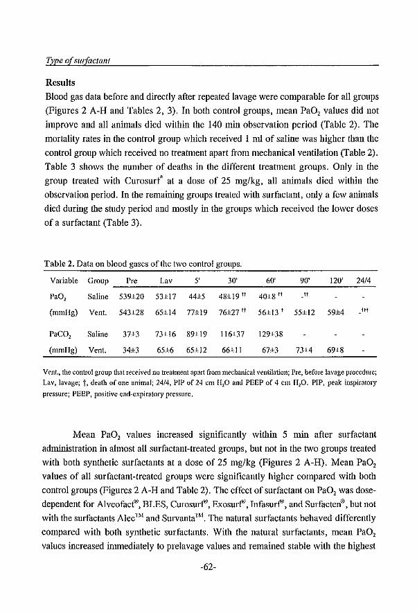
Type of surfactant
Results
Blood gas data before and directly after repeated lavage were comparable for all groups
(Figures 2 A-H and Tables 2, 3). In both control groups, mean PaO, values did not
improve and all animals died within the 140 min observation period (Table 2). The
mortality rates in the control group which received 1 ml of saline was higher than the
control group which received no treatment apart from mechanical ventilation (Table 2).
Table 3 shows the number of deaths in the different treatment groups. Only in the
group treated with Curosurt at a dose of 25 mg/kg, all aninlals died within the
observation period. In the remaining groups treated with surfactant, only a few animals
died during the study period and mostly in the groups which received the lower doses
of a surfactant (Table 3).
Table 2. Data on blood gases ofthe two control groups.
Variable Group Pre Lav 5' 30' 60' 90' 120' 2414
Pa01 Saline 539±20 53±17 44±5 48±19 tt 40±8 ff tt
(mmHg) Vent. 543±28 65±l4 77±19 76±27 fI 56±t3 t 55±t2 59±4 Itt
PaCO, Saline 37±3 73±16 89±19 116±37 129±38
(mmHg) Vent. 34±3 65±6 65±12 66±11 67±3 73±4 69±8
Vent., the control group that received no treatment apart from mechanical ventilation; Pre, before lavage procedure; Lav, lavage; t, death of one animal; 2414, PIP of 24 em HP and PEEP of 4 em HP, PIP, peak inspiratory
pressure; PEEP, positive end-expiratory pressure,
Mean PaO, values increased significantly within 5 min after surfactant
administration in almost all surfactant-treated groups, but not in the two groups treated
with both synthetic surfactants at a dose of 25 mg/kg (Figures 2 A-H). Mean PaO,
values of all surfactant-treated groups were significantly higher compared with both
control groups (Figures 2 A-H and Table 2). The effect of surfactant on PaO, was dose
dependent for Alveofact®, BLES, Curosurf', Exosurf', Infasurf', and Surfacten®, but not
with the surfactants AlecThI and SurvantaThI. The natural surfactants behaved differently
compared with both synthetic surfactants. With the natural surfactants, mean PaO,
values increased immediately to prelavage values and remained stable with the highest
-62-
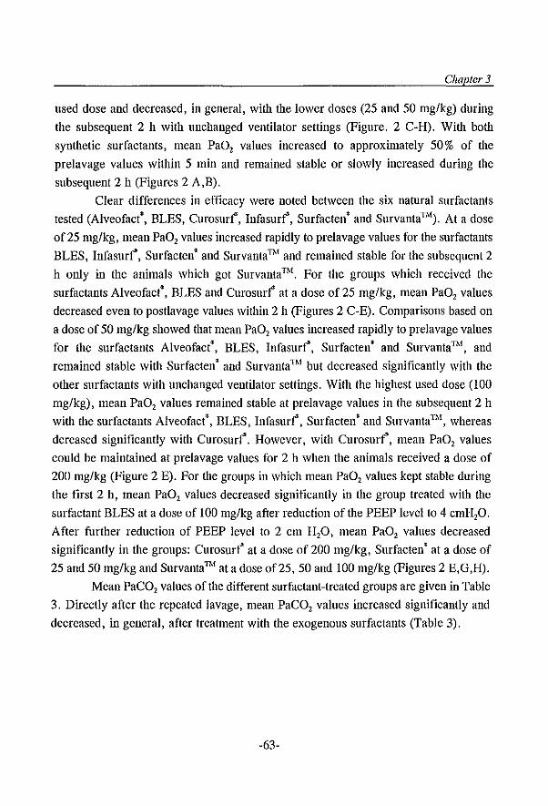
Chapter 3
used dose aud decreased, in general, with the lower doses (25 and 50 mg/kg) during
the subsequent 2 h with unchanged ventilator settings (Figure. 2 C-H). With both
synthetic surfactants, mean PaO, values increased to approximately 50% of the
prelavage values within 5 min and remained stable or slowly increased during the
subsequent 2 h (Figures 2 A,B).
Clear differences in efficacy were noted between the six natural surfactants
tested (Alveofact', BLES, Curosurt, lnfasurt, Surfacten' and SurvantaThI). At a dose
of25 mg/kg, mean PaO, values increased rapidly to prelavage values for the surfactants
BLES, lnfasurt, Surfacten' and SurvantaThI and remained stable for the subsequent 2
h only in the animals which got Survanta Thl. For the groups which received the
surfactants Alveofact', BLES and Curosurt at a dose of 25 mg/kg, mean PaO, values
decreased even to postlavage values within 2 h (Figures 2 C-E). Comparisons based on
a dose of 50 mg/kg showed that mean PaO, values increased rapidly to prelavage values
for the surfactants Alveofact', BLES, lnfasurt, Surfacten' and SurvantaThI, and
remained stable with Surfacten' and Survanta Thl but decreased significantly with tlle
other surfactants with uuchauged ventilator settings. With the highest used dose (100
mg/kg), mean PaO, values remained stable at prelavage values in the subsequent 2 h
WiOl the surfactants Alveofact', BLES, lnfasurt, Surfacten' and SurvantaThI, whereas
dcreased significantly with Curosurt. However, with Curosurt, mean PaO, values
could be maintained at prelavage values for 2 h when the animals received a dose of
200 mg/kg (Figure 2 E). For the groups in which mean PaO, values kept stable during
the first 2 h, mean PaO, values decreased significantly in the group treated with the
surfactant BLES at a dose of 100 mg/kg after reduction of the PEEP level to 4 cmH,O.
After further reduction of PEEP level to 2 cm H,O, mean PaO, values decreased
significantly in the groups: Curosurt at a dose of 200 mg/kg, Surfacten' at a dose of
25 and 50 mg/kg and Survanta"M at a dose of25, 50 and 100 mg/kg (Figures 2 E,G,H).
Mean PaCO, values of the different surfactant-treated groups are given in Table
3. Directly after the repeated lavage, mean PaCO, values increased significantly and
decreased, in general, after treatment with the exogenous surfactants (Table 3).
-63-
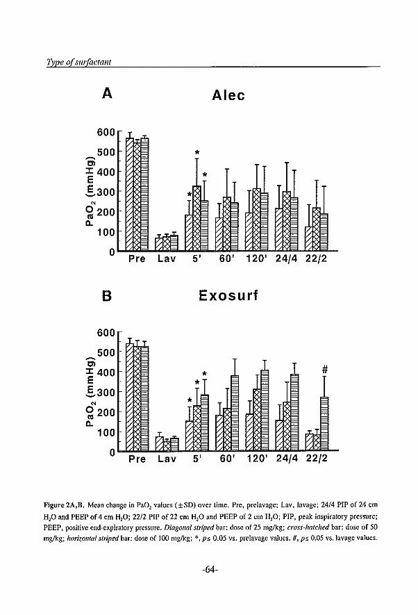
TJlpe of surfactant
A Alec
600
500 * ~
Cl ::t: 400 E * S300
N
0200 <tI a.
100
0 Pre Lav 5' 60' 120' 24/4 22/2
B Exosurf
600
500 ~
Cl # ::t: 400 * E *
S300 N *
0200 <tI a.
100
0 Pre Lav 5' 60' 120' 24/4 22/2
Figure 2A,B. Mean change in Pa02 values (±SD) over time. Pre, prelavage; Lav. lavage; 24/4 PIP of 24 em
HP and PEEP of 4 em H20; 2212 PIP of 22 em H20 and PEEP of 2 em H20; PIP, peak inspiratory pressure;
PEEP, positive end-expiratory pressure. Diagonal striped bar: dose of 25 mg/kg; cross-hatched bar: dose of 50
mg/kg; horiZOlllal striped bar: dose of 100 mg/kg; *, p5, 0.05 vs. prelavage values. H, p5, 0.05 vs.lavage values.
-64-
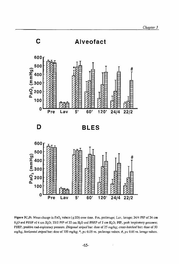
~
Ol
c
600
500
J: 400 E ,§,300
~
Ol
100
o
D
600
500
J: 400 E ,§,300
OJ
0200 (ll
Il. 100
Pre Lav
o ...... p:""r e ....... '7L""a v
Chapter 3
Alveofact
#
5' 60' 120' 24/4 22/2
BLES
#
5' 60' 120' 24/4 22/2
Figure 2C j D. Mean change in Pa02 values (±SD) over time. Pre. prelavage; Lav, lavage; 24/4 PIP of 24 em
HP and PEEP of 4 em HP; 22/2 PIP of 22 em HP and PEEP of 2 em H20; PIP, peak inspiratory pressure;
PEEP, positive end-expiratory pressure. Diagonal striped bar: dose of 25 rug/kg; cross-hatched bar: dose of 50
mg/kg; lIorizolIIai striped bar: dose of 100 mg/kg; *. ps 0.05 vs. prelavagc values. H, p!. 0.05 vs.lavage values.
-65-
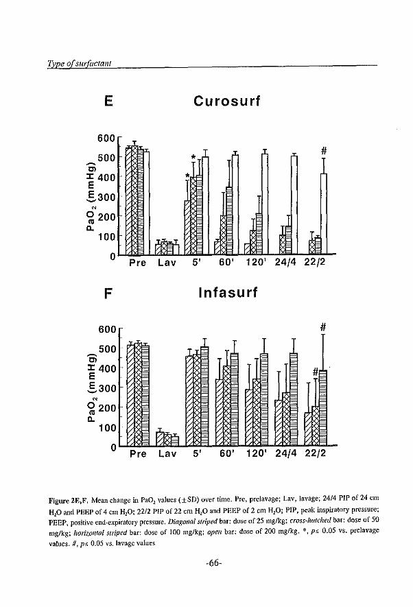
Type of surfactant
~
Cl
E
600
500
J: 400 E .5..300
N
0200 III a.
100
o
F
600
500 ~
Cl J: 400 E .5..300
N
0200 III a.
100
Pre Lav
o "'-'-:' ........ "7 Pre Lav
Curosurf
* #
5' 60' 120' 24/4 22/2
Infasurf
#
5' 60' 120' 24/4 22/2
Figure 2E,F. Mean change in PaD l values (±SD) over time. Pre, prelavage; Lav, lavage; 24/4 PIP of24 em
HP and PEEP of 4 em H20; 22/2 PIP of 22 cm H20 and PEEP of 2 em H20; PIP, peak inspiratory pressure;
PEEP, positive end-expiratory pressure. Diagollal striped bar: dose of 25 mg/kg; cross-hatched bar: dose of 50
mg/kg; lioril.ollfai striped bar: dose of 100 rug/kg; open bar: dose of 200 mg/kg. ,;., p5. 0.05 vs. prelavage
values. H, p5. 0.05 vs.lavage values
-66-
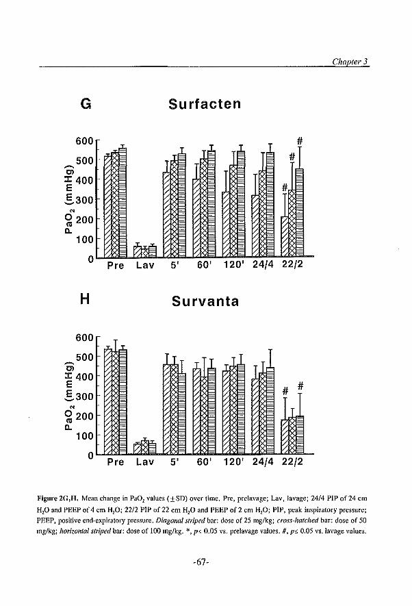
Chapter 3
G Surfacten
600
500 ~
OJ J: 400 E .§.300
'" 0200 <1l a.
100
0 Pre Lav 5' 60' 120' 24/4 22/2
H Survanta
600
500 ~
OJ J: 400 E # # .§.300
'" 0200 <1l a.
100
0 Pre Lav 5' 60' 120' 24/4 22/2
Figure 2G,H. Mean change in Pa02 values (±SD) over time. Pre, prclavage; Lav, lavage; 24/4 PIP of 24 elll
HP and PEEP of 4 em Hp; 22/2 PIP of 22 em H20 and PEEP of 2 em HP; PIP, peak inspiratory pressure;
PEEP, positive end-expiratory pressure, Diagonal stliped bar: dose of 25 mg/kg; cross-hatched bar: dose of 50
mg/kg; /torizolllai striped bar: dose of 100 mg/kg. *, p5. 0.05 vs. prelavage values. #, p5. 0.05 vs.lavage values.
-67-
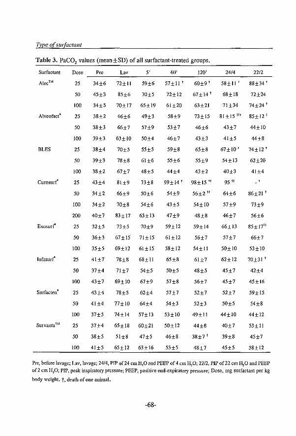
Type of surfactant
Table 3, PaCO, values (mean±SD) of all surfactant-treated groups,
Surfactant Dose Pre Lav 5' 60' 120' 24/4 22/2
Alec™ 25 34±6 72±11 59±6 57±11 t 60±9 t 58±11 t 88±34 t
50 45±3 85±6 70±5 72±12 67±14 t 68±18 72±24
100 34±5 70±17 65±19 61±20 63±21 71 ±34 74±24 t
AlveofactS 25 38±2 66±6 49±3 58±9 73±15 81+15 ttl 85±12 t
50 38±3 66±7 57±9 53±7 46±6 43±7 44±1O
100 39±3 63±1O 50H 46±7 43±3 41±5 44±8
BLES 25 38H 70±5 55±5 59±8 65±8 67±lOt 74± 12 t
50 39±3 78±8 61±6 55±6 55±9 54±13 62±20
100 38±2 67±7 48±5 44±4 43±2 40±3 41H
Curosurf" 25 43±4 81±9 73±8 99±14 t 98± 15 " 95 if
50 34±2 66±9 50±6 54±9 56±2 II 64±6 86±21 t
100 34±2 70±8 54±6 43±5 54±1O 57±9 73±9
200 40±7 83±17 63±13 47±9 48±8 46±7 56±6
Exosurt 25 32±5 73±5 70±9 59± 12 59±14 66±13 85± 17ft
50 36±3 67±15 71±15 61±12 56±7 57±7 66±7
100 35±5 69±12 61±15 58±12 54±11 50±1O 53±1O
Infasurr 25 41±7 78±8 68± 11 65±8 61±7 62±12 70±31 t
50 37±4 71±7 54±5 50±5 48±5 45±7 42±4
100 43±7 69± 10 67±9 57±8 56±7 45±7 45±16
Surfacten • 25 43H 78±5 62±4 57±7 52±7 52±7 59± 15
50 41±4 77±1O 64H 54±3 52±3 50±5 54±8
100 37±5 74±14 57±13 53±1O 49±11 44±1O 44±12
Survanta™ 25 37H 65±18 60±21 50±12 44±8 40±7 53±11
50 38±5 51±8 47±5 46±8 38±7 t 39±8 45±7
100 41±5 65±12 63±16 53±5 48±7 45±5 58±12
Pre, before lavage; Lav, lavagej 2414, PIP of24 emH10 and PEEP of4 em Hpj 22/2, PIP of22 em H20 and PEEP 0[2 em HP; PIP, peak inspiratory pressurej PEEP, positive end-expiratory pressure; Dose, mg surfactant per kg
body weight. t, death of one animal.
-68-
Chapler 3
Discussion
This is the first report of direct comparison of eight clinically used surfactants under
standardized conditions with respect to mechanical ventilation and surfactant
administration. Most in-vivo evaluations of pulmonary surfactants have been performed
in the rabbit fetus model [9]. The reason for using the rat lavage model in the present
study and not the immature fetus model needs some considerations. Using the rabbit
fetus model, only the survival rate and compliance calculated from the tidal volume, can
be studied. We have shown that high values of dynamic compliance can be obtained by
a 'pure' detergent in premature rabbits [10]. The detergent had a low static surface
tension but was lacking the appropriate properties to stabilize surfactant-deficient alveoli
in the expiratory phase. This resulted in a high thorax-lung compliance because the lung
volume swings between inspiration and expiration are greater if the alveoli change from
the collapsed to the open state [10]. The alveolar stabilisation is, however, one of the
main functions of the native pulmonary surfactant system [11]. Normally, this
phenomena can only be recognised if lung volume measurements (functional residual
capacity) are performed, however, this technique is not yet available for routine clinical
use [12]. Another possibility to truly evaluate the efficacy of surfactant preparations is
to measure blood gases; they present the global function of the lung. That is why we
used the surfactant-deficient animal model in the present study which allows the
evaluation of arterial blood gases after exogenous surfactant therapy as an indicator of
the quality of surfactant preparations with regard to their capacity to stabilize alveoli
in the end-expiratory phase [8,13,14].
The results of this study show significant differences in activity between the
used surfactant preparations; the natural surfactants were more effective to improve
blood gases to prelavage values than both synthetic surfactants, and between the natural
surfactants differences in efficacy were detectable, especially after reduction of the
PEEP level. In both control groups, arterial oxygenation did not inlprove and all
animals died within the observation period due to insufficient gas exchange. It has been
reported that the combination of saline instillation and mechanical ventilation can harm
the lungs [15]. This was confmned by the results of the present study in which the
mortality rate of the control group which received saline was higher compared to the
control group which received no treatment apart from mechanical ventilation (Table 2).
Recently, first clinical trials compared a natural with a synthetic surfactant
directly and showed a more rapid improvement in oxygenation and ventilation in the
-69-
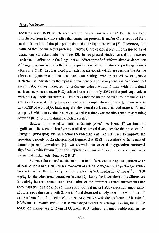
TJlpe of surfactant
neonates with RDS which received the natural surfactant [16,17]. It has been
established from in-vitro studies that surfactant proteins Band/or C are required for a
rapid adsorption of the phospholipids to the air-liquid interface [3]. Therefore, it is
assumed that the surfactant proteins Band/or C are essential for uniform spreading of
exogenous surfactant into the lungs [3]. In the present study, we did not measure
surfactant distribution in the lungs, but an indirect proof of uniform alveolar deposition
of exogenous surfactant is the rapid improvement of PaO, values to prelavage values
(Figures 2 C-H). In other words, all existing atelectasis which are responsible for the
observed hypoxemia at the used ventilator settings were recruited by exogenous
surfactant as indicated by the rapid improvement of arterial oxygenation. We found that
mean PaO, values increased to prelavage values within 5 min with all natural
surfactants, whereas mean PaO, values increased to only 50 % of the prelavage values
with both synthetic surfactants. This means that the increased right-to-left shunt, as a
result of the repeated lung lavages, is reduced completely with the natural surfactants
at a PEEP of 6 cm H,O, indicating that the natural surfactants spread more uniformly
compared with both synthetic surfactants and that there was no difference in spreading
between the different natural surfactants tested.
Between both tested synthetic surfactants (AlecTh! vs. Exosurt) we found no
significant difference in blood gases at all three tested doses, despite the presence of a
detergent (tyloxapol) and an alcohol (hexadecanol) in Exosurt' used to improve the
spreading capacity of the phospholipid (Figures 2 A,B) [2]. In contrast to the results of
Cummings and coworkers [4], we showed that arterial oxygenation inlproved
significantly with Exosurt', but this improvement was significant lower compared with
the natural surfactants (Figures 2 B-H).
Between the natural surfactants, marked differences in response pattern were
shown. A rapid and sustained improvement of arterial oxygenation to prelavage values
was achieved at the clinically used dose which is 200 mg/kg for Curosurt' and '100
mg/kg for the other used natural surfactants [2]. Using the lower doses, the differences
in activity became pronounced. Evaluation of the different natural surfactants after
administration of a dose of 25 mg/kg showed that mean PaO, values remained stable
at prelavage values only with Survanta Th! and decreased slowly over tinle with Infasurf
and Surfacten' but dropped back to postlavage values with the surfactants Alveofact',
BLES and Curosurt' within 2 h at unchanged ventilator settings. During the PEEP
reduction manoeuvre to 2 cm H,O, mean PaO, values remained stable only in the
-70-
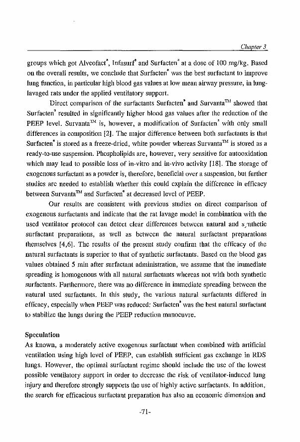
Chapter 3
groups which got Alveofact', Infasurf' and Surfacten' at a dose of 100 mg/kg. Based
on the overall results, we conclude that Surfacten' was the best surfactant to improve
lung function, in particular high blood gas values at low mean airway pressure, in lung
lavaged rats under the applied ventilatory support.
Direct comparison of the surfactants Surfacten' and SurvantaThI showed that
Surfacten' resulted in significantly higher blood gas values after the reduction of the
PEEP level. SurvantaL\I is, however, a modification of Surfacten' with only small
differences in composition [2]. The major difference between both surfactants is that
Surfacten' is stored as a freeze-dried, white powder whereas SurvantaThI is stored as a
ready-to-use suspension. Phospholipids are, however, very sensitive for autooxidation
which may lead to possible loss of in-vitro and in-vivo activity [18]. The storage of
exogenous surfactant as a powder is, therefore, beneficial over a suspension, but further
studies are needed to establish whether this could explain the difference in efficacy
between Survanta Thl and Surfacten' at decreased level of PEEP.
Our results are consistent with previous studies on direct comparison of
exogenous surfactants and indicate that the rat lavage model in combination with the
used ventilator protocol can detect clear differences between natural and s;'llthetic
surfactant preparations, as well as between the natural surfactant preparations
themselves [4,6], The results of the present study confirm that the efficacy of the
natural surfactants is superior to that of synthetic surfactants, Based on the blood gas
values obtained 5 min after surfactant administration, we assume that the inmlediate
spreading is homogenous with all natural surfactants whereas not with both synthetic
surfactants, Furtllemlore, fuere was no difference in inUllediate spreading between the
natural used surfactants, In this study, the various natural surfactants differed in
efficacy, especially when PEEP was reduced: Surfacten' was the best natural surfactant
to stabilize the lungs during the PEEP reduction manoeuvre,
Speculation
As known, a moderately active exogenous surfactant when combined wifu artificial
ventilation using high level of PEEP, can establish sufficient gas exchange in RDS
lungs, However, the optimal surfactant regime should include the use of the lowest
possible ventilatory support in order to decrease the risk of ventilator-induced lung
hljury and fuerefore strongly supports the use of highly active surfactants, In addition,
the search for efficacious surfactant preparation has also an economic dinlension and
-71-
Type of surfactant
it is therefore important to explore ways of keeping the amounts of administered
surfactant as low as possible (which of course requires administration of surfactant at
an early time point in lung injury), Based on the results of this study in lung-lavaged
rats, if one could apply these results to the clinical situation, then the ranking in order
of preference would be: Surfacten' > SurvantaThI > Infasurt > Alveofact' > BLES
> Curosurt > Exosurt > Alec Thl,
References 1. Jobe AH, Pulmonary surfactant therapy. N Engl J Med 1993;328:861·868
2. Fujiwara T, Robertson B. Pharmacology of exogenous surfactant. In: Robertson B, van Golde LMG,
Batenburg 11 (Eds). Pulmonary surfactant: from molecular biology to clinical practice. Elsevier,
Amsterdam, 1992, pp 561-592
3. Johansson J, Curstedt T, Robertson B. The proteins of the surfactant system. Eur Respir J 1994; 7: 372-391
4. Cummings 11, Holm BA, Hudak ML, Hudak BB, Ferguson WH, Egan EA. A controlled clinical
comparison of four different surfactant preparations in surfactant-deficient preterm lambs. Am Rev Respir
Dis 1992; 145: 999·1004
5. Seeger W, Grube C, Gunther A, Schmidt R. Surfactant inhibition by plasma proteins: differential sensitivity
of various surfactant preparations. Eur Respir J 1993; 6: 971-977
6. Hafner D, Beume R, Kilian U, Krasznai G, Lachmann B. Dose-response comparisons of five lung
surfactant factor (LSP) preparations in an animal model of adult respiratory distress syndrome (ARDS). Br
J Pharmacol1995; 115: 451·458
7. Lachmann B. In vivo tests for evaluation of pulmonary surfactant. In: Lachmann B (Ed). Surfactant
replacement therapy in neonatal and adult respiratory distress syndrome. Springer-Verlag, Berlin,
Heidelberg, New York, 1987, pp 28-36
8. Lachmann B, Robertson B, Vogel J. In vivo lung lavage as an experimental model of the respiratory
distress syndrome. Acta Anaeslh Scand 1980; 24: 231-236
9. Robertson B, Lachrnann B. Experimental evaluation of surf act ants for replacementlherapy. Exp Lung Res
1988; 14: 279·310
to. Lachmann B, Smit JM, Annbruster S, Gommers D, Tenbrink R, Schairer W. Detergens improve thorax
lung compliance in surfactant deficient immature lung similar to the improvement achieved with natural
surfactant. Am Rev Respir Dis 1987; 135: A357
11. Clements JA. FUllctions of the alveolar lining. Am Rev Respir Dis 1977; 115: 67-71
12. Gammers D. Lachmann B. Improved ventilation by re-aeration of atelectatic regions with exogenous
surfactant in acute respiratory failure. In: Gullo A (Ed). Anaestehsia Pain Intensive Care and Emergency
medicine (APICE) 11. Springer-Verlag, Berlin, Heidelberg, New York, 1996, pp 323-328 13. Lachmann Fujiwara T, Chida S, Morita T, Konishi M, Nakamura K, Maeta H. Surfactant replacement
therapy in experinlental adult respiratory distress syndrome, In: Cosmi EV, Scarpelli EM (Eds). Pulmonary surfactant system. Elsevier, Amsterdam, 1983, pp 231-235
14. Gommers D, Vilslrup C, Bos JAH, Larsson A, Werner D, Hannappel E, Lachmann B. Exogenous
surfactant therapy increases slatic lung compliance, and cannot be assessed by measurements of dynanlic
compliance alone. Cril Care Med 1993; 21: 567-574
15. Lachmann B. Combination of saline instillation with artificial ventilation damages bronchial surfactant.
-72-
Chapter 3
Lancet 1987; i: 1375.
16. Soil RF and Vermont-Oxford Neonatal Network. A multicenter, randomized trial comparing synthetic surfactant with modified bovine surfactant extract in the treatment of neonatal respiratory distress syndrome. Pediatrics 1996; 97: 1-6
17. Hudak ML, Farrell EE, Rosenberg AA. et al. A multicenter randomized, masked comparison trial of
natural versus synthetic surfactant for the (rcaUDen! of respiratory distress syndrome. J Pediatr 1996; 128: 396-406
18. Bergeison LD (Ed). Lipid biochemical preparations. Elsevier, Amsterdam, 1980.
-73-

Chapter 4
Prevention of respiratory failure after hydrochloric
acid aspiration by intratracheal surfactant
instillation in rats
E.P. Eijking, D. Gommers, K.L. So, M.P.M. de Maat', J.W. Mouton* and B.
Lacinnaml
Depts. of Anesthesiology, 'Internal Medicine II and *Clinical Microbiology,
Erasmus University, Rotterdam, The Netherlands
Published ill: Arlest" Allaig 1993,' 76: 472~477 Reprinted with permission (copyright holder: Williams & Wilkens)
-75-
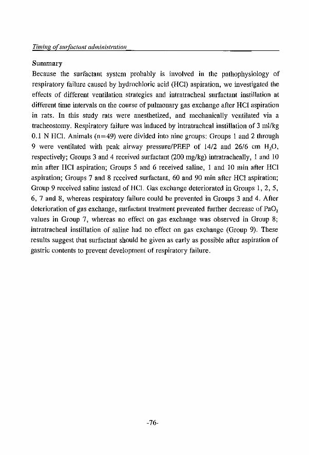
Timing of surfactant administration
Summary
Because the surfactant system probably is involved in the pathophysiology of
respiratory failure caused by hydrochloric acid (Hel) aspiration, we investigated the
effects of different ventilation strategies and intratracheal surfactant instillation at
different time intervals on the course of pulmonary gas exchange after Hel aspiration
in rats. In this study rats were anesthetized, and mechanically ventilated via a
tracheostomy. Respiratory failure was induced by intratracheal instillation of 3 ml/kg
0.1 N He!. Animals (n=49) were divided into nine groups: Groups I and 2 through
9 were ventilated with peak airway pressure/PEEP of 14/2 and 26/6 cm H,o,
respectively; Groups 3 and 4 received surfactant (200 mg/kg) intratracheally, 1 and 10
min after Hel aspiration; Groups 5 and 6 received saline, 1 and 10 min after Hel
aspiration; Groups 7 and 8 received surfactant, 60 and 90 min after Hel aspiration;
Group 9 received saline instead ofHCl. Gas exchange deteriorated in Groups 1,2,5,
6, 7 and 8, whereas respiratory failure could be prevented in Groups 3 and 4. After
deterioration of gas exchange, surfactant treatment prevented further decrease of PaD,
values in Group 7, whereas no effect on gas exchange was observed in Group 8;
intratracheal instillation of saline had no effect on gas exchange (Group 9). These
results suggest that surfactant should be given as early as possible after aspiration of
gastric contents to prevent development of respiratory failure.
-76-
Chapter 4
Introduction
Massive aspiration of gastric contents is one of tile most feared complications of general
anesthesia and is an important cause of the adult respiratory distress syndrome (ARDS)
[1,2]. ARDS caused by aspiration of gastric contents is characterized by deterioration
of gas exchange requiring mechanical ventilation with high inspiratory oxygen
concentration, high insufflation pressure, and positive end-expiratory pressure (PEEP),
and is associated Widl a mortality rate of over 90% (2). Hydrochloric acid (HCI) causes
direct damage to the alveolo-capillary membrane, leading to influx of protein-rich
edema fluid into the alveolar space [3-6]. These plasma-derived proteins are known to
be potent inhibitors of surfactant function [7-12]. It has also been proposed that HCI
directly damages the surfactant system [4,13].
Because the surfactant system probably is involved in the pathophysiology of
respiratory failure caused by HCI aspiration, studies have been performed to investigate
the effect of surfactant replacement therapy on lung function of aninlals suffering from
respiratory failure due to HCI aspiration. Kobayashi and colleagues [6] demonstrated
that surfactant instillation could only partly restore gas exchange in rabbits suffering
from respiratory failure due to HCI aspiration after lung edema was removed by
bronchoalveolar lavage; surfactant, when given without prior lung lavage, only
prevented further deterioration of blood gases. Lannn and colleagues [14] showed
improved lung recoil without improvement in gas exchange in rabbits receiving
surfactant 5 min after HCI aspiration.
The aim of this study was to investigate the effects of different ventilation
strategies and intratracheal surfactant instillation at different time intervals, on the
course of pulmonary gas exchange after HCI aspiration in rats.
Materials and methods
Exogenolls slIIfactallf: The surfactant used in these experiments is a freeze-dried natural
surfactant isolated from bovine lungs in basically the same manner as previously
described [15]. It consists of approxinJately 90% phospholipids, 1 % hydrophobic
proteins (SP-B and SP-C), the remainder being other lipids such as cholesterol,
glyceride and free fatty acids. There is no SP-A (the largest surfactant associated
protein, molecular weight 26,000-38,000) in this surfactant preparation as a resnlt of
isolation procedures. Surfactant was suspended in saline at a concentration of 50 mg
dry weightlml. This surfactant preparation has proven to be highly effective in
-77-
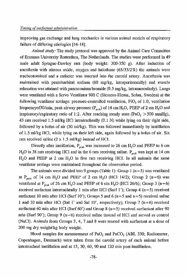
Timing o{sllr(actallt administration
improving gas exchange and lung mechanics in various animal models of respiratory
failure of differing etiologies [16-18].
Anilllal study: The study protocol was approved by the Animal Care ConlTIlittee
of Erasmus University Rotterdam, The Netherlands. The studies were performed in 49
male adult Sprague-Dawley rats (body weight: 300-350 g). After induction of
anesthesia with nitrous oxide, oxygen and halothane (65/3312 %) the animals were
tracheotomized and a catheter was inserted into the carotid artery. Anesthesia was
maintained with pentobarbital sodium (60 mg/kg, intraperitoneally) and muscle
relaxation was attained with pancuronium bromide (0.5 ITIg/kg, intramuscularly). Lungs
were ventilated WiOl a Servo Ventilator 900 C (Siemens-Elema, Solna, Sweden) at the
following ventilator settings: pressure-controlled ventilation, FiO, of 1.0, ventilation
frequencyof30/min, peak airway pressure (P ",,) of 14 cm H,O, PEEP of2 cm H,O and
inspiratory/expiratory ratio of 1 :2. After reaching steady state (PaO, > 500 llllnHg),
43 rats received 1.5 Illi/kg HCl intratracheally (0.1 N) while lying on their right side,
followed by a bolus of air (30 ml/kg). This was followed inlTIlediately by instillation
of 1.5 ml/kg HC1, while lying on their left side, again followed by a bolus of air. Six
rats received saline (2 x 1.5 nil/kg) instead of HCI.
Directly after instillation, P p',k was increased to 26 cm H,O and PEEP to 6 cm
H,o in 38 rats receiving HCl and in the 6 rats receiving saline. P p''' was kept at 14 cm
H20 and PEEP at 2 cm H20 in five rats receiving HCI. In all animals the same
ventilator settings were maintained throughont the observation period.
The aninlals were divided into 9 groups (Table 1): Group 1 (n=5) was ventilated
at Pp,,' of 14 cm H20 and PEEP of 2 cm H,o (HCl 14/2); Group 2 (n=6) was
ventilated at P",x of26 cm H20 and PEEP of6 cm H,O (HCI26/6); Group 3 (n=6)
received surfactant intratracheally 1 min after HCl (Surf 1 '); Group 4 (n=5) received
surfactant 10 min after HCl (Surf 10'); Groups 5 and 6 (n=5 and n=5) received saline
1 and 10 min after HCl (Sal I' and Sal 10', respectively); Group 7 (n=6) received
surfactant 60 min after HCl (Surf 60') and Group 8 (n=5) received surfactant after 90
min (Surf 90'); Group 9 (n=6) received saline instead of HCl and served as control
(NaCl). Animals from Groups 3, 4, 7 and 8 were treated with surfactant at a dose of
200 mg dry weight/kg body weight.
Blood samples for measurement of PaO, and PaCO, (ABL 330; Radiometer,
Copenhagen, Delllllark) were taken from the carotid artery of each animal before
intratracheal instillation and at 15, 30, 60, 90 and 120 min post-instillation.
-78-
Chapter 4
At the end of the experiments all animals were sacrificed with an overdose of
intra-arterially administered pentobarbital sodium. Lungs of a few animals of Groups
2-6 and 9 underwent bronchoalveolar lavage (BAL) with saline (37°C; 30 ml!kg).
Bronciloalveolar lavage fluid: To compare all BAL samples with those from
healthy controls, six additional male Sprague-Dawley rats (body weight 300-350 g)
were lavaged once with saline (37°C; 30 ml/kg). The BAL tluids of the different
groups were prepared as follows: all samples were centrifugated for 15 min at 2000 g
to remove cell material. Protein concentration was measured in all samples using a
modified Lowry method [19], with bovine serum albumin as standard. Also, surface
activity in BAL tluid was measured using a modified Wilhelmy balance (E. Biegler
GmbH, Mauerbach, Austria). In this method, a tight-fitting tetlon barrier reduces the
surface area of a tetlon trough from 100-20% at a cycle speed of 0.33/min. Saline is
used as subphase and is kept at 37°C. The force on a platinum slide (lxl cm), dipped
into the subphase, is measured by a force transducer and expressed as surface tension.
Further, maximal surface tension is measured at 100% surface area and minimal
surface tension at 80 % surface compression and expressed as milli Newton/meter
(mN/m). Surface tension characteristics of a BAL sample are measured after
application on the surface of the saline-ftlled trough. In this study 5001'1 of BAL tluid
was applied to the surface of the trough; maximal and minimal surface tensions were
measured after three cycles.
Statistical analysis: All data are expressed as mean ± standard deviation (SD).
For the animal study statistical analysis of data was performed using repeated
measurements ANOVAs [20], with tinle as the repeat variable. Three ANOVAs were
performed initially: the effect of P ",,/PEEP settings against controls, the effect of
saline instillation against controls, and fmally the effect of surfactant instillation against
controls. When significant differences between groups occurred, the difference between
tllese groups was analyzed further. For analysis of BAL parameters, standard ANOVA
procedures were performed. Statistical significance was accepted at psO.05.
-79-
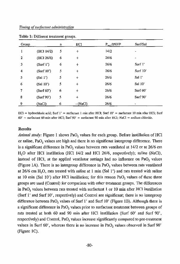
Timing of surfGctGnt administration
Table 1: Different treatment groups.
Group n HCI
(HCI 14/2) 5 + 2 (HCI26/6) 6 + 3 (Surf I') 6 + 4 (Surf 10') 5 + 5 (Sal I') 5 + 6 (Sal 10') 5 + 7 (Surf 60') 6 + 8 (Surf 90') 5 + 9 (NaCI) 6 - (NaCI)
P ",,/PEEP
14/2
26/6
26/6
26/6
26/6
26/6
26/6
26/6
26/6
Surf/Sal
Surf I'
Surf 10'
Sail'
Sal 10'
Surf 60'
Surf 90'
HCI = hydrochloric acid; Surf I' = surfactant 1 min after HCl; Surf 10' = surfactant 10 min after HCl; Surf 60' = surfactant 60 min after HCI; Surf 90' = surfactant 90 min after Hel; NaCI = sodium chloride.
Results
Animal stlldy: Figure I shows PaO, values for each group. Before instillation of HCl
or saline, PaO, values are high and there is no significant intergroup difference. There
is a significant difference in PaO, values between rats ventilated at 1412 or at 26/6 cm
H,O after HCl instillation (HCl 14/2 and HCl 26/6, respectively); saline (NaCl) ,
instead of HCl, at the applied ventilator settings had no influence on PaO, values
(Figure IA). There is no intergroup difference in PaO, values between rats ventilated
at 26/6 cm H,O, rats treated with saline at I min (Sal I ') and rats treated with saline
at 10 min (Sal 10') after HCl instillation; for this reason PaO, values of these three
groups are used (Control) for comparison with other treatment groups. The differences
in PaO, values between rats treated with surfactant I or 10 min after HCl instillation
(Surf I' and Surf 10', respectively) and Control are significant; there is no intergroup
difference between PaO, values of Surf I' and Surf 10' (Figure IB). Although there is
a significant difference in PaO, values prior to surfactant treatment between groups of
rats treated at both 60 and 90 min after HCl instillation (Surf 60' and Surf 90',
respectively) and Control, PaO, values increase significantly compared to pre-trearnlent
values in Surf 60', whereas there is no increase in PaO, values observed in Surf 90'
(Figure IC).
-80-
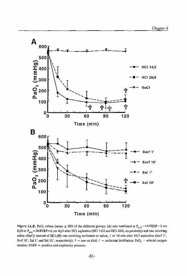
Chapter 4
A 600
--i---i-. ___ i--._.-i--_.-l
Figure lA,B. Pa02 values (mean ± SD) of the different groups: (A) rats ventilated at P I"'Ji= 14/PEEP=2 em
H20 or P ~=26IPEEP=6 em H20 after Hel aspiration (Hel 1412 and HC12616. respectively) and rats receiving saline (NaCl) instead of HCI;(B) rats receiving surfactant or saline, IorIO min after Hel aspiration (Surf 1 t.
Surf 10', SaIl' and Sal 10' , respectively); t = one rat died; I = surfactant instillation; PaOI = arterial oxygen
tension; PEEP = positive end-expiratory pressure.
-81-
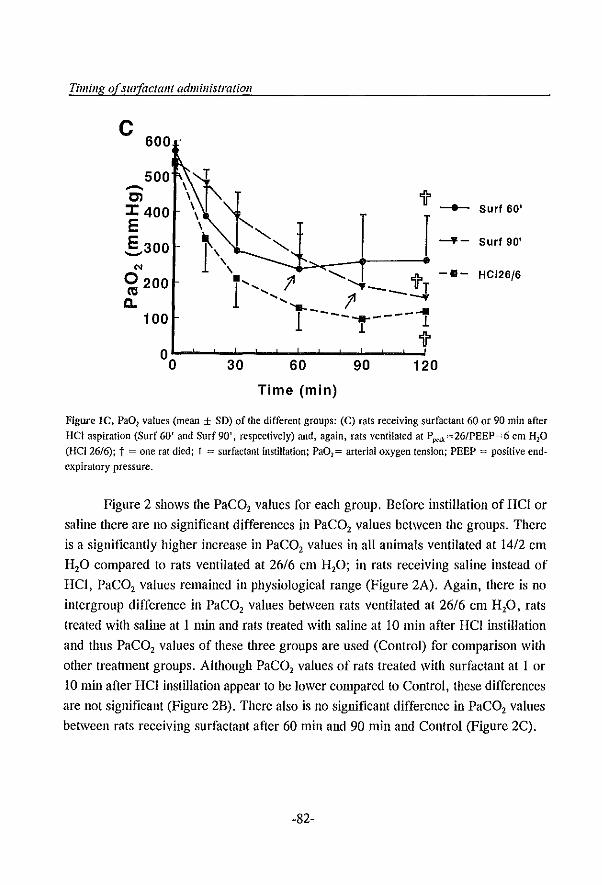
Timillg of surfactant administration
C 600
500 --en J: 400 E E 300 '-'
'" 0200 ('0
0.. 100
30 60
Time (min)
90
'if'
r
120
-+- Surf 60'
...... - Surf 90'
-8- HC126/6
Figure Ie. Pa02 values (mean ± SD) of the different groups: (C) rats receiving surfactant 60 or 90 min after HCI aspiration (Surf 60' and Surf 90', respectively) and. again, rats ventilated at P po.J;=26/PEEP=6 cm H20 (HCI26/6); t = one rat died; I = surfactant instillation; Pa02 = arterial oxygen tension; PEEP = positive endexpiratory pressure.
Figure 2 shows the PaC02 values for each group. Before instillation of HCI or
saline there are no significant differences in PaC02 values between the groups. There
is a significantly higher increase in PaC02 values in all animals ventilated at 1412 cm
H20 compared to rats ventilated at 26/6 Clll H20; in rats receiving saline instead of
HCI, PaC02 values remained in physiological range (Figure 2A). Again, there is no
intergroup difference in PaC02 values between rats ventilated at 26/6 cm H20, rats
treated with saline at 1 min and rats treated with saline at 10 min after HCI instillation
and thus PaC02 values of these three groups are used (Control) for comparison with
other treatment groups. Although PaC02 values of rats treated with surfactant at 1 or
10 min after HCI instillation appear to be lower compared to Control, these differences
are not significant (Figure 2B). There also is no significant difference in PaC02 values
between rats receiving surfactant after 60 min and 90 min and Control (Figure 2C).
-82-
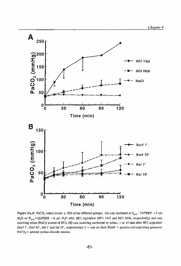
A 250
--200 en ::z:: E 150 E
'"
Chapte/' 4
1 -- HC114/2
1 -a- HC126/6
0 100 T T -\L- NaCI U III Il. 50
1 ....... ---- .... -*-------.----.-.-.-::::.....If-.---"f--.----.Jf-.-.~
°0L-~~3~0~~-6~0~---9~0~~-1~20
Time (min)
B 150
.-.. en
::z:: 100 E E
'" o U 50 III Il.
1 ,J----4 T ",~~ T /./
.... _-.,.. .it""'" T .... 11'.... T • __ --- T ..... ,........ . __ -r T
----! ~ _. ---- ----
°0L-~~3~0~~-6~0~~~9~0--~1~20
Time (min)
-- Surf l'
--.10- Surf 10'
-"'- Sal l' -- Sal 10'
Figure 2A,n. PaC02 values (mean ± SD) of the different groups; (A) rats ventilated at P p<>.\;:= 14/PEEP =2 COl
HP or P p<>k=26/PEEP =6 em HP after Hel aspiration (Hel 1412 and Hel 26/6, respectively) and rats
receiving saline (NaC!) instead of Hel; (B) rats receiving surfactant or saline, 1 or 10 min after Hel aspiration
(Surf 1', Surf 10', Sal I' and SaIlO', respectively); t = one rat died; PEEP = positive end-expiratory pressure;
PaC02 = arterial carbon dioxide tension.
-83-
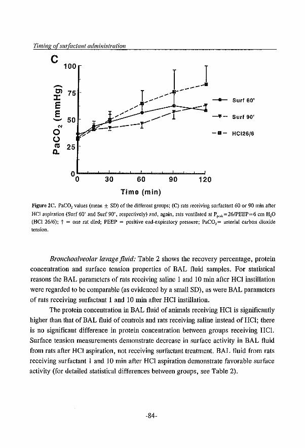
Timing of surfactant administration
C
.-.. til J: E E --'" 0 U ctI
CI..
100
r ~~~J 75 t//i~
............ ., ~ ,." - 1 50 -~-.... _~- .1.
25
°0~~~3~0~~-6~0~~-9~0~~1~20
Time (min)
--- Surf 60'
-¥- Surf 90'
-B- HC126/6
Figure 2e. PaC01 values (mean ± SD) of the different groups; (C) rats r~eiving surfactant 60 or 90 min after
HCl aspiration (Surf 60' and Surf 90', respectively) and, again, rats ventilated at P p<-.u=26IPEEP=6 em HP (Hel 26/6); t = one rat died; PEEP = positive end-expiratory pressure; PaC02= arterial carbon dioxide tension.
Brollc/walveolar lavage fluid: Table 2 shows the recovery percentage, protein
concentration and surface tension properties of BAL fluid samples. For statistical
reasons the BAL parameters of rats receiving saline I and 10 min after Hel instillation
were regarded to be comparable (as evidenced by a small SD), as were BAL parameters
of rats receiving surfactant 1 and 10 min after Hel instillation.
The protein concentration in BAL fluid of animals receiving Hel is significantly
higher tllan that of BAL fluid of controls and rats receiving saline instead of Hel; there
is no significant difference in protein concentration between groups receiving Hei.
Surface tension measurements demonstrate decrease in surface activity in BAL fluid
from rats after Hel aspiration, not receiving surfactant treatment. BAL fluid from rats
receiving surfactant I and 10 min after Hel aspiration demonstrate favorable surface
activity (for detailed statistical differences between groups, see Table 2).
-84-
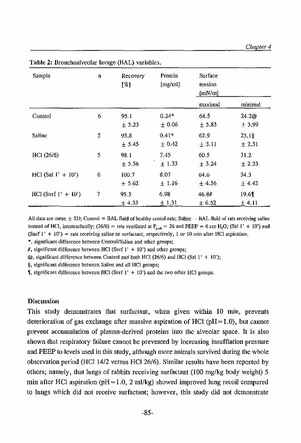
Table 2: Bronchoalveolar lavage (BAL) variables.
Sample n
Control 6
Saline 5
Hel (26/6) 5
Hel (Sal I' + 10') 6
Hel (Surf I' + 10') 7
Recovery
[%l
95.1
± 5.23
93.8
± 5.45
98.1
± 5.56
100.7
± 5.62
95.5
± 4.33
Protein [mg/mll
0.24*
± 0.06
0.41*
± 0.42
7.45
± 1.33
8.07
± 1.16
6.98
± 1.31
Chapter 4
Surface tension [mN/ml
maximal minimal
64.5 24.2@
± 5.83 ± 3.99
62.9 25.1§
± 3.11 ± 2.51
60.5 31.2
± 3.24 ± 2.33
64.6 34.3
± 4.56 ± 4.42
46.8# 19.6' + 6.52 + 4.11
All data are mean ± SD; Control = BAL fluid of healthy contol rats; Saline = BAL fluid of rats receiving saline instead of Hel, intralrachealIy; (26/6) = rats ventilated at P p:<k = 26 and PEEP = 6 em H20; (Sal l' + 10') and (Surf I' + 10') = rats receiving saline or surfactant, respectively, 1 or 10 min after Hel aspiration. *, significant difference between Control/Saline and other groups; D, significant difference between Hel (Surf I' + 10') and other groups; @, significant difference between Control and both Hel (26/6) and Hel (Sail' + 10'); §. significant difference between Saline and all Hel groups; " significant difference between HCI (Surf I' + 10') and the two other HCI groups,
Discussion
This study demonstrates that surfactaut, when given within 10 min, prevents
deterioration of gas exchange after massive aspiration of HCI (pH = 1.0), but call1lot
prevent accumulation of plasma-derived proteins into the alveolar space. It is also
shown that respiratory failure cannot be prevented by increasing insufflation pressure
and PEEP to levels used in this study, although more animals survived during the whole
observation period (HCII4/2 versus HCI26/6). Similar results have been reported by
others; namely, that lungs of rabbits receiving surfactant (100 mg/kg body weight) 5
min after HCl aspiration (pH = 1.0,2 mIlkg) showed inlproved lung recoil compared
to lungs which did not receive surfactant; however, this study did not demonstrate
-85-
Timing of surfactant administration
higher PaO, values in rabbits treated with surfactant, aitllOugh it must be noted that low
PEEP levels were used [14].
The mechanism of respiratory failure in HCl aspiration can be explained by (a)
damage to the alveolar-capillary membrane [4], leading to increased permeability
[5,21,22] and thus influx of protein-rich edema fluid into the alveolar space; (b) direct
damage to the surfactant system [4,13]; and (c) secondary inhibition of remaining
surfactant activity caused by plasma-derived proteins present in the edema fluid [7-12].
As a result of (functional) surfactant deficiency, suction force across the alveolo
capillary membrane increases, leading to further outpouring of edema fluid into the
alveolar space; this way a vicious circle exists. Also, as a result of diminished
surfactant function, surface tension on the alveolar wall increases, leading to increased
retractive forces across the alveolar-capillary membrane, with subsequent formation of
atelectasis and mismatch of the ventilation-perfusion ratio, leading to hypoxemia.
Surfactant is inhibited by plasma-derived proteins in a dose-dependent manner
[7-12]. Thus, with a high surfactant/inhibitor (S/l) ratio there is no inhibition of
surfactant function, whereas with a low S/l ratio most surfactant function is abolished.
Although Ole exact mechanism of inhibition remains unclear, it seems that competition
between surfactant and inhibitors at the air-liquid interphase of the alveolar wall plays
an inlportant role [9,12]. When surfactant is given before deterioration of gas exchange
(Le., before the remaining surfactant function is inhibited) the S/l ratio is increased,
thus preventing the pathophysiological mechanisms that lead to respiratory failure. It
is emphasized iliat ilie initial damage to lung tissue by HCl cannot be prevented, as HCl
is neutralized WiOlin 30 seconds [21] and, thus, lung structure is already damaged. This
is confirmed in the present study, where protein concentration in BAL fluid of rats
receiving surfactant within 10 min is almost as high as protein concentration of rats
receiving saline, or ventilated only. Comparable findings were also reported in the
study mentioned earlier [14], namely increased wet/dry weight ratio in lungs of rabbits
which received surfactant 5 min after HCl aspiration as well as in lungs of rabbits not
receiving surfactant. However, surface tension properties of BAL fluid of rats receiving
surfactant demonstrated low minimal and low maximal surface tensions compared to
BAL fluid of rats receiving saline. Thus, due to the lower surface tension of the
"intrapuhl1onary fluid" the applied ventilator settings (26/6 cm H,O) were sufficient for
maintaining (normal) gas exchange.
Earlier studies have demonstrated that intratracheal surfactant instillation fully
-86-
Chapter 4
restores lung function in animals suffering from respiratory failure due to pneumonia
[16-18]. Also, gas exchange in rats suffering from respiratory failure after bilateral
whole lung lavage could be fully restored after surfactant treatment [23]. In the present
study surfactant treatment, 60 min after Hel instillation, showed slight inlprovement
of gas exchange; surfactant treatment 90 min after Hel instillation did not prevent
further deterioration of gas exchange during the observation period. A possible
explanation could be that after Hel aspiration lung structure is more severely damaged
compared with aninlals suffering from pneumonia, or after whole lung lavage. This
would lead to increased permeability of the alveolo-capillary membrane and more
severe intra-alveolar edema accumulation. After deterioration of gas exchange due to
Hel aspiration, S/I ratio (before surfactant instillation) is low and lung structure is
damaged. When surfactant is given in these conditions, little or no effect is seen on gas
exchange. A possible explanation is that surfactant, at a concentration used in this study
(200 mg/kg) , is inhibited directly by high concentration of inilibitors. Another
explanation could be that the edema fluid and atelectatic areas form a mechanical
barrier, preventing surfactant from entering the alveolar spaces. However, there are
studies in which gas exchange could be improved either by giving a large amount of
surfactant (280-350 mg/kg body weight) [24], or by lavaging the lungs with saline
before surfactant instillation [6], as mentioned in the introduction. With both methods
an improved S/I ratio could be achieved.
Similar results have also been reported in animals suffering from respiratory
failure due to hyperoxic lung il~ury. Surfactant substitution (60 mg/kg body weight) can
prevent development of respiratory insufficiency in rabbits exposed to 100% oxygen for
64 hours: PaO, values of surfactant-treated rabbits, during mechanical ventilation with
100% oxygen, remained at high levels compared with saline-treated aninlals [25]. Also,
wet/dry ratio in anhnals receiving surfactant was high compared to lungs of healthy
controls. However, in guinea pigs suffering already from respiratory failure due to
prolonged exposure to 100% oxygen, surfactant treatment (200 mg/kg body weight)
was not able to restore gas exchange [7].
In the present study a "poillt of no return" mechanism seems to be applicable to
the situation: after deterioration of gas exchange, little or no beneficial effect can be
seen after surfactant instillation at a concentration used in this study (200 mg/kg body
weight), whereas gas exchange is preserved when surfactant is given within 10 min.
Although it is difficult to extrapolate from this aninlal study to the clinical situation, our
-87-
Timing of surfactant administration
results suggest that when massive aspiration of gastric contents is suspected, surfactant
should be given as soon as possible to prevent development of respiratory failure. An important consideration which favors direct treatment with surfactant is that ARDS, caused by aspiration of gastric contents, has a mortality rate of over 90 % [2).
In conclusion, increasing insufflation pressure and PEEP alone to levels used in this study has no beneficial effect on the course of hypoxemia after HCI aspiration in rats. Surfactant given within 10 min after HCI aspiration does prevent development of
respiratory failure but does not prevent damage to lung structure, as evidenced by high protein concentrations in BAL fluid. Surfactant, at the dose used in this study, given after deterioration of gas exchange, can only prevent further decrease in PaO, values.
References 1. Gibbs CP, Modell JH. Management of aspiration pneumonitis. In: Miller RD (Ed). Anesthesia. Churchill
Livingstone, New York, Edinburgh, London, Melbourne, 1990, pp 1293-1319 2. Fowler AA, Hanlllan RF, Good JT, Benson KN, Baird M, Eberle DJ, Petty TL, Hyers TM. Adult
respiratory distress syndrome: risk with common predispositions. Ann Intern Med 1983; 98: 593-597 3. Awe WC, Fletcher WS, Jacob SW. The pathophysiology of aspiration pneumonitis. Surgery 1966; 60: 232-
239 4. Greenfield U, Singleton RP, McCaffree DR, Coalson JJ. Pulmonary effects of experimental graded
aspiration of hydrochloric acid. Ann Surg 1969; 170: 74-86 5. Grimbert FA, Parker Je, Taylor AE. Increased pulmonary vascular penneability following acid aspiration.
J AppJ PhysioJJ981; 51: 335·345
6. Kobayashi T, Ganzuka M, Tanigushi J, Nitta K, Murakami S. Lung lavage and surfactant replacement for hydrochloric acid aspiration in rabbits. Acta Anaesthesio! Scand 1990; 34: 216-221
7. Ennema JJ. Kobayashi T, Robertson B, Curstedt T. Inactivation of exogenous surfactant in experimental respiratory failure induced by hyperoxia. Acta Anaesthesiol Scand 1988; 32: 665-671
8. Fuchimukai T, Fujiwara T. Takahashi A, Enhorning G. Artificial pulmonary surfactant inhibited by proteins. J Appl Physiol 1987; 62: 429-437
9. Holm BA. Enhorning G. Notter RH. A biophysical mechanism by which plasma proteins inhibit lung surfactant activity. Chern Phys Lipids 1988; 49: 49-55
10. Ikegami M, Jobe A, Jacobs H, Lam R. A protein from airways of premature lambs that inhibits surfactant function. J Appl Physiol1984; 57: 1134-1142
11. Seeger W, SIohr G, Wolf HRD, Neuhof H. Alteration of surfactant function due to protein leakage: special interaction with fibrin monomer. J Appl Physiol1985; 58: 326-338
12. Holm BA. Nolter RH. Effects of hemoglobin and cell membrane lipids on pulmonary surfactant activity. J AppJ PhysioJl987; 63: 1434·1442
13. Winn R, SIothert J, Nadir B, Hildebrandt 1. Lung mechanics following aspiration of 0.1 N hydrochloric acid. J AppJ Physiol1983; 55: 1051·1056
14. Lanun WJE, Albert RK. Surfactant replacement improves lung recoil in rabbit lungs after acid aspiration. Am Rev Respir Dis 1990; 142: 1279-1283
15. Metcalfe IL, Enhorning G, Possmayer F. Pulmonary surfactant-associated proteins: their role in the
-88-
Chapter 4
expression of surface activity. J Appl Physiol 1980; 49: 34-41 16. Van Daal GJ. So KL, Gammers D, Eijking BP, Fievez RB, Sprenger MJ, van Dam DW, Lachmann B.
Intratracheal surfactant administration restores gas exchange in experimental adult respiratory distress
syndrome associated with viral pneumonia. Aneslh AnaIg 1991; 72: 589-595
17. Eijking EP, Van Daal GI, Tenbrinck R, Luij"endijk A, SIuiters JF, HannappeJ E. Laclunann B. Effect of surfactant replacement on pllell11l0c),Jtis carinii pneumonia in rats, Intensive Care Med 1991; 17: 475-478
18. Van Daal OJ, Bos JAH, Eijking BP, Gammers D, HannappeI E t Lachmann B. Surfactant replacement
therapy improves pulmonary mechanics in end-stage influenza A pneumonia in mice, Am Rev Respir Dis 1992; 145: 859~863
19. Markwell MAK, Haas SM, Bieber LL, Tolbert NE. A modification of the Lowry procedure to simplify
protein determination in membrane and lipoprotein samples. Anal Biochem 1978; 87: 206-210 20. SAS Users Guide. SAS Institute Inc., Cary, N.C. (U.S.A.), 1989.
21. Jones JG, Berry M, HUlands GH, Crawley Jew. TIle time course and degree of change in alveolar-capillary membrane permeability induced by aspiration of hydrochloric acid and hypotonic saline, Am Rev Respir Dis 1978; 118; 1007-1013
22. Kennedy TP, Johnson KJ, Kunkel RG, Ward PA, Knight PR, Finch IS. Acute acid aspirationiung injury
in the rat: biphasic pathogenesis. Anesth Analg 1989; 69: 87-92 23. Berggren P, Lachmann B, Curstedt T, Grossmann G. Robertson B. Gas exchange and lung morphology
after surfactant replacement in experimental adult respiratory distress syndrome induced by repealed lung lavage. Acta Anaesthesiol Scand 1986; 30: 321~328
24. Lachmann B. Hallman M. Bergmann KC. Respiratory failure following anti-lung serum: study on mechanisms associated with surfactant system damage. Exp Lung Res 1987; 12: 163-180
25. Matalon S. Holm BA. Notter RH. Mitigation of pulmonary hyperoxic injury by administration of exogenous surfactant. J App1 Physiol1987; 62: 756~761
-89-

Chapter 5
In-vivo evaluation of the inhibitory capacity of
human plasma on exogenous surfactant function
B. Lachmann, E.P. Eijking, K.L. So, and D. Gonlll1ers
Dept. of Anesthesiology, Erasmus University Rotterdam, The Netherlands
Pllblished ill: lm€nsh'e Care Med 1994; 20: 6-11
Reprinted witlt permissioll (copyright holder: Springer-Verlag GmbH)
-91-
Sw/actaJlt inhibition by plasma
Summary
Objective: The adult respiratory distress syndrome (ARDS) and neonatal respiratory
distress syndrome (RDS) are characterized by high permeability pulmonary edema
which contains plasma-derived proteins inhibiting pulmonary surfactant function.
Currently, discussion continues as to what dose of surfactant is required for treatment
of these syndromes.
Design: The purpose of this study was to investigate the amount of exogenous
surfactant needed to overcome the inhibitory components in human plasma. Male adult
rats suffering from respiratory failure due to surfactant depletion after whole-lung
lavage received human plasma (4 ml/kg body weight) mixed with surfactant at different
concentrations, intratracheally. Rats receiving surfactant only at different concentrations
served as controls. Blood gas analysis was performed.
Measurements and results: It was demonstrated that plasma (4 mllkgz273 mg plasma
proteins/kg) mixed with surfactant at 300 mg/kg was able to increase and maintain
PaO, at normal values. Plasma mixed with surfactant at 100 mg/kg, after initial
restoration of blood gases, showed deterioration of PaO, values. Plasma mixed with
surfactant at a dose of 50 mg/kg did not improve PaO" whereas surfactant at 50 mg/kg,
without plasma, restored blood gases to prelavage values.
Conelusion: It is concluded that approxin1ately 1 mg surfactant phospholipids is
required to overcome the inhibitory effect of approxinlately 1 mg plasma proteins. For
clinical practice this means that an excess of surfactant should be given, or repeatedly
be substitnted ('titrated') at low concentrations, until blood gases improve.
-92-
Chapter 5
Introduction
The adult respiratory distress syndrome (ARDS) and neonatal respiratory distress
syndrome (RDS) are both characterized by respiratory failure and require therapy
consisting of intubation and mechanical ventilation with high oxygen concentrations.
RDS is characterized by primary surfactant deficiency due to inmlaturity of the lungs,
while in ARDS high permeability puhllonary edema exists, containing plasma-derived
proteins which inhibit pulmonary surfactant function [1-7]. Due to inlpaired surfactant
function, surface tension at the air-liquid interphase on the alveolar walls is increased,
leading to increased suction force across the alveolo-capillary membrane, favouring
further accumulation of protein-rich edema fluid into the alveolar space [8].
Several clinical trials have reported successful treatment with intratracheal
surfactant instillation in prematures suffering from RDS [9-14]. In these studies,
however, some infants did not respond to a single dose of surfactant and some infants
had an only transient improvement of lung function [10,11]. One reason for this could
be that surfactaut function is inhibited by plasma-derived proteins present in intra
alveolar edema fluid.
A few reports on ARDS patients treated with surfactant have been published.
Although the results from these studies are not consistent, the best results were seen in
those patients treated with higher surfactant concentrations [15-19].
Currently, discussion continues as to what dose of surfactant should be used in
prematures with established RDS and what dose should be used for treatment of ARDS.
To investigate this, a study was performed in an animal model of respiratory failure
induced by bronchoalveolar lavage (BAL). This model has proven useful for a variety
of experimental purposes, including e.g. testing of different surfactant preparations and
demonstrating that exogenous surfactant restores blood gases to normal [20-22]. After
respiratory failure was established, animals received plasma mixed with surfactant at
different concentrations. Plasma was used to simulate protein-rich edema, an
established characteristic of ARDS. Instead oflung mechanics, blood gas measurements
were measured, since these have proven to be more sensitive to therapeutic
interventions in this model [23].
Materials and methods
Materials. The surfactant used in these experiments was a freeze-dried natural
surfactant isolated from bovine lungs in basically the same manner as previously
-93-
Swfactant inhibition by plasma
described [24]. It consists of approximately 90% phospholipids, I % hydrophobic
proteins (SP-B and SP-C), the remainder being other lipids such as cholesterol,
glyceride and free fatty acids. There is no SP-A in this surfactant preparation. This
surfactant preparation has proven to be highly effective in improving gas exchange and
lung mechanics in various animal models of respiratory failure of differing etiologies
[25-27] and in newborn babies suffering from respiratory failure due to congenital
diaphragmatic hernia [28].
Pooled citrated plasma was collected from healthy volunteers and prepared
according standard techniques. Protein concentration was measured using a modified
Lowry method [29], with bovine serum albumin as standard. Protein concentration of
the pooled plasma was 68.3 mg/m!.
Animal study. The protocol was approved by the Animal Care and Use
Committee of the Erasmus University Rotterdam. The studies were performed in 45
male adult Sprague-Dawley rats (body weight 300-350 g). After induction of anesthesia
with nitrous oxide, oxygen and halothane (65/33/2%) Ole aninlals were tracheotomized
and a catheter was inserted into the carotid artery. Anesthesia was maintained with
pentobarbital sodium (60 mg/kg/h, Lp.) and muscle relaxation was attained with
pancuronium bromide (0.5 mg/kg/h, Lm.). The rats were ventilated with a Servo
Ventilator (900C, Siemens-Elema, Solna, Sweden) at the following ventilator settings:
pressure-controlled ventilation, FiO, of 1.0, ventilation frequency of 30 cycles/min,
peak airway pressure of 14 cm H,D, positive end-expiratory pressure (PEEP) of2 cm
H20, and inspiratory/expiratory ratio of 1 :2.
After reaching steady state (PaO, > 500 mmHg) respiratory failure was induced
by BAL as described by LaclmJa1111 ef al. [20]. In brief: lungs were lavaged 6-7 times
with warnl saline (37°C; 30 l11l/kg) to produce a PaO, < 80 nnnHg at peak pressure of
26 cm H20 and PEEP of 6 cm H,O. These ventilator settings were unchanged
throughout the entire observation period.
Approximately 5 min after PaO, < 80 mmHg animals were randomly divided into
seven groups: groups I, II, and III (n=6, n=7 and n=7, respectively) immediately
received intratracheally the undiluted plasma (4 nJ.!/kgz273 mg plasma proteins/kg)
lnixed wiOI surfactant at concentrations of 50, 100 and 300 mg phospholipids/kg body
weight, respectively; groups IV, V and VI (n=6, n=6 and n=7, respectively) received
surfactant suspended in saline at concentrations of 25, 50 and 100 mg phospholipids/kg
body weight, respectively (total amount of surfactant suspension was 4 I11l/kg); group
-94-
Chapter 5
VII (n = 6) received undiluted plasma (4 ml/kg) , intratracheally. In all groups
intratracheal instillation was followed by a bolus of air (32 mllkg), and mechanical
ventilation was continued. Before intratracheal instillation, surfactant-plasma mixtures
were incubated for 30 min at 37 "C.
Blood samples for measurement of PaO, and PaCO, were taken from the carotid
artery before BAL and 5 min after the last lavage (directly followed by treatment) and
at 5, 30, 60, 90, 120, 150 and 180 min post-treatment (ABL 330; Radiometer,
Copenhagen, Denmark). The aninlals were then sacrificed with an overdose of intra
arterially administered pentobarbital sodium.
Statistical allalysis. All data are expressed as mean ± standard deviation (SD).
Statistical analysis of data was performed using ANOVA for repeated measurements,
with time as the repeat variable. When differences between and/or within groups
occurred, these differences were further analysed with a test that compensated for
multiple comparison (Student-Newman-Keuls test). Statistical significance was accepted
at p~0.05.
Results
Figures lA and IB show PaO, values for all groups. The intergroup differences both
before and after lavage are not statistically different. After treatment, the PaO, values
of groups receiving surfactant only at 50 and 100 mg/kg (groups V and VI,
respectively) increase to prelavage values and remain high during the whole observation
period and the differences between these groups are not significant; for this reason
PaO, values of these two groups are used for statistical comparison with other groups.
Surfactant only at 25 mg/kg (group IV), after initial improvement to prelavage values,
did not stabilize PaO, values over the whole observation period; the difference with
groups V and VI was significant after 60 min. PaO, values of rats receiving plasma (4
ml/kg) mixed with surfactant at 100 and 300 mg/kg (groups II and III, respectively)
also increase to normal values. Innnediately after treatment there is no significant
difference in PaO, values between groups II and III and groups V and VI. However,
PaO, values of group II decrease significantly 60 min after treatment compared to
group III. PaO, values of group III remain at high levels throughout the observation
period and are not significantly different from groups V and VI. PaO, values of rats
receiving plasma mixed with surfactant at 50 mg/kg (group I) did not increase
significantly. Rats receiving plasma only (group VII) died within 60 min.
-95-
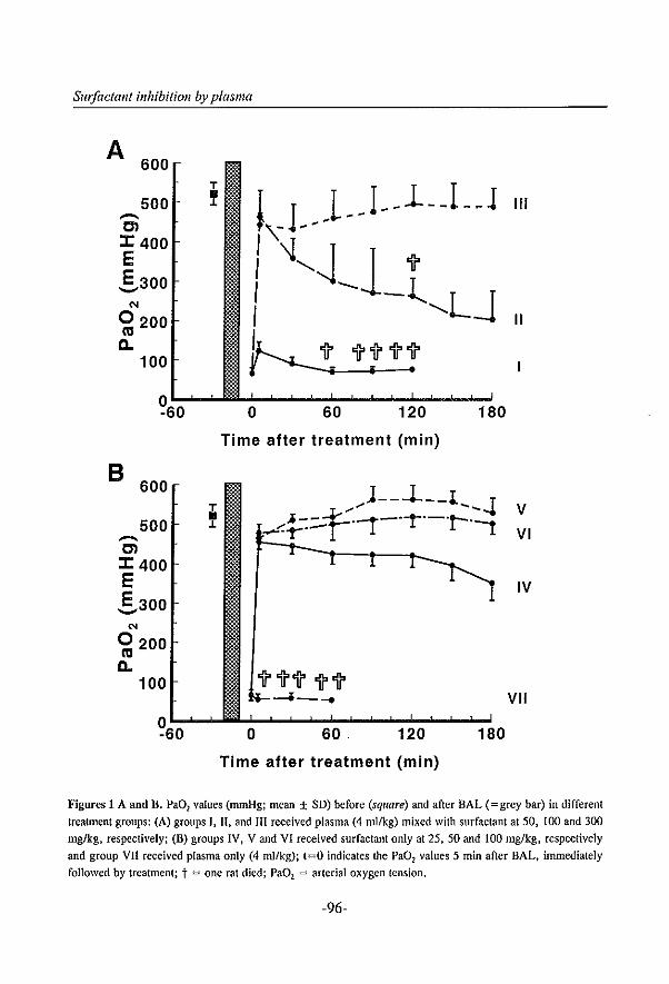
Sm!actant inhibition by plasma
A 600
T
500 • .L .-.. Cl :r: 400 E E 300 ....... '" 0200
III c..
100
~60 o 60 120 180
Time after treatment (min)
B 600
T • 500 .L .-.
j--...L--l. T ........ .... ........ ~.t::::...r·-r-·-r-·-·r-·l
Cl :r: 400 E E 300 --'" 0200 III c..
100
~60 0 60. 120 180
Time after treatment (min)
III
II
V
VI
IV
VII
Figures 1 A and B. Pa02 values (nunHg; mean ± SD) before (square) and after BAL (= grey bar) in different
treaUnent groups: (A) groups I, II, and III received plasma (4 mllkg) mixed with surfactant at 50, 100 and 300
mg/kg. respectively; (B) groups IV, V and VI received surfactant only at 25. 50 and 100 mg/kg, respectively
and group VII received plasma only (4 mllkg); t=O indicates the Pa02 values 5 min after BAL. immediately
followed by treatment; t = one rat died; Pa01 = arterial oxygen tension.
-96-
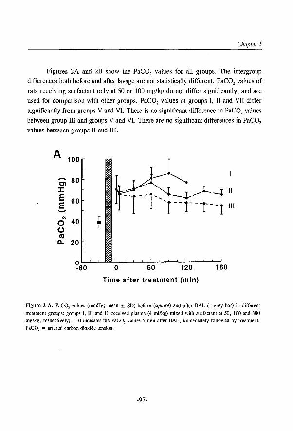
Chapter 5
Figures 2A and 2B show the PaCO, values for all groups. The intergroup
differences both before and after lavage are not statistically different. PaCO, values of
rats receiving surfactant only at 50 or 100 mg/kg do not differ significantly, and are
used for comparison with other groups. PaCO, values of groups I, II and VII differ
significantly from groups V and VI. There is no significant difference in PaCO, values
between group III and groups V and VI. There are no significant differences in PaCO,
values between groups II and III.
A 100
.-.. 80 Cl J: E 60 E --'" T 0 40 • (,.) L
III Il. 20
?60 o 60 120 180
Time after treatment (min)
Figure 2 A. PaC02 values (mmHg; mean ± SD) before (square) and after BAL (=grey bar) in different treatment groups: groups I, II, and III received plasma (4 ml/kg) mixed with surfactant at 50, 100 and 300 rug/kg, respectively; 1=0 indicates the PaC02 values 5 min after BAL, immediately followed by treatment; PaC01 = arterial carbon dioxide tension.
-97-
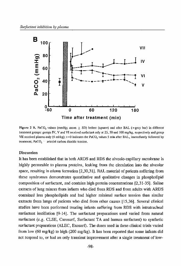
Swjactant inhibition by plasma
B 100
..-. Cl :I: E E
'"
80
60
o 40 C,.) C1I 0. 20
~60
;I; 1
o 60 120
Time after treatment (min)
VII
IV
VI
v
180
Figure 2 B. PaC02 values (mmHg; mean ± SD) before (square) and after BAL (=grey bar) in different treatment groups: groups IV, V and VI received surfactant only at 25,50 and 100 mglkg, respectively and group VJI received plasma only (4 mllkg); t=O indicates the PaeOl values 5 min after BAL, inmlediately followed by treatment; PaeOl = arterial carbon dioxide tension.
Discussion
It has been established that in both ARDS and RDS the alveolo-capillary membrane is
highly permeable to plasma proteins, leaking from the circulation into the alveolar
space, resulting in edema formation [2,30,31]. BAL material of patients suffering from
these syndromes demonstrates quantitative and qualitative changes in phospholipid
composition of surfactant, and contains high protein concentrations [2,31-35]. Saline
extracts of lung minces from infants who died from RDS and from adults with ARDS
contained less phospholipids and had higher minimal surface tension than sinlilar
extracts from lungs of patients who died from other causes [15,36]. Several clinical
studies have been performed treating infants suffering from RDS with intratracheal
surfactant instillation [9-14]. The surfactant preparations used varied from natural
surfactant (e.g. CLSE, Curosurf, Surfactant TA and human surfactant) to synthetic
surfactant preparations (ALEC, Exosurl). The doses used in Olese clinical trials varied
from low (60 mg/kg) to high (200 mg/kg). It has been reported that some infants did
not respond to, or had an only transient improvement after a single treatment of low-
-98-
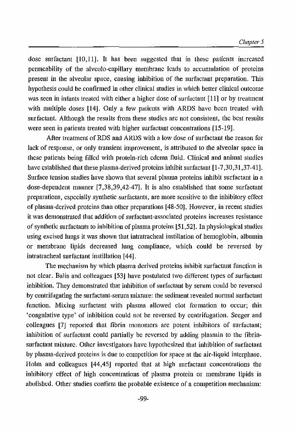
Chapter 5
dose surfactant [10,11]. It has been suggested that in these patients increased
permeability of the alveolo-capiIlary membrane leads to accumulation of proteins
present in the alveolar space, causing inhibition of the surfactant preparation. This
hypothesis could be confimled in other clinical studies in which better clinical outcome
was seen in infants treated with either a higher dose of surfactant [11] or by treatment
with multiple doses [14]. Only a few patients with ARDS have been treated with
surfactant. Although the results from these studies are not consistent, the best results
were seen in patients treated with higher surfactant concentrations [15-19].
After treatment of RDS and ARDS with a low dose of surfactant the reason for
lack of response, or only transient improvement, is attributed to the alveolar space in
these patients being fIlled with protein-rich edema fluid. Clinical and aninml studies
have established that these plasma-derived proteins inhibit surfactant [1-7,30,31,37-41].
Surface tension studies have shown that several plasma proteins inllibit surfactant in a
dose-dependent manner [7,38,39,42-47]. It is also established that some surfactant
preparations, especially synthetic surfactants, are more sensitive to the inhibitory effect
of plasma-derived proteins than other preparations [48-50]. However, in recent studies
it was demonstrated that addition of surfactant-associated proteins increases resistance
of synthetic surfactants to inhibition of plasma proteins [51,52]. In physiological studies
using excised lungs it was shown that intratracheal instillation of hemoglobin, albumin
or membrane lipids decreased lung compliance, which could be reversed by
intratracheal surfactant instillation [44].
The mechanism by which plasma derived proteins inhibit surfactant function is
not clear. Balis and colleagues [53] have postulated two different types of surfactant
inhibition. They demonstrated that inhibition of surfactant by sel1lm could be reversed
by centrifugating the surfactant-sel1lmmixture: the sedinlent revealed normal surfactant
function. Mixing surfactant with plasma allowed clot formation to occur; this
'coagulative type' of inhibition could not be reversed by centrifugation. Seeger and
colleagues [7] reported that fibrin monomers are potent inhibitors of surfactant;
inhibition of surfactant could partially be reversed by adding plasmin to the fibrin
surfactant mixture. Other investigators have hypothesized that inhibition of surfactant
by plasma-derived proteins is due to competition for space at the air-liquid interphase.
Holm and colleagues [44,45] reported that at high surfactant concentrations the
inhibitory effect of high concentrations of plasma protein or membrane lipids is
abolished. Other studies confirm the probable existence of a competition mechanism:
-99-
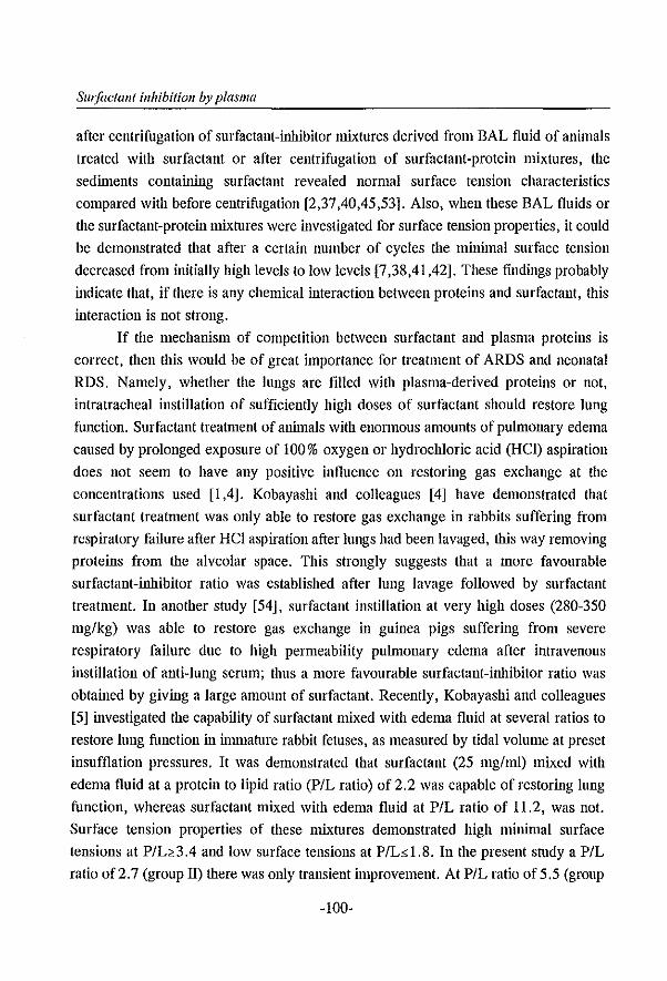
Swlae/allt inhibition by plasma
after centrifugation of surfactant-inhibitor mixtures derived from HAL fluid of animals
treated with surfactant or after centrifugation of surfactant-protein mixtures, the
sediments containing surfactant revealed normal surface tension characteristics
compared with before centrifugation [2,37,40,45,53]. Also, when these HAL fluids or
tile surfactant-protein mixtures were investigated for surface tension properties, it could
be demonstrated that after a certain number of cycles the minimal surface tension
decreased from initially high levels to low levels [7,38,41,42]. These fmdings probably
indicate that, if there is any chemical interaction between proteins and surfactant, this
interaction is not strong.
If tile mechanism of competition between surfactant and plasma proteins is
correct, then this would be of great importance for treatment of ARDS and neonatal
RDS. Namely, whether the lungs are filled with plasma-derived proteins or not,
intratracheal instillation of sufficiently high doses of surfactant should restore lung
function. Surfactant treatment of animals with enormous amounts of puhnonary edema
caused by prolonged exposure of 100% oxygen or hydrochloric acid (HCl) aspiration
does not seem to have any positive influence on restoring gas exchange at the
concentrations used [1,4]. Kobayashi and colleagues [4] have demonstrated that
surfactant treatment was oniy able to restore gas exchange in rabbits suffering from
respiratory failure after HCl aspiration after lungs had beenlavaged, this way removing
proteins from the alveolar space. This strongly suggests that a more favourable
surfactant-inhibitor ratio was established after lung lavage followed by surfactant
treatment. In another study [54], surfactant instillation at very high doses (280-350
mg/kg) was able to restore gas exchange in guinea pigs suffering from severe
respiratory failure due to high permeability pulmonary edema after intravenous
instillation of anti-lung serum; thus a more favourable surfactant-inhibitor ratio was
obtained by giving a large amount of surfactant. Recently, Kobayashi and colleagues
[5] investigated the capability of surfactant mixed with edema fluid at several ratios to
restore lung function in immature rabbit fetuses, as measured by tidal volume at preset
insufflation pressures. It was demonstrated that surfactant (25 mg/ml) mixed with
edema fluid at a protein to lipid ratio (P/L ratio) of 2.2 was capable of restoring lung
function, whereas surfactant mixed with edema fluid at P/L ratio of 11.2, was not.
Surface tension properties of these mixtures demonstrated high minimal surface
tensions at P/L~3.4 and low surface tensions at P/Ld.8. In the present study a P/L
ratio of2.7 (group II) there was only transient iroprovement. At P/L ratio of5.5 (group
-100-
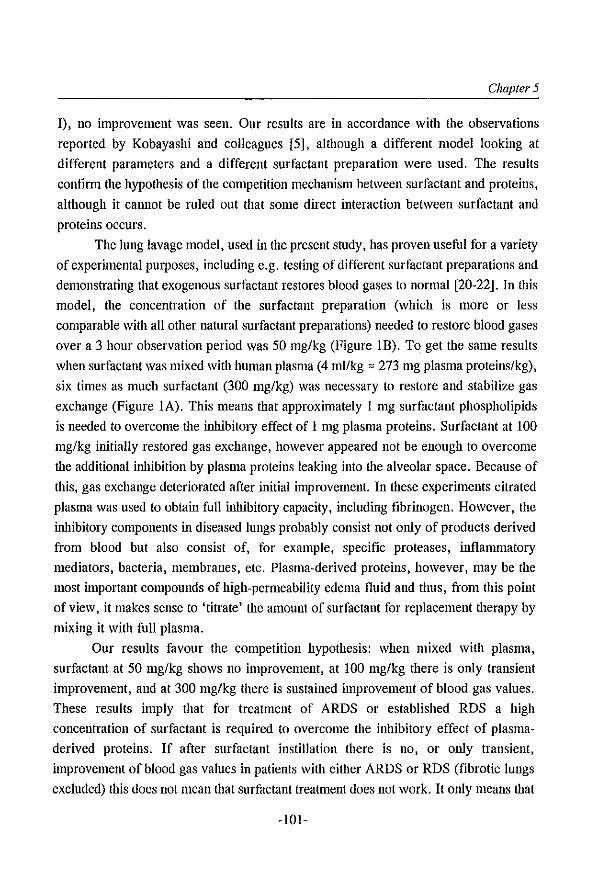
Chapter 5
I), no improvement was seen. Our results are in accordance with the observations
reported by Kobayashi and colleagues [5], although a different model looking at
different parameters and a different surfactant preparation were used. The results
confirm the hypothesis of the competition mechanism between surfactant and proteins,
although it call1lot be ruled out that some direct interaction between surfactant and
proteins occurs.
The lung lavage model, used in the present study, has proven nseful for a variety
of experimental purposes, including e.g. testing of different surfactant preparations and
demonstrating that exogenous surfactant restores blood gases to normal [20-22]. In this
model, the concentration of the surfactant preparation (which is more or less
comparable with all other natural surfactant preparations) needed to restore blood gases
over a 3 hour observation period was 50 mg/kg (Figure lB). To get the same results
when surfactant was mixed with human plasma (4 nll/kg " 273 mg plasma proteins/kg),
six times as much surfactant (300 mg/kg) was necessary to restore and stabilize gas
exchange (Figure lA). This means that approximately 1 mg surfactant phospholipids
is needed to overcome the inhibitory effect of 1 mg plasma proteins. Surfactant at 100
mg/kg initially restored gas exchange, however appeared not be enough to overcome
the additional inhibition by plasma proteins leaking into the alveolar space. Because of
this, gas exchange deteriorated after initial inlprovement. In these experiments citra ted
plasma was used to obtain full inhibitory capacity, including fibrinogen. However, the
inhibitory components in diseased lungs probably consist not DIlly of products derived
from blood but also consist of, for example, specific proteases, inflammatory
mediators, bacteria, membranes, etc. Plasma-derived proteins, however, may be the
most important compounds of high-permeability edema fluid and thus, from this point
of view, it makes sense to 'titrate' the amount of surfactant for replacement therapy by
mixing it with full plasma.
Our results favour the competition hypothesis: when mixed with plasma,
surfactant at 50 mg/kg shows no improvement, at 100 mg/kg there is only transient
improvement, and at 300 mg/kg there is sustained improvement of blood gas values.
These results imply that for treatment of ARDS or established RDS a high
concentration of surfactant is required to overcome the inhibitory effect of plasma
derived proteins. If after surfactant instillation there is no, or DIlly transient,
improvement of blood gas values in patients with either ARDS or RDS (fibrotic lungs
excluded) this does not mean that surfactant treatment does not work. It only means that
-101-
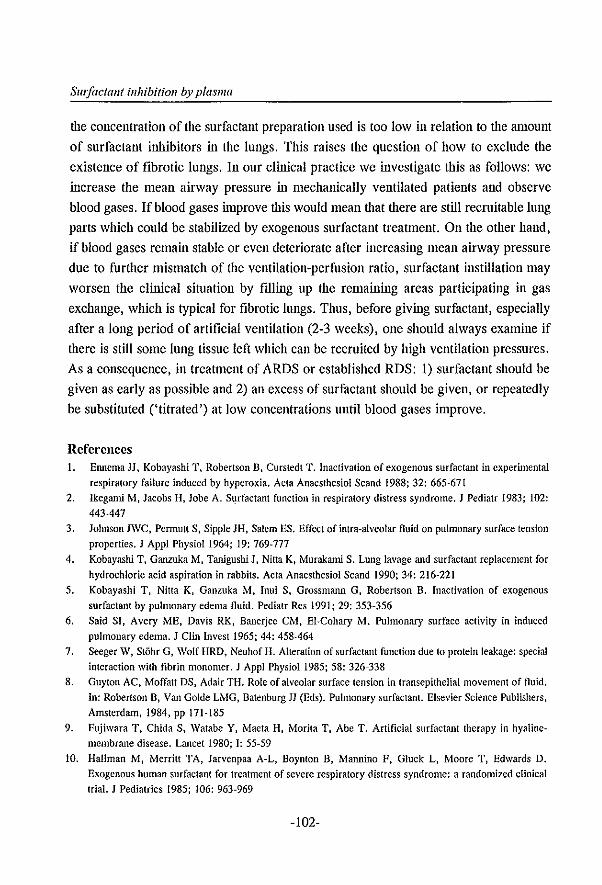
SIIl/actallt inhibition by plasma
the concentration of the surfactant preparation used is too low in relation to the amount
of surfactant inhibitors in the lungs. This raises the question of how to exclude the
existence of fibrotic lungs. In our clinical practice we investigate this as follows: we
increase the mean airway pressure in mechanically ventilated patients and observe
blood gases. If blood gases improve this would mean that there are still recruitable lung
parts which could be stabilized by exogenous surfactant treatment. On the other hand,
if blood gases remain stable or even deteriorate after increasing mean airway pressure
due to further mismatch of the ventilation-perfusion ratio, surfactant instillation may
worsen the clinical situation by filling up the remaining areas participating in gas
exchange, which is typical for fibrotic lungs. Thus, before giving surfactant, especially
after a long period of artificial ventilation (2-3 weeks), one should always examine if
there is still some lung tissue left which can be recruited by high ventilation pressures.
As a consequence, in treatment of ARDS or established RDS: 1) surfactant should be
given as early as possible and 2) an excess of surfactant should be given, or repeatedly
be substituted (,titrated') at low concentrations until blood gases improve.
References I. Ennema JJ, Kobayashi T, Robertson n, Curstedt T. Inactivation of exogenous surfactant in experimental
respiratory failure induced by hyperoxia. Acta Anaesthesiol Scand 1988; 32: 665·671 2. lkegami rvl, Jacobs H, Jobe A. S~rfactant function in respiratory distress syndrome. J Pedialr 1983; 102:
443-447
3. Johnson JWC, Pennutt S, Sipple JH, Salem ES. Effect of intra-alveolar fluid on pulmonary surface tension properties. J Appl PhysioI1964; 19: 769-777
4. Kobayashi T, GrulZUka M, Tanigushi J, Nitta K, Murakami S. Lung lavage and surfactant replacement for hydrochloric acid aspiration in rabbits. Acta Anaesthesiol Scand 1990; 34: 216-221
5. Kobayashi T, Nitta K, Ganzuka M, Inui S, Grossmann G, Robertson B. Inactivation of exogenous surfactant by pulmonary edema fluid. Pediatr Res 1991; 29: 353-356
6. Said SI, Avery ME, Davis RK, Brulerjee CM, EI-Cohary M. Pulmonary surface activity in induced pulmonary edema. J Clio Invest 1965; 44: 458-464
7. Seeger W, Stohr G, Wolf HRD, Neuhof H. Alteration of surfactant function due to protein leakage: special interaction with fibrin monomer. J AppJ Physiol 1985; 58: 326-338
8. Guyton AC, Moffatt DS, Adair TH. Role of alveolar surface tension in transepithelial movement of fluid. In: Robertson B, Van Golde LMG, Batenburg 11 (Eds). Pulmonary surfactant. Elsevier Science Publishers, Amsterdam, 1984, pp 171·185
9. Fujiwara T. Chida S, Watabe Y, Maeta H, Morita T, Abe T. Artificial surfactant therapy in hyalinemembrane disease. Lancet 1980; I: 55-59
10. Hallman M. Merritt TA, Jarvenpaa A-L, Boynton B, Mallllmo F. Gluck L, Moore T. Edwards D. Exogenous human surfactant for treatment of severe respiratory distress syndrome: a randomized clinical trial. J Pediatrics 1985; 106: 963-969
-102-
Chapter 5
11. Konishi M. Fujiwara T, Naito T, Takeuchi Y, Ogawa Y, Inukai K, Fujimura M, Nakamura H, Hashimoto
T, Surfactant replacement therapy in neonatal respiratory distress syndrome: a multi-centre, randomized clinical trial: comparison of high- versus low-dose of Surfactant TA. Eur J Pediatr 1988; 147: 20-25
12. Collaborative European Multicenter Study Group. Surfactant replacement therapy for severe neonatal
respiratory distress syndrome: an international randomized clinicallriaL Pediatrics 1988; 82: 683-691 13. Ten Centre Study Group. Ten centre trial of artificial surfactant (artificial lung expanding compound) in
very premature babies. Be Med J 1987; 294: 991-996 14. Dunn MS, Shennan AT, Possmayer F. Single- versus multiple-dose surfactant replacement therapy in
neonates of 30 to 36 weeks' gestation with respiratory distress syndrome. Pediatrics 1990; 86: 564-571 15. Lachmann B. The role of pulmonary surfactant in the pathogenesis and therapy of ARDS. In: Vincent JL
(Ed). Update in intensive care and emergency medicine. Springer-Verlag, Berlin, 1987, pp 123-134 16. Nosaka S, Sakai T, Yonekura M, Yoshikawa K. Surfactant for adults with respiratory failure. Lancet 1990;
[: 947-948 17. Richman PS, Spragg RG t Robertson B, Merritt TA, Curstedt T. The adult respiratory distress syndrome:
first trials with surfactant replacement. Eur Respir J 1989; 2(SuppJ 3): 109s-llls
18. Marraro G. Respiratory emergencies and supplementary surfactant in the treatment of severe RDS in leukaemic adolescents. In: Cosmi EV, Di Renzo GC t Anceschi MM (Eds). The surfactant system of the lung: prevention and treatment of neonatal and adult respiratory distress syndrome. t"facmillan Press, London, 1991, pp 198-206
19. Joka Th, Obertacke U. Neue medikamentose Behandlung im ARDS: effekt ciner intrabronchialen xenogenen Surfactantapplikation. Z Herz Thorax GeHillchir 1989; 3(Suppll): 21-24
20. Lachmann B, Robertson B, Vogel J. In vivo lung lavage as an experimental model of the respiratory distress syndrome. Acta Anaesthesiol Scand 1980; 24: 231-236
21. Berggren P t Lachmann Bt Curstedt T, Grossmann G, Robertson B. Gas exchange and lung morphology after surfactant replacement in experimental adult respiratory distress syndrome induced by repeated lung
lavage. Acta Anaesthesiol Scand 1986; 30: 321-328 22. Robertson B, Lachmann B. Experimental evaluation of surfactants for replacement therapy. Exp Lung Res
1988; 14: 279-310 23. Lachmann B, Jonson D. Lindroth M, Robertson B. Modes of artificial ventilation in severe respiratory
distress syndrome. Lung function and morphology in rabbits ailer wash-out of alveolar surfactant. Crit Care Med [982; 10: 724-732
24. Metcalfe IL, Enhorning G, Possmayer F. Pulmonary surfactant-associated proteins: their role in the expression of surface activity. J Appl Physiol 1980; 49: 34-41
25. Van Daal GJ, So KL, Gonmlers D, Eijking EP, Fievez RB, Sprenger MJ, Van Dam DW, Lachmann B. Intratracheal surfactant administration restores gas exchange in experimental adult respiratory distress
syndrome associated with viral pneumonia. Anesth Analg 1991; 72: 589·595 26. Eijking EP, Van Daal GJ, Tenbrinck R. Luijendijk A, Sluiters JF, Harlllappei E, Lachmann B. Effect of
surfactant replacement on plleulI/ocystis carillii pneumonia in rats. Intensive Care Med 1991; 17: 475-478 27. Van Daal GJ, Bas JAH, Eijking EP, Gammers D, Hannappei E, Lachmann B. Surfactant replacement
therapy improves pulmonary mechanics in end-stage influenza A pneumonia in mice. Am Rev Respir Dis 1992; 145: 859-863
28. Bas AP, Tibboel D, Hazebroek FWJ. Molenaar JC, Lachmann B, Gonuners D. Surfactant replacement therapy in high-risk congenital diaphragmatic hernia. Lancet 1991; I: 1279
29. Markwell MAK, Haas SM, Bieber LL, Tolbert NE. A modification of the Lowry procedure to simplify
-103-
Sw/actallt i1lhibition by plasma
protein determination in membrane and lipoprotein samples. Anal Biochem 1978; 87: 206-210
30. Robertson B, Berry D, Curstedt T, Grossmann G, Ikegami M, Iacobs H, lobe A, lanes S. Leakage of
protein in the immature rabbit lung; effect of surfactant replacement. Respir Physiol 1985; 61: 265-276
31. Hallman M, Spragg R, Harrell IH, Moser KM, Gluck L. Evidence of lung function abnormality in
respiratory failure, Study of bronchoalveolar lavage phospholipids, surface activity phospholipase activity,
and plasma myoinositoJ. I Clin Invest 1982; 70: 673-683
32. Gregory Ti, Longmore WI, Moxley MA, Whitsett lA, Reed CR, Fowler AA, Hudson LD, Maunder RI,
Crim C, Hyers TM. Surfactant chemical composition and biophysical activity in acute respiratory distress
syndrome. J Clin Invest 1991; 88: 1976-1981
33, Petty TL, Reiss OK, Paul GW, Silvers GW, Elkins ND. Characteristics of pulmonary surfactant in adult
respiratory distress syndrome associated with trauma and shock. Am Rev Respir Dis 1977; 115: 531-536
34. Pison U, Seeger W, Buchhorn R. Ioka T, Brand M, Obertacke U, Neuhof H, Schmit-Neuerburg KP.
Surfactant abnonnalities in patients with respiratory failure after multiple trauma. Am Rev Respir Dis 1989;
140; 1033-1039
35. Von Wichert P, Kohl FV. Decreased dipalmitoyllecithin content found in lung specimens from patients with
so-called shock-lung. Intensive Care Med 1977; 3: 27-30
36. Avery ME, Mead 1. Surface properties in relation to atelectasis and hyaline membrane disease. Am J Dis
Child 1959; 97; 517-523
37. Ikegami M, lobe A, Glatz T. Surface activity following natural surfactant treatment of premature lambs.
J App1 Physiol1981; 51; 306-312
38. Ikegami M, lobe A, Iacobs H, Lam R. A protein from airways of premature lambs that inhibits surfactant
function. J App1 Physiol1984; 57; 1134-1142
39. Ikegami M, lobe A, Berry D. A protein that inhibits surfactant in respiratory distress syndrome. BioI
Neonate 1986; 50: 121-129
40. Kobayashi T, Curstedt T, Grossmann G, Robertson B. Inhibition of exogenous surfactant in ventilated
immature newborn rabbits. Respir Physiol1989; 76: 1-12
41. Tierncy DF, 10hnson RP. Altered surface tension of lung extracts and lung mechanics. I Appl Physiol
1965; 20; 1253-1260
42. Fuchimukai T, Fujiwara T, Takahashi A, Enhorning G. Artificial pulmonary surfactant inhibited by
proteins. I Appl Physiol1987; 62: 429-437
43. Holm BA, Notter RH, Finkelstein IN. Surface property changes from interactions of albumin with natural
lung surfactant and extracted lung lipids. Chern Phys Lipids 1985; 38: 287-298
44. Holm BA, Notter RH. Effects of hemoglobin and cell membrane lipids on pulmonary surfactant activity.
J App1 Physio1 1987; 63; 1434-1442
45. Holm BA, Enhorning G, Notter RH. A biophysical mechanism by which plasma proteins inhibit lung
surfactant activity. Chern Phys Lipids 1988; 49: 49-55
46. Keough KMW, Parsons CS, Phang PT, Tweedale MG. Interactions between plasma proteins and pulmonary
surfactant: surface balance studies. Can I Physiol Pharmacol 1988; 66: 1166-1173
47. Taylor lr. FB, Abrams ME. Effect of surface active lipoprotein on clotting and fibrinolysis, and of
fibrinogen on surface tension of surface active lipoprotein (with a hypothesis on the pathogenesis of
pulmonary atelectasis and hyaline mcmbrane in respiratory distress syndrome of the newborn). Am 1 Med
1966; 40; 346-350
48. Holm BA, Venkitaraman AR, Enhorning G, Notter RH. Biophysical inhibition of synthetic lung surfactants.
Chern Phys Lipids 1990; 52: 243-250
-104-
Chapter 5
49. Ikegami M, Jobe A, Jacobs H, Jones S, Sequential treatments of premature lambs with an artificial
surfactant and natural surfactant. J Clio Invest 1981; 68: 491-496
50. Ikegami M. Agata Y, Elkady T, Hallman M, Berry D. Jobe A. Comparison of four surfactants: in vitro
surface properties and responses of preterm lambs to treatment at birth. Pediatrics 1987; 79: 38-46
51. Cockshutt AM, Weitz J, Possmayer F. Pulmonary surfactant-associated protein A enhances the surface
activity of lipid extract surfactant and reverses inhibition by blood proteins in vitro. Biochemistry 1990;
29: 8424-8429
52. Seeger W, Gunther A, Thede C. Differential sensitivity (0 fibrinogen inhibition of SP-C- vs, SP-B-based
surfactants, Am J Physiol 1992; 5: L286.L291
53. Balis JV, Shelley SA, McCue MJ, Rappaport ES. Mechanisms of damage to the lung surfactant system.
Ultrastructure and quantitation of normal and in vitro inactivated lung surfactant. Exp Mol Pathol 1971;
14: 243-262 54. Lachmann B, Hallman M, Bergmann KC. Respiratory failure following anti-lung serum: study on
mechanisms associated with surfactant system damage. Exp Lung Res 1987; 12: 163-180
-105-

Chapter 6
Bronchoalveolar lavage with a diluted surfactant
suspension prior to surfactant instillation improves
the effectiveness of surfactant therapy in
experimental acute respiratory distress syndrome
(ARDS)
D. Gammers, E.P. Eijking, K.L. So, A. van 't Veen, and B. Lachmallll
Dept. of Anesthesiology, Erasmus University Rotterdam, The Netherlands
Published ill: Intel/sive Care Med 1998,' 24: 494-500 Reprillfed with pennissioll (copyright holder: Springer-Verlag GmbH)
-107-
Sur/he/ant administration
Summary
Objective: To assess whether bronchoalveolar lavage (BAL) with a diluted surfactant
suspension prior to surfactant instillation prevents dIe only transient improvement in
lung function as reported after surfactant instillation in severe acute respiratory distress
syndrome (ARDS).
Design: Randomized, prospective, experimental study.
Setting: Laboratory and animal facility of a large university.
Materials: Adult male Sprague-Dawley rats (280±30 g).
IlIfervellfiollS: All animals underwent repetitive whole lung saline lavage to induce acute
lung injury. Then, animals were randomly divided into seven study groups: the first
group received surfactant (150 mg/kg) within 10 min after the last lavage (early
treatment), whereas in the other six groups mechanical ventilation was continued for
3 h before treatment (late treatment). Treatment consisted of: surfactant instillation at
a dose of 150 mg/kg; at a dose of 250 mg/kg; BAL with saline; BAL with a diluted
surfactant suspension (2.5 mg/ml); BAL with saline, immediately followed by
surfactant instillation (150 mg/kg); and BAL with a diluted surfactant suspension (2.5
mg/kg), immediately followed by surfactant instillation (150 mg/kg).
Measuremellfs and results: Blood gases were measured for 6 h and then BAL was
perfonned to measure protein concentration and surface tension properties. Mean PaD,
values increased inmlediately after surfactant instillation to pre-lavage values and
remained stable only in the group that received surfactant immediately after lavage
procedure and the group that underwent BAL with a diluted surfactant suspension prior
to surfactant instillation.
COllclusion: BAL with a diluted surfactant suspension prior to surfactant instillation at
a later tinle point in lung injury resulted in a stable improvement of lung function. This
improvement is comparable with the results seen after surfactant instillation
il11lllcdiately after lung lavage.
-108-
Chapter 6
Introduction
In worldwide clinical trials, surfactant therapy has been successfully used in newborns
with respiratory distress syndrome (RDS) and mortality has been decreased by 30-40 % [1]. Despite substantial evidence for surfactant dysfunction in acute respiratory distress
syndrome (ARDS), only a few case reports and results of linlited clinical studies are
available in which adult patients with ARDS were treated with exogenous surfactant
[2]. Although these reports showed that some patients did not respond to, or had only
a transient inlprovement after, a single dose of surfactant, better results were seen when
large or mUltiple doses were instilled (up to 400-500 mg of a natural surfactant per kg
body weight) [2-5]. It has been suggested that the increased pefl1leability of the alveolo
capillary membrane in ARDS leads to accumulation of plasma proteins in the alveolar
space, causing inhibition of the instilled surfactant in a dose-dependent way [6]. Thus,
for treatulent of ARDS, high cOllcentration of surfactant is required to overcome the
i1thibitory effect of the plasma proteins. However, the prohibitive price and the non
availability of large amounts of surfactants make surfactant therapy infeasible in adults
at present [2].
Experience in neonates has taught us that exogenous surfactant therapy is more
effective when administration takes place in the early stages of RDS [I]. Therefore, it
is expected that early treatment of ARDS may thus require smaller amounts of
exogenous surfactant due to smaller amounts of surfactant inhibitors. Indeed,
experilnental studies have demonstrated that admillistration of exogenous surfactant at
an early tinle point in lung injury resulted in stable improvement of lung function,
whereas transient i1nprovement of lung function was found with the later treatments
[7,8]. Further, Kobayashi and colleagues [9] have demonstrated, in a model of acid
aspiration, that removal of surfitctant inhibitors by bronchoalveolar lavage (BAL) with
saline prior to administration of surfactant was necessary to i1nprove arterial
oxygenation. Lung lavage with saline, however, can damage pulmonary function [10].
Therefore, we performed BAL with a diluted surfactant suspension and found that this
approach resulted in a superior response compared to exogenous surfactant
administration with or without previous BAL Witll saline [11]. In that study, however,
the effect of the different surfactant reginles on lung function was studied for only 1
hour and there was no group which received a combination of BAL with a diluted
surfactant suspension and exogenous surfactant [11]. This study was therefore designed
to evaluate the effect of different surfactant treatment regimes on lung function at a
-109-
Surfactant administration
later time point in lung injury compared with administration of surfactant directly after
the repeated lung lavage; tile combination of BAL WiOl a diluted surfactant suspension,
immediately followed by exogenous surfactant administration was of particular interest.
Materials and methods
This study was approved by the Institutional Animal Committee at the Erasmus
University Rotterdam; care and handling of the animals were in accord Witll the
European Community guidelines (86/609/EEG). The studies were performed in male
Sprague-Dawley rats (n=52) with a bodyweight of280±30 g (Harlan CPB, Zeist, The
Netherlands). After induction of anaesthesia with nitrous oxide, oxygen and halothane
(66/33/1-2%), a polyethylene catheter (0.8-mm outer diameter) was inserted into a
carotid artery for drawing arterial blood samples. Before tracheotomy, the aninlals
received pentobarbital sodium (Nembutal'; 60 mg/kg, Lp.). After a metal cannula was
inserted into the trachea, muscle relaxation was given with pancuronium bromide
(Pavulon'; 2 mg/kg, Lm.) and the animals were immediately cOlmected to the
ventilator. The animals were mechanically ventilated with a Servo Ventilator 900 C
(Siemens-Elema AB, Solna, Sweden) in parallel (6 animals simultaneously) in a
pressure-control mode with the following ventilator settings: frequency of 30
breaths/min, positive end-expiratory pressure (PEEP) of 2 cm H,o, inspiratory
/expiratory ratio of 1:2, PiO, of 1.0, and a peak inspiratory pressure of 10-12 cm H,O
to keep PaCO, between 30 to 40 nnnHg. Anesthesia was maintained with hourly
injection of pentobarbital sodium (Nembutal'; 60 mg/kg/h); muscle relaxation was
achieved with hourly injection of pan cur onium bromide (Pavulon'; 2 mg/kg/h). Body
temperature was kept within normal range by means of a heating pad.
Initially, the peak pressure was increased to 20 cm H,o for I min to open up
atelectatic regions in the lungs and after reaching steady state, blood gases were
measured. Next, respiratory failure was induced by repeated bronchoalveolar lavage
(BAL) according to Lachmann et al. [10]. Each lavage was performed with saline (32
mllkg) heated to 37°C. Just before the first lavage, peak inspiratory pressure and PEEP
were elevated to 26 and 6 cm H,O, respectively. Lung lavage was repeated 4-5 times
with 5 min intervals to achieve a PaO, s 125 nmilig. After the blood gases had reached
a steady state, they were measured and this was defined as tinle zero (t=O). Then,
animals were randOmly divided into seven groups of six animals: the first group
received exogenous surfactant (150 mg/kg) within 10 min (early treatment), whereas
-110-
Chapter 6
in the other six groups mechanical ventilation was continued for 3 h before treatment
(late treatment; t=3 h). Treatment consisted of: exogenous surfactant (150 mg/kg);
exogenous surfactant (250 mg/kg); BAL with saline; BAL with a diluted surfactant
suspension (2.5 mg surfactant per mI saline); BAL with saline, immediately followed
by exogenous surfactant (150 mg/kg); and BAL with a diluted surfactant suspension
(2.5 mg surfactant per mI, from which 0.7 mg/ml could be detected in the recovered
BAL fluid), innnediately followed by exogenous surfactant (150 mg/kg; in total 210
mg/kg). The amount of saline used in BAL was 32 ml/kg at 37 ·C. The phospholipid
content of the diluted surfactant suspension was determined by phosphorus analysis
before and after one lavage [12].
A freeze-dried natural surfactant was used, isolated from pig lungs as previously
described [13]. It consists of approxinlately 90-95% phospholipids, 1 % hydrophobic
proteins (surfactant-proteins Band C) and 1 % free fatty acids, the remainder being
neutral lipids; there is no surfactant-protein A in this surfactant preparation. The
surfactant powder was suspended in warm saline (37 ·C) at a concentration of 40
mg/ml. For instillation of the surfactant, aninlals had been disconnected from the
ventilator and received 1-2 mI of the surfactant suspension directly into the endotracheal
tube followed by insufflation of 28 ml/kg of air via a syringe. After surfactant
inEtillation, animals were inmlediately reconnected to the ventilator (ventilator settings
were not changed).
During the observation period, arterial blood samples were collected at the
following times: 5 min and 1, 2, 3 h after the lavage procedure; and 15 min after
treatment (late treatment) and every hour for 3 h. Blood samples of 0.20 ml were taken
and replaced by heparinized saline (5 !E/ml). Of each blood sample PaO" PaCO" and
pH were measured by conventional methods (ABL Thl 505, Radiometer A/S,
Copenhagen, Denmark).
At the end of the observation period, all animals were killed with an overdose
of pentobarbital sodium and then BAL was performed for measurements of total protein
concentration and surface tension properties. The lungs were lavaged once with saline
(32 mllkg) and cell material was removed by centrifugation (10 min at 300 g). The
lungs of those animals which died after late treatment were lavaged one time
inllllediately after death to get material for protein and surface tension measurements.
The protein concentration was determined nsing the modified Lowry method [14], with
bovine serum albumin as standard. The minimal surface tension of the BAL fluid was
-1l1-
Surfactant administration
measured using a modified Willlelmy balance (E. Biegler GmbH, Mauerbach, Austria)
by applying 100 ilL of BAL fluid to the surface of a saline-filled trough [15]. The
surface area was compressed and expanded with a cycling time of 3 min per cycle and
maximum and minimum surface areas of 64 and 12.8 em', respectively (100% and
20%). The minimal surface tension was measured after 3 cycles at 20% surface area,
and is expressed as milli Newton/metre (mN/m).
Analysis of variance (ANOV A) was used to assess whether there was an overall
difference within or between the groups. If a difference was found, a post hoc test was
used (Student-Newman-Keuls' multiple comparison procedure). Statistical significance
was accepted at p~0.05. All data are reported as mean ± standard deviation (SD).
Results
Blood gases before and after the repeated saline lavage were comparable in all animals
(Figure 1 and Table 1). Before late treatment (t=3 h), 10 animals died due to
insufficient gas exchange and these animals were excluded from the study and not
included in the statistical analyses. The total protein concentration and the minimal
surface tension of the BAL fluid obtained 3 h after the repeated saline lavage were
significantly increased compared to the first BAL fluid of the repeated saline lavage
which served as controls (Table 2).
Mean PaD, values increased rapidly to pre-lavage values in the group which
received exogenous surfactant (150 mg/kg) within 10 min after the last lavage (early
treatment) and remained stable during tlle subsequent 6 h study period (Figure 1). In
the four groups which received a bolus of surfactant (with or without previous BAL)
3 h after the repeated saline lavage (late treatment), mean PaD, values increased
immediately to pre-lavage values but remained stable only in the group that underwent
BAL with a diluted surfactant suspension prior to administration of surfactant. In the
other three groups (surfactant 150 mg/kg; surfactant 250 mg/kg; BAL with saline and
surfactant 150 mg/kg), mean PaD, values decreased over time and were significantly
lower compared to the group that underwent BAL with a diluted surfactant suspension
prior to administration of surfactant at the end of the observation period (Figure 1). In
addition, there were no significant differences in mean PaD, values between these 3
groups (surfactant (150 mg/kg); surfactant (250 mg/kg); and BAL with saline together
with surfactant) during the subsequent 3 h. Further, in the group in which the lungs
were lavaged once with a diluted surfactant suspension, mean PaD, values initially
-112-
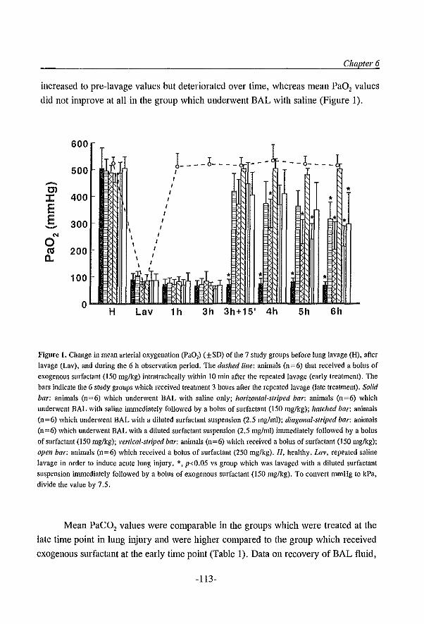
Chapler 6
increased to pre-lavage values but deteriorated over time, whereas mean PaO, values
did not improve at all in the group which underwent BAL with saline (Figure I).
600
L--6---- ---b---_ 500 I
I ...... I OJ • I :I: 400 I • E I
E 300 ....... N \
I
0 \ I
ra 200 \ I
a.. \ I
100
0 H Lav 1h 3h 3h+15' 4h 5h 6h
Figure 1. Change in mean arterial oxygenation (PaOl ) (±SD) of the 7 study groups before lung lavage (H), after
lavage (Lav), and during the 6 h observation period. TIle dashed Iille: animals (n=6) that received a bolus of
exogenous surfactant (150 mg/kg) intratracheally within 10 min after the repealed lavage (early treatment). The
bars indicate the 6 study groups which received treatment 3 hours after the repeated lavage (late treatment). Solid bar: animals (0=6) which underwent BAL with saline only; horizontal-striped bar: animals (n=6) which
underwent BAL with saline immediately followed by a bolus of surfactant (150 mg/kg); hatched bar: animals
(n=6) which underwent BAL with a diluted surfactant suspension (2.5 mg/ml); diagonal-striped bar: animals
(n=6) which underwent BAL with a diluted surfactant suspension (2.5 mg/ml) immediately followed by a bolus
of surfactant (150 rug/kg); vertical-stn'ped bar: animals (n=6) which received a bolus of surfactant (150 mglkg);
open bar: animals (n=6) which received a bolus of surfactant (250 mg/kg). H, healthy. Lav, repeated saline
lavage in order to induce acute lung injury. *, ps.O.05 vs group which was lavaged with a diluted surfactant
suspension immediately followed by a bolus of exogcnous surfactant (150 mg/kg). To convert mmHg to kPa.
dividc the value by 7.5.
Mean PaC02 values were comparable in the groups which were treated at the
late time point in lung injury and were higher compared to the group which received
exogenous surfactant at the early time point (Table 1). Data Oll recovery of BAL fluid,
-113-
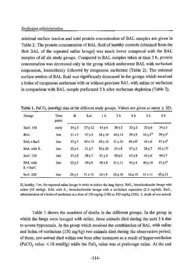
SwIaetant administration
minimal surface tension and total protein concentration of BAL samples are given in
Table 2. The protein concentration of BAL fluid of healtlly controls (obtained from the
first BAL of the repeated saline lavage) was much lower compared with the BAL
samples of all six study groups. Compared to BAL samples taken at time 3 h, protein
concentration was decreased only in the group which underwent BAL with surfactant
suspension, illlll\ediately followed by exogenous surfactant (Table 2). The minimal
surface tension of BAL fluid was significantly decreased in the groups which received
a bolus of exogenous surfactant with or without previous BAL with saline or surfactant
in comparison with BAL sample performed 3 h after surfactant depletion (Table 2).
Table 1. PaCO, (mmHg) data of the different study groups. Values are given as mean + SD.
Group: Time H Lav 1 h 3 h 4h Sh 6h
point:
Surf. ISO early 34±S S7±12 43±4 38±3 35±3 33±6 34±S
BAL late 31±3 47±S S2±l0 42±l3 39±9 35±3lt 39±4f
BAL+Surf. late 37±3 48±l3 49±16 SI±l0 40±St 44±6 47±4'
BALwith S. late 32±4 SIH SO±lO S2±8 47±S 38±7 42± IS'
Surf. 150 late 37±6 S8±7 SI±9 48±6 43±8 45±6 44±7
BALwith late 32±S 49±9 49±8 SI±ll 46±4 40±10 4l±3'
S. +Surf.
Surf. 2S0 late 29±4 Sl±l3 45±9 S3±lD S6±1D 51Hl 45±l3
H, healthy. Lav, the repeated saline lavage in order to induce the lung injury. BAL, bronchoalveolar lavage with saline (32 ml/kg). BAL with S., bronchoalveolar lavage with a surfactant supension (2.5 mg/ml). Surf., administration ofa bolus of surfactant at a dose of 150 mg/kg (150) or 250 mg/kg (250). t, death of one animal.
Table 1 shows the numbers of deaths in the different groups. In the group in
which the lungs were lavaged with saline, three animals died during the next 3 h due
to severe hypoxemia. In the group which received the combination of BAL with saline
and bolus of surfactant (150 mg/kg) two aninlals died during the observation period;
of these, one animal died within one hour after treatment as a result of hyperventilation
(PaCO, value < 18 mmHg) while the PaO, value was at prelavage value. At the end
-114-
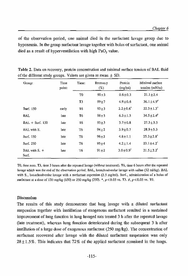
Chapter 6
of the observation period, one animal died in the surfactant lavage group due to
hypoxemia. In the group surfactant lavage together with bolus of surfactant, one animal
died as a result of hyperventilation with high PaO, value.
Table 2, Data on recovery, protein concentration and minimal surface tension of BAL fluid
of the different study groups. Values are given as mean ± SD.
Group: Time Time: Recovery Protein Minimal surface point: (%) (mg/ml) tension (mN/m)
TO 90±3 0.6±O.3 21.1 ±2.4
T3 99±7 4.9±O.6 36.1 ±4.9'
Surf. 150 early T6 93±3 2.2±0.6' 22.3±1.3'
BAL late T6 98±3 6.3±1.3 34.5±2.4'
BAL + Surf. ISO late T6 93±3 3.7±O.8 27.5±3.S
BAL with S. late T6 94±2 3.9±O.7 28.9±S.3
Surf. 150 late T6 96±2 4.8±1.I 2S.3±2.8'
Surf. 250 late T6 9S±4 4.2±1.4 23.1±4.2'
BALwith S. + laic T6 91±2 3.0±0.9' 21.5±2.2' Surf.
TO, time zero. TI, time 3 hours after the repealed lavage (without treatment), T6, lime 6 hours after the rcpeated lavage which was the end of the observation period. BAL, bronchoalveolar lavage with saline (32 ml/kg), BAL with S., bronchoalveolar lavage with a surfactant supension (2.5 mg/ml). Surf., administration of a bolus of surfactant at a dose of 150 mg/kg (150) or 250 rug/kg (250). *, p<O.OS VS. 1'3. #, p<O.05 vs. TO.
Discussion
The results of this study demonstrate that lung lavage with a diluted surfactant
suspension together with instillation of exogenous surfactant resulted in a sustained
improvement of lung function in lung lavaged rats treated 3 h after the repeated lavage
(late treatment), whereas lung function deteriorated during the subsequent 3 h after
instillation of a large dose of exogenous surfactant (250 mg/kg). The concentration of
surfactant recovered after lavage with the diluted surfactant suspension was only
28 ± 1.5 %. This indicates that 72 % of the applied surfactant remained in the lungs.
-115-
Surfactant administration
BAL was performed with 32 1lI1 per kg and the surfactant content of the surfactant
lavage was 2.5 mg/m1. Probably, a dose of approximately 60 mg/kg of surfactant has
been additionally applied by the lavage procedure. In other words, the group receiving
BAL with the diluted surfactant suspension and a bolus of surfactant (150 mg/kg) has
received In total a dose of21O mg/kg. Thus, despite that the latter gronp received 15-
20% less surfactant, the Inlprovement of arterial oxygenation at the end of the
observation period was significantly better compared to the 250 mg/kg bolus group.
Instillation of exogenous surfactant (150 mg/kg) directly after the repeated saline
lavage (early treatment) resulted In stable Inlprovement of lung function during the 6
h observation period. These data correspond to previous investigations in which the
therapeutic effects of exogenous surfactant improved when administration of surfactant
takes place in the early stages of lung injury [7,8]. It has been established that the
transient effect after surfactant dlerapy is due to inactivation of the instilled surfactant
by plasma-proteins and inflanllllatory mediators such as oxygen radicals, proteases and
phopholipases [for review see (6)]. The purpose ofBAL prior to exogenous surfactant
therapy is the removal of these potent surfactant inhibitors. It is known, however, that
BAL with saline can harm the lungs and dlis was confirmed by the results of this study
which showed that dnee of the six animals in the group that underwent BAL with saline
died within the observation period due to insufficient gas exchange (Table 1).
The improvement of arterial oxygenation after BAL with a diluted surfactant
suspension was only transient because dIe amount of surfactant remaining in the lungs
was low (about 60 mg/kg) and it has been shown that additional surfactant substitution
is needed to maintain blood gases at pre-lavage values (Figure 1). In contrast to our
results, Balaraman et ai. [16] demonstrated dmt administration of surfactant by lavage
is more effective in improving lung function compared to normal bolus instillation in
lung lavaged piglets. In that study, however, an artificial surfactant was used, which
is protein-free, whereas in the present study a natural surfactant was used, containing
1-2% of the specific surfactant proteins Band C. In lung lavaged rats, we have
demonstrated that mean PaO, values increased to prelavage values within 5 min with
all available natural surfactants whereas mean PaO, values increased to only 50% of
the prelavage values with the synthetic surfactants [17]. Using the sallie model, Hiiftler
and colleagues further showed that mean PaO, values increased rapidly to prelavage
values after dle addition of surfactant protein C to synthetic surfactant [18]. In addition,
it has been established in in-vitro studies that surfactant proteins Band/or Care
-116-
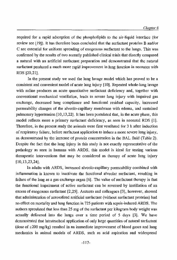
Chapter 6
required for a rapid adsorption of the phospholipids to the air-liquid interface (for
review see [19]). It has therefore been concluded that the surfactant proteins Band/or
C are essential for uniform spreading of exogenous surfactant to the lungs. This was
confIrmed by the results of two recently published clinical trials that directly compared
a natural with an artifIcial surfactant preparation and demonstrated that the natural
surfactant produced a much more rapid improvement in lung function in neonates with
RDS [20,21].
In the present study we used the lung lavage model which has proved to be a
consistent and convenient model of acute lung injury [10]. Repeated whole-lung lavage
with saline produces an acute quantitative surfactant defIciency and, together with
conventional mechanical ventilation, leads to severe lung injury with impaired gas
exchange, decreased lung compliance and functional residual capacity, increased
permeability changes of the alveolo-capillary membrane with edema, and sustained
pulmonary hypertension [10,13,22]. It has been postulated that, in the acute phase, this
model reflects more a primary surfactant deficiency, as seen in neonatal RDS [1].
Therefore, in the present study the animals were rust ventilated for 3 h after induction
of respiratory failure, before surfactant application to induce a more severe lung injury,
as demonstrated by the increase of protein concentration in the BAL fluid (Table 2).
Despite the fact that the lung injury in this study is not exactly representative of the
pathology as seen in humans with ARDS, this model is ideal for testing various
therapeutic interventions that may be considered as therapy of acute lung injury
[10,13,23,24].
In adults with ARDS, increased alveolo-capillary permeability combined with
inflanl1'llation is known to inactivate the functional alveolar surfactant, resulting in
failure of the lung as a gas exchange organ [6]. The value of surfactant therapy is that
the functional impairment of active surfactant can be reversed by instillation of an
excess of exogenous surfactant [2,25]. Anzueto and colleagues [3], however, showed
that administration of aerosolized artifIcial surfactant (without surfactant proteins) had
no effect on mortality and lung function in 725 patients with sepsis-induced ARDS. The
authors speculated that less than 25 mg of the surfactant per kilogram body weight was
actually delivered into the lungs over a time periOd of 5 days [3]. We have
demonstrated that intratracheal application of only large quantities of natural surfactant
(dose of ~200 mg/kg) resulted in an immediate improvement of blood gases and lung
mechanics in animal models of ARDS, such as acid aspiration and widespread
-117-
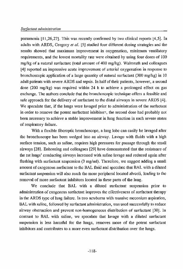
Surfactant administration
pneumonia [11,26,27]. This was recently confirmed by two clinical reports [4,5]. In
adults with ARDS, Gregory el al. [5] studied four different dosing strategies and the
results showed that maximum improvement in oxygenation, minimum ventilatory
requirements, and the lowest mortality rate were obtained by using four doses of 100
mg/kg of a natural surfactant (total amount of 400 mg/kg). Walmrath aud colleagues
[4] reported an impressive acute improvement of arterial oxygenation in response to
bronchoscopic application of a large quantity of natural surfactant (300 mg/kg) in 10
adult patients with severe ARDS and sepsis. In half of their patients, however, a second
dose (200 mg/kg) was required within 24 h to achieve a prolonged effect on gas
exchange. The authors conclude that the bronchoscopic technique offers a feasible and
safe approach for the delivery of surfactant to the distal airways in severe ARDS [4].
We speculate that, if the lungs were lavaged prior to administration of the surfactant
in order to remove the potent surfactant inhibitor" the second dose had probably not
been necessary to achieve a stable improvement in lung function in such severe states
of respiratory failure.
With a flexible fiberoptic bronchoscope, a lung lobe can easily be lavaged after
the bronchoscope has been wedged into an airway. Lavage with fluids with a high
surface tension, such as saline, requires high pressures for passage through the small
airways [28]. Enhorning and colleagues [29] have demonstrated that the resistance of
the rat lungs' conducting airways increased with saline lavage and reduced again after
flushing with surfactant suspension (3 mg/ml). Therefore, we suggest adding a small
amount of exogenous surfactant to the BAL fluid and speculate that BAL with a diluted
surfactant suspension will also reach the more peripheral located alveoli, leading to the
removal of more surfactant inhibitors located in these parts of the lung.
We conclude that BAL with a diluted surfactant suspension prior to
administration of exogenous surfactant improves the effectiveness of surfactant therapy
in the ARDS type of lung failure. In two newborns with massive meconium aspiration,
BAL with saline, followed by surfactant administration, was used successfully to reduce
airway obstlUction and prevent non-homogeneous distribution of surfactant [30]. In
contrast to BAL with saline, we speculate that lavage with a diluted surfactant
suspension is less harmful for the lungs, removes more of the potent surfactant
inhibitors and contributes to a more even surfactant distribution over the lungs.
-118-
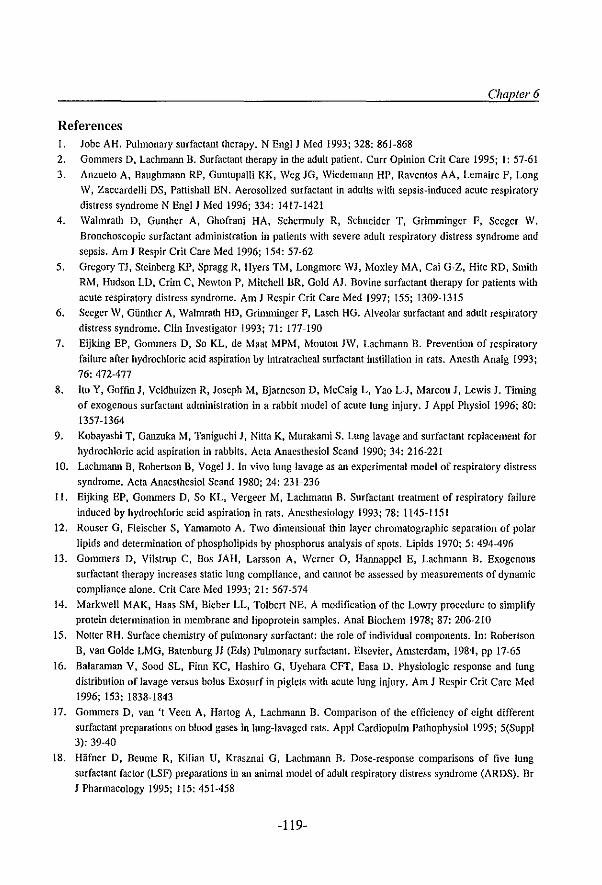
Chapfer6
References 1. Jobe AH. Pulmonary surfactant therapy. N EngJ J l\-Icd 1993; 328: 861-868
2. Gommers D, Lachmann B. Surfactant therapy in the adult patient. Curf Opinion Cril Care 1995; 1: 57-61
3. Anzueto A. Baughmann RP, Guntupalli KK, Wcg JG. Wiedemann HP, Ravcntos AA, Lemaire F, Long
W, Zaccardelli DS, Pattishall EN. Aerosolized surfactant in adults with sepsis-induced acute respiratory
distress syndrome N EngJ J Med 1996; 334: 1417-1421
4. Walmrath D, Gunther At Ghofrani HA, Schermuly R, Schneider T, Grimmingcr F, Seeger W.
Bronchoscopic surfactant administration in patients with severe adult respiratory distress syndrome and
sepsis. Am J Respir eric Care Med 1996; 154: 57-62
5. Gregory TIt Steinberg KP, Spragg R. Hyers TM, Longmore WI, r.,'foxley MA, Cai G·Z, Hite RD, Smith
RM, Hudson LD, Crim C, Newton P, Mitchell DR, Gold AJ. Bovine surfactant therapy for patients with
acute respiratory distress syndrome. Am J Respir Crit Care Med 1997; 155; 1309-1315
6. Seeger W, Gunther A, Walmrath HD. Grimminger P, Lasch HG. Alveolar surfactant and adult respiratory
distress syndrome. Clin Investigator 1993; 71: 177~190
7. Eijking EP, Gommers D, So KL, de Maat MPM, Mouton JW, Lachmann D. Prevention of respiratory
failure after hydrochloric acid aspiration by intratracheal surfactant instillation in rats. Anesth Analg 1993;
76: 472-477
8. Ito Y, Goffm J, Veldhuizen R, Joseph M, Bjarneson D, McCaig L, Yao L~J, Marcou J, Lewis J. Timing
of exogenous surfactant administration in a rabbit model of acute lung injury. J Appl Physiol 1996; 80:
1357-1364
9. Kobayashi T. Ganzuka M, Taniguchi J, Nitta K, Murakanli S. Lung lavage and surfactant replacement for
hydrochloric acid aspiration in rabbits. Acta Anaesthesiol Scand 1990; 34: 216~221
10. Lachmann B, Robertson B, Vogel 1. In vivo lung lavage as an experimental model of respiratory distress
syndrome. Acta Anaesthesiol Scand 1980; 24: 231~236
11. Eijking EP, Gommers D, So KL. Vergeer M, Lachmann B. Surfactant treatment of respiratory failure
induced by hydrochloric acid aspiration in rats. Anesthesiology 1993; 78: 1145-1151
12. Rouser G, Fleischer S, Yamamoto A. Two dimensional thin layer chromatographic separation of polar
lipids and determination of phospholipids by phosphorus analysis of spots. Lipids 1970; 5: 494~496
13. Gommers D, Vilstrup C, Bos JAH, Larsson A. Werner 0, Hannappel E, Lachmann B. Exogenous
surfactant therapy increases static lung compliance, and cannot be assessed by measurements of dynamic
compliance alone. Crit Care Med 1993; 21: 567-574
14. Markwell MAK, Haas SM, Bieber LL, Tolbert NE. A modification of the Lowry procedure to simplify
protein determination in membrane and lipoprotein samples. Anal Biochem 1978; 87: 206-210
15. Notter RH. Surface chemistry of pulmonary surfactant: the role of individual components. In: Robertson
B, van Golde LMG. Batenburg JJ (Eds) Pulmonary surfactant. Elsevier, Amsterdam, 1984, pp 17-65
16. Balaraman V, Sood SL, Finn KC. Hashiro G, Uyehara CFT, Easa D. Physiologic response and lung
distribution of lavage versus bolus Exosurf in piglets with acute lung injury. Am J Respir Crit Care Med
1996: 153: 1838-1843
17. Gommers D, van 't Veen A, Hartog A, Lachmann B. Comparison of the efficiency of eight different
surfactant preparations on blood gases in Iung-Javaged rats. Appl Cardiopulm Pathophysiol1995; 5(Suppl
3): 39-40
18. Hafner D, Beume R, Kilian U. Krasznai G, Lachmann B. Dose-response comparisons of five lung
surfactant factor (LSF) preparations in an animal model of adult respiratory distress syndrome (ARDS). Dr
J Pharmacology 1995: 115: 451-458
-119-
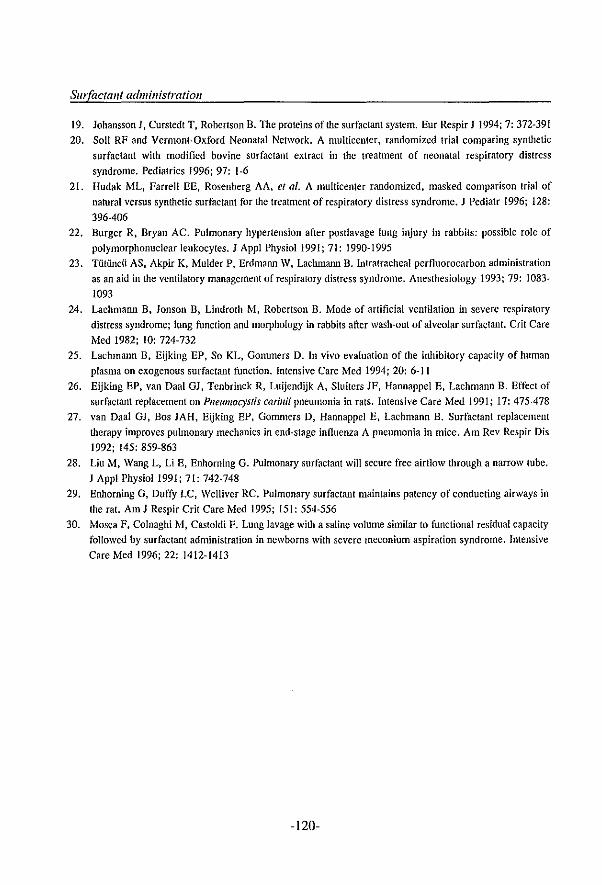
Surfactant administration
19. Johansson J, Curstedt T, Robertson B. The proteins of the surfactant system. Eur Respir J 1994; 7: 372-391
20. Soli RF and Vermont-Oxford Neonatal Network. A multicenter, randomized trial comparing synthetic
surfactant with modified bovine surfactant extract in the treatment of neonatal respiratory distress
syndrome. Pediatrics 1996; 97: 1-6
21. Hudak ML, Farrell EE, Rosenberg AA, et al. A multicenter randomized. masked comparison trial of
natural versus synthetic surfactant for the treatment of respiratory distress syndrome. J Pediatr 1996; 128:
396-406
22. Burger R, Bryan AC. Pulmonary hypertension after postlavage lung injury in rabbits: possible role of
polymorphonuclear leukocytes. J Appl Physiol1991; 71: 1990-1995
23. Tiitiincii AS, Akpir K. Mulder P, Erdmann W, Lachmann B. Intratracheal perfluorocarbon administration
as an aid in the vcntilatory management of respiratory distress syndrome. Anesthesiology 1993; 79: 1083-
1093
24. Lachmann B, Jonson B, Lindroth M, Robertson B. Mode of artificial ventilation in sevcre rcspiratory
distress syndrome; lung function and morphology in rabbits after wash-out of alvcolar surfactant. Crit Care
Med 1982; 10: 724-732
25. Lachmann B, Eijking EP, So KL, Gonuners D. In vivo evaluation of the inhibitory capacity of human
plasma on exogenous surfactant fllOction. Intensive Care Mcd 1994; 20: 6-11
26. Eijking BP, van Daal GJ, Tcnbrinck R. Luijendijk A, Sluilers JF, Hannappel E, Lachmann B. Effect of
surfactant replacement on Plieulllocysfis caril/Ii pneumonia in rats. Intensive Care Med 1991; 17: 475-478
27. van Daal GJ , Bos JAH, Eijking EP, Gommers D, Hannappel E, Lachmann B. Surfactant replacement
therapy improves pulmonary mechanics in end-stage influenza A pneumonia in mice. Am Rev Respir Dis
1992; 145: 859-863
28. Liu M, Wang L, Li E, Enhorning G. Pulmonary surfactant will secure free airflow through a narrow lube.
I Appl Physiol1991; 71: 742-748
29. Enhorning G. Duffy LC, Welliver RC. Pulmonary surfactant maintains patency of conducting airways in
the rat. Am J Respir Crit Care Mcd 1995; 151: 554-556
30. Mosca F. Colnaghi M, Castoldi F. Lung lavage with a saline volume similar to functional residual capacity
followed by surfactant administration in newborns with severe meconium aspiration syndrome. Intensive
Care Mcd 1996; 22: 1412-1413
-120-
Chapter 7
Surfactant therapy in combination with high
frequency oscillatory ventilation is not superior to
conventional mechanical ventilation in reducing
lung injury in lung-Iavaged rabbits
D. Gommers, A. Hartog, R. Schnabel', A. De Jaegere', and B. Lachmarm
Depts. of Anesthesiology, Erasmus University Rotterdam, The Netherlands;
'Pathology, Ruhr University Bochum, Germany; and
'Pediatrics, Sophia Children's Hospital, Rotterdam, The Netherlands
Sllbmiried for publicatioll
-121-
Surfactallt alld HFOV
Summary
The aim of this study was to compare high-frequency oscillatory ventilation (HFOV)
to conventional mechanical ventilation (CMV) with and without surfactant in the
treatment of acute lung injury caused by normal saline lavage in adult rabbits. The
efficacy of each therapy was assessed by evaluating gas exchange, lung deflation
stability and lung histopathology. After 4 hours of assigned therapy, we evaluated lung
deflation stability by decreasing mean airway pressure (MAwP) from 12 to 9 to 6 cm
H,O at 10 min intervals. After each change of MAwP blood gases were measured.
Following assessment of deflation stability, all 4 groups of animals were restarted on
CMV at the same settings as used during lung lavage and gas exchange was again
assessed. To evaluate lung injury, we assessed lung histopathology. The lungs were
flxed at PEEP + I cm H,O. Lungs of 3 additional animals were removed directly after
the lavage procedure and served as controls.
Arterial oxygenation in both HFOV groups (WiOl or without surfactant) was kept
above 350 mmHg (according the high-lung volume strategy) during the flrst 4 h by
using appropriate MAwP. In the CMV group with surfactant, PaO, increased from 62
±16 to 400±80 mmHg within 5 min and remained stable during the subsequent 4 h.
In the CMV group without surfactant, PaO, did not inlprove and all animals died within
the observation period. In both HFOV groups, a MAwP of 18-20 cm H,O had to be
applied initially to open up the lungs, that could be decreased during the subsequent 4
h while PaO, remained stable. During deflation stability, mean PaO, values decreased
to post-lavage values in the group that received HFOV alone after MAwP was
decreased to ~9 em H,G but not in both groups that received surfactant (HFOV and
CMV). The HFOV group without surfactant showed more cellular inflltration and
epithelial damage compared with the lavaged only animals and both surfactant-treated
groups (HFOV and CMV). In addition, the surfactant-treated animals (HFOV and
CMV) showed no additional structural lung damage compared to the lavaged only
animals.
We conclude that the use of surfactant therapy in combination with HFOV is not
snperior to CMV in improving gas exchange, lung deflation stability and in the
prevention of lung injury.
-122-
Chapter 7
Introduction High-frequency oscillatory ventilation (HFOV) and exogenous surfactant therapy are
two therapies to treat neonatal respiratory distress syndrome (RDS) but deal with the
pathophysiology of this disease in different ways. HFOV is intended to counterbalance
Ole increased tendency for collapse by applying a constant distending pressure ('mean
airway pressure') [I]. Exogenous surfactant therapy aims to decrease the alveolar
surface tension by application of a surface active material [2]. In contrast to the limited
success of the early HFOV trials [3], exogenous surfactant therapy has been used
successfully in worldwide clinical trials and has decreased mortality by 30-40% [2].
Therefore, exogenous surfactant therapy with conventional mechanical ventilation
(CMV) is now routinely used in most neonatal ICUs to prevent and treat neonatal RDS
[2].
In the first HFOV trial, a low distending airway pressure was used in order to
minimize the risk of barotrauma [3]. Experimental studies have, however, shown that
alveoli should be actively opened and Olat relatively high airway pressure has to be used
to stay above the closing pressure to avoid hypoxemia and lung injury [4,5]. This is
called the high-lung volume strategy; results of recent pilot studies in neonates with
RDS applying this strategy are encouraging [6-8].
Until now, only a few studies have been published on the combined use of
surfactant and HFOV in animals or humans [6, 8-11]. It was shown Olat after surfactant
therapy HFOV was superior to CMV in inlproving pulmonary function and reducing
lung injury [6, 8-11]. In these studies, however, HFOV was used in combination with
Ow high-lung volume strategy whereas CMV was not. Receutly, Froese and colleagues
[10] compared HFOV to CMV after surfactant therapy at low and high-lung volume
and confirmed that HFOV at high-lung volume was superior to the alternatives in
inlproving gas exchange and lung mechanics in lung-Iavaged rabbits. Surprisingly,
these authors were not able to maintain oxygenation above 350 nunHg (according to
the high-lung volume strategy) after surfactant therapy WiOl Ole use of CMV [10]. This
is in contrast to earlier results of CMV WiOl surfactant therapy in lung-Iavaged rabbits
in which oxygenation increased rapidly to prelavage values after surfactant instillation
and kept stable for hours [12-14]. Therefore, the purpose of the present study was to
compare the use of HFOV to CMV with and without surfactant therapy in the
management of acute lung iJ~ury caused by lung lavage in adult rabbits. During the first
4 h, target PaO, ranges were above 350 mmHg according to the high-lung volume
-123-
Surfactant alld HFOV
strategy [5]. To evaluate whether lung function was improved after 4 h of ventilation,
MAwP was decreased three times in succession and blood gases were measured. We
used a high PaO, value at low MAwP as an indicator for deflation stability conferred
by optimal surfactant function. Further, we performed histopathology of the lungs in
order to evaluate whether HFOV is superior to CMV, with or WitllOut surfactant, in the
prevention of additional lung injury secondary to mechanical ventilation.
Materials and methods
This study was approved by the local Animal Committee of the Erasmus University
Rotterdam; care and handling of the aninlals were in accord with the European
Community guidelines (86f609fEEG). A total of 27 adult New Zealand White rabbits
(IFFA-Credo, Brussels, Belgium) with a mean body weight of 2.7±0.3 kg were
anaesthetized with pentobarbital sodium (50 mgfkg) via an auricular vein and then
placed in a supine position. An endotracheal tube (Ld. 3.5 nml) was inserted via
tracheostomy and mechanical ventilation was initiated with a Servo Ventilator 900C
(Siemens-Elema AB, Solna, Sweden) in a pressure-control mode, indicating time-cycled
ventilation WiOI decelerating flow, with the following ventilator settings: FiO, of 1.0,
positive end-expiratory pressure (PEEP) of 2 cm H,O, frequency of 30 breathsfmin,
inspiratoryfexpiratory ratio of 1 :2 and a peak inspiratory pressure of 10-14 cm H,o to
keep PaCO, within normal range. An infusion of 2.5% glucose was continuously
administered via Ole auricular vein as a maintenance fluid (5 mIlkgfh). Anaesthesia was
maintained by hourly injection of pentobarbital sodium (5 mg/kgfh, Lv.); muscle
paralysis was achieved by hourly injection of pancuronium bromide (0.1 mg/kgfh,
Lm.).
A carotid artery was cannulated for continuous blood pressure measurements and
for intermittent blood sampling. Arterial samples were analyzed for blood gases, pH
and hemoglobin using conventional methods (ABL-505 and Osm-3; Radiometer,
Copenhagen, Denmark). Core temperature was monitored with an esophageal
thermistor (Elektrolaboratoriet, Copenhagen, Denmark) and maintained within normal
range by a heating pad.
In all aninlals, respiratory insufficiency was induced by repeated whole-lung
lavage according to Laclm1al11l et al. [15]. Each lavage was preformed with saline (30
mllkg) heated to 37°C. Lung lavage was repeated 5-8 tinles at 2-5 min intervals to
achieve a PaO, < 85 mmHg at a peak pressure and PEEP of 26 and 6 cm H,o,
-124-
Chapter 7
respectively (other ventilator settings were not changed). After reaching steady state,
24 animals were divided randomly into four groups of six animals. In the first group,
animals received a bolus of surfactant intratracheally (Alveofact'; 100 mg/kg) and
inmlediately followed by connection to a high-frequency oscillator (type OHF-l, S.A.
Dufour, Villeneuve d' Ascq, France). During HFOV, a frequency of 10 Hz was used
and mean airway pressure (MA wP) was increased until Pa02 was above 350 mmHg to
guarantee the high-lung volume strategy (i.e. open lungs) [4,5]. When Pa02 was above
550 mmHg during the study period, MAwP was decreased by steps of I cm H,o. Pressure amplitude was initiated with 30 cm H,o and was altered as necessary to keep
PaCO, within normal range (35-45 nmilig). The second group received exogenous
surfactant intratracheally (Alveofact'; 100 mg/kg) and conventional mechanical
ventilation (CMV) with the Servo Ventilator 900C was continued (ventilator settings
were not changed). For instillation of surfactant, the animals had been disconnected
from the ventilator and received the surfactant suspension (25 mg/ml) directly into the
endotracheal tube via a syringe; inmlediately followed by re-connection to the
ventilator. The other two groups served as controls and received HFOV or CMV alone
in the same way as described above.
After 4 h, MAwP was decreased to 12 cm H20 for 10 min in those animals that
received a MAwP of d2 cm H20 and then MAwP was decreased to 9 and 6 cm H,O,
respectively. Finally, all study groups were restarted on CMV at the same settings as
used during lung lavage, indicating a MAwP of ± 12 Col H20. After 10 min, blood
samples were taken and the lungs were intrathoracally fixated and subsequently
removed for histopathologic examination.
During the observation period (4 hand 40 min), arterial blood samples were
collected at the following times: before lavage; 5 and 10 min after the last lavage; 5,
15, 30 min after surfactant or HFOV application; every 30 min for 4 h; 10 min after
the three reduction steps of MAwP; and finally 10 min after CMV with the same
settings as during the lavage procedure. Continuous blood pressure monitoring enabled
observation of changes of mean arterial blood pressure and additional volume expansion
(Isodex'; 5 mllkg, Lv.) was given when mean arterial pressure was below 50 lnnilig.
At the end of the observation period, lungs were ventilated with air (other
ventilator settings were not changed), the abdomen of the rabbit was opened and the
diaphragm was inspected for evidence of pneumothorax. The inferior vena cava was
cannulated and a perfusion solution, consisting of saline saturated with 95 % 0, and 5 %
-125-
Sur[aCIGlII alld HFOV
CO,, 2.2 mM CaCl" 0.5 % procaine and 1 % heparin, was then infused at a rate of 3
IIh. The abdominal aorta was cut and the infusion was stopped when clear fluid flowed
from the aorta. Thereafter, the peak pressure above PEEP level was lowered to 1 cm
H,O, that was maintained while the lungs were fixated by infusing ± 100 ml of a
fixation solution, consisting of 3.6% formaldehyde and 0.25 % glutaraldehyde, via the
inferior vena cava. After fixation, tlle trachea was clamped at a pressure of 6 cm H,O,
the thorax was opened and the lungs were removed en bloc and stored in the fixation
solution. The lungs were numbered and histopathologic examinations of the lungs were
performed blindly. The lungs of the three remaining animals were fixed 10 min after
the lavage procedure as described above and histopathologic examinations were
performed. These animals were used to study the influence of the lavage procedure
itself on morphological changes.
The lungs were then embedded in paraffin, sectioned and stained with the
haematoxylin and eosin (HE) and elastica-van Gieson (EvG) technique. A semi
quantitative morphometric analysis of lung injury was performed under blinded
conditions by a pathologist who scored atelectasis, edema, vascular wall thickening and
leucocyte inftltration as none, light, moderate or severe (score 0-3). Lung injury score
was defined as the average from all parameters for each group. For transmission
electron microscopic examination, lung tissue was pre-contrasted with 2 % osmium
acid, dehydrated, and embedded in epoxy resin (Epon 812). Semi-thin sections (1.5
,Lim) and ultrathin sections (0.5 ,Lim) were produced. Semi-thin sections were stained
with 2 % methylene blue and 3 % alkaline fuchsin. Ultrathin sections were
counterstained with uranyl acetate and lead citrate. The specimens for the scanning
electron microscope were dehydrated WiOl increasing lines of alcoholic solutions, dried
with the critical point method, and sputtered with gold.
Analysis of variance (ANaYA) was used to assess whether there was an overall
difference within or between the two groups. If a difference was found, a post hoc test
was used (Student-Newman-Keuls' multiple comparison procedure). Lung injury data
were analysed using the Kurskal-Wallis nonparametric ANaYA test, followed by
Dunn's multiple comparisons test if a difference was found. Statistical significance was
accepted at p-values sO.05. All data are expressed as mean ± SO.
Results
Blood gases before lavage and directly after lavage were comparable in all animals.
-126-
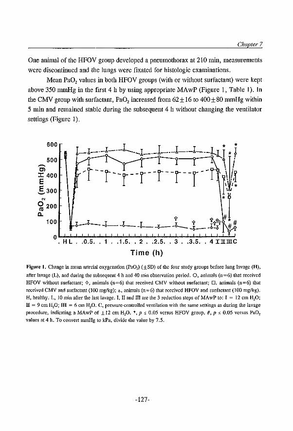
Chapter 7
One animal of the HFOV group developed a pneumothorax at 210 min, measurements
were discontinued and the lungs were fixated for histologic examinations.
Mean PaD, values in both HFOV groups (with or without surfactant) were kept
above 350 mmHg in the first 4 h by using appropriate MAwP (Figure I, Table 1). In
the CMV group with surfactant, PaD, increased from62± 16 to 400±80 mmHg wiOlin
5 min and remained stable during the subsequent 4 h without changing the ventilator
settings (Figure 1).
600
500
---Cl J: 400 E E 300 ~
'" 0 200 «J a.
100
o~~~~~~~~~~~~~~~~ H L .. 0.5 .. 1 .. 1.5 .. 2 .. 2.5 .. 3 .. 3.5 .. 4 IlImc
Time (h)
Figure I. Change in mean arterial oxygenation (Pa01) (±SD) of the four study groups before lung lavage (II),
after lavage (L), and during the subsequent 4 h and 40 min observation period. O. animals (0=6) that received HFOV without surfactant; 0, animals (0=6) that received CMV without surfactant; D. animals (n=6) that
received CMV and surfactant (100 mg/kg); ~,animals (0=6) that received HFOV and surfactant (100 mg/kg),
H, healthy. L, 10 min after the last lavage. I, II and III are the 3 reduction steps of MAwP 10: I = 12 em HP;
II = 9 em H20; ill = 6 em H20. C, pressure~conlrolled ventilation with the same settings as during the lavage
procedure. indicating a MAwP of ±12 cm H20. *, p:'> 0.05 versus HFOV group. H, p s 0.05 versus PaD!
values at 4 h. To convert nmll-lg to kPa, divide the value by 7.5.
-127-
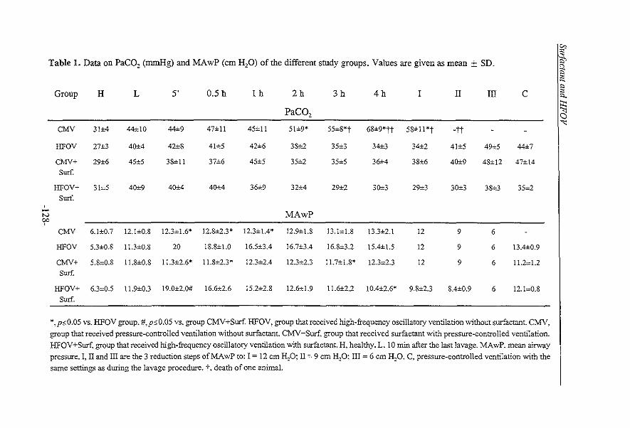
'" Table 1. Data on PaCO, (mmHg) and MAwP (em H20) of the different smdy groups. Values are given as mean ± SD. S.
11l ~ " Group H L 5' 0.5 h Ih 2h 3h 4h I II III C 5..
PaCO, ~ 0
'" CMV 31±4 44±10 44±9 47±11 45±11 51±9* 55±8*t 68±9'tt 58±11 *t ott
HFOV 27±3 40±4 42±8 41±5 42±6 38±2 35±3 34±3 34±2 41±5 49±5 44±7
CMV+ 29±6 45±5 38±11 37±6 45±5 35±2 35±5 36±4 38±6 40±9 48±12 47±14 Surf.
HFOV+ 31±S 40±9 40±4 40±4 36±9 32±4 29±2 30±3 29±3 30±3 38±3 35±2 Surf.
, - MAwP N 00 ,
CMV 6.1±0.7 12.1±0.8 12.3±1.6* 12.8±2.3* 12.3±1.4* 12.9±1.8 13.1±1.8 13.3±2.1 12 9 6
HFOV S.3±O.S l1.3±O.S 20 18.8±1.0 16.5±3A 16.7±3.4 16.8±3.2 15.4±1.5 12 9 6 13.4±0.9
CMV+ 5.8±0.8 11.8±0.S 11.3±2.6* 11.8±2.3* 12.3±2.4 12.3±2.3 11.7±1.8* 12.3±2.3 12 9 6 11.2±1.2
Surf.
HFOV+ 6.3±0.5 11.9±0.3 19.0±2.0# 16.6±2.6 IS.2±2.S 12.6±1.9 11.6±2.2 10.4±2.6* 9.8±2.3 8.4±0.9 6 12.1±0.8
Surf.
*,p:::;O.05 vs. HFOV group. #,p'5.0.05 vs. group CMY+Surf HFOV, group that received high-frequency oscillatory ventilation without surfactant. CMV, group that received pressure-controlled ventilation without surfactant. CMV+Surf, group that received surfactant with pressure-controlled ventilation. HFOV+Surf, group that received high-frequency oscillatory ventilation with surfactant. H. healthy. L, 10 min after the last lavage. MAwP, mean airway pressure. I, II and III are the 3 reduction steps ofMAwP to: 1= 12 cm H::P~ II:::: 9 cm H:p; III = 6 cm H~O. C. pressure-controlled ventilation with the same settings as during the lavage procedure. t, death of one animal.
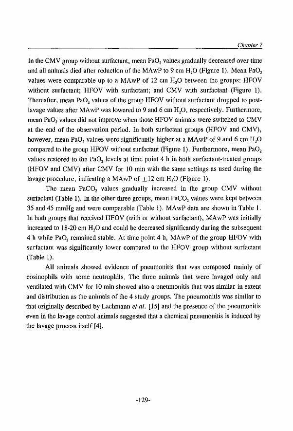
Chapter 7
In the CMV group without surfactant, mean PaO, values gradually decreased over time
and all animals died after reduction of the MAwP to 9 cm H,O (Figure 1). Mean PaO,
values were comparable up to a MAwP of 12 cm H,O between the groups: HFOV
without surfactant; HFOV with surfactant; and CMV with surfactant (Figure 1).
Thereafter, mean PaO, values of tlle group HFOV without surfactant dropped to post
lavage values after MAwP was lowered to 9 and 6 cm H,O, respectively. FurthemlOre,
mean PaO, values did not improve when those HFOV animals were switched to CMV
at the end of the observation period. In both surfactant groups (HFOV and CMV),
however, mean PaO, values were significantly higher at a MAwP of 9 and 6 cm H,O
compared to the group HFOV without surfactant (Figure 1). Furthermore, mean PaO,
values restored to the PaO, levels at time point 4 h in both surfactant-treated groups
(HFOV and CMV) after CMV for 10 min with the same settings as used during the
lavage procedure, indicating a MAwP of ± 12 cm H,O (Figure 1).
The mean PaCO, values gradually increased in the group CMV without
surfactant (Table 1). In the other three groups, mean PaCO, values were kept between
35 and 45 mmHg and were comparable (Table 1). MAwP data are shown in Table 1.
In both groups that received HFOV (with or without surfactant), MAwP was initially
increased to 18-20 cm H,o and could be decreased significantly during the subsequent
4 h while PaO, remained stable. At time point 4 h, MAwP of the group HFOV with
surfactant was significantly lower compared to the HFOV group without surfactant
(Table 1).
All animals showed evidence of pneumonitis that was composed mainly of
eosinophils with some neutrophils. The three animals that were lavaged only and
ventilated with CMV for 10 min showed also a pneumonitis that was sinlilar in extent
and distribution as the animals of the 4 study groups. The pneumonitis was similar to
that originally described by Lachmann el al. [15] and the presence of the pneumonitis
even in the lavage control animals suggested that a chemical pneumonitis is induced by
the lavage process itself [4].
-129-
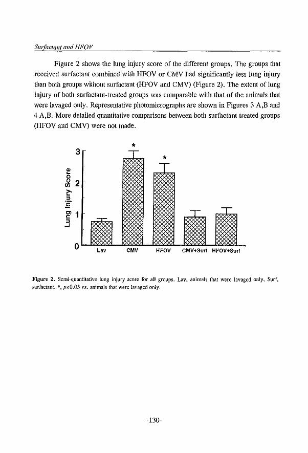
SIII'(aclallI alld HFOV
Figure 2 shows the lung injury score of the different groups. The groups that
received surfactant combined with HFOV or CMV had significantly less lung injury
than both groups without surfactant (HFOV and CMV) (Figure 2). The extent of lung
iI~ury of both surfactant-treated groups was comparable with that of the animals that
were lavaged only. Representative photomicrographs are shown in Figures 3 A,B and
4 A,B. More detailed quantitative comparisons between both surfactant treated groups
(HFOV and CMV) were not made.
* 3 - -* :gs -r
58l IX ~
:& --
T _-r-
58l 11 ~ ~ §§l :gs o Lav CMV HFOV CMV+Surf HFOV+Surf
Figure 2. Semi-quantitative lung injury score for all groups. Lay, animals that were lavaged only. Surf,
surfactant. *, p~O.05 vs. animals that were Ja\'aged only.
-130-
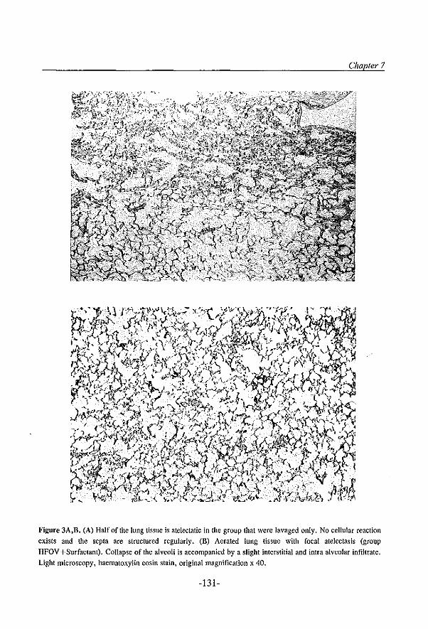
Chapter 7
Figure 3A,D. (A) Half of the lung tissue is atelectatic in the group that were lavaged only. No cellular reaction exists and the sepia are structured regularly. (B) Aerated lung tissue with focal atelectasis (group
HFOV +Surfaclant). Collapse of the alveoli is accompanied by a slight interstitial and intra alveolar infiltrate. Light microscopy, haematoxylin eosin stain, original magnification x 40.
-131-
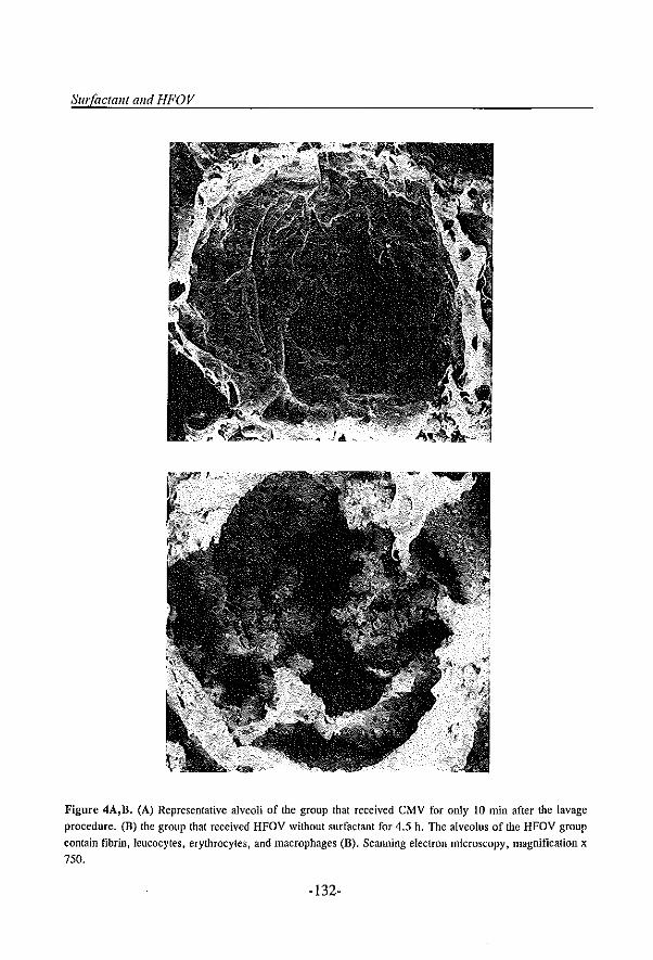
SIII'(actalll and HFOV
Figure 4A,B. (A) Representative alveoli of the group that received CMV for only 10 min after the lavage
procedure. (B) the group that received HFOV without surfactant for 4.5 h. The alveolus of the HFOV group
contain fibrin, leucocytes. erythrocytes, and macrophagcs (D), Scanning electron microscopy, magnification x
750.
-132-
Chapter 7
Discussion
In the present study we used the lung lavage model that has proved to be a consistent
and convenient model of acute lung injury [12-15]. Despite the use of adult animals,
it has been postulated that this model reflects in the acute phase a more primary
surfactant deficiency, as seen in neonatal RDS [2]. Several investigators have
confirmed the direct relationship in this model between arterial oxygenation and lung
volume [4,5,13,16]. It has been demonstrated that arterial oxygenation increased with
increasing lung volume as alveoli re-expanded and shunt flow decreased [16]. In the
present study, we therefore used arterial oxygenation as a reflection of lung volume.
The results of this study demonstrate that after surfactant therapy there is no
difference between the use of HFOV and CMV in improving gas exchange, lung
deflation stability and prevention of lung injury in lung-lavaged rabbits. These results
are in contrast to the results of Froese ef al. [10] who demonstrated that the effect of
exogenous surfactant on arterial oxygenation remained stable with HFOV, whereas it
decreased Significantly during the 4 h study period with CMV at high-lung volume. In
their study, the high-lung volume strategy with CMV was performed by a gradual
increase of PIP and PEEP but without an active volume recmitment manoeuvre as used
with HFOV [10]. Furthermore, CMV was used with high tidal volumes (20 ml/kg)
which is known to increase the conversion from active into non-active surfactant
subfractions; this leads to a shortage of 'active' surfactant at alveolar level [17,18]. In
the present study, we did not perform an active volume recmitment manoeuvre with
CMV after surfactant therapy, but arterial oxygenation increased to above 350 nmilig
within 5 min (without change of ventilator settings) and kept stable during the
subsequent 4 h. In contrast to the study of Froese ef al. [10], we used normal tidal
volumes (10 ml/kg) and installed the surfactant at a higher concentration (100 mg/kg)
and as one bolus, that is known to improve the surfactant distribution and its efficacy
[14].
Surfactant metabolism and turnover is known to be strongly influenced by
ventilation and some authors suggested that secretion of surfactant is increased with
HFOV [19-21]. In the present study, we confirmed that in lung-Iavaged rabbits optimal
gas exchange can be obtained with HFOV without surfactant, by using the high-lung
volume strategy. However, lung function does not inlprove over time, as shown by the
results that mean PaO, values at the end of the observation period were comparable
with the post-lavage values at the same ventilatory support (Figure 1). This indicates
-133-
Surfactallt alld HFOV
that the reduced end-expiratory stability, due to the repeated lung lavages (i.e.
surfactant deficiency), was apparently not improved by HFOV. This confirms the
results by Mereditll et ai. [22], who showed excellent gas exchange using HFOV with
the high-lung volume strategy in premature baboons, but no beneficial effect on lung
volume at zero pressure (functional residual capacity) detennined at 24 h. This indicates
that optimization of alveolar expansion with HFOV markedly improves oxygenation but
does not influence alveolar stability as long as the underlying cause, i.e. surfactant
deficiency, is not reversed. This will occur by gradual synthesis of endogenous
surfactant over time, or after exogenous surfactant instillation [2).
Morphologic changes were more pronounced in the animals that received CMV
or HFOV alone compared to the animals that were lavaged only (Figure 2). In
surfactant-deficient lungs, high shear forces between open and closed alveoli are, to a
great extent, held responsible for the damage caused by artificial ventilation [9).
Therefore, alveoli should be actively opened and kept open during the entire respiratory
cycle in order to minimize additional lung damage [4,5). Studies inlung-Iavaged rabbits
demonstrated that HFOV had beneficial effects on preventing development of lung
injury due to mechanical ventilation when arterial oxygenation was kept above 350
nnnHg (indicating alveolar expansion) and not when arterial oxygen tensions were kept
between 70 and 100 nnnHg [4,5). This may explain the higher degree of lung injury
as seen in the present study in the group HFOV without surfactant. In that group, mean
PaO, values dropped to below 100 mmHg for only 30 min, after MAwP was decreased
to 59 cm Hp at the end of the observation period (Figure 1). In contrast, mean PaO,
values remained above 350 nnnHg ahnost for the entire observation period, despite the
reduction of the MAwP, in both surfactant-treated groups (HFOV and CMV). Also,
histopathology examinations of bOtll groups tllat received surfactant (HFOV and CMV)
showed no additional structural lung damage in comparison with the animals that were
lavaged only (Figure 2). This result is supported by earlier experimental studies in
which surfactant therapy has shown to inlprove uniform alveolar expansion and end
expiratory alveolar stability and thereby effectively prevents the progression of
ventilator-induced lung injury [9,23).
In contrast to Jackson et ai. [11), we found no difference in the prevention of
lung damage between HFOV and CMV after surfactant therapy (Figure 2). Further, it
was shown that the HFOV group without surfactant showed more cellular infiltration
and epithelial damage tllan the HFOV group with surfactant (Figure 2). This indicates
-134-
Chapter 7
that achieving and maintaining alveolar expansion (i.e. open lung) is of more
importance than the type of mechanical ventilation (HFOV vs. CMV). The importance
of an open lung strategy is supported by the results of Amato and colleagues [24] who
recently demonstrated in adults with ARDS that CMV WiOl an open-lung approach had,
for the first time, a significant impact on survival and barotrauma. Therefore, we
concluded that surfactant therapy with CMV is equally effective to prevent ventilator
induced lung iJ~ury as HFOV combined with surfactant, as long as alveoli are opened
and kept open to avoid high shear stress. This can be achieved by the use of PEEP level
that sufficiently counterbalances the retractive forces or by Wgher and/or repeated doses
of exogenous surfactant to reduce the retractive forces.
We conclude that after surfactant therapy the use of HFOV was not superior to
CMV in improving gas exchange, lung deflation stability and reducing lung injury, if
lungs are kept expanded independently of the mode of ventilation. FurtllemlOre, it was
confirmed that HFOV with the high-lung volume strategy markedly improves blood
gases but without improvement of lung function, in particular arterial oxygenation at
low MAwP, in surfactant-depleted rabbits. This indicates that the high MAwP used
with HFOV only counterbalances the increased collapse tendency due to su -factant
deficiency and therefore exogenous surfactant therapy is still required.
References 1. Froese AB, Bryan AC. High frequency ventilation. Am Rev Respir Dis 1987. 135: 1363-1374 2. lobe AH. Pulmonary surfactant therapy. N Engl J Med 1993. 328: 861-868 3. HIPI study group: high-frequency oscillatory ventilation compared with conventional mechanical ventilation
in the treatment of respiratory failure in preterm infants. N Engl J Med 1989, 320: 88-93 4. Hamilton PP, Onayemi A, Smyth lA, Gillan IE, Cutz E, Froese AB, Bryan AC. Comparison of
conventional mechanical and high-frequency ventilation: oxygenation and lung pathology. J Appl Physioi 1983,55: 131-138
5. McCulloch PR, Forkert PG, Froese AB. Lung volume maintenance prevents lung injury during high frequency oscillatory ventilation in surfactant-deficient rabbits. Am Rev Respir Dis 1988, 137: 1185· 1192
6. Ogawa Y, Miyasaka K, Kawano T, Imura S, Inukai K, Okuyama K, Oguchi K, Togari N, Nishida H, Mishina J. A multicenter randomized trial of high frequency oscillatory ventilation as compared with conventional m~hanical ventilation in preterm infants with respiratory failure. Early Human Development 1992,32: 1-10
7. Clark RH, Yoder BA, Sell MS. Prospective, randomized comparison of high-frequency oscillation and conventional ventilation in candidates for exlracorporeal membrane oxygenation. J Pediatr 1994, 124: 447-454
8. Gerstmann DR, Minton SD, Stoddard RA, Meredith KS, Monaco P, Betrand JM, Battisti 0, Langhendries JP, Francois A, Clark RH. The provo multicenter early high-frequency oscillatory ventilation trial:
-135-
SlIrraclalll alld HFOV
improved pulmonary and clinical outcome in respiratory distress syndrome. Pediatrics 1996, 98: 1044-1057 9. Nilsson R, Berggren p. eursledt T, Grossmann G, Renheim G, Robertson B. Surfactant and ventilation by
high frequency oscillation in premature newborn rabbits: effect on survival, lung aeration, and bronchiolar epithelial lesions. Pedialr Res 1985, 19: 143-147
to. Froese AB, McCulloch PR, Sugiura M. Vaclavik S. Possmayer F, Moller F. Optimizing alveolar expansion prolongs the effectiveness of exogenous surfactant therapy in the adult rabbit. Am Rev Respir Dis 1993, 148: 569-577
11. Jackson Je, Truog WE, Standaert TA, Murphy JH, Juu! SE, Chi EY, Hildebrandt I, Hodson WA.
Reduction in lung injury after combined surfactant and high-frequency ventilation. Am I Respir Cril Care
Med 1994, 150: 534-539 12. Kobayashi T, Kataoka H, Ueda T, Murakami S, Takada Y, Kokubo M. Effects of surfactant supplement
and end-expiratory pressure in lung-Iavaged rabbits. I Appl Physiol1984, 57: 995-1001
13. Gommers D, Vilstrup C, Bos IAH, Larsson A, Wcrner 0, Hannappcl E, Lachmann B. Exogcnous
surfactant therapy increases static lung compliance, and cannot be assessed by measurements of dynamic
compliance alone. Crit Carc Med 1993, 21: 567-74
14. Segerer H, van Gelder W, Angenent FWM, van Woerkcns IPM, Curstedt T, Obladen M. Lachmann B.
Pulmonary distribution and eftlcacy of exogenous surfactant in lung-Iavaged rabbits are influenced by the
instillation technique. Pediatr Res 1993, 34: 490-494
15. Lachmann B, Robertson B, Vogel I. In vivo lung lavagc as an cxperimental model of respiratory distrcss
syndrome. Acta Anaesthesiol Scand 1980, 24: 231-236
16. Suzuki H, Papazoglou K, Bryan AC. Rclationship betwcen Pa02 and lung volume during high frequency
oscillatory ventilation. Acta Paediatr Ipn 1992, 34: 494-500
17. Veldhuizen RAW. Marcou I, Yao L-I, McCaig L, Ito Y, Lewis IF. Alveolar surfactant aggregate
conversion in ventilated normal and injured lungs. Am I Physiol 1996,270: L152-L158
18. Verbrugge SIC, Bohm SH, Gommers D, Zimmerman UI, Lachmrum B. Surfactant impairment after
mechanical ventilation with large alveolar surface area changes and effects of positivc end-expiratory
pressure. Br I Anaesth 1998; 80: 360-364
19. Houmes RJ, Bos IAH. Lachmann B. Effects of different ventilator settings on lung mechanics: with special
reference to the surfactant system. Appl Cardiopulm Pathophysiol1994, 5: 117-127
20. Froese AB. Hill PE, Bond DM. Moller F. Maintaining alveolar expansion facilitates surfactant repletion
in ventilated atelectasis-prone rabbits. FASEB I 1988, 2: Al183
21. Mannino FL, McEvoy RD, Hallmann M. Surfactant turnover in high frequcncy oscillatory vcntilation.
Pediatr Res 1982, 16: 356.
22. Meredith KS, DeLemos RA, Coalson II, King ru, Gerstmann DR, Kumar R, Kuehl TJ, Wintcr DC, Taylor
A, Clark RH, Null DM. Role oflung injury in the pathogenesis ofhyalinc membrane disease in premature
baboons. J Appl Physiol 1989,66: 2150-2158 23. Maeta H, Raju TNK, Vidyasagar D, Bhat R, Esterly J, Matsuda H, Shimada S, Krukenkamp IB, Shanklin
DR. Effect of cxogcnous surfactant on the development of bronchopulmonary dysplasia in a baboon hyaline
membrane disease model. Crit Care Med 1990, 18: 403-409
24. Amato MB, Barbas CSV. Medeiros DM, Magaldi RB, Schettino GPP, Lorcnzi-Filho G. Kairalla RA,
Deheinzelin D, Munoz C, Oliveira R, Takagaki TY, Carvalho CRR. Effect of a protective-ventilation
strategy on mortality in the acute respiratory distress syndrome. N Engl J Med 1998; 338: 347-354
-136-
Chapter 8
Conventional ventilation modes with small pressure
amplitudes and high end-expiratory pressure levels
optimize surfactant therapy
S.l.C. Verbrugge, D. Gommers, B. Lachmal1ll
Dept. of Anesthesiology, Erasmus University Rotterdam, The Netherlands
III press: Grit Care Med
Reprinted with penllissioll (copyright holder: Williams & Wilkens)
-137-
Sur(aclaut and tyPe o(venlitalion
Summary
Objective: High frequeucy oscillation studies have shown that ventilation at high end
expiratory lung volumes combined with small volume cycles at high rates best
preserves exogenous surfactant and gas exchange in lavaged lungs. We investigated
whether surfactant composition and gas exchange can also be preserved by conventional
modes of mechanical ventilation which combine high levels of positive end-expiratory
pressure (PEEP) with small pressure amplitudes.
Design: Prospective, randomized, non-blinded, controlled study.
Selling: Research laboratory.
Subjects: Thirty male Sprague-Dawley rats.
llllerventions: Rats were lung-Iavaged and treated with exogenous surfactant (100
mg/kg). After 5 minutes, four different ventilator settings (FiO, of 1.0) were applied
for three hours in 4 groups of rats [peak inspiratory pressure (cm H,o); static PEEP
(cm H,o); liE ratio; Ii'equency]: 26/211:2/30 (group 26/2); 26/611:2/30 (group 26/6);
20/1011:2/30 (group 20110-static); 20/6/7:3/130 creating an auto-PEEP of 4 cm H,O
(group 20/1O-auto).
Measurements and Main Results: In all groups, PaO, increased inllnediately to pre
lavage values after surfactant therapy. In group 2612, PaO, deteriorated to post-lavage
values within 30 min when PEEP was decreased to 2 cm H,O, whereas PaO, remained
stable for three hours in the other groups. The PaCO, increased in groups 26/2 and
20110-static; PaCO, could not be reduced by increasing ventilation frequency up to 130
ill group 201l0-static. Groups 26/6 and 201l0-auto remained normocapnic.
Bronchoalveolar lavage protein concentration was higher in groups 2612 and 26/6
compared to groups 201l0-static and 201l0-auto. There was significantly more
conversion of surface active large aggregates into non-active small aggregates in group
2612 compared to groups 201l0-static and 201l0-auto.
Conelusions: We conclude that exogenous surfactant composition is preserved by
conventional modes of mechanical ventilation which use small pressure amplitudes and
adequate oxygenation is maintained by high end-expiratory pressure levels. Effective
carbon dioxide removal could be achieved by applying a ventilation mode that creates
auto-PEEP and not by a mode which applies the same level of PEEP by static-PEEP
only.
-138-
Chapter 8
Introduction
In neonates with respiratory distress syndrome (RDS), exogenous surfactant
irnmediately reverses hypoxemia and has decreased mortality by 30-40% [1). There are
indications that surfactant therapy may also be beneficial in pediatric patients with
ARDS [2,3).
It is known from experimental studies that ventilation strategy influences the
effect of surmctant therapy [4-6). Studies by Froese et al. have shown that high
frequency oscillatory ventilation (HFOV) at high-lung volumes when combined with
surfactant therapy, could improve PaO, to a constant level with lower alveolar protein
influx and a higher amount of active surfactant at the end of the study period than
conventional mechanical ventilation (CMV) where PaO, decreased over time [5).
Recent data suggest that differences in conversion of active into non-active
surfactant in both healthy animals and animals with acute lung injury receiving
surfactant therapy are caused by differences in cyclic changes in alveolar surface area
[6-S). As tidal volumes were ten-fold higher during CMV tllan during HFO in the study
by Froese and colleagues [5], this raises the question whether lower rates of surfactant
conversion can also be obtained by modes of CMV that combine small volume cycles
at high rates with high PEEP levels. We therefore investigated the effect of different
pressure amplitudes and PEEP settings on exogenous surfactant therapy with respect
to gas exchange, protein influx and conversion of active into non-active surfactant
during a three-hour ventilation period in lung-lavaged rats. Moreover, the effect of
auto-PEEP on PaCO, versus a mode creating the same level of PEEP by static PEEP
only was investigated.
Materials and methods
This study was approved by the Institutional Review Board for the care of animal
subjects. Care and handling of the animals were in accordance with National Institute
of Health guidelines.
The non-blinded studies were performed in male Sprague-Dawley rats (n=24)
with a body weight of 260-330 g (Harlan CPB, Zeist, The Netherlands). After
induction of anesthesia with nitrous oxide, oxygen and ethrane (66/33/3%), a
polyethylene catheter (O.S-mm outer diameter) was inserted into the right carotid artery
for drawing arterial blood samples. Before tracheotomy, the animals received
pentobarbital sodium 60 mg/kg body weight, i.p. (Nembutal'; Algin BV, Maassluis,
-139-
Surfactant and type o(venlilation
The Netherlands) and the inhalation of ethrane was decreased by 50%. After a metal
cannula was inserted into the trachea, muscle relaxation was given with pancuronium
bromide 2.0 mg/kg, i.m. (paVIlIon'; Organon Technika, Boxtel, The Netherlands) and
the animals were connected to a ventilator. Anesthesia was maintained with
pentobarbital sodium, i.p. (Nembutal'; 60 mg/kg/h) and muscle relaxation was attained
with pancuronium bromide, i.m. (Pavulon'; 2 mg/kg/h). Body temperature was kept
within normal range by means of a heating pad.
The animals were mechanically ventilated in parallel (6 animals simultaneously)
with a Servo Ventilator 300 (Siemens-Elema AB, Solna, Sweden) at the following
ventilator settings: pressure constant tinle cycled mode, frequency of 30 breaths/min,
peak inspiratory pressure (PIP) of 12 cm H,O, PEEP of2 cm H,O, IIE ratio of 1:2,
and an FiO, of 1.0. Initially, PIP was increased to 20 cm H,O for 1 min to open up
atelectatic regions in the lungs. After this opening up procedure, the ventilator settings
were returned to the previous ones and a 0.15 ml blood sample was taken and replaced
by heparinized (10 IE/nll) saline (0.9% NaCI). PaO" PaCO" and pH were measured
by conventional methods (ABL 505, Radiometer, Copenhagen, Denmark). Next,
respiratory failure was induced by repeated whole-lung lavage as described by
Lacbmann el at. [9]. Each lavage was performed with saline (32 nll/kg) heated to 37
"C. Just before the first lavage, PIP and PEEP were elevated to 26 and 6 cm H,O,
respectively. Lung lavage was repeated 5-8 times with 5 min intervals to achieve a
PaO,<85 nnnHg (11.3 kPa). Five minutes after the last lavage, blood gases were
measured and within 10 min each animal received 1.2 nll of a surfactant suspension (25
mg/ml) at a dose of 100 mg/kg. The surfactant used in this experiment is a natural
surfactant isolated from pig lungs as previously described [10].
Five minutes after surfactant replacement the animals were randomized in groups
of six to be pressure-constant time-cycled ventilated with an FiO, of 1.0 at different
PIP, PEEP, I/E ratio and frequency settings. One group (n=6) was ventilated with a
peak pressure of26 cm H,o, a PEEP of2 cm H,O, an IIE ratio of 1:2 and a frequency
of 30 breaths/min (group 2612); a second group (n = 6) was ventilated with a PIP of 26
cm H,o, a PEEP of 6 cm H,o, an I/E ratio of 1:2 and a frequency of 30 breaths/min
(group 26/6); a third group (n=6) was ventilated at a PIP of20 cm H,O and a PEEP
of 10 cm H,O, an IIE ratio of 1:2 and a frequency of 30 breaths/min (group 20/10-
static). A [OurOl group (n=6) was ventilated at a PIP of20 cm H,o, a PEEP of6 cm
H,o, an IIE ratio of 7:3 and a frequency of 130 breaths/minute necessary to create an
-140-
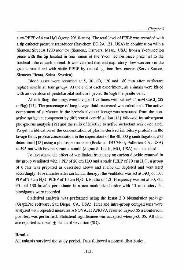
Chapter 8
auto-PEEP of 4 cm H,O (group 20/1O-auto). The total level of PEEP was recorded with
a tip catheter pressure transducer (Raychem EO 2A 121, USA) in combination with a
Siemens Sirecust 1280 monitor (Siemens, Danvers, Mass., USA) from a Y-comlection
piece with the tip located in one lumen of the Y -comlection piece proximal to the
tracheal tube in each aninlal. It was verified that end-expiratory flow was zero in the
groups ventilated with static PEEP by recording tinle-flow curves (Servo Screen,
Siemens-Elema, Solna, Sweden).
Blood gases were recorded at 5, 30, 60, 120 and 180 min after surfactant
replacement in all four groups. At the end of each experiment, all animals were killed
with an overdose of pentobarbital sodium injected through the penile vein.
After killing, the lungs were lavaged five times with saline/1.5 mM CaCI, (32
mlIkg) [11]. The percentage of lung lavage fluid recovered was calculated. The active
component of surfactant in the bronchoalveolar lavage was separated from the non
active surfactant component by differential centrifugation [11] followed by subsequent
phosphorus analysis [12] and the ratio of inactive to active surfactant was calculated.
To get an indication of the concentration of plasma-derived inhibitory proteins in the
lavage fluid, protein concentration in Ole supernatant of Ole 40,000 g centrifugation was
determined [13] using a photospectrometer (Beckman DU 7400, Fullerton CA, USA)
at 595 nm with bovine serum albumin (Sigma St Louis, MO, USA) as a standard.
To investigate the effect of ventilation frequency on carbon dioxide removal in
the group ventilated with a PIP of20 cm H,G and a static PEEP of 10 cm H,O, a group
of 6 rats was prepared as described above and surfactant depleted and ventilated
accordingly. Five minutes after surfactant therapy, the ventilator was set at FiO, of 1.0;
PIP of 20 cm H,O; PEEP of 10 cm H,G; liE ratio of I :2. Frequency was set at 30, 60,
90 and 130 breaths per minute in a non-randomized order with 15 min intervals;
bloodgases were recorded.
Statistical analysis was performed using the Instat 2.0 biostatistics package
(GraphPad software, San Diego, CA, USA). Inter and intra-group comparisons were
analyzed with repeated measures ANOV A. If ANOV A resulted in p<0.05 a Bonferroni
post-test was performed. Statistical significance was accepted whenp<0.05. All data
are reported as mean ± standard deviation (SD).
Results
All animals survived the study period. Data followed a normal distribution.
-141-
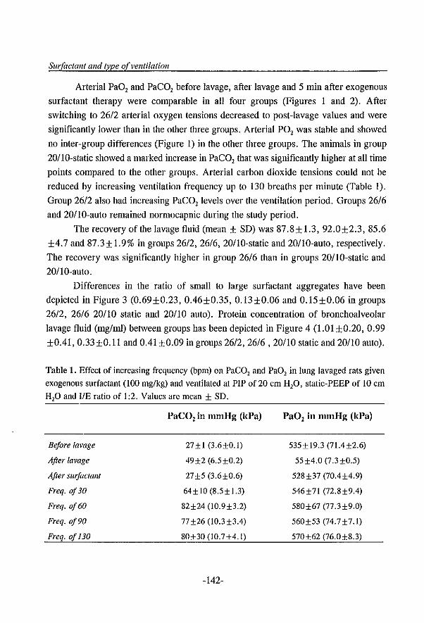
Surfactant alld type o(ventilatioll
Arterial PaO, and PaCO, before lavage, after lavage and 5 min after exogenous
surfactant therapy were comparable in all four groups (Figures 1 and 2). After
switching to 2612 arterial oxygen tensions decreased to post-lavage values and were
significantly lower than in the other three groups. Arterial PO, was stable and showed
no inter-group differences (Figure 1) in the other three groups. The animals in group
201l0-static showed a marked increase in PaCO, that was significantly higher at all tinle
points compared to the other groups. Arterial carbon dioxide tensions could not be
reduced by increasing ventilation frequency up to 130 breaths per minute (Table 1).
Group 26/2 also had increasing PaCO, levels over the ventilation period. Groups 26/6
and 20/1O-auto remained normocapnic during the study period.
The recovery of the lavage fluid (mean ± SD) was 87.8±1.3, 92.0±2.3, 85.6
±4.7 and 87.3 ± 1.9% ill groups 2612, 26/6, 20/1O-static and 20/1O-auto, respectively.
The recovery was significantly higher ill group 26/6 than in groups 20/1O-static and
20/1O-auto.
Differences in the ratio of small to large surfactant aggregates have been
depicted in Figure 3 (0.69±0.23, 0.46±0.35, 0.13±0.06 and 0.15±0.06 in groups
26/2, 26/6 20/10 static and 20110 auto). Protein concentration of bronchoalveolar
lavage fluid (mg/ml) between groups has been depicted in Figure 4 (1.01 ±0.20, 0.99
±0.41, 0.33±0.11 and 0.41 ±0.09 in groups 26/2,26/6,20110 static and 20/10 auto).
Table 1. Effect of increasing frequency (bpm) on PaCO, and PaO, in lung lavaged rals given exogenous surfactant (100 mg/kg) and ventilated at PIP of20 em H,O, stalic-PEEP of 10 em H,o and liE ratio of 1:2. Values are mean ± SD.
Before lavage
After lavage
After swfaetant
Freq.of30
Freq.of60
Freq.of90
Freq. of 130
PaCO, in llllllHg (kPa)
27±1 (3.6±0.1)
49±2 (6.5±0.2)
27±5 (3.6±0.6)
64± 10 (S.5± 1.3)
S2±24 (1O.9±3.2)
77±26 (1O.3±3.4)
SO+30 (10.7+4.1)
-142-
PaO, in mlllHg (!<pa)
535±19.3 (71.4±2.6)
55±4.0 (7.3±0.5)
528±37 (70.4±4.9)
546±71 (72.S±9.4)
5S0±67 (77.3±9.0)
560±53 (74.7±7.1)
570+62 (76.0±S.3)
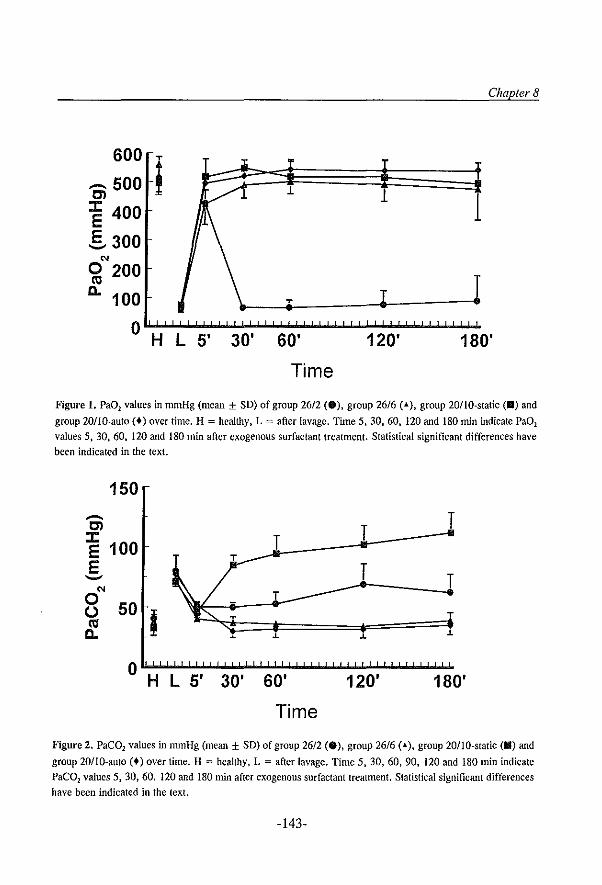
Chapter 8
600 T T
i ........ 500 1 1 1 Cl 1 J: 400
E §, 300
'" o 200 T
(II
a. 100 T T
o H L 5' 30' 60' 120' 180'
Time
Figure 1. Pa01 values in mmHg (mean ± SO) of group 2612 (e), group 26/6 ( .... ), group 20/IO-static (II) and
group 201l0-auto (+) over time. H = healthy, L = after lavage. Time 5,30,60,120 and 180 min indicate PaOl
values 5, 30. 60. 120 and 180 min after exogenous surfactant treatment. Statistical significant differences have been indicated in the text.
150 ........ Cl T ~ 100 T
T
E T --'" T 0 U 50 I (II a. J. J.
T T
.L
o H L 5' 30' 60' 120' 180'
Time
Figure 2. PaeD2 values in mmHg (mean ± SO) of group 26/2 (8), group 26/6 (-t..), group lOtIO-static (II) and
group 20/1O-auto (+) over time. H = healthy, L = after lavage. Time 5, 30, 60, 90, 120 and 180 min indicate
PaeD2 values 5, 30, 60. 120 and 180 min after exogenous surfactant treatment. Statistical significant differences
have been indicated in the text.
-143-
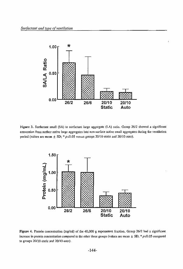
Sur{actmlt and type o(velltilatioll
1.00 * o
~ « 0.50 ...J
~
Figure 3. Surfactant small (SA) to surfactant large aggregate (LA) ratio. Group 26/2 showed a significant
conversion from surface active large aggregates into non-surface active small aggregates during the ventilation
period (values are mean ± SD; * psO.05 versus groups 201l0·static and 201l0-auto).
1.50
* ~
...J E OJ 1.00 E -I:
'Qj 0.50 ....
0 ... D-
0.00 26/2 26/6 20/10 20/10
Static Auto
Figure 4. Protein concentration (01g/01l) of the 40,000 g supernatant fraction. Group 26/2 had a significant
increase in protein concentration compared to the other three groups (values are mean ± SD; * psO.05 compared
to groups 20/IO-static and 20/1O-auto).
-144-
Chapter 8
Discussion
This study shows that during CMV, settings that combine small pressure amplitudes
with high levels of PEEP best preserve the large aggregate, surface-active component
of exogenous surfactant. The advantage of applying a mode of ventilation that creates
auto-PEEP, compared to a mode that applies the same level of PEEP by static PEEP
only, is a more effective CO, removal. Moreover, it is shown that PEEP has a major
impact on the effect of exogenous surfactant therapy on arterial oxygenation. A
ventilator setting with a low PEEP does not preserve oxygenation and blood gases
deteriorate immediately, whereas higher levels of PEEP restore oxygenation to pre
lavage values for at least 3 hours.
Exogenous surfactant therapy is now routinely applied in premature neonates
with RDS [1] and several studies have shown a beneficial effect of the use of exogenous
surfactant in ARDS [2,3]. However, in some neonates exogenous surfactant leads to
only transient improvements of arterial oxygenation [1]. Although tile exact reasons for
tilis are not fully understood, it is becoming increasingly realized that such differences
are attributable to: the efficacy of alveolar surfactant delivery of the application
technique [14]; the composition of the surfactant preparation [15]; differences in the
amount of surfactant inhibitors in the alveolar space [16]; and the mode of mechanical
ventilation [4-6].
Data on the influence of mechanical ventilation on exogenous surfactant therapy
are limited. Surfactant replacement studies by Kobayashi et al. inlung-lavaged rabbits
ventilated at a PIP of 20 cm H,O showed that a PEEP of 4 cm H,O improved survival
and normalized blood gases and compliance as opposed to surfactant-treated rabbits
ventilated without PEEP [4]. Our data confirm the importance of a sufficiently high
level of PEEP to maintain adequate oxygenation (group 26/6 versus group 26/2).
More recent studies by Froese and colleagues [5] in surfactant treated (80
mg/kg) lung-lavaged rabbits have clearly demonstrated that HFOV with small volume
cycles at high rates and high end-expiratory lung volumes is most beneficial in
exogenous surfactant treatment and leads to a constant improvement of PaO, Witil a low
alveolar protein influx and a high amount of active surfactant at the end of a 4 hour
study period. However, HFOV may not be routinely available in most neonatal
intensive care units. The inlprovements in oxygenation after exogenous surfactant
therapy with conventional volume-constant ventilation in the same study showed a
decline over time; analysis of surfactant composition at the end of the study period
-145-
Surfactant and type orvel/tilalion
demonstrated a greater conversion of active into non-active surfactant with CMV than
with HFO [5]. Such differences in conversion were explained by differences in volume
cycles, which were ten-fold higher during CMV than during HFOV [5]. Gross et al. were the ftrst to show that conversion of active into non-active surfactant sub fractions
is dependent on cyclic changes in surface area in vitro [17]. To maintain an adequate
pool of functional surfactant subfraction in the air spaces in vivo, it is necessary to
maintain a balance between secretion, uptake and clearance of the active and non-active
surfactant subfractions [18]. Recent in vivo studies by Veldhuizen et al. in rabbits
attribute the surfactant conversion to a change in alveolar surface area associated with
mechanical ventilation [6, 8]. They found that changing the respiratory rate did not
affect the rate of conversion but that conversion of surfactant subfractions is dependent
on tidal volume and time [8].
It may be reasoned that also conventional modes of mechanical ventilation that
combine small pressure amplitudes with high end-expiratory lung volumes have a
beneftcial effect on exogenous surfactant composition and function. The low surfactant
conversion rate and the adequate oxygenation in group 20110-static show that this is
indeed the case. However, the PaCO, level indicates that carbon dioxide could not be
effectively removed with these settings of the ventilator. Increasing the ventilation
frequency in the static-PEEP group to the same level as in the auto-PEEP group had
no influence on PaCO, (Table 1), which indicates that alveolar ventilation could not be
increased by increasing ventilation frequency. The more effective carbon dioxide
removal is therefore not explained by the ventilatory frequency, but rather by the
differences in driving pressures, which is 14 cm H,O (20 cm H,o PIP - 6 cm H,o
static PEEP at the start of expiration in the auto-PEEP group, whereas it is only 10 cm
H,o in the static-PEEP group. High levels of PaCO, may lead to pathophysiological
changes in the cardiovascular system and central nervous system [19]. Therefore,
although CMV with small pressure amplitUdes and high end-expiratory pressure levels
preserves the active exogenous surfactant subfraction, the resulting high levels of
PaCO, associated with such ventilator settings (at any of the set ventilatory frequencies)
may not be desirable in certain categories of patients [19].
If at pressure-constant ventilation one either increases the IIE ratio at a constant
frequency, or increases the frequency at a constant liE ratio (or both) to establish an
expiratory time which will be too short to allow emptying of the lung to the ambient
pressure, an auto-PEEP will be created [20-24]. This mode of mechanical ventilation
-146-
Chapter 8
can only be applied during pressure-constant time-cycled mechanical ventilation, and
not during volume-constant ventilation, where there is the risk of dangerous lung
overinflation. Our data show that when applying the same level of total-PEEP with such
a ventilation mode (by 4 cm H,O of auto-PEEP and 6 cm H,O static-PEEP), effective
oxygenation and carbon dioxide elimination can be achieved with the same level of
preservation of the active surfactant subfraction as with static PEEP only. Our fmdings
on carbon dioxide elimination confirm previous results with this type of mechanical
ventilation in patients with ARDS [23].
It has been established that (plasma-derived) proteins inhibit surfactant dose
dependently [16]. Therefore, to establish an optimal function of exogenous surfactant,
it is necessary to maintain an optinlal ratio between surfactant phospholipids and such
inhibitory proteins. Next to higher levels of active surfactant, CMV with small pressure
amplitudes and high end-expiratory lung volumes, both with static and auto PEEP, also
resulted in a lower intra-alveolar protein influx than in aninlals ventilated with higher
pressure amplitudes and low PEEP levels; such findings will also have influenced PaO,
levels.
We conclude that pressure-constant time-cycled ventilation with high PEEP
levels in a mode creating auto-PEEP may be a useful ventilation mode after exogenous
surfactant therapy. In our study it resulted in steadily inlproved blood gases and
effectively preserved surfactant. It may thus reduce the necessary amount of exogenous
surfactant and treatment costs in clinical practice. Moreover, our data confirm that even
changing ventilator settings during conventional mechanical ventilation has a major
impact on exogenous surfactant therapy. Future studies are necessary to confirm such
findings under clinical conditions and to compare this type of mechanical ventilation to
flow-constant, pressure-linlited ventilation and HFOV.
References 1. lobe AH. Pulmonary surfactant therapy. N Engl J Med 1993; 328: 861-868 2. Lachmann B. The role of pulmonary surfactant in the pathogenesis and therapy of ARDS. In: Vincent JL
(Ed). Update in intensive care and emergency medicine. Springer-Verlag, Berlin, 1987, pp 123-134
3, Willson OF. liao JH. Bauman LA. Zaritsky A, Crafl H. Dockery K, Conrad D, Dalton H. Calf's lung
surfactant extract in acute hypoxemic respiratory failure in children. Crit Care Mcd 1996; 24: 1316-1322 4. Kobayashi T. Kataoka H. Veda T. Marakami S, Takada Y, Kokubo M. Effects of surfactant supplement
and end-expiratory pressure in lung-Iavaged rabbits. J Appl Physiol 1984; 57: 995-1001 5. Froese AB, McCulloch PR, Suguira M, Vaclavik S, Possmayer F, Moller F. Optimizing alveolar expansion
prolongs the effectiveness of exogenous surfactant therapy in the adult rabbit. Am Rev Rcsp Dis 1993; 148:
-147-
Surfactant and tyPe o(venlilalioll
569-577
6. Ito Y, Veldhuizen RAW, Yao U, Mc Craig LA, Bartlett AJ, Lewis JF. Ventilation strategies affect
surfactant aggregate conversion in acute lung injury. Am J Resp Crit Care Med 1997; 155: 493-499
7. Verbrugge SIC, Bohm SH, Gammers D, Zimmerman UI, Lachmann B. Surfactant impairment after
mechanical ventilation with large alveolar surface area changes and the effects of positive end-expiratory
pressure. Br J Anaesth 1998; 80: 360-364
8. Veldhuizen RAW, Marcou I, Yao U, McGraig L, Ito Y, Lewis JF. Alveolar surfactant aggregate
conversion in ventilated normal and injured rabbits. Am Journal Physiol1996; 270: Ll52-Ll58
9. Lachmann B, Robertson B, Vogel J. In vivo lung lavage as an experimental model of the respiratory
distress syndrome. Acta Anaesthesiol Scand 1980; 24: 231-236
10. Gommers D, Vilstrup C, Bos JAH, Larsson A, Werner 0, Hannappel E, Lachmann B. Exogenous
surfactant therapy increases static lung compliance, and cannot be assessed by measurements of dynamic
compliance alone. Crit Care Med 1993; 21: 567-574
11. Veldhuizen RAW, Inchley K, Hearn SA. Lewis JF, Possmayer F. Degradation of surfactant associated
protein B (SP-B) during in vitro conversion of large into small surfactant aggregates. Biochem J 1993; 295:
141-147
12. Rouser G, Fleisher S, Yamanloto A. Two dimensional thin layer chromatographic separation of polar lipids
and determination of phospholipids by phosphorus analysis of spots. Lipids 1970; 5: 494-496
13. Bradford MM. A rapid and sensitive method for the quantitation of microgram quantities of protein utilizing
the principle of protein-dye binding. Anal Biochem 1976; 72: 248-254
14. Lewis I, Ikegami M, Higuchi R, Jobe A, Absolom D. Nebulized versus instilled exogenous surfactant in
an adult lung injury model. J Appl Physiol1991; 71: 1270-1276
15. Seeger \Y, Grube C, Gunther A, Schmidt R. Surfactant inhibition by plasma proteins: differential sensitivity
of various surfactant preparations. Eur Resp J 1993; 6: 971-977
16. Lachmann B, Eijking EP, So KL, Gommers D. In vivo evaluation of the inhibitory capacity of human
plasma on exogenous surfactant function. Intensive Care Med 1994; 20: 6-11
17. Gross NJ, Narine KR. Surfactant subtypes of mice: metabolic relationship and conversion in vitro. J Appl
Phys 1989; 66: 414-421
18. Magoon MW, Wright JR, Baritussio A, Williams MC, Goerke J, Benson 81, Hamilton RL, Clements JA.
Subfractions of lung surfactant. Implications for metabolism and surface activity. Biochem Biophys Acta
1983; 750: 18-31
19. Feihl F, Perret C. Permissive hypercapnia: how permissive should we be? Am J Resp Crit Care Moo 1994;
150: 1722-1737
20. Reynolds EOR. Effect of alterations in mechanical ventilator settings on pulmonary gas exchange in hyaline
membrane disease. Arch Dis Child 1971; 46: 152-159
21. Reynolds EOR, Taghizadeh A. Improved prognosis of infants mechanically ventilated for hyaline
membrane disease. Arch Dis Child 1974; 49: 505-515
22. Lachmann B, Jonson B, Lindroth M, Robertson B. Modes of artificial ventilation in severe respiratory
distress syndrome. Ceit Care Med 1982; 10: 724·732
23. Lachmann B, Danzmann E, Haendly B, Jonson B. Ventilator settings and gas exchange in respiratory
distress syndrome. In: Prakash 0 (Ed). Applied physiology in clinical respiratory care. Martinus Nijhoff
Publishers, The Hague. 1982, pp 141-176
24. Lachmann B. Open up the lung and keep the lung open. Intensive Care Med 1992; 18: 319-321
-148-
Chapter 9
Improved oxygenation by nitric oxide is enhanced
by prior lung reaeration with surfactant, rather
than positive-end expiratory pressure, in lung
lavaged rabbits
D. Gommers, A. Hartog, A. van 't Veen, and B. Lachmatill
Dept. of Anesthesiology, Erasmus University Rotterdam, The Netherlands
Published ill: erit Care Med 1997; 25: 1868-1873 Repnomed by permission (copyright holder: Williams & Wilkens)
-149-
Sll1factant and inhaled NO
Summary
Objective: The inhalation of nitric oxide increases oxygenation by improving
ventilation-perfusion ratios in neonates with respiratory distress syndrome and those
ratios in adults with acute respiratory distress syndrome. There is evidence that inhaled
nitric oxide is ineffective when the lung remains atelectatic and poorly inflated. This
study aimed to enhance nitric oxide delivery by improving lung aeration by means of
exogenous surfactant or by increasing positive end-expiratory pressure (PEEP).
Design: Experimental, comparative study.
Setting: Research laboratory of a large university.
SUbjects: Twenty-eight adult New Zealand white rabbits (2.7 ±0.3 kg).
lntervelllions: Lung injury was induced by repeated whole-lung lavage with saline. The
animals were mechanically ventilated with a tidal volume of 10 ml/kg, an FiO, of 1.0
and a PEEP of 6 cm H,O. Forty-five minutes after the last lavage, animals were
randomly assigned to five groups. In two groups, lung aeration was first increased
either by instillation of a low dose of exogenous surfactant (25 mg/kg) or by increasing
the PEEP to 10 cm H,o, before inhalation of nitric oxide was started. In each of these
animals, five different nitric oxide concentrations (4-20 parts per million) were inhaled
for 30 min, followed by a 30 min washout period. The other three groups served as
controls and received only one treatment protocol: nitric oxide (4-20 parts per million),
or surfactant (25 mg/kg), or PEEP (10 cm H,O).
Measurements and Main Results: Before and after lavage, blood gases and lung
mechanics were measured every 30 min. Both strategies to increase lung aeration
inlproved PaO, values from 61 ± 13 mmHg (8.1 ± 1.7 kPa) to 200 to 300 nunHg (26.6
to 39.9 kPa) in 30 min. After inhalation of nitric oxide, additional increases of
oxygenation were seen only in the aninlals that received a low dose (25 mg/kg) of
surfactant. The control group that inhaled nitric oxide showed no significant change in
oxygenation, and four of the six animals did not survive the observation period. In the
two groups in which PEEP was increased to 10 cm H,o, half of the animals developed
a pneumothorax during the observation period.
Conclusions: These data indicate that inhaled nitric oxide is able to improve arterial
oxygenation after alveolar recruitulent by means of a low dose of exogenous surfactant
and not by elevation of PEEP from 6 to 10 cm H,o, in lung-lavaged rabbits.
-150-

Chapter 9
Introdnction
In 1987, nitric oxide, the endothelium-derived relaxing factor synthesized from L
arginine by the enzyme nitric oxide synthase, was identified as an in,portant
endogenous vasodilator [I]. Inhalation of exogenous Ilitric oxide has been shown to be
beneficial with respect to reducing pulmonary hypertension and inlproving arterial
oxygenation in neonates with respiratory distress syndrome (RDS) and adults with acute
respiratory distress syndrome (ARDS) [2-7]. It is assumed that inhaled nitric oxide
rapidly diffuses across tiw alveolar barrier to vascular smooili muscle causing relaxation
of the vascular smooth muscle which, in turn, causes vasodilation of the pulmonary
circulation [8]. Excess nitric oxide that reaches the bloodstream binds rapidly and
avidly to hemoglobin; this binding to hemoglobin elinlinates the availability of nitric
oxide for causing systemic vasodilation [8].
Recently, Kinsella and colleagues [9] reported that ilie inhalation of nitric oxide
resulted in a limited success in improving blood gases, especially in newborns with
reduced lung compliance. It has been suggested that the reduced lung volume will
probably contribute to decreased efficacy of inhaled nitric oxide by decreased effective
delivery of nitric oxide to the pulmonary vasculature [9,10]. Therefore, it could be
expected iliat after reaeration of atelectatic lung regions by either exogenous surfactant
or by increasing the PEEP, the effect of inhaled nitric oxide on oxygenation will be
enhanced as long, as a ventilation/perfusion mismatch is present.
To test this hypothesis, a study was designed in which we investigated the effects
of inhaled nitric oxide combined either with a low dose of exogenous surfactant or
increased PEEP, on blood gases in surfactant-depleted rabbits.
Materials and methods
This study was approved by the local Animal Conmlittee of the Erasmus University
Rotterdam. Care and handling of the aninlals were in accord with the European
Connnunity guidelines (86/609/EEG). A total of 28 adult New Zealand white rabbits
(IFFA-Credo, Brussels, Belgium) with a mean body weight of 2.7 ±0.3 kg were
anesthetized with intravenous pentobarbital sodium (50 mg/kg) via an auricular vein.
Tracheostomy was performed, and an uncuffed endotracheal tube was introduced into
the trachea. Mechanical ventilation with an Fi02 of 1.0 was performed usiug a
ventilator (Servo 300, Siemens-Elema, Solna, Sweden) with volume-controlled mode,
tidal volume of 10 mllkg, positive end-expiratory pressure (PEEP) of 2 em H20,
-151-
SlIIiaetant and inhaled NO
frequency of 30 breaths/min, inspiration time of 25 % and a pause time of 10%. An
infusion of 2.5 % glucose was continuously administered via the auricular vein as a
maintenance fluid (5 ml/kg/h). Anesthesia was maintained with intermittent injection
of pentobarbital sodium (5 mg/kg/h, Lv.); muscle paralysis was achieved with
pancuronium bromide (0.1 mg/kg/h, Lm.). A femoral artery was cannulated with a
polyethylene catheter for continuous blood pressure measurements and for intermittent
blood sampling. Arterial samples were analysed for blood gases, hemoglobin and
methemoglobin saturation using conventional methods (ABL-505 and Osm-3;
Radiometer, Copenhagen, Delllnark). Core temperature was measured using an
esophageal themlistor (Elektrolaboratoriet, Copenhagen, Denmark) and maintained at
37 ±0.5 'C by a heating pad.
In all animals, respiratory insufficiency was induced by repeated whole-lung
lavage (30 mllkg) according to Laclmlann el al. [II]. After the fIrst lavage, PEEP was
increased to 6 cm H,O (other ventilator settings were not changed) and whole-lung
lavage was repeated until PaO, was < 80 tnnilig « 10.6 kPa). After the last lavage,
all aninlals were ventilated for 45 min, and after blood gases were measured, the
animals were randomly divided into five groups. The first group (n=6), received a low
dose of surfactant (25 mg/kg) intratracheally which, 30 min later, was followed by
inhalation of nitric oxide. Five different concentrations of nitric oxide (4, 8, 10, 16 and
20 parts per million [ppm]) were each inhaled for 30 min. The sequence of the fIve
nitric oxide concentrations was randomized for each animal. After each nitric oxide
inhalation for 30 min, blood gases were measured and nitric oxide was turned off for
30 min to get a new baseline blood gas value. In another group (n=6), PEEP was
increased from 6 to 10 cm H,O, and, after 30 min, inhaled nitric oxide was given in
the same way as described above. The other three groups served as controls and
received the following treatments: (a) nitric oxide alone in the same way as described
above (n=6); (b) 25 mg/kg surfactant alone (n=5); or (c) a PEEP of 10 cm H,o alone
(n=5).
The surfactant used in this study was a freeze-dried natural surfactant isolated
from minced pig lungs as previously described [12]. For instillation of the surfactant,
animals had been discOlmected from the ventilator and received the surfactant
suspension (25 mg/ml) directly into the endotracheal tube via a syringe. The aninlals
were then itnnlediately recOlmected to the ventilator (ventilator settings were not
changed).
-152-
Chapter 9
A new prototype of a ventilator was used (Siemens-Elema, Solna, Sweden), with
a built in, computerized, nitric oxide delivery system, consisting of an additional
digital-controlled nitric oxide valve. An on-line electrochemical sensor was used to
continuously measure expiratory nitric oxide and nitric dioxide concentrations. This
system has been used in adult patients with ARDS and has proven to be reliable [5].
Data were collected at the following times: before lavage; 5 and 45 min after the
lavage procedure; and every 30 min for 5.5 h. In the control group, that received nitric
oxide only, data were collected for only 5 h because there was no lung aeration
improvement procedure. At each data collection point, PaO" PaCO" methemoglobin,
blood pressures, peak airway pressure, PEEP and mean airway pressures were
measured.
Statistical analysis of the data was performed using the SAS statistical package
(SAS Users Guide, 1990, SAS Institute Inc., Cary NC). Between-group differences for
PaO" PaCO" mean arterial pressure, mean airway pressure and peak pressure were
tested with an analysis of variance (ANOV A) for repeated time measurements using the
general linear models procedure. In addition, a paired I-test was performed to test the
effect of each nitric oxide concentration on PaO, within one group. When nitric oxide
was switched on for 30 min, the mean PaO, value was compared with the mean
baseline PaO, value that was defmed as the mean PaO, of both washout periods before
and after nitric oxide was switched 011. Differences were accepted as significant at
p,,0.05. For the nitric oxide effects, no adjustments were made for multiple
comparisons.
Results
In a prelinlinary study [unpublished observations], it was found that giving a dose of
25 mg surfactant per kg body weight or increasing the PEEP level from 6 to 10 cm
H,o after the lavage procedure, led to an improvement of arterial oxygenation to 50 % of the prelavage PaO, values (PaO,lFiO, from 60-80 to 200-300 nnnHg).
In the two groups receiving exogenous surfactant (25 mg/kg), mean PaO, values
increased from 58± 11 nmlHg (7.7 ± 1.5 kPa) to 283±64 nmlHg (37.6±8.5 kPa) in 30
min (Figure I). After additional nitric oxide inhalations, PaO, further increased and
decreased when nitric oxide was switched off. The mean PaO, values of the group
receiving the combination of surfactant and nitric oxide were significantly higher
compared with the group that received surfactant only (Figure I). In this last group,
-153-
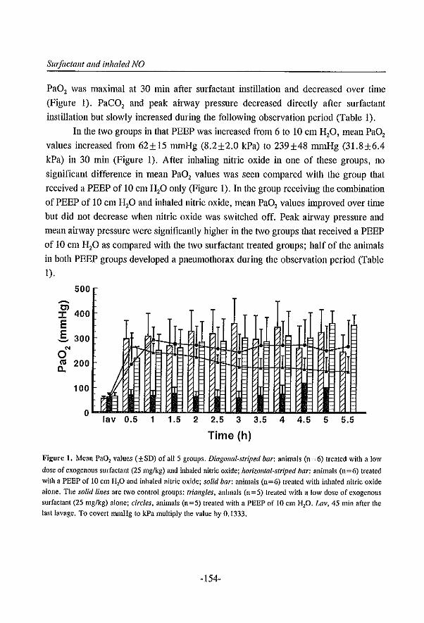
SIII/actallt alld illhaled NO
PaO, was maximal at 30 min after surfactant instillation and decreased over time
(Figure 1). PaeO, and peak airway pressure decreased directly after surfactant
instillation but slowly increased during the following observation period (Table 1).
In Ole two groups in that PEEP was increased from 6 to 10 cm H,O, mean PaO,
values increased from 62± 15 nunHg (8.2±2.0 kPa) to 239±48 mmHg (31.8±6.4
kPa) in 30 min (Figure 1). After inhaling nitric oxide in one of these groups, no
significant difference in mean PaO, values was seen compared with the group that
received a PEEP of 10 cm H,o omy (Figure 1). In Ole group receiving the combination
of PEEP of 10 cm H,o and inhaled nitric oxide, mean PaO, values improved over tinle
but did not decrease when nitric oxide was switched off. Peak airway pressure and
mean airway pressure were significantly higher in the two groups that received a PEEP
of 10 cm H,o as compared with Ole two surfactant treated groups; half of the animals
in both PEEP groups developed a pneumothorax during the observation period (Table
1).
500 -OJ :I: 400 E ,§. 300
0'" m 200
~ ":~ lav 0.5 1 1.5 2 2.5 3 3.5 4 4.5 5 5.5
Time (h)
Figure 1. Mean Pa02 values (±SD) of all 5 groups. Diagonal-striped bar: animals (0=6) treated with a low
dose of exogenous surfactant (25 mglkg) and inhaled nitric oxide; horizontal-striped bar: animals (0=6) treated
with a PEEP of to em HP and inhaled nitric oxide; solid bar: animals (0=6) treated with inhaled nitric oxide
alone. The solid lilies are two control groups: triangles. animals (0=5) treated with a low dose of exogenous
surfactant (25 mg/kg) alone; circles, animals (0=5) treated with a PEEP of 10 em Hp. Lav, 45 min after the
last lavage. To covert mmHg to kPa multiply the value by 0.1333.
-154-
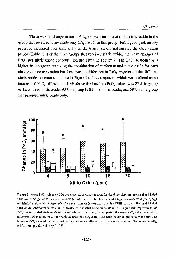
Chapter 9
There was no change in mean PaO, values after inhalation of nitric oxide in the
group that received nitric oxide only (Figure 1). In this group, PaCO, and peak airway
pressure increased over time and 4 of the 6 animals did not survive the observation
period (Table 1). For the three groups that received nitric oxide, the mean changes of
PaO, per nitric oxide concentration are given in Figure 2. The PaO, response was
higher in the group receiving the combination of surfactant and nitric oxide for each
nitric oxide concentration but there was no difference in PaO, response to the different
nitric oxide concentrations used (Figure 2). Non-response, which was defined as an
increase of PaO, of less than 10% above the baseline PaO, value, was 27 % in group
surfactant and nitric oxide; 93 % in group PEEP and nitric oxide; and 58 % in the group
that received nitric oxide only.
~ 100 *
Cl :r: * E 80 * E ~
'" 60 0
'" C-
.!: 40 Q) Cl c 20
'" .c U
0 4
Nitric Oxide (ppm)
Figure 2. Mean PaD) values (±SD) per nitric oxide concentration for the three different groups that inhaled
nitric oxide. Diagonal-striped bar: animals (0=6) treated with a low dose of exogenous surfactant (25 mg/kg)
and inhaled nitric oxide; horiZOlllal-striped bar: animals (0=6) treated with a PEEP of 10 em H20 and inhaled
nitric oxide; solid bar: animals (n=6) treated with inhaled nitric oxide alone. *- = significant improvement of
Pa02 due to inhaled nitric oxide (evaluated with a paired I-test by comparing the mean Pa01 value when nitric
oxide was switched on for 30 min with the baseline Pa02 value). The baseline blood gas value was defined as
the mean Pa02 value of both wash out periods before and after nilric oxide was switched on. To convert nunHg
10 kPa, multiply the value by 0.1333.
-155-
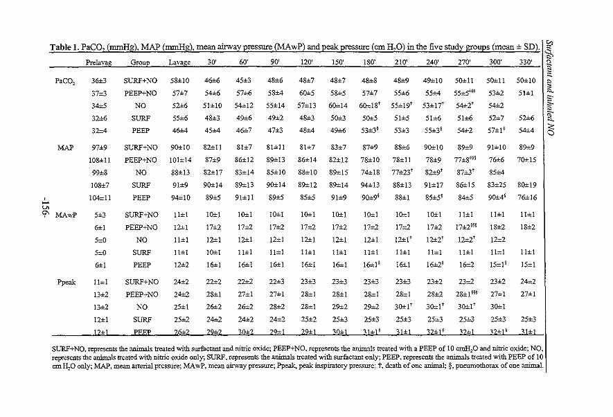
Table 1. PaCO, (mmHg). MAP (mmHg), mean airway pressure (MAwP) and peak pressure (em H,O) in the five study grOUpS (mean ± SD)'I~ Prelavag Group Lavage 30' 60' 90' 120' 150' 180' 210' 240' 270' 300' 330' ~
PaCO,
MAP
36±3
37±3
34±5
32±6
32±4
97±9
108±11
99±8
108±7
SURF+NO
PEEP+NO
NO
SURF
PEEP
58±10
57±7
52±6
55±6
46±4
SURF+NO 90±10
46±6
54±6
51±10
48±3
45±4
82±11
45::3
57±6
54±12
49±6
46±7
81±7
48±6
58±4
55±14
49±2
47±3
SI±ll
48±7
60±5
57±13
48±3
48±4
81±7
48±7
58±5
60±14
50±3
49±6
83±7
48±8 48±9 49±10 50±1l 50±1l
57±7 55±6 55±4 S5±5~§§ 53±2
60±1St 55±19" 53±171" 54±2" 54±2
50±5 51±5 51±6 51±6 52±7
53±3§ 53±3 55±3* 54±2 57±1*
50±1O
51±1
52±6
54±4
87±9 8S±6 90±10 89±9 91±10 89±9
PEEP+NO 101±14 87±9 86±12 89±13 86±14 82±12 78±10 78±11 78±9 77±S'" 76±6 70±15
NO 88±13 S2±I? 83±14 85±10 88±10 89±15 74±18 77±23" 82±9t 87±3" 85±4
SURF 91±9 90±14 89±13 90±14 89±12 89±14 94±13 88±13 91±17 86±15 83±25 80±19
I 104±11 PEEP 94±10 89±5 91±1l 89±5 85±5 91±9 90±9' 88±1 85±S§ 84±5 90±4' 76±16 -'" 9" MAwP 5±3 SURF+NO 11±1 10±1
17±2
12±1
10±1
16±1
10±1
17±2
12±1
11±1
16±1
10±1
17±2
12±1
11±1
16±1
10±1
17±2
12±1
11±1
16±1
10±1
17±2
12±1
11±1
16±1
10±1
17±2
12±1
11±1
16±1 ~
10±1
17±2
10±1
17±2
12±2t
11±1
16±2*
11±1
17±2H*
12±2'
11±1
16±2
11±111±1
ppeak
6±1
5±O
5±O
6±1
1l±1
13±2
13±2
12±1
PEEP+NO
NO
SURF
PEEP
SURF+NO
PEEP+NO
NO
SURF
12±1
11±1
1l±1
12±2
24±2
24±2
25±1
25±2
22±2
28±1
26±2
24±2
22±2
27±1
26±2
24±2
22±3
27±1
28±2
24±2
23±3
28±1
28±1
25±2
23±3
28±1
29±2
25±3
23±3
28±1
29±2
25±3
I2±!'
11±1
16±1
23±3
28±1
30±1t
25±3
23±2 23±2
18±2
12±2
11±1
15±1*
23±2
28±2 28± 1 H* 27± 1
30±1 t
25±3
30±1t
25±3
30±1
25±3
18±2
11±1
15±1
24±2
27±1
25±3
12±1 PEEP 26±2 ~9±2 _~_Q±~~ ____ .22:;tl ________ 3fr¥1_31±L ___ ~l±l __ ---.32±1_§_~~2±~2±1§ '::Il±l
SURF+NO, represents the animals treated with surfactant and nitric oxide; PEEP+NO, represents the animals treated with a PEEP of 10 cmH20 and nitric oxide; NO, represents the animals treated with nitric oxide only; SURF, represents the animals treated with surfactant only; PEEP, represents the animals treated with PEEP of 10 em HP only; :MAP, mean arterial pressure; :MA wP, mean airway pressure; ppeak. peak inspiratory pressure; t, death of one animal; §, pneumothorax of one animal.
"-" :s. s· "'" '" it ~
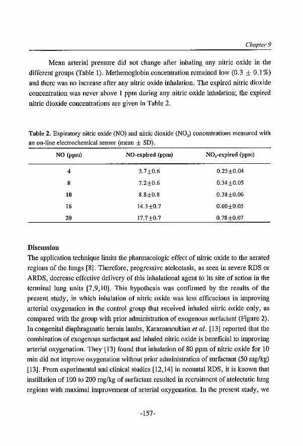
Chapter 9
Mean arterial pressure did not change after inhaling any nitric oxide in the
different groups (Table 1). Methemoglobin concentration remained low (0.3 ± 0.1 %)
and there was no increase after any nitric oxide inhalation. The expired nitric dioxide
concentration was never above 1 ppm during any nitric oxide inhalation; the expired
nitric dioxide concentrations are given in Table 2.
Table 2. Expiratory nitric oxide (NO) and nitric dioxide (NO,) concentrations measured with an on-line electrochemical sensor (mean ± SD).
NO (ppm) NO·expired (ppm) NO,-expired (ppm)
4 3.7±0.6 0.25±0.04
8 7.2±0.6 0.34±0.05
10 8.8±0.8 0.38±0.06
16 l4.3±O.7 0.60±0.05
20 17.7+0.7 0.78±0.07
Discussion
The application technique linlits the pharmacologic effect of nitric oxide to the aerated
regions of the lungs [8]. Therefore, progressive atelectasis, as seen in severe RDS or
ARDS, decrease effective delivery of this inhalational agent to its site of action in the
terminal lung units [7,9,10]. This hypothesis was confIrmed by the results of the
present study, in which inhalation of nitric oxide was less efficacious in improving
arterial oxygenation in the control group that received inhaled nitric oxide only, as
compared with the group with prior administration of exogenous surfactant (Figure 2).
In congenital diaphragmatic hernia lambs, Karamanoukian ef al. [13] reported that the
combination of exogenous surfactant and inhaled nitric oxide is benefIcial to inlproving
arterial oxygenation. They [13] found that inhalation of 80 ppm of nitric oxide for 10
min did not improve oxygenation without prior administration of surfactant (50 mg/kg)
[13]. From experimental and clinical studies [12,14] in neonatal RDS, it is known that
instillation of 100 to 200 mg/kg of surfactant resulted in recruitment of atelectatic lung
regions with maximal improvement of arterial oxygenation. In the present study, we
-157-
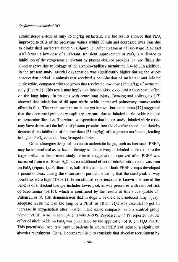
SIII/actallt alld illhaled NO
administered a dose of only 25 mg/kg surfactant, and the results showed that PaO,
improved to 50 % of the prelavage values within 30 min and decreased over time due
to diminished surfactant function (Figure 1). After treatment of late-stage RDS and
ARDS with a low dose of surfactant, transient improvement of PaO, is attributed to
inhibition of the exogenous surfactant by plasma-derived proteins that are ftIling the
alveolar space due to leakage of the alveolo-capillary membrane [14-16]. In addition,
in the present study, arterial oxygenation was significantly higher during the whole
observation period in animals that received a combination of surfactant and inhaled
nitric oxide, compared with the group that received a low dose (25 mg/kg) of surfactant
only (Figure 1). This result may inlply that inhaled nitric oxide had a therapeutic effect
on the lung injury. In patients with acute lung injury, Benzing and colleagues [17]
showed that inhalation of 40 ppm nitric oxide decreased pulmonary transvascular
albumin flux. The exact mechanism is not yet known, but the authors [17] suggested
that the decreased pulmonary capillary pressure due to inhaled nitric oxide reduced
transvascular filtration. Therefore, we speculate that in our study, inhaled nitric oxide
may have decreased the influx of plasma proteins into the alveolar space, and thereby
decreased the inhibition of the low dose (25 mg/kg) of exogenous surfactant, leading
to higher PaO, values in lung-lavaged rabbits.
Other strategies designed to recruit atelectatic lungs, such as increased PEEP,
may be as beneficial as surfactant therapy in the delivery of inhaled nitric oxide to the
target cells. In the present study, arterial oxygenation inlproved after PEEP was
increased from 6 to 10 cm H,o but no additional effect of inhaled nitric oxide was seen
on PaO, (Figure 1). Furthermore, half of the aninlals of both PEEP groups developed
a pneumothorax during the observation period indicating that the used peak airway
pressures were high (Table 1). From clinical experience, it is known that one of the
benefits of surfactant therapy includes lower peak airway pressures with reduced risk
of barotrauma [14,16], which is confirmed by the results of this study (Table 1).
Putensen el al. [18] demonstrated that in dogs with oleic acid-induced lung injury,
adequate recruitment of the lung by a PEEP of 10 cm H,O was essential to get an
increase in oxygenation after inhaled nitric oxide compared with a control group
without PEEP. Also, in adult patients with ARDS, Puybasset el al. [7] reported tllat the
effect of nitric oxide on PaO, was potentiated by the application of 10 cm H,o PEEP.
This potentiation occurred only in patients in whom PEEP had induced a significant
alveolar recruitment. Thus, it seems realistic to conclude that alveolar recruitment by
-158-
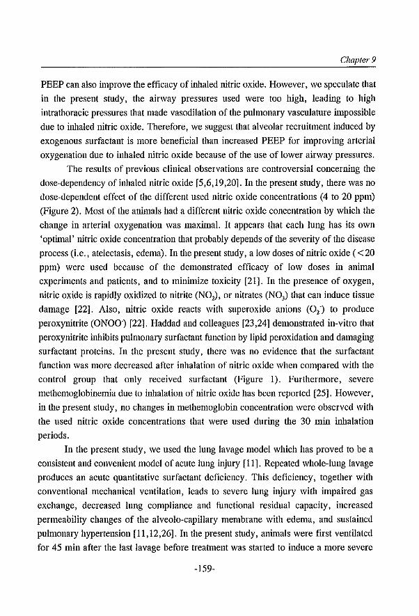
Chapter 9
PEEP can also improve the efficacy of inhaled nitric oxide. However, we speculate that
in the present study, the airway pressures used were too high, leading to high
intrathoracic pressures that made vasodilation of the pulmonary vasculature inlpossible
due to inhaled nitric oxide. Therefore, we suggest that alveolar recruitment induced by
exogenous surfactant is more beneficial than increased PEEP for improving arterial
oxygenation due to inhaled nitric oxide because of the use of lower airway pressures.
The results of previous clinical observations are controversial concerning the
dose-dependency of inhaled nitric oxide [5,6,19,20). In the present study, there was no
dose-dependent effect of the different used nitric oxide concentrations (4 to 20 ppm)
(Figure 2). Most of the animals had a different nitric oxide concentration by which the
change in arterial oxygenation was maximal. It appears that each lung has its own
'optimal' nitric oxide concentration that probably depends of the severity of the disease
process (i.e., atelectasis, edema). In the present study, a low doses of nitric oxide «20
ppm) were used because of the demonstrated efficacy of low doses in animal
experiments and patients, and to minimize toxicity [21). In the presence of oxygen,
nitric oxide is rapidly oxidized to nitrite (NO,), or nitrates (N03) that can induce tissue
damage [22). Also, nitric oxide reacts with superoxide anions (0,") to produce
peroxynitrite (ONOO') [22). Haddad and colleagues [23,24) demonstrated in-vitro that
peroxynitrite inhibits pulmonary surfactant function by lipid peroxidation and damaging
surfactant proteins. In the present study, there was no evidence that the surfactant
function was more decreased after inhalation of nitric oxide when compared with the
control group that only received surfactant (Figure 1). Furthermore, severe
methemoglobinemia due to inhalation of nitric oxide has been reported [25). However,
in the present study, no changes in methemoglobin concentration were observed with
the used nitric oxide concentrations that were used during the 30 min inhalation
periods.
In the present study, we used the lung lavage model which has proved to be a
consistent and convenient model of acute lung injury [11). Repeated whole-lung lavage
produces an acute quantitative surfactant deficiency. This deficiency, together with
conventional mechanical ventilation, leads to severe lung injury with impaired gas
exchange, decreased lung compliance and functional residual capacity, increased
permeability changes of the alveolo-capillary membrane with edema, and sustained
puhnonary hypertension [11,12,26). In the present study, animals were first ventilated
for 45 min after the last lavage before treatment was started to induce a more severe
-159-
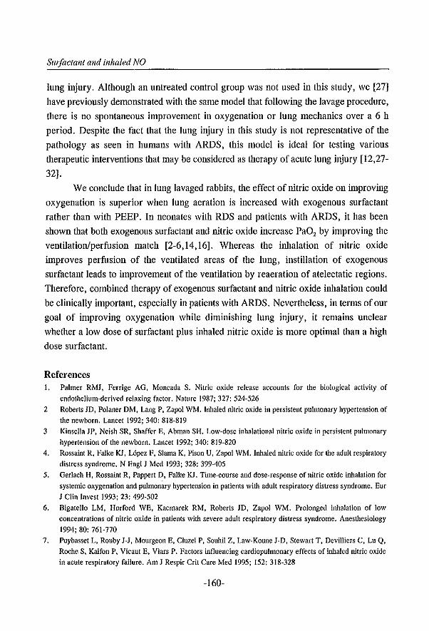
Swiaelalll alld illhaled NO
lung injury. Although an untreated control group was not used in this study, we [27]
have previously demonstrated with the same model that following the lavage procedure,
there is no spontaneous improvement in oxygenation or lung mechanics over a 6 h
period. Despite the fact that the lung injury in this study is not representative of the
pathology as seen in humans with ARDS, this model is ideal for testing various
OJerapeutic interventions that may be considered as therapy of acute lung injury [12,27-
32].
We conclnde that in lung lavaged rabbits, Ole effect of nitric oxide on improving
oxygenation is superior when lung aeration is increased with exogenous surfactant
rather than with PEEP. In neonates with RDS and patients with ARDS, it has been
shown that both exogenous surfactant and nitric oxide increase PaO, by improving the
ventilation/perfusion match [2-6,14,16]. Whereas the inhalation of nitric oxide
improves perfusion of the ventilated areas of the lung, instillation of exogenous
surfactant leads to improvement of the ventilation by reaeration of atelectatic regions.
Therefore, combined therapy of exogenous surfactant and nitric oxide inhalation could
be clinically important, especially in patients with ARDS. NeverOleless, in terms of our
goal of improving oxygenation while diminishing lung iI~ury, it remains unclear
wheOler a low dose of surfactant plus inhaled nitric oxide is more optimal than a high
dose surfactant.
References 1. Palmer RMJ, Pcrrige AG, Moncada S, Nitric oxide release accounts for the biological activity of
endothelium-derived relaxing factor. Nature 1987; 327: 524-526 2 Roberts JD, Polaner DM. Lang P. Zapol WM. Inhaled nitric oxide in persistent pulmonary hypertension of
the newborn. Lancet 1992; 340: 818-819 3 Kinsella JP, Neish SR, Shaffer E, Abman SH. Low-dose inhalational nitric oxide in persistent pulmonary
hypertension of the newborn, Lancet 1992; 340: 819-820 4. Rossaint R. Falke KJ, LOpez F, Slama K. Pison V, Zapol WM. Inhaled nitric oxide for the adult respiratory
distress syndrome. N Engl J Med 1993; 328: 399-405 5. Gerlach H, Rossaint R, Pappert D, Falke KJ. Time-course and dose-response of nitric oxide inhalation for
systemic oxygenation and pulmonary hypertension in patients with adult respiratory distress syndrome. Eur J eUn Invest 1993; 23: 499·502
6. Bigatello LM, Hurford WE, Kacmarek RM, Roberts JD, Zapol WM. Prolonged inhalation of low concentrations of nitric oxide in patients with severe adult respiratory distress syndrome. Anesthesiology 1994; 80: 761-770
7. Puybasset L, Rouby J-J, Mourgeon E, Cluzel P, Souhll Z, Law-Koune J-D, Stewart T, Devilliers C, Lu Q, Roche S, Kalfon P, Vieaut E, Viars P. Factors influencing cardiopulmonary effects of inhaled nitric oxide in acute respiratory failure. Am J Respir Crit Care Med 1995; 152: 318-328
-160-
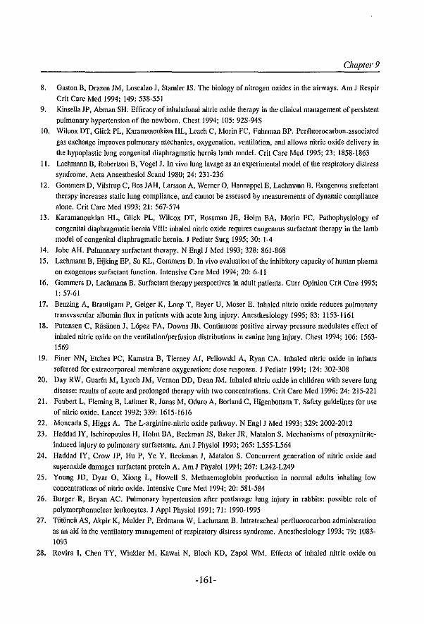
Chapter 9
8, Gaston B. Drazen JM, Loscalzo J. Stamler JS. The biology of nitrogen oxides in the airways. Am J Respir
Crit Care Med 1994; 149; 538-551 9. Kinsella JP, Abman SH. Efficacy of inhaIational nitric oxide therapy in the clinical management of persistent
pulmonary hypertension of the newborn. Chest 1994; 105: 92S-945 to. Wilcox DT. Glick PL, Karamanoukian HL, Leach C, I\.'iorin FC, Fuhrman BP. Perfluorocarbon-associated
gas exchange improves pulmonary mechanics, oxygenation, ventilation, and allows nitric oxide delivery in the hypoplastic lung congenital diaphragmatic hernia lamb model. erit Care Med 1995; 23: 1858-1863
11. Lachmann B, Robertson B. Vogel J. In vivo lung lavage as an experimental model of the respiratory distress syndrome. Acta Anaesthesial Scand 1980; 24: 231-236
12. Gonmters D, Vilstrup C, Bos JAH, Larsson A, Werner 0. Hannappel E, Lachmann B. Exogenous surfactant therapy increases static lung compliance, and cannot be assessed by measurements of dynamic compliance alone. Crit Care Med 1993; 21: 567-574
13. Karamanoukian HL. Glick PL, Wilcox DT. Rossman JE, Holm BA, Morin FC. Pathophysiology of congenital diaphragmatic hernia VIII: inhaled nitric oxide requires exogenous surfactant therapy in the lamb model of congenital diaphragmatic hernia. J Pediatr Surg 1995; 30: 1-4
14. Jobe AH. Pulmonary surfactant therapy. N Engl J Med 1993; 328: 861-868
15. Lachmann B, Eijking EP, So KL, Gommers D. In vivo evaluation of the inhibitory capacity ofhwnan plasma on exogenous surfactant function. Intensive Care Med 1994; 20: 6-11
16. Gommers D, Lachmann B. Surfactant therapy perspectives in adult patients. Curr Opinion Crit Care 1995; 1; 57-61
17. Benzing A, Brautigam P, Geiger K. Loop T. Beyer U, Moser E. Inhaled nitric oxide reduces pulmonary transvascular albumin flux in patients with acute lung injury. Anesthesiology 1995; 83: 1153-1161
18. Putensen C. Rasanen J, L6pez FA. Downs JE. Continuous positive airway pressure modulates effect of inhaled nitric oxide on the ventilation/perfusion distributions in canine lung injury. Chest 1994; 106: 1563-1569
19. Finer NN, Etches PC. Kamstra B, Tierney AJ. PeIiowski A, Ryan CA. Inhaled nitric oxide in infants referred for extracorporeal membrane oxygenation: dose response. J Pediatr 1994; 124: 302-308
20. Day RW, Guarfn M. Lynch JM, Vernon DD, Dean 1M. Inhaled nitric oxide in children with severe lung disease: results of acute and prolonged therapy with two concentrations. Crit Care Med 1996; 24: 215-221
21. Foubert L. Fleming B, Latimer R, Jonas M, Oduro A. Borland C, Higenbottam T. Safety guidelines for use of nitric oxide. Lancet 1992; 339: 1615-1616
22. Moncada S. Higgs A. TIle L-arginine-nitric oxide pathway. N Engl J Med 1993; 329: 2002-2012 23. Haddad IY, Ischiropoulos H, Holm BA, Beckman JS. Baker JR. Matalon S. Mechanisms ofperoxynitrite
induced injury to pulmonary surfactants. Am J Physioi 1993; 265: L555-L564 24. Haddad IY, Crow JP, Hu P, Ye Y, Beckman J, Matalon S. Concurrent generation of nitric oxide and
superoxide damages surfactant protein A. Am J Physiol 1994; 267: L242-L249 25. Young JD, Dyar 0, Xiong L. Howell S. Methaemogiobin production in normal adults inhaling low
concentrations of nitric oxide. Intensive Care Med 1994; 20: 581-584 26. Burger R, Bryan AC. Pulmonary hypertension after post1avage lung injury in rabbits: possible role of
polymorphonuclear leukocytes. J Appl Physiol1991; 71: 1990-1995 27. TUtUncU AS, Akpir K, Mulder P, Erdmann W, Lachmann B, Intratracheal perfluorocarbon administration
as an aid in the ventilatory management of respiratory distress syndrome. Anesthesiology 1993; 79: 1083-1093
28. Rovira I, Chen TY, Winkler M, Kawai N. Bloch KD, Zapol WM. Effects of inhaled nitric oxide on
-161-
Slil/actant alld inhaled NO
pulmonary hemodynamics and gas exchange in an ovine model of ARDS. J Appl Physiol 1994; 76: 345~355 29. Lewis JF, Tabor B, Ikegami M, lobe AH, Joseph M, Absolom D, Lung function and surfactant distribution
in salinc+lavaged sheep given instilled VS. nebulized surfactant. J Appl Physiol1993; 74: 1256-1264-
30. Segerer H, van Gelder W. Angenent FWM, van Woerkens UPM, Curstedt T, Obladen M, Lachmann B.
Pulmonary distribution and efficacy of exogenous surfactant in lung-Iavaged rabbits are influenced by the instillation technique. Pediatr Res 1993; 34: 490-494
31. TiitUncii AS, Faithfull NS, Lachmann B. Intratracheal perfluorocarbon administration combined with
mechanical ventilation in experimental respiratory distress syndrome: dose-dependent improvement of gas
exchange. Crit Care Med 1993; 21: 962-969
32. Lachmann B, Jonson B, Lindroth M, Robertson B. Mode of artificial ventilation in severe respiratory distress
syndrome. Lung function and morphology in rabbits after wash-out of alveolar surfactant. Crit Care Med
1982; 10: 724~732
-162-
SummafY
Summary and conclnsions
Pulmonary surfactant is synthesized by pneumocytes type II cells in the lung and
consists of phospholipids, neutral lipids and at least four specific surfactant proteins.
Its primary function is to reduce the surface tension of the air-liquid interface, which
promotes alveolar expansion and prevents end-expiratory collapse (see Chapter 1).
Pulmonary surfactant is therefore a prerequisite for nornlal breathing, as demonstrated
by the respiratory failure in premature infants suffering from respiratory distress
syndrome (RDS) in whom surfactant production has not yet occurred. Sine 1980,
exogenous surfactant therapy has been successfully used in worldwide trials in neonates
with RDS, almost without any side effects, and this treatment is now routinely used in
most neonatal intensive care units.
Chapter 1 describes diseases other than the neonatal RDS in which abnornlalities
of the pulmonary surfactaut system are at least involved, such as: Acute Respiratory
Distress Syndrome (ARDS), infectious lung disease, etc. Despite the diversity of the
etiologies, the common pathological characteristic is increased alveolo-capillary
permeability associated with damage of the epithelium. The permeability changes,
together with the inflammatory response, result in the influx of proteins and
inflanunatory cells into the airspace with disturbance of the function of the surfactant
system. As a logical consequence, the missing or inactivated surfactant should be
replaced by exogenous surfactant; case-reports and first pilot studies of surfactant
therapy are presented in Chapter 1. Although the results from these studies are not
unequivocal, the best results were seen in those patients treated with high
concentrations, or multiple doses, of surfactant. Besides the concentration of surfactant
used, the reason for lack, or only transient, response after surfactant therapy is further
attributed to the type of surfactant preparation, tinling of instillation, method of
administration and ventilatory support. The literature concerning these issues is briefly
reviewed in last part of Chapter 1, and some of these aspects have been studied in this
work.
Improved oxygenation, decreased ventilator requirements, and increased survival
have been extensively demonstrated in worldwide clinical trials of surfactant therapy
in neonates with RDS. However, little attention has been paid to the effect of
exogenous surfactant on lung mechanics. Chapler 2 presents a study that demonstrates
that, in lung-Iavaged rabbits, treatment with a bolus of exogenous surfactant
immediately reversed hypoxemia and improved functional residual capacity (PRC) and
-163-
Summmy
static compliance related to total lung volumes. However, no significant improvement
in dynamic compliance (calculated from tidal volumes) was found. It was shown that
this discrepancy can be attributed to the increase of FRC and the change of the shape
of Ole pUlmonary pressure-volume (P-V) curve; the increase of maximum compliance
was an expression of this shift of the P-V curve. Thus, the limited ability to detect
changes in pulmonary compliance during mechanical ventilation after surfactant
administration is attributed to the fact that mechanical breathing operates at the
'flattened' upper portion of the P-V curves, resulting in a low value of dynamic
compliance. It was concluded that the effects of surfactant therapy on lung mechanics
are best characterized by changes in functional residual capacity and maximum
compliance obtained from static pressure-volume curves.
Various surfactant preparations are already available on the market for the
treatment of neonatal RDS but data on direct comparison of surfactants are limited. In
the rat lavage model, we compared eight preparations (six natural and two synthetic)
of surfactant on their capacity to improve gas exchange under standardized conditions
with respect to concentration of surfactant, method of administration and ventilator
conditions (see Chapter 3). It was shown that the effect of surfactant was, in general,
dose-dependent. The naturally-derived surfactants were superior to both synthetic
surfactants in inlproving blood gases and it is assumed that the difference lies in the
presence of the surfactant proteins Band C in the natural surfactant preparations.
Further, clear differences in efficacy were found between the six natural surfactants
tested, especially after reduction of the PEEP level. In addition, the rat lavage model
in combination with Ole used ventilator protocol proved to be very useful for testing the
efficacy of a surfactant batch under in-vivo conditions.
In a model of acid aspiration, we have shown that respiratory failure can be
prevented when exogenous surfactant was given before deterioration of lung function
(Le. within 10 min after acid aspiration), whereas after development of respiratory
failure exogenous surfactant served only to prevent further deterioration of lung
function but did not restore gas exchange (see Chapter 4). From these results it was
concluded that surfactant should be given as soon as possible to prevent development
of respiratory failure. In addition, illlung-lavaged rats we demonstrated that exogenous
surfactant given at an early stage of lung injury resulted in a sustained improvement of
lung function, whereas lung function deteriorated when a bolus of surfactant was given
at a relative late time point in lung injury, probably due to an increased amount of
-164-
Summary
proteins (see Chapter 6). Therefore, it is expected that early treatment of patients with
ARDS may require smaller amounts of exogenous surfactant and the outcome results
will probably be better.
The surfactant deficiency that is likely to develop when ARDS is becoming
manifest could be treated by reducing the protein concentration and/or by increasing
the content of pulmonary surfactant. Plasma-derived proteins are known to be potent
inhibitors of surfactant function, in a dose-dependent way. Chapter 5 presents a study
we designed to investigate the amount of exogenous surfactant needed to overcome the
inhibitory components in human plasma. Lung lavaged rats received intratracheally
human plasma mixed WiOl surfactant at different concentrations. It was found that when
mixed WiOl plasma, surfactant at a concentration of 50 mg/kg showed no improvement,
at 100 mg/kg there was only a transient improvement, and at 300 mg/kg there was a
sustained improvement of arterial oxygenation during the 3 hour study period. From
these results, it was concluded that approximately 1 mg surfactant phospholipids is
required to overcome the inhibitory effect of 1 mg plasma proteins. For clinical practice
this means that an excess of surfactant should be given, or repeatedly be substituted
('titrated') at low concentrations, until blood gases inlprove. Chapter 6 focuses on the
therapeutic role of bronchoalveolar lavage (BAL). To evaluate this, prior to surfactant
therapy BAL was performed to lower the intrapulmonary protein content thereby
increasing the surfactant-inhibitor ratio. In this study, the rat lavage model was
modified: lung-Iavaged animals were ventilated for 3 hours, before surfactant
treatment, to induce a more severe lung injury. Different surfactant regimes were
evaluated: bolus of surfactant with or without prior lung lavage with saline or with a
diluted surfactant suspension. It was shown that lung lavage with a diluted surfactant
suspension together with instillation of exogenous surfactant resulted in a sustained
inlprovement of lung function, whereas lung function deteriorated during the
subsequent 3 hours with the other regimes. In contrast to lung lavage with saline, we
suggest that lavage with a diluted surfactant suspension is less harmful for the lungs,
removes more of the potent surfactant inhibitors, and contributes to a more even
surfactant distribution over the lungs. Bronchoscopic application of a large quantity of
exogenous surfactant has been used successfully in patients with ARDS. A lung lobe
can easily be lavaged with a flexible fiberoptic bronchoscope, and we therefore suggest
to lavage the lungs with a diluted surfactant suspension prior to surfactant instillation
in order to save exogenous surfactant in the treatment of severe ARDS.
-165-
Summaryl
Chapters 7 alld 8 adress the effect of different ventilator pattern on surfactant
function. In Chapter 7, the effect of high-frequency oscillatory ventilation (HFOV) was
compared with conventional mechanical ventilation (CMV) on lung function and
histopathology after surfactant instillation in lung-lavaged rabbits. HFOV was used in
combination with the so-called 'high-lung volume strategy' in order to mininlize
ventilator induced lung injury. It was shown that surfactant combined with CMV
resulted in the same sustained inlprovement of gas exchange as seen with HFOV and
surfactant. Also, histopathologic examinations showed no difference in lung injury
between HFOV and CMV after surfactant therapy. From these results, it was concluded
that surfactant therapy with CMV is equally effective to prevent ventilator-induced lung
injury as HFOV combined with surfactant, as long as alveoli are opened and kept open
to avoid shear stress. In another study, we investigated the effect of different ventilator
settings with CMV on exogenous surfactant therapy with respect to gas exchange,
protein influx and conversion of active into non-active surfactant in lung-lavaged rats
(see Chapter 8). It was shown that PEEP had a major impact on the effect of exogenous
surfactant therapy on arterial oxygenation and settings that combine small pressure
amplitudes with high levels of PEEP best preserve the large aggregate, surface-active
component of exogenous surfactant and resulted in lower protein concentration of the
BAL fluid. Further, it was shown that CMV with a mode of ventilation that creates
auto-PEEP, compared to a mode that applies the same level of PEEP by static PEEP
alone, was more effective to remove carbon dioxide.
Chapter 9 describes the use of inhaled nitric oxide in combination with
exogenous surfactant. In lung-lavaged rabbits, blood gases were flrst increased either
by instillation of a low dose of exogenous surfactant (25 mg/kg) or by increasing the
PEEP from 6 to 10 cm H20, before inbalation of nitric oxide was started. Additional
increases of oxygenation were seen after inhalation of nitric oxide but only in the
animals that received a low dose of surfactant. The control group, that received inhaled
nitric oxide only, showed no signiflcant change in oxygenation. From these results, we
hypothesized that progressive atelectasis decrease effective delivery of this inhalational
agent to its site of action in the terminal lung units, and that surfactant increased the
ventilated area in the lung by recruiting previously collapsed alveoli; inhaled nitric
oxide selectively improves the perfusion of these ventilated areas, causing the additional
effect of exogenous surfactant on the effect of nitric oxide inhalation on arterial
oxygenation. In this study, we suggest that the alveolar recruitment induced by
-166-
Summmy
exogeno.us surfactant is more beneficial than increased PEEP for improving arterial
oxygenation due to inhaled nitric oxide because of the use of lower airway pressures.
The work presented in this thesis demonstrates that the response of exogenous
surfactant can be improved by different aspects, such as; the use of a highly active
natural surfactant, early administration in the disease stage, removal of inhibitors by
lung lavage with a diluted surfactant suspension, mechanical ventilation with sufficient
level of PEEP and small pressure amplitudes, and in combination with inhaled nitic
oxide. It is expected that the improved responsiveness may require smaller amounts of
exogenous surfactant and the clinical outcome results will probably be better. The
prohibitive price and the non-availability of large amounts of surfactant, make
surfactant therapy not yet feasible for routine application in adults; it is therfore
important to explore ways of keeping the administered surfactant as small as possible
in the treatment of ARDS and ARDS-like syndromes.
-167-
Samet/vatting
Samenvatting en conclllsies: Het surfactant in de longen wordt gemaakt door de type II pneumocyten en bestaat uit
fosfolipiden, neutrale lipiden en uit ten minste vier specifieke surfactant eiwitten. Zijn
primaire funktie is het verlagen van oppervlakte spalUling op de lucht-vloeistof
overgang, wat de alveolaire expansie bevordert en eind-expiratoire collaps voorkomt
(zie Hoofdstllk 1). Longsurfactant is daarom een eerste vereiste voor een normale
adembaling zoals is aangetoond bij longfalen in te vroeg geboren kinderen, het zgn.
'Respiratory Distress Syndrome' (RDS), waar produktie van surfactant nog niet
plaatsvindt. Sinds 1980 is wereldwijd met succes exogeen surfactant gebruikt in studies
bij neonaten met RDS. Bijwerkingen traden hierbij nauwelijks op en de behandeling
wordt nu routinematig gebruikt op de meeste neonatale intensive care's.
Hoofdstllk 1 beschrijft naast de neonatale vornl van RDS andere ziektebeelden
waarin stoornissen in het pulmonair surfactant systeem een rol spelen, zoals: 'Acute
Respiratory Distress Syndrome' (ARDS), infectieuse longaandoeningen, etc. Ondanks
de verscheidenheid in oorzaken is de gemeenschappelijke pathologische basis een
toename in de permeabiliteit van capillairen met epitheel beschadiging. Permeabiliteits
veranderingen in comb ina tie met een onstekingsreaktie gaan gepaard met instroom van
eiwitten en onstekingscellen in de luchtwegen, waarbij het surfactant systeem verstoord
raakt. Als logisch gevolg zou het verdwenen of gelnactiveerde surfactant vervangen
moeten worden door exogeen surfactant; 'case reports' en de eerste orienterende
klinische studies met surfactant therapie worden gepresenteerd in Hoofdstllk 1.
Alhoewel de resultaten van deze studies niet eenduidig zijn, werden de beste resultaten
gezien in die patienten behandeld met hoge concentraties, of na herhaalde toediening
van surfactant. Naast de concentratie van het surfactant, wordt de reden van geen, of
aileen een tijdelijk effekt na surfactant behandeling, verder toegeschreven aan het type
surfactant, tijdstip van toedieuing, methode van toediening en methode van
beademingsondersteuuing. De literatuur betreffende deze onderwerpen wordt in grote
lijnen besproken in het laatste deel van Hoofdstllk 1 en een aantal van deze zaken wordt
bestudeerd in deze dissertatie.
In wereldwijde klinische studies naar surfactant behandeling in neonaten met
RDS zijn een verbeterde oxygenatie, verminderde beademingsbehoefte, en een
toegenomen overleving duidelijk aangetoond. Weinig aandacht is er echter besteed aan
de effecten van exogeen surfactant op de longmechanica. In Hoofdslllk 2 wordt
aangetoond dat in konijnen, na longlavage, behalldeling met een bolus van exogeell
-168-
Samellvattillg
surfactant onmiddellijk de bestaande hypoxie opheft en de functionele residuale
capaciteit (FRC) en de statische compliantie gerelateerd aan het totaal long volume
verbetert. Een significante verbetering van de dynamische compliantie (berekend uit het
ademvolume) werd echter niet gevonden. Aangetoond werd dat deze discrepantie kan
worden toegeschreven aan de toename van de FRC en de verandering van vorm van de
druk-volume curve; verschuiving van de druk-volume curve heef! een toename van de
maximale compliantie tot gevolg. Dus, de beperkte mogelijkheid om veranderingen in
pulmonaire compliantie te detecteren gedurende mechanische beademing na surfactant
toediening wordt toegeschreven aan het feit dat mechanische ademhaling opereert op
het 'vlakke' bovenste gedeelte van de druk-volume curve, resulterend in een lage
waarde vau de dynamische compliantie. Er wordt geconcludeerd dat de effecten van
surfactantbehandeling op de longmechanica het beste beschreven kunnen worden aan
de hand van veranderingen in de functionele residuale capaciteit en veranderingen in
de maximale compliantie zoals verkregen uit statische druk-volume curves.
Verschillende surfactant preparaten voor de behandeling vanneonataal RDS zijn
op de markt, maar er zijn slechts beperkte gegevens over direkte vergelijking tussen
surfactants beschikbaar. In het rat-lavage model hebben we van acht surfactant
preparaten (zes natuurlijke en twee synthetische) de capaciteit om de gasuitwisseling
te verbeteren vergeleken, onder gestandaardiseerde condities met be trekking tot
concentratie van surfactant, methode van toediening en beademingscondities (zie
Hoofdslllk 3). Aangetoond wordt dat het effect van surfactant in het algemeen
dosisafhankelijk is. De natuurlijk verkregen surfactants waren superieur ten opzichte
van be ide synthetische surfactants in de verbetering van bloedgaswaarden en
aangenomen werd dat de aanwezigheid van de surfactant eiwitten B en C in de
natuurlijke surfactants verantwoordelijk is voor dit verschil. VerdeI' werden duidelijke
verschillen in werkzaamheid gevonden tussen de zes geteste natuurlijke surfactants,
vooral na vermindering van het PEEP niveau. Daarnaast bewees het rat-lavage model
in comb ina tie met het gebruikte beademingsprotocol zeer bruikbaar te zijn om de
werkzaamheid van een surfactant batch onder in-vivo condities te testen.
In een zuur-aspiratie model werd aangetoond dat respiratoir falen kan worden
voorkomen wanneer exogeen surfactant werd toegediend v66rdat verslechtering van de
longfunktie optrad (dat betekent bitmen 10 minuten na het aspireren van zuur). Na de
ontwikkeling van respiratoir falen diende exogeen surfactant aIleen nog tel' voorkoming
van verdere verslechtering van de longfunktie, de gasuitwisseling herstelde niet (zie
-169-
Samellvattillg
Hoofdslllk 4). Aan de hand van deze resultaten werd geconcludeerd dat surfactant lo
snel mogelijk moet worden toegediend om de ontwikkeling van respiratoir falen te
voorkomen. Daarnaast hebben we in long-gelaveerde ratten aangetoond dat exogeen
surfactant toegediend in een vroeg stadium van longschade resulteerde in een blijvende
verbetering van de longfunktie, terwijl de longfunktie verslechterde wanneer een bolus
surfactant was toegediend op een relatief laat tijdstip in het proces van longschade,
waarschijnlijk als gevolg van de toegenomen hoeveelheid eiwitten in de alveolus (zie
Hoofdslllk 6). Daarom is het te verwachten dat er bij een vroege behandeling van
patienten met ARDS k1einere hoeveelheden van exogeen surfactant nodig zullen zijn
met waarschijnlijk een beter resultaat.
Het surfactant tekort dat zich naar verwachtiging ontwikkelt bij een manifest
ARDS beeld, kan worden behandeld door middel van vermindering van de eiwit
concentratie enlof verhoging van de hoeveelheid alveolair surfactant. Eiwitten
afkomstig uit plasma zijn bekende, krachtige inbibitoren van de surfactant funktie, met
een dosis-afbankelijke werking. In de studie beschreven in Hoofdslllk 5 onderzochten
we hoeveel exogeen surfactant nodig is am de rennning door humane plasma
componenten op te heffen. Long-gelaveerde ratten kregen intratracheaal humaan plasma
gemengd met surfactant in wisselende concentraties toegediend. Gevonden werd dat
surfactant, wamleer vermengd met plasma, in een concentratie van 50 mg/kg geen
verbetering liet zien. Bij 100 mg/kg was er slechts een voorbijgaande verbetering en
bij 300 mg/kg was er een aanhoudende verbetering van de arteriele oxygenatie tijdens
de studieperiode van 3 UUI. Uit deze resultaten werd geconcludeerd dat ongeveer 1 mg
surfactant fosfolipiden nodig is am het inhibitoire effekt van 1 mg plasma eiwitten op
te heffen. Voor de k1iniek betekent dit dat een overmaat aan surfactant gegeven zou
moeten worden of dat herhaaldelijk surfactant gesubstitueerd moet worden (,getitreerd ')
inlage concentraties, totdat de bloedgassen verbeteren. In Hoofdslllk 6 wordt broncho
alveolaire lavage uitgevoerd voorafgaand aan surfactant therapie teneinde het
intrapulmonaire eiwitgehalte te verJagen en daardoor de surfactant-inhibitor ratio te
verhagen. Voor deze studie werd het rat-lavage model zodanig veranderd dat long
gelaveerde dieren eerst 3 uur werden beademd v66r surfactant therapie om een
ernstigere longbeschadiging te induceren. Verschillende surfactant reginles werden
geevalueerd: een surfactant bolus met of lOnder voorafgaande long-lavage, met
fysiologische zoutoplossing of verdunde surfactant suspensie. Aangetoond werd dat
long-lavage met verdunde surfactant suspensie samen met toediening van exogeen
-170-
Samellvattillg
surfactant resulteerde in een aanhoudende verbetering van de long funk tie, terwijl de
longfunktie in de volgende 3 uren verslechterde bij de andere reginles. We suggereren
dan oak dat, waIlIleer vergeleken met long-lavage met fysiologische zoutoplossing,
long-lavage met verdunde surfactant suspensie minder schadelijk is voor de long, meer
van de krachtige surfactant inhibitoren verwijdert en bijdraagt aan een meer
gelijkmatige surfactant distributie over de longen. Bronchoscopische toediening van een
grote hoeveelheid exogeen surfactant is met succes toegepast in patienten met ARDS.
Met een flexibele fiberoptische bronchoscoop kan een longkwab eenvoudig worden
gespoeld en we suggereren daarom de longen te laveren met een verdunde surfactant
suspensie alvorens surfactant toe te dienen, teneinde te besparen op exogeen surfactant
in de behandeling van ernstige ARDS.
In de Hoojdslllkkell 7 ell 8 wordt het effect van verschillende beademingsvomlen
geevalneerd. In Hoojdslllk 7 worden de effecten van 'high-frequency-oscillatory
ventilation' (HFOV) op longfunktie en histopathologie vergeleken met die van
conventionele mechanische beademing (CMV) na toediening van surfactant aan long
gelaveerde konijnen. Om de door de beademing gelnduceerde longschade te
minimaliseren, werd HFOV gebruikt in combinatie met de zgn. 'high-Iung-volume
strategy'. Aangetoond werd dat surfactant gecombineerd met CMV resulteerde in
dezelfde aanhoudende verbetering van gasuitwisseling als gezien wordt bij HFOV en
surfactant. Daarbij liet histopathologisch onderzoek geen verschil in longschade zien
tussen HFOV en CMV na surfactant therapie. Uit deze resultaten werd de conclusie
getrokken dat surfactant therapie met CMV net zo effektief is in preventie van
longschade t.g. v. beademing als HFOV in combinatie met surfactant, zolang alveoli
open zijn en open gehouden worden om 'shear stress' te vermijden. In een andere
studie onderzochten we de invloed van verschillende ventilator instellingen bij CMV
op het effekt van exogene surfactant therapie betreffende gasuitwisseling, eiwit influx
en omzetting van aktief in luet-aktief surfactant in long-gelaveerde ratten (zie Hoojdslllk
8). Getoond werd dat PEEP een belangrijke inlpact heef! op het effekt van exogene
surfactant therapie op arteriele oxygenatie en dat instellingen waarbij kleine druk
amplitudes gecombineerd worden met een hoge PEEP het beste de grote aggregaten van
exogeen surfactant (de oppervlakte-aktieve component) preserveren en resulteren in een
lagere eiwit concentratie van de BAL vloeistof. Verder werd aangetoond dat CMV met
een beademingsvorm waarbij auto-PEEP optreedt effectiever is in het verwijderen van
kooldioxide (CO,), vergeleken met een vorm waarbij dezelfde PEEP-waarde aIleen
-171-
Samellvatting
door statische PEEP tot stand komt.
In HoofdsllIk 9 bestudeerden we het gebruik van stikstof monoxide (NO) in
combinatie met exogeen surfactant. Bij long-gelaveerde konijnen werden de bloed
gas sen eerst verhoogd, 6f door toediening van een lage dosis exogeen surfactant (25
mg/kg) 6f door de PEEP te verhogen van 6 naar 10 em H,o, voordat de inhalatie van
stikstof monoxide werd gestart. Na stikstof monoxide inhalatie werd aileen in de dieren
die een lage dosis surfactant ontvingen een extra stijging van de oxygenatie
waargenomen. De controle groep, die aileen stikstof monoxide inhaleerde, liet geen
significante verandering in oxygenatie zien. Uitgaande van deze resultaten,
postuleerden we de hypothese dat voortgaande atelectase de effectieve afzet van dit
gei'nhaleerde agens in de alveoli vermilldert en dat surfactant bet geventileerde gebied
in de long vergroot door beropening van eerder gecollabeerde longblaasjes; geinhaleerd
stikstof monoxide verbetert selectief de perfusie van deze geventileerde gebieden, wat
tot bet additionele effekt van exogeen surfactant op het effekt van stikstof monoxide
inhalatie leidt. In het huidige onderzoek stellen we dat heropening van alveoli die tot
stand gebracht wordt door exogeen surfactant gunstiger is met betrekking tot de
verbetering van de arterieJe oxygenatie door geinhaleerd stikstof monoxide dan die door
PEEP, omdat er sprake is vanlagere drukken in de long.
Het werk dat gepresenteerd wordt in dit proefschrift laat zien dat de respons op
exogeen surfactant verbeterd kan worden door verschillende factoren, zoals het gebruik
van een natuurlijk surfactant met een boge aktiviteit, vroegtijdige toediening ervan in
het ziekte proces, verwijdering van inhibitoren door longlavage met een verdunde
surfactant suspensie, combinatie met mecbanische beademing met een voldoende hoge
PEEP en kleine drukamplitudes, en combinatie met gei'nhaleerd stikstof monoxide. Het
is te verwachten dat door de verbeterde respons kleinere hoeveelheden exogeen
surfactant nodig zullen zijn met waarschijniijk betere resultaten. De exorbitant hoge
prijs en het niet beschikbaar zijn van grote hoeveelheden exogeen surfactant, maken dat
surfactant therapie in volwassenen nog niet haalbaar is. Het is daarom belangrijk verder
onderzoek te doennaar manieren om de de hoeveelheid toe te dienen surfactant zo laag
mogelijk te houden voor de behandeling van ARDS en ARDS-achtige aandoeningen.
-172-
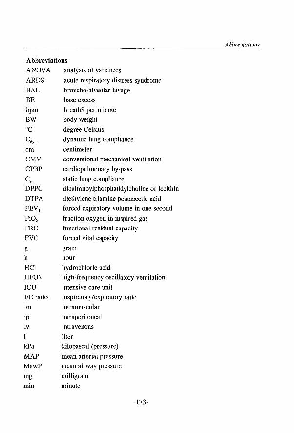
Abbreviations
ANOVA
ARDS
BAL BE bpm
BW
"C
Cdyn
cm
CMV
CPBP
C" DPPC
DTPA
FEV, FiO,
FRC FVC g
h
HCl
HFOV ICU
I/E ratio
im
ip
iv
kPa MAP
MawP
mg
min
analysis of variances
acute respiratory distress syndrome
broncho-alveolar lavage
base excess
breathS per minute
body weight
degree Celsius
dynamic lung compliance
centimeter
conventional mechanical ventilation
cardiopulmonary by-pass
static lung compliance
dipalmitoylphosphatidylcboline or lecithin
diethylene triamine pentaacetic acid
forced expiratory volume in one second
fraction oxygen in inspired gas
functional residual capacity
forced vital capacity
gram
hour
hydrochloric acid
high-frequency oscillatory ventilation
intensive care unit
inspiratory/expiratory ratio
intramuscular
intraperitoneal
intravenous
liter
kilopascal (pressure)
mean arterial pressure
mean airway pressure
milligram
minute
-173-
Abbreviations
Abbreviations
mI
mmHg
NaCl
NO p PaO,
PaCO,
PEEP
PG
Ppeak
PIP
P/L ratio
ppm
PV curve
milliliter
millimeters of mercury (pressure)
sodium chloride
nitric oxide
probability
arterial oxygen partial pressure or arterial oxygenation
arterial carbon dioxide partial pressure
positive end-expiratory pressure
phosphatidylglycerol
peak inspiratory pressure
peak inspiratory pressure
protein/lipid ratio
parts per million
pressure volume curve
RDS respiratory distress syndrome
SA/LA ratio small (non-active) to large (active) surfactant aggregate ratio
SD standard deviation
SF, S/I ratio
SP
TLC
sulfur hexafluoride surfactant/inhibitor ratio
surfactant protein (A, B, C, or D)
total lung capacity
tidal volume
-174-
Dankwoord
Dal1kwool'd
Eind 1985 kwam ik als student op de afdeling anesthesiologie en werd vrijwel meteen
gegrepen door het virus 'surfactant'. lk had toennog geen idee dat ik voor zo'nlange
tijd zau blijven hangen. Zonder de inspirerende ideeen, hulpvaardigheid, inzet,
doorzettingsvermogen, enthousiasme en vooral gezelligheid van vele mensen zou dit
proefschrift nooit tot stand gekomen zijn, en daarvoor wil ik de volgende mensen
bedanken.
Margriet, aan jou ben ik de meeste dank verscllUldigd. Het is bewonderens
waardig waar je telkens weer de energie en het gedllld vandaatl haalt om met een zeer
eigenwijze 'surfactant-monomaan' om te blijven gaan.
Prof. dr B. LachmalU1. Beste Burkhard, U wil ik bedanken voor de gelegenheid
die U mij gaf om onderzoek te doen en de resultaten te mogen presenteren op vele
mooie plekken op deze wereldbol. Uw inspirerende ideeen en enthousiasme creeren een I
sfeer op uw afdeling waardoor onderzoek doen geen werken is maar een hobbie.
Prof. dr W. Erdmann, prof. dr L.M.G. van Golde, prof. dr D. Tibboel en prof.
dr P.D. Verdouw, ben ik zeer erkentelijk voor de bereidheid dit proefschrift te
beoordelen op zijn wetenschappelijke waarde.
I wish to express my gratitude to prof. dr W. Kox and prof. dr P. Wollmer for
their willingness to be a member of the conmlittee at the defence of my thesis.
Laraine Visser. Jouw rode stift was altijd weer een verschrikking maar zander
dat was de boodschap van menig artikel beperkt gebleven tot Nederland en Belgie.
Eric Eijking. Eikel, door jouw inzet en zelfdiscipline had ik al heel wat jaartjes
eerder kullllen promoveren. De basis van dit proefschrift was ook de basis van een
mooie vriendschap.
AllIIemarie van 't Veen. Jouw kennis van de statistiek was een essentiele bijdrage
aan dit proefschrift. De drang om beter te zijll dan de matlllen op de afdeling gaf soms
aanleiding tot hevige emoties maar deze waren altijd goed 'oplosbaar' in alcohol.
Jelle Bos. Ouze wetenschappelijke discussies op het terras van Melief Bender
bleken de bron te zijn van mellig idee voor onderzoek zaals beschreven ill dit
proefscln·ift. Je aanbieding om de tekst van dit boekje te corrigeren viel helaas ill het
'zee' -water door een plotse, hevige 'verborgen' liefde.
De 'jongens' van de afdeling Serge, Edwin, Arthur, EIlIIO, Stephan en Gilberto
wil ik bedanken voor hun inzet ell hulp bij menig experiment. Tevens moet ik jllllie
maar eens excuses aanbieden voor het eenwige gepest en het irritante lachje. Serge,
-175-
Dankwoord
door jouw inzet en schrijfkunst kwam het einde van dit proefschrift toch ineens in ziehl.
Anne De Jaegere. ArnIe dank ik voor het introduceren van de HFO die de
'Lachrnann' -beademing bijna in gevaar brachl.
Mijn lotgenoten van de eerste jaren, Geert-Jan van Daal, Rob Tenbrinck, Johan
Mouton, Arthur Voogt, Robert-Jan Houmes en Ralph So, dank ik voor de zitmige,
maar zeker ook de onzinnige discussies in menig horeca gelegenheid in Rotterdam.
I thank Prof. dr Ewald Hannappel for the hospitality at his lab where I learned
how to make the surfactant which was a prerequisite for all experitnents in this thesis.
I thank Dr Carsten Vilstrup for his valuable contribution to parts of the work
presented in this thesis.
En als laatste dank ik mijn broers en ouders voor het geduld met mij en de altijd
aanwezige belangstelling voor mijn 'hobbies'.
-176-
Publikatieli;st
Artikelen: 1. D. Gonuners, K.L. So, S, Annbruster, R. Tenbrinck, I.L.M. van Rcmortcl, I.E. van Eyk, B. Lachmann.
Human surfactant extracted from vaginal-delivered amniotic tluid improves lung mechanics as effectively as natural surfactant derived from bovine lungs. In: Progress in respiration research; Basic research on lung surfactant. P. von Wichert, B. Muller (Eds), Karger, Basel, 1990, vol 25, pp 302-304
2. G.J. van Daal, K.L. So, D. Gonm1crs, E,P. Eijking, R.B. Fievez, M,L Sprenger, D.W. van Dam, B. Lachmann. Intratracheal surfactant administration restores gas exchange in experimental adult respiratory distress syndrome associated with viral pneumonia. Aneslh Analg 1991; 72: 589-595
3. \V.Th. Trouerbach, S,A. de Man, D. Gonmlcrs, A.W. Zwambom, D.E. Grobbee. Determinants of bone mineral content in childhood. Bone Mineral 1991; 13: 55-67
4. A.P. Bos, D. Tibboel, F.W.J. Hazebroek, J.C. Molenaar, B. Lachmann, D. Gommers. Surfactant replacement therapy in high-risk congenital diaphragmatic hernia. Lancet 1991; 338: 1279 (letter)
5. D. Gontmers, GJ. van Daal, B. Lachmann. Oxygen uptake in the lungs under pathological conditions and its therapeutic efforts. Adv Exp Med Bioi 1992; 317: 47-54
6. E.P. Eijking, D. Gountlers, L Kuliander, E. de Buijzer, R.W. Bcukenholdt, B. Lachmann. Different surfactant treatment strategies for respiratory failure induced by hydrochloric acid aspiration in rats. Adv E,p Med BioI1992; 317: 349-355
7. G.L van Daal, LA.H. Bas, E.P. Eijking, D. Gonmlers, E. Hannappel, B. Lachmann. Surfactant replacement therapy improves pulmonary mechanics in end-stage influenza A pneumonia in mice. Am Rev Respir Dis 1992; 145: 859-863
8. C. Vilstrup. D. Gonmlers, J.A.H. Bas, B. Lachmann, O. Werner, A. Larsson. Natural surfactant instilled in premature lambs increases lung volume and improves ventilation homogenity within five minutes. Pediatr Res 1992; 32: 595-599
9. E.P. Eijking, D. Gonmlcrs, K.L. So, M.P.M. de Maat, 1.W. Mouton, B. Lachmann. Prevention of respiratory failure after hydrochloric acid aspiration by intratracheal surfactant instillation in rats. Anesth An.1g 1993; 76: 472-477
10. D. Gonmlcrs. C. Vilstrup. l.A.H. Dos. A. Larsson, O. Werner, E. Hannappel, B. Lachmann. Exogenous surfactant therapy increases Sialic lung compliance, and cannot be assessed by measurements of dynamic compliance alone. Crit Care Med 1993; 21: 567-574
11. B. Lachmann. D. Gonmlers. Is it rational to treat pneumonia with exogenous surfactant? Eur Respir J 1993; 6: 1427-1428 (editorial)
12. E.P. Eijking. D. Gommers, K.L. So, M. Vergeer, B. Lachmann. Surfactant treatment of respiratory failure induced by hydrochloric acid aspiration in rats. Anesthesiology 1993; 78: 1145-1151
13. B. Lachmann, D. Gommers, E.P. Eijking. Exogenous surfactant therapy in adults. Atemsw-Lungenkrkh 1993; 19: 581-591
14. D. Gonmlers, D. Lachmann. Surfactant therapy: does it have a role in adults? Clin Intensive Care 1993; 4: 284-295
15. K.L. So, D. Gommers, B. Lachmann. Bronchoalveolar surfactant system and intratracheal adrenaline. Lancet 1993; 341: 120-121 (letter)
-177-
Publikalielijsl
16. B. Lachmann, B,P. Eijking, K.L. So, D. Gonmlcrs. In vivo evaluation of the inhibitory capacity of human plasma on exogenous surfactant function. Intensive Care Med 1994; 20: 6-1 I
17. B. Lachmann, D. Gammers. Surfactant treatment for neonatal lung diseases other than the idiopathic respiratory distress syndrome. Lung & Respiration 1994; 11: 35-39
18. A. van '( Veen, J.W. Mouton, D. Gommers, J.AJ.W. Kluytmans, P. Dekkers, B. Lachmann. Influence of pulmonary surfactant on in vitro bactericidal activities of Amoxicillin. Ceftazidime and Tobramycin. Anlimicrob Agents Chemolhcr 1995; 39: 329-333
19. D. Gonmlcrs, B. Lachmann. Surfactant therapy in the adult patient. Curr Opin Cril Care 1995; 1: 57-61
20. A. Hartog, D. Gammers. B. Lachmann. Role of surfactant in the pathophysiology of the acute respiratory distress syndrome (ARDS). Monaldi Arch Chest Dis 1995; 50: 372-377
21. E.P. Eijking, D. Gommers, K.L. So, M.P.M. de Maat, B. Lachmrum. A model for acute respiratory failure induced by tracheal instillation of homologous plasma or protein-rich pulmonary oedema fluid. Appl Cardiopulmon Pathophysioll995; 5: 129-134
22. D. Gonuners. A.S. TIitiincii, B. Lachrnann. ARDS treatment in neonatal period: rationale for exogenous surfactant therapy and partial fluid ventilation with perfluorocarbons. Dev Physiopath Clin 1995; 6: 19-32
23. S.LC. Verbrugge. D. Gommers. B. Lachmann. Liquid lung ventilation as an alternative ventilatory support. Curr Opin Anaesth 1995; 8: 551-556
24. A. van 't Veen, D. Gonuners. J.W. Mouton. J.AJ.W. Kluytmans. EJ. Krijt, B. Lachmann. Exogenous pulmonary surfactant as a drug delivering agent: influence of antibiotics on surfactant activity. Br J Pharmacol 1996; 118: 593-598
25. B. Lachmann, D. Gammers, N.G. Rajan. Rationale and techniques to improve ventilation and gas exchange in acute lung injury. J Jpn Med Soc Bioi Interface 1996; 26 (Supp!): 115-137
26. SJ.c. Verbrugge, D. Gonmlcrs. J.A.H. Bos, C. Hansson, P. \vollmer, \V.H. Bakker, B. Lachmann. Pulmonary ~e·human serum albumin clearance and effects of surfactant replacement after lung lavage in rabbits. Cri! Care Med 1996; 24: 1518-1523
27. B. Lachmann. D. Gammers, S. Bohm. Mechrulisms of respiratory failure and new management strategies. In: Update in intensive care and emergency medicine; Intensive care in childhood: a challenge to the furture. D. Tibboel and E. van der Voor! (Eds). Springer-Verlag, Berlin, Heidelberg, New York, 1996, yo125. pp 271-283
28. A. van 't Veen, LW. Mouton, D. Gommers, B. Lachmann. Pulmonary surfactant as vehicle for intratracheally instilled tobrrunycin in mice infected with Klebsiella plle/lJ1/olliae. Br J Pharmacal 1996; 119: 1145-1148
29. D, Gonmlcrs. A. van 't Veen, B. Lachmann. Usefulness of a combination of exogenous surfactant with inhaled nitric oxide or antibiotics to improve lung function in acute respiratory failure. J Jpn Med Soc BioI Interface 1996; 27: 5-9
30. B. Lachmann, SJ.C. Verbrugge, D. Gammers. Combining exogenous surfactant or perfluorocarbons with inhaled nitric oxide to improve lung function in acute respiratory failure. In: Anaesthesia, Pain, Intensive Care and Emergency Medicine (APICE). A. Gullo (Ed). Springer-Verlag, Berlin, Heidelberg. New York, 1996. yollI. pp 329-334
-178-
Publikatieli;st
31. D. Gonuners. B. Lachmann. Improved ventilation by fe-aeration of atelectatic regions with exogenous surfactant in acule respiratory failure. In: Anaesthesia, Pain, Intensive Care and Emergency Medicine (APICE). A. Gullo (Ed). Springer-Verlag, Berlin, Heidelberg. New York, 1996, valli, pp 323-328
32. A. van 't Veen, D. Gammers, B. Lachmann. Rationale for surfactant therapy in pneumonia. In: Yearbook of intensive care and emergency medicine, J.L. Vincent (Ed). Springer-Verlag, Berlin, Heidelberg, New York, 1997, pp 638-653
33. A. Hartog, D. Gonmlcrs, A. van 't Vecn, W. Erdmann, B. Lachmann. Exogenous surfactant and nitric oxide have a synergistic effect in improving gas exchange in experimental ARDS. Adv Exp Mcd Bioi 1997; 428: 277-280
34. D. Gammers, A. Hartog, A. van 't Veen, B. Lachmann. Improved oxygenation by nitric oxide is enhanced by prior lung reaeration with surfactant, rather than positive end-expiratory pressure, in lunglavagcd rabbits. Crit Care Med 1997; 25: 1868-1873
35. R.J. Houmes, D. Gonmlers, K.L. So, B. Lachmann. Experimental and clinical research to improve ventilation. In: Topics in anaesthesia and critical care; Applied physiology in respiratory mechanics. J. Milic-Emili (Ed). Springer-Verlag, Milano. 1998, pp 217-225
36. S.LC. Verbrugge, V. Sorm, A. van 't Veen, LW. Mouton. D. Gonmlers, B. Lachmann. Lung overinflation without positive end-expiratory pressure promotes bacteremia after experimental Klebsiella pllellmolliae inoculation. Intensive Care Med 1998; 24: 172-177
37. D. Gommers, E.P. Eijking, K.L. So, A. van '( Veen, B. Lachmann. Bronchoalveolar lavage with a diluted surfactant suspension prior to surfactant instillation inlproves the effectiveness of surfactant therapy in experimental acute respirtatory distress syndrome (ARDS). Intensive Care Med 1998; 24: 494-500
38. SJ.C. Verbrugge, S.H. Bohm, D. Gommers, L.J.1. Zimmerman, B. Lachmann. Surfactant impairment after mechanical ventilation with large alveolar surface area changes and effects of positive end-expiratory pressure. Br J Anaesth 1998; 80: 360-364
39. K.L. So, E. de Buijzer, D. Gonmlers, U. Kaisers, PJJ. van Genderen, B. Lachmann. Surfactant therapy restores gas exchange in lung injury due to paraquat intoxication in rats. Eur Respir J 1998; 12: 284-287
40. SJ.C. Verbrugge, G. Vazquez de Anda, D. Gommers, SJ.C.M.M. Neggers, V. SorOl, S. Bohm, B. Lachmann. Exogenous surfactant preserves lung function and reduces alveolar evans blue dye influx in a rat model of ventilation-induced lung injury. Anesthesiology 1998: 89: 467-474
41. D. Gonmlers, A. van 't Veen, SJ.C Verbrugge, B. Lachmann. Comparison of eight different surfactant preparations on inlprovement of blood gases in lung-Iavaged rats. Appl Cardiopulm Pathophysiol (In press)
42. SJ.C. Verbrugge, D. Gonmlers, B. Lachmann. Exogenous surfactant therapy in lung lavaged rats is optimized by conventional pressure-constant time-cycled ventilation modes with small pressure amplitudes and high end· expiratory lung volumes. Crit Care Med (In press)
43. A. van 't Veen, P. Wollmer, L-E Nilsson, D. Gonmlers, J.W. Mouton, P.P.M. Kooij, B. Lachmann. Lung distribution of intratracheally instilled ~c-Tobramycin-surfaclant mixture in rats with a Klebsiella pllellJllOlliae lung infection. Appl Cardiopulm Pathophysiol (In press)
44. D. Gonnners, R-LM. Houmes, SJ.C. Verbrugge, B. Lachmann. The effect of inhaled nitric oxide on arterial oxygenation is superior after prior alveolar recruitment. In: New technologies in reproductive medicine, neonatology and gynecology. E.V. Cosmi (Ed). Parthenon Publishing (In press)
-179-
Publikalielijsl
45. A. Hartog, G. Vazquez de Anda. D. Gommers, U. Kaisers, s.J.e. Verbrugge, R. Schnabel, B. Lachrnann. Comparison of exogenous surfactant therapy, mechanical ventilation with high end-expiratory pressure and partial liquid ventilation in a model of acute lung injury. Be J Anaesth (In press)
46. A. van 't Veen, D. Gontmcrs, SJ.C. Verbrugge, P. \vollmer, l.W. Mouton, P.P.M. Kooij, B. Lachmann. Lung clearance of intralracheally instilled ~c-Tobramycill using pulmonary surfactant as vehicle. Be J Pharmacal (In press)
47. D. Gommers, A. Hartog, R. Schnabel, A. De Jaegere. B. Lachmann. Surfactant therapy in combination with high frequency oscillatory ventilation is not superior to conventional mechanical ventilation in lunglavagcd rabbits. (Submitted)
48. A. Hartog, G. Vazquez de Anda, D. Gommers, U. Kaiser, B. Lachmann. Profylactic use of high PEEP during surfactant depletion allenuates the deterioration of pulmonary function. (Submitted)
49. G. Vasquez de Anda, A. Hartog, SJ.C. Verbrugge, D. Gonmlcrs, B. Lachmann. Pressure control ventilation (PCV) with small pressure amplitudes is as effective as high frequency oscillatory ventilation (HFOV), (Submitted)
50. D. Gommers, E.R. Hendrik, A. van 't Veen, B. Lachmann. Exogenous pulmonary surfactant as a drug delivering agent: influence of cyclosporine and rapamycine on surfactant activity. (Submitted)
51. G. Vasquez de Anda, D. Gommers, SJ.C. Verbrugge, B. Lachmann. Conventional mechanical ventilation with smal pressure anlplirudes and high PEEP is as efective as high-frequency oscilallatory ventilation to preserve exogenous surfactant efficacy in lung lavaged rats. (Submitted)
52. A. De Jaegere, D. Gommcrs, Vasquez de Anda, A. de Jong, L.J.1. Zimmerman, B. Lachmann. Pulmonary distribution of exogenous surfactant during high-frequency oscillatory ventilation in lunglavaged rabbits. (Submitted)
Abstracts: 1. B. Lachmann, 1.M. Smit, S. Armbruster, D. Gonmlers, R. Tenbrink, W. Schairer. Detergents improve
thorax-lung compliance in surfactant deficient immature lung similar to the improvement achieved with natural surfactant. Am Rev Respir Dis 1987; 135(part 2): A357
2. G.l. van Daal, D. Gommers, 1.A.H. Bos, P.H.M. van Golde, R. van de Kamp, B. Lachmann. Human recombinant surfactant is as effective as commercially available natural surfactants. Eur Respir J 1991;4(SuppI14): 492,
3. E.P. Eijking, D. Gommers, E. Hannappel, B. Lachmann. Prevention of respiratory failure after HCL aspiration by intratracheal surfactant instillation in rats. Anaesthesist 1991; 40(Supp12): S203
4. J. Kullander. E.P. Eijking, D. Gonmlcrs, E. Hannappel, B. Lachmann. Bronchoalveolar lavage (BAL) with a diluted surfactant suspcll5ion improves gas exchange after HCL aspiration in rats. Eur Respir Rev 1992; 2: 226
5. D. Gonmlcrs, K.L. So, C. Vilstrup, B. Lachmann. Surfactant improves blood gases but not dynamic lung compliance in ARDS lungs - what are the mechanisms involved? Intensive Care Med 1992; 18(Suppl 2): SI03
6. K.L. So, E.P. Eijking, D. Gonmlcrs, E. de Buijzer, R.W. Beukenholdt, B. Lachmann. Different surfactant treatment strategies for respiratory failure induced by hydrochloric acid aspiration in rats. Intensive Care Med 1992; 18(Suppl 2): SIll
-180-
Publikatieli;st
7. K.L. So, D. Gonmlcrs, B. Lachmann. The broncho-alveolar surfactant system diminishes the cardiocirculatory effect of intratracheal administered adrenaline, Clio Intensive Care 1993; 4(part 2): 65
8. B. Lachmann, E.P. Eijking, K.L. So, D. Gommers. In vivo evaluation of the inhibitory capacity of human plasma on exogenous surfactant function. Am Rev Respir Dis 1993; 147{part 2): A719
9. K.L. So, P.I.I. van Genderen, D. Gammers, B. Lacluuann, Different surfactant treatment strategies for respiratory failure induced by Iracheally instilled pooled human plasma in rats. Am Rev Respir Dis 1993; 147(part 2): A351
10. A. van 't Veen, D. Gammers, J.W. Mouton, I.A.J.W. Kluytmans, B. Lachmann, Influence of surfactant on activity of antibiotics. Eur Respir J 1993; 6(Suppl 17): 559s
11. KL. So, E. de Buijzer. D. Gonmlers, B. Lachmann. Effect of exogenous surfactant therapy on paraquat intoxication in rats. Eur Respir J 1993; 6(Suppl 17): 301s
12. H. Segerer, A. van 't Veen, D. Gonmlcrs, B. Lachmann, M. ObJaden. Multiple small doses of exogenous surfactant in surfactant-deficient rats: How often should the standard dose be subdivided? Bioi Neon 1993; 64: 173-174
13. K.L. So, D. Gonmlcrs, B. Lachmann. The broncho-alveolar surfactant system diminishes the cardiocirculatory effect of intratracheal administered adrenaline. Medicina Intensiva 1993; 17(Supll): Sl7l
14. B. Lachmann, E.P. Eijking, K.L. So, D. Gommers. In vivo evaluation of the inhibitory capacity of human plasma on exogenous surfactant function. Medicina Intensiva 1993; 17(Supl I): S18
15. K.L. So, D. Gonllners, B. Lachmann. Different surfactant treatment strategies for respiratory failure induced by tracheally instilled pooled human plasma in rats. Medicina Intensiva 1993; 17(Supll): S25
16. D. Gonmlcrs, R.J.M. Houmes, K.L. So, S.G. Olsson, B. Lachmann. Exogenous surfactant and nitric oxide have a synergetic effect in improving respiratory failure. Am J Respir Crit Care Med 1994; 149(part 2): A568
17. K.L. So, P.J.J. van Genderen, D. Gonmlcrs, B. Lachmann. Progressive respiratory failure in rats induced by intratracheal instilIlation of pooled human plasma is due to inactivation of pulmonary surfactant. Am J Respir Crit Care Med 1994; 149(part 2): AI2S
18. R-J .M. Houmes, S. Verbrugge, L. Zimmermann, D. Gommers, B. Lachmann. The influence of nitric oxide on the pulmonary surfactant system. Intensive Care Med 1994; 20(SuppI2): S43
19. K.L. So, E. de Buijzer. D. Gammers, B. Lachmann. Surfactant therapy in progressive lung injury induced by paraquat intoxication in rats. Intensive Care Med 1994; 20(SuppI2): S55
20. D. Gammers, R.J.M. Houmes, S.G. Olsson, B. Lachmann. Combination therapy of exogenous surfactant and nitric oxide in experimental acute respiratory failure. Intensive Care Med 1994; 20(Suppl 2): S40
21. A. van 't Veen, E.R. Hendrik, D. Gonmlers, J.W. Mouton, B. Lachmann. Surfactant therapy enhances survival ofinOuenza A infected mice. Intensive Care Med 1994; 20(SuppI2): S58
22. J.A.H. Bos, D. Gommers, R-J.M. Houmes, C. Hansson, P. Wollmer, B. Lachmann. Effects of pulmonary surfactant replacement on pulmonary 99rr7c-albumin clearance in lung lavaged rabbits. Intensive Care Med 1994; 20(SuppI2): S34
-181-
Publikatieli;st
23. D. Gonmlcrs. R-J.M. Houmes, D. Lachmann. Exogenous surfactant and nitric oxide in experimental acute respiratory failure. Pediatr Res 1994; 36(part 2): 15A
24. A van 't Veen, G. Schobbc. D. Gonmlcrs, E.R. Hendrik, J.F. van Iwaarden. B. Lachmann. SP-A improves survival of mice infected with influenza A virus. Eur Respir J 1994; 7(SuppI18): 12ls
25. A. van 't Veen, EJ. Krijl, D. Gonmlcrs, l.W. Mouton, J.AJ.W. Kluytmans, B. Lachmann. Surfactant as a carrier for antibiotics: influence of amphotcrcin B, pentamidine and tobramycin on surfactant activity. Am J Rcspir Crit Care Med 1995; 151(4): A715
26. A. De Jaegere. D. Gonmlcrs, L. Zimmermann. R-J ,M. Houmes. B. Lachmann. High frequency ventilation of rabbits with three types of ventilators. Pediatr Res 1995; 38 (3): 431
27. D. Gommers, A. Hartog, R-l.M. Houmes, A. van 't Veen, K.L. So, B. Lachmann. EffC(:t of nitric oxide on improving oxygenation is superior when lung volume is increased with exogenous surfactant than with PEEP. Appl Cardiopulmon Pathophysioll995; 5(SuppI3): 41-42
28. D. Gonmlers, A. van 't Veen, A. llartog, K.L. So, B. Lachmann. Comparison of the efficiency of eight different surfactant preparations on blood gases in lung-lavaged rats. Appl Cardiopulmon Pathophysiol 1995; 5(5uppI3): 39-40
29. A. van '( Veen, E.R. Hendrll::, D. Gommers, B. Lachmann. Efficacy of a surfactant-antibiotic mixture in a pneumonia model in mice. Appl Cardiopulmon Pathophysiol 1995; 5(Supp13): 124
30. A. van 't Veen, D. Gammers, 1.W. Mouton, 1.AJ.W. Kluytmans, B. Lachmann. Pulmonary surfactant as a carier for tobramycin. Intensive Care Med 1996; 22(SuppI3): S319
31. A. van 't Veen, l.W. Mouton, D. Gonmlers, J.AJ.W. Kluytmans, B. Lachmann. Efficacy of pulmonary surfactant as a carier for tobramycin. ICAAC 1996; 36: 15
32. D. Gammers, B. Lachmann. Improved ventilation by re-aeration of atelectatic regions by means of exogenous surfactant. J Anasthesic und Intensivbehandelung 1997; 4(1): S38-S39
33. B. Lachmann, D. Gommers. Improving ventilation-perfusion mismatch by a combination of nitric oxide with perfluorocarbons or exogenous surfactant. J Anasthesie und lntensivbehandelung 1997; 4(1): S65-566
34. S.LC. Verbrugge, S. Bohm, D. Gommers, B. Lachmann. Functional surfactant impairment due to mC(:hanicai ventilation with large inspiratory lung volumes in rats; prevention by positive end-expiratory pressure. Am J Respir Crit Care Med 1997; 155(part 2): A87
35. B. Laclullann, A. Van 't Veen, D. Gommers, P. \Vollmer. Lung distribution of tracheally administered WrrYrc-Tobramycin in pneumonia: effect of pulmonary surfactant as vehicle. Am J Respir Crit Care Med 1997; 155(part 2): A108
36. D. Gammers, A. Van " Veen, A. Hartog B. Lachmann. Comparison of the effects of ten different surfactant preparations on blood gases in lung lavaged rats. Am J Respir Cdt Care Med 1997; 155(part 2): A213
37. SJ.C. Verbrugge. D. Gonmlers, A. Hartog, B. Lachmann. Functional exogenous surfactant is preserved by ventilation modes with small pressure amplitudes and high end-expiratory lung volumes. Am 1 Respir Crit Care Mcd 1997; 155(part 2): A505
38. D. Gonmlers, R. Schnabel, A. De Jaegere, A. Hartog, B. Lachmann.. Surfactant therapy is superior to
-182-
Publikatieliist
high frequency oscillatory ventilation to improve lung function in lung-Iavaged rabbits. Am J Respir Crit Care Med 1997; 155(part 2): A771
39. A. Hartog, U. Kaisers, S. Verbrugge, D. Gontmers, E. Hendrik. n. Lachmann. Partial liquid ventilation is not superior to "THE OPEN LUNG CONCEPT" in improving gas exchange in rats with acute lung injury. Br J Anaesth 1997; 78(SuppI2): 51-52
40. G. Vasquez de Anda. S, Verbrugge, D. Gommers, A. De Jaegerc, B. Lachmann. Pressure control ventilation (peV) with small pressure amplitudes is as effective as high frequency oscillatory ventilation (HFOY) to preserve surfactant function. Diab NUlr Melab 1997; lO(Suppl to no. 6): 30
-183-
Curriculum Vitae
Curriculum vitae
De schrijver van dit proefschrift werd geboren op 23 mei 1964 te Gorinchem. In 1982
behaalde hij het VWO-diploma aan het Maurik College te Vught. In datzelfde jaar
begon hij de studie Geneeskunde aan de Rijksuniversiteit te Gent in Belgie en behaalde
in 1985 het kandidaats examen. Aansluitend werd de studie Geneeskunde voortgezet
aan de Erasmus Universiteit te Rotterdam, alwaar hij in 1989 het doctoraal examen
behaalde. Gedurende zijn stlldie vervulde hij student-assistentschappen bij Prof. dr B.
Laclunann op de afdeling anesthesiologie, bij dr W. Trouerbach op de afdeling
experimentele radiologie en bij dr J. Grashllis op de afdeling medische informatica.
Vanaf 1990 is hij coordinator van een project rond dr toepassing van long
surfactant in volwassen patientenmet akullt longfalen. Naast deze werkzaamheden werd
wetenschappelijk onderzoek verricht naar het gebruik van long surfactant onder leiding
van Prof. dr B. Laclunann. Gedurende dit onderzoek werd samengewerkt met
verschillende vakgroepen: biochemie van de 'Friedrich-Alexander-Universitat' te
Erlangen in Dllitsland, anesthesiologie van de 'Lunds Universitet' te Lund in Zweden,
pathologie van de 'Ruhr-Universitat' te Bochllm in Duitsland en neonatologie van het
Sophia Kinderziekenhuis te Rotterdam.
In 1994 begon hij zijn co-assistentschappen, welke in maart 1996 werden
afgeslotenmet het artsexamen. Daarna werkte hij aan de atTonding van dit proefschrift.
Vanaf 1 oktober 1998 is hij arts-assistent op de afdeling anesthesiologie van het
Dijkzigt Ziekenhuis te Rotterdam onder leiding van Prof. dr W. Erdmatm.
-184-