proceedings of the international . symposium on
TRANSCRIPT
~ '. ", " " ( ~. ,,' . . , '.:" .'
'" ,.. U.S. Department of Justice Federal Bureau of Investigation
PROCEEDINGS OF THE INTERNATIONAL . SYMPOSIUM ON FORENSIC HAIR COMPARISONS
FBI ACADEMY QUANTICO, VIAGINIA JUNE 25 - 27, 1985
If you have issues viewing or accessing this file contact us at NCJRS.gov.
N 0'\ Q) "0 >..-L{') :N~; ill '-0 r-l '" ",::J 10
eU)CD~ c: .... 1 - CO (J) Ol r-l "O.Q~.E Ol
.D
.~ '5. (5 rn '" rn g ~ c.~ .<::
iii \.0.00(0
OlOl ",;:"OZ ~ cu.~-g<lJ .~~ .2:-;:ro£ E '0g: H 0 "0°00_
I ~ ... /Xl m (/) ~ 0 x-a '015 Q).5 £; m f:t..J -Ol ~d:ffig .......... ~ c-.,::J g.-dJ! 8.. ~ ill E= • ..-j !:l t:'" E.~: 0 '" 01.5 £ cO -!-l 0._ g.~ 0 c: !:l H Ol 01 \..0._ m.E! Ol C c c .g> 0 ''55. 0 0 cO 0 ::J cri= OJo.c:o u Q P-l • rn Ol - a. e =>z ~.Q ~ rn a. ill
roC5cu'o ~ 0 Q ..c.~ 'E~ .8 • ..-j cffim o c: >ri . OlClEOl .~~..Q (j) E~::J'<::
00-
5e;-8"E .!!l Ol::J . §~P-i P o Ol'
~ g~ ~.g Ol ~ o..Cl
.- \,.;, - a. (I)
~ ~.5 ~-S
fi 0: .., ()
~ Ol .2 i:! Ol
(J)
Ol 0 c: ~ ~ Ol 0: Ol .2 1ii ::J .., iii c: 'E (5 iii c: 0
'iii z Ol
-= .8
u, '§ Ol a. '" ~ ·s C" ~ E .l!l '" '" IJ)
(J) 0: .., () Z Ol
-= '0 Ol "O~ '- Ol £lc: ::J ;:: 00
'§I U· ::J "0 e ~~ ~-Ol-.<::0 1:;C:
~.~
Proceedings
of the
International Symposium
on
Forensic Hair Comparisons
,,'
NCJRS
APR 17 1~89
ACQUISITIONS
Host Laboratory Division
Federal Bureau of Investigation
June 25 - 27, 1985
Forensic Science Research and Training Center
FBI Academy
Quantico, Virginia
..
NOTICE
This publication was prepared by the United States Government. Neither the United States Government nor the United States Department of Justice, nor any of their employees, makes any warranty, express or implied, or assumes any legal liability or responsibility for the accuracy, completeness, or usefulness of any information, apparatus, product, or process disclosed, or represents that in use would not infringe privateiy owned rights. Reference herein to any specific commercial product, process, or service by trade name, mark, manufacturer, or otherwise, does not necessarily constitute or imply its endorsement, recommendation, or favoring by the United States Government or any agency thereof. The views and opinions of authors expressed herein do not necessarily state or reflect those of the United States Government or any agency thereof.
Published by: The Laboratory Division
James H. Geer
Assistant Director in Charge
Federal Bureau of Investigation
United States Department of Justice
Washington, D.C. 20535
International Standard Book Number 0-932115-03-9
Library of Congress Number 87·619825
Printed by the U.S. Government Printing Office
Cover: Aerial photograph of the FBI Academy by George February.
For sale by the Superintendent of Documents, U.S. Government Printing Office Waahington, DC 20402
FOREWORD
Over the past several years the law enforcement community in the United States has placed extra emphasis on the solution of violent crimes. Extra emphaab, on the part of the laboratory, means learning more about a criminal from the evidence left at the scene of a crime.
Advances in applicable instrumentation and hair examination techniques led the FBI Laboratory to host several meetings of a working group of forensic hair experts. The ultimate goal of this working group was to publish a definitive volume of forensic hair examinations. That goal is largely reached with this proceedings.
The working group recognized that in order to obtain the newest and best information relating to forensic hair examinations, they would have to gather scientists from industry and academia as well as forensic laboratories at a single forum.
The FBI Laboratory, in conjunction with this working group, hosted an "International Symposium on Forensic Hair Comparisons" at the FBI Academy from June 25-27, 1985. This symposium was attended by 172 scientists from industrial, university and forensic laboratories around the world. Prominent scientists from the United States, Australia, Canada, France, Great Britain, India, Japan, China, Switzerland and West Germany attended lectures on topics such as hair growth, the chemistry and morphology of hair and the comparison of hairs by protein analysis, to name a few. In addition, short oral presentations and poster sessions described techniques for examing hairs.
The exchange of ideas at this symposium will undoubtedly generate future research interest into forensic hair comparisons and result in a strengthening of the scientific n lerit of these examinations.
On behalf of the FBI, I would like to thank all those who participated in making this symposium a success.
WILLIAM H. WEBSTER
Director
iii
Program Organizing Committee
BARRY L. BROWN
FBI Laboratory
HAROLD A. DEADMAN, JR.
FBI Laboratory
BARRY D. GAUDETIE
Royal Canadian Mounted Police
KENNETH W. NIMMICH
FBI Laboratory
ANDREWG.PODOLAK
FBI Laboratory
Session Moderators
CHESTER E. BLYTHE
FBI Laboratory
BARRY D. GAUDETTE
Royal Canadian Mounted Police
JOHN W. HICKS
FBI Laboratory
MICHAEL P. MALONE
FBI Laboratory
v
WAYNE W. OAKES
FBI Laboratory
LARRY PETERSON
Georgia Bureau of Investigation
GEOFFREY M. ROE
Metropolitan Police Forensic Science Laboratory
MANFRED WITIIG
Bundeskriminalamt
Contents
FOREWORD .................................................................................................................................................... iii
SECTION I - LECTURES
The Morphology and Chemistry of Human Hair .......................................................................................... 3 Clarence R. Robbins
Discussion................... ..... .... ........... .............. ...... ......... .......... ........... ....... .... .... ... ....... .... ........ ...... 20 Hair Growth: Mechanism and Regulation........ ................ ......... ........ .......... ......... ............................. ........ ..... 23
Edwin Kaszynski Discussion.................................................................................................................................... 33
Human Hair in a Forensic Perspective ........................................................................................................... 35 Richard E. Bisbing
Discussion.. .............. ........... .............. ............ ........ ............ ........ .......... ............... ...... ........ ............ 44 Human Hair Comparisons Based on Microscopic Characteristics ............................................................... , 45
Harold A. Deadman Scanning Electron Microscopic Analysis of Hair .................................................... ...................................... 51
Algie Curry Brown Discussion........... ...... ........ ...... .................... ........... ............ ........ ........... .......... ........ ...... .... ........... 60
The Detection of Cosmetic Treatments on Human Scalp Hair. Screening of Forensic Casework Samples........................................................................................................................................................... 63
Geoffrey M Roe, Wendy McArdle and Karen Pole Discussion........................ ........... .......................... ............................... ....... ........... ............. ......... 68
Hair Comparison by Protein Analysis............................................................................................................. 71 Robert C. Marshall
Discussion. ................ ..... .............. .... .... ............. ............. ......... ........... ....... ..... ......... ........ ... ..... ..... 86 Selecting Genetic Markers for Analysis of Forcibly Removed Hairs.......................................................... 89
Bruce Budowle and Linda C. Davidson Discussion.............. ...... .................... ........................ ................. .................................... ............... 92
Sex Determination From Hair.......................................................................................................................... 95 James L. Mudd and Douglas W. Deedrick
Discussion.................................................................................................................................... 100 Trace Element Analysis: A Review of Forensic Neutron Activation Analysis of Human Hair............... 103
Norman E. Erickson Discussion.................................................................................................................................... 106
Committee on Forensic Hair Comparison - Subcommittee 4 ,. Report Writing, Conclusions and Court Testimony ................................. ,..................................................................................................................... 107
Norman E. Erickson, David A. Metzger, Nicholas Petraco, Mary Ann Strauss and Russell Stockdale
Discussion.................................................................................................................................... 111 Draft Guidelines for the Establishment of Quality Assurance Programs in the Forensic Comparison of
Human Hair.................................................................................................................................................... 113 Stephen A. Shaffer, Edward L. Burwitz, Thomas A. Kubic, Theodore E. Mozer III and Larry
Peterson Discussion. ..... ................................ ................... ....... ............ ............... .......... .............. ....... .......... 118
Training of Forensic Hair Examiners............................................................................................................... 123 Harold A. Deadman
Discussion.................................................................................................................................... 125 The Future of Forensic Hair Comparison ...................................................................................................... 127
Barry D. Gaudette Discussion.................................................................................................................................... 134
vii
SECTION II - EXTENDED ABSTRACTS
An Evaluation of the Microscopic Characteristics Which Serve as Racial Indicators in Head Hairs of Newborn Infants ............................................................................................................................................ 137
H. M Warren and A. G. Podolak A Collaborative Study in Forensic Hair Comparison.................................................................................... 139
B. D. Gaudette The Transfer Theory of Hairs Applied to the Normal Work Day.............................................................. 141
J. L. Quill An Appraisal of the Use of Macroscopic and Microscopic Data in Japanese Head Hair Comparison.... 143
H. Sato and S. Seta Hair on Victim's Hands: Value of Examination............................................................................................. 145
T. C. Fallon, L C. Stone and C. S. Petty Precision of the Average Curvature Measurement in Human Head Hairs.................................................. 147
J. G. Bailey and S. A. Schliebe A Study of the Feasibility of Establishing a Computer Data Bank for Hair Characterization Using
Standard Descriptive Criteria....................................................................................................................... 149 A. G. Podolak and C. E. Blythe
Syntax-Directed Concept Analysis in the Reasoning Foundations of Human Hair Comparisons ............ 151 MS. Verma
The Detection and Occurrence of Human Hair Oxidation by Fourier Transform Infrared Spectroscopy ................................................................................................................................... ............... 153
C. S. Tumosa and L. Brenner Application of Elemental Analytical Data with SEM/EDX System to Hair Comparison ....................... 157
S. Seta and H. Sato Physical Properties and Individualization of Human Head Hairs ................................................................ 159
J. L. Clement Stress and the Forensic Hair Examiner ........................................................................................................... 161.
E. L. Burwitz Hair Comparison by Pyrolysis Capillary Gas Chromatography/Mass Spectrometry: A Progress
Report............................................................................................................................................................. 163 T. O. Munson
The Potential Significance of Skin Adnexa in Identification of Tissue from Unknown Sources .............. 167 L. D. Herold
Microscopic Identification of Human Hair Shaft Anomalies........................................................................ 169 J. T. Wilson
The Forensic Value of the Cuticle of Human Hair ....................................................................................... 173 W. K. Bruschweiler
Simultaneous Typing of Erythrocyte Acid Phosphatase (EAP), Adenylate Kinase (AK), and Adenosine Deaminase (ADA) in Human Hair Root Sheaths ................................................................... 175
M A. Tahir alld J. V. Welch Hair Identification Using Physical Characteristics and Isoenzyme Phenotyping: Three Case Histories.. 177
J. R. Petersack and F. Gdowski Double Enzyme Typing of Hair Root Sheaths Using Two Single Electrophoretic Systems.................... 179
L. C. Davidson and B. Budowle ABO Grouping of Human Hair Roots by Absorption-Elution Method ...................................................... 181
V. N. Sehgal and R. K. Bhatnagar
SECTION III - SHORT ABSTRACTS
Collection and Preservation for Analysis of Trace Contaminants Found on Hairs.................................... 185 D. Depczynski and D. A. Metzger
Misleading Color Changes in Hair that has been Heated but not Exposed to Flame..... ............. ........ ...... 187 L. M Ayres
viii
Hair Used in the Identification of a Dismembered Body .............................................................................. 189 F. A. Springer
SECTION IV - PANEL DISCUSSION
Evidential Value of Hair Examinations........................................................................................................... 193 Harold A. Deadman, Peter R. De Forest, John W. Hicks, Barry D. Gaudette, Geoffrey M Roe
and Manfred Wittig
PARTICIPANTS ............................................................................................................................................. 213
AUTHOR INDEX ........................................................................................................................................... 217
SUBJECT INDEX ........................................................................................................................................... 219
APPENDIX I.................................................................................................................................................... 221
ix

SECTION I
LECTURES
1
THE MORPHOLOGY AND CHEMISTRY OF HUMAN HAIR Clarence R. Robbins
Colgate-Palmolive Research Center Piscataway, New Jersey
SYNOPSIS
This review summarizes the morphology, structure and chemistry of human scalp hair from the macrolevel to the molecular level. The chemistry of whole hair fiber is described according to its four primary chemical entities: minerals, pigment, lipid and protein. The proteins of the different morphological regions are described and the chemical composition of hair modified by surface treatments and whole fiber treatments is summarized with special emphasis on oxidized hair, permanent waved hair, relaxed hair and dyed hair.
INTRODUCfION
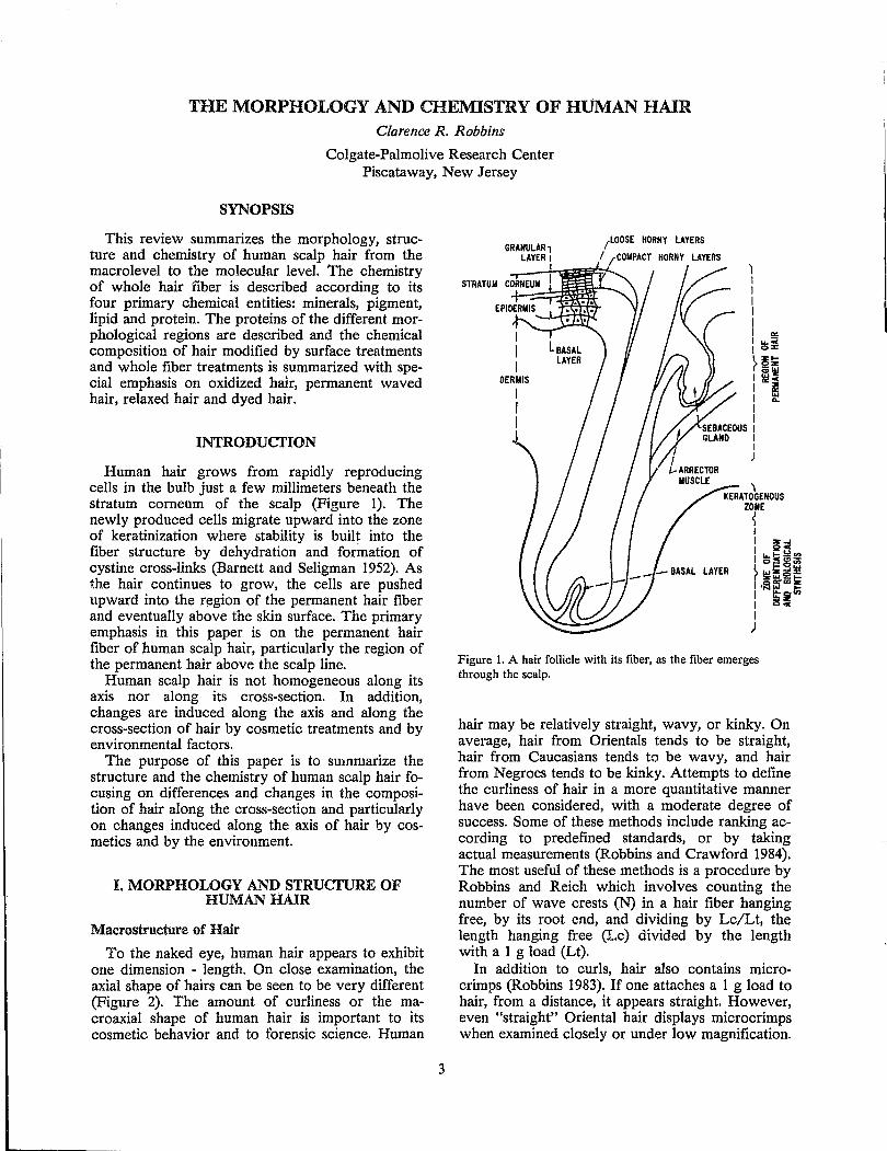
Human hair grows from rapidly reproducing cells in the bulb just a few millimeters beneath the stratum corneum of the scalp (Figure 1). The newly produced cells migrate upward into the zone of keratinization where stability is built into the fiber structure by dehydration and formation of cystine cross-links (Barnett and Seligman 1952). As the hair continues to grow, the cells are pushed upward into the r~gion of the permanent hair fiber and eventually above the skin surface. The primary emphasis in this paper is on the permanent hair fiber of human scalp hair, particularly the region of the permanent hair above the scalp line.
Human scalp hair is not homogeneous along its axis nor along its cross-section. In addition, changes are induced along the axis and along the cross-section of hair by cosmetic treatments and by environmental factors.
The purpose of this paper is to smnmarize the structure and the chemistry of human scalp hair focusing on differences and changes in the composition of hair along the cross-section and particularly on changes induced along the axis of hair by cosmetics and by the environment.
I. MORPHOLOGY AND STRUcrURE OF HUMAN HAIR
Macrostructure of Hair
To the naked eye, human hair appears to exhibit one dimension - length. On close examination, the axial shape of hairs can be seen to be very different (Figure 2). The amount of curliness or the macroaxial shape of human hair is important to its cosmetic behavior and to forensic science. Human
3
GRANULAR, LAYER I
I
I I I
OERllIS
I I I
(LOOSE HORNY LAYERS
I ,rCOIiPACT HORNY LAYERS
~ KERATOGENOUS
ZONE
~ I I Z..I I QC
I~~~~ ~"'~5~ I ,iaffia;~ I It~ I 25.,. I )
Figure 1. A hair follicle with its fiber, as the fiber emerges through the scalp.
hair may be relatively straight, wavy, or kinky. On average, hair from Orientals tends to be straight, hair from Caucasians tends to be wavy, and hair from Negroes tends to be kinky. Attempts to define the curliness of hair in a more quantitative manner have been considered, with a moderate degree of success. Some of these methods include ranking according to predefined standards, or by taking actual measurements (Robbins and Crawford 1984). The most useful of these methods is a procedure by Robbins and Reich which involves counting the number of wave crests (N) in a hair fiber hanging free, by its root end, and dividing by Lc/Lt, the length hanging free (Lc) divided by the length with a 1 g load (Lt).
In addition to curls, hair also contains microcrimps (Robbins 1983). If one attaches a 1 g load to hair, from a distance, it appears straight. However, even "straight" Oriental hair displays microcrimps when examined closely or under low magnification.
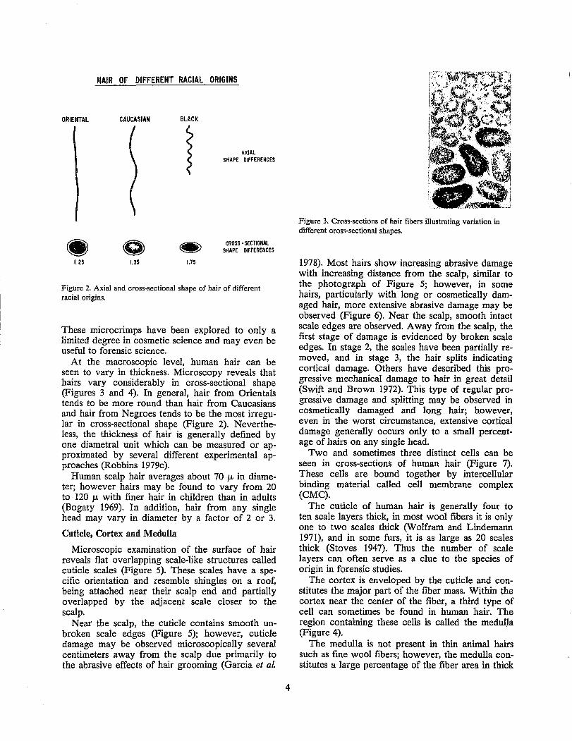
HAIR OF DIFFERENT RACIAL ORIGINS
ORIENTAL CAUCASIAN
1.25 1.35
BLACK
}
1.75
AXIAL SHAPE DIFFERENCES
CROSS - SECTIONAl SHAPE DIFFERENCES
Figure 2. Axial and cross-sectional shape of hair of different racial origins.
These micro crimps have been explored to only a limited degree in cosmetic science and may even be useful to forensic science.
At the macroscopic level, human hair can be seen to vary in thickness. Microscopy reveals that hairs vary considerably in cross-sectional shape (Figures 3 and 4). In general, hair from Orientals tends to be more round than hair from Caucasians and hair from Negroes tends to be the most irregular in cross-sectional shape (Figure 2). Nevertheless, the thickness of hair is generally defined by one diametral unit which can be measured or approximated by several different experimental approaches (Robbins 1979c).
Human scalp hair averages about 70 J.L in diameter; however hairs may be found to vary from 20 to 120 J.L with finer hair in children than in adults (Bogaty 1969). In addition, hair from any single head may vary in diameter by a factor of 2 or 3.
Cuticle, Cortex and Medulla
Microscopic examination of the surface of hair reveals flat overlapping scale-like structures called cuticle scales (Figure 5). These scales have a specific orientation and resemble shingles on a roof, being attached near their scalp end and partially overlapped by the adjacent scale closer to the scalp.
Near the scalp, the cuticle contains smooth unbroken scale edges (Figure 5); however, cuticle damage may be observed microscopically several centimeters away from the scalp due primarily to the abrasive effects of hair grooming (Garcia et al.
4
Figure 3. Cross-sections of hair fibers illustrating variation in different cross-sectional shapes.
1978). Most hairs show increasing abrasive damage with increasing distance from the scalp, similar to the photogralph of Figure 5; however, in some hairs, particularly with long or cosmetically damaged hair, more extensive abrasive damage may be observed (Figure 6). Near the scalp, smooth intact scale edges are observed. Away from the scalp, the first stage of damage is evidenced by broken scale edges. In stage 2, the scales have been partially removed, and in stage 3, the hair splits indicating cortical damage. Others have described this progressive mechanical damage to hair in great detail (Swift and Brown 1972). This type of regular progressive damage and splitting may be observed in cosmetically damaged and long hair; however, even in the worst circumstance, extensive cortical damage generally occurs only to a small percentage of hairs on any single head.
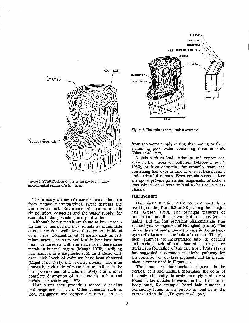
Two and sometimes three distinct cells can be seen in cross-sections of human hair (Figure 7). These cells are bound together by intercellular binding material called cell membrane complex (CMC).
The cuticle of human hair is generally four to ten scale layers thick, in most wool fibers it is only one to two scales thick (Wolfram and Lindemann 1971), and in some furs, it is as large as 20 scales thick (Stoves 1947). Thus the number of scale layers can often serve as a clue to the species of origin in forensic studies.
The cortex is enveloped by the cuticle and constitutes the major part of the fiber mass. Within the cortex near the center of the fiber, a third type of cell can sometimes be found in human hair. The region containing these cells is called the medulla (Figure 4).
The medulla is not present in thin animal hairs such as fine wool fibers; however, the medulla constitutes a large percentage of the fiber area in thick
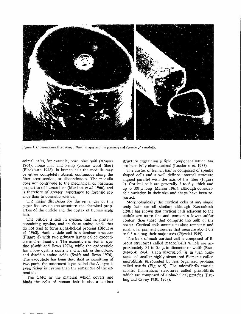
Figure 4. Cross-sections illustrating different shapes and the presence and absence of a medulla.
animal hairs, for example, porcupine quill (Rogers 1964), horse hair and kemp (coarse wool fiber) (Blackburn 1948). In human hair the medulla may be either completely absent, continuous along ~he fiber cross-section, or discontinuous. The medulla does not contribute to the mechanical or cosmetic properties of human hair (Menkart et ai. 1966), and is therefore of greater importance to forensic science than to cosmetic science.
The major discussion for the remainder of this paper focuses on the structure and chemical properties of the cuticle and the cortex of human scalp hair.
The cuticle is rich in cystine, that is, proteins containing cystine, and in those amino acids that do not tend to form alpha-helical proteins (Blout et al. 1960). Each cuticle cell is a laminar structure (Figure 8) with two primary layers called exocuticle and endocuticle. The exocuticle is rich in cystine (Swift and Bews 1976), while the endocuticle has a low cystine content and is rich in the dibasic and diacidic amino acids (Swift and Bews 1976). The exocuticle has been described as consisting of two parts, the outermost layer called the A-layer is even richer in cystine than the remainder of the exocuticle.
The CMC or the material which covers and binds the cells of human hair is also a laminar
5
structure containing a lipid component which has not been fully characterized (Leeder et ai. 1983).
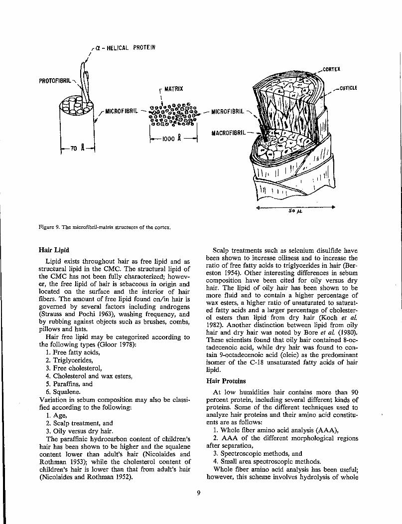
The cortex of human hair is composed of spindle shaped cells and a well defined internal structure aligned parallel with the axis of the fiber (Figure 9). Cortical cells are generally 1 to 6 J-L thick and up to 100 J-L long (Mercer 1961), although considerable variation in their size and shape have been reported.
Morphologically the cortical cells of any single scalp hair are all similar; although Kassenbeck (1981) has shown that cortical cells adjacent to the cuticle are more nat and contain a lower sulfur content tha.n those that comprise the bulk of the cOltex. Cortical cells contain nuclear remnants and small oval pigment granules that measure abo'lt 0.2 to 0.8 ~L along their major axis (Gjesdal 1959).
The bulk of each cortical cell is composed of fibrous structures called macrofibrils which are approximately 0.1 to 0.4 J-L in diameter or width (Randebrook 1964). Each macrofibril is in turn composed of smaller highly structured filaments called micro fibrils surrounded by less organized proteins called matrix (Figure 9). The microfibrils contain smaller filamentous structures called proto fibrils which are composed of alpha-helical proteins (Pauling and Corey 1950, 1953).
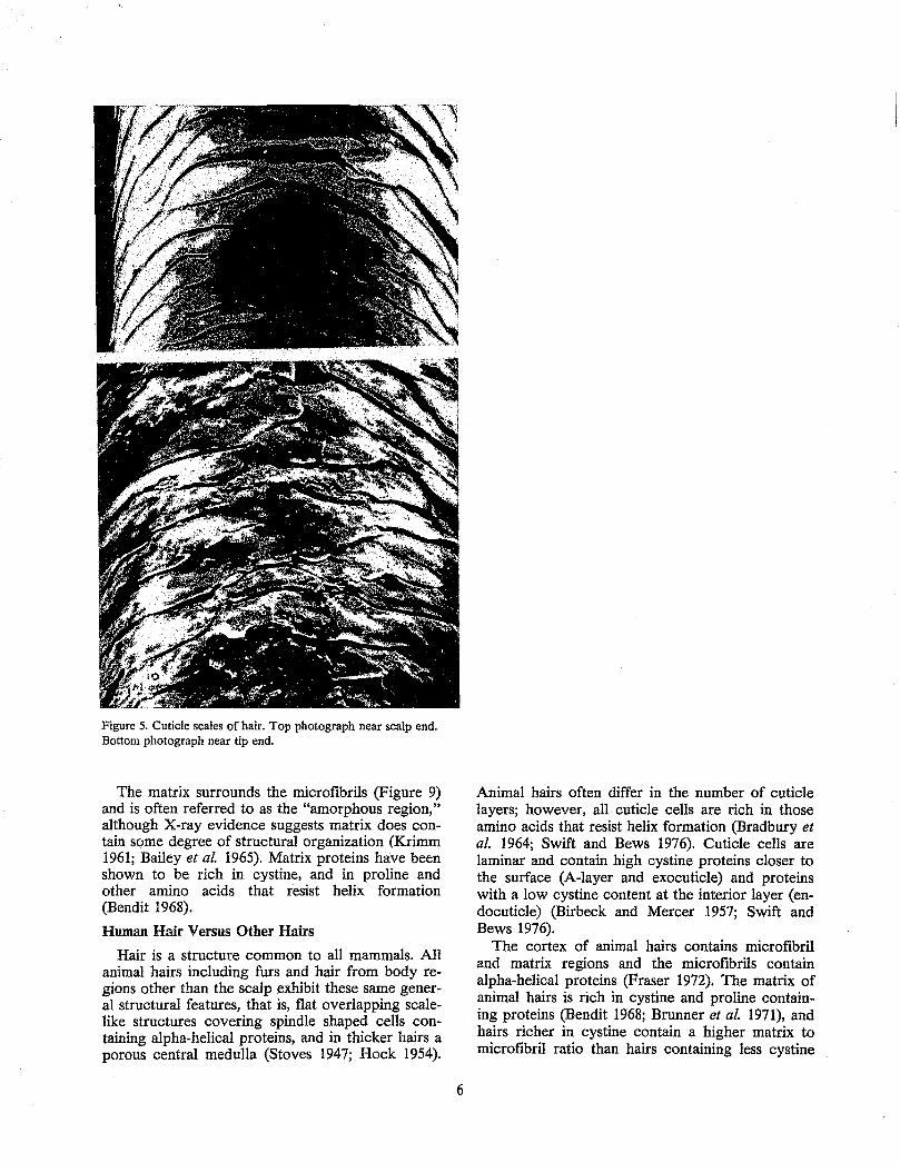
Figure 5. Cuticle scales of hair. Top photograph near scalp end. Bottom photograph near tip end.
The matrix surrounds the microfibrils (Figure 9) and is often referred to as the "amorphous region," although X-ray evidence suggests matrix does contain some degree of structural organization (Krimm 1961; Bailey et al. 1965). Matrix proteins have been shown to be rich in cystine, and in proline and other amino acids that resist helix formation (Bendit 1968).
Human Hair Versus Other Hairs
Hair is a structure common to all mammals. All animal hairs including furs and hair from body regions other than the scalp exhibit these same general structural features, that is, flat overlapping scalelike structures covering spindle shaped cells containing alpha-helical proteins, and in thicker hairs a porous central medulla (Stoves 1947; Hock 1954).
6
Animal hairs often differ in the number of cuticle layers; however, all cuticle cells are rich in those amino acids that resist helix formation (Bradbury et al. 1964; Swift and Bews 1976). Cuticle cells are laminar and contain high cystine proteins closer to the surface (A-layer and exocuticle) and proteins with a low cystine content at the interior layer (endocuticle) (Birbeck and Mercer 1957; Swift and Bews 1976).
The cortex of animal hairs contains microfibril and matrix regions and the microfibrils contain alpha-helical proteins (Fraser 1972). The matrix of animal hairs is rich in cystine and proline containing proteins (Bendit 1968; Brunner et al. 1971), and hairs richer in cystine contain a higher matrix to microfibril ratio than hairs containing less cystine
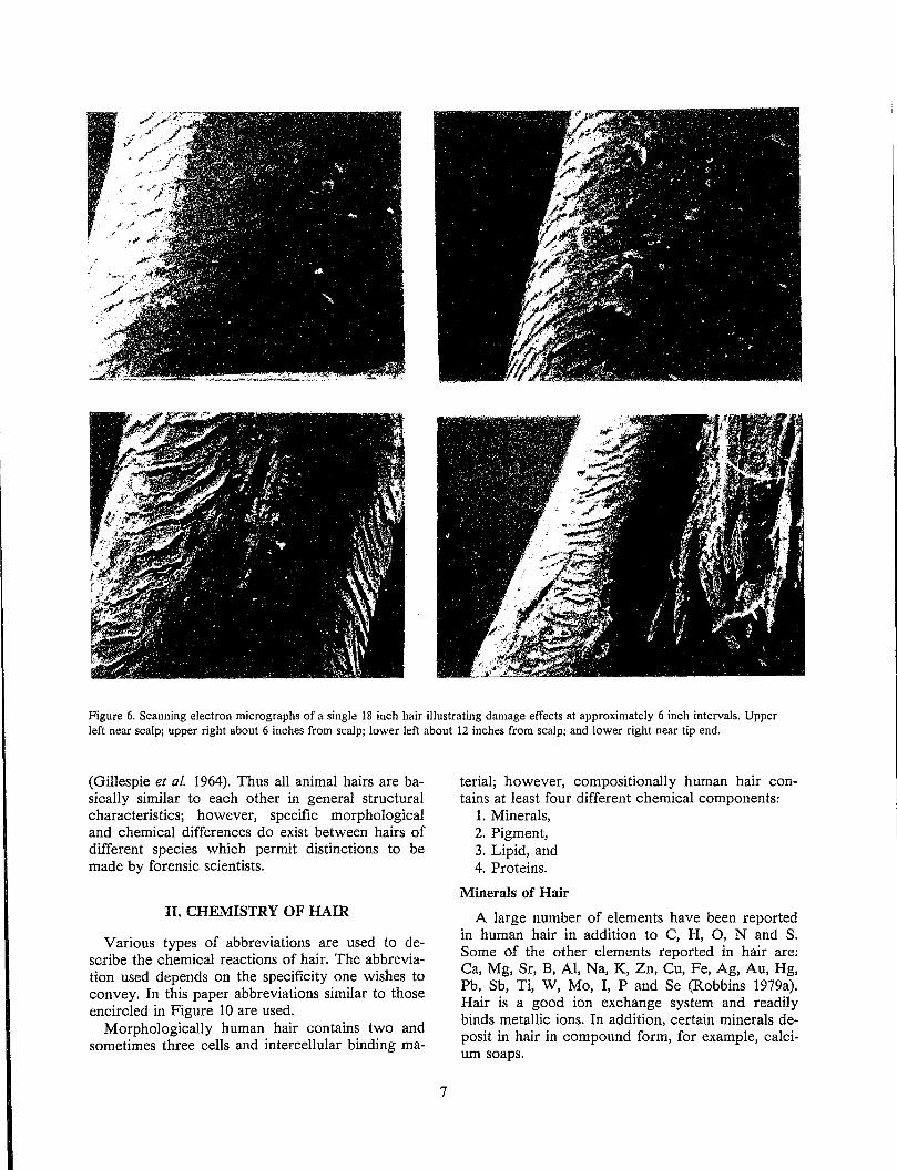
Figure 6. Scanning electron micrographs of a single 18 inch hair illustrating damage effects at approximately 6 inch intervals. Upper left near scalp; upper right about 6 inches from scalp; lower left about 12 inches from scalp; and lower right near tip end.
(Gillespie et al. 1964). Thus all animal hairs are basically similar to each other in general structural characteristics; however, specific morphological and chemical differences do exist between hairs of different species which permit distinctions to be made by forensic scientists.
II. CHEMISTRY OF HAIR
Various types of abbreviations are used to describe the chemical reactions of hair. The abbreviation used depends on the specificity one wishes to convey. In this paper abbreviations similar to those encircled in Figure 10 are used.
Morphologically human hair contains two and sometimes three cells and intercellular binding ma-
7
terial; however, compositionally human hair contains at least four different chemical components:
1. Minerals, 2. Pigment, 3. Lipid, and 4. Proteins.
Minerals of Hair
A large number of elements have been reported in human hair in addition to C, H, 0, Nand S. Some of the other elements reported in hair are: Ca, Mg, Sr, B, AI, Na, K, Zn, Cu, Fe, Ag, Au, Hg, Pb, Sb, Ti, W, Mo, I, P and Se (Robbins 1979a). Hair is a good ion exchange system and readily binds metallic ions. In addition, certain minerals deposit in hair in compound form, for example, calcium soaps.
CUTicLe
Figure 7. STEREOGRAM illustrating the two primary morphological regions of a hair fiber.
I I I
The primary sources of trace elements in hair are from metabolic irregularities, sweat deposits and the environment. Environmental sources include air pollution, cosmetics and the water supply, for example, bathing, washing and pool water.
Although heavy metals are found at low concentrations in human hair, they sometimes accumulate at concentrations well p,bove those present in blood or in urine. Concentrations of metals such as cadmium, arsenic, mercury and lead in hair have been found to correlate with the amounts of these same metals in internal organs (Maugh 1978), justifying hair analysis as a diagnostic tool. In dyslexic children, high levels of cadmium have been observed (Capel et al. 1981), and in celiac disease there is an unusually high ratio of potassium to sodium in the hair (Kopito and Shwachman 1974). For a more complete description of trace metals in hair and metabolism, see Maugh 1978.
Hard water areas provide a source of calcium and magnesium in hair. Other minerals such as iron, manganese and copper can deposit in hair
8
...-CUTIClE ....... :'1-.,J ... I-.~WI. .. ' '-"
IIICROFIIlRIL .......
Figure 8. The cuticle and its laminar structure.
I I 1-0.7 p.-l
from the water supply during shampooing or from swimming pool water containing these minerals (Bhat et al. 1979).
Metals such as lead, cadmium and copper can arise in hair from air pollution (MiIosevic et al. 1980), or from cosmetics, for example, from lead containing hair dyes or zinc or even selenium from antidandruff shampoos. Even certain soaps and/or shampoos provide potassium, magnesium or sodium ions which can deposit or bind to hair via ion exchange.
Hair Pigments
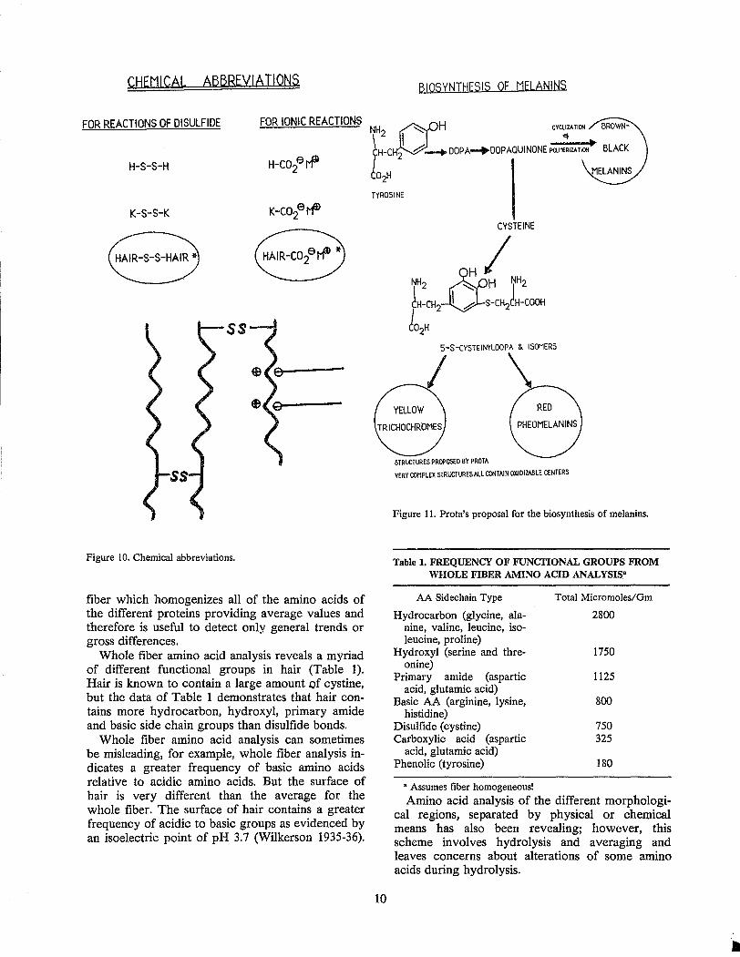
Hair pigments reside in the cortex or medulla as ovoid granules, from 0.2 to 0.8 }J. along their major axis (Gjesdal 1959). The principal pigments of human hair are the brown-black melanins (eumelanins) and the less prevalent phaeomelanins (the red and yellow pigments of biological species). The biosynthesis of hair pigments occurs in the melanocyte cells located in the bulb of the hair. The pigment granules are incorporated into the cortical and medulla' cells of scalp hair at an early stage during the formation of the hair fiber. Prota (1980) has suggested a common metabolic pathway for the formation of all three pigments and his mechanism is summarized in Figure 11.
The amount of these melanin pigments in the cortical cells and medulla determines the color of the hair. Generally, in scalp hair, pigment is not found in the cuticle; however, in hair from other body parts, for example, beard hair, pigment is commonly found in the cuticle as weU as in the cortex and medulla (Tolgyesi et al. 1983).
I
PROTOFIBRIL ""
ra - HELICAL PROTEIN I
Figure 9. The microfibril-matrix structures of the cortex.
Hair Lipid
Lipid exists throughout hair as free lipid and as structural lipid in the CMC. The structural lipid of the CMC has not been fully characterized; however, the free lipid of hair is sebaceous in origin and located 011 the surface and the interior of hair fibers. The amount of free lipid found on/in hair is governed by several factors including androgens (Strauss and Pochi 1963), washing frequency, and by rubbing against objects such as brushes, combs, pillows and hats.
Hair free lipid may be categorized according to the following types (Gloor 1978):
1. Free fatty acids, 2. Triglycerides, 3. Free cholesterol, 4. Cholesterol and wax esters, 5. Paraffins, and 6. Squalene.
Variation in sebum composition may also be classified according to the following:
1. Age, 2. Scalp treatment, and 3. Oily versus dry hair. The paraffinic hydrocarbon content of children's
hair has been shown to be higher and the squalene content lower than adult's hair (Nicolaides and Rothman 1953); while the cholesterol content of children's hair is lower than that from adult's hair (Nicolaides and Rothman 1952).
9
...-CUTICLE
So ;.L
Scalp treatments such as selenium disulfide have been shown to increase oiliness and to increase the ratio of free fatty acids to triglycerides in hair (Bereston 1954). Other interesting differences in sebum composition have been cited for oily versus dry hair. The lipid of oily hair has been shown to be more fluid and to contain a higher percentage of wax esters, a higher ratio of unsaturated to saturated fatty acids and a larger percentage of cholesterol esters than lipid from dry hair (Koch et al. 1982). Another distinction between lipid from oily hair and dry hair was noted by Bore et al. (1980). These scientists found that oily hair contained 8-octadecenoic acid, while dry hair was found to contain 9-octadecenoic acid (oleic) as the predominant isomer of the C-18 unsaturated fatty acids of hair lipid.
Hair Proteins
At low humidities hair contains more than 90 percent protein, including several different kinds of proteins. Some of the different techniques used to analyze hair proteins and their amino acid constituents are as follows:
1. Whole fiber amino acid analysis (AAA), 2. AAA of the different morphological regions
after separation, 3. Spectroscopic methods, and 4. Small area spectroscopic methods. Whole fiber amino acid analysis has been useful;
however, this scheme involves hydrolysis of whole
CHEMICeL eBBREVleTIONS
FOR REACTIONS OF DISULFIDE FOR IONIC REACTIONS
H-S-S-H
K-S-S-K
Figure 10. Chemical abbreviations.
fiber which homogenizes all of the amino acids of the different proteins providing average values and therefore is useful to detect only general trends or gross differences.
Whole fiber amino acid analysis reveals a myriad of different functional groups in hair (Table 1). Hair is known to contain a large amount of cystine, but the data of Table 1 demonstrates that hair contains more hydrocarbon, hydroxyl, primary amide and basic side chain groups than disulfide bonds.
Whole fiber amino acid analysis can sometimes be misleading, for example, whole fiber analysis indicates a greater frequency of basic amino acids relative to acidic amino acids. But the surface of hair is very different than the average for the whole fiber. The surface of hair contains a greater frequency of acidic to basic groups as evidenced by an isoelectric point of pH 3.7 (Wilkerson 1935-36).
BIOSYNTHESIS OF MELANItiS.
TYROSINE
10
5-$-CYSTEINYLOOPA & ISOMERS
I
STRUCTURES PROPOSI:D BY PROTA
VERY COMPLEX STRUCTURES ALL CONTAIN OXIDIZABLE CENTERS
Figure 11. Prota's proposal for the biosynthesis of melanins.
Table 1. FREQUENCY OF FUNCTIONAL GROUPS FROM WHOLE FIBER AMINO ACID ANALYSIS·
AA Sidechain Type
Hydrocarbon (glycine, alanine, valine, leucine, isoleucine, proline)
Hydroxyl (serine and threonine)
Primary amide (aspartic acid, glutamic acid)
Basic AA (arginine, lysine, histidine)
Disulfide (cystine) Carboxylic acid (aspartic
acid, glutamic acid) Phenolic (tyrosine)
• Assumes tiber homogeneousl
Total Micromoles/Gm
2800
1750
1125
800
750 325
180
Amino acid analysis of the different morphological regions, separated by physical or chemical means has also been revealing; however, this scheme involves hydrolysis and averaging and leaves concerns about alterations of some amino acids during hydrolysis.
The proteins of the different morphological regions of hair contain different relative ratios of their component amino acids described in the section entitled, Cuticle, Cortex and Medulla uf Part 1.
Spectroscopic methods, on the other hand, do not alter the fiber prior to analysis and therefore offer an advantage. In addition, the newly emerging small area spectroscopic methods offer hope for revealing new insights into the fiber chemistry, and for providing detailed axial or even cross-sectional profiles of the chemical composition of human hair fibers.
Hair comes in frequent contact with cosmetics and its chemistry in some cases is altered considerably. Therefore, a discussion of the chemistry of hair is incomplete without describing the chemistry of cosmetically altered hair, the subject of the remaining portion of this paper.
III. HAIR ALTERED BY COSMETIC TREATMENT AND THE ENVIRONMENT
The chemistry of hair altered by cosmetic treatments may be classified according to (1) surface treatments or (2) whole fiber treatments.
Surface treatments are those cosmetics which interact at or near the surface of hair and are principally shampoos, hair conditioners, hair sprays, setting lotions (or mousses) and hair dressings. Whole fiber treatments are those cosmetics which chemically alter the whole fiber and consist of hair
DIRTY HAIR
bleaches, permanent waves, relaxers or straighteners, and some hair dyes.
A. Surface Treatments
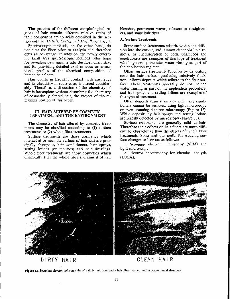
Some surface treatments adsorb, with some diffusion into the cuticle, and interact either via lipid removal or chemisorption or both. Shampoos and conditioners are examples of this type of treatment which generally includes water rinsing as part of the application regimen.
Other surface treatments function by depositing onto the hair surface, producing relatively thick, non-uniform deposits which adhere to the fiber surface. These treatments generally do not include water rinsing as part of the application procedure, and hair sprays and setting lotions are examples of this type of treatment.
Often deposits from shampoos and many conditioners cannot be resolved using light microscopy or even scanning electron microscopy (Figure 12). While deposits by hair sprays and setting lotions are readily detected by microscopy (Figure 13).
Surface treatments are generally mild to hair. Therefore their effects on hair fibers are more difficult to characterize than the effects of whole fiber treatments. Some methods useful for studying surface changes to hair are as follows:
1. Scanning electron microscopy (SEM) and light microscopy,
2. Electron spectroscopy for chemical analysis (ESCA),
CLEAN HAIR
Figure 12. Scanning electron micrographs of a dirty hair fiber and a hair fiber washed with a conventional shampoo.
11
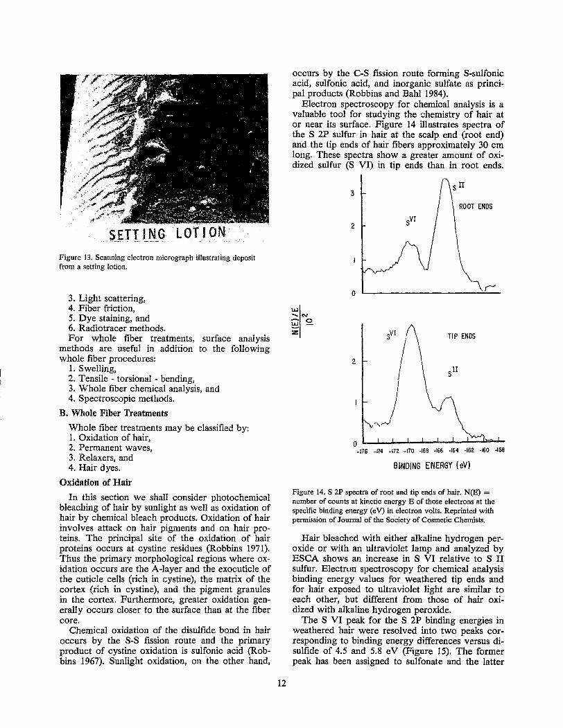
Figure 13. Scanning electron micrograph illustrating deposit from a setting lotion.
3. Light scattering, 4. Fiber friction, 5. Dye staining, and 6. Radiotracer methods. For whole fiber treatments, surface analysis
methods are useful in addition to the following whole fiber procedures:
1. Swelling, 2. Tensile - torsional - bending, 3. Whole fiber chemical analysis, and 4. Spectroscopic methods.
B. Whole Fiber Treatments
Whole fiber treatments may be classified by: 1. Oxidation of hair, 2. Permanent waves, 3. Relaxers, and 4. Hair dyes.
Oxidation of Hair
In this section we shall consider photochemical bleaching of hair by sunlight as well as oxidation of hair by chemical bleach products. Oxidation of hair involves attack on hair pigments and on hair proteins. The principal site of the oxidation of hair proteins occurs at cystine residues (Robbins 1971). Thus the primary morphological regions where oxidation occurs are the A-layer and the exocuticle of the cuticle cells (rich in cystine), the matrix of the cortex (rich in cystine), and the pigment granules in the cortex. Furthermore, greater oxidation generally occurs closer to the surface than at the fiber core.
Chemical oxidation of the disulfide bond in hair occurs by the S-S fission route and the primary product of cystine oxidation is sulfonic acid (Robbins 1967). Sunlight oxidation, on the other hand,
12
occurs by the COS fission route forming S-sulfonic acid, sulfonic acid, and inorganic sulfate as principal products (Robbins and Bahl 1984).
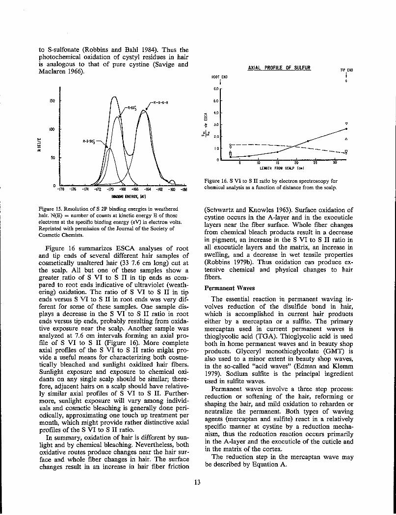
Electron spectroscopy for chemical analysis is a valuable tool for studying the chemistry of hair at or near its surface. Figure 14 illustrates spectra of the S 2P sulfur in hair at the scalp end (root end) and the tip ends of hair fibers approximately 30 cm long. These spectra show a greater amount of oxidized sulfur (S VI) in tip ends than in root ends.
LU ....... co.!
~ 0
z
3
2
0
2
o L-~~~--~~~~--~--176 ·174 -172 -170 ·168 ·166 ·164 ·162 ·160 ·158
BINDING ENERGY (eV)
Figure 14. S 2P spectra of root and tip ends of hair. N(E) = number of counts at kinetic energy E of those electrons at the specific binding energy (eV) in electron volts. Reprinted with permission of Journal of the Society of Cosmetic Chemists.
Hair bleached with either alkaline hydrogen peroxide or with an ultraviolet lamp and analyzed by ESCA shows an increase in S VI relative to S II sulfur. Electrun spectroscopy for chemical analysis binding energy values for weathered tip ends and for hair exposed to ultraviolet light are similar to each other, but different from those of hair oxidized with alkaline hydrogen peroxide.
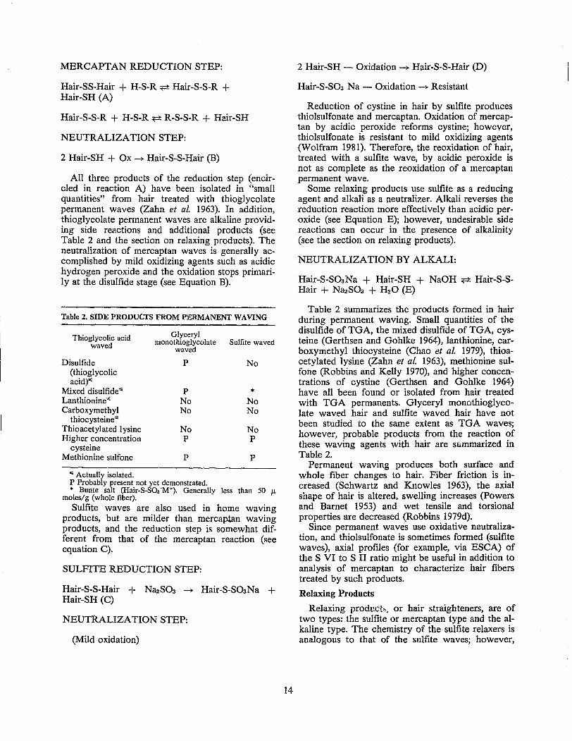
The S VI peak for the S 2P binding energies in weathered hair were resolved into two peaks corresponding to binding energy differences versus disulfide of 4.5 and 5.8 eV (Figure 15). The former peak has been assigned to sulfonate and the latter
to S-sulfonate (Robbins and Bahl 1984). Thus the photochemical oxidation of cystyl residues in hair is analogous to that of pure cystine (Savige and Maclaren 1966).
UJ .... 8 z
150
100
50
o ~------~~~~~--~----~~~ .. -178 -176 -174 -172 -170 -168 -166 -164 -162 -lEO -158
B1H1l1N6 ENERGY, (dJ)
Figure 15. Resolution of S 2P binding energies in weathered hair. N(E) = number of counts at kinetic energy E of those electrons at the specific binding energy (eV) in electron volts. Reprinted with permission of the Journal of the Society of Cosmetic Chemists.
Figure 16 summarizes ESCA analyses of root and tip ends of several different hair samples of cosmetically unaltered hair (33 7.6 cm long) cut at the scalp. All but one of these samples show a greater. ratio of S VI to S II in tip ends as compared to root ends indicative of ultraviolet (weathering) oxidation. The ratio of S VI to S II in tip ends versus S VI to S II in root ends was very different for some of these samples. One sample displays a decrease in the S VI to S II ratio in root ends versus tip ends, probably resulting from oxidative exposure near the scalp. Another sample was analyzed at 7.6 cm intervals forming an axial profile of S VI to S II (Figure 16). More complete axial profiles of the S VI to S II ratio might provide a useful means for characterizing both cosmetically bleached and sunlight oxidized hair fibers. Sunlight exposure and exposure to chemical oxidants on any single scalp should be similar; therefore, adjacent hairs on a scalp should have relatively similar axial profiles of S VI to S II. Furthermore, sunlight exposure will vary among individuals and cosmetic bleaching is generally done periodically, approximating one touch up treatment per month, which might provide rather distinctive axial profiles of the S VI to S II ratio.
In summary, oxidation of hair is different by sunlight and by chemical bleaching. Nevertheless, both oxidative routes produce changes near the hair surface and whole fiber changes in hair. The surface changes result in an increase in hair fiber friction
13
AXIAL PROFILE OF SULFUR TIP END
ROOT END I I o
6.0
5.0
.. 4.0 ~ ... ,! 3.0 v
;:1 = ",,,, 2.0
A
1.0 0--------- -____ [] ---0
0 10 15 20 25
LENGTH FROIII SCALP (,,,I
Figure 16. S VI to S II ratio by electron spectroscopy for chemical analysis as a function of distance from the scalp.
(Schwartz and Knowles 1963). Surface oxidation of cystine occurs in the A-layer and in the exocuticle layers near the fiber surface. Whole fiber changes from chemical bleach products result in a decrease in pigment, an increase in the S VI to S II ratio in all exocuticle layers and the matrix, an increase in swelling, and a decrease in wet tensile properties (Robbins 1979b). Thus oxidation can produce extensive chemical and physical changes to hair fibers.
Permanent Waves
The essential reaction in permanent waving involves reduction of the disulfide bond in hair, which is accomplished in current hair products either by a mercaptan or a sulfite. The primary mercaptan used in current permanent waves is thioglycolic acid (TGA). Thioglycolic acid is used both in home permanent waves and in beauty shop products. Glyceryl monothioglycolate (GMT) is also used to a minor extent in beauty shop waves, in the so-called "acid waves" (Edman and Klemm 1979). Sodium sulfite is the principal ingredient used in sulfite waves.
Permanent waves involve a three step process: reduction or softening of the hair, reforming or shaping the hair, and mild oxidation to reharden or neutralize the permanent. Both types of waving agents (mercaptan and sulfite) react in a relatively specific manner at cystine by a reduction mechanism, thus the reduction reaction occurs primarily in the A-layer and the exocuticle of the cuticle and in the matrix of the cortex.
The reduction step in the mercaptan wave may be described by Equation A.
MERCAPTAN REDUCTION STEP:
Hair-SS-Hair + H-S-R ~ Hair-S-S-R + Hair-SH (A)
Hair-S-S-R + H-S-R ~ R-S-S-R + Hair-SH
NEUTRALIZATION STEP:
2 Hair-SH + Ox -)- Hair-S-S-Hair (B)
All three products of the reduction step (encircled in reaction A) have been isolated in "small quantities" from hair treated with thioglycolate permanent waves (Zahn et al. 1963). In addition, thioglycolate permanent waves are alkaline providing side reactions and additional products (see Table 2 and the section on relaxing products). The neutralization of mercaptan waves is generally accomplished by mild oxidizing agents such as acidic hydrogen peroxide and the oxidation stops primarily at the disulfide stage (see Equation B).
Table 2. SIDE PRODUcrS FROM PJ!:RMANENT WAVING
Thioglycolic acid Glyceryl
waved monothioglycolate Sulfite waved waved
Disulfide P No (thioglycolic acid)'"
Mixed disulfide'" P * Lanthionine'" No No Carboxymethyl No No
thiocysteine'" Thioacetylated lysine No No Higher concentration P P
cysteine Methionine sulfone P P
'" Actually isolated. P Probably present not yet demonstrated. * Bunte salt (Hair-S-S03 ·M+). Generally less than 50 !L
moles/g (whole fiber).
Sulfite waves are also used in home waving products, but are milder than mercaptan waving products, and the reduction step is somewhat different from that of the mercaptan reaction (see equation C).
SULFITE REDUCTION STEP:
Hair-S-S-Hair + Na2S03 -+ Hair-S-S03Na + Hair-SH (C)
NEUTRALIZATION STEP:
(Mild oxidation)
14
2 Hair-SH - Oxidation -+ Hair-S-S-Hair (D)
Hair·oS-SOa Na - Oxidation -+ Resistant
Reduction of cystine in hair by sulfite produces thiolsulfonate and mercaptan. Oxidation of mercaptan by acidic peroxide reforms cystine; however, thiolsulfonate is resistant to mild oxidizing agents (Wolfram 1981). Therefore, the reoxidation of hair, treated with a sulfite wave, by acidic peroxide is not as complete as the reoxidation of a mercaptan permanent wave.
Some relaxing products use sulfite as a reducing agent and alkali as a neutralizer. Alkali reverses the reduction reaction more effectively than acidic peroxide (see Equation E); however, undesirable side reactions can occur in the presence of alkalinity (see the section on relaxing products).
NEUTRALIZATION BY ALKALI:
Hair-S-S03Na + Hair-SH + NaOH ~ Hair-S-SHair + Na2S03 + H20 (E)
Table 2 summarizes the products formed in hair during permanent waving. Small quantities of the disulfide of TGA, the mixed disulfide of TGA, cysteine (Gerthsen and Gohlke 1964), lanthionine, carboxymethyl thiocysteine (Chao et al. 1979), thioacetylated lysine (Zahn et al. 1963), methionine sulfone (Robbins and Kelly 1970), and higher concentrations of cystine (Gerthsen and Gohlke 1964) have all been found or isolated from hair treated with TGA permanents. Glyceryl monothioglycolate waved hair and sulfite waved hair have not been studied to the same extent as TGA waves; however, probable products from the reaction of these waving agents with hair are summarized in Table 2.
Permanent waving produces both surface and whole fiber changes to hair. Fiber friction is increased (Schwartz and Knowles 1963), the axial shape of hair is altered, swelling increases (Powers and Barnet 1953) and wet tensile and torsional properties are decreased (Robbins 1979d).
Since permanent waves use oxidative neutralization, and thiolsulfonate is sometimes formed (sulfite waves), axial profiles (for example, via ESCA) of the S VI to S II ratio might be useful in addition to analysis of mercaptan to characterize hair fibers treated by such products.
Relaxing Products
Relaxing product>l, or hair straighteners, are of two types: the sulfite or mercaptan type and the alkaline type. The chemistry of the sulfite relaxers is analogous to that of the sulfite waves; however,
neutralization with hair relaxers is generally with alkali rather than by oxidation (see equation E).
The primary reactions of alkali with hair involve hydrolysis of amide and peptide bonds and reaction at cystine. Hydrolytic reactions occur at either the amide bonds of aspartic and glutamic acid or at the peptide bonds of the backbone of the hair proteins. Hydrolysis of lipid esters also occurs. Amide hydrolysis increases the ratio of acidic groups to basic sites, changing the ionic character of the morphol~gical region where hydrolysis occurs.
Peptide bond hydrolysis increases both acidic and basic sites simultaneously, increasing the swelling properties and decreasing the dry tensile properties of hair (Alexander et al. 1963).
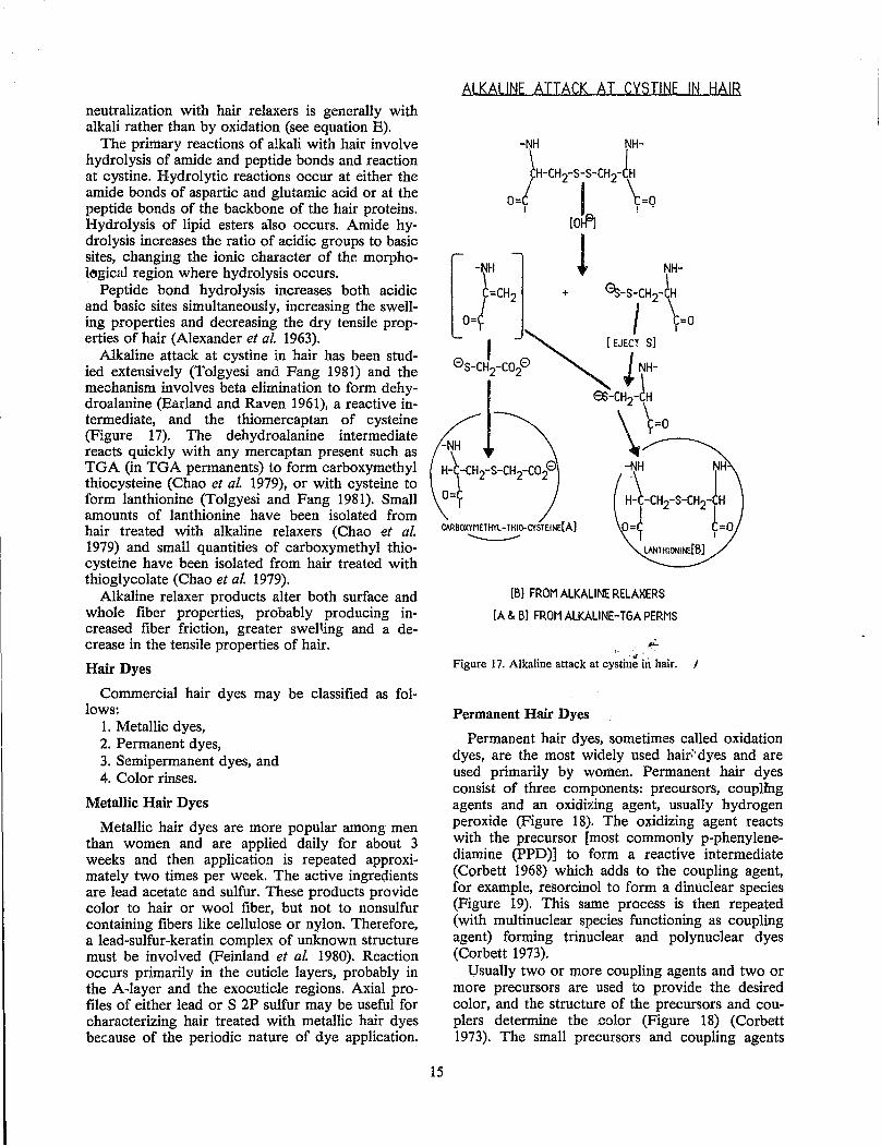
Alkaline attack at cystine in hair has been studied extensively (Tolgyesi and Fang 1981) and the mechanism involves beta elimination to form dehydroalanine (Earland and Raven 1961), a reactive intermediate, and the thiomercaptan of cysteine (Figure 17). The dehydroalanine intermediate reacts quickly with any mercaptan present such as TGA (in TGA permanents) to form carboxymethyl thiocysteine (Chao at al. 1979), or with cysteine to form lanthionine (Tolgyesi and Fang 1981). Small amounts of lanthionine have been isolated from hair treated with alkaline relaxers (Chao et al. 1979) and small quantities of carboxymethyl thiocysteine have been isolated from hair treated with thioglycolate (Chao et al. 1979).
Alkaline relaxer products alter both surface and whole fiber properties, probably producing increased fiber friction, greater swel'ing and a decrease in the tensile properties of hair.
Hair Dyes
Commercial hair dyes may be classified as fol-lows:
1. Metallic dyes, 2. Permanent dyes, 3. Semipermanent dyes, and 4. Color rinses.
Metallic Hair Dyes
Metallic hair dyes are more popular among men than women and are applied daily for about 3 weeks and then application is repeated approximately two times per week. The active ingredients are lead acetate and sulfur. These products provide color to hair or wool fiber, but not to nonsulfur containing fibers like cellulose or nylon. Therefore, a lead-sulfur-keratin complex of unknown structure must be involved (Feinland et aL 1980). Reaction occurs primarily in the cuticle layers, probably in the A-layer and the exocuticle regions. Axial profiles of either lead or S 2P sulfur may be useful for characterizing hair treated with metallic hair dyes because of the periodic nature of dye application.
ALKALINE ATTACK AT CYSTINE IN !:lAIR
CAABOXYMETHYL- THIO-CYSTEINE[A1 ----
15
[6] FROM ALKALINE RELAXERS
[A & 61 FROM ALKALlNE-TGA PERMS
,:..
Figure 17. Alkaline attack at cy~tin~in hair.
Permanent Hair Dyes
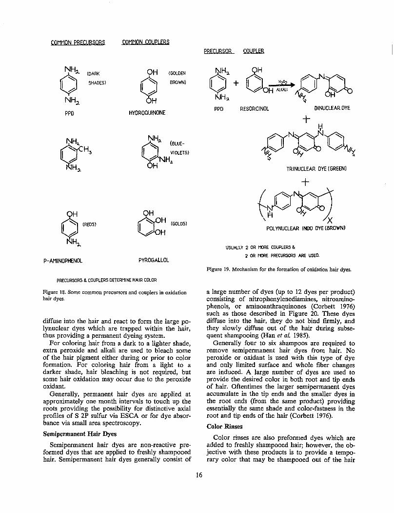
Permanent hair dyes, sometimes called oxidation dyes, are the most widely used hair:- dyes and are used primarily by women. Permanent hair dyes consist of three components: precursors, couplmg agents and an oxidizing agent, usually hydrogen peroxide (Figure 18). The oxidizing agent reacts with the precursor [most commonly p-phenylenediamine (PPD)] to form a reactive intermediate (Corbett 1968) which adds to the coupling agent, for example, resorcinol to form a dinuc!ear species (Figure 19). This same process is then repeated (with multinuclear species functioning as coupling agent) forming trinuclear and polynuclear dyes (Corbett 1973).
Usually two or more coupling agents and two or more precursors are used to provide the desired color, and the structure of the precursors and couplers determine the color (Figure 18) (Corbett 1973). The small precursors and coupling agents
COMMON PRECURSORS
NH" o NH;t
PPO
(DARK
SHADES)
o (REDS)
KlHl..
P-AMINOPHENOL
COMMON COUPLERS
OH
Q HYOROQUINONE
(GOLDEN
BROWN)
NH
Q::I. (BLUE-
I VIOLETS) ~ H H :I.
00H (GOLD5I UH
PYROGALLOL
PRECURSORS & COUPLERS DETERMINE HAIR COLOR
Figure 18. Some common precursors and couplers in oxidation hair dyes.
diffuse into the hair and react to form the large polynuclear dyes which are trapped within the hair, thus providing a permanent dyeing system.
For coloring hair from a dark to a lighter shade, extra peroxide and alkali are used to bleach some of the hair pigment either during or prior to color formation. For coloring hair from a light to a darker shade, hair bleaching is not required, but some hair oxidation may occur due to the peroxide oxidant.
Generally, permanent hair dyes are applied at approximately one month interv~ls to touch up the roots. providing the possibility for distinctive axial profiles of S 2P sulfur via ESCA or for dye absorbance via small area spectroscopy.
Semipermanent Hair Dyes
Semipermanent hair dyes are non-reactive preformed dyes that are applied to freshly shampooed hair. Semipermanent hair dyes generally consist of
16
PRECURSOR COUPLER
PPO RESORCINOL DlNUCLEAR DYE
TRINUCLEAR DYE (GREEN)
+ .( ()N~~ ~~ , X
POLYNUCLEAR INDO DYE (BROWN)
USUALLY 2 OR I'J:>RE COUPLERS &
2 OR t1)RE PRECURSORS ARE USED.
Figure 19. Mechanism for the formation of oxidation hair dyes.
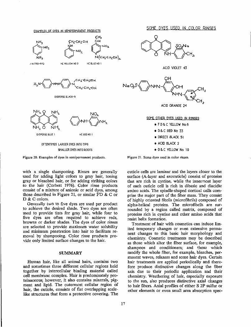
a large number of dyes (up to 12 dyes per product) consisting of nitrophenylenediamines, nitroaminophenols, or aminoanthraquinones (Corbett 1976) such as those described in Figure 20. These dyes diffuse into the hair, they do not bind firmly, and they slowly diffuse out of the hair during subsequent shampooing (Han et al. 1985).
Generally four to six shampoos are required to remove semipermanent hair dyes from hair. No peroxide or oxidant is used with this type of dye and only limited surface and whole fiber changes are induced. A large number of dyes are used to provide the desired color in both root and tip ends of hair. Oftentimes the larger semipermanent dyes accumulate in the tip ends and the smaller dyes in the root ends (from the same product) providing essentially the same shade and color-fastness in the root and tip ends of the hair (Corbett 1976).
Color Rinses
Color rinses are also preformed dyes which are added to freshly shampooed hair; however, the objective with these products is to provide a temporary color that may be shampooed out of the hair
EXAMPLES OF DYES IN SEMIPERMANENT PRODUCTS
,'-NITRO-PPD
yH3
f=H:rCHrOH oNH NH I ~ NO:;!. 0 NO
.1.. U
V N(CJ.I;-CHrOH)"
HC YELLOW NO 2 He BLUE NO I
DISPERSE BLACK-9
NO:L
~H-O'JH~ DISPERSE BLUE I HC RED NO I
OFTENTIMES LARGER DYES INTO TIPS
SMALLER DYES INTO ROOTS
Figure 20. Examples of dyes in semipermanent products.
with a single shampooing. Rinses are generally used for adding light colors to gray hair, toning gray or bleached hair, or for adding striking colors to the hair (Corbett 1976). Color rinse products consist of a mixture of anionic or acid dyes, among those described in Figure 21, or similar FD & C or D & C colors.
Generally two to five dyes are used per product to achieve the desired shade. Two dyes are often used to provide tints for gray hair, while four to five dyes are often required to achieve reds, browns or darker shades. The dyes of color rinses are selected to provide maximum water solubility and minimum penetration into hair to facilitate removal by shampooing. Color rinse products provide only limited surface changes to the hair.
SUMMARY
Human hair, like all animal hairs, contains two and sometimes three different cellular regions held together by intercellular binding material called cell membrane complex. Hair is predominately proteinaceous; however, it also contains minerals, pigment and lipid. The outermost cellular region of hair, the cuticle, consists of flat overlapping scalelike structures that form a protective covering. The
SOME DYES USED IN COLOR RINSES
ACID VIOLET 43
SOME OTHER DYES USED IN RINS~
• F D ~ C YEllOW No 6
• D 5. C RED No 33
• DIRECT BLACK 51
• ACID BLACK 2
• D ~ C YELLOW No I 0
Figure 21. Some dyes used in color rinses.
cuticle cells are laminar and the layers closer to the surface (A-layer and exocuticle) consist of proteins that are rich in cystine, while the innel'most layer of each cuticle cell is rich in dibasic and diacidic amino acids. The spindle-shaped cortical cells comprise the major part of the fiber mass. They consist of highly oriented fibrils (microfibrils) composed of alpha-helical proteins. The microfibrils are surrounded by a region called matrix, composed of proteins rich in cystine and other amino acids that resist helix formation.
Treatment of hair with cosmetics can induce limited temporary changes or even extensive permanent changes to this basic hair morphology and chemistry. Cosmetic treatments may be described as those which alter the fiber surface, for example, shampoos and conditioners; and those which modify the whole fiber, for example, bleaches, permanent waves, relaxers and some hair dyes. Certain hair treatments are applied periodically and therefore produce distinctive changes along the fiber axis due to their periodic application and their chemistry. Weathering of hair, especially exposure to the sun, also produces distinctive axial changes to hair fibers. Axial profiles of either S 2P sulfur or other elements or even small area absorption spec-
tra may serve to provide a useful means for characterizing hair fibers.
REFERENCES
Alexander, P., Hudson, R. F. and Ear/and, C. (1963). Wool, its Chemistry and Physics, 2nd ed. Franklin Publishing Co., New Jersey, pp. 61-65.
Bailey, C. J., Lewis, J., Priestley, G. c., Speakman, P. T. and Woodhouse, J. M. (1965). Two aspects of the morphogenesis of keratin fibers. In: Proceedings 3rd International Wool Textile Research Conference, Paris, 1:77-84.
Barnett, R. J. and Seligman, A. M. (1952). Histochemical demonstration of protein bound sulphydryl groups, Science 116:323-327.
Bendit, E. G. (1968). The distribution of high- and low-sulfur fractions in alpha-keratin, Text. Res. J. 38:15-21.
Bereston, E. S. (1954). Use of selenium sulfide in seborrheac dermatitis, J. Am. Med. Assoc. 156: 1246-1247.
Bhat, G. R., Lukenbach, E. R., Kennedy, R. R. and Parreira, R. M. (1979). The green hair problem: a preliminary investigation, J. Soc. Cosmet. Chem. 30:1-8.
Birbeck, M. S. and Mercer, E. H. (1957). Electron microscopy of the human hair follicle. II. The hair cuticle, J. Biophys. Biochem. Cytol. 3:215.
Blackburn, S. (1948). The composition and reactivity of medullated keratins, Biochem. J. 43: 114-117.
Blout, E. R., de Loze, c., Bloom, S. M. and Fasman, G. D. (1960). The dependence of the conformations of synthetic polypeptides on amino acid composition, J. Am. Chem. Soc. 82:3787-3789.
Bogaty, H. J. (1969). Differences between adult and childrens hair, J. Soc. Cosmet. Chern. 20: 159-171.
Bore, P., Goetz, N. and Caron, J. C. (1980). Differential thermal analysis of human sebum as a new approach to rheological behavior, Int. J. Cosmet. Sci. 2:177-191.
Bradbury, J. H. (1964). The chemical composition of wool, Aust. J. BioI. Sci. 18:353-364.
Bradbury, J. H., Chapmarl, G. v., Hambly, A. N. and King, N. L. R. (1966). Separation of chemically unmodified histological components of keratin fibres and analysis of cuticles, Nature 210:1333-1334.
Brunner, H., Brunner, A. and Gerenda's, J. (1971). A fraction from oxidized wool with a high tyrosine content. In: Proceedings 4th International Wool Research Conference 1:55-64.
Capel, L D., Pinnock, M. H., Dorrell, H. M., Williams, D. C. and Grant, E. C. (1981). Comparison of some trace, bulk and toxic metals in the hair
18
of normal and dyslexic children, Clin. Chern. 27 :879-881.
Chao, J., Newsom, A. E., Wainwright, L M. and Mathews, R. A. (1979). Comparison of the effects of some reactive chemicals on the proteins of whole hair, cuticle, and cortex, J. Soc. Cosmet. Chem. 30:401-413.
Corbett, J. (1968). p-Benzoquinonediamine-a vital intermediate in oxidative hair dyeing, J. Soc. Cosmet. Chem. 20:253-263.
Corbett, J. (1973). The role of meta difunctional benzene derivatives in oxidative hair dyeing. I. Reaction with p-diamines, J. Soc. Cosmet. Chern. 24:103-134.
Corbett, J. (1976). Hair dyes - their chemistry and toxicology, Cosmet. Toiletries 91 :21-28.
Earland, C. and Raven, D. J. (1961). Lanthionine formation in keratin, Nature 191:384.
Edman, w: and Klemm, E. J. (1979). Permanent waves - patent review, Cosmet. Toiletries 94:35-38.
Feinland, R., Platko, .E E., White, L., DeMarco, R., Varco, J. J. and Wolfram, L. J. (1980). Hair preparation. In: Encyclopedia ~ ,f r.h~mical Techno!ogy, (Kirk, R. E. and Othwf':l, :r.. P., eds.), Vol. 12. John Wiley and S011S, ri':'l~'\", H;:hP{ York, C"l. 106. -
Fraser, R. D. B., MacRae, T. P. IJnd .Roger'S, G. E. (1972). Keratins, Their Composition, Structure, and Biosynthesis. Charles C. Thomas, Springfield, pp. 83-120.
Garcia, M. L., Epps, J. A. and Yare, R. S. (1978). Normal cuticle-wear patterns in human hair. J. Soc. Cosmet. Chem. 29:155-175.
Gertltsen, T. and Gohlke, C. (1964). The isolation and properties of some soluble proteins from wool. IX. The proteins of wools of increased sulfur content, Aust. J. BioI. Sci. 17:548-560.
Gillespie, J. M, Reis, P. J. and Schinkel, P. G. (1964). The isolation and properties of some soluble proteins from wool. IX. The proteins in wools of increased sulfur content, Aust. J. BioI. Sci. 17:548-560.
Gjesdal, F. (1959). Investigations of the melanin granules with special consideration of the hair pigment, Acta Pathol. Microbiologica Scand. Suppl. 133:1-112.
Gloor, M. (1978). Determination and analysis of skin and hair. In: Cosmetic Science (Breuer, M., ed.), Vol. 1. Academic Press, Inc., New York, p. 218.
Han, S. K., Kamath, Y. K. and Weigmann, H. D. (1985). Diffusion of semipermanent dyestuffs in human hair, J. Soc. Cosmet. Chern. 36:1-16.
Hock, C. W. (1954). Microscopy of textile fibers. In: Handbook of Textile Fibers. (Harris, M., ed.).
Harris Research Laboratories, Inc., Washington, D.C., pp. 67-87.
Kassenbeck, J. (1981). Morphology and fine struc .. ture of hair. In: Hair Research--Status and Future Aspects. (Orfanos, C. E., Montagna, W. and Stiittgen, G., eds.). Springer-Verlag, Heidelberg, pp. 52-64.
Koch, J., Aitzetmuller, K, Bittorj, G. and Waibel, J. (1982). Hair lipids and their contribution to the perception of hair oiliness. Parts I and II, J. Soc. Cosmet. Chern. 33:317-343.
Kopito, L. and Shwachman, H. (1974). Alterations in the elemental composition of hair in some diseases. In: Human Hair Symposium, 1st (Brown, A.C., ed.). Medcom Press, New York, pp. 363-376.
Krimm, S. (1961). Structure of alpha-keratin, Nature 185:810-811.
Leeder, J. D., Bishop, D. G. and Jones, L. N. (1983). Internal lipids of wool fibers, Text. Res. J. 53:402-407.
Maugh, T. N. (1978). Hair: A diagnostic tool to complement blood serum and urine, Science 202:1272-1273.
Mercer, E. H. (1961). Keratin and keratinization. In: International Series of Pure and Applied Biology (Alexander, P. and Bacq, Z., eds.), Vol. 12. Pergamon Press, New York, p. 72.
Menkart, J., Wolfram, L. J. and Mao, 1 (1966). Caucasian hair, Negro hair, and wool: similarities and differences, J. Soc. Cosmet. Chern. 17:769-787.
Milosevic, M. (1980). Epidemiological significance of determination of lead, cadmium, copper and zinc in hair and permanent teeth in persons living in the vicinity of a lead smelter, Arh. Hig. Rada Toksikol. 31:209-217.
Nicolaides, N. and Rothman, S. (1953). Studies on the chemical composition of human hair fat. II. The overall composition with regard to age, sex and race, J. Invest. Dermatol. 21:9-14.
Pauling, L. and Corey, R. B. (1950). Two hydrogen-bonded spiral configurations of the polypeptide chain, J. Amer. Chern. Soc. 77:5349.
Pauling, L. and Corey, R. B. (1953). Compound helical configurations of polypeptide chains; structure of proteins of the a-keratin type, Nature 171:59-61.
Powers, D. and Barnett, G. (1953). A study of the swelling of hair in thioglycolate solutions and its reswe1ling, J. Soc. Cosmet. Chern. 4:92-100.
Prota, G. (1980). Recent advances in the chemistry of melanogenesis in mammals, J. Invest. Dermato1. 75: 122-127.
Randebrook, R. (1964). Neue erkenntnesse uber den morpologischen afbau des menschlichen haares, J. Soc. Cosmet. Chern. 15:691-706.
19
Robbins, C. R. (1967). Infrared analysis of oxidized keratins, Text. Res. J. 37:811-813.
Robbins, C R. (1971). Chemical aspects of bleaching human hair, J. Soc. Cosmet. Chern. 22:339-348.
Robbins, C. R. (1979a). Chemical and Physical Behavior of Human Hair. Van Nostrand Rheinhold Co., New York, p. 39.
Robbins, C. R. (1979b). Chemical and Physical Behavior of Human Hair. Van Nostrand Rheinhold Co., New York, pp. 159-162.
Robbins, C R. (J979c). Chemical and Ph~lsical Behavior of Human Hair. Van Nostrand Rheinhold Co., New York, pp. 173-176.
Robbins, C R. (1979d). Chemical and Physical Behavior of Human Hair. Van Nostrand Rheinhold Co., New York, pp. 160-162 and 171.
Robbins, C. R. (1983). Load-elongation of single hair fiber coils, J. Soc. Cosmet. Chern. 34:227-239.
Robbins, C. R. and Bah I, M. (1984). Analysis of hair by electron spectroscopy for chemical analysis, J. Soc. Cosmet. Chern. 35:379-390.
Robbins, C R. and Crawford, R. (1984). A method to evaluate hair body, J. Soc. Cosmet. Chern. 35:369-377.
Robbins, C. R. and Kelly, C H. (1970). Amino acid composition of human hair, Text. Res. J. 40:891-896.
Rogers, G. E. (1964). Structural and biochemical features of the hair follicle. In: The Epidermis (Montagna, W. and Kobitz, W., eds.). Academic Press, Inc., New York, p. 205.
Savige, W. E. and Maclaren, J. A. (1966). Oxidation of disulfide with special reference to cystine. In: The Chemistry of Organic Sulfur Compounds (Kharasch, N. and Meyers, C., eds.), Vol. 2. Pergamon Press, New York, pp. 367-402.
Schwartz, A. and Knowles, D. C. (1963). Frictional effects in human hair, J. Soc. Cosmet. Chern. 14:455-464.
Stoves, J. L. (1947). Some biological l:l.nd chemical properties of animal hairs, J. Soc. Dyers Colours 63:65-77.
Strasheim, A. and Buijs, K. (1961). An infrared study of the oxidation of the disulfide bond in wool, Biochim. Biophys. Acta 47:538-541.
Strauss, J. and Pochi, P. (1963). Hormonal control of human sebaceous glands. In: Advances in Biology of Skin. The Sebaceous Glands. (Montagna, W., Ellis, R. and Silver, A., eds.), Vol 4. Pergamon Press, New York, pp. 220-254.
Swift, J. A. and Bews, B. (1976). The chemiJtry of human hair cuticle, J. Soc. Cosmet. Chern. 27:289-300.
Swift, J. A. and Brown, A. C. (1972). The critical determination of fine changes in the surface ar-
chitecture of human hair due to cosmetic treatment, J. Soc. Cosmet. Chern. 23:695-702.
Tolgyesl: E., Coble, D. w., Fang, S. and Kairinen, E. o. (1983). A comparative study of beard and scalp hair, J. Soc. Cos met. Chern. 34:361-382.
Tolgyesi. E. and Fang, F. (1981). Action of nucleophilic reagents on hair keratin. In: Hair Research--Status and Future Aspects (Orfanos, C. E., Montagna, W. and Stiittgen, G., eds.). Springer-Verlag, Heidelberg, pp. 116-122.
Wilkerson, V. (1935-36). The isoelectric points of the stratum corneum, hair and nails as determined by electrophoresis, J. BioI. Chern. 112:329-338.
Wolfram, L. J. (1981). The reactivity of human hair. A review. In: Hair Research--Status and Future Aspects (Orfanos, C. E., Montagna, W. and Stiittgen, G., eds.). Springer-Verlag, Heidelberg, pp. 479-500.
Wolfram, L. J. and Lindemann, M (1971). Some observations on the hair cuticle, J. Soc. Cosmet. Chern. 22:839-850.
Zahn, H., Gerthsen, T. and Kehren, M L. (1963). Anwendung schwelfelchemischer analysenmethoden auf dauergewelltes haar, J. Soc. Cosmet. Chern. 14:529-543.
DISCUSSION
Roe: Approximately how long would it take to use the ESCA technique on a single hair to detect the full axial profile that you described?
Robbins: At this time, too long. We have never performed ESCA on single hairs. I do know that the techniques are available. This is a whole experimental situation; you do not just start and determine the profile. You have to determine what is the proper resolution. If you have too high a resolution, you will not be able to get anything of any meaning at all because the hairs will be too individual. So you have to first determine what is the proper resolution.
I imagine it can be worked to the point where it would probably take a day to determine the single axial profile.
Bailey: Has there been any changes in the work done in the last 5 years regarding the chemistry of the medulla?
Robbins: To my knowledge this really has not changed appreciably. Now as far as the cosmetic scientist is concerned, the medulla as John Menkhart, the vice president of Clariol, has said is a worthless region. The cosmetic scientists are not going to venture into the medulla. If anything has been done it was at the CSIRO Laboratory. Check the literature of Rodgers.
20
Question: By analyzing the hair could you tell what manufacturer produced the dye?
Robbins: To my knowledge it cannot be done.
Harding: Regarding the question of the medulla, I have spent some time observing the medulla protein. It contains virtually no sulphur amino acids, but it contains citrulline which is not normally in protein linkage. So it is in amino acids in the bulb region in the protein, and at the same time the protein is cross-linked extensively between lysomes and becomes insoluble and very hard to work with.
It might be worth mentioning here that the internal sheath proteins are similar to the medulla proteins but do not contain high levels of citrulline.
Hensley: What is the difference between the pericortical cell system and the orthocortical cell system as they relate to wool?
Robbins: There has been a considerable amount of work done in this area since 1979. There have been a few papers trying to identify differences between the orthocortical and pericortical cells and the crimp of wool fiber. As I recall distinct chemical differences have been identified between orthocortical and pericortical cells. One is higher in cystine than the other, among other distinctions. Human hair contains only the one. Although we generally say human hair contains only one cortical cell, Kassenbeck has identified that in actuality the cortical cells of the bulk are a bit different than the cortical cells that are adjacent to the cuticle. They are somewhat different in their shape and in their composition. We typically speak of human hair as having only one cortical cell. It appears to have at least two cortical cells, although not really, they are ordered: one type of cortical cell is around the periphery, and the other is internal.
Marshall: There is not a large amount known about the structure of the orthocortical and pericortical cells. As Dr. Robbins has said there is a difference in composition, but when it gets to the actual structure itself there is not a large amount known. In fact, within the next 2 years the CSIRO Laboratory in Melbourne, Australia will try to determine what is the actual difference in the structure between the orthocortical and pericortical cells.
As Dr. Robbins has said there are some indications that orthocortical and pericortical cells may relate to the crimp in wool fibers although there are other studies suggesting it does not.
Wittig: Normally it is said that the type of cortical cell in human hair is orthocortex, but it is also reported that there are different types, namely pericortex and even metacortex. In other words, there exists morphologically described a continuous manner of orthocortex to pericortex. There is no
strict distinction between the different overlappings in human hair. Often it is called orthocortex.
Scolec: Is there any relationship between the microcrimping of human hair and the alteration in shape of the cortical cells - the differences in shape of the edges of the cortex?
21
Robbins: That is a nice observation. I do not know if that relationship does or does not exist. We really have not looked at that. There is a possibility there, but we have no evidence that it is so.
HAIR GROWTH: MECHANISM AND REGULATION Edwin Kaszynski
Gillette Research Institute Rockville, Maryland
INTRODUCfION
Hair is an object of study among biologists, physicians, physicists, chemists and forensic scientists. Short, comprehensive reviews written from each of these points of view have been published (Price and Griffiths 1985; Robbins 1979; Porter and Fouweather 1975). In recent years, forensic scientists have used a variety of methods to individualize hairs for use as evidence in criminal investigations (Porter and Fouweather 1975). Traditional light microscopy (Hicks 1977) has been supplemented with scanning electron microscopy (Brown 1986), trace element determination (Renshaw 1976), assay for medicinal (Viala et al. 1983), and abused (Baumgartner et al. 1979; Suzuki et al. 1984) drugs, and, in cases in which root sheaths are available, sex determination (Brinkman and Jobst 1973; Mudd 1984), and the electrophoretic characterization of the hair and sheath proteins, including isoenzyme profiling (Wittig 1982; Marshall and Gillespie 1982; Lee et al. 1978; Burgess et al. 1979).
Any given segment of a hair represents a specific stage in the developmental history of a growing, constantly changing structure. For this reason, it is instructive to consider the mechanisms by which hairs acquire their specific physical, chemical and morphological characteristics, and the internal and external regulating factors which can modify those characteristics.
The growth of hair is under genetic control. The number of hair follicles, their distribution on the body, their growth rate, their pigmentation and their structural characteristics are all determined by proteins which are products of gene action. The detailed biochemical pathways by which genetic determinants are expressed, and the means by which their expressions can be modified are just beginning to be worked out. In the case of hair, the expression of the keratin complex (Rogers et al. 1981) and hair follicle proteins (Lawton and Sutton 1982). some of which have value in hair individualization, are being investigated. The expression of hair color has also been studied (Ortonne and Thivolet 1981). It is evident the internal mechanisms controlling hair growth and the regulating events which modify hair structure are best understood at the molecular level. It is also true the grossly and microscopically observable manifestations of these molecular events are mediated at the supramolecular level of organization by cells and tissues.
23
It is the purpose of this paper to examine some of the ways in which the features of hair which are of interest to forensic scientists come into existence.
HAIR GROWTH IS UNDER GENETIC CONTROL
The conclusions warranted by the examination of a hair sample during a criminal investigation are that the characteristics of hair from a questioned source are consistent with or dissimilar to those from a known source, or that no conclusion can be stated owing to the presence of both similarities and unaccountable differences (Hicks 1977). The similarities and differences upon which these conclusions are based have their origin in the genetic make-up of the individual from whom the hairs have been obtained (Rook 1975). Current genetic theory holds that every nucleated cell in the body contains all of the information in coded form, which has been inherited from the parents of each individual. However, not all of this information is expressed in every cell of the body, and much of it is expressed sequentially, in the course of time, during the life of an individual. The specific form in which the information is expressed is, in turn, regulated by other genetic factors and modified by environmental influences. These general considerations apply to hair growth as well as to other characteristics, and are the basis for our ability to distinguish human hairs from animal hairs, the body area from which hairs originate and, to a limited extent, the ethnic origin of hairs. They are also the basis for the individualization of hairs. Hair color, morphology, body distribution, and other hair characteristics originate from the activity of several genes from different chromosomes and, therefore exhibit inheritance patterns which are complex (Merril 1985). This complexity defines a range of variation for an individual which presents some features which overlap those of other individuals and some features which constitute a pattern which is unique for the individual. The range of variation in hair structure within a species is sufficiently different from that in another species to permit the identification of the species from which the hair originated. Within a species, the overlap in the range of variation is greater, the more closehy the sources of hair samples are genetically related (Steggerda and Seibert 1941; Hrdy 1973). Homogeneous, isolated, interbreeding genetic groups tend
to have similar ranges of variations, and individualization of hairs taken from persons within such a group is difficult or impossible. However, it is feasible to distinguish the hairs of one such group from those of another, genetically distant group. This is the basis for a limited ability to distinguish the ethnic origin of hair. The contemporary high mobility of human populations as well as increased outbreeding has tended to broaden the range of hair variations in these populations and make individualization more difficult.
GENETIC CONTROL IS EXPRESSED THROUGH PROTEIN SYNTHESIS
Hair as it is observed with the unaided eye, presents recognizable features such as color, length, curl, coarseness and fineness. Assisted by the microscope, the eye can detect variations in cross-section shape, twisting, buckling, diameter, pigment distribution, and the organization into cuticle, cortex and medulla (Tolgyesi et al. 1983). All of these features are manifestations of the activity of protein molecules within the zone of differentiation and biological synthesis of the hair follicle (Robbins 1979), below the skin surface.
The chemical reactions which occur within the cells of the hair follicle result from the activity of proteins called enzymes. The enzymes are catalysts which facilitate energy requiring reactions and direct the synthesis of other proteins which have a structural or functional role in hair growth, including the phosphoglucomutases, glyoxalase and other enzymes which have been reported to be useful in hair characterization (Whitehead et al. 1981). All of these proteins are assembled from polypeptides, chains of amino acids, which are coded by the genes on the chromosomes of each hair follicle cell.
Deoxyribonucleic acids (DNA), of which the genes are constructed, duplicate themselves by the opening of their double chain structure and the formation of complementary chains utilizing deoxyribonucleotides. They can also transcribe their genetic codes onto single-stranded messenger ribonucleic acids (mRNA) by forming complementary chains utilizing ribonucleotides. This process of the transcription of genetic information requires an enzyme called RNA polymerase. Subsequently, the mRNA molecule acts as a template for the assembly of amino acids into the specific sequence which characterizes a given protein. This assembly process requires another variety of RNA called transfer RNA (tRNA) which functions, together with specific enzymes, in the attachment of amino acids to the mRNA template. The assembly of the amino acid chain according to the genetically coded sequence on the mRNA template is called transla-
24
tion, and it occurs on structures called ribosomes which are themselves made of a third variety of ribonucleic acid called ribosomal RNA (rRNA). The translated protein molecules may undergo posttranslational modification under the influence of other enzymes to yield proteins with specific structural, functional and antigenic properties.
HAIR GROWTH IS A PROCESS OF CELL PROLIFERATION, CELL DIFFERENTIATION
AND CELL-SPECIFIC SYNTHESIS
Hair grows by the division of cells in the matrix of the hair bulb. Some of the cells leave the pool of dividing cells to differentiate into the various cell types which characterize the hair follicles and the hair shaft. These differentiating cells synthesize proteins which are characteristic of the cell type.
Aspects of hair growth can be put into perspective by a consideration of the concept of the "cell cycle," which was advanced by Howard and Pelc (1953). The cell cycle is the sequence of events occurring between the division of cells of successive generations. The phases of the cycle were first described in the root cells of the broad bean (Vida faba) and subsequently applied to all cell types, both plant and animal. Howard and Pelc distinguished the M phase (mitosis) which includes the time occupied by cell division, the G1 phase (preDNA synthesis) which is the time between the completion of mitosis and the beginning of nuclear DNA synthesis, the S phase representing the duration of DNA synthesis, and the G2 phase (postDNA synthesis) which encompasses the time between the end of nuclear DNA synthesis and the beginning of the next mitosis.
Baserga (1968) reviewed the biochemical events accompanying the cell cycle. He distinguished three cell popUlations in an organism:
1. Continuously dividing cells (for example, basal layer of the skin and matrix cells of the hair bulb).
2. Rarely dividing cells which can be stimulated to divide (for example, liver cells stimulated by partial hepatectomy).
3. Nondividing cells which permanently leave the cycle and die without dividing (for example, red blood cells and keratinizing cells of the skin and hair).
The rarely dividing cells may be considered as blocked in G1 although some may be blocked in G2. Both the rarely dividing and nondividing populations can be considered as differentiated in the sense they carry out cell-specific activities' such as keratin synthesis, which subserve the coordinated activity of the whole organism, as opposed to those general synthetic activities which serve only to maintain and perpetuate the cell line, such as structural protein synthesis and energy metabolism.
The time occupied by each phase of the cell cycle is constant for the cell type, and the rate of tissue growth is increased by increasing the number of cycling cells rather than by speeding up the cell cycle (Mazia 1974). Bullough and Laurence (1958) found the number of mitotic figures arrested by colchicine in the hair follicle matrix was decreased by decreasing the oxygen and carbohydrate (glucose, fructose and pyruvate) available to the epidermal slices. The oxygen requirement was critical only at some stage before M. Cells which had already entered M when the cells were deprived of oxygen and carbohydrate completed mitosis. Considerable glycogen reserves are available in the outer root sheath, and these became greatly decreased in the course of 5 hours in glucose-free medium. From these and other experiments Bullough and Laurence (1958) concluded that any factor which stimulates tissue respiration (energy metabolism) stimulates mitosis, and any factor which inhibits tissue respiration inhibits mitosis. Thus, in their view, insulin and estrogen stimulate mitosis by stimulating the activity of the enzyme, glucokinase, and capillary dilation stimulates mitosis by increasing the supply of oxygen and carbohydrate available to the matrix cells of the hair follicle.
Hair growth involves not only the cycling of follicular matrix cells, but also the progressive differentiation of some of the daughter cells into the components of the follicle and hair. Cells become different from each other in a way which results in their acquisition of functions which serve to integrate the structure and activities of the whole organism. This diversification of cell functions has its biochemical basis in the synthesis of a distinct group of proteins according to a specific timetable within each cell type. The overall pattern of protein synthesis is believed to be similar in all cells. The program for all of the synthetic capabilities of an organism is present in all cells, but different parts of the program are carried out in different cells. The program is coded in the nucleotide sequence of nuclear DNA, and parts of the program are activated at different times during cellular differentiation. The mechanisms of activation, while at present are the object of intense study, are still not completely worked out; but the results of this "gene activation" are known and follow the pattern of transcription, translation and post-translational modification described earlier.
HAm GROWTH IS A CYCLIC PHENOMENON
Hair growth in the human, as well as in other mammals, is a cyclic phenomenon (Chase 1954). A hair grows for a period of time, then undergoes a brief transition to a resting state after which a new
25
hair begins to grow at the same site and the old hair is shed. In many mammals the cycle is synchronized and waves of hair growth occur. In the human and guinea pig the cycle of each hair appears to be independent of any synchronizing influence (Chase and Eaton 1959). With the exception of the formation of the original hair germ from the basal epidermal layer and the differentiation of the hair canal connecting the deep pcrtions of the hair follicle to the outside of the body, the hair cycle is generally considered to recapitulate the embryonic development of human hair follicles (Chase 1965).
It is instructive to consider briefly the embryonic development of human hair follicles because, at this time, the follicles exhibit a wave-like pattern of synchronous growth, and because the pattern of hair synthesis appears the same as in later life. The following account follows those of Pinkus (1958) and of Zagurochenko (1974). The production of epidermal hair germs begins at about 8 weeks of fetal development and lasts until about 17 weeks, when their de novo production practically ceases for the rest of the life of the organism. Exceptions are nonretracting wounds (Billingham 1958) and neoplasms (Kligman 1959a). The hair germs arise in a regional pattern with the earliest developing at the eyebrows, and next the upper and lower legs and the crown of the head; then, simultaneously at the temples, anterior ribs, chest, abdomen and genital areas. Subsequently, they appear at the back of the head and shoulders, and finally, at the upper back, posterior ribs and extremities. The time sequence of the appearance of hair germs coincides with the time of ossification of the underlying bone, a fact which suggests a connection between hair growth and connective tissue metabolism.
Superimposed on this regional pattern is a local pattern. In each region the hair is laid down in three primary and two secondary generations, separated from each other in time, giving rise to hair groups according to a topographically recognizable pattern. Subsequent growth of the epidermis abolishes the pattern and makes the hair groups appear randomly distributed (Ellis 1958).
Pinkus divided the initial steps of the embryonic development of a hair follicle into the pre-germ, hair germ, hair peg and bulbous peg stages, and he has described their microscopic appearance in thin sections cut perpendicular to the skin surface. He has similarly described the subsequent differentiation of the parts of the follicle.
The pre-germ stage is characterized by a crowding of nuclei in the basal layer of the epidermis. No ultrastructural differences from interfollicular cells are seen at this stage (Breathnach and Smith 1968).
In the hair germ stage, the basal cells become columnar, their nuclei become elongated, and the structure bulges downward into the dermis. The
hair germ cells have lost their glycogen and a "basement membrane" appears. Simultaneously, mitochondria, rough endoplasmic reticulum and ribosomes become abundant in the cytoplasm. The antero-posterior differentiation of the structure is already evident at this stage. The hair germ stage also marks the differentia~:.on of the underlying mesoderm into the anlage (the primordium, or the structural basis for further development) of the dermal papilla. These cells change from elongated to rounded shape and form interlocking processes with each other. Mitochondria, which mediate energy metabolism, are the most abundant organelles. Rough endoplasmic reticulum and ribosomes, instrumental in protein synthesis, are also prominent. Slender processes project from the basal cells into the intercellular space next to the papilla cells.
The hair peg stage is characterized by the obliquely downward and forward growth of a solid column of epithelial cells. The cells of the advancing end are longitudinal, and the end becomes concave in the region of the future dermal papilla. A sheath of mesodermal cells, continuous with the papilla surrounds the column. The follicle cells, with the exception of the hair bulb matrix, reacquire glycogen. .
As the solid column of epithelial cells continues to grow, it becomes bulbous and begins to surround the mesodermal papillary cells. These events signal the beginning of the bulbous peg stage. During this stage the first morphological evidence for all of the structures of the pilosebaceous unit become identifiable. Two or three swellings appear on the posterior side of the column. The upper one is the anlage of the apocrine gland. The middle one is the anlage of the sebaceous gland. The lowest one is the "bulge" which is the site of the insertion of the arrector muscle of the hair. The anlage of the "hair canal" is a solid core of cells extending posteriorly and becoming continuous with the surface epidermis.
Subsequent differentiation occurs as follows: The bulb surrounds the mesodermal papilla.
Matrix cells are always free of glycogen. Numerous mesodermal cells surround the bulb and form a thick pad at the lower end. Capillaries lie between the cells of the pad and around the bulb, but not within the papilla. The beginning of the vitreous membrane is seen.
The lower follicle becomes the outer root sheath. Young collagen fibrils form circular and longitudinal layers of the fibrous root sheath (mesodermal). Elastic fibers appear later. The vitreous membrane becomes 2-3 /-Lm thick.
The bulge is usually the insertion of the arrector muscle. The vitreous membrane ends abruptly at its upper edge. It marks the lower end of the part of the follicle which persists during telogen in later life.
26
The isthmus is the part of the follicle between the bulge and the sebaceous gland. It is not covered by the vitreous membrane. Keratinization is absent.
The sebaceous gland is initially a solid knob which subsequently begins to show cells with foamy cytoplasm as lipid accumulates. Later a septum divides the follicle into a hair-tube and a short sebaceous duct.
The upper follicle develops keratohyalin granules and keratin before the hair is formed. The subepidermal portion is the infundibulum and intraepidermal part is the hail' canal. The point at which the follicle and the epidermis form an acute angle is the only fixed reference point in the topographical development of the pilary complex (pinkus 1958). The hair canal is formed by the upward growth of the follicle from its origin in the basal layer. Later the hair canal disappears.
The hair and its inner root sheath are formed by the matrix at the lower end of the follicle. Cells surrounding the lower half of the papilla divide frequently. The new cells are pushed upward in the follicle and form:
1. The inner root sheath which consists of a. Henle's layer, a single layer of cells next to the
outer root sheath. This layer acquires trichohyaline granules (TH) in the hair bulb, which soon become keratinized. It is a perforated tubular sheath consisting of long, flat cells, and it forms a constricting network around the follicle.
b. Huxley's layer, a tube several cell layers thick, lying inside of Henle's layer. It forms TH and keratinizes higher in the follicle than Henle's layer.
c. The cuticle of the inner root sheath, which is a thin tube of shingle-like flat cells with edges directed downward. It coats the inner surface of Huxley's layer.
2. The hair, which is eccentrically placed within the above three layers and consists of
a. The cuticle of the hair, a tube of shingle-like cells with the edges directed upward. It forms the outer layer of the hair.
b. The cortex, which is the inner hair. c. The medulla, a central column of air-filled
spaces seen in some adult hair, does not form during fetal hair development.
The hair cells become pigmented from melanocytes in the matrix (Nordlund et aJ. 1985; Ortonne and Thivolet 1981). The growing hair meets the keratinized infundibulum and hair canal. By the continued outward growth of the hair, the plugged end of the canal is shed, setting the hair free. This fetal hair is of small diameter and is lost post-natally. However, the follicles laid down during embryonic development persist and go on to form the hairs of the child, adolescent and adult human.
After all follicles have been laid down, each new hair generation goes through the same develop. mental processes observed during fetal development, except for the original down growth from epidermis and the fact that there is an established canal.
Chase (1954) described substages of the anagen phase, the active hair growth phase of the hair cycle, which correspond to the stages of embryon· ic hair development. In late anagen the hair emerges beyond the surface of the skin and the substantial growth of the hair taken place. This stage may last for 2 to 4 years in human scalp hair. Then a transitional stage termed catagen occurs, which lasts 2 to 3 weeks in human scalp hair, during which the follicle is converted from an active to a resting state. The dynamics of catagen have been described in human scalp hair by Kligman (1959b) using light microscopy.
Catagen begins with the cessation of cell division and melanin synthesis in the matrix of the follicle. The follicle cell contents are digested from the matrix upward by the formation of vacuoles containing acid phosphatases and est erases. The remains of the digested cells form an epithelial strand between the dermal papilla and the newly forming hair germ. The germ, from which new hair growth is initiated, is formed by the transformation of outer sheath cells from the middle of the follicle at about the level of the embryonic bulge. This transformation involves the digestion of mitochondria, ribosomes and endoplasmic reticulum the loss of glycogen, and the migration of cells around the "club" end of the hair. The "hair club" appears to consist of cortical cells containing randomly oriented 8.0 f.Lm fllaments and is anchored to the germ cells by interdigitating areas with desmosomal attachments. As the follicular cells are digested, the surrounding connective tissue thickens to fill the follicular site. The cells of the dermal papilla become rounded and migrate upward within the thickened connective tissue sheath. They come to rest, as a ball of cells with large nuclei and little cytoplasm, under the capsule of germ cells. Simul· taneously, the connective tissue sheath becomes corrugated, collapses and is digested by macrophages. Upon completion of these changes the follicle is in a resting state (telogen), which lasts about 3 months in human scalp hair.
Dermal tissue undergoes changes which are correlated with the hair cycle. It is well established that the dermal papilla of the follicle plays a role in the induction of hair growth. Oliver (1969) demonstrated that the implantation of papillae into surgically truncated rat whisker follicles induced hair growth. And, the occurrence of pili multigemini, the production of more than one hair from a single follicle, is accompanied by a separate dermal papilla for each hair arising from the common follicular
27
matrix (Montagna and Van Scott 1958). Morphological and chemical changes in the papilla occur with the hair cycle. The papilla "dedifferentiates" during catagen. Acid mucopolysaccharides and alkaline phosphatases are present during anagen and absent during telogen (Montagna et al. 1952; Johnson et al. 1945). The extrapapillary dermal connective tissue also exhibits activity which is correlated with the hair cycle.
HAIR GROWTH IS REGULATED BY INTERNAL CHEMICAL AND PHYSICAL
FACTORS
A. Stages 1)f Keratinization and Cellular Differentiation at Successive Levels of the Hair Follicle
The production of a mature hair is accomplished by a succession of cellular events within the hair follicle and of concurrent molecular events within the various cell types which constitute the follicle. The molecular events are reflected in the ultrastructure of the cells, as well as in their measurable chemical activities. Breathnach and Smith (1968), Robins and Breathnach (1969, 1970) and Hashimoto (1970) have given accounts of the ultrastructure of developing human fetal hair follicles, which correspond to adult early anagen stages. Birbeck and Mercer (1957a, b, c) and Puccinelli et al. (1967) have described the ultrastructural features associated with keratin synthesis and hair formation by late anagen human hair follicles. Ultrastructural features are indicative of the synthesis occurring within a cell. For example, the germinal cells at the base of the hair bulb, surrounding the constriction of the dermal papilla, show numerous ribosomes and polyribosomes and well developed endoplasmic reticulum, suggesting active protein synthesis. Mitochondria are fairly numerous, indicating active energy metabolism. At the same time, the cytoplasm and desmosomic cell attachments are devoid of fllaments, indicating keratin synthesis is not in a very advanced stage. At a higher level in the follicle, lateral to the dermal papilla but below its greatest diameter, the matrix cells are similar to the germinal cells, except they have a greater proportion of cytoplasm which here begins to show tonofllaments. The fllaments occur both free in the cytoplasm and attached to desmosomes, which are more numerous than in the germinal cells. These fllaments are considered to be the earliest visible stage of keratin formation. The germinal cells and many undifferentiated matrix cells are undergoing cell division. Surrounding the apex of the dermal papilla is a zone of melanocytes which send dendritic processes among the developing cells of the hair cortex. The processes transfer melanin granules to the cortex cells. The melanocytes contain a
well developed endoplasmic reticulum, probably concerned with the synthesis of the protein component of the melanin granules, but they are devoid of tonofilaments.
The cells of the outer root sheath form a single layer in the lower follicle, but become multi-layered in the upper follicle. Their cytoplasm contains ribosomes, mitochondria and glycogen granules. They are separated from each other by wide spaces which also contain glycogen. Although they pos~ss abundant desmosomes at the junction of the outer sheath with Henle's layer of the inner sheath, there are very few desmosome attachments between the cells of the outer sheath.
The cells of the inner root sheath differentiate from the matrix cells which lie between those giving rise to the outer sheath and those giving rise to the hair. Directly above the matrix' the cells of' Henle's and Huxley'S layers become lengthened in the direction of the follicle axis and acquire trichohyalin granules. At this level the inner sheath cuticle and hair cuticle cells have a long axis perpendicular to the follicle axis. They show rare tonofilaments, but very numerous ribosomes. At higher levels of the follicle, the trichohyalin grannIes of Henle's and Huxley'S layers become more numerous, then give way to masses of tonofilaments which, at still higher levels, completely fill the cells. Nuclei and cytoplasmic components disappear. This process occurs at lower levels in Henle's layer than in Huxley's layer. The cells of the inner root sheath cuticle and hair cuticle follow a similar developmental course. The cells of the two cuticles incline, become flattened, lengthen along the follicle axis, and overlap one another. Keratinization progresses at a slower rate than in the surrounding Henle's and Huxley's layers.
In the mature terminal hair, the cuticle appears multi-layered. The cells are connected by a band of intercellular substance 12.5-15.0 nm thick which is separated from the cell membrane by 4-5 nm bands. The cortex consists of cellular elements filled with fibrils oriented parallel to the long axis. Nuclear and cytoplasmic structures have disappeared. Melanin granules are found frequently. In the medulla, the cellular elements are free from fibrils and characterized by areas of vacuolization. Nuclear residues and melanin granules are found here. An intercellular substance similar to that between cortex cells occurs in the medulla.
B. The Histochemistry of Hair Keratinization
The histochemical aspects of hair keratinization have been reviewed by Braun-Falco (1958), Matoltsy (1958) and Jarrett (1964). The account which follows is drawn from their work.
The process of hair keratinization is at different stages at successive structurally identifiable levels of the hair follicle. This fact is useful not only in
28
describing the sequence of events in keratinization, but also in isolating (for example, by microdissection) keratin at different stages of differentiation. From this point of view, it is possible to distinguish:
1. a matrix, at the lowest part of the follicle bulb: 2. a pre-elongation zone, at the level of the
greatest diameter of the bulb; 3. a cellular elongation zone, near the neck of
the bulb; 4. a pre-keratinization zone, just beyond the neck
of the bulb; 5. a keratogenous zone, at a slightly higher level
of the follicle; then 6. a pre-cortex; and 7. a mature cortex in the shaft of the mature hair. The zones describe the level in the follicle at
which the events in the keratinization of the hair cortex occur.
It is in the pre-elongation zone, where many cells are still undergoing active mitosis, that the presumptive cortex cells are first distinguishable, microscopically, from the matrix cells.
The pre-keratinization zone is distinguished from the elongation zone by the occurrence of the ahelix X-ray pattern and the greater elongation of its cells parallel to the long axis of the follicles. However, these two zones have the following features in common. Fine cytoplasmic filaments are seen in electron micrographs, while, with the light microscope, minute, weakly anisotropic fibrils are evident. The cells are metabolically more active than at higher follicular levels, and they exhibit a high rate of nucleic acid synthesis. There is a moderate amount of protein-bound sulfhydryl groups (-SH), but the uptake of sulfur-containing amino acids is low. The source of sulfur at these sites may be the mucopolysaccharides of the papilla and the outer root sheath. The pre-keratinization zone is· also characterized by high acid phosphatase activity. This activity may be instrumental in the breakdown of cell nuclei and cytoplasmic organelles, which is initiated at this level.
In the keratogenous zone, the cytoplasmic fibrils are coarse. They possess the same a-keratin structure and degree of anisotropy as mature keratin. Nucleic acid synthesis is low. The quantity of protein-bound -SH is very high, and the rate of uptake of sulfur - containing amino acids is also high. Among enzyme activities, aminopeptidase and DNA-depolymerase are high in the cortex of this zone, while phosphorylase, cytochrome oxidase and non-specific esterases occur in the outer root sheath. The cortical cell nuclei are moribund, but they may still contain some DNA and RNA which functions in kentin synthesis. The pre-keratin does not contract on heating to 90° C, swells moderately
in distilled water, and dissociates in urea. Disulfide bonds are not yet formed.
In the transition from pre-cortex to mature hair cortex, acid groups of the pre-keratin disappear as they make salt bridges between monobasic and dicarboxylic acids. This is reflected in the progressive decrease in affinity for basic dyes with increasing keratinization. In mature cortex the final organization of keratin occurs at submicroscopic levels and involves the coalescence, consolidation and hardening of fibrils. Stability is achieved primarily by the oxidation of all -SH into disulfide (-S-S-), with copper acting as a catalyst. Dehydration and hydrogen bond formation also contribute to this process. All cell organelles have disappeared. Aqueous extracts of hair reveal the breakdown products: pentoses, uric acid, purines and free amino acids. However, no sulfur-containing free amino acids are found.
C. Hair Keratin Structure
The structural aspects of keratin fibers were reviewed by Leon (1972). Cuticle cells, seven or eight layers in humans, are 10 percent of the hair fiber, cortical cells are 88 percent and the cell membrane complex (CMC) is 2 percent. The CMC is held together by an intercellular cement. The cement can be digested by trypsin, and the complex can be extracted by formic acid. Intracellularly, the fibers consist of macrofibrils (tertiary aggregates of a-helices) which, in turn, consist of microfibrils (secondary aggregates). These consist of protofibrils (primary aggregates) embedded in matrix protein.
The proteins of hair (wool) are of two main kinds, high and low sulfur. Low sulfur proteins are from proto fibrils, high sulfur proteins are from matrix. Cross-linking of the a-helices occurs primarily by disulfide bonds, which must be broken before the rest of the structure can be enzymatically digested. Harding and Rogers (1971, 1972) described the isolation from hair follicles of a transamidase which is capable of producing the E-(y-glutamyl) lysine cross-link in hair proteins. However, this cross-link was found principally in medulla and inner root sheath and may not be a major factor in hair fiber integrity.
Seventeen of the naturally occurring amino acids have been reported from human hair protein hydrolysates. Apparently absent are hydroxy lysine, methionine and tryptophane, while the cystine, proline and glycine content appears to vary with genetic, nutritional and environmental differences. Human hair has a higher proportion of high sulfur protein and a greater degree of cross-linking than wool. However, its content of ionizable side chains is relatively less than in wool. N-terminal amino acids of human hair keratin include threonine, halfcystine, glycine, valine, alanine, serine, glutamic
29
acid and aspartic acid. C-terminal amino acids include glycine, serine, alanine and threonine.
Oxidation of -S-S- bonds of hair keratin and extraction with ammonia leaves insoluble ,8-keratose. Acidification of the filtered extract precipitates low-sulfur a-keratose, while high-sulfur y-keratose stays in solution. a-, ,8- and y-keratoses have been identified with fibrillar, cell membrane and matrix proteins, respectively, of hair fiber. Much of the E(y-glutamyl) lysine cross-link in wool is found in the ,8-keratose fraction. This suggests the link may be important in maintaining the integrity of the CMC.
D. Hormonal Co.ntrol of Hair Growth
Steroid hormor'.es have been widely reported to influence the growth of hair and interfollicular epidermis. The thickening and accelerated growth of axillary, pubic, chest and beard hair in humans occurs concurrently with increased androgen production at puberty. Prednisone promotes hair regrowth in alopecia areata (Van Scott and Ekel 1958), and Kligman (1973) has patented steroid formulations for the restoration of hair growth in male pattern baldness. Whereas testosterone promotes -hair development in androgen-sensitive skin areas, it is a factor in initiating male pattern baldness in genetically predisposed persons (Hamilton 1942).
Haddow and Gall (1975) reported the absence of axillary hair and odor in three related females who otherwise had normally distributed body hair, and Jenkins and Ash (1973) recorded a case of unilateral female hirsutism with premature epiphyseal fusion of the hirsute side (another possible correlation between hair growth and connective tissue metabolism). Male beardlessness is a normal variant in American Indians and cases of beardless Caucasian males with normal or high Sa-reductase activity have also been reported. Such cases of androgen unresponsiveness of target organs may be caused by the absence of the cytosol receptor for the active androgen (Bullock and Barden 1972) rather than a defective Sa-reductase (Northcutt et al. 1969). The key to understanding steroid hormone effects on the hair follicle must be sought in an elucidation of the mechanisms of steroid hormone action.
HAIR GROWTH IS MODIFIED BY ENVIRONMENTAL FACfORS WHICH ARE MEDIATED BY INTERNAL MECHANISMS
A study of hair from different racial groups by amino acid analysis, electrophoresis, X-ray diffraction and stress-strain analysis indicates biochemical structure of the hair is probably not the explanation for the gross and microscopic differences seen in
hair (Hrdy and Baden 1973). Rather, genetic and environmental influences on the hair follicle probably account for these differences. For example, curl and kink appear to result from hereditary variations in the shape of the hair follicle, so that the hair acquires the shape of that part of the follicle where the hardening or abrupt transition to the disulfide cross-linked state of keratin occurs. Similarly, cuticle shape has been attributed to hydrostatic pressure within the follicle and the rate of hair growth of the location where cuticle hardening occurs. In the case of animals with a slow rate of hair growth, this process results in a sculpturing of the hair cuticle cell surface with an imprint of the inner root sheath cuticle, and the cuticle shows seasonal variations. In rapidly growing hairs, as in the human, such sculpturing does not occur. The hair medulla may be a channel for the elimination of water which accompanies the keratinization of the hair cortex (Kassenbeck 1981). In thin hairs, such a channel is unnecessary because water can diffuse through the inner root sheath. However, in thick hairs water cannot diffuse through the sheath fast enough to accommodate the rate of keratinization.
In the introduction to their papers, Ono and Abe (1963) and Ono et al. (1968) briefly summarized the early, ambiguous literature on periodicity of human hair growth.
In their own experiments, Ono and Abe (1963) collected, weighed and measured beard hairs from 11 Japanese subjects who were shaved for 3 years with an electric shaver. They concluded beard growth increased with increasing temperature and decreased with decreasing temperature. They reported perturbations in the pattern during May, when growth was less than predicted, and during October and November, when growth was greater than predicted (as though beard growth was anticipating warm and cold seasons). They further reported that five or more cold washes (15S C) evenly spaced throughout a 24-hour day significantly decreased the growth rate.
In the Ono et al. study (1968) using two Japanese subjects in which a 2-hour longer interval between successive Jhaves was allowed up to a 28-hour interval over a 3-year period, diurnal variations in beard growth were reported. Growth rate was greatest between 6 a.m. and 10 a.m. These peaks were greatest in summer and winter, and the differences between day and night rates was greatest in spring and lowest in autumn. Studies in other ethnic groups are required before diurnal and seasonal variations can be reliably attributed to human hair growth.
30
SUMMARY
Unequivocal identification of an individual from a hair sample does not appear to be possible in the present state of the science (Porter and Fouweather 1975; Montagna and Carlisle 1981; Whitehead 1984). However, a combination of microscopically observable phenotypic characters, electrophoretically determined genetic markers and chemically identified environmental modifications may be useful in determining whether hair from a questioned source is consistent with or can be excluded from a known source.
REFERENCES
Baserga, R. (1968). Biochemistry of the cell cycle. A review, Cell Tissue Kinet. 1:167-191.
Baumgartner, A. M, Jones, P. R, Baumgartner, W. H. and Black, C. T. (1979). Radioimmunoassay of hair for determining opiate-abuse histories, J. Nucl. Med. 20:748-752.
Billingham, R. E. (1958). A reconsideration of the phenomenon of hair regenesis with particular reference to the healing of cutaneous wounds in adult mammals. In: The Biology of Hair Growth (Montagna, W. and Ellis, R. A., eds.). Academic Press, Inc., New York, pp. 451-468.
Birbeck, M S. C. and Mercer, E. H. (19570). The electron microscopy of the human hair follicle. Part 1. Introduction and the hair cortex, J. Biophys. Biochem. Cytol. 3:203-212.
Birbeck, M S. C. and Mercer, E. H. (1957b). The electron microscopy of the human hair follicle. Part 2. The hair cuticle, J. Biophys. Biochem. Cytol. 3:215-222.
Birbeck, M S. C. and Me,.ce,~ E. H. (1957c). The electron microscopy of the human hair follicle. Part 3. The inner root sheath and trichohyaline, J. Biophys. Biochem. Cytol. 3:223-230.
Braun-Falco, O. (1958). The histochemistry of the hair follicle. In: The Biology of Hair Growth (Montagna, W. and Ellis, R. A., eds.). Academic Press, Inc., New York, pp. 135-169.
Breathnach, A. S. and Smith, J. (1968). Fine structure of the early hair germ and dermal papilla in the human fetus, J. Anat. 102:511-526.
Brinkmann, B. and Jobst, U. (1973). Sex determination in biological stains. Forensic conclusiveness and scope. Z. Rechtsmed, J. Leg. Med. 73:1-6.
Brown, A. C. (1986). Scanning electron microscopic analysis of hair. In: Proceedings of the International Symposium on Forensic Hair Comparisons. U.S. Government Printing Office, Washington, D.C., pp. 51-62.
Bullock, L. P. and Bardin, C. W. (1972). Androgen receptors in testicular feminization, J. Clin. Endocrino!. Metab. 35:349-354.
Bullough, W. S. and Laurence, E. B. (1958). The mitotic activity of the follicle. In: The Biology of Hair Growth (Montagna, W. and Ellis, R. A., eds.). Academic Press, Inc., New York, pp. 171-187. '
Burgess, R. M., Sutton, J. G. and Whitehead, P. H. (1979). An improved means of enzyme typing of hair roots using isoelectric focusing, J. Forensic Sci. 24:392-396.
Chase, H. B. (1954). Growth of hair, Physio!. Rev. 34:113-126.
Chase, H. B. (1965). Cycles and waves of hair growth. In: Biology of the Skin and Hair Growth (Lyne, A. G. and Short, B. F., eds.). Angus & Robertson, Ltd., Sydney, p. 461.
Chase, H. B. and Eaton, G. J. (1959). The growth of hair follicles in waves, Ann. N.Y. Acad. Sci. 83:365-368.
Ellis, R. A. (1958). Ageing of the human male scalp, In: The Biology of Hair Growth (Montagna, W. and Ellis, R. A., eds.). Academic Press, Inc. New York, p. 477.
Gelfant, S. and Candelas, G. C. (1972). Regulation of epidermal mitosis, J. Invest. Dermatol. 59:7-12.
Haddow, J. E. and Gall, D. G. (1975). The hairless odorless axilla: an example of selective and endorgan unresponsiveness to androgen, J. Maine Med. Assoc. 66:11 and 25.
Hamilton, J. B. (1942). Male hormone stimulation is prerequisite and incitant in common baldness, Amer. J. Anat. 71:451-480.
Harding, H. W. J. and Rogers, G. E. (1971). €-(yglutamyl) lysine cross linkage in citrulline-containing protein fractions from hair, Biochem. 10:624-630.
Harding, H. W. J. and Rogers, G. E. (1972). Formation of the €-(y-glutamyl) lysine cross link in hair proteins. Investigation of transamidases in hair follicles, Biochem. 11 :2858-2863.
Hashimoto, K. (1970). The ultrastructure of the skin of human embryos. IX. The formation of the hair cone and intraepidermal hair canal, Arch. Klin. Exp. Derm. 238:333-345.
Hicks, J. W. (1977). Microscopy of Hair. A Practical Guide and Manual, Issue 2. U.S. Government Printing Office, Washington, D.C.
Howard, A. and Pelc, S. R. (1953). Synthesis of deoxyribonucleic acid in normal and irradiated cells and its relation to .chromosome breakage, Heredity 6:261-273.
Hrdy, D. (1973). Quantitative hair form variation in seven populations, Am. J. Phys. Anthropol. 39:7-17.
31
Hrdy, D. and Baden, H. P. (1973). Biochemical variation of hair keratin in man and non-human primates, Am. J. Phys. Anthropol. 39:19-24.
Jarrett, A. (1964). Histochemistry of keratinization. In: The Mammalian Epidermis and Its Derivatives, Symposium No. 12., Zoological Society of London, pp. 55-65.
Jenkins, J. S. and Ash, S. (1973). The metabolism of testosterone by human skin in disorders of hair growth, J. Endocrino!' 59:345-351.
Johnson, P. L., Butcher, E. O. and Bever/ander, G. (1945). The distribution of alkaline phosphatases in the cyclic growth of the rat hair follicle, Anat. Rec. 93:355.
Kassenbeck, P. (1981). Morphology and fine structure of hair. In: Hair Research--Status and Future Aspects (Orfanos, C. E., Montagna, W. and Stiittgen, G., eds.). Springer-Verlag, Heidelberg, pp. 52-64.
Kligman, A. M. (1959a). Neogenesis of human hair follicles, Ann. N.Y. Acad. Sci. 83:507-511.
Kligman, A. M. (1959b). The human hair cycle, J. Invest. Dermatol. 33:307-316.
Kligman, A. M (1973). Non-estrogenic steroid hair dressing preparation. Canadian Patent No. 126774, issued May 22, 1973.
Lane, C. (1976). Rabbit hemoglobin from frog eggs, Sci. Amer. 235:61-71.
Lawton, M E. and Sutton, J. G. (1982). Multiple enzyme typing of the sheath cells associated with the root of a single human head hair, J. Forensic Sci. Soc. 22:203-209.
Lee, L. D., Ludwig, K. and Baden, H. P. (1978). Matrix proteins of human hair as a tool for identification of individuals, Forensic Sci. 11: 115-121.
Leon, N. H. (1972). Structural aspects of keratin fibers, J. Soc. Cosmet. Chem. 23:427-445.
Matoltsy, A. G. (1958). The chemistry of keratinization. In: The Biology of Hair Growth (Montagna, W. and Ellis, R. A., eds.). Academic Press, Inc., New York (1964).
Marshall, R. C. and Gillespie, J. M (1982). Comparison of samples of human hair by two dimensional electrophoresis, J. Forensic Sci. Soc. 22:377-385.
Mazia, D. (1974). The cell cycle, Sci. Am. 230:55-64.
Merri!, C. R. (1985). Genetics, forensics and electrophoresis. In: Proceedings of the International Symposium on the Forensic Applications of Electrophoresis. U.S. Government Printing Office, Washington, D.C., pp. 3-13.
Montagna, W. and Carlisle, K S. (1981). Considerations on hair research and hair growth. In: Hair Research--Status and Future Aspects (Orfanos, C. E., Montagna, W. and Stiittgen, G., eds.). Springer-Verlag, Heidelberg, pp. 205-209.
Montagna, W. and Van Scott, E. J. (1958). The anatomy of the hair follicle. In: The Biology of Hair Growth (Montagna, W. and Ellis, R. A., eds.). Academic Press, Inc., New York, pp. 39-64.
Montagna, w., Chase, H. E., Malone, J. D. and Melaragno, H. P. (1952). Cyclic changes in polysaccharides of the papilla of the hair follicle, Q. J. Microscop. Sci. 93:241.
MtKld, J. L. (1984). The determination of sex from forcibly removed hairs, J. Forensic Sci. 29:1072-1080.
Nordlund, J. J., Saber, A. J. and Hansen, T. W. (1985). Periodic synopsis on pigmentation, J. Amer. Acad. Dermatol. 12:359-363.
Northcutt, R. c., Island, D. C. and Liddle, G. W. (1969). An explanation for the target organ unresponsiveness to testosterone in the testicular feminization syndrome, J. CHn. Endocrino!. Metab. 29:422-425.
Oliver, R. F. (1969). Regeneration of the dermal papilla and its influence on whisker growth. In: Hair Growth. Advances in the Biology of Skin (Montagna, W. and Ellis, R. A., eds.), Vol. 9. Pergamon Press, New York, pp. 19-33.
Ono, M and Abe, T. (1963). Studies on the hair growth of beard and scalp hair. 1. Influencing factor on the rhythms of hair growth, J. Physiol. Soc. Japan 25:251-261.
Ono, M, Yamashita, F. and Ogino, M. (1968). Studies on the hair growth of beard and scalp hair. II. On the diurnal variation of the rate of beard growth, J. Physiol. Soc. Japan 30:784-789.
Ortonne, J. P. and Thivolet, J. (1981). Hair melanin and hair color. In: Hair Research--Statu~ and Future Aspects (Orfanos, C. E., Montagna, W. and Stiittgen, G., eds.). Springer-Verlag, Heidelberg, pp. 146-162.
Pinkus, H. (1958). Embryology of hair. In: Biology of Hair Growth (Montagna, W. and Ellis, R. A., eds.). Academic Press, Inc., New York, pp. 1-32.
Porter, J. and Fouweather, C. (1975). An appraisal of human head hair as forensic evidence, J. Soc. Cosmet. Chern. 26:299-313.
Price, M L. and Griffiths, W. A. D. (1985). Normal body hair--a review, CHn. Exp. Dermatol. 10:87-97.
Puccinelli, V. A., Caputo, R. and Ceccarelli: B. (1967). The structure of the human hair follicle and hair shaft. An electron microscope study, Giornale Italiano di Dermatologia Sif. 108:453-498.
Renshaw, G. D. (1976). The distribution of trace elements in human hair and its possible effect on reported elemental concentration levels, Med. Sci. Law 16:37-39.
32
Robbins, C. R. (1979). Chemical and Physical Behavior of Human Hair. Van Nostrand Reinhold Co., New York.
Robins, E. J. and Breathnach, A. S. (1969). Fine structure of the human foetal hair follicle at hairpeg and early bulbous-peg stages of development, J. Anat. 104:553-569.
Robins, E. J. and Breathnach, A. S. (1970). Fine structure of the bulbar end of human fetal hair follicle at stage of differentiation of the inner root sheath, J. Anat. 107:131-146.
Rogers. G. E., Frenkel. M J. and Lock. R. A. (] 981). Ribonucleic acids coding for the keratin complex of hair. In: Hair Research--Status and Future Aspects (Orfanos, c. E., Montagna, W. and Stiittgen, G., eds.). Springer-Verlag, Hddelberg, pp. 84-93.
Rook, A. (1975). Racial and other genetic variations in hair form, Brit. J. Dermatol. 92:599-600.
Steggerda, M and Seibert, H. C. (1941). Size and shape of head hair from six racial groups, J. Heredity 32:315-318.
Suzuki, 0., Hattori, H., and Asamo. M (1984). Detection of methamphetamine and amphetamine in a single human hair by gas chromatography / chemical ionization mass spectrometry, J. Forensic Sci. 29:611-617.
Tolgyesi, E., Coble, D. w., Fang, S. and Kairinen, E. O. (1983). A comparative study of beard and scalp hair, J. Soc. Cosmet. Chern. 34:361-382.
Van Scott, E. J. and Eke/, T. M. (1958). Geometric relationships between the matrix of the hair bulb an.d its dermal papilla in normal and alopecic scalp, J. Invest. Dermatol. 31:281-287.
Viala, A., Deturmeny, E., Aubert, c., Estadieu, M, Durand, A., Cano, J. P. and De/mont, J. (1983). Determination of chloroquine and monodesethylchloroquine in hair, J. Forensic Sci. 28:922-928.
Whitehead, P. H. (1984). Ten years of forensic science 1974-83, Crim. Law Rev. Nov.:663-673.
Whitehead, P. H., Sutton, J. S. and Boseley, C. M. (1981). Enzymes in human hair sheath cells--a new means of characterizing hair. In: Hair Research--Status and Future Aspects (Orfanos, c. E., Montagna, W. and Stiittgen, G., eds.). Springer-Verlag, Heidelberg, pp. 36-39.
Wittig, M. (1982). Protein patterns of keratins--the probable role in forensic hair examination, J. Forensic Sci. Soc. 22:387-389.
Zagurochenko, E. A. (1974). Succession in the development of hair germs and a principle of formation of hair groups in humans, Arkh. Anat. GistoI. Embriol. 67:30-36.
DISCUSSION
Question: Please comment on the appearance of cortical fusi and ovoid bodies during hair growth?
Kaszynskl:' My understanding of cortical fusi is that they are initially liquid-filled spaces within the cortex. As the hair emerges from the follicle, the hair dehydrates and there are simply air-filled spaces which probably have a basis in the genetic make-up of the individual in which they occur; that is, in some individuals they may be seen as a regular feature, others they may not. I do not have a good explanation for the ovoid bodies. I am aware that they are frequently cited in forensic literature describing microscopic examination of hair.
Burwitz: You showed us the slides of four different views of the same hair from the same follicle and there is quite a difference in diameter and scale pattern. Would you be apt to see a range of examples of scale patterns over the surface of an animal's body.
Kaszynski: I cannot answer that from firsthand information. I would expect that because the rate of hair growth varies over the animal's body, there may be an opportunity for cuticular sculpturing such as we described in the slide showing different sites.
Ballantyne: What variation, if any, do you note in the rate of growth of human head hair, and secondly, at what distance from the root does metabolic activity cease?
Kaszynski: My understanding from the literature is that human head hair grows at the rate of about three tenths or four tenths of a millimeter per day. Variation is relatively small. Hair grows at different rates in different parts of the body. I am aware of at least one case in which a forensic matter connected with the rate of hair growth occurred. An accused individual had a full beard of a certain length at the time of apprehension and he was described as being clean shaven at the time of the crime. An expert from our laboratory testified that it was impossible for the accused individual to grow that length of beard in the time which had elapsed between the crime and his apprehension.
The variations which occur are described in the literature. In Advances in the Biology of Skin,
33
Volume 9, edited by Montagna and Dobson, there are several studies in which the rate of hair growth and the variations are described.
Question: Please comment further on the relationship between hair curl and hardening of keratin. Secondly, what is the basis for the occurrence of buckling in pubic hair?
Kaszynski: The curl or kink in hair is probably related to the shape of the hair follicle which is under genetic control. As the hair emerges through the follicle it takes on the shape of the follicle. If the follicle happens to run a tortuous path through the skin, the growing hair also follows that path and at the point where hardening occurs or the formation of disulfide bond occurs that sets the hair. It is analogous to the situation which Dr. Robbins described from the waving where one relaxes the hair or reduces it, sets it into something that holds it in shape, and then reoxidizes the hair. Biologically, what holds it into a given shape is the hair follicle. The shape of the follicle is under genetic control, so that hardens into the shape which that follicle has at the time the hardening process occurs.
I would like to comment on a previous question because I think it is telated. The question being" how far beyond the skin does the metabolic activi .. ty cease? The answer to that was essentially, aU metabolic activity ceases at the ski:;]. line. It is C01/1-
siderably below the skin line where metabolic a.ctivity in the hair ceases so that you have no active cell proliferation or differentiation occurring out",~de of the hair follicle except whatever changes are due to weathering and aging.
In regards to the question of the buckling of pubic hair I think the curl which occurs is also the result of the shape of the follicle. A frank buckling, I am not sure whether that is in the follicle or whether that represents some kind of a structural variation, or a weak spot in the hair which causes it to buckle.
The tortuous hair follicle of Negroes tends to kinky. We did some studies on a condition of ingrown hairs of Negroes: the kind of hajr that as a result of the tortuous path which it takes through the skin can re-enter the skin and cause ingrown hair. These ingrown hairs become a problem for shaving persons.
HUl\1AN HAIR IN A FORENSIC PERSPECTIVE Richard E. Bisbing
Michigan State Police Bridgeport Laboratory Bridgeport, Michigan
INTRODUCTION
From the earliest recorded times, hair - its length, texture, color, growth and loss - has exerted a strange fascination on us. Once people discovered hair was good tempered, pliable and regenerative, and learned we could cut, shave, shape, dye, braid, crimp, curl, wave, puff, pad and frizz it, we proceeded to permute length, style and color in search of novelty, beauty and fashion. Likewise, hair has been exploited by criminal investigators and often used as forensic evidence since the middle of the last century.
Although there are many things that can be done with hair and many scientific determinations can be made, it usually cannot be positively identified exclusively with one person. Nonetheless, human hair provides criminalists with valuable evidence. Although examinations required for the forensic identification and association of human hair are familiar techniques, the forensic association between hair and participant of crime is the province of criminalists and forensic microscopists.
Forensic scientists see human hair differently than biologists, biochemists or anthropologists. Human hair can be something special, if criminalists view it as three things: first, a marker of human individuality, second, a microscopic particle, and third, as evidence in a wide variety of cases. Although forensic scientists are still seeking some magical way to positively associate human hair, we should not be discouraged. Science is not magic, it is a workmanlike strategy for understanding and explaining phenomena. And at the same time, we can unleash more of hair's hidden potential as evidence. I believe, in the last decade, we have progressed and have made better use of human hair evidence.
Three aspects of human hair will be introduced in this paper: (1) the individuality of human hair, (2) the microscopical and biochemical comparison and (3) the proper use of human hair as evidence. Some may consider it magical, but it is simply the practice of science. Forensic hair examiners provide a vital service to law enforcement and the criminal Justice system. Examiners provide objective scientific evidence as to the probable origin of hairs found at the crime scene or on the pal1:icipants. The purpose is to assist in successful crime solving - and it often does.
35
MARKER OF HUMAN INDIV'IDUALITY
It is often observed that every human being is unique. Our superficial differences, however, scarcely hint at the differences found at the genic and cellular level. With respect to human hair, David Stoney expressed it best: "Each hair is a piece of tissue which could conceivably possess the biochemical individuality of blood or the structural individuality of fingerprints. Although this potential has repeatedly inspired forensic research, progress toward this goal has been exceedingly slow and very difficult." (personal communication). Human hair variability is obvious in species, somatic, racial and biotopic differences. Still, structural characterization is impeded by the hair's extreme intra-individual variability, and biochemical characterization is impeded by the nature of keratin. These difficulties consistently block attempts to individualize hair. The forensic hair examiners of today are restricted to microscopic features and a few polymorphic biochemical characters useful only in special circumstances. Unfortunately, neither allow individualizing single human hairs to the exclusion of all others.
Undoubtedly human hair is unique, but the structural variation is polygenic and some variability is hidden. Hair variability can be illustrated by visualizing the sum of the variability of (n± 1) traits, the resultant appearing like a hill, the transverse section giving the variability of practically an unlimited number of traits (Yablokov 1974). Although the hill's apex may occupy a unique place in space, all the necessary individualizing traits cannot be identified and the foothills overlap with other individuals. Therefore, it remains impossible to positively identify the source of a single hair. By simply measuring the diameter of numerous hairs from several individuals, the data will illustrate the variation seen in morphological characteristics. Many hair comparison problems can be understood in light of the continuous variation of structural and physiological characteristics. On the other hand, with a sufficient number of questioned hairs, the intra-individual variation serves to personalize the larger sample by adding dimensions, thereby individualizing it enough to more conclusively identify the sample (Bisbing and Wolner 1984).
Besides continuous features, polymorphisms contribute to. the individuality of human hair. There has been some limited delineation of polymorphic
keratin proteins (Lee et al. 1978; Marshall and Gillespie 1982; Wittig 1982) but the detection of isozymes in the root sheaths has been more successful (Twibell and Whitehead 1978; Montgomery and Jay 1982; Sutton et al. 1982; Lawton and Sutton 1982). Absorption-elution techniques for ABO blood groups have also been successful (Bisbing 1975). The outer root sheath can also be used to observe Barr bodies or fluorescent X and Y chromosomes thereby determining the donor's sex (Bassett 1978; Mudd 1984). Sometimes these procedures are impossible with evidential hairs because the outer root sheath is old, fragmented or absent, but the human hair's polymorphisms are demonstrable and occasionally useful for comparison.
Variation due to nurture or accident also adds to the uniqueness of hair. Environmental variation may be manifested as disease, trace element concentrations, cosmetic effects or weathering. Lice and trichomycosis are two examples.
The significance of trace elements has been reviewed (Cornelis 1972; Pillay and Kuis 1978), and the use of trace elements for comparison purposes has been justly criticized. A human hair biopsy diagnosing genetic and acquired disease continues to draw considerable literary attention. Any continuous physiological characteristic probably could be profiled by a multi-analysis and discriminant analysis which would reflect the donor's identity and pathophysiological condition (Robertson et al. 1980). With hair, just as we have learned with neutron activation analysis, care must be taken to find out if the questioned and known specimens are contemporary,if they are examined at the same relative position along the hair shaft, if they are free from external contamination, and a whole host of other cautions. It is imperative that intra-individual variation is not allowed to confuse the comparison.
The most useful acquired features are those microscopic traits related to hair care and treatment. Artificial color, the condition of the distal tips and adhering dirt all provide valuable comparative features. The vagaries of hygiene and fashion fortunately produce a wide range of variation.
MICROSCOPIC PARTICLES
The microscope may be the most valuable scientific instrument the world has ever seen. The far reaching application of this invention has made it almost impossible to lead a civilized life without the microscope's service. By means of the microscope, many a criminal, who might otherwise have escaped, has been brought to justice.
In 1663, Hooke, the inventor of the microscope, was solicited to bring to every Royal Society meeting, one or more microscopical observations and
36
one of his first presentations was observations of hair. He wrote the following:
Viewing some of the hairs of my head with a very good microscope, I took notice of these particulars: 1. That they were for the most part cylindrical some of these were somewhat prismatical, but generally they were very near round ... 2. That the part which was next the top, was bigger than that which was nearing the root. 3. That they were all along from end to end transparent, though not very clear, the end next the root appearing like a black transparent piece of hom, the end next the top more brown, somewhat like transparent hom. 4. That the root of the hairs were pretty smooth, tapering inwards, almost like a parsneb; nor could I find that it had any filaments, or any other vessels, such as the fibers of plants. 5. That the top when split (which is common in long hair) appeared like the end of a stick, beaten till it be all splittered, there being not only two splinters, but sometimes half a score and more. 6. That they were all, as far as I was able to find, solid cylindrical bodies, not pervious, like a cane or bulrush, nor could I find that they had any pith, or distinction of rind, or the like, such as I had observed in horse hairs, the bristles of a cat, the indian deers hairs, etc.
It is evident that human hair has been the subject of microscopical study for a long time. From Hooke to Kirk, the history of forensic hair microscopy has been reported elsewhere and is quite well known (Bisbing 1982).
From the beginning, forensic hair examiners have concentrated on certain microscopic features for comparison. By a detailed microscopical analysis, human hair can be reliably identified and associated with its probable source.
Table 1. HUMAN HAIR CHARACI'ERISTICS
Color Structure
Cuticular Traits Acquired Characters
In 1983 and 1984, the Committee on Human Hair Comparison met as guests of the FBI. They collaborated to prepare an illustrated list of human hair microscopic features. The list was to include hair characteristics and traits seen by practicing forensic hair examiners. The aim was to list objectively described traits that could be unequivocally illustrated, diagrammatically or photographically. The list was not intended to be inclusive of all characteristics used by forensic hair examiners. There may be traits not easily placed in one of the categories even though the traits may be very useful for comparison. Also, forensic hair examiners may describe features in their own way for comparison purposes.
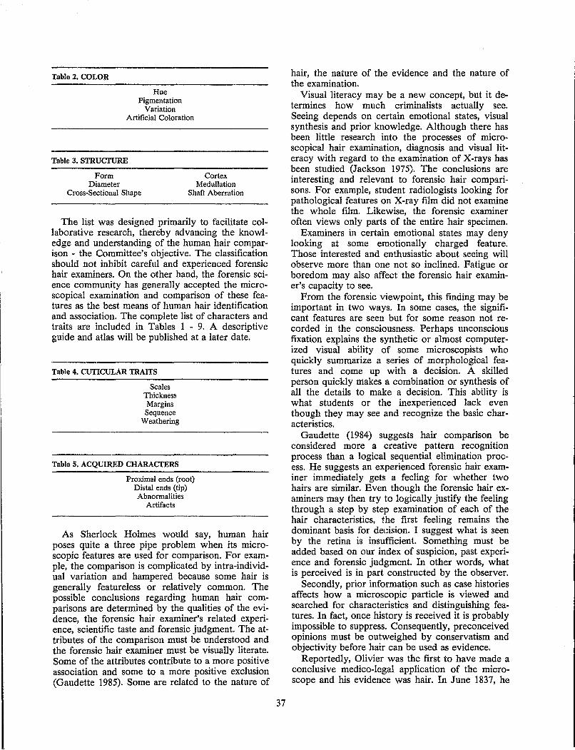
Table 2. COLOR
Hue Pigmentation
Variation Artificial Coloration
Table 3. STRUCTURE
Form Diameter
Cross-Sectional Shape
Cortex Medullation
Shaft Aberration
The list was designed primarily to facilitate collaborative research, thereby advancing the knowledge and understanding of the human hair comparison - the Committee's objective. The classification should not inhibit careful and experienced forensic hair examiners. On the other hand, the forensic science community has generally accepted the microscopical examination and comparison of these features as the best means of human hair identification and association. The complete list of characters and traits are included in Tables 1 - 9. A descriptive guide and atlas will be published at a later date.
Table 4. CUTICULAR TRAITS
Scales Thickness Margins Sequence
Weathering
Table 5. ACQUmED CHARACTERS
Proximal ends (root) Distal ends (tip) Abnormalities
Artifacts
As Sherlock Holmes would say, human hair poses quite a three pipe problem when its microscopic features are used for comparison. For example, the comparison is complicated by intra-individual variation and hampered because some hair is generally featureless or relatively common. The possible conclusions regarding human hair comparisons are determined by the qualities of the evidence, the forensic hair examiner's related experience, scientific taste and forensic judgment. The attributes of the comparison must be understood and the forensic hair examiner must be visually literate. Some of the attributes contribute to a more positive association and some to a more positive exclusion (Gaudette 1985). Some are related to the nature of
37
hair, the nature of the evidence and the nature of the examination.
Visual literacy may be a new concept, but it determines how much criminalists actually see. Seeing depends on certain emotional states, visual synthesis and prior knowledge. Although there has been little research into the processes of microscopical hair examination, diagnosis and visual literacy with regard to the examination of X-rays has been studied (Jackson 1975). The conclusions are interesting and relevant to forensic hair comparisons. For example, student radiologists looking for pathological features on X-ray film did not examine the whole film. Likewise, the forensic examiner often views only parts of the entire hair specimen.
Examiners in certain emotional states may deny looking at some emotionally charged feature. Those interested and enthusiastic about seeing will observe more than one not so inclined. Fatigue or boredom may also affect the forensic hair examiner's capacity to see.
From the forensic viewpoint, this finding may be important in two ways. In some cases, the significant features are seen but for some reason not recorded in the consciousness. Perhaps unconscious fixation explains the synthetic or almost computerized visual ability of some microscopists who quickly summarize a series of morphological features and come up with a decision. A skilled person quickly makes a combination or synthesis of all the details to make a decision. This ability is what students or the inexperienced lack even though they may see and recognize the basic characteristics.
Gaudette (1984) suggests hair comparison be considered more a creative pattern recognition process than a logical sequential elimination process. He suggests an experienced forensic hair examiner immediately gets a feeling for whether two hairs are similar. Even though the forensic hair examiners may then try to logically justify the feeling through a step by step examination of each of the hair characteristics, the first feeling remains the dominant basis for dedsion. I suggest what is seen by the retina is insufficient. Something must be added based on our index of suspicion, past experience and forensic judgment. In other words, what is perceived is in part constructed by the observer.
Secondly, prior informati(fn such as case histories affects how a microscopic particle is viewed and searched for characteristics and distinguishing features. In fact, once history is received it is probably impossible to suppress. Consequently, preconceived opinions must be outweighed by conservatism and objectivity before hair can be used as evidence.
Reportedly, Olivier was the first to have made a conclusive medico-legal application of the microscope and his evidence was hair. In June 1837, he

Tobie e COLOR
Colorless
w Blonde ::::> Golden brown ::r:
Red
Density (value)
Distribution
z 0
~ I- Aggregation z LU :E (!) a::
Aggregate size
Pigment shape
Pigment size
Pigment color
was charged with determining if hair was adhering to an axe iron seized at the home of an individual indicted for murder and if affirmed to indicate the hair color. He recognized by microscopical examination that the filaments submitted for examination resembled the fur of horse, beef or cow, thus differing completely from human hair. A Judicial inquiry confirmed the correctness of his observations (Gaensslen 1983).
38
Auburn brown
Brown
Gray brown
Black
Absent Ught Medium Heavy Opaque Uniform Peripheral One-sided. Random Central Gapping Pigment in Cuticle Streaked Clumped Round Oval Banded Large Medium Small Round Oblong Other Fine Coarse Mixed Red Brown Black Mixed
EVIDENCE
Most of all, human hair can be useful in a variety of ways as associative evidence. Human hairs were much beloved by fictional detectives who identified single htlirs aided only be a magnifying glass and their own rare talent. The hairs always solved the crime. Were this truly possible, human hair would be a most valuable tool in criminal investigations. Nonetheless, our inability to positively identify human hair has not restricted its use in criminal investigations. Hair has been used for a long time and I predict for a long time to come.
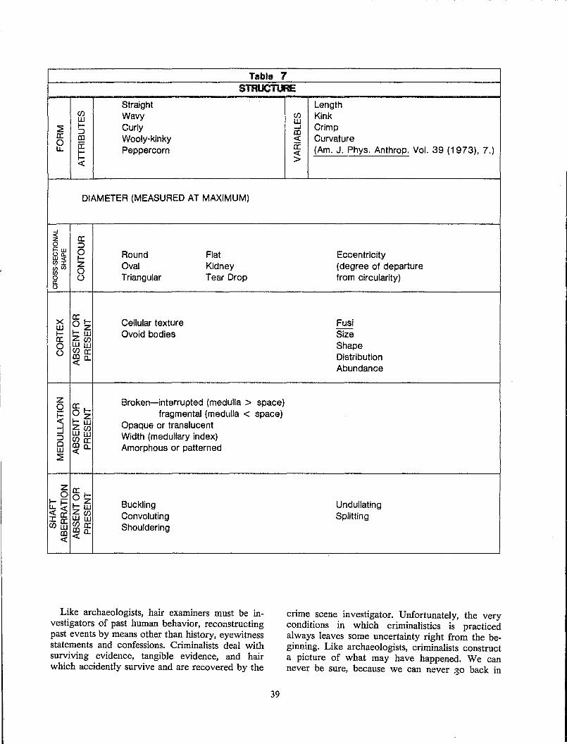
Table 7 STRUCTURE
Straight CIJ Wavy LU
:E I- Curly :J a: CD Wooly-kinky 0 a: u. 1= Peppercorn
c:x:
DIAMETER (MEASURED AT MAXIMUM)
...J « a: z 0 :J i=w 0 Round Flat Oa. W« I-(I):I: Z Oval Kidney 00(1) 0 (I)
Triangular Tear Drop 0 () a:: 0
X a: I- Cellular texture LU Oz I- I-LU OVoid bodies a: z(J) 0 LULU () (J)a:
CDc.. «
Z Broken-interrupted (medulla> space) 0 a: I-
~ Oz I-LU
fragmental (medulla < space) Opaque or translucent ...J z(J)
...J LULU Width (medullary index) :J ClJa: Cl CDc.. Amorphous or patterned LU c:x: ~
Z a: 0 . 0 l-I--
I- z Buckling u.1-c:x:C:X: ZLU
Convoluting Ia: LUCIJ ClJffi
(J)LU Shouldering Q)a:
CD c:x:c.. c:x:
Like archaeologists, hair examiners must be investigators of past human behavior, reconstructing past events by means other than history, eyewitness statements and confessions. Criminalists deal with surviving evidence, tangible evidence, and hair which accidently survive and are recovered by the
39
Length CIJ Kink LU ...J Crimp CD c:x: Curvature a: (Am. J. Phys. Anthrop. Vol. 39 (1973), 7.) c:x: >
Eccentricity (degree of departure from circularity)
Fusi Size Shape Distribution Abundance
Undullating Splitting
crime scene investigator. Unfortunately, the very conditions in which criminalistics is practiced always leaves some uncertainty right from the beginning. Like archaeologists, criminalists construct a picture of what may have happened. We can never be sure, because we can never -30 back in
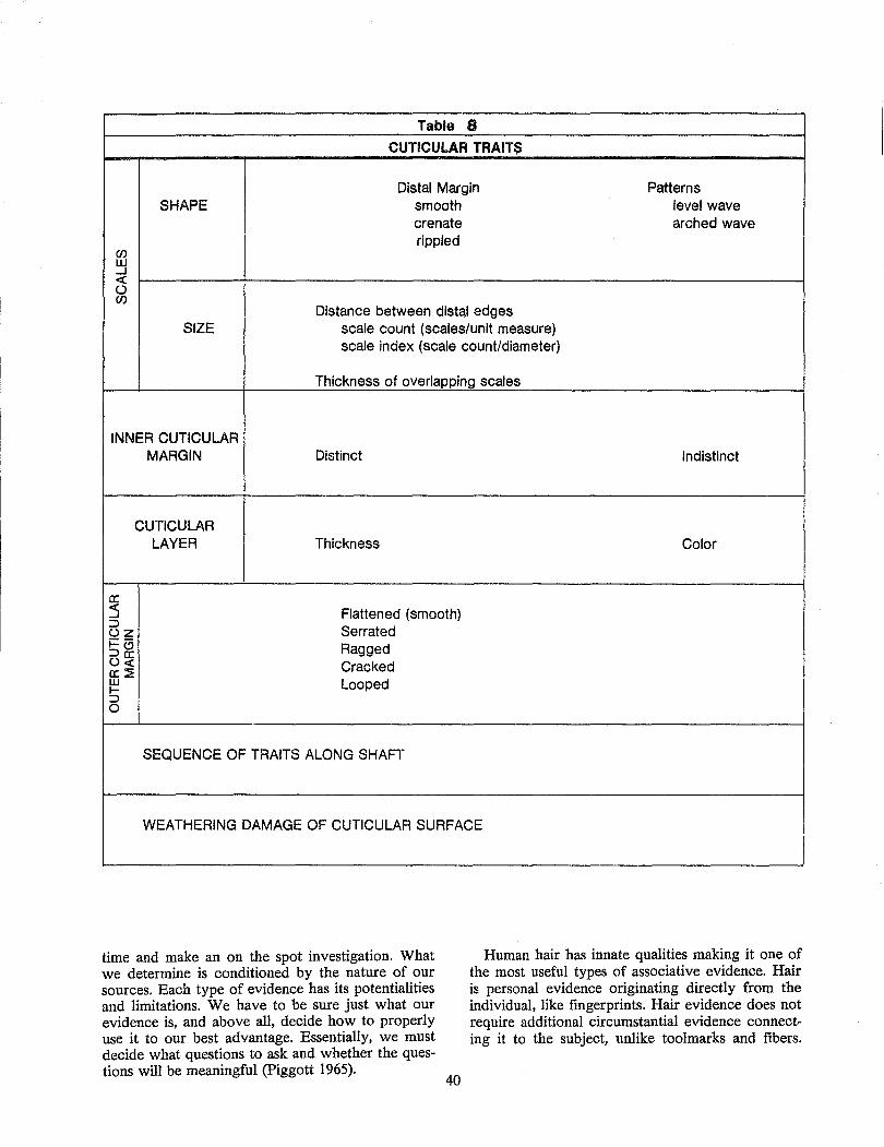
Table 8
CUTICULAR TRAITS
Distal Margin Patterns SHAPE smooth level wave
crenate arched wave rippled
en UJ ..J « 0 en
Distance between distal edges SIZE scale count (scales/unit measure)
scale index (scale count/diameter)
Thickness of overlapping scales
INNER CUTICULAR MARGIN Distinct Indistinct
CUTICULAR LAYER Thickness Color
0: :s Flattened (smooth) ::> oz Serrated i=c:5 Ragged ::>0: 0« Cracked a:~ w Looped I-::> 0
SEQUENCE OF TRAITS ALONG SHAFf"
WEATHERING DAMAGE OF CUTICULAR SURFACE
time and make an on the spot investigation. What we determine is conditioned by the nature of our sources. Each type of evidence has its potentialities and limitations. We have to be sure just what our evidence is, and above all, decide how to properly use it to our best advantage. Essentially, we must decide what questions to ask and whether the questions will be meaningful (Piggott 1965).
Human hair has innate qualities making it one of the most useful types of associative evidence. Hair is personal evidence originating directly from the individual, like fingerprints. Hair evidence does not require additional circumstantial evidence connecting it to the subject, unlike toolmarks and fibers.
40
,
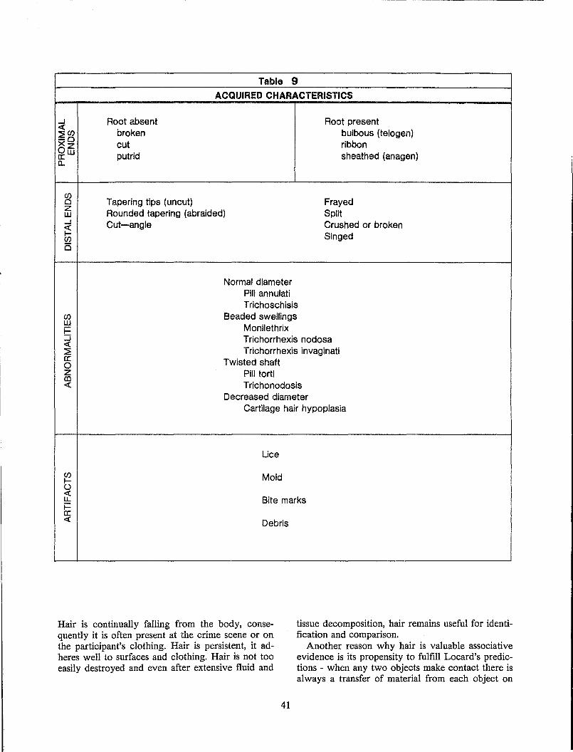
...J « ~Cf) -0 Xz Ow a:: a..
Cf) o z W ...J
~ Cf)
is
Cf) w i= :J « ::2 a:: o z en «
Cf) I-0 « lJ.. i= a:: «
Root absent broken cut putrid
Table 9 ACQUIRED CHARACTERISTICS
Root present bulbous (telogen) ribbon sheathed (anagen)
Tapering tips (uncut) Rounded tapering (abraided) Cut-angle
Frayed Split Crushed or broken Singed
Normal diameter Pili annulati Trichoschisis
Beaded swellings Monilethrix Trichorrhexis nodosa Trichorrhexis invaginati
Twisted shaft Pili torti Trichonodosis
Decreased diameter Cartilage hair hypoplasia
Lice
Mold
Bite marks
Debris
Hair is continually falling from the body, consequently it is often present at the crime scene or on the participant's clothing. Hair is persistent, it adheres well to surfaces and clothing. Hair is not too easily destroyed and even after extensive fluid and
tissue decomposition, hair remains useful for identification and comparison.
Another reason why hair is valuable associative evidence is its propensity to fulfill Locard's predictions - when any two objects make contact there is always a transfer of material from each object on
41
to the other. Our experience reinforces this principle because we can remember many cases of coition or assault that resulted in one participant's hair transferring to the other's clothing or body.
Table 10. QUESTIONS ANSWERED BY LABORATORY EXAMINATION OF HAIR
1. Is the fiber a hair? 2. If a hair, is it human? 3. What is the racial origin? 4. What is the somatic origin? 5. Was the hair broken, crushed, cut, or forcefully
pulled? 6. Was the hair singed? 7. Could it have originated from the suspected
source?
Although human hair has a variety of uses aiding the reconstruction of criminal events and associating the perpetrator with the victim or crime scene; only a limited number of questions can be answered by a laboratory examination of human hair evidence (Table 10). Although most of these questions can be easily answered, one is problematical. There remains the possibility of error regarding the conclusion that a given hair could have originated from a given person. There are two types of errors. Gaudette (1982) addressed each of these errors with remarkable clarity and persistence (Table 11).
Table 11. PROBABILITY OF HUMAN HAIR COMPARISON ERRORS
Fact! Conclusion
Associated
Excluded
Hair is from individual
Correct P = I-a
Type I Error P=a
Hair is not from individual
Type II Error P=/3
Correct P=I-/3
Our ethical tradition considers errors of the second kind (type II errors) much more serious. It is better 99 guilty men go free than one innocent man be convicted. Or when in doubt, throw it out. The premise of our judicial system, that is, the accused is innocent until proven guilty beyond a reasonable doubt, reflects this concern to avoid type II errors. Our aim as conscientious forensic scientists should be to provide reliable and valuable investigative evidence. Our conclusions should be true and powerful.
One way to help ensure reliability and limit forensic examination errors is to follow the criteria
42
first proposed for voice print identification (Table 12).
Table 12. CRITERIA FOR PROPER HAIR COMPARISON
1. Examiner must be properly and thoroughly trained.
2. Examiner must have extensive experience. 3. Examiner must be allowed time to reach the
decision. 4. Examiner must be allowed negative and
inconclusive determinations when evidence available does not warrant more.
5. Examiner must be allowed access to the background and investigative information.
6. Examiner must be allowed to consult with a colleague to verify the analysis.
Another way to consider improving the quality of our hair comparison conclusions is the concept of power. A test is powerful if it correctly detects that the null hypothesis is false, that is, correctly detects that the questioned hair did not originate from the suspected donor. Power is the complement of beta (1 - f3 ). It is the probability of rejecting the null hypothesis when in fact it is false and the alternative hypothesis is correct. Obviously, for any given comparison we would like the power to be as large as possible (approaching one) and the quantity beta as small as possible. The power falls off sharply as the alternate hypothesis approaches the null hypothesis or when the two suspected sources of hair are not easily distinguished. The power of the test can be improved by increasing the sample sizes, by increasing the probability of rejecting the true null hypothesis (alpha), or by changing the nature of the test by adding dimensions to the comparisons (Sokal and Rohlf 1969).
In human hair comparisons we have difficulty controlling sample size and alpha; therefore, we must reduce beta by conservatism. Alternatively, we could increase the power to one by always excluding the questioned hair. That is not very valuable. Therefore Gaudette has reported another statistic value. Gaudette (1982) defines value as dependent directly on the complement of alpha and inversely on beta. He further discusses alpha in the Canadian Society of Forensic Science Journal (1985). Regardless of the statistical basis, we are looking for evidence which is true, powerful and valuable to the investigation.
Presumably, one difference between forensic science and some other endeavors is our aversion for error. It should be remembered the scientific method is a potentiation of common sense, exercised freely but always with a specially firm determination against persisting in error, if any exertion of hand or mind can deliver us from it. Bronowski
(1978a) describes progress as the exploration of our own error. We need to better understand our errors. Gaudette and his critics have helped us in that regard (Barnett and Ogle 1982).
CONCLUSION
Bronowski also wrote (1978a), "We are constantly deceived about the nature of the outside world because we interpret it in terms of a built-in search mechanism." Likewise, describing what a hair is to the forensic scientist is based on this author's experiences and inclinations. A more scientific approach would be to organize these sensory messages into a structured form. Hopefully, the International Symposium on Forensic Hair Comparisons served that purpose. In the end, the strategy for the future of hair evidence is to regard it as something special, something valuable to criminal investigations and something important. As Plato said, "What is honored in a country will be cultivated there." (Austin 1977).
Science is a progressive activity, a codification of that essential human characteristic, the making of plans (Bronowski 1978b). Through continued interest, we may never find our panacea because that is magic, but we will better understand this important evidence and make better plans for the future.
REFERENCES
Austin, J. H (1977). Chase, Chance, and Creativity. Columbia Univ. Press, New York.
Barnett, P. D. and Ogle, R. R. (1982). Probabilities and human hair comparison, J. Forensic Sci. 27:272-278.
Bassett, W. A. G. (1978). Sex determination by sex chromatin identification in the hair root sheath, Can. Soc. Forensic Sci. J. 11:221-228.
Bisbing, R. E. (1975). Immunohematological grouping of human hair for ABO, Forensic Serology Newsletter 1:1-4.
Bisbing, R. E. (1982). The forensic identification and association of human hair. In: Forensic Science Handbook (Saferstein, R., ed.). PrenticeHall, Englewood Cliffs.
Bisbing, R. E. and Wolner, jJtJ. F. (1984). Microscopical discrimination of twins head hair, J. Forensic Sci. 29:780-786.
Bronowski, J. (1978a). The Origins of Knowledge and Imagination. Yale Univ. Press, New Haven.
Bronowski, J. (1978b). Magic, Science, and Civilization. Columbia Univ. Press, New York.
Cornelis, R. (1972). Is it possible to identify individuals by neutron activation analysis of hair? Med. Sci. Law 12:188-194.
43
Gaenss/en, R. E. (1983). Sourcebook in Forensic Serology, Immunology, and Biochemistry, Unit IX: Translations of Selected Contributions. National Institute of Justice, Washington, D.C.
Gaudette, B. D. (1982). A supplementary discussion of probabilities and human hair comparisons, J. Fonnsic Sci. 27:279-289.
Gaudette, B. D. (1984). A collaborative study in forensic hair comparison. Presented at the International Association of Forensic Science, 10th Triennial Meeting, Oxford.
Gaudette, B. D. (1985). Strong hegative conclusions in hair comparison--A rare event, Can. Soc. Forensic Sci. J. 18:32-37.
Hooke, R. (1961). Micrographia. Dover, New York.
Jackson, R. (1975). The importance of being visually literate, Arch. Dermatol. 111 :632-636.
Lawton, M. E. and Sutton, J. G. (1982). Multiple enzyme typing of the sheath cells associated with the root of a single human head hair, J. Forensic Sci. Soc. 22:203-209.
Lee, L. D., Ludwig, K. and Baden, H P. (1978). Matrix proteins of human hair as a tool for identification, Forensic Science 11:115-121.
Marshall, R. C. and Gillespie, J. M. (1982). Comparison of samples of human hair by two-dimensional electrophoresis, J. Forensic Sci. Soc. 22:377-385.
Montgomery, D. and Jay, B. W-H (1982). Multiple enzyme systems grouping of human hair root sheaths. Can. Soc. Forensic Sci. J. 15:1-10.
Mudd, J. L. (1984). The determination of sex from forcibly removed hairs, J. Forensic Sci. 29:1072-1080.
Piggott, S. (1965). Approach to Archaeology. McGraw-Hill, New York.
Pillay, K. K. S. and Kuis, R. L. (1978). The potentials and limitations of using neutron activation analysis data on human hair as a forensic evidence, J. Radioanalyt. Chem. 43:461-478.
Robertson, E. A., Van Steirteghem, A. c., Byrkit, J. E. and Young, D. S. (1980). Biochemical individuality and the recognition of personal profiles with a computer, Clin. Chem. 26:30-36.
Sokal, R. R. and Rohlf, P. J. (1969). Biometry--The Principles and Practice of Statistics in Biological Research.
Sutton, J. G., Bosley, C. M. and Whitehead, P. H (1982). Polymorphic enzyme systems in human hair sheath cells, J. Forensic Sci. Soc. 22:199-202.
Twibell, J. and Whitehead, P. H. (1978). Enzyme typing of human hair roots, J. Forensic Sci. 23:356-360.
Wittig, M (1982). Protein patterns of keratins--the probable role in forensic hair examination, J. Forensic Sci. Soc. 22:387-389.
Yablokov, A. V. (1974). Variability of Mammals. Amerind Publishing Co., New Delhi.
DISCUSSION
Metzger: What characteristics do you use to determine that a questioned hair is not representative of the questioned source, and what characteristics do you use to make that determination?
Bisbing: You have to begin with good quality known exemplars. Then you have to be willing to consider the questioned hair to be an odd hair, if indeed it does not possess many of the normal characteristics of hair.
If it is short and curly, and is only a quarter of an inch long but tapered, it is probably not representative of a source of Caucasian head hair. But in the end it is a judgmental decision whether or not to exclude and run the risk of making a type I error increasing alpha, or making the decision to be inconclusive and not making that error. But certainly we make a lot of type I errors when we are trying to decrease alpha where possible without interfering with beta, without increasing our probability of making mostly type II errors. Sometimes that just simply can not be done; therefore, a judgmental decision is made. A paper authored by Barry Gaudette in the March 1985 issue of the Canadian Society of Forensic Sciences Journal speaks to that.
Roe: What percentage of cases in the United States is enzyme typing carried out?
Bisbing: A very low percentage I assume. Two reasons, one is that we do not often get a root sheath in the questioned hair. Most of the hairs are club hairs. The second reason is that in some lab-
44
oratories we do not get to the hair comparisons until our serologists have finished their work and then they are forced to do the hair comparisons. It becomes a little late for that. But I think you will find it being done more and more, and hopefully we are in a position that if there is a root sheath present, it will be done.
Question: What do you mean by "characteristics" versus "to characterize the hair using many features"?
Bisbing: We have used the term "characteristic" here to mean a particular type of feature like medulla; "traits" on the other hand, to mean the various types of medulla; and "characterize" to mean individualize or compare: to describe in a lot of different features.
Question: In your laboratory is there any peer review of positive hair comparisons or hairs that are found to be similar?
Bisbing: Yes we do have peer review but not in every case. We do often share our specimens and consult with each other. We showed, and I think Mr. Gaudette showed in one of his projects, the value of that. We showed that when we were looking at two samples, indeed one person may detect differences where another person may not on that day. I think the process of verifying is a very valuable procedure; one that is used in a lot of other disciplines, for example, electrophoresis and fingerprints.
Simms: Do you work to resolve that difference, or at that point do you go back to an inconclusive statement in the report? What happe .. ls when the two people looking at the same hair disagree?
Bisbing: Obviously try to resolve it in some way possible but when in doubt throw it out. There are differences 'that cannot be reconciled, then they have to be thrown out.
HUMAN HAIR COMPARISONS BASED ON MICROSCOPIC CHARACTERISTICS Harold A. Deadman
FBI Laboratory Division Microscopic Analysis Unit
Washington, D.C.
Earlier speakers at this Symposium have discussed the properties and characteristics possessed by human hair. Richard E. Bisbing presented a classification of the numerous microscopic characteristics that can be examined under a bright-field transmitted light microscope. This paper will comment upon the comparison of human hairs using these microscopic characteristics.
From a forensic science point of view, the comparison of human hairs based on their microscopic characteristics is the most important part of a hair examination. Forensic hair comparisons have resulted in significant associations in many types of criminal cases. These comparisons, conducted in crime laboratories for many years, have generally involved side by side comparison of the microscopic characteristics.
Comparison microscopy has been criticized as being too subjective and, therefore, lacking in evidentiary value. Consequently certain forensic scientists have called for the development of more objective techniques. A number of these techniques are discussed in these proceedings. This author personally welcomes exhaustive study in these areas but does not believe any new technique can or will replace comparison microscopy. These new techniques may be shown to add additional discrimination or simplify the comparison process (especially for the inexperienced microscopist), but will still be used only after the microscopical comparison occurs. Although in the future the comparison process may be improved through new procedures, this author believes microscopical hair comparisons, when properly conducted, are presently very discriminating and can be the basis for strong associations.
A report entitled, Preliminary Report - Committee on Forensic Hair Comparison, which contains a paper entitled, Recommendations Concerning the Process of 11,licroscopical Human Hair Comparison, was distributed to the attendees at this Symposium. This paper was the work-;;~oduct of a subcommittee which included Stephen A. Shaffer, Theodore Mozer, Norman E. Erickson and this author. Barry D. Gaudette, as Chairman of the Committee on Forensic Hair Comparison also made significant contributions to its preparation. These recommendations, highlighted in the following discussion, are not set forth as specific protocol, but rather as information with which an examiner should be famil-
45
iar when performing a hair comparison. Procedural considerations are also discussed.
As mentioned earlier microscopical hair comparison, as generally conducted in crime laboratories, is considered by some critics to be too subjective to be a valid method of comparison. Human hair comparisons are subjective in the sense that there are few quantitative or objective measurements that can be made. Those characteristics which can be measured in a quantitative manner are usually of limited value for comparison purposes. It must be remembered, however, that the experienced hair examiner's ability to reason subjectively has ideally been sharpened during the examinations and comparisons of large numbers of human hairs. In addition, a comparison process whereby SUbjective impressions, each of which contain large amounts of information, are examined side by side is different from a person making a subjective determination of the properties of an object. It is also common experience that trained and skilled individuals in many areas can make valid comparisons of different Irl1-
terials based on subjective processes. As with other subjective examinations conducted
in crime laboratories, it is important to recognize that proper training and experience are necessities for the hair examiner. Because the hair comparison process is also often difficult and time-consuming, the examination must not be rushed or conducted in a casual manner. The subjectivity and difficulty of the hair comparison process force the hair examiner to devise procedures so as to minimize all errors, especially the incorrect inclusion-type of error. This type of error results in evidencl~ incorrectly linking a person with another person or specific location. The consequences of this type of error are much more serious than the error incurred when hairs that did, in fact, originate from an individual are not linked or associated with that individual.
Human hairs differ in many ways from other types of physical evidence used in courts of law. Hairs are biological materials that grow. Head hair, for example, grows at the rate of approximately 1 cm per month. This growth results in different portions of a single head hair 'having different ages. The root and tip ends, extremes in terms of age differences, can differ considerably in microscopic characteristics because of environmental and cosmetic effects. The fact that the microscopic characteristics of a human hair often vary along its length
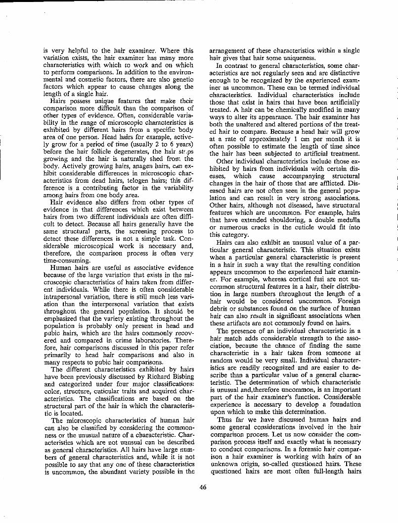
is very helpful to the hair examiner. Where this variation exists, the hair examiner has many more characteristics with which to work and on which to perform comparisons. In addition to the environmental and cosmetic factors, there are also genetic factors which appear to cause changes along the length of a single hair.
Hairs possess unique features that make their comparison more difficult than the comparison of other types of evidence. Often, considerable variability in the range of microscopic characteristics is exhibited by different hairs from a specific body area of one person. Head hairs for example, actively grow for a period of time (usually 2 to 6 years) before the hair follicle degenerates, the hair st/,ps growing and the hair is naturally shed from the body. Actively growing hairs, anagen hairs, Cdn exhibit considerable differences in microscopic characteristics from dead hairs, telogen hairs; this difference is a contributing factor in the variability among hairs from one body area.
Hair evidence also differs from other types of evidence in that differences which exist between hairs from two different individuals are often difficult to detect. Because all hairs generally have the same structural parts, the screening process to detect these differences is not a simple task. Considerable microscopical work is necessary and, therefore, the comparison process is often very time-consuming.
Human hairs are useful as associative evidence because of the large variation that exists in the microscopic characteristics of hairs taken from different individuals. While there is often considerable intrapersonal variation, there is still much less variation than the interpersonal variation that e'xists throughout the general popUlation. It should be emphasized that the variety existing throughout the population is probably only present in head and pubic hairs, which are the hairs commonly recovered and compared in crime laboratories. Therefore, hair comparisons discussed in this paper refer primarily to head hair comparisons and also in many respects to pubic hair comparisons.
The different characteristics exhibited by hairs have been previously discussed by Richard Bisbing and categorized under four major classifications: color, structure, cuticular traits and acquired characteristics. The classifications are based on the structural part of the hair in which the characteristic is located.
The microscopic characteristics of human hair can also be classified by considering the commonness or the unusual nature of a characteristic. Characteristics which are not unusual can be described as general characteristics. All hairs have large numbers of general characteristics and, while it is not possible to say that anyone of these characteristics is uncommon, the abundant variety possible in the
46
arrangement of these characteristics within a single hair gives that hair some uniqueness.
In contrast to general characteristics, some characteristics are not regularly seen and are distinctive enough to be recognized by the experienced examiner as uncommon. These can be termed individual characteristics. Individual characteristics include those that exist in hairs that have been artificially treated. A hair can be chemically modified in many ways to alter its appearance. The hair examiner has both the unaltered and altered portions of the treated hair to compare. Because a head hair will grow at a rate of approximately 1 cm per month it is often possible to estimate the length of time since the hair has been subjected to artificial treatment.
Other individual characteristics include those exhibited by hairs from individuals with certain diseases, which cause accompanying structural changes in the hair of those that are afflicted. Diseased hairs are not often seen in the general population and can result in very strong associations. Other hairs, although not diseased, have structural features which are uncommon. For example, hairs that have extended shouldering, a double medulla or numerous cracks in the cuticle would fit into this category.
Hairs can also exhibit an unusual value of a particular general characteristic. This situation exists when a particular general characteristic is present in a hair in such a way that the resulting condition appears uncommon to the experienced hair examiner. For example, whereas cortical fusi are not uncommon structural features in a hair, their distribution in large numbers throughout the length of a hair would be considered uncommon. Foreign debris or substances found on the surface of human hair can also result in significant associations when these artifacts are not commonly found on hairs.
The presence of an individual characteristic in a hair match adds considerable strength to the association, because the chance of finding the same characteristic in a hair taken from someone at random would be very small. Individual characteristics are readily recognized and are easier to describe than a particular value of a general characteristic. The determination of which characteristic is unusual and,therefore uncommon, is an important part of the hair examiner's function. Considerable experience is necessary to develop a foundation upon which to make this determination.
Thus far we have discussed human hairs and some general considerations involved in the hair comparison process. Let us now consider the comparison process itself and exactly what is necessary to conduct comparisons. In a forensic hair comparison a hair examiner is working with hairs of an unknown origin, so-called questioned hairs. These questioned hairs are most often full-length hairs
~--------~ ---------------
that have been naturally shed from the body. In crimes of violence, forcibly removed hairs and damaged hairs are often seen. In order to conduct hair comparisons in an attempt to associate questioned hairs with a particular individual, it is first necessary to obtain a known hair sample from that individual.
Because of the variation in microscopic characteristics among different hairs from the same body area of one person, it is important to obtain a sufficient number of hairs in order to adequately represent the ranges of all characteristics present. Because they differ in their characteristics it is important to obtain hairs from different areas of the scalp. Full-length hairs with roots should be obtained for the examiner to adequately examine and compare the variations along the length of a single hair and determine its growth stage. Since the vast majority of pulled hairs will be in an active growing stage, a combing procedure is also desirable to obtain hairs in the telogen or dead stage.
It is recommended a known head hair sample consist of at least 20 hairs from each of five different areas of the scalp (center, front, back and both sides) and these hairs be obtained by both pulling and combing. The recommended procedure for obtaining combed hairs is to use either a comb packed with cotton or a multibristle brush. The various areas of the scalp should be repeatedly combed over a large sheet of clean paper in a direction opposite to that in which the person usually combs the hair.
From these 100 hairs, a number of hairs (usually 6 to 20, depending on the homogeneity of the sample) should be selected by the hair examiner as representative of the entire known sample. The selection should be based primarily on gross characteristics such as length, coarseness and color as observed by macroscopic and stereomicroscopic examination. These hairs should be used for comparison. The remaining hairs are then available for future use if subsequent examinations reveal that, whereas a questioned hair has characteristics close to those of the known sample, a good match to any of the 6 to 20 hairs originally selected cannot be found.
A known pubic hair sample should consist of at least 30 hairs obtained by both pulling and combing from different areas of the pubic region.
For exclusionary purposes known samples should be requested from all persons who might reasonably be considered a source of a questioned hair. If such samples are obtained and excluded, the significance of any ensuing association is increased.
Some hair examiners believe that a known sample does not require a large number of hairs and that hairs cut close to the scalp, being easier to obtain, should be used for the comparison process. The Subcommittee mem1:>ers, in formulating the
47
recommendation of 100 pulled and combed hairs, believe 100 pulled and combed hairs are required to guarantee full-length hairs, and the number chosen should help minimize type I errors. Since an individual can lose an average of 100 scalp hairs a day as part of the normal hair cycle, the collec-110n of 100 hairs is not unreasonable. While hairs cut close to the skinline can, if necessary, be used for comparison purposes, the root and root end are important parts of the hair and should be obtained whenever possible. It is recommended that research be conducted as to the content and methods of collection of known hair samples.
In order to microscopically compare hairs, the hairs must be properly prepared for examination. To adequately observe the microscopic characteristics within a hair, the hair must be placed in a medium of refractive index similar to that of the hair itself (the isotropic or average refractive index of hair is approximately 1.55). A synthetic semiper·. manent mounting medium with a refractive index around 1.52 is recommended. Using a mounting medium with a refractive index much different from that of hair will result in excessive shadows and contrast that will tend to mask the internal characteristics of a hair. A semipermanent medium is recommended for convenience. The hair or hairs can then be mounted so as to remain in a fixed location, with no chance of loss from the glass microscope slide. Some of the commercially available semipermanent synthetic mounting media allow mounted hairs to be easily removed from the slide if desired.
Because of the subjectivity involved in the comparison of images that are difficult to describe and often difficult to recall, the Subcommittee believes a high quality comparison microscope must be used in the comparison process. This is necessary to distinguish between the subtle or slight differences that may exist between hairs from different individuale. High quality objectives are important, but highly corrected planapochromats are not necessary. The objectives and eyepieces selected, however, should permit observations in the range of approximately 40X to 4OOX. A high intensity tungsten light source, suitable for photomicrography and equipped with a daylight correction filter, provides adequate lighting. A comparison microscope can be equipped with several types of stages. The types of stages used, and their placement on the microscope, are a matter of personal preference and should not affect one's ability to compare hairs.
Both low and high power microscopic examinations are necessary in the comparison of hairs. Stereomicroscopic examinations at magnifications around lOX, of both unmounted and mounted hairs, are important for screening purposes. Obser-
vations of mounted hairs with the compound microscope must encompass a variety of magnifications generally around 40X, WOX, 250X and 400X. The use of high power (large numerical aperture) objectives allows the examination and comparison of the fine detail present in such characteristics as pigmentation and cuticular scales. These objectives also allow a hair examiner to view thin sections of the hair. This "optical sectioning" is important because a hair is three-dimensional and certain characteristics vary throughout its thickness. Information about the shape and surface appearances of the hair can also be obtained by optical sectioning.
The procedure used by the hair examiner should incorporate the general considerations discussed in this paper, proper evidence handling and the correct utilization of proper equipment. The procedure must involve a thorough and careful examination of both the gross and microscopic characteristics exhibited by properly prepared hairs while using a high quality comparison microscope at different magnifications. The questioned hairs should be compared to the known hairs with regard to as many as possible uf the characteristics recommended by the Committee. The hair examiner should strive to attain a level of discrimination in hair comparisons such that type II errors will be minimized without at the same time incurring an unreasonable number of type I errors. As long as these considerations are kept in mind, the Subcommittee believes a considerable amount of leeway should be allowed in choice of procedure to satisfy individual preferences.
Procedural differences among hair examiners in four areas were considered by the Subcommittee. Whereas these should not affect the ability to correctly compare hairs, they are discussed here for informational and educational purposes. These procedural differences are in the folk~wing areas: preliminary screening, note-taking, sequence of examination and number of hairs mounted on a single slide.
Some hair examiners believe the macroscopic or stereomicroscopic examination of unmounted hairs is of value for screening purposes; others do not, and utilize low power examinations with mounted hairs only. It is recommended that research be conducted to determine the value of low power examination of unmounted hairs. In the meantime, the use of such preliminary screening should remain a matter of individual preference.
It should be emphasized that preliminary screening is an important part of the comparison process. If hairs of no value for significant comparison purposes can be eliminated from further consideration during a stereoscopic examination, a considerable savings in time results. Once hairs have been mounted it is also important that a hair examiner develop proficiency in using low power sterco-
48
sC0pic examination to determine which hairs should be selected for detailed and time consuming comparison microscopy. In the screening of hairs prior to detailed comparisons, study of the root and tip ends of hair can be beneficial for several reasons. These areas are easy to locate. They contain essentially all the characteristics present in the hair and these characteristics reflect environmental and cosmetic changes.
There was a considerable difference of opinion among hair examiners regarding the quality and quantity of note-taking considered necessary, the detail that should be present, and the use of notes during the comparison process. Some hair examiners use preprinted forms while others make personalized notes to describe the characteristics of hairs. While note-taking should be a matter of personal preference, it is generally believed notes that accurately reflect the complete results of a case and individually identify the questioned hairs, especially those that are involved in a match, should be taken.
Most hair examiners prefer to examine known hairs first, but some prefer to examine the questioned hairs first. Again, this is best left to the examiner's preference and judgment, bearing in mind the circumstances of the case.
Some hair examiners mount all hairs, both questioned and known, one to a slide. Others mount as 1T\any as five or more hairs to a single slide depending on their length. Yet other hair examiners collectively mount questioned hairs, but singly mount known hairs, or vice versa. Apparently the more experienced the hair examiner the more likely hel she will mount many hairs per slide. This method is not as simple as mounting a single hair per slide, but considerahle savings in cost and time result if many hairs can be mounted together.
The most important decision made by the hair examiner is deciding when a hair can be said to match or be consistent with originating from a particular individual. In order to make the determination that a questioned hair is consistent in macroscopic and microscopic characteristics with a known sample from a particular person, it must first be determined that there are no significant differences in these characteristics or their arrangement. Since no two hairs will ever be exactly the same in all minute details, it is important to determine what differences are significant. It must first be determined that the characteristics exhibited by the questioned hair fit within the range of characteristics present in the known sample. After that, the ideal situation for the hair examiner is to find one or more hairs in the known sample that correspond in all respects (no significant differences) with the questioned hair. If this goal is strived for, the frequency of type II errors will be minimized.
Depending on the characteristics involved and the circumstances surrounding a case, it may sometimes be possible to utilize several hairs from the known sample to locate characteristics which cor-
49
respond to those of a questioned hair. However, the "duplicate hair" criterion for a match should be the ultimate goal.
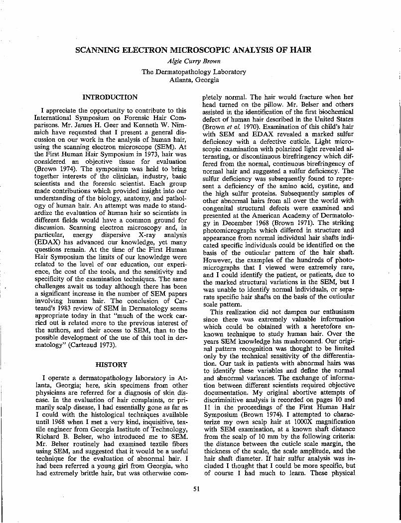
SCANNING ELECTRON MICROSCOPIC ANALYSIS OF HAIR Algie Curry Brown
The Dermatopathology Laboratory Atlanta, Georgia
INTRODUCI'ION
I appreciate the opportunity to contribute to this International Symposium on Forensic Hair Comparisons. Mr. James H. Geer and Kenneth W. Nimmich have requested that I present a general discussion on our work in the analysis of human hair, using the scanning electron microscope (SEM). At the First Human Hair Symposium in 1973, hair was considered an objective tissue for evaluation (Brown 1974). The symposium was held to bring together interests of the clinician, industry, basic scientists and the forensic scientist. Each group made contributions which provided insight into our understanding of the biology, anatomy, and pathology of human hair. An attempt was made to standardize the evaluation of human hair so scientists in different fields would have a common ground for discussion. Scanning electron microscopy and, in particular, energy dispersive X-ray analysis (EDAX) has advanced our knowledge, yet many questions remain. At the time of the First Human Hair Sym"osium the limits of our knowledge were related to the level of our education, our experience, the cost of the tools, and the sensitivity and specificity of the examination techniques. The same challenges await us today although there has been a significant increase in the number of SEM papers involving human hair. The conclusion of Carteaud's 1983 review of SEM in Dermatology seems appropriate today in that "much of the work carried out is related more to the previous interest of the authors, and their access to SEM, than to the possible development of the use of this tool in dermatology" (Carteaud 1973).
HISTORY
I operate a dermatopathology laboratory in Atlanta, Georgia; here, skin specimens from other physicians are referred for a diagnosis of skin disease. In the evaluation of hair complaints, or primarily scalp disease, I had essentially gone as far as I could with the histological techniques available until 1968 when I met a very kind, inquisitive, textile engineer from Georgia Institute of Technology, Richard B. Belser, who introduced me to SEM. Mr. Belser routinely had examined textile fibers using SEM, and suggested that it would be a useful technique for the evaluation of abnormal hair. I had been referred a young girl from Georgia, who had extremely brittle hair, but was otherwise com-
51
p1etely normal. The hair would fracture when her head turned on the pillow. Mr. Belser and others assisted in the identification of the first biochemical defect of human hair described in the United States (Brown et al. 1970). Examination of this child's hair with SEM and EDAX revealed a marked sulfur deficiency with a defective cuticle. Light microscopic examination with polarized light revealed alternating, or discontinuous birefringency which differed from the normal, continuous birefringency of normal hair and suggested a sulfur deficiency. The sulfur deficiency was subsequently found to represent a deficiency of the amino acid, cystine, and the high sulfur proteins. Subsequently samples of other abnormal hairs from all over the world with congenital structural defects were examined and presented at the American Academy of Dermatology in December 1968 (Brown 1971). The striking photomicrographs which differed in structure and appearance from normal individual hair shafts indicated specific individuals could be identified on the basis of the cuticular pattern of the hair shaft. However, the examples of the hundreds of photomicrographs that I viewed were extremely rare, and I could identify the patient, or patients, due to the marked structural variations in the SEM, but I was unable to identify normal individuals, or separate specific hair shafts on the basis of the cuticular scale pattern.
This realization did not dampen our enthusiasm since there was extremely valuable information which could be obtained with a heretofore unknown technique to study human hair. Over the years SEM knowledge has mushroomed. Our original pattern recognition was thought to be limited only by the technical sensitivity of the differentiation. Our task in patients with abnormal hairs was to identify these variables and define the normal and abnormal variances. The exchange of information between different scientists required objective documentation. My original abortive attempts of discriminitive analysis is recorded on pages 10 and 11 in the proceedings of the First Human Hair Symposium (Brown 1974). I attempted to characterize my own scalp hair at lOOOX magnification with SEM examination, at a known shaft distance from the scalp of 10 mm by the following criteria: the distance between the cuticle scale margin, the thickness of the scale, the scale amplitude, and the hair shaft diameter. If hair sulfur analysis was included I thought that I could be more specific, but of course I had much to learn. These physical
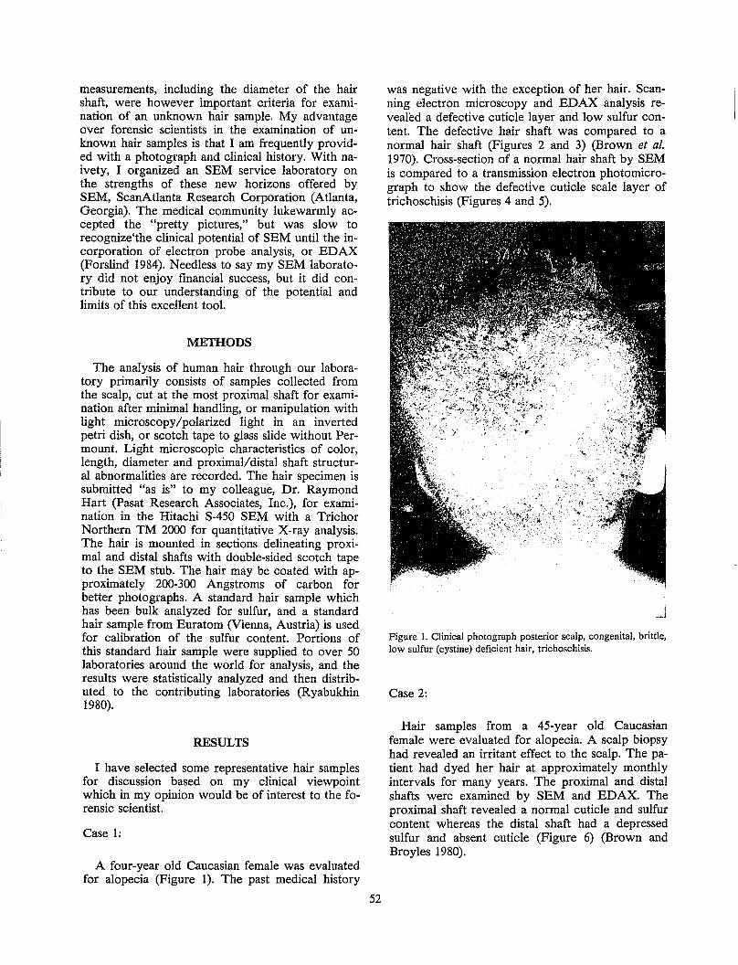
measurements, including the diameter of the hair shaft, were however important criteria for examination of an unknown hair sample. My advantage over forensic scientists in the examination of unknown hair samples is that I am frequently provided with a photograph and clinical history. With naivety, I organized an SEM service laboratory on the strengths of these new horizons offered by SEM, ScanAtlanta Research Corporation (Atlanta, Georgia). The medical community lukewarmly accepted the "pretty pictures," but was slow to recognize'the clinical potential of SEM until the incorporation of electron probe analysis, or EDAX (Forslind 1984). Needless to say my SEM laboratory did not enjoy financial success, but it did contribute to our understanding of the potential and limits of this excellent tool.
METHODS
The analysis of human hair through our laboratory primarily consists of samples collected from the scalp, cut at the most proximal shaft for examination after minimal handling, or manipulation with light microscopy/polarized light in an inverted petri dish, or scotch tape to glass slide without Permount. Light microscopic characteristics of color, length, diameter and proximal/distal shaft structural abnormalities are recorded. The hair specimen is submitted "as is" to my colleague, Dr. Raymond Hart (Pasat Research Associates, Inc.), for examination in the Hitachi S-450 SEM with a Trichor Northern TM 2000 for quantitative X-ray analysis. The hair is mounted in sections delineating proximal and distal shafts with double-sided scotch tape to the SEM stub. The hair may be coated with approximately 200-300 Angstroms of carbon for better photographs. A standard hair sample which has been bulk analyzed for sulfur, and a standard hair sample from Euratom (Vienna, Austria) is used for calibration of the sulfur content. Portions of this standard hair sample were supplied to over 50 laboratories around the world for analysis, and the results were statistically analyzed and then distributed to the contributing laboratories (Ryabukhin 1980).
RESULTS
I have selected some representative hair samples for discussion based on my clinical viewpoint which in my opinion would be of interest to the forensic scientist.
Case 1:
A four-year old Caucasian female was evaluated for alopecia (Figure 1). The past medical history
52
was negative with the exception of her hair. Scanning electron microscopy and EDAX analysis revealed a defective cuticle layer and low sulfur content. The defective hair shaft was compared to a normal hair shaft (Figures 2 and 3) (Brown et al. 1970). Cross-section of a normal hair shaft by SEM is compared to a transmission electron photomicrograph to show the defective cuticle scale layer of trichoschisis (Figures 4 and 5).
Figure 1. Clinical photograph posterior scalp, congenital, brittle, low sulfur (cystine) deficient hair, trichoschisis.
Case 2:
Hair samples from a 45-year old Caucasian female were evaluated for alopecia. A scalp biopsy had revealed an irritant effect to the scalp. The patient had dyed her hair at approximately monthly intervals for many years. The proximal and distal shafts were examined by SEM and EDAX. The proximal shaft revealed a normal cuticle and sulfur content whereas the distal shaft had a depressed sulfur and absent cuticle (Figure 6) (Brown and Broyles 1980).
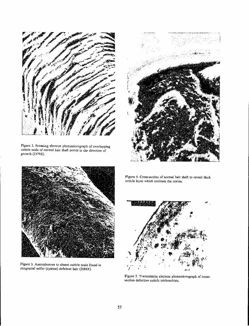
Figure 2. Scanning electron photomicrograph of overlapping cuticle scale of normal hair shaft points in the direction of growth (2379X).
Figure 3. Amorphorous to absent cuticle scale found in congenital sulfur (cystine) deficient hair (2086X).
53
Figure 4. Cross-section of normal hair shaft to reveal thick cuticle layer which encloses the cortex.
Figure 5. Transmission electron photomicrograph of crosssection defective cuticle trichoschisis.
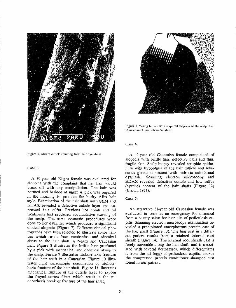
Figure 6. Absent cuticle resulting from hair dye abuse.
Case 3:
A 30-year old Negro female was evaluated for alOPecia with the complaint that her hair would break off with any manipulation. The hair was permed and braided at night A pick was required in the morning to produce the bushy Afro hair style. Examination of the hair shaft with SEM and EDAX revealed a defective cuticle layer and depressed hair sulfur. Previous hot comb and oil treatments had produced accumulative scarring of the scalp. The same cosmetic procedures were done to her daughter which produced a significant clinical alopecia (Figure 7). Different clinical photographs have been selected to illustrate abnormalities which result from mechanical and chemical abuse to the hair shaft in Negro and Caucasian hair. Figure 8 illustrates the brittle hair produced by a pick with mechanical and chemical abuse to the scalp. Figure 9 illustrates trichorrhexis fracture of the hair shaft in a Caucasian. Figure 10 illustrates light microscopic examination of trichorrhexis fracture of the hair shaft. Figure 11 illustrates mechanical rupture of the cuticle layer to expose the frayed cortex fibers which result in the trichorrhexis break or fracture of the hair shaft.
54
Figure 7. Young female with acquired alopecia of the scalp due to mechanical and chemical abuse.
Case 4:
A 49-year old Caucasian female complained of alopecia with brittle hair, defective nails and thin, fragile skin. Scalp biopsy revealed atrophic epithelium with hypoplasia of the hair follicle and sebaceous glands consistent with hidrotic ectodermal dysplasia. Scanning electron microscopy and EDAX revealed defective cuticle and low sulfur (cystine) content of the hair shafts (Figure 12) (Brown 1971).
Case 5:
An attractive 31-year old Caucasian female was evaluated in tears as an emergency for dismissal from a beauty salon for hair nits of pediculosis capitis. Scanning electron microscopy examination revealed a precipitated amorphorous protein cast of the hair shaft (Figure 13). The hair cast in a different patient results from a retained internal root sheath (Figure 14). The internal root sheath cast is freely moveable along the hair shaft, and is associated with several dermatoses, which differentiates it from the nit (egg) of pediculosis capitis, and/or the compressed protein conditioner shampoo cast found in our patient.
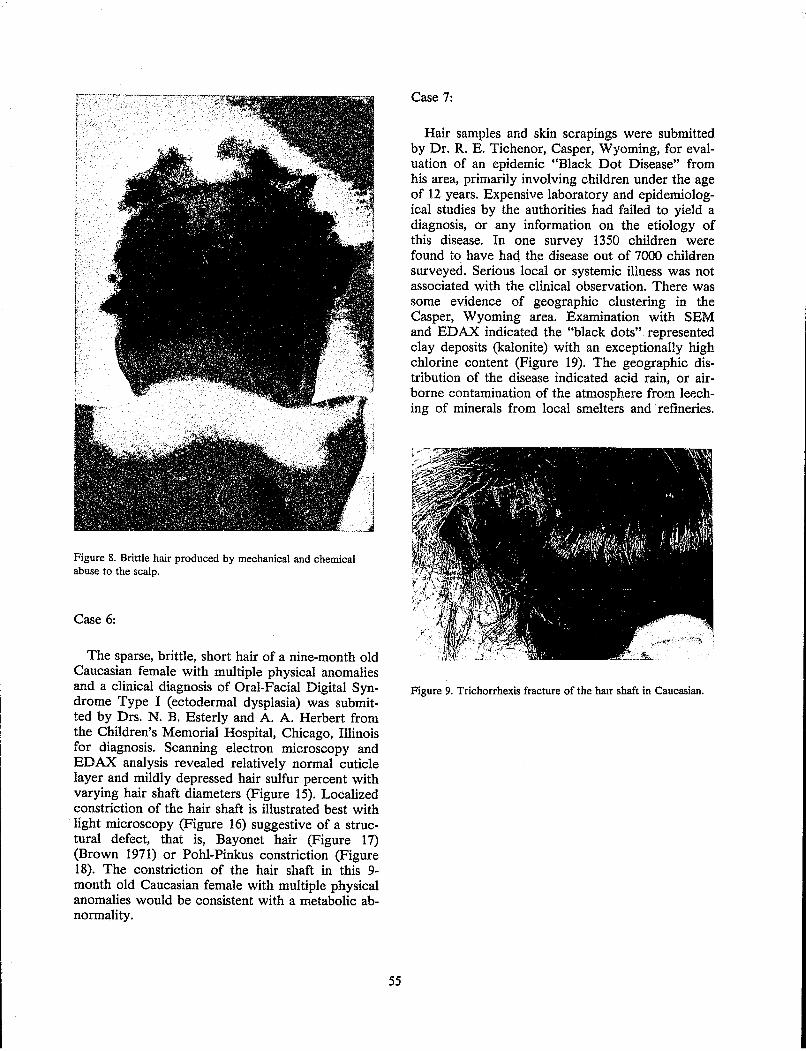
Figure 8. Brittle hair produced by mechanical and chemical abuse to the scalp.
Case 6:
The sparse, brittle, short hair of a nine-month old Caucasian female with multiple physical anomalies and a clinical diagnosis of Oral-Facial Digital Syndrome Type I (ectodermal dysplasia) was submitted by Drs. N. B. Esterly and A. A. Herbert from the Children's Memorial Hospital, Chicago, Illinois for diagnosis. Scanning electron microscopy and EDAX analysis revealed relatively normal cuticle layer and mildly depressed hair sulfur percent with varying hair shaft diameters (Figure 15). Localized constriction of the hair shaft is illustrated best with
, light microscopy (Figure 16) suggestive of a structural defect, that is, Bayonet hair (Figure 17) (Brown 1971) or Pohl-Pinkus constriction (Figure 18). The constriction of the hair shaft in this 9-month old Caucasian female with multiple physical anomalies would be consistent with a metabolic abnormality.
55
Case 7:
Hair samples and skin scrapings were submitted by Dr. R. E. Tichenor, Casper, Wyoming, for evaluation of an epidemic "Black Dot Disease" from his area, primarily involving children under the age of 12 years. Expensive laboratory and epidemiological studies by the authorities had failed to yield a diagnosis, or any information on the etiology of this disease. In one survey 1350 children were found to have haq the disease out of 7000 children surveyed. Serious local or systemic illness was not associated with the clinical observation. There was some evidence of geographic clustering in the Casper, Wyoming area. Examination with SEM and EDAX indicated the "black dots" represented clay deposits (kalonite) with an exceptionally high chlorine content (Figure 19). The geographic distribution of the disease indicated acid rain, or airborne contamination of the atmosphere from leeching of minerals from local smelters and' refineries.
Figure 9. Trichorrhexis fracture of the half shaft in Caucasian.
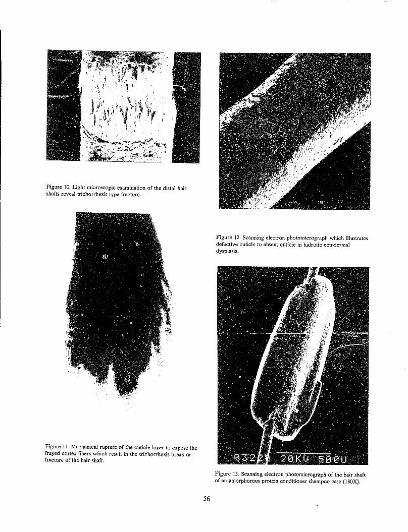
Figure 10. Light microscopic examination of the distal hair shafts reveal trichorrhexis type fracture.
Figure 11. Mechanical rupture of the cuticle layer to expose the frayed cortex fibers which result in the trir.horrhexis break or fracture of the hair shaft.
56
Figure 12. Scanning electron photomicrograph which illustrates defective cuticle to absent cuticle in hidrotic ectodermal dysplasia.
Figure 13. Scanning electron photomicrograph of the hair shaft of an amorphorous protein conditioner shampoo case (180X).
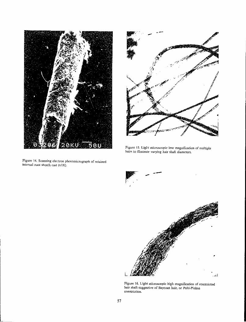
Figure 14. Scanning electron photomicrograph of retained internal root sheath cast (ti3X).
57
Figure 15. Light microscopic low magnification of mUltiple hairs to illustrate varying hair shaft diameters.
---
L
Figure 16. Light microscopic high magnification of constricted hair shaft suggestive of Bayonet hair, or Pohl-Pinkus constriction.
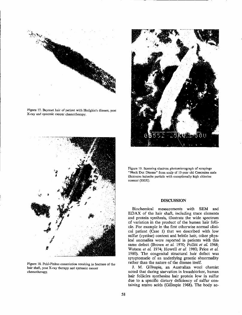
Figure 17. Bayonet hair of patient with Hodgkin's disease, post X-ray and systemic cancer chemotherapy.
J I
:1
Figure 18. Pohl-Pinkus constriction resulting in fracture of the hair shaft, post X-ray therapy and systemic cancer chemotherapy.
58
Figure 19. Scanning electron photomicrograph of scrapings "Black Dot Disease" from scalp of lO-year old Caucasian male illustrates kalonite particle with exceptionally high chlorine content (950X).
DISCUSSION
Biochemical measurements with SEM and EDAX of the hair shaft, including trace elements and protein synthesis, illustrate the wide spectrum of variation in the product of the human hair follicle. For example in the first otherwise normal clinical patient (Case 1) that we described with low sulfur (cystine) content and brittle hair, other physical anomalies were reported in patients with this same defect (Brown et af. 1970; Pollitt et al. 1968; Watson et al. 1974; Howell et al. 1980; Price et al. 1980). The congenital structural hair defect was symptomatic of an underlying genetic abnormality rather than the nature of the disease itself.
J. M. Gillespie, an Australian wool chemist noted that during starvation in kwashiorkor, human hair follicles synthesize hair protein low in sulfur due to a specific dietary deficiency of sulfur containing amino acids (Gillespie 1968). The body ac-
commodates to the availability, or non-availability, of amino acid "building blocks" to produce an altered prod.uct. I explain to my patients that human hair growth represents a card game. The "body" is the "dealer" and each "player" based on turnover time or growth rate, receives a card "amino acid" in order of basic, growth priority. Hair is first in growth rate, due to its fast turnover of cells (every 24 hours); bone marrow is second; liver third; gastrointestinal tract fourth (every 5 days); and skin 7-30 days later. If the card game is "busted" (injury, accident, surgery) or the "body" insulted (starvation, fever, alcoholism) then the amino acid building blocks go to the most essential areas first, that is, the bone marrow, liver, etc., to protect the body. The deal of cards (amino acids or building blocks) to hair is cut off, until the "game" can return to normal. I also tell my patients that we were put on this earth to reproduce, and through genetics, or environ~ental adaptation, we have lost some body hair, but the only function of scalp hair is to attract a mate, or keep the brain warm. The dermatologist or dermatopathologist uses the information derived from changes in the hair cycle to evaluate this type of "card game" interruption. The active, growing hair (anagen phase) responds to an acute insult, toxic poisoning, malnutrition, or radiation by ceasing growth to produce a structural constriction of the hair shaft, tapering and/or final fracture.
The transitional stage of the hair follicle (catagen phase) could result from pulling the hair, that is, trichotillomania (Miller 1980). The resting stage of the hair follicle (telogen phase) results in a closing down of the (factory) hair follicle, or shaft growth to produce a club hair over a three month period (post-partum, drug, acute caloric deprivation, or post-febrile episode). Hair abnormalities may result from an altered product of the factory which is a reflection of growth, or cycle alteration.
The single most important physical alteration is diameter which may reflect a congenital (cartilage hair hypoplasia) or acquired defect (alcoholism). The acquired defect would be more common for the forensic scientist which would indicate caloric or protein deprivation, common for example in alcoholism (Bregar et al. 1978). Structural or congenital alterations in shape would be significant, and certainly helpful, but a less common observation for the forensic scientist.
In known genetic, structural abnormalities of human hair, initially we studied families to see if we could correlate the structural pattern as demonstrated in pili torti, a genetic structural flattening of the hair scale (cuticular layer) orientation indicative of the flattened hair shaft, but we were unable to separate individuals. The technique was helpful in describing specific defects. For example, if the gene was autosomal dominant then I would expect
59
one of the parents to have a similar hair abnormality although the structural defect in pili torti apparently improves with puberty. The exposures of human hair in today's environment however with destructive hair cosmetics, or contamination due to weathering would seem to make our job more difficult; however, in reality it may be very helpful, particularly since the forensic scientist examines hair for objective data. I explain to my patients that scalp hair represents a fllter system, and depending on its length frames the face, and carries valuable information from the environment depending on particle density and individual height. The particle debris from this hair fllter system can be very helpful to the forensic scientist.
Alteration in composition of the hair may be acquired or congenital. Sulfur protein content is altered by hair cosmetics and influences the absorbed trace elements, or other surface data. Environmental exposures utilizing EDAX with SEM or quantitative neutron activation analysis, etc., may be helpful, but must be evaluated from different viewpoints of statistical significance. There is a significant difference in the amount of sample available for examination. I doubt there is much-argument in the examination of hair for heavy metal toxicity, endogenous or exogenous, but I would be very careful in the interpretation of some hair analysis data based on the subject, collection methods and examination techniques due to the wide environmental variability.
In The First Human Hair Symposium (Brown 1974) I concluded science had accepted the routine analysis of blood and urine, but routine analysis of hair had not been accepted. Pattern recognition or identification of any object should be limited only by the variables, and if we define our variableR objectively with a reproducible baseline, routine analysis of blood, urine and hair could be a reality in our lifetime, but much work remains to be done! In my opinion the forensic scientist can make a significant contribution if the hair evidence from a crime or victim is not taken out of circulation, that is, placed in a permanent slide. Minimal data is obtained from this practice. I have attempted to illustrate helpful clues in my practice for a definitive diagnosis of mUltiple human hair abnormalities. Identification of surface debris, or extraneous material associated with the hair shaft with SEM and EDAX analysis, could be very informative to the forensic scientist.
REFERENCES
Barrett, Stephen (1985). Commercial hair analysis, JAMA 254:1041-1045.
Bregar, R. R., Gordon M and Whitney, E. N. (1978). Hair root diameter measurement as an in-
-------~--- -~- ----
dicator of protein deficiency in nonhospitalized alcoholics, Amer. 1. Clin. Nutr. 31:230-236.
Brown, A. c., Belser, R. B., Crounse, R. G. and Wehr, R. F. (1970). A congenital hair defect: trichoschisis with alternating birefringence and low sulfur content, J. Invest. Dermatol. 54:496-509.
Brown, A. C. (1971). Congenital hair defects. Birth Defects: Original Article Series 7:52-68.
Brown, A. C (1974). Introduction: blood, urine, and hair. In: The First Human Hair Symposium (Brown, A. C., ed.). Medcom Press, New York, pp. 1-24.
Brown, A. C. and Broyles, J. A. (1980). Accumulative scarring of the scalp due to hair dye. In: Hair, Trace Elements, and Human Illness (Brown, A. C. and Crounse, R. G., eds.). Praeger Publishers, New York, pp. 348-360.
Carteaud, J. P. (1973). Scanning electron microscopy in skin pathology. In: Scanning Electron Microscopy (Part III) Proceedings of the Workshop, lIT Research Institute, Chicago, pp. 629-636.
Forslind, B. (J984). Clinical applications of scanning electron IrIicroscopy and X-ray microanalysis in dermatology. Scanning Electron Microscopy, SEM Inc., AMF O'Hare, Chicago, pp. 183-206.
Gillespie, J. M. (1968). The dietary regulation of the synthesis of hair keratin. In: Symposium of Fibrous Proteins, Australia, 1967 (Crewther, W. G., ed.). Plenum Press, New York.
Hart, R. K. (1980) Microanalytical analysis of hair. In: Hair, Trace Elements and Human Illness (Brown, A. C. and Crounse, R. G., eds.). Praeger Publishers, New York, pp. 82-101.
Howell, R. R., Collie, W. R" Cavasos, O. I, Arbisser, A. L, Fraustadt, U" Marcks, S.N. and Parsons, D. (1980). The sabinas brittle hair syndrome. In: Hair, Trace Elements, and Human Illness (Brown, A. C. and Crounse, R. G., eds.). Praeger Publishers, New York, pp. 210-219.
Muller, S. A. (1980). Trichotillomania. In: Hair, Trace Elements, and Human Illness (Brown, A. C. and Crounse, R. G., eds.). Praeger Publishers, New York pp. 305-312.
Pollitt, R. J., Jeller, F. A. and Davies, M. (1968). Sibs with mental and physical retardation and trichorrhexis nodosa with abnormal amino acid composition of the hair, Arch. Dis. Child. 43:211.
Price, V. H., Oiom, R. B., Jones, F. T. and Ward, W. H. (1980). Trichothiodystrophy: Sulfur-Deficient Brittle Hair (Brown, A. C. and Crounse, R. G., eds.). Praeger Publishers, New York, pp. 220-227.
Ryabukhin, 1~ S. (1980). International coordinated program on activation analysis of trace element pollutants in human hair. In: Hair, Trace Ele-
60
ments, and Human Illness (Brown. A. C. and Crounse, R. G., eds.). Praeger Publishers, New York, pp. 3-34.
Watson, J. H. L., Weiss, L. and Jackson, C. E. (1974). Scanning electron microscopy of human hair in a syndrome of trichoschisis with mental retardation. In: The First Human Hair Symposium (Brown, A. c., ed.). Medcom Press, New York, pp. 170-184.
DISCUSSION
Question: You mentioned briefly an attempt to compare hair or mass of hairs with the correct source using the SEM. Did you take a photomicrograph and compare those?
Brown: Essentially what we did was try to look at different variables as far as analysis is concerned, and see if we could pick them out.
For example, if I took an autosomal dominantly inherited hair defect, I could pick out the parents of those that expressed the defect. But most of the photomicrographs I see are very rare. I do not feel comfortable enough as far as identifying individual hairs is concerned to say that is exactly a match. The reason I made a comment about putting a hair in Permount is because that is evidence that is lost forever, and if I have any complaints from people who do contract work, or who do other things (I advise lawyers on all kinds of things), the complaint is that once the hair is put in Permount the evidence is taken out of circulation, and they do not have a chance of being able to make any further comments.
Other than inherited problems I would say we have a long way to go. Originally when we started discussing this, we received publicity on the BBC from London, but that was not warranted. I think we are getting closer. I think we can make certain observations, but it is just like making a diagnosis as far as any type of illness is concerned. We use certain criteria, for example, the blood count, what is in their urine, how did they present it, and how long they had the problem. I think the forensic scientist can use the same type of information in examining a tissue, which hair is, with that type of information and going further than that.
If you are an expert witness and testifying you say, "I do not know."
Question: Will a police officer in the field be able to recognize these hair diseases or unusual microscopic features?
Brown: If you have seen some of the things I have shown you I do not think there would be any question. The discussions that are in The First Human Hair Symposium (a member of the FBI was present at our symposium and contributed to it)
called for some standards for communications. Your committee has accomplished this now. But in regard to the type of specimens I have shown you, if you do see one of them then that will be very helpful to you. But the other little things, that is the reason surface debris criteria is mentioned.
The sample that was sent from Casper, Wyoming, where a fortune was spent on the epidemic of "Black Spot Disease" in children, included scrapings obtained from the scalps, the black spots/ dots and the hair. For me the hail' is a filter system that frames the face and so I get a certain amount of information.
For example, if I am examining a patient, I draw a straight line between the nose and the ears. If there is association above the line between the nose and ears, it is directly related to the hair; if it is below the line it is from the hands.
Question: Can you give us an idea of how rare some of these diseases are?
Answer: Very rare. Very rare, that is the reason I am prejudiced. You probably imagine that I have photographs sitting around and am seeing all of these cases because they are referred. They are screened through a number of physicians before I even see them. So my viewpoint is very prejudiced as far as the patients are concerned because the triage has occurred and when they get a chance to see them then it is a little bit different. My viewpoint is prejudiced and I have been called on that before. I operate a laboratory and I primarily see referred patients from all over the world for hair problems.
Question: --from Tokyo. Please tell us the kind of standard sample you use for quantitative analysis on the microscope, or the standard risk quantitative analysis you make?
Brown: The hair - sample came through. If you look in chapter five of Hair, Trace Elements and Human Illness (1980), "Microanalytical Analysis of Hair," by Dr. Raymond K. Hart, Dr. Hart explains his analysis since I no longer do scanning electron microscopy with dispersive X-ray analysis in my laboratory. The laboratory samples are from matched, quantitative sulphur samples versus the international control. He uses a Hitachi. It is a 450 SEM with a TM 2000 for quantitative X-ray analysis.
The comparative analysis of hair sulphur as a screen for cysteine content, therefore will separate out, for example, acquired damage to the hair shaft clue to permanents and hair dye versus, for example, the congenital abnormalities where there is an abnormal incorporation of cysteine. These studies are based on our original paper and discussion on statistical analysis, "Hair Sulphur Content, A Critique," in The First Human Hair Symposium.
61
Question: You had requested we flot mount hairs in Permount. Do you have an alternative method to mount hairs, and secondly, what is done to the hairs in order to produce the scanning electron microscopy?
Answer: The answer to the first question, I turn that over to the Committee on Forensic Hair Comparison.
In regards to the second question, we try to get the best objective information we can in evidence.
The only reason I made that comment is because one of my colleagues said I have always had a problem as far as the hair bit is concerlled because every time I go to the crime laboratory the hair is in Permount so I cannot use it. That is the only reason why I mentioned that. I mean we all have to have some meeting of the minds and, of course, the reason for this entire meeting is how should evidence be handled.
I am not handling evidence to that extent. Remember as a general rule I can get most of the samples that I want because they are voluntary, or they are for information except, for example, with children I am limited in the amount of samples I can get. As far as screening goes, before the specimens go the less manipulation possible, the less contamination possible, so therefore that is evidence. The less manipulation of the hair, the more information we will get. If we look and talk about, in your category, for example, total surface area; if we want to clean up, for example, if I want to compare one shampoo with another and we talk about efficiency of cleaning hair in a given period of time, and we are talking about cleaning a filter system that will vary from fiberglass of the afro hairstyle to thick hair, or to no hair like me; there is a big difference. The less manipUlation as far as the hair is concerned, the more information you will get. I think that is becoming more and more important, and the reason for that is shown in the environmental pollution example I have shown you from children.
Now how would I take, for example, that little small black spot and separate it out from Georgia versus Wyoming? Well, it could be in regards to iron content? In other words, separate out certain crystals in certain areas of the country based on iron content, but we could talk all day about that. So what we are talking about is getting as much information out of that sample as possible. Therefore, do the least amount of manipulation as possible.
Now if we are all doing the same thing, no problem. I know what he did with it when he got it. I know exactly what the specimen has gone through; how many people have looked at it and all this other kind of stuff. Did he wash it? Did he not wash it? I have covered this in the paper but I have not gone any further in a discussion.
Now the reason that hair analysis became important is if I did somethLTlg for the Food and Drug Administration andlor looked at something for the Federal Trade Commission. If we look around at commerical laboratories that are doing trace element analysis, well, what are they examining?
62
If I took one sample and sent it to 20 laboratodes, how many results do you think I would get back? And we have done just that (Barrett 1985). So the less manipulation we do, the better, as long as we are aU agreeing on the same criteria.
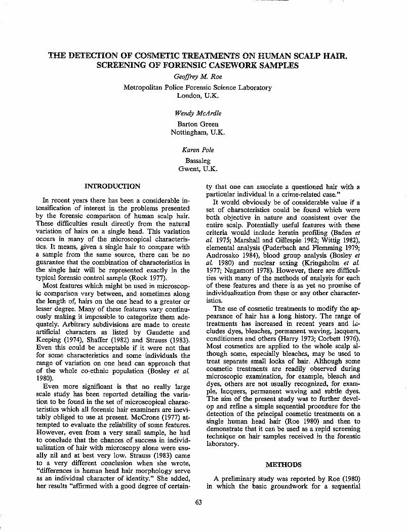
THE DETECfION OF COSMETIC TREATMENTS ON HUMAN SCALP HAIR. SCREENING OF FORENSIC CASEWORK SAMPLES
Geoffrey M. Roe
Metropolitan Police Forensic Science Laboratory London, U.K.
Wendy McArdle
Barton Green Nottingham, U.K.
Karen Pole
Bassaleg Gwent, U.K.
INTRODUCI'ION
In recent years there has been a considerable intensification of interest in the problems presented by the forensic comparison of human scalp hair. These difficulties result directly from the natural variation of hairs on a single head. This variation occurs in many of the microscopical characteristics. It means, given a single hair to compare with a sample from the same source, there can be no guarantee that the combination of characteristics in the single hair will be represented exactly in the typical forensic control sample (Rock 1977).
Most features which might be used in microscopic comparison vary between, and sometimes along the length of, hairs on the one head to a greater or lesser degree. Many of these features vary continuously making it impossible to categorize them adequately. Arbitrary subdivisions are made to create artificial characters as listed by Gaudette and Keeping (1974), Shaffer (1982) and Strauss (1983). Even this could be acceptable if it were not that for some characteristics and some individuals the range of variation on one head can approach that of the whole co-ethnic population (Bosley et al. 1980).
Even more significant is that no really large scale study has been reported detailing the variation to be found in the set of microscopical characteristics which all forensic hair examiners are inevitably obliged to use at present. McCrone (1977) attempted to evaluate the reliability of some features. However, even from a very small sample, he had to conclude that the chances of success in individualization of hair with microscopy alone were usually nil and at best very low. Strauss (1983) came to a very different conclusion when she wrote, "differences in human head hair morphology serve as an individual character of identity." She added, her results "affirmed with a good degree of certain-
63
ty that one can associate a questioned hair with a particular individual in a crime-related case."
It would obviously be of considerable value if a set of characteristics could be found which were both objective in naturl': and consistent over the entire scalp. Potentially useful features with these criteria would include keratin profiling (Baden et al. 1975; Marshall and Gillespie 1982; Wittig 1982), elemental analysis (Puderbach and Flemming 1979; Androsako 1984), blood group analysis (Bosley et al. 1980) and nuclear sexing (Kringsholm et al. 1977; Nagamori 1978). However, there are difficulties with many of the methods of analysis for each of these features and there is as yet no promise of individualization from these or any other characteristics.
The use of cosmetic treatments to modify the appearance of hair has a long history. The range of treatments has increased in recent years and h~eludes dyes, bleaches, permanent waving, lacquers, conditioners and others (Harry 1973; Corbett 1976). Most cosmetics are applied to the whole scalp although some, especially bleaches, may be used to treat separate small locks of hair. Although some cosmetic treatments are readily observed during microscopic examination, for example, bleach and dyes, others are not usually recognized, for example, lacquers, permanent waving and subtle dyes. The aim of the present study was to further develop and refine a simple sequential procedun:: for the detection of the principal cosmetic treatments on a single human head hair (Roe 1980) and then to demonstrate that it cw be used as a rapid screening technique on hair samples received in the forensic laboratory.
METHODS
A preliminary study was reported by Roe (1980) in which the basic groundwork for a sequential

screening procedure was tested. Some of the methods examined were shown to be unreliable and it was clear that further work was necessary to improve the scheme to a point at which it could safely be used in forensic casework.
1. Dyes
Four different dye extractants were tested on hairs treated with a range of different dye types from rinses and tints to permanent dyes. The extractants were pyridine-water (4:3), dimethylformamide (DMF) - water (2:1), DMF-formic acid (1:1) and formic acid-water (1:1). It was found that pyridine-water was the most effective general dye extractant.
Nine different developing solvent mixtures for thin layer chromatography (TLC) were tried on the same range of dye types. It was found that chloroform-methanol-water-ammonia (S.G. 0.88) (11:7:1:1) provided the most effective dye separati.on for the majority of samples examined. The solvent mixture chosen by Bailey (1982) for semi-permanent dyes was not one of those tested.
The sequential procedure was devised for the rapid screening of hair samples for the various cosmetic treatments. There are likely to be some cases in which pyridine is not the most effective dye extractant and the given solvent mixture not the best TLC system. It was intended that if the presence of dyes has been established in a known sample then the optimal system for extraction and TLC must be determined provided a crime hair is found to match the known sample microscopically.
2. Ck,mical Treatments
Corbett (1976) reviewed the chemistry of hair care products. Bleaching, permanent waving and other chemical treatments involving an oxidative process cause changes in keratin. Cystine is oxidized to give cysteic acid residues. In bleaching, the oxidatiof..:, usually with alkaline hydrogen peroxide, is direct. The perming process consists of the appH,;:ation of a reducing agent, often ammonium thioglycollate, which breaks the cystine disulphide bonds cross-linking the keratin fibrils. The hair is then shaped according to fashion, the parallel a-keratin fihriIs now free to slide against each other. An oxidizing agent, oftell hydrogen peroxide in acidic solution, is applied and new disulphide bonds are formed, fixing the hair in its new config·· uration. Both of these oxidative processes result in an increased concentration of anionic cysteic acid residues. These free groups will react with the cationic methylene blue. The intensity of the staining reaction is proportional to the number of free anionic residues, that is, to the severity of the chemical treatment.
Methylene blue staining (Porter and Fouweather 1975) was shown in the preliminary study (Roe
64
1980) to be the simplest and least error-prone of the techniques for detecting any oxidative chemical treatment. However, there was a problem with two weak false positive reactions. In both cases there was no distinct demarcation between stained and unstained sections of the hairs when complete hairs were treated with methylene blue. This showed the reaction was not due to any deliberate chemical treatment. Robbins and Kelly (1970) reported "natural" variation in cysteic acid residue levels between individuals with no reported oxidative treatments. It is possible that the problem samples came from the upper end of the range. A second problem with this technique was the tendency for the hair to float if stained in dishes, or become pressed against the sides of a staining tube, sometimeo giving patchy reactions. To improve wetting, the methylene blue was made up in 70 percent ethanol. When tested on a large number of hairs with various treatments (bleaches, perms and untreated controls) it was found that although the staining reaction was less strong than with the aqueous stain the patchiness was eliminated and there was no difficulty in recognizing the demarcation line. The preliminary study and subsequent work clearly showed it was neceS8.\lfY to stain the whole hair and not just the distal 0.5 - 1.0 cm, so interpretation of the results should be unequivocal.
In addition, the aqueous and alcoholic stains were tested to determine whether pre-treatment with acetone (used for lacquer extraction) or pyridine (for dye extraction) had any significant effect. It was found acetone had no effect on the intensity of the staining reaction. However, pyridine enhanced the staining in some way. This was considered beneficial because it made the demarcation line more obvious. No false positive rea>'1tions were caused by either solvent.
Screening Procedure
A minimum of five hairs were taken from each of 186 crime-related hair samples submitted to U. K. Forensic Science laboratories for examination. The hairs were taken after the casework had been completed or when no hair comparisons were to be made. All of the samples selected were from adults of Northern European origin (134 male and 52 female). In each case two single hairs and a group of three were subjected to the following sequential analytical procedure. The hairs were examined in this way so that weak treatments which might not be detected on a single hair should be identified in the group of three.
1. Lacquers
The hairs were extracted in acetone for 10 minutes at room temperature to remove lacquers and other soluble surface deposits. The extracts were prepared for infrared (IR) spectroscopy as de-
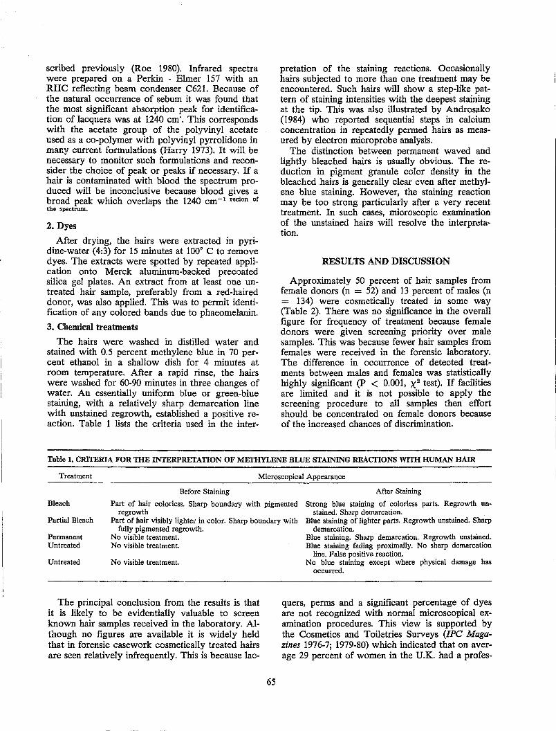
scribed previously (Roe 1980). Infrared spectra were prepared on a Perkin - Elmer 157 with an RIle reflecting beam condenser C621. Because of the natural occurrence of sebum it was found that the most significant absorption peak for identification of lacquers was at 1240 cm·. This corresponds with the acetate group of the polyvinyl acetate used as a co-polymer with polyvinyl pyrrolidone in many current formulations (Harry 1973). It will be necessary to monitor such formulations and reconsider the choice of peak or peaks if necessary. If a hair is contaminated with blood the spectrum produced will be inconclusive because blood gives a broad peak which overlaps the 1240 cm- 1 region of the spectrum.
2. Dyes
After drying, the hairs were extracted in pyridine-water (4:3) for 15 minutes at 100° C to remove dyes. The extracts were spotted by repeated application onto Merck aluminum-backed precoated silica gel plates. An extract from at least one untreated hair sample, preferably from a red-haired donor, was also applied. This was to permit identification of any colored bands due to phaeomelanin.
3. Chemical treatments
The hairs were washed in distilled water and stained with 0.5 percent methylene blue in 70 percent ethanol in a shallow dish for 4 minutes at room temperature. After a rapid rinse, the hairs were washed for 60-90 minutes in three changes of water. An essentially uniform blue or green-blue staining, with a relatively sharp demarcation line with unstained regrowth, established a positive reaction. Table 1 lists the criteria used in the inter-
pretation of the staining reactions. Occasionally hairs SUbjected to more than one treatment may be encountered. Such hairs will show a step-like pattern of staining intensities with the deepest staining at the tip. This was also illustrated by Androsako (1984) who reported sequential steps in calcium concentration in repeatedly permed hairs as measured by electron microprobe analysis.
The distinction between permanent waved and lightly bleached hairs is usually obvious. The reduction in pigment granule color density in the bleached hairs is generally clear even after methylene blue staining. However, the staining reaction may be too strong particularly after a very recent treatment. In such cases, microscopic examination of the unstained hairs will resolve the interpreta~ tion.
RESULTS AND DISCUSSION
Approximately 50 percent of hair samples from female donors (n = 52) and 13 percent of males (n = 134) were cosmetically treated in some way (Table 2). There was no significance in the overall figure for frequency of treatment because female donors were given screening priority over male samples. This was because fewer hair samples from females were received in the forensic laboratory. The difference in occurrence of detected treatments between males and females was statistically highly significant (P < 0.001, X2 test). If facilities are limited and it is not possible to apply the screening procedure to all samples then effort should be concentrated on female donors because of the increased chances of discrimination.
Table 1. CRITERIA FOR THE INTERPRETATION OF METHYLENE BLUE STAINING REACTIONS WITH HUMAN HAIR
Treatment Microscopical Appearance
Before Staining After Staining
Bleach Part of hair colorless. Sharp boundary with pigmented regrowth
Strong blue staining of colorless parts. Regrowth unstained. Sharp demarcation.
Partial Bleach Part of hair visibly lighter in color. Sharp boundary with fully pigmented regrowth.
Blue staining of lighter parts. Regrowth unstained. Sharp demarcation.
Permanent Untreated
Untreated
No visible treatment. No visible treatment.
No visible treatment.
The principal conclusion from the results is that it is likely to be evidentially valuable to screen known hair samples received in the laboratory. Although no figures are available it is widely held that in forensic casework cosmetically treated hairs are seen relatively infrequently. This is because lac-
65
Blue staining. Sharp demarcation. Regrowth unstained. Blue staining fading proximally. No sharp demarcation
line. False positive reaction. No blue staining except where physical damage has
occurred.
quers, perms and a significant percentage of dyes are not recognized with normal microscopical examination procedures. This view is supported by the Cosmetics and Toiletries Surveys (IPe Magazines 1976-7; 1979-80) which indicated that on average 29 percent of women in the U.K. had a profes-
sional perm at least once a year, and a further 10 percent had at least one home perm in the same period (1976-7). Similarly, about 23 percent of women used hair bleaches or colorants (1976-80) and 52 percent (1976) - 40 percent (1980) used hair sprays. Allowing for an unknown proportion of women using more than one type of treatment, the usage rate is still much higher than observed in conventional forensic hair examination.
Table 2. DETECTED COSMETIC TREATMENTS ON HUMAN SCALP HAIR
Female Donors Male Don0rs Total
Number Treated
52 134 186
26 17 43
Percent Treated
50 13
Treated as
Percent of Total
14 9
23 ------------_._--_.-
The frequencies for each type of treatment were lacquers - females 2 percent, males 2 percent; dyes - females 33 percent, males 6 percent; bleaching -females 17 percent, males 5 percent; permanentwaving - females 10 percent, males 2 percent. Clearly, care must be taken in the use of such figures because some individuals used two or more different treatments together. However, in practical forensic comparison mUltiple treatments will be more significant as evidence. A breakdown of the results into the different cosmetic groups is given in Table 3 for males, and in Table 4 for females.
Table 3. COSMETIC TREATMENTS TO HAIR SAMPLES
Perms Bleach Dye Lacquer
Perms 2 Bleach 0* 4 Dye 0 2 6 Lacquer 0 0 0 3
• Perming, or other non-bleaching oxidative treatment, would only be detected on bleached hair if it occurred at least two weeks after bleaching.
Perming, or other non-bleaching oxidative treatment, would only be detected on bleached hair if it occurred at least two weeks after bleaching. This is because the detection of both relies on the same staining reaction and two weeks allows about 5 mm of pigmented hair growth to occur. In the course of this work no hairs with three or more different cosmetic treatments were found. The relatively high frequency of bleach and dye treatments together should not be considered highly significant. Most professional bleaching regimes incorporate dyes, usually blue, in order to counteract the yellow effects caused by the greater resistance of phaeomelanin to bleaching (Zviak 1966). In addi-
66
tion, some dyes contain bleaching agents in order to lighten the hair shade a little. These preparations would produce positive staining reaction with methylene blue. Methylene blue staining is effectively destructive in permanently altering the appearance of the h~ir. However, it will only need to be carried out in about 10 percent of cases with female samples and 2 percent of male samples.
Table 4. COSMETIC TREATMENTS TO HAIR SAMPLES
Perms Bleach Dye Lacquer
Perms 3 Bleach 0* 4 Dye 2 4 10 Lacquer 0 1 1
* Perming, or other non-bleaching oxidative treatment, would only be detected on bleached hair if it occurred at least two weeks after bleaching.
The sequential screening procedure appears to be effective in detecting cosmetic treatments to human scalp hair at the single hair level. On the available usage figures the detection of lacquers is least efficient. In addition, the IR method is the most time consuming of the techniques. The whole routine takes about three hours for a single sample. Excluding lacq\ler detection saves only fifteen minutes in total but does allow a free two hour period in the middle. Batch processing of at least ten samples simultaneously is the most sensible use of time. It must be noted that lacquer detection cannot be contemplated once the hair has been mounted. The search procedure for crime hairs must also be considered. If hairs are to be examined for lacquers, that is, the control samples in a case have been screened and found positive, then high adhesive tape must not be used. This is because removal of the hair from the tape without solvent will leave some of the lacquer in the adhesive. If a solvent is used it is likely to dissolve the lacquer. Most formulations are softened or dissolved by xylene. In addition there is the risk of contamination of the lacquer extract with the tape adhesive. Crime hairs must be carefully picked with clean forceps from the surface of the item being searched. Preferably each hair should be extracted in acetone immediately because lacquer traces are ephemeral. If this is not practicable then the hair must be treated with all due care. The hair can be mounted for microscopical comparison only after extraction. If it matches the known sample then tests for the cosmetics found in the sample can be carried out on the crime hair. It will in fact be correct procedure to examine the matching crime hair for all the cosmetic treatments even when none has been found in the known sample. In this way it may be possible to eliminate some hairs. This is directly compa-
rable with the procedures carried out on textile fibers after a microscopic match is found (MPFSL 1978).
The methods described for cosmetic detection are all relatively rapid. It may be possible to find more sensitive methods but these would probably not permit rapid screening by almost any hair examiner. For example, the detection of cosmetic treatments by scanning electron microscopy (SEM) (Swift personal communication) is very time consuming and involves considerable expertise in the use of the instrument. The SEM microprobe analysis of calcium levels in permed hairs (Androsako 1984) may be more sensitive than methylene blue staining but it is certainly very much slower.
In conclusion, a sequential analytical procedure for the rapid screening of hair samples for cosmetic treatments offers a set of objective characteristics which are of undoubted value in forensic hair comparison. They do not rely on the statistical analysis of intra and interhead variation for their discriminating power. The statistics of population usage should enable the significance of the various cosmetic factors detected to be expressed in real terms in evidence. However, because of the capricious nature of fashion it will be necessary to constantly monitor fluctuations in usage of the various substances. The Cosmetics and Toiletries Surveys carried out in the U.K. (fPC Magazines 1976-7; 1979-80) did show a remarkable consistency over a six year period in the use of colorants and bleaches. They also revealed steady changes in use of some other products thus emphasizing the need for monitoring.
The use of the screening procedure can only enhance the chances of discrimination between hairs from different sources when used in conjunction with conventional microscopical comparison techniques. If other objective characteristics are found to be reliable then they should also be used in the forensic comparison of human hair.
ACKNOWLEDGMENTS
Thanks are due to the case reporting officers of the Metropolitan Police Forensic Science Laboratory, and to the Heads of Biology Divisions in the u.K. Forensic Science Service who provided the samples of hair examined in the screening procedure. Permission from Mr. Cotes of fPC Magazines to publish extracted data from the Cosmetics and Toiletries Surveys is gratefully acknowledged. Finally, we wish to thank Roger Cook for his advice and support throughout this work.
67
REFERENCES
Androsako, J. (1984). Calcium profiles in permanent-waved hair and its application to forensic science. In: Proceedings of the 10th International Association of Forensic Sciences Conference, Oxford.
Baden, H. P., Lee, L. D. and Kubilus, J. (1975). A genetic electrophoretic variant of human hair apolypeptides, Am. J. Hum. Genet. 27:472-477.
Bailey, J. G. (1982). The characterization of semipermanent dyes on a single hair. Presented at the 1st Inter-American Congress of Forensic Sciences, Sacramento.
Bosley, c., Burgess, R. M, Sutton, J. G. and Whitehead, P. H. (1980). Enzymes in human hair sheath cells - a new means of characterising hair. CRE Topic Number 359, pp. 17-25.
Corbett, J. F. (1976). The chemistry of hair-care products, J. Soc. Dyers Colour 92:285-303.
Gaudette, B. D. and Keeping, E. S. (1974). An attempt at determining probaryilities in human scalp hair comparison, J. Forensic Sci. 19:599-606.
Harry, R. G. (1973). Harry's Cosmeticology, (revised J. B. Wilkinson), 6th ed. Leonard HIll Books, London.
fPC Magazines Limited (1976-7). Cosmetics and Toiletries Survey. London.
fPC Magazines Limited (1979-80). Cosmetics and Toiletries Survey. London.
Kringsholm, B., Thomsen, J. L. and Henningsen, K. (1977). Fluorescent Y-chromosomes in hairs and blood stains, Forensic Sci. 9:117-126.
Marshall, R. C. and Gillespie, J. M (1982). Comparison of samples of human hair by two-dimensional electrophoresis, J. Forensic Sci. Soc. 22:377-385.
McCrone, W. C. (1977). Characterization of human hair by light microscopy, Microscope 25:15-30.
Metropolitan Police For:msic Science Laboratory (1978). Biology Methods Manual. Metropolitan Police, London.
Nagamori, H. (1978). Sex determination from plucked human hairs without epithelial root sheath, Forensic Sci. Int. 12:167-173.
Porter, J. and Fouweather, C. (1975). An appraisal of human head hair as forensic evidence, J. Soc. Cosmet. Chem. 26:299-313.
Puderbach, H. and Flemming, P. (1979). X-rayanalysis on the scanning electron microscope in hair cosmetic evaluation and development, Cosmet. Toiletries 94:79-84.
Robbins, C. R. and Kelly C. H. (1970). Amino acid composition of human hair, Text. Res. J. 40:891-896.
Rock, P. (1977). A study of the distribution of human head hair colour and its forensic implications. B. Sc. (Honors) Thesis, Trent Polytechnic.
Roe, G. M. (1980). The detection of cosmetic treatments on hair, Cosmet. Toiletries 95:40-44.
Shaffer, S. A. (1982). A protocol for the examination of hair evidence, Microscope 30: 151-161.
Strauss, M. A. (1983). Forensic characterization of human hair 1, Microscope 31:15-29.
Wittig, M (1982). Protein patterns of keratins - the probable role in forensic hair examination, J. Forensic Sci. Soc. 22:387-389.
Zviak, C. (1976). Decoloration des cheveux. In: Problems Capillaries. Gauthier-Villars, Paris, pp. 111-128.
DISCUSSION
Question: I have had only moderate success with dye extracts and thin layer chromatography. The problem is that I do not always get nice spots; my dye seems to streak across the plate. Am I the only one who sees streaked dyes on thin layer chromatography plates or do you always have very nice little spots, and is there a reason for that?
Roe: The streaking is not unique to your own plates, I can assure you. I think these are the permanent dyes where you have, as described by Dr. Robbins, so many different combinations of different degrees of polymerization of your dye units and I think this results in a whole complex which often smears.
The simple preform dyes, for example, rinses and tints, will give single good spots, and the semipermanent dyes, as well, usually give nice clear spots. But a fairly large proportion of dyes used are of the permanent group and they do cause that sort of problem.
However you still have a comparison. It is still possible despite the streaking. You will get some nodes in the streaks which can certainly be used as points of comparison.
Wittig: You can compare certain factors of permanent dyes, of course?
Roe: Sometimes. De Forest: Please explain the apparatus you use
to extract the hairs? Roe: Extraction for the lacquers or the dyes? For
dyes, the hair is simply placed in a small glass tube, a small duram tube and the hair compressed as far as possible to the bottom so that the minimum extractant pyridine can be applied so the hair is submerged. After 15 minutes, the hair is removed and, if necessary, the extract is condensed until it is a manageable volume and it is spotted onto the plate by repeated application with double drawn glass capillaries.
"
68
Burwitz: Please comment on what the defense experts' reactions have been to the methylene blue staining of the hair?
Roe: In the United Kingdom the defense experts rarely examine cases. I have not had a case in which the methylene blue staining has been used in practical case work. There has been no problem yet. I can imagine that it could cause some discussion. The treatment is effectively destructive. I would agree that you are staining hair effectively and permanently, and the original microscopical comparison can not be considered subsequently. The procedure I have described was experimental. All the samples were taken from cases in which the hair examination had already been finished or was not being contemplated, which was probably the majority of the cases.
It is not an operational procedure as a screening system in the Metropolitan Police Forensic Science Laboratory yet, mainly because of the shortage of funds. Also, we do not have a sufficient number of staff who can take time off from their other duties for a full day once every month perhaps.
Shaffer: Did you say questioned hair should be screened for lacquers, etc., only if you have already found those on your known samples?
Roe: Yes, the absence of a treatment like lacquer when you are dealing with a relatively small number of hairs cannot be taken as an absence of treatment from the whole head, because lacqm:rs do brush off fairly easily, To find none on the five from the head and then to find it on the questioned hair would not mean a negative result. It would mean you have to go back to the known sample and test more hairs. I am not sure at this stage how many you would have to test before you could be reasonably certain there was none there. It is a problem.
Shaffer: It seems as if you are testing your questioned sample conditionally depending on what you have found on your known sample. It seems there is an assumption going into your examination procedure that the known and the questioned hair did in fact come from the same source, whereas there is realistic possibility that even if known to be different by other comparative mechanisms you could detect, for example, lacquer on the questioned hair when there is none on the known hair. That represents a genuine distinC'tion between the two, which again if I understand your recommendation, would not be found in your procedure.
Roe: Yes. I was basically answering the same point. Yes, it probably would be theoretically better to look for lacquer on the questioned hair even if not detected in the initial examination of the known sample.

Shaffer: From a defense point of view I would hope this does not necessarily represent the fmal procedure people would use.
I do like the idea of being able to detect these treatments. I would hope that perhaps with a little
69
more development we can come up with some mechanism that would still allow these kinds of distinctions and would not be entirely destructive or destructive to the entire hair.
Roe: Yes, I wholeheartedly support that view.
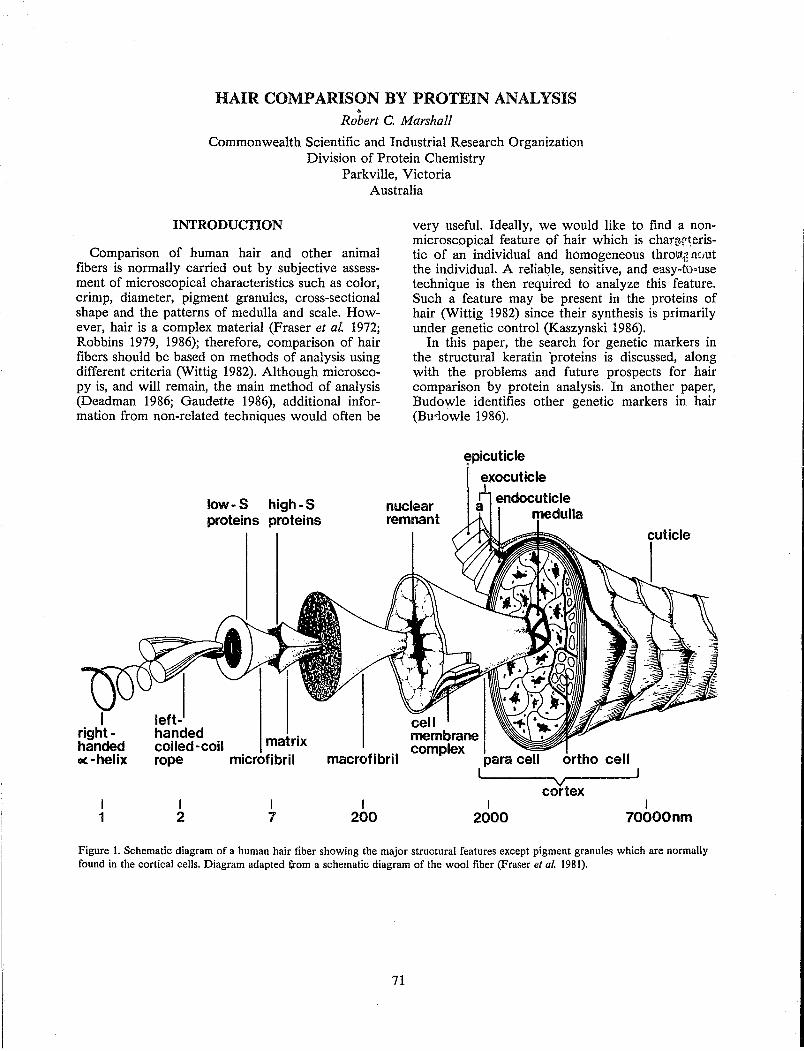
HAIR COMPARISON BY PROTEIN ANALYSIS " Robert C. Marshall
Commonwealth Scientific and Industrial Research Organization Division of Protein Chemistry
Parkville, Victoria Australia
INTRODUCTION
Comparison of human hair and other animal fibers is normally carried out by subjective assessment of microscopical characteristics such as color, crimp, diameter, pigment granules, cross-sectional shape and the patterns of medulla and scale. However, hair is a complex material (Fraser et aL 1972; Robbins 1979, 1986); therefore, comparison of hair fibers should be based on methods of analysis using different criteria (Wittig 1982). Although microscopy is, and will remain, the main method of analysis (Deadman 1986; Gaudette 1986), additional information from non-related techniques would often be
left-
low- S high-S proteins proteins
handed . coiled-coil matrix
very useful. Ideally, we would like to find a nonmicroscopical feature of hair which is chan~rteristic of an individual and homogeneous thro\(t11 (lout the individual. A reliable, sensitive, and easy-tu>use technique is then required to analyze this feature. Such a feature may be present in the proteins of hair (Wittig 1982) since their synthesis is primarily under genetic control (Kaszynski 1986).
In this paper, the search for genetic markers in the structural keratin 'proteins is discussed, along with the problems and future prospects for hair comparison by protein analysis. In another paper, Budowle identifies other genetic markers in hair (Bud.owle 1986).
nuclear remnant
cuticle
I right -handed oe-helix rope microfibril macrofibril para cell
l ortho cell
I 1
I 2
I 7
I 200
~------~vr--------J cortex
I 2000
I 70000nm
Figure 1. Schematic diagram of a human hair fiber showing the major structural features except pigment granules which are normally found in the cortical cells. Diagram adapted !;rom a schematic diagram of the wool fiber (Fraser et al. 1981).
71
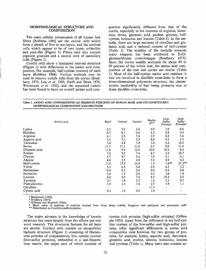
MORPHOLOGICAL STRUCTURE AND COMPOSITION
The main cellular components of all human hair fibers (Robbins 1986) are the cuticle cells which form a sheath of five to ten layers, and the cortical cells which appear to be of two types, ortho-like and para-like (Figure 1). Fibers may also contain pigment granules and a central core of medullary cells (Figure 1).
Cuticle cells show a laminated internal structure (Figure 1) with differences in the amino acid composition (for example, half-cystine content) of each layer (Robbins 1986). Various methods may be used to remove cuticle cells from the cortex (Bradbury 1973; Ley et al. 1985; Swift and Bews 1974; Wortmann et al. 1982), and the separated cuticle has been found to have an overall amino acid com-
position significantly different from that of the cortex, especially in the contents of arginine, threonine, serine, glutamic acid, proline, glycine, halfcystine, isoleucine and leucine (Table 1). In the medulla, there are large amounts of citrulline and glutamic acid, and a reduced content of half-cystine (Table 1). The stability of the medulla towards many reagents has been attributed to E-(Gglutamyl)lysine cross-linkages (Bradbury 1973). Since the cortex usually accounts for about 80 to 90 percent of the whole hair, the amino acid composition of the hair and cortex are similar (Table 1). Most of the half-cystine amino acid residues in hair are involved in disulfide cross-links to form a three-dimensional polymeric structure, the characteristic insolubility of hair being primarily due to these disulfide cross-links.
Table 1. AMINO ACID COMPOSITIONS (AS RESIDUES PERCENT) OF HUMAN HAIR AND ITS CONSTITUENT MORPHOLOGICAL COMPONENTS AND PROTEINS
Lysine Histidine Arginine Aspartic acid Threonine Serine Glutamic acid Proline Glycine Alanine Half-cystine Valine Methionine Isoleucine Leucine Tyrosine Phenylalanine Citrulline Cysteic acid
• Bindewald (1983). b Bradbury (1973).
Amino acid Hair" Cuticle"
2.3 2.9 0.7 0.3 6.1 2.3 5.4 3.5 7.4 4.8
11.7 17.1 11.8 9.6 7.8 9.3 5.7 9.7 4.6 5.5
17.0 17.5 5.3 6.3 0.4 0.3 2.4 1.5 6.6 4.9 2.7 1.8 1.9 1.3
0.3 1.5
Medul- Low- High-Cortex· lab,. Sulfur Sulfur
ProteinsC ProteinsC
2.4 5.0 3.9 0.6 0.8 1.3 0.8 0.9 7.4 3.9 6.8 5.4 5.5 6.8 9.3 2.5 7.8 3.4 5.4 10.3
11.0 6.3 8.9 11.9 12.4 22.9 16.5 8.4 7.4 4.7 3.8 12.7 5.3 7.0 5.1 6.1 4.8 5.5 6.9 2.3
14.8 1.5 7.6** 27.2** SA 2.9 6.1 5.2 0.4 0.6 0.4 0.0 2.9 2.5 3.6 1.8 7.2 8.S 10.2 2.9 2.3 1.6 2.5 0.7 1.8 2.7 1.9 1.1
11.S 0.5 1.4
C Gillespie and Marshall (1981). • Mean value of analyses of medulla isolated from three fibers (rabbit, kangaroo and platypus) and porcupine quill. ** Determined as S-carboxymethylcysteine.
The major advance in the knowledge of keratin structure has come largely from the efforts put into wool research. The structural features for all hairs are similar. Cortical cells contain an intracellular biphasic structure (Figure 1) consisting of filamentous proteins of comparatively low cystine content (low-sulfur proteins), embedded in a non-filamentous matrix, the major part of which consists of
72
cystine rich proteins (high-sulfur proteins) (Gillespie 1983). Apart from the difference in the half-cystine content of the low-sulfur and high-sulfur proteins. other significant differences in amino acid composition exist between the two groups of proteins, for example, lysine, aspartic acid, threonine, glutamic acid, proline, alanine, isoleucine, leucine and tyrosine (Table 1). Many hairs also contain an-
other family of matrix proteins and these are rich in glycine and tyrosine (high-tyrosine proteins) (Gillespie 1983). Some hairs (for example, human hair and Lincoln wool) contain little or no hightyrosine proteins (Gillespie 1983).
ANALYSIS OF PROTEINS
The characterization and comparison of hair proteins can only occur after the proteins have been solubilized. The flrst necessary step in hair solubilization is the cleavage of the disulflde bonds KerlS-S-Ker2 where "Kerl" and "Ker2" represent either the same (intramolecular disulflde bond) or different keratin proteins (intermolecular bond). Nonspeciflc chemical degradation and modiflcation are decreased by the use of the reduction procedure (Equation 1) with reagents such as mercaptoethanol, thioglycollate or dithiothreitol.
Kerl-S-S-Ker2 + 2[H] ~ Kerl-SH + Ker2-SH (1)
Since this reaction is readily reversible under oxidizing conditions, the reduced keratin proteins (Ker-SH) are normally stabilized by alkylation with a reagent such as iodoacetate (Equation 2): this modiflcation also increases protein solubility.
Ker-SH + ICH2COO- ~ Ker-SCH2COO- + HI (2)
The keratin proteins can be radiolabelled at this point by using 1<1C- or 3JI-labelled iodoacetate.
Other methods for flssion of disulflde bonds include oxidation (Equation 3) with performic acid, peracetic acid or pe,.'oxide, and sulfltolysis with sodium bisulflte (Equation 4):
Kerl-S-S-Ker2 + 5[0] + H20 ~ Kerl-SOaH + Ker2-SOsH (3)
Kerl-SH + NaOsS-S-Ker2
Kerl-S-S-Ker2 + NaHSOs
Kerl-S-SOsNa + HS-Ker2 (4)
The degree of extraction of hair keratin proteins increases in the presence of 8M urea or 6M guani. dine hydrochloride. With human hair, the solubility is normally 50 to 75 percent, although solubilities as low as 5 percent have been encountered (Gillespie 1983). Cosmetic treatments, heating, or exposure to sunlight may influence solubility. In some cases, low solubility appears to be an inherited trait, and therefore a possible identiflcation characteristic.
In general, characterization of soluble proteins may be carried out by a large number of tech-
73
niques, but most of these are dependent on protein properties such as charge, size, composition, solubility under different conditions, and amino acid sequence For hair proteins, different methods of analysis inc1U()e:
1. Amino acid analysis. This method is satisfactory for observing large changes in the protein composition of major groups [for example, genetic abnormalities (Gillespie and Marshall 1983) or the effect of nutrition] but is not suffIciently sensitive to detect variation in one or two proteins, any effect being swamped by the large number of other proteins.
2. Chromatography. Conventional ion-exchange and gel flltration chromatography have been used to show differences between keratin proteins (Gillespie and Reis 1966; Gillespie and Marshall 1983), but are normally not suffIciently discriminatory and require relatively largt" amounts of material. Recent developments in instrumentation and chromatographic support material have led to the introduction of high performance liquid chromatography (HPLC) which is more highly resolving and sensitive than conventional procedures. Solubility of Scarboxymet.~ylated hair proteins in HPLC solvents is sometimes poor, but initial results (Gillespie and Marshall 1983; Marshall, unpublished observations) are sufficiently promising to indicate that further research sho1,1ld be carried out. Recent results on the use of HPLC for the analysis of high-sulfur proteins from hair also indicate the potential of the method (Said et aI. 1985).
3. Antibodies. Detection of genetic variants in human hair proteins is probably not likely with polyclonal antibodies because of the similarity in proteins, although detection. with monoclonal antibodies may be possible. So far only a small number of monoclonal &ntibodies are available (Hewish et al. 1984; French and Hewish, unpublished observations) and although these have been raised against wool proteins they cross-react with homologous proteins from other keratins. The use of monoclonal antibodies to show genetic variation is potentially useful in forensic science but, in order to reduce the amount of further research and development, initial observations of genetic variation by other procedures (for example, electrophoresis) are needed.
4. Electrophoresis. Present electrophoretic procedures are highly resolving and sensitive, and this method is the most useful at the moment to detect both intra- and inter-species differences. The flrst examinations of charge heterogeneity in keratin proteins were carried out in free solution using the moving boundary electrophoretic technique. A typical pattern for high-sulfur proteins is shown in Figure ·2. The procedure was relatively time consuming with the analysis of one to two samples per
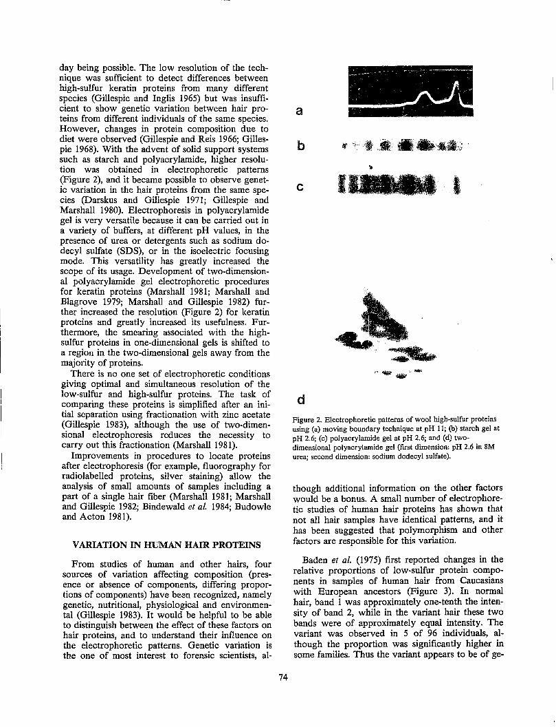
day being possible. The low resolution of the technique was sufficient to detect differences between high-sulfur keratin proteins from many different species (Gillespie and Inglis 1965) but was insufficient to show genetic variation between hair proteins from different individuals of the same species. However, changes in protein composition due to diet were observed (Gillespie and Reis 1966; Gillespie 1968). With the advent of solid support systems such as starch and polyacrylamide, higher resolution was obtained in electrophoretic patterns (Figure 2), and it became possible to observe genetic variation in the hair proteins from the same species (Darskus and Gillespie 1971; Gillespie and Marshall 1980). Electrophoresis in polyacrylamide gel is very versatile because it can be carried out in a variety of buffers, at different pH values, in the presence of urea or detergents such as sodium dodecyl sulfate (SDS), or in the isoelectric focusing mode. This versatility has greatly increased the scope of its usage. Development of two-dimensional polyacrylamide gel electrophoretic procedures for keratin proteins (Marshall 1981; Marshall and Blagrove 1979; Marshall and Gillespie 1982) further increased the resolution (Figure 2) for keratin proteins and greatly increased its usefulness. Furthermore, the smearing associated with the highsulfur proteins in one-dimensional gels is shifted to a region in the two-dimensional gels away from the majority of proteins.
There is no one set of electrophoretic conditions giving optimal and simultaneous resolution of the low-sulfur and high-sulfur proteins. The task of comparing these proteins is simplified after an initial separation using fractionation with zinc acetate (Gillespie 1983), although the use of two-dimensional electrophoresis reduces the necessity to carry out this fractionation (Marshall 1981).
Improvements in procedures to locate proteins after electrophoresis (for example, fluorography for radiolabelled proteins, silver staining) allow the analysis of small amounts of samples including a part of a single hair fiber (Marshall 1981; Marshall and Gillespie 1982; Bindewald et al. 1984; Budowle and Acton 1981).
VARIATION IN HUMAN HAIR PROTEINS
From studies of human and other hairs, four sources of variation affecting composition (presence or absence of components, differing proportions of components) have been recognized, namely genetic, nutritional, physiological and environmental (Gillespie 1983). It would be helpful to be able to distinguish between the effect of these factors on hair proteins, and to understand their influence on the electrophoretic patterns. Genetic variation is the one of most interest to forensic scientists, al-
74
.-:----- • - - -:=."'- .~- -- ----:--- -
~ , , .....
, "
a
c I
""9.'-
d Figure 2. Electrophoretic patterns of wool high-sulfur proteins using (a) moving boundary technique at pH 11; (b) starch gel at pH 2.6; (c) polyacrylamide gel at pH 2.6; and (d) twodimensional polyacrylamide gel (first dimension: pH 2.6 in 8M urea; second dimension: sodium dodecyl sulfate).
though additional information on the other factors would be a bonus. A small number of electrophoretic studies of human hair proteins has shown that not all hair samples have identical patterns, and it has been suggested that polymorphism and other factors are responsible for this variation.
Baden et al. (1975) first reported changes in the relative proportions of low-sulfur protein components in samples of human hair from Caucasians with European ancestors (Figure 3). In normal hair, band 1 was approximately one-tenth the intensity of band 2, while in the variant hair these two bands were of approximately equal intensity. The variant was observed in 5 of 96 individuals, although the proportion was significantly higher in some families. Thus the variant appears to be of ge-
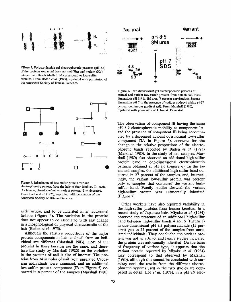
4 3 :i 1
t til , .' , HN
. Jt , . II I , ~. Hv . ,
~ure 3. Polyacrylamide gel electrophoretic patterns (PH 8.3) of the proteins extracted from normal (Hn) and variant (Hv) human hair. Bands labelled 1-4 correspond to low-sulfur proteins. From Baden et aL (1975), reprinted with permission of the American Society of Human Genetics.
Figure 4. Inheritance of low-sulfur protein variant electrophoretic pattern from the hair of four families. 0 - male, o - female; closed symbol = variant pattern; d = deceased. From Baden et al. (1975), reprinted with permission of the American Society of Human Genetics.
netic ongm, and to be inherited in an autosomal fashion (Figure 4). The variation in the proteins does not appear to be associated with any change in a morphological or physical characteristic of the hair (Baden et 01. 1975).
Although the relative proportions of the major protein components in hair and nail from an individual are different (Marshall 1983), most of the proteins in these keratins are the same, and therefore the study by Marshall (1980) on the variation in the proteins of nail is also of interest. The proteins from 74 samples of nail from unrelated Caucasian individuals were examined, and an additional low-sulfur protein component (lB in Figure 5) occurred in 8 percent of the samples (Marshall 1980).
75
NormaL
.. + 'WI 4321
43 ., 2A ~1A
2B
pH a-g 8M urea
+
pH 7 SDS
Variant
.+ ;\ : 1111
Figure 5. Two-dimensional gel electrophoretic patterns of normal and variant low-sulfur proteins from human nail. First dimension: pH 8.9 in 8M urea (7 percent acrylamide). Second dimension: pH 7 in the presen,ce of sodium dodecyl sulfate (4-27 percent continuous gradient gel), From Marshall (1980), reprinted with pennission of J. Invest. Dermatol.
The observation of component lB having the same pH 8.9 electrophoretic mobility as component lA, and the presence of component lB being accompanied by a decreased amount of a normal low-sulfur component (2A in Figure 5), accounts for the change in the relative proportions of the electrophoretic bands reported by Baden et 01. (1975) (Marshall 1980). In the study of nail samples, Marshall (1980) also observed an additional high-sulfur protein band in one-dimensional electrophoretic patterns obtained at pH 2.6 (Figure 6). In the examined samples, the additional high-sulfur band occurred in 27 percent of the samples, and, interestingly, the variant low-sulfur protein was present only in samples that contained the variant highsulfur band. Family studies showed the variant high-sulfur protein was autosomally inherited (Figure 7).
Other workers have also reported variability in the high-sulfur proteins from human keratins. In a recent study of Japanese hair, Miyake et 01. (1984) observed the presence of an additional high-sulfur band between high-sulfur bands 4 and 5 (Figure 8) in one-dimensional pH 8.3 polyacrylamide (15 percent) gels in 22 percent of the samples from unrelated individuals. They concluded the variant protein was not an artifact and family studies indicated the protein was autosomally inherited. On the basis of frequency of variant type, it appears that the variant protein reported by Miyake et 01. (1984) may correspond to that observed by Marshall (1980), although this cannot be concluded with certainty until the results from the different electrophoretic systems used in the two studies are compared in detail. Lee et 01. (1978), in a pH 8.9 elec-
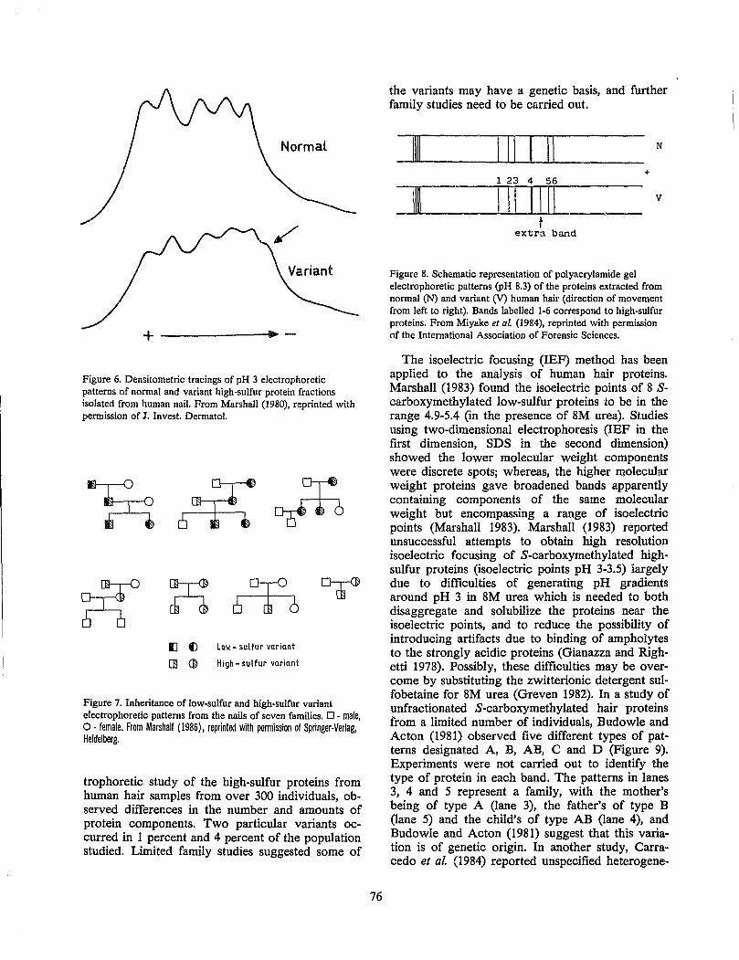
Normal
+ -------------. -
Figure 6. Densitometric tracings of pH 3 electrophoretic patterns of normal and variant high-sulfur protein fractions isolated from human nail. From Marshall (1980), reprinted with permission of J. Invest. Dermatol.
Il f) Low - sulfur variant
O:l ~ High - su I fur variant
Figure 7. Inheritance of low-sulfur and high-sulfur variant electrophoretic patterns from the nails of seven families. 0 - male, 0: female. From Marshall (1986), reprinted with permission of Springer-Verlag, Heidelberg.
trophoretic study of the high-sulfur proteins from human hair samples from over 300 individuals, observed differences in the number and amounts of protein components. Two particular variants occurred in 1 percent and 4 percent of the population studied. Limited family studies suggested some of
76
the variants may have a genetic basis, and further family studies need to be carried out.
_1111_----L....l...L-III--L-1 .l..L.-11 __ = N
+
---JJJL.]I~ _--1-11I...L....l...I 1..L-1...l..-Ili .__ v t
extra band
Figure 8. Schematic representation of polyacrylamide gel electrophoretic patterns (PH 8.3) of the proteins extracted from normal (N) and variant (V) human hair (direction of movement from left to right). Bands labelled 1-6 correspond to high-sulfur proteins. From Miyake et al. (1984), reprinted with permission of the International Association of Forensic Sciences.
The isoelectric focusing (IEF) method has been applied to the analysis of human hair proteins. Marshall (1983) found the isoelectric points of 8 Scarboxymethylated low-sulfur proteins to be in the range 4.9-5.4 (in the presence of 8M urea). Studies using two-dimensional electrophoresis (IEF in the first dimension, SDS in the second dimension) showed the lower molecular weight components were discrete spots; whereas, the higher molecular weight proteins gave broadened bands apparently containing components of the same molecular weight but encompassing a range of isoelectric points (Marshall 1983). Marshall (1983) reported unsuccessful attempts to obtain high resolution isoe1ectric focusing of S-carboxymethylated highsulfur proteins (isoelectric points pH 3-3.5) iargely due to difficulties of generating pH gradients around pH 3 in 8M urea which is needed to both disaggregate and solubilize the proteins near the isoelectric points, and to reduce the possibility of introducing artifacts due to binding of ampholytes to the strongly acidic proteins (Gianazza and Righetti 1978). Possibly, these difficulties may be overcome by substituting the zwitterionic detergent sulfobetaine for 8M urea (Greven 1982). In a study of unfractionated S-carboxymethylated hair proteins from a limited number of individuals, Budowle and Acton (1981) observed five different types of patterns designated A, B, AB, C and D (Figure 9). Experiments were not carried out to identify the type of protein in each band. The patterns in lanes 3, 4 and 5 represent a family, with the mother's being of type A (lane 3), the father's of type B (lane 5) and the child's of type AB (lane 4), and Budowle and Acton (1981) suggest that this variation is of genetic origin. In another study, Carracedo et al. (1984) reported unspecified heterogene-
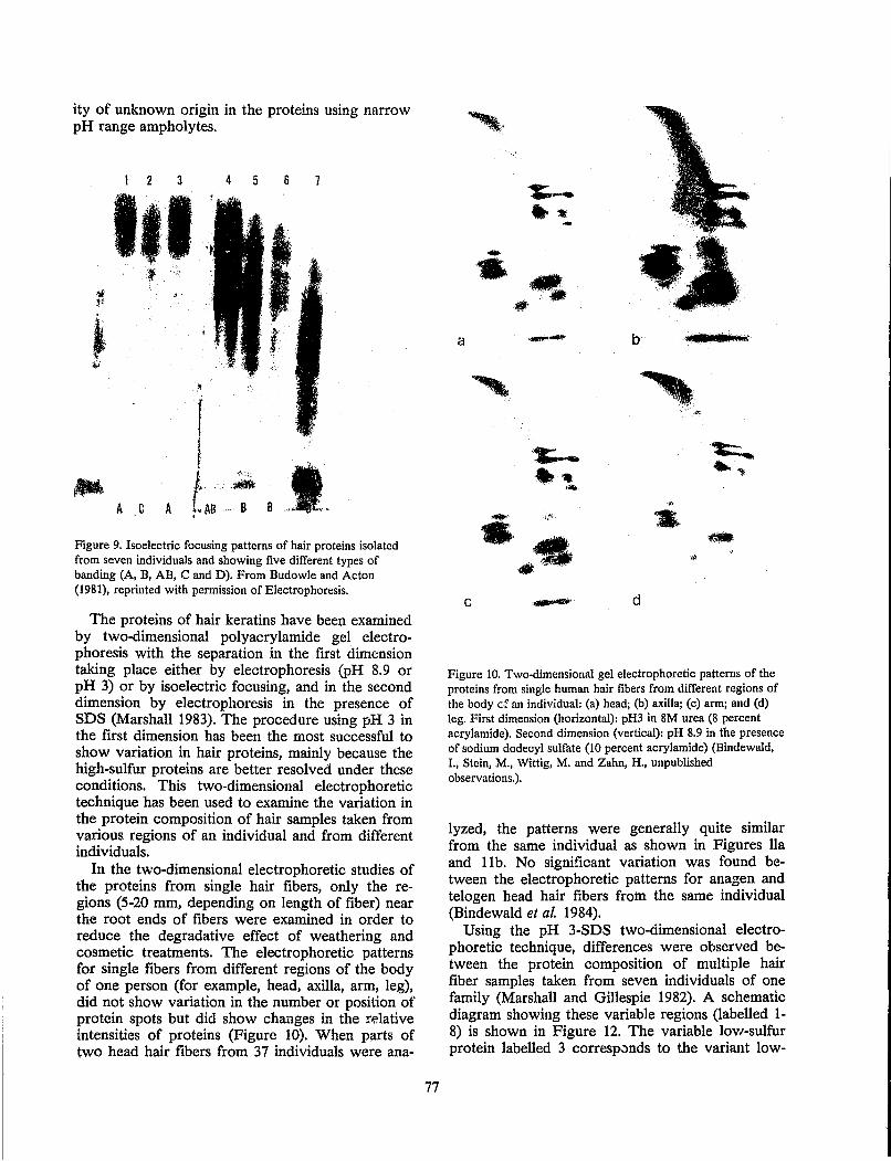
ity of unknown origin in the proteins using narrow pH range ampholytes.
2 3 4 5 6 7
"
A
. : g 8 t.
Figure 9. Isoelectric focusing patterns of hair proteins isolated from seven individuals and showing five different types of banding (A, B, AB, C and D). From Budowle and Acton (1981), reprinted with permission of Electrophoresis.
The proteins of hair keratins have been examined by two-dimensional polyacrylamide gel electrophoresis with the separation in the first dimension taking place either by electrophoresis (PH 8.9 or pH 3) or by isoelectric focusing, and in the second dimension by electrophoresis in the presence of SDS (Marshall 1983). The procedure using pH 3 in the first dimension has been the most successful to show variation in hair proteins, mainly because the high-sulfur proteins are better resolved under these conditions. This two-dimensional electrophoretic technique has been used to examine the variation in the protein composition of hair samples taken from various regions of an individual and from different individuals.
In the two-dimensional electrophoretic studies of the proteins from single hair fibers, only the regions (5-20 mm, depending on length of fiber) near the root ends of fibers were examined in order to reduce the degradative effect of weathering and cosmetic treatments. The electrophoretic patterns for single fibers from different regions of the body of one person (for example, head, axilla, arm, leg), did not show variation in the number or position of protein spots but did show changes in the relative intensities of proteins (Figure 10). When parts of two head hair fibers from 37 individuals were ana-
77
-:.-.. :t --- IlfL - '
a -- b , •• I.illiil[
'" ~
''t;.-.. -t..,
• .. .. ~ ... ... .' ;',~ .. .- .~
»-
C ,~' d
Figure 10. Two-dimensional gel electrophoretic patterns of the proteins from single human hair fibers from different regions of the body cf an individual: (a) head; (b) axilla; (c) arm; and (d) leg. First dimension (horizontal): pH3 in 8M urea (8 percent acrylamide). Second dimension (vertical): pH 8.9 in the presence of sodium dodecyl sulfate (to percent acrylamide) (Bindewald, I., Stein, M., Wittig, M. and Zahn, H., unpublished observations.).
lyzed, the patterns were generally quite similar from the same individual as shown in Figures lla and llb. No significant variation was found between the electrophoretic patterns for anagen and telogen head hair fibers from the same individual (Bindewald et al. 1984).
Using the pH 3-SDS two-dimensional electrophoretic technique, differences were observed between the protein composition of mUltiple hair fiber samples taken from seven individuals of one family (Marshall and Gillespie 1982). A schematic diagram showing these variable regions (labelled 1-8) is shown in Figure 12. The variable low-sulfur protein labelled 3 corrt.~sp..)nds to the variant low-
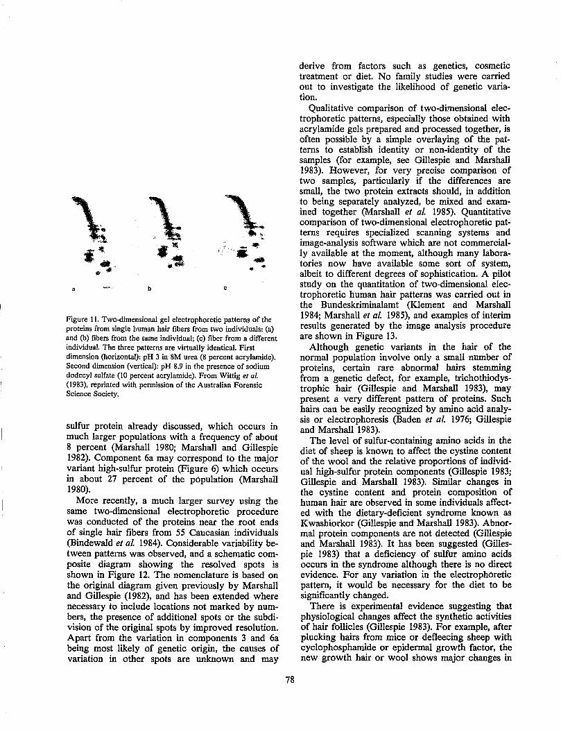
a b c
Figure 11. Two-dimensional gel electrophoretic patterns of the proteins from single human hair fibers from two individuals: (a) and (b) fibers from the same individual; (c) fiber from a different individual. The three patterns are virtually identical. First dimension (horizontal): pH 3 in 8M urea (8 percent acrylamide). Second dimension (vertical): pH 8.9 in the presence of sodium dodecyl sulfate (10 percent acrylamide). From Wittig et al. (1983). reprinted with permission of the Australian Forensic Science Society.
sulfur protein already discussed, which occurs in much larger popUlations with a frequency of about 8 percent (Marshall 1980; Mal'shall and Gillespie 1982). Component 6a may correspond to the major variant high-sulfur protein (Figure 6) which occurs in about 27 percent of the popUlation (Marshall 1980).
More recently, a much larger survey using the same two-dimensional electrophoretic procedure was conducted of the proteins near the root ends of single hair fibers from 55 Caucasian individuals (Bindewald et aL 1984). Considerable variability between patterns was observed, and a schematic composite diagram showing the resolved spots is shown in Figure 12. The nomenclature is based on the original diagram given previously by Marshall and Gillespie (1982), and has been extended where necessary to include locations not marked by numbers, the presence of additional spots or the subdivision of the original spots by improved resolution. Apart from the variation in components 3 and 6a being most likely of genetic origin, the causes of variation in other spots are unknown and may
78
derive from factors such as genetics, cosmetic treatment or diet. No family studies were carried out to investigate the likelihood of genetic variation.
Qualitative comparison of two-dimensional electrophoretic patterns, especially those obtained with acrylamide gels prepared and processed together, is often possible by a simple overlaying of the patterns to establish identity or non-identity of the samples (for example, see Gillespie and Marshall 1983). However, for very precise comparison of two samples, particularly if the differences are small, the two protein extracts should, in addition to being separately analyzed, be mixed and examined together (Marshall et al. 1985). Quantitative comparison of two-dimensional electrophoretic patterns requires specialized scanning systems and image-analysis software which are not commercially available at the moment, although many laboratories now have available some sort of system, albeit to different degrees of sophistication. A pilot study on the quantitation of two-dimensional electrophoretic human hair patterns was carried out in the Bundeskriminalamt (Klement and Marshall 1984; Marshall et aL 1985), and examples of interim results generated by the image analysis procedure are shown in Figure 13.
Although genetic variants in the hair of the nonnal popUlation involve only a small number of proteins, certain rare abnormal hairs stemming from a genetic defect, for example, trichothiodystrophic hair (Gillespie and Marshall 1983), may present a very different pattern of proteins. Such hairs can be easily recognized by amino acid analysis 01' electrophoresis (Baden et al. i976; Gillespie and Marshall 1983).
The level of sulfur-containing amino acids in the diet of sheep is known to affect the cystine content of the wool and the relative proportions of individual high-sulfur protein components (Gillespie 1983; Gillespie and Marshall 1983). Similar changes in the cystine content and protein composition of human hair are observed in some individuals affected with the dietary-deficient syndrome known as Kwashiorkor (Gillespie and Marshal11983). Abnormal protein components are not detected (Gillespie and Marshall 1983). It has been suggested (Gillespie 1983) that a deficiency of sulfur amino acids occurs in the syndrome although there is no direct evidence. For any variation in the electrophoretic pattern, it would be necessary for the diet to be significantly changed,
There is experimental evidence suggesting that physiological changes affect the synthetic activities of hair follicles (Gille~\pie 1983). For example, after plucking hairs from mice or defleecing sheep with cyclophosphamide or epidermal growth factor, the new growth hair or wool shows major changes in
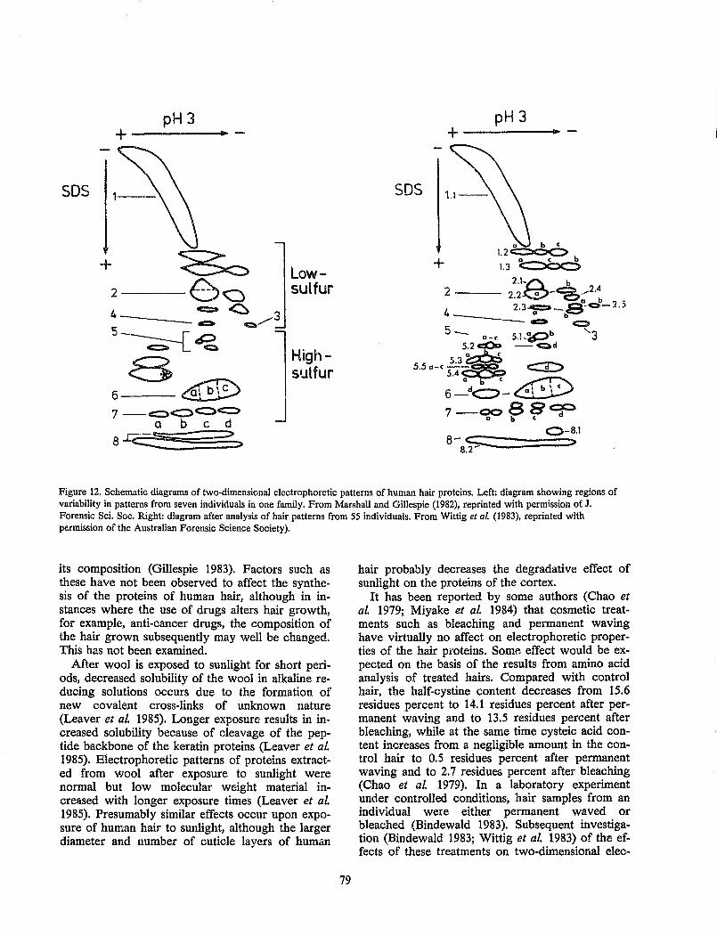
pH 3 +------.... -
50S
Lowsulfur
Highsulfur
pH 3 +-----_IP -
50S
+ o b c 1.2~b 1.3 <:=:X::x:::>
5_ Q-f.
5.2 cp.:a 5.50-c 5.3~~ c:::D
5.4 o c ~
6_dc:::>-~
7-ooB8~ o b c
O-B.l 8- c:::::
B.2'" >
Figure 12. Schematic diagrams of two-dimensional electrophoretic patterns of human hair proteins. Left: diagram showing regions of variability in patterns from seven individuals in one family. From Marshall and Gillespie (1982), reprinted with permission of J. Forensic Sci. Soc. Right: diagram after analysis of hair patterns from 55 individuals. From Wittig et al. (1983), reprinted with permission of the Australian Forensic Science Society).
its composition (Gillespie 1983). Factors such as these have not been observed to affect the synthesis of the proteins of human hair, although in instances where the use of drugs alters hair growth, for example, anti-cancer drugs, the c.omposition of the hair grown subsequently may well be changed. This has not been examined.
After wool is exposed to sunlight for short periods, decreased solubility of the wool in alkaline reducing solutions occurs due to the formation of new covalent cross-links of unknown nature (Leaver et 01. 1985). Longer exposure results in increased solubility because of Gleavage of the peptide backbone of the keratin proteins (Leaver et 01. 1985). Electrophoretic patterns of proteins extracted from wool after exposure to sunlight were normal but low molecular weight material increased with longer exposure times (Leaver et 01. 1985). Presumably similar effects occur upon exposure of human hair to sunlight, although the larger diameter and number of cuticle layers of human
79
hair probably decreases the degradative effect of sunlight on the proteins of the cortex.
It has been reported by some authors (Chao et 01. 1979; Miyake et 01. 1984) that cosmetic treatments such as bleaching and perman~nt waving have virtually no affect on electrophoretic properties of the hair pi'oteins. Some effect would be expected on the basis of the results from amino acid analysis of treated hairs. Compared with control hair, the half-cystine content decreases from 15.6 residues percent to 14.1 residues percent after permanent waving and to 13.5 residues percent after bleaching, while at the same time cysteic acid content increases from a negligible amount in the control hair to 0.5 residues percent after permanent waving and to 2.7 residues percent after bleaching (Chao et al. 1979). In a laboratory experiment under controlled conditions, hair samples from an individual were either permanent waved or bleached (Bindewald 1983). Subsequent investigation (Bindewald 1983; Wittig et 01. 1983) of the effects of these treatments on two-dimensional elec-
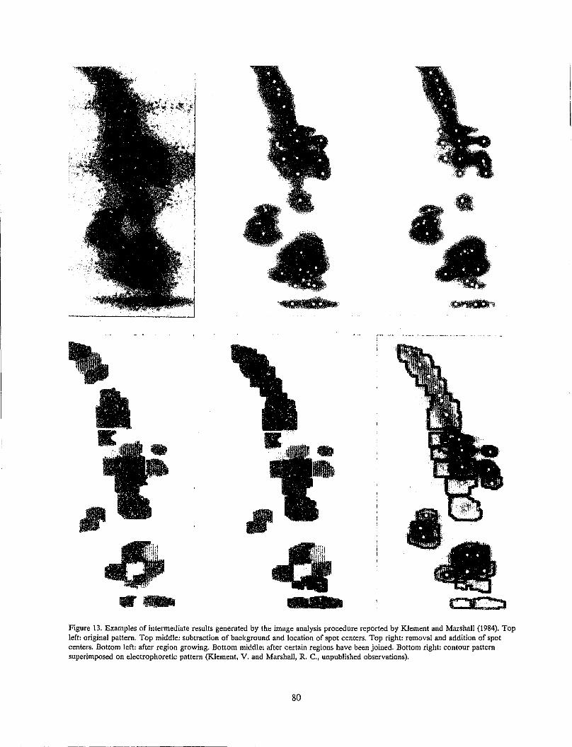
.,
.WIICI Figure 13. Examples of intermediate results generated by the image analysis procedure reported by Klement and Marshall (1984). Top left: original pattern. Top middle: subtraction of background and location of spot centers. Top right: removal and addition of spot centers. Bottom left: after region growing. Bottom middle: after certain regions have been joined. Bottom right: contour pattern superimposed on electrophoretic pattern (Klement, V. and Marshall, R. C., unpublished observations).
80
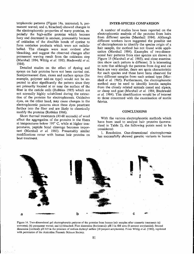
trophoretic patterns (Figure 14a, untreated; b, permanent waved; and c, bleached) showed changes in the electrophoretic properties of many proteins, especially the high-sulfur proteins which became hazy and decreased in amount, presumably because of oxidation of the disulfide bonds of cystine to form oxidation products which were not radiolabelled. The changes were most evident after bleaching, and suggest the observed changes after permanent waving result from the oxidation step (Marshall 1984; Wittig et al. 1983; Bindewald et al. 1984).
Detailed studies on the effect of dyeing and sprays on hair proteins have not been carried out. Semipermanent dyes, rinses and surface sprays (for example, polymer add-on type) would not be expected to alter significantly the pattern since they are primarily located at or near the surface of the fiber in the cuticle cells (Robbins 1985) which are not normally highly solubilized during the extraction of the proteins for electrophoresis. Oxidative dyes, on the other hand, may cause changes in the electrophoretic patterns since these dyes penetrate further into the fiber and are likely to chemically modify the proteins (Robbins 1986).
Short thermal treatments (40-60 seconds) of wool affect the aggregation of the proteins in the fibers at temperatures below 195 0 C, while at higher temperatures, peptide bond cleavage becomes important (Marshall et at. 1983). Presumably similar modifications occur with human hair proteins on heat treatment.
I I
a b
INTER· SPECIES COMPARISON
A number of studies have been reported on the electrophoretic analysis of the proteins from hairs from different species (Marshall 1984). Although different workers have suggested the possible use of electrophoresis to identify the species origin of a hair sample, the method has not found wide application (Marshall 1984). Examples of two-dimensional hair patterns from nine species are shown in Figure 15 (Marshall et al. 1985), and close examination show each pattern is different. It is interesting to note that although the patterns from dog and cat hairs are very similar, there are spots characteristic for each species and these have been observed for two different samples from each animal type (Marshall et al. 1985). Furthermore, the electrophoretic method may be used to identify keratin samples from the closely related animals camel and alpaca, or sheep and goat (Marshall et al. 1984; Bindewald et al. 1984). This identification would be of interest to those concerned with the examination of textile fabrics.
CONCLUSIONS
With the various electrophoretic methods which have been used to analyze hair proteins (summarized in Table 2), the following points need to be considered:
1. Resolution. One-dimensional electrophoresis has successfully showed genetic variants in human
c
Figure 14. Two-dimensional gel electrophoretic patterns of the proteins from human hair samples after cosmetic treatment: (a) untreated; (b) permanent waved; and (c) bleached. First dimension (horizontal): pH 3 in 8M urea (8 percent acrylamide). Second dimension (vertical): pH 8.9 in the presence of sodium dodecyl sulfate (10 perpent acrylamide). From Wittig et 01. (1983), reprinted with permission of the Australian Forensic Science Society.
81
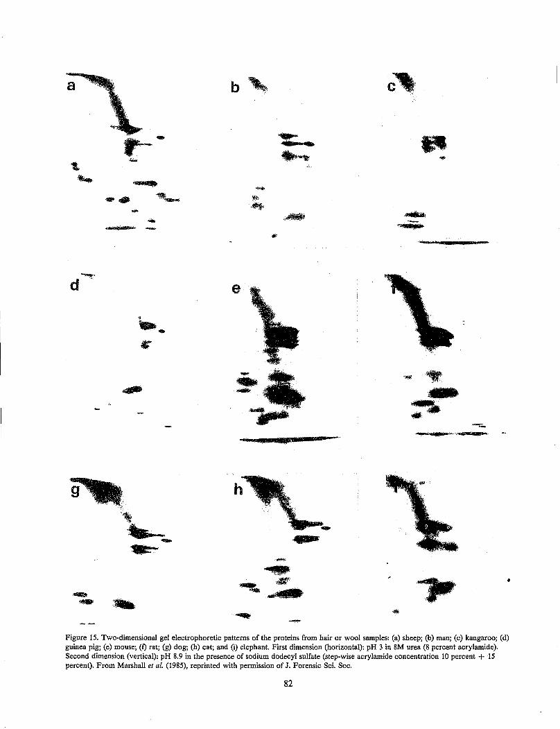
a
-
",.'
~ :~
.. -
-.
..... '. -.~
,;~
..... 14-
'~.'.'.; ""':
,., ....... '. . . I
II
Figure 15. Two-dimensional gel electrophoretic patterns of the proteins from hair or wool samples: (a) sheep; (b) man; (c) kangaroo; (d) guinea pig; (e) mouse; (f) rat; (g) dog; (h) cat; and (i) elephant. First dimension (horizontal): pH 3 in 8M urea (8 percent acrylamide). Second dimension (vertical): pH 8.9 in the presence of sodium dodecyl sulfate (step-wise acrylamide concentration 10 percent + 15 percent). From Marshall et at. (1985). reprinted with permission of J. Forensic Sci. Soc.
82
hair, although the higher resolving two-dimensional procedures have more potential to identify variants. Within one-dimensional procedures, some systems are more successful than others, for example SDS electrophoresis is better than electrophoresis at pH 8.3-8.9 for the low-sulfur proteins, but the reverse is true for the high-sulfur proteins. Isoelectric focusing has higher resolving power than one-dimensional electrophoresis.
2. Sensitivity. SenHitivity may be increased by the use of smaller gel rods or slabs (microgels), or better methods of locating the proteins. Although Miyake et aL (1984) were unsuccessful with the silver staining technique, Budowle and Acton (1981) advocate its use. An alternative approach is to radiolabel the proteins after solubilization (for example, with 14C-iodoacetate), then locate the proteins in the gel by fluorography (Marshall and Gillespie 1982). Apart from allowing the analysis of small parts of a single fiber, the technique permits the visualization of proteins in widely different concentrations by using different times of exposure.
3. Experimental difficulty. Isoelectric focusing is marginally more difficult than conventional one-dimensiona.1 electrophoresis. Two-dimensional electrophoresis requires more steps but is not more difficult than one-dimensional procedures, and is routine in many laboratories. One laboratory with three technicians has reported the analysis of 10,000 samples (non-keratin) by two-dimensional electrophoresis in a year.
4. Quantitation and comparison. With one-dimensional gels, these steps are simpler and easier than with two-dimensional gels, the latter requiring specialized image analysis equipment and computer software.
Table 2. SUMMARY OF ACRYLAMIDE GEL ELECI'ROPHORETIC MEmODS FOR mE SEPARATION OF HUMAN HAIR PROTEINS
Protein Type
One-dimension Total mixture
Low-sulfur
System Reference
IEF Budowle and Acton (1981)
Carracedo et al. (1984)
IEF Marshall and Blagrove (1979)
Marshall (1983) Greven (1982)
8M urea pH 8.3- Baden et al. 8.9 (1975)
Marshall (1980,1983)
SDS Marshall (1980,1983)
83
Protein Type System Reference
High-sulfur 8M urea pH 8.3- Lee et al. (1978) 8.9 Miyake et al.
(1984) 3M urea pH 2.6 Marshall
(1980,1983)
Two-dimension Total mixture LIEF 8M urea Marshall (1983)
2.SDS 1.8M urea pH 8.9 Marshall
(1981,1983) Bindewald (1983) Bindewald et al.
(1984) 2.SDS 1.8M urea pH 3 Marshall and
Gillespie (1982)
Bindewald (1983) Bindewald et aL
(1984) 2.SDS
Low-sulfur l.IEF 8M urea Marshall and Blagrove (1979)
2.SDS High-sulfur 1.8M urea pH 3 Marshall (1980)
2.SDS
Electrophoretic studies of the keratin proteins of human hair have been carried out on only a limited number of samples. Since many of these studies have used different procedures (Table 2), it is difficult to compare the results and to summarize the amount of variation. Nevertheless there are indications of con3iderable variability between the proteins of hair from different individuals. With currently available procedures, it appears an electrophoretic pattern would be characteristic of a group of individuals, rather than being unique to an individual, for example the pattern in Figure 11c is virtually identical to those in Figures lla and lIb which are from a different individual. More individualization may be possible through improvements in present electrophoretic techniques [for example, the incorporation of zwitterionic detergents in isoelectric focusing experiments (Greven 1982)], or through the development of new analytical methods for keratin proteins such as highly resolving HPLC techniques for protein analysis, the use of enzymes with subsequent analysis of peptides by HPLC, or the development of highly specific monoclonal antibodies.
The number of genetically-distinct groups of hair within the human popUlation is unknown at this stage, and a detailed study which includes families is required to establish the extent of this genetic variation. In order to eliminate the effects from factors such as cosmetic treatments and sunlight, it is
recommended that part of the study include the proteins of follicles. It is also essential to investigate variations arising from nutritional, physiological and environmental sources. As pointed out previously by Marshall and Gillespie (1982), the effects observed in the electrophoretic patterns after cosmetic treatments could be of help in forensic analysis even though they are not genetically based variations.
Although consistently high solubilities are possible with many keratins, a range of solubilities between 5 percent and 75 percent has been encountered with human hair (Gillespie 1983). In some cases, low solubility appears to be an inherited trait, and therefore a possible identification characteristic. Other factors such as cosmetic treatments. and exposure to sunlight may reduce the solubility. In order to make quantitative biochemical comparisons between hairs, this variation in solubilities is a potential problem. Improved methods for extraction of hair need to be developed to give a consistently high degree of solubility.
The results of electrophoretic studies of the proteins from human hair samples are sufficiently encouraging to recommend that a trial be undertaken in a forensic laboratory to evaluate the potential of the method in hair analysis. This trial should be undertaken in conjunction with studies to establish the extent and sources of variation in the patterns. The electrophoretic method finally chosen for use in a forensic laboratory would depend on a number of factors including the requirements of the analysis, the amount and type of sample, the number of samples to be examined in a year, background and experience of personnel in the laboratory, and the available equipment.
ACKNOWLEDGMENTS
I thank the Federal Bureau of Investigation for the invitation to attend the International Symposium on Forensic Hair Comparisons, and Dr. J. M. Gillespie, Dr. M. Wittig and Professor H. Zahn for their support and advice.
REFERENCES
Baden H. P., Lee L. D. and Kubilus J. (1975). A genetic electrophoretic variant of human hair a-polypeptides, Am. J. Hum. Genet. 27:472-477.
Baden H. P., Jackson C. E, Weiss L., Jimbow K., Lee L., Kubilus J. and Gold R. J. M (1976). The physicochemical properties of hair in the BIDS syndrome, Am. J. Hum. Genet. 28:514-521.
Bindewald I (1983). Beitrag zur Analytik des Humanhaares mit forensischem Aspekt. Thesis, Rheinisch-Westfalischen Technischen Hochschule, Aachen.
84
Bindewald, I, Greven, R., Marshall, R., Stein, M, Stephani, G., Wittig, M and Zahn, H. (1984). Two dimensional keratin patterns of human hair. In: Proceedings of 10th Triennial Meeting of the International Association of Forensic Sciences, Oxford.
Bradbury, J. H. (1973). The structure and chemistry of keratin fibers, Adv. Prot. Chern. 27:111-211.
Budowle, B. (1986). Selecting genetic markers for analysis of forcibly removed hairs. In: Proceedings of the International Symposium on Forensic Hair Comparisons. U. S. Government Printing Office, Washington, D.C., pp. 89-93.
Budowle, B. and Acton, R. T. (1981). A technique for the detection of variable electrophoretic patterns of hair proteins, Electrophoresis 2:333-334.
Carracedo, A., Concheiro, L. and Requena, T. (1984). Species identification of hair by the isoelectric focusing of keratins followed by silver staining, J. Forensic Sci. Soc. 24:422.
Chao, J., Newsom, A. E., Wainwright, L M. and Mathews, R. A. (1979). Comparison of the effects of some reactive chemicals on the proteins of whole hair, cuticle and cortex, J. Soc. Cosmet. Chem. 30:401-413.
Darskus, R. L. and Gillespie, J. M (1971). Breed and species differences in the hair proteins of four genera of Caprini, Aust. J. BioI. Sci. 24:515-524.
Deadman, H. (1986). Human hair comparisons based on microscopic characteristics. In: Proceedings of the International Symposium on Forensic Hair Comparisons. U. S. Government Printing Office, Washington, D.C., pp.
Praser, R. D. B., MacRae, T. P. and Rogers, 0. E (1972). Keratins. Their Composition, Structure, and Biosynthesis. Charles C. Thomas, Springfield.
Praser, R. D. B., Gillespie, J. M., MacRae, T. P. and Marshall, R. C. (1981). Internal report. CSIRO, Division of Protein Chemistry.
Gaudette, B. (1986). The future of forensic hair comparisons. In: Proceedings of the International Symposium on Forensic Hair Comparisons. U. S. Government Printing Office, Washington, D.C., pp. 127-134.
Gianazza, E and Righetti, P. G. (1978). Binding of polyanions to carrier ampholytes in is 0 electric focusing, Biochim. Biophys. Acta 540:357-364.
Gillespie, J. M (1968). The dietary regulation of the synthesis of hair keratin. In: Symposium on Fibrous Proteins (Crewther, W. G., ed.). Butterworth, Sydney, pp. 362-363.
Gillespie, J. M (1983). The structural proteins of hair: isolation, characterization, and regulation of biosynthesis. In: Biochemistry and Physiology of
the Skin (Goldsmith, L. A., ed.). Oxford University Press, Oxford, pp. 475-510.
Gillespie, J. M and Inglis, A. S. (1965). A comparative study of high-sulfur proteins from a-keratins, Compo Biochem. Physiol. 15:175-185.
Gillespie, J. M and Marshall, R. C. (1980). Variability in the proteins of wool and hair. In: Proceedings of 6th International Wool Textile Research Conference, Volume II, pp. 67-77.
Gillespie, J. M and Marshal/, R. C. (1981). The proteins of normal and aberrant hair keratins. In: Hair Research--Status and Future Aspects (Orfanos, C. E., Montagna, W. and Stiittgen, G., eds.). Springer-Verlag, Heidelberg, pp. 76-83.
Gillespie, J. M and Marshall, R. C. (1983). A comparison of the proteins of normal and trichothiodystrophic human hair, J. Invest. Dermatol. 80: 195-202.
Gillespie, J. M and Reis, P. J. (1966). The dietaryregulated biosynthesis of high-sulphur wool proteins, Biochem. J. 98:669-677.
Greven, R. (1982). Zur Analytik morphologischer Wollkomporienten am Beispiel der makrofibrillen. Thesis, Rheinisch-Westfalischen Technischen Hochschule, Aachen.
Hewish, D. R., Robinson, C. P. and Sparrow, L. G. (1984). Monoclonal antibody studies of a-keratin low-sulfur proteins, Aust. J. Biol Sci. 37:17-23.
Kaszynski, E. (1986). Hair growth: mechanism and regulation. In: Proceedings of the International Symposium on Forensic Hair Comparisons. U. S. Government Printing Office, Washington. D.C., pp.23-33.
Klement, V. and Marshall, R. C. (1984). The use of computer analysis for the quantification of 2-D electrophoretic hair keratin patterns - a pilot study. In: Proceedings of 10th Triennial Meeting of the International Association of Forensic Sciences, Oxford.
Leaver, I H., Marshall, R. C. and Rivett, D. E. (1985). Light-induced changes in the composition of wool. In: Proceedings of 7th International Wool Textile Research Conference, IV:11-20.
Lee, L. D., Ludwig, K. and Baden, H. P. (1978). Matrix proteins of human hair as a tool for identification of individuals, Forensic Sci. 11: 115-121.
Ley, K. F., Marshall, R. C. and Crewther, W. G. (1985). Further studies on the release and characterisation of cuticle and membranous components from wool. In: Proceedings of 7th International Wool Textile Research Conference, 1:152-161.
Marshall, R. C. (1980). Genetic variation in the proteins of human nail. J. Invest. Dermatol. 75:264-269.
Marshall, R. C. (1981). Analysis of the proteins from single wool fibers by two-dimensional polyacrylamide gel electrophoresis, Text. Res. J. 51:106-108.
85
Marshall, R. C. (1983). Characterization of the proteins of human hair and nail by electrophoresis, J. Invest. Dermatol. 80:519·524.
Marshall, R. C. (1984). Forensic identification of hairs by electrophoresis. In: Proceedings of 10th Triennial Meeting of the International Association of Forensic Sciences, Oxford.
Marshall, R. C. (1986). Nail, claw, hoof and horn keratin. In: Biology of the Integument (BereiterHahn, J., Matoltsy, A. G. and Richards, K. S., eds.). Springer-Verlag, Heidelberg, pp. 722-738.
Marshall, R. C. and Blagrove, R. J. (1979). Successful isoelectric focusing of wool low-sulfur proteins, J. Chromatogr. 172:351-356.
Marshall, R. C. and Gillespie, J. M (1982). Comparison of samples of human hair by two-dimensional electrophoresis, J. Forensic Sci. Soc. 22:377-385.
Marshall, R. c., Gillespie, J. M and Klement, V. (1985). Methods and future prospects for forensic identification of hairs by electrophoresis, J. Forensic Sci. Soc. 25:57-66.
Marshall, R. c., Souren, I and Zahn, H. (1983). Protein changes after short thermal treatments of wool fabrics, Text. Res. J. 53:792-794.
Marshall, R. c., Zahn, H. and Blankenburg, G. (1984). Possible identification of specialty fibers by electrophoresis, Text. Res. J. 54:126-128.
lVliyake, B., Mukoyama, H. and Seta, S. (1984). Hair grouping by electrophoresis of solubilized hair protein. In: Proceedings of 10th Triennial Meeting of the International Association of Forensic Sciences, Oxford.
Robbins, C. (1979). Chemical and Physical Behavior of Human Hair. Van Nostrand Reinhold Company, New York.
Robbins, C. (1986). The morphology and chemistry of human hair. In: Proceedings of the International Symposium on Forensic Hair Comparisons. U. S. Government Printing Office, Washington, D.C., pp. 3-21.
Said, H. M., Newsom, A. E., Tippins, B. L. and Mathews, R. A. (1985). Reversed-phase high-performance liquid chromatography of some ultraheterogeneous and covalently-modified proteins from human hair, J. Chromatogr. 324:65-73.
Swift, J. A. and Bews, B. (1974). The chemistry of human hair cuticle I: A new method for the physical isolation of cuticle, J. Soc. Cosmet. Chern. 25:13-22.
Wittig, M (1982). Protein patterns of keratins - the probable role in forensic hair examination, J. Forensic Sci. Soc. 22:387-389.
Wittig, M, Bindewald, I, Marshall, R. c., Stein, M and Zahn, H. (1983). Two-dimensional keratin patterns of single human hair fibres. In: Proceed
ings 8th Australian International Forensic Science Symposium, Perth.
Wortmann. F. J., Greven. R. ,and Zahn, H. (1982). A method for isolating the cortex of keratin fibers, Text. Res. J. 52:479-481.
DISCUSSION
Budowle: I would like to comment on why we used one-dimensional gel electrophoresis (Budowle and Acton 1981) as opposed to two-dimensional analysis. With two-dimensional electrophoresis a greater number of proteins can be detected, often because of differences in molecular weight and not single amino acid differences. Resolution of variants with very close isoelectric points is best with ultrathin layer isoelectric focusing where the voltage gradient is higher than in the first dimension of conventional 2-D IEF-SDS electrophoresis. As an example, genetic markers such as group specific components transferrin and alpha-! antitrypsin can be genetically typed by one-dimensional analysis but cannot be typed by two-dimensional analysis.
The results in our paper (Budowle and Acton 1981) were obtained before we went to using ultrathin layer gels. When we used the thinner gel with the higher voltage gradient, much sharper banding patterns were obtained. At that point it was necessary for us to stop because blood work was considered by the FBI to be higher priority.
During our hair research, we also looked at the extractability of proteins from hairs of different racial origins and found that more protein was solubilized from Negro hairs than from Caucasian hairs.
Lee: I agree with you that two-dimensional electrophoresis is a good method, especially if you do it every day. However, if you do it only once a month or once every 6 months, forget about it. With our case load, we have 200 to 500 hairs to examine per day.
Do you have an explanation for the observation of the additional protein in some samples? Is the variance arising from genetic expression, post-translational modification or protein modification during solubilization?
Marshall: I would like to comment further on two-dimensional electrophoresis. This procedure is used routinely in many types of protein analysis, including the analysis of serum for genetic markers. There are many laboratories capable of carrying out and analyzing a large number of two-dimensional gels. A few years ago, one laboratory reported generating 10,000 two-dimensional gel patterns per year using two or three technicians.
86
It is difficult to know the reason for the protein variation in some hair samples. From studies carried out on follicles, we know the variation is already present in the proteins in the lower region of the follicle. Other studies suggest a mutation in a regulatory gene is responsible rather than a structural gene mutation. Peptide mapping experiments show the additional protein differs from the other low-sulfur proteins by more than one peptide, indicating the presence of more than one mutation in amino acid sequence or post-translational modification in more than one place. It is unlikely that the solubilization procedure is responsible for the presence of the variant.
Kaszynski: Is there any advantage in using a constant pH of 3 in the first dimension over using a pH gradient such as ~soelectric focusing?
I would like to comment Oil Dr. Budowle's observations on resolution in isoelectric focusing. When isoelectric focusing is used in the first dimension and SDS in the second dimension, a high degree of resolution is obtained. I recall a paper in which proteins were progressively carbamylated in such a manner that it was possible to detect single charged differences in the carbamylation.
Marshall: In the procedure we use to solubilize and stabi1iz~ the hair proteins, the proteins are converted from the SH form to the S-carboxymethylated derivative. Isoelectric focusing experiments of the carboxymethylated low-sulfur and high-tyrosine proteins are quite successful. However, for the carboxymethylated high-sulfur proteins the isoelectric points are around pH 3. In order to solubilize these proteins near the isoelectric region, 8M urea is used, and under these conditions we have not been successful in carrying out high resolution isoelectric focusing on the proteins. The best resolution for the high-sulfur proteins is with electrophoresis at pH 3.
De Forest: Have you investigated the possibility of using keratinases? Would this be a workable way of producing peptides for analysis?
Marshall: We have used enzymes to degrade keratin proteins prior to determulation of amino acid sequences but have not investigated the identification of hairs by analysis of enzymatic peptides. A necessary requirement for a successful procedure is reproducible peptide production.
Podlecki: 'When we examine hairs microscopically in a standard of one person, hair characteristics from different areas of the head may be different. With the high-sulfur and low-sulfur proteins that you observe electrophoretically, do they change from the front part of the head to the back?

Marshall: In studies carried out at the Deutsches W ollforschungsinstitut (Aachen, Federal Republic of Germany), hairs from different regions of the head were examined. No differences in the number
87
or type of proteins were observed, although occasionally there were small differences in the proportions of the proteins.
SELECTING GENETIC MARKERS FOR ANALYSIS OF FORCIBLY REMOVED HAIRS
Bruce Budowle and Linda C. Davidson
Forensic Science Research Group FBI Laboratory
Quantico, Virginia
The analysis of hair from a forensic viewpoint usually has been limited to microscopic analyses and comparisons. Genetic marker analysis, particularly on forcibly removed hairs, can contribute to an increased individualization potential for hair characterization. Genetic information is constant within an individual throughout his life. The gene systems, composed of deoxyribonucleic acid (DNA), can exist in a number of alternative forms (alleles) within a population. In order for a gene system to be considered polymorphic it must have at least two alleles, each occurring at a frequency greater than 1 percent in the population. The various alleles encode for proteins which actually are the genetic markers observed in forensic serology. Since the alleles of a gene system are biochemically different, the protein markers produced from each allelic template are slightly different. These differences can be observed by immunologic and/or electrophoretic methods. The result of an assay is termed a phenotype and from this a possible genetic make-up (or genotype) of an individual can be deduced. In forensic serology, the identification of phenotypes from various genetic marker systems is used for the determination of the relationship between the biological material deposited on items of evidence at a crime scene and allegedly involved individuals.
Table 1. FORENSIC GENETIC MARKERS FOUND IN HAIR SHEATH CELLS
ABon Phosphoglucomutase-l
Esterase D Glyoxalase
Glucose 6-phosphoglucon. ate dehydrogenase
6-Phosphogluconate dehydrogenase
Adenylate kinase Adenosine deaminase
Phospho hexose isomerase Sex chromosomes
Phosphoglucomutase-3
KeratinS
n These genetic markers are located in the hair shaft. When an anagen phase or actively growing hair
is plucked from the body it often will have sheath cells associated with the root. A number of genetic markers found in blood also have been found in sheath cells (Table 1). Bosley et al. (1980) have reported that as much as 10 percent of the hairs received as case evidence in the Home Office Laboratory have had sheath cells associated with their roots suggesting genetic marker analysis of forcibly
89
removed hairs may be of value to the forensic scientist.
When analyzing forcibly removed hairs certain constraints must be considered before choosing which genetic markers will be analyzed. First, not all genetic markers that are used for blood analysis may be found in hair. Other markers may exist but present methodologies may not be sensitive enough to detect the minute quantities of the components in sheath cells. Second, there is a limited amount of available material for analysis. Other than the occurrence of ABO (Yada et al. 1966a, b; Kimura and Yokoyama 1969) and keratins (Baden et al. 1975; Hrdy et al. 1977; Lee et al. 1978; Marshall 1984) in the hair shaft, the material for analysis is a very small portion of the hair. Third, the available methodology must be considered. Isoelectric focusing (IEF) is superior to conventional electrophoretic methods for genetic mll.rker analysis (Murch and Budowle 1986). Isoelectric focusing has a greater sensitivity of detection due to its concentrating effect and has superior resolving capabilities, especially for subtyping of genetic marker systems such as phosphoglucomutase-l (pGM) (Burdett and Whitehead 1977; Sutton and Burgess 1978), alpha I-antitrypsin (Allen et al. 1974), transferrin (Kuhnl and Spielmann 1978, 1979), esterase D (EsD) (Olaisen et al. 1981, Budowle 1984) and group-specific component (Constans and Viau 1977; Constans et al. 1978). However, only a few laboratories in the United States are routinely using IEF. Therefore, until IEF is implemented, forensic laboratories will have to rely heavily on conventional electrophoretic methods. The ability to type ABO is available in all laboratories. Although there appears to be a sufficient concentration of ABO blood group substance in the cortex of the hair shaft, the standard adsorption-elution assay has been found to yield unequal agglutination reactions for the different antigens (Mudd, personal communication). This could result in a misinterpretation of the actual phenotype. Perhaps a microplate assay (Wegmann and Smithies 1966), utilizing monoclonal antibodies, could resolve this problem. Sex typing of hair is another genetic system that forensic laboratories are adopting (Mudd 1984). However, with the above mentioned constraints, there must also be the consideration of restriction due to case1oad. Forensic serologists have extremely large caseload de-
mands placed upon them. Therefore, simple and easy methods that do not detract from the flow of the laboratory are preferred.
Bearing in mind the above constraints, other criteria for the selection of genetic markers for hair also should be considered. These are the discriminating probability (DP) of the genetic marker(s), the phenotypic patterns each marker produces, the intensity of the band patterns, the simplicity and ease of the assay, the stability of the markers, and the analytical requirements of the case itself.
The forensic genetic markers found in hair with the highest DP are PGM, erythrocyte acid phosphatase (EAP), glyoxalase I (GLO), EsD and the sex chromosomes. Sex typing of hairs is discussed elsewhere by Mudd (1984) and, therefore, will not be presented here. Adenosine deaminase (ADA) and adenylate kinase (AK) have low DPs; thus, it is not logical to routinely type for these markers in hair.
All markers analyzed in hair except for EAP (Twibell and Whitehead 1978; Sutton et al. 1982) produce band patterns the same as in blood. We have observed difficulties typing EAP derived from sheath cells subjected to IEF. Therefore, EAP is not recommended for hair analysis.
This leaves three genetic markers for analysis: PGM, EsD and GLO. Although typeable, EsD produced weak patterns even in fresh hairs (BudowIe, unpublished data). Similar findings were observed for EsD (Twibell and Whitehead 1978; Y 0-
shida et al. 1979), as well as AK (Twibell and Whitehead 1978; Sutton et al. 1982) and ADA (Twibell and Whitehead 1978). In contrast, PGM and GLO produced intense, easily discernable patterns. Therefore, our attention was directed to these genetic marker systems and away from less intense, less genetically informative ones. Many investigators have observed intense PG1vl and GLO pattems from freshly plucked hairs (Twibell and Whitehead 1978; Oya et al. 1978; Burgess and Twibell 1979; Yoshida et al. 1979; Lawton and Sutton 1982; Montgomery and Jay 1982; Sutton et al. 1982; Gambel and Budowle 1986).
Several individuals have developed systems for typing either PGM or GLO (Oya et al. 1978; Twibell and Whitehead 1978; Burgess and Twibell 1979; Burgess et al. 1979; Yoshida et al. 1979; Lawton and Sutton 1982). However, an ideal approach would be to develop a system that was simple and could assay for both markers in a single hair. If possible PGM subtyping would be desirable. One approach that has been used is the Group I system (Wraxall et al. 1978). This conventional electrophoretic method simultaneously types EsD, GLO and PGM. Although EsD activity is low, PGM and GLO should be typeable in hairs. In fact, Montgomery and Jay (1982) have typed all three Group I markers from a single hair root con-
90
taining sheath cells. The DP for this system is 0.88 and 0.84 for Caucasians and Negroes, respectively. Gambel and Budowle (1986) developed a relatively simple method for typing GLO and then subtyping PGM from the same hair. This approach yielded a DP of 0.90 and 0.89 for Caucasians and Negroes, respectively. Although it required more sample preparation time and technical skill, Lawton and Sutton (1982) introduced a method for cutting a hair root into sections. Each section was used for a different assay, thus permitting multiple enzyme tests on a single hair.
The testing of the stability of the enzymes in sheath cells is important from two aspects. The first is to determine reasonable expectation on the age of a hair that an investigator may still detect the appropriate genetic marker. Second, it is important to determine whether or not any of the known phenotypes will change over time and produce an erroneous result. The genetic markers - PGM-l, PGM-3, EsD, AK, ADA, GLO, glucose 6-phosphate dehydrogenase, phosphohexose isomerase, 6-phosphogluconate dehydrogenase, ABO and sex chromosomes (Yada et al. 1966a, b; Kimura and Yokoyama 1969; Lawton and Sutton 1982; Oya et al. 1978; Montgomery and Jay 1982; Twibell and Whitehead 1978; Sutton et al. 1982; Burgess and Twibell 1979; Mudd 1984; Yoshida et al. 1979; Gambel and Budowle 1986) have never been reported to alter their patterns over time in hair. Of all the markers, PGM and GLO have been found to produce the most intense patterns and to be the most stable. The rest of the markers produced weak or variable results. Even ADA and AK, which are extremely stable in blood, do not appear to be as stable in hair (Twibell and Whitehead 1978; Bosley et al. 1980; Sutton et al. 1982). Sex typing would be a reliable alternative to PGM and GLO as a stable informative system (Mudd 1984).
While all the above criteria are acceptable, the case itself can dictate the appropriate marker(s) for hair analysis. Some situations may require determining the sex of the individual who might have deposited the hair and not the phenotypes of the enzyme systems, or vice versa. If evidence is typed as it is received by the laboratory, it would seem reasonable to assay whatever systems yield the highest DP. In contrast, some laboratories may retain evidence until a suspect is in custody. It is then possible to type the suspect for the marker(s) and determine which phenotypes would produce the best probability for exclusion.
Whatever the approach, an investigator must remember that the genetic markers in hair are degradable. Therefore, it is recommended that hairs be maintained at _20 0 C until analyzed. Even then they should be analyzed as soon as possible. Permounts and/or Permount solvents have been re-
ported to have negative effects on the stability of genetic markers (Lawton and Sutton 1982; Mudd and Deedrick 1986). Thus, it may be appropriate to analyze the hair first electrophoretically and then perform microscopic analyses. Bosley et al. (1980) have reported that there was no change in hair root morphology after electrophoresis. However, this approach could result in the loss of trace material on the hairs. If destruction of the hair is prohibited, sex typing and ABO blood grouping may not be possible - only electrophoretic approaches may be a viable avenue. Whatever the approach, selection of the genetic markers for hair analysis is going to be dictated by the particular case and the needs and constraints of the laboratory.
REFERENCES
Allen, R. c., Harley, R. and Talamo, R. (1974). A new method for determination of alpha I-antitrypsin phenotypes using isoelectric focusing on polyacrylamide gel slabs, Am. J. Clin. Pathol. 62:732-739.
Baden, H. P., Lee, L. D. and Kubilus, J. (1975). A genetic electrophoretic variant of human hair a polypeptides, Am. J. Hum. Genet. 27:472-477,
Bosley, c., Burgess, R., Sutton, J. G. and Whitehead, P. H. (1980). Characterizing hair. Home Office Central Research Establishment, Report No. 359.
Budowle, B. (1984). Typing esterase D by isoelectric focusing, Electrophoresis 5:314-316.
Burdett, P. and Whitehead, P. (1977). The separation of the phenotypes of phosphoglucomutase, erythrocyte acid phosphatase and some hemoglobin variants by isoelectric focusing, Anal. Biochern. 77:419-428.
Burgess, R. M., Sutton, J. G. and Whitehead, P. H. (1979). An improved means of enzyme typing of hair roots using isoelectric focusing, J. Forensic Sci. Soc. 24:392-396.
Burgess, R. M and Twibell, J. (1979). The typing of human hair sheath cells in the glyoxalase system using cellogel membranes, J. Forensic Sci. Soc. 19:283-286.
Constans, J. and Viau, M (1977). Group-specific component: Evidence for two subtypes of the Gc 1 gene, Science 198:1070-1071.
Constans, J., Viau, M, Cleve, H., Jaeger, G., Quilici, J. and Pallison, M (1978). Analysis of the Gc polymorphism in human populations by isoelectric focusing on polyacrylamide gels. Demonstration of subtypes of the Gc 1 allele and of additional Gc variants, Hum. Genet. 41:53-60.
Gambel A. M and Budowle, B. (1986). Glyoxalase I typing and phosphoglucomutase-! subtyping of a single hair, J. Forensic Sci. (submitted).
91
Hrdy, D. B., Baden, H. P., Lee, L. D., Kubilus, J. and Ludwig, K. W. (1977). Frequency of an electrophoretic variant of hair and keratin in human populations, Am. J. Hum. Genet. 29:98-100.
Kimura, W. and Yokoyama, M. (1969). ABO blood groups in human hair, Hawaii Med. J. 28:384-386.
Kuhn/, P. and Spielmann, W. (1978). Transferrin: Evidence for two common subtypes of the TfC allele, Hum. Genet. 43:91-95.
Kuhnl, P. and Spielmann, W. (1979). A third common allele in the transferrin system, TfC3, detected by isoelectric focusing, Hum. Genet. 50:193-198.
Lawton, M E. and Sutton, J. G. (1982). Multiple enzyme typing of the sheath cells associated with the root of a single human head hair, J. Forensic Sci. Soc. 22:203-209.
Lee, L D., Ludwig, K. and Baden, H. P. (1978). Matrix proteins of human hair as a tool for idene.fication of individuals, Forensic Sci. 11:115-121.
Marshall, R. C. (1984). Forensic identification of hairs by electrophoresis. In: Proceedings of the 10th Triennial Meeting of the International Association of Forensic Sciences, Oxford.
Montgomery, D. and Jay, B. (1982). Multiple enzyme systems grouping of human hair root sheaths, Can. Soc. Forensic Sci. J. 15:1-10.
Mudd, J. L. (1984). The determination of sex from forcibly removed hairs, J. Forensic Sci. 29:1072-1080.
Mudd, J. L. and Deedrick, D. W. (1986). Sex determination from hair. In: Proceeding of the International Symposium on Forensic Hair Comparisons. U.S. Government Printing Office, Washington, D.C., pp. 95-101.
Murch, R. and Budowle, B. (1986). Application of isoelectric focusing in forensic serology, J. Forensic Sci. 31 :869-880.
Olaisen, B., Siverts, A., Jonassen, R., Mevag, B. and Gedde-Dahl, T. (1981). The EsD polymorphism: Further studies of the EsD2 and EsD5 allele products, Hum. Genet. 57:351-353.
Oya, M, Ito, H., Kido, A., Suzuki, 0., Katsumata, Y. and Yada S. (1978). Phosphoglucomutase (PGM1) and 6-Phosphogluconate dehydrogenase (PGD) types in the human hairbulb, Forensic Sci. 11:135-138.
Sutton, J. G., Bosley, C. M and Whitehead, P. H. (1982). Polymorphic enzyme systems in human hair sheath cells, J. Forensic Sci. Soc. 22:199-202.
Sutton, J. G. and Burgess, R. M (1978). Genetic evidence for four common alleles of the phosphoglucomutase-I locus (PGM-1) detectable by isoelectric focusing, Vox Sang. 34:97-103.
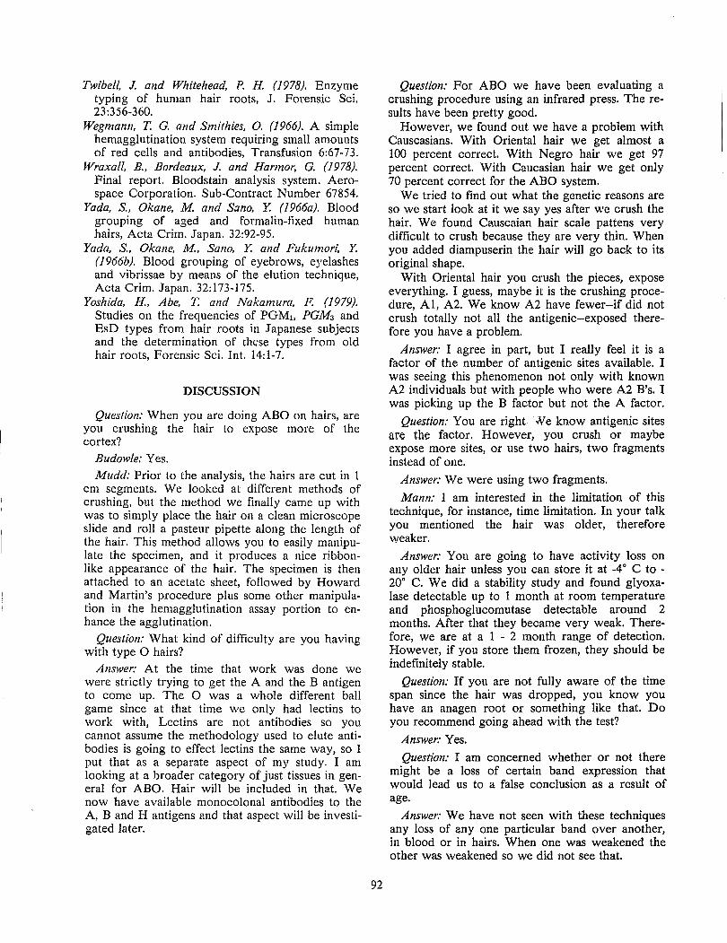
Twibell, J. and Whitehead, P. H. (1978). Enzyme typing of human hair roots, J. Forensic Sci. 23:356-360.
Wegmann, T. G. and Smithies, O. (1966). A simple hemagglutination system requiring small amounts of red cells and antibodies, Transfusion 6:67-73.
Wraxall, B., Bordeaux, J. and Harmor, G. (1978). Final report. Bloodstain analysis system. Aerospace Corporation. Sub-Contract Number 67854.
Yada, S., Okane, M and Sano, Y. (1 966a}. Blood grouping of aged and formalin-fixed human hairs, Acta Crim. Japan. 32:92-95.
Yada, S., Okane, M, San 0, Y. and Fukumon: Y. (1966b). Blood grouping of eyebrows, eyelashes and vibrissae by means of the elution technique, Acta Crim. Japan. 32:173-175.
Yoshida, H., Abe, T. and Nakamura, F. (1979). Studies on the frequencies of PGMl, PGM3 and EsD types from hair roots in Japanese subjects and the determination of these types from old hair roots, Forensic Sci. Int. 14:1-7.
DISCUSSION
Question: When you are doing ABO on hairs, are you crushing the hair to expose more of the cortex?
Budowle: Yes. Mudd: Prior to the analysis, the hairs are cut in 1
cm segments. We looked at different methods of crushing, but the method we finally came up with was to simply place the hair on a clean microscope slide and roll a pasteur pipette along the length of the hair. This method allows you to easily manipulate the specimen, and it produces a nice ribbonlike appearance of the hair. The specimen is then attached to an acetate sheet, followed by Howard and Martin's procedure plus some other manipulation in the hemagglutination assay portion to enhance the agglutination.
Question: What kind of difficulty are you having with type 0 hairs?
Answer: At the time that work was done we were strictly trying to get the A and the B antigen to come up. The 0 was a whole different ball game since at that time we only had lectins to work with, Lectins are not antibodies so you cannot assume the methodology used to elute antibodies is going to effect lectins the same way, so I put that as a separate aspect of my study. I am looking at a broader category of just tissues in general for ABO. Hair will be included in that. We now have available monocolonal antibodies to the A, Band H antigens and that aspect will be investigated later.
92
Question: For ABO we have been evaluating a crushing procedure using an infrared press. The results have been pretty good.
However, we found out we have a problem with Causcasians. With Oriental hair we get almost a 100 percent correct. With Negro hair we get 97 percent correct. With Caucasian hair we get only 70 percent correct for the ABO system.
We tried to find out what the genetic reasons are so we start look at it we say yes after we crush the hair. We found Causcaian hair scale pattens very difficult to crush because they are very thin. When you added diampuserin the hair will go back to its original shape.
With Oriental hair you crush the pieces, expose everything. I guess, maybe it is the crushing procedure, AI, A2. We know A2 have fewer--if did not crush totally not all the antigenic--exposed therefore you have a problem.
Answer: I agree in part, but I really feel it is a factor of the number of antigenic sites available. I was seeing this phenomenon not only with known A2 individuals but with people who were A2 B's. I was picking up the B factor but not the A factor.
Question: You are right -Jle know antigenic sites are the factor. However, you crush or maybe expose more sites, or use two hairs, two fragments instead of one.
Answer: We were using two fragments.
Mann: I am interested in the limitation of this technique, for instance, time limitation. In your talk you mentioned the hair was older, therefore weaker.
Answer: You are going to have activity loss on any older hair unless you can store it at _40 C to -200 C. We did a stability study and found glyoxalase detectable up to 1 month at room temperature and phosphoglucomutase detectable around 2 months. After that they became very weak. Therefore, we are at a 1 - 2 month range of detection. However, if you store them frozen, they should be indefinitely stable.
Question: If you are not fully aware of the time span since the hair was dropped, you know you have an anagen root or something like that. Do you recommend going ahead with the test?
Answer: Yes.
Question: I am concerned whether or not there might be a loss of certain band expression that would lead us to a false conclusion as a result of age.
Answer: We have not seen with these techniques any loss of anyone particular band over another, in blood or in hairs. When one was weakened the other was weakened so we did not see that.
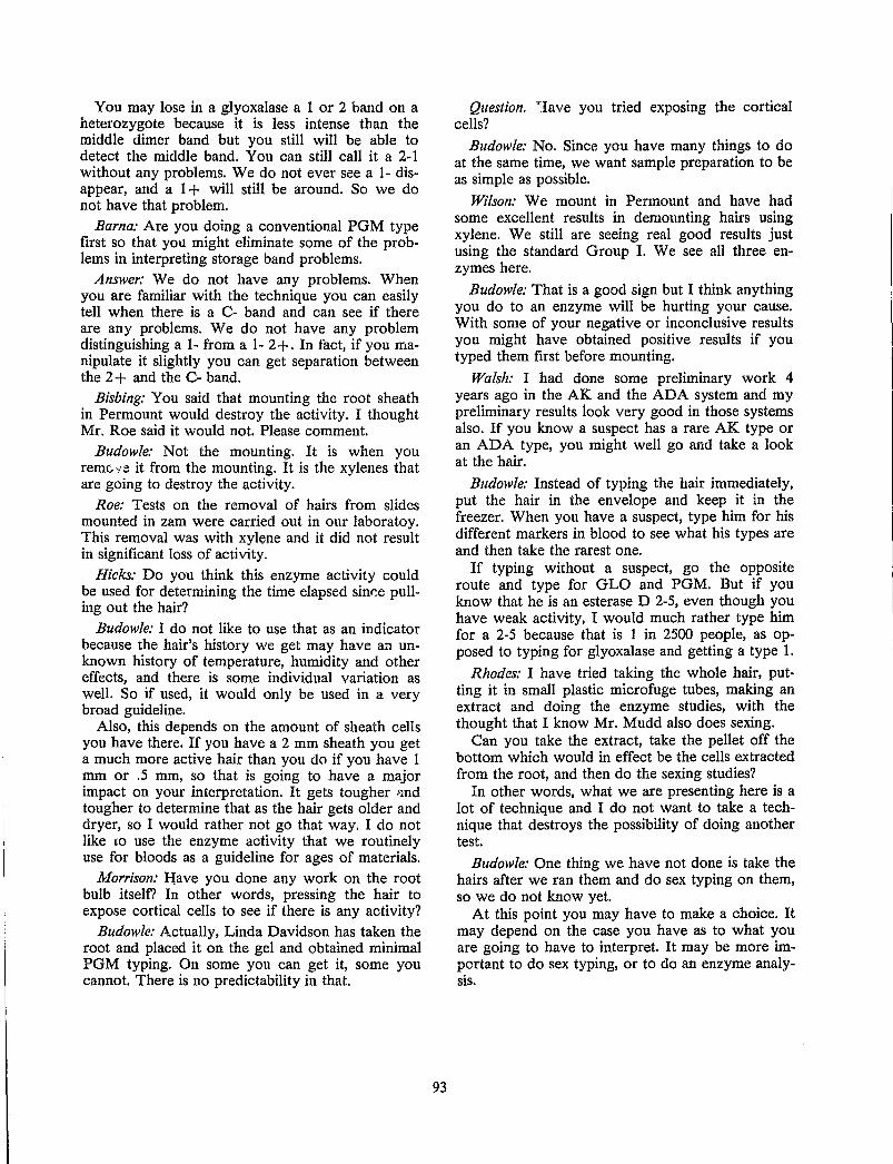
You may lose in a glyoxalase a 1 or 2 band on a heterozygote because it is less intense than the middle dimer band but you still will be able to detect the middle band. You can still call it a 2-1 without any problems. We do not ever see a 1- disappear, and a 1 + will still be around. So we do not have that problem.
Barna: Are you doing a conventional PGM type first so that YOIl might eliminate some of the problems in interpreting storage band problems.
Answer: We do not have any problems. When you are familiar with the technique you can easily tell when there is a C- band and can see if there are any problems. We do not have any problem distinguishing a 1- from a 1- 2 +. In fact, if you manipulate it slightly you can get separation between the 2 + and the C- band.
Bisbing: You said that mounting the root sheath in Permount would destroy the activity. I thought Mr. Roe said it would not. Please comment.
Budowle: Not the mounting. It is when you remG;ie it from the mounting. It is the xylenes that are going to destroy the activity.
Roe: Tests on the removal of hairs from slides mounted in zam were carried out in our laboratoy. This removal was with xylene and it did not result in significant loss of activity.
Hicks: Do you think this enzyme activity could be used for determining the time elapsed sinr:e pulling out the hair?
Budowle: I do not like to use that as an indicator because the hair's history we get may have an unknown history of temperature, humidity and other effects, and there is some individual variation as well. So if used, it would only be used in a very broad guideline.
Also, this depends on the amount of sheath cells you have there. If you have a 2 mm sheath you get a much more active hair than you do if you have 1 mm or .5 mm, so that is going to have a major impact on your interpretation. It gets tougher <'lnd tougher to determine that as the hair gets older and dryer, so I would rather not go that way. I do not like to use the enzyme activity that we routinely use for bloods as a guideline for ages of materials.
Morrison: H;ave you done any work on the root bulb itself? In other words, pressing the hair to expose cortical cells to see if there is any activity?
Budowle: Actually, Linda Davidson has taken the root and placed it on the gel and obtained minimal PGM typing. On some you can get it, some you cannot. There is no predictability in that.
93
Question. "'lave you tried exposing the cortical cells?
Budowle: No. Since you have many things to do at the same time, we want sample preparation to be as simple as possible.
Wilson: We mount in Permount and have had some excellent results in demounting hairs using xylene. We still are seeing real good results just using the standard Group I. We see all three enzymes here.
Budowle: That is a good sign but I think anything you do to an enzyme will be hurting your cause. With some of your negative or inconclusive results you might have obtained positive results if you typed them first before mounting.
Walsh: I had done some preliminary work 4 years ago in the AK and the ADA system and my preliminary results look very good in those systems also. If you know a suspect has a rare AK type or an ADA type, you might well go and take a look at the hair.
Budowle: Instead of typing the hair immediately, put the hair in the envelope and keep it in the freezer. When you have a suspect, type him for his different markers in blood to see what his types are and then take the rarest one.
If typing without a suspect, go the opposite route and type for GLO and PGM. But if you know that he is an esterase D 2-5, even though you have weak activity, I would much rather type him for a 2-5 because that is 1 in 2500 people, as opposed to typing for glyoxalase and getting a type 1.
Rhodes: I have tried taking the whole hair, putting it in small plastic microfuge tubes, making an extract and doing the enzyme studies, with the thought that I know Mr. Mudd also does sexing.
Can you take the extract, take the pellet off the bottom which would in effect be the cells extracted from the root, and then do the sexing studies?
In other words, what we are presenting here is a lot of technique and I do not want to take a technique that destroys the possibility of doing another test.
Budowle: One thing we have not done is take the hairs after we ran them and do sex typing on them, so we do not know yet.
At this point you may have to make a choice. It may depend on the case you have as to what you are going to have to interpret. It may be more important to do sex typing, or to do an enzyme analysis.
SEX DETERMINATION FR:OM HAIR James L. Mudd
Forensic Science Research Group FBI Laboratory
Quantico, Virginia
Douglas W. Deedrick
Microscopic Analysis Unit FBI Laboratory
Washington, D.C.
Presently in the forensic laboratory, the analysis of human hair is based on both a macroscopic and microscopic examination. Through comparison of various morphological characteristics, such as shape, color and pigmentation, an experienced examiner often can distinguish between the hairs of different individuals (Hicks 1977).
In violent crimes, the examiner occasionally can come across hairs which have been forcibly removed and thus may have follicular tissue attached to the root. When these specimens are encountered in the laboratory, the attached tissue can at times be subsequently analyzed for a number of genetic markers which may provide a further means of individualizing the hair (Budowle and Davidson 1986). Of the genetic markers present in the follicular cells of the hair root, the identification of the X and Y chromosomes allows the examiner to determine that a questioned hair was of male or female origin.
In man, the sex chromosomes (X and Y) comprise one of the 23 pairs of chromosomes which are present in the nuclei of somatic cells. Of the 46 different chromosomes (23 pairs) present, half are of maternal origin and half are of paternal origin. Each of the different pairs of chromosomes can be identified based upon its size, shape and staining pattern. Since the male genotype is XY and the female genotype is XX, the identification of these chromosomes in the follicular cells of the hair root can be used to determine the sexual origin of the hair.
Somatic cells from the female contain two X chromosomes. During early embryonic development, one of the two X chromosomes is randomly inactivated and appears as a chromatin mass along the perimeter of the nucleus. This inactivation, or dosage compensation, is explained by Lyon's hypothesis. Lyon's hypothesis states when there is more than one X chromosome present, all but one of the X chromosomes will be nonfunctional. This mechanism provides for a balancing of genetic information that is expressed by X-linked genes be-
95
tween males and females. A review of this topic has been given elsewhere (Stern 1973; Novitski 1977).
The Y chromosome is the male determining chromosome and is absent in females. Its size is constant between common male descendants but varies among unrelated males. This variation is due to differences in the lengths of the long arms of the chromosomes (McKenzie et al. 1972).
Table 1. APPLICATIONS OF SEX CHROMOSOME STAINING IN FORENSIC SCIENCE
Tissue Chromosome Reference Studied
Hair sheath cells Y Schmid (1967) y Ishizu (1972) y King and
Wigmore (1980)
Epithelial cells X Renard (1971) y Berghaus et al.
(1973) Blood Y Berghaus et al.
y (1973) Y Ishizu (1973)
Kringsholm et al. (1976)
Hair cortical Y Nagamori (1978) cells X Nagamori and
. Takeda (1981)
The procedures used for the identification of the X and Y chromosomes are well established. The inactivated X chromosome was first recognized by Barr and Bertram (1949), while the Y chromosome was first stained with quinacrine mustard (QM) by Caspersson et al. (1969). Through the years, various stains and techniques have been used by cytogeneticists for the identification of the sex chromosomes and these advancements have not gone unnoticed by forensic scientists. As shown in Table 1, the application of this technology to forensic science dates back more than 15 years and has been
applied to both blood and tissue as well as hairs. As seen in this table, the procedures rely almost exclusively on the presence or absence of the Y chromosome as a basis for determining the sexual origin of the tissue. In those specimens which lacked a Y chromosome, the tissue was considered to have originated from a female. However, it has been demonstrated, that in some instances the Y chromosome may not exhibit any staining with the quinacrine dye (Borgaonkar and Hollander 1970; Richards and Stewart 1978; Magenis and Dolan 1982). Therefore, a false conclusion regarding the sex of the individual may be reached in cases where somatic cells from a normal male have a weakly fluorescent or nonfluorescent Y chromosome.
To circumvent this potential problem, when determining the sex from forcibly removed hairs, an approach was taken that relies on the identification of both sex chromosomes. Since the male genotype is XY and the female genotype is XX, a technique was developed in which hair root sheath cells were stained first for the Y and then for the X chromosome using QM. This technique, which has been covered in detail elsewhere (Mudd 1984), was based on the differentiation of X and Y chromosome fluorescence at pH 3.0 and pH 5.5 respectively, after QM staining each specimen (Korf et al. 1975).
While the Y chromosome must be identified in order to conclude that the specimen had originated from a male, the absence of the Y chromosome was not sufficient in itself, to conclude that the specimen had originated from a female. In order to conclusively determine that a specimen had originated from a female, a sufficient number of inactiva.ted X chromosomes (Barr bodies) must be identi-; fied. To this end, a technique was described (Mudd 1984) which permits the X and Y chromosome to be identified in hair root sheath cells after staining with QM. Once the cells have been removed from the hair root and fixed to the microscope slide, the specimen is stained with QM. After staining, the specimen is placed in the pH 5.5 differentiation buffer and then examined for Y chromosome fluorescence. Following this examination, the slide is then placed in the pH 3.0 differentiation buffer and then examined for the presence of the fluorescent Barr body. It should be pointed out that when a hair root from a male is examined, the Y chromosome may, at times, be weakly visible in the cells when the slide is examined for the X chromatin at pH 3.0. Since the Y chromosome morphology is markedly different from that of the inactivated X, its presence does not interfere with the X chromatin count. The use of the pH 3.0 differentiation buffer causes a quenching of the background fluorescence in which the X chromatin becomes more apparent and easily recognized. The pH 3.0 buffer
96
is not suitable for use during the Y chromatin examination, as the quenching effect of this buffer would prevent the identification of those Y chromosomes exhibiting weak fluorescence.
Figure 1. Quinacrine mustard stained hair root follicular cells from a normal male showing condensed Y chromosome on periphery.
Figure 2. Quinacrine mustard stained hair root follicular cell from a normal male showing duplex form of Y chromosome.
The appearance of the X and Y chromosomes, after QM staining of follicular cells from the hair root, are shown in Figures 1 - 4. In the nuclei from the male, the Y chromosome usually appears as a single bright spot (Figure 1). However, in some cases the Y chromosome may appear in the duplex form (Figure 2). This duplex form of the Y chromosome may result from the mechanical treatment of the hair root during preparation of the follicular cell smear (Wyandt and Hecht 1973). In the nuclei
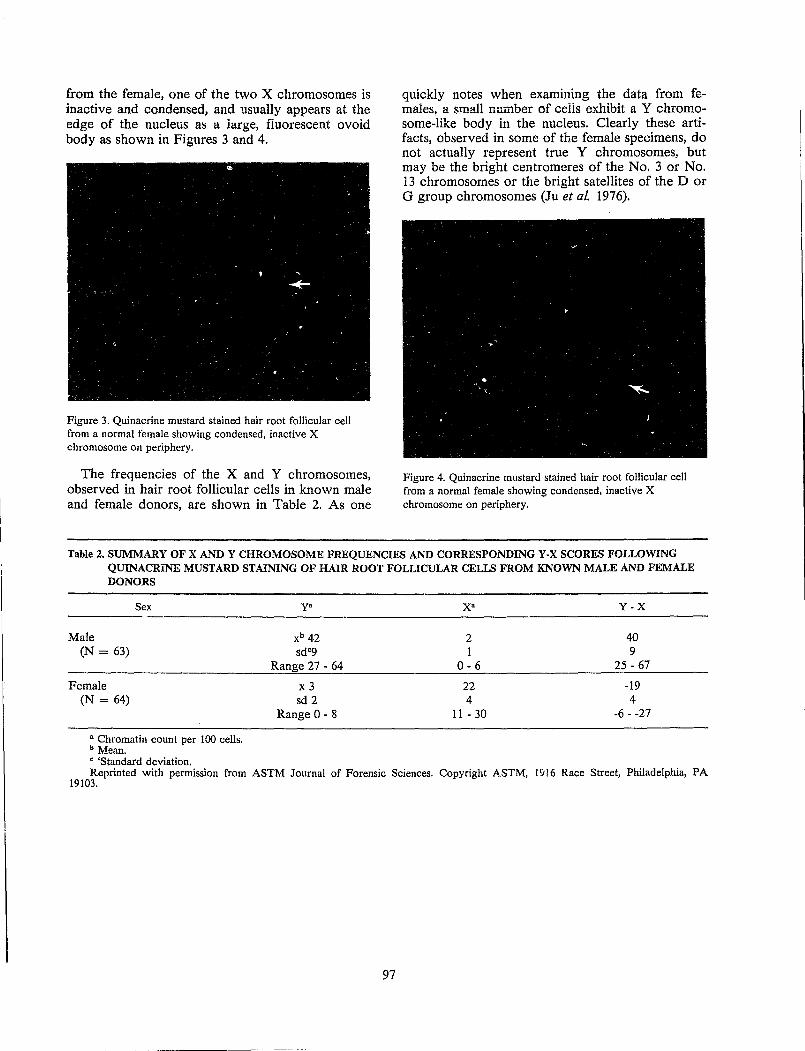
from the female, one of the two X chromosomes is inactive and condensed, and usually appears at the edge of the nucleus as a large, fluorescent ovoid body as shown in Figures 3 and 4.
Figure 3. Quinacrine mustard stained hair root follicular cell from a normal female showing condensed, inactive X chromosome on periphery.
The frequencies of the X and Y chromosomes, observed in hair root follicular cells in known male and female donors, are shown in Table 2. As one
quickly notes when exammmg the data from females. a !<mall number of ceils exhibit a Y chromosome~like body in the nucleus. Clearly these artifacts, observed in some of the female specimens, do not actually represent true Y chromosomes, but may be the bright centromeres of the No. 3 or No. 13 chromosomes or the bright satellites of the D or G group chromosomes (Ju et al. 1976).
Figure 4. Quinacrine mustard stained hair root follicular cell from a normal female showing condensed, inactive X chromosome on periphery.
Table 2. SUMMARY OF X AND Y CHROMOSOME FREQUENCIES AND CORRESPONDING Y·X SCORES FOLLOWING QUINACRINE MUSTARD STAINING OF HAIR ROOT FOLLICULAR CELLS FROM KNOWN MALE AND FEMALE DONORS
Sex
Male (N = 63)
Female (N = 64)
• Chromatin count per 100 cells. b Mean. C 'Standard deviation.
y.
xb 42 sdc9
Range 27·64
x3 sd 2
Range 0 - 8
X· Y·X
2 40 1 9
0-6 25 - 67
22 -19 4 4
11 - 30 ·6 - -27
Reprinted with permission from ASTM Journal of Forensic Sciences. Copyright ASTM, 1916 Race Street, Philadelphia, PA 19103.
97
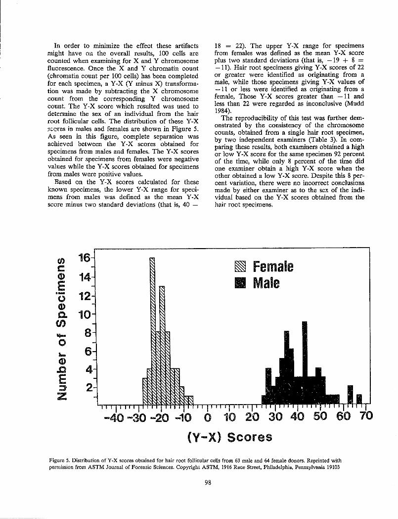
In order to mInImlZe the effect these artifacts might have on the overall results, 100 cells are counted when examining for X and Y chromosome fluorescence. Once the X and Y chromatin count (chromatin count per 100 cells) has been completed for each specimen, a Y-X (Y minus X) transformation was made by subtracting the X chromosome count from the corresponding Y chromosome count. The Y-X score which resulted was used to determine the sex of an individual from the hair root follicular cells. The distribution of these Y-X 'j,<:;ores in males and females are shown in Figure 5. As seen in this figure, complete separation was achieved between the Y-X scores obtained for specimens from males and females. The Y-X scores obtained for specimens from females were negative values while the Y-X scores obtained for specimens from males were positive values.
Based on the Y-X scores calculated for these known specimens, the lower Y-X range for specimens from males was defined as the mean Y-X score minus two standard deviations (that is, 40 -
(J) C CD E .-o Q) c. (/)
.... (l) .0 E ::J' Z
16
1
12
10
8
6
18 = 22). The upper Y-X range for specimens from females was defined as the mean Y-X score plus two standard deviations (that is, -19 + 8 = -11). Hair root specimens giving Y-X scores of 22 or greater were identified as originating from a male, while those specimens giving Y-X values of -11 or less were identified as originating from a female, Those Y-X scores greater than -11 and less than 22 were regarded as inconclusive (Mudd 1984).
The reproducibility of this test was further demonstrated by the consistency of the chromosome counts, obtained from a single hair root specimen, by two independent examiners (Table 3). In comparing these results, both examiners obtained a high or low Y-X score for the same specimen 92 percent of the time, while only 8 percent of the time did one examiner obtain a high Y-X score when the other obtained a low Y -X score. Despite this 8 percent variation, there were no incorrect conclusions made by either examiner as to the sex of the individual based on the Y-X scores obtained from the hair root specimens.
~ Female II Male
-30 .... 20 -10 0 10 20
(y'-X> Scores
Figure 5. Distribution of Y-X scores obtained for hair root follicular cells from 63 male and 64 female donors. Reprinted with permission from ASTM Journal of Forensic Sciences. Copyright ASTM, 1916 Race Street, Philadelphia, Pennsylvania 19103
98
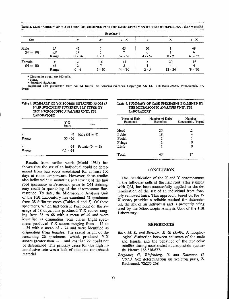
Table 3. COMPARISON OF Y-X SCORES DETERMINED FOR THE SAME SPECIMEN BY TWO INDEPENDENT EXAMINERS
Examiner I
Sex
Male xb
(N = 10) sdc
Range
Female x (N = 10) sd
Range
• Chromatin count per 100 cells, b Mean. e 'Standard deviation.
Y· X·
42 1 14 1
33 - 56 0-3
2 16 2 7
0-6 7 - 30
Y-X Y X Y-X
45 50 1 49 7 6 1 6
32 - 56 43 - 57 0-2 40 - 57
'14 4 20 '16 8 1 4 4
'4 - '30 2-5 13 - 24 '9 - '20
Reprinted with permission from ASTM Journal of Forensic Sciences. Copyright ASTM, 1916 Race Street, Philadelphia, PA 19103
Table 4. SUMMARY OF Y-X SCORES OBTAINED FROM 17 HAIR SPECIMENS SUCCESSFULLY TYPED BY THE MICROSCOPIC ANALYSIS UNIT, FBI LABORATORY
x Range
x Range
Y-x Score Sex
49 Male (N = 9) 35 - 66
-24 Female (N = 8) -15 - -34
Results from earlier work (Mudd 1984) has shown that the sex of an individual could be determined from hair roots maintained for at least 100 days at room temperature. However, these studies also indicated that mounting and storing of the hair root specimens in Permount, prior to QM staining, may result in quenching of the chromosome fluorescence. To date, the Microscopic Analysis Unit of the FBI Laboratory has examined 43 specimens from 38 different cases (Tables 4 and 5). Of these specimens, which had been in Permount on the average of 18 days, nine produced Y-X scores ranging from 35 to 66 with a mean of 49 and were identified as originating from males. Eight specimens produced Y-X scores ranging from -15 to - 34 with a mean of - 24 and were identified as originating from females. The sexual origin of the remaining 26 specimens, which produced Y-X scores greater than - 11 and less than 22, could not be determined. The primary cause for this high inconclusive rate was a lack of adequate root sheath material.
99
Table 5. SUMMARY OF CASE SPECIMENS EXAMINED BY THE MICROSCOPIC ANALYSIS UNIT, FBI LABORATORY
Types of Hair Number of Hairs Number Examined Examined Successfully Typed
Head 20 12 Pubic 18 4 Facial 2 1 Fringe 2 0 Limb 1 0
Total 43 17
CONCLUSION
The identification of the X and Y chromosomes in the follicular cells of the hair root, after staining with QM, has been successfully applied to the determination of the sex of an individual from forcibly removed hairs. This approach, based on the YX score, provides a reliable method for determining the sex of an individual and is presently being used by the Microscopic Analysis Unit of the FBI Laboratory.
REFERENCES
Barr, M L. and Bertram, E G. (1949). A morphological distinction between neurones of the male and female, and the behavior of the nucleolar satellite during accelerated nucleoprotein synthesis, Nature 166:676-677.
Berghaus, G., R iefen berg, U. and Dotzauer, G. (1973). Sex determination on skeleton parts, Z. Rechsmed. 72:255-268.
Borgaonkar, D. S. and Hollander, D. H. (1970). Quinacrine fluorescence of the human Y chromosome, Am. Hum. Genet. 22:23a.
Rudowle, B. and Davidson, L. (1986). Selecting genetic markers for analysis of forcibly removed hairs. In: Proceedings of the International Symposium on Forensic Hair Comparisons. U.S. Government Printing Office, Washington, D.C., pp. 89-93.
Caspersson, T., Zech, L., Modest, E. J., Foley, G. E., Wagh, U. and Simonsson, E. (1969). DNA binding fluorochromes for the study of the organization of the metaphase nucleus, Exp. Cell Res. 58:141-152.
Hicks, J, W. (1977). Microscopy of Hairs. A Practical Guide and Manual. U.S. Government Printing Office, Washington, D.C.
Ishizu, H. (1972). Sex identification of hairs by Ychromosome, Jpn. J. Leg. Med. 26:403-406.
Ishizu, H. (1973). Studies on sex identification of human blood and blood stains, Jpn. J. Leg. Med. 27:168-181.
Ju, K. S., Park, I 1., Jones, H. W. and Whu, K. 1. (1976). Prenatal sex determination by observation of the X-chromatin and Y-chro~latin of exfoliated amniotic fluid cells, Obstet. Gynecol, 47:287-290.
King, L. A. and Wigmore, R. (1980). Sexing of hair sheath cells using Y-chromosome fluorescence, J. Forensic Sci. Soc. 20:263-265.
Kor/, B. R., Schuh, B. E. and Sa liven, M 1. (1975). Optimum pH for nuclear sex identification using quinacrine, Clin Genet. 8:145-148.
Kringsholm B., Thomsen, 1. L., Henningsen, K. T. and Niebuhr, E. (1976). Fluorescent Y-bodies in interphase nuclei: medicolegal aspects of false negative males, Forensic Sci. 7: 171-172.
McKenzie, W. H., Hostetter" T. L. and Lubs, H. A. (1972). Y family study: heritable variation in the length of the human Y chromosome, Am. J. Hum. Genet. 4:686-693.
Magem's, E and Dolan, T. (1982). Nonfluorescent Y chromosomes. Cytologic evidence of origin, Hum. Genet. 60:133-138.
Mudd, 1. L. (1984). The determination of sex from fC'Icibly removed hairs, J. Forensic Sci. 29:1072-1080.
Nagamori, H. (1978). Sex determination from plucked hairs without epithelial root sheath, Forensic Sci. Int. 12:167-173.
Nagamori, H. and Takeda .. K. (1981). Sex determination from plucked hairs without epithelial root sheath. III. Fluorescent feulgen reaction using acriflavine, Forensic Sci. Int. 17:85-90.
Novitski, E. (1977). Sex determination. In: Human Genetics. Macmillan Publishing Co., Inc., New York, pp. 92-109.
100
Renard, S. (1971). Determination of sex of exfoliated epithelial cells and its significance in forensic science, J. Forensic Sci. Soc. 11:15-20.
Richards, B. W. and Stewart, A. (1978). XO/XY mosaicism and non-fluorescing Y chromosome in a male, Hum. Genet. 45:331-338,
Schmid, W. (1967). Sex chromatin in hair roots, Cytogenet. 6:342-349.
Stern, C. (1973). Sex determination. In: Principles of Human Genetics. 3rd ed. W. H. Freeman and Co., San Francisco, pp. 502-528.
Wyandt, H. E. and Hecht, F. (1973). Human Ychromatin. I. Dispersion and condensation, Exp. Cell Res. 81:453-461.
DISCUSSION
Ayres: How do you report your results in court in regards to any other genetic problem that an individual may have that shows up in either the bar body or the Y chromosome?
Burwitz: (Unit Chief or the Microscopic Analysis Unit). We report it as an indication of female origin or an indication of male origin.
Mudd: This is only in cases where there is a match or no match situation.
Kubic: What if you had 50 cells rather than 100 cells and your count was 35 or 37? What would you do in that case?
Mudd: Based on our protocol it would be inconclusive. I want 100 cells.
Bush: Have you had a chance to analyze any mosaics or extra Y males?
Mudd: No. Wittig: Have you always found in your different
equalization buffer treated slides a faint dying for the X body counting and very bright fluorescence for the Y body counting? When I did it in the conventional way, I often found with one specimen, where there are 50 nuclei, part of them or some of them to be very weak and others of a very bright fluorescence.
Mudd: I have noticed a variation in the fluorescence in the X and Y chromatin bodies in the nucleus and this could be accounted for by, as far as the Y chromosome is concerned, the size of the Y chromosome. Some Y chromosomes bind more than other Y chromosomes therefore they will fluoresce more that others. Some Y chromosomes will not bind any and will not fluoresce. It may be due to the way cells are mounted on the slide. If you do not have the cells sufficiently flattened you may be looking down through the cellular material at the Y chromosome. It may be at the bottom of the cell where if you have a completely flattened cell,
the Y chromatin may be more near the surface and therefore more apparent. The same thing may be true of the X.
Wittig: The whole keratin of the nucleus was very weak in a pH 5.5 buffer, and very bright in a pH 3.0 buffer.
Mudd: If you were to take a single slide and look at one cell, whether it be from a male or female, and look at it at pH 5.5, then go back and look at it at pH 3, the fluorescence of that cell will be less than what it was at pH 5.5. In some cases I have been able to go back and take hairs from a known male, examine the hair,and find one particular cell and mark it on the microscope, P'lt it at pH 3 and come back to that exact location a ad that exact cell and find that I can no longer see the Y chromosome in there because the fluorescen.ce in that cell is much weaker. There is an apparent difference.
Bailey: You could do the same analysis after looking at the Y chromosome in the pH 5.5 buffer instead of remounting in pH 3.0. You could let the pH 5.5 buffer evaporate, then rest21in with a nuclear stain. I use crystal violet acetate and look for the bar bodies. This is the method of Dr. Ishizu from Okayama, Japan. He published this in 1983 in the Japanese Journal of Legal Medicine.
Mudd: The problem there is once you have gone back and stained for the X chromosome you cannot pick up the fluorescence from the Y chromosome. It causes a quenching of the Y chromo-
101
some fluorescence. In this technique, you only stain once and put a new cover slip on with the new pH mounting buffer. You only stain once, and it allows you to go back and forth.
You can make the Y examination, put it at pH 3 and examine for the X. If you want to go back and check it again or show someone else your slide, you can go back, put it at pH 5 and reexamine it without losing anything.
When you have a differential stain like the stain that you use, you would not be able to go back and examine the Y chromosome fluorescence because it would interfere. This technique allows you, if yon have a question or somebody wants to look at your specimen, or questions your call, you can then go back and say here is the same slide at pH 5.5 and you can reexamine it many, many times. There yon limit yourself to an extent.
Wittig: Have you eVcer found a fading of fluorescence by prolongation of your investigation during excitation?
Mudd: Yes. The longer you leave the excitation light on your slide you will get a quenching or a burning effect. This is particularly true if you do not use the edge filter. If used without an edge filter, and fixed in anyone place on your slide, as soon as you move it you would notice a ring due to the quenching effect of the excitation light. To avoid that, place a cover in the light path as soon as you have finished your examination.
TRACE ELEMENT ANALYSIS: A REVIEW OF FORENSIC NEUTRON ACTIVATION ANALYSIS OF HUMAN HAIR
Norman E. Erickson
Centre of Forensic Sciences Toronto, Ontario, Canada
There are presently several methods available to determine the elemental composition of human scalp hair. These include atomic absorption spectrometry (Schroeder and Nason 1969), proton induced X-ray spectroscopy (PIXE) (Valkovic 1973), photo-induced X-ray fluorescence (XRF) (Toribara et al. 1982) and neutron activation analysis (NAA) (Jervis 1956) - probably the most widely publicized forensic technique for this application. Approximately 25 years ago the Journal of the Forensic Science Society in an editorial (1962) hailed, A Breakthrough in Forensic Science and said in part, "the results from a Toronto group applying NAA may herald the start of a new chapter in the history of forensic identification methods." These statements were based upon the initial studies of two university researchers, one of whom later earned a Ph.D. based in part on his thesis Hair Individualization Study By Neutron Activation (Perkons 1965). Subsequent papers by these authors and others including, Individualization By Neutron Activation Analysis (Perkons and Jervis 1967) describing preliminary studies on forensic materials such as drugs, paint, glass, etc., further promoted the technique. The use of NAA and the treatment of the data by these early workers became a subject of controversy because of their lack of appreciation of the examination of forensic materials, particularly hair. Their treatment of the data used in hair matching was criticized for lacking proper statistical methods (Kuis, personal communication). It was not until practicing forensic scientists began to question the validity of NAA for hair comparison that there was genuine concern. Unfortunately, this premature optimism that trace element analysis was the panacea for hair characterization resulted in the true value of NAA in other potential areas of forensic sciences, not being appreciated.
The principles of NAA were established by Heversy and Levy in 1936 in Denmark (Perkons 1965). However, it was not until after the Second W orId War that reliable neutron sources became available to routinely apply the technique to both qualitative identification and quantitative determination of elements making up a sample. This is accomplished by irradiating a sample in a nuclear reactor with thermal neutrons over a controlled period of time resulting in some of the atoms of the elements making up the sample being made radioactive. There are several factors which determine the amount of radioactivity an element will
103
produce. Since these radionuc1ides are unstable they tend to undergo some type of nuclear transformation (de-excitation of a compound nucleus) to return to a stable form. This process is called decay and is characterized by the emission of various forms of radiation, namely, alpha, beta and gamma rays. In NAA, gamma rays are selectively "analyzed" for their energies, intensities and decay halflives by a detector coupled to a multi-channel analyzer. Various standards irradiated at the same time can be used for the quantitative determination of the elements found within the sample.
The major advantages claimed for the technique are that it is a non-destructive technique allowing for multi-elemental analysis with exceptional sensitivity (nanogram and picrogram levels) and specificity for most elements within minute samples. Neutron activation analysis has proven to be an accurate and reliable method for elemental analysis, and had been used for many purposes within this laboratory for over 20 years. It continues to be used extensively for many forensic materials by the FBI Laboratory.
In excess of 50 elements have been identified in human scalp hair in concentrations ranging from 0.1 to 100 ug/g. The major elements, C, H, 0, N, S and P which comprise over 99 percent of the mass, are not detected by this particular procedure. It was the minor and trace elements such as Cu, Na, Zn, AI, Au, etc., that were of interest to the early workers involved in hair characterization. These elements are incorporated into the hair either internally from blood within the hair follicle during the keratinization process or by external contamination from atmospheric fallout, cosmetic preparations, dust, sweat, etc.
The use of elemental analysis on hair specimens was first introduced into a Canadian Court during a murder trial in 1958. Using beta-absorption analysis of irradiated hair samples, the P-32/S-35 betacount ratios of the suspect' fl. hair and a single strand taken from the deceased's hand matched (Per-kons 1965). It was not until the early 1960s that a large scale study on several hundred scalp hairs weighing between 2 mg and 15 mg (several hairs) was carried out (Perkons 1965). These studies of bundles of hair showing variation both in composition and concentration led these researchers to believe this would be a unique technique for hair individualization and could be applied to criminal investigations. Unfortunately, it is seldom in criminal
cases where hairs are shed that several separate hairs can be assumed to be from a single source. In practice, when a number of hairs are recovered from an item, each questioned hair must be considered as a single entity. It is scientifically fallacious to put several separately found hairs which appear macroscopically and microscopically similar together and then perform an additional test for the trace element composition as if these hairs came from one individual. Even if the assumption that they originally came from the same head is correct, their individual histor:es, treatment and exposure to contamination may be quite different after leaving the head.
In 1963, the present author conducted a small study on single hair analysis using NAA and concluded that several problems existed which seriously questioned the use of the technique on a routine basis for single hairs. At the 1965 meeting of the Criminal Congress in Montreal a paper, Present Status of NAA in Forensic Sciences was presented which outlined these initial findings on single hair analysis and some of the concerns about the technique (Erickson 1965). Perkons acknowledged this work in his thesis stating that his conclusions on bundles of hair should not be extended uncritically to single hair strands. The results of this small study on single hairs showed that unlike bundles of hairs in which an excess of 20 elements were claimed to have been detected, there were on average only 6 elements, namely, Na, Br, Cu, Mn, Zn and Au, detected in single hairs. Sodium and bromine accounted for much of the radioactivity and their presence masked the other elements of interest. Thus it became necessary to remove these two elements by using a post-irradiation wash and analyzing both hair and wash separately. Other elements sometimes found were Hg, Cr, As, Sb, Se and Sr. There were often wide variations in concentrations of these elements in hairs from the same individual.
There was also a fundamental statistical concern about the number of single hairs needed to constitute a representative sample to show the variation in trace element content in hairs from the same head. The reactor container held approximately 15 single hair strands for one irradiation. Since there can be from 100,000 to 150,000 hairs on an individual's scalp, even the analysis of 100 hairs represents only approximately 0.1 percent of the total population. There were also difficultie<; in bundling single hairs in the pre-irradiation loading of the samples and in post-irradiation unpacking. The lengthy irradiation times necessary for single hairs made the hairs brittle and difficult to handle. Similar experiences on post-irradiation damage of single hairs was reported by Forslev (1966) in his critical review of the technique for hair comparison. Papers by Cornelius (1973; 1979a, b) give compre-
104
hensive reviews of the literature and the use of NAA in the various disciplines of criminalistics and were very critical of its use for hair comparisons. Coleman and Cripps (1973) do not support the position of Cornelius and stated the technique can be useful if applied within limits of background knowledge. Pillay and Kuis (1978) also do not share the views of Cornelius but report because of statistical uncertainty a specialist in NAA could not individualize hairs based solely upon elemental analysis.
Some of the difficulties associat~ci with hair characterization using NAA are as follows:
1. Logistics. To study short-lived and immediatelived radionuclides it is necessary to be near or on site where the nuclear reactor is located. To complete a total analysis on all elements would require a few weeks.
2. Possible contamination. Because NAA is an extremely sensitive technique it was felt that cleansing of hair samples would be required to remove any contamination such as dust, sweat, soil, blood, etc. However, the work of Maes and Pate (1976; 1977a, b) showed the absorption of metallic ions into hair. Elements can also be leached out of hair by the washing process. Conversely, it was argued that external contamination could be a valuable attribute in the comparison, and both hair and contamination should be studied. The aspect of cleansing of hair by various methods was covered in the work of Bate and Dyer (1966). These authors recommended a washing procedure of the hair and advised that the washing agent also be analyzed. Kuis (personal communication) stated that the hair's surface contamination can be of critical importance in cases of unusual environmental contamination; however, these elemental values appear to be statistically random. Thus hair wash values are unsuitable for this type of statistical analysis. Another consideration with respect to contamination lies in the process of hair examinations within the forensic laboratory. Hairs are normally prepared for microscopical examination using a mounting medium. The trace elements, if any, present in the mounting medium and their absorption into the hair must be considered.
3. Concentration variation along the hair shaft and growth cycle. Much has been written about the elemental concentration changes that occur along the length of the hair shaft. Renshaw et at. (1973); Obrusnik et al. (1973); Jolly et al. (1971). In one study, Maes and Pate (1976), there was a twenty-fold increase in the concentration of most of the trace elements from the root end to the distal end of the hair. Kerr (1961) and Lima et al. (1961) studied the influence of growth cycle in trace element variation. To be relevant the comparison hair sample lengths and growth phase would
have to be the same as that of the questioned hair(s) if one contemplated using NAA for hair comparison.
4. Time Factor. With the passage of time there will be changes in the trace element composition of an individual's haIr. Coleman's work (1966) showed that NAA in hair comparisons could be useful provided proper statistical procedures were used, but in his studies he introduced another factor encountered in criminal matters which imposes limitations on the method. If a suspect does not provide a hair specimen shortly after a criminal offense where questioned hairs have been found, then the elemental profile of that suspect's hair may change with time. Coleman (1966) indicated that a time lapse of a few weeks between the offense and the collection of the suspect's comparison sample would not affect the composition. Alteration of the trace element composition of hair by various external applications would make the analysis totally useless.
5. Concentration values and precision. Although Perkons reported an excess of 20 elements could be detected in bundles of hair and used in hair matching, other authors reported reservations regarding his concentration values and the precision with which these elements could be measured. Valkovic (1977) stated his doubts about the absolute values for elemental concentrations reported by Perkons. The values were much higher than those reported by several other research groups. Pillay and Kuis (1978) in their studies found 22 elements were detectable but only 10 of them could be determined with good precision at the concentrations normally found.
The work of Dybcznski and Boboli (1976) on single strands of hair showed the presence of more than ten elements. They were able to demonstrate in 'simulated' cases elimination of most or nearly all of the suspects using a new statistical criterion for elimination/identification. This study was based on clinical samples and did not take into account possible contamination or the time factor.
Because of these potential sources of error and the limitations of the technique this laboratory stopped using NAA for hair comparisons. The method was last used by this laboratory in 1977 in a homicide case where human scalp hairs were found at the crime scene. Two individuals, one of whom was a suspect and the other a resident at the crime scene location, provided head hair specimens shortly after the offense. Microscopical examinations could not satisfactorily discriminate the hairs of these two individuals; therefore, elemental analysis was performed. After analyzing numerous single hair strands, along with subsequent additional control samples, it was shown that one element was found in the suspect's hairs which could not be detected in the resident's hairs. There was also differ-
105
entiation with respect to the concentrations of two other elements. Analysis of the questioned hairs found at the crime scene indicated they were similar to those of the resident.
The present author is not aware of any forensic laboratory presently using NAA for hair. Perkons still Ilses the technique occasionally in his consultant practice. Other individual consultants may also use it.
It was unfortunate that premature optimism accompanied the use of this technique as a practical identification tool before detailed studies were carried out. As stated by Pillay and Kuis (1978), "the ultimate goal is individualization of single hair and the development of reliable trace element profiles of single hair strands weighing less than a few milligrams still offers considerable challenge but because of several limitations, NAA is not suitable for routine evidence analysis at present."
Being aware of these limitations and the background knowledge required in the use of this technique, this author is of the view NAA can be applicable on a very selective basis in forensic hair investigations.
REFERENCES
Bate, L. C. and Dyer, F. F. (1965). Trace elements in human hair, Nucleonics 23:74-81.
Coleman, R. F. (1966). The application of neutron activation analysis to forensic science, Forensic Sci. Soc. J. 6:19-27.
Coleman, R. F. and Cripps, F. H. (1973). Letters to the editor, Med. Sci. Law 13:224.
Cornelius, R. (1973). Neutron activation analysis of hair. Failure of a mission, J. Radioanal. Chern. 15:305-316.
Cornelius, R. (1979a). Hair: the neutron activation analysis story. Home Office Central Research Establishment Symposium.
Cornelius, R. (1979b). Truth has many facets: the neutron activation analysis story, J. Forensic Sci. Soc. 20:93-98.
Dybczynski, R. and Boboli, K. (1976). Forensic and environmental aspects of neutron activation analysis of single hairs, J. Radioanal. Chem. 31:267-289.
Erickson, N. E. (1965). Present status of N.A.A. in forensic science. Presented at the Criminal Con·· gress Meeting, Montreal.
Forslev, A. W. (1966). "Non-destructive" neutron activation analysis of hair, J. Forensic Sci. 11:217-232.
Jervis, R. E. (1956). Presented at the 4th Annual Meeting of the Forensic Society of Canada.
Jolly, R. K., Pehrson, G. R., Gupta, S. K., Buckle, D. C. and Aceto, H. (1974). Linear scans of hair
strands for trace elements by proton induced Xray emission. In: Proceedings of the Third Conference on Applications of Small Accelerators CONF-741040. pp. 203-208.
Kerr, P. M. (1964). A study of the influence of the hair cycle on the trace element content of human hair by means of neutron activation analysis. Master's thesis, University of Ottawa.
Li~a, F. w., Shibata, H. and Atalle, L. T. (1964). Activation analysis applied to forensic investigations, IAEA Radiochem. Methods of Analysis SM 55/144, Salzburg, 199.
Maes, D. and Pate, B. D. (1976). The spatial distribution of copper in individual human hairs, J. Forensic Sci. 21:127-149.
Maes, D. and Pate, B. D. (1977a). The absorption of arsenic into single human head hairs, J. Forensic Sci. 22:89-94.
Maes, D. and Pate, B. D. (J977b). The spatial distribution of zinc and cobalt in single human head hairs, J. Forensic Sci. 22:75-78.
Obrusnik, L, Gigiason, L, Maes, D., McMillan, D. K., D'Avria, J. and Pate, B. D. (.1973). The variation of trace element concentrations in single human head hairs, J. Radioanal. Chern. 15:115-134.
Perkons, A. K. (1965). Hair individualization study by neutron activation. Dissertation. University of Toronto.
Perkons, A. K. and Jervis, R. E. (1967). Individualization by neutron activation analysis, Law Enf. Sci. Tech. 257-265. ,
Pillay, K. K. S. and Kuis, R. L., (1978). The potentials and limitations of using neutron activation analysis data on human hair as a forensic evidence, J. Radioanal. Chern. 43:461-478.
Renshaw, G. D., Pounds, C. A. and Pearson, E. P., (1973). Determination of lead and copper in hair by non-flame atomic absorption spectrometry, J. Forensic Sci. 18:143-151.
Schroeder, H. A. and Nason, A. P. (1969). Trace metals in human hair,]. Invest. Dermatol. 53:71-78.
Toribara, T. y., Jackson, D. A., French, W. R., Thompson, A. C. and Jakievic, J. M. (1982). Nondestructive X-ray fluorescence spectrometry for determination of trace elements along a single strand of hair, Anal. Chem. 51: 1844-1849.
106
Valkovic, V. (1973). X-ray emISSIOn spectroscopy, Contemp. Phys. 14:415-439.
Valkovic, V. (1977). Trace elements in human hair. Garland STPM Press.
DISCUSSION
Fallon: In regards to the cleaning procedures which remove the debris off the hair before the analysis, would you feel confident if you had a hair that was deposited in a sewer for as long as a week that it could be cleaned and then analyzed?
Erickson: No. That is what I say with respect to the cleaning procedure even in clinical samples that were obtained. That area alone raised great controversy because you have to appreciate that hair is a good ion exchange; the,refore, the medium you wash the hair in has to be free of trace contaminants. Perkons and Jervis were using distilled water and acetone. A paper by Bates and Lyons written in the mid-1960s went to a shampooing type of treatment which was free of any trace contaminants. So that area alone even without the problems of samples in sewers or in water raised a concern.
Question: Do you know of anyone other than Dr. Randall at Texas A & M doing neutron activation?
Erickson: I encountered Dr. Randall in one case. I questioned the basis of him using three hairs from an individual as representative of the trace element composition on that individual's head. I was quoted a statistic that the trace element concentrates he found on this individual's hair from three hairs analyzed exceeded the population of the earth. I testified on behalf of the defense in that particular case.
Question: In the case of atomic absorption the hair has to be in solution, whereas in NAA it is not required. Could your suggest some suitable technique for atomic absorption work?
Erickson: No. I would only use it as a technique that one could use if they were looking for a particular element in a hair sample such as mercury, or arsenic, but not on a forensic comparison basis. A case of possible toxicity could use this or other techniques but from a forensic comparative basis, no.
COMMITTEE ON FORENSIC HAIR COMPARISON· SUBCOMMITTEE 4 • REPORT WRITING, CONCLUSIONS AND COURT TESTIMONY
CHAIRMAN
Norman E. Erickson
Centre of Forensic Sciences Toronto, Ontario, Canada
COMMITTEE MEMBERS
Paul Krupenie
Law Enforcement Standards Laboratory National Bureau of Standards
Gaithersburg, Maryland
David Metzger
Illinois Department of Law Enforcement Joliet, Illinois
Nicholas Petraco
New York Police Laboratory New York, New York
Mary Ann Strauss
Minnesota Bureau of Criminal Apprehension St. Paul, Minnesota
Russell Stockdale
Home Office Central Research Establishment Reading, Berkshire, England
The mandate given this Subcommittee was to provide guidelines with respect to topics under the following four categories:
directly to the prosecutor on a Statement of Witness Form and are filed with the court; in Australia and some American states the report goes solely to the investigator who in turn decides whether it should be introduced into court. In other countries the report goes to both the prosecuting attorney and the investigating officer. Another consideration is the institutions policy on report format and conclusions. Should this organization provide training to outside agencies the report writing of these agencies may be affected. For example, in the United States, the FBI has trained many state laboratory personnel in hair examinations. From replies to the survey, several laboratories indicated they have adopted the criteria for report writing set down by the FBI. Some laboratories which have several hair examiners indicated that wording of reports may vary slightly from person to person depending on how the analyst feels about the value of hair evidence and the particular case he has just
1. Report format and conclusions; 2. Court testimony; 3. The significance of hair comparison evidence;
and 4. The use of probabilities.
REPORT FORMAT AND CONCLUSIONS
A study of some 70 ldboratory reports on forensic hair comparisons received from Australia, Canada, Denmark, South America, the United Kingdom, the United States of America and West Germany showed a wide variation in both report format and wording of conclusions. One reason for this variation may be the fact that the chief recipient of the report differed depending upon regions. For example, in the United Kingdom all reports go
107
analyzed. These and other factors all contribute to variation noted in report writing.
The purpol'e of any forensic report is to convey to the reader information which is comprehensible, logical and technically accurate. In principle, if two competent examiners from different laboratories examine the same material using state of the art techniques and each has a thorough knowledge and understanding of the subject matter, both should, upon the interpretation of their results, issue a report containing the same opinion. Variation in report conclusions suggest this is not the case. It should be noted that reports may be read by other scientists retained as counter experts.
It is not the intention of this Subcommittee to design a standardized universal report format; however, it is our view that all forensic reports should include basic information with respect to:
1. Items that were examined; 2. General examinations conducted; 3. Results of the examinations; 4. Conclusions made from the interpretation of
the results; 5. Qualifying statements to strengthen or weaken
the conclusions; and 6. Requests for additional standard samples.
Items Examined
The report should contain information which specifically identifies what items were received and examined, and should include:
1. Submitting agency and their me number; 2. Principals involved by name in terms of de-
ceased, complainant, accused and suspect; 3. Type of occurrence; 4. Laboratory case number; 5. Date report was typed; 6. Relevant continuity information; and 7. Brief description of items.
Examinations
The extent of all testing procedures conducted should be indicated in general terms. Was only a gross examination required to formulate an opinion or was it necessary to do a lengthy microscopical examination? If additional analyses were made, such as the determination of genetic markers, scanning electron microscopy, trace element analyses, etc., these should also be indicated.
Results of Examination
The analytical results should be stated in a factual manner using the most positive terms possible and should be separate from any conclusion. They may be listed separately for each item or similar results for various items may be grouped together. There should not be a detailed description in the report of all the hair features such as length, color, thickness, microscopical characteristics, etc. These
108
should be confined to the working notes unless this information may be useful for investigative purposes.
Examples of Results
1. Animal hair identified, no further examinations were done.
2. Four brown, Caucasian scalp hairs were found. These four hairs exhibited similar microscopical characteristics to the known sample.
There will be occasions, because of various factors involving either the questioned hair or the known sample, when the results will be uninterpretable and therefore the examiner will not be able to formulate an opinion.
Conclusions
For the reader to understand the significance of the results, it is necessary for the examiner to render a conclusion. Some cases, for various reasons permit no conclusions and only the results are reported. From the reports received, with conclusions, there was a diversity of opinion with respect to these conclusions. These ranged from a single sentence statement to nine possible conclusions and the criteria for making these conclusions. Meaningful conclusions can only be made on the basis of several factors such as the adequacy in quantity and quality of the comparison sample, the individual characteristics of each hair and most importantly, the examiner's personal experience. This experience can only be gained by proper training including numerous blind test exercises on various hair specimens followed by a continuous application of hair examination procedures through casework.
It is this Subcommittee's view that there are several basic conclusions a hair examiner could reach.
1. The questioned hair is consistent with having come from John Doe.
This conclusion must be based upon a strong association between the questioned hair and the known sample. There are several factors, such as the questioned hairs having intrasample variation which is found to be microscopically similar to the comparison sample, or the presence of unusual hair characteristics or hair treatment such as dying, bleaching, etc., which strengthen the association. Other tests such as sex determination or enzyme typing may provide additional support for this conclusion.
2. The questioned hair could have come from John Doe.
When only a limited association can be made between the questioned hair and the comparison sample, this conclusion would apply. Some of the factors which influence arriving at this conclusion
are the presence of hair fragments, or the lack of any distinctive features in the questioned hair, for exmple, white hair.
3. John Doe qualifies as being the donor of the questioned hair.
This statement indicates to the reader that John Doe cannot be eliminated as a possible source of the questioned hair. It does not take into account whether there is a strong or limited association.
4. The questioned hairs could not have originated from John Doe.
This conclusion is based upon finding significant macroscopic and/or microscopic dissimilarities between the questioned hair and the comparison sample. The examiner must ensure that several factors are satisfied before this statement can be made, for example, the possibility that the hairs are atypical is remote, the known sample is adequate and representative with little intrasample variation.
5. The questioned hair is not consistent with having come from John Doe.
This conclusion applies when there are dissimilarities between the questioned hair and the comparison sample; however, there are factors present which do not allow the examiner to conclude categorically that John Doe could not be the donor of the questioned hair, for example, inadequate comparison samples, incomplete questioned hairs, a lengthy time lapse between the offense and the collection of the comparison sample.
6. No conclusion as to origin could be reached.
As stated earlier, there are cases in which no conclusion can be reached. One might find both similarities and differences between the questioned hair and the comparison sample or the questioned hair may be very minute. In these situations, the results are uninterpretable and no conclusion can be made as to whether the questioned hair could have originated from a specified source.
Qualifying Statements
Qualifying statements should strengthen or weaken the conclusion. Factors such as unusual characteristics or dyeing pattern which would strengthen the opinion should be indicated. Conversely, if there are factors which tend to weaken the conclusion, that is, common featureless hairs, then this should also be stated. These qualifying statements will vary depending upon each specific case.
Additional Samples
It may be necessary to request additional samples when inadequate known hair samples have been
109
submitted, or when elimination samples are required.
COURT TESTIMONY
The report should be a guide for the prosecutor or defense attorney enabling him/her to put into evidence the results of the scientific examinations and their significance. The prosecutors role is to maximize the impact of this information on the jury; the opposing attorney's position is to minimize these findings, and in some instances, to impeach the witness' credibility as an expert.
The most important precondition for an expert witness to undergo a rigorous cross-examination is preparation. The examiner should be thoroughly familiar with the examinations and findings of the case, and how he wants to present them. Prior to giving evidence it is useful to have a conference with the prosecutor and possibly the defense. This pretrial discussion should be an educational exercise for counsel. It is also an opportunity for them to review the evidence required and the way it should be presented. They may agree that only the report is required for court. It may cut down the number of items wanted for trial. A meeting with the defense may reduce the amount of time in cross-examination and hopefully show the objectivity the witness has in the hearing. The subjective nature of hair examination and how the conclusions are supported should be discussed. One must exercise extreme caution during these meetings, particularly with opposing counsel, because anything stated can be repeated in the witness box; nothing should be assumed to be off the record.
Prior to giving evidence you must firstly be qualified by the court as an expert in the microscopic examination and comparison of hairs. This is done by the prosecutor asking a series of relevant questions relating to your occupation, educational background training within the field and any further specialized training. Other areas relating to qualifications are how long and what percent of your time is devoted to these types of examinations, papers written and lastly, have you given evidence before, at what level and how often.
The plight of the expert has been described by Wily and Stall worthy (1962).
He has to answer when asked and stop when told; he cannot give his evidence as he chooses, but must confine himself to the questions asked. He can be interrupted at any moment and ridiculed, reprimanded or contradicted. High qualifications may not prevent him cutting a hopeless figure in the witness box, poise and sense of what is called for in a witness may make him impressive though his qualifications be mediocre. If he is ill-informed upon fact and hasty in opinions he will seldom escape discomfort.

And maybe no more so than when giving evidence on hair comparisons.
Despite these formidable obstacles the key to expert testimony is the ability to effectively communicate scientific terms and principles to a jury. Tanton (1981a) stated, "Good communication is essential for the forensic scientist who wishes to deliver a quality product to the court." A second paper by Tanton (1981b) outlined a procedure of improving the means of communication. Failure to communicate ideas and concepts in terms understandable to the court can only lead to misinterpretation and misconceptions.
Once qualified, the witness should be asked by the prosecutor to present an overview of hair ex· amination. Some of the questions to bc covered include:
1. Why and how is a hair examination conducted?
2. What can be determined from a hair examination?
3. What cannot be determined from a hair examination?
4. What characteristics, macroscopically and microscopically are observed?
5. What conclusions can be made regarding hair comparison?
The use of demonstrative evidence such as charts or photographs showing general hair characteristics or using a pencil to describe hair morphology is helpful to the court in understanding hair structure. However the use of photomicrograph!'. showing side by side areas of similarities or dissimilarities under the comparison microscope should be discouraged. To depict areas of side by side comparisons showing only a very minute fraction of the total hair can be very prejudicial to the lay person. The exception might be to show some unusual characteristic(s) which could better be described through the use of photographs.
, THE SIGNIFICANCE OF HAIR
EXAMINATION
The consensus from all reports received was that microscopical examination of hairs, unlike fmgerprints, did not permit a positive identification to be made, except in a few rare instances. An examination of similarity of structure, color, pigmentation and other characteristics can be strong probative value in the majority of cases when considered along with other evidence. The use of additional techniques such as root sheath examination for genetic markers and sex determination, etc., will greatly improve the evidentiary value of hair examination.
110
THE USE OF PROBABILITIES
When a questioned hair exhibits similar macroscopic and microscopic characteristics to the known comparison sample, two possibilities exist as to the sO'J:ce of the questioned hair. It has either originated from the donor of the known sample or there is a coincidental match. We are aware two individuals may have matching hair characteristics and the two hair samples cannot be differentiated by present day methodology, hence the chance of a coincidental match can occur.
The probability data from the published research of Gaudette, coauthored by a statistician and verified by other statisticians, has shown when a positive hair comparison has been made by a qualified hair examiner the chances of coincidental matches are relatively rare. The hair could match that of another individual but it is much more likely to have originated from the same source to which it was compared. Gaudette's probability data on positive hair comparisons on Caucasian scalp hairs can be stated in several ways such as an estimate of the average odds against that one questioned hair having originated from another specific Caucasian individual would be about 4500 to 1 (800 to 1 for pubic hairs).
All hair examiners should be aware of the several published papers of Gaudette on this topic. On this topic this Subcommittee recommends:
1. The results of the Gaudette studies should not be introduced into court unless asked directly about his published research.
2. It will be left to laboratory policy or the individual examiner whether he can personnaly substantiate and defend in court the findings from these studies.
3. Further research by other examiners is required in this very important area.
I personally wish to give thanks to the members of this Subcommittee who assisted me with this report.
REFERENCES
Tanton, R. L. (1 981a}. Court interpretation of forensic science phraseology. Presented at the 33rd Annual American Academy of Forensic Sciences Meeting, Los Angeles.
Tanton, R. L. (1 981b}. Courtroom presentation of scientific testimony. Presented at thl': 33rd Annual American Academy of Forensic Sciences Meeting, Los Angeles.
Wily, H. J. and Stal/worthy, K. R. (1962). Mental Abnormality & the Law. Peryer, Christchurch, p.446.
DISCUSSION
Bisbing: Did the Subcommittee consider as one of your conclusions instead of the two for the negative conclusions of being, cannot be shown to be consistent or dissimilar, the statement that the hairs cannot be associated with that person, meaning you do not know if they came from that person or not but cannot be matched with them?
Erickson: No we did not consider that. This is the first draft proposal, and if there are any critiques or suggestions as to how this report might be better finalized I would solicit any cooperation from any members of the audience on this particular topic.
Gary: In your conclusion you said the hair could have come from the source, one of the qualifications for that was that this hair was non-Caucasian. I do not understand why you said that.
Erickson: Maybe it is only from a Canadian persepective and maybe we have not looked at any non-Caucasian or other races, but Mongoloid and Negroid hairs by and large did not allow for that distinction to be made. A number of examiners felt that on the basis of examining these types of hairs microscopically, because of microscopical properties of these hairs this was probably the most common conclusion one could reach.
I acknowledge what your point is. In some situations one could put a stronger degree of certainty or possibility on these types of examinations, but I looked at it in terms of the proponents of examining these types of hairs would form in that particular category.
Barna: In regards to qualifying statements, you had commented these could be optional and you had further commented that if a hair is featureless it should also be noted. I am interested in how you identify featureless hairs as far as definition is concerned, and how you might possibly work a qualifying statement to a featureless hair.
Erickson: It is a misnomer when we call it a "featureless" hair. What is referred to are hairs that have very few features, for example, blond, Caucasian hairs or white hairs. They have very few individualistic characteristics. There are very few properties in that particular type of hair.
Question: My main concern is how you would work your qualifying statement to the featureless hair as to the value of that hair.
Erickson: I do not know that I would make a qualifying statement in respect to featureless hairs. The use of a qualifying statement was that if one were to strengthen the opinion that one should also, should not charge into court and because we have a good hair comparison use it to the hilt. By the same token if we have ones that are what we
111
feel are rather common type of hair comparisons then we should not specify something with respect to that. I myself do not use qualifying statements, but again it can be taken under consideration as to how one would best word that type of qualifying statement.
Cwiklik: I am a member of the Committee on Forensic Hair Comparison. I recall during the general session there was considerable disagreement among hair examiners as to whether that opinion about Negroid hairs Wf;S true.
Many of the members of the Conclusions, Report Writing and Court Testimony Subcommittee felt one could not reach a strong conclusion with Negroid hair. I do not thing that was the consensus of the Committee. I think it should be a matter for discussion and for further work as opposed to a statement that it should be so.
Erickson: This is a first draft. I am sure there arc many criticisms of it. I am prepared to review this with the Committee and if the consensus is, particularly with those hair examiners who in fact look at Negroid hairs, that maybe there was not enough representation on the Committee in terms of hair examiners who look at hairs other than Caucasian hairs.
Suh: We are seeing a great move toward including a large narrative explanation of the results on the reports. Was there a feeling or a consensus on the Subcommittee as to whether or not one should include a narrative explanation with regards to hair statements - putting your statements up front and then explain exactly what category your results are going under?
In other words, you have given us a whole range of examples and what they would imply, but was there a feeling a:s to whether or not one should put those inferences or implications in the report itself.
Erickson: It was the belief of the Subcommittee that the report really acts as a guide to the prosecutor. There were differences of opinion that were given in the examples we received as to what should actually go into the report.
You stand or fall when you get on the witness stand with respect to your report. I believe one should stay away from the large descriptive narrative of the hair examination.
Metzger: I am on the Terminology Definition and Standardization Subcommittee. We recognize a very wide variety of styles ill report writing and it was not the intent of the Subcommittee to dictate the style of report to write. We comprised a list of particular general topics all reports should cover, and left the actual style of the report and whether people wanted to write short forms or narratives
up to the individual and the policy of the laboratory.
Brown: What is the feeling of the Subcommittee in regards to using charts that show cross transfer of hairs between the victim's clothes and the suspect and the suspect's clothes?
Erickson: The use of demonstrative evidence such as charts or photographs should be used to best depict or convey what you are trying to state. The Subcommittee is strongly opposed to showing side by side photomicrographs of actual similarities or dissimilarities unless under very special circumstances, particularly in hair examination. Not so much with fibers because fibers are a different matter in terms of trying to show color. You are trying to convey a color as opposed to a match.
Springer: If you are giving an opinion of similarity, why not show a photograph depicting them to be similar to an unknown?
Erickson: To show one small segment of something (on the order of less than, in many cases, a few millimeters), to show a side by side comparison, if you were to be objective about it, you would show the whole length of hair in which you based your opinion, which would probably involve maybe 400 photographs maybe, depending on what magnification you used.
Along the length these photographs should show similarities and conversely show dissimilarities between two hairs. That is my own personal view. It can be highly inflammatory to a jury. You have to explain the variation along the lengths, explain there are differences along the lengths and explain it is not one uniform area, that is, all the same in the particular area that your are looking at under the microscope.
Nippes: In reference to the side by side comparison, is this your opinion or the opinion of the Subcommittee?
Erickson: It was the opinion of the majority of the Subcommittee.
Luten: More than half of the cases in Florida involve Negroid hairs and we have found just the opposite.
112
We feel Negroid hairs have more of a variation, especially with the color treatment and other treatments that have been done.
Moderator: Those of us in areas of the country who do see a lot of Negroid hairs see a common featureless type which was described in Canada. Perhaps in other areas of the country we have an overwhelming fill of feature hairs.
Sometimes with the Negroid hairs there is an almost total opaqueness to the hair. There are so many features that you cannot even see all of them. A conclusion, therefore, would be similar to a common featureless hair in that due to the overwhelming known amount of features present, an adequate comparison could not be made.
Malone: In regard to the uniqueness of Negroid and Mongoloid hairs, it all boils down to how many you look at. The FBI Laboratory is in a unique position because it conducts all the examinations for Washington, D.C. which has a population of approximately 80 percent Negroid, plus we do a number of other cases. The FBI Laboratory is also in a good position for examining Mongoloid hairs because it conducts the examinations for most of the Indian reservations in the United States. It also conducts most of the examinations for the state of Alaska, which also has a large population.
Basically I have run into this myself about how unique Mongolid hairs are. I have been opposed by a few experts. It boils down to the experience of the examiner: the more you look at hairs, the more uniqueness they have.
Again the color is extremely important in both, especially the color brown because you can have literally almost hundreds of shades of brown. You can have yellow brown, reddish brown, greyish browns, etc.
Any indication that they are not as unique as Causcasian hairs gives a defense attorney an open season on these types of examinations. I do not think you can make a flat statement that they are not as unique. It depends on the experience of the examiner and the situation.
DRAFT GUIDELINES FOR THE ESTABLISHMENT OF QUALITY ASSURANCE PROGRAMS IN THE FORENSIC COMPARISON OF HUMAN HAIR
Interim Report of the Subcommittee on Quality Assurance of the Committee on Forensic Hair Comparison
CHAIRMAN
Stephen A. Shaffer
The Institute of Forensic Sciences Criminalistics Laboratory Oakland, California
COMMITTEE MEMBERS
Edward L. Burwitz
Microscopic Analysis Unit FBI Laboratory
Washington, D.C.
Thomas A. Kubic
Police Scientific Investigation Bureau Mineola, New York
Theodore E. Mozer III
New Jersey State Police East Regional Laboratory
Sea Girt, N ew Jersey
Larry Peterson
Georgia State Crime Laboratory Decatur, Georgia
Quality assurance is important in all aspects of laboratory science, perhaps particularly so in forensic science where our laboratory results routinely impact on the lives and freedom of individuals. Peculiarities of the hair comparison problem make the establishment of a quality assurance program in this area particularly difficult. A draft proposal was developed for the establishment of quality assurance programs for those engaged in human hair comparisons.
The proposal suggests the use of a sequence of four test sets, each in turn consisting of several known and questioned samples to be intercompared. Each test set is circulated to all of the participants of the trial. One quality assurance trial thus consists of many examinations, each made by a series of examiners. The results for each examiner are evaluated in comparison to those of the other participants in the trial.
The proposal is currently being tested with actual samples in circulation to eight test subjects.
113
The purpose of this field evaluation is to revise the guidelines if required before a final recommendation is made. Input is also being sought from others in the profession.
The Subcommittee feels quality assurance is reasonable and practical through the use of the draft guidelines presented.
I. HISTORICAL NOTES
The Committee on Forensic Hair Comparison had its genesis at the Inter-Micro '82 meeting in Chicago, Illinois where, by coincidence, It number of papers were given on the subject of the forensic examination of human hair. A panel discussion on forensic microscopy was also held and the discussion, not surprisingly, turned to hair examination. It became apparent that there was little uniformity in the approach taken by various examiners, in what they thought significant in conducting a hair exam-
ination and in how they viewed and expressed their conclusions.
At the suggestion of Dr. Walter McCrone, a number of those present who conducted hair examinations agreed to work together to try and reach some consensus views on a number of the aspects of hair examination. The Committee on Forensic Hair Comparison was thus formed. The Committee had (and still has) no official status of any kind, no sanction by any parent group and no official endorsements. It is simply a voluntary assembly of individuals knowledgeable on the subject of the forensic examination of human hair who have agreed to act cooperatively in their attempts to improve the state of their art.
Barry Gaudette agreed to chair the Committee which initially consisted of 11 individuals and has now grown to approximately 20 individuals. There has been a total of six subcommittees established to work on the subject areas of Definition and Standardization of Terms, Protocol for Hair Comparison, Hair Comparison Characteristics, Report Writing and Court Testimony, Training, and Quality Assurance.
The FBI Forensic Science Research and Training Center at Quantico, Virginia very kindly offered to host meetings of the Committee. Two meetings of the Committee were held in the past two years, and a third, in conjunction with the International Symposium on Forensic Hair Comparison, was held on June 25-27, 1985 at Quantico. The products of these meetings will be a volume of the proceedings, an Atlas of Photomicrographs of Hair Characteristics of Forensic Significance and a final report of the Committee of Forensic Hair Comparison. All of these are scheduled for publication within the next year. The availability of these publications will be announced in the Crime Laboratory Digest.
II. GOALS OF THE SUBCOMMITTEE
The primary goal of the Subcommittee is to place before the professional community of forensic hair examiners a procedure which can be used for quality assurance or proficiency testing. Such a procedure will allow practicing forensic scientists and their laboratories to achieve several other valuable benefits as well, for example:
1. Determination of the present performance level of the cross section of the professional community participating in the study through an experimental process which approximates the process used in casework.
2. Determination of the performance level of individual examiners relative to the norm of performance established by the test.
114
3. Examination of the relative performance levels of two groups using different methods (methods evaluation).
4. Determination of the reproducibility of an examination made on different hairs of the same source.
5. Determination of the reproducibility of results when the same comparison is made by different examiners.
Note these last three points are essentially research questions which may be addressed using the testing process outlined below.
III. PLAN OF ATTACK
The Subcommittee began work at the close of the First Symposium on Forensic Hair Comparison in June 1983. By May 1984 we had formulated the draft of our Quality Assurance Program. Between May and December 1984 we assembled a "resource pool" of hairs (explained in Section VI), and in April 1985 we began a collaborative test to evaluate our Draft Quality Assurance Program.
We anticipate completing the collaborative test in December 1985. We will then reevaluate the design of the Program, make any necessary revisions and issue a final recommendation to the community.
Again, it is emphasized that we are simply a voluntary assembly of individuals concerned with quality assurance. As such, our recommendations will be just that, recommendations. They carry no weight and there is no mechanism for forcing their use on any laboratory. We will simply place them before the community to use as individual laboratories see fit.
IV. GENERAL COMMENTS ON THE TEST PROGRAM
It is a round"robin type of testing system where all participants in a given test series examine the same hairs. The results are evaluated as a set: all of the results of the participants in the test series being considered in comparison with one another.
A test series will take approximately 8 months to complete, and it is anticipated that the users would participate in different test series as time passes. Thus an ongoing quality assurance program within a laboratory would consist of perhaps one such series being conducted per year.
The Program is designed so that larger laboratories can conduct the tests entirely in-house. Smaller laboratories could participate in such a test program on a cooperative basis with other laboratories. The test sets can be produced in-house follow-
ing the guidelines given below, or they could perhaps be purchased commercially.
The Program is designed to simulate the more difficult hair comparison problems: those where there are multiple, grossly similar known sources to which evidence hairs will be compared. As such, it is not truly representative of the "average" or "typical" case, and it cannot produce information about the overall significance of hair evidence. It only tests the examiner's ability to distinguish grossly similar hairs.
It should be pointed out, however, that the layman can distinguish grossly different hairs without the aid of the expert, so this is essentially a measure of the difference between the trained examiner and the layman.
As mentioned, an ongoing quality assurance program might consist of one test series per year. Greater numbers might be indicated for new examiners, for examiners of questioned proficiency, for research or for other reasons. Perhaps fewer numbers would be required for examiners of demonstrated proficiency.
It is emphasized that the Program is designed to be used continuously over time and that the interpretation of a given individual's proficiency is determined by a series of many comparisons carried out over time. An individual's proficiency is determined relative to other examiners so that a particularly difficult comparison will not skew the picture of one individual's performance.
V. THE DRAFT PROPOSAL
The draft proposal describes a resource pool, test sets drawn from that pool, test set circulation, test participants, test questions, test period and evaluation of the test results. Each of these will be further described below.
VI. THE RESOURCE POOL
The resource pool, from which samples to be examined will be drawn, will have the following characteristics:
1. All included individuals will have grossly similar hair, for example, male, Caucasoid, medium-brown scalp hairs.
2. Twenty donors will be sought, as follows: a. Ten will serve as known subjects and as ques
tioned subjects. These donors will supply a minimum of 100 hairs each, half plucked and half combed.
b. Ten will serve as questioned subjects only and will supply a minimum of ten hairs each, all combed.
115
3. Triplicate known sets will be prepared for each of the ten known subjects. Each known set will consist of ten selected hairs, representative of the variation exhibited under reflected light stereoscopic binocular microscopy, for the known individual. Each hair selected for mounting will be measured in length and curl, then mounted in synthetic resin. Each slide will be assigned a coded number for identification and the true source of the hair recorded.
4. An additional five hairs will be selected from each known individual to serve as questioned hairs. These will be measured, mounted and coded as just described.
5. Five hairs will be selected from each of the questioned-only individuals to serve as additional questioned hairs. These will be measured, mounted and coded as just described.
6. The resource pool thus assembled will consist of 400 hairs from which four test sets will be drawn.
VII. THE TEST SETS
There will be four test sets drawn from the resource pool. Each test set will have the following general characteristics:
1. Known samples consisting of ten slides from each of two to five individuals.
a. Occasional duplication of known sources in single test sets.
b. Occasional repeat use of known sources in different sets.
2. Questioned samples from one to three individuals.
a. One to five questioned hairs per individual in a set.
b. Some set(s) containing only questioned hairs represented in the known sources for those sets.
c. Some set(s) containing only questioned hairs not represented in the known sources for those sets.
d. Some set(s) containing some questioned hairs represented in the known sources for the set and some questioned hairs not represented in the known sources for the set.
VIII. TEST SET CIRCULATION
Each test set will be circulated to each participating examiner according to a specified, rigid schedule. Each examiner will see all four test sets during the course of the test period. Each examiner will be allowed 1 month to examine each test set and 1 month free between test sets. This is designed to allow the examiners to complete the test
sets without undue impact on their normal casework schedule.
IX. TEST PARTICIPANTS
The number of participants in the initial circulation of the test sets is limited by the time required to conduct the examinations, the necessity of working those examinations into a regular schedule of casework and the necessity that all test sets be circulated to each test participant. It is anticipated that approximately 8 - 12 participants will be the reasonable maximum for a test series circulated to many different laboratories. Perhaps 20 will be a reasonable maximum for a test series circulated within a large laboratory.
The results of a test series are interpreted as a group. Individual performance is measured relative to the performance of the group on that test series. Thus it is necessary to have a base level of data for the performance of many individuals on that test set in order to interpret the performance of any single individual.
After the initial circulation of a test series, it will be possible to recirculate the test series to additional groups of individuals or to single individuals. At that point there will exist a basis for interpretation of the performance of single individuals who might take the test.
X. TEST QUESTIONS
Each participant is asked a series of questions with regard to the comparisons made. The most fundamental question is, of course, could the known and questioned hairs have shared a common origin? When the conclusion is "yes," the examiner is asked to specify which of the ten known hairs most closely resembles the questioned hair. For each combination of known set and questioned hair, the examiners are asked to select one of the following three expressions of conclusion;
L The Q and K samples are microscopically alike and could therefore have shared a common origin.
2. The Q and K samples are microscopically dissimilar and could not therefore have shared a common origin.
3. No conclusion could be drawn about the possibility of common origin for the Q and K samples.
The participants are also asked to comment on the various comparisons made, on the test set and on the testing process.
116
XI. TEST PERIOD
The test period will be determined in part by the number of participants, the time required for shipment of samples from one laboratory to the next, the amount of time allowed for the examinations, etc. The test period should not exceed 1 year, however. For test series which are circulated to various laboratories, a 12-month test period would allow 12 examiners to participate in a test series with a reasonable schedule. In such a series each examiner would have 1 month to conduct the examinations and 2 months between test sets.
When a test is being set up, it is critically important that a rigid test schedule be established which each participant is aware of and will follow. The success of the test process rests more on this point than on any other, and the importance of establishing and maintaining such a schedule cannot be overemphasized. The individual acting as coordinator for the circulation of the test series "vill have to contact the participants regularly to insure the test sets move according to the schedule.
XII. REVIEW AND INTERPRETATION OF THE TEST RESULTS
As the examinations are completed, test results will be forwarded to a collection point for tabulation and evaluation. The results, together with the comments generated during the testing process, will be evaluated by the administrator of the series and the product of this review process returned to the participants. The administrator of the seri.es may be an individual within t~e laboratory or laboratory system conducting the test, or may be one outside of the participating laboratories when a cooperative test series is undertaken.
Hair comparisons based on microscopic characteristics suffer from the same fundamental limitation shared by other class-type comparisons made in forensic science. Finding that a questioned hair is similar to the hairs in a known set indicates the questioned hair Gould have come from the same source as the known set or from some other source which coincidentally exhibits the same combination of characteristics. It is universally recognized that the microscopic comparison of hair does not allow a unique or individual association of an evidence hair with one person to the exclusion of all others. This is acknowledged both in written reports and in court \'~·stimony. However, this class-type of comparison presents certain difficulties when attempting to measure or guarantee "quality" in the work product.
When the factual condition is that a questioned hair did come from a particular known source and the stated comparison conclusion is that it did not,
then one of two possibilities exists. One possibility is the known set did not adequately represent the actual variation expressed within the known source and the questioned hair was in fact dissimilar to the sample of the known source which was examined. Here the comparison process was carried out correctly, but the conclusion is they are not similar when they are in fact from the same source. The second possibility is the questioned hair was in fact similar to the sample of the known source which was examined and the examiner made an error in concluding they were not. This is an error in the comparison process which results in an error in the conclusion.
When the factual condition is that a questioned hair did come from a particular known sonrce and the stated comparison conclusion is that it is similar, then, perhaps surprisingly, there are still two possibilities. One is the known set did not adequately represent the actual variation expressed within the known source and the questioned hair was in fact dissimilar to the sample of the known source which was examined. Here the correct conclusion was reached but for the wrong reason and an error of comparison was reached while an error of conclusion was not. The second possibility is the questioned hair was in fact similar to the sample of the known source and the correct result was obtained.
When the factual condition is that a questioned hair did not come from a particular known source and the stated comparison conclusion is that it is similar, then again one of two possibilities exists. One possibility is there exists a coincidental but genuine similarity between the questioned hair and the hairs of the known source that were examined. Here there is no error of comparison but the conclusion is they are similar when in fact they are from different sources. The second possibility is the questioned hair was in fact not similar to the known source which was examined and the examiner made an error in concluding it was. This again is an error of comparison leading to an error of conclusion.
When the factual condition is that a questioned hair did not come from a particular known source and the stated comparison conclusion is also that it did not, once again one of two possibilities exists. One possibility is there exists a coincidental but genuine similarity between the questioned hair and the hairs of the known source that were examined, but this similarity was not detected. Thus here, as just described, there is an error of comparison but no error in conclusion. The second possibility is the questioned hair was not in fact similar to the known source which was examined and the examiner correctly recognized the difference.
Thus in each combination of factual condition and reported conclusion, there are two possible explanations. Each involves a potential for error of
117
comparison and error of conclusion. However, our interest in quality assurance is to make a clear distinction between these two. Errors of comparison call for corrective action directed at the examiner, while errors of conclusion when there is no error of comparison simply illustrate the limitations of the hair comparison process.
The decision to have all participants examine the same hairs was made so an individuals' performance could be evaluated in comparison to the performance of others. This is in an attempt to differentiate between errors of comparison and errors of conclusion. It is expected that an error of comparison on the part of an individual being tested will be detected because most examiners will not make such an error. Whether a questioned hair is microscopically similar to a known set or not is essentially defined by the condition of most examiners fmding it is, in their views, similar.
The determination of the quality of an individual's work should not be made on the basis of one or a very few comparisons. Thus the test sequence involves the examination of a large number of hairs and the comparison of many questioned hairs to known sources. In the interpretation of the test results, the actual relationships between known and questioned hairs will be known.
Good performance will be indicated by two nonindependent factors. One factor will be a lower than average (for the test series) error rate, where "error" here means a difference between actual and reported relationships between known and questioned hairs. The second indicator of good performance will be an above average ability to distinguish similar hairs. Thus for those particular combinations of known and questioned hairs for which a significant number of inconclusive or incorrect results are reported, the consistent reporting of correct results will be regarded as an indication of good performance. Poor performance will also be indicated by two nonindependent factors which are just the opposite of those given for the good performance. Average performance will be indicated by no significant difference between the individual's performance and the norm of performance established in that test series.
The fact that all hairs in the test series are selected to be grossly similar means the results will be relevant only for the type of hair included in the test. Thus a test using Caucasoid, medium-brown head hair is distinctly different from one involving Negroid pubic hair. Care must be taken not to assume or report that the results with one type of hair will be directly related to those with another type.

XIII. ADMINISTRATION OF THE QUALITY ASSURANCE PROGRAM
The ultimate administration of a quality assurance program is, of course, not the function of the Subcommittee or the Committee on Forensic Hair Comparison. That will be up to individual laboratories or outside entities such as the Forensic Science Foundation. The Subcommittee does believe quality assurance programs should be used, but we recognize the decision is ultimately in the hands of individual laboratories and examiners.
We also believe there are advantages to tests administered over many laboratories instead of entirely in-house. Such tests can reveal differences in performance between laboratories, leading perhaps to the identification of improved methods in use at the better performing laboratories. They would promote the exchange of information between laboratories. They would also result in the development of a greater data base of results and more information about the profession-wide performance level.
XIV. MISCELLANEOUS RECOMMENDATIONS
The Subcommittee believes all practicing examiners should be SUbjected to at least one test set as described above per year, ideally to one test series per year, as outlined in the entire program above.
We believe quality assurance testing should be an ongoing process for several reasons. It would provide for a gradual accumulation of a large amount of data on the performance of examiners under varying conditions. There would be the gradual accumulation of extensive, well characterized sets of hairs for training, research and on-going quality assurance. And, most importantly, the proficiency of the individual, participating examiners would be established.
XV. A COLLABORATIVE TEST EVALUATION OF THE DRAFT PROPOSAL
A collaborative test to evaluate the draft proposal is under way at the present time (June 1985). Eight volunteers were recruited at the Second Symposium on Forensic Hair Comparison to participate in the collaborative test. The purp?se ~f this test is to evaluate the Draft Proposal, to IdentIfy any necessary revisions in the proposal and to make such revisions before the final report of the Committee on Forensic Hair Comparison is issued.
The test period for the collaborative test is from April 1 to December 1, 1985, an 8-month period. During this period each participant will receive all four test sets. A specified, rigid schedule for the
118
circulation of the samples was established which provides for I-month examination periods for each test set with 1 month free between the receipt of sets for each examiner. This is designed to allow the examiners to complete the test sets without undue impact on their normal casework schedule. Within the month each set is assigned to a single participant. The participant must complete the examinations called for and forward the samples to the next examiner.
As examinations are completed, test results will be forwarded to a collection point for tabulation and evaluation. The results, together with comments generated during the testing process, will then be evaluated by the Subcommittee. The primary purpose of the evaluation of results in the trial now under way will be to identify and correct deficiencies in the Draft Proposal. When this is completed, a final recommendation will be made.
We decided to do an additional experiment while conducting this trial evaluation of the testing process because many of the participants are interested in this question. For each of the questioned hairs in each test set, each examiner is given a data sheet to fill out. These data sheets offer the examiner the opportunity to check off various characteristics expressed in the particular hair. The Subcommittee is interested in the degree of similarity or difference expressed when eight examiners characterize the same hairs according to the same system of description.
XVI. CONCLUDING REMARKS
The purpose of this paper is to inform the forensic science community of the Subcommittee's direction and the nature of the recommendation that may be made when the evaluation of the draft program is completed. The Subcommittee solicits your input and thoughts on this Draft Proposal prior to finalizing our recommendations.
DISCUSSION
Roe: Will the results from the individual participants in the question - answer exercise be published and will they be available to the worldwide forensic services? Also, will it involve the current FBI question - answer system and perhaps the Canadian system as well? By the publishing of individual results, we may be made aware of the high standards which you achieve.
Shaffer: Yes. The Quality Assurance in Forensic Hair Comparison Subcommittee has not discussed the specific question of whether to publish the results of the collaborative test. The appropriateness of publishing the results would depend 110t so
much on the results of the test itself but the evaluation of whether the test appropriately tests our skills. If we feel the test needs substantial revisions and does not closely reflect the work that is actually being done in casework situations, then I do not think it would be appropriate to publish the results.
If, on the other hand, we are satisfied with the design of the test in that it appropriately tests some aspect of our work, then personally I have nothing against the results being published.
Since the Subcommittee has not discussed the specific question of including the results in a final report, I cannot tell you what the consensus view of the Subcommittee is. In speaking to a number of people on the general subject of publication of esuIts, there is an understandable reluctance on the part of many to publish.
There is the fear that if you openly acknowledge any error rate, you will be subject to unwarranted or unfair attacks in court. I do not personally view it quite that way, but it is a very understandable point of view.
I think if we acknowledge up front, it seems to me that there is the possibility that given evidence errors when found to be similar to a known set could have come in fact from that known individual, or it could in fact have c.ome from another individual who coincidentally had similar hair. I do not think any of us would deny that, so I do not think a finding in some percentage of the cases that a false association was made really represents an error. It more represents a documentation of what we expect to occur on some occasions anyway.
It is the potential for misunderstanding and misusing that causes a great apprehension. I think the true fact of the matter is that it is not too surprising that there will be a certain number of false associations, and that does not really represent error. It represents the demonstration of what we already believe occurs.
Burwitz: I was discussing some of these results with Mr. Shaffer and I feel his format for a testing procedure is a good one. The difficulty I have with it is just what you mentioned. I do not know of another way to describe wrong results. In other words, someone makes some matches that should not have been matches. There may have been an overlap of characteristics for whatever reason. Other people got that same matching question properly.
If you are 97 percent proficient in your test results, what are the 3 percent? Do you call that a 3 percent error? How do you describe it in such a way that a defense attorney does not use it against you on the witness stand?
We acknowledge in our testimony and in our reports a qualifying statement that hairs are not unique, and they cannot be associated with one person to the exclusion of all others.
119
All I see the proficiency testing doing by being published with a 90 or a 3 percent error rate is an indication to the defense attorney to say, "You could be wrong here, could you not? This could be your error."
You would have to acknowledge that it is your error. If you are still allowed to continue to testify at that point, you have to acknowledge at the end of your testimony after you state that the hairs match that in fact this might not be the person's hair after all. So you are compounding the jury's problem of making any sense of what you had just said.
Moderator: What is the Royal Canadian Mounted Police view on this?
Gaudette: At present it is not the policy of the Royal Canadian Mounted Police to publish individual results of proficiency testing.
Moderator: There is a divergence of viewpoints over the statement of potential misuse of this kind of information. It is simply a danger that we face in generating this kind of information. We have to weigh whether the benefits of this kind of a program outweigh the potential problems.
The consensus view of the Subcommittee is that the benefits outweigh the danger. The dangers exist and will have to be dealt with one way or another by individuals testifying and by us as a community, but we feel there are significant benefits to be gained from having this kind of a program.
Roe: Perhaps I might have used the wrong word in terms of the word "publish." "Circulate to restricted law enforcement agencies" might have been a better phrase.
Answer: That simply does not work in the United States.
Roe: Our internal quality assurance declared trials are circulated within our laboratory system so that each laboratory knows what the other laboratories have done. We are not so much interested in whether it is an error or a coincidental match, but the frequency of occurrence of coincidental matches. It appears to me to be more frequent in our examinations than it would appear to be in yours. That information would be extremely useful to us.
Shaffer: Whether it is in fact a coincidental match or an error resulting from either bad examination practices, poor training or whatever, might be largely revealed by whether the same error was made by many people in a collaborative test or made by one individual. That is the value of doing this in a way that allows the comparison of one individual result to a norm.
Podolak: You have overlooked the most important part of proficiency testing; that is, if someone
does make a mistake or they inappropriately match a hair, what do you do about it? You have completely eliminated that question. You have given no guidelines as to how to help that individual. At the FBI Laboratory, we have qualified examiners sit down with these people and show them where the mistakes are. That is the whole issue here. We incorporate this not only in our training program, which is extensive (approximately 1 year), but in our proficiency testing which we use on a routine basis.
Your primary concern should not be the percentage of hair that do not match. That really does not make any difference. The concern should be on addressing the problem when you have a problem: how you are going to help that individual. That is a function of the laboratory's administrator, not a committee that oversees the entire forensic hair comparison population. So the publication of this material will really mean nothing.
Shaffer: You are right. The Subcommittee regards that as a separate question, and it dearly is not the purpose of the Submcommitte. The Subcommittee will not tell laboratories what to do about examiners they find in their ranks that are not performing as well as other examiners.
What we are trying to do here is give them a mechanism for making that determination. Clearly, if one examiner is not performing as well as he should be, something should be done, but that is not our function.
We were trying to agree on a fair testing process to allow that kind of determination to be made, rather than to state what would be done following that.
Question: If that is your specific goal, why worry about them calling the data? Why not take the results and give it to the individual administrators and let them worry about it?
What most people are concerned about is what you are going to do with the data collected.
Shaffer: The test is designed so that a laboratory that had the facilities and inclination could use the results effectively and entirely in-house, and people outside of the agency would never know the results.
Our point is to design a testing process which can be used in many ways. It can be used entirely in-house if that is the choice of the agency. It can be used cooperatively among people if they want to derive some information about how different people perform or how different laboratories perform. And as I mentioned, there are some potential benefits to having cooperative studies between laboratories.
There are some benefits that could be gained by having cooperative studies. For example, if the performance in one laboratory is consistently better
120
than performance in another laboratory, you have good reason to believe there is something being done differently in the one laboratory that generates better results. I think it entirely appropriate to identify the difference in methodology that results in better performance so as to improve not only the second laboratory that may not be perforrr!Jlg as well, but to inform the rest of the forensic science community.
There are advantages to testing many laboratories and circulating the information, but you do not have to do that. There are very legitimate concerns about results being circulated outside the laboratory and misused. I do not think there is any concern with the proper use of the information. There is a concern with the misuse of it.
Even if you are going to involve yourself in a cooperative effort with other laboratories, we would like to give you a testing process that you can use to evaluate people in your laboratory or your system.
Question: I see a lot of members here and quite a few examiner participants of quality assurance tests. Some of you may know, some may not, that you have actually been tested for over 3 years now. There is a quality assurance program, run by a Virginia firm, in which our director prepares the samples. Therefore, we do compile information. However, we never circulate it outside the laboratory. Only the laboratory director rJlows how well the examiner did on the test. I agree with a published result. I do not think you need another publication. but the published result can create a lot of problems for the community.
Each laboratory is assigned a number. Only the laboratory director knows the number. The examiners do not. No one knows how one laboratory does. Upon completion of the test, all results are destroyed. Through the quality assurance program we want to know where we stand on individual performance, not in comparison of one laboratory performing better than another laboratory.
Question: I would like to comment further on the problems with the community. Our past proficiency tests have generated much discussion about what is the wrong answer. People will ask if I used the method appropriately. I obtained the right result with whatever method I used but, it was listed as a wrong answer. One can get really upset about this.
Medical technologists have a dual quality assurance program which is administered through one of their associations. Methods and individuals are tested, and they distinguish between those two. Part of the problem I think we are having in this discussion is we do not know how good the method is communitywide. There is nothing with which to compare individuals. One of your points
is thi:lt you want to see how good the method is, then you can compare it. Then you could decide whether the individual is performing properly or not.
For example, if I were to do a proficiency test. in my laboratory and an examiner could not distinguish between two hairs which came from two individuals, does that mean helshe is a poor examiner? They may be a good examiner, but the samples may overlap. They may be reaching the right answer for the comparison, but still be wrong as to the origin of the hair.
People in blood grouping have the same problem. When they would get the first set of proficiency tests, they list the blood types and everyone who did not list all the systems was marked as having the wrong answer.
I think our first goal must be to assess the method before we begin to assess the examincr or comparisons in method. I also think this goes back to training, and until we do something uniformly we cannot really compare the methods. We may all be using slightly different methods.
We ought to be careful about pUblication until we have more basic data because it is not clear what a rate of error represents, and it is not clear whether the method is being tested or whether the individuals are being tested.
Once we have that, I see no reason why we could not publish. Then we could put it into the category that the Royal Canadian Mounted Police do - overlap of characteristics. We could say these samples have an overlap of characteristics. These samples are distinguishable. If the samples that do not have an overlap of characteristics cannot be distinguished by a particular examiner, then that examiner should be looked at by their own system whatever that particular system does.
If the community as a whole cannot distinguish between two samples even after they are using reasonable methods, then we know the samples overlap.
Shaffer: I think we need to be careful about jumping ahead of where we really are here. This business about communitywide performance, what the norm or the standards are, or things like that are things that we do not have any handle on right now. Certainly things could only be measured in comparison to that. You cannot do any of those things until you have some handle on the norm of performance. But you cannot even get there, you cannot even get started on any of this process until you have some method for evaluating performance whether it be by different methods, or by a different examiner using the same method. You have to have some test, and this is what we set out to design. We tried to design a test that can be put to use in many ways and perhaps over time if there is
121
data accumulated that could be put to use in some other ways.
But initially you have to have the test. As a subcommittee comprised of hair examiners, we set out to design the test rather than have someone else come into our community and say this is the test you are going to use, regardless if hair examiners feel it is a poor mechanism for testing our performanre.
We wanted to design a testing system that was fair and could approximate difficult case work situations as much as possible.
In regard to secondary goals or benefits, there is a lot of potential for the use of this kind of a testing scheme and the use of data over time. Many constructive things could be done with it, but you cannot do any of those or even begin any of those until you have some decent mechanism for testing people.
Blythe: You commented that when you set up this testing program you intentionally used ten donors with hair that appeared to be similar.
Most of the casework that I have been involved in usually involved two people, and whether or not the hair appeared similar would strictly be a random occurrence in these situations. Did you in setting up the quality assurance testing consider a situation that would more nearly align itself with actual casework involving a few people, and if you did, why did you use that sort of thing rather than jump in up to your ears, so to speak?
Shaffer: We did consider that and that is a good point. We felt we had a choice to make of whether we could randomly select donors of hair to approximate casework in the way that you suggest. Randomly selected donors would have many different kinds of soft hair, and that would be closer to casework in the situation that you just described, and of course you are right, most casework is of that kind of situation.
We decided that would not effectively challenge the examiner in most cases. You would occasionally get a situation where the examiner had a difficult decision to make, but you would not routinely get a difficult situation.
There are really two reasons for designing the test the way we did. The purpose of our test was to challenge the examiner, to give them the nontrivial comparison because as I said, a layman can make the trivial distinction in many cases. That is the first reason.
The second reason is that no matter how you design the test, the results will only reflect on what went into that test. As Mr. Gaudette's figures point out, we know that it is a very different hair comparison problem when you are dealing with a pubic hair comparison, a pubic hair comparison that is Negroid versus one that is Caucasian, or a head
hair comparison. We know those circumstances are different, we know they represent different degrees of difficulty in the comparison process, and we know they represent a different type of comparison in many cases b~cause you have to look at different features and evaluate them differently.
For this reason we felt it was appropriate to design a test which specified those things so that it would not be random, but if we are talking about Caucasian head hairs and we get this kind of performance, that is good as far as it goes with Caucasian head hairs. If we get a different kind of performance level with pubic hairs or with hairs of different racial origins, it is good to know it is different because it is a different type of hair. The
122
comparison process as I have said may be somewhat difficult.
For these reasons we made a conscious decision to structure the test like this. As I have pointed out, what we gave up in that decision was the ability to make any kind of statement about overall significance because significance gets at what you suggest, which is that casework is essentially a random selection of types of hairs that come into a particular case. If you want to make a general statement about significance, you have to incorporate that kind of random possibility. We gave that up when we said we were not going to do it that way. So we would make, on the basis of this kind of testing, no comment about the overall significance of hair evidence largely for that reason.
TRAINING OF FORENSIC HAIR EXAMINERS Harold A. Deadman
Microscopic Analysis Unit FBI Laboratory
Washington, D.C.
This report, which discusses in detail the training of forensic scientists in the comparison of human hairs, is the work-product of a subcommittee which included Harold A. Deadman, Chesterene Cwiklik, David D. Metzger and Peter R. De Forest. Barry D. Gaudette, the Chairman of the Committee on Forensic Hair Comparison, has also contributed considerable information in the form of discussions and materials developed by him and others in the Royal Canadian Mounted Police Forensic Laboratory system.
The goal of the Training Subcommittee is to develop guidelines for a program that 1) will produce knowledgeable and skilled examiners of human hairs and 2) can be implemented in most crime laboratories. The above goals will not be easily accomplished. The comparison of human hairs for association purposes is utilized only by the criminal justice system; therefore, these types of examinations are conducted almost exclusively by forensic scientists. Relatively few research papers or texts have been published in the field of forensic hair comparison and even fewer have been published specifically relating to training in this area.
The hair comparison process is a difficult one. The forensic scientist is regularly confronted with numerous SUbjective decisions during an examination. Often these decisions involve differences that are not easily detected between hairs from different individuals. Because of the required subjectivity the technique of human hair comparison cannot be easily taught in a classroom or "learned" by a person with access to the available literature. No cookbook approach to learning the human hair comparison process exists. The forensic scientist becomes skilled only by examining a large number of hairs from different individuals and by conducting a large number of comparisons.
It is important to emphasize, however, that the skills acquired by a trainee while performing these numerous examinations and comparisons are much more likely to be the correct skills if they are utilized with a foundation of proper training. Most previous training programs have placed the trainee in an apprenticeship relationship with an experienced hair examiner. Ideally, training will occur under the guidance of an experienced hair examiner; however, this situation will not always be possible. Therefore, the development of a training program which can be utilized both with and without
123
access to an experienced hair examiner is the objective of the Training Subcommittee.
Two factors, which while important in all successful training programs, are especially important in the training of hair examiners. First, the trainee must be given the proper equipment. Funding problems often do not allow the purchase of suitable equipment and, furthermore, limit the hiring of manpower necessary for specialization in hair comparisons. Secondly, the trainee must be given a sufficient length of time to attain proficiency. There has been considerable lack of understanding among laboratory directors about the hair comparison 1 ;"ocess and the time necessary to attain proficiency in the comparison of human hairs. Apparently, many laboratory directors believe the comparison of human hairs is a skill that can be learned quickly and used properly after a relatively short training program. Nothing could be further from the truth. Because of the nature of human hair and the similar characteristics often present in hairs from different individuals, only the person with proper training and experience should be allowed to testify in a judicial proceeding to an association based on a hair match.
The training program currently being developed is presented here in preliminary form. The program, when completed, can be used in several ways. Laboratory systems without a comprehensive training program may wish to adopt the described program in its entirety. Other laboratories may wish to adopt portions of the final report for guidance and/or as training aids in their own already established programs.
Part I of the four part program being developed will include identification by the Training Subcommittee of selected reading assignments, preparation of instructions for obtaining human hair samples for use throughout the training program, and preparation of discussion papers dealing with the recovery and preparation of hair evidence for study purposes. Exercises to allow the trainee to practice skills in evidence recovery and preparation will also be included in Part 1. The reading assignments will contain selected references with basic information in the following areas:
1. Associative evidence. 2. Hair structure. 3. Hair growth. 4. Forensic hair comparisons. 5. Microscopy.
6. Evidence recovery. 7. Evidence preparation. 8. Handling of evidence. 9. Adminstration of cases.
A comprehensive training program will require the trainee to spend much of hislher time microscopically examining numerous hairs from the various body areas of many different individuals. Tht: trainee, who, therefore, will need access to many hairs, should be required to obtain the hairs from hislher own sources. The collection of hair samples from available sources will benefit the trainee in several ways. The trainee will be able to obtain additional hairs from the same sources at a later date, be able to correlate the microscopic characteristics of the collected hairs with the gross features of the sources, and acquire practical experience in hair collection which can later be transferred to the collection of known hair samples from actual victims and suspects in criminal cases. The collected samples should include 100 hairs from the scalp of each of five individuals obtained by both pulling and combing: adult Negroid, Mongoloid and Caucasoid maies, an adult Caucasoid female and a Caucasoij female child less than ten years of age. In addition, sets of approximately five full-length hairs from all body areas of ten other individuals encompassing different races, sexes and ages should be collected. Hair samples collected as the training progresses should correspond somewhat to the racial population in the area served by the trainee's crime laboratory. In this way the trainee will develop a known hair collection which will best serve hislher needs as a hair examiner in later casework.
Part II of the proposed training program is to be patterned after the first week of the introductory training course presently taught by the FBI Laboratory. A number of different determinations can be made from a microscopical examination of a hair. Detailed instruction sheets will be provided in the following areas, all of which are related to microscopical determinations possible from a single hair:
1. Hair definitions. 2. Hair characteristics. 3. Racial differences in microscopic character.
istics. 4. Body area differences in microscopic char-
acteristics. 5. Removal of hair. 6. Damage to hair. 7. Artificial treatment of human hair. 8. Human hair comparisons.
Exercises will be proposed using the previously mounted hairs and the instruction sheets for the areas listed above to allow the trainee to develop skills in the areas of race and body area determination and the comparison of human head hair.
124
The most critical and time-consuming portion of the training program is Part III. The emphasis here will be placed on the use of matching tests; the trainee will be asked to compare hairs from an unknown source (unknown to the trainee) with known hair samples. These matching tests will be designed to simulate the most important aspect of a forensic hair examination - the hair comparison -and will provide practical experience in this difficult area.
No two objects are ever completely alike. It foliows then, that no two human hairs, even those from a particular body area of one particular individual, are completely alike. In addition, the differences that exist between hairs from the same body area of two different individuals can often be small and very subtle. Therefore, it is imperative the trainee learn to recognize what significant differences are. This can best be accomplished with the frequent administration of matching tests where correct answers are available. In fact, this is the only method suitable for a trainee who has no access to an experienced hair examiner. When mistakes are made in a matching test (either through an incorrect inclusion error or an incorrect exclusion error), easy review of them is possible and the reason for the mistake can be determined. These matching tests also give the trainee's supervisor an excellent means of monitoring progress and assessing skill in the comparison process. These tests are easy to devise, even by the non-hair examiner and can be easily changed or modified. It is important that these tests be graded as to difficulty; therefore, instructions for setting up a series of graded matching tests will be prepared.
A trainee must become familiar with many other important areas outside of the comparison process itself. It is usually difficult for someone to develop skill in these areas simply by reading the available literature. Perhaps the best way to develop these skills is to have the trainee conduct several research projects. These projects should be designed so that the experimental work (primarily microscopical observations) can be completed in a relatively short period of time (2 to 3 days) and can be interspersed between different matching tests. Suggested research topics will be provided and instructions dealing with the design of the projects will be developed. For each project, the trainee will prepare a report generally consisting of the trainee's microscopical observations. These observations will be similar to those an experienced hair examiner encounters on a daily basis. Suggested topics for these research projects are as follows:
1. Hair damage. 2. Cosmetic treatments of hair. 3. Variation within a known head or pubic hair
sample.
4. Shedding and recovery experiments. 5. Association of distal ends with treatment. 6. Differences within hair samples from one
family. Other similar topics will be identified. An exam
ple of a set of instructions for a typical project dealing with hair damage would be to have the trainee obtain a number of head hairs from one person and subject single hairs to a variety of different types of damage. This might include the cutting of hair with various types of sharp and dull instruments, the crushing of hair with various objects in different manners, the burning of hair, etc. Photomicrographs of the damaged hair might also be prepared and be maintained by the trainee as standards for future use.
Many techniques other than comparison microscopy have been previously applied to the characterization and comparison of human hair and reported in the literature. In addition, considerable research in a number of different areas has recently been published. Also, workers and researchers in the medical and cosmetic fields have studied and reported on many techniques that, although not directed at forensic scientists, can provide valuable information to the forensic scientist. Therefore, a bibliography will be prepared that will contain articles that reflect state of the art information on particular topics and provide basic reference material dealing with the many non-microscopical areas of hair analysis and hair comparison. These areas are listed below.
1. Diseases and abnormalities of hair. 2. Cosmetic treatment of hair. 3. Physical properties of hair. 4. Biochemical properties of hair. 5. Enzyme typing of hair. 6. ABO Grouping of hair. 7. Sex determination from hair. 8. Trace element analysis of hair. 9. Scanning electron microscopy of hair.
Selected reading assignments in these areas will also be assigned during the third part of the training program.
Part IV will deal with three matters. Initially, methods designed to assess and measure the trainee's ability to correctly compare human hairs and to measure the trainee's knowledge about human hairs and the hair comparison process will be evaluated by the Training Subcommittee. Both practical tests including race and body area tests and matching tests as well as written examinations will be necessary to accomplish the above.
The second portion of Part IV will include work in the areas of evidential value, report writing and testimony matters. All hair matches do not represent the same evidential value. The factors which determine the strength of the association resulting from a hair match (the evidential value) will be
125
considered and a discussion paper on evidential value will be prepared. The work product of the Subcommittee dealing with conclusions, report writing and court testimony will be incorporated into discussion papers and training aids on reporting the results of a hair examination and on testifying in court to the results and to the meaning of those results.
DISCUSSION
Ayres: There is difficulty in enrolling people who are ideally suited for analyzing hair into a hair program. It seems many people may be suited for other areas of the laboratory but do not take to hair examinations very well.
I would like to know how you would select a person for hair examination training and how you would encourage people to like doing hair comparison work a lot more?
We have some really good hair examiners but they do not always enjoy doing hair as much as other people do.
Deadman: Yes, those are both problems. I am not sure how you determine one's aptitude to become a good hair examiner. I do not know if that has ever been studied. In our system we have the luxury of being able to order people back into the laboratory and that is how we get many of our hair examiners. I am not sure how you would go about measuring aptitude. Certainly a big problem with people not liking to conduct hair examinations is the fact that they are not given a sufficient amount of time for training, they are not provided with good equipment, they do not become that good at it and they never develop any self-confidence. That is what I hear as complaints from my students in the Introduction to Hairs and Fibers course.
Question: I would like to comment on the experience in our laboratory. I have trained another person, and while both of us were in the middle of a lot of frustrating cases we talked about how we hated hair and how horrible it was. Finally we realized that we were not going to get anybody else to train in hairs unless we started talking differently about them.
We realized it was in our self-interest to do so and we did. We stopped saying negative things about hairs and we actually both started liking hair comparison work better ourselves. I pass this on to those in the audience who in the course of frustrations bad-mouth hairs.
As for the first part, I think it is possible to assess a person's ability of discrimination even in those laboratories which handle many different kinds of case work. For example, our laboratory
personnel do not do hairs alone, even in the training program. We do not have anyone training full time. We have them training and doing case work simultaneously. An excellent way to see a person's ability of pattern recognition is to consider their evaluation of shoe prints. If they have problems with this, they may have problems with hairs. If they are good at it, they may be good at hairs.
A second suggestion is to have a matching test as is done with questioned document examiners. We do not employ this but it might be a thing for people to think about.
And the third is people who have a background in the biological sciences sometimes have an edge over chemists when starting out. That does not mean as a final result, but when starting out. I am a chemist myself. People who are biologists are familiar with describing things through a microscope, drawing things and using observations as their data. Those of us who are chemists tend to think of numbers and graph data and ignore observations as such. It may be helpful to teach someone biological identification first.
Deadman: There are very few formalized training programs. We have a training program at the FBI Academy but we can only handle 50 people a year at maximum. The Midwestern Association of Forensic Scientists has had a: number of training programs. I have participated in their 3-day course in fibers. I know they have had other programs dealing with different topics. This may be a way of using experienced hair examiners in the regional associations throughout the United States to set up training programs as a part of a regional or national meeting. This may be a way to expand the number of courses that are available.
Question: What considerations did the Subcommittee weigh concerning the length of time allotted for training within a laboratory?
Deadman: We have not addressed that specifically because it would depend on the individual situation. It would depend on whether or not a person is allowed to be in a training status with no other responsibilities.
I think the Subcommittee will look at that later on. It will be a problem and I do not think we will
126
be able to assign a time. I personally do not think you can assign a time to something like this. It depends on the progress of the individual. It depends on their other duties and so forth.
Bisbing: Did the Subcommittee consider or are they intending to consider training in other aspects of human hair comparison such as scanning electron microscopy, root sheath grouping, sexing and so forth?
Deadman: I am not really sure of the best way to answer that other than to say we are basically concerned with training individuals in human hair comparisons based on microscopic characteristics.
The sexing of hair is a microscopical technique that is much like a cookbook technique in that if you have the proper equipment I think you can teach it to most people in a short period of time. I am not sure if that is true or not but Mr. James Mudd has taught sexing of hair to the hair examiners in the FBI Laboratory in a day or two.
I do not feel we should get into the other areas at this time. Most of them are still in a research status. I think if we just stick with human hair comparisons based on microscopic characteristics we are going to have our hands full.
Walsh: I would like to address a problem I have had on how you were beginning to require certain standards for admission to the FBI Introduction to Hairs and Fibers course.
Deadman: We have considered that. We do not have such a requirement right now but some of the courses at the Forensic Science Research and Training Center have the requirement that certain equipment be available to the students before they are accepted into the course.
Question: I would wholeheartedly endorse such sanctions. I am continuously frustrated with trying to talk to Directors about the type of training that is required in hairs. If the FBI takes a stand on this we will go a long ways toward persuading Directors to encourage longer training periods.
Deadman: I agree with you, but I am not sure how the policy makers in the FBI feel about that right now. It is a requirement in some of the classes that are presently taught.
THE FUTURE OF FORENSIC HAIR COMPARISON Barry D. Gaudette
Central Forensic Laboratory Royal Canadian Mounted Police
Ottawa, Canada
The past quarter century has seen many exciting advances in forensic science. Comparing methodologies used routinely today to examine blood, fibers or paint with methods used in the 1960s dramatically demonstrates this point. Forensic hair comparison, on the other hand, differs little from that practiced 25 years ago. Nevertheless, forensic hair comparison has seen one significant change - a vast increase in the number of people interested in the subject. In 1960, a symposium on forensic hair comparison could hav!! been held in a very small room. The number of people attending the International Symposium on Forensic Hair Comparisons, and the excellent papers that were presented there, show that interest in forensic hair comparison is rapidly approaching a critical mass which portends well for the future.
The exact future is, of course, unknown. By combining the ideas which have been presented at the symposium with some of those found in the literature, we can, however, foresee some of what the future is likely to brin,!;. Accordingly, the purpose of this paper is to discuss developments that can, or should, shape the future of forensic hair comparison.
To be a true forensic scientist as opposed to an applied biochemist, chemist or biologist, one must be concerned not merely with analysis but with all five stages of the physical evidence process: occurrence of the evidence, its recovery, analysis, interpretation and presentation. Successful execution of each stage is a prerequisite for the next. Accordingly, in discussing the future of forensic hair comparison, I will consider each of the five stages.
Another distinguishing characteristic of a forensic scientist is a concern for the concept of evidential value. This concept is currently the subject of many studies and much discussion. The future will likely bring increased emphasis on evidential value. In discussing various future developments, I will, therefore, also consider their effect on the evidential value of forensic hair comparison. To do this, I will make reference to a value equation,
v /3E + (/3F • /30) + /3xr
which I formulated in a paper presented at the 1984 International Association of Forensic Sciences
127
Meeting in Oxford (Gaudette 1984). Stated simply, this equation means that V, the value of a piece of physical evidence in establishing an association, is dependent on a, the probability of type I errors (incorrect exclusions) and on /3, the probability of type II errors (incorrect associations). In particular, it is dependent on the probability of these errors due to examiner error (aE and /3E) their probability due to a coincidental match (ae and /3F • /30) and their probability due to alternative explanations for the evidence such as secondary transfer and contamination (ax and /3x).
Let us now begin our discussion of the future of forensic hair comparison by considering the first stage of the physical evidence process - occurrence. In the future, we will need to answer many basic questions about how hair occurs as evidence. Questions that come readily to mind include:
1. Which is the most common hair transfer mechanism:
Suspect's scalp --+ victim's clothing or Suspect's scalp --+ suspect's clothing --+
victim's clothing? 2. How long after an association will transferred
hairs persist on clothing? 3. How frequently does contamination occur and
how can it be minimized? Several extensive transfer and persistence studies
have been conducted with regard to fibers (Pounds and Smalldon 1975a, 1975b; Kidd and Robertson 1982, Robertson et al. 1982). Similar studies are required for hair. It was therefore encouraging that this symposium saw Quill (1986) describe the first transfer study to be applied to hair. I predict that the future will bring more research in this area. Studies such as that done by Petraco (1985) on the occurrence of hair evidence in relation to crime classification will also be important. As well, forensic hair examiners will need to train investigators as to where and how to look for questioned hairs. Research, such as that conducted by Fallon et al. (1986), can help determine where hairs of good evidential value are likely to be found.
The second stage in the physical evidence process, recovery, has three facets - recovery at the scene, recovery in the laboratory, and procurement of known samples.
Education of investigators again comes to the forefront with regard to future improvements in the recovery of questioned hairs at the scene.
In the laboratory, several methods are presently used to recover hairs from clothing and bedding. These include oblique light visual searching, taping, scraping and vacuuming. In a study similar to that done for fibers (Pounds 1975), these methods should be evaluated and compared. New methods of hair recovery should also be investigated. One possibility is the use of laser light in exhibit searching. Hair examiners need also be alert to the possibility of trace contaminants being on a hair. Depczynski and Metzger (1986) have discussed the collection and preservation of such contaminants. The future is likely to bring an increased emphasis on contaminants on hair.
Improving the quality of known samples is an important future goal for forensic hair examiners. Better known samples would lead to a reduction of type I errors and, as can be seen from the value equation (Gaudette 1984), result in an increase in the evidential value of hair comparisons. The quality of known samples can be improved in the following ways:
1. Through standardization of present recommendations as to numbers required, collection methods, etc. [The recommendations of the Committee on Forensic Hair Comparison as described by Deadman (1986a) should make a contribution in this regard.];
2. By educating investigators; 3. By making improvements in evidence collec
tion kits (such as sexual assault evidence collection kits); and
4. Through research. The Central Research Establishment of the Brit
ish Home Office Forensic Science Service is presently conducting research into what constitutes a representative known sample (Peabody et aL 1984). More such research will be required in the future, as will research into the number and distribution of hair types on the scalp.
With regard to analysis, the third stage of the physical evidence process, it seems safe to say that the future of forensic hair comparison will be built on the present. This means that the microscope will continue to be the single most important tool in forensic hair comparison. I make this statement based on observation of other forensic disciplines, in which as new instrumental methods have been developed, they have supplemented - but not replaced - microscopical methods.
In 1978 I stated (Gaudette 1978), "The most promising approach to the individualization of hair would be to obtain enough additional variable characteristics of hair so that when they are added to the present macroscopic and microscopic characteristics a statistical analysis would conclusively demonstrate in all instances that the chances of two people having similar hairs would be negligible." This still holds true. Even if a new method suffi-
128
cient by itself to individualize hair were to be developed, unless it was extremely rapid and inexpensive, microscopical comparisons would continue to be employed as a screening method. Anyone wanting to get involved in forensic hair comparison without learning macroscopic and microscopic methods is sadly deluded.
Future analytical techniques can be divided into four broad and somewhat overlapping categories: microscopical methods, instrumental chemical methods, physical methods and biochemical methods.
Future microscopical methods might take several forms. An atlas of human hair characteristics would make it possible for forensic hair examiners to conduct research into additional macroscopic and microscopic hair comparison characteristics. Computers and image analysis might come to the forefront here. It may be possible through image analysis to statistically compare characteristics such as pigment size and density. It is even possible that, with image analysis, scale count (Gamble and Kirk 1940; Beeman 1941) might once again become a viable comparison feature.
Past investigations of birefringence and refractive indices as hair comparison characteristics have found them to be unreliable (Duggins 1954; McCrone 1977). With computers available to do the prerequisite complicated statistical analysis, a future reinvestigation of these characteristics might prove fruitful.
As Brown (1986) and Bruschweiler (1986) have both pointed out, scanning electron microscopy (SEM) has many potential future applications to forensic hair comparison. Scanning electron microscopy and scanning electron microscopy/energy dispersive X-ray spectroscopy can be useful in detecting and comparing damage due to weathering or combing, in examination of hair abnormalities resulting from disease, and in examining and comparing adhering debris that occurs as a result of occupational or environmental exposure or as a result of cosmetic treatment. Scanning electron microscopy/energy dispersive X-ray spectroscopy has also been proposed as a method that can assist in age and sex determination.
Speaking of sex determination, Mudd (1986) has described the methodology for sex determination using hair root sheaths. This has now been developed to the point of being suitable for routine use and is a valuable addition to a hair examiner's repertoire. It does, however, have limitations. Unfortunately, root sheaths occur in only a small percentage of questioned hairs. Accordingly, future research in sex determination should be aimed at methods which can detect the sex from the hair itself, as opposed to the root sheath (N agamori 1978; Nagamori and Takeda 1980).
Roe (1986) has discussed some microscopical techniques for determining cosmetic treatment on hair. Methylene blue staining to detect bleached and permanent waved hairs is a valuable comparison characteristic that should be adopted by all forensic hair examiners for routine use. The future will likely bring additional research on microscopical detection of cosmetic treatment.
The reverse of detecting treatments on hairs is to deliberately treat a hair with various dyes or reagents and then microscopically observe and compare the changes produced. An example of such microchemical testing can be found in the paper by Choudhry et af. (1983) in which changes produced on hairs by treatment with mercaptoacetic acid were observed by SEM. Another example is the work of Loxley (1984) with Lucifer Yellow CH dye. These papers demonstrate that microchemical testing is a fruitful area for future research.
Research is important not only for developing new methods but also for evaluating those already in existence. At this symposium we have seen how the results of evaluative research can be applied to macroscopic and microscopic characteristics. The papers by Sato and Seta (1986), Bailey and Schliebe (1986) and Warren and Podolak (1986) are particularly noteworthy in this regard.
The next category of future analytical techniques is instrumental chemical methods. Many such methods will likely play a role in the future of forensic hair comparison.
Munson (1986) has informed us of the potential of pyrolysis capillary column gas chromatography.
Tumosa and Brenner (1986) have discussed the use of Fourier transform infrared spectroscopy (FTIR) to measure the extent of oxidation of human hair. Fourier transform infrared spectroscopy also has potential in the analysis of hair sprays and other cosmetic treatments.
Bailey (1982) used thin layer chromatography to detect and compare semi-permanent hair dyes. In the future, high pressure liquid chromatography (HPLC) may be useful for the analysis of hair dyes and rinses.
In 1975, Jones pointed out the potential of spectrofluorometry in the comparison of photoluminescent properties of hairs (Jones 1975).
Many different instrumental methods are currently used to analyze trace elements in human hair. Although this is a viable method for environmental and medical studies, as Erickson (1986a) has discussed there are many problems associated with its use in forensic hair comparison. Accordingly, the future of trace element analysis in forensic hair comparison is somewhat limited. The method of Seta and Sato (1986), whereby a comparison is made of the ratio of two elements assumed to reflect the physiological status of the individual, does, however, appear to have some potential.
129
Much current work in instrumental analysis is related to applications of combined instruments such as gas chromatography/mass spectrometry and HPLC-FTIR. It is possible that the future will bring applications of these combined instrumental methods to forensic hair comparison.
As pointed out by Clement (1986), the use of physical properties of hair in forensic hair comparison has been limited. Johri and Jatar (1981) investigated the significance of some elastic constants in hair comparison. Many years ago, density was proposed as a hair comparison characteristic (Goin et al. 1952). Those physical properties thus far investigated have been found to have large within hair and within individual variations which have tended to discourage their use. The sophisticated computers and statistical packages of today and the future might be able to overcome this problem, provided that these variations are significantly l~ss than the variations that exist between individuals.
Biochemical methods provide some of the most promising areas for future research in forensic hair comparison. For several years, ABO grouping of hairs has been successfully used routinely in Japan and other East Asian countries (Yada et al. 1974; Ballantyne 1984; Sehgal and Bhatnagar 1986). However, when applied to non-Mongoloid hairs, ABO grouping has been found to give results that are somewhat erratic (Lee and De Forest 1984). In a recent paper, Miyasaka et al. (1984) indicate that blood group substances appear to be localized in the medulla. This knowledge should lead to future research which might increase the reliability of ABO grouping with Caucasian and Negroid hairs.
As Budowle (1986) mentioned, enzyme typing of hair root sheaths is now a fairly routine method in many forensic laboratories. Phosphoglucomutase, esterase D and glyoxalase are the systems most commonly run (Montgomery and Jay 1982). Tahir and Welch (1986) have developed a procedure for simultaneous typing of erythrocyte acid phosphatase, adenylate kinase and adenosine deaminase in human hair root sheaths. The future will likely see an increase in the number of systems run. Again, however, since root sheaths occur in only a small percentage of questioned hairs, the application of this method is somewhat limited.
Marshall (1986) has given us a good discussion of hair comparison by electrophoresis. To quote from an earlier paper by Marshall (1984): "The results of a study of a limited number of human hair samples by two-dimensional electrophoresis are sufficiently encouraging to recommend that a trial be started in which hair samples from a much larger number of individuals be examined .... Further research is required to assess the potential of the technique but present indications are that it may become a useful supplement to microscopic description of hair."
Amino acid analysis (Robbins and Kelly 1970) is another biochemical method with some future possibilities, although the variability in amino acid composition of hairs may cause problems (Marshall 1984).
In regard to drug profiling of human hair, the literature in the last five years has seen reports of opiates (Puschel et al 1983), barbiturates, methaqualone, codeine (Arnold and Puschel 1981), morphine (Klug 1980) and phencyclidine (Baumgartner ?t al. 1981) being detected in hair. Radioimmunoassay has been the most common method of analysis. The large number of licit and illicit drugs available and the correlation between crimes of violence and drug abuse make this a promising area for future research in forensic hair comparison.
Let us now look at the effect some of the previously discussed future analytical methods will have on evidential value. Making as many sets of independent comparisons as possible will greatly reduce the probability of coincidental matches. By doing so, provided they do not also greatly increase the probability of examiner errors, a look at the value equation (Gaudette 1984) shows that the future analytical methodologies should lead to a large increase in the evidential value of forensic hair comparison.
Two notes of caution should, however, be introduced when considering any future research on hair comparison methodology. First, the probabilities of type I and type II errors vary inversely. As the probability of type II errors is decreased by new methods, the probability of type I errors will often increase. Accordingly, in evaluating new methods we need to ask whether or not the method significantly decreases the probability of type II errors and, if so, whether it also increases the probability of type I errors to an unreasonable level.
A second reservation or caution must be expressed with regard to research on hair characteristics which are sensitive to the environment or consumer products. If, for example, a suspect is not apprehended shortly after a crime, his or her hair could have been subject to cosmetic treatment between commission of the crime and the subsequent submission of the known hair sample to a forensic laboratory. Thus the cosmetic treatment characteristics of the known hair sample could be either deliberately or accidentally altered. When compared with a questioned hair found at the crime scene or in the victim's clothing, a type I error would likely result if too much emphasis were placed on cosmetic treatment characteristics in such circumstances.
Collaborative studies provide an excellent means of assessing the costs and benefits of any new hair comparison methodology. I have previously given
130
an example of the use of such studies in evaluating cross sectioning (Gaudette 1986).
The fourth stage in the physical evidence process, interpretation, requires background knowledge, specialized training and experience. Papers presented at the symposium have assisted forensic hair examiners in obtaining background knowledge on hair growth (Kaszynski 1986), morphology (Bisbing 1986) and chemistry (Robbins 1986). Hopefully, the future will bring many other conferences and symposia where forensic hair examiners can get together to learn and exchange background knowledge.
The FBI Hair and Fiber Schools have provided many forensic scientists with a useful introduction to hair comparison. The work of the Training Subcommittee of the Committee on Forensic Hair Comparison described by Deadman (1986b) should also greatly assist in training hair examiners in the future. Since macroscopic and microscopic hair comparison is a SUbjective process, an examiner needs many matching tests to obtain a proper level of discrimination. An analogy can be made to adjusting the gain control on an infrared spectrophotometer. Just as the gain control will require readjustment after a period of use, a hair examiner's level of discrimination should be monitored and readjusted as necessary. This is the role of quality assurance, a topic I predict will be very important in the future of forensic hair comparison. As discussed by Shaffer (1986), the Quality Assurance Subcommittee of the Committee on Forensic Hair Comparison is attempting to produce a proficiency testing protocol. Use of such a protocol in a voluntary quality assurance program would do much to raise the standards of forensic hair comparison.
Background knowledge, training and quality assurance all reduce the probability of examiner errors. As can be seen from the value equation (Gaudette 1984), a reduction of examiner errors leads directly to an increase in evidential value.
Interpretation has two components. The first is determination of similarity, thus leading to a conclusion; the second is determination of evidential value, thereby producing an expert opinion. Because of the intrapersonal variation in known samples, I predict that, regardless of future analysis methodology, the criteria for similarity will remain· the same, namely:
1. The questioned hair should fit within the range of characteristics of the known sample.
2. The questioned hair should have characteristics indistinguishable from at least one hair in the known sample.
As I mentioned earlier, the future will likely bring more research aimed at eqtablishing the evidential value of forensic hair comparison. My past research (Gaudette and Keeping 1974; Gaudette
1976) attempted to establish the average value of forensic hair comparisons. We now need research aimed at determining the value of hair comparison evidence in establishing associations. This means that we will need collaborative testing research to determine the average probability of incorrect associations resulting from examiner errors. The value equation (Gaudette 1984) shows that we will also need research to attempt to determine the probability of alternative explanations for association, especially secondary transfer of hairs.
This type of research is useful in determining the average value of hair comparison evidence in establishing associations. To help shed some light on the value of the evidence in a particular case, data bases such as that started by Sheehan et al. (1984) can assist in assessing which hair types are common and which are unusual. As I have pointed out previously (Gaudette 1984), we must, however, be extremely careful in the use of such data bases. As well, Podolak and Blythe (1986) have discussed the problems that variations among hair examiners in describing hair characteristics present to those attempting to computerize descriptive data on hair.
Speaking of computers, developments in expert systems (Lambert et al. 1984) and pattern recognition (Verma 1986) could mean that in the future computers might assist, and even replace, hair examiners in the analysis and interpretation stages.
Regardless of developments with computers, human beings will, however, continue to be required for the final stage of the physical evidence process, presentation. This step, which includes report writing and court testimony, is nevertheless also likely to be changed in the future. Through definition and standardization of terminology and production of recommendations concerning report writing and court testimony (Erickson 1986b), the Committee on Forensic Hair Comparison has made a contribution in this area. Future research aimed at improving report writing and court testimony in forensic hair comparison would likely be fruitful. Such research could take the form of assessing the impact of various statements and phrases on lay audiences. The future will also likely bring increased use of visual aids in court. It is also possible that in the future, videotaping will obviate the need for forensic scientists to travel to court.
Court testimony is one of many sources of stress for forensic scientists. The future prospect of having to keep up with exponentially increasing scientific advances while dealing with increasing workloads wi11likely bring more stress. Accordingly, some of the ideas presented by Burwitz (1986) should be of interest to forensic hair examiners.
This concludes our look at the future of forensic hair comparison as it relates to the various steps in the physical evidence process. Although the techniques of forensic hair comparison may change in
131
the future, the basic concepts involved should remain essentially the same. Accordingly, it is hoped that the symposium and the work of the Committee on Forensic Hair Comparison have made a contribution towards putting those in attendance on a strong footing to not only conduct routine casework examinations on hair, but also to plan, conduct and evaluate research aimed at improving the methodology and background knowledge in the field and thereby take their place as a part of the future of forensic hair examination.
REFERENCES
Arnold, W. and Puschel, K. (1981). Experimental studies on hair as an indicator of past or present drug use. Presented at the International Association of Forensic Sciences 9th Triennial Meeting, Bergen.
Bailey, J. G. (1982). The characterization of semipermanent dyes on a single hair. Presented at the First Interamerican Congress of Forensic Sciences.
Bailey, J. G. and Schliebe, S. A. (1986). Precision of the average curvature measurement in human head hairs. In: Proceedings of the International Symposium on Forensic Hair Comparisons. U.S. Government Printing Office, Washington, D.C., pp. 147-148.
Ballantyne, J. (1984). The ABO grouping of the hair shaft as a routine casework method. Presented at the 10th Triennial Meeting of the International Association of Forensic Sciences, Oxford.
Baumgartner, A. M., Jones, P. .F. and Black, C. T. (1981). Detection of phencyclidine in hair, J. Forensic Sci. 26:576-581.
Beeman, J. (1941). The scale count of human hair, J. Crim. Law Criminol. 32:572-574.
Bisbing, R. (1986). Human hair in a forensic perspective. In: Proceedings of the International Symposium on Forensic Hair Comparisons. U.S. Government Printing Office, Washington, D.C., pp.35-44.
Brown, A. C. (1986). Scanning electron microscopic analysis of hair. In: Proceedings of the International Symposium on Forensic Hair Comparisons. U.S. Government Printing Office, Washington, D.C., pp. 51-62.
Bruschweiler, W. K. (1986). The forensic value of the cuticle of human hair. In: Proceedings of the International Symposium on Forensic Hair Comparisons. U.S. Government Printing Office, Washington, D.C., pp. 173-174.
Budowle, B. (1986). Selecting genetic markers for analysis of forcibly removed hair. In: Proceedings of the International Symposium on Forensic
Hair Comparisons. U.S. Government Printing Office, Washington, D.C., pp. 89-93.
Burwitz, E. L. (1986). Stress and the forensic hair examiner. In: Proceedings of the International Symposium on Forensic Hair Comparisons. U.S. Government Printing Office, Washington, D.C., pp. 161.
Chaudhry, M Y., Kingston, C. R., Kobilinsky, L. and De Forest, P.R. (1983). Individual characteristics of chemically modified human hairs revealed by scanning electron microscopy, J. Forensic Sci. 28:293-306.
Clement, J. (1986). Physical properties and individualization of human head hairs. In: Proceedings of the International Symposium on Forensic Hair Comparisons. U.S. Government Printing Office, Washington, D.C., pp. 159-160.
Deadman, H. (1986a). Human hair comparisons based on microscopic characteristics. In: Proceedings of the International Symposium on Forensic Hair Comparisons. U.S. Government Printing Office, Washington, D.C., pp. 45-49.
Deadman, H. (1986b). Training of forensic hair examiners. In: Proceedings of the International Symposium on Forensic Hair Comparisons. U.S. Government Printing Office, Washington, D.C., pp. 123-126.
Depezynski, D. and Metzger, D. A. (1986). Collection and preservation for analysis of trace contaminants found on hairs. In: Proceedings of the International Symposium on Forensic Hair Comparisons. U.S. Government Printing Office, Washington, D.C., pp. 185.
Duggins, O. H. (1954). Age changes in head hair from birth to maturity. IV. Refractive index and birefringence of the cuticle of hair of children, Am. J. Phys. Anthropol. 12:85-114.
Erickson, N. (1986a). Trace element analysis: a review of forensic neutron activation analysis of human hair. In: Proceedings of the International Symposium on Forensic Hair Comparisons. U.S. Government Printing Office, Washington, D.C., pp. 103-106.
Erickson, N., Metzger, D., Petraeo, N., Strauss, M A. and Stockdale, R. (1986b). Committee on Forensic Hair Comparison - Subcommittee 4 - conclusi.ons and court testimony. In: Proceedings of the International Symposium on Forensic Hair Comparisons. U.S. Government Printing Office, Washington, D.C., pp. 107-112.
Fallon, T. c., Stone, I C. and Petty, C. S. (1986). Hairs on victim's hands: value of examination. In: Proceedings of the International Symposium on Forensic Hair Comparisons. U.S. Government Printing Office, Washington, D.C., pp. 145.
Gamble, L. H. and Kirk, P. L. (1940). Human hair studies II. Scale counts, J. Crim. Law Criminol. 31:627-636.
132
Gaud.ette, B. D. (1976). Probabilities and human pubic hair comparisons, J. Forensic Sci. 21:514-517.
Gaudette, B. D. (1978). Some further thoughts on probabilities and human hair comparisons, J. Forensic Sci. 23:758-763.
Gaudette, B. D. (1984). Assessing the value of associative evidence. Presented at the 10th Triennial Meeting of the International Association of Forensic Sciences, Oxford.
Gaudette, B. D. (1986). A collaborative study in forensic hair comparison. In: Proceedings of the International Symposium on Forensic Hair Comparisons. U.S. Government Printing Office, Washington, D.C., pp. 139.
Gaudette, B. D. and Keeping, E. S. (1974). An attempt at determining probabilities in human scalp hair comparison, J. Forensic Sci. 19:599-606.
Goin, L. J., McKee, W. H. and Kirk, P. L. (1952). Human hair studies. Application of the microdetermination of comparative density, J. Crim. Law Criminol. 43:263-273.
Johri, M. C. and Jatar D. P. (1981). Significance of some elastic constants in the individualization of human head hair, Police Research and Development 3:22-26.
Jones, P. (1975). New applications of photoluminescence techniques for forensic science. In: Forensic Science A.C.S. Symposium Series 13. American Chemical Society, Washington, D.C., pp. 183-184 and 191-196.
Kaszynski, E. (1986). Hair growth: mechanism and regulation. In: Proceedings of the International Symposium on Forensic Hair Comparisons. U.S. Government Printing Office, Washington, D.C., pp.23-33.
Kidd, C. and Robertson, J. (1982), The transfer of textile fibers during simulated contacts, J. Forensic Sci. Soc. 22:301.-308.
Klug, B. (1980). Zur Morphinbestimmung in Kopfhaaren, Z. Rechtsmed 84:189-193.
Lambert, J., Estathiou, H. J. and Hepworth, N. (1984). Expert systems in the interpretation of forensic evidence, J. Forensic Sci. Soc. 24:622.
Lee, H. C. and De Forest, P. R. (1984). Forensic hair examination. In: Forensic Science (Wecht, C., ed.). Matthew Bender, New York.
Loxley, D. S. (1984), The use of Lucifer Yellow CH dye to stain human hair scales. Presented at the 10th Triennial Meeting of the International Association of Forensic Sciences, Oxford.
Marshall, R. C. (1984). Forensic identification of hairs by electrophoresis. Presented at the 10th Triennial Meeting of the International Association of Forensic Sciences, Oxford.
Marshall, R. C. (1986). Hair comparison by protein analysis. In: Proceedings of the International
Symposium on Forensic Hair Comparisons. U.S. Government Printing Office, Washington, D.C., pp.71-87
McCrone, W. C. (1977). Characterization of human hair by light microscopy, Microscope 25:15-30.
Miyasaka, S., Yoshino, 1\1., Sato, H, Mukoyama, H (1984). The ABO blood grouping of a minute sample from human scalp hair by immunohistochemical technique. Presented at the 10th Triennial Meeting of the International Association of Forensic Sciences, Oxford.
Mo;1tgomery, D. and Jay B. (1982). Multiple enzyme systems grouping of human hair root sheaths, J. Can. Soc. Forensic Sci. 15:1-10.
Mudd, J. L. (1986). Sex determination from hair. In: Proceedings of the International Symposium on Forensic Hair Comparisons. U.S. Government Printing Office, Washington, D.C., pp. 95-101.
Munson, T. O. (1986). Hair comparison by pyrolysis capillary gas chromatography/mass spectrometry: a progress report. In: Proceedings of the International Symposium on Forensic Hair Comparisons. U.S. Government Printing Office, Washington, D.C., pp. 163-166.
Nagamori, H (1978). Sex determination from plucked human hairs without epithelial root sheath, Forensic Sci. Int. 12:167-173.
Nagamori, H and Takeda, K. (1980). Sex determination from plucked human hairs without epithelial root sheath. II. Depigmentation of melanin in the hair cortex before Feulgen reaction, Forensic Sci.Int. 15:169-175.
Peabody, A. J., Thomas, K. and Stockdale, R. E. (1984). On the nature of human hairs shed onto various types of headgear, J. Forensic Sci. Soc. 24:383-384 ..
Petraco, N. (1985). The occurrence of trace evidence in one examiner's casework, J. Forensic Sci. 30:485-493.
Podolak, A. G. and Blythe, C. E. (1986). A study of the feasibility of establishing a computer data bank for hair characterization using standard descriptive criteria. In: Proceedings of the International Symposium on Forensic Hair Comparisons. U.S. Government Printing Office, Washington, D.C., pp. 149-150.
Pounds, C. A. (1975). The recovery of fibers from the surface of clothing for forensic examinations, J. Forensic Sci. Soc. 15:127-132.
Pounds, C. A. and Smalldon, K. W. (1975a). The transfer of fibers between clothing materials during simulated contacts and their persistence during wear: Part I - fiber transference, J. Forensic Sci. Soc. 15:17-27.
Pounds, C. A. and Smalldon, K W. (1975b). The transfer of fibers between clothing materials during simulated contacts and their persistence
133
during wear: Part II - fiber persistence, J. Forensic Sci. Soc. 15:29-37.
Puschel, K, Thomasch, P. and Arnold, W. (1983). Opiate levels in hair, Forensic Sci. Int. 21:181-186.
Quill. J. L. (1986). The transfer theory of hairs applied to the normal work day. In: Proceedings of the International Symposium on Forensic Hair Comparisons. U.S. Government Printing Office, Washington, D.C., pp. 141-142.
Robbins, C. R. (1986). The chemistry and morphology of hair. In: Proceedings of the International Symposium on Forensic Hair Comparisons. U.S. Government Printing Office, Washington, D.C., pp. 3-21.
Robbins, C. R. and Kelly, C. H (1970). Amino acid composition of human hair, Text. Res. J. 40:891-896.
Robertson. J., Kidd, C. and Parkinson, H (1982). The persistence of textile fibers transferred during simulated contacts, J. Forensic Sci. Soc. 22:353-360.
Roe. G. N. (1986). The detection of cosmetic treatments on human scalp hair. Screening of forensic casework samples. In: Proceedings of the International Symposium on Forensic Hair Comparisons. U.S. Government Printing Office, Washington, D.C., pp. 63-69.
Sato. H and Seta, S. (1986). An appraisal of the use of macroscopic and microscopic data in Japanese head hair comparison. In: Proceedings of the International Symposium on Forensic Hair Comparisons. U.S. Government Printing Office, Washington, D.C., pp. 143.
Sehgal, V. N. and Bhatnagar, R. K (1986). ABO grouping of human hair roots by absorption-elution method. In: Proceedings of the International Symposium on Forensic Hair Comparisons. U.S. Government Printing Office, Washington, D.C., pp. 181-182.
Seta, S. and Sato, H (1986). Application of elemental analytical data with SEM/EDX system to hair comparison. In: Proceedings of the International Symposium on Forensic Hair Comparisons. U.S. Government Printing Office, Washington, D.C., pp. 157-158.
Shaffer. S. A.. Burwitz, E. L., Kubic, T. A., Mozer. T. E. and Peterson, L. (1986). Draft guidelines for the establishment of quality assurance programs in the forensic comparison of human hair. In: Proceedings of the International Symposium on Forensic Hair Comparisons. U.S. Government Printing Office, Washington, D.C., pp. 113-122.
Sheehan, F. x., Kingston, C. R.. Petraco, H. and De Forest, P. R. (1984). A computerized human head hair database, J. Forensic Sci. Soc. 24:451.
Tahir, M A. and Welch, J. V. (1986). Simultaneous typing of erythrocyte acid phosphatase (EAP), adenyl ate kinase (AK) and adenosine deaminase (ADA) in human hair root sheaths. In: Proceedings of the International Symposium on Forensic Hair Comparisons. U.S. Government Printing Office, Washington, D.C., pp. 175-176.
Tumosa, C. S. and Brenner, L. (1986). The detection and occurrence of human hair oxidation by Fourier transform infrared spectroscopy. In: Procet!dillgs of the International Symposium on Forensic Hair Comparisons. U.S. Government Printing Office, Washington, D.C., pp. 153-155.
Verma, M S. (1986). Syntax-directed concept analysis in the reasoning foundations of human hair comparisons. In: Proceedings of the International Symposium on Forensic Hair Comparisons. U.S. Government Printing Office, Washington, D.C., pp. 151-152.
Warren, M and Podolak, A. G. (1986). An evaluation of the microscopic characteristics which serve as racial indicators in head hairs of newborn infants. In: Proceedings of the International Symposium on Forensic Hair Comparisons. U.S. Government Printing Office, Washington, D.C., pp. 137.
Yada, S., Tsugawa, N., Yamada, S., Kido, A. and Ohashi, K. (1974). Absorption-elution grouping of single pieces of human hair measuring only 0.5 cm in length, Acta. Crim. Japan 40:187-189.
DISCUSSION
Lee: One area that deserves some attention is the location of the hair; where you found it. If you found the hair on a victim's pubic area, it has different value than if found on the victim's clothing, or if found on the bath sheet compared to on the
134
carpet, outdoor or in the hallway. The location of the hair I feel deserves some attention here.
Gaudette: I agree. That is an excellent point. Along those lines we should also present attention in terms of instructing investigators in the packaging of exhibit materials to make sure the location of the hair is not altered drastically in the packaging process.
Podlecki: Much has been said about human hair comparisons but we have not mentioned much about animal hair comparisons. In Maywood, Illinois about 20 percent of our cases are of animal hair. The cases are not so much comparing the hairs from the animal but maybe the suspect owned a dog and there might be dog hairs or cat hairs on the v1ctim's body.
I was wondering if animal hair comparisons or animal hair identification will increase or decrease in the future?
Gaudette: As people become more confident and familiar with human hair comparison they will feel more confident to look at animal hair comparison. If people develop the techniques with regards to animal hair comparisons they will see there is indeed some evidential value in doing animal hair comparisons.
I have done dog hair comparisons on a number of cases and have had good success at it.
Hicks: I also think as forensic scientists further develop the evidential value of hair comparisons, we will find that those who are actively involved in the collection of evidence (the initial phase, which is where all of our conclusions have to start based on the inadequate collection process) will pay more attention to the collection process once they feel the conclusions they can obtain from that information may be of a higher quality or their confidence higher.

SECTION II
EXTENDED ABSTRACTS
135
AN EVALUATION OF THE MICROSCOPIC CHARACTERISTICS WHICH SERVE AS RACIAL INDICATORS IN HEAD HAIRS OF NEWBORN INFANTS
H. M. Warren and A. G. Podolak
Microscopic Analysis Unit FBI Laboratory
Washington, D. C.
INTRODUCfION
Within the past year the Microscopic Analysis Unit of the FBI Laboratory has received six cases in which the Laboratory has been requested to identify, through the microscopic examination of head hairs, the race of a newborn infant or aborted fetus, These cases, submitted by state medical examiners, have involved the examination of skeletal remains, as well as the examination of hair samples identified as having come from the decomposed bodies of infants. In view of the advanced state of decomposition, racial determination by routine physical examination is extremely difficult, if not impossible. Most cases submitted to the Laboratory involve the identification of missing children; consequently, the determination of the child's identity is complicated by the fact that the race of the child cannot be accurately determined. Since microscopic exami.nation of adult hairs reveals certain key characteristics which serve as indicators of racial origin (Caucasoid, Negroid and Mongoloid), it was assumed that infant hairs would also exhibit these same characteristics.
METHODS AND MATERIALS
The study conducted involved the examination of head hairs from seven infants, four Caucasoid
137
and three Negroid. Their ages ranged from three to eight days.
Head hairs from these infants were mounted on glass microscope slides using Permount as the mounting medium.
Characteristics used to determine racial origin were those accepted throughout the forensic community: 1) apparent shaft diameter variation, 2) pigment granule distribution and 3) cross-sectional shape. It should be understood that these characteristics serve only as "indicators" of racial origin.
RESULTS AND DISCUSSION
The microscopic characteristics of infant head hairs are not as clearly defined as adult head hairs. In general, the infant head hairs were all very fine (14-35 um), exhibited very little diameter fluctuation or twist, appeared oval in crosfl-sectional shape, and varied from even distribution to clumping of pigment granules in both racial groups.
This study represented only seven infants from two racial groups. However, it becomes evident that racial determinations by microscopic examination of infant head hairs poses a difficult problem for the forensic scientist.
A COLLABORATIVE STUDY IN F'ORENSIC HAIR COMPARISON B. D. Gaudette
Central Forensic Laboratory Royal Canadian Mounted Police
Ottawa, Canada
INTRODUCI'ION
There has been a considerable amount of controversy concerning the necessity of using cross-sections in forensic hair comparison. Proponents claim cross-sectioning provides valuable additional information, thereby decreasing the probability of incorrect associations (type II errors) and forestalling defense arguments of incomplete examination.
On the other hand, the opponents argue crosssectioning:
1. Provides no information beyond that provided by longitudinal examination;
2. Immensely increases the probability of incor-rect exclusions (type I errors);
3. Is time consuming; and 4. Is destructive to the hair. To resolve the conflicting opinions, it was decid
ed to conduct a collaborative study to assess the value of cross-sectioning.
METHOD
Each of 20 experienced hair examiners was asked to compare a total of 70 common featureless questioned human head hairs to a known sample obtained from an individual whose head hairs were predominantly featureless. The questioned hairs were all deliberately selected to macroscopically resemble hairs from the known sample. Sixty-seven of the questioned hairs were obtained from 67 different individuals, none of whom was the source of the known sample. The remaining three hairs were selected from the known sample. One of these three was macroscopically chos(m to be in the middle of the range of hairs present in the known sample. The other two were macroscopically selected to represent the extremes of the range of hairs in the known sample - one being among the lightest hairs present and the other being among the darkest hairs present.
In a first trial, the examiners were instructed to base their conclusions only on longitudinal examination. The hairs were then cross-sectioned for a second trial. This time the examiners were told to use cross-sectional examinations in addition to their previous longitudinal results. Both trials were conducted blind - the examiners were not told how many, if any, of the questioned hairs were actually from the known source.
139
RESULTS AND DISCUSSION
For purposes of analysis, the participants were divided into two groups depending on whether they routinely used cross-sectioning (proponents) or did not (opponents). In the second trial (with cross-sectioning), the proponents significantly reduced their number of type II errors. On the other hand, cross-sectioning did not cause any of the opponents to change their results between the two trials. The interesting point is that, after cross-sectioning, both groups ended up with exactly the same number of type II errors and about the same number of type I errors. These results indicate that both sides in the controversy who generated this research project were right. Cross-sectioning is of value to those who routinely use it, but it is of no value to those who do not. Furthermore the discriminating power of both groups is the same. Consequently, it becomes difficult to justify the indisputable costs of cross-sectioning - the preparation time and the resultant destruction of the hair. Therefore, the results of this study indicate that there is no valid reason for those hair examiners who do not presently employ cross-sectioning to start doing so.
Since the average number of type II errors was found to be the same with or without cross-sectioning, this study indicates that the probabilities determined by Gaudette and Keeping (1974), which were determined using cross-sectioning, can be applied equally well to hair comparisons made without cross-sectioning.
Collaborative studies are a valuable research tool which cai'l be used to obtain information about other aSPects of forensic hair comparison. For example, collaborative studies provide an excellent means of assessing the costs and benefits of any new hair comparison methodology. Accordingly, the research design and method of analysis of this study are presertted as a potential model for such future research.
REFERENCES
Gaudette, B. D. and Keeping, E. S. (1974). An attempt at determining probabilities in human scalp hair comparison, J. Forensic Sci. 19:599-606.
THE TRANSFER THEORY OF HAIRS APPLIED TO THE NORMAL WORK DAY J. L. Quill
Microscopic Analysis Unit FBI Laboratory
Washington, D. C.
INTRODUCI'ION
Forensic hair examinations are conducted under the premise that when two people come into bodily contact there will be an interchange of hairs and fibers. This is called the transfer theory or exchange principle of hair. Frequently during testimony experts are asked, "Don't people pick up hairs foreign to their natural environment during the course of a normal work day?" The response has been, "Yes, it is possible." The purpose of this preliminary study is to determine how many foreign hairs one might pick up during the course of a normal work day.
METHOD
For a 31-day period, April 15 - May 15, 1985, tape was used to remove hairs adhering to my clothing. These tapings were conducted twice a
day at various times of the morning and evening. It also extended through weekends and travel to Twin Falls, Idaho and Cheyenne, Wyoming.
RESULTS AND DISCUSSION
After a 31-day period a total of 81 hairs were collected from my clothing (Table 1). Only 14 of the 81 hairs were suitable for comparison with known hair standards. A.ll 14 were head hairs of Caucasian origin. Nine hairs originated from my daughter, 3 originated from my son and 2 originated from my wife. All of the hairs had fallen out of the scalp naturally. This preliminary study indicates the few hairs recovered during the course of the day were from my immediate family. Through this study one may conclude for a foreign hair to be present on a persons clothing, close personal contact is required.
Table 1. THE DAY TO DAY RESULTS OF THE 31·DAY HAIR STUDY
Number of Hairs Suitable for Forcibly Removed or Date Time Clothing Examination and Recovered Source of Hair Natural
4/15/85 8:00 a.m. Gray and white 5 1 Daughter Na 5:05 p.m. Wool suit 2 bNS 7:00 a.m. Wool coat and 7 1 Daughter, 1 son N
slacks 4/16/85 5:15 p.m. 1 NS
7:00 a.m. Summer suit (wool 3 1 Daughter, 1 son N and polyester)
4/17/85 5:25 p.m. NS 7:13 a.m. Summer jacket and :3 I Daughter N
slacks (cotton and polyester)
4/18/85 4:55 p.m. 1 NS 7:00 a.m. Summer suit, 4 1 Son N
haircut 12:00 p.m. 4/19/85 5:30 p.m. 10 NS
8:03 a.m. Jeans and pullover 8 NS 4/20/85 8:19 p.m. Shirt 6 NS
9:30 a.m. Summer suit 1 NS 4/21/85 7:19 p.m. 4 NS
7:50 a.m. Summer suit 3 NS 4/22/85 5:40 p.m. 1 NS
5:45 a.m. Summer suit 2 NS 4/23/85 12:05 a.m. NHc NS
8:00 a.m. Summer suit 1 1 Daughter N 4/24/85 12:00 a.m. NH
7:00 a.m. Summer suit NH
141
Number of Hairs Suitable for Forcibly Removed or Date Time Clothing Examination and Recovered Source of Hair Natural
4/25/85 9:20 p.m. NH 6:35 a.m. Summer suit 2 2 Daughters N
4/26/85 5:15 p.m. NH 7:30 a.m. Jeans and T-shirt 1 NS
4/27/85 8:30 p.m. NH 11:00 a.m. Shorts and T-shirt NH
4/28/85 9:00 p.m. NH 6:45 a.m. Summer suit 1 1 Daughter N
4/29/85 5:25 p.m. 1 NS 7:30 a.m. Gray and white NH
4/30/85 4:50 p.m. Wool suit NH 6:50 a.m. Summer suit 1 NS
5/1/85 4:20 p.m. NH 7:10 a.m. Summer suit NH
5/2/85 5:00 p.m. NH 9:30 a.m. Summer suit 1 NS
5/3/85 5:45 p.m. NH 8:45 a.m. Sweat shirt and 1 NH
jeans 5/4/85 11:00 p.m. Jeans 4 NS 5/5/85 8:30 a.m. Summer suit NH
9:00 p.m. 2 NS 5/6/85 7:15 a.m. Summer suit 2 1 Daughter N
5:40 p.m. NH 6:00 a.m. Summer suit NH
5/7/85 9:50 p.m. NH 7:00 a.m. Summer suit 1 Wife N
5/8/85 6:00 a.m. Summer suit NH 7:00 a.m. Summer suit NH
5/9/85 5:30 p.m. NH 7:00 a.m. Summer suit 1 Wife N
5/10/85 5:30 p.m. NH 9:55 a.m. T-shirt and shorts NH
5/11/85 8:55 p.m. NH 7:30 a.m. Shirt and slacks NH
5/12/85 8:00 p.m. NH 6:30 a.m. Summer suit NH
5/13/85 8:00 p.m. NH 6:00 a.m. Wool blue NH
5/14/85 5:30 p.m. Suit NH 5:30 a.m. Summer suit NH
5/15/85 8:00 p.m. NH Total 81 14
a NS • not suitable for comparison with known hair standards. b N - fallen out of scalp naturally. b NH - no hair recovered.
142
AN APPRAISAL OF THE USE OF MACROSCOPIC AND MICROSCOPIC DATA IN JAPANESE HEAD HAIR COMPARISON
H. Sato and S. Seta
National Research Institute of Police Science Tokyo, Japan
INTRODUCfION
Recently, the importance of objectivity of hair examination has been pointed out by many authors aiming at establishing hair comparison with probability assessment. As a basic problem, the prohability assessment in the hair comparison is based on the range of macroscopic and microscopic characteristics of hairs in the same individual. From this point of view both intra- and inter-individual variations of Japanese head hairs were investigated.
METHOD
Head hairs from 20 adult Japanese (male and female) were studied. Five single hairs were taken from each of five locations on the head of each individual. The locations were right and left temple, front, top and back of the head. The morphological features examined were classified into several types appropriate for Japanese head hair. The morphological features examined are shown in Table 1.
Table 1. CLASSIFICATION OF MORPHOLOGICAL FEATURES OF JAPANESE HEAD HAIR
General Form:
Color:
Tip Form:
Medulla Appearance:
Pigment Density:
Pigment Distribution: Scale Pattern:
Cross-Sectional Form:
Straight, Arch, Hemicircle, Slight wave, Wave, Other (Complicated wave, etc.)
Black, Brown-black, Brown, White
Needle-shape, Transverse cut, Oblique cut, Other (Split, Crushed, Frayed, etc.)
Absent, Dotted, Fragmental, Continuous, Unobservable
Absent, Sparse, Light, Medium, Heavy
Uniform, Peripheral Slightly flat wave,
Flat wave, Irregular wave, Extremely irregular wave, Abrasion
Circle, Oval, Ellipse, Pearshape (Kidney-shape), Triangle (Quadrangle)
143
RESULTS AND DISCUSSION
As a result, it was noted that general form, color, medulla appearance, cross-sectional form and length generally showed relatively large variation within the same individual. Except for medulla appearance and cross-sectional form, the range of their characteristics varied from one individual to another. The variability of the range of characteristics with individuals may be useful for hair comparison. The range of characteristics of medulla appearance and cross-sectional form tended to overlap between different individuals; therefore, it was thought these characteristics are not useful for hair comparison.
Tip form, pigment density and distribution, scale pattern and count, diameter, medulla index and hair index showed small variation within the same individual. Among these characteristics, tip form showed large variation between different individuals, suggesting the characteristics are useful for hair comparison. Pigment distribution was definitely characterized as uniform type or peripheral type. The occurrence of the two types on the same head was inclined to either of them with above 70 percent frequency, suggesting the characteristic of pigment distribution is useful for hair comparison. Scale pattern and values for scale count, medulla index and hair index showed very small variation between different individuals as well as within the same individual, which result in the overlapping of the ranges of their characteristics between different individuals; therefore, it was thought these characteristics are not useful for hair comparison.
Japanese head hairs have been generally thought to be homogeneous in their morphological features, but the detailed investigation of the range of hair characteristics shown on the same head would make them potentially useful in determining if an unknown hair or groups of hairs could have come from a given individual. In the present study of Japanese head hairs it was made clear that characteristics such as general form, color, tip form, pigment distribution and length are serviceable for hair comparison. Further investigation of the morphological variation of Japanese head hairs among individual's heads should be made, enabling us to make a probability assessment in head hair comparison.
HAIR ON VICTIM'S HANDS: VALUE OF EXAMINATION T. C. Fallon, L C. Stolle and C. S. Petty
Institute of Forensic Sciences Dallas, Texas
Frequently, the scientific and probative value of routine examination of hairs found in a victim's fingernails and on the hands are questioned by examiners, investigators and prosecutors. Fiction writers and the entertainment media have conditioned the public into believing that hair in a victim's hand(s) is always linked to a suspect. Conclusions of this sort are often found to be false, based on the hair examiner's professional experience. This paper will examine one laboratory's investigation into the frequency of hair found in a victim's hand(s), the source of this hair and the likely actions that put this hair in a victim's hand(s).
A total of 400 cases received at the Institute of Forensic Sciences from 1982 to 1984 involving fingernail clippings and loose hair(s) from the victim's hand(s) were investigated. Of these 400 cases, head hairs were found in the hand(s) of 53 victims (13 percent). In nine of the cases (2 percent) head hairs were identified as different from the victim's head hair. In 44 of the cases (11 percent) involved the victim's own head hair. It is interesting to note that in 23 of the cases (6 percent), the head hairs not only were the same as the victim's head hairs but also had suffered severe head trauma.
In most cases involving stab wounds or strangulation (18 of 31 cases, 58 percent), body hairs were discovered, albeit no comparisons were made between body hairs found and body hairs of the victim or suspect. Body hairs were found in 31 of the total number of cases examined (8 percent).
In two cases involving the examination of hairs obtained through the use of autopsy sexual assault kits, pubic hairs were discovered in the victim's hand(s). The hairs in both cases were the same as the victim's pubic hair.
145
DISCUSSION
These results indicate that the presence of hair in the victim's hand(s) is not a rare occurrence. In greater that 20 percent of the cases examined, hairs of some kind was found. However, the incidence of foreign head or foreign pubic hair (hairs different from the victim) found in a victim's hand(s) is a rare occurrence (less that 2 percent). In addition to frequency data, a correlation can be made betwe~n injuries to a victim and the type of hair found m the hands. Of the 86 cases in which hair was found in the victim's hand(s), 43 of the cases (50 percent) contained hairs specific to the area of injury. Body hairs were found in the hand(s) of stabbing victims, head hairs were found in the hand(s) of severe head trauma victims and pubic hairs were found in the hand(s) of suspected sexual assault victims. Apparently, the victim grabs the area of injury and pulls out hislher own hair. This act of grasping by the victim may account for the presence of extraneous foreign hairs on the hand(s). This may be enhanced by blood on the hand(s) acting as an adhesive.
CONCLUSION
According to data collected, the presence of hair in the victim's hand(s) is not a rare occurrence and has more to do with the victim's injury than with evidence of a struggle. This strengthf\ns the "value" of fmding foreign hairs in the victim's hand(s) by showing its uniqueness. Therefore, it can be concluded that routine examinations of hair and fmgernails from a victim's hand(s) should be done but they should be tempered with the knowledge that the hair is rarely foreign to the victim.
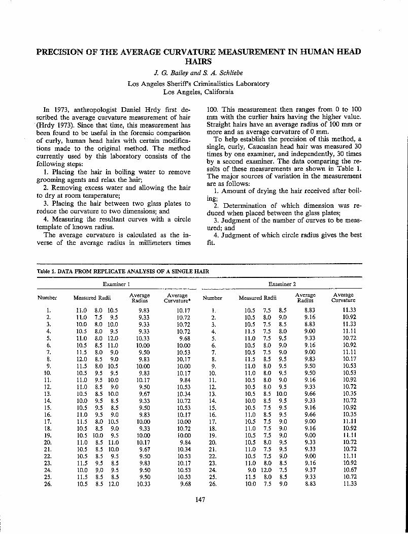
PRECISION OF THE AVERAGE CURVATURE MEASUREMENT IN HUMAN HEAD HAIRS
J. G. Bailey and S. A. Schliebe
Los Angeles Sheriffs Criminalistics Laboratory Los Angeles, California
In 1973, anthropologist Daniel Hrdy first described the average curvature measurement of hair (Hrdy 1973). Since that time, this measurement has been found to be useful in the forensic comparison of curly, human head hairs with certain modifications made to the original method. The method currently used by this laboratory consists of the following steps:
1. Placing the hair in boiling water to remove grooming agents and relax the hair;
2. Removing excess water and allowing the hair to dry at room temperature;
3. Placing the hair between two glass plates to reduce the curvature to two dimensions; and
4. Measuring the resultant curves with a circle template of known radius.
The average curvature is calculated as the inverse of the average radius in millimeters times
100. This measurement then ranges from 0 to 100 mm with the curlier hairs having the higher value. Straight hairs have an average radius of 100 mm or more and an average curvature of 0 mm.
To help establish the precision of this method, a single, curly, Caucasian head hair was measured 30 times by one examiner, and independently, 30 times by a second examiner. The data comparing the results of these measurements are shown in Table 1. The major sources of variation in the measurement are as follows:
1. Amount of drying the hair received after boiling;
2. Determination of which dimension was reduced when placed between the glass plates;
3. Judgment of the number of curves to be measured; and
4. Judgment of which circle radius gives the best fit.
Table 1. DATA FROM REPLICATE ANALYSIS OF A SINGLE HAIR
Examiner I Examiner 2
Number Measured Radii Average Average Number Measured Radii Average Average Radius Curvature· Radius Curvature
1. 11.0 8.0 10.5 9.83 10.17 1. 10.5 7.5 8.5 8.83 11.33 2. 11.0 7.5 9.5 9.33 10.72 2. 10.5 8.0 9.0 9.16 10.92 3. 10.0 8.0 10.0 9.33 10.72 3. 10.5 7.5 8.5 8.83 11.33 4. 10.5 8.0 9.5 9.33 10.72 4. 11.5 7.5 8.0 9.00 11.11 5. 11.0 8.0 12.0 10.33 9.68 5. 11.0 7.5 9.5 9.33 10.72 6. 10.5 8.5 11.0 10.00 10.00 6. 10.5 8.0 9.0 9.16 10.92 7. 11.5 8.0 9.0 9.50 10.53 7. 10.5 7.5 9.0 9.00 11.11 8. 12.0 8.5 9.0 9.83 10.17 8. 11.5 8.5 9.5 9.83 10.17 9. 11.5 8.0 10.5 10.00 10.00 9. 11.0 8.0 9.5 9.50 10.53
10. 10.5 9.5 9.5 9.83 10.17 10. 11.0 8.0 9.5 9.50 10.53 11. 11.0 9.5 10.0 10.17 9.84 11. 10.5 8.0 9.0 9.16 10.92 12. 11.0 8.5 9.0 9.50 10.53 12. 10.5 8.0 9.5 9.33 10.72 13. 10.5 8.5 10.0 9.67 10.34 13. 10.5 8.5 10.0 9.66 10.35 14. 10.0 9.5 8.5 9.33 10.72 14. 10.0 8.5 9.5 9.33 10.72 15. 10.5 9.5 8.5 9.50 10.53 15. 10.5 7.5 9.5 9.16 10.92 16. 11.0 9.5 9.0 9.83 10.17 16. 11.0 8.5 9.5 9.66 10.35 17. 11.5 8.0 10.5 10.00 10.00 17. 10.5 7.5 9.0 9.00 11.11 18. 10.5 8.5 9.0 9.33 10.72 18. 11.0 7.5 9.0 9.16 10.92 19. 10.5 10.0 9.5 10.00 10.00 19. 10.5 7.S 9.0 9.00 11.11 20. 11.0 8.5 11.0 10.17 9.84 20. 10.5 8.0 9.5 9.33 10.72 21. 10.5 8.5 10.0 9.67 10.34 21. 11.0 7.5 9.5 9.33 10.72 22. 10.5 8.5 9.5 9.50 10.53 22. 10.5 7.5 9.0 9.00 11.11 23. 11.5 9.5 8.5 9.83 10.17 23. 11.0 8.0 8.5 9.16 10.92 24. 10.0 9.0 9.5 9.50 10.53 24. 9.0 12.0 7.5 9.37 10.67 25. 11.5 8.5 8.5 9.50 10.53 25. 11.5 8.0 8.5 9.33 10.72 26. 10.5 8.5 12.0 10.33 9.68 26. 10.0 7.5 9.0 8.83 11.33
147
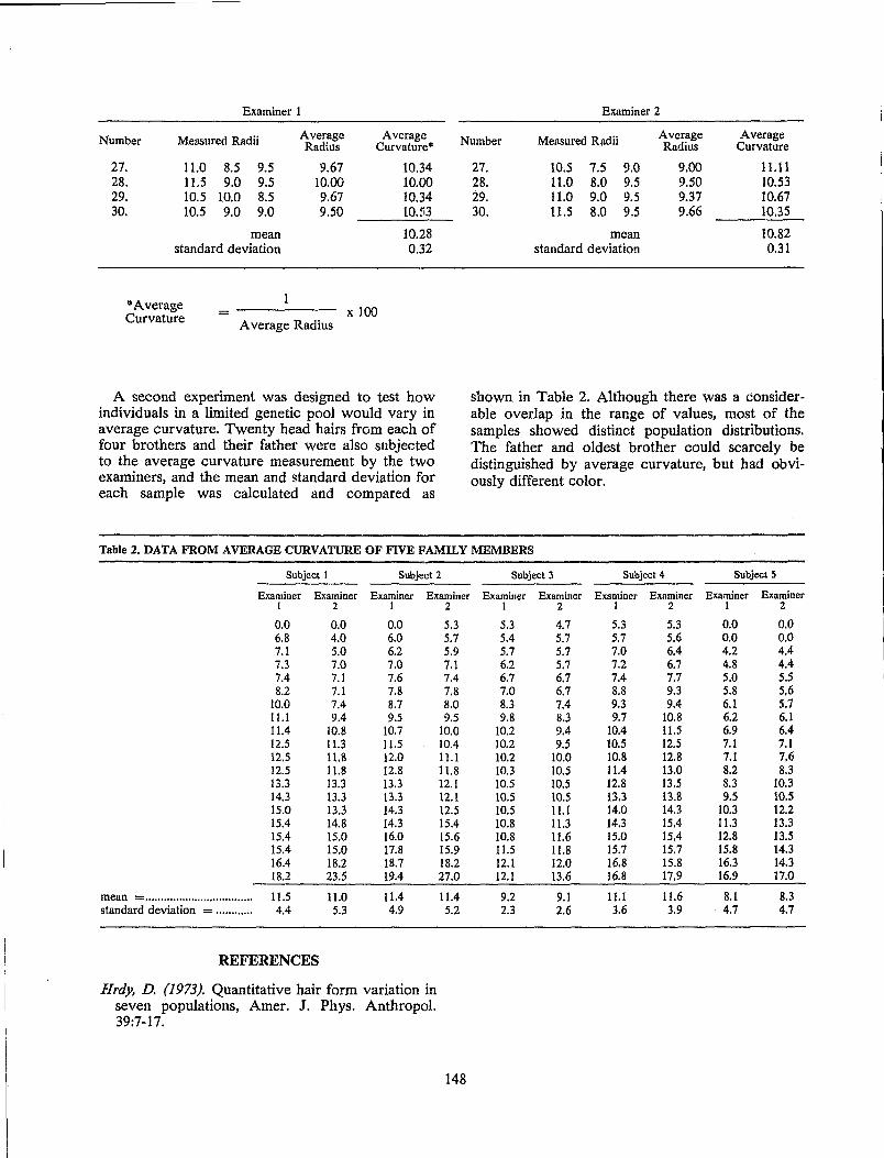
Examiner 1 Examiner 2
Number Measured Radii Average Average Number Measured Radii Average Average Radius Curvature· Radius Curvature
27. 11.0 8.5 9.5 9.67 28. 11.5 9.0 9.5 10.00 29. 10.5 10.0 8.5 9.67 30. 10.5 9.0 9.0 9.50
mean standard deviation
"'Average Curvature = x 100
A verage Radius
10.34 10.00 10.34 10.53
10.28 0.32
A second experiment was designed to test how individuals in a limited genetic pool would vary in average curvature. Twenty head hairs from each of four brothers and their father were also subjected to the average curvature measurement by the two examiners, and the mean and standard deviation for each sample was calculated and compared as
27. 10.5 7.5 9.0 9.00 11.11 28. 11.0 8.0 9.5 9.50 10.53 29. 11.0 9.0 9.5 9.37 10.67 30. 1l.S 8.0 9.5 9.66 10.35
mean 10.82 standard deviation 0.31
shown in Table 2. Although there was a considerable overlap in the range of values, most of the samples showed distinct population distributions. The father and oldest brother could scarcely be distinguished by average curvature, but had obviously different color.
Table 2. DATA FROM AVERAGE CURVATURE OF FIVE FAMILY MEMBERS
Subject I Subject 2 Subject 3 Subjecf 4 Subject 5
Examiner Examiner Examiner Examiner Examiner Examiner Examiner Examiner Examiner Examiner 1 2 I 2 1 2 1 2 1 2
0.0 0.0 0.0 5.3 5.3 4.7 5.3 5.3 0.0 0.0 6.8 4.0 6.0 5.7 5.4 5.7 5.7 5.6 0.0 0.0 7.1 5.0 6.2 5.9 5.7 5.7 7.0 6.4 4.2 4.4 7.3 7.0 7.0 7.1 6.2 5.7 7.2 6.7 4.8 4.4 7.4 7.1 7.6 7.4 6.7 6.7 7.4 7.7 5.0 5.5 8.2 7.1 7.8 7.8 7.0 6.7 8.8 9.3 5.8 5.6
10.0 7.4 8.7 8.0 8.3 7.4 9.3 9.4 6.1 5.7 11.1 9.4 9.5 9.5 9.8 8.3 9.7 10.8 6.2 6.1 11.4 10.8 to.7 10.0 10.2 9.4 10.4 11.5 6.9 6.4 12.5 11.3 11.5 10.4 to.2 9.5 to.5 12.5 7.1 7.1 12.5 11.8 12.0 11.1 10.2 10.0 10.8 12.8 7.1 7.6 12.5 11.8 12.8 11.8 10.3 10.5 11.4 13.0 8.2 8.3 13.3 13.3 13.3 12.1 10.5 10.5 12.8 13.5 8.3 10.3 14.3 13.3 13.3 12.1 10.5 10.5 13.3 13.8 9.5 10.5 15.0 13.3 14.3 12.5 10.5 11.1 14.0 14.3 10.3 12.2 15.4 14.8 14.3 15.4 10.8 11.3 14.3 15.4 11.3 13.3 15.4 15.0 16.0 15.6 10.8 11.6 15.0 15.4 12.8 13.5 15.4 15.0 17.8 15.9 11.5 11.8 15.7 15.7 15.8 14.3 16.4 18.2 18.7 18.2 12.1 12.0 16.8 15.8 16.3 14.3 18.2 23.5 19.4 27.0 12.1 13.6 16.8 17.9 16.9 17.0
mean = ................................... 11.5 11.0 11.4 11.4 9.2 9.1 11.1 11.6 8.1 8.3 standard deviation = ............ 4.4 5.3 4.9 5.2 2.3 2.6 3.6 3.9 4.7 4.7
REFERENCES
Hrdy, D. (1973). Quantitative hair form variation in seven populations, Amer. J. Phys. Anthropol. 39:7-17.
148
A STUDY OF THE FEASIBILITY OF ESTABLISHING A COMPUTER DATA BANK FOR HAIR CHARACTERIZATION USING STANDARD DESCRIPTIVE CRITERIA
A. G. Podolak and C. E. Blythe
Microscopic Analysis Unit FBI Laboratory
Washington, D.C.
The ability to state the frequency with which a particularly described hair occurs in the population would be of considerable assistance in assigning significance to hair associations or matches in criminal investigations. Toward this end, a survey was conducted to determine the diverse descriptions examiners would relate to hairs. The long range goal was to establish a computer data bank of hair descriptions to determine the frequency of occurrence of a particular hair.
Five hairs were characterized by nine examiners using a standard form allowing 26 characteristics to be described from a multiple choice list appropriate to the particular characteristic. Table 1 lists the characteristics from which examiners made selections in describing the hairs in this survey. The percentages to the right of the descriptive characteristics indicate the percentage of examiners who se-
149
lected that particular characteristic in describing one hair selected at random. Race, sex, artificial treatment, length, root and tip were not considered in this survey. Each examiner described the same hairs differently.
After approximately eight weeks, the hairs were reexamined by the same nine examiners and again each examiner described the same hairs differently. Furthermore, the results showed that each examiner described the characteristics of the same hairs differently between the first and second examinations.
This survey indicates that because of variations among hair examiners in describing hair characteristics, it is not feasible to computerize descriptive data of hairs for the purpose of determining the frequency of occurrence of a particUlar hair.
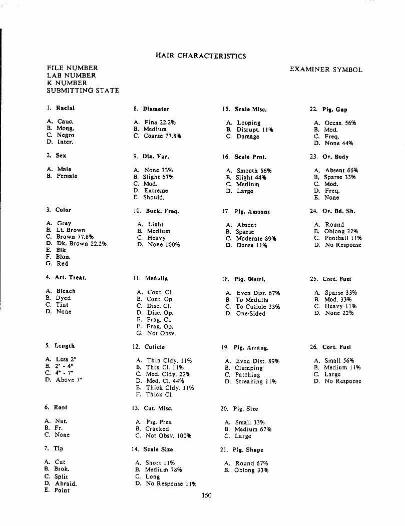
FILE NUMBER LAB NUMBER K NUMBER SUBMITTING STATE
1. Radal
A. Cauc. B. Mong. C. Negro D. Inter.
2. Sex
A. Male B. Female
3. Color
A. Grey B. Lt. Brown C. Brown 17.8% D. Dk. Brown 22.2% E. Blk F. Blon. G. Red
4. Art. Treat.
A. Bleach B. Dyed C. Tint D. None
5. Length
A. Less 2" B. 2" - 4" C. 4" - 7" D. Above r
6. Root
A. Nat. B. Fr. C. None
7. Tip
A. Cut B. Brok. C. Split D. Abraid. E. Point
HAIR CHARACTERISTICS
8. Diameter 15. Scale Misc.
A. Fine 22.2% A. Looping B. Medium B. Disrupt. 11 % e. Coarse 17.8% C. Damage
9. ma. Var. 16. Scale Prot.
A. None 33% A. Smooth 56% B. Slight 67% B. Slight 44% C. Mod. C. Medium D. Extreme D. Large E. Should.
10. Buck. Freq. 17. Pig. Amount
A. Light A. Absent B. Medium B. Sparse C. Heavy C. Moderate 89% D. None 100% D. Dense 11%
11. Medulla 18. Pig. Distrl.
A. Cont. Cl. A. Even Dist. 67% B. Cont. Op. B. To Medulla C. Disc. CI. C. To Cuticle 33% D. Disc.Op. D. One-Sided E. Frag. CI. F. Frag.Op. G. Not Obsv.
12. Cuticle 19. Pig. Arrang.
A. Thin Cldy. 11 % A. Even Dist. 89% B. Thin CI. 11% B. Clumping C. Med. Cldy. 22% C. Patching D. Med. CI. 44% D. Streaking 11% E. Thick Cldy. 11% F. Thick CI.
13. Cu t. Misc. 20. Pig. Size
A. Pig. Pres. A. Small 33% B. Cracked B. Medium 67% C. Not Obsv. 100% C. Large
14. Scale Size 21. Pig. Shape
A. Short 11% A. Round 67% B. Medium 78% B. Oblong 33% C. Long D. No Response 11%
150
EXAMINER SYMBOL
22. PII. Cap
A. Occas.56% B. Mod. C. Freq. D. None 44%
23. Ov. Body
A. Absent 66% B. Sparse 33% C. Mod. D. Freq. E. None
24. Ov. Bd. Sh.
A. Round B. Oblong 22% C. Football 11 % D. No Response
25. Cort. Fust
A. Sparse 33% B. Mod. 33% C. Heavy 11% D. None 22%
26. Cort. Fusi
A. Small 56% B. Medium 11% C. Large D. No Response
SYNTAX-DIRECTED CONCEPT ANALYSIS IN THE REASONING FOUNDATIONS OF HUMAN HAIR COMPARISONS
MS. Verma
Colorado Bureau of Investigation Denver, Colorado
The purpose of this paper is to explore one possible mathematical formalism that might serve to simulate the implicit reasoning processes utilized by hair examiners in handling the problem of a multitude of characteristics and morphological patterns. The method presented here is designed for immediate, direct application and may serve as a :;uggested basis from which more practical procecli..res can be developed.
Two well-known mathematical disciplines, symbolic logic and probability theory, contribute to the reasoning foundations of hair comparisons. These two basic concepts are inherent in hair examination procedures, even when the examiner utilizes them subconsciously, on an "intuitive" level. In a previous work, this author outlined the nature of the logical concepts inherent in hair examination and introduced the concept of "total morphological pattern" which emphasizes the fundamental importance of considering combination of unitary characteristics and morphological complexes (Verma 1984). The first step in making a logical analysis of this process is to apply the symbolism associated with the propositional calculus of symbolic logic.
The probabilistic concepts inherent in hair examination arise because the analysis can rarely be made with absolu.te certainty; the end result of this "complex reasoning process" usually gives a "most probable" determination. The unique task of the examiner from this point of view is primarily a goaldirected intellectual process involving decisionmaking leading to probability estimate. Some degree of formalism of this process is attainable by reference to information theory.
The formulation of methods used for coding the computer to extract quantitative information from qualitative ones is a problem in pattern recognition. The many different mathematical techniques used to solve pattern recognition problems may be grouped into two general approaches (Fu 1979). They are decision-theoretic (or discriminant) approach and syntactic (or structural) approach. In the syntactic approach, each pattern is expressed as a composition of its components, called subpatterns and pattern primitives. This approach draws an analogy between structure of patterns and syntax of a language. The recognition of each pattern is made by parsing the pattern structure according to a given set of syntax rules.
In an exposition of an automatic-programming computer language for syntactical analysis of hairs,
151
the descriptions themselves can be classified into the following types: syntactical definitions, semantic discussions and grammatical forms.
The syntactical definitions make explicit what characteristics of hairs are to be meaningful; these definitions are concerned with proper construction of words, expressions and statements. The semantic discussions are concerned with the precise meaning to be attributed to the various characteristics. The grammatical forms are concerned with the proper relationships among the different constructs, particularly the statements.
The spatial relation correspondence of morphological patterns (Appendix I) is achieved through "dividing" a hair into regions of 1 cm length, each one of which is assigned a symbolic notation (A, B, C, D, etc.). These notations serve a dual purpose, they not only symbolize a region, but also provide a symbolic definition of a pattern. Syntax rules are written to recognize and distinguish between patterns and regions and their spatial relations. The specific location of a pattern or characteristic may not be important; we may only wish to know whether or not a pattern(s) is an aspect of a given hair, or a control sample of hairs from a given individual, and finally, their frequency of distribution in the population studied.
One of the main reasons for appealing to !Iyntax is stated to be economy of symbolic notati..:ms in describing a morphological pattern, for (.xample, the symbolic definition of a pattern B (Appendix I) is recursive in that it can be used repeatedly. This is especially useful since many hairs show patterns which are redundant in that they extend through several regions. In addition, in situations which are fairly common, where only minor deviations from a previously identified pattern is noted, syntax rules are provided which allow for repeated use of the same symbol in conjunction with modified characteristics. This will automatically replace and/or add in the computer memory the new characteristic(s) to that pattern. The end result is a substantial saving of time and labor in tedious rerecording of all characteristics of a pattern in various regions.
At the Colorado Bureau of Investigation Laboratory, a computer program for hair analysis with syntax approach has been developed for operation with the SPSSx batch system v.hich is a comprehensive tool for managing, analyzing and display~ ing data. The computer program is designed foJ.'
"one-way" and "manova" procedures both of which can provide a one-way analysis of variance and test for trends across categories, specify contrasts, perform variety of range tests, univariate and multivariate linear estimation and tests for hypothesis for any crossed and/or nested design with or without covariates. There is a complete control of model specification. For example, you can test for effects jointly, or you can specify single degree of freedom partitions. Also, you c:an specify interaction effects between factors and co variates.
The raw statistical data through this computer program can be manipulated through the SPSSx system to develop population frequencies which we need as a basis for the estimates of conditional
152
probabilities and thus quantify the individualization of the hair source.
REFERENCES
Pu, K. S. (1979). Syntactic Approach to Pattern Recognition. Biomedical Pattern Recognition and Image Processing (Fu, K. S. and Pavlidis, T., eds.). Berlin! Dahlem Konferenzen. Verlag Chemie GMBH. D-6940 Weinheim, pp. 27-48.
Verma, M S. (1984). Human Hair Comparison: An Anthropological Approach. In: Publication of the Proceeding of the International Association of Forensic Sciences Meeting, Oxford.
THE DETECTION AND OCCURRENCE OF HUMAN HAIR OXIDATION BY FOURIER TRANSFORM INFRARED SPECTROSCOPY
c. S. Tumosa and L. Brenner
Philadelphia Police Laboratory Philadelphia, Pennsylvania
INTRODUCI'ION
The analysis of human hair has been controversial because of the apparent SUbjective parameters associated with microscopic observations. In an attempt to quantify some of the properties of hair, examination was performed using Fourier transform infrared spectroscopy. The infrared spectrum was examined for quantitative and qualitative variation. One of the prominent observations noted in this study was the ease in detecting the oxidation of the hair. The oxidation as a result of the breaking of sulfur-sulfur bonds and the formation of an easily detected sulfonic acid gave a peak of interest at 1044 cm"l.
MATERIAL AND METHODS
Infrared spectra were collected on a DIGILAB FTS-10 C/D spectrometer (Digilab, Cambridge, MA) with cesium iodide optics, a TGS detector and a diamond cell with a 4X beam condenser. Typical spectra were obtained with two hair fragments at a resolution of 8 cm"l.
Chemical treatments of hairs were performed as described in Brenner et al. (1985). The hair samples
153
in the population study were collected from Caucasians among the laboratory staff and friends.
RESULTS AND DISCUSSION
Figure 1 shows the comparison of a cysteic acid standard spectrum with a difference spectrum derived from the oxidized minus unoxidized hair. The spectra show a general coincidence and a prominent peak at 1044 cm' l which has been assigned to the symmetric s=o stretching vibration of a sulfonic acid by Strassburger and Breuer (1985).
Qualitative and quantitative experiments performed showed the oxidation of hair by hydrogen peroxide to proceed at a faster rate at pH 10 than at pH 5 as previously mentioned in Robbins (1979).
Examination of damaged (oxidized) hair showed the most oxidation at the tip and the least at the root.
Parameters such as the natural color of the hairs, greying, storage, variation in an individual over time and head area, washing, moisture and certain reducing agents were found to have little or no effect on the presence of the 1044 cm"l oxidation pellk.
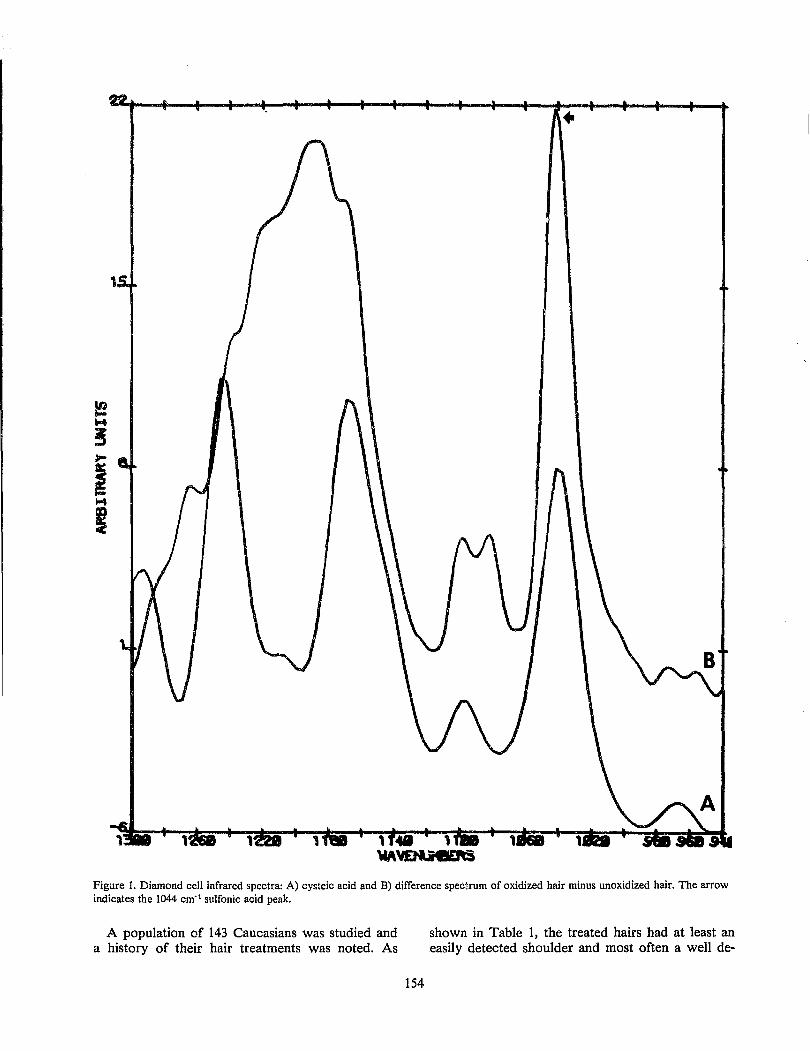
1
Figure 1. Diamond cell infrared spectra: A) cysteic acid and B) difference spec;\rum of oxidized hair minus unoxidized hair. The arrow indicates the 1044 cm'1 sulfonic acid peak.
A population of 143 Caucasians was studied and a history of their hair treatments was noted. As
154
shown in Table 1, the treated hairs had at least an easily detected shoulder and most often a well de-
fined absorption peak. Untreated hairs sometimes exhibited a shoulder but most often did not show an absorption. In the case of 4 out of 30 women with untreated hair, a distinct peak was present.
Table 1. CAUCASIAN HEAD HAIR SAMPLES
Detection of 1044 cm- 1 IR Peak
Neither Peak Shoul-
Number nor der Peak
of Shoul- Number Number
Samples der (Per- (Per-Number cent)
(Per- cent)
cent)
Males Not Treated 47 28 (60%) 19 (40%) 0(0%) Treated 9 0(0%) 4 (44%) 5 (56%)
Females Not Treated 30 17 (57%) 9 (30%) 4 (13%) Treated 57 0(0%) 12 (21%) 45 (79%)
Total 143
The occurrence of an oxidation peak in other body hair was explored and the results of a small population is given in Table 2. No oxidation was detected in these samples.
155
Table 2. CAUCASIAN BODY HAIR SAMPLES
No Detectable 1044 cm- 1 Peak or Shoulder
Number of Samples
Male Female
Facial 2 Arm 4 Pubic 5 2 Leg 4
In conclusion, the presence of an oxidation peak was found in all treated head hair samples from Caucasians and may be useful in differentiating hair sources.
REFERENCES
Brenner, L., Squires, P. L., Garry, M and Tumosa, C. S. (1985). A measurement of human hair oxidation by Fourier transform infrared spectroscopy, J. Forensic Sci. 30:420-426.
Robbins, C. R. (1979). Chemical and Physical Behavior of Human Hair. Van Nostrand Reinhold Co., Inc., New York.
Strassburger, J. and Breuer, M M (1985). Quantitative Fourier transform infrared spectroscopy of oxidized hair, J. Soc. Cosmet. Chern. 36:61-74.
APPLICATION OF ELEMENTAL ANALYTICAL DATA WITH SEM/EDX SYSTEM TO HAIR COMPARISON
S. Seta and .lL Sata
National Research Institute of Police Science Tokyo,Japan
It has been often pointed out that it is not possible to positively identify a sample of hair as having come from a particular individual. However, it may be expected that the probability of identity increases with every point of resemblance including chemical, physical and morphological characteristics. From this point of view, elemental analysis of hair samples with scanning electron microscopy / energy dispersive X-ray (SEM/EDAX) system has been utilized for hair comparison. In the present report, the classification of hair samples on the basis of EDAX spectra were experimentally investigated.
For this investigation two experimental sets were designed. In each set head hairs which were taken from the top of the head of several individuals were mingled. The examiner was not informed as to how many hairs from different individuals were mingled and how many hairs of the same individual were contained. For EDAX analysis each hair sample of 0.5 cm length was exposed to oxygen plasma, thus allowing the efficient detection of biological elements such as sodium, phosphorus, potassium and calcium. Energy dispersive X-ray spectra were depicted within an analyzing time of 100 seconds and at 512 full scale, excepting for two hairs which were depicted at 2000 full scale.
Twenty head hair samples of the first set were divided into five groups (Figure 1). Four hairs were combined in one group (group 1) because of the resemblance of EDAX spectrum patterns. In fact, they were from the same individual (subject S) but one hair of this subject was falsely eliminated from the group because of high generation of each X-ray peak. For the same reason one hair of subject N was classified as group 2 along with one hair of subject S. This result would indicate that
157
EDAX spectrum pattern is more helpful in characterizing one hair than the X-ray peak-height. Three hairs which were classified as group 3 or 4 were, in fact, from the same individual, respectively. Eight hairs were combined in one group (group 5) mainly because of the resemblance in the generation of a high peak of calcium along with a slight peak of potassium through the samples. Hairs of group 1 were different from those of group 3 in the reciprocal relationship between potassium and calcium peak heights, and hairs of group 4 were different from those of group 5 in the peak heights of calcium and potassium. The results of another set are shown in Figure 2. The 23 hairs examined were divided into five groups. Seven hairs were classified as group 1 because of the common appearance of a very high peak of calcium along with a slight peak of sodium through the samples. These hairs consisted of both subject 0 and F hairs and they were treated with perma.nent waving. Four hairs which were classified as group 2 were differentiated from hairs of group 1 because hairs of group 2 commonly generated potassium peaks through the samples. Other hairs were divided into three groups (group 3,4 and 5). Four hairs of group 3 were characterized by the similar reciprocal relationship between potassium and calcium peaks and they were differentiated from three hairs of group 4. Four hairs were combined in one group (group 5) because of the clear appearance of a phosphorus peak through the samples but it was necessary to add one hair of subject K to this group because of its resemblance in general low X-ray intensity. These results may suggest EDAX spectrum pattern can be efficiently used for hair comparison if it is combined with morphological characteristics and results of blood grouping.
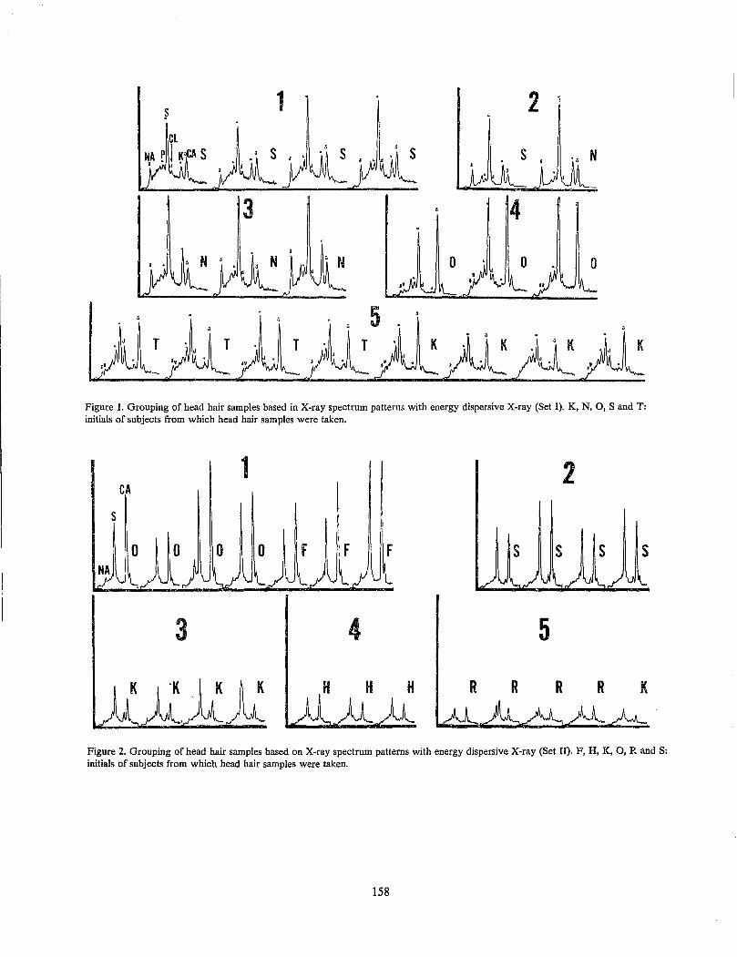
1 2
N
3 4
Figure 1. Grouping of head hair samples based in X-ray spectrum patterns with energy dispersive X-ray (Set I). K, N, 0, Sand T: initials of subjects from which head hair samples were taken.
1 2 CA
s s s s
3 4 5
H H H R R R R K
Figure 2. Grouping of head hair samples based on X-ray spectrum patterns with energy dispersive X-ray (Set II). F, H, K, 0, Rand S: initials of subjects from which head hair samples were taken.
158
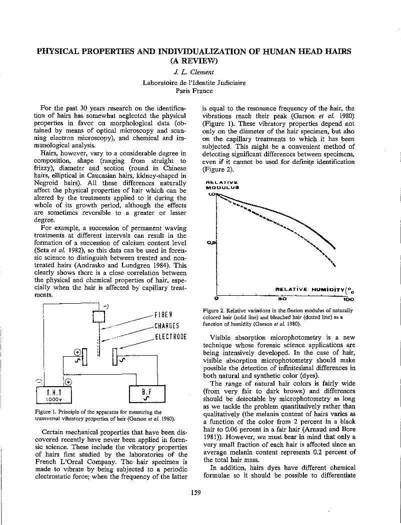
PHYSICAL PROPERTIES AND INDIVIDUALIZATION OF HUMAN HEAD HAIRS (A REVIEV'/)
J. L. Clement
Laboratoire de l'Identite Judiciaire Paris France
For the past 30 years research on the identification of hairs has somewhat neglected the physical properties in favor on morphological data (obtained by means of optical microscopy and scanning electron microscopy), and chemical and immunological analysis.
Hairs, however, vary to a considerable degree in composition, shape (ranging from straight to frizzy), diameter and section (round in Chinese hairs, elliptical in Caucasian hairs, kidney-shaped in Negroid hairs). All these differences naturally affect the physical properties of hair which can be altered by the treatments applied to it during the whole of its growth period, although the effects are sometimes reversible to a greater or lesser degree.
For example, a succession of permanent waving treatments at different intervals can result in the formation of a succession of calcium content level (Seta et al. 1982), so this data can be used in forensic science to distinguish between treated and nontreated hairs (Andrasko and Lundgren 1984). This clearly shows there is a close correlation between the physical and chemical properties of hair, especially when the hair is affected by capillary treatments.
________ FIB E R
_.-'- _____ CHARGES
.. ----- ELECTRODE : -- -------
Figure 1. Principle of the apparatus for measuring the transversal vibratory properties of hair (Garson et of. 1980).
Certain mechanical properties that have been discovered recently have never been applied in forensic science. These include the vibratory properties of hairs first studied by the laboratories of the French L'Oreal Company. The hair specimen is made to vibrate by being subjected to a periodic electrostatic force; when the frequency of the latter
159
is equal to the resonance frequency of the hair, the vibrations reach their peak (Garson et al. 1980) (Figure 1). These vibratory properties depend not only on the diameter of the hair specimen, but also on the capillary treatments to which it has been subjected. This might be a convenient method of detecting significant differences between specimens, even if it cannot be used for definit,e identification (Figure 2).
RIli.LAT/VIii MOOULua
1.0
RELATiVE HUMiOiTV(Oo o~--~-------'--:;;-o~· . . 100
Figure 2. Relative variations in the flexion modulus of naturally colored hair (solid line) and bleached hair (dotted line) as a function of humidity (Garson et of. 1980).
Visible absorption microphotometry is a new technique whose forensic science applications are being intensively developed. In the case of hair, visible absorption microphotometry should make possible the detection of infinitesimal differences in both natural and synthetic color (dyes).
The range of natural hair colors is fairly wide (from very fair to dark brown) and differences should be detectable by microphotometry as long as we tackle the problem quantitatively rather than qualitatively (the melanin content of hairs varies as a function of the color from 2 percent in a black hair to 0.06 percent in a fair hair (Arnaud and Bore 1981». However, we must bear in mind that only a very small fraction of each hair is affected since an average melanin content represents 0.2 percent of the total hair mass.
In addition, hairs dyes have different chemical formulae so it should be possible to differentiate

both quantitatively and qualitatively various natural and synthetic hair colors.
The microphotometry technique consequently has to be adapted to detect minute differences on this scale.
REFERENCES
Andrasko, J. and Lundgren, B. (1984). Forensic examination of permanent waved hair. Report Number 16 of the Centrum for Forensic Veienkap. Linkoping, Sweden.
160
Arnaud, J. L. and Bore, P. (1981). Isolation of melanin pigments from human hair, J. Soc. Cosmet. Chern. 32:137-152.
Garson, J. C. et al. (1980). Les proprietes vibratoires transversales des fibres de keratine, Int. J. Cosmet. Sci. 2:231-241.
Seta, S., Safo, H., Yoshino, M and Miyasaka, S. (1982). SEM/EDX analysis of inorganic elements in human scalp hairs with special reference to the variation with different locations on the head, Scanning Electron Microsc. 1:127-140.

STRESS AND THE FORENSIC HAIR EXAl'\1INER E. L. Burwitz
Microscopic Analysis Unit FBI Laboratory
Washington, D. C.
Dr. Hans Selye, foremost researcher of human stress, has defined stress as the nonspecific response of the body to any demand made upon it. A nonspecific response is elicited from the body each time an individual experiences a change in life or a threat to status, ego or job security. This response consists of physiological activity which increases the heart rate, elevates the blood pressure, causes the secretion of hormones, mobilizes glucose and produces other reactions. This activity is identical to that produced in an animal confronted by a predator and enables the animal to fight or flee. The animal's course of action to fight or flee dissipates stress symptoms. Neither combat nor flight is appropriate for changes in life or threats to status, ego or job security; therefore, the stress symptoms are not dissipated as readily. It should be noted that the adrenal glands which playa major role in the body's physiological response to stress can become overworked. If they do, physiological disorders can develop which are characterized by mental lethargy, lack of concentration reduced stamina and an inability to think clearly. From the standpoint of a forensic hair examiner, these symptoms could have a significant impact on case working ability inasmuch as the ability to concentrate
161
and think clearly are essential to the detection of sometimes minute differences between hairs.
It is important for the hair examiner to know that many aspects of a hair examiner's job are stressful. They include time pressures and deadlines, excessive travel, a heavy continuous case load and the desire to see justice done but not at the unfair expense of the defendant. Secondly, the hair examiner should realize stress is a matter of perception which is a skill that can be learned, altered and changed. Thirdly, everyone is susceptible to stress to some extent; therefore, the hair examiner should be able to recognize stress symptoms. They may be manifest on the emotional, behavioral and/or physical level. Lastly, the hair examiner should understand that if stress has negatively affected productivity and/or health, the hair examiner can alleviate stress by changing old habits and learning stress management techniques. These include taking care of the physical body, utilizing relaxation methods, understanding the emotions, providing for a controllable work and social environment and providing for spiritual needs. These techniques cannot eliminate stress; however, they can eliminate most stress symptoms which tend to interfere with productivity and good health.
HAIR COMPARISON BY PYROLYSIS CAPILLARY GAS CHROMATOGRAPHY/ MASS SPECTROMETRY: A PROGRESS REPORT
T. O. Munson
Forensic Science Research Group FBI Laboratory
Quantico, Virginia
The goal of this research project is to determine whether pyrolysis capillary gas chromatography (PyGC2) or pyrolysis capillary gas chromatography/mass spectrometry (PyGC2/MS) can be useful for forensic comparisons of human hair. Initial hair comparisons by PyGC2 and PyGC2/MS have shown some differences which might prove useful (Munson and Vick 1985), and have shown the pyrolysis profiles to be so very reproducible that even small differences might be significant. The comparison of hair from three donors is shown in Figure 1. In order to evaluate these observed differences and seek others, attempts have been made to identify the components which constitute the hair pyrolyzate. Compounds which have been identified in the hair pyrolyzate include carbon dioxide, carbon oxide sulfide, methanethiol, carbon disulfide, toluene, pyridine, phenol, cresol, styrene, pyrrole, methylpyrrole and indole -- many of which could arise from cleavage of the amino acid substituent groups in protein molecules.
By analogy with the pyrolysis products formed from polyamides by lactam ring formation (Ohtani et al. 1982), one might expect to observe cyclic polyamides formed by route I shown in Figure 2.
163
Finding a compound which closely matched the spectrum of 5-methyl-2, 4-imidazolidinedione suggested route II in Figure 2 for the formation of a family of 5-substituted 2, 4- imidazolidinediones. Five members of this series have been tenatively identified in the hair pyrolyzate: 5-methyl, 5-(1-methylethyl), 5-(2-dimethylethyl) and 5-(1-methylpropyl) corresponding to the incorporation into the ring of the amino acids alanine, valine, leucine and isoleucine, respectively. Another series of compounds, 5-substituted pyrrolidino[l, 2a]-3, 6-piperazinediones, have been tenatively identified, and are thought to arise from the pyrolysis of hair protein by the mechanism shown in Figure 3. Four members in this series have been tenatively identified: unsubstituted at the 5 position, 5-(1-methylethyl), 5-(2-dimethylethyl) and 5-(1-methylpropyl), corresponding to the incorporation into the molecule of the amino acids proline plus glycine, proline plus valine, proline plus leucine and proline plus isoleucine, respectively. All of the above identifications have been substantiated by PyGC2/MS/ MS (electron impact ionization/collision ally activated dissociation and chemical ionization/collisionally activated dissociation experiments).
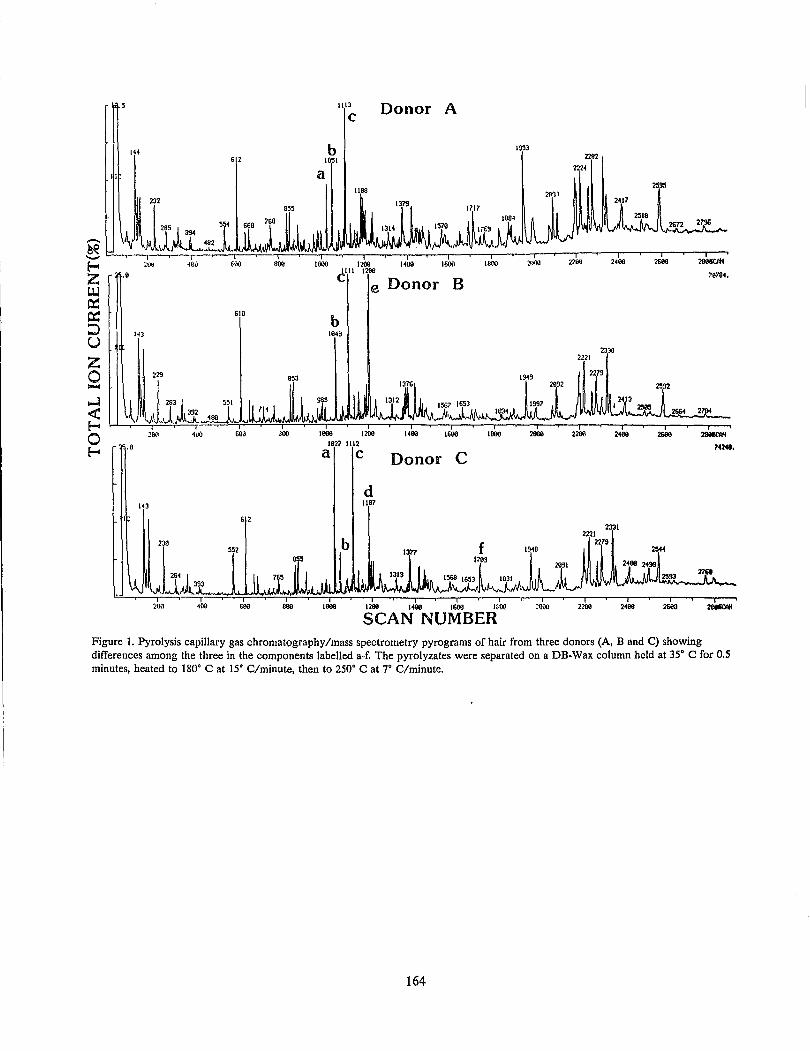
•• 9
143
•. a
I 400
612
610
, I ' i i
200 4uu 600 300
400
IlIl
C
b 1051
a Ilea
Donor A
1379
I 1600
I 1200
ell 1 1200
e Donor B
b la49
I I 1000 1200
102? 1112
a c
d lIa7
Donor
I 1600
c
I IBOO
I 1000
I!r.!J
I 2IlOO
b 1377 f 1948
"""'MJ'\o,J''''i\A-.I56SIV\I...IN65",:Ll~JL m' I I I
1200 1600 1(,'00
SCAN NUMBER I
:000
2282
2224
2417
2JlO 2221
2279
i i 2200 2468
2331 2221
I 2400
2595
I 2600
I 2600
70704.
I 2fl8IICIV/
Figure 1. Pyrolysis capillary gas chromatography/mass spectrometry pyrograms of hair from three donors (A, Band C) showing differences among the three in the components labelled a-f. The pyrolyzates were separated on a DB-Wax column held at 35° C for 0.5 minutes, heated to 180° Cat 15° C/minute, then to 250° Cat 7° C/minute.
164
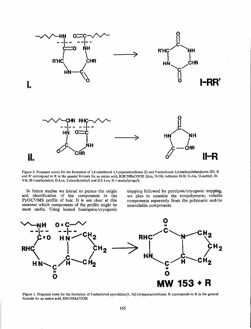
II.
~ RI~--v'V'-- -1- - - - --HN 0=
I I ~c, /NH 0/ ....... CRR
')
I-RR'
II-R Figure 2. Proposed routes for the formation of 3,6-substituted 2,5-piperazinediones (I) and 5-substituted 2,4-imidazolidinediones (11), R and R' correspond·to R in the general formula for an amino acid, RHCNH2COOH (thUS, II-Gly indicates II-H; II-Ala, II-methyl; IlVal, II-l-methylethyl; II-Leu, 2-dimethylethyl; and II-I Leu, II-I-methylpropyl).
In . future studies we intend to pursue the origin and identification of the components in the PyGC2/MS profile of hair. It is not clear at this moment which components of the profile might be most useful. Using heated headspace/cryogenic
trapping followed by pyrolysis/cryogenic trapping, we plan to examine the non polymeric, volatile components separately from the polymeric and/or nonvolatile components.
o II
RHC/C"~CH2
-7~ I I fH2 HN"'-C/"~~CH2
II
o MW 153 + R
Figure 3. Proposed route for the formation of 5-substituted pyrrolidino[l, 2a]-3,6-piperazinediones. R corresponds to R in the general formula for an amino acid, RHCNH2COOH.
165
REFERENCES
Munson, T. O. and Vick, J. (1985). Comparison of human hair by pyrolysis-capillary column gas chromatography and gas chromatography-mass spectrometry, J. Anal. Appl. Pyrol. 8:493-501.
166
Ohtani, H., Nagaya, T., Sugimura, Y. and Tsuge, S. (1982). Studies on thermal degradation of aliphatic polyamides by pyrolysis-glass capillary gas chromatography, J. Anal. Appl. Pyrol. 4: 117-131.
THE POTENTIAL SIGNIFICANCE OF SKIN ADNEXA IN IDENTIFICATION OF TISSUE FROM UNKNOWN SOURCES
L. D. Herold
Forensic Laboratory Division Los Angeles County Chief Medical Examiner-Coroner
Los Angeles, California
The Los Angeles County Chief Medical Examiner-Coroner's Office received a case in which decomposing tissue from an unknown source was recovered from a water treatment plant following the disappearance of a sanitation worker under suspicious circumstances. The initial objective was to identify the type of tissue or organ submitted. The possibility of a more precise phylogenetic identification was investigated by a limited comparative morphology study of skin and adnexa from selected mammals.
Standard histological techniques were used to prepare skin tissue sections for light microscopy. Formalin fixed tissues were processed for paraffin embedding, and were cut and stained with hemotoxylin and eosin according to the generally used procedures described by Luna (1968). It later proved useful to deviate from the standard practice of sectioning skin perpendicular to the epithelial surface (cross section) and re-embed the same tissues rotated 90° for sections tangential to the epithelial surface.
In this particular case, the unknown was identified as mammalian skin. It displayed the basic structure of dermal dense irregular connective tissue with an overlying squamous epithelium (though it was noted to be severely degenerating and missing clear stratification on the surface) and subcutaneous adipose. The decomposing remanents
167
of hair follicles, sweat gland ducts and sebaceous glands were noted. An appropriate review of skin and adnexa can be found in Bloom and Fawcett (1975).
The limited study of skin and adnexa comparative morphology suggests potentially useful features that may be considered when trying to identify or eliminate an animal of origin. These include:
1. Epidermal thickness, if surface is severely decomposing the extent of stratification may be reflected in the more protected pilary canal.
2. Dermal dimension and density of collagen fibers.
3. Pilary patterns including the size, density, orientation and position of guard and vellus hairs in relation to each other and in relation to the overall skin structure.
4. Overall absence, or presence, dimension and density of specific adnexa.
REFERENCES
Bloom, W. and Fawcett, D. W. (1975). A Textbook of Histology. W. B. Saunders Co., Philadelphia.
Luna, L. G. (ed.) (1968). Manual of Histologic Staining Methods of the Armed Forces Institute of Pathology. McGraw-Hill Book Co., New York.
MICROSCOPIC IDENTIFICATION OF HUMAN HAIR SHAFT ANOMALIES J. T. Wilson
Regional Criminalistics Laboratory Kansas City, Missouri
The occurrence of various microscopically identifiable anomalies of the human hair shaft can provide significant discrimination power in forensic hair comparisons. These anomalies may be of a congenital, hereditary or acquired nature and are classified according to structural characteristics of the hair shaft observable by light microscopy.
Monilethrix is a genetic anomaly which produces proximal periodic constrictions with a frequency of 0.6 mm to 1.0 mm (Figure la). The periodicity of the constrictions remains constant in the hair shaft although the severity and periodicity may vary between hairs. All of the head hair is usually involved and the body hair may also be affected. The hair will break with a sharp fracture at a proximal constriction, consequently, pulling a hair with a follicle intact is very difficult. This is a genetic anomaly inherited as an autosomal dominant with incomplete penetrance (Solomon and Green 1963).
Pili torti produces rotations about the long axis of the hair shaft from 900 to as much as 3600 in a clockwise direction (Figure Ib). Not all of the head hair is affected; those affected usually have only an occasional rotation. However, some hairs may have a high frequency of rotation causing an appearance similar to monilethrix. At high magnification no rotation is apparent at the constriction in monilethrix whereas a rotation is clear in pili torti (Figure lc). Pili torti has been associated with congenital defects (Beare 1952). When investigating crimes of violence, care must be taken to distinguish artificial rotation in the hair shaft. Such mechanical twisting forms loops in the hair and causes the hair shaft to twine upon itself. It is not uncommon to find mild rotation of the hair shaft in normal hair. This rota-
169
tion may occur in either direction. In many hair diseases it is not uncommon to observe some rotations in the hair shaft.
Pili annulati refers to a condition of alternating dark and light bands in the hair shaft (Figure 1 d). The dark bands in transmitted light occur due to randomly distributed cavities in the cortex of 1 to 10 u (Price et al. 1968). The bands may occur over short segments of the hair or the entire length of the hair. The frequency of the bands may vary in the individual and may affect only a small percentage of hairs. Some hair may produce numerous dark patches due to randomly distributed cavities in the cortex but will not appear in a banded pattern. This anomaly appears to be a congenital defect. Malnutrition may also produce bands,but this is due, however, to depigmentation coinciding with the malnutrition period.
Trichorrhexis nodosa is a more common condition producing nodes no more than twice the shaft diameter at irregular intervals if multiple nodes are present (Figure Ie). The nodes impart a fragile condition which produce "broomlike" fractures in the hair shaft (Figure If). Some individuals seem genetically predisposed to this condition, while others produce such nodes due to mechanical stimulation such as a nylon brush (Chernosky and Owen 1966), other conditions producing nodes, such as fungus and inse(:t infestations are readily discernable microscopically.
A number of other anomalies may be found in human hair such as double medullas and acquired anomalies (knotted hair, abrupt constrictions, cosmetic damage, and conditions such as dye lines and conditioners used to "repair" damaged hair). Identification of such anomalies lends itself to further individuEllization of the forensic comparison and may explain discrepancies in such comparisons.
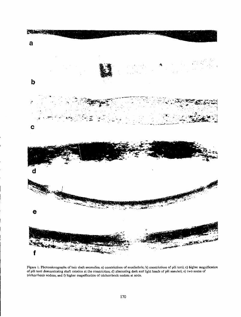
a
b
' .. t
c
d
.,",
.t
.:\:t' -:':;;J',t;.\l\
"" ;~ I¢" ~~ ... ,
""~~ .. ".
Figure I. Photomicrographs of hair shaft anomalies: a) constrictions of monilethrix; b) constrictions of pili torti; c) higher magnification of pili torti demonstrating shaft rotation at the constriction; d) alternating dark and light bands of pili annulati; e) two nodes of trichorrhexis nodosa; and f) higher magnification of trichorrhexis nodosa at node.
170
REFERENCES
Beare, J. M. (1952). Congenital pilar defect showing features of pili torti, Br. J. Dermatol. 64:366-372.
Chernosky, M E. and Owen, D. W. (1966). Trichorrhexis nodosa. Clinical and investigative studies, Arch. Dermatol. 94:577-585.
Price, V. H., Thomas, R. S. and Jone!;~ F. 1: (1968). Pili annulati. Optical and electron microscopic studies, Arch. Dermatol. 98:640-647.
Solomon, L L. and Green, O. C. (1963). Monilethrix: its occurrence in seven generations, with one case that responded to endocrine therapy, New Eng. J. Med. 269:1279-1282.
171
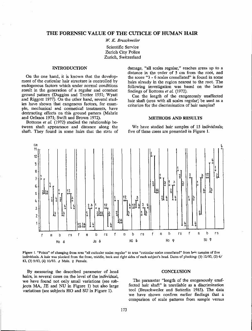
THE FORENSIC VALUE OF THE CUTICLE OF HUMAN HAIR w. K. Bruschweiler
Scientific Set-vice Zurich City Police Zurich, Switzerland
INTRODUCI'ION
On the one hand, it is known that the development of the cuticular hair structure is controlled by endogenous factors which under normal conditions result in the generation of a regular and constant ground pattern (Duggins and Trotter 1951; Wyatt and Riggott 1977). On the other hand, several studies have shown that exogenous factors, for example, mechanical and cosmetical treatments, have destructing effects on this ground pattern (Mahrle and Orfanos 1971; Swift and Brown 1972).
Bottoms et al. (1972) studied the relationship between shaft appearance and distance along the shaft. They found in some hairs that the state of
an 11
10
9
8
1
G
5
4-
3
2
1
L: 1 :'l 2. 0 o 0
4-0
~ 0 La
f m b
Ma (;
12.
'1 1
1 2. 3 1 12.
1 °2h~ 1 o 0
1'2 3~ oo....ao
rs f m b rs f
Je (;
12
damage, "all scales regular," reaches areas up to a distance in the order of 5 cm from the root, and the score "3 - 6 Bcales crenellated" is found in some hairs already in the region nearest to the root. The following investigation was based on the latter findings of Bottoms et al. (1972).
Can the length of the exogenously unaffected hair shaft (area with all scales regular; be used as a criterion for the discrimination of hair samples?
METHODS AND RESULTS
We have studied hair samples of 15 individuals; five of these cases are presented in Figure 1.
0
1 0
~ 0
1 1 0 0
1 2. ~ * 12. ~ g 00
2. 1 .:I ? 1 0 0 0
~~ 4 ?, 0 Lo
:3 1 ~ 1 t l
0 0
12r~ 2. 4 00 0 ll° ~
4- 4 0 0
m b rs f m b rs f m b rs
NO /) Ho Y SO Ii!
Figure 1. "Points" of changing from area "all cuticular scales regular" to area "cuticular scales crenellated" from h~;r :;"mples of five individuals. A hair was plucked from the front, middle, back and right sides of each subject's head. Dates of plucking: (1) 12/82, (2) 6/ 83, (3) 9/83, (4) 10/83. a Male. <t Female.
By measuring the described parameter of head hairs, in several cases on the level of the individual, we have found not only small variations (see subjects MA, JE and NU in Figure 1) but also large variations (see subjects HO and SU in Figure 1).
173
CONCLUSION
The parameter "length of the exogenously unaffected hair shaft" is unreliable as a discrimination tool (Bruschweiler and Sutterlin 1985). The data we have shown confirm earlier findings that a comparison of scale patterns from sample versus
questioned hairs has to be carried out in the region next to the root (approximately 5 mm) where the exposed area of the scale is still unaffected or the break off of scale edges is minor. In this region the original pattern is still visible but exogenous factors begin to have their effects. This configuration, which is found exclusively in the region next to the root, provides the best discrimination rate for hair comparison based on the interpretation of cuticular scale patterns.
Beside this investigation we analyzed hair samples along the whole shaft on the light microscopic or scanning electron microscopic level for damages of the scales by cosmetic treatments or diseases. Abnormal findings such as outstanding or bulgy scales can provide a tool for hair discrimination.
The cuticular scale pattern next to the root and abnormalities along the whole shaft are very valuable features for hair discrimination.
REFERENCES
Auber, L. (1952). The anatomy of follicles producing wool - fibres with special reference to keratinization, Trans. R. Soc. Edinb. 62:191-254.
Bottoms, E, Wyatt, E. and Comaish, S. (1972). Progressive changes in cuticular pattern along the
174
shafts of human hair as seen by scanning electron microscopy, Br. J. Dermatol. 86:379-384.
Bruschweiler, W. and Sutterlin, H. (1985). Die Auswertung der Kutikularstruktur am Hamanhaar, Arch. Kriminol. 175: 156-164.
Duggii:S. O. and Trotter, M (1951). Changes in morphology of hair during childhood, Ann. N. Y. Acad. Sci. 53:569-575.
Mahrle, G. and Oljanos, C. (1971). Haar und Haaroberflache. Variationen des Aufbaus und des Cuticulamusters verschiedener Haare und vershiedener Haarabschnitte, Hautarzt 22:113-120.
Or/anos, C. and Mahrle, G. (1971). Das menschliche Haar und seine Veranderungen unter kosmetischen Prozeduren in Vivo; II, Parfum. Kosmet. 52:235-249.
Or/anas, C. and Ruska, H. (1968). Die Feinstruktur des menschllchen Haares. I. Die Haar-Cuticula, Arch. Klin. Exp. Derm. 231:97-110.
Swift, A. and Brown, A. C. (1972). The critical determination of fine changes in the surface architecture of human hair due to cosmetic treatment, J. Soc. Cosmet. Chern. 23:695-702.
Wyatt, E. H. and Riggott, J. M (1977). Scanning electron microscopy of hair. Observations on surface morphology with respect to site, sex and age in man, Br. J. Dermatol. 96:627-633.
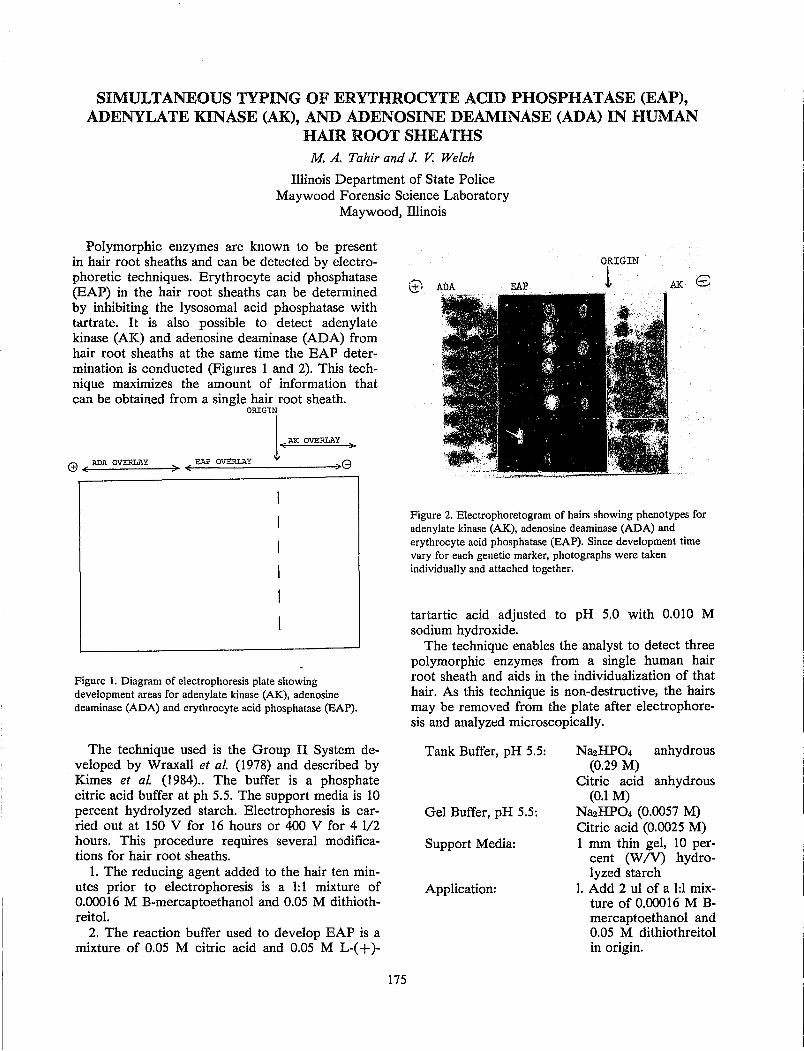
SIMULTANEOUS TYPING OF ERYTHROCYTE ACID PHOSPHATASE (EAP), ADENYLATE KINASE (AK), AND ADENOSINE DEAMINASE (ADA) IN HUMAN
HAIR ROOT SHEATHS M. A. Tahir and J. V. Welch
Illinois Department of State Police Maywood Forensic Science Laboratory
Maywood, Illinois
Polymorphic enzymes are known to be present in hair root sheaths and can be detected by electrophoretic techniques. Erythrocyte acid phosphatase (EAP) in the hair root sheaths can be determined by inhibiting the lysosomal acid phosphatase with tartrate. It is also possible to detect adenylate kinase (AK) and adenosine deaminase (ADA) from hair root sheaths at the same time the EAP determination is conducted (Figures 1 and 2). This technique maximizes the amount of information that can be obtained from a single hair root sheath.
ORIG1:N
l·~~' · (9 ( ADA OVERLAY ;. < EAP OVERLAY :> 8
Figure 1. Diagram of electrophoresis plate showing development areas for adenylate kinase (AK), adenosine deaminase (ADA) and erythrocyte acid phosphatase (EAP).
The technique used is the Group II System developed by Wraxall et al. (1978) and described by
. Kimes et al. (1984).. The buffer is a phosphate citric acid buffer at ph 5.5. The support media is 10 percent hydrolyzed starch. Electrophoresis is carried out at 150 V for 16 hours or 400 V for 4 1/2 hours. This procedure requires several modifications for hair root sheaths.
1. The reducing agent added to the hair ten minutes prior to electrophoresis is a 1: 1 mixture of 0.00016 M B-mercaptoethanol and 0.05 M dithiothreitol.
2. The reaction buffer used to develop EAP is a mixture of 0.05 M citric acid and 0.05 M L-( + )-
175
ORIGIN
1 AK8
Figure 2. Electrophoretogram of hairs showing phenotypes for adenylate kinase (AK), adenosine deaminase (ADA) and erythrocyte acid phosphatase (EAP). Since development time vary for each genetic marker, photographs were taken individually and attached together.
tartartic acid adjusted to pH 5.0 with 0.010 M sodium hydroxide.
The technique enables the analyst to detect three polymorphic enzymes from a single human hair root sheath and aids in the individualization of that hair. As this technique is non-destructive, the hairs may be removed from the plate after electrophoresis and ana1yzt;d microscopically.
Tank Buffer, pH 5.5:
Gel Buffer, pH 5.5:
Support Media:
Application:
Na2HP04 anhydrous (0.29 M)
Citric acid anhydrous (0.1 M)
Na2HP04 (0.0057 M) Citric acid (0.0025 M) 1 mm thin gel, 10 per
cent (W IV) hydrolyzed starch
1. Add 2 u1 of a 1:1 mixture of 0.00016 M Bmercaptoethano1 and 0.05 M dithiothreitol in origin.
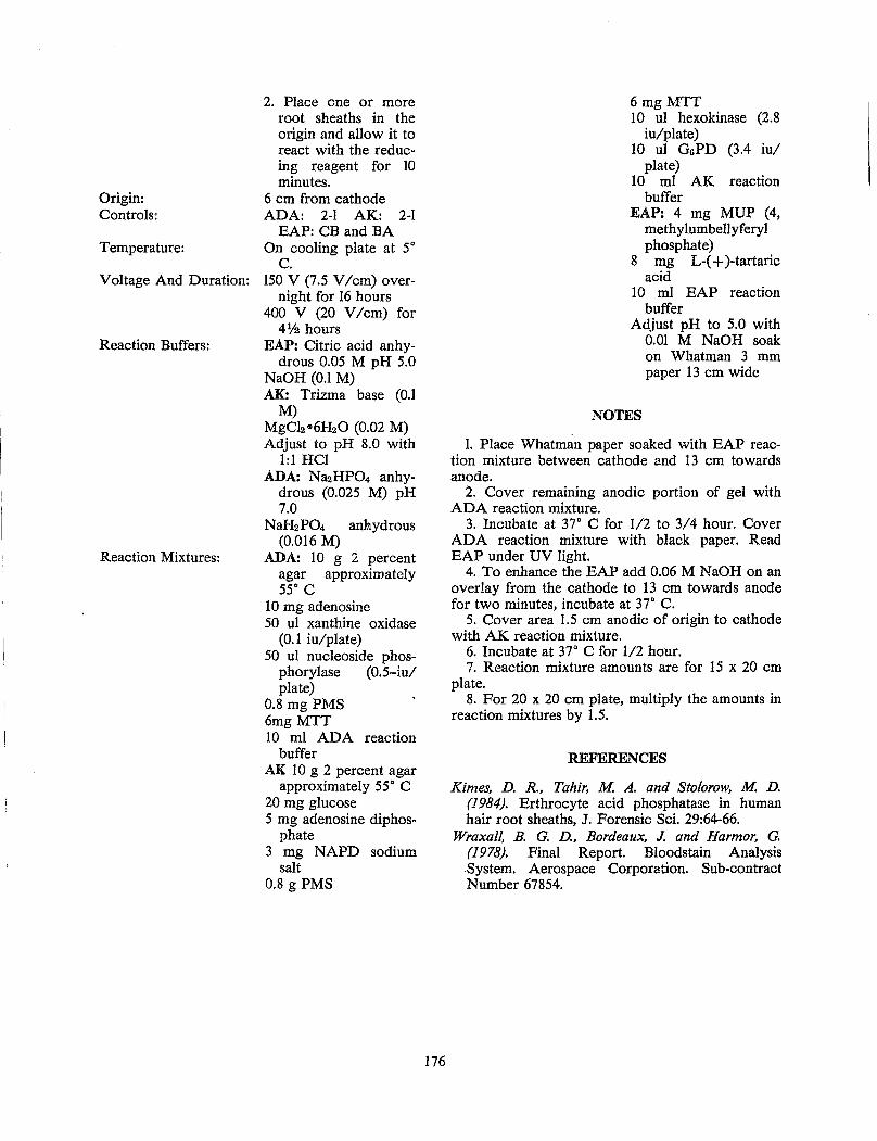
Origin: Controls:
Temperature:
Voltage And Duration:
Reaction Buffers:
Reaction Mixtures:
2. Place one or more root sheaths in the origin and allow it to react with the reducing reagent for 10 minutes.
6 cm from cathode ADA: 2-1 AK: 2-1
EAP: CB and BA On cooling plate at 5°
C. 150 V (7.5 V /cm) over
night for 16 hours 400 V (20 V /cm) for
4% hours EAP: Citric acid anhy
drous 0.05 M pH 5.0 NaOH (0.1 M) AK: Trizma base (0.1
M) MgCb .. 6H20 (0.02 M) Adjust to pH 8.0 with
1:1 HCl ADA: Na2HP04 anhy
drous (0.025 M) pH 7.0
NaH2P04 anhydrous (0.016 M)
ADA: 10 g 2 percent agar approximately 55° C
10 mg adenosine 50 ul xanthine oxidase
(0.1 iu/plate) 50 ul nucleoside phos-
phorylase (0.5-iu/ plate)
0.8 mg PMS 6mgMTT 10 ml ADA reaction
buffer AK 10 g 2 percent agar
approximately 55° C 20 mg glucose 5 mg adenosine diphos
phate 3 mg NAPD sodium
salt 0.8 g PMS
176
6mgMTT 10 ul hexokinase (2.8
iu/plate) 10 ul GsPD (3.4 iu/
plate) 10 ml AK reaction
buffer EAP: 4 mg MUP (4,
methylumbellyferyl phosphate)
8 mg L-( + )-tartaric acid
10 ml EAP reaction buffer
Adjust pH to 5.0 with 0.01 M NaOH soak on Whatman 3 mm paper 13 cm wide
NOTES
1. Place Whatman paper soaked with EAP reaction mixture between cathode and 13 cm towards anode.
2. Cover remaining anodic portion of gel with ADA reaction mixture.
3. Incubate at 37° C for 1/2 to 3/4 hour. Cover ADA reaction mixture with black paper. Read EAP under UV light.
4. To enhance the EAP add 0.06 M NaOH on an overlay from the cathode to 13 cm towards anode for two minutes, incubate at 37° C.
5. Cover area 1.5 cm anodic of origin to cathode with AK reaction mixture.
6. Incubate at 37° C for 1/2 hour. 7. Reaction mixture amounts are for 15 x 20 cm
plate. 8. For 20 x 20 cm plate, mUltiply the amounts in
reaction mixtures by 1.5.
REFERENCES
Kimes, D. R., Tahir, M A. and Stolorow, M. D. (1984). Erthrocyte acid phosphatase in human hair root sheaths, J. Forensic Sci. 29:64-66.
Wraxall, B. G. D., Bordeaux, J. and Harmor, G. (1978). Final Report. Bloodstain Analysis .system. Aerospace Corporation. Sub-contract Number 67854.
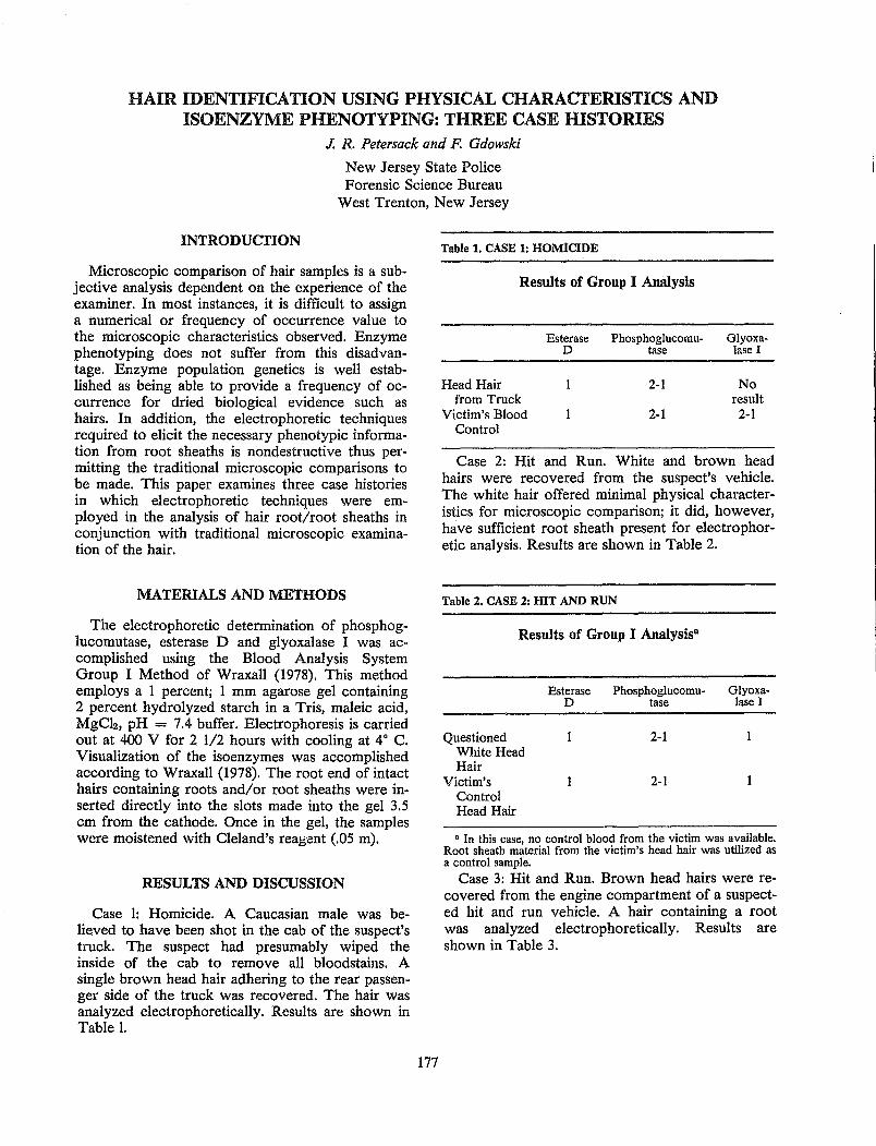
HAIR IDENTIFICATION USING PHYSICAL CHARACTERISTICS AND ISOENZYME PHENOTYPING: THREE CASE HISTORIES
J. R. Petersack and F. Gdowski
New Jersey State Police Forensic Science Bureau
West Trenton, New Jersey
INTRODUCfION
Microscopic comparison of hair samples is a subjective analysis dependent on the experience of the examiner. In most instances, it is difficult to assign a numerical or frequency of occurrence value to the microscopic characteristics observed. Enzyme phenotyping does not suffer from this disadvantage. Enzyme population genetics is well established as being able to provide a frequency of occurrence for dried biological evidence such as hairs. In addition, the electrophoretic techniques required to elicit the necessary phenotypic information from root sheaths is nondestructive thus permitting the traditional microscopic comparisons to be made. This paper examines three case histories in which electrophoretic techniques were employed in the analysis of hair root/root sheaths in conjunction with traditional microscopic examination of the hair.
MATERIALS AND METHODS
The electrophoretic determination of phosphoglucomutase, esterase D and glyoxalase I was accomplished using the Blood Analysis System Group I Method of Wraxall (1978). This method employs a 1 percent; 1 mm agarose gel containing 2 percent hydrolyzed starch in a Tris, maleic acid, MgCI2, pH = 7.4 buffer. Electrophoresis is carried out at 400 V for 2 1/2 hours with cooling at 40 C. Visualization of the isoenzymes was accomplished according to Wraxall (1978). The root end of intact hairs containing roots and/or root sheaths were inserted directly into the slots made into the gel 3.5 cm from the cathode. Once in the gel, the samples were moistened with Cleland's reagent (.05 m).
RESULTS AND DISCUSSION
Case 1: Homicide. A Caucasian male was believed to have been shot in the cab of the suspect's truck. The suspect had presumably wiped the inside of the cab to remove all bloodstains. A single brown head hair adhering to the rear passenger side of the truck was recovered. The hair was analyzed electrophoretically. Results are shown in Table 1.
177
Table 1. CASE 1: HOMICIDE
Results of Group I Analysis
Esterase Phosphoglucomu-D tase
Head Hair 2-1 from Truck
Victim's Blood 2-1 Control
Glyoxalase I
No result
2-1
Case 2: Hit and Run. White and brown head hairs were recovered from the suspect's vehicle. The white hair offered minimal physical characteristics for microscopic comparison; it did, however, have sufficient root sheath present for electrophoretic analysis. Results are shown in Table 2.
Table 2. CASE 2: HIT AND RUN
Results of Group I Analysisa
Questioned White Head Hair
Victim's Control Head Hair
Esterase Phosphoglucomu- Glyoxa-D tase lase I
2-1
2-1
• In this case, no control blood from the victim was available. Root sheath material from the victim's head hair was utilized as a control sample.
Case 3: Hit and Run. Brown head hairs were recovered from the engine compartment of a suspected hit and run vehicle. A hair containing a root was analyzed electrophoretically. Results are shown in Table 3.
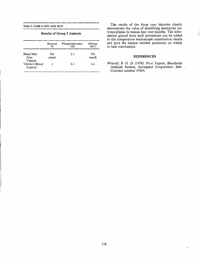
Table 3. CASE 3: HIT AND RUN
Results of Group I Analysis
Esterase Phosphoglucomu-[) tase
Head Hair No 2-1 from result Vehicle
Victim's Blood 2-1 Control
Glyoxalase I
No result
2-1
178
The results of the three case histories clearly demonstrate the value of identifying isoenzyme polymorphisms in human hair root sheaths. The information gained from such procedures can be added to the comparative microscopic examination results and give the analyst another parameter on which to base conclusions.
REFERENCES
Wraxall, B. G. D. (1978). Fih:.V Eeport, Bloodstain Analysis System, Aerospace Corporation. SubContract number 67854.
DOUBLE ENZYME TYPING OF HAIR ROOT SHEATHS USING TWO SINGLE ELECTROPHORETIC SYSTEMS
L. C. Davidson and B. Budowle
Forensic Science Research Group FBI Laboratory
Quantico, Virginia
Attempts to identify the source of hairs found at a crime scene have generally been limited to microscopic comparisons of physical characteristics. Recently, much research has centered around the analysis of hair root sheaths (follicular cells) for polymorphic enzymes and other genetic markers of interest to forensic scientists (Mudd 1984; Oya et al. 1978; Montgomery and Bryan 1982; Sutton et al. 1982). A method has been developed that allows one hair with less than 2.0 mm of sheath to be analyzed for two enzyme systems: glyoxalase I (GLO I) by conventional agarose gel electrophoresis (Budowle 1985) and phosphoglucomutase-1 (PGM-1) by isoelectric focusing (Budowle 1984). The procedures are rapid (30 minutes for GLO I; 80 minutes for PGM-1), reliable, reproducible and require no sample preparation since the hairs are placed directly on the gel. The bands (dots) are highly resolved for both GLO I and PGM-I. It is also apparent from these figures that the migration patterns for GLO I and PGM-1 in hair are indistinguishable from those in blood.
Each hair is analyzed first for GLO I by placing the hair on the gel for 3 minutes prior to electrophoresis. The hair is then removed and the gel is run for 30 min1J.tes at 450 V. Following this analysis, the hair is analyzed for PGM-l. After prefocusing the ultrathin layer gel, the hair is applied at the anode for 10 minutes at 500 V. The hair is then removed and the run is continued. Removing the hair after 10 minutes avoids streaking. Combining data from both systems yields a discriminating probability (DP) of 0.90 for Caucasians and 0.89 for Negroes. This DP is increased over that which can be obtained from analyzing for three markers (PGM-1, EsD and GLO I) by the group I method (0.88 for Caucasians and 0.84 for Negroes) (Wraxall et al. 1978). This improvement in DP is due to the ability to detect PGM-1 subtypes using isoelectric focusing (IEF). Isoelectric focusing techniques distinguish ten phenotypes for PGM-1, whereas the conventional gel electrophoresis used in the group I system approach can only distinguish three phenotypes for PGM-I. Hairs with less than 0.5 mm of sheath should be typed for PGM-1 only, since IEF is a more resolving and concentrating technique
179
than conventional agarose electrophoresis. Conclusive GLO I analysis requires that the hair have at least 1.0 mm of sheath. The PGM-1 analysis, on the other hand, requires that the hair have at least 0.5 mm of sheath.
Results from a stability study showed that 100 percent of PGM-1 phenotypes were conclusive at eight weeks. At 15 weeks, 30 percent of the hairs gave conclusive results for PGM-I. All the hairs were typeable for GLO I activity at four weeks. At eight weeks, GLO I activity was still detectable, but results were inconclusive. Although the results indicate that GLO I and PGM-l are relatively stable at room temperature, it is suggested that when hairs are received in the laboratory, the root portions should be placed in a envelope, and stored in the freezer to maintain stability of the markers for a longer period of time.
REFERENCES
Budowle, B. (1985). An agarose gel method for typing phosphoglucomutase-I, esterase D or glyoxalase I, J. Forensic Sci. 30:1216-1220.
Budowle, B. (1984). Phosphoglucomutase-1 subtyping of human bloodstains on ultrathin-layer polyacrylamide gels, Electrophoresis 5:165-167.
Montgomery, D. and Bryan, W-H.J. (1982). Multiple enzyme systems grouping of human hair root sheaths, Can. Soc. Forensic Sci. 15:1-10.
Mudd, J. L. (1984). The determination of sex from forcibly removed hairs, J. Forensic Sci. 29:1072-1080.
Oya, M, Ito, H., Kido, A., Suzuki, 0., Katsumata, Y. and Yada, S. (1978). Phosphoglucomutase-l (PGM-1) and 6-phosphogluconate dehydrogenase (PGD) types in the human hairbulb, Forensic Sci. 11:135-138.
Sutton, J. G., Bosley, C.M and Whitehead, P.H. (1982). Polymorphic enzyme systems in human hair sheath cells, J. Forensic Sci. Soc. 22:199-202.
Wraxall, B., Bordeaux, J. and Harmor, G. (1978). Final Report, Bloodstain Analysis System. Aerospace Corporation, Sub-Contract Number 67854.
ABO GROUPING OF HUMAN HAIR ROOTS BY ABSORPTION-ELUTION METHOD
v. N. Sehgal and R. K. Bhatnagar
Central Forensic Science Laboratory Central Bureau of Investigation
New Delhi, India
INTRODUCfION
In India, hair research in forensic science has been confined to mostly species identification only. However, successful attempts have been made by the authors to characterize the human head hairs in the phosphoglucomutase (PGM) and esterase D (EsD) systems (Bhatnagar and Srivastava 1979, 1982; Bhatnagar et al. 1984), but attempts to group human hair without sheath cells in the ABO system have met with little success. Nevertheless, hair roots exhibited promising results. Lincoln and Dodds (1968) also did not find antigenic activity in hair, and the results were not conclusive enough for routine typing and court evidence.
Recently, a study on the detectability of ABO antigens in human hair roots was undertaken by the authors in order to ascertain its applicability in case work under the Indian tropical climatic conditions. The results of the study have been compared with PGM and EsD genetic markers.
MATERIAL AND METHODS
Collection of Hair Samples
Hairs were plucked from various regions of the scalp of 200 members, mostly males, of the staff of the Central Forensic Science Laboratory/CBI, New Delhi, their families and other donors. These hairs were stored in screw-capped glass tubes at room temperature until tested. ABO grouping of hair root was carried out by the absorption-elution method of Howard and Martin (1969) using disposable cellulose acetate sheets. This method has been adopted in the author's laboratory since 1970 (Bhatnagar et al. 1974).
The hair strands with intact hair root were treated the same way as blood/semen staim;d threads using high titre anti-A and anti-B sera. Cell suspension of 0.2 percent prepared in physiological saline was used for detecting the eluted antibodies.
RESULTS AND DISCUSSION
More that 200 known and unknown hairs have been correctly grouped in the ABO system during the past several years in this laboratory. The group of the known sample tallied with their corresponding blood samples. Some of the hairs were from
181
actual cases. It has been found that it is possible to correctly determine the ABO blood group in dropped or pulled hairs containing sheath cells. Generally, the hairs without the root showed no antigenic reaction; therefore, they were not included in this study. These samples were excluded after the initial microscopic examination.
The study has revealed it is possible to correctly determine the group of hair within a period of approximately 6 months. Thereafter the antigenic activity diminished and the group could not be determined due to completely negative reactions.
Characterization of hair based upon various polymorphic enzyme systems found in the hair sheath cells has provided very powerful methods of discriminating hair of different persons. However, the limitation of these methods is that PGM and EsD isoenzymes in hair roots can be detected only up to 6 weeks in India. On the other hand, the present study revealed that ABO antigens in the same material could be detected up to 6 months. Hence, in cases in which the hair has lasted more than 6 weeks, ABO grouping proves more useful than the PGM or EsD systems.
REFERENCES
Bhatnagar, R. K. and Srivastava, O. S. (1979). Phosphoglucomutase (PGM) typing of hair roots. Symposium Science and Law, Punjabi University, Patiala.
Bhatnagar R. K. and Srivastava, O. S. (1982). Esterase D (EsD) typing of plucked human head hairs by thin layer starch gel electrophoresis. In: Proceedings of the Scientific Meeting Indian Academy of Forensic Sciences, New Delhi.
Bhatnagar R. K., Srivastava O. S. and Sehgal V. N. (1984). Detection of phosphoglucomutase (PGM) and esterase (EsD) isoenzymes in plucked human hairs, J. Forensic Sci. Soc. 24:316.
Bhatnagar R. K., Bhandari, S. K., Sijher, J. S. and Srivastava, P. P. (1974). Preservation effects in forensic ABO-determinations using analysis of results as evidence in criminal cases, Arch. Kriminol. 154:91-9.
Howard, H. D. and Martin, P. D. (1969). An improved method for ABO and MN grouping of dried blood stains using cellulose acetate sheets, J. Forensic Sci. Soc. 9:28-30.
Lincoln, P. J. and Dodds, B. E. (1968). Mixed agglutination as a method for the determination of
182
A, Band H blood groups of hair, Med. Sci. Law 8:38.

SECTION III
SHORT ABSTRACTS
183
COLLECTION AND PRESERVATION FOR ANALYSIS OF TRACE CONTAMINANTS FOUND ON HAIRS
D. Depczynski and D. A. Metzger
Illinois Department of State Police Fairview Heights, Illinois
As the areas within forensic science become more and more specialized the scientist may begin to overlook evidence which could be significant to another analyst. In the field of hair examination, there will often be contamination present which, if collected and preserved, may provide valuable information when examined by another specialist. The purpose of this paper is to provide a guideline which will aid the hair examiner in identifying the possible contaminants that may be encountered, devising the best method for the removal and preservation of that contaminant, and overcoming any problems which may be encountered in this process.
These contaminants have been placed into four categories: biological substances which may dry
185
onto hairs, particulate substances which may coat hairs, organic substances, and substances which may commingle with hairs. Once observed, the problems associated with removal must be overcome. The contaminant must be removed and preserved in a concentrated state suitable for further analysis. The method of collection depends on the type of contaminant and its quantity, and the type of analysis which will ultimately be performed. Some methods include chemical removal with organic solvents, removal with distilled water and manual removal. Equipment utilized includes the magnified illuminator, stereoscope and aids such as the tungsten needle.
MISLEADING COLOR CHANGES IN HAIR THAT HAS BEEN HEATED BUT NOT EXPOSED TO FLAME
L. M. Ayres
Kentucky State Police Central Forensic Laboratory
Frankfort, Kentucky
Last year we received two separate cases in which hair that had been exposed to intense heat for a prolonged period of time played a crucial role in the investigation. The first case involved a woman who was found dead in a partially burned mobile home. The investigating officer who collected loose hair from her body wanted to know if it matched a head hair standard from an area of her head protected from the heat, or had it originated from an assailant. The second case involved a woman who was raped, sexually abused and shot in the abdomen. The assailant cut a large amount of the victim's head hair, placed it in a pan and heated it on the stove. The sample of hair from the pan and a hair standard were submitted to our laboratory in order to substantiate the victim's story. In both cases the hair exposed to heat had a different appearance than was expected.
This study was undertaken to determine the changes between hair exposed to gradually increased heat and hair exposed to flame. Light brown Caucasian head hair was placed in a glass beaker and heated on a hot plate for approximately four minutes until a color change was noted. Mi-
187
croscopic observations revealed that the areas of the hair exposed to the greatest heat had a color change to dark reddish brown. Bubbles were sometimes noted in the area of the medulla; occasional slight swelling of the hair and looped appearance of the cuticle were noted in the reddish-brown area. The adjacent area was orange, followed by yellow and then the natural color of the hair with no apparent heat damage. Hair treated in this manner had the appearance of having been dyed. The medulla appeared normal, but the cuticle was colored through each of these heat damaged stages. In order to determine if the difference in color was due to changes in the pigment or in the keratin, white hair was also tested. The same appearance was noted with white hair exposed to heat as with the pigmented hair. Hair exposed directly to flame showed a charred, bubbled effect, but no color change in the adjacent area was noted.
The color changes noted following gradually increased heat appear to be due to the deterioration of the keratin rather than just an effect on the surface of the hair or in the pigment.
HAIR USED IN THE IDENTIFICATION OF A DISMEMBERED BODY F. A. Springer
California Department of Justice Bureau of Forensic Services
Criminalistics Laboratory Riverside, California
On May 20, 1983, the upper torso of a female was discovered in the California desert. The head, arms and most of the internal viscera were missing. Long black hairs were attached to the neck area. Some of the hairs were approximately 0.9144 m. Microscopic examination of the hairs revealed some interesting features. Approximately three fourths of the hair length had natural microscopic features. Near the distal end of the hair, evidence of black dye with patches of reddish-brown dye interspersed was apparent.
On May 21, 1983, an American Indian woman from Los Angeles, California was reported missing. According to the woman's roommate, the missing woman had dyed her hair reddish brown or auburn in 1975. Approximately one year later the missing woman dyed her hair back to the original color of black.
Subsequently, the victim's purse was recovered from a trash bin. A hair brush containing hair was found in the purse. Comparison of the hairs from the torso to hairs from the brush showed similar microscopic features including the dye patterns. The victim was thus identified as the missing Los Angeles woman based on the hair comparisons, the physical build, and the presence of a freckle or mole on the chest of the victim. The position of the mole or freckle was substantiated by a photo-
189
graph provided to investigators by the victim's roommate.
The last person to have seen the victim alive became the prime suspect. Pursuant to a search warrant, small amounts of blood were located in the suspect's residence. The various types of blood matched that of the victim's. Several pairs of boots were taken from the suspect's residence during the execution of the search warrant. One boot had a wad of pink gum on the sole. Approximately two weeks later, the victim's vehicle was discovered in a shopping center parking lot near the suspect's residence. Pink gum was present on the accelerator pedal of the victim's vehicle. Fibers and hairs embedded in the gum from the boots compared favorably to fibers from the victim's vehicle and the victim's head hairs.
Approximately 4 months after the aforementioned incident, the victim's skull was located several miles from the scene where the torso was discovered. Upon sifting sand in a shallow cavity where the skull was probably buried, a bullet was located. This bullet was identified as having been fired by a weapon recovered from the suspect's residence in the original searcr. warrant.
One year later, the suspect was tried and found guilty of first degree murder.
SECTION IV
PANEL DISCUSSION
191
PANEL DISCUSSION EVIDENTIAL VALUE OF HAIR EXAMINATIONS
PANEL MODERATOR: John W. Hicks, FBI Laboratory
PANEL MEMBERS: Harold A. Deadman, FBI Laboratory Peter R. De Forest, John Jay College Barry D. Gaudette, Royal Canadian Mounted Police Geoffrey M Roe, Metropolitan Police Forensic Science Laboratory Manfred Wittig, Bundeskriminalamt
Hicks: We sincerely appreciate the enthusiasm of those who have participated in our program and their willingness to participate. I think it has made a much richer program to this point and so my thanks go to you.
Our goal in this symposium has been to bring togetherthe perspectives of industry, academia and practicing forensic scientists, and to create a forum for discussion. Through this process, hopefully, we will develop a better understanding of a topic area; gain a better appreciation for the various individual perspectives and ouf' mutual interests; and identify areas that are needed for further research and inquiry. So I hope that through this meeting these goals will be the end result for us.
The panelists are Barry D. Gaudette, Chief Scientist for the Hair and Fibres Section for the Royal Canadian Mounted Police in Ottawa, Canada; Dr. Geoffrey M. Roe, Chief Scientific Officer for the Metropolitan Police Forensic Science Laboratory in London, England; Dr. Manfred Wittig, Scientific Director for the General Biology Section for the Bundeskriminalamt in Wiesbaden, Federal Republic of Germany; Dr. Harold A. Deadman, Special Agent assigned to the Microscopic Analysis Unit, of the FBI Laboratory in Washington, D. C.; and Dr. Peter De Forest, Professor of Criminalistics at John Jay College of Criminal Justice in New York City.
Each of the panelists will make a brief statement on the status of hair examinations within their respective country, in their respective agencies and then perhaps include a comment as to the assessment of the significance and the value of hair cOmparisons.
Gaudette: Hair examinations have been conducted in the Royal Canadian Mounted Police Laboratory system for the last 46 years. We now have approximately 24 hair and fiber examiners in our system. Every year our system receives over 1000 cases in which hair comparison is involved, and every year our examiners spend a total of more than 200 work days attending court giving testimony about forensic hair comparison. These figures
193
demonstrate our belief that forensic hair comparisons are valuable and that hair comparison evidence is good evidence. In determining the evidential value of hair comparison or any other physical or associative evidence for that matter, the fundamental question to consider is not what proportion of the suspect population would have hair with a particular set of characteristics, but rather what is the value of the evidence in establishing a particular association. The fundamental question incorporates the frequency of occurrence aspect implicit in the first question. It also deals with many other factors including (1) the probability of incorrect association resulting from the occurrence of other prerequisite events (with hair this is the probability that one particular hair type out of the approximately nine types on the scalp would be the one to be found in evidence); (2) the probability of examiner errors; and (3) the probability of incorrect association due to other factors such as secondary transfer, contamination, or deliberate planting of evidence.
When: we consider all these various factors and aU the variables involved in a particular case it can be seen that it would be extremely difficult (if not impossible) to give exact statistical answers to the fundamental question for each individual case.
Does this then mean that statistical determination of evidential value of hair should be abandoned? I think not. Consideration of the factors involved in statistical evaluation of hair comparison evidence helps an examiner develop a conceptual framework that can increase his or her understanding and be of assistance in making several important decisions such as research design and research priorities. Furthermore, since laymen have little intuitive feeling for the value of hair comparison evidence, evidential value statistics can be informative in court testimony provided that they are used pruperly.
To provide proper statistical evaluation of hair comparison evidence an examiner should first insure that the statistics used relate as closely as possible to the fundamental question. Second, insure that statistics are placed in the proper con-
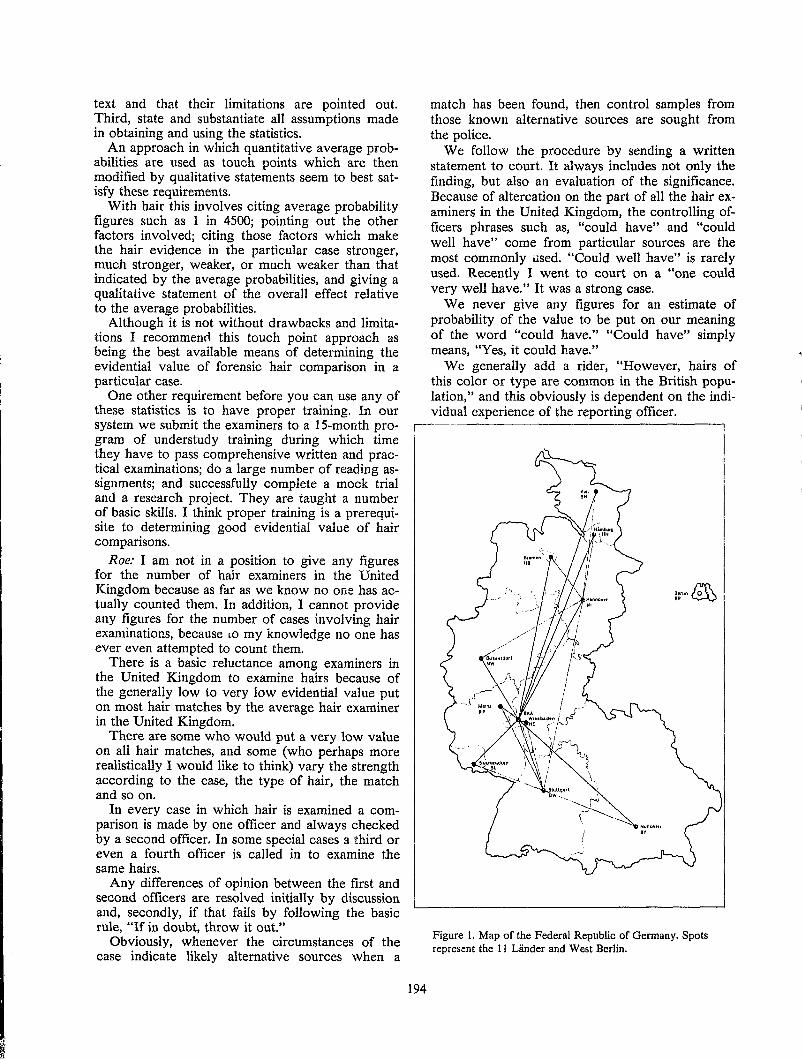
text and that their limitations are pointed out. Third, state and substantiate all assumptions made in obtaining and using the statistics.
An approach in which quantitative average probabilities are used as touch points which are then modified by qualitative statements seem to best satisfy these requirements.
With hair this involves citing average probability figures such as 1 in 4500; pointing out the other factors involved; citing those factors which make the hair evidence in the particular case stronger, much stronger, weaker, or much weaker than that indicated by the average probabilities, and giving a qualitative statement of the overall effect relative to the average probabilities.
Although it is not without drawbacks and limitations I recommend this touch point approach as being the best available means of determining the evidential value of forensic hair comparison in a particular case.
One other requirement before you can use any of these statistics is to have proper training. In our system we submit the examiners to a IS-month program of understudy training during which time they have to pass comprehensive written and practical examinations; do a large number of reading assignments; and successfully complete a mock trial and a research project. They are taught a number of basic skills. I think proper training is a prerequisite to determining good evidential value of hair comparisons.
Roe: I am not in a position to give any figures for the number of hair examiners in the United Kingdom because as far as we know no one has actually counted them. In addition, I cannot provide any figures for the number of cases involving hair examinations, because LO my knowledge no one has ever even attempted to count them.
There is a basic reluctance among examiners in the United Kingdom to examine hairs because of the generally low to very low evidential value put on most hair matches by the average hair examiner in the United Kingdom.
There are some who would put a very low value on all hair matches, and some (who perhaps more realistically I would like to think) vary the strength according to the case, the type of hair, the match and so on.
In every case in which hair is examined a comparison is made by one officer and always checked by a second officer. In some special cases a third or even a fourth officer is called in to examine the same hairs.
Any differences of opinion between the first and second officers are resolved initially by discussion and, secondly, if that fails by following the basic rule, "If in doubt, throw it out."
Obviously, whenever the circumstances of the case indicate likely alternative sources when a
194
match has been found, then control samples from those known alternative sources are sought from the police.
We follow the procedure by sending a written statement to court. It always includes not only the finding, but also an evaluation of the significance. Because of altercation on the part of all the hair examiners in the United Kingdom, the controlling officers phrases such as, "could have" and "could well have" come from particular sources are the most commonly used. "Could well have" is rarely used. Recently I went to court on a "one could very well have." It was a strong case.
We never give any figures for an estimate of probability of the value to be put on our meaning of the word "could have." "Could have" simply means, "Yes, it could have."
We generally add a rider, "However, hairs of this color or type are common in the British population," and this obviously is dependent on the individual experience of the reporting officer.
Figure 1. Map of the Federal Republic of Germany. Spots represent the 11 Lander and West Berlin.
We feel that until we have a large data base for each hair characteristic we have to rely on our own experience in presenting evidence in our courts.
The final comment, the United Kingdom Forensic Science Services does have a coordinating committee called Inter-Laboratory Advisory Committee (ILAC). Currently there is one specifically for hair. Shortly it will be amalgamated with the Committee for Fibers as I understand.
The ILAC does set up both declared blind and, allegedly, undeclared blind trials which are sent to all the laboratories in the United Kingdom. These are expected to be treated as normal forensic case examinations.
These have shown some considerable discrepancies between examiners in the occurrence of both type I and type II errors. This further reinforces our view that we should never overstate the strength of our hair match in our courts.
Currently in the United Kingdom there is no formal training program for hair examiners. This is something I think we can learn an awful lot from the Canadians and the Americans. It is something I think we really should have.
195
Wittig: Essentially at this time we regard the examination of hair in a similar manner as my colleague from Britain, unfortunately.
To explain the forensic science system in the Federal Republic of Germany, I refer to figures 1-3. This is the Federal Republic of Germany (Figure 1), and you will see there are 11 black spots plus 1 white spot. These are the 11 Lander, (comparable to your states) and West Berlin.
In all these Lander there is a Landeskriminaliimt with its own laboratory. Of course there are communications between these laboratories as shown by the lines.
The next two figures (2 and 3) are copies of a former effort undertaken by Dr. Curry of the HOCRE in th~ United Kingdom. In the early 1970s, he listed all police-associated forensic science laboratories within the member countries of the European Economic Community (EEC). Their location and distribution are shown in Figure 2. In Figure 3 the circles numbered 31 to 42 represent the different Landeskriminaliimt and the Bundeskrimina\amt. You can see which facilities (divisions) are established there.
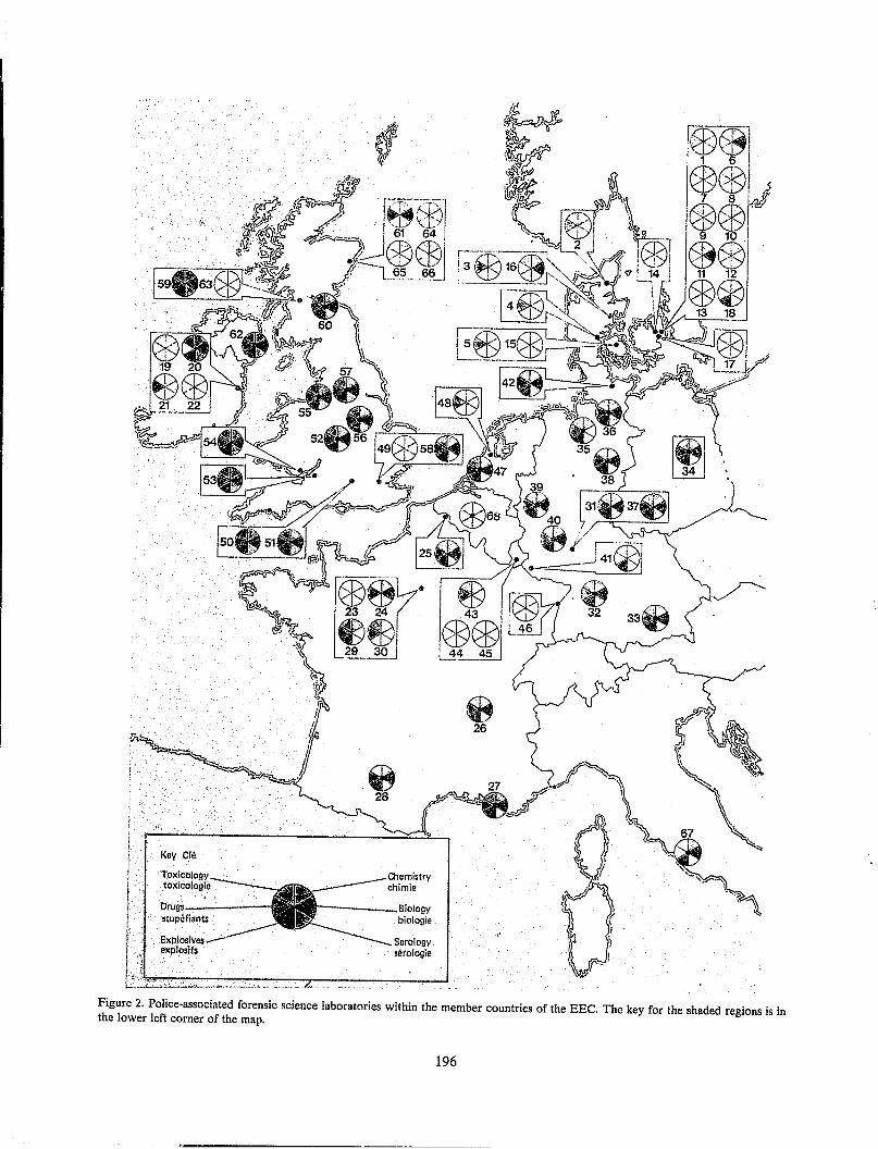
foxicology toxicologle
stop6fiants .
Explosives exploslfs
r---___ Biology biologie
Figure 2. Police-associated forensic science laboratories within the member countries of the EEe. The key for the shaded regions is in the lower left corner of the map.
196
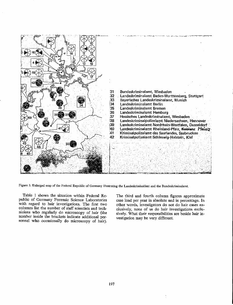
31 Bundeskriminalamt, Wiesbaden 32 Landeskriminalamt Baden-Wurttemberg, Stuttgart 33 Bayerisches Landeskriminalamt, Munich ;34 Landeskriminalamt Berlin 35 Landeskriminalamt Bremen 36 Landeskriminalamt Hamburg 37 Hessisches Landeskriminalamt, Wiesbaden 38 LandeskriminalpolizelalJ1t Niedersaahsen, Hannover 139 Landeskriminalamt Nordrhein~Westfalen, Dusseldorf . 140 Landeskriminalamt Rheinland-Pfalz, ~91I1Rii! MQ."2, :41 Kriminalpolizeiamt des Saarlandes, Saabrucken ;42 Kriminalpolizeiamt Schleswig-Holstein, Kiel
Figure 3. Enlarged map of the Federal Republic of Germany illustrating the Landeskriminaliimt and the Bundeskriminalamt.
Table 1 shows the situation within Federal Republic of Germany Forensic Science Laboratories with regard to hair investigations. The first two columns list the number of staff scientists and technicians who regularly do microscopy of hair (the number inside the brackets indicate additional personnel who occasionally do microscopy of hair).
197
The third and fourth column figures approximate case load per year in absolute and in percentage. In other words, investigators do not do hair cases exclusively, none of us do hair investigations exclusively. What their responsibilities are beside hair investigation may be very different.
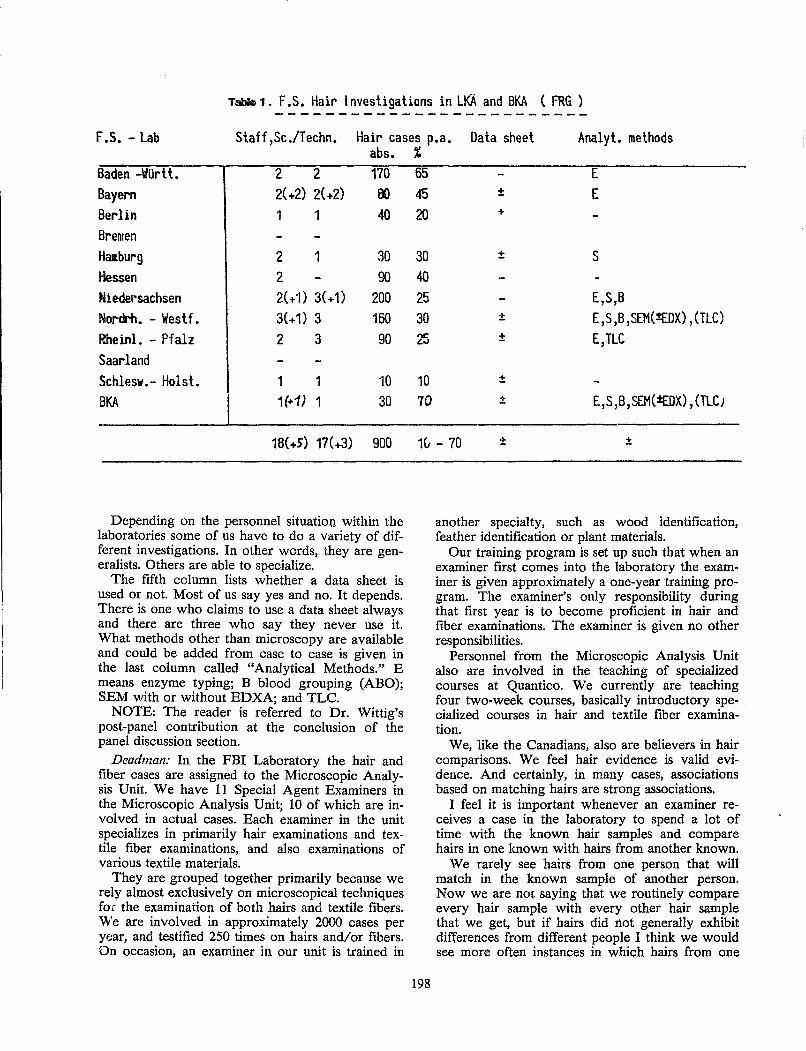
Table 1. f .S. Hair I nvestigations in LKA and BKA ( FRG ) -------------------------
f .S. - lab Staff,Sc./Techn. Hair cases p.a. Data sheet Analyt. methods abs.
Baden -Wiirtt. 2 2 170 Bayern 2(+2) 2(+2) 00 Berlin 1 1 40 Bremen Haraburg 2 1 30 Hessen 2 90 Niedersachsen 2(+1) 3(+1) 200 Nordrh. - Westf. 3{+1) 3 160 Rheinl. - Pfalz 2 3 90 Saarland Schlesw.- Holst. 1 1 10 BKA 1(+1) 1 30
18(+5) 17(+3) 900
Depending on the personnel situatioo within the laboratories some of us have to do a variety of different investigations. In other words, they are generalists. Others are able to specialize.
The fifth column lists whether a data sheet is used or not. Most of us say yes and no. It depends. There is one who claims to use a data sheet always and there are three who say they never use it. What methods other than microscopy are available and could be added from case to case is given in the last column called "Analytical Methods." E means enzyme typing; B blood grouping (ABO); SEM with or without EDXA; and TLC.
NOTE: The reader is referred to Dr. Wittig's post-panel contribution at the conclusion of the panel discussion section.
Deadman: In the FBI Laboratory the hair and fiber cases are assigned to the Microscopic Analysis Unit. We have 11 Special Agent Examiners in the Microscopic Analysis Unit; 10 of which are invol ved in actual cases. Each examiner in the unit specializes in primarily hair examinations and textile fiber examinations, and also examinations of various textile materials.
They are grouped together primarily because we rely almost exclusively on microscopical techniques for the examination of both hairs and textile fibers. We are involved in approximately 2000 cases per year, and testified 250 times on hairs and/or fibers. On occasion, an examiner in our unit is trained in
% 65 E 45 ± E 20 +
30 ± S 40
25 E7S,S 30 ± E,S,B,SEM(
sample, from one individual, would match in the sample, or among hairs in the other sample.
We are also involved with taking matching tests in our training program and our other activities. The experience of the Canadians in the tests they utilize in their training program I believe demonstrates that individuals can do an excellent job of matching hairs with the source that they originated from.
In the training of students in our hair and fiber school, I routinely look at the hairs generally of each student in the class. Admittedly, this is a random group of 12 individuals that usually involve different sexes and different races, but a test set up with hairs from those individuals would generally be a very trivial test. It would be no problem for an inexperienced examiner to take a matching test utilizing hairs from students in the class and not make any mistakes in terms of making the proper associations.
So we believe in hair comparisons. Certainly the strength in the association depends a lot on the hairs, the characteristics that the hairs exhibit. Some hair matches are not perhaps the basis of a strong association. The common featureless hair Barry Gaudette has described in some of his writings perhaps is not an association that should be argued as a strong one in court. But when hair matches involve hairs that have very individual characteristics, uncommon characteristics, when you have multiple hairs forming the basis of your association, and a variety of other factors, all of which contribute to the strength of the association, we testify in some instances quite strongly about hairs.
Certainly hair evidence being trace evidence is often accompanied by other types of trace evidence which form the basis of our associations. I rarely have had a case in which I testify only to hair evidence. Often there will be fiber evidence involved. Often other examiners will testify and the combination of the hairs along with the other evidence often results in extremely strong associations.
De Forest: I am going to discuss the evidential value of hair. Then I will discuss some of the defense expert's perspectives on the hair question. Although I do some civil work and occasional prosecution work as well, I would like to share with you some of the perspectives I have in doing defense work.
I think we all agree that hair is good evidence. It is an appealing kind of evidence because of its ubiquitousness; its frequency of occurrence at crime scenes; and its stability as contrasted with some of the biological types of evidence.
We all realize it is difficult; we are talking about a subjective process in comparing these things and it is also a great time-consuming process when done properly. But it works.
199
Things that make it complicated were indicated in the presentations by Richard Bisbing (pages 35-44 this proceedings) and others. The large number of variables we are concerned with have to be assimilated somehow in the mind, and we are talking about a large number of continuously variable parameters. We have to also take into account the fact that combined with this complexity is the complexity of significant intrasample variability.
The power of hair examination is based on the a priori potential to exclude hair. In other words, if you receive two samples, a known and questioned sample, the power of the technique is based on its ability ahead of time to exclude these.
At worst, hair evidence is very competitive with other kinds of non-absolute associative evidence. Because of the type of selection process that goes on and the fact that we are talking about randomly selected hairs being submitted, there are many times when an exclusion is trivial. And so for that very reason the advantage of being able to exclude hairs that are obviously different, has value that is competitive with other kinds of evidence such as limited blood typing, and so forth.
It is when the hairs that have been brought in are similar to each other (the known and questiond are similar) that we, of course, face the difficulty of the long hours and long involved examinations. We really basically come down in the end to failing to exclude the hairs and concluding from that, and interpreting that failure to exclude as a match or an inclusion.
I think for the foreseeable future, as others would agree, microscopy is going to be our approach to the sexing, typing and so forth of hairs with the help of adjunct techniques such as electrophoresis of the enzymes and root sheath, etc. Despite the fact we are going to have microscopy with us for a long time I see no way around that. I think we should proceed with work on developing other techniques that would make this process shorter, less time consuming and more objective.
Now I will provide the perspective of a defense expert. The approach I take when I am contacted by an attorney is basically to look at who has done the work on the case and what kind of procedure has been followed. Normally, I advise against a reexamination, and there are reasons for that.
First of all, to do a bona fide reexamination will take many many hours and I do not work for free. So it makes for very large bills which I think are ridiculous in light of the fact that I do not anticipate finding something different. In other words, my prediction about being able to come up with an exclusion that would please the defense at the end of the examination I think is an outside chance. I have only had one case in which I have disagreed with another examiner and that was a case in
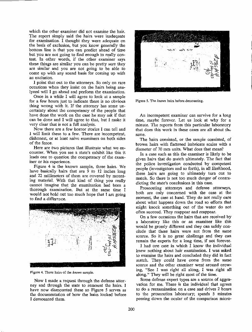
which the other examiner did not examine the hair. The report simply said the hairs were inadequate for examination. I thought they were adequate on the basis of exclusion, but you know generally the bottom line is that you can predict ahead of time but you are not going to find enough to really contest. In other words, if the other examiner says these things are similar you can be pretty sure they are similar and you are not going to be able to come up with any sound basis for coming up with an exclusion.
I point that out to the attorneys. So only on rare occasions when they insist on the hairs being analyzed will I go ahead and perform the examination.
Once in a while I will agree to look at a sample for a few hours just to indicate there is no obvious thing wrong with it. If the attorney has some uncertainty about the competency of the people that have done the work on the case he may ask if that can be done and I will agree to that, but I make it very clear that is not a full analysis.
Now there are a few horror stories I can tell and I will limit these to a few. There are incompetent, dishonest, or at least naive examiners on both sides of the fence.
Here are two pictures that illustrate what we encounter. When you see a state's exhibit like this it leads one to question the competency of the examiner or his experience.
Figure 4 is the known sample, three hairs. We have basically hairs that are 9 to 12 inches long and 22 millimeters of them are covered by mounting material. With that kind of thing you really cannot imagine that the examination had been a thorough examination. But at the same time I would not hold out too much hope that I am going to find a difference.
Figure 4. Three hairs of the known sample.
Now I made a request through the defense attorney and through the state to remount the hairs. I have now dismounted these so Figure 5 serves as the documentation of how the hairs looked before I demounted them.
200
Figure 5. The known hairs before demounting.
An incompetent examiner can survive for a long time, maybe forever. Let us look at why for a minute. The reports from this particular laboratory that does this work in these cases are all about th" same.
The hairs consisted, or the sample consisted, of brown hairs with flattened imbricate scales with a diameter of 70 mm units. What does that mean?
In a case such as this the examiner is likely to be given hairs that do match ultimately. The fact that the police investigation conducted by competent people (investigators and so forth), in all likelihood, these hairs are going to ultimately turn out to match. So there is not too much danger of contradicting the state's conclusions in this case.
Prosecuting attorneys and defense attorneys, both are only concerned with the case at the moment, the case at hand. They do not really care about what happens down the road so efforts that might knock something out of the water do not often succeed. They reappear and reappear.
On a few occasions the hairs that are received by a laboratory like this or an examiner like this would be grossly different and they can safely conclude that these hairs were not from the same source. So it is no great challenge and they can remain the experts for a long time, if not forever.
I had one case in which I know the individual knew nothing about hair examination. I was asked to examine the hairs and concluded they did in fact match. They could have come from the same source and the other examiner went around crowing, "See I was right all along, I was right all along." They will be right most of the time.
Some defense expert types are a source of aggravation for me. There is the individual that agrees to do a reexamination on a case and drives 2 hours to the prosecution laboratory; spends 5 minutes peering down the ocular of the comparison micro-
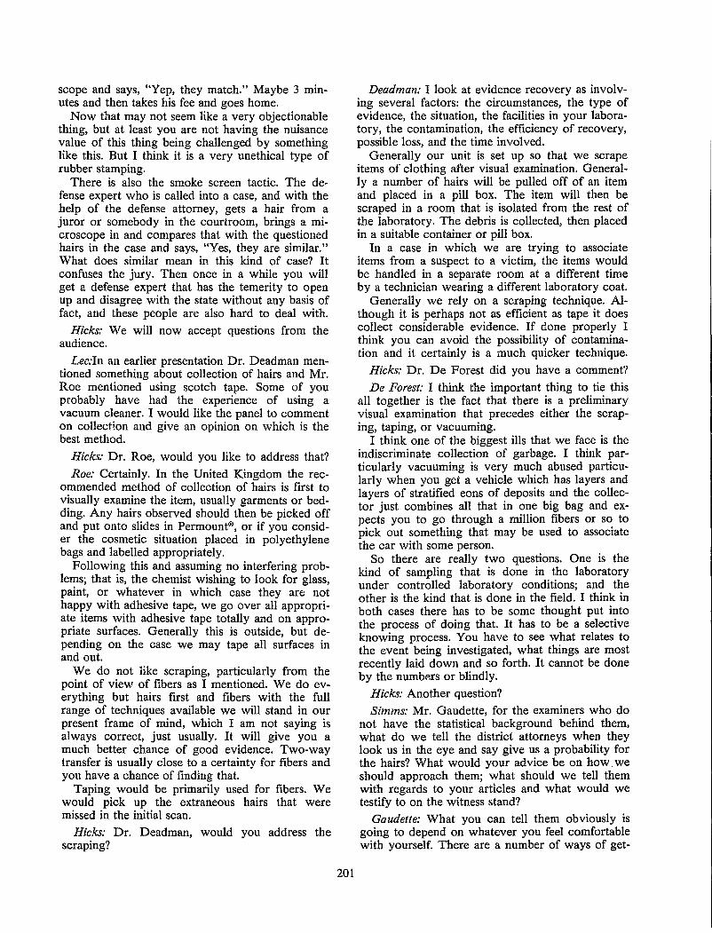
scope and says, "Yep, they match." Maybe 3 minutes and then takes his fee and goes home.
Now that may not seem like a very objectionable thing, but at least you are not having the nuisance value of this thing being challenged by something like this. But I think it is a very unethical type of rubber stamping.
There is also the smoke screen tactic. The defense expert who is called into a case, and with the help of the defense attorney, gets a hair from a juror or somebody in the courtroom, brings a microscope in and compares that with the questioned hairs in the case and says, "Yes, they are similar." What does similar mean in this kind of case? It confuses the jury. Then once in a while you will get a defense expert that has the temerity to open up and disagree with the state without any basis of fact, and these people are also hard to deal with.
Hicks: We will now accept questions from the audience.
Lec:ln an earlier presentation Dr. Deadman mentioned something about collection of hairs and Mr. Roe mentioned using scotch tape. Some of you probably have had the experience of using a vacuum cleaner. I would like the panel to comment on collection and give an opinion on which is the best method.
Hicks: Dr. Roe, would you like to address that?
Roe: Certainly. In the United Kingdom the recommended method of collection of hairs is first to visually examine the item, usually garments or bedding. Any hairs observed should then be picked off and put onto slides in Permount®, or if you consider the cosmetic situation placed in polyethylene bags and labelled appropriately.
Following this and assuming no interfering problems; that is, the chemist wishing to look for glass, paint, or whatever in which case they are not happy with adhesive tape, we go over all appropriate items with adhesive tape totally and on appropriate surfaces. Generally this is outside, but depending on the case we may tape all surfaces in and out.
We do not like scraping, particularly from the point of view of fibers as I mentioned. We do everything but hairs first and fibers with the full range of techniques available we will stand in our present frame of mind, which I am not saying is always correct, just usually. It will give you a much better chance of good evidence. Two-way transfer is usually close to a certainty for fibers and you have a chance of finding that.
Taping would be primarily used for fibers. We would pick up the extraneous hairs that were missed in the initial scan.
Hicks: Dr. Deadman, would you address the scraping?
201
Deadman: I look at evidence recovery as involving several factors: the circumstances, the type of evidence, the situation, the facilities in your laboratory, the contamination, the efficiency of recovery, possible loss, and the time involved.
Generally our unit is set up so that we scrape items of clothing after visual examination. Generally a number of hairs will be pulled off of an item and placed in a pill box. The item will then be scraped in a room that is isolated from the rest of the laboratory. The debris is collected, then placed in a suitable container or pill box.
In a case in which we are trying to associate items from a suspect to a victim, the items would be handled in a separate room at a different time by a technician wearing a different laboratory coat.
Generally we rely on a scraping technique. Although it is perhaps not as efficient as tape it does collect considerable evidence. If done properly I think you can avoid the possibility of contamination and it certainly is a much quicker technique.
Hicks: Dr. De Forest did you have a comment?
De Forest: I think the important thing to tie this all together is the fact that there is a preliminary visual examination that precedes either the scraping, taping, or vacuuming.
I think one of the biggest ills that we face is the indiscriminate collection of garbage. I think particularly vacuuming is very much abused particularly when you get a vehicle which has layers and layers of stratified eons of deposits and the collector just combines all that in one big bag and expects you to go through a million fibers or so to pick out something that may be used to associate the car with some person.
So there are really two questions. One is the kind of sampling that is done in the laboratory under controlled laboratory conditions; and the other is the kind that is done in the field. I think in both cases there has to be some thought put into the process of doing that. It has to be a selective knowing process. You have to see what relates to the event being investigated, what things are most recently laid down and so forth. It cannot be done by the numb~rs or blindly.
Hicks: Another question? Simms: Mr. Gaudette, for the examiners who do
not have the statistical background behind them, what do we tell the district attorneys when they look us in the eye and say give us a probability for the hairs? What would your advice be on how we should approach them; what should we tell them with regards to your articles and what would we testify to on the witness stand?
Gaudette: What you can tell them obviously is going to depend on whatever you feel comfortable with yourself. There are a number of ways of get-
ting across the value of hair evidence. Statistical data is only one of those kind of possibilities.
You can quote additional studies in the literature that have been done by other authors. You can do some tests yourself. One of the things that we try to encourage our people to do is to take 100 questioned hairs and compare them all to a known sample and see what kind of results they get. Assuming you get good results on a test like that you can use that kind of example, provided you used it only as an example, and you did not try to get any statistical data from a study like that. That would be another way of approaching it.
If you do wish to use my statistical data, the way I suggest it being used is just as a sort of an anecdote to explain what one investigator found with regard to the evidential value of hair and as I indicated to you earlier this is only a touch point average approach. It cannot hope to cover the circumstances of any and every particular case. But we do need some kind of data as everybody will appreciate because many people do not know whether every other person's hair is going to match or whether it is one in a billion. So I would just simply cite it as one example.
Again another consideration, depending on your individual law would be with regard to the hearsay rule as to how much you can go with another examiner. Then if you did quote you should be cognizant of that.
Question: I would like to make a comment and challenge the panel to make some statements from a statement that Dr. De Forest made. For years I would go to court and testify, and the attorneys would say, "Is it not true that the only thing you can say about this hair is that you cannot eliminate it?" That is a very sore point or a difficult thing for me to comprehend, because I very much believe in a more positive approach to a hair comparison, that it is not merely looking for an elimination and that is not the only value in doing the hair comparison. Wh~n you come from an area in which 70 per
cent of the people, have the same last name, the number of eliminations that you actually make are very small. I choose to think of saying either a positive statement of association or no statement of association unless it is an obvious elimination. I would like the panel to comment on that.
Gaudette: Maybe it is really a semantic question. I do not think one should take a defensive attitude toward that question. Really what you are doing as a scientist is making an aggressive effort to exclude these hairs. When, after making this very thorough effort you are unable to do it by employing all kind of criteria, that is a great accomplishment. That is the basis, then, for your concluding that these could have had a common origin. So I am not sure
202
I really appreciate the defensive nature of the reaction to that kind of a question from an attorney. I think you can answer that question very readily and point out it takes a great effort to arrive at that point. That is really what we do with any kind of non-absolute, associative evidence.
Hicks: Does that answer your question?
Question: Well, listening to the initial presentations by the panel members, there is very much a difference in approach as to how positive somebody is about hair association. It goes from Mr. Gaudette and Dr. Deadman with a very positive approach to' a wishy-washy approach and I find this very interesting. I find attorneys have a very negative approach toward hair comparisons. I like to think of my work as being positive or associative and working for the positive or associative evidence, not so much from the point of view of an elimination. I just thought I would stimulate a little conversation.
Gaudette: Along that line I would mention a recent paper that I had written entitled, Strong Negative Conclusions in Hair Comparison - A Rare Event (Gaudette 1985). I think that title says it all. I do not think you can often positively eliminate a hair as coming from somebody and that is because of the presence of atypical hair.
Mann: I have two questions but first I would like to address the audience. How many peoplellaboratories represented here actually use electrophoretic techniques in their casework of hairs?
Hicks: There appears to be a response of approximately 40 percent of the 170 people in the audience.
Mann: I would like to address this question to the panel. The examination of body or limb hairs is a touchy subject, that is, the comparison examination of these hairs. And yet, sometime in our careers we will inevitably work on a case involving such a comparison. I would like each of the panelists to comment on that, particularly with reference to their confidence levels regarding the comparison examination of limb hairs and how they would testify if they went to court on it.
Gaudette: We occasionally deal with limb hairs. Usually they are more often investigative aid type cases or sudden death that sort of thing. The strength of any conclusion or any comparisons with regard to any of the limb hairs is obviously much less than it would be with scalp hairs or pubic hairs.
However, again there is the possibility that even limb hairs can have some unusual characteristic, some unusual adhering debris. There is no doubt about it, that you certainly cannot go nearly as strong as another hair type.
Hicks: Dr. Roe.
Roe: Occasionally I have had cause to examine hairs from various parts of the body. Like Mr. Gaudette I think the value of the match is likely to be significantly less than that of the head hair match.
One problem we occasionally encounter is even if we find some in a case they are more likely to be refused as samples than head hair. I cannot speak for any other examiner in the United Kingdom because as far as we know there is no collective data on limb hairs. I would have less confidence in the match.
Hicks: Dr. Wittig.
Wittig: I will illustrate with a case about pubic hairs. In our case, three finger hairs were found in the stockings of the victim which did not match the victim's hair. In the first approach there were other different hairs, especially head hairs, and all of these matched the victim hairs.
In the second submission there were pubic hairs from a suspect together with the head hair. With respect to pubic hair comparison we found two items of evidence; trace material of the hairs, one matched microscopically and one did not. Possibly this was not a pubic hair. It was possibly an axilla hair. So in that case it did not match with the victim's hair. There were no other suspects in question.
In addition, we did ABO typing in tlus case with all three evidence hairs. All three gave '{ery strong OA1, the same as the suspect had, but t},e victim I recall had O.
We presented before the court two of 'he three questioned hairs found in the stocking~ of the victim which did not match their own, two of them matched microscopically with the suspect's hair. In addition, in a different systeITt, namely ABO, there was also a match, the same Ll the questioned suspects.
One can stress and even positively support a point of view, but we do not give figures of frequency, of course, in ABO.
Hicks: Dr. Deadman.
Deadman: Generally I would compare only pubic hairs and scalp hairs. In some instances of racial determination I may comment on the racial characteristics in the limb hair but I normally will group limb hairs, arm hairs, leg hairs other body hairs into a category of hairs that are of no value for body area determination or association with a particular person. And I will try to work only with what I consider to be good pubic hairs and scalp hairs.
Hicks: Dr. De Forest.
203
De Forest: I would echo the use of them certainly as investigative aids and use adjunct techniques with them.
Hicks: Question from the audience. Question: I am a little dismayed by the feeling ex
pressed by Mr. Roe as to the examiners in your country. Are those same examiners afraid to perhaps report on a single layer house paint, or perhaps a sliver of window glass, or perhaps a bloodstain that just gave an ABO antigen reaction?
Roe: You must appreciate, of course, the same examiners would not be examining glass and paint. ABO obviously is a rather different situation in that it is a straightforward clean reaction. You know it is an A. There is no question. It either matches or it does not match. You also have frequency figures for that result.
Having your single ABO results you still have only a 40 percent frequency. It does not tell you a lot about the case, only you have a match.
The reason that many of the British, and I suspect the Germans as well (but Manfred will have to comment himself), feel cautious about overstating the value of a match is that in court without any published figures on the frequency of occurrence on any particular character or combination of characters in human scalp hair on a large sample the defense can put significant pressure on you.
It is not safe when you are not backed up by the scientific community. The reason for the caution I think partially is the small samples which were actually examined. You have a hundred thousand plus hairs on your head.
In the United Kingdom we are lucky to get a sample with as many as 25 in it. We try to get a minimum of 25 and hope for more, but usually it is less. This is hardly an adequate sample.
Having that to work with you then select somewhere between 6 and 15 hairs, perhaps, for full examination. If the question were asked in court, "Given a small sample, is it pnssible that the apparent extremes of the range which you have examined are not the actual extremes of the range of the head from which the sample was taken?", you would have to say, "Yes, that is possible."
You are allowing therefore an increased chance of an overlap of hairs from one particular source and hairs from another source. It is caution in this area which causes our caution.
Hicks: Thank you. Another question? Question: I totally agree with Mr. Roe. I think
we should be more cautious to report the statistical probability of hairs. As you all know there is something called population genetics and we really do not know what the local data is. Also we still do not know what is the genetic importance and what characteristic is inherited, which is not. To report
the probability of the hair I think we should be more cautious.
I do not list in my laboratory the probability statistics. I do not want an examiner to go to court with each hair, and the defense attorney ask of each hair, "Hair number one, what is the probability; hair number two; hair number three." There is no end.
I would like to ask Mr. Gaudette, how much sample did you study. In other words, what is your data base and did you find any variations? In my own tead hair I have found five different kinds. So if I want to assign a volume on each of them I really do not know how to assign it. How do you handle those problems?
Gaudette: As I said in my opening statement, you are addressing frequency concerns which are really only a component of the fundamental question, namely, what is the value of hair evidence in establishing the association?
I think that is a problem with a lot of hair examiners, particularly those who are also serologists. They want to try to use the statistics in the same way that the serologists use their population frequency statistics.
I think that the kind of statistics that I generate in my study are probably closer, or at least one step closer, to the answer to the question that the courts are asking than any popUlation frequency statistics would be. I caution the use of popUlation statistics by themselves without putting them in their proper place. Because if you say with blood grouping this culmination of blood types is found in one person in 1000, there is a great danger that if you just simply say that and do not put it in its context the courts will take that as being that there is one chance in 1000 that their man is, or 999 chances in 1000 that the man is guilty. Either that or 999 chances in 1000 that the man was in association. Neither of those is the correct interpretation.
Hicks: Other questions? Question: I would like to ask somebody on the
panel a question, particularly Dr. Roe. You gave in your introduction your normal way of reporting hair comparisons and your normal procedure. I did not find that to be too much different than what most of us are doing in the United States. But you keep on mentioning the concept of overstating the value of the hair comparison. Excluding the overstatement that the hair can be positively identified, which none of us should be doing, what do you consider to be an overstatement of the value of the hair comparison?
Roe: An overstatement of the hair match would be quite clearly one in which the court got a stronger impression or likelihood of association of a particular individual with the questioned hairs than is realistic.
204
It is very difficult to put into words. Lee: Dr. Roe, maybe I can help you. I had a
transcript sent to me to review. The examiner said he examined the hair without mounting it. This was his first conclusion. In a second conclusion he said in an absolute and unequivocal manner that it matched the suspect's hair. This kind of conclusion is really dangerous.
De Forest: I have a problem with the divergence from a laboratory report in which the conclusion is these hairs could have shared a common origin to the presentation of testimony in court when the expert says something to the effect that, "Yes, these hairs were found to be similar and in my experience I have examined thousands of hairs and I have never found two hairs from different sources that were alike." I think that is very misleading and it is not substantiated by any data. Any comment on that.
Question: I would like to hear another panel member's viewpoint on statistics. Dr. Deadman, would you share your philosophy in reference to Barry Gaudette's statistical work?
Deadman: I do not think I have ever made a statement dealin~ with statistics. I think I have used various numbers if a defense attorney has asked me about Mr. Gaudette's study and generally I relate my understanding of what the study consisted of and what the numbers mean.
A lot of people tend to use that number without really understanding how the study was set up and how that number really came about. As you read Mr. Gaudette's papers and go along to the fourth paper it gets much harder to understand what was involved. But then again, I know Dr. De Forest just said he did not like the comment that an examiner wiII get up in court and talk about his experience of having never seen hairs from different individuals that exhibit the same microscopic characteristics. I think it needs to be pointed out if that statement is made the examiner is not saying he has compared every hair sample that he has examined with every other hair sample.
But certainly an experienced examiner, when I am talking about hairs as the basis of a strong association, I am talking about a person who is experienced; a person who has the proper experience and the proper training.
If you point out that you are examining cases daily. You are examining hairs from mUltiple suspects, from suspects and victims and the first part of your examination involves comparison of hairs within the known samples. If you had the same criteria for a match in the hairs from the known sample as you would in a case, you would regularly find that hairs from different individuals exhibit different characteristics. I think that is a valid state-
ment and I actually feel it is the only basis you have for testifying that your hair match means anything.
I do not know what other basis you would have with the exception of, perhaps, Mr. Gaudette's study. If someone else has a basis out there for testifying this in court other than their own experience of rarely finding hairs from different individuals that match, I would like to hear what it is.
Hicks: Dr. Wittig, did you have a comment? Wittig: Yes. There was no comment made that
we do not put trust in a match. Of course we do, but what does it mean?
Another area I want to make a comment on, and I want Mr. Bisbing to help me, is with respect to his last paper published in which he compared twins. In cases where the circumstances allow you to make your statement before court in a verbal but different qualified manner, in other words, when there are some hairs provided from a given situation that belong together as from a twin, then you may be able to say there is an extra dimension for comparison. And under given circumstances of a certain number of suspects then you do in your comparison find all but one can be excluded. Mr. Bisbing, please expand on this.
Bisbing: You are talking about the difference between comparing and having for comparison a questioned sample that includes a large number of hairs from one source, because it is a clump of hairs or something. In those circumstances like Mr. Gaudette's statistics showed, the probability of a proper match, or the probability of an incorrect match is very low. In other words, you can with great certainty match up a group of hairs because you have the extra dimensions of more hairs and characteristics and a variety and a range of variables, as compared to a single hair which you have a much greater chance of making an error. Certainly with a twin we could tell the difference between all of our twin samples because we had a lot of hairs. But when we dealt with one or two single hairs we made errors, and I think that refutes the concept that you will not find hairs from individuals that are not the same. We explain it is unlikely to find two hairs from two different individuals that might be differentiated. In fact, we have proven again that you can find that situation occurring when two hairs from two different individuals indeed will match.
Wittig: But the same is true, do not put too much trust in single hair.
Bisbing: Yes, in comparison to a quantity of hairs a single hair comparison is much less reliable. I agree with Mr. Gaudette's work.
Deadman: In most of the hair cases I am involved in, I treat every hair as a single hair and
205
generally make single hair comparisons. I compare one hair with hairs from a known sample; again it depends completely on the characteristics.
If you have a common featureless hair, a relatively short light brown to brown, Caucasian head hair with no individual characteristics, nothing uncommon about that hair, its evidential value certainly does not rate a strong association.
If the characteristics in your experience are uncommon, the chance of finding a characteristic like that in the hair chosen or selected from an individual at random would be very low. I think then you can start to have strong associations and depending on the hair if it is a long hair; if it has a lot of variety; if it has a lot of changes in characteristics; and if your criteria for a hair match is set high enough, then I think you can work with single hairs with no problem.
Question: How does that relate to my original question about at what point do you overstate that opinion? At what point, Dr. Roe, do we reach the point where we have overstepped that bound of forensic conservatism?
Roe: I do not know. I wish we did know. We instinctively feel that dealing with a single or a small number of hairs which you have of necessity to treat as individuals that unh:ss there is something fairly obviously significant, something out of the ordinary in the known sample, then the significance of a match is not likely to be very high.
When we have an unusual feature our opinion is obviously much stronger. There is little obvious coordination in the strength of stated opinion in court because that is something that is very difficult to control. It is something rather individual and based on experience.
I would point out now that I am not one of those who always feels the match does not mean anything. Two or three weeks ago, in evidence in courts in the United Kingdom, I stated the hairs in this case were unique in my experience and the match was significant. That is not to say I examined a fraction of the number of hairs that are examined by forensic scientists in America or Canada, because as I said before, we examine them only when we have to. This was a case in which we had to and it came up trumps. That is sort of an internal pun. One of the suspects was called Trump. I never realized it, and the other one was Burn, but they were not singed.
We do not feel that most cases, most hair samples are sufficiently unique when dealing with a single hair to effectively rule out the chances of that hair having come from a majority of the samples, or even a large number of the samples. Overstating the case would be to allow that that was the case. Somewhere between our own conservatism and the American-Canadian optimism is the
probable situation. Until we know where we are, we are going to be conservative.
I suspect we are too cautious. Hicks: We can come back to that if you would
like but let us now take another question from the audience.
Kerns: We have heard your opinion and your emphasis on hair examinations. Could you give us an idea of your administration's emphasis giving us an idea of the percentage of personnel devoted toward hair examinations versus all other types of examinations? It may be difficult for the FBI in that there are Drug Enforcement Administration; Bureau of Alcohol, Tobacco and Firearms; and other Federal laboratories in criminal investigations.
Hicks: If I understand your question correctly, it is what are the relative percentages cf personnel within each country that are dedicated to hair examination. Is that correct?
Dr. Deadman, wasn't a study done recently that addressed that?
Deadman: I think it is hard to use figures of hair examiners from a variety of laboratories because many laboratories do not have examiners who specialize in hair.
In the FBI we have 11 examiners in the Microscopic Analysis Unit. We have approximately 14 full-time examiners in the Serology Unit who handle serology, body fluids and so forth; approximately 13 examiners in the Firearms-Toolmarks Unit; and a smaller number of examiners in other units.
The difficulty I think would be in that so many other laboratories have a generalist concept, so as far as determining what percentage of their time is dedicated to hair examinations, I do not know.
Question: How about from United Kingdom, Germany and Canada?
Gaudette: In the Royal Canadian Mounted Police Laboratory there are 24 hair examiners; hair examiners who do both hair and fiber. We also do physical matching. The total number of personnel in our laboratory system is in the neighborhood of 300, maybe 325. So if you are good at mathematics you can work out the percentages.
Roe: In the United Kingdom we have a generalist policy. Our system is based on case reporting officers who control the examination of the case. Any biological material that we are looking for will be dealt with by that one case reporting officer, and any specialist work done after the general identification of say blood is done by blood grouping technicians. Other things are also sent to specialty sections as necessary. The result then comes back to be checked, and I would also look at the hairs or I would have an assistant do that. Then it
206
comes back and I check the examination myself. This is how every case is checked by two examiners.
I cannot give you any figures for the Home Office Forensic Science Service. You may be aware that we have a complicated system in the United Kingdom. We are the Metropolitan Police Forensic Science Laboratory; we are separate from the Home Office. The other laboratories in the United Kingdom come under different control.
We have approximately 15 case reporting officers in general biology and slightly more assistants, each of whom will examine hairs amongst other things. Trevor Howitt, who is the head of Biology at the Birmingham Laboratory under the Home Office, may be able to give you figures for Birmingham and the rest of the Home Office.
Howitt: We are very much in the same position as you. We do not keep figures. I think each of the Home Office laboratories probably have about seven or eight reporting officers in it, all of whom do hair examinations of some part of the hair in their work.
We have quite a stringent quality assurance program which involves several examinations of hairs. So the actual number of people examining hairs is, in fact, higher than the number of people you expect it to be.
What I would, in fact, like to ask the members of the panel is what level of quality assurance goes on in the North American Laboratories, both internal, external, blind and open?
Hicks: I will comment briefly on the quality assurance program in the FBI Laboratory. It is a five-level program. We believe that training is a key element to the program. We have a one-year training program. It is a documented program on entry into the examiner position in the FBI Laboratory.
After training, each and every case does not necessarily undergo duplicate analysl1> as Dr. Roe has described, but the working notes and the documentation for the case are all subjected to review by the unit chief, the examiner's immediate supervisor, as a quality review process.
In some units, such as the Firearms Unit, we have photographs made of associations where the supervisor's review can actually be a reassessment or a reexamination, in effect, by the examination of the photographs themselves, albeit a limited reexamination.
Another element of the quality program is the blind testing phase, which is administered from the Forensic Science Research and Training Center at Quantico. It is a double blind system in that samples are submitted through our field offices. The FBI has 59 investigative field offices. There is no technical support in those field offices. Those of-
~.--------------------------------------------------------------------------
fices cooperate to the extent that they will submit contrived cases to the laboratory. They come in just as any other case and then the results go back to those offices. Those offices, in turn, submit the results back to the Forensic Science Research and Training Center at Quantico to assess whether or not the examinations were conducted as they should have been and the right answers were found.
Even though I am the Chief of the Scientific Analysis Section (the operational laboratory in Washington, D.C.) I am not given direct access to test results. That is administered at Quantico by Kenneth Nimmich, and he reports directly to the Assistant Director in Charge of the Laboratory.
That is essentially the quality assurance program. There are other practices (good laboratory practices) which contribute to the program. But that is essentially what our quality process involves. Mr. Gaudette, would you want to comment?
Gaudette: The Royal Canadian Mounted Police Laboratory program is quite similar to what Mr. Hicks has outlined for the FBI. Again it has many elements starting with proper training and the section heads carefully monitoring the work of the people in their individual sections.
With regards to proficiency testing we have only a fairly rudimentary program which we have just begun in the last few years. We have not gone to the step of dking blind testing as we are onhy dking declared trials at the moment.
Our Director has stated that quality assurance is a high priority item within the laboratory and I imagine we will be seeing increased efforts in that area in the near future.
Roe: I think I have basically covered what we do in the United Kingdom during introductory comments, so I will ask Manfred to comment on the German situation.
Wittig: We have neither a quality assurance system nor do we currently do proficiency testing. We have also the ridiculous situation that prosecution and criminal police place a high demand on the value of hair to the laboratory.
On the other hand, some say because you do not want to give figures, the value of hair comparisons will not be valuable. Therefore providing laboratories with more hair is insufficient. We want, of course, more, but not at the price of giving figures which we cannot justify.
Hicks: Is there another question? Bryson: Several of the panel members have men
tioned adequate sample. I would like .... to ask each of the panel members to define adequate sample. I would like to know if this is based on the characteristics they fmd or if they have very stringent rules about the location of the hair on the body and the length of the hair that they always go by.
207
Hicks: Good question. Dr. Deadman, would you please comment?
Deadman: The committee that was involved in coming up with a recommended procedure recommenced 100 hairs from five different areas of the head and 20 hairs from each area obtained by combing and pulling.
In my own experience, prior to serving on the Committee I generally asked for 20 to 30 full length hairs pulled and combed as a known sample of scalp hair.
Bryson: Excuse me. I was referring more to considering a questioned hair for comparison, not what you require for your known samples.
Hicks: What do you mean by "known?" Considering what is suitable for comparison purposes?
Bryson: Yes, exactly.
Deadman: If I have a scalp hair or what I consider a good pubic hair - a hair that exhibits characteristics of the pubic region - I would consider those suitable for comparison purposes.
Now I admit there are a number of hairs certainlyon male heads that are from follicles that have atrophied and basically are nothing more than limb hairs in terms of their appearance, and there a~e hairs from the scalp that have no value for comparison purposes. Also if you are dealing with hair clippings and fragments of hair. Generally I work with head hairs and pubic hairs; although, I will work with hair clippings and fragments of hair if there are good individual characteristics present.
Hicks: Does any other panel member want to address that question?
Wittig: Let me say a bundle of hair is excellent. Some hairs when they are pulled out forcibly have root and sheath materials. This is wonderful. A fragment of hair - the size of a fingertip - we do not compare. So it is not the norm to say normally because it depends on the many different situations that occur in a case.
We first look at the case situation and then decide, if we can, if it is meaningful to compare.
Hicks: Good. Dr. Roe, would you please comment?
Roe: I feel Drs. Deadman and Wittig have summarized my feelings, and I think I speak for my colleagues at the Metropolitan Police Forensic Science Laboratory and probably my Home Office colleagues as well.
Hicks: Mr. Gaudette, would you please comment?
Gaudette: As far as what is required for comparison it does not really matter what type of a sample you have for comparison purposes. The quality of the questioned hair in terms of evidential value is
measured when you state your conclusion and your opinion, and there are a number of factors which have been enumerated by other people that can strengthen or weaken the evidential value of given hair. Sometimes they can be found in combination even when they balance out. So it depends on the evidential case.
Hicks: Is there another question?
Question: Dr. De Forest, several years ago you coauthored or at least sponsored a paper that was published by a gentlemen who was studying under you, I believe his name was Ron Cahuday, who did a paper on scanning electron microscopic analysis of hairs using a sulphur treatment. He claimed individualization of hairs, at least with the 30 hair samples he worked with.
I was wondering what is the status of that study, because I have not seen a follow-up at all. I am curious.
De Forest: Like a lot of things that should be taken further it was not. It was a limited study. For those who are not familiar with the study, it was a study of chemically modified hairs: hairs that were modified by disulphide agents. The X-ray or typography of the hair was very atypical and it seemed to be tied into the individuals who donated the hairs. The hairs gave a consistent morphology generated for each individual, but there was a limited number of people in the study and it was not taken any further.
Hicks: There is a question in the back.
Podlecki: Ten years ago I was on a murder case in which the only evidence they had was one hair and it was found on the victim's body. I compared it to see if it was similar. When we are on the stand we always wonder what is the defense attorney going to ask us?
Within the last 14 years an attorney said to me, "A person who does not do a lot of hair comparisons or just starts out in the field sometimes get stumped." Then he asked, "How similar was that hair?"
How would members of the panel answer that question. If an attorney asked you that, and you then went through your explanation and told him, "Well, you know they are similar" and "They are consistent with the known hair," then the attorney asks you, "How similar? Does that mean like very similar?" You know you do not want to get involved in percentages. The way I have answered that question, and it has been upheld in the appellate court, was they were similar to the extent that I could not find any apparent dissimilarities among the standards.
I was wondering if there is anything that would be different in a panel member's answer to that question?
208
Hicks: Dr. De Forest, do you want to start with that?
De Forest: I think your answer was good. It was indistinguishable from the known sample and exhibited all the characteristics of a known sample. It could not be differentiated from a known sample, things like that.
Deadman: I want to point out that generally no two objects are identical in all characteristics and when I say something is similar or exhibits the same characteristics there are no significant differences.
Wittig: Let me comment on similarity. I hope Dr. Werner is with us. He is an anthropologist and anthropologists as well as biologists, namely evolutionists, are well aware of the fact that the measurement of similarity means relationship.
This is a point I mentioned at Oxford University too. When I answer the need to look for analytical techniques (in addition, I never said microscopy is not flawed), it is the first necessary step, of course, but being aware of hairs as a pattern - composed of different components and consisting of appearing properties and features which we can see by microscopy - being aware of these effects we cannot say identity in the sense of similarity.
We should then focus our efforts on adding different analytical techniques, making measurable features, or numerical, then transfer it in the scale of one to two. It is not different from the idea Mr. Gaudette has, if I understood correctly, namely, to figure to the scale of similarity which is arbitrary but then can be given in reliable percentages.
Roe: I have been asked basically that same question more than once and I usually comment something to the effect that the hairs on the head usually exhibit a range of characteristics.
The questioned hair in this case fits within that range. I was not able to find anything which indicated that it might not have come from that source; therefore, it was microscopically similar to that source.
Gaudette: A phrase I prefer to use in that kind of situation is, "no unexplained forensically significant differences between the questioned hair and the known sample."
Along the line of the comment you made, I should point out an interesting approach that is being taken in a collaborative test that is presently I think in Dr. Roe's laboratory. In that test it is my understanding that participants are being asked not only to determine whether or not the questioned and known hairs are similar, but to rate on a scale of one to ten their confidence in that finding, and this I think should generate some interesting results.
Hicks: One last question.
Question: It seems the panel members came up with a good explanation of what they thought similar means.
I am sure all the hair examiners here run into the same problem that lay people do; similar and nonsimilar are equivalent statements. We all know that they are not equivalent statements, but it seems it 'would behoove us to perhaps change some of our terminology. Similar is so ambiguous that I do not think, it projects our confidence in the examination itself.
I would like to pose a question to Dr. De Forest (;ol1cerning what Mr. Gaudette said about bringing up the levels of confidence of rating. Dr. De Forest, from a defense point of view how would you approach a report from a laboratory that rates their examinations from good, better, or best and inconclusive?
De Forest: Normally, when I get a case like this, I first get the laboratory report and if it is not very informative, then with the permission of the attorney I will ask to call the examiner and find out the examiner's confidence, if it is somebody I know and so forth. Then I can assure the attorney there is a very good likelihood that there would be no point in having the hairs reexamined.
Question: Some laboratories simply use a positive association, negative association, or inconclusive. Some laboratories I understand will grade the terminology in the report as how confident they are in the match. My q.uestion is if they are not confident in the match, why did they grade how confident they are in the report. It either is not or it is inconclusive.
De Forest: There is discussion in the Committee about how a report could be worded. Dr. Roe, you are the best one to deal with that?
Deadman: I think the only thing we concluded at this point was that we felt the terms "microscopically like" as the most suitable ones for report. Unless someone else would like to comment on that. Stephen Shaffer, did you dd that in the Terminology Definition and Standardization Subcommittee?
Shaffer: No. I might point out that it does not matter what terminology is used as long as it is explained to the court and to the jury in an educational manner before the results are mentioned.
It does not really matter what terms are used. Certainly with hairs, most people are not even aware hairs are utilized as evidence. I think you need to explain to the jury what your examination involves and what your results mean. Certainly if there is a defense expert involved, the defense expert will explain hislher understanding of the examination and what it means, and so forth. You should certainly do this in any type of case, especially with hairs.
209
In reference to your other comment about interpreting the conclusion that an association has been made, I have always debated attempting to put in my report an interpretation of the strength of the association that I have made through hairs or fibers.
In some cases I have had extremely strong fiber evidence or hair evidence, or a combination of the two, and have made a statement to the effect that the chances of these hairs originating from some other source or these fibers from some other source are extremely remote.
I do not often do that. I set forth my results and my associations and I interpret those results in court. In some respects, that is a cop out, because basically you are going into court with a defense attorney who does not have the faintest idea, if he has not done his homework, what you are going to say. He assumes you are going to be making some associations and that is it, and then you hit him with an extremely strong association.
But it is very hard to interpret. If you have an overwhelming association, certainly you can interpret that and you probably should. If you have a very weak association you should qualify your association. It is the ones in between that are very hard to put a comment on as to the value of the hair or fiber evidence.
Hicks: From the nature of the questions and responses it is very apparent that hair identifications or hair comparisons are a very SUbjective type of analysis. We seem to be struggling with questions like, what really constitutes an adequate sample; what constitutes an adequate questioned specimen; and how significant is a hair comparison?
We have stressed the importance of adequate training. But even with training I suspect there are not any well established standards of what really constitutes an adequate training program and what experience level of an examiner is really meaningful.
Would any of the panel members like to comment on that thought? Where do we go from here? How do we address the problem of subjectivity of the hair examination? How can we get around these questions so as to help establish the significance of the hair association? How can we articulate what reany constitutes an adequate sample?
De Forest: It is clear more has to be done. We have to have ongoing research, more meetings and discuss terminology so as further our knowledge of hair. We are on our way through the FBI sponsorship of the Committee of Forensic Hair Cooparison. I feel it should be an ongoing committee. We have not solved all the problems by any means.
Deadman: In my opinion the only way to really measure the skill or ability of a hair examiner is to subject that examiner to matching tests. I think this
should be the basis of all training programs. I think this should be the basis of quality assurance and proficiency testing program. We are trying to develop such a vehicle in the Committee on Forensic Hair Comparison.
As mentioned earlier, the Metropolitan Police Forensic Science Laboratory is trying to develop a proficiency test which a number of laboratories in the United States and Canada have agreed to take. Based on the test's description, it looks like it would be extremely difficult. Again, it depends quite a bit on the hairs and how they are selected.
But matching tests are essentially the same thing you are doing in case work; I think it is the only way to measure an examiner's ability.
Wittig: I would be happy if we could take part in such a system. We have not, as I have said. I hope that in the future we may establish the need for independent analysis, as I have said in lectures at various meetings.
I had an idea as we were discussing confidence in matching. A serologist will be asked, "have you done one, two, three and so on?" Should we not be asked the same when having found a matching hair in microscopy so we are convinced it should be coming from the same source? Then to support this view by ABO grouping.
We do not normally do so in every case, because it is time consuming. But would not it be consequent to demand this when we find a match and want to get more confidence?
Hicks: Thank you. Roe: I think further research is vital, particularly
on objective characteristics which will be used to back up the microscopic match.
I believe it is essential for the United Kingdom to maintain full liaison with the United States and Canada in this regard. 1 thing they have positively made us sit up and think. We have to move toward your way of doing things. I am firmly convinced of this, and our training quite frankly would appear inadequate at the moment. Of course, I speak for myself here. I do not speak for any other. I do not even speak for the whole Metropolitan Police Forensic Science Laboratory in that case. That is a personal opinion and seeing as I am the one who does a fair proportion of it in the Metropolitan Police Forensic Science Laboratory, it is obviously not up to your standards.
I think our quality assurance program should involve much more of the testing in the way as has been described that is used in the United States.
Where do we both go from here, I am not sure, but as long as we cooperate perhaps we will get there together.
Gaudette: I would like to echo Dr. Deadman's comment with regard to matching tests. I think it is the key to the whole question: matching tests first
----- ---- ----
210
of all for training; matching tests for quality assurance (in that case they are called proficiency tests); matching tests to evaluate new methods (in which case I found the collaborative studies along the lines of what I did with the cross-section research project I spoke of earlier); and matching tests finally to evaluate how well the system works. If we can really do good work on hair comparison, the proof is in the pUdding. The matching tests will tell whether it is good or bad.
Hicks: Thank you very much. Panel members, thank you for your responses and your participation.
Post-panel contribution comments from Dr. Wittig.
There is just one point I would like to mention: communication. Mathematics would be the most exact and unambiguous form to communicate. Applied to the problems of hair evidential value the work of Barry Gaudette (1982, 1984) and contributions by others (Barnett and Ogle 1982; Aitken and Robertson 1984; Peabody et al. 1983) deserve particular appreciation. However, for the greater part of hair matters and related problems we necessarily have to discuss things verbally, even by use of colloquial language. What we need to have is common vocabulary, not terms only but definitions too [see respective efforts of the Subcommittee on Terminology Definition and Standardization of the Committee on Porensic Hair Comparison (Committee on Forensic Hair Comparison 1985)]. We have become aware of this need again during this symposium. The problem becomes even more complicated when it comes to communicate internationally. Thus common language is a sine qua non. Believe it or not, it was hair that caused me to "brush up" my English knowledge. I had to because I felt that scientific communication in regards to forensic science hair matters was, and still is, a need. Stuart Kind, succeeder of Dr. Curry as Director at HOCRE, was the first I attempted to contact that way. Being a biologist he is one of those heads in Forensic Sciences who may justly be called a supporter of efforts at our so complex "hairy" problem. By the way, communicating in a foreign language is almost as much an adventure as scientifically dealing with hair is; and advancing depends on experiencing, likewise.
Anyways, meetings like this symposium, freedom of prejUdice and mutual efforts to learn from each other, make up valuable elements in the process of advancing forensic hair comparison as a science, and above that, of establishing trichology as a scientific discipline within the international forensic science community (Wittig 1984).
REFERENCES
Aitken, C. G. G. and Robertson, J. (1984). A contribution to the discussion of probabilities and human hair comparisons. Presented at the 10th International Association of Forensic Sciences Meeting, Oxford.
Barnett, P. D. ana Ogle, R. R. (1982). Probabilities and human hair comparison, J. Forensic Sci. 27:272-278.
Gaudette, B. D. (1982). A supplementary discussion of probabilities and human hair comparisons, J. Forensic Sci. 27:279-289.
Gaudette, B. D. (1984). Assessing the value of associative evidence. Presented at the 10th Interna-
211
tional Association of Forensic Sciences Meeting, Oxford.
Gaudette, B. D.1985). Strong negative conclusions in hair comparison - a rare event, Can. Soc. Forensic Sci. 18:32-37.
Peabody, A. J., Oxborough, R. J., Cage, P. E. and Evett, L W. (1983). The discrimination of cat and dog hairs, J. Forensic Sci. Soc. 23:121-129.
Preliminary Report - Committee on Forensic Hair Comparison (1985). Crime Lab. Digest 12:50-59.
Wittig, M (1984). Forensic hair investigation -present state and future prospects. Presented ~t the 10th International Association of ForensIc Sciences Meeting, Oxford.
PARTICIPANTS
Larry M. Ayres Kentucky State Police Laboratory 1250 Louisville Road Frankfort, Kentucky 40601 James G. Bailey Criminalist Los Angeles Sheriff's Criminalistics Laboratory 2020 W. Beverly Boulevard Los Angeles, California 90057 Richard Bisbing Supervisor, Forensic Science Division Michigan State Police 6296 Dixie Highway Bridgeport, Michigan 48722 Chester E. Blythe Special Agent Microscopic Analysis Unit Federal Bureau of Investigation 10th and Pennsylvania Avenues, N.W. Washington, D.C. 20535 Dr. Algie C. Brown Physician Director The Dermatopathology Lab 817 Douglas Road Atlanta, Georgia 30342 Walter K Bruschweiler Scientific Service Zurich City Police Zeughausstrassee 11 CH-8oo4 Zurich, Switzerland Dr. Barry L. Brown Research Chemist Forensic Science Research Group FBI Laboratory Division FBI Academy Quantico, Virginia 22135 Dr. Bruce Budowle Research Chemist Forensic Science Research Group FBI Laboratory Division FBI Academy Quantico, Virginia 22135 Edward L. Burwitz Special Agent Microscopic Analysis Unit Federal Bureau of Investigation 10th and Pennsylvania Avenues, N.W. Washington, D.C. 20535 Jean-Louis Clement Dr.es Sc. Lab Identite Judiciaire 3 Quai De L'H9rloge 75001 - Paris, France
213
Linda C. Davidson Physical Scientist Forensic Science Research Group FBI Laboratory Division FBI Academy Quantico, Virginia 22135
Dr. Harold A. Deadman, Jr. Special Agent Microscopic Analysis Unit Federal Bureau of Investigation 10th and Pennsylvania Avenues, N.W. Washington, D.C. 20535
Dr. Peter R. De Forest Professor John Jay College of Criminal Justice The City University of New York 445 West 59th Street New York, New York 10019 Debora Depczynski Illinois Department of Law Enforcement 10338 Lincoln Trail Fairview Heights, Illinois 62208
Norman E. Erickson Section Head Centre of Forensic Sciences 25 Grosvenor Street Toronto, Ontario, Canada M7 A 2G9
Timothy C. Fallon Dallas County SW . Institute of Forensic Sciences 5230 Medical Center Drive Dallas, Texas 75235
Barry D. Gaudette Chief Scientist Royal Canadian Mounted Police Central Forensic Laboratory P.O. Box 8885 Ottawa, Ontario, Canada KIG 3M8 Lynne D. Herold Los Angeles County Chief Medical Examiner-Cor-
oner 1104 North Mission Road Los Angeles, California 90033
John W. Hicks Special Agent Scientific Analysis Section Federal Bureau of Investigation 10th and Pennsylvania Avenues, N.W. Washington, D.C. 20535
I Edwin Kaszynski Senior Research Associate Gillette Research Institute 1413 Research Boulevard Rockville, Maryland 20850 Michael P. Malone Special Agent Microscopic Analysis Unit Federal Bureau of Investigation 10th and Pennsylvania Avenues, N.W. Washington, D.C. 20535 Dr. Robert C. Marshall Leader, Wool and Fibrous Proteins Program Commonwealth Scientific and Industrial Research
Organization 343 Royal Parade, Parkville Victoria, 3052, Australia James L. Mudd Research Chemist Forensic Science Research Group FBI Laboratory Division FBI Academy Quantico, Virginia 22135 Dr. Thomas O. Munson Research Chemist Forensic Science Research Group FBI Laboratory Division FBI Academy Quanitco, Virginia 22135 Kenneth W. Nimmich Special Agent Forensic Science Research Group FBI Laboratory DiVision FBI Academy Quantico, Virginia 22135 Wayne W. Oakes Special Agent Microscopic Analysis Unit Federal Bureau of Investigation 10th and Pennsylvania Avenues, N.W. Washington, D.C. 20535 Joseph R. Petersack New Jersey State Police P.O. Box 7068 West Trenton, New Jersey 08625 Larry Peterson Senior Microanalyst Georgia State Crime Laboratory 3121 Panthersville Road Decatur, Georgia 30034 Andrew G. Podolak Special Agent Microscopic Analysis Unit Federal Bureau of Investigation 10th and Pennsylvania Avenues, N.W. Washington, D.C. 20535
214
John L. Quill Special Agent Microscopic Analysis Unit Federal Bureau of Investigation 10th and Pennsylvania Avenues, N.W. Washington, D.C. 20535 Dr. Clarence Robbins Senior Scientist Research and Development Division Colgate-Palmolive Company 909 River Road Piscataway, New Jersey 08854 Geoffrey M. Roe Senior Scientific Officer Metropolitan Police Forensic Science Laboratory 109 Lambeth Road London, SEI 7LP, United Kingdom Hajime SaID National Research Institute of Police Science 6, Sanban-Cho Chiyoda-Ku, Tokyo 102, Japan Dr. V. N. Sehgal Director Central Forensic Science Laboratory Central Bureau of Investigation Block 4, eGO Complex Lodhi Road New Delhi-ll0003 India Sueshige Seta National Research Institute of Police Science 6, Sanban-Cho Chiyoda-Ku, Tokyo 102, Japan Stephen A. Shaffer Criminalist/Forensic Scientist Institute of Forensic Science Criminalistics Laboratory 2945 Webster Street Oakland, California 94609 Faye A. Springer Criminalist California Department of Justice P.O. Box 3679 Riverside, California 92509 Mohammad A. Tahir Forensic Scientist III Illinois Department of Law Enforcement Bureau of Scientific Services Maywood Forensic Science Laboratory 1401 S. Maybrook Drive Maywood, Illinois 60153 Dr. Charles S. Tumosa Philadelphia Police Laboratory Eighth and Race Streets Philadelphia, Pennsylvania 19106
Mool S. Verma Forensic Anthropologist Colorado Bureau of Investigation 2002 S. Colorado Boulevard Denver, Colorado 80222
H. Michael Warren Special Agent Microscopic Analysis Unit Federal Bureau of Investigation 10th and Pennsylvania Avenues, N.W. Washington, D.C. 20535
215
John T. Wilson Chief Forensic Chemist Regional Crime Laboratory 1525 Holmes Kansas City, Missouri 64108 Dr. Manfred Wittig Wissenschaftl. Direktor Leiter des Fachbereiches Bundeskriminalamt Kriminaltechnisches Institut Postfach 1820 D-6200 Wiesbaden Federal Republic of West Germany
AUTHOR INDEX Ayres, L. M., 147 DeForest, J. W., 193 Mozer, T. E., 113 Bailey. J. G., 147 Depczynski, D., 185 Mudd, J. L., 95 Bhatnagar, R. K., 181 Erickson, N. E., 103, 107 Munson, T. 0., 163 Bisbing, R. E., 35 Fallon, T. C., 145 Petersack, J. R., 177 Blythe, C. E., 149 Gaudette, B. D., 129, 139, Peterson, L., 113 Brenner, L., 153 193 Petraco, N., 107 Brown, A C., 51 Gdowski, F., 177 Petty, C. S., 145 Bruschweiler, K., 173 Herold, L. D., 167 Podolak, A. G., 137, 149 Burwitz, E. L., 113, 161 Hicks, J. W., 193 Pole, K., 63 Budowle, B., 89, 179 Kaszynski, E., 23 Quill, J. L., 141 Clement, J. L., 159 Krupenie, P., 107 Robbins, C. R., 3 Davidson, L. c., 89, 179 Kubic, T. A, 113 Roe, G. M., 63, 193 Deadman, H. A., 45, 123, Marshall, R. c., 71 Sato, H., 143, 157
193 McArdle, W., 63 Schliebe, S. A, 147 Deedrick, D. W., 95 Metzger, D. A, 107, 185 Sehgal, V. N., 181
217
Seta, S., 143, 157 Shaffer, S. A., 113 Springer, F. A., 189 Stockdale, R., 107 Stone, I. C., 145 Strauss, M., 107 Tahir, M. A, 175 Tumosa, C. S., 153 Verma, M. S., 151 Warren, H. M., 137 Welch, J. V., 175 Wilson, J. T., 169 Wittig, M., 193
SUBJECT INDEX ABO blood groups, 36, 89,91, 129, 181, 189 Adenosine deaminase (ADA), 89-90, 175 Adenylate kinase (AK), 89-90, 175 Alpha I-antitrypsin, 89 Alpha-helical proteins, 5-6 Amino acids of hairs, 5, 11
analysis (AAA), 9-10, 73, 130 composition of hairs, 72
Amorphous region, 6 Anagen phase, 59, 89 Animal hairs, 4, 6 Associative evidence, 38, 46 Barr bodies, 36 Black Dot Disease, 55 Bleaches, 17, 23, 79 Cell membrane complex, (CMC), 4-5, 9, 29 Cell cycle, 24-25 Chemical com!,onents of human hair, 3, 6-7, 9, 17,
63 Chemical treatments of hair, 13, 63, 65 Collaborative testing, DO Color of human hair, 37, 46
rinses, 16-17 Common hair transfer, 127, 141 Continuous features of hair, 35 Cortex of human hairs, 5-6, 8, 24
of animal hairs, 6 Cortical cells, 5, 17
ortho and para-like, 72 Cosmetic treatment of hair, detection and effects,
11-12, 45, 48, 63-64, 67, 73, 79, 124-125, 129 Court testimony, 109, 130 Cross-section of hair, 4, 52, 143 Cuticle cells of hair, 5-6, 17,24
ortho and para-like, 72 scales, 4 structure, 173 traits, 46
Damaged hairs, 47, 179 Diameter or hairs, 4, 35, 51-52 Discriminating probability (DP), 90, 179 Diseased hair, 46, 125 DNA, 25-25, 89 Drug profiling of human hair, 130 Dyes of hair, 15-17,63-65,145 Electron spectroscopy for chemical analysis
(ESCA), 11-14, 16 Electrophoresis of human hair, 73, 129, 177
two dimensional, 76-78, 83 Electrophoretic patterns of human hair, 74-75, 7S-
SO,84 Elemental analysis of human hair, 63, 102-104, 157 Endocuticle, 5 Energy dispersive X-ray analysis (EDAX), 51, 52,
54-55, 57-59, 157 Erythrocyte acid phosphatase (EAP), 90, 175 Esterase D, 89-90, 129, 177-178, 181 Exocuticle, 5, 12
219
Fluorescent chromosome, X and Y, 36, 96-97 Fluorography, 83 Forensic hair examiner and stress, 161 Forensic hair comparison,
collaborative study, 139 future, 127 quality assurance, 113
Forensic serology, 89 Fourier transform infrared spectroscopy (FTIR),
129, 153-155 Genetic make-up, markers, variants in human hair,
23, 46, 71, 74, 80, 89 Glucose 6-phosphogluconate dehydrogenase, 89 Glyceryl monothioglycolate (GMT) 13 Glyoxalase (GLO), 89-90, 129, 177-179 Group specific component (GC), 89 Growth of hair, 25, 27, 29, 45, 64, 130 Heavy metals in hair, 8 Henle's layer, 26, 28 High pressure liquid chromatography (HPLC), 129 Human scalp hair, 34, 63 Human hair cuticle, forensic value, 173 Human hair oxidation detection and occurrence,
153-155 Human hair comparisons, syntax directed concept
analysis, 151 Human head hair, average curvature measurement,
147-148 Human hair shaft anomalies, 169 Huxley'S layer, 26, 28 Image analysis of human hair proteins, 83 Indices, birefringence and refractive, 124, 128 Individuality of human hair, 35 Infrared spectroscopy (IR), 64-65 Isoelectric focusing (IEF), 74, 76, 83, 89, 179 Isoenzyme phenotyping of hair, 177, 179 Japanese head hair characteristics and comparison,
143, 163 Keratin in human hair, 75, 77, 79
fibers, 36 proteins, 36, 71, 74
Keratinization, 3, 26-29 Light microscope, 45, 51 Macroaxial shape, 3 Macrofibrils, 5 Macrostructure of hair, 3 Matrix,
cells, 27-28 proteins, 6, 73
Medulla of hairs, 4-6, 8, 24, 37 Melanins, 8 Methylene blue staining, 63, 65-66 MicrofibrUs, 5-6 17 Microscopic characteristics, 45, 46 Microscopical comparison and characteristics, 36,
45,63
----------------------------------,--------------
Neutron activation analysis (NAA) of human hair, 102-104
Oxidation of hair, 13-15, 64-65 Papilla, morphological and chemical changes in, 27 Peg stage, (hair, bulb) 2/i Phosphoglucomutase (.PGM), 89-90, 129, 177-179,
181 6-Phosphogluconate dehydrogenase, 89 Phosphohexose isomerase, 89 Photoluminescent properties of hair, 129 Photomicrography, 47, 51 Pigment of hair, 8, 12, 17, 65, 187 Polymorphic enzymes in hair, 35, 175 Probabilities, 107, 110 Proteins in human hair, 5, 12, 71, 73, 75, 77-78, 80
analysis, 71 synth'!sis, 24, 58 sulfur, low and high, 72
Pyrolysis capillary gas chromatography, 129 Pyrolysis capillary gas chromatography/mass spec-
trometry, 163-165 Quality assurance of hair comparison, 114-118, 130 Quinacrine mustard (QM), 95-99 Racial indicators in head hairs of newborn infants,
137 Racial differences in hair, 124 Relaxing products for hair, 14, 17
220
Results of examinations, 108 RNA,24 Scale-like structures, 4, 174 Scanning electron microscope (SEM), 51-52, 54-55,
58-59,67, 128-129, 157, 174 Sebum, 9 Sex determination from hair, 125, 128 Sex chromosomes, 89-90, 95-99 Skin adnexa, 167 Surface analysis of hair, 12 Telogen phase, 46, 59 Thioglycolic acid (TGA), 13-15 Trace elements in hair, 8 Trace contamination on hair, 128, 185 Trichohyaline granules (TH), 26 tRNA,24 Two dimensional electrophoresis, 129 Type I errors, 47-48, 127, 130, 139 Type II errors, 48, 127, 130, 139 Visible absorption microphotometry, 159 Wave-like pattern, 11, 13-14, 17, 25, 79 X chromosome, 95-99 X-ray, 37
fluorescence, 102 spectroscopy, 128
Y chromosome, 95-99 Zwitterionic detergents, 83
*U.S. G'OVERNMENT PRINTING OFFICE: 1987 0-194-196: QL 2

~---~---~~---------------------
APPENDIX I
221
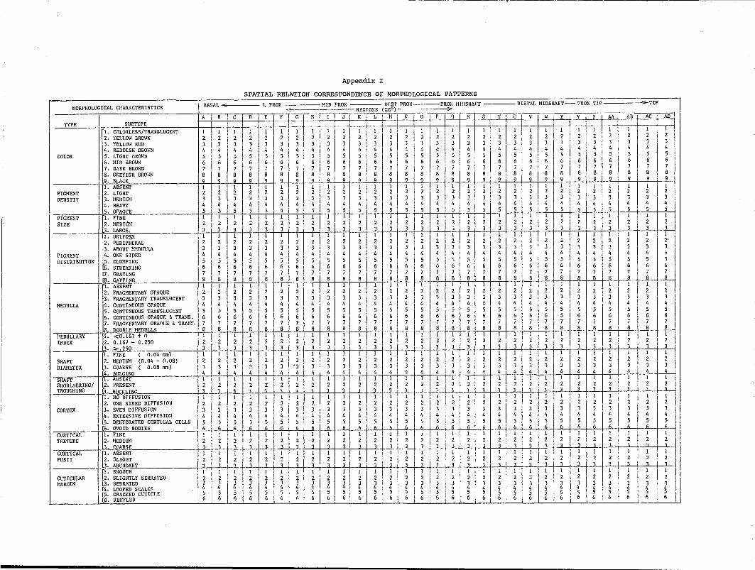
Appendix I
SPATIAL RELATION CORRESPONDENCE OF MORPHOLOGICAL PATTERNS
DIST PROX ,PRO); IIIDSHAFT DISTAL MlDSHAFT ?ROX TIP ~TIP
I!ORPHOLOGICAL CHARACTERISTICS BASAL ~ L PROX !lID PROX REGIONS (CHs ) --------7-~
I ABC D E F G H I J K L M T NO' P 1-'1 R S T r-U v ~X • Y Z AA AB AC' I\flJ TYPE SUBTYPE ~ 1----,-- ~ 1
'lo COLORLESS/TRANSLUCENT 1 1 1 -: 1 1 I 1 1 1 I 1 1, I 1 1 1 1 1 1 11 1 1 I 1 - 1 1 1 I 1- 1 1\ 2. YELLOW BROWN 2 2 2 2 2 2 2 2 2 2 2 2 2 2 2 2 2 2 2 2 2 2 2 I 2 2 2 2 2 2 2 3. YELLOW RED 3 3 3 3 3 3 3 3 3 3 3 3 3 3 3 3 3 3 3 3, 3 3 3 3 3 3 3 1 3 3 I
4. REDDISH BRDlm 4 4 4 4 4 4 4 4 4 4 4 4 4 4 4 4 4 4 4 4 4 4 4 4 I 4 4 4 4 4 4 I COLOR
PIGMENT DENSITY
PIGMENT SIZE
PIGHENT DISTRIBUTION
IIEDULLA
~'EDULLARY INDEX
SHAFT DIAMETER
5. LIGHT BROWN 5 5 5 5 5 5 5 5 5 5 5 5 5 5 5 5 5 5 5 5 5 5 5 5 5 5 5 5 5 5 6. MID BROWN 6 6 6· 6 6 6 61 6 6 6 6 6 6 6 6 6 6 6 6 6 6 I 6 6 6 I 6 6 6 6 6 6 7. DARK BROWN 7 7 7 I 7 7 7 7 7 7 7 7 7 7 7 7 7 7 7 7 7 7 7 7 7 7 7 7 7 7 7 8. GREYISH BROWN 8 8 8 I 8 8 8 8 8 8 8 8 8 8 8 I 8 8 8 8 8 8 8 8 8 8, 8 8 8 8 8 8 ,9. BLACK 9 9 q 9 9 9 9 9 9 9 9 9 9 _ 9 9 9 I 9 _ 9 9 9 9 I 9 9 9 I 9 9 9 9 9 9
~-: ABSENT - --- 1 1 1 1 1 1 1 1 1 1 1 1 1 1 I 1 I 1 1 1 1 1 1 1 I' 1 ill 1 1 1 1 2. LIGHT 2 2 2 2 2 2 2 2 2 2 2 2 2 2 2 2 2 2 2 2 2 2 2 2 I 2 2 2 2 2 2 3. IlEDIUlI 3 3 3 3 3 3 3 3 3 3 3 3 3 3 3 3 3 3 3 3 3 3 3 3 3 3 3 3 3 3 4. HEAVY 4 4 4 4 4 4 4 4 4 4 4 4 4 4 4 4 4 4 4 4 4 4 4 4 4 " 4 4 4 4
• OPAOUE 5 5 5 5 5 5 5 5 5 5 5 5 5 5 5 5 5 5 5 5 5 5 5 5 5 5 5 5 5 5
II
[lo FINE, 1 1 1 1 1 1 1 1 1 1 1 1 1 1 1 I 1 1 1 1 1 1 1 1 I 1 I 1 1 1 I~' flEDIllN 2 2 2 2 2 2 2 2 2 2 2 2 2 2 2 2 2 2 2 2 2 2 2 2 2 2 2 2 2 2 u. LARGE, J 3 3 3 3 J J 3 3 3 3 3 3 3 3 3 3 3 3 3 3 3 3 3 3 3 3 3 3 3
I [lo UNIFOI\lI 1 1 1 I 1 1 1 1 1 1 1 I 1 1 1 I 1 1 I 1 1 1 1 1 1 IIi 1 1 2. PERIPHERAL 2 2 2 2 2 2 2 2 2 2 2 2 2 2 2 2 2 2 2 2 2 2 2 2 2 2 2 2 2 2' .3. ABOUT MEDULLA J 3 J J J J J J 3 3 3 3 3 3 3 3 J 3 3 3 3 3 3 3 3 3 J 3 3 3 :4. ONE SIDED 4 4 4 4 I, 4 I, 4 4 4 4 4 4 4 4 4 4 4 4 4 4 4 4 4 4 4 4 4 I, 4 ~5. CLUlIPING 5 I 5 5 5 5 5 5 5 5 5 5 5 5 5 5 5 5 5 5 5 5 5 5 5 5 I 5 5 5 5 5 [6. STREAKING 6 6 6 6 6 6 6 6 6 6 6 6 6 6 6 6 6 6 6 6 6 6 6 6 6 I 6 (, 6 6 6 [7. GRAYING 7 7 7 7 7 7 7 7 7 7 7 7 7 7 7 7 7 7 7 7 7 7 7 7 7 7 7 7 7 7 [8. GAPPING 8 8 8 I 8 8 8 8 8 8 8 8 8 8 8 8 8 8 8 8 8 8 B B, 8 8 B B B 8 B
~• ABSENT 1 1 r-- -I C -]- ell 1 lIe I TIl C --T 1 1 1 1 1
2. FRAGHENTARY OPAQUE 2 2 2 2 2 2 2 2 2 2 2 2 2 2 2 2 2 2 2 2 2 2 2 2 3. FRAGHEN).'ARY TRANSLUCENT 3 3 3 3 3 3 3 3 3 3 3 3 3 3 3 3 3 3 3 3 3 3 3 3 4. CONTINUOUS OPAQUE 4 4 4 4 4 4 4 4 4 4 4 4 4 4 4 4 4 4 4 4 4 4 4 4 5. CONTINUOUS TRANSLUCENT 5 5 5 5 5 5 5 5 5 5 5 5 5 5 5 5 5 5 5 5 5 5 5 5 6. CONTINUOUS OPAQUE & TRANS. 6 6 6 6 6 6 6 6 6 6 6 6 6 6 6 6 6 6 6 6 6 6 6 6 7. FRAGMENTARY OPAOlJE & TRAUo,. 7 7 7 7 7 7 7 7 7 7 7 7 7 7 7 7 7 7 7 7 7 7 7 7
• DOUBLE HEDULLA 8 8 8 8 8 8 8 B 3 8 8 8 8 8 8 8 8 8 8 8 8 8 8 8
I J. <0.167 t nIl 1 1 1 1 1 1 lIt 1 1 1 - -I I 1 1 1 1 1 1 1 11 ' 1 I 1 1 1 1 1 1 1 2. 0.167 - 0.250 2 2 2 2 2 2 2 2 2 2 2 2 2 2 2 2 2 2 2 2 2 2 2 2 2 2 2 2 2 2
. "'.250 J 3 3 3 3 3 3 3 3 3 3 3 3 3 3 3 3 3 3 3 3 3 3 3 3 3 3 3 3 3
~• FINE (0.04 mm) 1 1 I 1 1 1 I 1 I 1 I 1 1 1 1 1 1 1 I 1 1 I I 1 1 1 1 1 1 1 1
2. MEDIUl! (0.04 - 0.08) 2 2 2 2 2 2 2 2 2 2 2 2 2 2 2 2 2 2 2 2 2 2 1 2 2 2 2 2 2 I 2 2 • COARSE ( 0.08 1lU11) 3 3 3 3 3 3 • 3 3 3 3 3 3 3 3 3 3 3 3 3 3 3 3" I 3 3 3 J I 3 3 3 1 3
SHAFT T,r. ABSENT 1 1 r 1 1 1 I 1 1 1 1 1 1 ftl 1 T 1 WilT 1 1 I 1 1 1-1 1 1 1 1 1 1 I 1 -SHOULDERING/ ~2. PRESENT 2 2 2 2 2 2 2 2 2 2 2 2 2 2 2 2 2 I 2 2 2 2 2' 2 2 2 2 ~ :;2 2 2 I TROUGHING 13. HlI~K' ,TNr, 3 3 1 1 1 1 1 3 3 i 3 1 1 1 _ 1 1 1 1 3 1 1 1 3 3' .. 1
[
1. NO DIFFUSION 1 1 1 1 1 1 1 1 1 1 1 1 1 1 I T 1 'I II! 1 I 1 1 1 1 1 1 1 I 1 1 I 2. ONE SIDED DIFFUSION 2 2 2 2 2 2 2 2 2 2 2 2 2 2 2 I 2 2 ut2 2 2 2 2 2 2 2 2 2 2 2 I 3. gVEN DIFFUSION 3 3 3 3 3 3 3 3 3 3 3 3 3 3 3 3 1 3 3 3 3 3. 3 3 3 3 3 3 3 3 3 4. EXTENSIVE DIFFUSION 4 4 4 4 4 4 " 4 4 4 4 4 4 I 4 4 4' 4 I 4 4 4 4 4 4 4 I, 4 I. 4 4 4
[4. BULGING 4 4 4 4 4 4 4 4 4 4 I 4 4 4 4 4 I, 4 I 4 4 4 I. 4 4 _ 4 4 4 4 4 4 4 4
CORTEX
CORTICAL TEXTURE
CORTICAL FUSII
CUTICULAR MARGIN
[5. DEHYDRATED CORTICAL CELLS 5 5 5 5 5 5 5 5 5 5 5 5 5 I 5 51 i 5 I 5 '5 5 5 5 5 5 5 5 5 5 5 I 5 5 [6. OVOID BODIES 6 6 6 6 6 6 6 6 6 6 6 6 6 6 6 ~ I ~ 6 6 1+ (, 6 6 6 ~ • • ~ •
~• FINE 1 1 1 1 I I 1 1 1 I 1 1 1 1 1 I, 1 ,I ~ 1 1 1 1 'I! 1 1 l' 1 1 1 1
2. BED lUll 2 2 2 2 2 2 2 2 2 2 2 I 2 2 2 2 i 2 ; 2 2 ~ I ~ 2 2 2 27 1 2 ~ 2 2 2 • COARSE 1 1 3 3 3 3 3 3 3 3 3' 3 3 3 J: 3 '3 3 ~ 3 3 3 1 1 1 1 , ,
J. ABSENT 1 I 1 I I 1 I I 1 1 1 I 1 1 1 1 I 1 I 1 I I! 1 1 1 I 1 I 1 1 1 1 1 1 1 1 2. SLIGHT 2 2 2 2 2 2 2 2 2 2 2 2 2 2 I 2 2. 2 2 2 2, 2 2 2 2 2 2 2 2 2 2 ~. ABUNDANT 1 I 1 1 1 3 I 3 J! 3 3 ,3.1 3J 31 3 13 i 3_JL_L~LL_..l I 3 i 3 3 J 3 I 3 3 I 3 3 1 I 1 1
[1. SHOOTH-- 11 I 1 ITI' 1 -'IITl! 1 1 I 1 1 1 1 1 1 1 1 1 1 1 1 2. SLIGHTLY SERRATED 2 2 2 2 2 2 2 I. 2 2 2 2 2 2 2 2 2 2 2 2 2 2 2 2 2 2
LJ. SERRATED 3 3 3 J 3 J' 3 i 3 3 3 3 3 3 3 3 3 3 3 3 3 3 3 3 3 3 3 3 [4. LOOPED SCALES ,4 4 4 4 4:. 4 I 4 I 4 I 4 4 4 " 4 "" 4 4 4 4 4 4 4 4 4, 4 4 4 [5. CRACKED CUTICLE 5 5 5 5 5, ,5, : 5 I 5l 5 5 5 5 5 5 5 5 5 5 5 5 5 5 5 5 5 5 [6. RUFFLED : 6 6 6 6 6 J_~_L A 6 6 6 6 6 6 6 6 6 6 6 6 6 6 6 (, 6 6
1 2
3 3 ~ g 6 6
<!lAIR FORM~ ~OUS I. STRAICHT
2. SLEEK 3. FLAmAVED
CYHATOTRICHOUS 4. BROADWAVED 5. NARRO\; WAVED 6. CURL?
ULOTRICHOUS 7. FRIZZLY 8. CLOSELY KNIT
<:: HAIR COLoR> (REFLECTED LIGHT) VII GRAYING
BLONDE A ASH BLONDE B-E LIGHT BLONDE F-L BLONDE
DARK BROWN/BLACK 11-0 DARK BLONDE P-T BROWN U-Y DARK BROWN/BLACK
RED I-IV RED V-VI REDDISH BROWN
<LENGTH> [' <, ~ 2. 5-10 CH
3. 10-15 CH 4. 15-20 Cll 5. >20 CH
<ROOT/PROXIMAL r' ABSENT REGION> 2. STRETCHED 3. ANAGEN mTH SHEATH 4. CATAGEN/TELOGEN
[5. BASAL FUS!!
,SHAFT> [1. CONSTANT DIAHETER 2. SLIGHT SHOOTH VARIATION 3. WIDE SHOOTH VARIATION 4. ABRUPT VARIATION
<TIP/DISTAL REGION> 1. NATURAL TAPER 2. CUT - ANGLE 3. ROUNDED TAPERING 4. ROUNDED AND FRAYED 5. SPLIT AND FRAYED 6. CRUSHED 7. BROKEN
<TREATHENT> r' NONE OBSERVABLE 2. DYED 3. BLEACHED
<CUTICLE> r' INDISTINCT 2. THIN 3. THICK e. CLEAR 5. TINGED ~. EVEN THICKNESS 7. CUTICULAR FLUCTUATIONS
<MAXlllUI! SHAFT DIAlIETER> VALUE
<HEANSHAFT DIAHETER> VALUE
<SCALE INDEX>
! VALUE
1 2 3
4 5
j 6
7 8 I VII A BCD E FGHIJKL
II N 0 P Q R S T U V W x Y
I II III IV V VI 1 2 3 4 5 1 2 3 4 5 1 2 3 4 1 2 3 4 5 6 7 1 2 3 1 2 3 4 5 6 7
I ------
Appendix I (cent.)
HAIR ;j . SUBJECT # -----
NAlIE
SEX
RACE
AGE J
NOTES
----~------------.-----
CBI IISV/EH
IJ ~:f.: .:~ a~~ i!~ I~:
~H .!j N ~~
h