presynaptic mglu1 and mglu5 autoreceptors facilitate glutamate exocytosis from mouse cortical nerve...
TRANSCRIPT

Presynaptic mGlu1 and mGlu5 autoreceptors facilitate glutamateexocytosis from mouse cortical nerve endings
Veronica Musantea, Elisa Neria, Marco Feligionib, Aldamaria Pulitic,d,e, Marco Pedrazzie,f,Valerio Contid,g, Cesare Usaih, Alberto Diasproi, Roberto Ravazzoloc,d,e, Jeremy M.Henleyb, Giuseppe Battagliaj, and Anna Pittalugaa,c,*
aDepartment of Experimental Medicine, Section of Pharmacology and Toxicology, University ofGenoa, Genoa, ItalybMRC Centre for Synaptic Plasticity, Department of Anatomy, School of Medical Sciences,University of Bristol, Bristol BS8 1TD, UKcCenter of Excellence for Biomedical Research, University of Genoa, Genoa, ItalydLaboratory of Molecular Genetics and Cytogenetics, G. Gaslini Institute, Genoa, ItalyeDepartment of Pediatric Sciences, University of Genoa, Genoa, ItalyfDepartment of Experimental Medicine, Section of Biochemistry, University of Genoa, Genoa,ItalygRenal Child Foundation, G. Gaslini Institute, Genoa, ItalyhInstitute of Biophysics, National Research Council, Genoa, ItalyiDepartment of Physics, University of Genoa, Genoa, ItalyjI.N.M. Neuromed, Pozzilli, IS, Italy
AbstractThe effects of mGlu1 and mGlu5 receptor activation on the depolarization-evoked release of[3H]D-aspartate ([3H]D-ASP) from mouse cortical synaptosomes were investigated. The mGlu1/5receptor agonist 3,5-DHPG (0.1–100 μM) potentiated the K+(12 μM)-evoked [3H]D-ASPoverflow. The potentiation occurred in a concentration-dependent manner showing a biphasicpattern. The agonist potentiated [3H]D-ASP exocytosis when applied at 0.3 μM; the efficacy of3,5-DHPG then rapidly declined and reappeared at 30–100 μM. The fall of efficacy of agonist atintermediate concentration may be consistent with 3,5-DHPG-induced receptor desensitization.Facilitation of [3H]D-ASP exocytosis caused by 0.3 μM 3,5-DHPG was prevented by the selectivemGlu5 receptor antagonist MPEP, but was insensitive to the selective mGlu1 receptor antagonistCPCCOEt. In contrast, CPCCOEt prevented the potentiation by 50 μM 3,5-DHPG, while MPEPhad minimal effect. Unexpectedly, LY 367385 antagonized both the 3,5-DHPG-induced effects. Atotal of 0.3 μM 3,5-DHPG failed to facilitate the K+-evoked [3H]D-ASP overflow from mGlu5receptor knockout (mGlu5−/−) cortical synaptosomes, but not from nerve terminals prepared fromthe cortex of animals lacking the mGlu1 receptors, the crv4/crv4 mice. On the contrary, 50 μM3,5-DHPG failed to affect the [3H]D-ASP exocytosis from cortical synaptosomes obtained fromcrv4/crv4 and mGlu5−/−mice. Western blot analyses in subsynaptic fractions support the existenceof both mGlu1 and mGlu5 autoreceptors located presynaptically, while immunocytochemistryrevealed their presence at glutamatergic terminals. We propose that mGlu1 and mGlu5
© 2008 Elsevier Ltd. All rights reserved.*Corresponding author. Tel.: +39 010 3532120; fax: +39 010 3993360. [email protected]. .
Europe PMC Funders GroupAuthor ManuscriptNeuropharmacology. Author manuscript; available in PMC 2012 March 23.
Published in final edited form as:Neuropharmacology. 2008 September ; 55(4): 474–482. doi:10.1016/j.neuropharm.2008.06.056.
Europe PM
C Funders A
uthor Manuscripts
Europe PM
C Funders A
uthor Manuscripts
autoreceptors exist on mouse glutamatergic cortical terminals; mGlu5 receptors may represent the“high affinity” binding sites for 3,5-DHPG, while mGlu1 autoreceptors represent the “lowaffinity” binding sites.
KeywordsMouse cortical synaptosomes; [3H]D-aspartate release; mGlu1 autoreceptor; mGlu5 autoreceptor;crv4 Mice; mGlu5 Receptor knockout mice
1. IntroductionPresynaptic modulation of neurotransmitter release represents one of the fundamentalmechanisms of regulation of central transmission; most of the central neurotransmitters,including glutamate, undergo this control, exerted either by auto or by heteroreceptorslocated presynaptically on nerve endings.
Of the known autoreceptor systems, the glutamatergic system appears the most complex(Raiteri, 2008 and references therein). Both ionotropic and metabotropic glutamate (mGlu)receptors belong to this system. However, although several lines of evidence support theexistence and the role of glutamate ionotropic autoreceptors, such as NMDA (Luccini et al.,2007a and references therein), AMPA (Barnes et al., 1994) and kainate (Chittajallu et al.,1996; Pittaluga et al., 1997) receptors, mGlu autoreceptors appears predominant (Raiteri,2008).
mGlu receptors belong to the family 3 of G protein coupled receptors (GPCRs, Bockaert andPin, 1999) and possess a very large N-terminal domain containing the agonist binding sites.These receptors are structurally and pharmacologically heterogeneous and exist as eightsubtypes (mGlu1–mGlu8 receptors) having discrete regional, cellular and subcellularlocalizations (Conn and Pin, 1997; Pin et al., 2003). Depending on the specific receptorinvolved, the main transduction mechanisms can be activation of the phosphatidylinositol(PI) pathway (group I mGlu receptors, that are mGlu1 and 5 receptors) or inhibition ofadenylyl cyclase (AC) activity (group II mGlu receptors, namely mGlu2 and 3, and group IIImGlu receptors, including mGlu4, 6, 7 and 8).
Because of the negative control on AC and K+ channels, group II and III mGluautoreceptors function as negative feedback mechanisms that inhibit glutamate release(Corti et al., 2007; Raiteri, 2008 and references therein). In contrast, group I mGlu receptorsfavour Ca2+-dependent phosphorylative processes and facilitate glutamate release (Herreroet al., 1992; Raiteri, 2008 and references therein).
Group I mGlu receptors have been proposed to be located presynaptically at glutamatergicterminals (Raiteri, 2008 and references therein), but the relative contributions of mGlu1 andmGlu5 receptors to the facilitation of glutamate release remain matter of discussion.
To directly address this question we have pharmacologically characterized the receptor/sinvolved in the facilitation of the K+-evoked [3H]D-ASP release from mouse corticalglutamatergic terminals exerted by the group I selective agonist 3,5-DHPG. We also usedtransgenic mouse models lacking mGlu1 (crv4/crv4; Conti et al., 2006) or mGlu5(mGlu5+/−) receptors. Our results are consistent with the existence of both mGlu1 andmGlu5 autoreceptors in cortex. Although activation of both these receptors increases evokedglutamate exocytosis, the two receptors do not compensate one for each other. Thepharmacological profile as well as the results obtained with transgenic mice suggest that
Musante et al. Page 2
Neuropharmacology. Author manuscript; available in PMC 2012 March 23.
Europe PM
C Funders A
uthor Manuscripts
Europe PM
C Funders A
uthor Manuscripts

mGlu1 autoreceptors behave as “low affinity” binding site for 3,5-DHPG, while mGlu5autoreceptors appears to be the high affinity binding sites.
2. Methods2.1. Animals
Adult male mice (Swiss; 20–25 g) were used as control animals in all the experiments withthe exception of the experiments carried out with cervelet-4 (crv4) mice and mGlu5−/−
knockout mice. The crv4 mutation is a spontaneous recessive mutation occurred in theBALB/c/Pas inbred strain. It consists in an LTR intronic insertion which disrupts splicing ofmGluR1 gene and causes absence of the protein (Conti et al., 2006). Crv4 homozygous micepresent mainly with an ataxic phenotype. Affected (crv4/crv4) and control (+/+) mice aremaintained on the same genetic background by inter-crossing +/crv4 mice at the animalfacility of the National Institute of Cancer Research (Genoa, Italy). The genotype of thewild-type and crv4 mice was determined by PCR with use of tail genomic DNA and a pairof primers (5′-GAGTGTTCACTAGTTCACCCAAGA-3′ and 5′-TCAGGCAACAATAAGGCAAG-3′) which flank the insertion; the PCR productssynthesized with these primers were 688 bp for the crv4 mutant and 498 bp for wild-type.
Heterozygous mGlu5 receptor knockout mice (129-Gprc1etmt1rod) were obtained from TheJackson Laboratories (Bar Harbor, ME, USA). Mice heterozygous for the targeted mutationwere intercrossed to homozygosity at the “Istituto Neurologico Mediterraneo Neuromed”(Pozzilli, IS, Italy). Homozygous females and males were fertile, but poor breeders. Thus,all mice were generated by heterozygous breeding. Primers for the genotyping of knockoutmice were from Jackson Laboratory. Transgenic mice were then sent to the animal facilityof our laboratory (Department of Experimental Medicine, Pharmacology and ToxicologySection). Mice were kept under environmentally controlled conditions (ambient temperature22 ± 1 °C, humidity 50%) on a 12-h light/dark cycle with food and water ad libitum. Micewere identified by PCR analysis on tail samples after birth.
The animals were killed by decapitation; the cortices rapidly removed and purifiedsynaptosomes prepared within minutes. The experimental procedures were approved by theDepartment Ethical Committee, in accordance with the European legislation (EuropeanCommunities Council directive of 24 November 1986, 86/609/ EEC). Experiments wereperformed following the Guidelines for Animal Care and Use of the National Institutes ofHealth.
2.2. Preparation of synaptosomesTo isolate purified synaptosomes, the tissue was homogenized in 10 volumes of 0.32 Msucrose, buffered to pH 7.4 with TRIS (final concentration 0.01 M) using a glass Teflontissue grinder (clearance 0.25 mm).The homogenate was centrifuged at 1000 g for 5 min, toremove nuclei and debris, and the supernatant was gently stratified on a discontinuousPercoll gradient (6%, 10% and 20% v/v in Tris-buffered sucrose) and centrifuged at 33,500g for 5 min. The layer between 10% and 20% Percoll (synaptosomal fraction) was collectedand washed by centrifugation. The synaptosomal pellets were resuspended in aphysiological medium having the following composition (mM): NaCl, 125; KCl, 3; MgSO4,1.2; CaCl2, 1.2; NaH2PO4,1; NaHCO3, 22; glucose, 10 (aeration with 95% O2 and 5%CO2); pH, 7.2–7.4.
2.3. Experiments of release from superfused synaptosomesSynaptosomes were labelled with [3H]D-aspartate ([3H]D-ASP; final concentration 30 nM) at37 °C, for 15 min, in a rotary water bath and in an atmosphere of 95% O2 and 5% CO2.
Musante et al. Page 3
Neuropharmacology. Author manuscript; available in PMC 2012 March 23.
Europe PM
C Funders A
uthor Manuscripts
Europe PM
C Funders A
uthor Manuscripts
After the labelling period, identical portions of the synaptosomal suspensions were layeredon microporous filters at the bottom of parallel superfusion chambers (Ugo Basile, Comerio,Varese, Italy; Raiteri and Raiteri, 2000) thermostated at 37 °C and superfused at 0.5 ml/minwith standard physiological solution aerated with 95% O2 and 5% CO2.
When studying the effect of 3,5-DHPG on [3H]D-ASP release evoked by high K+,synaptosomes were transiently (90 s) exposed at t = 39 min to 12 μM KCl containingmedium (NaCl substituting for an equimolar concentration of KCl) in absence or in presenceof the agonist. To evaluate whether presynaptic mGlu5 autoreceptors can desensitize andswitch from facilitation to inhibition (Herrero et al., 1998; Rodriguez-Moreno et al., 1998;Sistiaga et al., 1998; Nicoletti et al., 1999), synaptosomes were pre-exposed to 3,5-DHPGfrom t = 30 min to 35 min of superfusion, then substituted until t = 39 min with standardphysiological medium and then rechallenged with 3,5-DHPG concomitantly added with thedepolarizing stimulus (12 or 30 μM K+). Antagonists were added 8 min before agonist.Starting from t = 36 min of superfusion, four consecutive 3 min fractions were collected.Fractions collected and superfused synaptosomes were counted for radioactivity.
2.4. Isolation of detergent-soluble fraction from synaptosomesPurified synaptosomes were prepared as already described and collected by centrifugationfor 15 min at 4 °C and 14,000 g. The pellet was diluted and lysed in 1 mL of ice-cold 20 μMTris/HCl (pH 7.4, containing 10 μM NaCl and protease inhibitor cocktail). In order toisolate the detergent-soluble membrane fraction (DS-Syn; see Pedrazzi et al., 2006), after 15min at 4 °C, 450 mg of synaptosome lysate (Tot-Syn) were centrifuged at 200,000 g,4 °C for15 min and the pellet was solubilized in 800 μl of 20 μM Tris/HCL (pH 7.4, containing 140μM NaCl, 0.2% Triton X-100 and protease inhibitor cocktail). After 15 min at 4 °C, sampleswere centrifuged at 200,000 g,4 °C for 15 min and the supernatant, the DS-Syn, collected.Proteins were quantified using Lowry Assay.
2.5. ImmunoblottingProteins (25 μg/lane) were separated by SDS-7.5% PAGE, and then transferred onto PVDFmembranes. Membranes were blocked in 20 μM sodium phosphate buffer (PBS, pH 7.4,containing 140 μM NaCl, 5% non fat dry milk, 0.1% Tween-20) and probed with one of thefollowing primary antibodies (60 min at 20 °C): rabbit anti-mGlu1 receptor (1:500), rabbitanti-mGlu5 receptor (1:1000), mouse anti-syntaxin-1A (Stx-1A; 1:10000). After extensivewashes, membranes were incubated for 1 h at 20 °C with the appropriate horseradishperoxidase-linked secondary antibody (1:4000), and immunoblots were visualized with anECL (enhanced chemiluminescence) Plus Western blotting detection system.
2.6. Sub-synaptic fractionation of nerve terminalsSub-synaptic fractionation was prepared as described in literature (Feligioni et al., 2006).Briefly, synaptosomes were prepared, as described above, from cortex of three mouse brainsin order to obtain enough proteins for the preparation. The pellets from purifiedsynaptosomes were resuspended in 300 μl of 0.32 M sucrose and 0.1 μM CaCl2. Proteaseinhibitors were used in all purification steps. Synaptosomes were then diluted 1:10 in ice-cold 0.1 μM CaCl2 and mixed with an equal volume of 2× solubilisation buffer (2% TritonX-100, 40 μM Tris, pH 6.0; 4 °C). Following a 30 min incubation at 4 °C, the insolublematerial (synaptic junctions) was pelleted (40,000 g, 30 min, 4 °C) and the supernatant (non-synaptic synaptosomal protein; NSSP) was decanted and proteins precipitated with 6volumes of acetone at −20 °C and centrifuged (18,000 g; 30 min; −15 °C). The synapticjunction pellet was resuspended in 10 volumes of 1× solubilisation buffer (1% Triton X-100,20 μM Tris, pH 8.0; 4 °C) and incubated for 30 min at 4 °C and then centrifuged (40,000 g,30 min, 4 °C). The pellet contained the insoluble postsynaptic density and the supernatant
Musante et al. Page 4
Neuropharmacology. Author manuscript; available in PMC 2012 March 23.
Europe PM
C Funders A
uthor Manuscripts
Europe PM
C Funders A
uthor Manuscripts

contained the presynaptic active zone. The protein in the supernatant (presynaptic fraction)was acetone precipitated and collected as above. Proteins obtained from this preparationcontained about 1.70 μg/μl per fraction.
2.7. Sub-fractionation immunoblottingA10 mg protein/lane where loaded on 10% SDS-PAGE gel and then transferred onto PVDFmembranes. Non-specific binding sites were blocked over-night with Tris-buffered saline-Tween (t-TBS; 0.02 M Tris, 0.137 M NaCl, and 0.1% Tween 20) containing 5% non-fatdried milk and probed for protein of interest with the following primary antibodies: rabbitanti-mGlu1 receptor (1:500), rabbit anti-mGlu5 receptor (1:1,000), mouse anti-syntaxin-1A(Stx-1A; 1:10,000), mouse anti-Synaptophysin, (1:20,000), mouse anti-PSD95, (1:1,000).After washes, membranes were incubated for 1 h at room temperature with the appropriatehorseradish peroxidase-linked secondary antibody (1:5000), and immunoblots werevisualized with an ECL (enhanced chemiluminescence) Plus Western blotting detectionsystem.
2.8. Immunocytochemical analysis in mouse cortical nerve terminalsFor immunohistochemical analysis, cortical synaptosomes were obtained throughdiscontinuous Percoll gradient and 80 mg of synaptosomal proteins were placed ontocoverslips, previously coated with poly-L-ornithine, fixed with 2% paraformaldehyde for 15min. and washed with PBS. The synaptosomes were permeabilized with 0.05% TritonX-100 PBS for 5 min. After extensive washes with 0.5% BSA PBS, synaptosomes wereincubated with: rabbit anti-mGlu1 receptor (1:500); rabbit anti-mGlu5 receptor (1:500);mouse antisintaxin-1A (Stx-1A; 1:10,000) and guinea pig anti-vesicular glutamatetransporters type 1 (VGLUT-1; 1:500) as indicated for 1 h at room temperature. Thesynaptosomes were then extensively washed with 0.5% BSA PBS and incubated for 1 h atroom temperature with AlexaFluor-488 (green)-labelled donkey anti-mouse IgG antibodies,or AlexaFluor-647 (red) donkey anti-rabbit IgG antibodies or AlexaFluor-647 (red) donkeyanti-mouse IgG antibodies or Donkey, FITC Conjugate (green), anti-guinea pig IgGantibodies (1:1,000 for all).
Fluorescence image acquisition was performed by a three-channel Leica TCS SP5 laser-scanning confocal microscope, equipped with 458, 476, 488, 514, 543 and 633 nmexcitation lines. Images (512 × 512 × 8 bit) were taken through a planapochromatic oilimmersion objective 100x/NA1.4. Light collection configuration was optimized according tothe combination of chosen fluorochromes and sequential channel acquisition was performedto avoid cross-talk phenomena. Leica LCS software package was used for acquisition,storage and visualization. Each coverslip was analyzed by counting at least three differentfields.
2.9. CalculationsThe amount of radioactivity released into each superfusate fraction was expressed as % ofthe total synaptosomal tritium content at the start of the fraction collected (fractional efflux).The K+-induced tritium overflow was expressed as % induced overflow and was estimatedby subtracting the neurotransmitter content into the first and the fourth fractions collected(basal release) from that in the second and in the third fractions collected during and afterthe depolarization pulse (evoked release). Analysis of variance was performed by ANOVAfollowed by Newman Keuls multiple-comparison test; direct comparisons were performedby applying Student’s t-test. Data were considered significant for p < 0.05 at least.Appropriate controls with antagonists were always run in parallel. Western blots werequantified by densitometric analysis using NIH Image J software.
Musante et al. Page 5
Neuropharmacology. Author manuscript; available in PMC 2012 March 23.
Europe PM
C Funders A
uthor Manuscripts
Europe PM
C Funders A
uthor Manuscripts

The quantitative estimation of colocalized proteins in immunocytochemical studies has beenperformed calculating the co-localization coefficients (Manders et al., 1993) from the red-and green-channel scatterplot. Co-localization coefficients express the fraction ofcolocalizing molecular species in each component of a dual-colour image and are based onthe Pearson’s correlation coefficient, a standard procedure for matching one image withanother in pattern recognition (Gonzalez and Wintz, 1987). If two molecular species arecolocalized, the overlay of their spatial distributions has a correlation value higher than whatwould be expected by chance alone. Costes et al. (2004) developed an automated procedureto evaluate correlation between the green and red channels with a significance level >95%.The same procedure automatically determines an intensity threshold for each colour channelbased on a linear least-square fit of the green and red intensities in the image’s 2Dcorrelation cytofluorogram. Costes’ approach has been accomplished by macro routinesintegrated as plug-ins (WCIF Co-localization Plugins, Wright Cell Imaging Facility,Toronto Western Research Institute, Canada) in the ImageJ 1.39 software (Wayne Rasband,NIH, USA).
2.10. Drugs1-[7,8 3H]D-aspartate (specific activity 16.3 Ci/mmol) was from Amersham RadiochemicalCenter (Buckinghamshire, UK). (RS)-3,5-Dihydroxyphenylglycine (3,5-DHPG) was fromAscent Scientific (Weston Super-Mare, UK); 2-methyl-6-(phenylethynyl) pyridinehydrochloride (MPEP), 7-(hydroxyimino)cyclo propa[b]chromen-1a-carboxylate ethyl ester(CPCCOEt) and (S)-(+)-a-Amino-4-carboxy-2-methylbenzene acetic acid (LY 367385)were obtained from Tocris Bioscience (Bristol, UK). Anti-mGlu5 receptor polyclonal rabbitimmunoaffinity purified IgG, anti-mGlu1 receptor polyclonal rabbit immunoaffinity IgG,and mouse anti-PSD95 were from Upstate Biotechnology (Lake Placid, NY, USA). Anti-syntaxin-1A monoclonal mouse IgG was from Synaptic System, Germany; mouse anti-Syn-aptophysin, was from Calbiochem, San Diego, CA, USA; Guinea Pig anti-VGLUT-1 andDonkey FITC Conjugated anti-Guinea Pig antibodies were purchased from Chemicon (CA,USA). Horseradish peroxidase-conjugated anti-mouse and anti-rabbit secondary antibodiesand the ECL Plus Western blotting detection system were purchased from GE Healthcare.Protease Inhibitor Cocktail was from Sigma–Aldrich. AlexaFluor-647 or AlexaFluor-488antibodies are from Molecular Probes (Alfagene) (Oregon, USA).
3. Results3.1. 3,5-DHPG potentiates biphasically the release of [3H]D-ASP evoked by KCl
We investigated the effects of 3,5-DHPG, a broad spectrum mGlu1/5 receptor agonist, onthe release of [3H]D-ASP evoked by 12 μM K+ from mouse cortical nerve endings. It isknown that, when mouse cortical synaptosomes are exposed in superfusion to mild K+
depolarization, a Ca2+-dependent, exocytotic-like release of prelabelled [3H]D-ASP occurs.Indeed, recent work showed that the 12 μM K+-induced release of [3H]D-ASP represents areliable measure of endogenous glutamate from this synaptosomal preparation (Raiteri et al.,2007).
Fig. 1 shows that 3,5-DHPG (0.1–100 μM) facilitated the 12 μM KCl-evoked release of[3H]D-ASP in a concentration-dependent manner; a significant facilitation of [3H]D-ASPexocytosis was observed when the agonist was added at concentrations around 0.3 andabove 10 μM 3,5-DHPG.
Musante et al. Page 6
Neuropharmacology. Author manuscript; available in PMC 2012 March 23.
Europe PM
C Funders A
uthor Manuscripts
Europe PM
C Funders A
uthor Manuscripts

3.2. Effects of selective mGlu1 and mGlu5 receptor antagonists on the 12 μM K+-induced[3H]D-ASP overflow
The pharmacological characterization of the mGlu receptors involved in the facilitation by3,5-DHPG of [3H]D-ASP exocytosis was then carried out using the selective mGlu1 receptornon-competitive antagonist CPCCOEt, the competitive antagonist LY 367385 and theselective non-competitive mGlu5 receptor antagonist MPEP. Preliminary experimentsinvestigated the effects of these antagonists, added alone, on the K+-evoked release of [3H]D-ASP.
LY 367385 and CPCCOEt (0.5–10 μM) failed to affect the K+-evoked release of [3H]D-ASP, whereas MPEP (0.3–3 μM) decreased it significantly (Table 1). Notably, the inhibitionby MPEP of glutamate exocytosis appears well in line with the results by Wang and Sihra(2004) who suggested the existence mGlu5 autoreceptors constitutively activated in corticalglutamatergic terminals. The inhibition by MPEP of the K+-evoked release of [3H]D-ASPappeared maximal when the mGlu5 receptor antagonist was applied at 1 μM and it wasunaffected by the contemporary addition of 5 μM CPCCOEt (Table 1).
3.3. Effects of selective mGlu1 and mGlu5 receptor antagonists on the 3,5-DHPGpotentiation of the K+-evoked [3H]D-ASP overflow
MPEP (0.1–1 μM) prevented the 3,5-DHPG (0.3 μM) potentiation of 12 μM K+-induced[3H]D-ASP overflow, which was totally abrogated when the antagonist was applied at 1 μM(Fig. 2A). On the contrary, CPCCOEt (5–10 μM) was ineffective (Fig. 2A). The antagonismbrought about by 1 μM MPEP was unmodified by the contemporary addition of 5 μMCPCCOEt (Fig. 2A). Selective blockade of mGlu1 receptors by CPCCOEt counteracted the3,5-DHPG (50 μM) potentiation of 12 μM K+-induced [3H]D-ASP overflow, which wastotally prevented at the concentration of 5 μM (Fig. 2B). MPEP (0.1–1 μM) slightly reducedthe effect of 50 μM 3,5-DHPG (Fig. 2B). The antagonism by 5 μM CPCCOEt was notmodified by the contemporary addition of 1 μM MPEP (Fig. 2B). As expected, thecompetitive mGlu1 antagonist LY 367385 inhibited in a concentration-dependent fashionthe 3,5-DHPG (50 μM) potentiation of 12 μM K+-evoked [3H]D-ASP release, beingmaximally effective when added up to 0.1 μM (Fig. 3B). Surprisingly, the facilitation ofglutamate exocytosis caused by 0.3 μM 3,5-DHPG was significantly affected by 0.1–1 μMLY 367385 (Fig. 3A).
3.4. Deletion of mGlu1 or mGlu5 receptors prevents selective activation of mGlu1 or mGlu5autoreceptors
To further confirm the existence of mGlu1 and mGlu5 autoreceptors controlling [3H]D-ASPexocytosis from mouse cortical glutamatergic terminals, we used mouse models lackingmGlu1 (crv4/crv4 mice, Conti et al., 2006) or mGlu5 (mGlu5−/− mice) receptors.
Preliminary experiments were carried out to evaluate whether receptor deletion had causedadaptative changes in the efficiency of release. The absence of mGlu5 receptors did notaffect the spontaneous release of [3H]D-ASP (control 0.81 ± 0.15%; mGlu5−/− = 0.73 ±0.04%; n = 5; n.s.; data expressed as % of the total synaptosomal content in the first fractioncollected), nor it affected the 12 μM K+-evoked release of [3H]D-ASP (Fig. 4A). The lack ofmGlu1 receptors did not modify the spontaneous release of [3H]D-ASP from corticalsynaptosomes (control = 1.24 ± 0.11%; crv4/crv4 = 1.23 ± 0.08%; n = 6; n.s.; dataexpressed as % of the total synaptosomal content in the first fraction collected), but itsignificantly affected the amount of tritium released by depolarizing stimulus. The 12 μMK+-induced [3H]D-ASP overflow from crv4/crv4 cortical synaptosomes was significantlygreater (36.05 ± 4.3%, p < 0.05 at least) than that released from control synaptosomes (Fig.4B).
Musante et al. Page 7
Neuropharmacology. Author manuscript; available in PMC 2012 March 23.
Europe PM
C Funders A
uthor Manuscripts
Europe PM
C Funders A
uthor Manuscripts

We then evaluated to what extent the lack of mGlu5 receptor expression could affect thefacilitation by 0.3 or by 50 μM 3,5-DHPG of the 12 μM K+-induced [3H]D-ASP overflow.Interestingly, the 12 μM K+-induced release of glutamate from mGlu5−/− corticalsynaptosomes was unmodified when 0.3 or 50 μM 3,5-DHPG was added concomitantly tothe depolarizing stimulus (Fig. 4A). On the contrary, the facilitation caused by 50 μM, butnot that caused by 0.3 μM, 3,5-DHPG was absent in cortical synaptosomes isolated fromcrv4/crv4 animals (Fig. 4B).
3.5. Mouse cortical synaptosomal membranes contain mGlu1 and mGlu5 receptor proteinsThe functional results obtained with mGlu1 and mGlu5 receptor antagonists and with mousemodels are compatible with the existence of mGlu1 and mGlu5 autoreceptors onglutamatergic terminals. To substantiate this view, we firstly investigated whether thedetergent soluble membrane fraction (DS-syn) isolated from cortical synaptosomes wasendowed with mGlu1 and mGlu5 receptor proteins. We focussed on this synaptosomalpreparation, since DS-Syn contains all the synaptosomal membrane proteins, including thosefrom intraterminal vesicles and organelles, with the exception of the soluble proteins. To thisaim, mouse cortical synaptosomal lysates (Tot-Syn) and the corresponding DS-Syn wereprobed with anti mGlu1 and anti mGlu5 receptor antibodies. The results obtained reported inFig. 5A are consistent with the existence of both receptors in mouse cortical synaptosomalmembranes. Unfortunately, however, such an approach does not account for the exactlocation of receptor proteins. In order to assess if mGlu1 and mGlu5 receptor proteins areindeed located presynaptically, as suggested by the functional results, we applied afractionation procedure that permit to purify the presynaptic active zone (Phillips et al.,2001; Pinheiro et al., 2003; Feligioni et al., 2006). Experiments were carried out to validatethe separation of the presynaptic active zone and of the postsynaptic density from otherpresynaptic proteins not located in the active zone, here termed non-synaptic synaptosomalproteins (NSSP). PSD95 was found to be predominant in the postsynaptic fraction,syntaxin-1A (Stx-1A) in the presynaptic component (although traces are also present in theNSSP) while Synaptophysin in the NSSP fraction (Fig. 5B). The mGlu5 receptor proteinwas expressed both pre- and postsynaptically and to a lower extent extrasynaptically, whilethe mGlu1 receptor protein was found to be preferentially located pre- and postsynaptically(Fig. 5B).
3.6. Identification of mGlu1 and mGlu5 receptors in mouse cortical glutamatergic terminalsConsidering that the above fractionation procedure does not permit discrimination betweenfamilies of nerve terminals, it was important to ascertain whether mGlu1 and mGlu5receptor immunoreactivities could be detected in glutamatergic nerve endings.Glutamatergic synaptosomes were identified by using antibodies against the vesicularglutamate transporter type 1 (VGLUT-1) and against Stx-1A and monitoring the co-localization of VGLUT-1 and Stx-1A.
The mouse cortical synaptosomal preparations efficiently stained for VGLUT-1 (Fig. 6A, a,green) and Stx-1A (Fig. 6A, b, red) and merging these images revealed that a greatpercentage of Stx-1A-positive particles was also positive for VGLUT-1 protein (Fig. 6A, c).Analysis of six different image couples indicated that about 77 ± 1% of the Stx-1A-positiveparticles were also VGLUT-1-positive. The mouse cortical synaptosomal preparationsefficiently stained for mGlu5 receptor (Fig. 6A, e and h, red), and significant percentages ofthe Stx-1A-positive terminal particles (Fig. 6A, d, green) and of the VGLUT-1-positiveparticles (Fig. 6A, g, green) were positive for mGlu5 receptor (Fig. 6B). Similarly, themouse cortical synaptosomal preparations stained efficiently for mGlu1 receptor (Fig. 6,panel A, k and n, red), and significant percentages of the Stx-1A-positive terminal particles
Musante et al. Page 8
Neuropharmacology. Author manuscript; available in PMC 2012 March 23.
Europe PM
C Funders A
uthor Manuscripts
Europe PM
C Funders A
uthor Manuscripts

(Fig. 6A, j, green) and of the VGLUT-1-positive particles (Fig. 6A, m, green) were alsopositive for mGlu1 receptor (Fig. 6B).
3.7. Effects of 3,5-DHPG pre-exposure on the 3,5-DHPG potentiation of the K+-evoked[3H]D-ASP overflow
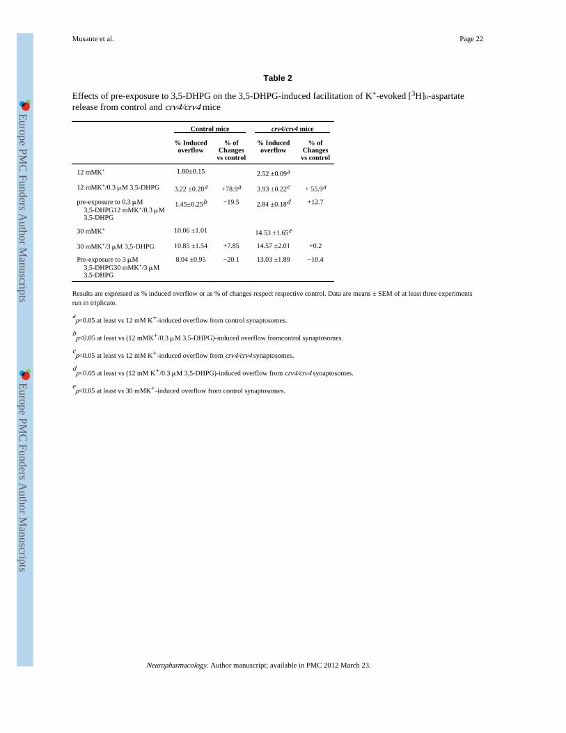
Group I mGlu receptors were reported to undergo desensitization (Catania et al., 1991;Herrero et al., 1994) shortly after a brief pre-exposure to agonist. Since this event was alsoobserved in cortical synaptosomes isolated from mGlu1−/− animals (Sistiaga et al., 1998), ithad been proposed that mGlu5 receptors were those preferentially involved in the agonist-induced modifications. To investigate whether this phenomenon also occurred in ourexperimental conditions, control or crv4/crv4 cortical synaptosomes were exposed to 3,5-DHPG (0.3 μM), then washed to remove the agonist and finally re-challenged with 3,5-DHPG (0.3 μM) in the presence of a depolarizing stimulus (12 μM). 3,5-DHPG (0.3 μM),added contemporary to the depolarizing stimulus, potentiated the 12 μM K+-evoked releaseof [3H]D-ASP from cortical synaptosomes isolated from control and crv4/crv4 mice (seeresults in Figs. 1–4). Accordingly to results in literature, the positive effect of 3,5-DHPGwas abolished when control or crv4/crv4 synaptosomes were pre-exposed to the agonist(Table 2).
Evidences have been also provided showing that 3,5-DHPG is unable to affect the release ofglutamate caused by a depolarizing stimulus stronger than 12 μM K+ (i.e. 30 μM K+,Herrero et al., 1998), but that pre-exposure to the agonist unveil a switch from facilitation toinhibition of group I mGlu receptors (Herrero et al., 1998; Rodriguez-Moreno et al., 1998), amolecular event thought to be involved in the dual role of group I antagonists inneurotoxicity and neuroprotection (Nicoletti et al., 1999). Also in this case the effect wasretained in synaptosomes isolated from mGlu1−/− mice, suggesting the prominent role ofmGlu5 receptors (Sistiaga et al., 1998). To investigate whether this phenomenon could beobserved also in our experimental conditions, synaptosomes were exposed to 3 μM 3,5-DHPG and then re-challenged with 30 μM K+/3 μM 3,5-DHPG. Results were compared tothose obtained from synaptosomes that were not pre-exposed to the agonist. The release of[3H]D-ASP evoked by 30 μM K+ from wild type or crv4/crv4 cortical synaptosomes wasunaffected by 3 μM 3,5-DHPG contemporary added to the depolarizing stimulus (Table 2).A slight inhibition of the 30 μM K+/3 μM 3,5-DHPG-evoked release was observed shortlyafter a preliminary exposure of wild type, but not of crv4/crv4, cortical synaptosomes to theagonist (Table 2). Differences in animal models as well as in the experimental approachesapplied could, perhaps, account for the discrepancies between our and previous results(Herrero et al., 1998; Rodriguez-Moreno et al., 1998).
Notably, the amount of glutamate release elicited by high K+ from cortical crv4/crv4glutamatergic terminals was altered when compared to control synaptosomes (Fig. 4B, Table2). Further investigations are needed to clarify this aspect.
4. DiscussionThe major findings of this work are: (i) mouse cortical glutamatergic terminals are endowedwith both mGlu1 and mGlu5 autoreceptors, whose activation favours the release ofglutamate induced by a mild depolarizing stimulus (i.e. 12 μM K+); (ii) mGlu5autoreceptors may represent the “high affinity” binding sites, while mGlu1 autoreceptorsmay represent the “low affinity” binding sites for glutamate. Data obtained with Westernblot after subsynaptic fractionation of synaptosomal membranes and withimmunocytochemistry support the functional results and sustain the location of both mGlu1and mGlu5 on glutamatergic nerve endings of the cerebral cortex.
Musante et al. Page 9
Neuropharmacology. Author manuscript; available in PMC 2012 March 23.
Europe PM
C Funders A
uthor Manuscripts
Europe PM
C Funders A
uthor Manuscripts

The present demonstration of the existence of both presynaptic mGlu1 and mGlu5autoreceptors is of interest; in fact there has been much controversy on whether group ImGlu autoreceptors are mGlu1 or mGlu5. mGlu1 receptor subtypes were proposed to have apresynaptic location by Moroni et al. (1998), based on “in vivo” microdialysis studies in ratparietal cortex. Sistiaga et al. (1998), however, argued against this view and proposed theexistence of mGlu5 autoreceptors on mouse cortical nerve endings. The existence of mGlu1autoreceptors was then reconsidered by Reid et al. (1999), who showed that the 3,5-DHPG-evoked increase of [3H]glutamate release from rat cortical synaptosomes was prevented bythe preferential mGlu1 receptor antagonist AIDA. More recently, however, Fazal et al.(2003) provided evidence favouring the existence of mGlu5, but not of mGlu1,autoreceptors on cortical glutamatergic terminals. Thus, our results, consistent with theexistence of both mGlu1 and mGlu5 presynaptic autoreceptors, deserve particular attention.
Functional experiments were performed by monitoring release from superfused monolayersof purified synaptosomes, a technique particularly appropriate to study release-regulatingpresynaptic receptors (Raiteri and Raiteri, 2000). Using this experimental approach wefound that the addition of the mGlu1/mGlu5 receptor agonist 3,5-DHPG at 0.3 μMenhanced glutamate release evoked by mild K+ depolarization; the effect of 3,5-DHPG wasmuch weaker and not significant between 0.3 and 10 μM, but it reappeared when the agonistwas added at concentrations higher than 10 μM.
These dual effects seemed predictive of the existence of “high affinity” and “low affinity”binding sites for 3,5-DHPG. In line with this idea, the pharmacological profile of thereceptors involved obtained by using non-competitive antagonists revealed that the twoeffects were due to activation of different receptors. In particular, the response caused bylow 3,5-DHPG concentrations was totally antagonized by the selective mGlu5 receptorantagonist MPEP, suggesting that the “high affinity” site may correspond to the mGlu5autoreceptor. On the other hand, CPCCOEt blocked the potentiating effect caused by high3,5-DHPG concentrations, indicating that the “low affinity” site could coincide with themGlu1 autoreceptor. Consistent with the results obtained with CPCCOEt, LY 367385, aselective competitive antagonist at mGlu1 receptor subtype, totally antagonized thepotentiation by 50 μM 3,5-DHPG of glutamate exocytosis, but, unexpectedly, it alsoaffected significantly the facilitation caused by 0.3 μM 3,5-DHPG. These results are in away surprising enough, in particular when considering that the effect caused by 0.3 μM 3,5-DHPG was CPCCOEt-insensitive, but MPEP-sensitive. Since the binding of LY 367385 tomGlu5 receptors appears unlikely, an alternative explanation might consider that, in corticalglutamatergic terminals, mGlu1 homodimers may colocalize with mGlu5 homodimers,influencing their functions. It is known that mGlu homodimers can adopt a symmetric openconformation in the absence of the agonist that turns to an asymmetric open-closedconformation in presence of glutamate. Both conformations have been observed inplasmamembranes, also in the absence of the agonist, suggesting that the equilibriumbetween the two forms exist (De Blasi et al., 2001). Assuming that mGlu1 and mGlu5homodimers coexist on glutamatergic cortical terminals, we speculate that LY 367385 canforce mGlu1 homodimers to adopt the open conformation and that this modification to thedimer structure could result in changes to homodimer–homodimer interactions. Futurestudies will be useful to clarify this hypothesis.
In recent years, mouse models lacking expression of a given receptor have been used tocorroborate functional results from wild type animals. In order to substantiate the existenceof “high affinity” mGlu5 and of “low affinity” mGlu1 autoreceptors and to shed light ontheir reciprocal role as presynaptic modulators of glutamate exocytosis, we took advantageof animals lacking mGlu1 (crv4/crv4) or mGlu5 (mGlu5−/−) receptors, although, in general,receptor deletion were shown to induce compensative adaptations to central
Musante et al. Page 10
Neuropharmacology. Author manuscript; available in PMC 2012 March 23.
Europe PM
C Funders A
uthor Manuscripts
Europe PM
C Funders A
uthor Manuscripts

neurotransmission. In particular, as group I mGlu receptor is concerned, changes to mGlu1receptor-mediated functions were reported to be caused by mGlu5 receptor deletion (Volk etal., 2006), while mGlu5 receptor expression and functions appeared unaltered in mGlu1knockout animals (Herrero et al., 1998; Sistiaga et al., 1998).
The results obtained when studying the effects of 3,5-DHPG on glutamate exocytosis fromcortical synaptosomes isolated from crv4/crv4 mice sustained, in our opinion, the existenceof a “high affinity” mGlu5 and a “low affinity” mGlu1 autoreceptors. Actually, in thissynaptosomal preparation, 0.3 μM 3,5-DHPG facilitated glutamate exocytosis, while thepotentiation caused by 50 μM 3,5-DHPG was undetectable. The role of mGlu5 receptors asthe “high affinity” binding site for 3,5-DHPG was also supported by the finding that thefacilitation by 0.3 μM 3,5-DHPG of glutamate exocytosis was abrogated in mGlu5−/−
cortical synaptosomes. Differently from expectation, however, 50 μM 3,5-DHPG failed topotentiate the K+-induced release in mGlu5−/− cortical synaptosomes. Although unexpected,this finding could be consistent with the above-mentioned alterations in mGlu1 receptorfunctions observed in mGlu5−/− animals (Volk et al., 2006) and might further support thehypothesis that mGlu1 and mGlu5 receptors can coexist and reciprocally interact onglutamate nerve terminals.
The pattern of the concentration–response curve of 3,5-DHPG deserves a comment,particularly regarding the fall of efficacy at the intermediate concentrations (0.5–10 μM).Our explanation may be found taking into account that mGlu5 receptors can undergo rapidagonist-induced desensitization (results in Table 2, but see also Catania et al., 1991; Herreroet al., 1994, 1998; Rodriguez-Moreno et al., 1998; Fazal et al., 2003); therefore the fall ofefficacy of 3,5-DHPG at intermediate concentrations might represent the resultant of anagonist-induced mGlu5 autoreceptor desensitization that precedes the 3,5-DHPG-inducedactivation of mGlu1 autoreceptors.
Contradictory results are present in the literature on the presynaptic location of mGlu1 andmGlu5 receptors investigated by immunocytochemistry (Fothui et al., 1993; Shigemoto etal., 1993; Romano et al., 1995; Lujan et al., 1996; Marino et al., 2001; Hubert et al., 2001;Muly et al., 2003). When we looked at the protein subsynaptic distribution, we found thatmGlu1 and mGlu5 receptor proteins were present in the presynaptic zone and largelyexpressed in the postsynaptic density. Furthermore, restricting the analysis to theglutamatergic nerve terminals, immunocytochemistry revealed that mGlu1 and mGlu5receptor proteins were present in a significant percentage of VGLUT-1 positiveglutamatergic nerve terminals. By combining the data obtained in functional studies withthose obtained in Western Blot experiments after subsynaptic fractionation and byimmunocytochemistry, it seems reasonable to conclude that presynaptic mGlu1 and mGlu5autoreceptors exist on mouse cortical glutamatergic nerve terminals.
The apparent affinity of 3,5-DHPG for mGlu5 autoreceptors is three orders of magnitudegreater than that for mGlu1 autoreceptors. Assuming that this also occurs when glutamate isthe autoreceptor agonist, one may speculate that the two receptors, although able to mediateglutamate release, play different roles. In fact, mGlu5 autoreceptors would be expected to beactivated by physiological concentrations of glutamate, while ”pathological” concentrationsof the endogenous ligand would be needed to activate mGlu1 receptors. We do not knowwhether mGlu1 and mGlu5 autoreceptors colocalize on glutamatergic terminals or if thereexist a mixed populations of nerve terminals bearing either mGlu1 or mGlu5 autoreceptors,although co-existence would seem more compatible with our findings as well as with thedifferential implication of the receptors in synaptic plasticity and excitotoxic events.Notably, mGlu1 and mGlu5 heteroreceptors were already shown to be co-localized at thepresynaptic terminals, in human and rodent cortical and hippocampal noradrenergic nerve
Musante et al. Page 11
Neuropharmacology. Author manuscript; available in PMC 2012 March 23.
Europe PM
C Funders A
uthor Manuscripts
Europe PM
C Funders A
uthor Manuscripts

endings (Luccini et al., 2007b). Differently from what observed on noradrenergic terminals(Luccini et al., 2007b), however, mGlu1 and mGlu5 autoreceptors do not compensate onefor each other, but exert distinct modulations of glutamate release.
Notably, there is abundant evidence that group I mGlu receptors and NMDA receptorsreciprocally interact in neurons (Luccini et al., 2007b and references therein). NMDAreceptors also exist on glutamatergic terminals, where they mediate different modes ofglutamate release (exocytosis or carrier-mediated release) depending on the glutamateconcentrations acting at the receptor complex (Luccini et al., 2007a). If NMDAautoreceptors and group I mGlu autoreceptors colocalize on glutamate nerve terminals, onecould hypothesize that glutamate exocytosis evoked by low glutamate concentrations actingat release-enhancing NMDA autoreceptors could facilitate mGlu5 autoreceptor activation.On the other side, mGlu1 receptors should be preferentially activated by the “pathological”,carrier-mediated release of glutamate induced by high glutamate concentrations acting atNMDA presynaptic autoreceptors. Further studies, however, are needed to substantiate thishypothesis.
AcknowledgmentsWe wish to thank Maura Agate for careful editorial assistance. This work was supported by grants from IstitutoSuperiore di Sanità (V Programma Nazionale di Ricerca sull’AIDS: Progetto “Patologia, Clinica e Terapiadell’AIDS), from Italian “Ministero dell’Istruzione, dell’Università e della Ricerca Scientifica”(MIUR) and fromIRCCS Istituto Neurologico Mediterraneo NEUROMED (Pozzilli, IS, Italy).
ReferencesBarnes JM, Dev KK, Henley JM. Cyclothiazide unmasks AMPA-evoked stimulation of [3H]-L-
glutamate release from rat hippocampal synaptosomes. British Journal of Pharmacology. 1994;113:339–341. [PubMed: 7530567]
Bockaert J, Pin J-P. Molecular tinkering of G-protein-coupled receptors: an evolutionary success.EMBO Journal. 1999; 18:1723–1729. [PubMed: 10202136]
Catania MV, Aronica E, Sortino MA, Canonico PL, Nicoletti F. De-sensitization of metabotropicglutamate receptors in neuronal cultures. Journal of Neurochemistry. 1991; 56:1329–1335.[PubMed: 1672146]
Chittajallu R, Vignes M, Dev KK, Barnes JM, Collingridge GL, Henley JM. Regulation of glutamaterelease by presynaptic kainate receptors in the hippocampus. Nature. 1996; 379:78–81. [PubMed:8538745]
Conn PJ, Pin J-P. Pharmacology and functions of metabotropic glutamate receptors. Annual Review ofPharmacology and Toxicology. 1997; 37:205–237.
Conti V, Aghaie A, Cilli M, Martin N, Caridi G, Musante L, Candiano G, Castagna M, Fairen A,Ravazzolo R, Guenet JL, Puliti A. crv4, a mouse model for human ataxia associated withkyphoscoliosis caused by an mRNA splicing mutation of the metabotropic glutamate receptor 1(Grm1). International Journal of Molecular Medicine. 2006; 18:593–600. [PubMed: 16964410]
Corti C, Battaglia G, Molinaro G, Riozzi B, Pittaluga A, Corsi M, Mugnaini M, Nicoletti F, Bruno V.The use of knock-out mice unravels distinct roles for mGlu2 and mGlu3 metabotropic glutamatereceptors in mechanisms of neurodegeneration/neuroprotection. Journal of Neuroscience. 2007;27:8297–8308. [PubMed: 17670976]
Costes SV, Daelemans D, Cho EH, Dobbin Z, Pavlakis G, Lockett S. Automatic and quantitativemeasurement of protein-protein colocalization in live cells. Biophysical Journal. 2004; 86:3993–4003. [PubMed: 15189895]
De Blasi A, Conn PJ, Pin JP, Nicoletti F. Molecular determinants of metabotropic glutamate receptorsignaling. Trends in Pharmacological Sciences. 2001; 22:114–120. [PubMed: 11239574]
Fazal A, Parker F, Palmer AM, Croucher MJ. Characterisation of the actions of group I metabotropicglutamate receptor subtype selective ligands on excitatory amino acid release and sodium-
Musante et al. Page 12
Neuropharmacology. Author manuscript; available in PMC 2012 March 23.
Europe PM
C Funders A
uthor Manuscripts
Europe PM
C Funders A
uthor Manuscripts

dependent re-uptake in rat cerebrocortical mini slices. Journal of Neurochemistry. 2003; 86:1346–1358. [PubMed: 12950444]
Feligioni M, Holman D, Haglerod C, Davanger S, Henley JM. Ultrastructural localisation anddifferential agonist-induced regulation of AMPA and kainate receptors present at the presynapticactive zone and postsynaptic density. Journal of Neurochemistry. 2006; 99:549–560. [PubMed:16903873]
Fothui M, Sharp AH, Glatt CE, Hwang PM, Vonkrosigk M, Snyder SH, Dawson TM. Differentiallocalization of phosphoinositide-linked metabotropic glutamate receptor (mGluR1) and theinositol 1,4,5-trisphosphate receptor in rat brain. Journal of Neuroscience. 1993; 13:2001–2012.[PubMed: 8386753]
Gonzalez, RC.; Wintz, P. Digital Image Processing. second ed. Addison Wesley Publication Company;Mass, USA: 1987.
Herrero I, Miras-Portugal MT, Sanchez-Prieto J. Positive feedback of glutamate exocytosis bymetabotropic presynaptic receptor stimulation. Nature. 1992; 360:163–166. [PubMed: 1359425]
Herrero I, Miras-Portugal MT, Sanchez-Prieto J. Rapid desensitization of the presynaptic metabotropicreceptor that facilitates glutamate exocytosis. European Journal of Neuroscience. 1994; 6:163–166.
Herrero I, Miras-Portugal MT, Sanchez-Prieto J. Functional switch from facilitation to inhibition in thecontrol of glutamate release by metabotropic glutamate receptors. Journal of Biological Chemistry.1998; 273:1951–1958. [PubMed: 9442030]
Hubert GW, Paquet M, Smith Y. Differential subcellular localization of mGluR1a and mGluR5 in therat and monkey substantia nigra. Journal of Neuroscience. 2001; 21:1838–1847. [PubMed:11245668]
Luccini E, Musante V, Neri E, Raiteri M, Pittaluga A. N-methyl-D-aspartate autoreceptors respond tolow and high agonist concentrations by facilitating, respectively, exocytosis and carrier-mediatedrelease of glutamate in rat hippocampus. Journal of Neuroscience Research. 2007a; 85:3657–3665.[PubMed: 17671992]
Luccini E, Musante V, Neri E, Brambilla Bas M, Severi P, Raiteri M, Pittaluga A. Functionalinteractions between presynaptic NMDA receptors and metabotropic glutamate receptors co-expressed on rat and human noradrenergic terminals. British Journal of Pharmacology. 2007b;151:1087–1094. [PubMed: 17592518]
Lujan R, Nusser Z, Roberts JD, Shigemoto R, Somogyi P. Perysinaptic location of metabotropicglutamate receptors mGluR1 and mGluR5 on dendrites and dendritic spines in the rathippocampus. European Journal of Neuroscience. 1996; 8:1488–1500. [PubMed: 8758956]
Manders EMM, Verbeek FJ, Aten JA. Measurement of colocalization of objects in dual-colourconfocal images. Journal of Microscopy. 1993; 169:375–382.
Marino MJ, Wittmann M, Bradley Risso, Hubert GW, Smith Y, Conn PJ. Activation of group Imetabotropic glutamate receptors produces a direct excitation and disinhibition of GABAergicprojection neurons in the substantia nigra pars reticulata. Journal of Neuroscience. 2001; 21:7001–7012. [PubMed: 11549710]
Moroni F, Cozzi A, Lombardi G, Sourtcheva S, Leonardi P, Carfì M, Pellicciari R. Presynaptic mGlu1type receptors potentiate transmitter output in the rat cortex. European Journal of Pharmacology.1998; 347:189–195. [PubMed: 9653880]
Muly EC, Maddox M, Smith Y. Distribution of mGluR1a and mGluR5 immunolabelling in primateprefrontal cortex. Journal of Comparative Neurology. 2003; 467:521–535. [PubMed: 14624486]
Nicoletti F, Bruno V, Catania MV, Battaglia G, Copani A, Barbagallo G, Cena V, Sanchez-Prieto J,Spano PF, Pizzi M. Group-I metabotropic glutamate receptors: hypothesis to explain their dualrole in neurotoxicity and neuroprotection. Neuropharmacology. 1999; 38:1477–1484. [PubMed:10530809]
Pedrazzi M, Raiteri L, Bonanno G, Patrone M, Ledda S, Passalacqua M, Milanese M, Melloni E,Raiteri M, Pontremoli S, Sparatore B. Stimulation of excitatory amino acid release from adultmouse brain glia subcellular particles by high mobility group box 1 protein. Journal ofNeurochemistry. 2006; 99:827–838. [PubMed: 16911580]
Musante et al. Page 13
Neuropharmacology. Author manuscript; available in PMC 2012 March 23.
Europe PM
C Funders A
uthor Manuscripts
Europe PM
C Funders A
uthor Manuscripts

Phillips GR, Huang JK, Wang Y, Tanaka H, Shapiro L, Zhang W, Shan WS, Arndt K, Frank M,Gordon RE, Gawinowicz MA, Zhao Y, Colman DR. The presynaptic particle web: ultrastructure,composition, dissolution, and reconstitution. Neuron. 2001; 32:63–77. [PubMed: 11604139]
Pin J-P, Galvez T, Prézeau L. Evolution, structure, and activation mechanism of family 3/C G protein-coupled receptors. Pharmacology and Therapeutics. 2003; 98:325–354. [PubMed: 12782243]
Pinheiro PS, Rodrigues RJ, Silva AP, Cunha RA, Oliveira CR, Malva JO. Solubilization andimmunological identification of presynaptic alpha-amino-3-hydroxy-5-methyl-4-isoxazolepropionic acid receptors in the rat hippocampus. Neuroscience Letters. 2003; 336:97–100. [PubMed: 12499049]
Pittaluga A, Bonfanti A, Raiteri M. Differential desensitization of ionotropic non-NMDA receptorshaving distinct neuronal location and function. Naunyn Schmiedeberg’s Archives ofPharmacology. 1997; 356:29–38. [PubMed: 9228187]
Raiteri M. Presynaptic metabotropic glutamate and GABAB receptors. Handbook of ExperimentalPharmacology. 2008; 184:373–407. [PubMed: 18064420]
Raiteri L, Raiteri M. Synaptosomes still viable after 25 years of superfusion. Neurochemical Research.2000; 25:1265–1274. [PubMed: 11059801]
Raiteri L, Zappettini S, Milanese M, Fedele E, Raiteri M, Bonanno G. Mechanisms of glutamaterelease elicited in rat cerebrocortical nerve endings by ‘pathologically’ elevated extraterminal K+
concentrations. Journal of Neurochemistry. 2007; 103:952–961. [PubMed: 17662048]
Reid ME, Toms NJ, Bedingfield JS, Roberts PJ. Group I mGlu receptors potentiate synaptosomal[3H]glutamate release independently of exogenously applied arachidonic acid.Neuropharmacology. 1999; 38:477–485. [PubMed: 10221751]
Rodriguez-Moreno A, Sistiaga A, Lerma J, Sanchez-Prieto J. Switch from facilitation to inhibition ofexcitatory synaptic transmission by group I mGluR desensitization. Neuron. 1998; 21:1477–1486.[PubMed: 9883739]
Romano C, Sesma MA, McDonald CT, O’Malley K, Van den Pol AN, Olney JW. Distribution ofmetabotropic glutamate receptor mGluR5 immunoreactivity in rat brain. Journal of ComparativeNeurology. 1995; 335:455–469. [PubMed: 7636025]
Shigemoto R, Nomura S, Ohishi H, Nakanishi S, Mizuno N. Immunohistochemical localization of ametabotropic glutamate receptor, mGluR5, in the rat brain. Neuroscience Letters. 1993; 163:53–57. [PubMed: 8295733]
Sistiaga A, Herrero I, Conquet F, Sanchez-Prieto J. The metabotropic glutamate receptor 1 is notinvolved in the facilitation of glutamate release in cerebrocortical nerve terminals.Neuropharmacology. 1998; 37:1485–1492. [PubMed: 9886671]
Volk LJ, Daly CA, Huber KM. Differential roles for group 1 mGluR subtypes in induction andexpression of chemically induced hippocampal long-term depression. Journal of Neurophysiology.2006; 95:2427–2438. [PubMed: 16421200]
Wang S-J, Sihra TS. Noncompetitive metabotropic glutamate5 receptor antagonist (E)-2-methyl-6-styryl-pyridine (SIB1893) depresses glutamate release through inhibition of voltage-dependentCa2+ entry in rat cerebrocortical nerve terminals (synaptosomes). Journal of Pharmacology andExperimental Therapeutics. 2004; 309:951–958. [PubMed: 14982967]
Musante et al. Page 14
Neuropharmacology. Author manuscript; available in PMC 2012 March 23.
Europe PM
C Funders A
uthor Manuscripts
Europe PM
C Funders A
uthor Manuscripts

Fig. 1.Concentration–effect relationship of 3,5-DHPG on the 12 μM K+-evoked release of [3H]D-aspartate ([3H]D-ASP) from mouse cortical synaptosomes. Mouse cortical synaptosomeswere exposed to 12 μM KCl in absence (white bar) or in presence (grey bar) of 3,5-DHPG(concentration as indicated). Results are expressed as induced overflow. Data are mean ±SEM of 3–12 experiments run in triplicate (three determinations for each experimentalconditions). *p < 0.05 vs control; **p < 0.01 vs control.
Musante et al. Page 15
Neuropharmacology. Author manuscript; available in PMC 2012 March 23.
Europe PM
C Funders A
uthor Manuscripts
Europe PM
C Funders A
uthor Manuscripts

Fig. 2.Effects of CPCCOEt and MPEP on the release of [3H]D-aspartate evoked by 12 μM K+ inpresence of 3,5-DHPG. Mouse cortical synaptosomes were exposed to 12 μM KCl inabsence (white bar) or in presence (grey bar) of 3,5-DHPG (0.3 μM, panel A; 50 μM, panelB). CPCCOEt (rising right grey bar), MPEP (rising left grey bar) or both agonists (cross-hatched grey bar) were added starting from t = 30 min of superfusion till the end of theexperiment. Results are expressed as induced overflow. Data are mean ± SEM of 4–10experiments run in triplicate. *p < 0.05 at least vs 12 μM K+; #p < 0.05 at least vs 12 μMK+ in the presence of 0.3 μM 3,5-DHPG; §p < 0.05 at least vs 12 μM K+ in the presence of50 μM 3,5-DHPG.
Musante et al. Page 16
Neuropharmacology. Author manuscript; available in PMC 2012 March 23.
Europe PM
C Funders A
uthor Manuscripts
Europe PM
C Funders A
uthor Manuscripts

Fig. 3.Effects of LY 367385 on the release of [3H]D-aspartate evoked by 12 μM K+ in presence of3,5-DHPG. Mouse cortical synaptosomes were exposed to 12 μM KCl in absence (whitebar) or in presence (grey bar) of 3,5-DHPG (0.3 μM, panel A; 50 μM, panel B). LY 367385(rising right grey bar) was added starting from t = 30 min of superfusion till the end of theexperiment. Results are expressed as induced overflow. Data are means ± SEM of sevenexperiments run in triplicate. *p < 0.05 at least vs 12 μM K+; #p < 0.05 at least vs 12 μMK+ in the presence of 0.3 μM 3,5-DHPG; §p < 0.05 at least vs 12 μM K+ in the presence of50 μM 3,5-DHPG.
Musante et al. Page 17
Neuropharmacology. Author manuscript; available in PMC 2012 March 23.
Europe PM
C Funders A
uthor Manuscripts
Europe PM
C Funders A
uthor Manuscripts

Fig. 4.Effects of 3,5-DHPG on the release of [3H]D-aspartate evoked by 12 μM K+ fromsynaptosomes isolated from the cortex of mouse lacking mGlu1 or mGlu5 receptors.Synaptosomes were prepared from the cortex of transgenic mice lacking mGlu5 receptors(mGlu5−/− mouse, panel A) or mGlu1 receptors (crv4/crv4 mouse, panel B) and fromrelative controls. Synaptosomes were exposed to 12 μM KCl in absence (white bar) or inpresence (grey bar) of 3,5-DHPG (concentration as indicated). Results are expressed asinduced overflow. Data are mean ± SEM of 12 experiments run in triplicate. *p < 0.05 atleast vs respective control. WT, wild type.
Musante et al. Page 18
Neuropharmacology. Author manuscript; available in PMC 2012 March 23.
Europe PM
C Funders A
uthor Manuscripts
Europe PM
C Funders A
uthor Manuscripts

Fig. 5.mGlu1 and mGlu5 receptor protein expressions in mouse cortical synaptosomes. (A)Western blotting of mGlu1 and mGlu5 receptor proteins in the synaptosomal lysate (Tot-Syn) and in its detergent soluble fraction (Ds-Syn). Protein weights are expressed in kDa.(B) Immunoblot of synapse fractionation showing mGlu1 receptor and mGlu5 receptorrelative distribution in synaptosomal compartments in the NSSP fraction (NSSP), in thepostsynaptic fraction (Post) and in the presynaptic fraction (Pre). Anti-PSD95, anti-synaptophysin and anti-Stx-1A were used as markers for ultrasynaptic fraction todeterminate the purity of the preparation that is more than 90%. Protein weights areexpressed in kDa. The blots are representative of four to six analyses.
Musante et al. Page 19
Neuropharmacology. Author manuscript; available in PMC 2012 March 23.
Europe PM
C Funders A
uthor Manuscripts
Europe PM
C Funders A
uthor Manuscripts

Fig. 6.mGlu1 and mGlu5 receptors are present on mouse cortical glutamatergic terminals. (A)Representative double-labelling images of anti mGlu5 (panels d–i) and of mGlu1 (panels j–o) with anti A-1 (marker of all nerve terminals) and with anti VGLUT-1 (specific marker ofglutamatergic nerve terminals). (B) Percent of co-localization of mGlu1 and mGlu5 receptorimmunoreactivities with VGLUT-1 or Stx-1A staining. Results are expressed as co-localization of Stx-1A and VGLUT-1 with mGlu1 and mGlu5 receptor proteins,respectively. Data are the mean ± SEM of 6–8 different image couples.
Musante et al. Page 20
Neuropharmacology. Author manuscript; available in PMC 2012 March 23.
Europe PM
C Funders A
uthor Manuscripts
Europe PM
C Funders A
uthor Manuscripts

Europe PM
C Funders A
uthor Manuscripts
Europe PM
C Funders A
uthor Manuscripts
Musante et al. Page 21
Table 1
Effects of group I mGlu receptor antagonists on the K+-evoked release of [3H]D-ASP from mouse corticalsynaptosomes
12 mMK+-evoked release of [3H]D-ASP
(% Induced overflow) (% of Changes vs control)
Control 1.61± 0.13
0.01 μM LY 367385 1.68± 0.09 +4.3
0.01 μM LY 367385 1.62± 0.07 +0.6
0.01 μM LY 367385 1.67± 0.11 +3.7
0.5 μM CPCCOEt 1.72± 0.11 +6.8
5 μM CPCCOEt 1.64± 0.08 +1.8
10 μM CPCCOEt 1.74± 0.26 +0.8
0.3 μM MPEP 1.26± 0.04 −21.8
1 μM MPEP 1.11± 0.11a −31.9
3 μM MPEP 1.52± 0.13 −5.6
5 μM CPCCOEt+1 μM MPEP 1.13± 0.11a −29.9
Results are expressed as % induced overflow or as % of changes respect respective control. Data are means± SEM of at least three experiments runin triplicate.
ap< 0.05 at least vs control.
Neuropharmacology. Author manuscript; available in PMC 2012 March 23.
Europe PM
C Funders A
uthor Manuscripts
Europe PM
C Funders A
uthor Manuscripts
Musante et al. Page 22
Table 2
Effects of pre-exposure to 3,5-DHPG on the 3,5-DHPG-induced facilitation of K+-evoked [3H]D-aspartaterelease from control and crv4/crv4 mice
Control mice crv4/crv4 mice
% Inducedoverflow
% ofChanges
vs control
% Inducedoverflow
% ofChanges
vs control
12 mMK+ 1.80±0.15 2.52 ±0.09a
12 mMK+/0.3 μM 3,5-DHPG 3.22 ±0.28a +78.9a 3.93 ±0.22c + 55.9a
pre-exposure to 0.3 μM 3,5-DHPG12 mMK+/0.3 μM 3,5-DHPG
1.45±0.25b −19.5 2.84 ±0.18d +12.7
30 mMK+ 10.06 ±1.01 14.53 ±1.65e
30 mMK+/3 μM 3,5-DHPG 10.85 ±1.54 +7.85 14.57 ±2.01 +0.2
Pre-exposure to 3 μM 3,5-DHPG30 mMK+/3 μM 3,5-DHPG
8.04 ±0.95 −20.1 13.03 ±1.89 −10.4
Results are expressed as % induced overflow or as % of changes respect respective control. Data are means ± SEM of at least three experimentsrun in triplicate.
ap<0.05 at least vs 12 mM K+-induced overflow from control synaptosomes.
bp<0.05 at least vs (12 mMK+/0.3 μM 3,5-DHPG)-induced overflow fromcontrol synaptosomes.
cp<0.05 at least vs 12 mM K+-induced overflow from crv4/crv4 synaptosomes.
dp<0.05 at least vs (12 mM K+/0.3 μM 3,5-DHPG)-induced overflow from crv4/crv4 synaptosomes.
ep<0.05 at least vs 30 mMK+-induced overflow from control synaptosomes.
Neuropharmacology. Author manuscript; available in PMC 2012 March 23.