presence of functional cannabinoid receptors in human endocrine pancreas
TRANSCRIPT
ARTICLE
Presence of functional cannabinoid receptors in humanendocrine pancreas
F. J. Bermúdez-Silva & J. Suárez & E. Baixeras &
N. Cobo & D. Bautista & A. L. Cuesta-Muñoz &
E. Fuentes & P. Juan-Pico & M. J. Castro & G. Milman &
R. Mechoulam & A. Nadal & F. Rodríguez de Fonseca
Received: 11 July 2007 /Accepted: 12 October 2007 / Published online: 19 December 2007# Springer-Verlag 2007
AbstractAims/hypothesis We examined the presence of functionalcannabinoid receptors 1 and 2 (CB1, CB2) in isolatedhuman islets, phenotyped the cells producing cannabinoidreceptors and analysed the actions of selective cannabinoidreceptor agonists on insulin, glucagon and somatostatinsecretion in vitro. We also described the localisation on isletcells of: (1) the endocannabinoid-producing enzymes N-
acyl-phosphatidyl ethanolamine-hydrolysing phospholipaseD and diacylglycerol lipase; and (2) the endocannabinoid-degrading enzymes fatty acid amidohydrolase and mono-acyl glycerol lipase.Methods Real-time PCR, western blotting and immunocy-tochemistry were used to analyse the presence of endocanna-binoid-related proteins and genes. Static secretion experimentswere used to examine the effects of activating CB1 or CB2 oninsulin, glucagon and somatostatin secretion and to measurechanges in 2-arachidonoylglycerol (2-AG) levels within islets.Analyses were performed in isolated human islets and inparaffin-embedded sections of human pancreas.Results Human islets of Langerhans expressed CB1 andCB2 (also known as CNR1 and CNR2) mRNA and CB1and CB2 proteins, and also the machinery involved insynthesis and degradation of 2-AG (the most abundantendocannabinoid, levels of which were modulated byglucose). Immunofluorescence revealed that CB1 wasdensely located in glucagon-secreting alpha cells and lessso in insulin-secreting beta cells. CB2 was densely presentin somatostatin-secreting delta cells, but absent in alpha andbeta cells. In vitro experiments revealed that CB1 stimula-tion enhanced insulin and glucagon secretion, while CB2agonism lowered glucose-dependent insulin secretion,showing these cannabinoid receptors to be functional.Conclusions/interpretation Together, these results suggest arole for endogenous endocannabinoid signalling in regula-tion of endocrine secretion in the human pancreas.
Keywords Anandamide . 2-Arachidonoylglycerol .
Beta cell . Cannabinoid receptors .
Fatty acid amidohydrolase . Glucagon . Human . Insulin .
Islets of Langerhans . Somatostatin
Diabetologia (2008) 51:476–487DOI 10.1007/s00125-007-0890-y
Electronic supplementary material The online version of this article(doi:10.1007/s00125-007-0890-y) contains supplementary material,which is available to authorised users.
F. J. Bermúdez-Silva (*) : J. Suárez : E. Baixeras :N. Cobo :A. L. Cuesta-Muñoz : F. Rodríguez de Fonseca (*)Fundación IMABIS, Hospital Carlos Haya,Avenida Carlos Haya 82, 7a Planta, Pabellón A,29010 Málaga, Spaine-mail: [email protected]: [email protected]
D. BautistaServicio de Anatomía Patológica, Hospital Carlos Haya,Málaga, Spain
E. Fuentes : P. Juan-Pico :A. NadalInstituto de Bioingeniería, Universidad Miguel Hernández,Elche, Spain
M. J. CastroServicio de Cirugía General y Digestiva, Hospital Carlos Haya,Málaga, Spain
G. Milman :R. MechoulamDepartment of Medicinal Chemistry and Natural Products,Ein Kerem Campus, Hebrew University,Jerusalem, Israel
AbbreviationsAEA anandamideACEA arachidonoyl-2′-chloroethylamide2-AG 2-arachidonoyl glycerolAM251 N-(piperidin-1-yl)-5-(4-iodophenyl)-1-
(2,4-dichlorophenyl)-4-methyl-1H-pyrazole-3-carboxamide
CB1 cannabinoid receptor 1CB2 cannabinoid receptor 2DAGL diacylglycerol lipaseFAAH fatty acid amidohydrolaseIEQ islet equivalentJWH 133 3-(1’,1’-dimethylbutyl)-1-deoxy-
Δ8-tetrahydrocannabinolMAGL monoacyl glycerol lipaseNAPE-PLD N-acyl-phosphatidyl ethanolamine-
hydrolysing phospholipase D
Introduction
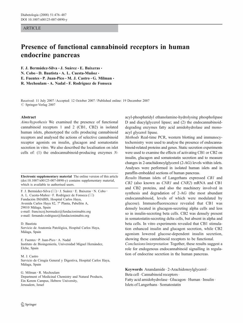
The endogenous cannabinoids, i.e. the endocannabinoidsanandamide (AEA) and 2-arachidonoyl glycerol (2-AG),are lipid transmitters that were identified in the brain asrelevant modulators of synaptic transmission [1–4]. Theyact through different receptors (cannabinoid receptors 1 and2 [CB1, CB2]) and are produced through specific enzymes(diacylglycerol lipase [DAGL] α and β, for 2-AG and N-
acyl-phosphatidyl ethanolamine-hydrolysing phospholipaseD [NAPE-PLD] for AEA) and degraded by at least twodifferent enzymes (fatty acid amidohydrolase [FAAH] andmonoacyl glycerol lipase [MAGL]) [5] (Fig. 1). In additionto the physiological role of these transmitters in the centralnervous system, recent studies have established their func-tionality in peripheral organs involved in feeding control,energy homeostasis and metabolism [6–8]. Endocannabi-noids counteract satiety signals at both the gastrointestinaland hypothalamic levels and promote overfeeding, as wellas lipid biosynthesis and storage [7–12].
The endocannabinoids are relevant homeostatic signalswhose dysregulation contributes to obesity and type 2diabetes [6, 13]. A clinical trial in obese patients treatedwith Rimonabant, a CB1 antagonist, resulted in effectivereduction in body weight, waist circumference and insulinresistance [14, 15]. Additional studies demonstrated thatCB1 blockade improves insulin resistance, insulinaemiaand glycosylated haemoglobin in obese patients with type 2diabetes [16]. Since these actions are not totally explainedby the weight loss induced by the anorectic actions derivedfrom CB1 blockade, the endocannabinoids may alsomodulate metabolism in peripheral organs. This hypothesishas been confirmed in animal models where CB1 werefound to modulate lipid and glucose metabolism in insulin-sensitive tissues such as the adipose tissue [7] and the liver[12]. Recent studies extended this notion to the endocrinepancreas, where the endogenous cannabinoid system has
NAPE
PhChol+
PhEth
AEA
PEA
OEA
CB1CB2GPR55VR1
PKAMAPK
TAG
DAG
2-AG
PPARαGPR119
AA+
ETHN
AA+
GlycerolMAGL
PLDFAAH
DAGL
Endogenous cannabinoidsignalling system
PLC
NAT
AT
Fig. 1 Endogenous cannabinoid signalling system. The two mainendocannabinoids are AEA and 2-AG. They act mainly through CB1and CB2. The parents acylethanolamides palmitoylethanolamide(PEA) and oleoylethanolamide (OEA) act through different receptorsystems (the orphan receptor GPR119 and the peroxisome proliferatoractivated-receptor alpha [PPARα]). We examined the presence offunctional CB1 and CB2 in isolated human islets, as well as thelocalisation of AEA-producing enzyme NAPE-PLD, AEA-degrading
enzyme FAAH and the enzymes involved in generation (DAGL) anddegradation (MAGL) of 2-AG in sections of human pancreas. AA,arachidonic acid, DAG, diacylglycerol; ETHN, ethanolamide; MAPK,mitogen-activated protein kinase; PhChol, phosphatidyl choline;PhEth, phosphatidyl ethanolamine; PKA, protein kinase A; PLC,phospholipase C; PLD, phospholipase D; TAG, triacylglycerol, VR1,vanilloid receptor 1
Diabetologia (2008) 51:476–487 477
recently been identified in mice, rats and the rat insulinomabeta cell line RIN-m5F [17–20]. Whereas stimulation ofCB1 in the rat leads to glucose intolerance, activation ofCB2 improves glucose handling after a glucose load [18,19]. In mice, CB2 modulate calcium oscillations and insulinsecretion in vitro [17]. These actions are derived fromglucose-induced alterations in endocannabinoid production,as demonstrated in the pancreatic beta cell line RIN-m5F.Thus, elevations of glucose concentration in the culturemedia are associated with a rise in the levels of both 2-AGand AEA [20].
To date, no studies have addressed the presence andfunctional significance of cannabinoid receptors in humanendocrine pancreas. However, the clear and conclusiveeffects of chronic treatment with the CB1 antagonistRimonabant on insulin resistance in obese humans, withor without type 2 diabetes, clearly suggest the presence ofthis system in human pancreatic islets [14–16]. In order toconfirm this hypothesis, we examined the presence offunctional CB1 and CB2 in isolated human islets, as well asthe localisation of the machinery for synthesis anddegradation of endocannabinoids in human pancreatictissue.
Methods
Human islet isolation Islets were isolated and purified fromhuman pancreases using the Ricordi method [21, 22]. Thepresent studies were performed in pancreas from four brain-dead, heart-beating, non-diabetic, non-obese (mean BMI28.3±0.7 kg/m2) adult organ donors (mean age 50±14 years; two women, two men). Cause of death wasstroke for two donors and anoxic encephalopathy for theother two. All procedures were performed according tospecific legal guidelines and written informed consent wasobtained from each donor’s family; the local ethicscommittee of Carlos Haya Hospital approved and super-vised the experiments.
The pancreas was cut into two parts, cannulated andperfused with cold (4–8°C) liberase (Liberase-HI; RocheMolecular Biochemical, Indianapolis, IN, USA). Afterperfusion, the pancreas was minced, transferred to theRicordi chamber and digestion carried out at 37°C.Digestion was stopped and the digest diluted with 2 litresdilution solution (Mediatech Cellgro, Herndon, VA, USA).The digest was collected and centrifuged for 3 min at1,000 rpm (225 g) and 4°C. Pellets were washed andrecombined in cold modified University of Wisconsinsolution. The tissue digest was purified on a continuousficoll (Biochrom, Berlin, Germany) density gradient, usingthe Cobe 2991 cell separator/processor (Gambro, Lake-wood, CO, USA).
The fraction containing the purified islet mass wasanalysed and the purity of the preparation assessed withdithizone. The preparations used in this study had morethan 80% purity. Fresh aliquots of 1,000 islet equivalents(IEQs) were snap-frozen for subsequent mRNA quantifica-tion and western blotting analysis. The remaining purifiedislets were cultured at 30,000 IEQs per 75 cm2 non-treatedflask (Nunc, Wiesbaden, Germany) in a final volume of
Table 1 mRNA expression of CB1 and CB2
CB1: β-actin (×10−4)a,b CB2: β-actin (×10−4)a,b
Cerebellum 8.63±1.07 0.0191±0.0010Islets 2.95±0.81 0.0224±0.0110Leucocytes 0.0148±0.0001 1.86±0.56
Islet data are mean±SD from three different donors. Data from humancerebellum and leucocytes are mean±SD from triplicate measurementof commercially available total RNA reference samples.a Real-time PCR quantification of CB1 and CB2 cDNA using specificprimers: a comparative analysis of the expression of both cannabinoidreceptors in human islets was done using total RNA reference samplesfrom human cerebellum and leucocyte commercial standardsbCB1 expression is about 100-fold higher than that of CB2 in humanislets and threefold less than in CB1-enriched tissue, the cerebellum
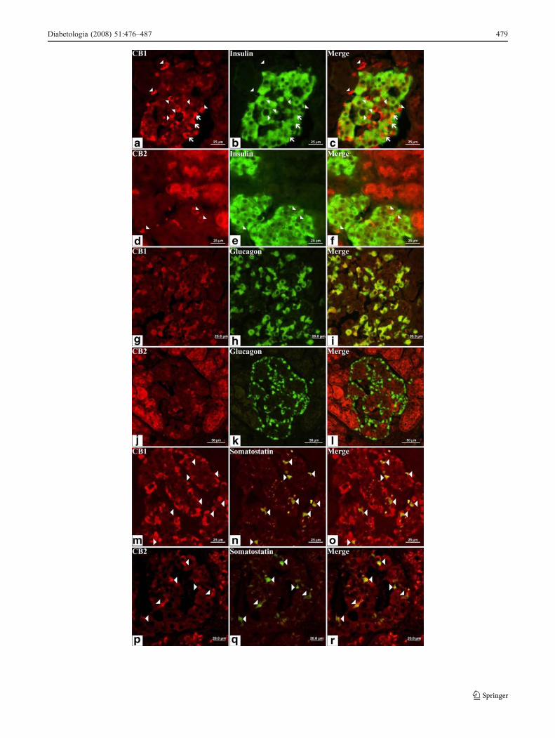
Fig. 2 Double immunofluorescence of cannabinoid receptors andinsulin, glucagon and somatostatin. Representative photomicrographsof sections through a human pancreas showing the same areaimmunolabelled for both CB1 and CB2 (red), and for insulin,glucagon and somatostatin (green), with merged images in (yellow).a Immunostaining of cells in a pancreatic islet with an antiserumagainst CB1. Note that most of these cells have a typical morphologyof alpha cells (arrowheads), with small diameter and big nucleus.b Islet cells stained in green are insulin-containing beta cells.c Colocalisation of insulin and CB1. Note that most CB1-containingcells do not produce insulin, so they are non-β cells (arrowheads).However, some cells have co-production of insulin and CB1 (arrows).d Immunostaining of pancreatic cells with an antiserum against CB2.Exocrine tissue has high CB2 production, but only scattered cells inislets are CB2-positive (arrowheads). e Islet cells stained in green areinsulin-containing beta cells. f Merged image (d) and (e). CB2-producing cells in the pancreatic islets do not show insulin immu-noreactivity. g Immunostaining of cells in a pancreatic islet with anantiserum against CB1. h Islet cells stained in green are glucagon-containing alpha cells. i Double staining with glucagon and CB1.Nearly all alpha cells produce CB1 (yellow). j Immunostaining ofpancreatic cells with an antiserum against CB2. k Islet cells stained ingreen are glucagon-containing alpha cells. l Merged image of (j) and(k) clearly showing that CB2-containing cells in the pancreatic isletare not alpha cells. m Immunostaining of cells in a pancreatic isletwith an antiserum against CB1. n Islet cells stained in green arescattered cells containing somatostatin. o Double staining withsomatostatin and CB1. Nearly all delta cells are immunonegative forCB1. p Immunostaining of pancreatic cells with an antiserum againstCB2. q Islet cells stained in green are somatostatin-containing deltacells, some with a high intensity. r Merged images of (p) and (q)clearly show that delta cells preferentially produce CB2. Scale, asindicated
�
478 Diabetologia (2008) 51:476–487
Diabetologia (2008) 51:476–487 479
30 ml CMRL-1066 medium (Mediatech Cellgro) supple-mented with 10% FCS (v/v), 100 IU/ml penicillin, 100 μg/ml streptomycin, 2.8 μg/ml amphotericin and 2 mmol/lL-glutamine; this was done for 3 to 5 days at 37°C, 95%relative humidity and 5% CO2. Culture medium wasreplaced every 2 days. After 3 to 5 days of culture thepreparations were checked for viability by Trypan Blueexclusion test and found to contain <5% damaged cells.
Immunohistochemistry in human pancreas samples Immu-nohistochemical and immunofluorescence studies wereperformed in human pancreatic tissue obtained from thePathology Department tissue bank at Carlos Haya Hospital.Biopsy samples were taken from four different non-diabetic, non-obese (mean BMI 27.5±1.5 kg/m2) adultpatients (mean age 68±9 years; three women, one man)with pancreatic adenocarcinoma (non-endocrine tumour).All procedures were performed according to specific legalguidelines and written informed consent were obtainedfrom patients; the local ethics committee of Carlos HayaHospital approved and supervised the experiments. Seeadditional information in Electronic supplementary material(ESM).
NAPE-PLD-, DAGLα- and DAGLβ-specific antibody gen-eration Polyclonal rabbit antibodies were generated againstcannabinoid machinery proteins as described in ESM.Immunising peptides were: (1) a 13-amino acid peptidecomprising part of both the C-terminal and the N-terminalregion of NAPE-PLD (MDENSCDKAFEET); (2) a 16-amino acid peptide from the C-terminal region of DAGLα(CGASPTKQDDLVISAR); and (3) a 16-amino acid pep-tide from an internal sequence of DAGLβ (SSDSPLDSPTKYPTLC). Extensive validation studies of these anti-bodies as immunocytochemistry markers were performed inbrain samples (ESM Fig. 1).
Double immunofluorescence and immunohistochemistryParaffin-embedded sections of human pancreases wereanalysed for the presence of CB1, CB2 and FAAH in alpha(glucagon), beta (insulin) and delta (somatostatin) pancre-atic islet cells by double immunofluorescence. Sectionswere incubated overnight at room temperature with mouseanti-insulin (dilution 1:200; Sigma-Aldrich Quimica S.A.,Madrid, Spain), anti-glucagon (1:200; Sigma) or anti-somatostatin (1:200; Genetex, San Antonio, TX, USA)antibody and a rabbit anti-CB1 (dilution 1:100; ABR—Affinity Bioreagents, Golden, CO, USA), anti-CB2 (1:100;ABR) or anti-FAAH (1:50; Cayman Chemical, Ann Arbor,MI, USA) antibody. After extensive washes in PBS, thesections were incubated for 2 h at room temperature in asecondary anti-mouse IgG–FITC antibody (dilution 1:200;Sigma) and a secondary anti-rabbit IgG–Cy3 antibody
(1:300; Jackson Immunoresearch Laboratories, West Grove,PA, USA). Finally, the sections were washed in PBS andanalysed under epifluorescence microscopy (OlympusEuropa, Hamburg, Germany). Additional immunohisto-chemistry studies were done to determine the levels ofNAPE-PLD, DAGLα, DAGLβ, FAAH, MAGL, insulinand chromogranin A. In all cases, the specificity of theimmunostaining was confirmed by omission of the firstantibody or the use of preadsorption of primary antibodieswith the immunising peptide. Digital photographs weretaken with an Olympus BX41 microscope (Olympus).
Real-time quantitative PCR Real-time quantitative PCRwas used to measure CB1 (also known as CNR), CB2 (alsoknown as CNR2) and NAPE-PLD mRNA expressionaccording to Hansson et al. [23]. Briefly, total RNA wasisolated from snap-frozen pellets containing 1,000 IEQsfrom three different donors by using Trizol reagent (GibcoBRL Life Technologies, Baltimore, MD, USA). All RNAsamples had A260:280 ratios of 1.8 to 2.0. Total RNA fromeach sample and random hexamers were used to generatefirst strand cDNA using transcriptor reverse transcriptase(Roche Applied Science, Indianapolis, IN, USA). Negativecontrols included reverse transcription reactions omittingreverse transcriptase. The cDNA obtained was used as thetemplate for real-time quantitative PCR with an iCyclersystem (Bio-Rad, Hercules, CA, USA) using the Quanti-Tect SYBR Green PCR kit (Qiagen, Hilden, Germany). Acomparative analysis of the expression of both cannabinoidreceptors in human islets was made using reference totalRNA samples from commercial standards of both humancerebellum and human leucocytes (BD Biosciences, PaloAlto, CA, USA). Primers for PCR reactions and annealingtemperatures are shown in the ESM. Quantification wascarried out with a standard curve run at the same time as thesamples with each reaction run in duplicate. Absolute valuesfrom each sample were normalised with regard to beta actin.
Western blot analysis Frozen cell pellets from humanpancreatic islets (1,000 IEQs) were suspended in 50 μlSDS sample buffer containing dithiothreitol and heat-denatured for 5 min at 95°C. For immunoblotting, equalamounts of protein from lysates (10 μl per lane) weresubjected to 7.5% (w/v) SDS-PAGE and homogeneoustransfer to nitrocellulose membranes controlled by Ponceaured staining. Blots were preincubated for 1 h at roomtemperature with PBS containing 0.1% (v/v) Tween 20 and2% (w/v) albumin fraction V from bovine serum (blockingbuffer). For protein detection, each blotted membrane lanewas incubated separately with the specific CB1 (1:250; ABR),CB2 (1:250; ABR), FAAH (1:100; Cayman), MAGL (1:500;kindly donated by D. Piomelli, Department of Pharmacology,University of California, Irvine, CA, USA), DAGLα (1:100),
480 Diabetologia (2008) 51:476–487
DAGLβ (1:100) and NAPE-PLD (1:75) antibodies fromrabbit, diluted in PBS containing 0.1% (v/v) Tween 20 and2% (w/v) albumin fraction V from bovine serum. This wasdone overnight at room temperature. The specific proteinbands were visualised using the enhanced chemilumines-cence technique (Amersham International, Amersham, UK)and an imaging system (Auto-Biochem; LTF Labortechnik,Wasserburg/Bodensee, Germany). Western blots showed thateach primary antibody detected a protein of the expectedmolecular size. As additional controls, blotted membranelanes were incubated with the primary antibody preadsorbedwith the corresponding immunising peptide.
Static secretion of insulin, glucagon and somatostatin inisolated islets After 5 days in vitro, total IEQs and acinarcontamination were determined by staining with dithizone.Viability was checked by Trypan Blue exclusion. Less than5% of cells within each islet were non-viable and no acinartissue was adhered. Human cultured islets were thenwashed with Hanks’ solution and suspended in a serum-free medium. Groups of 50 IEQs were plated on sterile 24-well plates (Nunc) with 1 ml medium containing (mmol/l):115 NaCl, 10 NaHCO3, 5 KCl, 1.1 MgCl2, 1.2 NaH2PO4,2.5 CaCl2 and 25 HEPES; plus 1% albumin and either3 mmol/l or 11 mmol/l glucose. Additionally, one of thefollowing drugs was added to the medium (Tocris Biosci-ence, Bristol, UK): 2-AG (10−5 to 10−6 mol/l); AEA (10−8
to 10−7 mol/l); arachidonyl-2′-chloroethylamide (ACEA), aspecific CB1 activator [24] (10−7 to 10−9 mol/l); 3-(1′,1′-dimethylbutyl)-1-deoxy-Δ8-tetrahydrocannabinol (JWH133), a specific CB2 activator [25] (10−7–10−9 mol/l); orN-(piperidin-1-yl)-5-(4-iodophenyl)-1-(2,4-dichlorophenyl)-4-methyl-1H-pyrazole-3-carboxamide (AM251), a selectiveCB1 antagonist [26] (10−7 mol/l). Concentrations of canna-binoid receptor agonists and antagonists were selectedaccording to the reported Ki value of each compound. Eachexperimental condition was assayed in quadruplicate andislets isolated from four different donors were employed.The islets were incubated at 37°C and 5% CO2 for 1 h, afterwhich the medium containing the islets was collected in1.5 ml tubes with 520 kIU aprotinin and centrifuged for5 min at 1,000×g and 4°C. The supernatant fractions weredivided into aliquots and stored at −80°C for further quanti-fication of insulin, glucagon and somatostatin by means ofspecific commercial radioimmunoassays or enzyme immu-noassay kits. The cellular pellets were frozen at −80°C tomeasure total protein amount (see ESM). Secretion ofinsulin, glucagon and somatostatin from each well wasnormalised with the protein content of their 50 IEQs.
Analysis of islet 2-AG levels Because the immunohisto-chemical analysis revealed that the islets contained themachinery for synthesis and degradation of 2-AG but not
AEA, we measured intracellular levels of 2-AG in isletsincubated either in low (3 mmol/l) or high (11 mmol/l)glucose concentrations. Seven-hundred IEQs per well wereincubated and 1 h after incubation islets were centrifuged andassayed in triplicate for 2-AG content using quantitative GC-MS in a TRACE GC/MS 2000 system with electron impactionisation detector (Finnigan, San Jose, CA, USA; see ESM).
Statistical analysis Results are expressed as the mean±SEM. The significance of differences between the groupswas evaluated by either Student’s t test (two-tailed, pairedgroups) or one-way ANOVA followed by Newman–Keulstest as post hoc (multiple group comparison). A p value of<0.05 was considered significant.
Results
Human islets express CB1 and CB2 mRNA CB1 and CB2mRNAs are present in fresh isolated islets. Table 1 shows thatCB1 expression was almost 100-fold higher than that of CB2in human islets and threefold lower than that of a CB1-enriched tissue, the cerebellum. CB2 expression was 80-foldlower than a CB2-enriched tissue, the leucocytes. Despite thepresence of exocrine tissue on the islet pellets used formRNA isolation, immunoreactivity to CB2 antibody withinthe islets supports the expression of CB2 mRNA inendocrine tissue in a specific small population of cells.
Double immunofluorescence revealed the cell subtypecontaining both CB1 and CB2 in human islets Cannabinoidreceptors had a specific distribution pattern in human isletcells. Figure 2a–f depicts the double immunofluorescence ofcannabinoid receptors and insulin. Only a small portion ofbeta cells produced CB1 (Fig. 2c), the majority of CB1-positive cells being insulin-negative. Exocrine tissue did notproduced CB1 (ESM Fig. 2a–c). The few CB2-positive cellsin islets were insulin-negative (Fig. 2d–f). Clear CB2 immu-nostaining was present in exocrine tissue (ESM Fig. 2d).Figure 2g–l shows the level of cannabinoid receptor proteinsin human alpha cells. Immunostaining patterns of both CB1and glucagon were nearly identical, suggesting that all alphacells produce CB1 (Fig. 2g–i). In contrast, none of theglucagon-positive cells detected colocalised with CB2,indicating null production of CB2 in alpha cells (Fig. 2j–l).Finally, double immunofluorescence with anti-somatostatinand anti-cannabinoid receptors showed that virtually all deltacells produce CB2 (Fig. 2p–r), whereas CB1 immunostain-ing was nearly absent in this cell subtype (Fig. 2m–o).
CB1 and CB2 as well as the enzymes for synthesis anddegradation of endocannabinoids were detected by Westernblot analysis in human islets Human islet homogenates
Diabetologia (2008) 51:476–487 481
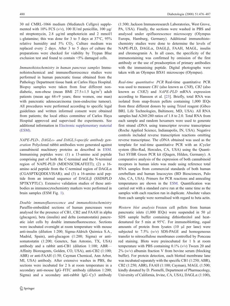
were analysed by immunoblotting to detect the cannabinoidreceptors, FAAH, MAGL, DAGLα, DAGLβ and NAPE-PLD (Fig. 3). The image shows a representative immunoblotof pancreatic islet lysates from three different preparations.The main bands correspond to the expected proteinmolecular masses [27–34], although additional bandscorresponding to posttranslational modifications weredetected (details, Fig. 3). Omission of the first antibodiesor incubation of the primary antibodies pre-adsorbed withthe corresponding immunising peptides abolished thedescribed bands (Fig. 3a,b). Exocrine contamination (up to20%) could be responsible for part of the CB2 and MAGLimmunoblot reactivity. However, both immunohistochemis-try and immunofluorescence supported the existence ofCB2 and MAGL protein within islets.
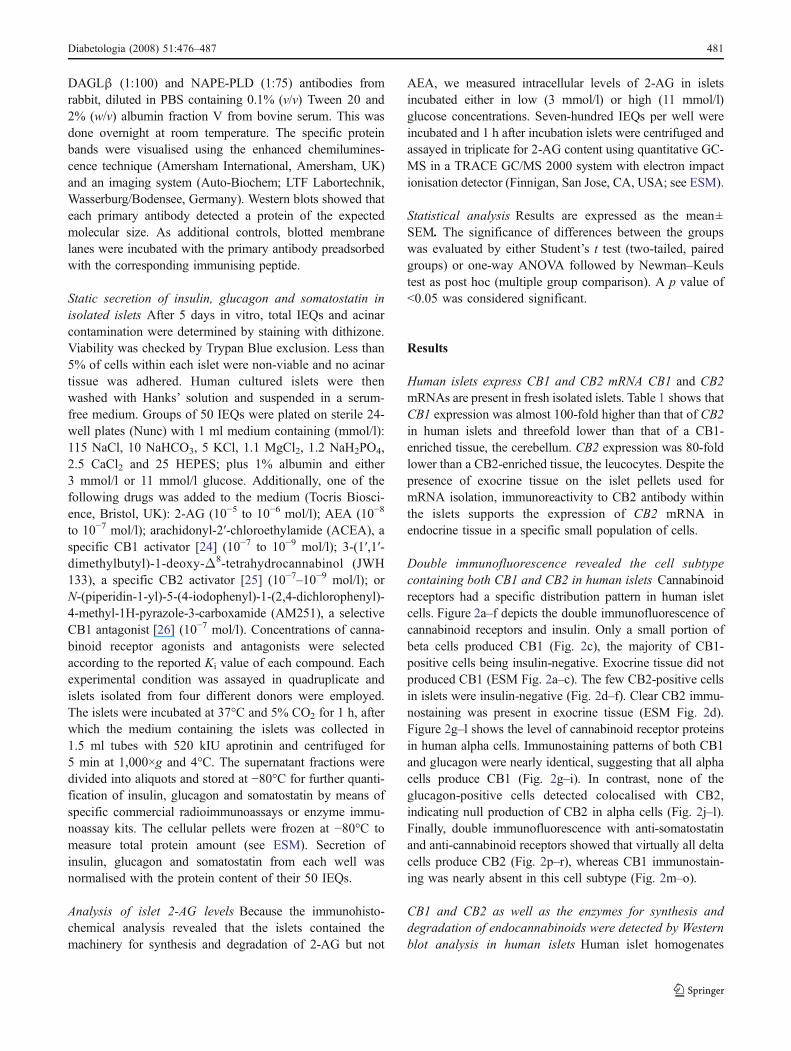
High glucose increases 2-AG content within islets Figure4a shows the insulin response of islet preparations culturedunder either 3 (low) or 11 (high) mmol/l glucose. Figure 4bshows 2-AG content within those islets cultured underdifferent glucose concentrations. As reported in insulinomacells [20], a rise in glucose concentration increased bothinsulin secretion and 2-AG levels (Fig. 4a,b). A drop inglucose concentration reduced 2-AG to below the thresholdof detection. Levels of 2-AG detected were: 11 mmol/l
glucose-containing wells, 8.71±0.11 pmol 2-AG/mg pro-tein; 3 mmol/l glucose-containing wells, less than 5.49 pmol2-AG/mg protein; n=2 different experiments, each mea-sured in triplicate.
In vitro stimulation of CB1 and CB2modifies insulin, glucagonand somatostatin secretion Figure 4c shows how stimulationof CB1 with both selective (ACEA) and natural (AEA and2-AG) CB1 agonists increased insulin release from isletscultured under high glucose concentrations. This effect wasalso observed at low glucose concentrations (3 mmol/l) andwas antagonised by the selective CB1 antagonist AM251[26] (Fig. 4d). CB2 stimulation with the selective agonist JWH133 lowered glucose-dependent insulin release (Fig. 4c), aneffect mediated by CB2, since it was found to be antagonisedby the selective CB2 antagonist AM630 (data not shown).
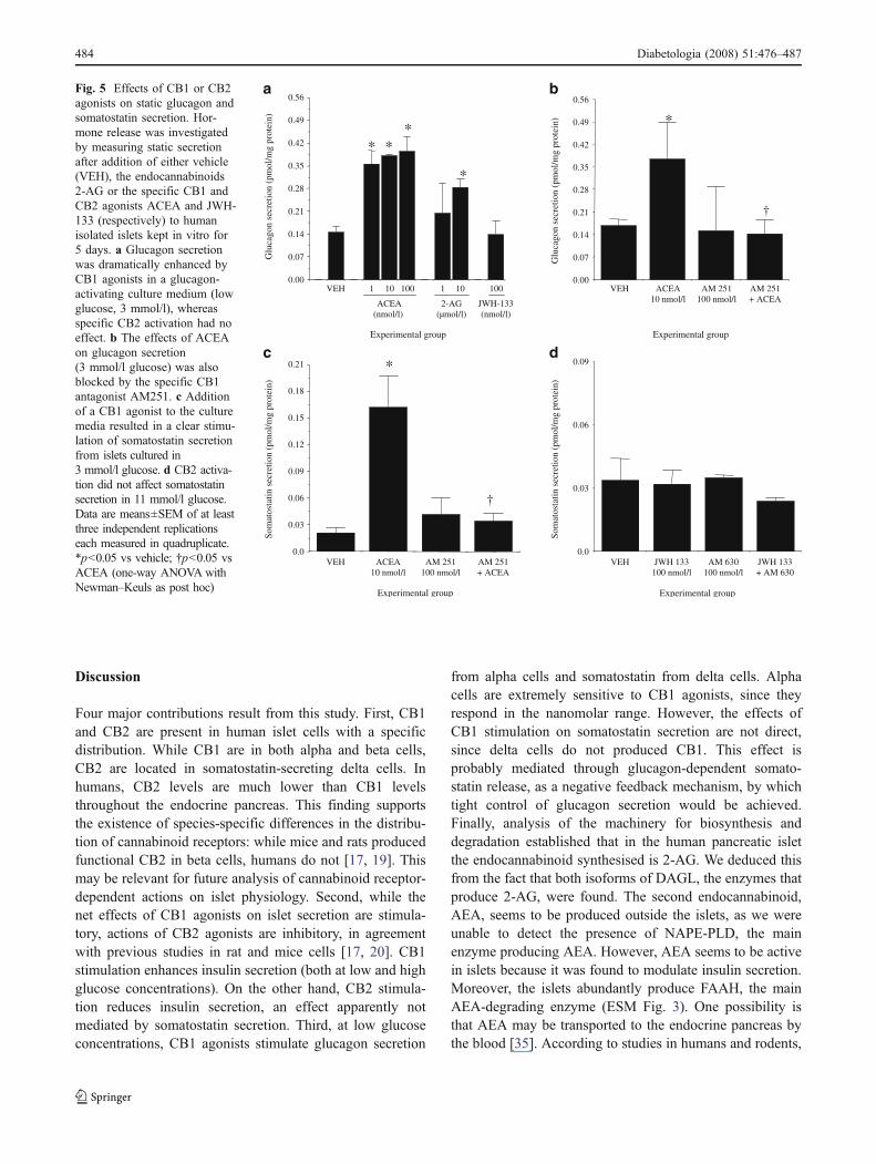
CB1 agonism also resulted in stimulation of glucagonsecretion (Fig. 5a,b). Both 2-AG and ACEA, but not theCB2 agonist JWH 133, stimulated glucagon release underlow glucose culture conditions. The stimulatory effects ofACEA on glucagon secretion were dependent on CB1stimulation since it was counteracted by incubation with thespecific CB1 antagonist AM251 [26] (Fig. 5b).
Regarding somatostatin secretion, we were unable underour experimental conditions to find any specific effect of the
Fig. 3 Western blot analysis of CB1, CB2, NAPE-PLD, DAGLα,DAGLβ, FAAH and MAGL. Whole cell lysate was prepared from1,000 IEQs, solubilised in SDS–dithiothreitol sample buffer, separatedin 7.5% (w/v) polyacrylamide gel, transferred to nitrocellulose andexamined by enhanced chemiluminescence analysis for the presenceof cannabinoid receptors and the synthesising and degrading enzymes.Molecular mass (MM) markers are shown on the left in kDa). Asshown a, a strong immunoreactive band at ~43 kDa and a less intenseband at ~60 kDa were observed for CB1 protein (lane 1), consistentwith glycosylated and non-glycosylated CB1 protein described inprevious studies [29–32]. Anti-CB2 antibody was reactive for threebands of approximately ~45, ~70 and ~60 kDa (lane 9) that probablyreflect glycosylated forms, as previously described [32–34]. Immuno-
blots for NAPE-PLD revealed no staining (lane 7); however, a mainband with a molecular mass of ~62 kDa could be seen in acinarhomogenates (data not shown). DAGLα and DAGLβ immunoblotsshowed unique bands of ~120 and ~70 kDa respectively (lanes 3 and5), identical to those observed previously by Bisogno and collabo-rators [24]. A major band of ~37 kDa for MAGL protein (lane 11)[35] and ~ 63 kDa for FAAH protein (lane 13) [36] were detected.Omission of the first antibodies or incubation of the primaryantibodies preadsorbed with the corresponding immunising peptidesabolished the described bands in lanes 2, 4, 6, 8, 10, 12 (a) and lane14 (b). The image shows a representative immunoblot of pancreaticislet lysates from three different preparations
482 Diabetologia (2008) 51:476–487
CB2 agonist JWH 133 in islets cultured at 11 mmol/l glucose(Fig. 5d). However, delta cells from islets incubated at3 mmol/l glucose responded to the CB1 agonist ACEAwitha potent release of somatostatin (Fig. 5c). Because CB1 arenot located in somatostatin cells, the effects of this CB1agonist on somatostatin are probably derived from thepotent stimulation of glucagon and insulin secretion.
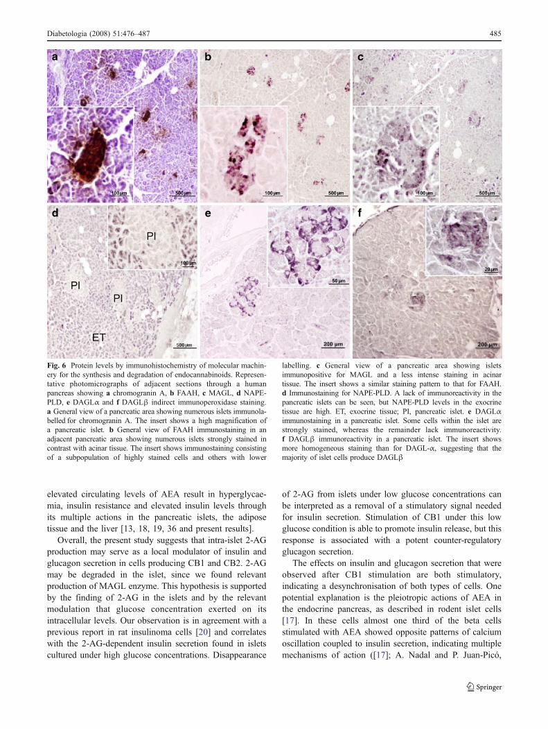
The molecular machinery needed to release and process 2-AG signalling is present in islets Figure 6 shows the
presence of the biosynthetic enzymes NAPE-PLD andDAGLα and the endocannabinoid-degrading enzymesFAAH and MAGL in human islets. Figure 6a shows thelocation of Langerhans islets stained with Cromogranin A.An adjacent slide stained with anti-FAAH antibody showsthe expression of this enzyme in islets but its absence inacinar tissue (Fig. 6b). MAGL, the other degrading enzyme,was also produced in acinar tissue, although with lessintensity than in islets (Fig. 6c). Additional doubleimmunohistochemistry studies localised FAAH enzyme inbeta cells but not in alpha cells secreting glucagon (ESMFig. 3). NAPE-PLD immunoreactivity (Fig. 6d) was almostabsent in islets, whereas it was dense in acinar surroundingtissue. We confirmed this by using a different antibody(ESM Fig. 4) and real-time quantitative PCR, which showedvery low expression of NAPE-PLD mRNA in isolatedpancreatic islets, probably reflecting acinar contamination.The enzymes for the synthesis of 2-AG, DAGLα andDAGLβ (Fig. 6e,f, respectively) displayed an invertedlocalisation to NAPE-PLD, being highly produced in isletcells and scarce in acinar tissue. The insert (Fig. 6e) shows aheterogeneous pattern of distribution of DAGLα: some cellswithin the islet were strongly stained, whereas the remaininglacked immunoreactivity. By contrast, DAGLβ staining wasmore homogeneous (insert, Fig. 6f), suggesting that themajority of islet cells express this enzyme to a similardegree. These data suggest that, in the pancreatic islet, themain transmitter is 2-AG, since the machinery for biosyn-thesis and degradation of this endocannabinoid is present,whereas that of AEA is incomplete. However, since FAAH,the main AEA-degrading enzyme, was densely present in theislet cells secreting insulin, we cannot exclude that extra isletAEA may reach the endocrine cells at sufficient concen-trations to produce relevant effects on insulin secretion [35].
0.0
7.7
15.5
23.2
31
Experimental group
ACEA (nmol/l) AEA (nmol/l) 2-AG (µmol/l) JWH (nmol/l)
*
*
*
*
VEH
VEH ACEA 100 nmol/l
AM 251 100 nmol/l
AM 251 + ACEA0.0
7.7
15.5
23.2
Experimental group
*
†
0
2
4
6
8
10
2-A
G (
pmol
/mg
prot
ein)
ND
Low glucose
High glucose
Low glucose
High glucose
0.0
7.7
15.5
Insu
lin s
ecre
tion
(pm
ol/m
g pr
otei
n)
**a b
c
d
Insu
lin s
ecre
tion
(pm
ol/m
g pr
otei
n)In
sulin
sec
retio
n (p
mol
/mg
prot
ein)
1 10 1 10 1 10100 10 100
Fig. 4 Effects of CB1 or CB2 agonists on static insulin secretion:functionality of cannabinoid receptors in human islets. Hormonerelease was investigated by measuring static secretion after addition ofeither vehicle (VEH), the endocannabinoids 2-AG/AEA or the specificCB1 and CB2 agonists ACEA and JWH-133 (respectively) to humanisolated islets kept in vitro for 5 days. a Human islets actively secretedinsulin when exposed to a high glucose culture medium (11 mmol/lglucose), as compared with basal release at low glucose (3 mmol/l)concentration in the culture medium. b While 2-AG levels in the isletswere not detectable (ND) under low glucose culture conditions,elevation of glucose concentration to 11 mmol/l increased 2-AGcontents. c CB1 agonist ACEA, AEA and 2-AG promoted insulinsecretion in a medium that enhances beta cell insulin secretion(supplemented with 11 mmol/l glucose). Under the same conditions,CB2 activation by JWH-133 significantly decreased insulin release.d The effect of CB1 agonists on insulin secretion was also observed inlow glucose medium (3 mmol/l) where insulin was only released atbasal levels. This effect was specific since it was reversed by the CB1antagonist AM251. Data are means±SEM of at least three independentreplications each measured in quadruplicate. *p<0.05 vs control group;†p<0.05 vs ACEA (one way ANOVAwith Newman–Keuls as post hoc)
R
Diabetologia (2008) 51:476–487 483
Discussion
Four major contributions result from this study. First, CB1and CB2 are present in human islet cells with a specificdistribution. While CB1 are in both alpha and beta cells,CB2 are located in somatostatin-secreting delta cells. Inhumans, CB2 levels are much lower than CB1 levelsthroughout the endocrine pancreas. This finding supportsthe existence of species-specific differences in the distribu-tion of cannabinoid receptors: while mice and rats producedfunctional CB2 in beta cells, humans do not [17, 19]. Thismay be relevant for future analysis of cannabinoid receptor-dependent actions on islet physiology. Second, while thenet effects of CB1 agonists on islet secretion are stimula-tory, actions of CB2 agonists are inhibitory, in agreementwith previous studies in rat and mice cells [17, 20]. CB1stimulation enhances insulin secretion (both at low and highglucose concentrations). On the other hand, CB2 stimula-tion reduces insulin secretion, an effect apparently notmediated by somatostatin secretion. Third, at low glucoseconcentrations, CB1 agonists stimulate glucagon secretion
from alpha cells and somatostatin from delta cells. Alphacells are extremely sensitive to CB1 agonists, since theyrespond in the nanomolar range. However, the effects ofCB1 stimulation on somatostatin secretion are not direct,since delta cells do not produced CB1. This effect isprobably mediated through glucagon-dependent somato-statin release, as a negative feedback mechanism, by whichtight control of glucagon secretion would be achieved.Finally, analysis of the machinery for biosynthesis anddegradation established that in the human pancreatic isletthe endocannabinoid synthesised is 2-AG. We deduced thisfrom the fact that both isoforms of DAGL, the enzymes thatproduce 2-AG, were found. The second endocannabinoid,AEA, seems to be produced outside the islets, as we wereunable to detect the presence of NAPE-PLD, the mainenzyme producing AEA. However, AEA seems to be activein islets because it was found to modulate insulin secretion.Moreover, the islets abundantly produce FAAH, the mainAEA-degrading enzyme (ESM Fig. 3). One possibility isthat AEA may be transported to the endocrine pancreas bythe blood [35]. According to studies in humans and rodents,
VEH ACEA 10 nmol/l
AM 251 100 nmol/l
AM 251 + ACEA
0.0
0.03
0.06
0.09
0.12
0.15
0.18
0.21
Som
atos
tatin
sec
retio
n (p
mol
/mg
prot
ein)
VEH JWH 133 100 nmol/l
AM 630 100 nmol/l
JWH 133 + AM 630
0.0
0.03
0.06
0.09
VEH 1 10 100 1 10 1000.00
0.07
0.14
0.21
0.28
0.35
0.42
0.49
0.56
Experimental group
Glu
cago
n se
cret
ion
(pm
ol/m
g pr
otei
n)
ACEA (nmol/l)
2-AG (µmol/l)
JWH-133 (nmol/l)
a
*
* **
VEH ACEA 10 nmol/l
AM 251 100 nmol/l
AM 251 + ACEA
Experimental group
*
b
Experimental group Experimental group
c d
0.00
0.07
0.14
0.21
0.28
0.35
0.42
0.49
0.56
Glu
cago
n se
cret
ion
(pm
ol/m
g pr
otei
n)So
mat
osta
tin s
ecre
tion
(pm
ol/m
g pr
otei
n)
†
†
*
Fig. 5 Effects of CB1 or CB2agonists on static glucagon andsomatostatin secretion. Hor-mone release was investigatedby measuring static secretionafter addition of either vehicle(VEH), the endocannabinoids2-AG or the specific CB1 andCB2 agonists ACEA and JWH-133 (respectively) to humanisolated islets kept in vitro for5 days. a Glucagon secretionwas dramatically enhanced byCB1 agonists in a glucagon-activating culture medium (lowglucose, 3 mmol/l), whereasspecific CB2 activation had noeffect. b The effects of ACEAon glucagon secretion(3 mmol/l glucose) was alsoblocked by the specific CB1antagonist AM251. c Additionof a CB1 agonist to the culturemedia resulted in a clear stimu-lation of somatostatin secretionfrom islets cultured in3 mmol/l glucose. d CB2 activa-tion did not affect somatostatinsecretion in 11 mmol/l glucose.Data are means±SEM of at leastthree independent replicationseach measured in quadruplicate.*p<0.05 vs vehicle; †p<0.05 vsACEA (one-way ANOVA withNewman–Keuls as post hoc)
484 Diabetologia (2008) 51:476–487
elevated circulating levels of AEA result in hyperglycae-mia, insulin resistance and elevated insulin levels throughits multiple actions in the pancreatic islets, the adiposetissue and the liver [13, 18, 19, 36 and present results].
Overall, the present study suggests that intra-islet 2-AGproduction may serve as a local modulator of insulin andglucagon secretion in cells producing CB1 and CB2. 2-AGmay be degraded in the islet, since we found relevantproduction of MAGL enzyme. This hypothesis is supportedby the finding of 2-AG in the islets and by the relevantmodulation that glucose concentration exerted on itsintracellular levels. Our observation is in agreement with aprevious report in rat insulinoma cells [20] and correlateswith the 2-AG-dependent insulin secretion found in isletscultured under high glucose concentrations. Disappearance
of 2-AG from islets under low glucose concentrations canbe interpreted as a removal of a stimulatory signal neededfor insulin secretion. Stimulation of CB1 under this lowglucose condition is able to promote insulin release, but thisresponse is associated with a potent counter-regulatoryglucagon secretion.
The effects on insulin and glucagon secretion that wereobserved after CB1 stimulation are both stimulatory,indicating a desynchronisation of both types of cells. Onepotential explanation is the pleiotropic actions of AEA inthe endocrine pancreas, as described in rodent islet cells[17]. In these cells almost one third of the beta cellsstimulated with AEA showed opposite patterns of calciumoscillation coupled to insulin secretion, indicating multiplemechanisms of action ([17]; A. Nadal and P. Juan-Picó,
Fig. 6 Protein levels by immunohistochemistry of molecular machin-ery for the synthesis and degradation of endocannabinoids. Represen-tative photomicrographs of adjacent sections through a humanpancreas showing a chromogranin A, b FAAH, c MAGL, d NAPE-PLD, e DAGLα and f DAGLβ indirect immunoperoxidase staining.a General view of a pancreatic area showing numerous islets immunola-belled for chromogranin A. The insert shows a high magnification ofa pancreatic islet. b General view of FAAH immunostaining in anadjacent pancreatic area showing numerous islets strongly stained incontrast with acinar tissue. The insert shows immunostaining consistingof a subpopulation of highly stained cells and others with lower
labelling. c General view of a pancreatic area showing isletsimmunopositive for MAGL and a less intense staining in acinartissue. The insert shows a similar staining pattern to that for FAAH.d Immunostaining for NAPE-PLD. A lack of immunoreactivity in thepancreatic islets can be seen, but NAPE-PLD levels in the exocrinetissue are high. ET, exocrine tissue; PI, pancreatic islet. e DAGLαimmunostaining in a pancreatic islet. Some cells within the islet arestrongly stained, whereas the remainder lack immunoreactivity.f DAGLβ immunoreactivity in a pancreatic islet. The insert showsmore homogeneous staining than for DAGL-α, suggesting that themajority of islet cells produce DAGLβ
Diabetologia (2008) 51:476–487 485
unpublished results). Additional mechanisms may be theinterference induced by AEA on GAP junctions [37]involved in electrical beta cell coupling [38]. However,the fact that the selective blockade of CB1 with AM251attenuated the stimulation of both insulin and glucagonsecretion supports a more relevant contribution of the CB1.
Finally, we found that CB2 are located in somatostatin-secreting cells. Since this peptide is a potent intra-isletmodulator of insulin and glucagon secretion [39], it isfeasible to expect somatostatin-dependent responses tocannabinoid receptor agonists. Under our experimentalconditions, CB2 activation did not change somatostatinsecretion in 11 mmol/l glucose, suggesting that CB2-dependent inhibition of insulin secretion is not related toCB2-induced somatostatin release. Further research isneeded to clarify the role of this receptor in delta cells.
After consideration of all the findings in this study andplacing them within the context of endocannabinoid physi-ology, we propose a major role for CB1 and 2-AG in theregulation of islet physiology and energy homeostasis. CB1stimulation was able to increase insulin and glucagonsecretion, as well as lipogenesis in liver and adipose tissue,while it blocked the incorporation of glucose into the musclecells, leading to a ‘saving cycle’: reduced energy expenditureand increased energy storage [6, 7, 12, 13]. Enhanced insulinrelease may help to facilitate the incorporation of glucoseinto adipocytes, while enhanced glucagon release maysustain high plasma glucose levels, favouring this ‘savingcycle’. Under this hypothesis, CB1 can be considered a new‘thrifty gene’. Its physiological and pharmacological profileis strikingly similar to that of the μ-opioid receptor, arecently proposed contributor to the ‘saving cycle’ [40] anda major partner of CB1 signalling in the brain [41]. In lightof this potential role, and supported by clinical trials usingthe CB1 antagonist, the endocannabinoid system can beconsidered a promising target for pharmacological develop-ment in diabetes and complicated obesity.
Acknowledgements This study was supported by Consejerias deSalud e Innovación, Junta de Andalucia (SAS 144/04 and SAS 0064/05), Instituto de Salud Carlos III (grants 03/0178, 07/0880 and 07/1226), Redes Temáticas ISCIII-RETIC RD06/001 and RD06/0015/008 and MEC (grants SAF 2004-07762, SAF 2005-08014, SAF 2006-12863, BFU2005-01052). P. Juan-Pico has a PhD scholarship fromInstituto de Salud Carlos III. G. Milman was supported by USNational Institute on Drug Abuse. The authors are grateful to: J. M.Mellado (Human Islet Isolation Facility, Carlos Haya Hospital,Malaga, Spain) for kindly helping in human islet isolation; D.Piomelli (Department of Pharmacology, University of California,Irvine, CA, USA) for providing rabbit anti-MAGL and anti-NAPE-PLD; J. Romero (Research laboratory, Fundacion Hospital Alcorcon,Madrid, Spain) and K. Mackie (Department of Psychiatry and BrainSciences, Indiana University, Bloomington, IN, USA) for kindlydonating CB1 and CB2 blocking peptides; I. Sanchez (RegenerativeMedicine Laboratory, IMABIS Foundation, Malaga, Spain) for
technical assistance; J.C. Aledo (Department of Biochemistry andMolecular Biology, University of Malaga, Spain) for helping inantibodies generation; and A. Garcia-Ocaña (Division of Endocrinol-ogy, University of Pittsburgh, PA, USA) for initial support inimmunohistochemistry.
Duality of interest The authors declare that there is no duality ofinterest associated with this manuscript.
References
1. Devane WA, Dysarz FA 3rd, Johnson M, Melvin LS, Howlett AC(1998) Determination and characterization of a cannabinoidreceptor in rat brain. Mol Pharmacol 34:605–613
2. Devane WA, Hanus L, Breuer A et al (1992) Isolation andstructure of a brain constituent that binds to the cannabinoidreceptor. Science 258:1946–1949
3. Stella N, Schweitzer P, Piomelli D (1992) A second endogenouscannabinoid that modulates long-term potentiation. Nature388:773–778
4. Mechoulam R, Ben-Shabat S, Hanus L et al (1995) Identificationof an endogenous 2-monoglyceride, present in canine gut, thatbinds to cannabinoid receptors. Biochem Pharmacol 50:83–90
5. Piomelli D (2003) The molecular logic of endocannabinoidsignalling. Nat Rev Neurosci 4:873–884
6. Di Marzo V, Matias I (2005) Endocannabinoid control of foodintake and energy balance. Nat Neurosci 8:585–589
7. Cota D, Marsicano G, Tschop M et al (2003) The endogenouscannabinoid system affects energy balance via central orexigenicdrive and peripheral lipogenesis. J Clin Invest 112:423–431
8. Gomez R, NavarroM, Ferrer B et al (2002) A peripheral mechanismfor CB1 cannabinoid receptor-dependent modulation of feeding.J Neurosci 22:9612–9617
9. Kirkham TC, Williams CM, Fezza F, Di Marzo V (2002)Endocannabinoid levels in rat limbic forebrain and hypothalamusin relation to fasting, feeding and satiation: stimulation of eatingby 2-arachidonoyl glycerol. Br J Pharmacol 136:550–557
10. Di Marzo V, Goparaju SK, Wang L et al (2001) Leptin-regulatedendocannabinoids are involved in maintaining food intake. Nature410:822–825
11. Pavon FJ, Bilbao A, Hernandez-Folgado L et al (2006) Anti-obesity effects of the novel in vivo neutral cannabinoid receptorAntagonist 5-(4-chlorophenyl)-1-(2,4-dichlorophenyl)-3-hexyl-1H-1,2,4-triazole–LH 21. Neuropharmacology 51:358–366
12. Osei-Hyiaman D, DePetrillo M, Pacher P et al (2005) Endocan-nabinoid activation at hepatic CB1 receptors stimulates fatty acidsynthesis and contributes to diet-induced obesity. J Clin Invest115:1298–1305
13. Engeli S, Bohnke J, Feldpausch M et al (2005) Activation of theperipheral endocannabinoid system in human obesity. Diabetes54:2838–2843
14. Van Gaal LF, Rissanen AM, Scheen AJ, Ziegler O, Rossner S,RIO-Europe Study Group (2005) Effects of the cannabinoid-1receptor blocker rimonabant on weight reduction and cardiovas-cular risk factors in overweight patients: 1-year experience fromthe RIO-Europe study. Lancet 365:1389–1397
15. Pi-Sunyer FX, Aronne LJ, Heshmati HM, Devin J, Rosenstock J,RIO-North America Study Group (2006) Effect of rimonabant, acannabinoid-1 receptor blocker, on weight and cardiometabolicrisk factors in overweight or obese patients: RIO-North America:a randomized controlled trial. JAMA 295:761–775
486 Diabetologia (2008) 51:476–487
16. Scheen AJ, Finer N, Hollander P, Jensen MD, Van Gaal LF, RIO-Diabetes Study Group (2006) Efficacy and tolerability ofrimonabant in overweight or obese patients with type 2 diabetes:a randomised controlled study. Lancet 368:1660–1672
17. Juan-Pico P, Fuentes E, Bermúdez-Silva FJ et al (2006)Cannabinoid receptors regulate Ca2+ signals and insulin secretionin pancreatic β-cells. Cell Calcium 39:155–162
18. Bermudez-Silva FJ, Serrano A, Diaz-Molina FJ et al (2006)Activation of cannabinoid CB1 receptors induces glucose intoler-ance in rats. Eur J Pharmacol 531:282–284
19. Bermudez-Silva FJ, Suarez J, Sanchez Vera I et al (2007) Role ofcannabinoid CB2 receptors in glucose homeostasis in rats. Eur JPharmacol 565:207–211
20. Matias I, Gonthier MP, Orlando P et al (2006) Regulation,function and dysregulation of endocannabinoids in models ofadipose and beta-pancreatic cells and in obesity and hyperglyce-mia. J Clin Endocrin Metab 91:3171–3180
21. Ricordi C, Lacy PE, Finke EH, Olack BJ, Scharp DW (1988)Automated method for isolation of human pancreatic islets.Diabetes 37:413–420
22. Cuesta-Munoz AL, Briones RM, Mellado-Gil JM et al (2005)Internal assessment of a novel islet isolation facility in Spain.Transplant Proc 37:3404–3406
23. Hansson AC, Bermudez-Silva FJ, Malinen H et al (2007) Geneticimpairment of frontocortical endocannabinoid degradation andhigh alcohol preference. Neuropsychopharmacology 32:117–126
24. Hillard CJ, Manna S, Greenberg MJ et al (1999) Synthesis andcharacterization of potent and selective agonists of the neuronalcannabinoid receptor (CB1). J Pharmacol Exp The 289:1427–1433
25. Huffman JW, Liddle J, Yu S et al (1999) 3-(1′,1′-Dimethylbutyl)-1-deoxy-delta8-THC and related compounds: synthesis of selec-tive ligands for the CB2 receptor. Bioorg Med Chem 7:2905–2914
26. Gatley SJ, Lan R, Pyatt B, Gifford AN, Volkow ND, MakriyannisA (1997) Binding of the non-classical cannabinoid CP 55,940,and the diarylpyrazole AM251 to rodent brain cannabinoidreceptors. Life Sci 61:PL191–PL197
27. Pettit DAD, Harrison MP, Olson JM et al (1998) Immunohisto-chemical localization of the neural cannabinoid receptor in ratbrain. J Neurosci Methods 51:391–402
28. Egertová M, Elphick MR (2000) Localisation of cannabinoidreceptors in the rat brain using antibodies to the intracellular C-terminal tail of CB1. J Comp Neurol 422:159–171
29. Ashton JC, Zheng Y, Liu P et al (2004) Immunohistochemicalcharacterisation and localisation of cannabinoid CB1 receptorprotein in the rat vestibular nucleus complex and the effects ofunilateral vestibular deafferentation. Brain Res 1021:264–271
30. Roche R, Hoareau L, Bes-Houtmann S et al (2006) Presence ofthe cannabinoid receptors, CB1 and CB2, in human omental andsubcutaneous adipocytes. Histochem Cell Biol 126:177–187
31. Van Sickle MD, Duncan M, Kingsley PJ et al (2005) Identifica-tion and functional characterization of brainstem cannabinoid CB2receptors. Science 310:329–332
32. Gong JP, Onaivi ES, Ishiguro H et al (2006) Cannabinoid CB2receptors: Immunohistochemical localization in rat brain. BrainRes 1071:10–23
33. Dinh TP, Carpenter D, Leslie FM et al (2002) Brain monoglyc-eride lipase participating in endocannabinoid inactivation. ProcNatl Acad Sci U S A 99:10819–10824
34. Cravatt BF, Giang DK, Mayfield SP et al (1996) Molecularcharacterization of an enzyme that degrades neuromodulatoryfatty-acid amides. Nature 384:83–87
35. Giuffrida A, Rodriguez de Fonseca F, Nava F, Loubet-Lescoulie P,Piomelli D (2000) Elevated circulating levels of anandamide afteradministration of the transport inhibitor, AM404. Eur J Pharmacol408:161–168
36. Pagotto U, Marsicano G, Cota D, Lutz B, Pasquali R (2005) Theemerging role of the endocannabinoid system in endocrineregulation and energy balance. Endocr Rev 27:73–100
37. Venance L, Piomelli D, Glowinski J, Giaume C (1995) Inhibitionby anandamide of gap junctions and intercellular calciumsignalling in striatal astrocytes. Nature 376:590–594
38. Ravier MA, Guldenagel M, Charollais A et al (2005) Loss ofconnexin36 channels alters beta-cell coupling, islet synchroniza-tion of glucose-induced Ca2+ and insulin oscillations, and basalinsulin release. Diabetes 54:1798–1807
39. Singh V, Brendel MD, Zacharias S et al (2007) Characterization ofsomatostatin receptor subtype-specific regulation of insulin andglucagon secretion: an in vitro study on isolated human pancreaticislets. J Clin Endocrinol Metab 92:673–680
40. Tabarin A, Diz-Chaves Y, Carmona M del C et al (2005) Resistanceto diet-induced obesity in mu-opioid receptor-deficient mice:evidence for a “thrifty gene”. Diabetes 54:3510–3516
41. Cota D, TschopMH, Horvath TL, Levine AS (2006) Cannabinoids,opioids and eating behavior: the molecular face of hedonism? BrainRes Rev 51:85–107
Diabetologia (2008) 51:476–487 487