bsped2018abstractbook.pdf - endocrine abstracts
TRANSCRIPT
Online version available at www.endocrine-abstracts.org
Endocrine Abstracts
published by
bioscientifica
November 2018 Volume 58 ISSN 1479-6848 (online)
46th Annual Meeting of the British Society for Paediatric Endocrinology and Diabetes 20187–9 November 2018, Birmingham, UK
Endocrine AbstractsVolume 58
November 2018
46th Annual Meeting of the British
Society for Paediatric Endocrinology
and Diabetes 2018
7–9 November 2018, Birmingham, UK
Mandy Drake, Edinburgh, UK
Fiona Ryan, Oxford, UK
Charlotte Elder, Sheffield, UK
Catherine Owen, Newcastle, UK
Nils Krone, Sheffield, UK
Helen Storr, London, UK
Li Chan, London, UK
Helen Johnstone, Newcastle, UK
Justin Davies, Southampton, UK
Vrinda Saraff, Birmingham, UK
Moira Cheung, London, UK
Talat Mushtaq, Leeds, UK
Christine Burren, Bristol, UK
Nick Shaw, Birmingham, UK
Paul Dimitri, Sheffield, UK
Tim Cheetham, Newcastle, UK
Senthil Senniappen, Liverpool, UK
John Gregory, Cardiff, UK
Pooja Sachdev, Nottingham, UK
Chizo Agwu, Birmingham, UK
Antoinette McAulay, Poole, UK
Jo Flowers, Sunderland, UK
Declan Cody, Ireland, UK
Jason Gane, Newcastle, UK
Louise Denvir, Nottingham, UK
James Greening, Leicester, UK
Sabah Alvi, Leeds, UK
Mars Skae, Manchester, UK
Rod Mitchell, Edinburgh, UK
Liz Crowne, Bristol, UK
Vaya Tziaferi, Leicester, UK
Debbie Matthews, Newcastle, UK
Guftar Shaikh, Glasgow, UK
Jarod Wong, Glasgow, UK
Helena Gleeson, Birmingham, UK
Indi Banerjee, Manchester, UK
Nicola Bridges, London, UK
Pratik Shah, London, UK
Charles Buchanan, Tonbridge, UK
Neil Wright, Sheffield, UK
Justin Warner, Cardiff, UK
Vipan Datta, Norwich, UK
Anuja Natarajan, Doncaster, UK
Caroline Steele, Leeds, UK
Catherine Peters, London, UK
Jo Blair, Liverpool, UK
Mehul Dattani, London, UK
Assunta Albanese, London, UK
Joe Raine, London, UK
Stephanie Kerr, Southampton, UK
EDITORSProgramme Organising Committee
T Barrett, Birmingham, UK (Convenor)R Dias, Birmingham, UK (Co-Convenor)
Justin Davies, Southampton, UK (Chairman, BSPED)Tim Cheetham, Newcastle, UKStephanie Kerr, Southampton, UKAnitha Kumaran, Southampton, UKJustin Warner, Cardiff, UK
The abstracts were marked by the Abstract Marking Panel below:
The BSPED would like to thank the following benefactors for their support:
Partners:Novo NordiskPfizerSandoz
Gold:AlexionDiurnalMerck
Silver:FerringIpsenNovartis Pharmaceuticals
Bronze:LillySanofi
BSPED c/o Bioscientifica, Starling House, 1600 Bristol Parkway North,Bristol, BS34 8YU, UK
46th Annual Meeting of the British Society for Paediatric Endocrinology and Diabetes 2018
46th Annual Meeting of the British Society for Paediatric Endocrinology and Diabetes 2018
Endocrine Abstracts (2018) Vol 58
CONTENTS
46th Annual Meeting of the British Society for Paediatric Endocrinology and Diabetes 2018
CME TRAINING DAY ABSTRACTS . . . . . . . . . . . . . . . . . . . . . . . . . . . . . . . . . . . . . . CME1.1–CME4.2
MAIN SYMPOSIA
Endocrine Track 1: Symposium 1 . . . . . . . . . . . . . . . . . . . . . . . . . . . . . . . . . . . . . . . . . . . . S1.1–S1.3Endocrine Track 1: Symposium 2 . . . . . . . . . . . . . . . . . . . . . . . . . . . . . . . . . . . . . . . . . . . . S2.1–S2.2Endocrine Track One: Keynote Speaker . . . . . . . . . . . . . . . . . . . . . . . . . . . . . . . . . . . . . . . . . . . . KNS1Diabetes Track 1: Symposium 3 . . . . . . . . . . . . . . . . . . . . . . . . . . . . . . . . . . . . . . . . . . . . . S3.1–S3.2Diabetes Track 1: Symposium 4 . . . . . . . . . . . . . . . . . . . . . . . . . . . . . . . . . . . . . . . . . . . . . S4.1–S4.3Nurses Day for Endocrine Professionals . . . . . . . . . . . . . . . . . . . . . . . . . . . . . . . . . . . . . . . . . S5.1–S5.2
PENS Presentation . . . . . . . . . . . . . . . . . . . . . . . . . . . . . . . . . . . . . . . . . . . . . . PENS1.1–PENS1.2
DIABETES PROFESSIONALS SESSIONS
Diabetes Professionals Day: Session 1 . . . . . . . . . . . . . . . . . . . . . . . . . . . . . . . . . . . . . . . . . DP1.1–DP1.3Diabetes Professionals Day: Session 2 . . . . . . . . . . . . . . . . . . . . . . . . . . . . . . . . . . . . . . . . . DP2.1–DP2.2Diabetes Professionals Day: Session 3 . . . . . . . . . . . . . . . . . . . . . . . . . . . . . . . . . . . . . . . . . DP3.1–DP3.3Diabetes Professionals Day: Session 4 . . . . . . . . . . . . . . . . . . . . . . . . . . . . . . . . . . . . . . . . DP4.1–DP4.2
ENDOCRINE NURSE SESSION . . . . . . . . . . . . . . . . . . . . . . . . . . . . . . . . . . . . . . . . . . EN1.1–EN2.2
ORAL COMMUNICATIONS
Oral Communications 1 . . . . . . . . . . . . . . . . . . . . . . . . . . . . . . . . . . . . . . . . . . . . . . . OC1.1–OC1.2Oral Communications 2 . . . . . . . . . . . . . . . . . . . . . . . . . . . . . . . . . . . . . . . . . . . . . . . OC2.1–OC2.2Oral Communications 3 . . . . . . . . . . . . . . . . . . . . . . . . . . . . . . . . . . . . . . . . . . . . . . . OC3.1–OC3.2Oral Communications 4 . . . . . . . . . . . . . . . . . . . . . . . . . . . . . . . . . . . . . . . . . . . . . . . OC4.1–OC4.8Oral Communications 5 . . . . . . . . . . . . . . . . . . . . . . . . . . . . . . . . . . . . . . . . . . . . . . . OC5.1–OC5.9Oral Communications 6 . . . . . . . . . . . . . . . . . . . . . . . . . . . . . . . . . . . . . . . . . . . . . . . OC6.1–OC6.3Oral Communications 7 . . . . . . . . . . . . . . . . . . . . . . . . . . . . . . . . . . . . . . . . . . . . . . . OC7.1–OC7.8Oral Communications 8 . . . . . . . . . . . . . . . . . . . . . . . . . . . . . . . . . . . . . . . . . . . . . . . OC8.1–OC8.3
POSTER PRESENTATIONS
Adrenal . . . . . . . . . . . . . . . . . . . . . . . . . . . . . . . . . . . . . . . . . . . . . . . . . . . . . . . . . P001–P006Bone . . . . . . . . . . . . . . . . . . . . . . . . . . . . . . . . . . . . . . . . . . . . . . . . . . . . . . . . . . . P007–P012Gonadal . . . . . . . . . . . . . . . . . . . . . . . . . . . . . . . . . . . . . . . . . . . . . . . . . . . . . . . . P013–P019Growth . . . . . . . . . . . . . . . . . . . . . . . . . . . . . . . . . . . . . . . . . . . . . . . . . . . . . . . . . P020–P025Miscellaneous Endocrinology . . . . . . . . . . . . . . . . . . . . . . . . . . . . . . . . . . . . . . . . . . . . . P026–P037Thyroid . . . . . . . . . . . . . . . . . . . . . . . . . . . . . . . . . . . . . . . . . . . . . . . . . . . . . . . . . P038–P043Diabetes . . . . . . . . . . . . . . . . . . . . . . . . . . . . . . . . . . . . . . . . . . . . . . . . . . . . . . . . . P045–P084
AUTHOR INDEX
46th Annual Meeting of the British Society for Paediatric Endocrinology and Diabetes 2018
CME Training Day Abstracts
Endocrine Abstracts (2018) Vol 58

46th Annual Meeting of the British Society for Paediatric Endocrinology and Diabetes 2018
CME Day: Session One
CME1.1
Abstract Unavailable.
CME1.2
Abstract Unavailable.
CME Day: Session Two
CME2.1
Abstract Unavailable.
CME2.2Management of growth and body composition in children withPrader-Willi SyndromeShankar KanumakalaBrighton and Sussex University Hospitals NHS Trust, Brighton, UK.
Prader-Willi Syndrome (PWS) is a rare genetic disorder with a multitude ofproblems, often attributed to hypothalamic dysfunction. A child with PWS has agenetic predisposition to develop obesity due to appetite dysregulation,hyperphagia and excess calorie intake on the one hand; hypotonia, decreasedmuscle mass and decreased ability to spend the calories on the other hand.Although, there is no cure for PWS, lives of children with PWS can besignificantly improved with specialist multi-disciplinary care and obesity shouldnot be considered or viewed as an inevitable end point. Optimal management ofgrowth and body composition are discussed, including growth hormone therapy;active calorie restriction and dietary monitoring; improving physical activities;supporting families to overcome challenging behaviours and overall helping PWSchildren lead healthy and fulfilling lives.
DOI: 10.1530/endoabs.58.CME2.2
CME Day: Session Three
CME3.1Approach to a child with recurrent fracturesPaul ArundelSheffield Children’s NHS Foundation Trust, Sheffield, UK.
Long bone fractures are common in childhood. However, recurrent fractures andcertain types of fractures may indicate an underlying problem such as bonefragility. It is important to be able to identify those children who require closerevaluation and to consider how best to investigate such children. This should bedone with an understanding of the likelihood and range of disease that maypresent with fractures, as well as the role of various modes of assessment. Forthose with underlying bone disease, treatment choices will vary depending on thediagnosis and likely prognosis. A current understanding of the threshold for theuse of drugs such as bisphosphonates and how to manage associated risk isessential for the physician seeking to treat children with bone fragility.
DOI: 10.1530/endoabs.58.CME3.1
CME3.2
Abstract Unavailable.
CME Day: Session Four
CME4.1Structured pathway for management of High HbA1cTabitha RandellNottingham Children’s Hospital, Nottingham, UK.
Maintaining good diabetes control is essential to avoid long-term complications.NPDA data show that the percentage of children and young people achievinggood control has increased year on year since 2011 and the percentage with verypoor control (HbA1c O80 mmol/mol) has nearly halved in that time (28.7% in2010-11, 16.4% in 2016-17). In Nottingham, we have managed to reduce thepercentage of children and young people with very poor control from nearly 40%when we first submitted data to the NPDA to 4.6% in 2016-17. In addition, theadjusted percentage of children and young people with and HbA1C !58mmol/mol was 56.5% for the same year. I will discuss our pathway for managingyoung people with poor control but hope to convince you that the answer to thisproblem is to prevent poor control in the first place.
DOI: 10.1530/endoabs.58.CME4.1
CME4.2
Abstract Unavailable.
Endocrine Abstracts (2018) Vol 58
46th Annual Meeting of the British Society for Paediatric Endocrinology and Diabetes 2018
Main Symposia
Endocrine Abstracts (2018) Vol 58
46th Annual Meeting of the British Society for Paediatric Endocrinology and Diabetes 2018
Endocrine Track 1: Symposium 1
S1.1
Abstract Unavailable.
appearance of a particular disease, to studying trends in the prevalence of diseaseover time and space. Joint disease and dental disease are by far the most commondisorders found in the skeleton, but examples of infectious disease, malignantdisease, and cardio-vascular disease may also be found, and so are manyexamples of trauma, both accidental and deliberate. Although children always
S1.2Abstract Unavailable.
Diabetes Track 1: Symposium 3
S1.3Abstract Unavailable.
Abstract Unavailable.
Endocrine Track 1: Symposium 2
S2.1Vitamin D – beyond boneMartin HewisonUniversity of Birmingham, Birmingham, UK.
The role of vitamin D in human health continues to attract much attention, bothfrom academic research and the public media. This is due, in part, to continuedconcern about the prevalence of vitamin D-deficiency in countries such as the UKand the impact this may have on skeletal health, notably in children. However, inrecent years vitamin D has also been linked to a wide range of extra-skeletalfunctions, suggesting that vitamin D-deficiency has a much broader impact onhuman health. Although much of this new perspective on vitamin D stems fromdisease association studies, it is also important to recognise the novel basicvitamin D biology research that supports a wider role for vitamin D in humanphysiology. The review presentation will highlight key developments in extra-skeletal vitamin function, notably immune regulation and placental and fetaldevelopment that have attracted recent attention. The overall aim of the talk willbe to highlight key areas of vitamin D function that are likely to providemeaningful targets for future research and vitamin D supplementation studies.
DOI: 10.1530/endoabs.58.S2.1
S2.2
Diabetes Track 1: Symposium 4
Abstract Unavailable.
Endocrine Abstracts (2018) Vol 58
Endocrine Track One: Keynote Speaker
KNS1Palaeopathology: diseases and excavationsTony WaldronUniversity College London, London, UK.
Palaeopathology is the study of disease in human remains, most often, skeletalremains. The discipline serves a number of functions, from noting the first
form a large proportion of a skeletal assemblage, it is seldom that any lesions arefound in their skeletons.
DOI: 10.1530/endoabs.58.KNS1
S3.1
Abstract Unavailable.
S3.2
S4.1
Abstract Unavailable.
46th Annual Meeting of the British Society for Paediatric Endocrinology and Diabetes 2018
S4.2
Abstract Unavailable.
S4.3
of a child with PCKD and HH was reviewed. The role of clinical nursespecialist (CNS), with particular emphasis on the holistic approach to thechild and family in relation to the medical and nursing intervention wasdiscussed. To conclude, it was important to reflect on current clinicalpractices, new therapies in HH and demonstrating openness to new ways of
Abstract Unavailable.
Nurses Day for Endocrine Professionals
S5.1
Abstract Unavailable.
S5.2
Abstract Unavailable.
PENS Presentation
PENS1.1Case study – polycystic kidney disease and hyperinsulinaemichypoglycaemiaKate Morgan & Pratik ShahGreat Ormond Street NHS Foundation Trust, London, UK.
This case study presentation formed the summative assessment aspect of“The principles of care for the child and young person in Endocrinology”module at London Southbank University. Hyperinsulinaemic Hypoglycae-mia (HH), is characterised by the inappropriate secretion of insulin from thepancreatic b-cells in relation to the blood glucose concentration, and is themost common cause of severe and persistent hypoglycaemia in infancy andchildhood. Approximately one-third of patients with HH develop some formof developmental delay. Insulin inhibits fatty acid release and ketone bodysynthesis, the main alternative fuels which protect the brain duringhypoglycaemia. Therefore, early recognition and successful managementis critical to prevent hypoglycaemic brain injury. The incidence of HH isestimated 1 in 35,000-40,000 and up to 1 in 2500 in areas with higher ratesof consanguinity. Mutations in at least 12 different genes involved in b-cellinsulin release have been identified so far, including the recent reportedco-existence of HH and congenital polycystic kidney disease (PCKD)caused by a promoter mutation in the phosphomannomutase 2 gene(PMM2). This case study demonstrates the effectiveness of Nifedipinetherapy in a child with PMM2 mutation. A review of the literature wasundertaken to identify effectiveness of L-type calcium channel blockerssuch as Nifedipine in the treatment of different forms of HH, when usedeither on its own or in combination with other medications and dietarymanagement. The clinical presentation, diagnosis and medical management
working, that will help to improve our patients and families experience.
DOI: 10.1530/endoabs.58.PENS1.1
PENS1.2
Abstract Unavailable.
Endocrine Abstracts (2018) Vol 58
46th Annual Meeting of the British Society for Paediatric Endocrinology and Diabetes 2018
Diabetes Professionals Sessions
Endocrine Abstracts (2018) Vol 58
46th Annual Meeting of the British Society for Paediatric Endocrinology and Diabetes 2018
Diabetes Professionals Day: Session 1
DP1.1Empowering type 1 diabetes patients to self-manage by embracing thedigital landscape of DiasendJohn Pemberton, Ruth Krone & Renuka DiasBirmingham Women’s and Children’s Hospital, Birmingham, UK.
Successful management of diabetes requires an empowered patient/familythat is well-educated about their condition and feels confident to self-manage with the support of their medical team. The linchpin to assessmentand effective change is the quality of available information. Mostpatients/families have no way of pulling all their diabetes informationtogether efficiently, and consequently often feel helpless and do not takecharge of making their therapy adjustments. Diasend allows upload of alldevices, creates easy to read reports and allows a productive dialoguebetween healthcare professionals and patient/family. We put a team togetherto onboard 50% of our type 1 diabetes cohort to Diasend in 2017. The auditproduced some surprising results for 2016 (no diasend) vs. 2017 (homediasend):† For all patients (nZ103), HbA1c did not change: 64.5 mmol/mol vs.
64.9 mmol/l.† For the Pro-Diasend Group, those who uploaded at home twice or more
every three months (nZ52), HbA1c dropped: 67.2 mmol/mol vs.61.9 mmol/mol.
† For the No-Diasend, those who uploaded one time or less every threemonths (nZ51), HbA1c increased: 61.7 mmol/mol vs. 68.1 mmol/mol.
Qualitative inquiry suggested that the No-Diasend group stopped reviewingdiabetes results because it was easier to upload once before clinic and ‘Letthe diabetes team do it!’ – Disempowerment. Whereas the Pro-Diasendgroup increased the frequency of review of diabetes results because it was‘quicker than keeping a diary and easier to see patterns.’ Following thisaudit, we have created a new policy. Patients wanting to use Diasend mustupload before every clinic and in-between clinics. Also, they must takeproactive steps to review diabetes results, identify patterns and considerchanges. If not, we ask them to keep a written diary. Our next step is toreview this policy.
DOI: 10.1530/endoabs.58.DP1.1
DP1.2
Abstract Unavailable.
DP1.3Optimising transition in young adults with diabetesRebecca Skelding & Sophia SalahuddinQueen Elizabeth Hospital Birmingham, Birmingham, UK.
An MDT approach to facilitating a workable transition between paediatricand adult services for young people with T1 and T2 diabetes. Managementof young people with T2 diabetes is an emerging field that provides newchallenges to the healthcare team in enabling young people to live with alifelong condition that may also be affecting the wider family. Our service isdelivered across two sites looking after young people with diabetes agedbetween 14–19 years in paediatric services and 16–24 years in adultservices. It is often difficult to bridge the gap between services, especiallywhen the ‘wrap around services’ provided in paediatric care are not readilyavailable within adult services. We aim to Engage individuals in their ownhealthcare to encourage an environment of open-ness and acceptance. Inorder to achieve this we have a team that is interested in the specialitymanagement of young people with diabetes.
DOI: 10.1530/endoabs.58.DP1.3
Endocrine Abstracts (2018) Vol 58
Diabetes Professionals Day: Session 2
DP2.1
Abstract Unavailable.
DP2.2
Abstract Unavailable.
Diabetes Professionals Day: Session 3
DP3.1
Abstract Unavailable.
DP3.2
Abstract Unavailable.
DP3.3
Abstract Unavailable.
Diabetes Professionals Day: Session 4
DP4.1
Abstract Unavailable.
DP4.2
Abstract Unavailable.

46th Annual Meeting of the British Society for Paediatric Endocrinology and Diabetes 2018
Endocrine Nurse Session
Endocrine Abstracts (2018) Vol 58

46th Annual Meeting of the British Society for Paediatric Endocrinology and Diabetes 2018
Nurses Day for Endocrine Professionals: Session One
EN1.1
Abstract Unavailable.
EN1.2
Abstract Unavailable.
Nurses Day for Endocrine Professionals: Session Two
EN2.1Growing up with Silver Russell SyndromeJenny Child, Nick Child & Georgia Child
The additional issues associated with parenting a child, teenager and thenadult with Silver Russell Syndrome are wide and diverse. They can startearly in the pregnancy when it can be noted that the foetus is not growing as
Endocrine Abstracts (2018) Vol 58
it should. There is then an expectation that when the baby is born it will besmall but will rapidly catch up, but hope erodes as the baby shows littleinterest in feeding, vomits the small quantity of milk ingested and fails tothrive. There can be a feeling of helplessness as in its unhealthy state, thebaby easily succumbs to illnesses, and the search for understanding thecause of the problems continues, with many different possible causes andsyndromes being considered. But often, finding the appropriate HealthcareProfessionals can be extremely challenging. The age at which children arediagnosed with SRS is getting younger and younger, and now can even bein-utero. Our daughter Georgia was clinically diagnosed at the age of 14months and received a clinical diagnosis of SRS MUPD7 at the age of 8years. In September Georgia celebrated her 21st birthday. We will speakfrom a very personal experience of the difficulties, experiences and highpoints of bringing up our wonderful daughter. Medical issues have includedgastrostomy feeding, growth hormone, hypoglycaemia, hypermobility andmany other problems. Education has been a mix of special needs nursery,main stream school and then special needs college. Currently Georgia is notquite ready to commence employment but we expect she soon will be; she isa valuable member of the community. Current issues include her mentalhealth and wellbeing, anxiety and a recent diagnosis of Autism.
DOI: 10.1530/endoabs.58.EN2.1
EN2.2
Abstract Unavailable.
46th Annual Meeting of the British Society for Paediatric Endocrinology and Diabetes 2018
Oral Communications
Endocrine Abstracts (2018) Vol 58

46th Annual Meeting of the British Society for Paediatric Endocrinology and Diabetes 2018
Oral Communications 1
OC1.1Differentiating between SIADH and NSIAD in an infant presenting withhyponatraemiaXanthippi Tseretopoulou, Hitesh Prajapati & Talat MushtaqLeeds Teaching Hospitals NHS Trust, Leeds, UK.
IntroductionAn 18 day old term male baby presented with faltering growth andhyponatraemia. Extensive investigations suggested the cause of hyponatraemiawas water excess which may result from either overproduction of Antidiuretichormone (SIADH) or the nephrogenic syndrome of inappropriate antidiuresis(NSIAD). Genetic testing demonstrated a hemizygous mutation in the AVPR2gene.Case reportThe infant presented with 8% weight loss and hyponatraemia (lowest Na:114 mmol/l), serum osmolality was 233 mosm/kg with an inappropriatelyelevated urine osmolality of 629 mosm/kg. Endocrine investigations included anormal 17OHP (3.7 nmol/l), Synacthen test (peak cortisol 1013 nmol/l) and urinesteroid profile. The plasma aldosterone was 1032 pmol/l with a suppressed plasmarenin of !0.2 nmol/l per hr. Metabolic tests were normal apart from a heavyaminoaciduria. There was no evidence of proximal tubular dysfunction. The renalultrasound scan and magnetic resonance imaging of the brain were normal. Theresults indicated excess free water secondary to SIADH or NSIAD. The Copeptin(marker of vasopressin levels) was in the low normal range at 2.9 pmol/l.TreatmentSodium supplements had minimal effect on the serum sodium. Fluid restrictionincreased the sodium to 126 mmol/l, however this resulted in inadequate calorieintake and poor weight gain. Addition of a vasopressin receptor antagonist(Tolvaptan) allowed some relaxation of the fluid restriction with unchangedsodium levels. The poor growth however persisted. Genetic tests confirmed a raregain of function mutation in the AVPR2 gene leading to excessive water retention.Diuresis was achieved by providing an increased osmotic load by increasingdietary protein intake to 6 g/kg per day and subsequently giving oral urea. Thesemeasures normalized the serum sodium levels and the child is now thriving. Theurinary aminoaciduria is improving.ConclusionsBoth SIADH and NSIAD respond to fluid restriction, but this is at the expense ofnutritional intake. SIADH but not NSIAD respond to Tolvaptan. Measurement ofcopeptin levels and genetic testing can help differentiate between the twoconditions. Osmotic diuresis with the introduction of a high protein diet and oralurea can normalize hyponatraemia in NSIAD.
DOI: 10.1530/endoabs.58.OC1.1
OC1.2The complications of a goitre secondary to iodine deficiencyElspeth Ferguson & Paul DimitriDepartment of Paediatric Endocrinology and Diabetes, Sheffield Children’sNHS Foundation Trust, Sheffield, UK.
IntroductionIodine deficiency in Western countries is considered a historic disease. In 1924 upto 30% of school aged children in the UK had a goitre. The UK however remainsone of the most iodine deplete countries in the world. Those following restricteddiets are also at particular risk of iodine deficiency.Case ReportA nine year old female on a significantly restricted diet due to multiple foodallergies presented with a goitre. Ultrasound scan confirmed a diffusely enlargedheterogenous thyroid gland. The patient was clinically and biochemicallyeuthyroid, TSH 2.16 mIU/l (0.5–3.6), FT4 14.2 pmol/l (10–16.9). Urinary iodinelevels confirmed a goitre secondary to iodine deficiency (urinary iodine 0.09micromol/l) (0.39–1.97)). Treatment of the iodine deficiency proved challenging,with difficulties finding an appropriate preparation and ensuring compliance withthe regime. Preparatory sea kelp, containing 150 micrograms iodine was used.Support from dieticians and the allergy team was also required. One year later,she presented with tremor, heat intolerance and poor concentration. Clinicalassessment was in keeping with a hyperthyroid state, confirmed on biochemicaltesting: TSH !0.01 mIU/l (0.5–3.6), FT4 42.8 pmol/l (10–16.9), T3 O30.7 pmol/l (4.4–6.8). TSH receptor antibodies, thyroid peroxidase antibodiesand anti-thyroglobulin antibodies were negative. The Jod-Basedow Effect wasdiagnosed secondary to repletion of iodine stores. She was initially managed withcarbimazole and propranolol. As her thyroid function normalises, her medicationsare now being weaned and stopped.
Endocrine Abstracts (2018) Vol 58
ConclusionsThis case highlights the importance of a dietary history and consideration ofiodine deficiency in any patient presenting with a goitre. The Jod-Basedow Effectmay be seen as a rare complication of treatment with iodine, and should beconsidered if iodine-deficient patients on treatment present with thyrotoxicosis.Prognosis is excellent with complete resolution in antibody-negative patients
DOI: 10.1530/endoabs.58.OC1.2
Oral Communications 2
OC2.1An audit of hypoglycemia screens in paediatric and neonatal patients intwo district general hospitalsHarry Dougherty, Georgina Cameron, Sriparna Kar & Aileen AlstonEpsom and St Helier University Hospitals, London, UK.
IntroductionThe thorough investigation and prompt management of hypoglycaemia is crucialin determining the diagnosis and preventing associated morbidity and mortality.Delay in obtaining blood samples during the ‘Golden Hour’ of hypoglycaemia,and sampling incorrectly or insufficiently may result in missed diagnosis andnecessitates readmission for a fasting glucose profile. This audit was undertakento evaluate the successful implementation of hypoglycaemia screens in childrenand neonates in secondary care setup, against current trust guidelines on themanagement of Paediatric Hypoglycaemia.MethodsRetrospective audit of case notes of Children (0–16 years) attending theemergency department, and newborn babies (0–3 days) in SCBU withdocumented hypoglycaemia (blood glucose !2.6 mmol on glucometer orblood gas measurement) who had blood and urine samples obtained in accordanceto recommended hypoglycaemia screening. The aim was to critically evaluate thetime lapse from initial recorded hypoglycaemia to samples obtained and thecompleteness of the investigations carried out. Time scale Z four months.ResultsOverall four children and seven newborns had blood and urine samples obtainedduring hypoglycaemia (n total Z11). 67% of the children and 40% of theneonates had samples collected appropriately and sufficiently for analysis. 50% ofchildren, versus 57% of newborns, had samples obtained within the first hour ofhypoglycaemia. 100% children, versus only 14% of newborns (nZ1) had ketones(point of care, urine or laboratory Beta-hydroxybutyrate) measured during ahypoglycaemic event.ConclusionsTo improve the quality of hypoglycaemia screens in secondary care we haveincorporated several measures to benefit clinical practice. Firstly, to educatehealthcare professionals on ketotic versus non-ketotic hypoglycaemia, to guidewhich samples are of the utmost importance to obtain during hypoglycaemia;focusing on diagnosis. Secondly, ensuring availability of point of care Ketonetesting and hypoglycaemia blood sampling packs in clinical areas, creating anonline hypoglycaemia order set, to assist in the correct ordering and labelling ofinvestigations. Finally, we updated our local paediatric hypoglycaemiaguidelines.
DOI: 10.1530/endoabs.58.OC2.1
OC2.2The relationship of baseline, incremental and peak cortisol followinga Short Synacthen Test – single-centre analysis of three years’ dataApoorva Aji1, Sharon Colyer2, Sarah Burn2, Paul Dimitri2, Neil Wright2,Nils Krone1,2 & Charlotte Elder1,2
1The Univeristy of Sheffield, Sheffield, UK; 2Sheffield Children’s NHSFoundation Trust, Sheffield, UK.
IntroductionThere is evidence that an early morning plasma cortisol (EMC) below!160 nmol/l is predictive of failing the SST and the corollary is seen with anEMC above O340 nmol/l. Using an EMC to screen patients for AI has beenadvocated, although there is a paucity of paediatric studies. Modern sensitive andspecific cortisol assays make deriving local diagnostic thresholds important. Weanalysed our SST data since the introduction of a new cortisol assay to derivescreening thresholds for SST and examined the relationship between the basal,incremental and peak plasma cortisol.

46th Annual Meeting of the British Society for Paediatric Endocrinology and Diabetes 2018
MethodsAll SST performed between September 2014 and 2017 were retrospectivelyanalysed. Cortisol quantification was performed on the Abbott Architect i1000chemiluminescent immunoassay (CVs !5%). A ‘pass’ for the SST is currently430 nmol/l. Basal cortisol was used as a surrogate for EMC and correlationcoefficients with increment and peak examined. Subgroup analysis was performedusing sex and an age-approximate for pubertal status (0–9 and 10–16 years of age).Positive and negative predictive values using a basal plasma cortisol of!160 nmol/l and O340 nmol/l respectively were calculated. Predictive valueswere calculated using different cuts offs.ResultsOverall 393 SSTs were included (209M, 184F, 175 ‘prepubertal’, 218 ‘post-pubertal’). The correlation coefficient for basal and peak cortisol was 0.63, (0.63female, 0.62 male; 0.65 0–9 years, 0.66 10–16 years). There was no relationshipin any of the groups between basal and incremental cortisol (overall datacorrelation coefficient K0.061). Of the cohort 28% had basal cortisols!160 nmol/l of whom 58% ‘failed’ the SST, PPVZ0.58. Correspondingly13% had basal cortisols O339 nmol/l, none of whom ‘failed’ the SST, NPVZ1.Moving the basal cut off to O320 nmol/l would have resulted in missing threepatients who subsequently failed their SST.ConclusionsThere is a reasonably strong relationship between basal and peak cortisol on theSST. No relationship exists between basal and incremental cortisol. Subgroupanalysis did not strengthen the correlations. On the Abbott Architect plasmacortisol assay an EMC of O339 nmol/l appears to safely predict passing the SSTand !160 nmol/l yields a high PPV for failing the SST.
DOI: 10.1530/endoabs.58.OC2.2
Oral Communications 3
OC3.1Serial overnight growth hormone profiling in diagnosing growthhormone excess in McCune Albright SyndromeNadia Amin1 & Talat Mushtaq2
1University of Leeds, Leeds, UK; 2Leeds Children’s Hospital, Leeds, UK.
IntroductionMcCune Albright syndrome (MAS) is characterised by at least 2 of 3 features:polyostotic fibrous dysplasia (FD), cafe-au-lait skin pigmentation and autono-mous endocrine hyperfunction. Growth hormone excess if present (GH) canworsen symptoms of FD.Case reportA 3 year old girl presented with vaginal bleeding. A single cafe-au-lait patch waspresent (3 cm). A diagnosis of MAS was made, with a c.601OT mutation foundin the GNAS gene. Gonadotrophin independent precocious puberty was treatedwith an aromatase inhibitor. On imaging she had cranial FD encircling the leftoptic nerve. There were concerns that GH excess associated with FD couldcompromise her vision. The IGF-I levels were in the upper normal ranges. A GHsuppression test reduced the GH from 6.9 to 0.8 ug/l. Due to incomplete GHsuppression she had overnight profiling (GH taken at 20min intervals from 8pm to8am). The GH showed good variability (mean 3.9 mg/l, range 0.7–9.8 mg/l). Inconjunction with a normal growth rate it was deemed to be an acceptable profile.Her height SDS ranged from C0.4 to C1.0 SDS. Two further overnight GHprofiles followed by GH suppression tests were repeated at annual intervals. Bythe third profile the GH did not fall below 1 mg/l either on the overnight profile(mean 4.4 mg/l, range 1.1–11.8 mg/l) or suppress below 0.5 mg/l on the OGTT.Due to concerns about the impact of excess GH on FD she was commenced on asomatostatin analogue (Lanreotide LA injections), resulting in a reduction ofIGF-I to within the mid-normal range for her age.ConclusionGH excess in MAS and is associated with a worsening of the fibrous dysplasia.Overnight GH profiling can aid in the diagnosis of GH hypersecretion,particularly when an OGTT is equivocal. This is crucial in patients withcraniofacial fibrous dysplasia as early diagnosis and treatment of GH excess mayprevent GH excess associated morbidity, specifically vision loss.
DOI: 10.1530/endoabs.58.OC3.1
OC3.2Haemolytic Uraemic Syndrome (HUS) – a rare cause of diabetesAlghanay Abubaker Alghanay, Paul Leach, Shikha Jain, Nirupa D’Souza &Alissa VereschinskyPrincess of Wales Hospital, Bridgend, UK.
HUS is a condition well known to have multi-systemic effects. Whilst thepredominant organ to be affected is the kidneys, it is also well recognisedthat the pancreas can be affected during the acute phase of the illness,
causing a transient diabetes mellitus. Less well documented, however, is thatHUS has been linked to long-term diabetes, with patients developing insulindeficiency, years after contracting HUS. We report on the case of a twelve-year-old girl who developed insulin dependent diabetes by the age of ten,with a past medical history of haemolytic uraemic syndrome at the age oftwo. She displayed transient hyperglycaemia requiring insulin duringrecovery from HUS. Autoimmune antibody testing was negative in thispatient when she presented with diabetes, as described in similar cases. Theliterature suggests up to one third of survivors may develop permanentdiabetes as late as eleven years after the acute illness. The pathophysiologyis very different to immune-mediated type one diabetes mellitus, with testingfor autoantibodies being negative in case reports of these patients. The fullextent of the mechanism by which insulin deficiency occurs is unknown,however, it is recognised that the pathological processes occurring in thebody during the acute illness can lead to physical destruction of thepancreas. Fibrosis, pancreatic arteriolar thrombosis and microangiopathy ofthe pancreatic microvasculature leading to beta cell death have all beendescribed. Whilst absolute numbers of patients developing diabetessecondary to HUS remain small, the literature supports the theory thatdiabetes may be a lasting consequence in patients who have had HUS andthat these patients may benefit from routine follow up screening or increasedawareness of this possible long-term complication.Keywords: Haemolytic uraemic syndrome; E. coli O157:H7; diabetesmellitus; long-term complications.DOI: 10.1530/endoabs.58.OC3.2
Oral Communications 4
OC4.1Hydrocortisone granules in capsules for opening: phase 3 trial inchildren with adrenal insufficiency and long-term safety dataAlexander Lewis2, Uta Neumann1, Susanna Wiegand1, Heiko Krude1,Dina Digweed2, Bernard Voet2, Richard Ross3, Madhu Davies2 &Oliver Blankenstein1
1Institute for Experimental Paediatric Endocrinology, ChariteUniversitatsmedizin Berlin Campus Virchow-Klinikum, Berlin, Germany;2Diurnal Ltd., Cardiff, UK; 3Department of Endocrinology, The Universityof Sheffield, The Medical School, Sheffield, UK.
IntroductionChildren with adrenal insufficiency requiring hydrocortisone rely on compoundedadult medication. This study aimed to evaluate the absorption, palatability andsafety of Alkindiw (hydrocortisone granules in capsules for opening).MethodsThe phase 3 study was an open-label, single-dose study in a total of 24 children(aged 0–6 years) with adrenal insufficiency. Fasted children were given a singledose of Alkindiw as dry granules administered directly from capsule or spoonfollowed by a drink. The primary endpoint was serum cortisol concentration60 min after administration. Secondary endpoints were palatability and adverseevents.ResultsAll children showed an increase in cortisol above baseline after administration ofAlkindiw (P!0.0001), with geometric mean GS.D. cortisol concentration at60 min of 575.8G299.5 nmol/l. There were no difficulties with administrationand 95.5% of parents/carers reported they preferred Alkindiw over their child’scurrent medication. Six children completed an age-appropriate palatabilityquestionnaire: 80% responses were very good, good, or neutral and 20% were bador very bad. No serious or severe treatment-emergent adverse events werereported. Subjects were invited to continue to receive Alkindi in an ongoingextension study, in which Alkindi was administered at home, according to usualclinical practice (three times per day). The primary endpoint was safety. Interimanalysis up to 12 months reported 80 Treatment Emergent Adverse Events, alltypical illnesses in young children, and none suspected to be related to Alkindi.One SAE of moderate erysipelas was reported and successfully managed withstress dosing of Alkindi. No cases of choking or adrenal crises have been reportedto date. Cortisol levels remained above baseline at most visits. All Tanner
Endocrine Abstracts (2018) Vol 58
46th Annual Meeting of the British Society for Paediatric Endocrinology and Diabetes 2018
developmental stage assessments remained at grade 1. Z scores for height andweight showed no trends for accelerated or reduced growth.ConclusionsAlkindi is well tolerated, easy to administer, and produces cortisol levels similarto those reported in healthy children. In an extension study, no adverse eventswere suspected to be related to Alkindi, and no adrenal crises have been reported.
DOI: 10.1530/endoabs.58.OC4.1
OC4.2Gene expression signatures in children with growth hormone deficiency(GHD) and Turner syndrome (TS) predict response to growth hormonePeter Clayton, Adam Stevens, Philip Murray & Terence GarnerUniversity of Manchester, Manchester, UK.
BackgroundRecombinant human growth hormone (r-hGH) is the primary therapeutic agentfor disorders of growth including growth hormone deficiency (GHD) and Turnersyndrome (TS). There is a high cost associated with treatment and existingmethods to predict response (and hence alter management) can only account for40–60% of the variance.MethodsGHD (nZ71) and TS patients (nZ43) were recruited as part of a study(PREDICT) on the long term response to r-hGH over five years of therapy1.Change in height over the entire study and height velocity at each year of thestudy were used as endpoints to measure the effect of r-hGH. Pharmacogenomicanalysis was performed using 1219 genetic (DNA) markers along with wholegenome transcriptome (mRNA) from blood. Transcriptomic data were initiallyanalysed using partial least square discriminant analysis (PLS-DA) to determinepotential predictive value. Similarity in response to r-hGH between GHD and TSwas assessed using gene interaction networks. Random forest, a machine learningtechnique, was used to define predictive value of gene expression data associatedwith growth response at each year of the study.ResultsNo genetic marker passed the stringency criteria required for predictive value.Using PLS-DA and random forest we demonstrated that the transcriptomic datacan be used to predict growth response to r-hGH at each of the five years and overthe entire duration of the study in GHD and TS. Network models identified anidentical core set of genes present in both GHD and TS at each year of therapywhose expression can be used to classify therapeutic response to r-hGH.ConclusionsDNA markers are useful in growth prediction. However the transcriptome can beused to predict both short and long term therapeutic response to r-hGH. For thefirst time, core sets of genes identical in both TS and GHD patients can be used topredict response to r-hGH at each of the five years of the study.Reference1. Clayton P, Chatelain P, Tato L, et al. Eur J Endocrinol 2013; 169(3): 277-89.Acknowledgement: PREDICT investigator group.
DOI: 10.1530/endoabs.58.OC4.2
OC4.3Recommendations for management of paediatric phaeochromocytoma/paraganglioma (PCC/PGL): On behalf of the UK Paediatric PCC/PGLGuideline Development GroupHarshini Katugampola1, Barney Harrison2, Samuel Quek3, Prateek Yadav3,Helen Spoudeas1 & Stephen Marks1,3
1Great Ormond Street Hospital for Children NHS Foundation Trust,London, UK; 2Sheffield Teaching Hospitals NHS Foundation Trust,Sheffield, UK; 3University College London, London, UK.
BackgroundPhaeochromocytoma/paraganglioma (PCC/PGL) are rare in children and youngpeople (CYP) under 19 years of age. National registry data reveal an annualincidence between 0.2 and 0.3 per million in 5–9 and 10–14 year age groupsrespectively. Almost all result from a genetic predisposition and can present asignificant management challenge.AimsWe aimed to provide the first interdisciplinary management guidelines using theAGREEII framework for CYP with confirmed or suspected PCC/PGL, andendorsed by the Royal College of Paediatrics and Child Health, UK Children’sCancer & Leukaemia Group and the British Society for Paediatric Endocrinology& Diabetes.
Endocrine Abstracts (2018) Vol 58
MethodsA specialist Guideline Development Group (GDG) formulated 113 PICO clinicalquestions, and systematic literature searches were conducted via Ovid MEDLINEand Cochrane Library Databases, identifying 526 articles. 397 publications werereviewed using GRADE. A two-stage Delphi consensus process was conductedwhere evidence was lacking or conflicting in order to make recommendations.ResultsThirty-nine recommendations spanning clinical assessment, investigations,medical/surgical management and long-term follow up of survivors are made.Importantly, the GDG recommend CYP with PCC/PGL are managed in aspecialist endocrine centre, linked to tertiary paediatric oncology, by adesignated, age-appropriate multi-disciplinary team and experienced leadclinician. Clinical assessment and a 3-generation family history should betargeted to identify genetically determined PCC/PGL, and genetic testing offeredfor all CYP with PCC/PGL after appropriate counselling. For CYP who undergobilateral/completion adrenalectomy or cortical sparing surgery, peri-operativesteroid replacement should be led by a nominated endocrinologist, and continueduntil adrenocortical reserve is tested post-operatively. All CYP diagnosed withPCC/PGL should have life-long follow up because of the propensity for newevents.ConclusionsThese national guidelines provide the first evidence- and consensus-basedrecommendations for the management of PCC/PGL in CYP, and highlight a needfor further audit and research in this rare, but potentially serious, condition. Theirimplementation should improve the quality of care and long-term health relatedsurvival of CYP with PCC/PGL.
DOI: 10.1530/endoabs.58.OC4.3
OC4.4Identification and characterisation of a small-molecule ACTHreceptor/Melanocortin-2-receptor antagonistLi Chan1, Mashal Hussain1, Rachel Forfar2, Puneet Khurana2,Jennifer Cook2, Steve Lewis2, Ed McIver2, Jeff Jerman2, Debra Taylor2
& Adrian Clark1
1William Harvey Research Institute, London, UK; 2LifeArc, Stevenage,UK.
The overproduction of ACTH, in conditions such as Congenital AdrenalHyperplasia (CAH) leads to significant morbidity. Current treatment withglucocorticoids does not adequately suppress plasma ACTH, resulting inexcess adrenal androgen production. At present, there is no effectivemedical treatment that would directly block ACTH action. Such a therapy,especially one that can be orally administered, would be of great clinicalvalue allowing a ‘block and replace’ treatment strategy. ACTH acts on ahighly selective receptor, the ACTH-receptor, also known as themelanocortin-2-receptor (MC2R). ACTH is the only known naturallyoccurring agonist for this receptor. This lack of redundancy and high degreeof ligand specificity suggests that antagonism of this receptor could providea useful therapeutic strategy in the treatment of CAH. Here we describe theidentification of a specific small-molecule MC2R antagonist. We screenedw200,000 low molecular weight drug-like compounds from the MedicalResearch Council Technology library for MC2R antagonist activity using ahigh throughput cAMP homogeneous time-resolved fluorescence assay inCHO cells stably co-expressing human MC2R and its accessory proteinMRAP. w700 unique hits with MC2R antagonist properties were counter-screened against the b2-adrenergic receptor, another Gs-coupled GPCR. 208compounds capable of inhibiting activation of MC2R by 50% or more, withlittle effect on b2 activity, were profiled further. Hit confirmation and dose-response analysis on these MC2R compounds revealed four novel leadcompounds. 10 mM of these compounds caused a log shift in the halfmaximal effective concentration (EC50) of ACTH. Schild plot analyses anddetermination of antagonist affinity (pA2), suggest a competitive nature ofantagonism (pA2 5.872-5.737). Using mouse Y-1 adrenocortical cells,endogenously expressing murine MC2R and MRAP, we demonstrated thatone of the four lead molecules, Compound 4, could significantly inhibit cellsignalling and steroidogenesis (lowering progesterone release from 400pg/ml to 150 pg/ml). No antagonistic activity of Compound 4 was seen withthe other four melanocortin receptor family members when stimulated witha-MSH or ACTH, highlighting the specificity of Compound 4 for theMC2R. Studies are now underway to study the effectiveness of Compound 4in-vivo.
DOI: 10.1530/endoabs.58.OC4.4
46th Annual Meeting of the British Society for Paediatric Endocrinology and Diabetes 2018
OC4.5Delayed or Absent? – use of next generation sequencing diagnostic toolsin a UK puberty cohortSasha R Howard1, Claudia P Cabrera2, Michael R Barnes2 & Leo Dunkel11Centre for Endocrinology, William Harvey Research Institute, QueenMary, University of London, London, UK; 2Centre for TranslationalBioinformatics, William Harvey Research Institute, Barts and the London13 School of Medicine and Dentistry, Queen Mary University of London,London, UK.
ObjectivesSeveral different pathogenic mechanisms may converge on a final commonpathway to produce the phenotype of delayed pubertal timing. Abnormal pubertaltiming affects O4% of adolescents and is associated with adverse healthoutcomes. Up to 80% of variation in the timing of pubertal onset is geneticallydetermined. Self-limited delayed puberty (DP) segregates in an autosomaldominant pattern, but in the majority the neuroendocrine pathophysiology andgenetic regulation remain unclear.MethodsWe have been actively recruiting a UK cohort of patients with severely delayedpubertal onset, or arrested puberty, since 2013. To date, 32 probands and18 family members DNA have been collected. We have performed nextgeneration sequencing (NGS) - either whole exome sequencing (WES) or wholegenome sequencing (WGS) in 20 probands and 4 relatives from this UK self-limited DP patient cohort. The data returned was filtered for genes with rare,predicted deleterious variants that segregated with trait within families withpotential biological relevance for delayed puberty.ResultsTo date, NGS has been carried out in 48% of the collected cohort (nZ24,probands Z20, family members Z4). A definitive pathogenic mutation has beenidentified in 18% of those sequenced. Notable mutations include a knownhomozygous mutation in the GnRH receptor GNRHR, a known homozygousmutation in the gene encoding Neurokinin B, TAC3, a novel heterozygousmutation in FGFR1 and several other potentially pathogenic variants in relevantgenes and pathways, including SEMA3E and CCDC141.ConclusionsThe clinical diagnostic distinction between hypogonadotropic hypogonadism andself-limited DP in adolescence is often a difficult one to make. In some cases,identification of definitive genetic mutations can be very informative formanagement and future planning. These genetic diagnoses also inform ourunderstanding of biology of absent and delayed puberty. There remainsuncertainty about the clinical significance of many of the potentially pathogenicvariants identified by NGS. This is likely to improve with better knowledge ofNGS interpretation, but despite this a definitive genetic diagnosis may not bepossible in all patients.
DOI: 10.1530/endoabs.58.OC4.5
OC4.6Implementation of a novel non-invasive test for monitoring control inindividuals with congenital adrenal hyperplasiaIrina Bacila1, Carlo L Acerini2, Ruth E Krone3, Leena Patel4, Sabah Alvi5,Tabitha Randel6, Evelin F Gevers7, Mehul Dattani8, Timothy Cheetham9,Fiona Ryan10, Elizabeth Crowne11, Justin Davies12, Ahmed Faisal13,Andreas Kyriakou13, Jo Adaway14, Lina Schiffer15, Brian Keevil16 &Nils Krone1
1Academic Unit of Child Health, Department of Oncology and Metabolism,University of Sheffield, Sheffield, UK; 2Department of Paediatrics,University of Cambridge, Cambridge, UK; 3Birmingham Women’s &Children’s Hospital, Birmingham, UK; 4Royal Manchester Children’sHospital, University of Manchester, Manchester, UK; 5Leeds GeneralInfirmary, Leeds, UK; 6Nottingham Children’s Hospital, Nottingham, UK;7Barts Health NHS Trust, London, UK; 8UCL GOS Institute of Child Healthand Great Ormond Street Hospital, London, UK; 9Great North Children’sHospital, University of Newcastle, Newcastle, UK; 10Oxford Children’sHospital, Oxford, UK; 11Bristol Royal Hospital for Children, UniversityHospitals Bristol Foundation Trust, Bristol, UK; 12University HospitalSouthampton, Southampton, UK; 13Royal Hospital for Children, Universityof Glasgow, Glasgow, UK; 14Department of Biochemistry, UniversityHospital South Manchester, Manchester, UK; 15Institute of Metabolism andSystems Research, University of Birmingham, Birmingham, UK; 16ClinicalBiochemistry, Wythenshawe Hospital, Manchester, Manchester, UK.
IntroductionMonitoring of hormonal control represents a key part in the management ofcongenital adrenal hyperplasia (CAH). It remains suboptimal and relies on
frequent blood tests, which are traumatising in children and young persons (CYP).Recent evidence suggests a crucial role of adrenal-derived 11-oxygenatedC19androgens in the pathogenesis of CAH. Therefore, we aimed to establish a non-invasive test for monitoring of adrenal-specific androgens in CAH.ObjectiveTo establish the correlation between plasma and salivary androgens in CYP withCAH.Patients and methodsPatients (nZ78, 43 girls, 35 boys, 8–18 years (12.87G3.04 years) and matchedcontrols (nZ62) were recruited in a multicentre prospective study of CYP withCAH across the United Kingdom. Using liquid chromatography tandem massspectrometry, we measured plasma and salivary concentrations for five steroidhormones: 17-hydroxyprogesterone, androstenedione, testosterone, 11-hydro-xyandrostenedione and 11-ketotestosterone and established the correlation(Spearman) between plasma and salivary steroids to assess their usefulness inclinical practice.ResultsPlasma and salivary steroid concentrations show a good correlation, withandrostenedione and 11-ketotestosterone providing the best information whenused as non-invasive measurement from saliva: androstenedione (rsZ0.928,P!0.001), testosterone (rsZ0.864, P!0.001), 17-hydroxyprogesterone (rsZ0.871, P!0.001), 11-hydroxyandrostenedione (rsZ0.877, P!0.001), 11-keto-testosterone (rsZ0.944, P!0.001). In addition, a high correlation was found inCYP with CAH when analysing subgroups based on gender and age. Cleardifferences were found for all plasma and salivary steroids between patients andcontrols. Analysing patients according to CAH control by 17-hydroxyprogester-one concentrations (!15 nmol/l; 15–30 nmol/l; O30 nmol/l), established clearcorrelations with plasma and salivary 11-ketotestosterone.ConclusionsWe have established close correlation between plasma and salivary concen-trations of steroid hormones assessed for therapy control in CAH patients.Importantly, the best correlations were found for the adrenal-derived 11oxyge-natedC19 androgen 11-ketotestosterone as well as 17-hydroxyprogesterone andandrostenedione, which are widely used for CAH monitoring. Thus, we believethat this novel and improved combination of salivary steroid hormones can serveas a non-invasive monitoring tool in CAH providing a significant amount ofadditional information, and will ultimately improve patient acceptability,management and outcomes in CAH.
DOI: 10.1530/endoabs.58.OC4.6
OC4.7Clinical outcomes of focal congenital hyperinsulinism – a UKperspectiveAntonia Dastamani1, Daphne Yau2, Clare Gilbert1, Kate Morgan3,Elaine O’Shea4, Helen Pimlott4, Paolo DeCoppi5, Ross Craigie6,Sarah Flanagan7, Jayne Houghton7, Senthil Senniappan8, Mohammed Didi8,Indi Banerjee4 & Pratik Shah9,10
1Endocrinology Department, Great Ormond Street Hospital for Children,London, UK; 2Department of Paediatric Endocrinology, Royal ManchesterChildren’s Hospital, Manchester, UK; 3Endocrinology Department, GreatOrmond Street Hospital for Children, London, UK; 4Department ofPaediatric Endocrinology, Royal Manchester Children’s Hospital,Manchester, UK; 5Department of Surgery, Great Ormond Street Hospital forChildren, London, UK; 6Department of Paediatric Surgery, RoyalManchester Children’s Hospital, Manchester, UK; 7Institute of Biomedicaland Clinical Science, University of Exeter Medical School, Exeter, Exeter,UK; 8Department of Paediatric Endocrinology, Alder Hey Children’sHospital NHS Trust, Liverpool, UK; 9Great Ormond Street Hospital,London, UK; 10GGM Programme, GEHD section, Institute of Child Health,University College London, London, UK.
BackgroundThe focal type of Congenital Hyperinsulinism (CHI) is characterized by a clusterof abnormal insulin over-secreting b-cells within a restricted area of the pancreas.Early identification and intervention of the focal lesion is critical in CHImanagement, preventing both acute and chronic complications.ObjectiveThe purpose of this study is to review outcomes of treatment response in focalCHI.DesignRetrospective review of patients diagnosed with focal type of CHI from2003–2018 at 2 regional specialist centres.ResultsData from 52 individuals with focal CHI were analysed; 37 were male and15 female. Paternally-inherited heterozygous mutations in K-ATP channel genes
Endocrine Abstracts (2018) Vol 58
46th Annual Meeting of the British Society for Paediatric Endocrinology and Diabetes 2018
(ABCC8, KCNJ11) were identified in 48 patients (42 ABCC8; 6 KCNJ11). Threepatients had negative CHI genetic testing, while one of them showed somatic lossof heterozygosity in the resected pancreatic tissue. The Fluorine-18 L-3,4-dihydroxyphenylalanine positron emission tomography computerizedtomography (18F-DOPA-PET/CT scan) confirmed a focal lesion in 48 patients,with the pancreatic head being the most prevalent lesion location. In theremaining 4 patients, imaging was inconclusive; in these patients the diagnosiswas established by frozen section histopathology at surgery. Prior to surgery themajority of the patients (nZ49) were unresponsive to Diazoxide treatment, with19 responding to Octreotide, 3 partially responsive to Sirolimus and 1 toLanreotide. Ten patients were treated conservatively without surgery; at lastreview 2 patients had stopped medications, while 8 were still on medications butable to tolerate age-appropriate fasting. Forty-five patients underwent pancreaticsurgery; 35 had lesionectomy, 7 had sub-total pancreatectomy and 3 had biopsies.Post-surgery, CHI resolved in 36 patients, while 6 required medication(Diazoxide or Octreotide) and four stopped mediations 2–10 years post-surgery.Among those whose underwent sub-total pancreatectomy, 2 patients developedpancreatic insufficiency and one developed diabetes 10 years post-operatively.ConclusionSurgical excision of the focal lesion remains the treatment of choice for focalCHI. However, our data support the possible implementation of medical therapyin select cases.
DOI: 10.1530/endoabs.58.OC4.7
OC4.8Prolactinoma in Childhood and Adolescence – outcomes relating to thesize of tumourVed Bhushan Arya1, Ritika Kapoor1, Tony Hulse2, Michal Ajzensztejn2,Jennifer Kalitsi1, Nicolas Kalogirou1, Istvan Bodi1, Nick Thomas1,Tim Hampton1, Simon Aylwin1 & Charles R Buchanan1
1Kings College Hospital NHS Trust, London, UK; 2Guy’s and St. Thomas’NHS Trust, London, UK.
ObjectiveTo describe the clinical presentation, management and treatment outcomes ofprolactinomas diagnosed in childhood and adolescence in a consecutive series.Design and MethodsA retrospective review of medical records of patients with prolactinoma less than20 years at diagnosis, referred to a tertiary paediatric endocrine service between1996 and 2018.ResultsTwenty-three patients (14 females) were identified; median age at diagnosis 15.7years (range 13–19) and median follow up 36 months (range 2–156 months); 13patients had a macroprolactinoma. Pubertal disorder, galactorrhoea and headachewere the commonest presenting symptoms. Although the difference was notstatistically significant, there was a definite trend towards larger tumour size atpresentation in males as compared to females (21 mm vs 9 mm; PZ0.076). Sevenpatients (all macroprolactinomas) had associated pituitary hormone deficienciesat presentation. Co-existing growth hormone excess was present in one patient.The majority (82%) of patients demonstrated good clinical, biochemical andradiological response to dopamine agonist therapy (cabergoline). Six patientsunderwent surgical resection – cabergoline unresponsive (3), pituitary apoplexy(2), and diagnostic confirmation (1). Patients requiring surgical intervention wereinvariably macroadenomas greater than 20 mm in diameter at presentation.Patients undergoing surgery had larger tumours (PZ0.008) and higher serumprolactin concentration (PZ0.046). Two of these six patients who underwentsurgical resection also required Temezolomide and radiotherapy to achievedisease control. New anterior pituitary function deficits were infrequent aftersurgical resection (17%). One patient was known to be MEN1 positive. Of theremaining patients (9) tested, none were found to have an AIP or MEN1 mutation.ConclusionsProlactinomas are rare below the age of 20 years, occurring mainly duringadolescence. Microprolactinoma predominantly occurs in girls and is veryeffectively treated with cabergoline. Macroprolactinoma occurs predominantly inboys, presents with mass effect and particularly if greater than 20 mm in size, mayrequire multimodal therapy and are more likely to have hormone deficiencies.Due to rarity of these tumours in this age group, paediatric and adolescent patientsbenefit from being managed in shared/transitional care with the adult endocrineand neurosurgical teams.
DOI: 10.1530/endoabs.58.OC4.8
Endocrine Abstracts (2018) Vol 58
Oral Communications 5
OC5.1Growth outcomes in adolescents and adults with Silver-Russellsyndrome and the effects of childhood growth hormone treatmentOluwakemi Lokulo-Sodipe1,2, Ana P M Canton3,4, Eloise Giabicani5,6,7,Nawfel Ferrand8, Jenny Child9, Emma L Wakeling10, Gerhard Binder11,Irene Netchine5,6,7, Deborah J G Mackay12, Hazel M Inskip13,14,Christopher D Byrne14,15, Justin H Davies16 & I Karen Temple12,17
1Human Development and Health, Faculty of Medicine, University ofSouthampton, Southampton, UK; 2Wessex Clinical Genetics Service,Princess Anne Hospital, University Hospital Southampton NHS FoundationTrust, Southampton, UK; 3Sorbonne Universite, INSERM, UMR_S 938Centre de Recherche Saint Antoine, APHP, Hopital Armand Trousseau,Explorations Fonctionnelles Endocriniennes, F-75012, Paris, France;4Unidade de Endocrinologia do Desenvolvimento, Laboratorio de Hormo-nios e Genetica Molecular LIM/42 do Hospital das Clinicas, Disciplina deEndocrinologia da Faculdade de Medicina da Universidade de Sao Paulo,05403-900,, Sao Paulo, Brazil; 5Sorbonne Universities, UPMC UNIV Paris06, 4 place Jussieu, 75005, Paris, France; 6AP-HP, Hopitaux UniversitairesParis Est (AP-HP) Hopital des Enfants Armand Trousseau, Serviced’Explorations Fonctionnelles Endocriniennes, 26 avenue du Dr ArnoldNetter, 75012, Paris, France; 7Centre de Recherche Saint Antoine, INSERMUMR S938, 34 rue Crozatier, 75012, Paris, France; 8University Children’sHospital, Pediatric Endocrinology, Hoppe-Seyler-Strasse 1, 72072, Tue-bingen, Germany; 9Child Growth Foundation, Edgware, HA8 7RA, UK;10North West Thames Regional Genetics Service, London North WestHealthcare NHS Trust, Watford Road, Harrow, HA1 3UJ, UK; 11UniversityChildren’s Hospital, Pediatric Endocrinology, Hoppe-Seyler-Strasse 1,72070, Tuebingen, Germany; 12Human Development and Health, Faculty ofMedicine, University of Southampton, Southampton, SO16 6YD, UK;13MRC Lifecourse Epidemiology Unit, University of Southampton, South-ampton, SO16 6YD, UK; 14NIHR Southampton Biomedical ResearchCentre, University of Southampton and University Hospital SouthamptonNHS Foundation Trust, Southampton, SO16 6YD, UK; 15Nutrition andMetabolism, Human Development and Health Academic Unit, Faculty ofMedicine, University of Southampton, Southampton, SO16 6YD, UK;16Department of Endocrinology, Southampton Children’s Hospital,University Hospital Southampton NHS Foundation Trust, Southampton,SO16 6YD, UK; 17Wessex Clinical Genetics Service, Princess AnneHospital, University Hospital Southampton NHS Foundation Trust, CoxfordRoad, Southampton, SO16 5YA, UK.
Childhood short stature in Silver-Russell syndrome (SRS) is frequently treatedwith growth hormone (GH), however final height and long-term body mass index(BMI) data are limited.ObjectiveTo assess height and BMI in older individuals with molecularly confirmed SRSand compare those previously treated with GH to those untreated.MethodsGrowth data on individuals aged R13 years with SRS were evaluated from UK,French and German cohorts. Height and BMI standard deviation scores (SDS)were calculated using country-specific reference data.Results71 individuals (40 females) aged 13.17–69.71 years (median 22.03) wererecruited. Molecular diagnoses: loss of methylation at H19/IGF2 (80.3%),maternal uniparental disomy for chromosome 7 (16.9%), IGF2 mutation (2.8%).77.5% received GH for a median of 7.10 years (IQR 3.96 to 11.00). The mediantime since GH cessation was 9.97 years (IQR 2.68–15.94). Median early heightSDS in GH-untreated and GH-treated individuals were K2.91 (IQR K3.62 toK2.40) and K3.46 (IQR K5.15 to K2.76) respectively (PZ0.055). Medianheight gain from early to final height SDS in GH-untreated and GH-treatedindividuals were 0.53 (IQR K0.13 to 1.37) and 1.53 (IQR 0.80 to 2.52)respectively (PZ0.006). The median final height SDS of GH-untreated andGH-treated individuals were K2.74 (IQR K3.36 to K1.13) and K2.22 (IQRK3.66 to K1.16) respectively (PZ0.720). Median change in BMI from early tofinal BMI SDS in GH-untreated and GH-treated individuals were 3.58 (IQR 1.85to 5.18) and 1.95 (IQR 0.76 to 2.69) respectively (PZ0.005). The median BMISDS of GH-untreated versus GH-treated individuals were 1.66 (IQR K0.73 to2.03) and K1.10 (IQR K1.80 to 0.00) respectively (PZ0.002).ConclusionsHeight gain was significantly greater in GH-treated individuals who (were shorterat treatment initiation but) reached similar final heights to GH-untreatedindividuals. Historical GH treatment was associated with reduced BMI andreduced gain in BMI, indicating long-term effects.
DOI: 10.1530/endoabs.58.OC5.1

46th Annual Meeting of the British Society for Paediatric Endocrinology and Diabetes 2018
OC5.2Screening for co-morbidities in fibrous dysplasiaZilla Huma, Natasha Mackinnon, Rob Pollock & William AstonRoyal National Orthopaedic Hospital, London, UK.
Fibrous dysplasia (FD) is a rare bone disease which usually presents toEndocrinologists as part of McCune albright syndrome or as precocious puberty.A variety of other co-morbidities have been described for FD including renalphophate wasting secondary to an excess of FGF23; abnormal thryoid and growthhormone production and abnormal cortisol production. A large number ofchildren are referred to our Regional Sarcoma Service with lytic bony lesions,many of which are subsequently diagnosed as FD. There is currently no screeningprogramme in place for children with this diagnosis, as is recommended by theFD Foundation. We therefore audited all patients diagnosed with FD since 2009.Of the over 1100 patients suspected to have FD, we were able to conclusivelyarrive at the diagnosis in 74 patients, 43 Males and 31 females; 19/74 (25%) hadpolyostotic disease and 55/74 (75%) had monostotic FD. Only 3 polyostoticpatients had extra skeletal signs leading to a diagnosis of McCune AlbrightSyndrome. 3 other patients (2 polyostotic, one monostotic)patients had a clinicalpresentation of hypophosphataemic rickets. No other abnormalities had beennoted. Using the FD Foundation recommendations, patients were screened byWhole Body MRI(WBMRI). 20/55 monostotic and 12/19 polyostic patients werescreened with 1/20 and 6/12 patients, respectively, diagnosed with ‘additional’lesions. None of the additional lesions found, in either group, has resulted in achange to planned management but has changed advice on ongoing surveillanceand physical activity. 38/74 patients have thus far undergone blood testing. 4/38(11%) were found to have hypophosphateamia that required treatment. 2/15(20%)were in patients with polyostotic disease and 2/23(8%) were in patients withmonostotic disease. No one had an endocrinopathy found on screening.ConclusionAlthough patients with FD can have co-morbidities, the prevalence reported in theliterature (up to 40%) is not reflected in our prospective screening programme.Hypophosphataemia occurs in a significant proportion (11%) of all FD patients,polyostotic and monostotic. Therefore, testing for phosphate wasting isworthwhile, but screening for endocrinopathy and other skeletal lesions byroutine WBMRI, especially in monostotic FD, is of questionable value.
DOI: 10.1530/endoabs.58.OC5.2
OC5.3Can novel stem cell models help unpick the pathogenesis of the Triple Asyndrome?Alexandra Rodrigues Da Costa1, Shamma Qarin2, Teisha Bradshaw1,David Watson3, Rathi Prasad1, Louise A Metherell1, Michael R Barnes3,William Skarnes4, J Paul Chapple1 & Helen L Storr1
1Centre for Endocrinology, William Harvey Research Institute, Barts andthe London School of Medicine, Queen Mary University of London,London, UK; 2Wellcome Trust Sanger Institute, Cambridge, UK; 3Centrefor Translational Bioinformatics, William Harvey Research Institute, Bartsand the London School of Medicine, Queen Mary University of London,London, UK; 4The Jackson Laboratory for Genomic Medicine, Farmington,USA.
Triple A syndrome (AAAS) is a rare, incurable, homozygous disorder,characterised by tissue-specific degeneration resulting in adrenal failure andneurodisability. The AAAS gene encodes ALADIN, a nuclear pore complex(NPC) protein necessary for nuclear import of DNA protective molecules,important for redox homeostasis. ALADIN’s role is not fully characterised: itsdiscovery at the centrosome and the endoplasmic reticulum suggests a roleoutside the NPC. The interrogation of ALADIN’s function is limited bysuboptimal disease models not representative of the affected tissue type.AimTo generate cellular models of AAAS with isogenic controls and undertakecharacterisation.MethodWe developed induced pluripotent stem cell (iPSC) models of AAAS usingCRISPR-Cas9 gene-editing: 1) Bi-allelic exon 2 deletion (AAAS-KO) and2) AAAS homozygous patient mutation: a splice donor hotspot mutation p.G14fs
(c.43COA, exon 1) (AAAS-mutant). These are paired with the original healthywild-type (WT) iPSC line and mono-allelic exon 2 deletion (AAAS-het) asisogenic controls.ResultsImmunoblotting did not detect ALADIN in AAAS-KO or AAAS-mutant cells.There was no difference in cellular proliferation between AAAS-KO compared toWT by cell counting (P value 0.24). Immunofluorescence for Ki67 confirmed nosignificant changes in cellular proliferation (WT: 100% of cells exhibit Ki67,AAAS-KO cells: 88.46%, P-value 0.40). RNA sequencing was performed toidentify transcriptomal differences between iPSC lines, comparing WT to AAAS-KO, AAAS-mutant and a heterozygous exon 2 deletion. This identified 8 geneswith significantly altered transcription (q values !0.05, LogFC values O1.1 and!K1.1). Preliminary analysis suggests an impact of AAAS deficiency on genesinvolved in apoptosis and oxidative stress. We demonstrated that AAAS-KO andAAAS-mutant cells will differentiate along a neurocortical lineage, expressingneuronal transcription factors OTX2, PAX6 and Nestin.ConclusionWe present a viable iPSC model for the study of ALADIN in a near endogenousenvironment. These can be differentiated along a neurocortical lineage, to reflectthe tissue affected in the Triple A Syndrome. We present a detailed transcriptomeanalysis, which will inform further functional experiments to clarify thepathogenesis of AAAS.
DOI: 10.1530/endoabs.58.OC5.3
OC5.4Fourteen years’ experience of hydrocortisone pump therapy for cortisolreplacement in adrenal insufficiencyPeter Hindmarsh1 & John Honour2
1University College London Hospitals, London, UK; 2University CollegeLondon, London, UK.
Conventional hydrocortisone dosing does not mimic the normal cortisolcircadian rhythm making treatment optimisation difficult in patients withadrenal insufficiency. We described the first use of a continuous variablesubcutaneous hydrocortisone infusion (CSHI) via an insulin pump to replacecortisol in a patient with congenital adrenal hyperplasia (CAH) to mimic thenormal plasma cortisol circadian rhythm. We report the long termexperience of CSHI in seven patients with adrenal insufficiency (5M)aged at start of therapy between 14 and 21 years with adrenal insufficiency(2 CAH, 4 Addison, 1 hypopituitarism). Median duration of therapy - 6 years(range 1 – 14). Indications for therapy were rapid clearance (2), gastricproblems (2) and loss of energy (1), difficulty managing diabetes (2).Cortisol half-life was calculated from intravenous studies (median value of67.5 min (range 40–120)). Clearance was used to estimate pump deliveryrates to match hourly plasma cortisol concentrations derived from 24 hourplasma cortisol profiles obtained in 80 adults. Cortisol replacement wascompared to the normal dataset and in addition in those with CAH, 17OHPand Addison’s ACTH. CSHI therapy was well tolerated and over 42 patient-years there was one cannula site problem due to thigh insertion and oneallergy to Efcort resolved by Solucortef substitution. There have been nopump failures or hospitalisations and all patients managed sick days usingincreased infusion rates successfully. Data from the normal subjects weretranslated into z scores for each hourly measurement so that each time pointwas 0G1. Compared to the normative set 24h plasma cortisol profilesobtained yearly had a mean z score of K0.2G0.15 indicating a normalcircadian rhythm. 17OHP was measurable within the normal range(!5 nmol/l) but was not suppressed. 0800 h ACTH averaged 15 pg/mland none were suppressed or above the normal range. All patients reported avast improvement in quality of life, as well as energy levels, reduction inheadaches, concentration, stamina and school and work performance. CSHIis the only method currently available to reproduce the cortisol circadianrhythm. The therapy is effective both clinically and economically andimproves quality of life.
DOI: 10.1530/endoabs.58.OC5.4
Endocrine Abstracts (2018) Vol 58

46th Annual Meeting of the British Society for Paediatric Endocrinology and Diabetes 2018
OC5.5New insights into the low dose dexamethasone suppression test inpaediatric Cushing’s syndrome (CS)Ingrid CE Wilkinson1, Fiona Riddoch2, Lesley A Perry3, Lee Martin4,Ashley B Grossman5, John P Monson1, Scott Akker1, Martin O Savage1,William M Drake1 & Helen L Storr1
1Centre for Endocrinology, William Harvey Research Institute, Queen MaryUniversity of London, Barts and The London School of Medicine andDentistry, London, UK; 2Department of Clinical Biochemistry, Barts HealthNHS Trust, London, UK; 3Pathology Department, Croydon Health Services,Croydon, UK; 4Department of Paediatric Endocrinology, Royal LondonHospital, London, UK; 5Oxford Centre for Diabetes, Endocrinology andMetabolism, University of Oxford, Oxford, UK.
BackgroundThe low dose dexamethasone suppression test (LDDST) is an importantinvestigation for suspected Cushing’s syndrome (CS). The traditional definitionof normal suppression of serum cortisol to %50 nmol/l (0.5 mg 6 hrly ! 48 hrs)comes from a time when biochemical auto analysers did not routinely detect verylow values. Previous studies reported 5.1–8.3% of patients with Cushing’s disease(CD) suppressed to !50 nmol/l at 48 hrs. Many clinicians experienced in theassessment of suspected CS consider that ‘normal’ individuals suppress to%20 nmol/l and that values of 20–50 nmol/l represent uncertainty. Currentsensitivity and specificity are reported as 90% and 100%, respectively for%50 nmol/l.MethodsWe reviewed a retrospective cohort of paediatric patients referred to our centrewith suspected CS between 1982 and 2018.ResultsOf 82 suspected CS patients, 50 had Cushing’s Disease (CD), 8 had primarypigmented nodular adrenocortical disease (PPNAD) and 24 ‘control’ subjects,in whom the diagnosis of CS was excluded following detailed biochemicalevaluation and prolonged clinical/auxological follow-up. The serum cortisolremained O50 nmol/l in 44/50 (88%) CD patients (29 males, median age 13.31years, range 5.6–17.8) during LDDST. In contrast, cortisol was O20 nmol/l in49/50 (98%) CD patients. One patient with cortisol %20 nmol/l during LDDSThad a high clinical suspicion of CD and investigations including bilateralsimultaneous inferior petrosal sinus sampling confirmed this. The sensitivity andspecificity of a LDDST cut off value of %20 nmol/l is 98%(CI95 89.4–100%) and96% (78.9–99.9%). None of the eight PPNAD patients (four male, median age 12.5years, range 10.5–16.9) had cortisol levels %50 nmol/l during LDDST. Cortisollevels in 23/24 controls (five males, median age 13.9 years, range 4.3–17.0)suppressed to %20 nmol/l. One control patient: diagnoses of mosaic turnerssyndrome; high androgens; hypertension and obesity, suppressed to 22 nmol/l.ConclusionChanging the LDDST cut off from %50 nmol/l to %20 nmol/l improves thesensitivity of the test from 90 to 98% in our paediatric CS patients. This does notgreatly reduce the specificity from 100 to 96%. We therefore suggest using serumcortisol of %20 nmol/l as a new diagnostic cut off value.
DOI: 10.1530/endoabs.58.OC5.5
OC5.6Successes and challenges around cohorted introduction of Burosumabin clinical treatment of X-linked hypophosphataemia (XLH)Ian Tucker1, Christine Burren1, John Barton1 & Rachel Crampton2
1Department of Paediatric Endocrinology, Bristol Royal Hospital forChildren, University Hospitals Bristol NHS Foundation Trust, Bristol, UK;2Department of Paediatric Pharmacy, Bristol Royal Hospital for Children,University Hospitals Bristol NHS Foundation Trust, Bristol, UK.
BackgroundBurosumab (a monoclonal antibody inhibiting elevated FGF23 activity) targetsthe pathophysiology of XLH better than conventional phosphate and activatedVitamin D and shows encouraging research findings. Whilst marketingauthorisation underway, enrolment into a Named Patient Scheme was possible.Delivery of new treatment modalities can present practical challenges. We reportour experience of initiating the first UK cohort.MethodsSeven patients (2M:5F), median age 8.3 (range 2.6–12.1) years met inclusioncriteria of physical or radiological evidence of bone disease despite conventionaltreatment, aged O1year and incomplete linear growth. XLH diagnosed medianage 4.2 years, all had confirmed PHEX mutation (nZ6 and nZ1 in familymember). We developed a fortnightly cohorted nurse-led clinic; processesincluded clear SOP documentation, patient treatment card and monitoring
Endocrine Abstracts (2018) Vol 58
proforma. Conventional medications were stopped 7 days ahead. FortnightlyBurosumab subcutaneously commenced 0.4 mg/kg, venepuncture performedvisits 1,2,3,5,7, dose increments of 0.4 mg/kg at monthly intervals until serumphosphate levels normalised. Quality of Life measure EQ-5D-Y administered atbaseline and visit 7.ResultsNormophosphataemia achieved in all within 3 months: two patients normopho-sphataemic by 1 month, 5 patients required dose increase to 0.8 mg/kg at Visit 3,of which two needed further dose increments. No hyperphosphataemia occurred.ALP improved in 6/7 patients (median reduction 103IU/l). Burosumab injectionwell tolerated, 5 reported mild side effects: injection site reaction (nZ1), shortduration dizziness G headaches/malaise (nZ4). Mean EQ-5D-Y score improvedfrom 60 at baseline to 92.0 at 3 months (nZ6). Maintenance doses on transfer tohomecare administration at 3 months were 0.4 mg/kg (nZ2), 0.8 mg/kg (nZ3),1.2 mg/kg (nZ1) and 1.6mg/kg (nZ1) fortnightly.ConclusionBurosumab was successful in promptly normalising phosphate and reducing ALP.Implementing this new therapy with a cohorted approach, improved consistencyof monitoring, dose optimisation, side effects monitoring and was healthcareresource efficient, although did increase patient travel burden for this initiation.Concordance with treatment was good and well tolerated. The cohorted approachalso facilitated social interaction between affected families with this rare bonedisease with the emergence of an ongoing informal patient support network.
DOI: 10.1530/endoabs.58.OC5.6
OC5.7A novel GHR pseudoexon mutation causing frameshift and severepostnatal growth failureEmily Cottrell1, Avinaash Maharaj1, Sumana Chatterjee1, Anna Grandone2,Grazia Cirillo2, Emanuele Miraglia del Giudice2, Louise A Metherell1 &Helen L Storr1
1Centre for Endocrinology, William Harvey Research Institute, Barts andThe London School of Medicine, Queen Mary University, London, UK;2Department of Woman, Child, General and Specialized Surgery atUniversita egli Studi della Campania ‘L. Vanvitelli’, Naples, Italy.
BackgroundGrowth Hormone Insensitivity is usually caused by mutations in the GrowthHormone receptor (GHR). Patients present with short stature, high GH levels, lowIGF-I levels and typical Laron syndrome facial features. Our centre previouslydescribed the first GHR pseudoexon mutation (42700896AOG, c.618C792AOG).The inclusion of this pseudoexon is predicted to cause in-frame insertion of 36amino acid residues between exons 6 and 7. This insertion in the dimerizationdomain of the GHR results in defective trafficking rather than impaired signalling,causing a partial loss-of-function. As such, moderate postnatal growth failure isobserved (Height SDS K3.3 to K6.0).HypothesisPseudoexons outnumber exons by 10-1 and variants in them may be a majorcontributor to disease burden.MethodsWe designed a custom short stature gene panel that interrogates both coding andnon-coding regions to uncover such mutations. In-vitro splicing assays wereperformed using an exon trap vector (pET01, MoBiTec GmbH, Germany).ResultsWe identified a homozygous GHR variant (42700940TOG, c.618C836TOG) inan Italian patient with classical Laron phenotype, severe postnatal growth failureand height SDS K7.5. Both unaffected, non-consanguineous parents wereheterozygous for the mutation. This mutation is 44bp downstream of the originalpseudoexon mutation and predicted in silico to create a donor splice site. Splicinganalysis of this variant confirmed inclusion of a 152bp mutant pseudoexon in alltranscripts with no evidence of normal splicing in contrast to the wild-typepseudoexon which showed no such inclusion. Inclusion of the pseudoexon willlead to a frameshift and premature truncation of the mRNA.DiscussionThis novel pseudoexon inclusion event will result in a truncated message whichwill either be destroyed by nonsense mediated mRNA decay or will lead to atruncated protein lacking the transmembrane and intracellular domainsresponsible for anchoring the protein in the membrane and signallingrespectively. Given the undetectable GHBP levels in this patient, the formereffect is suspected. This mutation will cause complete loss-of-function, consistentwith the severe growth failure observed. This discovery highlights the potentialfor such splicing events to be more commonly causal for this and other rarediseases.
DOI: 10.1530/endoabs.58.OC5.7
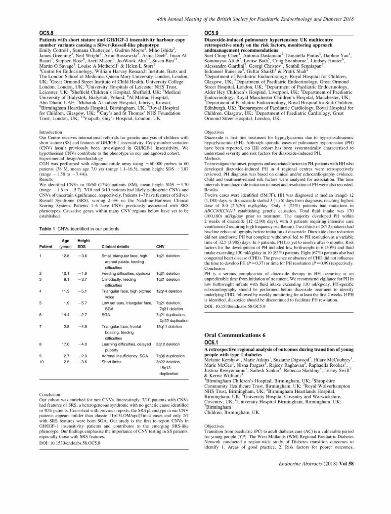
46th Annual Meeting of the British Society for Paediatric Endocrinology and Diabetes 2018
OC5.8Patients with short stature and GH/IGF-1 insensitivity harbour copynumber variants causing a Silver-Russell-like phenotypeEmily Cottrell1, Sumana Chatterjee1, Gudrun Moore2, Miho Ishida2,James Greening3, Neil Wright4, Artur Bossowski5, Asma Deeb6, Iman AlBasiri7, Stephen Rose8, Avril Mason9, JooWook Ahn10, Susan Bint11,Martin O Savage1, Louise A Metherell1 & Helen L Storr1
1Centre for Endocrinology, William Harvey Research Institute, Barts andThe London School of Medicine, Queen Mary University London, London,UK; 2Great Ormond Street Institute of Child Health, University CollegeLondon, London, UK; 3University Hospitals of Leicester NHS Trust,Leicester, UK; 4Sheffield Children’s Hospital, Sheffield, UK; 5MedicalUniversity of Bialystok, Bialystok, Poland; 6Al Mafraq Hospital,Abu Dhabi, UAE; 7Mubarak Al-kabeer Hospital, Jabriya, Kuwait;8Birmingham Heartlands Hospital, Birmingham, UK; 9Royal Hospitalfor Children, Glasgow, UK; 10Guy’s and St Thomas’ NHS FoundationTrust, London, UK; 11Viapath, Guy’s Hospital, London, UK.
IntroductionOur Centre receives international referrals for genetic analysis of children withshort stature (SS) and features of GH/IGF-1 insensitivity. Copy number variation(CNV) hasn’t previously been investigated in GH/IGF-1 insensitivity. Wehypothesised CNVs contribute to the phenotype in our undiagnosed cohort.Experimental design/methodologyCGH was performed with oligonucleotide array using w60,000 probes in 60patients (38 M, mean age 7.0 yrs (range 1.1–16.5), mean height SDS K3.87(range K1.58 to K7.44)).ResultsWe identified CNVs in 10/60 (17%) patients (8M), mean height SDS K3.70(range K1.6 to K5.7). 7/10 and 3/10 patients had likely pathogenic CNVs andCNVs of uncertain significance, respectively. Patients 1–7 have features of Silver-Russell Syndrome (SRS), scoring 2–3/6 on the Netchine-Harbison ClinicalScoring System. Patients 1–6 have CNVs previously associated with SRSphenotypes. Causative genes within many CNV regions below have yet to beestablished.
Table 1 CNVs identified in our patients
Patient
Age
(years)
Height
SDS Clinical details CNV
1 12.8 K3.6 Small triangular face, high
arched palate, feeding
difficulties
1q21 deletion
2 10.1 K1.6 Feeding difficulties, dyslexia 1q21 deletion
3 9.1 K3.7 Clinodactly, feeding
difficulties
1q21 deletion
4 11.3 K5.1 Triangular face, high pitched
voice
12q14 deletion
5 1.9 K5.7 Low set ears, triangular face,
SGA
7q21 deletion,
7q31 deletion
6 14.4 K2.7 SGA 7q21 duplication,
Xp22 duplication
7 2.8 K4.9 Triangular face, frontal
bossing, feeding
difficulties
15q11 deletion
8 17.0 K4.0 Learning difficulties, delayed
puberty
5q12 deletion
9 2.7 K2.0 Adrenal insufficiency, SGA 7q36 duplication
10 2.5 K3.6 Short limbs 3p22 deletion,
15q13
duplication
ConclusionOur cohort was enriched for rare CNVs. Interestingly, 7/10 patients with CNVshad features of SRS, a heterogeneous syndrome with no genetic cause identifiedin 40% patients. Consistent with previous reports, the SRS phenotype in our CNVpatients appears milder than classic 11p15LOM/upd(7)mat cases and only 2/7with SRS features were born SGA. Our study is the first to report CNVs inGH/IGF-1 insensitivity patients and contributes to the emerging SRS-likephenotype. Our findings emphasise the importance of CNV testing in SS patients,especially those with SRS features.
DOI: 10.1530/endoabs.58.OC5.8
OC5.9Diazoxide-induced pulmonary hypertension: UK multicentreretrospective study on the risk factors, monitoring approachandmanagement recommendationsSuet Ching Chen1, Antonia Dastamani2, Donatella Pintus3, Daphne Yau4,Sommayya Aftab2, Louise Bath5, Craig Swinburne1, Lindsey Hunter6,Alessandro Giardini7, Georgi Christov7, Senthil Senniapan3,Indraneel Banerjee4, Guftar Shaikh1 & Pratik Shah2
1Department of Paediatric Endocrinology, Royal Hospital for Children,Glasgow, UK; 2Department of Paediatric Endocrinology, Great OrmondStreet Hospital, London, UK; 3Department of Paediatric Endocrinology,Alder Hey Children’s Hospital, Liverpool, UK; 4Department of PaediatricEndocrinology, Royal Manchester Children’s Hospital, Manchester, UK;5Department of Paediatric Endocrinology, Royal Hospital for Sick Children,Edinburgh, UK; 6Department of Paediatric Cardiology, Royal Hospital forChildren, Glasgow, UK; 7Department of Paediatric Cardiology, GreatOrmond Street Hospital, London, UK.
ObjectivesDiazoxide is first line treatment for hypoglycaemia due to hyperinsulinaemichypoglycaemia (HH). Although sporadic cases of pulmonary hypertension (PH)have been reported, no HH cohort has been systematically characterised tounderstand severity and risk factors for diazoxide-induced PH.MethodsTo investigate the onset, progress and associated factors in PH, patients with HH whodeveloped diazoxide-induced PH in 4 regional centres were retrospectivelyreviewed. PH diagnosis was based on clinical and/or echocardiography evidence.Child and treatment-related risk factors were analysed for association. The timeintervals from diazoxide initiation to onset and resolution of PH were also recorded.ResultsTwelve cases were identified (5M:7F). HH was diagnosed at median (range) 12(1,180) days, with diazoxide started 3 (1,76) days from diagnosis, reaching highestdose of 8.0 (2.5,20) mg/kg/day. Only 3 (25%) patients had mutations inABCC8/KCNJ11 establishing genetic causation. Total fluid intake was 170(100,180) ml/kg/day prior to treatment. The majority developed PH within2 weeks of diazoxide [12 (2,90) days], with 3 patients requiring intensive careventilation (2 requiring high frequency oscillation). Two-thirds of (8/12) patients hadbaseline echocardiography before initiation of diazoxide. Diazoxide dose reductiondid not ameliorate PH but complete withdrawal led to PH resolution at a variabletime of 32.5 (3,985) days. In 3 patients, PH has yet to resolve after 6 months. Riskfactors for the development of PH included low birthweight in 6 (50%) and fluidintake exceeding 130 ml/kg/day in 10 (83%) patients. Eight (67%) patients also hadcongenital heart disease (CHD). The presence or absence of CHD did not influencethe time to develop PH (PZ0.37) or time for PH resolution (PZ0.99) respectively.ConclusionPH is a serious complication of diazoxide therapy in HH occurring at anunpredictable time from initiation of treatment. We recommend vigilance for PH inlow birthweight infants with fluid intake exceeding 130 ml/kg/day. PH-specificechocardiography should be performed before diazoxide treatment to identifyunderlying CHD, followed by weekly monitoring for at least the first 2 weeks. If PHis identified, diazoxide should be discontinued to facilitate PH resolution.
DOI: 10.1530/endoabs.58.OC5.9
Oral Communications 6
OC6.1A retrospective regional analysis of outcomes during transition of youngpeople with type 1 diabetesMelanie Kershaw1, Marie Atkins1, Suzanne Digwood2, Hilary McCoubrey1,Marie McGee1, Nisha Pargass3, Rajeev Raghavan3, Raphaella Rookes4,Justina Rweyemamu4, Sailesh Sankar5, Rebecca Skelding6, Lesley Swift7
& Kerrie Williams4
1Birmingham Children’s Hospital, Birmingham, UK; 2ShropshireCommunity Healthcare Trust, Birmingham, UK; 3Royal WolverhamptonNHS Trust, Birmingham, UK; 4Birmingham Heartlands Hospital,Birmingham, UK; 5University Hospital Coventry and Warwickshire,Coventry, UK; 6University Hospital Birmingham, Birmingham, UK;7BirminghamChildren, Birmingham, UK.
ObjectivesTransition from paediatric (PC) to adult diabetes care (AC) is a vulnerable periodfor young people (YP). The West Midlands (WM) Regional Paediatric DiabetesNetwork conducted a region-wide study of Diabetes transition outcomes toidentify 1. Areas of good practice, 2. Risk factors for poorer outcomes,
Endocrine Abstracts (2018) Vol 58
46th Annual Meeting of the British Society for Paediatric Endocrinology and Diabetes 2018
3. Regional objectives for services and 4. Provide baseline data against whichfuture performance can be measured.MethodsRetrospective regional audit of follow up and HbA1c 12 months pre and24 months post transfer in YP transitioning between January 2012 and December2013.ResultsData was submitted by 9 of 13 (69%) Trusts, accounting for 298 YP moving fromPC to transition clinic (TC), and 195 from TC to AC. 41/298 (13.8%) YP were lostto follow up (FU) in TC (range 0–29%). 41/195 (21%) were lost to FU within 2years of AC (range 0–40%). Median age at move to AC was 17.8 years, range 16 -19.84 years. 22%, were seen in AC within 3 months of their last TC visit. For 45%and 13% their first AC visit occured 6–12 and O12 months respectively,following their last TC. Pre and post HbA1c data was available on 144 YP (73%)transferring from TC to AC. Median HbA1c was 75 (Trust range 64–83)mmol/mol in the year prior to AC and 78mmol/mol (Trust Range 63.5 to 89.5) infirst 2 years of AC. Lag time between the TC and first AC visit did not correlatewith HbA1c. YP in TC with HbA1c !53 (7.5%) deteriorate (PZ0.028), andHbA1c O9.0% improve (P!0.00009) on move to AC. Age at move to ACpositively correlates with fall in HbA1c (RZK0.201, PZ0.048).ConclusionWM Trusts need to focus on1. Reducing high lost to FU rates of 1 in 5 YP (up to 1 in 2.5 in some Trusts)2. Reducing lag time from TC to first AC.3. Careful consideration could be given to transferring some YP in TC with
HbA1c O 9.0% to AC at an earlier point.4. Further studies should look at impact of age and deprivation on rates of
attrition following move to AC.
DOI: 10.1530/endoabs.58.OC6.1
OC6.2Perceptions of diabetes education: a questionnaire-based survey of117 patients and familiesAllison Low1 & Charlotte Elder2
1Sheffield Children’s Hospital NHS Foundation Trust, Sheffield, UK;2Sheffield Children’s Hospital, Sheffield, UK.
BackgroundDiabetes is a primarily self-managed condition, and education to patients andfamilies is an essential component of a diabetes service. In line with accepted UKpractice, our diabetes team provides a comprehensive education program atdiagnosis and regularly thereafter.MethodsFrom October 2017 to January 2018, all patients attending diabetes clinic wereoffered a pseudo-anonymised mixed methods survey, covering experience ofeducation, barriers to education attendance, and ideas to improve our educationprovision. The respondent’s HbA1c, collected on the same day, was recorded.ResultsOf the 117 surveys returned (51% of our population), 46% were completed bypatients, 48% by parents/carers, and 4% by both together. Of the respondents,59% had attended an annual education clinic (mean HbA1c 64 mmol/mol). The41% who hadn’t attended had a mean HbA1c of 68mmol/mol. Those who chosenot to attend because they ‘already know about diabetes’ had an HbA1c rangingfrom 42–104 mmol/mol, with a mean of 61 mmol/mol [95%CIZ52–70 mmol/mol].Most respondents indicated confidence managing their diabetes: 85% confident/veryconfident, 1% underconfident. In the qualitative analysis, several unexpected themesemerged. First, respondents indicated that meeting other children and families waswhat they most valued about education clinic. They suggested peer-led education,sessions delivered by young adults who had recently transitioned, and moresocialising opportunities. Second, respondents requested information about researchsuch as novel technologies, artificial pancreas systems, and the search for a cure. Thethird theme that emerged was mental health, both as a barrier to and a consequenceof diabetes management. Finally, respondents suggested more technologicalapproaches to education provision such as Facebook, YouTube videos, emails andwebinars.DiscussionThere continue to be unmet needs in diabetes education, and patients’ and families’priorities may differ from the diabetes teams’. Peer support and peer educationshould play larger roles, and broadening education clinics to include topics such as‘advances in diabetes research’ may widen their appeal. While some of this data isunit-specific, much of it could be applied UK-wide to make education more patient-focused.
DOI: 10.1530/endoabs.58.OC6.2
Endocrine Abstracts (2018) Vol 58
OC6.3Has the reduction in maintenance fluid rates following introduction of2015 BSPED-recommended diabetic ketoacidosis guidelines impactedon complication rates?Amish Chinoy, Ian Doughty, Mark Bone, Mars Skae, Philip Murray &Raja PadidelaRoyal Manchester Children’s Hospital, Manchester, UK.
IntroductionIn August 2015, a revision of the BSPED-recommended guideline for themanagement of diabetic ketoacidosis (DKA) was published. A key differencefrom the previous guideline was a reduction in maintenance fluid rates, with theaim of reducing the incidence of cerebral oedema. Since implementation ofthese national guidelines, there have been reports within regional networks ofan increased incidence in mild acute renal impairment and hypokalaemia.This retrospective audit aimed to compare the incidence of these complicationspre- and post-implementation of the guidelines.MethodsDatabases were reviewed for all children newly-diagnosed to have diabetes whopresented in DKA, in the 3 years prior to and following implementation of thelatest BSPED DKA guidelines at the Royal Manchester Children’s Hospital. Datawas collected on potassium (K), urea (Ur) and creatinine (Cr) at presentation andlowest K, peak Ur and Cr during admission.ResultsTwenty four children presented with DKA prior to guideline implementation and23 following guideline implementation. There was no significant difference inmean lowest K (3.3 mmol/l pre, 3.1 mmol/l post, PZ0.32) or mean differencebetween admission and lowest K (K0.9 mmol/l pre, K0.9 mmol/l post).Hypokalaemia, as defined by the BSPED DKA guideline (!3.0 mmol/l), duringadmission significantly increased since new guideline implementation – 21%(5/24) pre, 48% (11/23) post (PZ0.05). There was no significant differencebetween mean peak Ur (6.5 mmol/l pre, 5.6 mmol/l post, PZ0.40), or mean peakCr (47 mcmol/l pre, 52.3 mcmol/l post, PZ0.35). There was no mean differencebetween admission and peak Ur (C0.2 mmol/l pre, C0.3 mmol/l post), orbetween admission and peak Cr (C1.7 mcmol/l pre, C6.3 mcmol/l post,PZ0.13).ConclusionAlthough there was no appreciable difference between the two groups in thelowest K or difference between admission K and lowest K, there is noted to be anincrease in incidence of hypokalaemia during admission since the new DKAguidelines were introduced. In light of recent evidence that suggests the rate ofintravenous fluid administration does not influence neurological outcomes,perhaps the current guidelines need review to reduce the incidence ofhypokalaemia.
DOI: 10.1530/endoabs.58.OC6.3
Oral Communications 7
OC7.1DeAPP (Diabetes Education APP): using flipped learning to deliverstructured education in newly diagnosed type 1 diabetes patientsJames Greening1, Sarah Lickwood Lee1, Marie Atkins2 & David Terris3
1Univerisity Hospitals of Leicester NHS Trust, Leicester, UK; 2CYPPaediatric Diabetes Network, Birimingham, UK; 3Demontfort Design Unit,Demontfort University, Leicester, UK.
BackgroundAt diagnosis of diabetes is a critical time in the life course of diabetes. This acritical time for patient take on knowledge and using it (ref). Utilisation of thisinformation is suboptimal due to logistical factors such as ward environment, timeconstraints of staff, psychological impact of the diagnosis. Current models ofeducation are mainly didactic with limited impact in some patients.AimsIn collaboration with demontfort design unit, we aimed to develop a structurededucation program combining APP technology and kinaesthetic learningresources to deliver flipped learning. Patients learn 1st the theoretical knowledgebefore healthcare professionals evaluate and re-enforce patients learning for moreeffective management of their diabetes.MethodsEast Midland CYP education subgroup (CYPES) held focus groups withhealthcare professionals,parents, patients to formulate a curriculum and lessonplans. CYPES presented to Demontfort Design Unit who synthesised theseaccording to age, numeracy, literacy and language. Developing video storyboardswith scripted animation, alongside kinaesthetic learning resources. From whichthey developed a mobile optimised APP to host vimeo animated videos and other
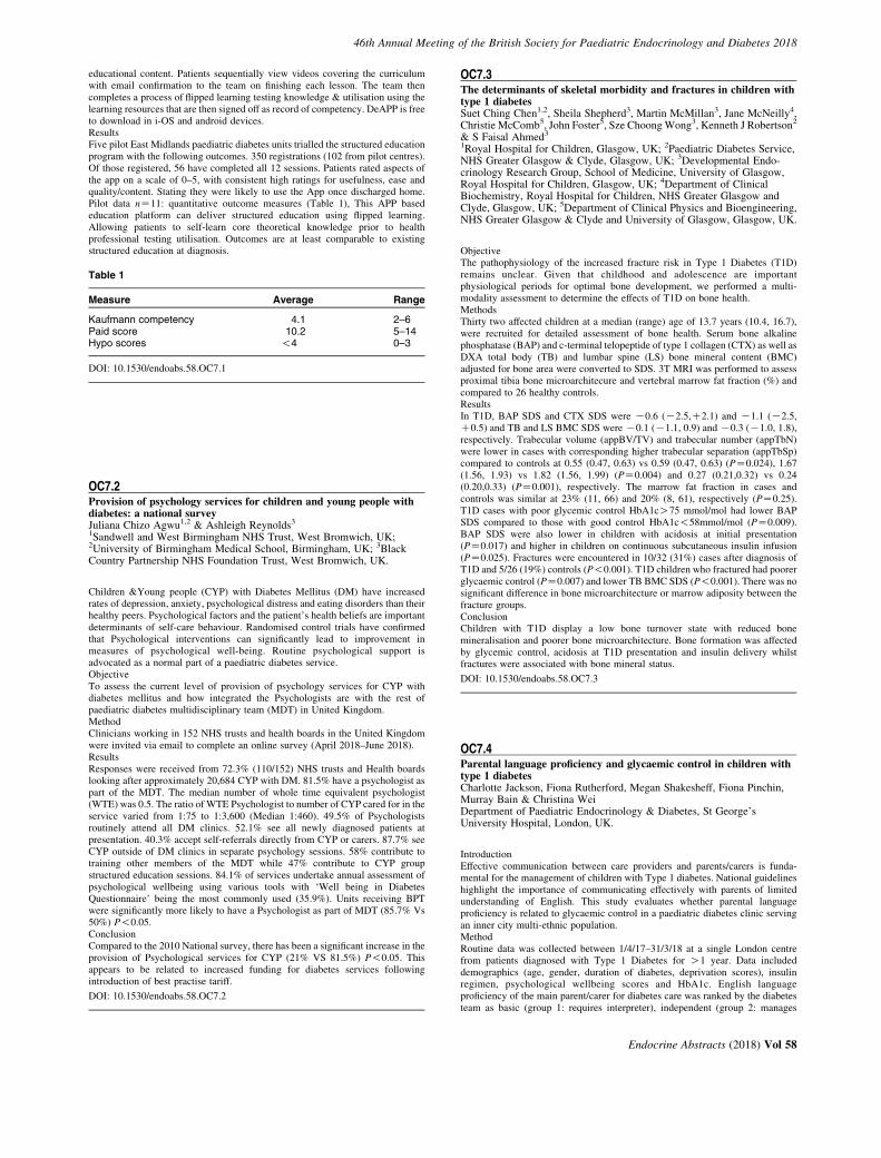
46th Annual Meeting of the British Society for Paediatric Endocrinology and Diabetes 2018
educational content. Patients sequentially view videos covering the curriculumwith email confirmation to the team on finishing each lesson. The team thencompletes a process of flipped learning testing knowledge & utilisation using thelearning resources that are then signed off as record of competency. DeAPP is freeto download in i-OS and android devices.ResultsFive pilot East Midlands paediatric diabetes units trialled the structured educationprogram with the following outcomes. 350 registrations (102 from pilot centres).Of those registered, 56 have completed all 12 sessions. Patients rated aspects ofthe app on a scale of 0–5, with consistent high ratings for usefulness, ease andquality/content. Stating they were likely to use the App once discharged home.Pilot data nZ11: quantitative outcome measures (Table 1), This APP basededucation platform can deliver structured education using flipped learning.Allowing patients to self-learn core theoretical knowledge prior to healthprofessional testing utilisation. Outcomes are at least comparable to existingstructured education at diagnosis.
Table 1
Measure Average Range
Kaufmann competency 4.1 2–6Paid score 10.2 5–14Hypo scores !4 0–3
DOI: 10.1530/endoabs.58.OC7.1
OC7.2Provision of psychology services for children and young people withdiabetes: a national surveyJuliana Chizo Agwu1,2 & Ashleigh Reynolds3
1Sandwell and West Birmingham NHS Trust, West Bromwich, UK;2University of Birmingham Medical School, Birmingham, UK; 3BlackCountry Partnership NHS Foundation Trust, West Bromwich, UK.
Children &Young people (CYP) with Diabetes Mellitus (DM) have increasedrates of depression, anxiety, psychological distress and eating disorders than theirhealthy peers. Psychological factors and the patient’s health beliefs are importantdeterminants of self-care behaviour. Randomised control trials have confirmedthat Psychological interventions can significantly lead to improvement inmeasures of psychological well-being. Routine psychological support isadvocated as a normal part of a paediatric diabetes service.ObjectiveTo assess the current level of provision of psychology services for CYP withdiabetes mellitus and how integrated the Psychologists are with the rest ofpaediatric diabetes multidisciplinary team (MDT) in United Kingdom.MethodClinicians working in 152 NHS trusts and health boards in the United Kingdomwere invited via email to complete an online survey (April 2018–June 2018).ResultsResponses were received from 72.3% (110/152) NHS trusts and Health boardslooking after approximately 20,684 CYP with DM. 81.5% have a psychologist aspart of the MDT. The median number of whole time equivalent psychologist(WTE) was 0.5. The ratio of WTE Psychologist to number of CYP cared for in theservice varied from 1:75 to 1:3,600 (Median 1:460). 49.5% of Psychologistsroutinely attend all DM clinics. 52.1% see all newly diagnosed patients atpresentation. 40.3% accept self-referrals directly from CYP or carers. 87.7% seeCYP outside of DM clinics in separate psychology sessions. 58% contribute totraining other members of the MDT while 47% contribute to CYP groupstructured education sessions. 84.1% of services undertake annual assessment ofpsychological wellbeing using various tools with ‘Well being in DiabetesQuestionnaire’ being the most commonly used (35.9%). Units receiving BPTwere significantly more likely to have a Psychologist as part of MDT (85.7% Vs50%) P!0.05.ConclusionCompared to the 2010 National survey, there has been a significant increase in theprovision of Psychological services for CYP (21% VS 81.5%) P!0.05. Thisappears to be related to increased funding for diabetes services followingintroduction of best practise tariff.
DOI: 10.1530/endoabs.58.OC7.2
OC7.3The determinants of skeletal morbidity and fractures in children withtype 1 diabetesSuet Ching Chen1,2, Sheila Shepherd3, Martin McMillan3, Jane McNeilly4,Christie McComb5, John Foster5, Sze Choong Wong3, Kenneth J Robertson2
& S Faisal Ahmed3
1Royal Hospital for Children, Glasgow, UK; 2Paediatric Diabetes Service,NHS Greater Glasgow & Clyde, Glasgow, UK; 3Developmental Endo-crinology Research Group, School of Medicine, University of Glasgow,Royal Hospital for Children, Glasgow, UK; 4Department of ClinicalBiochemistry, Royal Hospital for Children, NHS Greater Glasgow andClyde, Glasgow, UK; 5Department of Clinical Physics and Bioengineering,NHS Greater Glasgow & Clyde and University of Glasgow, Glasgow, UK.
ObjectiveThe pathophysiology of the increased fracture risk in Type 1 Diabetes (T1D)remains unclear. Given that childhood and adolescence are importantphysiological periods for optimal bone development, we performed a multi-modality assessment to determine the effects of T1D on bone health.MethodsThirty two affected children at a median (range) age of 13.7 years (10.4, 16.7),were recruited for detailed assessment of bone health. Serum bone alkalinephosphatase (BAP) and c-terminal telopeptide of type 1 collagen (CTX) as well asDXA total body (TB) and lumbar spine (LS) bone mineral content (BMC)adjusted for bone area were converted to SDS. 3T MRI was performed to assessproximal tibia bone microarchitecure and vertebral marrow fat fraction (%) andcompared to 26 healthy controls.ResultsIn T1D, BAP SDS and CTX SDS were K0.6 (K2.5,C2.1) and K1.1 (K2.5,C0.5) and TB and LS BMC SDS were K0.1 (K1.1, 0.9) and K0.3 (K1.0, 1.8),respectively. Trabecular volume (appBV/TV) and trabecular number (appTbN)were lower in cases with corresponding higher trabecular separation (appTbSp)compared to controls at 0.55 (0.47, 0.63) vs 0.59 (0.47, 0.63) (PZ0.024), 1.67(1.56, 1.93) vs 1.82 (1.56, 1.99) (PZ0.004) and 0.27 (0.21,0.32) vs 0.24(0.20,0.33) (PZ0.001), respectively. The marrow fat fraction in cases andcontrols was similar at 23% (11, 66) and 20% (8, 61), respectively (PZ0.25).T1D cases with poor glycemic control HbA1cO75 mmol/mol had lower BAPSDS compared to those with good control HbA1c!58mmol/mol (PZ0.009).BAP SDS were also lower in children with acidosis at initial presentation(PZ0.017) and higher in children on continuous subcutaneous insulin infusion(PZ0.025). Fractures were encountered in 10/32 (31%) cases after diagnosis ofT1D and 5/26 (19%) controls (P!0.001). T1D children who fractured had poorerglycaemic control (PZ0.007) and lower TB BMC SDS (P!0.001). There was nosignificant difference in bone microarchitecture or marrow adiposity between thefracture groups.ConclusionChildren with T1D display a low bone turnover state with reduced bonemineralisation and poorer bone microarchitecture. Bone formation was affectedby glycemic control, acidosis at T1D presentation and insulin delivery whilstfractures were associated with bone mineral status.
DOI: 10.1530/endoabs.58.OC7.3
OC7.4Parental language proficiency and glycaemic control in children withtype 1 diabetesCharlotte Jackson, Fiona Rutherford, Megan Shakesheff, Fiona Pinchin,Murray Bain & Christina WeiDepartment of Paediatric Endocrinology & Diabetes, St George’sUniversity Hospital, London, UK.
IntroductionEffective communication between care providers and parents/carers is funda-mental for the management of children with Type 1 diabetes. National guidelineshighlight the importance of communicating effectively with parents of limitedunderstanding of English. This study evaluates whether parental languageproficiency is related to glycaemic control in a paediatric diabetes clinic servingan inner city multi-ethnic population.MethodRoutine data was collected between 1/4/17–31/3/18 at a single London centrefrom patients diagnosed with Type 1 Diabetes for O1 year. Data includeddemographics (age, gender, duration of diabetes, deprivation scores), insulinregimen, psychological wellbeing scores and HbA1c. English languageproficiency of the main parent/carer for diabetes care was ranked by the diabetesteam as basic (group 1: requires interpreter), independent (group 2: manages
Endocrine Abstracts (2018) Vol 58
46th Annual Meeting of the British Society for Paediatric Endocrinology and Diabetes 2018
without interpreter but not fluent) or proficient (group 3: native or fluent inEnglish as second language). HbA1c was compared and adjusted for confounders.Statistical analysis: ANOVA, ANCOVA, t-test and c-Square, using SPSSv24 at5% significance.ResultsNinety-five (m:fZ45:50) patients were included and categorised into group1(nZ18), 2(nZ9), and 3(nZ68). Their age ranged between 3.4 and 19.7 years(MedianZ14.7), diabetes duration ranged between 1 and 16.3 years (Med-ianZ5.4) and HbA1c ranged between 43 and 130 mmol/mol (MedianZ69.4).Within group 1, Somalian (39%) was the most commonly spoken of the ninedifferent non-English languages. HbA1c scores (geometric means) weresignificantly higher in group 1 (84.6 mmol/mol) compared with groups 2(70.5 mmol/mol, PZ0.04) and 3 (68 mmol/mol, PZ0.01). No significantdifference was found between groups 2 and 3. Despite significant differences indeprivation scores between groups, (PZ0.001), when controlled for, HbA1cremained significantly different between groups (group 1 vs 2: PZ0.01, 1 vs 3:PZ0.011). No group differences in gender, age, duration of diagnosis,psychological wellbeing scores. There were more pump users in group 3(32%)vs 1(6%), but no differences in HbA1c scores were identified.ConclusionLanguage proficiency is identified as a potential barrier to optimal glycaemiccontrol. Targeted measures such as language-specific education material/activities, support groups and key workers should be considered.
DOI: 10.1530/endoabs.58.OC7.4
OC7.5Diabetes control is worse in children and young people with type 1diabetes requiring interpreter supportJan Idkowiak1,2, Sabba Elhag1, Vrinda Saraff1, Renuka Dias1,Timothy Barrett1, Melanie Kershaw1 & Ruth Krone1
1Birmingham Women’s and Children’s Hospital NHS Foundation Trust,Birmingham, UK; 2Institute for Metabolism and Systems Research,University of Birmingham, Birmingham, UK.
IntroductionLanguage barriers can pose a significant barrier to successful education ofchildren and young people (CYP) with type 1 diabetes (T1DM) and their families,potentially influencing their glycaemic control.MethodsRetrospective analysis from diagnosis to 18 months post diagnosis of glycaemiccontrol in 41 CYP with T1DM requiring interpreter support (INT) under care ofour diabetes centre based within a multi-ethnic community. Median HbA1c at 0,3, 6, 9, 12 and 18 months following diagnosis were compared to 100 age-, sex-and mode-of-therapy-matched controls who did not require interpreter service.English indices of deprivation are based on the 2015 census and were retrievedfrom: www.gov.uk/government/statistics.ResultsThe main languages spoken were Somali (27%), Urdu (19.5%), Romanian (17%)and Arabic (12%), but also Polish, Hindi, Tigrinya, Portuguese, Bengali and signlanguage. Overall deprivation was worse in the INT group according to Index ofMultiple Deprivation (IMD [median]: INT 1.642; control 3.741; PZ0.001). Themedian HbA1c was higher at diagnosis in the control group (9.95 versus 9.0%,PZ0.046) but was higher in the INT group after diagnosis: the median HbA1c at18 months post diagnosis was 8.3% (INT) versus 7.9% (controls) (PZ0.014).There were no hospital admissions required due to diabetes-related complicationsin both subgroups.Summary and conclusionsGlycaemic control is worse in CYP with T1DM who face language barriers in ourcentre. Socioeconomic deprivation may confound these findings but indicate thatthis is a disadvantaged subset of patients regardless. In order to improve diabetescare for CYP with language barriers, we propose that health care providersdevelop strategies to provide tailored support, including provision of diabetes-specific training for interpreters. Equally, patients and their families should besupported to acquire language skills for ongoing diabetes education. The findingsof this study suggest that equally poor health outcomes for CYP with languagebarriers and anychronic condition is a broader concern. This highlights the needfor a sustained medical and political effort toward the effective integration andsupport of CYPs from disadvantaged backgrounds.
DOI: 10.1530/endoabs.58.OC7.5
Endocrine Abstracts (2018) Vol 58
OC7.6Type A Insulin Resistance Syndrome due to an INSR mutation Presentingwith diabetes mellitus evolving to hyperandrogenism and PCOSArameh Aghababaie1, Martha Ford-Adams2, Charles R Buchanan2,Ved Arya2, Andrew Hattersley3, Kevin Colclough4 & Ritika R Kapoor2
1King’s College London School of Medicine, London, UK; 2School ofPaediatric Diabetes and Endocrinology, King’s College Hospital NHSTrust, London, UK; 3University of Exeter Medical School, Exeter, UK;4Molecular Genetics Laboratory, Royal Devon & Exeter Hospital, Exeter,UK.
BackgroundMutations in the insulin receptor (INSR) gene are rare and cause a spectrum ofsevere insulin resistance syndromes including Donohue syndrome, RabsonMendenhall syndrome, and Type A Insulin Resistance Syndrome (IRS). Wedescribe a young female with a heterozygous INSR mutation, who presented withantibody positive diabetes mellitus (DM) and subsequently developed features ofType A IRS.Case ReportA 12 year old Jamaican girl with a BMI of 25 kg/m2 presented with polyuria,polydipsia and hyperglycaemia (blood glucose 18 mmol/l). HbA1c was raised at85 mmol/mol, urinary ketones were negative. Antibodies to islet antigen-2 werepositive (0.56 U/ml; normal range: 0–0.35 U/ml), and antibodies to glutamic aciddecarboxylase and insulin were negative. She was diagnosed with Type 1 DM andstarted on a basal-bolus insulin regime. She previously had precocious puberty,treated with GnRH analogues. She achieved menarche at 13 years with irregularcycles. Over 2 years, she developed hirsutism, acanthosis nigricans, andpostprandial hypoglycaemia, with no evidence of lipodystrophy. Biochemicalinvestigations showed raised serum C-peptide (854 pmol/l), LH (29.1 IU/l), andtestosterone levels (3.6 nmol/l), with normal lipids. Ultrasound pelvis revealedenlarged ovaries with multiple peripheral follicles suggestive of PCOS. Familyhistory included DM affecting mother, maternal uncle and maternal grandfather.Targeted next generation sequencing of the monogenic diabetes genes revealed aheterozygous missense INSR variant p.(His1157 Gln), c.3471TOG in the patientand affected mother. The p.His1157 residue is present within the catalytic loop ofthe tyrosine kinase domain of the INSR. A different missense variant affecting thesame residue, p.(His1157Arg), has been previously associated with Type A IRS.The patient was started on metformin and weaned off insulin therapy. Hermenstrual cycles then normalised and glycaemic control improved (HbA1C36 mmol/l).ConclusionThis is a novel description of a p.His1157Gln mutation in INSR causing Type AIRS. Our findings highlight the role of puberty in the manifestation of insulinresistance. This case also emphasises the importance of exploring an INSR defectin slim patients with DM and features of insulin resistance in the absence oflipodystrophy and normal lipids.
DOI: 10.1530/endoabs.58.OC7.6
OC7.7Knowledge and confidence of paediatric middle grade doctors inmanaging out of hours diabetes advice callsElspeth Ferguson1, Charlotte Elder1,2, Mark Denial3 & Astha Soni31Department of Paediatric Diabetes and Endocrinology, Sheffield Children’sNHS Foundation Trust, Sheffield, UK; 2Department of Oncology andMetabolism, University of Sheffield, Sheffield, UK; 3Department ofPaediatric Diabetes, Sheffield Children’s NHS Foundation Trust, Sheffield,UK.
BackgroundPaediatric diabetes is predominantly an outpatient based specialty, limiting thetraining opportunities available to paediatric trainees. At our centre, out of hoursdiabetes advice calls are managed by paediatric specialty trainees working on themiddle grade rota, with 24 hour support from an oncall paediatric diabetesconsultant. Feedback locally from families has highlighted the quality of out ofhours advice as an area of concern. We aimed to assess the knowledge andconfidence of paediatric trainees in managing out of hours advice calls to helpinform training and service provision.MethodologyThe study took two parts. Firstly all paediatric trainees at level ST4C were askedto complete an anonymous questionnaire, with six scenarios covering commonadvice calls. Model answers were agreed by the diabetes team. Answers weremarked by two independent diabetologists. The scoring system gave separate
46th Annual Meeting of the British Society for Paediatric Endocrinology and Diabetes 2018
marks for knowledge and for safety. The second part involved a questionnaire fortrainees who responded to advice calls during a three month study period, aimingto assess confidence in dealing with such calls. Timing of advice calls wasrecorded by the hospital switchboard.ResultsEighteen of twenty-eight (64%) trainees completed the scenarios. Knowledge wasvariable, with technology based scenarios generally scoring poorly (averageknowledge score for lost pump handset scenario 22% versus 63% for sick day rulescenario). Trainees however tended to act safely (average safety score 87%).There was no correlation between knowledge and safety. Sixteen out of hoursadvice calls were made during the 3 month study period. Questionnaires werecompleted for 75% of the calls. All trainees felt confident in managing the call and92% felt they had adequate training to manage the call.ConclusionsThis project demonstrates a discrepancy between registrar confidence and familysatisfaction with out of hours telephone advice. The knowledge base of paediatrictrainees in dealing with common diabetes advice scenarios is very variable. Wediscuss the approach we are taking to the challenges of providing diabetes trainingto paediatric trainees and how this should be balanced with patient safety andsatisfaction.
DOI: 10.1530/endoabs.58.OC7.7
OC7.8Continuous glucose monitoring with regular clinical review ofglycaemic control in children with type 1 diabetes experiencing frequentunpredictable hypoglycaemiaMeera Parmar1 & Nikki Davis2
1University of Southampton, Southampton, UK; 2Southampton Children’sHospital, Southampton, UK.
IntroductionOptimal glycaemic control can be extremely challenging to achieve in childrenand adolescents with Type 1 Diabetes (T1D) who have labile glycaemic controlwith frequent hypoglycaemia despite intensive blood glucose monitoring (BGM).Effectiveness of continuous glucose monitoring (CGM) in children/adolescents athigh risk of hypoglycaemia has been poorly studied. Frequent patient follow-upand review is recommended to support successful CGM management. This pilotstudy explored the impact of CGM and regular clinical review on glycaemiccontrol in children/adolescents with T1D experiencing frequent unpredictablehypoglycaemia.MethodsChildren/adolescents aged 2–18 with T1D (nZ10) wore a CGM device for sixweeks. During this period, each participant had four clinical reviews with aPaediatric Diabetes Consultant, who assessed the participant’s CGM download,adjusted insulin dosage and offered relevant advice to optimise glycaemic control.Glycaemic outcomes from Week 1 and Week 6 of CGM were compared.ResultsMean daily glucose was 11.5 mmol/L (SD 1.8) in Week 1 and 9.2 mmol/L (0.8) inWeek 6: mean reduction of 2.3 (95% CI K3.4 to K1.3; PZ0.002). Weekly SD ofglucose levels (as a measure of glycaemic variability) decreased by 1.1 over sixweeks (K1.5 to K0.7; P!0.001). Total weekly time spent with a glucose levelbetween 4 and 10 mmol/L increased by 38.25 hours (25.99 to 50.51; P!0.001).This was accompanied by a mean reduction of 8.17 hours in the total weekly timespent with a glucose level !4 mmol/L (K11.2 to K5.13; P!0.001) and1.42 hours in the total weekly time spent with a glucose level !2.8 mmol/L(K2.23 to K0.59, PZ0.004). Additionally, weekly time spent with a glucoselevel O10 mmol/L decreased by 30.08 hours (K43.61 to K16.54; PZ0.001).ConclusionCGM with regular clinical review over six weeks significantly improvedglycaemic control and reduced hypoglycaemia in children/adolescents with T1D.These are important and meaningful clinical outcomes. Larger and longer studiesare required to examine longer term impact of CGM on glycaemic control andavoidance of hypoglycaemia in this vulnerable group. These improvements mayalso have a positive impact on other outcomes including quality of life and sleepquality.
DOI: 10.1530/endoabs.58.OC7.8
Oral Communications 8
OC8.1Five year outcomes in a cohort with hypoglycaemia due to congenitalhyperinsulinismLaila Al-Hashmi1, Maria Estebanez1,2 & Indi Banerjee1,2
1Royal Manchester Children’s Hospital, Manchester University NHSFoundation Trust, Manchester, UK; 2University of Manchester, Manchester,UK.
BackgroundCongenital hyperinsulinism (CHI) is one of the commonest causes of recurrenthypoglycaemia due to excess production of insulin in infancy and results inneurological impairment in a third to a half of patients. The treatment of CHI isoften complex and complicated by side effects; the medium-term effects of suchtreatment are not known.AimTo describe five year clinical outcomes in a cohort of infants with CHI.MethodsData from 37 infants (26 males) diagnosed and treated for CHI in 2011- 2012 at asingle centre were reviewed. Five year outcomes included auxology, feeding,treatment and neurodevelopment.ResultsSmall for gestational age was a common diagnostic association with birthweight!2 SDS. CHI was transient in 32 (86%) patients with the rest requiringpersistent treatment. At one year, 20 (54%) patients treated with diazoxidereduced and stopped treatment. Five patients underwent 18-fluoro-DOPAPET-CT scanning which identified focal CHI, treated by focal lesionectomy inone patient only. Two patients required subtotal pancreatectomy, while themajority were successfully managed by medical therapy. Auxology parametersnormalised by age one year, with height and weight SDS !K2.0 SDS in only at8% and 21% respectively. In this cohort, only 4 (11%) patients had abnormalfeeding which required either feeding through nasogastric or gastrostomy tubes.However feeding remained problematic in 3 patients as late as the 5 year review.Neurodevelopmental concerns (speech delay/cognitive dysfunction) were presentin 9 out of 32 patients (28%) examined.ConclusionThe majority of patients in a CHI cohort were born small for gestational age,responded to medical therapy, improved growth and had disease resolution inclinical review. Neurodevelopment was abnormal in a third of patients.
DOI: 10.1530/endoabs.58.OC8.1
OC8.2The effect of GnRHa treatment on bone density in young adolescentswith gender dysphoria: findings from a large national cohortTobin Joseph1,2, Joanna Ting1,2 & Gary Butler1,3
1Department of Paediatrics and Adolescent Endocrinology, UniversityCollege London Hospital, London, UK; 2University College LondonMedical School, London, UK; 3UCL Great Ormond Street Institute of ChildHealth, London, UK.
Background/AimsMore young people with gender dysphoria are undergoing hormonal interventionwith GnRHa treatment. The impact on bone density is not known in the veryyoung transgender adolescents, with guidelines mentioning that Bone MineralDensity (BMD) should be monitored without a suggestion on how. This studyaimed to investigate whether there were any changes in BMD or Bone MineralApparent Density (BMAD) whilst on GnRHa therapy.MethodsA retrospective analysis of 70 transgender subjects aged 12–14 years who werereferred to a national centre for the management of gender dysphoria (2011–16)and had had yearly DEXA scans. A longitudinal analysis (nZ31) where subjectshad had scans over 3 years and a cross-sectional analysis (nZ70) were performedfor BMD differences between each scan.ResultsAlthough there was a decline in age-related Z-scores, there was no significantchange in BMAD after one year on GnRHa and no significant changes betweenDEXA scans for tBMD or BMAD when analysed cross-sectionally orlongitudinally over 3 years.ConclusionWe have shown that there is no actual change in BMAD or tBMD in youngtransgender adolescents on long term GnRHa therapy, and certainly no true fall as
Endocrine Abstracts (2018) Vol 58
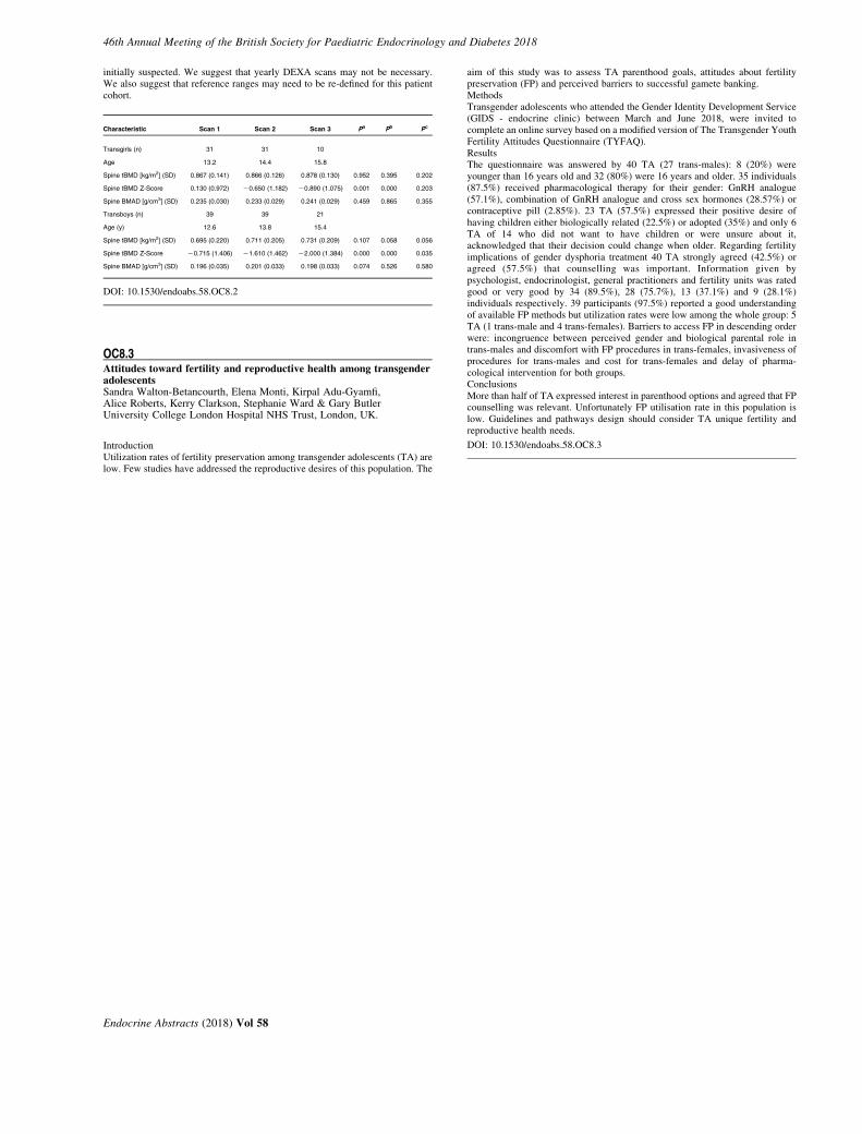
46th Annual Meeting of the British Society for Paediatric Endocrinology and Diabetes 2018
initially suspected. We suggest that yearly DEXA scans may not be necessary.We also suggest that reference ranges may need to be re-defined for this patientcohort.
Characteristic Scan 1 Scan 2 Scan 3 Pa Pb Pc
Transgirls (n) 31 31 10
Age 13.2 14.4 15.8
Spine tBMD [kg/m2] (SD) 0.867 (0.141) 0.866 (0.126) 0.878 (0.130) 0.952 0.395 0.202
Spine tBMD Z-Score 0.130 (0.972) K0.650 (1.182) K0.890 (1.075) 0.001 0.000 0.203
Spine BMAD [g/cm3] (SD) 0.235 (0.030) 0.233 (0.029) 0.241 (0.029) 0.459 0.865 0.355
Transboys (n) 39 39 21
Age (y) 12.6 13.8 15.4
Spine tBMD [kg/m2] (SD) 0.695 (0.220) 0.711 (0.205) 0.731 (0.209) 0.107 0.058 0.056
Spine tBMD Z-Score K0.715 (1.406) K1.610 (1.462) K2.000 (1.384) 0.000 0.000 0.035
Spine BMAD [g/cm3] (SD) 0.196 (0.035) 0.201 (0.033) 0.198 (0.033) 0.074 0.526 0.580
DOI: 10.1530/endoabs.58.OC8.2
OC8.3Attitudes toward fertility and reproductive health among transgenderadolescentsSandra Walton-Betancourth, Elena Monti, Kirpal Adu-Gyamfi,Alice Roberts, Kerry Clarkson, Stephanie Ward & Gary ButlerUniversity College London Hospital NHS Trust, London, UK.
IntroductionUtilization rates of fertility preservation among transgender adolescents (TA) arelow. Few studies have addressed the reproductive desires of this population. The
Endocrine Abstracts (2018) Vol 58
aim of this study was to assess TA parenthood goals, attitudes about fertilitypreservation (FP) and perceived barriers to successful gamete banking.MethodsTransgender adolescents who attended the Gender Identity Development Service(GIDS - endocrine clinic) between March and June 2018, were invited tocomplete an online survey based on a modified version of The Transgender YouthFertility Attitudes Questionnaire (TYFAQ).ResultsThe questionnaire was answered by 40 TA (27 trans-males): 8 (20%) wereyounger than 16 years old and 32 (80%) were 16 years and older. 35 individuals(87.5%) received pharmacological therapy for their gender: GnRH analogue(57.1%), combination of GnRH analogue and cross sex hormones (28.57%) orcontraceptive pill (2.85%). 23 TA (57.5%) expressed their positive desire ofhaving children either biologically related (22.5%) or adopted (35%) and only 6TA of 14 who did not want to have children or were unsure about it,acknowledged that their decision could change when older. Regarding fertilityimplications of gender dysphoria treatment 40 TA strongly agreed (42.5%) oragreed (57.5%) that counselling was important. Information given bypsychologist, endocrinologist, general practitioners and fertility units was ratedgood or very good by 34 (89.5%), 28 (75.7%), 13 (37.1%) and 9 (28.1%)individuals respectively. 39 participants (97.5%) reported a good understandingof available FP methods but utilization rates were low among the whole group: 5TA (1 trans-male and 4 trans-females). Barriers to access FP in descending orderwere: incongruence between perceived gender and biological parental role intrans-males and discomfort with FP procedures in trans-females, invasiveness ofprocedures for trans-males and cost for trans-females and delay of pharma-cological intervention for both groups.ConclusionsMore than half of TA expressed interest in parenthood options and agreed that FPcounselling was relevant. Unfortunately FP utilisation rate in this population islow. Guidelines and pathways design should consider TA unique fertility andreproductive health needs.
DOI: 10.1530/endoabs.58.OC8.3
46th Annual Meeting of the British Society for Paediatric Endocrinology and Diabetes 2018
Poster Presentations
Endocrine Abstracts (2018) Vol 58
46th Annual Meeting of the British Society for Paediatric Endocrinology and Diabetes 2018
Adrenal
P001A Cochrane Review of glucocorticoid replacement regimens in thetreatment of 21-hydroxylase deficiency congenital adrenal hyperplasiaSze Ng1,2 & Karolina Stepien3
1Southport and Ormskirk Hospitals NHS Trust, Southport, UK; 2Universityof Liverpool, Liverpool, UK; 3Salford Royal NHS Foundation Trust,Salford, UK.
Congenital adrenal hyperplasia (CAH) is an autosomal recessive condition whichleads to glucocorticoid deficiency. During childhood, aims of treatment are toprevent adrenal crisis and to achieve optimal adult height and normal puberty.In adults, aims of treatment are to prevent adrenal crisis, ensure normal fertilityand avoid long-term consequences of glucocorticoid use. Current regimens withglucocorticoids cannot optimally replicate the normal physiological cortisol level.Overtreatment or undertreatment is often reported and there is no current standardtreatment for CAH. It remains unclear which treatment regimen is most effective.ObjectiveThis Cochrane review (protocol published) aims to determine the efficacy ofdifferent glucocorticoid replacement regimens of CAH.MethodsWe included any RCT or quasiRCT comparing different glucocorticoidreplacement regimens in the treatment of CAH due to 21-hydroxylase deficiencyin children and adults. GRADE was used to assess quality of the evidence.ResultsThe initial search identified 297 records which identified 20 publications forfurther examination. After screening, we included five RCTs with 101 peoplewith CAH. The number of participants in each trial varied from 6 to 44 withparticipants’ ages ranging from 1.2 to 21 years. The majority of the trials weincluded were small and many had methodological weaknesses. The number oftrials assessing different glucocorticoid regimens varied as well as the trialdurations. Although 17OHP and androstenedione are frequently used to monitortreatment, there is a great amount of variability in the measurements. Overall, wejudged trials to be moderate to high risk of bias across many domains.ConclusionsThere is insufficient evidence to indicate which glucocorticoid replacementregimen results in better outcomes. There were no trials on modified-releaseformulation of hydrocortisone or use of 24-hour circadian continuoussubcutaneous infusion of hydrocortisone. This review identified the need forwell-designed, adequately-powered trials to assess the efficacy of differentglucocorticoid replacement regimens in the treatment of CAH.
DOI: 10.1530/endoabs.58.P001
P002Borderline peak plasma cortisol following Synacthen stimulation –single-centre analysis of three years’ dataSarah Burn1, Sharon Colyer1, Paul Dimitri1, Neil Wright1, Nils Krone1,2
& Charlotte Elder1,2
1Sheffield Children’s NHS Foundation Trust, Sheffield, UK;2The Univeristy of Sheffield, Sheffield, UK.
IntroductionDiagnostic cut-offs for plasma cortisol on Short Synacthen Test (SST) arecontroversial, made more complicated by modern assays and paediatricnormative values extrapolated from adult data. Some advocate a divisionbetween biochemical and clinical AI, with different cut-offs and managementstrategies. For asymptomatic children, with a low-index of suspicion, andborderline SST results our department has evolved a tendency to advisehydrocortisone replacement in times of stress only. We analysed three years ofSST data, examining the cases with borderline peak cortisol results foraetiological links and subsequent management strategies.MethodsRetrospective analysis of all SST performed between September 2014 and 2017was undertaken. Plasma cortisol samples were analysed on the Abbott Architecti1000 immunoassay (CVs !5%). Our diagnostic threshold for a ‘pass’ for bothhigh (HDT) and low-dose SST (LDT) is 450 nmol/l. ‘Borderline’ peak cortisolwas considered to be 300–449 nmol/l and this group was further subdivided into300–349, 350–399 and 400–449 nmol/l for analysis in terms of demographics,test indication, dose of synacthen and resultant management plan.Results433 SSTs were performed, 74 (41M) of whom had a borderline peak cortisol(16.7%). The proportion of borderline tests remained similar each year, despite anincreasing trend towards HDT over LDT. Management of patients with borderlinepeak cortisol varied, however there was a tendency to reduce or stop replacement
Endocrine Abstracts (2018) Vol 58
glucocorticoids with higher results, particularly after a HDT. Steroids were morelikely to be started or continued with lower borderline values. Patients withknown AI were more likely to have their steroids continued or SST repeated atlower peak cortisols and weaned at higher peak cortisols. Those withoutestablished AI were less likely to have glucocorticoid replacement commenced ifhigher borderline value, particularly following a LDT.ConclusionsThere was significant variation in the management of borderline SST results withthe same cortisol result warranting commencement of regular replacement for onephysician and stopping replacement for another. There is a paucity of research inthis area and studies to examine both the natural course of children withborderline SSTs and whether stress cover represents pragmatic but safemanagement are warranted.
DOI: 10.1530/endoabs.58.P002
P003Differences in hydrocortisone absorption during the 24 hour period inpatients with adrenal insufficiencyPeter Hindmarsh1, Lia Charmandari2 & John Honour3
1University College London Hospitals, London, UK; 2First Department ofPaediatrics University of Athens Medical School, Athens, Greece;3University College London, London, UK.
Hydrocortisone therapy should be individualised in patients with adrenalinsufficiency to avoid over and under replacement. We assessed hydrocortisoneabsorbtion at different times of day which may impact on treatment regimens. Weassessed the oral absorption of hydrocortisone in 48 patients (21M) aged between6.1 and 20.3 years with congenital adrenal hyperplasia due to P450c21 deficiency.Hydrocortisone dosing ranged between 11.5 and 22.6 mg/m2 per day in three orfour doses. Each patient underwent a 24 h plasma cortisol and 17-hydroxy-progesterone (17OHP) concentration profile with the morning dose used tocalculate absorption parameters. Parameters derived were maximum plasmaconcentration (Cmax), time of maximum plasma concentration (tmax), the cortisolconcentration leading to 50% inhibition of 17OHP) (IC50) and time to attainingplasma cortisol concentration less than 100 nmol/l (100 nmol/l was the IC50 value).Cmax, tmax and IC50 were derived using conventional pharmacokinetic techniquesfrom the absorption profiles. In a further 6 patients sampling was undertaken at 30,60 and 120 minutes after the first (06.00–07.00 h) and last (23.00–00.30 h) dose ofthe day. The Cmax for the 48 patients was 780.7G61.6 nmol/l and tmax 66.7 (range20–118) min. The IC50 for plasma cortisol suppression of plasma 17OHP was100 nmol/l. Time taken to a plasma cortisol concentration less than 100 nmol/l was289 (range 140–540) min. In the six patients who had studies morning and eveningthe evening dose Cmax was significantly less than the morning dose (PZ0.01) whichreflected the lower dose overall given in the evening compared to the morning.Despite the reduction in Cmax,tmax was prolonged with the evening dose comparedto the morning peak (detected in most of the patients at 30 minutes). In the eveningtmax occurred usually after 60 minutes and was associated with a longer time to acortisol concentration less than 100 nmol/l than the morning peak (PZ0.01).Absorption of hydrocortisone varies throughout the 24 hour period probably due toalterations in clearance. To determine the true peak and exposure to cortisolfrequent sampling around a dose is required. Dosing regimens need to beincorporate these observations.
DOI: 10.1530/endoabs.58.P003
P004Adrenocortical function in infants admitted to PICUDonatella Pintus, Urmi Das, Poonam Dharmaraj, Mohammed Didi,Renuka Ramakrishnan, Senthil Senniappan, Keith Thorburn & Joanne BlairAlder Hey Children’s Hospital, Liverpool, UK.
BackgroundHypocortisolaemia is common in neonates1 and infants following cardiac surgery.2
In critically ill children with other pathologies, hypocortisolaemia may resultfrom accelerated cortisol metabolism and reduced protein binding. However, thetiming and frequency of normalisation of cortisol concentrations followinginfantile critical illness is poorly described.ObjectiveTo describe the natural history of hypocortisolaemia in critically ill infantsadmitted to a single paediatric intensive care unit (PICU).

46th Annual Meeting of the British Society for Paediatric Endocrinology and Diabetes 2018
MethodsRetrospective observational study of infants admitted to PICU from 2016–2018.If random cortisol was !450 nmol/l during critical illness a low dose shortSynacthen test (LDSST) was performed on recovery. Results were classifiedbased on basal and peak cortisol concentrations: Normal (basalO100 nmol/l,peak O450 nmol/l); suboptimal (basalO100 nmol/l, peak 350–450 nmol/l),abnormal (basal!100 nmol/l, and /or peak !350 nmol/l).ResultsData from 63 infants (40M, 23F), mean age 2.4 months (range 1 wk to 10 m), wereanalysed. 30 patients (47.6%) had cardiac malformations, 11 (17.5%) abdominalpathologies, 5 (8.0%) sepsis, 4 (6.3%) brain pathologies, 3 (4.7%) spina bifida,3 (4.7%) respiratory pathologies and 7 (11.0%) other pathologies. During criticalillness random cortisol was !450 nmol/l in 57/63 infants (90%): undetectable in25 (43.8%); 50–350 nmol/l in 28 (49%), and 350–450 nmol/l in 4 (7%). 49 infants(86%) underwent a LDSST which was normal in 33 infants (67.3%) in whomrandom cortisol was !50 nmol/l in 23 (47%) and 50–350 nmol/l 10 (20.4%)during critical illness. 15 patients (30%) with abnormal (NZ11) or suboptimal(NZ4) LDSST were treated with hydrocortisone for ! 6 months in 5 patients(33.3%) and O 6 months in 10 patients (66.7%).ConclusionsHypocortisolaemia persisted for longer following critical illness than reportedpreviously in other infant cohorts.1 Greater understanding of the mechanisms ofhypocortisolaemia during and following critical illness should enable moreaccurate prognosis, diagnosis and management.References1. Tan et al, Arch Dis Child. 2018. doi: 10.1136/archdischild-2017-313819.2. Crawford et al., Paediatr Anaesth. 2017;27:77–84.
DOI: 10.1530/endoabs.58.P004
P005Sphingosine-1-phosphate lyase (SGPL1) deficiency is associated withmitochondrial dysfunctionRathi Prasad1, Avinaash Maharaj1, Teisha Bradshaw1, Jack Williams1,Tulay Guran2, Deby Braslavsky3, Britta Brugger4 & Lou Metherell11William Harvey Research Institute, Centre for Endocrinology, John VaneScience Centre, Queen Mary, University of London, London, UK;2Department of Paediatric Endocrinology and Diabetes, School ofMedicine, Marmara University, Istanbul, Turkey; 3Centro de Investiga-ciones Endocrinologicas ‘Dr. Cesar Bergada’ (CEDIE) – CONICET – FEI –Division de Endocrinologıa, Hospital de Ninos ‘Ricardo Gutierrez’, BuenosAires, Argentina; 4Heidelberg University Biochemistry Center (BZH),Heidelberg, Germany.
IntroductionLoss of function mutations in SGPL1, a key component of sphingolipidmetabolism, are associated with accumulation of sphingolipid intermediatesgiving rise to a multisystemic disease incorporating primary adrenal insufficiency(PAI) and progressive renal and neurological disease. Sphingolipid accumulationis implicated in mitochondrial pathology.ObjectiveTo investigate the impact of SGPL1 deficiency on mitochondrial morphology/function.MethodsCell lines: Primary cell cultures of dermal fibroblasts from patients with SGPL1deficiency (Patient 1 - p.F545del; PAI, later onset renal/neurologicalcompromise; Patient 2 - p.S65Rfs*6G; PAI, early onset renal/neurologicalcompromise) and a CRISPR SGPL1- knockout HeLa cell line. The followingwere investigated: steroidogenic capacity (ELISA of cortisol from progesteronestimulated fibroblasts); mitochondrial architecture (confocal microscopy);oxidative phosphorylation rate (Seahorse XF Extracellular Flux Analyser) andexpression levels of fusion and fission genes, MFN1/2 and DRP1 respectively(RT-qPCR).ResultsCortisol production was significantly reduced in patient fibroblasts vs controls(p.F545del; P!0.05; p.S65Rfs*6G; P!0.001, nZ3). Total mitochondrialvolume in patient fibroblasts and SGPL1-KO-HeLa cell lines vs controls wasreduced: (p.F545del; P!0.05; nZ20; p.S65Rfs*6G; P!0.001, nZ20,SGPL1-KO-HeLa, P!0.01; nZ20). Additionally, the number of fragmentedmitochondria was increased in p.S65Rfs*6G vs control (P!0.0001; nZ20). Therespiratory flux profile of p.F545del fibroblasts was unaltered, however,p.S65Rfs*6G fibroblasts showed a significant reduction in non-mitochondrialrespiration (P!0.01, nZ3), maximal respiration (P!0.05), ATP production(P!0.05) and spare respiratory capacity (P!0.05). Mitochondrial morphologydiffered; SGPL1-KO-HeLa and p.F545del had elongated, hyper-fused mito-chondria whereas p.S65Rfs*6G had rounded, fragmented mitochondria.
Accordingly, MFN1/MFN2 expression were upregulated in SGPL1-KO- andp.F545del fibroblasts (P!0.0001; nZ3) and downregulated in p.S65Rfs*6G(P!0.0001; nZ3). However, DRP1 was uniformly downregulated in SGPL1-KO-HeLa and patient fibroblasts (P!0.0001, nZ3).ConclusionAberrant sphingolipid metabolism in SGPL1 deficiency leads to disruption ofmitochondrial morphology/function with a reduction in mitochondrial volumeand an impact on steroidogenesis. Decreased DRP1 expression suggests animbalance tilted towards reduced fission. The degree of SGPL1 deficiency orother genetic modifiers may account for differences seen. Further work is requiredto characterize the potential multi-systemic effects of mitochondrial dysfunctionin SGPL1 deficiency.
DOI: 10.1530/endoabs.58.P005
P006Questionnaire survey identifies timing of last dose of hydrocortisone asimportant determinant of side effectsPeter Hindmarsh & John HonourUniversity College London Hospitals, London, UK.
The aim of cortisol replacement in adrenal insufficiency is to mimic the normalcortisol circadian rhythm. Timing of the last dose varies. Paediatric practice dosesas close to midnight or after as possible compared to no dose after 18.00 h inadults. Using a detailed questionnaire, we ascertained side effect prevalence in226 patients with adrenal insufficiency (77 CAH, 82 Addison, 67 hypopituitarism)and compared frequency of problems with timing glucocorticoid replacement.Age range differed between the groups with 88% CAH under 20 years and 67% ofAddison and 81% hypopituitary over 20 years. Hydrocortisone was used bynearly all individuals with thrice (58% of total) and four (24.3%) times dailyregimens commonest. 59% took their first dose between 06.00 and 07.00 h withno difference between the groups. Timing of the last dose differed betweengroups: 60% CAH; 7.3% Addison; 17.9% hypopituitary; between 22.00 and01.00h PZ0.01). 54% Addison and 44.7% hypopituitary took last dose between17.00 and 19.00 h. A greater proportion of Addison patients had side effects ofdizziness (55%), low blood glucose (67%), low blood pressure (64%), headaches(54%), hyperpigmentation (78%), stretch marks (51%) and gastritis (48%) thanthe CAH (12, 8, 0, 18, 11, 16, 20% respectively) and hypopituitary (33, 25, 21, 29,11, 33, 32% respectively) groups (P!0.01). Osteopenia/porosis was lowest inCAH (0.9%) and similar between the Addison (9.8%) and hypopituitary groups(9.3%). The number of Addison patients that had trouble getting off to sleep wasgreater (48%) than CAH (25%) and hypopituitary (26%) groups. Overall, therewas no effect of dose timing on ability to get to sleep. The major factor associatedwith an increased prevalence of side effects was taking the last hydrocortisonedose early in the evening (PZ0.01). These data demonstrate a high prevalence ofside effects in Addison and hypopituitary patients compared to CAH. Thedifference appears to be explained by the timing of the last dose of steroid. Theredid not appear to be any benefit on sleep onset from taking the dose earlier.Hyperpigmentation probably reflects unrestrained ACTH secretion from mid-night onwards and stretch marks due to over exposure to hydrocortisone between06.00 and 18.00 h.
DOI: 10.1530/endoabs.58.P006
Bone
P007Impact of type 1 diabetes mellitus on skeletal integrity and strengthassessed by HRpQCTJanani Devaraja1, Paul Dimitri1, Richard Jacques2, Margaret Paggiosi2,Carolyn Clark1 & Nick Bishop2
1Sheffield Children’s Hospital NHS Trust, Sheffield, UK; 2University ofSheffield, Sheffield, UK.
IntroductionAdults with Type 1 Diabetes Mellitus (T1DM) are at risk of prematureosteoporosis and fractures. The onset of T1DM typically starts during childhoodand adolescence thus the effects of diabetes on the skeleton may be established inthis period. Studies in children primarily use DXA to evaluate the effects of
Endocrine Abstracts (2018) Vol 58
46th Annual Meeting of the British Society for Paediatric Endocrinology and Diabetes 2018
T1DM on bone with conflicting results. We present the first study in childrenassessing the impact of T1DM on skeletal microstructure and strength, usingHRpQCT (High Resolution peripheral Quantitative Computed Tomography).MethodsWe recruited 22 patients aged 12–16 years with T1DM who were matched by ageand gender with healthy controls. Recruits underwent a standard medical andfracture history; diabetic therapy and control was assessed in T1DM patients.Paired t-tests were applied to assess differences in total body and lumbar DXAparameters, cortical and trabecular microstructural parameters (assessed byHRpQCT) and skeletal strength assessed by microfinite element analysis.ResultsThere was no significant difference in the total body and lumbar spine bonemineral density between T1DM and control pairs. Tibial trabecular thickness waslower in T1DM patients (K0.005 mm; CI K0.01,K0.001, PZ0.029). There wasa reduction in trabecular loading at the radius (Tb.F/TF distal: K6.2; CI K12.4,K0.03, PZ0.049) and tibia (Tb.F/TF distal: K5.2; CI K9.2, K1.2, PZ0.013),(Tb.F/TF proximal: K5.0; CI K9.8, K0.1, PZ0.047). Regression modelsdemonstrated a reduction in tibial stiffness (kN.mm, PZ0.03) and tibial failureload (kN, PZ0.03) with higher HbA1C before and after adjusting for age andgender.ConclusionAlteration of tibial trabecular microarchitecture is associated with an alteration intibial loading properties. Similar loading alterations at the radius also appear toemerge in children with T1DM. Poor diabetic control may contribute to reducedtibial bone strength. Larger patient cohorts are required to determine if T1DMresults in changes in skeletal integrity driven by duration and control of T1DM inchildhood.
DOI: 10.1530/endoabs.58.P007
P008Incidental morphological findings on bone age radiographs: theirimportance in clinical diagnosisHeather Stirling, Sana Ali, Mariyah Selmi, Anuja Joshi, Emma Helm &Caron ParsonsUniversity Hospital Coventry and warwickshire, Coventry, UK.
BackgroundX-rays of the hand and wrist are routinely performed to evaluate bone age (BA) inchildren with endocrine and growth disorders. BA is used essentially to assessgrowth potential, but the BA x-ray can reveal additional morphologicalabnormalities. Incidental findings on the BA x-ray can aid clinical diagnosisand prompt appropriate further investigations to establish the child’s finaldiagnosis.MethodsA 10-year retrospective review of 1535 BA x-rays performed at a single teachinghospital was conducted. The age range of the patients was 2–17 yrs, and 60%were male. Bone age is routinely reported using Greulich and Pyle methodology.From this cohort, x-rays were identified where morphological abnormalities werereported and the incidence and nature of the abnormalities was determined. Thex-ray findings were correlated with the original x-ray request details, clinicalnotes and subsequent diagnosis.Results99 (7%) of the x-rays were reported showing additional morphologicalabnormalities. Abnormalities such as clinodactyly, Madelung deformity andshort phalanges were noted, as well as more diffuse abnormalities such asosteopaenia or osteosclerosis. Many suspected diagnoses were supported by theadditional findings on the bone age radiograph. However, in nine cases the boneage x-ray alone pointed to a previously unsuspected diagnosis and promptedfurther imaging including skeletal survey (nZ7) and bone lesion characterization(nZ2) to establish the correct final diagnoses.ConclusionThis large retrospective study demonstrated that 7% of BA x-rays showedincidental morphological abnormalities. In 9 (9%) of these cases, the findingspointed to a previously unsuspected diagnosis. Recently there has been a focus onthe use of automated BA assessment software in place of radiologist reporting.We believe that ongoing paediatric radiologist review of BA x-rays (and closeworking between radiologist and paediatric endocrinologist) is essential toidentify additional morphological abnormalities and prompt appropriate furtherinvestigations, which can prove crucial to the accurate diagnosis of the patient’sunderlying condition.
DOI: 10.1530/endoabs.58.P008
Endocrine Abstracts (2018) Vol 58
P009A case of persistent hypercalcaemia, following accidental denosumabadministrationGeorge Paltoglou1, Caroline Brain1, Rakesh Amin1, Sina Durani2,Vidhooshni Rajamoorthy3, Freeman Esther3, Nirit Braha2, Paul De Keyser2,Alistair Calder4 & Jeremy Allgrove1
1Department of Paediatric Endocrinology, Great Ormond Street Hospital forChildren, London, UK; 2Department of Paediatrics, Barnet Hospital,London, UK; 3Department of Endocrinology, Barnet Hospital, London, UK;4Department of Radiology, Great Ormond Street Hospital for Children,London, UK.
IntroductionDenosumab, a human monoclonal antibody that inhibits activation of osteoclastsprovides sustained suppression of bone turnover in osteolytic bone disease.Denosumab has rarely been associated with late hypercalcaemia followingtherapeutic administration for bone tumours in children, bone metastases in adultsand in children with Osteogenesis Imperfecta Type VI.Case reportA male child of Indian origin was referred for advice to GOSH from a localhospital at 2.24 years for limping and hypercalcaemia (cCa: 3.32–3.91 mmol/l).Four months before presentation, 60 mg of denosumab were erroneouslyadministered intramuscularly instead of a hepatitis immunization (not at eitherhospital involved). Following this, the child was reviewed at another hospital withan unremarkable clinical examination and serial bone profile testing which werenormocalcaemic (cCa: 2.35 mmol/l). He was started on vitamin D supplements(6000 units OD). After presentation with the limp, calcium concentrationsremained markedly elevated (cCa: 3.15 mmol/l), with an appropriatelysuppressed PTH (!0.7 pmol/l) while 25OH-vitamin D was markedly elevated(299 nmol/l). No signs of malignancy were noted on peripheral blood film andbone marrow aspirate was normal. Provisional diagnosis at this point was vitaminD intoxication. Second line investigations performed were negative formalignancy. Since hypercalcaemia was resistant to hyperhydration andfrusemide, pamidronate (0.5 mg/kg iv) was used. Calcium concentrationsreturned to normal following the second dose (cCa: 2.47 mmol/l). 25OHDrapidly returned to normal while still hypercalcaemic. To elucidate thepathophysiology of hypercalcaemia, N-terminal telopeptide (NTX), as a markerof increased bone turnover, was elevated and 1,25(OH)2D was low. PTHrP wasundetectable. Lower leg x-rays showed dense metaphyseal bands (bilateral distalfemurs, distal tibias and fibulas), indicating osteoclast inhibition that correlatedwith the time of denosumab administration.ConclusionsThere have been previously highlighted cases of rebound hypercalcaemiafollowing cessation of Denosumab for therapeutic use. However, this is to ourknowledge the first case of resolving late hypercalcaemia thought likely to besecondary to accidental administration of a toxic dose of Denosumab. This casesuggests that this rare side effect is pharmacological and not a result of underlyingconditions as previously noted.
DOI: 10.1530/endoabs.58.P009
P010Changes in bone mineral density from age 10 to 30 years in individualswith cystic fibrosisThomas Bucknall1, Catrin Bucknall2, Camille Parsons1, Kate Ward1,Julian Legg2 & Justin H Davies3
1University of Southampton, Southampton, UK; 2University HospitalSouthampton NHS Foundation Trust Southampton, UK; 3PaediatricEndocrinology, University Hospital Southampton NHS Foundation Trust,Southampton, UK.
BackgroundOsteoporosis and increased fracture risk associated with cystic fibrosis (CF) arebecoming more relevant with improved life expectancy in this disease. Theevolution of CF-related bone disease remains unclear.AimsTo evaluate change in bone mineral density (BMD) in individuals with CF fromage 10 to 30 y.MethodsData from the UK CF Trust registry, which contains anonymised clinicalinformation, was used to evaluate BMD at age 10, 18 and 30 y against the
46th Annual Meeting of the British Society for Paediatric Endocrinology and Diabetes 2018
population mean. BMD z score was adjusted for size by including height z scorein linear regressions. Association of BMD with lung function (FEV1%) wasevaluated.ResultsData was available for total body (TB) BMD at 10y nZ62 (30 male) and 18 ynZ78 (41 male); for lumbar spine (LS) BMD at 10y nZ75 (37 male), 18 ynZ148 (77 male), 30y nZ133 (72 male). Mean TB BMD z scores at 10y and18 y were K0.135 and K0.921 respectively. In a one-sample t test against thepopulation mean, TB BMD z score was significantly different at 18 y (P!0.01)but not at 10 y. There was a significant relationship between BMD z score andFEV1% at 18 y (P!0.01) but not at 10 y. Mean LS BMD z score at 10 y, 18 y and30 y were K0.167 (PO0.05), K0.958 (P!0.01) and K0.874 (P!0.01)respectively, P values representing significant difference to the population mean.There was a significant relationship between LS BMD z score and FEV1% at 18 y(P!0.01) and 30 y (P!0.05) but not at 10 y. For the TB BMD cohort, thepercentage with a BMD z score of !K2 was 3.23% at 10 y and 25.6% at 18 y; forthe LS BMD cohort, this was 2.67%, 20.3% and 21.8% at 10 y, 18 y and 30 yrespectively.ConclusionsThere was a trend to decreasing BMD with increasing age in individuals with CF.A relationship between reduced FEV1% and reduced BMD was observed inadulthood. These results highlight the importance of monitoring BMDin CF.
DOI: 10.1530/endoabs.58.P010
P011Congenital hyperinsulism of infancy in a child with autosomal dominanthypocalcaemia type1 due to an activiting calcium sensing receptormutationEvelien Gevers1, Ana Sastre Martinez1 & Pratik Shah2
1Royal London Hospital, London, UK; 2Great Ormond Street Hospital,London, UK.
IntroductionAutosomal dominant hypocalcaemia (ADH) is caused by activating mutations ofthe calcium sensing receptor (CaSR). Symptomatology ranges from asympto-matic hypocalcaemia to paraesthesia, tetani, laringospasm and, seizures. This isthe first report of congenital hyperinsulinism (CHI) in a child with ADH.Case reportA female infant, born at term from non-consanguineous parents, presented on D2with persistent asymptomatic hypoglycaemia requiring 11 mg/kg/m in glucose.Investigations showed raised insulin and C-peptide, low beta-hydroxybutyrateand NEFA, consistent with CHI. She was started on diazoxide and chlorothiazideby D15. Diazoxide was stopped thrice, unsuccessfully, and she is currently, at theage of 7 months, on a dose of 3.7 mg/kg/day. On D4 she developed tonic-clonicseizures, with hypocalcemia and hypomagnesemia. She had undetectable PTHand high calcium/creatinine ratio, suggestive of ADH. Hypocalcemic seizureswere difficult to control with high doses of alpha-calcidol, calcium andmagnesium. At 11 weeks of age she was started on treatment with subcutaneouscontinuous PTH via a Medtronic pump, after which, seizures improved. Fromearly age, she had difficulties in gaining weight, and had polyuria, raised urea andcreatinine, hypokalemia and hyperaldosteronism, in line with a Bartter type V andrequires potassium supplementation. Sequencing of CASR showed a de novomutation c.2528COA(Ala843Glu) previously described in ADH with BartterSyndrome. Functional studies show constitutively active CaSR. No mutations ingenes on the extended CHI panel were found.Conclusionsc.2528COA(Ala843Glu) CaSR leads to severe ADH1 and can cause BartterSyndrome typeV, likely due to the effect of constitutively active CaSR on theNa:K:2Clco-transporter and ROMK in thick ascending limb of Henle’s loop. Nolink between hyperinsulinism and ADH has been previously described.CaSRNuf/Nuf mice that harbor an activating CaSR show hyperglycemia due toimpaired b-cell function and higher number of a-cells. We hypothesized that, likein other forms of CHI, the active CaSR interferes with regulation of insulinsecretion at young age, resulting in CHI, and at later age, results in beta celldefects. Further work is required to understand the relation between CaSR,potassium transport and beta cell function in ADH.
DOI: 10.1530/endoabs.58.P011
P012An unusual case of an infant presenting with Tetany and seizuressecondary to phosphate supplementationNagapratheek Gopalakrishna & Kiran KumarChesterfield Royal Hospital, Chesterfield, UK.
IntroductionHypocalcemia is a well known adverse reaction of phosphate supplements butrarely reported. We report a case of Infant presenting with hypocalcemic seizuresafter initiation of phosphate supplements.Clinical caseA baby Girl was born at 34C4 weeks gestation. She was admitted in NNU forpreterm care. The admission was complicated by severe Gastroesophageal refluxdisease(confirmed by PH impedance study), anemia and sepsis. She wasdischarged home on Neocate LCP for a suspected cows milk protein allergy.Following discharge from the Neonatal unit, she developed faltering growth,recurrent chest infections, and rib fractures. Generalised osteopenia withradiological rickets as well as multiple long bone fractures was evident on theskeletal survey. Due to severe reflux and recurrent aspiration pneumonia, PEGwas inserted. She was suspected to have a Metabolic bone disease with lowphosphate (0.70), raised alkaline phosphatase (1043), normal calcium, Vitamin d(73) and low PTH (8). She was discharged home with 2 mmol/kg BD ofphosphate supplement. Unexpectedly, she was admitted at the age of 9 monthswith a rigidity of extremities, vomiting, irritability and inconsolable cry with afever. She developed generalized hypertonia with hypoxia needing oxygen.Anterior fontanelle was tense and bulging. She subsequently developedconvulsions. Venous gas showed a low ionized calcium of 0.55. She wassuspected to have Hypocalcemic tetany. Intravenous calcium replacement wasgiven in form of 10% calcium gluconate followed by maintenance intravenousfluid with calcium supplement. She responded well to the infusion with reductionof tone. The diagnosis was confirmed with a low corrected calcium (1.32), normalmagnesium and an elevated phosphate (2.77) on samples obtained prior totreatment. The parents confirmed that the first dose of Phosphate supplement wasadministered one hour prior to the episode via the PEG.ConclusionThis case demonstrates the risk of Hypocalcemic tetany with phosphateadministration. The first dose of phosphate should be considered to beadministered in hospital under supervision particularly with a PEG in situ.
DOI: 10.1530/endoabs.58.P012
Gonadal
P013Pubertal induction among girls with turner syndrome: an audit ofpractice from 2008–2017Hassan Abdullahi Elechi1,2, James Law2, Joanna Benson2, Louise Dever2,Tabitha Randell2 & Pooja Sachdev2
1University of Maiduguri, Maiduguri, Nigeria; 2Nottingham UniversityHospital, Nottingham, UK.
BackgroundPubertal induction with incremental doses of oestrogen replacement is animportant component of care offered to hypogonadal patients with TurnerSyndrome (TS). Low dose oral ethinylestradiol (EE) has been extensively used inthe UK but natural 17-b oestradiol (more physiological, cheaper and easilymonitored in blood) is becoming increasingly popular. We undertook this audit tocompare the efficacy and acceptability of oral (EE) and patch (Evorel) oestrogenpreparations used in our centre.Subjects and methodA retrospective audit was undertaken analysing the clinical records of all girlswith TS who started pubertal induction 2008–2017, excluding those yet to startprogestogens (nZ27). Data is meanGS.D.ResultPubertal induction was started at 13.1G1.8 years and progestogen introduced at16.1G1.9 years; duration of unopposed oestrogen action was 2.8G0.8 years.Eleven (40.7%) patients used oral EE, 10 (37.0%) patches and 6 (22.2%) changedfrom one form to the other. Where recorded, 15 (62.5%) were in Tanner stage 1, 7(29.2%) in stage 2, while 2 (8.3%) were in stage 3 before induction. Atintroduction of progestogen, 19 (82.6%) were in stage 3 and the rest in stage 4.Height SDS (UK-WHO reference) was K2.3G1.0 at pubertal induction andK1.9G1.0 at completion. Height SDS change during induction was 0.5G1.0.There was no significant difference between oestrogen regimens in height SDSchange (oral: 0.4G1.0, patches: 0.8G1.1, PZ0.4). Pelvic USS was undertaken in9 (33.3%) before pubertal induction, of which there was a normal prepubertaluterus in 8 and normal ovaries in 1. Six (21.4%) had a pelvic USS at the end of
Endocrine Abstracts (2018) Vol 58
46th Annual Meeting of the British Society for Paediatric Endocrinology and Diabetes 2018
puberty; 5 had normal sized post-pubertal uterus and 1 remained infantile.Seventeen (63.0%) patients had DEXA at transition, 2 had low bone mineraldensity (BMD). Both presented at 13 and 16 years with short stature and delayedpuberty; both used patches. BMD status was not significantly different betweenoestrogen regimens (PZ0.5).ConclusionInduction of puberty with oral or patch oestrogen appears to be equally effectivein girls with TS. One third of girls who started on patches switched to an oralpreparation. Uterine imaging was not consistently undertaken.
DOI: 10.1530/endoabs.58.P013
P014Turners syndrome – clinical presentation, genetics, investigation andmanagement: a 10 year reviewHassan Abdullahi Elechi1,2, James Law1, Jacqui Alexander1,Louise Denvir1, Tabitha Randell1 & Pooja Sachdev1
1Nottingham University Hospital, Nottingham, UK; 2University ofMaiduguri, Maiduguri, Nigeria.
BackgroundTurner syndrome (TS) is characterised by a wide phenotype and age atpresentation. We reviewed our over-12s Turner clinic over a period of 10 years toevaluate pattern of diagnosis, co-morbidities and management.Subjects and methodRetrospective data analysis of patients with TS who attended the over-12s clinic(2008–2017, nZ28). Data is median (IQR) or meanGSD as appropriate.ResultThe age at diagnosis was 10.4(1.9–15.0) years. Presenting complaints wereidentified in 18 (64.3%) patients: congenital heart disease (CHD) in 2 of 3diagnosed at birth; short stature in all 9 diagnosed at 1–13.9years;delayed/arrested puberty in 7 diagnosed R14 years). Karyotype results wereavailable for 27(96.4%) patients (11 45,XO; 16 mosaic). Those diagnosed at birthhad Classic TS (100%) compared to 23% of those diagnosed after infancy.Routine referrals as recommended by the TS Consensus Study Group were madefor echocardiogram (21, 75.0%), renal ultrasound (17, 60.7%), dental review(5,17.9%) and to ENT (21, 75.0%). Twenty-five (89.2%) patients haddocumented comorbidities; ENT disorders (nZ12), CHD (nZ5), lymphedema(nZ5), renal/urological disorders (nZ7), visual impairment (nZ5), psycho-logical problems (nZ3); thyroid dysfunction (nZ3) and coeliac disease (nZ1).Raised ALT (R35 iu/l) in the absence of clinical symptoms of liver disease wasseen both pre- and post- puberty (2/26 and 5/25 respectively) and was unlikely tobe related to oestrogen therapy. The 2 girls with pre-pubertal raised ALTremained so after puberty. Raised triglycerides (TGL) noted pre-puberty (2/12)persisted (3/22 post puberty). There was no significant difference in the BMI SDSchange of either those with normal and raised ALT or TGL. Twenty-six (92.9%)had growth hormone therapy (GHT), duration 3.7 (2.6–5.6) years with animprovement in height-SDS at the end of GHT of 0.3G1.0. Patients with latediagnosis were relatively shorter at the start of GHT (R14 years: K2.9G0.6;%13 years; K2.1G0.7; PZ0.05) and the final height-SDS difference wassignificantly different (P!0.01).ConclusionTS is diagnosed all through childhood with some age specific presentations.Comorbidities result in a significant disease burden and ENT disordersparticularly are common. GHT is associated with an overall positive gain inheight-SDS.
DOI: 10.1530/endoabs.58.P014
P015
Abstract Unavailable.
Endocrine Abstracts (2018) Vol 58
P016A review of region-wide consultant knowledge of the management ofdisorders of sexual development (DSD)James Blackburn1, Sunanda Sudarsanan2, Senthil Senniappan1, Urmi Das1,Joanne Blair1 & Hussain Alsaffar2
1Department of Endocrinology, Alder Hey Children’s Hospital, Liverpool,UK; 2Mid Cheshire Hospitals Trust, Crewe, UK.
BackgroundDisorders of sexual development (DSD) are estimated to occur in every 1 in 4500births. This project builds upon previous work by the authors to explore theknowledge and management of paediatric junior doctors in a single deanery. Themajority of junior doctors stated that they would seek the advice and explanationof paediatric consultant. We therefore attempted to qualify the knowledge andmanagement strategies of the region-wide consultant body with regards to DSD.MethodsWe used an online survey which was distributed to all general paediatric andneonatal consultants in a single deanery. The survey comprised of 10 questionsdesigned to investigate the knowledge of history taking, examination,investigations and management.ResultsA total of 15 consultants completed the questionnaire. 93% of respondents weregeneral paediatricians and 7% neonatologists. Knowledge of key points to recordin history and examination findings was high at 80% compliance with nationalstandards. Management of a patient with suspected DSD (eg., hypospadias withunilateral testicle) was highly variable with 74% referral to urology, 13% referralto DSD co-ordinator of the DSD service based at tertiary centre and 13%organising follow-up in the clinic in 2–3 months. Knowledge of orderinginvestigations in cases of suspected DSD was good with 100% of respondentsordering chromosomal studies, abdominal ultrasound and serial electrolytesamples. However, only 75% of respondents would order 17-OH progesterone.Only 27% of respondents were aware of the regional DSD pathway andco-ordinator. Most (87%) recognised they would appreciate further training onDSD with an online module being the most preferable form of delivery.ConclusionThis work builds upon previous a previous study which highlighted a lack ofknowledge and management in junior doctors with regards to DSD. Given that allrespondents in the original study indicated they would involve senior colleagues,this study demonstrates that even at consultant level the management of DSD isstill suboptimal. Most professionals indicated that further training would bebeneficial to their knowledge and suggested an online module to be the mostconvenient method of delivering this.
DOI: 10.1530/endoabs.58.P016
P017Paediatric doctors’ experience and knowledge of the initialmanagement of neonatal ambiguous genitaliaSinead McGlacken-Byrne, Kathryn Byrne, Mark O’Rahelly & Ethel RyanGalway University Hospital, Galway, Ireland.
IntroductionNeonatal ambiguous genitalia can herald sensitive, time-critical, and life-threatening diagnoses and thus paediatric doctors must be competent in theirmanagement. However, ambiguous genitalia are rare, limiting clinical exposure.We assessed paediatric doctors’ knowledge of and confidence in managing thiscondition.MethodsA questionnaire was circulated to paediatric doctors at six paediatric teachinghospitals. It established doctors’ clinical experience of ambiguous genitalia andused a Likert scale to assess their confidence in its management (1ZI am veryunconfident, 5ZI am very confident). A clinical vignette followed by multiplechoice questions (MCQ) assessed knowledge of diagnostic tests and differentialdiagnoses. A response was deemed correct if a right answer was selected or if awrong answer was not selected. An educational module was designed and thequestionnaire re-administered.ResultsResponse rate was 100% (nZ42; 26.2% male; 71.4% (nZ30) junior trainees,14.3% (nZ6) senior, 14.3% (nZ6) consultants). 61.9% (nZ26) worked intertiary centres. 42.9% (nZ18) had never seen ambiguous genitalia. Juniortrainees had seen fewer cases (MZ0.9, SD 1.4) than senior (MZ2.4, SDZ2.2),(t(14.7)ZK2.2, PZ0.04). 33.3% (nZ14) had helped manage a case. 21.4%(nZ9) had been the first to review an infant with ambiguous genitalia, and 11.9%(nZ5) the first to inform parents of the finding. On 1 5 Likert scoring, doctorswere not confident in the overall management of ambiguous genitalia (MZ2.5),
46th Annual Meeting of the British Society for Paediatric Endocrinology and Diabetes 2018
in discussing findings with parents (MZ2.9), or in examining ambiguousgenitalia (MZ2.9). Seniority, number of cases seen, and tertiary experience didnot significantly influence confidence levels. MCQ responses were correct a meanof 64.0% of the time, and improved to 83.4% when re-tested after the educationalsession (P!0.01). Seniority, number of cases seen, and tertiary experience didnot significantly influence performance. Reported confidence levels did notimprove after the educational session.DiscussionPaediatric doctors, regardless of seniority, have insufficient knowledge andconfidence to manage neonatal ambiguous genitalia. This reflects limited clinicalexposure. As we cannot rely on experiential learning, paediatric doctors mustreceive targeted educational sessions on the management of ambiguous genitaliato improve their knowledge of this rare condition.
DOI: 10.1530/endoabs.58.P017
Initial Height 166G10.4 cm Pubertal spurt 8.1G5.7 cm
Initial Weight 69.2G22.1 Kg Testicular volume increase 5G2 mL
Initial BMI 24.8G6.4 Kg/m2 Final BMI 26.5G8.7 Kg/m2
Basal FSH 0.5G0.7 U/L End of treatment FSH 3.4G2.1 U/L
Inhibin B 62.7G7.9 pg/mL Sperm bank success rate 3/5
LHRH test LH peak 2.8G1.2 U/L Duration of treatment 1.29G0.73 y
LHRH test FSH peak 1.6G1 U/L
P018XY-DSD due to haematological chimerism in twin pregnancySophia Sakka1, Ved Arya2, Richard Hall3 & Muriel Holder3
1Department of Endocrinology and Diabetes, Evelina London Children’sHospital, London, UK; 2Department of Endocrinology and Diabetes, King’sCollege Hospital, London, UK; 3Department of Clinical Genetics, Guy’sHospital, London, UK.
IntroductionBlood chimerism is the condition characterized by the presence of cells from atleast 2 zygote lineages in only the lympho-hematopoietic system. It is very rare inhumans and is most commonly seen in monozygotic monochorionic twinsthrough placental anastomoses.Case reportA 12 years old young girl, one of dichorionic dizygotic twins, had a genetic testperformed for paternity issues. CGH array showed 46XY genotype (90%) andanother genotype at a low level. She had female external and internal genitaliawith no evidence of virilisation. Her puberty staging was breast stage: 3–4, pubichair stage 3 to 4, axillary hair stage 2, menstrual stage 0. There was noclitoromegaly. Her endocrinology has been normal (pubertal LH:5.3 U/L,FSH:3.0 U/L, oestradiol:130 pmol/l, normal prolactin:261 mIU/L,TSH:1.22 mIU/L, anti-mullerian hormone:31 pmol/L, inhibinB:77.1 pg/ml, lowtestosterone:!0.5 nmol/L, 17-OH-Progesterone:!1.3 nmol/L) with normalurine steroid profile. Her pelvic ultrasound showed normal anteverted uterus,endometrial stripe 6 mm and normal ovaries. Her investigations, therefore,excluded androgen insensitivity and inborn errors of steroidogenesis. Further testsincluded a skin biopsy from the patient and blood tests for genetics on her twinbrother. The skin biopsy revealed female genotype which was the same as the lowlevel genotype detected in her blood sample. There was very low level of malegenotype (same as her blood and majority of brother’s genotype). There was lowlevel of same XX genotype in her brother’s blood sample. At her follow up visitshe attained menarche.ConclusionsThe results indicated haematologic chimerism due to twin-twin transfusion.Blood chimerism has been reported before in cases of dizygotic monochorionictwins, but there is only one previously described case of XY-DSD in dizygoticdichorionic twins. When an admixture of cells with 46,XX and 46,XY is detected,it should be determined whether the admixture is present in the entire body orlimited to the blood. The risk for tumour formation and for passing Ychromosome to her oocytes is unknown and needs to be monitored.
DOI: 10.1530/endoabs.58.P018
P019Adolescents with HH: what outcome do they want from thegonadotropin stimulation process?Elena Monti1, Alice Roberts1, Sara Kleczewski1,Sandra Walton-Betancourth1 & Gary Butler1,2
1University College London Hospital, London, UK; 2University CollegeLondon, London, UK.
BackgroundMale adolescents with hypogonadotropic hypogonadism (HH) or delayed pubertystart pubertal induction from 12–14 years. Testosterone obtains secondarysexualcharacteristics and improves psychosocial well being. Gonadotropin releasinghormone (GnRH) or recombinant FSH (rFSH) and human chorionic gonadotropin
(hCG) generate testicular growth (fertility) and testicular testosterone production.HCG-rFSH treatment increase testis size and improves the quality of life of HH.Aim and MethodThis retrospective study (2014–2018) examines the pubertal induction resultswith rFSHCtestosterone followed by rFSHCHCG (rFSHCT - rFSHCHCG) inmale adolescent with HH in a single clinic.ResultsSeven boys (16.2 yG0.7) opted for the rFSHCT - rFSHCHCG treatment (2 stillon the treatment). The fertility laboratory was accessed by 4 of the 5 patients whocompleted the treatment (a patient opted not have sperm analysis). Sperm bankingwas successful in 3 (1 azoospermic).ConclusionsIn less than 18 months, the rFSHCT - rFSHCHCG treatment enables theduplication of the initial testicular volume in HH. Adolescent interest in thegonadotropin treatment seemed related to obtaining testis enlargement as well assperm production. Sperm banking decision and success rate are affected by theindividual’s background condition. The study also confirm that LHRH test didn’tadd information to the basal gonadotropin measurement. Prospective controlledstudies will need not only to compare the gonadotropin treatment in terms ofsexual maturation results and treatment length but also to explore the adolescentsprospective and pshychophisical needs in order to improve the counsellingprofessionals provide.
DOI: 10.1530/endoabs.58.P019
Growth
P020The challenges of managing pituitary gigantismOwen Bendor-Samuel1, Aparna Pal2, Simon Cudlip3, Gemma Anderson2,Sanjay Salgia4 & Taffy Makaya5
1RBH NHS Foundation Trust (Paediatrics), Reading, UK; 2OUH NHSFoundation Trust (OCDEM), Oxford, UK; 3OUH NHS Foundation Trust(Department of Neurosurgery), Oxford, UK; 4Buckinghamshire HealthcareNHS Trust, Stoke Mandeville, UK; 5OUH NHS Foundation Trust(Department of Paediatric Endocrinology), Oxford, UK.
IntroductionPituitary gigantism is a rare but clinically significant paediatric condition.Transphenoidal surgery is the treatment of choice, however medical treatment isoften considered as adjuvant therapy.Case reportA 10.6 year old boy presented with tall stature. With hindsight he was noticeablytall from age 5 years. At 10 years his height velocity was 12 cm/year, promptingreferral and further investigation. His height was 178.7 cm (C5.79 sds abovemean), and well outside the family target centile range. IGF-1 was elevated at78.6 nmol/L (10.6-60.8 nmol/l) and an OGTT demonstrated non-suppression ofGH (trough value 5.5 mcg/L, normal Z!0.5). His bone age was 11.03 years. Theremaining pituitary profile and visual fields were normal. Initial contrast MRI wasinconclusive but C11 methionine PET/CT at Addenbrooke’s identified a pituitarymicroadenoma as a potential surgical target. Genetics for AIP, MEN, MEN4 werenegative, while results for Carney complex are pending. He has been recruitedinto an on-going national research project. An MDT clinic held with the patient,his parents, paediatric and adult endocrinologists, neuroradiology and neuro-surgical teams allowed discussion about medical and surgical treatmentoptions.In view of his young age, his prepubertal status, a wish to allow him tosettle into his new high school, and his desire to reach a final height taller than hisfather’s own height, it was decided to try medical therapy first with octreotide, asomatostatin analogue. In addition, pubertal induction was commenced andbilateral epiphysiodesis surgery performed. While the initial response tooctreotide was positive (IGF-1 43.2 nmol/L, average GH on profilingZ2.0 mcg/L, aiming for !2.5), four months into therapy it was noted that theIGF-1 was climbing (57.8 nmol/L) and a repeat GH profile was not fullysuppressed (average GH 2.8 mcg/L). In view of this the patient proceeded to havesuccessful transphenoidal surgical de-bulking of the adenoma.
Endocrine Abstracts (2018) Vol 58
46th Annual Meeting of the British Society for Paediatric Endocrinology and Diabetes 2018
ConclusionRare cases such as this require the sharing of knowledge, experience and expertiseso the best possible care is offered. It is often necessary to work across sites anddisciplines. Each case requires an individual approach tailored to the patient andtheir family.
DOI: 10.1530/endoabs.58.P020
P021Adherence and long-term outcomes of growth hormone therapy inpatients from the UK: the easypod connect observational study (ECOS)Jeremy Kirk1, Leroy Ovbude2 & Ekaterina Koledova3
1Birmingham Women’s and Children’s Hospital, Birmingham, UK;2Business & Decision Life Sciences, Brussels, Belgium; 3Merck KGaA,Darmstadt, Germany.
IntroductionThe easypode Connect Observational Study (ECOS) was a global study witheasypode, an electronic injection device for recombinant human growthhormone (r-hGH; Saizenw). This study aimed to assess adherence and growthoutcomes of patients treated with r-hGH via easypode in the UK-based ECOScohort (NCT01263457).MethodsPatients aged 2–18 years, with R3 months of easypode data after enrolment intothe study comprised the ECOS data analysis set (DAS); ECOS DAS patients withR3 months of easypode data after initiating easypode use comprised theeasypode DAS. The primary objective was to assess adherence; growthoutcomes (Dheight [DHt], DHt standard deviation score [DHtSDS], heightvelocity [HV], HVSDS), correlation between adherence and growth outcomes(Spearman’s product moment), and the impact of adherence on IGF-I weresecondary objectives. All analyses were descriptive.ResultsThe study included 63 patients; growth hormone deficiency (nZ44), small forgestational age (nZ8), Turner syndrome (nZ7) and other diagnoses (nZ4).Median age was 12 years with 21 (33%) female. In the ECOS DAS, medianadherence was 92.75% over 1 year (nZ56), 89.70% over 2 years (nZ35) and71.90% over 3 years (nZ13). After 1 year, median results showed DHt 7.6 cm,DHtSDS 0.42, HV 7.69 cm/year and HVSDS 2.36. Significant correlations weredetected between adherence and DHtSDS (nZ44; PZ0.018) and HVSDS(nZ43; PZ0.006) in the ECOS DAS after 1 year. In GH-naıve patients from theeasypode DAS (nZ42/63; 67%), median adherence was 93.3% over 1 year(nZ40), 92.7% over 2 years (nZ35) and 77.8% over 3 years (nZ22); after1 year, median growth outcomes were DHt 8.25 cm, DHtSDS 0.46, HV 8.55cm/year and HVSDS 2.01. IGF-I concentrations were normal after 1 year in 17/22patients with data [77.3%]); 3 (13.6%) had abnormally low and 2 (9.1%) hadabnormally high concentrations.ConclusionsTreatment with r-hGH via easypode led to high adherence in this UK ECOSpopulation although slightly lower than in the global analysis. After 1 year,treatment efficacy, IGF-I normalisation and significant correlations betweenadherence and DHtSDS and HVSDS were reported.
DOI: 10.1530/endoabs.58.P021
P022You are what you eat: gonadotrophin independent precocious pubertyCharlotte Jackson1, Amanda Peacock2, Talat Mushtaq2,Nazma Chowdhury3 & Christina Wei11Department of Paediatric Endocrinology & Diabetes, St George’sUniversity Hospital, London, UK; 2Paediatric Endocrine Department, LeedsChildren’s Hospital, Leeds, UK; 3Croydon University Hospital, Croydon,UK.
IntroductionPhytoestrogens are derived from plants that are structurally and functionallysimilar to oestrogens. Their health benefits are widely extolled, althoughexcessive consumption in children may cause adverse effects.Case 1A 5.7-year old female presented with a one-month history of breast developmentand a 3-day history of vaginal bleeding. Prior to presentation, she was taking ahealth drink containing fennel and sesame seeds, in addition to eating a vegetariandiet high in soya. Examination was B4, P1, A1 with no cliteromegaly, and herheight was above her mid-parental target range. Baseline gonadotrophins were
Endocrine Abstracts (2018) Vol 58
suppressed (LH !0.1 IU/L, FSH 0.1 IU/L, oestradiol !92 pmol/L). GnRHstimulation test indicated gonadotrophin independent puberty. Tumour markerswere normal, as was her urine steroid profile (USP) and brain MRI. Pelvicultrasound scan (USS) reported a peri-pubertal uterus with an endometrial stripeand marginally increased ovarian volumes with small follicles. Bone age wasadvanced by 2.5 years. A diagnosis of pseudo-precocious puberty secondary tohigh dietary phytoestrogen was proposed. Advice was given to remove the seedsand soya products from her diet. Three months later her pubertal features hadregressed.Case 2A 3-year old Malaysian female presented with a 1-month history of breastdevelopment and 1-week history of vaginal bleeding. Examination was B3, P1,A1, with oestrogenisation of the labia and clitoromegaly, but no cutaneouslesions. Baseline gonadotrophins were suppressed with an elevated oestrogen of493pmol. USP and tumour markers were normal. GnRH test excluded centralpuberty. USS showed a pubertal uterus, and bone age was advanced by 18months. MRI of head and adrenals were normal. On further questioning, thegonadotrophin-independent precocious puberty was thought to be secondary toher parents’ phytoestrogen containing herbal extract (Kacip Fatimah) which shehad been intermittently taking. Once this was stopped, all pubertal featuresregressed.ConclusionsPhytoestrogens are widely available and their increased consumption may play arole in earlier pubertal development. These cases highlight the importance of athorough history of diet and non-prescription supplements when assessingpremature pubertal changes.
DOI: 10.1530/endoabs.58.P022
P023Optimising diagnostic performance of IGF-I and IGFBP-3measurement: importance of reference range and cut-off valueLaila Al-Hashmi1, Claire Manfredonia2, Indi Banerjee1,3, Lesley Tetlow2,Philip Murray1 & Peter Clayton1,4
1Royal Manchester Children’s Hospital, Manchester University NHSFoundation Trust, Manchester, UK; 2The Biochemistry Department,Manchester University NHS Foundation Trust, Manchester, UK;3University of Manchester, Manchester, UK; 4Faculty of Biology, Medicine& Health, University of Manchester, Manchester, UK.
BackgroundThe diagnosis of growth hormone deficiency (GHD) is based on a combination ofclinical suspicion supported by growth hormone stimulation testing. IGF-1 andIGFBP-3 are used as supportive markers. Reports over the last decade haveexamined the performance of IGF-I and IGFBP-3 in the diagnosis of GHD. It isimportant that each unit evaluates the utility of these markers in the assay used bytheir laboratory based on relevant normative data.AimTo evaluate the diagnostic accuracy of IGF-I and IGFBP-3 serum levels inrelation to the diagnosis of GHD in short children using updated reference rangeswith different cut offs.MethodsData from every short child in our unit, who had been investigated for shortstature with GH stimulation testing and measurements of IGF-1 and IGFBP-3between March 2017 and January 2018, were evaluated. Their performance inrelation to the diagnosis of GHD was examined using updated IDS-iSYSreference ranges for both at a cut off value of K2 SDS and cut-off levels of K1.6SDS for IGF-1 and K1.8 SDS for IGFBP-3 (based on levels in previous studies).ResultsSeventy-three patients (44 males) fulfilled the entry criteria. 30 patients wereclassified as GHD based on either two failed stimulation tests or a single failedtest with abnormal pituitary imaging. Using the updated reference ranges, IGF-1had a sensitivity of 55% and a specificity of 83%. IGFBP-3 had a sensitivity of35% and a specificity of 90%. The sensitivity for IGF-1 increased to 77% and thespecificity reduced to 64% when using K1.6 SDS as a cut off value. Similarly, thesensitivity of IGFBP-3 using K1.8 SDS increased to 52%, but the specificityreduced to 88%.ConclusionIGF-I and IGFBP-3 at levels below K2 SDS increase the likelihood of confirmingGHD. Modifying the cut-off values improves concordance with the diagnosis ofGHD, but increases false positives. However it must be recognised that these‘tried and tested’ markers are only modestly accurate markers of GH status asdefined by stimulation testing.
DOI: 10.1530/endoabs.58.P023
46th Annual Meeting of the British Society for Paediatric Endocrinology and Diabetes 2018
P024Genotype–phenotype correlation in patients with homozygous GHRpseudoexon (6J) mutationSumana Chatterjee1, Stephen J Rose2, Talat Mushtaq3, Emily Cottrell1,Avinaash V Maharaj1, Jack Williams1, Martin O Savage1, LouiseA Metherell1 & Helen L Storr1
1Centre for Endocrinology, William Harvey Research Institute, Barts andLondon School of Medicine, Queen Mary University London, London, UK;2Birmingham Heartlands Hospital, Heart of England NHS Foundation Trust,Birmingham, UK; 3The Leeds Teaching Hospital NHS Trust, Leeds, UK.
ObjectivesThe homozygous GHR pseudoexon (6J) mutation leads to aberrant splicing ofthe GHR gene with clinical and biochemical heterogeneity. We investigatedwhether the phenotypic variability could be explained by transcript heterogeneityi.e. ratio of abnormal (6J GHR) to normal (WT GHR) transcripts and/or thepresence of concurrent defects in other short stature (SS) genes.Methods6 J GHR and WT GHR mRNA transcripts from 4 6J patients’ fibroblasts (heightSDS K3.6, K5.9, K4.2 and K3.8) and 1 control subject were investigated byreverse-transcriptase PCR (RT-PCR) using intron skipping primers. Transcripts(meanGSD) were quantified by qRT-PCR and double delta CT analysis (5experimental repeats) and compared using ANOVA with Bonferroni correction.In eleven 6J patients, 63 genes known to cause SS were analysed by targetedsequencing.ResultsRT-PCR confirmed the presence of WT transcript (193 bp) in 6J patients andcontrol. 6J transcript (217 bp) was seen in all 4 6J patients but not control.Direct sequencing verified predicted mRNA sequences. 6J transcript expressionwas significantly different amongst patients (1G0, 0.334G0.032, 0.549G0.005,0.960G0.071) P values!0.001, except between patients 1 & 4. The mean6J:WT transcript ratios (40.33, 72.74, 47.39) correlated negatively with heightSDS (RZ0.99, P value 0.079) in 3 out of 4 6J patients. Genetic analysis of 116J patients revealed 9 deleterious variants in 6 genes. However, there was nocorrelation between the number of gene hits and degree of short stature inindividual 6J patients.ConclusionVarying amounts of 6J and WT GHR transcripts were identified in 6J patients,with no 6J transcript identified in the control. A higher 6J:WT GHR transcriptratio correlates with the severity of short stature in 3 patients. Genetic changes inother known SS genes do not appear to account for the phenotypic variation. Acombination of genetic defects in multiple, unknown genes and/or environmentalfactors may contribute to the phenotypic variability in height seen in the 6Jpatients.
DOI: 10.1530/endoabs.58.P024
P025The phenotype and cardio-metabolic associations of Silver-Russellsyndrome in an older cohort and the effects of childhood growthhormone treatmentOluwakemi Lokulo-Sodipe1,2, Emma L Wakeling3, Jenny Child4,Deborah JG Mackay1, Hazel M Inskip5,6, Christopher D Byrne6,7,Justin H Davies8 & I Karen Temple1,2
1Human Development and Health, Faculty of Medicine, University ofSouthampton, Southampton, SO16 6YD, UK; 2Wessex Clinical GeneticsService, Princess Anne Hospital, University Hospital Southampton NHSFoundation Trust, Coxford Road, Southampton, SO16 5YA, UK; 3NorthWest Thames Regional Genetics Service, London North West HealthcareNHS Trust, Watford Road, Harrow, HA1 3UJ, UK; 4Child GrowthFoundation, Edgware, HA8 7RA, UK; 5MRC Lifecourse EpidemiologyUnit, University of Southampton,, Southampton, SO16 6YD, UK; 6NIHRSouthampton Biomedical Research Centre, University of Southampton andUniversity Hospital Southampton NHS Foundation Trust, Southampton,SO16 6YD, UK; 7Nutrition and Metabolism, Human Development andHealth Academic Unit, Faculty of Medicine, University of Southampton,Southampton, SO16 6YD, UK; 8Department of Endocrinology,Southampton Children’s Hospital, University Hospital Southampton NHSFoundation Trust, Southampton, SO16 6YD, UK.
The classical features of Silver-Russell syndrome (SRS) appear to become lesspronounced with increasing age. Small-for-gestational-age (SGA) birth isassociated with adult metabolic syndrome. SRS is associated with SGA but the
adult sequelae and long-term effects of childhood growth hormone (GH)treatment are unclear.ObjectiveTo determine the phenotype and cardio-metabolic profile in older individuals withSRS and compare individuals previously untreated with GH to those treated.MethodUK participants aged R13 years with molecularly confirmed SRS were invited toa study appointment involving a medical history, examination and investigations.Height, weight and head circumference measurements were converted to standarddeviation scores (SDS) using UK reference data.Result33 individuals (18 females) aged 13.32-69.71 years (median 29.58) were recruited.Loss of methylation at H19/IGF2 was diagnosed in 81.8%; maternal uniparentaldisomy for chromosome 7 in 18.2%. 69.7% had previously been treated with GH.Median height SDS was K2.67; median weight SDS K1.72; median body massindex SDS K0.53; and median head circumference SDS K0.95. Height SDS%K2 was present in 60.6%. Asymmetry and relative macrocephaly were presentin 66.7% and 57.6% respectively. Blood glucose concentration R6.1 mmol/L waspresent in 20.0% (6/30); type 2 diabetes mellitus (nZ3), impaired fastingglycaemia (nZ2) and impaired glucose tolerance (nZ1). Treatment forhypertension (nZ2) and hypercholesterolaemia (nZ2) was observed. 15.4%(4/26) met criteria for diagnosis of metabolic syndrome. Hypertriglyceridaemiawas present in 16.1% (5/31) overall; in 44.4% (4/9) in GH-untreated versus 4.5%(1/22) of GH-treated individuals (PZ0.017). Median waist-to-hip ratios infemales and males aged R18 years were 0.83 and 0.93 respectively.ConclusionRelative macrocephaly in older individuals with SRS was less prevalent than inchildhood. Metabolic syndrome and hypertriglyceridaemia were more prevalentthan previously reported. Historical GH treatment was associated with improvedtriglyceride levels, suggesting a beneficial longterm effect after treatmentdiscontinuation.
DOI: 10.1530/endoabs.58.P025
Miscellaneous Endocrinology
P026Using a testicular simulation model as an educational tool to improvetesticular volume estimationsJessica Craig1, Megan Sharman1, Ciara Fitzgerald1, Dominic Wigg2,Beth Williams1, Ellen Wilkinson1, Neil Wright3, Joe Langley2 &Charlotte Elder1,3
1The Univeristy of Sheffield, Sheffield, UK; 2Sheffield Hallam University,Sheffield, UK; 3Sheffield Children’s NHS Foundation Trust, Sheffield, UK.
BackgroundMeasuring testicular volume (TV) by orchidometer is a standard method of malepubertal staging. Previously we have developed a simulation model for TVestimation with different sized silicon testes housed in latex scrotum anddisplayed on paediatric mannequins. When used in a study of 215 paediatricendocrinologists TV was measured accurately on only 33% of occasions. Intra-observer reliability was also lacking with participants giving different estimationsfor the same size testicle on 61% of occasions. We have investigated whethertraining naıve medical students, using a workshop involving our simulationmodels, could improve the accuracy and reliability of TV estimation.MethodAll participating preclinical medical students watched a 5min video to representstandard undergraduate training in male pubertal assessment. Volunteers were thenrandomised directly to assessment or to attend a workshop consisting of a morein-depth video and five stations contextualising and practicing the skills requiredfor accurate and reliable TV estimation, prior to assessment. The workshop wasdesigned to promote skill acquisition through the four different learning modalities.The assessment consisted of three child mannequins displaying testes of 3ml, 4ml(twice), 5ml, 10ml and 20ml. To assess intra-observer reliability, the effect ofrepeated examinations on accuracy and the effect of time on skill retention,participants were asked to return a fortnight later for repeat assessment.ResultsNinety students participated (55F), 46 of whom attended the workshop and wereconsidered ‘trained’. Of the total number of estimates across both assessments(1020), 31% in the trained group were correct, compared to 26% in the untrainedgroup. Both groups had a tendency to overestimate (40% trained, 48% untrained).The trained group were more accurate (estimating the correct TV or one sizeaway), 77% versus 65%, and the untrained group significantly more inaccurate(estimating 3 or more sizes away) 14% versus 3%. On reassessment the trainedgroup improved their overall accuracy whereas the untrained group marginallyworsened.
Endocrine Abstracts (2018) Vol 58

46th Annual Meeting of the British Society for Paediatric Endocrinology and Diabetes 2018
ConclusionOverall TV estimation accuracy was poor. Workshop style training improvedaccuracy and retention of skill acquisition and could be considered as a learningtool for those new to the specialty.
DOI: 10.1530/endoabs.58.P026
P027Audit of transition pathway to adult services for girls/young womenwith turner syndromePaula Toner1, Kruthika Narayan2 & Elizabeth Crowne2
1University of Bristol, Bristol, UK; 2University Hospitals BristolFoundation Trust, Bristol, UK.
IntroductionChildren and young people with Turner Syndrome have regular follow up witha paediatric multidisciplinary team to monitor growth, puberty and cardiaccomorbidities. As these young women transition to adult care, they need ongoingfollow-up for surveillance of potential morbidity and reproductive issues.Following publication of the International Turner Syndrome Consensus Groupguidelines on transitional care, a review of current clinical practice in our tertiarycentre was undertaken.MethodYoung women who had previously transitioned to adult services were identifiedthrough clinical databases in a tertiary paediatric centre. Data was collected ontransition pathways such as attendance at joint clinics, cardiac investigation, BMIand blood pressure measurements and continuing adult specialist care. Theseclinical parameters were chosen in keeping with the consensus guidelines. It wasanticipated that some women would transition to local clinics and adherence tothe guidelines was explored. Full institutional audit approval was granted.Result16 women transitioned to adult care. 5 continued in the trust whilst 11 werefollowed up by regional adult teams. The 5 who remained within the trust weretransitioned to an adult Turner clinic. 80% attended a joint transition clinic thatincluded a paediatric endocrinologist and gynaecologist. An adult endocrinologistwas present in 60% of clinics. All women underwent a cardiac investigationaround the time of transition and were followed up in an adult clinic. 80% ofwomen had height, weight, body mass index and blood pressure measured at thetime of transition. The 11 women who transitioned to six regional clinics weremanaged through a variety of clinical pathways, one of which involved a specificadult Turner clinic.ConclusionThis audit has mapped the various pathways of care for young women with TurnerSyndrome within the region. It describes the points of care, particularly cardiacinvestigation, which are being delivered, either through a tertiary centre orlocally. A patient-held information document and checklist of required follow-upare under development to help ensure that adherence to the transition consensusguidelines is maintained.
DOI: 10.1530/endoabs.58.P027
P028A 10 year experience of the management of severe hypocalcaemiaassociated with thymus transplantation in a United Kingdom tertiarycentreNicole Goff1, Harshini Katugampola1, Elena Monti1, Katherine Taylor1,Rakesh Amin1,2, Peter Hindmarsh1,3, Catherine Peters1, Pratik Shah1,2,Helen Spoudeas1, Mehul Dattani1,2, Jeremy Allgrove1 & Caroline Brain1
1Great Ormond Street Hospital, London, UK; 2UCL Great Ormond StreetInstitute of Child Health, London, UK; 3University College LondonHospital, London, UK.
BackgroundThymus transplantation is undertaken for conditions associated with severeimmunodeficiency. These comprise various genetic and syndromic associationsincluding 22q deletion syndrome, CHARGE association, diabetic embryopathy,and other rarer conditions. Some of these conditions are associated withhypoparathyroidism and therefore hypocalcaemia. There are no establishedguidelines on the management and prevention of hypocalcaemia during thetransplant period.Case Series29 patients underwent thymus transplantation in our centre between 2009–2018(age 2 months-2 years, 9 female). The underlying diagnoses included 22q11.2
Endocrine Abstracts (2018) Vol 58
(nZ17, 1 only phenotypically 22q11.2), CHARGE association (nZ8), diabeticembryopathy (nZ2), FOXN1 mutation (nZ1), and TBX1 mutation (nZ1). 93%had hypoparathyroidism prior to transplant. 79% had hypocalcaemia (defined ascorrected calcium (cCa)!2.0mmol/L) during admission. The mean nadir in theentire cohort was cCaZ1.7 mmol/L (1.2–2.4 mmol/L). This occurred from45 days pre-transplant to 35 days post-transplant (meanZday C1 post-transplant). 55% of patients required intravenous calcium during admission,and 35% required continuous calcium infusions. A diagnosis of 22q11.2 wasassociated with a slight increase in likelihood of requiring intravenous calcium(Likelihood RatioZ1.4, 63% of patients with 22q11.2 compared to 46% withalternate diagnosis). The mean duration of intravenous treatment was 4.7 days(1–39 days) and calcium requirements varied from 0.7–2.4 mmol/Kg/day(meanZ0.7 mmol/Kg/day.) Associated complications included prolonged lengthof stay [medianZ28 days (11–255)], admission to intensive care (24%),hypocalcaemic seizures (14%), nephrocalcinosis (20% of those who underwentsonographic evaluation), infection (68%), mortality (10%).ConclusionThis cohort is at significant risk of hypocalcaemia due to transplant conditioning,hypoparathyroidism, surgery itself and post-operative reduced enteral absorption.This case series highlights the variability of severe hypocalcaemia in patientsundergoing thymus transplantation. Our practice has evolved over time to includeprophylactic intravenous calcium infusions in patients with borderlinehypocalcaemia at the start of conditioning. Further studies are warranted toevaluate whether early pre-operative intravenous calcium therapy reduceshypocalcaemia related post-operative complications. The lack of standardisedevidence-based guidelines for managing these patients has important implicationsfor morbidity, mortality and healthcare cost.
DOI: 10.1530/endoabs.58.P028
P029Using CRISPR/Cas9gene editing to study the molecular genetics ofcongenital hyperinsulinismPreetha Purushothaman1, Ahmad Aldossary1, Ileana Guerrini1,Stephen Hart1 & Khalid Hussain2
1Institute of Child Health, University College London, London, UK;2Sidra Medical and Research Center, Doha, Qatar.
BackgroundCongenital Hyperinsulinism (CHI) is characterized by the unregulated secretionof insulin in the presence of hypoglycaemia. The mutations in ABCC8 andKCNJ11, which encode the sulfonylurea receptor 1 (SUR1) and potassiuminward-rectifying 6.2 (Kir6.2) subunits of ATP-sensitive potassium channel(K channel), are the most common identified cause of the condition.AimsThe aim is to use the novel CRISPR/Cas9 gene editing technique to create a KOmouse cell model of Congenital Hyperinsulinism. Such cellular models wouldplay a key role in the elucidation of the function of the two genes of interest-ABCC8 and HADH. In addition, this cell model would be used to develop andscreen for novel therapeutic drugs.MethodsSeveral CRISPR sgRNAs were designed to target both genes in order to generatethe KO cellular model. Optimisation of the delivery of CRISPR/Cas9 systemincluded the evaluation of different formats such as plasmid DNA, mRNA andRNP complex using a reporter gene. As a pilot, optimisation of ELISA using wildtype (WT) Beta TC6 cells to demonstrate glucose-stimulated insulin secretion(GSIS) has been undertaken.ResultsProgress so far has addressed the optimisation of transfection conditions to deliverCRISPR/Cas9. Several sgRNAs have been designed for the disruption of theAbcc8 and Hadh genes. In addition, the optimisation of the ELISA insulin assay inwild type Beta TC6 cells has demonstrated a dose dependent GSIS which can beused as a standard to compare the GSIS from the KO cell model.ConclusionsThe results of our study so far has demonstrated the potential of the use ofCas9/gRNA system as an efficient reverse genetic tool in studying the molecularmechanisms underlying CHI. Our future aims are to: conduct further molecularinterrogation to confirm the KO in Abcc8 gene; create a KO allele of Hadh gene inthe Beta TC6 cell line and further, use the newly generated KO mutant cells toanalyse the function of these genes and furthermore, to test and develop noveltherapeutic drugs for CHI.
DOI: 10.1530/endoabs.58.P029
46th Annual Meeting of the British Society for Paediatric Endocrinology and Diabetes 2018
P030Fertility preservation for transgender adolescents: The parent’s viewSandra Walton-Betancourth, Elena Monti, Kirpal Adu-Gyamfi,Alice Roberts, Kerry Clarkson, Stephanie Ward & Gary ButlerUniversity College London Hospital NHSTrust, London, UK.
BackgroundFertility preservation (FP) amongst transgender adolescents (TA) is receivingmore attention as individuals are transitioning at younger ages. Parents have a keyrole at exploring options for their children and are usually the ones responsible forconsenting to interventions. Our aim was to explore parent’s attitudes regardingtheir offspring parenthood goals, FP counselling and perceived barriers tosuccessful gamete banking.MethodsParents of TA who attended the Gender Identity Development Service (GIDS -endocrine clinic) between March and June 2018, were invited to complete anonline survey based on a modified version of The Transgender Youth FertilityAttitudes Questionnaire (TYFAQ).Results32 parents answered the questionnaire: 22 (68.75%) respondents described theirchildren as trans-males, 9 (28.1%) as trans-females and 1 parent as gender fluid(3.1%). 16 TA (50%) were younger than 16 years old and 28 TA (87.5%) receivedpharmacological treatment for gender dysphoria. 13 parents of TA (40.6%)indicated their own desire of having grandchildren biologically related but only 9(28.1%) agreed would feel disappointment if their children could not reproduce.50% of respondents agreed that their children decision about FP could changewhen older. Regarding fertility implications of gender dysphoria treatment, 20individuals (62.5%) strongly agreed and 11 (34.3%) agreed that counselling wasimportant. Information given by psychologist, endocrinologist, general prac-titioners and fertility units was rated good or very good by 19 (59.37%), 16 (50%),5 (15.6%) and 9 (28.12%) individuals respectively. 14 respondents had notreceived any information prior their first appointment at GIDS. Parents reported avery low FP uptake among their offspring (1 trans-female). Their perceptionabout the most common barriers to access FP in descending order were: delay tostart pharmacological intervention, funding issues, discomfort with FP proceduresand incongruence between perceived gender and biological parental role.ConclusionsParents in our cohort expressed similar attitudes to those of their childrenregarding fertility and FP. Overall they did not emphasize the importance of theirchild having biological children but agreed that that FP counselling was relevant.Parent’s expectations seem to adjust to their children’s life course and wishes.
DOI: 10.1530/endoabs.58.P030
P031Gonadotropins and free testosterone in obese adolescent males:relationships to depressive symptomsAlaa Baioumi1,2, Noha Mohamed1, Nahla El Sawy1 & Eman Abd Elaziz1
1Faculty of Medicine, Ain Shams University, Cairo, Egypt; 2HealthEducation England, Kent, Surrey and Sussex, UK.
IntroductionOne tenth of children worldwide are obese and disturbance in pubertalprogression is one of the unpleasant health consequences of adolescent obesity.Data on pubertal hormones in obese adolescents are scarce and contradictory.MethodsThis study was a cross-sectional, case-control study conducted on 120participants; 60 randomly chosen obese adolescent males and 60 age and sexmatched controls. All participants had measurements of their weight, height,waist circumference and calculation of their body mass index (BMI), waist heightand waist hip ratios. Pubertal status was assessed by Tanner staging. Assessmentfor depression was carried out using the Center for Epidemiological StudiesDepression Scale for Children (CES-DC), Children’s Depression Inventory (CDI),and Mini International Neuropsychiatric Interview for Children and Adolescents(MINI-KID). Laboratory investigations included free testosterone (fT), estradiol,follicle stimulating hormone (FSH), and luteinising hormone (LH) measurementfor all cases and controls.ResultsObese adolescents had significantly lower testicular volumes, higher FSH, LH,fT, and estradiol compared to lean controls. fT correlated negatively in cases withthe BMI (P!0.001), waist circumference (PZ0.009), waist height ratio(PZ0.003), waist SDS (PZ0.017) and waist height ratio SDS (PZ0.006).However, fT didn’t show any significant correlations with the aforementionedparameters in controls. Cases differed significantly from controls regarding thepresence of depression, presence of depressive symptoms and absence of
depression (P! 0.001 for each category). Comparison between presence andabsence of depressive symptoms (according to the MINI KID module A)regarding the mean free testosterone level showed that the following symptomswere significantly different between cases and controls: feeling depressed(PZ0.036), loss of interest (PZ0.032), eating disorders (PZ0.000), sleepingdisorders (PZ0.003), feeling restless (PZ0.000), feeling tired (PZ0.002),feeling guilty (PZ0.000) and Suicidality (PZ0.000).ConclusionIn adolescent males, obesity was associated with lower testosterone levels whichwere associated with higher incidence of depression and depressive symptoms.
DOI: 10.1530/endoabs.58.P031
P032Are caucasian children at risk of sub-optimal vitamin D levels?Krutika Shrikhande1,2, Nancy Liu1,2, Sankavi Thavakumar1,2 &Yadlapalli Kumar1,2
1Royal Cornwall Hospital, Truro, UK; 2University of Exeter, Exeter, UK.
BackgroundLow vitamin D levels have been linked to stunted growth, lower bone mineraldensity and health. Optimal levels of vitamin D are not adequately defined andguidance regarding supplementation is limited.AimTo identify vitamin D levels in a cohort of Caucasian children aged 0–16 years,and describe how preterm birth, obesity, age and malabsorptive conditionscorrelate with sub-optimal levels.MethodsWe obtained 368 25(OH)D test results conducted over 8 months from electronicrecords at Royal Cornwall Hospital. 314 results were screened for demographicand clinical factors, and vitamin D status. Vitamin D levels are defined asdeficient at !25 nmol/l, insufficient between 25–50 nmol/l and sub-optimalbetween 50–75 nmol/l.ResultsInsufficient vitamin D levels or lower were identified in 40.46% of the cohort and75.19% had sub-optimal levels or lower. The mean vitamin D level across thecohort was 58.27 nmol/l. Mean vitamin D levels were 70.41 nmol/l in childrenaged 0–4, 58.30 nmol/l in children aged 5–10 and 50.26 nmol/l in childrenaged 11–16 (P!0.0001). In overweight children, there was a weak negativecorrelation of K0.2 between BMI and vitamin D levels. 9.68% of children bornpreterm had vitamin D deficiency compared to 4.17% of children born at term.Optimal levels were found in 35.48% of preterm children and 24.31% of those bornat term. There was no significant difference in insufficient or lower vitamin D levelsbetween children with (36.98%) and without (43.27%) malabsorptive conditions.ConclusionsThe prevalence of insufficient and sub-optimal vitamin D levels was higher thanpreviously reported. Our study found that adolescents and overweight children areat risk of sub-optimal or lower vitamin D levels. Supplementation with regularreview could be extended to these groups.
DOI: 10.1530/endoabs.58.P032
P033Novel HNF1Avariant associated with congenital hyperinsulinism ininfancy and maturity onset diabetes of young (MODY 3) in later lifeRuchi Parikh1, Mohammed Didi1, Natalie Canham2 & Senthil Senniappan1
1Alder Hey Children’s Hospital, Liverpool, UK; 2Liverpool Women’sHospital, Liverpool, UK.
IntroductionCongenital Hyperinsulinism (CHI) is the most common cause of persistenthypoglycemia in neonates, infants and children with an incidence of 1/25000-1/50000 live births. HNF4A and HNF1A mutations lead to maturity onset diabetesof the young (MODY 1 and 3 respectively) with a potential for causing CHI in thenewborn period. Whilst HNF4A mutations causing CHI is well known, reports onCHI due to HNF1A mutations are very limited.Case reportA baby boy was born by emergency caesarean section for foetal distress with abirth weight of 4.26 kg (C1.65SD). The mother had gestational diabetes mellitusmanaged on diet. He suffered a caecal perforation on day 3 of life and underwentright hemicolectomy with colostomy. His echocardiogram demonstratedpersistent duct arteriosus and left ventricular hypertrophy. He developedpersistent hypoglycaemia on day 2 of life and the hypoglycaemia screening
Endocrine Abstracts (2018) Vol 58

46th Annual Meeting of the British Society for Paediatric Endocrinology and Diabetes 2018
(blood glucose: 2 mmol/l, insulin: 406 pmol/l, c-peptide: 2095 pmol/l, 3-hydroxybutyrate: 87 umol/l, free fatty acid: 143 umol/l and cortisol: 659 nmol/l)confirmed CHI. He was initially managed with high concentration dextrosefluids [GIR: 21.5 mg/kg per min] and glucagon [10 mg/kg per hour] andsubsequently he was commenced on diazoxide (3 mg/kg/day) and chlorothiazide(7 mg/kg per day) to which he responded well. The dose of diazoxide had to begradually increased over the first year of his life to establish euglycaemia [up to10 mg/kg per day]. His mother was subsequently diagnosed with Type 2 DiabetesMellitus requiring metformin. The genetic analysis identified a novelheterozygous variant in HNF1A [c.713GOT p.(Arg238Met)], which wasmaternally inherited (consistent with MODY 3 clinical phenotype in mother).ConclusionWe present a rare cause of diazoxide-responsive CHI due to a novel HNF1Amutation which has also caused MODY 3 in mother. Infants and children withtransient, diazoxide-responsive CHI might merit from screening for HNF4A andHNF1A mutations, especially in the presence of family history of diabetesmellitus, to predict the long term risk of MODY in adulthood.
DOI: 10.1530/endoabs.58.P033
P034Hypoglycaemia in paediatrics – a quality improvement projectHarsita Patel, Marnie Bruce & Jayanti RangasamiWest Middlesex University Hospital, Isleworth, UK.
IntroductionHypoglycaemia is a common paediatric medical emergency, hence prompttreatment with appropriate investigations of causes is essential.Aims(1) Review current practice of investigating hypoglycaemia in children; (2)Improve awareness of local guidelines; (3) Overcome practical hurdles wheninvestigating hypoglycaemiaMethods† Retrospective audit of investigation of hypoglycaemic patients over a year
using clinical notes† Education regarding local guidelines with pre/post teaching quiz† Introduction of ‘Hypo-packs’ containing sample bottles and list of investi-
gationsAudit resultsOver a 12-month period (May 2017–18), 20 hypoglycemic patients wereidentified.Demographics11/20Z Male; Average ageZ35 months (7 months - 6 years); Most commonpresentation: gastroenteritis (11/20).8/20 had BM ! 2.6 at/during admission; 7/8 had a hyposcreen. 1/8 had anabnormal result: cortisol 17 – with a final diagnosis of hypoadrenalism.9/20 patients had ketones measures with the initial low BM.7/20 with BM O 2.6 at/during admission (range: 2.8–3.1) also had hyposcreens.0/14 patients who had a hypo-screen had complete investigations as per our localguidelines.Final diagnoses13/20 ketotic hypoglycaemia, 3/20 - gastroenteritis, 1/20 - epilepsy, 1/20hypoadrenalism, 1/20 suprasellar mass, 1/20 unknown (transferred out)Hyposcreens were missed on 2 patients requiring extended overnight fast andresulted in delayed discharge. 2 patients were readmitted for furtherinvestigations.Quiz results18 responders pre-teaching and 19 responders post-teaching: medical students,GP trainees, paediatric trainees and consultants. Results pre-teaching in brackets.† (22%) 47% correctly defined hypoglycaemia as ! 2.6† (89%) 89% recognised ketones as the most important bedside test† (39%) 84% could locate trust guidelines† (22%) 28% could list correct investigations and (56%) 74% correct sample
bottles for a hyposcreen† 100% thought that pre-made “hypo-packs” would be usefulConclusions† Preliminary results show that we may be over-investigating hypoglycaemia and
that our current practice is variable in terms of investigation and mangement.† Educating the clinical team has improved awareness of local guidelines.† This project is ongoing; we are also reviewing our guidelines to define criteria
for 1st and 2nd line investigationswe.† We plan to re-audit in 6 months’ time to evaluate the effectiveness of the
introduction of hypo-packs and further education.
DOI: 10.1530/endoabs.58.P034
Endocrine Abstracts (2018) Vol 58
P035Use of recombinant human growth hormone in a neonate with PraderWilli Syndrome to improve respiratory statusMeera Shaunak1, Svilena Dimitrova2, Nicola Bridges1 & Jasjit Bhandari21Chelsea & Westminster Hospital, London, UK; 2Hillingdon Hospital,London, UK.
BackgroundPrader Willi Syndrome (PWS) is a disorder of genetic imprinting caused byuniparental disomy of chromosome 15. It can present in the neonatal period withdysmorphic features, hypotonia and feeding difficulties. In the UK, recombinanthuman Growth Hormone (rhGH) is licensed for use in PWS, improving bodycomposition and motor development, as well as final adult height. Some studieshave also demonstrated an improvement in respiratory function in children withPWS. The age at which rhGH treatment is initiated requires careful considerationof the intended benefits and risks of treatment, but is generally recommended tostart by the the age of 2 years.AimsWe describe a case of neonatal PWS, where rhGH was initiated at 16 weeks’gestational age, with the primary aim of improving respiratory function, in aninfant requiring long term non-invasive ventilation (NIV).Case reportInfant B was the second of DCDA twins. She was delivered by emergencycaesarean section at 28C1 weeks’ gestation. She displayed respiratory distress atbirth and subsequently developed pulmonary haemorrhage and persistentpulmonary hypertension of the newborn, necessitating oscillatory ventilation,inhaled nitric oxide therapy and inotropic support. Following extubation to NIV,she was noted to be hypotonic with dysmorphic features. She was diagnosed withPWS at 9 weeks’ gestational age. Due to her continuing need for NIV, rhGH wasinitiated at 16 weeks’ gestational age. Her anthropometry moved from the 9th tothe 50th centile and her tone improved. She was successfully weaned off NIVover the following 8 weeks.ConclusionEarly genetic diagnosis of PWS enables consideration of initiation of rhGHtreatment. In PWS, rhGH initiation is known to worsen respiratory compromise inthose with pre-existing sleep disordered breathing. However, rhGH has also beenshown to have a positive effect on respiratory function in PWS, primarily via astimulatory effect on central respiratory centres. In addition, in this case wehypothesise that rhGH initiation in the neonatal period contributed to improvedtone and thus weaning from NIV support.
DOI: 10.1530/endoabs.58.P035
P036Outcomes of a quality improvement project integrating continuousglucose monitoring systems into the routine management of neonatalhypoglycaemiaSinead McGlacken-Byrne & John MurphyNational Maternity Hospital, Holles St, Dublin, Ireland.
IntroductionEmpirical research studies suggest that continuous glucose monitoring systems(CGMS) are safe and could optimize neonatal hypoglycaemia management.However, they need to be tested within resource-limited, time-constrainedclinical practice. CGMS was piloted in our Level 3 Neonatal Intensive Care Unit(NICU) in June 2017. Five key barriers to its effective implementation wereidentified: 1) Lack of NICU staff confidence in device usage 2) Infant discomfortduring device removal 3) Calibration errors 4) Wireless connection disruptionsduring nursing cares 5) Bruising after device removal.MethodsWe designed a quality improvement project that aimed to reduce the number ofproblems per patient associated with CGMS use in our NICU from 5 to 0 over aone-month period. This study was conducted from June-July 2017. Eligible forinclusion were term neonates 1.5 kg who were admitted for hypoglycaemia(!2.6 mmol/l) within the first 48 hours of life. A New Generation EnliteTM
Sensor (Medtronic, Northridge, California) was inserted into five consecutivebabies admitted with hypoglycaemia and removed when normoglycaemia wasachieved. The sensor transmitted interstitial glucose readings to a MinimedR
REAL-Time Transmitter and displayed glucose values every 5 minutes on aMiniMedR 530G System (both Medtronic, Northridge, California). Five ‘Plan-Do-Study-Act’ (PDSA) cycles tested the change intervention.ResultsThe first two cycles tested CGMS acceptability and practicality of the deviceusing qualitative feedback from nursing staff and families and quantitative datafrom the Neonatal Infant Pain Scale (NIPS). Subsequent cycles focused on
46th Annual Meeting of the British Society for Paediatric Endocrinology and Diabetes 2018
optimizing the insertion process, trouble-shooting calibration errors, and onpromoting NICU staff confidence in device usage. Key recommendationsincluded manually inserting the device on smaller babies, using Duodermw toreduce subcutaneous bruising, timely insertion of calibration readings to avoidsensor errors, adaption of nursing cares to avoid signal loss, and using near-peerteaching techniques to educate medical and nursing staff on CGMS usage. Bland-Altman analysis comparing point-of-care and sensor glucose readings showed nosignificant proportional bias.ConclusionPDSA cycles revealed aspects of CGMS use that need to be adapted for itssuccessful implementation in real-life clinical practice. Further studies shouldassess the potential of CGMS as a decision-making tool in hypoglycaemiamanagement.
DOI: 10.1530/endoabs.58.P036
Table 1
Patient
NBS TSH(Upper limit 6
or 10mU/l)
Venous TSH at birth(Upper limit 7.5 or
5.42mU/l)
Current treatmentdose (micrograms)
and age (years)
1 16.9 33.84 25 (AgeZ1.0)2 24.26 62.42 50 (AgeZ4.1)3 1.29 11.09 37.5 (AgeZ4.8)4 1.54 16.07 62.5 (AgeZ4.75)
P037PTRF novel mutation causing congenital generalized lypodistrophytype 4Ana Sastre Martinez & Ruben WillemsenRoyal London Hospital, London, UK.
IntroductionCongenital generalized lipodystrophy (CGL) is a heterogeneous, rare, monogeniccondition, characterized by loss of subcutaneous fat, muscular hypertrophy,acanthosis, hepatomegaly and impaired lipid metabolism. Four types have beenidentified, caused by mutations in AGPAT2, BSCL2, CAV1 and, PTRF,respectively. Mutations in PTRF cause CGL type 4, characterized by musculardystrophy, myopathy, cervical spine instability, high CK levels, cardiacarrhythmias, skeletal abnormalities and gastrointestinal dysmotility.Case reportWe report a 10 year old girl of Somali origin, with generalised lack ofsubcutaneous fat, acanthosis nigricans and, contractures. Parents were distantlyrelated (3rd grade cousins). Father’s sister has a similar phenotype but lives inSomalia. At the age of 9 years, genetic evaluation revealed a novel homozygousframeshift mutation in PTRF (exon 2 c.519dup p(Glu174fs)), consistent with adiagnosis of CGL type 4. She had normal glucose tolerance (OGTT: glucose5.0/6.6 mmol/l; insulin 6.6/36 mU/l), raised ALT of 87 U/l and high CK of2980 U/l, lipid profile was normal. Physical examination: Height 128 cm(K2.11SDS), weight 28.5kg, BMI 17.7 (C0.17SDS). Generalised lipodystrophywith obvious lack of subcutaneous fat and prominent muscles. Restrictedmovements and contractures in wrists, shoulders, elbows, knees and, ankles.Lordosis and thoracolumbar scoliosis. Mild umbilical hernia. Tanner A3 B2 Prefused. Neurological review showed normal neurology with no signs of muscleweakness. Cardiac and orthopaedic review is pending.ConclusionsCGL type 4 is an extremely rare autosomal recessive disorder caused bymutations in PTRF. So far, 11 mutations have been described. PTRF encodes thecavin protein, which is a highly abundant caveolae component and plays a role inthe stabilization of caveolins. Caveolae are invaginations of the plasmamembrane involved in many cellular processes (endocytosis, cholesteroltransport, signal transduction). Knockdown of PTRF in mammalian cells andzebrafish leads to a reduction in caveolae density, and PTRF knockout micemimic lypodystrophy in humans. Whilst the metabolic phenotype in CGL type 4is mild, cardiac arrhythmias have been described, highlighting the importance ofobtaining a genetic diagnosis and other specialist review.
DOI: 10.1530/endoabs.58.P037
Thyroid
P038Long-term outcomes of thyroid function in babies with Down syndromeand congenital or early hypothyroidismOwen Bendor-Samuel1, Supriyo Basu2, Clare Robertson3 & Taffy Makaya2
1RBH NHS Foundation Trust (Paediatrics), Reading, UK; 2OUH NHSFoundation Trust (Department of Paediatric Endocrinology), Oxford, UK;3OUH NHS Foundation Trust (Community Paediatrics), Oxford, UK.
IntroductionChildren with Down syndrome (trisomy-21/T21) are at risk of developingcongenital and early (diagnosed %1 year) hypothyroidism. Our current
monitoring follows the Down Syndrome Medical Interest Group (DSMIG)guidance: performing thyroid function tests (TFT) at birth and 12 months.Aims/objectives1) Determine the incidence of congenital/early hypothyroidism in babies with
T21 and determine long-term thyroid function outcomes (by age 4-years).2) Audit local practice against DSMIG guidelines.
MethodsWe identified children with T21 born in our region over 10years: June 2007-June2017. We reviewed their new-born screening (NBS), laboratory TFT, radiologicalresults and follow-up input.OutcomeEighty-five children fulfilled the criteria (M:F sex ratio:1.58, incidence 0.95/1,000livebirths). Day 5–7 NBS results were available for 84 patients (99%). Fourchildren (4.8%, M:F 1:1) were diagnosed with congenital hypothyroidismrequiring thyroxine treatment. Of these, 2 had normal NBS results (2.4%). 30/84(36%) babies had transiently abnormal TFTs within 1 year. The majority occurredwithin month one of life (nZ25, 83%), and were mostly elevated TSH values(nZ13, 43%). 93% (nZ28) had normal TFTs by 15months and of the remaining2, one had normal TFTs by age 3-years and one has untreated persistentcompensated hypothyroidism at age 2. Compliance with DSMIG guidelines:† At birth: Between June 2007–2012, 40.5% (15/37 patients) had venous TFTs
checked, compared to 81% (38/47) over July 2012–2017.† 12-months: Between June 2007–2012, 76% (28/37) had venous TFTs;
compared to 96% (45/47) over July 2012–2017.
DiscussionOur regional sex and birth rates for T21 compare with national rates (SR 1.3; BR1.08 in 1000). Our incidence of congenital/early hypothyroidism in children withT21 (4.8%) compares with reported rates (1–20%). The majority of abnormalTFTs are transient and don’t require treatment. Over the second half of the10-year period, compliance with DSMIG guidelines improved significantly.
DOI: 10.1530/endoabs.58.P038
P039Isolated central congenital hypothyroidism (CCH) due to(Immunoglobulin SuperFamily member 1) IGSF-1 gene deficiencyAnu Sharma1, Kruthika Narayan2, Ingrid Scurr3, Justin Warner1 &Christine P Burren2
1Children’s Hospital for Wales, Cardiff, UK; 2Bristol Royal Hospital forChildren, University Hospitals Bristol NHS Foundation Trust, Bristol, UK;3St Michaels Hospital, University Hospitals Bristol NHS Foundation Trust,Bristol, UK.
IntroductionCentral congenital hypothyroidism (CCH), undetected by TSH-based New-bornScreening, occurs from TSH synthesis or secretion defects. An extremelyrare (!1:100,000) cause concerns the recently described ImmunoGlobulinSu-perFamily member 1 (IGSF1) gene, critical in TSH biosynthesis. These 2 infantshighlight intriguing clinical features.Case-1Term male (BW 3.95 kg) with poor feeding and persistent jaundice (max bilirubin362 umol/l) despite phototherapy, had mildly elevated TSH on Guthrie (11.4mU/l). Repeat TFTs demonstrated low fT4 (6.8 pmol/l) with low/normal TSH(9.46 mU/l) suggesting possible central hypothyroidism. Examination showedsmall umbilical hernia and large testes for age (3 ml). Broader evaluation showednormal Synacthen (592 nmol/l), LH 5.8 IU/l, FSH 3.7 IU/l, although low prolactin(66 mIU/l). MRI was normal. Genetic analysis confirmed novel variant mis-senseIGSF1 mutation, c.3691 TOC, p.(1231R).
Endocrine Abstracts (2018) Vol 58
46th Annual Meeting of the British Society for Paediatric Endocrinology and Diabetes 2018
Case-2Term male (BW 2.7 kg) with polyhydramnios and short limbs on antenatal scans.Clinical features at 10 weeks were roving nystagmus, short limbs, relativemacrocephaly and large anterior fontanelle prompting skeletal dysplasiaradiological and genetic investigations. Radiology identified only mild skeletalimmaturity. Array CGH identified Xq26.2 1.02Mb deletion, encompassingFRMD7gene (associated with X-linked congenital nystagmus) and IGSF1gene(associated with central hypothyroidism). CGH array interpretation promptedTFTs: low-normal TSH 3.3 mIU/l with low FT4 8.4 pmol/l. Clinical features at5 months included large anterior fontanelle, persistent posterior fontanelle, flatnasal bridge, moderate sized umbilical hernia, testes 2ml. Broader evaluationshowed normal Synacthen (475 nmol/l), LH 1.0 IU/l, FSH 2.1 IU/l, prolactin 284mIU/l. Thyroxine initiation achieved rapid fT4 normalisation and gooddevelopmental progress in both boys.ConclusionBiochemical screening will not detect Isolated TSH-deficiency. So, rememberingsubtle features aid early diagnosis to achieve good neurodevelopmental outcome:appreciation of inappropriately low-TSH and low-fT4 on jaundice screening andof subtle hypothyroidism with skeletal abnormalities. New rarer causes ofIsolated TSH deficiency add extra unusual clinic-points: IGSF1 mutations causean X-linked disorder with intriguing additional features of macro-orchidism(boys) and ovarian cysts (girls) and variable prolactin deficiency. A high index ofsuspicion for CCH in appropriate clinical settings along with timely therapeuticintervention (thyroxine replacement) can help prevent long term sequelae.
DOI: 10.1530/endoabs.58.P039
P040MEN2B and MTC: the challenge of early diagnosisSonia Gomes1,2, Louise Izatt3 & Tony Hulse1
1Paediatric Endocrinology Department, Evelina London Children’sHospital, London, UK; 2Paediatric Department, Hospital Dona Estefania,CHLC-EPE, Lisbon, Portugal; 3Genetics Department, Guy’s andSt Thomas’ Hospital, London, UK.
IntroductionMultiple endocrine neoplasia type 2B (MEN2B) is a rare autosomal dominantneuroendocrine neoplasia syndrome characterised by early onset medullarythyroid carcinoma (MTC) in all patients, phaeochromocytoma and multiple extra-endocrine features. We present a case of late diagnosis of MEN2B illustrating thechallenge of early diagnosis.Case reportA 12 year old boy presented after lifetime of investigation. There was no relevantfamily history. He had atypical left talipes equinovarus caused by a tetheredspinal cord which was surgically released, swallowing difficulties, gastroesopha-geal reflux, constipation, left duplex kidney with grade 3 reflux, tracheomalacia,amblyopia, speech and language delay, articulatory difficulties, asthma andallergic rhinitis. At the age of 4, genetic investigation revealed normal karyotype,MLPA, and array CGH. At 10 he was enrolled into the pilot 100,000 genomeproject and about 2000 genes were analysed. This revealed a single pathogenicvariant: a heterozygous missense pathogenic RET variant, c.2753TOC p.(Met918Thr) diagnostic of MEN2B. Examination revealed marfanoid habitus witharm span exceeding height, hyperflexibility, prognathism, enlarged tongue withmucosal neuromas, high arched palate, full lips, eversion of upper lids andunremarkable thyroid examination but enlarged cervical lymph nodes.Biochemical measurements showed a Calcitonin level 7000 ng/l [0–12.7], CEA28.7 mg/l [0–3.4] and normal plasma metanephrines. Thyroid ultrasonographyrevealed bilateral medullary thyroid nodules with local nodal involvement andchest CT showed bilateral lung nodules. The patient had total thyroidectomy withmodified radical neck dissection, the cancer had also infiltrated the right recurrentlaryngeal nerve. Pathology confirmed the presence of bilateral medullary thyroidcancer and involvement of 55/137 excised lymph nodes. Calcitonin levelsdecreased to 5010 ng/l. Postoperatively he had stridor from vocal cord paresis. Heis currently well, no chemotherapy is planned presently.ConclusionMEN2B is a rare disease consisting of a de novo mutation in 90% of patients.Also, MTC may be asymptomatic until advanced stage of disease. However,different extra-endocrine features will be present in 100% of patients andawareness of these is essential for early diagnosis and improved outcome.
DOI: 10.1530/endoabs.58.P040
Endocrine Abstracts (2018) Vol 58
P041Single UK tertiary centre experience of newly presenting thyrotoxicosisin childhood and adolescence (2013–2018)Sarrah El Munshid1, Ved B Arya2, Jennifer Kalitsi1, Ritika R Kapoor2 &Charles R Buchanan1
1King’s College NHS Foundation Trust, London, UK; 2Kings College NHSFoundation Trust, London, UK.
IntroductionThyrotoxicosis, the commonest cause of which is Graves’ disease, is rare inchildhood and adolescence.We report a consecutive series of patients referred to asingle tertiary paediatric endocrine centre over 5 years.MethodsRetrospective case note and database review of patients referred 01/2013–02/2018.Results27 patients (21F) with Graves’ disease in 21, and Hashitoxicosis in 6. During thesame period an additional 30 patients (29 Graves’, 1 Hashitoxicosis) were referred tothe service but managed jointly at their local General Hospital. We report thecharacteristics of patients managed solely at our Teaching hospital. Median age atdiagnosis was 13 years (Range: 7 days – 17 years). 48% of patients were BlackAfrican/Caribbean origin. The majority were referred from primary care (52%).Ophthalmopathy was present in 11, proptosis being commonest (9); all weremanaged conservatively. Median fT4 was 69pmol/l; fT3 26pmol/l. 59% had TSH-Receptor antibodies (TRAB) measured: 94% positive. The two patients withnegative TRAB had Hashitoxicosis. 72% had Thyroid Peroxidase antibodymeasured, (75% positive); 52% had Thyroglobulin Antibody measured (43%positive). Patients with Hashitoxicosis did not require treatment. Graves’ patientsreceived standard antithyroid treatment, usually 40 mg Carbimazole for a minimum2 years initially. 17(81%) were treated with ‘block and replace’ therapy. Median timeto euthyroid status was 6 weeks (range 3–20) before introduction of thyroxine 62%experienced side effects (commonest urticaria, 38%) patients. One patient developed1st degree heart block within 48 hours of starting Atenolol, which resolved promptlywith withdrawal of b-Blockade. 10 patients completed 1 course of treatment: sevenpatients remain in remission, 3 relapsed and restarted treatment. 9 patients presentlyremain on treatment. Two had care transferred to another hospital.ConclusionWe report the profile a large Paediatric series of newly diagnosed thyrotoxicosisover the last five years from a single UK centre. The high incidence may be due tothe high Afro-Carribean population in the catchment area. Ophthalmopathy was notuncommon. All patients responded to carbimazole which was generally welltolerated. Poor adherence was common. Longer term follow up of this cohort willinform us of treatment outcomes in this population.
DOI: 10.1530/endoabs.58.P041
P042Impact of iodine deficiency on thyroid function in vegan siblingsAgnieszka Brandt1,2, Michal Ajzensztejn1, Sophia Sakka1, Moira Cheung1
& Tony Hulse3
1Evelina London Children’s Hospital, London, UK; 2Medical University ofGdansk, Gdansk, Poland; 3Evelina London Children’s Hospital, London,UK.
BackgroundIodine deficiency is the most common cause of acquired hypothyroidismworldwide. Incidence of iodine deficiency may be rising due to usage of vegandiet.Case presentationWe present siblings aged 2.5 years boy and 6 years old girl from family who arestrict vegans. Both children have been on vegan diet since birth. Boy attendedhospital urgently due to significant concerns about hypothyroidism. His TSHlevel 2 months before admission was 187 mIU/l and fT4 was ! 4 pmol/l. At thehospital admission his weight was between the 2nd and 4th centile and his heightwas below the 0.2nd centile. He was diagnosed with iron deficiency and vitaminB12 deficiency and hypothyroidism 2 months before admission and parents statedgiving him multivitamins with iodine and iron. In terms of thyroid relatedsymptoms, apart from growth the main issue was hair thinning and loss, whichparents have noticed over the past few months. Energy levels seemed appropriate.He was not having any skin or nail manifestations. Developmentally no concernsat all and he was bilingual and his speech was appropriate for his age. His motordevelopment had not been a concern either. At the admission in his bloods test hewas still iron deficient, but his thyroid function was normal with TSH of 0.44mIU/l and fT4 15.2 pmol/l. His 6 year old sister was referred to endocrine clinicdue to goitre, which parents noticed at the age around 21⁄2 years old. She also wasfound to have subclinical hypothyroidism with slightly elevated TSH with normalFT4 (FT4: 16 pmol/l, TSH: 7.04 miu/l) and an enlarged thyroid gland on

46th Annual Meeting of the British Society for Paediatric Endocrinology and Diabetes 2018
ultrasound scan. Following this the family started both children on vitaminsupplements with iodine. Both children were referred to dietician to help familybalance their diet.ConclusionsThis case highlights the risk for iodine deficiency in children on a vegan diet andthat it can lead to acquired hypothyroidism and goitre. Doctors should be aware ofthe danger of not supplementing with iodine in a vegan diet children.
DOI: 10.1530/endoabs.58.P042
P043Audit of management of neonates born to mothers with thyroiddisordersPriti Tamhankar & Swathi UpadrastaSt Helens and Knowsley NHS Foundation Trust, Prescot, UK.
IntroductionMaternal thyroid disorders influence neonatal thyroid status. Neonatal thyrotoxi-cosis (NT) is a potentially fatal condition with high risk in those with clinicalsigns, high or unknown maternal thyroid receptor antibody (TRAb) status andwhere mother is clinically hyperthyroid or on treatment. The aim of post-natalmanagement of these babies is to identify those at risk of NT and monitor them.There is, however, no available national or international consensus.AimTo evaluate local practice in order to develop local guideline to aid themanagement of these infants.MethodThis is a retrospective analysis of case notes and investigations of infants born to motherswith thyroid problems during a one-year period between 1/4/2017 to 30/3/2018. Thefollowing criteria were assessed – mother’s thyroid disorder, cause of maternalhypothyroidism, maternal treatment, biochemical and clinical status, neonatal clinicalreview, timing of neonatal bloods, results and if treatment was started.ResultThere were 49 neonates born to 48 mothers with thyroid disorders (one set of twins). 38women had hypothyroidism out of which 5 were post treatment for Grave’s disease and9 women were hyperthyroid. 14 neonates in total were at risk of NT with 11 being athigh risk. Among these, maternal thyroid disorder details were recorded in 2 notes,none of the neonatal notes had maternal TRAb status documentation. Only one neonatehad a clinical review. 3 infants had thyroid function tests, all performed after day of life5 with no abnormal results and no infant required treatment. The audit showed thatmajority of babies at risk of NT were not recognized.ConclusionThis audit served to highlight the importance of identifying neonates at risk of NT andto ensure appropriate monitoring and follow up of these infants. It highlighted the needto develop local guidelines which we have since completed.
DOI: 10.1530/endoabs.58.P043
P044
Abstract Unavailable.
Table 1 HbA1c before, during and after stopping CSII.
HbA1c (mmol/l) Median (range)
Average HbA1c during 1 year before starting CSII 67.3 (60–81)
Average HbA1c during CSII 82 (49.6–90.4)
Last HbA1c before stopping CSII 89 (50–116)
Average HbA1c during 1st year after stopping CSII 88 (46–103.5)
Average HbA1c during 2nd year after stopping CSII 75 (42–90)*
*P!0.05 vs last HbA1c before stop.
Diabetes
P045A rare and unexpected cause of diabetes in a teenagerTaffy Makaya, Aishatu Ali & Supriyo BasuPaediatric Endocrinology, Oxford University Hospitals NHS FoundationTrust, Oxford, UK.
IntroductionPancreatogenic diabetes (Type 3c diabetes-T3cDM) is rare in children, andfrequently underdiagnosed. The prevalence is 5–10% of all adult diabetes cases inthe developed world. Over 78% of people with T3cDM have chronic pancreatitis.CaseA 15-year old presented with polyuria, polydipsia, abdominal pain, weight loss(WL) and a three-generational family history of type-1 (T1DM) and type-2
diabetes mellitus (T2DM). Examination revealed mild epigastric tenderness, withmoderate dehydration. Bloods showed high blood glucose (BG):18.8 mmol/L(NR: 4–7) and HbA1C: 90mmol/mol (NR: 20–42). Blood ketones were negative.Serum amylase was 185 IU/L (NR: 25–125). Further tests including diabetesautoantibodies: Islet cell, Islet antigen 2, Glutamic acid decarboxylase- werenegative and OGTT results showed a 2-hour BG of 18.8 mmol/l; and c-peptide of498pmol/L. The results confirmed diabetes but not conclusive of either T1DM orT2DM. Next generation sequencing for all known monogenic diabetes genes wasnegative, hence excluding genetic maturity onset diabetes of the young (MODY).He was started on subcutaneous insulin (Glargine and NovoRapid). Over severalweeks he complained of on-going abdominal pain, WL (8 kg over 6 weeks) andearly satiety. A repeat amylase was very high (415 IU/l) and an abdominal MRIrevealed a large pancreatic pseudocyst with pancreatic duct disruption,confirming diabetes secondary to chronic pancreatitis, also referred to as type-3c diabetes mellitus (T3cDM). Following bloods for fat soluble vitamins (A, Dand E) he was commenced on vitamin D supplementation. His clotting profile wasnormal. After endoscopic drainage of the pseudocyst his abdominal symptomsresolved, however he remains on insulin therapy. Genetic tests for idiopathicpancreatitis (SPINK1 and PRSS1 genes) were negative.Discussion and learning pointsT3cDM is often mis-diagnosed as T2DM.The pathophysiology is pancreaticinflammation and destruction of islet cells. It is often complicated bymalabsorption and malnutrition, and patients with T3cDM require insulin therapymore urgently than those with T2DM. Making the correct diagnosis is importantfor managing both exocrine and endocrine pancreatic insufficiency. Avoidingalcohol and smoking will reduce pancreatic inflammation. Early pancreaticimaging is recommended in suspected cases.
DOI: 10.1530/endoabs.58.P045
P046Coming off the insulin pump- outcomes from a single centreYoke Sin Hoh, Nazia Bhatti, Ajay Thankamony & Rachel WilliamsDepartment of Paediatrics, Cambridge University Hospitals NHSFoundation Trust, Cambridge, UK.
IntroductionContinuous subcutaneous insulin infusion (CSII) can improve glycaemic control andquality of life in Type 1 Diabetes patients. It is uncommon for paediatric patients todiscontinue CSII and return to multiple daily insulin injections (MDI) often due toconcerns regarding worsening metabolic control. However a trial off CSII is arequirement before transition to the adult clinic. We review the outcomes of thosewho discontinued CSII in our centre (total 248 patients with w50% on CSII).MethodsBetween 01/04/2014 and 31/03/2016, 7 patients!19 years old discontinued CSII.Data were collected from electronic patient records. Differences between groups wereexplored using paired t-tests. P value !0.05 (2- tailed) was considered significant.ResultsSeven (2 male) patients out of 112 pump patients (61 male) discontinued CSII andchanged to MDI. The median (range) age of starting CSII was 11.3 (9.3–12.7) yearsand discontinuing CSII at 14.3 (11.8–18.5)years. Median (range) duration of CSIIwas 4 (1.8–7.9) years. Six of the seven patients chose to remain on MDI. 57.1% (4patients) personally chose to discontinue CSII. Other reasons cited from theremaining 3 patients include poor control, physician recommendation andtechnology difficulties. Following CSII discontinuation, HbA1c remained unchangedfor the first 12 months and fell over the 2nd 12 months (Table 1).ConclusionWe describe outcomes in seven young people who discontinued CSII. Majorityremained off CSII with no deterioration in control. Indeed, control improved afterstopping CSII for 2 years although this cannot be directly attributed to coming offCSII. These data could give confidence to families and diabetes teams when makingthe choice to trial off insulin pump in preparation for transition.
DOI: 10.1530/endoabs.58.P046
Endocrine Abstracts (2018) Vol 58
46th Annual Meeting of the British Society for Paediatric Endocrinology and Diabetes 2018
P047Audit on the recognition and response to hypertension in young peoplewith type 1 diabetes mellitusEwa Posner, Rachel Lee & Elizabeth UttleyCounty Durham and Darlington NHS Foundation Trust, Durham, UK.
BackgroundRaised blood pressure (BP) in adolescents with type 1 Diabetes Mellitus (T1DM)has been associated with adverse cardiovascular outcomes independently ofraised HbA1c. It is therefore of critical importance that these patients areidentified early and appropriate treatment instigated. Of concerns, studies haverecognised suboptimal management of hypertension in most of these youngpeople, allowing them to succumb to complications of diabetes with hypertensionin their adulthood. The aim of this audit is to assess our local practice inrecognising and managing hypertension in adolescents with T1DM.MethodsThis audit reviewed recorded BP for adolescents with T1DM, aged between12 and 16 in our trust over 12 months. The records were extracted from TwinkleDatabase where all clinical encounters were recorded. The primary endpoints ofinterest for this audit are i) frequency of BP measurements ii) intervention takenfor abnormal BP.Results98 patients were included in this audit. 10 patients had no BP checked ordocumented for 12 months. A total of 230 clinic episodes were attended by these98 patients over the year and adolescents were found to be hypertensive in 37 ofthem. Out of these 37 clinic episodes, BP was subsequently rechecked within3 months in only 12 (32%) of them. One quarter of these adolescents remainedhypertensive. No clear action was documented in response to these abnormal BPrecordings.ConclusionOur audit has identified poor awareness in monitoring hypertension in adolescentswith T1DM. Only a small fraction of children with recorded episode ofhypertension had appropriate follow up as recommended by the AmericanDiabetes Association. As a consequence the service is reviewing the screeningtools and the management of adolescent with borderline and actual hypertension.We are also in a process of writing a regional guideline to ensure improveddetection and management of borderline and actual hypertension in adolescentswith T1DM.
DOI: 10.1530/endoabs.58.P047
P048Diabetic retinopathy in childhood – patient profilesHolland Olivia & Mark RobinsonWWL NHS FT, Wigan, UK.
Retinopathy is one of the more serious complications of type 1 diabetes. There isemerging knowledge re the development of retinopathy in childhod diabetes. Westudied the clinical profiles of children with retinopathy in an industrial town inNorthern England. The current clinic size is 160. 42 patients were identied fromclinic records as having retinopathy. Formal screening is offered to all patientsfrom age 12. The age range was 11–20 years; median age 16. 55% were femaleand 45% male. 41 patients had type 1 diabetes mellitus and 1 patient hadglucokinase deficiiency We looked at average HBA1C of each of these patientssince diagnosis. The average HBA1C was 80.38 mmol. The range of averageHBA1c was from 41.7 to 112 mmol. The NPDA audit for the corresponding timeperiod showed a clinic mean HBA1C of 68.8. The average duration of diabetes inthe 42 patients was 8 years and 9 months. 40 out of the 42 patients had beenadmitted to hospital because of their diabetes (the North-West average is that17.2% of paediatric diabetes patients get admitted annually). Altogether therewere 167 admissions: 55 due to DKA, 25 due to a hypoglycaemic episoderequiring third party assistance and 86 admissions as a result of high blood sugars.Only two patients never had a hospital admission related to their diabetes. Fourpatients had a history of hypoglycaemic seizures. Four patients had preproliferative or proliferative changes. The others had background changes only.These four patients had an average HBA1c of 103.4 and a cumulative total of 10admissions due to DKA. There were three cases of maculopathy This study showsthat development of diabetes retinopathy correlates with higher HBA1C andlonger duration of diabetes. The development of retinopathy in individuals withexcellent HBA1C scores also indicates the presence of individual susceptibility tomicrovascular disease. There is clearly a much higher than usual number ofadmissions and episodes of severe hypoglycaemia and DKA in this group. Further
research could show if the relationship between retinopathy and DKA/severehypoglycaemia is in itself causative or merely an association with poorer control.
DOI: 10.1530/endoabs.58.P048
P049Use of diluted insulin in the management of very young children withtype 1 diabetes: case report and literature reviewA Emile J Hendriks1,2, Ross L Ewen1, Yoke Sin Hoh2, Nazia Bhatti2,Rachel M Williams2 & Ajay Thankamony2
1Department of Paediatrics, University of Cambridge, Cambridge, UK; 2TheWeston Centre, Department of Paediatric and Adolescent Diabetes andEndocrinology, Cambridge University Hospitals NHS Foundation Trust,Cambridge, UK.
IntroductionThe management of type 1 diabetes (T1D) in young children can be extremelychallenging due to high insulin sensitivity, unpredictable eating and activity anddifficulty recognizing symptoms of hypoglycaemia. Continuous subcutaneousinsulin infusion (CSII) therapy is beneficial in managing young children, howeverthe small insulin doses required challenge the accuracy of standard concentration(100 IU/ml) CSII.Case report and literature reviewA 15-month-old, previously healthy boy was admitted with the diagnosis ofdiabetic ketoacidosis in new T1D after presenting with a 7-day history ofpolyuria, polydipsia, lethargy, vomiting and increased work of breathing. Due tooscillating consciousness and refractory tachycardia he was transferred to apaediatric intensive care unit where he was treated for severe shock and cerebraloedema. During his admission he developed increasing abdominal distension andhe was diagnosed with extensive bowel necrosis. Subtotal colectomy wasperformed and total parenteral nutrition started. Twenty-seven days into hisadmission sensor-augmented CSII was started and based on his low insulinrequirements the following calculations were made: 3.5 IU/day at 0.025 IU permini-bolus (lowest rate) Z1 bolus every 10 minutes. We believed this would beinsufficient to achieve good glycaemic control and we turned to the literature forsupport. A study by Borot (J Diabetes Sci Technol. 2014) reports increased ratesof flow errors and significant occlusion detection delays with low infusion rates. Astudy by Elleri (BMJ Open Diabetes Res Care 2014) showed a tendency towardsreduced hypoglycemia and reduced glucose variability using diluted insulin.Finally, the study by Ruan (Diabetologia 2015) concluded that diluting insulindoes not change its pharmacokinetics and may result in less variable absorption.The decision was made to start our patient on diluted insulin 10 IU/ml usingdiluting medium for insulin aspart (Novo Nordisk A/S). Good glycaemic control(HbA1c 47 mmol/mol) was achieved despite multiple challenges in his slowtransition from parenteral to enteral nutrition.ConclusionVery young children with low insulin requirements challenge the accuracy ofstandard concentration CSII which may be overcome by diluting insulin. Thisreport adds our experience to the limited evidence on using diluted insulin.
DOI: 10.1530/endoabs.58.P049
P050A survey of the use of medical identification in children and youngpeople with diabetes at a large children’s hospitalJessica Yorke1, Lesley Drummond1, Louise Collins1, Donna Sands1,Ruth Krone1, Vrinda Saraff1, Renuka Dias1, Timothy Barrett1,2 &Melanie Kershaw1
1Birmingham Children’s Hospital, Birmingham, UK; 2University ofBirmingham, Birmingham, UK.
ObjectivesChildren and young people (CYP) with Type 1 Diabetes Mellitus (T1DM) are atrisk of acute clinical emergencies. Wearing medical identification (ID) isrecommended by the National Institute for Clinical excellence (NICE).Information on adherence to this recommendation in CYP with T1DM is scarce.
Endocrine Abstracts (2018) Vol 58
46th Annual Meeting of the British Society for Paediatric Endocrinology and Diabetes 2018
This study aimed to assess parent and CYP’s knowledge of the recommendation,to explore adherence and barriers to carrying ID and understand preferred formsof ID.MethodsThis was a prospective, cross-sectional questionnaire survey of CYP aged 10–19years with T1DM of more than 6 weeks, and carers, conducted at diabetes clinicsin a large diabetes centre between 6- 31/3/2018.ResultsQuestionnaires were completed by 47/47 CYP (mean age 14.1 years), and 38/39carers, accounting for 19% of registered 10–19 year olds. Medical ID was ownedby 29/47 CYP(61.7%) and 81%(13/16) of 16–18 year olds with no significantgender differences. 25/33 (75%) of young persons recalled having advice on ID,in contrast to 17/38 (45%) of carers. Diabetes ID cards (44%), given free to CYPon diagnosis by the clinic, and ID wristbands (34.5%) were the most frequentforms of ID owned. Although 8/29 (27%) CYP reported carrying ID daily, only7/29 (24%) wore it on the day they attended. 20/44 (45%) CYP and 13/30 (43%)carers identified forgetting ID was the greatest barrier to carrying ID. 20/30 (60%)carers thought appearance was a barrier in contrast to 8/44 (18%) of YP. For 10/32(31%) YP, in contrast to 6/45 (13%) carers, ‘feeling safe’ was a factor in wearingID.ConclusionUp to 40% of CYP do not possess medical ID and up to 85% of CYP arepotentially at risk if experiencing a diabetes emergency away from their maincarers. Professionals have a role in awareness of ID amongst CYP and carers bychecking this at every clinic. Making ID cards available at every clinic, inaddition to providing at diagnosis, may prove beneficial.
DOI: 10.1530/endoabs.58.P050
Table 1 Results of audit-1 & audit-2.
Audit standards audit-1,n/21(%) audit-2,n/21(%)
Appropriate identification of hypoglycaemia episodes 20/21 (95.2%) 20/21 (95.2%)
Treatment with rapid-acting carbohydrates 14/21 (66.7%)# 10/21 (47.6%)#
Recheck BG within 15 minutes after treatment. 2/21 (9.5%)* 11/21 (52.4%)*
#Treatment types were unrecorded in 19% (4) of hypoglycaemia episodes in audit-1 and 9 (42.9%) in
P051Audit to look at the effectiveness of sensor augmented pump inmanaging type 1 diabetes mellitusSanjana Kattera, Joo Enn Ooi & Mark RobinsonWrightington, Wigan and Leigh NHS Foundation Trust, Manchester, UK.
AimType I Diabetes Mellitus is a chronic metabolic disorder with an incidence of24,000 children in the UK. NICE has recommended the use of the sensoraugmented pump (SAP) in managing short and long term diabetes relatedcomplications. Limited literature is available in the paediatric population. SAP isdesigned to allow continuous glucose monitoring, with real-time adjustment ofinsulin; making it superior to other pump systems. 0.8% of children suffer fromType I DM in the Wigan Borough. SAP was introduced in these children as a trial;who were previously on insulin pumps with limited success in managinghypoglycaemic episodes. The aim of the audit was to identify how effective SAPwas in reducing HBA1c and preventing hypoglycaemic episodes.Method14 children were commenced on the SAP between 2016–17 March. Evidence wascollected from electronic records, which, summarized clinic letters from theconsultant and diabetic nurse team. Date of diagnosis and initiation of SAPfollowing that was dictated. The number of hypoglycaemic episodes causingadmission to hospital and HBA1c was compared before and after the SAP wasstarted.ResultsThere were equal number of males and females. The average age of Type I DMdiagnosis was 6 years 2 months. The average age when SAP was commenced was8 years 1 month. Average HBA1c prior to SAP was 63.1 and showed 5.3%improvement following SAP with 59.8. 64.3% showed improvement, whereas,21.4% of children showed deterioration in HBA1c. These were males andbelonged to an older age group (O5 years). 14.3% of children did not show anychange in their HBA1c. 29% of children had severe hypoglycaemic episodes priorto SAP. Following SAP, 7.1% of children had a severe hypoglycaemic episode.ConclusionNICE 2016 has recommended the use of SAP in improving the quality of life forpeople with Type I DM. The audit highlighted improvement in glucose controlwith positive effect on HBA1c and reduction in hypoglycaemic episodes. This canbe explained with increased adherence to treatment with the use of automaticinsulin monitoring and delivery. Ultimately, SAP offers benefit to the NHS withcost and resource saving.
DOI: 10.1530/endoabs.58.P051
Endocrine Abstracts (2018) Vol 58
P052Improved inpatient hypoglycaemia management followingimplementation of a multipronged strategyHelen Hysted, Yoke Sin Hoh, Rachel Williams & Ajay ThankamonyDepartment of Paediatrics, Cambridge University Hospitals NHSFoundation Trust, Cambridge, UK.
IntroductionRecently a serious incident due to failure in rechecking blood glucose (BG) afterhypoglycaemia treatment was reported in an adult inpatient. We evaluatedadherence to network guidance on hypoglycaemia management in inpatients andchanges in practice following an intervention based on education and systemchanges.MethodsIn an audit (audit-1), hypoglycaemia episodes (BG level !4 mmol/l) wereidentified in paediatric inpatients (age !17 years) with diabetes mellitusreceiving insulin from electronic patient records between May-July 2017.Hypoglycaemia management was evaluated by three parameters: identification ofhypoglycaemia, appropriate treatment and rechecking after treatment. Sub-sequently, we implemented following recommendations: availability of widerrange of oral hypoglycaemia treatments, hypoglycaemia treatment orders sets inelectronic patient record linked to clinical guidelines and an educationprogramme for ward nurses. The effects of these changes were evaluated by asecond audit (audit-2) between January and July 2018.Results21 hypoglycaemia episodes (Table 1) were identified in each audit [five patientswith median age 10 (range, 1–16) years in audit-1; seven patients with median age11.3 (2.3–16.5) years in audit-2]. A greater proportion (66.7% vs 14.3%;PZ0.0054) of BG rechecking occurred within 30 minutes after treatment inaudit-2 compared with audit-1.ConclusionThis audit cycle demonstrated considerable improvement in rechecking BG afterhypoglycaemia treatment which suggests effectiveness of the educationprogramme and easier access to clinical guidance. However, these efforts needto be further intensified. We were unable to evaluate the appropriateness oftreatment due to the large proportion of unrecorded treatments most likely due toself-administered treatments.
DOI: 10.1530/endoabs.58.P052
audit-2. *P-valueZ0.0027.
P053Using quality improvement [QI] in diabetes care to drive betteroutcomes for newly diagnosed patients - experience of RCPCH QIProgrammeCarrie MacKenzieSheffield Children’s Diabetes Team Sheffield Children’s Hospital, Sheffield,UK.
BackgroundOur Diabetes Team look after approximately 220 T1DM patients aged 0–17years. In 2014/15 NPDA we ranked amongst the top 5 paediatric units with meanHBA1c !65 mmol/mol. Susequently our ranking slipped, with increased meanHBA1c and fewer patients achieving HBA1c !58 mmol/mol.ObjectivesWe recognised a particular problem with poor control in the first year afterdiagnosis and identified the need to drive change to be able to achieveimprovement in outcomes for our patients. We also identified the need to provideour patients with bespoke education that will empower them to manage theirDiabetes effectively.AimHBA1c !48 mmol/mol at 3 & 12 months post diagnosis.

46th Annual Meeting of the British Society for Paediatric Endocrinology and Diabetes 2018
MethodsWe joined the RCPCH National QI Initiative and over the past 7 months haveplanned and undertaken a number of small projects designed to help achieve ouraim. First was the introduction of carbohydrate counting from diagnosis whichrequired a huge amount of work to upskill ward staff and design new charts for usein educating staff and patients. Subsequent projects included early introduction ofExpert Meters and downloading these at home via Diasend which allows teammembers to offer advice on blood glucose readings remotely and empower self-management. We are also developing a package of bite-sized teaching to deliverin clinic. PDSA cycles and Driver Fish Bone diagrams were used to designprojects and metrics including time to first carbohydrate counting, HBA1c andaverage blood glucose are plotted on run charts. A knowledge survey prior to bite-sized teaching was performed and will be repeated once bite-sized teaching isembedded.ResultsMedian HBA1c 44.5 mmol/mol at 3 months v’ 53 mmol/mol pre-QI. Mean BloodGlucose at 30 days post discharge 6.7 mmol/l. Further data will be presented.ConclusionParticipation in the National QI Initiative has been enormously valuable in termsof improved ways of working and innovative ideas to improve the care we deliver.We have learned to be brave, fail fast and change the way we do things based on‘Bright Spots’ and ‘Dark Spots’ as well as the evidence accumulating on runcharts.
DOI: 10.1530/endoabs.58.P053
Aim
Areas identified for
improvement
Solutions
implemented
Results based on
patient survey
To improve clinic
experience
for patients
Clinic feeling
‘interview like’
Clinic layout and
furniture rearranged
into ‘coffee table’
setting
88% were positive
about changes
in clinic layout,
12% neutral
Patient engage-
ment
in clinic and
wanting greater
involvement in
care decisions
Development of
‘getting ready
for Clinic’ sheet
enabling
patient-led
consultation
and to provide
clear written
action plans to
be taken away
from clinic
94% positive
responses,
6% neutral
Need for patients to
have greater
ownership of their
diabetes
management
Providing
instruction leaflets
and guidance on
Diasend
downloading in
clinic to enable all
patients to
download from
home Introduction
of Hba1c record
charts in clinic
86% found instructions
valuable 72% were
happy to download
at home. Some
indicating that
knowing they would
need to download in
clinic would
encourage to
download at home
prior to clinic.
Reduce time spent
waiting in the MDT
clinic
Data on clinic waiting
times were collected.
Amending
appointment letters to
request patients
arrive 15 minutes
early for clinic
Amendments made
recently and
clinic times being
reviewed (as
3 monthly clinics)
P054Network-wide audit of ‘hypo supplies’ availability in children andadolescents with type 1 diabetes mellitus – interim resultsShankar Kanumakala1,2, Michal Ajzensztejn2,3, Christina Jones4,Sereesha Veleshala1,2, Usha Parkash2 & Tony Hulse2,3
1Brighton & Sussex University Hospitals NHS Trust, Brighton, UK; 2SouthEast Coast Paediatric Diabetes Network, London, UK; 3Guys and StThomas’ NHS Foundation Trust, London, UK; 4Brighton & Sussex MedicalSchool, Brighton, UK.
IntroductionHypoglycaemic episodes (Hypos) can happen at any time in type 1 diabetespatients; sometimes quite unpredictably. Carrying ‘hypo supplies’ at all times isbasic to good diabetes care; often delegated to parents for children with diabetes.The primary aim was to assess how this basic care activity is practised in real life;secondary aim was to correlate lack of hypo supplies with diabetes control orother patient characteristics.MethodologyThe prospective audit was conducted over 2 weeks, where all patients attendingany paediatric diabetes clinic across both South East Coast (East & West)Networks were requested to show their hypo supplies, glucose meter & strips andbolus insulin present with them. Non-type 1 patients were not included in analysisdue to small numbers. Non-identifiable patient data like age, sex, duration ofdiabetes, post code and HbA1c were collected.Results405 patients (87.9%) attended their appointment; and 52.4% were male. Agesignificantly differed between attenders (meanZ12.07, SDZ4.05) versus non-attenders (meanZ14.66, SDZ3.37) (P!0.001); but no difference observed withgender or duration of diabetes. Patients age ranged from 1.5 years to 19 years(meanZ12.39, SDZ4.06) and duration of diabetes from 0.2 to 17 years(meanZ4.87, SDZ3.93). HbA1c ranged from 30-130 mmol/mol (meanZ65.33,SDZ17.88). Patients on 4C injections had higher HbA1c (meanZ67.34, SDZ17.73) (mmol/mol) than those on pump therapy (meanZ58.98, SDZ11.96)(P!0.001). 399 patients were checked and 308 (77.2%) had one or more hyposupplies with them (49.5% glucose tablets, 38.7% glucose drink and 31.3% sugarjelly beans). Patients with higher HbA1c were less likely to have hypo supplies(P!0.001). Older children and patients with longer duration of diabetes hadhigher HbA1c (both P!0.001). No correlation was observed between HbA1c andgender or having bolus insulin present (PO0.281).ConclusionsThe majority of patients carried their hypo supplies with them. This study wasconducted without fore-warning and very likely represents real life practice.Continued and targeted education for older children, those with longer duration ofdiabetes and higher HbA1c is needed, to promote good diabetes self-care.AcknowledgementSincere thanks to all colleagues and diabetes teams across SEC networks forsupporting this audit.
DOI: 10.1530/endoabs.58.P054
P055RCPCH QI diabetes collaborative: improving the clinic experienceVellore Abithakujambal, Joanne Brown, Debbie Harris, Sarah Cheney,Mihirani Balapatabendi, Kate Dembenski, Sam Adams, Emily Yiend,Faye Cave, Jodie Padmore, Rosie Shield, Raphaella Rookes, Irene Odeny,Henry Tellwright, Iga Kasperek, Nicky Dobbin, Sandra Harrison,Alison Wood & Susan MatthaiGloucestershire Hospitals NHS Foundation Trust, Gloucester, UK.
IntroductionGloucestershire Paediatric Diabetes Team has implemented several innovationsincluding carbohydrate counting from diagnosis, annual review clinics and familyevents. We seek to improve our patient engagement and experience to improvehealth outcomes.AimImprove the clinic experience for patients, their families and staff, based on theirinput, thereby encouraging greater engagement and patient attendance as part ofthe RCPCH quality Improvement programme.MethodsWe undertook mapping of our current clinic process and ideal clinic. Wecollected feedback from patients, families, clinic staff and our team members. Weidentified main areas for improvement. Interventions were implemented andevaluation done.Interventions and results
ConclusionVerbal and written feedback has shown families and children are engaging to agreater extent and more involved in their own diabetes management. They aremore relaxed and have now found their voice. The project has enabled us toimprove our team working.
DOI: 10.1530/endoabs.58.P055
Endocrine Abstracts (2018) Vol 58
46th Annual Meeting of the British Society for Paediatric Endocrinology and Diabetes 2018
P056Paediatric diabetes education and empowerment quality improvementprojectShahida Ahmed, Lynn White, Birgit Van Meijgaarden, Gemma Sedgwick,Elizabeth Daniel, Sanjay Rawal, Ben Kyaw, Ruth Francks, Mairi Emerson-Smith, Sahar Shah, Sarah Radley, Laura Hunt, Tracey Bagridge,Maria Fernandes & Leah CrolyBasildon and Thurrock University Hospital, Basildon, UK.
AimTo empower our patients/parents to self-manage Type 1 diabetes (T1dm) byproviding them with required knowledge and skills.MethodWe identified 10 motivated, mostly newly diagnosed T1dm patients and theirfamilies. We organised a master-class to educate them on how to use DIASENDto review blood sugars, identify patterns and adjust Insulin doses. We conductedweekly DIASEND clinics for 6 weeks and fortnightly for another 6 weeks (phonecall/email). The whole diabetes team participated in this process including nurses,doctors and dieticians. An objective assessment was made on the degree ofcompetence of patient/parents in identifying patterns in blood sugars and abilityto make appropriate changes in Insulin doses at each DAISEND clinic using astandardised format. A qualitative questionnaire was completed by families at thebeginning and end to feedback on the effectiveness of this project.Results100% of families were competently identifying patterns in blood sugars at week 5and 89% of families were competently making appropriate adjustments in Insulindoses by week 5. Subsequently patient/parent engagement declined suggestingthat 5 contacts are sufficient and practical for families. HbA1c improved in 75%of the patients, mean HbA1c before initiating QI project was 52.8 mmol/mol and50.2 mmol/mol at the end. Qualitative feedback at the end of the project showedthat 100% of families felt that this project helped them to gain more knowledgeand understanding about their/their child’s diabetes (60% reported ‘definitely’,40% reported ‘partly’). 100% of the families reported that they felt more confidentand empowered in making changes to Insulin doses at the end of the project (75%reported ‘definitely’ and 25% reported ‘partly’). 75% of the families reported thatthey would continue to review blood sugars at the end of the project and 45% ofthese reported that they would do so using DIASEND as opposed to 0% in thebeginning of the project.ConclusionFrequent DIASEND download clinics with T1dm patients/parents is an effectiveway of educating and empowering T1dm them to competently self manage theirdiabetes by identifying patterns in blood sugars and adjusting Insulin doses.
DOI: 10.1530/endoabs.58.P056
P057Type 1 diabetes cohort with HbA1c % 48 mmols/mol April 2017 –March 2018 – what have we learnt?Caroline Saddington, Pooja Sachdev, Tabitha Randell & Louise DenvirNottingham Children’s Hospital, Nottingham, UK.
Background71/270 (26%) of our patients with T1DM, diagnosed for more than 1 year, had anideal HbA1c of less than or equal to 48 mmol/mol. Are there factors within thisgroup that may be transferred into groups with higher HbA1cs to improvecontrol?MethodsClinical records were reviewed for the whole year and download data from arandomly selected 2 week period was reviewed. Age, gender, time fromdiagnosis, ethnicity, postcode, other medical conditions, social care involvementand number of contacts with the diabetes team were recorded. From thedownload, information was collected on: CGM/FGS/fingerprick blood glucose(SMBG) use, mean SMBG tests per day and number and severity of SMBGhypos, time in target (CGM/FGS/SMBG). Data was not included for 2 patients(closed loop research trial) and was unavailable for 1 patient who did not consentto downloads being saved.Results33/71 were female. Mean age was 11.5 yrs with 31/71 being teenagers. Meanduration of diabetes was 4 years and 4 months. 38/71 were treated with insulinpumps. Data from the two week period showed: mean number of daily SMBGtests was 6 for those not on CGM/FGS. Average time spent in target was 41%.Average number of hypos (!4.0 mmol/l) per patient was 7. Variability for all 3factors, however, was large and there were no observed correlations.Hypoglycaemia was looked at in more detail. There were only 5 recordedSMBG levels under 2.5 mmol/l in total, with two thirds of SMBG readings being
Endocrine Abstracts (2018) Vol 56
between 3 and 4 mmol/l. Contact with the diabetes team was overall much higherthan the minimum specified by Best Practice Tariff (BPT) with the average being32 (12–154).ConclusionContact with the diabetes team was frequent in this group. This may be animportant factor in enabling such high numbers to achieve an ideal HbA1c. Wewill look at our whole clinic cohort to determine whether this contact frequencyhas increased over time in line with improving clinic HbA1c.
DOI: 10.1530/endoabs.58.P057
P058Can Download Clinics’ improve diabetes metabolic control?Sonia Gomes1,2 & Michal Ajzensztejn2
1Paediatric Department, Hospital Dona Estefania, CHLC-EPE, Lisbon,Portugal; 2Paediatric Endocrinology Department, Evelina LondonChildren’s Hospital, London, UK.
IntroductionOptimal care in Type 1 Diabetes (T1DM) requires intensive insulin managementwith regular dose adjustments. A Download clinic (DC) was created for allpatients attending the Paediatric Diabetes clinic. Patients downloaded theirglucometers, pumps and Continuous Glucose Monitors (CGM) either at home orhospital. After analysis, the diabetes team provided feedback by email and phone.ObjectivesTo analyse the effects of the DC on the metabolic control of paediatric patientswith T1DMMethodsRetrospective study of all patients using the DC during its first 9 months ofexistence. Demographic, clinical and metabolic control data was recorded at thebeginning and during 3 months follow up. Patients in partial remission, diagnosedwithin the past 6 months or with less than 2 DC were excluded. Primary outcomewas the difference in HbA1c levels. Secondary outcomes were differences inhypoglycaemic events, average daily blood glucose testing, average bloodglucose level and total daily insulin dose (TDI).Results21 (11 female) out of 39 patients fulfilled the study’s conditions. Median age was9 years old (min 3 – max 15). Median time of diagnosis was 3.9 yrs (min 1 – max13.15). Median clinics per patient was 3 (min 2 – max 8). When DC started, 6patients were on multiple daily injections (MDI), 15 had insulin pumps (IP), fourof which had CGMs. One patient changed from MDI to IP two weeks afterinitiating DC. Median HbA1c significantly decreased from 8.1% (min 7.50 – max12.4) to 7.91% (min 7 – max 9.74) after 3 months (P!0.05). Mean TDI/Kgsignificantly increased from 0.74 (SD 0.3) to 0.82 (SD 0.3) after 3 months(P!0.05). There was no statistical significance in hypoglycaemic events, averageblood glucose levels and testing before and after using the DC. No patientrequired medical care because of poor metabolic control.ConclusionThis study demonstrates an improvement of patients’ HbA1c level after using theDC without significant increase of adverse events revealing a potential benefit ofDC clinics in metabolic control of Diabetic patients.
DOI: 10.1530/endoabs.58.P058
P059Experience of FreeStyleLibreuse in the management of anasymptomatic child in the pre-diabetes stateEllada Sotiridou & Vipan DattaNorfolk & Norwich University Hospital, Norwich, UK.
IntroductionType 1 diabetes (T1DM) occurs as a result of autoimmune destruction ofpancreatic b-cells which is supported by the presence of autoantibodies wellbefore the onset of T1DM. Individuals at risk for T1DM are characterised by adeficient first-phase insulin response and impaired glucose tolerance which ispresent before the onset of T1DM.CaseWe describe a case of 6 year-old asymptomatic girl who took part in the TrialNetPathway-to-Prevention (PTP) study in which relatives of people with T1D werescreened to assess their risk of developing T1D. She was found to be positive for
46th Annual Meeting of the British Society for Paediatric Endocrinology and Diabetes 2018
all 5 autoantibodies thus increasing her risk of developing T1D. Moreover herHbA1c was 40 mmol/mol at presentation consistent with pre-diabetes state.Although she was clinically asymptomatic her blood glucose (BG) wasintermittently above 11 mmol/l. Thus it was agreed to commence her onFreeStyle Libre for monitoring of her BG which demonstrated post-meal glucoseexcursion only after teatime with BG above 12 mmol/l. Therefore althoughasymptomatic she was commenced on Novorapid using ICR 1:50 to manage herevening meal. Within 6 months she was found to have post-meal BG excursion atbreakfast and was started on Novorapid. Currently 18 months after theintroduction of sc insulin for only breakfast and teatime her glycaemic controlis excellent as is demonstrated from the Libre download and HbA1c of36 mmol/mol.ConclusionWe present 6 year-old asymptomatic girl who was found to have 5 positiveautoantibodies which were identified as part of the TrailNet research and had herBG levels monitored by FreeStyle Libre flash glucose monitoring system whichdetected post-meal BG excursion at breakfast and tea and were managed on smallamount of sc Novorapid. She has remained asymptomatic 18 months since startedon insulin with excellent glycaemic control. This case raises interesting questionabout how best to manage asymptomatic children with high risk of developingT1D. Further studies are required to determine which factors contribute toprevention and delayed progression of pre-diabetes to type 1 diabetes in paediatricpopulation.
DOI: 10.1530/endoabs.58.P059
P060Co-designing purpose between three hospital diabetes teams within thesame trustVictoria Dublon1, Steve Green2, Jenna Pogorzelski2 & Esther Freeman1
1Royal Free Hospital NHS Trust, London, UK; 2Royal Free HospitalChildrens School, London, UK.
IntroductionThe aim of having an away day was to enhance relationships across three sites byimproving the understanding of each other’s roles, their contributions and ways toprovide mutual support. Also the intention was to establish parity throughco-designing common practices of working effectively and developing qualityimprovement projects.MethodStaff members from three Paediatric Diabetes Teams within the same trust wereinvited to attend an away day led by a leadership development specialist. To uniteand focus the group, after outlining the programme of the day, staff were set apicture profiling icebreaker, where participants split into pairs to assess strengths,gaps and opportunities in the workplace. The group then reconvened to engage ina quality improvement exercise, identifying just one QI project that could beimmediately implemented, with the aim of initiating a model for future collectiveaction plans that could be introduced across the trust.ResultsFrom the comprehensive list of possible QI projects generated, it was mutuallyagreed through visual voting, that the first initiative following the Plan Do StudyAct (PDSA) cycle would be to encourage all patients to upload their own Diasenddata either before visiting the clinic or in the waiting room.Other QI suggestions.Quotations from day:‘There was very much a feeling that we worked through our problems togetherand that our views were considered and taken seriously’‘I valued the time away from clinic. I could think without being interrupted orhurried’‘Listening to the questions and the issues that the other sites face was reallyhelpful’ConclusionsFocus was encouraged and distractions minimised by organising this Away Dayoff site. In order to facilitate group harmony and productivity, using an impartialleadership development specialist was paramount. It was clear that communi-cation, purpose and understanding was improved between the three hospitalswithin a Trust. We easily came to a decision regarding which improvementproject we wanted to take forward, which all three sites implemented that sameweek and staff are still developing this idea using the PDSA tool.
DOI: 10.1530/endoabs.58.P060
P061Moving on: a review on the diabetes transfer care of young peopleYoke Sin Hoh, Natasha Ip & Rajiv GoonetillekeChildren’s Department, Hinchingbrooke Hospital, Huntingdon, UK.
IntroductionYoung people with type 1 diabetes (T1DM) face great challenges in managingtheir disease. Puberty poses strains on glycaemic control. Transferring frompaediatric to adult diabetic services is another obstacle. The study aims toevaluate our local transition and transfer service provision and monitoring of theyoung people’s HbA1c control during this challenging period.MethodsT1DM patients transferred from our hospital’s paediatric to adult diabeticservices from 1/9/10 to 31/8/14 were identified. Those who left adult servicewithin 2 years following transfer were excluded. Case notes and electronic labrecords were analysed.ResultsOver 4 years, 21 patients were transferred from paediatric to adult services. 4moved out of region within 2 years of transfer, leaving 17 patients in the study.94% (16/17) received transition care. The mean age of transition was 17.9 years(range 15.8–19.4) while the mean age of transfer to adult care was 19.2 years(range 17–20.9). Thus, the mean duration in transition was only 1.3 years. Theclinic attendance rate also dropped significantly from 82.7% while in transition to71.3% after transfer to adult care (PZ0.037). Only 58.8% patients were offeredtheir first adult clinic within 3 months after transfer. Overall, first adult clinicswere attended at a mean of 4.8 months (range 3–9) post-transfer. Their meanHbA1c was 84.1 mmol/mol before transition and peaked at 89.6 mmol/mol after1 year in adult care (w20.2 years), before significantly improving (PZ0.01) to83.7 mmol/mol 2 years after transfer (w21.2 years).ConclusionsHbA1c worsened first year after transfer, peaking at an age and level that is laterand higher than literature reports, thus exposing these young people to a longerperiod of poor glycaemic control. This study has identified areas forimprovement: start transition clinic at an earlier age to allow longer transitionand reduce gap between last transition and first adult clinics for continuity of care.
DOI: 10.1530/endoabs.58.P061
P062KISS Advanced Bolus System for managing the post-prandial glycemiceffect of fat and protein in young people with T1DJohn Pemberton, Catarina Leal & McCoubrey HilaryBirmingham Children’s Hospital, Birmingham, UK.
ObjectivesThe Diabetes Team at Birmingham Children’s Hospital modified the advancedbolus algorithm suggested by Bell (2015), to develop the KISS (Keep it Simpleand Safe). KISS involves adding 25% extra insulin to meals identified as veryhigh fat and protein, and spreading the insulin by a split bolus, 50% now and 50%over 2.5 hours. KISS also has an adjustment tool allowing extra insulin to beadded and the split modified from monitoring. KISS is intended to be a simple.The purpose of this one year audit was to assess the effectiveness of KISS, andidentify key determinants of when extra insulin is needed. It was hypothesizedover half of the patients would use KISS, and that meal carbohydrate grams perkilogram (CHOg/Kg) of the high fat and protein meals would be the maindeterminant of the need for extra insulin.MethodsFrom April 2016 to April 2017, 25 children aged 10.8 years (S.D. 3.7) wereinitiated onto insulin pump therapy. On the 3rd education session, KISS wastaught. All 25 patients pump downloads were analyzed 2 weeks after the session.ResultsOf the 25 patients educated, 68% (nZ17) tried KISS at least once. The KISS userswere significantly older than the non-users (11.9 years vs 8.5 years, T-TestP!0.01) and no patients aged under 7 years used KISS. Default 1 shows the 28high fat and protein meals grouped by the extra amount of insulin they required.There was a significant difference between groups for CHOg/Kg. Posthoc analysisshows the meals requiring more than 25% extra insulin had significantly higherCHOg/Kg, when compared to 0% (P!0.01), and 25% extra (P!0.05).ConclusionsThe audit results highlight KISS is simple to use, but may not be used by childrenunder seven years. When deciding if to add extra insulin to defined high fat andprotein meals, it may be possible to use the meal CHOg/Kg to determine the extrarequired. Specifically, no extra insulin for meals under 2.0 CHOg/Kg, 25% extrafor 2.0 – 2.5 CHOg/Kg and more than 25% extra for more than 2.5 CHOg/Kg.
DOI: 10.1530/endoabs.58.P062
Endocrine Abstracts (2018) Vol 58
46th Annual Meeting of the British Society for Paediatric Endocrinology and Diabetes 2018
P063An annual review questionnaire in children and young people withtype 1 diabetesAikaterini Myrsilidi & Juliana Chizo AgwuSandwell and West Birmingham Hospitals, Birmingham, UK.
ObjectivesTo evaluate the usefulness of an Annual Review Questionnaire (ARQ) which wasintroduced as a clinical tool to assess Knowledge and Compliance of Children andYoung People (CYP) with Type 1 diabetes with aspects of good clinical care asdefined by National Institute of Clinical Excellence (NICE) guideline.MethodsAll CYP with Type 1 diabetes, were expected to complete the ARQ as part ofannual review. The questionnaire asked about compliance and knowledge ofaspects of good clinical care. CYP with diabetes duration ! 3 months prior toaudit period and those with Type 2 diabetes were excluded.ResultsData on the first 80 patients who completed the questionnaire between Feb-march2018 was analyzed. 48.8% were males, 50% O12, 43.8% 12-5 and 6.3% !5years old. 60.8% were White, 26.6% Asian, 11.4% African and Caribbean and1.3% mixed race. 47.5% were on insulin pump. They were divided into 3 groupsaccording to HbA1c level. Group A: HBA1c !7.5% (29/80), Group B: HBA1cZ7.5–9% (36/80) and Group C HBA1cO9% (15/80). Compared to the wholegroup, significantly more children in group A were aware of their glucose targets(89.7% vs 70% PZ0.01), reported never missing insulin bolus (85.7% vs 79.5%PK0.00) and reported testing blood glucose O4 times a day (93.1% vs 75.9%PZ0.00). Only 76.3% of all patients were using level 3 carbohydrate counting todetermine premeal bolus insulin dose and only 57.5% were bolusing insulin withsnacks.ConclusionsThe ARQ identified areas of both good and poor compliance and knowledgeamongst CYP with Type 1 diabetes. This helped the diabetes team to try to targeteducation to individual CYP in problem areas identified. CYP in group A showedstatistically significant better compliance in some areas.
DOI: 10.1530/endoabs.58.P063
P064A case of HHS mixed with DKA and severe hypernatraemiaEdward Holloway & Sinny LauCroydon University Hospital, Croydon, UK.
IntroductionA 7 year old boy with history of Joubert’s syndrome presented with firstpresentation of type 1 diabetes mellitus in Diabetic ketoacidosis (DKA) and ahyperglycaemic hyperosmolar state (HHS). His underlying neurologicalcondition made clinical assessment difficult and he had reduced GCS withintermittent apnoeas but no respiratory compromise. Using the BSPED guideline(2015) for DKA he was managed with cautious fluid replacement despite hishyperosmolar state as he did not demonstrate clinical evidence of shock.MethodsIV fluids were started at maintenance rates with 5% rehydration as 0.9% salineand replacement of urinary losses ml for ml. First laboratory blood glucose was95.7 mmol/l, pH 7.21, blood ketones 5.9, serum osmolality 422 osmol/l. BloodpH and ketones first normalised at 18 hours from starting insulin therapy at 0.05units/kg per hour. Intravenous heparin infusion was used to reduce risk ofthrombosis.ResultsSerum sodium (corrected for glucose level) was 148 and rose to 200 mmol/l(estimated as laboratory assay range usual cut off 183 mmo/l) on day 3. iv Fluidrates were unchanged but content changed to 0.45% sodium chloride.Rhabdomyolysis ensued from day 3 (peak CK 40,000 day 5). Serum sodiumnormalised by day 10 with no further intervention. Renal function (peakcreatinine 290 mmo/l) normalised by day 8 of admission. Serum antibodies werehigh for GAD, IA-2 and Zn transporter antibodies confirming likely type 1diabetes mellitus. The child made a full recovery and is now managed on MDIinsulin regimen of 0.5 units/kg per day.DiscussionHHS is more typically seen in adult patients or adolescents usually with type 2diabetes mellitus. It has high associated mortality rate (10–20%) and high volumeiv fluid resuscitation and haemofiltration are recommended due to paucity ofpaediatric data in the literature. However, this case illustrates that DKA and HHScan co-exist in a young patient with type 1 diabetes. There is limited literature tosuggest it more commonly occurs in children with type 1 DM and learning
difficulties. Despite very high osmolality and severe hypernatraemia slow fluidreplacement was associated with a successful outcome in this case.
DOI: 10.1530/endoabs.58.P064
P065Audit of management of diabetic ketoacidosis in children and youngpeople at the Children’s Hospital for WalesPrasad Parvathamma, Matthew Ryan & Ambika ShettyDepartment of Paediatric Diabetes & Endocrinology, UHW, Cardiff, UK.
IntroductionDiabetic Ketoacidosis (DKA) is a life-threatening complication of Type-1diabetes mellitus (T1DM) in children and young people (CYP). An IntegratedCare Pathway (ICP) for management of DKA is established in Wales with the3rd edition published in March 2016. This is based on the 2015 British Society forPaediatric Endocrinology and Diabetes (BSPED) guidelines.AimsTo audit the management of DKA in a teaching hospital following theintroduction of the third edition of the ICP, with a focus on fluid therapy.MethodologyRetrospective case note review of all children admitted in DKA to the Children’sHospital between June 2016 and June 2018.ResultsA total of 24 episodes of DKA were recorded in 23 patients (13 female). Themedian age was 11 years (range 1–16 years). 9 of the DKA episodes were in newlydiagnosed CYP. Omission of insulin was the most common precipitant in thosewith established TIDM. In all episodes, the diagnosis of DKA was made usingblood glucose O11 mmol/l, blood ketones O3 mmol/l and pH !7.3. Elevenpatients presented with a pH of !7.1, of which 10 received a 10 ml/kg fluid bolusof 0.9% saline, 6 of whom required a further fluid bolus following clinicalassessment. Of the 13 patients with a pH R7.1, 6 required 10 ml/kg of 0.9% salinefluid boluses, 2 of whom required the bolus after 2-3 hours of initiation of DKAmanagement, and 1 required a further 10 ml/kg bolus. The maintenance fluidinfusion rate was increased in 1 case. Hypoglycaemia was documented in 10 of the24 episodes whilst on the pathway, despite having dextrose in their fluids.ConclusionA significant proportion of CYP being treated for DKA needed a change in fluidtherapy when treated in accordance with the current guideline. This change wasmade based on ongoing clinical assessments. No adverse outcomes wereidentified. There was an increased incidence of hypoglycaemia despite areduction in the rate of insulin in the current guideline. This audit suggests thatfluid therapy in DKA requires further evaluation and comparison with othercentres using the ICP and BSPED guidance.
DOI: 10.1530/endoabs.58.P065
P066Are we getting it right? An evaluation of diabetes transitional service ata district general hospitalAnil Gopalakrishna1, Vinod Patel2 & Satish Hulikere1
1Warrington and Halton Hospitals NHS Foundation Trust, Warrington, UK;2Warwick Medical School, University of Warwick, Warwick, UK.
IntroductionPoorly managed transition could adversely impact upon adjustment to diabetesand potentially result in non-adherence to treatment, loss to follow-up andworsening of health-related outcomes. In our DGH, approximately 20 YoungPerson (YP) with T1DM get transferred to YP Clinic every year. YP between 17–19 years of age attend a joint clinic before the transfer. The YP Clinic provides acontinuity of diabetes care up to the age of 25 years.Aim of the studyTo evaluate the experiences and satisfaction of YP with current diabetestransitional service at a DGH, identify aspects of the service that could beimproved and propose plans to improve the service.MethodsA survey was conducted evaluating the experiences and satisfaction of YPtransitioned in the years 2014 and 2015. The challenges faced by the currenttransitional service was evaluated through a survey of paediatric and adultdiabetes professionals. The patient and professional survey was conducted usingSurveyMonkey.
Endocrine Abstracts (2018) Vol 58
46th Annual Meeting of the British Society for Paediatric Endocrinology and Diabetes 2018
ResultsThiryt out of 40 YP responded to our survey. 22 YP responded that transition wasfirst discussed after 15 years of age. 23 YP reported their involvement in planningtheir transition care but only 30% reported that their transition care plan (TCP)was specific to their needs. 90% had a key worker at transition. 14 YP reportedattending age-specific ‘Teen Diabetes Clinic’ between 14–17 years of age. 80%met with YP diabetes team at the joint/transfer clinic. 86% reported attendingtheir first appointment at the YP Clinic. Majority expressed high rate ofsatisfaction with the transition support. Provision of peer support, reminders forappointments and an opportunity to familiarise with YP Clinic environmentbefore the transfer were highlighted as most crucial by YP. Clinic capacity andlack of in-house dietitian and psychology support were the major challengesidentified with the YP Clinic.ConclusionThere was high rate of attendance at the handover as well as first clinic post-handover. Majority respondents reported good satisfaction with the service.Recommendations were made to improve the TCP, to assess YP’s transitionalreadiness using ‘Ready Steady Go’ and to address the gaps in post-transferdietetic and psychology support.
DOI: 10.1530/endoabs.58.P066
Number ofchildren
MedianHbA1c atdiagnosis(mmol/mol)
MedianHbA1c 1year sincediagnosis(mmol/mol)
MedianHbA1c 2years sincediagnosis(mmol/mol)
Pump start withinone year ofdiagnosis
14 82 56 60.5
Pump start 1 yearafter diagnosis
37 83.5 66 62
Not on pump 24 95 65 67All children 75 87 64 63
P067Pump Vs MDI: Which is the superior treatment option for themanagement of type 1 diabetes in the paediatric population?Alexandra Grey1 & Surendran Chandrasekaran2
1University of Manchester, Manchester, UK; 2Macclesfield District GeneralHospital, Cheshire, UK.
AimsTo understand whether insulin treatment for type 1 diabetes mellitus (T1DM) inthe paediatric population is most effective at providing diabetic control whendelivered by Multiple Daily Injections (MDI) as compared to ContinuousSubcutaneous Insulin Infusion (CSII).MethodsA District General Hospital’s submissions to the National Paediatric DiabetesAudit (NPDA) between 2012 and 2018 were analysed to establish if either methodof insulin delivery is superior. Patients newly diagnosed with type T1DM andestablished on a single treatment regimen, either CSII or MDI, for a minimum of 2years were identified. Over the 6 years of NPDA submissions analysed, a total of43 patients newly diagnosed with T1DM, established on a single treatmentregimen for a minimum of 2 years were identified. Data for patients treated for 3,4 and 5 years was also obtained, however as length of treatment increased, thenumber of patients eligible for inclusion decreased.ResultsMDI and CSII produce similar reductions in levels of glycated haemoglobin, andsimilar attainment of national targets between two and five years post diagnosis,whilst patients are established on a single insulin regimen. There was no statisticalsignificance between the CSII and MDI results at 2 and 3 years. Four and 5-yeardatasets were too small to be statistically analysed.ConclusionThere is no significant difference in diabetic control achieved by multiple dailyinjections of insulin and continuous subcutaneous insulin infusions. To improvethe reliability and validity of these results, readily available data could beobtained from other units across England and Wales, using NPDA submissions, toincrease the sample size. With larger samples, it will be possible to further analysethe data, for example according to age and sex, to ascertain whether there aresuperior treatment options for specific groups of patients.
MDI Users CSII Users
Years of
Treatment
Average HbA1c
reading at
diagnosis
Average HbA1c
reading after
treatment
Average HbA1c
reading at
diagnosis
Average HbA1c
reading after
treatment
2 75.1 62.0 76.5 62.3
3 72.1 63.1 76.8 62.0
4 70.6 63.6 80.7 60.4
5 71.3 69.7 82.2 63.1
DOI: 10.1530/endoabs.58.P067
Endocrine Abstracts (2018) Vol 58
P068Characteristics of pre-school children diagnosed with type 1 diabetes atUniversity Hospital of LeicesterAyman Elhomoudi, V Tziaferi, James Greening & Prem SundaramUniversity Hospitals of Leicester, Leicester, UK.
BackgroundThere is an increased incidence of Type 1 diabetes in preschool children and themanagement of diabetes in this group is challenging. Preschool children aredependent on others for all aspects of their care. Normoglycaemia in preschoolchildren reduces the likelihood of acute and chronic complications.ObjectivesOur goals were to:1. Outline the demographic characteristics of children diagnosed with diabetes
before 5 years of age.2. Compare HbA1C levels at diagnosis, one and two years of diagnosis.3. Outline the clinical mode of presentation of diabetes in this age group.4. Review diabetes auto-antibodies profile in this group.5. Benefits of early initiation of insulin pump therapy in this age group.MethodsWe studied 75 children under 5 years with Type 1 diabetes diagnosed betweenJanuary 2009 and December 2017. Patients’ data were collected from localdiabetes database, medical notes, and electronic notes.ResultsThe median age was 3.1 years at diagnosis and 41% were boys. Caucasiansrepresented 74% followed by Asian 21%. 28 (37%) children presented in diabeticketoacidosis (DKA). 66 children had diabetic antibodies checked with GADbeing the commonest (60%) followed by IA2 (55%) and Islet cell antibodies(33%). 58 out of 67 children had 1 or more positive antibodies. Insulin pumptherapy is used presently by 51 (68%) children.ConclusionsOver one-third of children in our study presented in DKA. Median HbA1c levelsimproved at 1 year and 2 years since diagnosis and the best improvement was inthe group started on insulin pump within 1 year of diagnosis. Table comparingHbA1c levels at diagnosis, 1 year and 2 years.
DOI: 10.1530/endoabs.58.P068
P069Annual lipid checks in children and young people with Type 1 Diabetes:Just statin the obviousMahrose Mohsin & Muhammad JavedWalsall Healthcare NHS Trust, Walsall, UK.
BackgroundAs per 2011–15 National Paediatric Diabetes Audit (NPDA) data set, thepaediatric diabetes units (PDU) contributed data on annual lipids levels check forchildren and young people (CYP) over 12 years of age. Blood cholesterol wereused to stratify the risk of complications of diabetes. National Institute of Healthand Care Excellence (NICE) guideline NG18 did not recommend lipid screeningduring annual review of type 1 diabetes due to lack of robust evidence. A surveyof members of Association of Children’s Diabetes Clinicians (ACDC) publishedin 2017 revealed significant number of respondents still measured cholesterol onannual basis. The NPDA dataset 2017 mentions annual cholesterol check in type 1diabetes as optional.AimTo check if cholesterol screening in CYP with type 1 diabetes complied withNICE NG18 in our unit.

46th Annual Meeting of the British Society for Paediatric Endocrinology and Diabetes 2018
Materials and MethodsPaediatric diabetes database was used to identify CYP who had blood cholesterolchecked between 1-4-16 and 31-3-17. Results were confirmed from Pathologysystem. Medical records of CYP with cholesterol level O 5.2mmol/L werereviewed to identify treatment strategies and subsequent cholesterol levels werefollowed till 28-2-18.ResultsOut of 144 CYP, 138 had cholesterol levels checked during audit period. Out ofthese 138 CYP, 20 (14.5%) had cholesterol level above 5.2mmol/L including 7(5%) with levels above 6.2 mmol/L. All CYP received dietary advice. In allexcept one CYP, subsequent cholesterol testing revealed the levels wereimproving. The highest cholesterol level was 9.4mmol/L at diagnosis thatreduced to 4.2 mmol/L with diabetes treatment. One CYP in late teens withcholesterol level 7.7mmol/L was started on lipid lowering medicine in transitionclinic. This CYP was diagnosed with type 1 diabetes over 10 years ago and hadpoor compliance and control for several years.DiscussionOur local audit revealed no benefit of routine annual cholesterol checks in CYPwith established diabetes thus we have changed to a more targeted approach. Inaddition, it is not clear if inclusion of cholesterol checks in old NPDA data set wasdriving PDUs across country to do this test on annual basis.
DOI: 10.1530/endoabs.58.P069
P070Additional appointments for children and young people with highHbA1c: Does it work?Yatagama A Arundathi Jayasena, Shakeel Rahman & Michael CoxHarrogate and District NHS Foundation Trust, Harrogate, UK.
IntroductionThis is an audit on the outcome of children and young people (CYP) with Type 1Diabetes with high HbA1c who were offered extra appointments with a paediatricdiabetes specialist nurse (PDSN) over a three-year period at Harrogate DistrictHospital.Patient population26 CYP with Type 1 Diabetes with HbA1C more than 75mmol/mol, were offeredextra diabetes clinic appointments with a PDSN.Audit MethodologyA retrospective audit was carried out between January 2013 and December 2016.26 CYPs were identified. Their extra clinic appointments ranged from 1 to 10 withan average of 3.6.OutcomeAny causation is hard to establish, but 15 patients showed an improvement inHbA1C readings. Their average HbA1c at the beginning was 91.8mmol/mol andafter the appointments it had come down to 65 mmol/mol. Interestingly 11patients showed deterioration in HbA1C readings. Their average HbA1c at thebeginning was 85 mmol/mmol and after the appointments it had gone up to 107mmol/mol. This reminds us that education and training can only produce results ifit is put into action. Ways of engaging patients to actually implement training,should be the focus of our intervention to achieve our goals. The most importantfinding was high numbers of clinics appointments don’t seem to have an extraeffect on improving diabetes control.
DOI: 10.1530/endoabs.58.P070
Pre-transition Post-transition
HbA1c 66.2 mmol/mol 75.2 mmol/mol
Emergency
admissions
Total
number
Number of
patients
Total
number
Number
of patients
DKA 6 5 22 5
Hypoglycaemia 10 8 4 4
Overdose or intentional
insulin omission
10 5 6 4
P071Standardising the management of hypoglycaemia in paediatric patientswith type 1 diabetes mellitusAfiya Andrews1, Catarina McLaughlin1, Sue Coldwell1, Louise Budd1,Rowena Remorino2, Vanessa Irvine2, Dorothy Hawes2, Anne Ford1,Helen Watson1, Tom Ofstad1, Mike Wilding1, Emily Hillyer2,Jane Edwards2 & Sue Davies2
1Western Sussex NHS Foundation Trust, Worthing, UK; 2Western SussexNHS Foundation Trust, Chichester, UK.
BackgroundDuring 2017–2018 the diabetic team at our Foundation Trust participated in theRCPCH diabetes quality improvement collaborative to support work in reducingthe mean HbA1c of our patient cohort. It is widely acknowledged that a consistentapproach is vital in working towards HbA1c targets. One area of focus of ourquality improvement work was to standardise the management of hypoglycaemia.As a team we decided to follow the BSPED hypoglycaemia guideline (2016)
giving clear and personalised recommendations for treating hypoglycaemicepisodes with 0.3 g/kg of fast acting carbohydrates.AimThe aim of this audit was to evaluate patients’ management of hypoglycaemia inaccordance to BSPED guidance and to assess the impact of standardising adviceon patient management and patients’ HbA1c.Patient PopulationOur Trust has a caseload of 230 patients with T1DM across two hospitals.MethodBetween January and March 2018, patients’ management of hypoglycaemia wasreviewed with a questionnaire when they attended a multidisciplinary clinic. Theywere educated and given a patient leaflet including an individualised plan in linewith BSPED guidance. Their management of hypoglycaemic episodes wassubsequently re-evaluated at future clinic appointments.ResultsOf the 121 patients initially assessed, 83% used the correct threshold of bloodglucose !4mmol/L to treat hypoglycaemia and 34% managed hypoglycaemicepisodes appropriately. After education in clinic and the provision of a patientleaflet this improved to 90% of patients using the correct threshold. For the 52patients who were assessed pre and post education, initially 23% had appropriatemanagement of hypoglycaemic episodes and this improved to 60%. Theindividual reduction in HbA1c was not significant however over the audit periodthe average HbA1c dropped from 66.6 to 64.7 mmol/mol at one hospital and 70 to66 mmol/mol at the other site.ConclusionThis audit shows that implementing a standardised approach to the managementof hypoglycaemia improves adherence to BSPED guidance and giving aconsistent message with individualised written plans has a positive impact onreducing overall HbA1c.
DOI: 10.1530/endoabs.58.P071
P072A diabetes transition programme: outcomes and scope for improvementBronte Paice1, Josephine Drew2, Tabitha Randell2, Pooja Sachdev2 &Jennifer Calvert21University of Nottingham, Nottingham, UK; 2Nottingham UniversityHospitals, NHS, Nottingham, UK.
Introduction‘Transition’ is the period of movement from paediatric to adult healthcare. Thesepatients are in their adolescence when the brain is maturing, causing changes inbehaviors including greater risk-taking. The period of transition can be associatedwith poor adherence to insulin and blood glucose monitoring regimens and anincreased risk of diabetic ketoacidosis (DKA). This study looks into how effectiveour transition programme has been at addressing this.MethodWe have assessed the outcomes of diabetes transition healthcare from 2013–2017by looking at attendance at appointments, HbA1C levels and emergencyadmissions for hypoglycaemia, overdose, insulin omission and DKA. We studied60 patients, within the age range of 16–19 years, previously diagnosed with Type1 or Type 2 diabetes and who had continued to attend the young adult service aftertransition clinic.ResultsOut of the 60 patients studied 36 were female. The mean age when they firstattended transition clinic was 17.5 years. Duration of transition spanned from 0-21 months with a mean duration of 7.8 months. A mean of 3 appointments wereoffered and a mean of 2 were attended. The overall attendance rate to transitionclinic was 63%. Two out of the 60 patients attended no appointments in transitionclinic. The table below depicts the HbA1C values before the first transition
Endocrine Abstracts (2018) Vol 58

46th Annual Meeting of the British Society for Paediatric Endocrinology and Diabetes 2018
appointment and one year after the last transition appointment. Emergencyadmissions shown include all records outside of the transition period, excludingnew diagnoses of diabetes.ConclusionDiabetes control, measured by HbA1C, deteriorates post-transition. 87.5% of allyoung people completing transition had a recorded HbA1C one year post-transition, showing they were still engaged with the adult services. Admissionrates also increase for DKA (albeit affecting the same 4 patients with recurrentDKA pre-transition). Improving support for young people in the young adultservice is imperative to improve outcomes.
DOI: 10.1530/endoabs.58.P072
Table 1
Case 1 Case 2 Case 3
Age at diagnosis
(years)/sex
15/Male 13/Male 12/Male
BMI at diagnosis kg/m2
(SDS)
33 (2.96) 18.4 (0.06) 34 (3.35)
HbA1c at diagnosis 117 (12.8%) 55 (7.1%) 52 (6.9%)
P073Psychological spectrum in DM1Ankit Khanna & Asankha RanasingheMedway Maritime Hospital, Kent, UK.
AbstractThe psychosocial impact of diabetes in childhood is ubiquitous and involvesthe entire family, as well as schools and society as a whole. The InternationalSociety for Pediatric and Adolescent Diabetes (ISPAD) developedguidelines in 2000 to assist health professionals in the management ofyoung people with diabetes. These guidelines are based on the St. VincentDeclaration, the Declaration of Kos, and the Declaration of the Americas,which define the rights of all people with diabetes and focus on significantareas of responsibility for those involved in the care of diabetic children andadolescents. We, at our trust, undertook a prospective, observational,quantitative study in order to get an idea about the nature and intensity ofmajor psychological issues faced by the patients and families with DM1.Aim† to formulate a template for ‘meeting-the-threshold’ criteria; in term of major
psychological issues affecting the patient/family.† to determine the top three major psychological issues in the study group.† to design the (psy) services in such a way as to target the specific psychological
issues, during follow ups.Study† recruting patients from DM1 clinics over 6 months† segregated them according to age groups - pre-school/school/adolescentsMain Findings† PRE-SCHOOL - Major Psy factor is anxiety-depression in parents from the fear
of Hypoglycaemia (3.3 times relative-risk)† SCHOOL - Major Psy factor is ‘PEER-PRESSURE/BULLYING’ (2.9 time
RR)† ADOLESCENT - Major psy factor was ‘Eating-disorder’(2.3 times RR)Action† AS a part of BPT, psy services are offered to all the patients.† After the study, the follow-ups were more ‘targetted’ and ‘focussed’† Data is being collected to see the impact of this intervention.Misc† Some of the other psy issues, prevalent (but not making in to the top three
causes) were also innumerated and discussed.† However the cost-benefit-analysis did not support the specific/targetted psy
sessions for those psy issues, and rather the standard general psy sessions werecontinuted.
DOI: 10.1530/endoabs.58.P073
(mmol/mol)
Initial treatment MDI regime Gliclazide Metformin
Renal scan findings Normal Echogenic kidneys and
single cyst
Single renal cyst
MODY genetics HNF1B whole gene
deletion
Chromosome 17q12
micro-deletion
including HNF1B
gene deletion
HNF1B c.884GOA
Other morbidities Depression, ASD,
ADHD
Exocrine pancreas
insufficiency
Depression
Present treatment MDI insulinC
Metformin
Insulin BD regime Metformin
Family history Type 2 diabetes Mother and half-sister
have same mutation
Mother type1,
grandmother type2
Recent HbA1c
(mmol/mol)
42 (6%) 51 (6.8%) 42 (6%)
Diabetes antibodies Negative Anti-GAD antibody 28 Negative
P074Deaths within the first year of handover from transition diabetes clinicSharon LimBroomfield Hospital, Chelmsford, UK.
BackgroundSelf-management of Type 1 diabetes (T1DM) is difficult and complex,particularly so during the transition period. Deterioration in glycaemic controlis common, in part due to psychosocial and environmental changes (going tocollege or university), poor adherence to insulin regimens, risk-taking behavioursand family stressors. Transition to adult diabetes services can be a traumaticperiod for young people with diabetes, who commonly fall between services andare lost to follow up if appointments are not kept. Our Children’s diabetes servicefollows the NICE guidelines and cares for diabetic patients up to 18 years old.University students are handed over to the adult diabetes team of their university
Endocrine Abstracts (2018) Vol 58
town. Two 18-year old boys are described here. One who just started universityand was living in a medically supported flat (NB). The other was at home (NP)and had been independently travelling and self-caring at home on his own before.NBNB was diagnosed at the age of 11 years not in ketoacidosis. He was carbohydratecounting a year later. He was handed over to the adult diabetes team of his universitytown after numerous letters to support safe residence. HbA1c at handover was 8.1%.He contacted his family with gastroenteritis symptoms and abdominal pain andself managed as before. He was discovered by his flat mate the next morning.NPNP was diagnosed at the age of 7 years not in ketoacidosis. He was initially on pre-mixed twice daily insulin injections and subsequently on the basal bolus regimen 2years later. He had high HbA1c (11.1% when transitioned) for several years. Flashcontinuous glucose monitoring was offered with self funding but he did not use it.He left a family holiday to return home to celebrate his 18th birthday with friends.He felt unwell with abdominal pain and went to bed and was discovered by a familymember and died in intensive care 3 days later. His insulin pens were not unpacked.ConclusionBoth boys never experienced diabetic ketoacidosis and were on an ultra long-actingbasal insulin. Perhaps they never knew the red flags to call for help.
DOI: 10.1530/endoabs.58.P074
P075Case series of monogenic diabetes due to HNF1B mutationAyman Elhomoudi, V Tziaferi, James Greening & Prem SundaramUniversity Hospitals of Leicester, Leicester, UK.
IntroductionA case series of three children diagnosed with MODY at a university hospitalbetween November 2015 and May 2018. All of them was found to have HNF1Bmutation.BackgroundHNF1b-MODY (MODY5) is a rare form of monogenic diabetes that is oftenassociated with a wide range of urinary tract anomalies including renal cysts. It’sa dominantly inherited disease including maturity-onset diabetes of the young(MODY), pancreatic insufficiency and some psychological disorders. Clinicalpresentation of renal cyst and diabetes syndrome (RCAD) includes renalinvolvement, deranged liver function, hyperuricemia and early-onset gout andreproductive tract abnormalities. In this case series report, we describe 3 cases ofatypical non-autoimmune diabetes associated with a confirmed HNF1b mutation.ManagementTwo children needed insulin for their diabetes management. Emphasis should beon lifestyle modifications such as weight loss and diet control. All three cases andtheir families had extensive genetic counseling and advise.
46th Annual Meeting of the British Society for Paediatric Endocrinology and Diabetes 2018
ConclusionIn our case series, we have shown that early diagnosis of HNF1B disease inchildhood can help in early detection and management of diabetes and otherco-morbidities. Co-morbidities include electrolytes imbalance, liver enzymesderangement, exocrine pancreas insufficiency and Psychological disorders weredetected in case one and three. Molecular diagnosis has huge implications for thecounselling and treatment of the patients and their family members.Clinical presentationsSee Table 1.
DOI: 10.1530/endoabs.58.P075
P076Feeding and autoimmunity in down’s syndrome evaluation study(FADES)Georgina Williams1,2,3, Georgina Mortimer3, Kathleen Gillespie3 &Julian Hamilton-Shield1,2,4
1The Nutrition Theme of the NIHR Bristol Biomedical Research Centre,Bristol, UK; 2Bristol Royal Hospital for Children, University HospitalsBristol NHS Foundation Trust, Bristol, UK; 3Diabetes and Metabolism,Bristol Medical School, Translational Health Sciences, University ofBristol, Bristol, UK; 4Bristol Medical School, Translational HealthSciences, Bristol, UK.
IntroductionChildren with Down’s Syndrome (DS) have altered immunity with higher rates ofdiabetes, thyroid autoimmunity, coeliac disease, respiratory tract infections andleukemia. Diabetes in children with DS appears to be accelerated with an earlierage of onset compared with the general population, with 22% diagnosed beforethe age of 2 years. Increased HLA class II DR3/4 susceptibility is seen in inchildren with DS and diabetes but the prevalence is reduced compared childrenwith diabetes in the general population. FADES (Feeding and Autoimmunity inDown’s Syndrome Evaluation Study) aims to study the association between earlyinfant feeding, infections, antibiotic use and the gut microbiome in thedevelopment of autoimmunity in DS.MethodsFADES is a longitudinal UK birth cohort of children with DS, with follow up untilthe age of seven years. Parents complete detailed medical and feedingquestionnaires online close to birth, at six months and annually. DNA samplesare collected at recruitment and samples for urine C peptide, gut microbiome andblood samples are collected close to birth at 6 months, 12 months and annually.ResultsThe study has to date, enrolled 80 infants (Male nZ41), mean age at recruitment17.3 weeks (S.D.10 weeks). Initial analysis of questionnaires from 61 participantsreveals that all had hospital admissions during infancy. Hypothyroidism wasdiagnosed during infancy in 2 cases, one before the age of three months. Transientabnormal myelopoiesis was diagnosed in 5% of participants. HLA class IIanalysis of the first 62 participants shows that 6% have the highest risk genotypefor Type 1 diabetes compared to 3% in the general population. Analysis ofquestionnaires and samples to further characterise the cohort for the presence ofmarkers of autoimmunity and associations with feeding and health is ongoing.ConclusionThis longitudinal birth cohort of children with DS is an important resource forunderstanding the causes of autoimmunity including Type 1 diabetes in a ‘highrisk’ population.
DOI: 10.1530/endoabs.58.P076
P077Audit of HbA1c improvement in newly diagnosed type 1 diabetespaediatric patients in Gloucestershire Hospitals NHS Foundation TrustCarly Jenkinson & Susan MatthaiGloucestershire Hospitals NHS Foundation Trust, Gloucester, UK.
AimTo investigate the trend of HbA1c levels of newly diagnosed type 1 diabetespaediatric patients in Gloucestershire Hospitals NHS Foundation Trust.
MethodologyThe Infoflex diabetes database was used to collate information on the type 1diabetes paediatric patients, diagnosed in Gloucester NHS Trust betweenSeptember 2015 and December 2017. The cohort totalled 74. Their HbA1clevels at diagnosis, 4–7 weeks, 3–4 months, 6–8 months and 12–16 months wererecorded and compared. The cohort was sub-divided into smaller subgroups sothat the outcomes for different age groups and genders could be calculated andcompared. These groups were: boys; girls; 0–4 years; 5–9 years; 10–14 years; andR15 years old. Outpatient appointments and any other contact with the diabetesteam were recorded in the first 12 weeks.ResultsAt 12 weeks post diagnosis, the mean HbA1c for the whole cohort was47.39 mmol/mol which is below the target level (%48 mmol/mol). At 6–8months and 12–16 months the mean HbA1c level of the cohort was52.71 mmol/mol and 58 mmol/mol respectively. These were above the targetlevel. The 10–14-year-old group at 12 weeks was the only age group to achieve amean HbA1c level below the target (44.89 mmol/mol). The mean number ofcontacts with the diabetes team in the first 12 weeks was 31.97 for boys and 34.69for girls.ConclusionThe post-diagnosis interventions were effective in improving the HbA1c levelsfor all the sub groups. However, sustaining the HbA1c level below target over alonger period was problematic. Increasing age in itself does not appear to be anexacerbating factor as the most successful group in this audit was the 10–14-year-old age group. The HbA1c level for boys was consistently lower at all checkpoints than for girls. There appears to be no correlation between the number ofcontacts with the diabetes team and improved HbA1c level control.
DOI: 10.1530/endoabs.58.P077
P078Our experience in the use and effect of insulin dugludec in children withdiabetes in a secondary care settingKirn Sandhu & Nilanjana RayLondon Northwest University Hospitals NHS Trust, London, UK.
IntroductionInsulin Dugludec is an ultra- long acting basal insulin. The management ofdiabetes in children is challenging due to the need of regularly injecting insulin,risk of hypoglycaemia, variation in the activity of different insulins available.Dugludec has a long half-life (25 hrs), lower variability and lasts 42 hours.Currently there are no guidelines or NICE recommendations advising the use ofinsulin dugludec in children with diabetes. Studies are few but have shownimproved glycaemic control and reduced episodes of diabetic ketoacidosis andhypoglycaemia.MethodsRetrospective study looking at clinic letters, blood results of children andadolescents with diabetes on dugludec over 4 years (2014–2018).ResultsTwenty-four children and adolescents (41.6% males, 58.3% females) with a meanage of 15.2 years were started on Degludec, of which 21 had type 1 diabetes andthe remaining, type 2. Majority (92%) were commenced due to poor glycaemiccontrol and poor compliance to other insulins. 1 child was started on it to reduceepisodes of hypoglycaemia. 1 was started at diagnosis from overseas. MeanHbA1c levels before dugludec was 98 mmol/mol (69–134). HbA1c levelsdecreased in 15(63%), increased in 6(25%), static in 2(8%) and is too early toassess in 3(12%). The mean reduction of HbA1c levels was 12.5 mmol/mol (2–34). Social difficulties was the main deterrant to improvement.13(54%) patientshad no hypoglycaemia episodes noted and 9 (37.5%) had hypoglycaemia.ConclusionDugludec should be considered in patients with poorly controlled diabetes,multiple emergency admissions for hypoglycaemia/diabetic ketoacidosis anddifficulty with injections. Choosing the cohort of patients most likely to benefitfrom dugludec is likely to be cost-effective.ReferencesKalra S, Unnikrishnan AG, Sahay R Paediatric diabetes: Potential for insulindegludec 2014. Indian J Endocrinol Metab; 18 (Suppl): S6–S8.Thalange N et al. Insulin dugludec in combination with bolus insulin aspart is safeand effective in children and adolescents with type 1 diabetes. Paediatric diabetes2015: 16: 164–176.
DOI: 10.1530/endoabs.58.P078
Endocrine Abstracts (2018) Vol 58
46th Annual Meeting of the British Society for Paediatric Endocrinology and Diabetes 2018
P079Maturity onset diabetes of the young (MODY): A report of 7 related andunrelated cases in a university hospitalAparna K R Nambisan & Piyusha KapilaNorth Middlesex University Hospital, London, UK.
IntroductionMODY is a group of monogenic disorders contributing to about 1% of diabetesdue to a primary defect in the function of the beta cells of the pancreas. Althoughincreasingly recognised, it is often misdiagnosed as type 1 or Type 2 DiabetesMellitus(T2DM). We describe 7 cases of MODY of which 6 were geneticallyconfirmed.Case reportsSiblings with loss of function mutation of ABCC8 gen. 2 brothers were diagnosedto have diabetes at 15 and 12 years of age. The first was thought to have T2DMbut was investigated based on a normal BMI. A loss of function mutation ofABCC8 gene was identified. The brother had chronic ITP, hyperglycaemia andthe same mutation. Siblings with deletion of HNF-1beta gene. Two siblings aged12 and 16 were identified to have deletion of HNF-1beta gene. The youngersibling was screened based on a strong family history of diabetes. Her renal scanwas normal. The brother who was already diagnosed to have diabetes at the time,and had weakly positive islet cell antibodies, was screened based on this report.He also had severe unilateral hydronephrosis. Whole gene deletion of HNF1betawas confirmed. He was not treated with drugs. Parents were found to be gonadalmosaics for HNF1B mutation. Another sibling was a carrier and was referred forannual surveillance. Mutation of HNF-1alphaP291fsinsC gene. This 13-year-oldgirl had diabetes in both grandparents. Her sister, an adult, was diagnosed to havetype 1 Diabetes Mellitus. Splicing mutation of the GCK (Glucokinase) gene. This15-year-old boy with a strong family history of diabetes has impaired OGTT.Undiagnosed but likely GCK-MODY. This 5-year-old boy with high randomblood glucose and impaired Oral Glucose Tolerance Test (OGTT) and negativeantibodies is thought to have GCK-MODY though no mutations have beenidentified.ConclusionA high index of suspicion is necessary for diagnosis of MODY, which in turnallows for a different approach to management compared to Type 1 or T2DM.Diagnosis also makes it possible for screening of family members andsurveillance of asymptomatic carriers.
DOI: 10.1530/endoabs.58.P079
P080Optimal use of resources and teamwork improves glycaemic control in amulti-ethnic population-Evidence from the National PaediatricDiabetes Audit (NPDA)Aparna K R Nambisan, Fionnghuala Fuller, Charlotte Reed, Mary Owens,Lila Digkliou, Caroline O’Bierne, Davina Jean-Jacques, Shannon Boyaram& Piyusha KapilaNorth Middlesex University Hospital, London, UK.
IntroductionPatients who have diabetes are at risk of complications, both acutely and in thelong term. Although care is individualised, it may not be practical to continuouslyreview this on an individual basis. An audit evaluating outcomes is a useful tool toreflect on multidisciplinary team management. We describe how effective use ofresources led to better results in a multi-ethnic population.PopulationA total of 130 children and young people who received care at North MiddlesexHospital(NMH) were included.MethodologyThe Paediatric Diabetes Unit at NMH has been participating in the NationalPaediatric Diabetes Audit (NPDA) since 2010. Data currently submitted is basedon routine care, screening, outcomes and admissions. Data collated fromelectronic recording systems is submitted to the Royal College of Paediatrics andChild Health(RCPCH). Once centrally analysed, reports are released toparticipating units and also published in public domain.ResultsOf the 130 children, 62% were above 10 years of age. The population wasmultiethnic and included 28.5% white, 23% black ad 5% Asian. Nearly 18% wereon insulin pumps. Over 90% had Type 1 Diabetes Mellitus and 7% Type 2. Therewas sustained and remarkable improvement in median HbA1c levels over the past
Endocrine Abstracts (2018) Vol 58
years, dropping from 74 mmol/mol in 2013/14 to 63 mmol/mol in 2015/16, thusdropping below the average for London (65.5 mmol/mol) and England(65 mmol/mol).ConclusionThe remarkable improvement in glycaemic control was achieved throughprovision of adequate resources such as recruitment of staff to facilitate initiationof insulin pump therapy, providing more intensive support and teachingcarbohydrate counting from diagnosis. This was boosted by team commitmentand cohesiveness leading to improved patient engagement. The achievement iscommendable given the patients’ diverse cultural and ethnic backgrounds, whichare more marked compared to some other boroughs in London and outside.
DOI: 10.1530/endoabs.58.P080
P081Introduction of carbohydrate counting from diagnosis is associated withsignificant reduction in HbA1c in children with type 1 diabetesShyam S M Saranga, David Henderson, Vicky Thomas, Sian Copley &Neil W HopperSunderland Children and Young People’s Diabetes Service, City HospitalsSunderland NHSFT, Sunderland, UK.
IntroductionNICE NG18 recommends level 3 carbohydrate counting from diagnosis for CYPdiagnosed with Type 1 Diabetes. Our unit introduced in-patient level 3 carbcounting from day 1 in 2015 in response to an audit demonstrating sub-optimalcontrol in the first year of treatment.MethodsWe carried out a longitudinal study of HbA1c in paediatric patients during thefirst year of diagnosis in two time periods, before and after the introduction of carbcounting from day 1; December 2012 – October 2014 (nZ39) and August 2015 –August 2017 (nZ42).ResultsThe mean HbA1c achieved 12 m post diagnosis reduced from 62.9 mmol/mol inthe first group to 54.3 mmol/mol in the second group (PZ0.0015).ConclusionsIntroduction of immediate level 3 carbohydrate counting from diagnosis wasassociated with a clinically and statistically significant reduction in mean HbA1c12 months after diagnosis.
DOI: 10.1530/endoabs.58.P081
P082An alternative approach to beating the September school rush!Louise Collins & Donna SandsBirmingham Women’s and Children’s NHS Foundation Trust, Birmingham,UK.
The management of children and young people (CYP) with diabetes is foreverevolving, with tools and management strategies becoming much more intense.Year on year this has led to an overwhelming burden of educating schools whenproviding training for over 300 patients with type 1, type 2 and rarer types ofdiabetes. Children spend on average 1,267.5 hours per year in school – it isessential children’s diabetes management is optimised during this time, andhealth care professionals must ensure their diabetes care needs are met in school.Our historical approach was to attend each school, fighting Birmingham trafficand roadwork’s taking up to 15 minutes to travel just 1 mile! A new paradigm wasneeded to meet the school training demand. The Diabetes Team created a boldnew vision by mind mapping. The concept was to invite schools to attend a‘school clinic’ at the diabetes centre. Six separate training slots every Monday,June to October, prioritising those new to diabetes/changing school. The trainingaimed to deliver comprehensive, consistent training that met the needs of schoolpersonnel and CYP diabetes. The ‘School Clinic’ approach was calculated tomeet demand, and half the nursing and dietetic time required for school training.The ‘School Clinic’ approach was piloted in 2015. Findings from our first yearwere, schools being initially resistant to change, no consistency in paperwork andtraining resources and time pressure. Each year the training has been evaluatedand audited by both the diabetes team and the schools. The time taken to educateall schools was halved a significant efficiency. The team is in the process ofdelivering their 4th successive year of this innovative approach to training schoolsin the management of childhood diabetes. There is now a standard operatingprocedure, teaching slides, equipment and standardised care plans for each type oftherapy, MDI, CSII, Sensors & Libre along with exercise formulation charts.
46th Annual Meeting of the British Society for Paediatric Endocrinology and Diabetes 2018
Feedback from both sets of staff is positive, and the team will continue to developand respond to technological advances in diabetes in the future.
DOI: 10.1530/endoabs.58.P082
P083A review of the freestyle libre’s use in a paediatric clinic over 3 monthsMustafah Kazi1 & Surendran Chandrasekaran2
1University of Manchester, Manchester, UK; 2East Cheshire NHS Trust,Macclesfield, UK.
ObjectivesThe purpose of this study was to review the effectiveness of the Freestyle Libreflash glucose monitoring system at improving glycaemic control in a paediatricclinic.MethodsData from 13 participants with type 1 diabetes for the first 3 months of use of theFreestyle Libre system either continuously or intermittently was collected usingboth clinic readings of glycated haemoglobin (Hba1c) and from the Libre itselfusing the Diasend diabetes management and analysis web platform. Retrospectiveanalysis was carried out to evaluate improvements in glycaemic control over thisperiod using the following parameters; Hba1c, %time spent above and belownormal range (4–8 mmol/l) and average blood glucose.ResultsResults obtained showed modest mean reduction in Hba1c over 3 months(K2.9 mmol/mol). Reductions in time spent below normal range were observedwith a 8.7% reduction in time spent in hypo seen. Unexpectedly, a 7.1% increasein time spent above range was observed as well as a 0.5 mmol/l increase inaverage blood glucose after 3 months.ConclusionsAlthough reductions in Hba1c and time spent in hypoglycaemia were observed,increases in mean blood glucose and patients do not seem to be gaining maximalbenefit from use of the Freestyle Libre System which has the potential to hugelyimprove glycaemic control in most patients. The key to improving engagementand effective use of the Freestyle Libre system is through education of patients,parents, care-givers and healthcare professionals. Improvements in educationacross these groups should lead to greater improvements in glycaemic control intype 1 patients.
DOI: 10.1530/endoabs.58.P083
P084Impact of transition on diabetes related outcomesAnil Gopalakrishna, Satish Hulikere & Premkumar MartinWarrington and Halton Hospitals NHS Foundation Trust, Warrington, UK.
IntroductionThe period of adolescence for young people (YP) with diabetes is a significantlyhigh-risk time and is linked with worsening metabolic control, non-adherence totreatment, and increased risk of mental health issues. Arranging an efficient andsmooth transfer for young person with type 1 diabetes (T1DM) from paediatric toadult care is one of the great challenges facing the diabetes multidisciplinaryteams.Aim of the studyThis audit looks at the diabetes related health-outcomes for YP who transitionedover a 2-year period in a DGH.MethodsA retrospective audit of diabetes related outcomes for YP who transitioned in theyears 2014 and 2015 was conducted. Data was collected from paper andelectronic patient records with regards to number of clinic attendances, HbA1c,keyworker, support from dietitian and psychologist. The data for the 12 monthspreceding the transfer was compared to that for 12 months post-transfer.Results40 YP were transitioned from paediatric to adult diabetes service over the 2-yearperiod. The median age at transfer was 17.9 years. 90% had a key worker attransition. 78% attended the joint transfer clinic and 80% attended the first clinicpost-handover. However only 28% had their first appointment within 3–4 monthspost-transfer. In total 63% were seen within 6 months of transfer. Median HbA1cwas 77 mmol/mol post-transfer compared to 66 mmol/mol in 12 monthspreceding the transfer. Only 30% YP attended 3 or more YP Clinics comparedto 75% in the pre-transfer period. 40% missed their annual review in YP Clinic.There was no significant difference in the incidence of diabetes relatedcomplications or hospital admissions. The YP Clinics had significantly lessdietetic support and no psychology support.ConclusionThe metabolic control worsened, and the clinic attendance rate reduced post-transition. However, there was no change in diabetes related acute or chroniccomplications. This audit showed the challenges faced by the YP diabetes team interms of clinic capacity and lack of access to specialist service like exercise andpsychology. The audit helped in developing action plans to address and re-designthe elements of current transitional service.
DOI: 10.1530/endoabs.58.P084
Endocrine Abstracts (2018) Vol 58
46th Annual Meeting of the British Society for Paediatric Endocrinology and Diabetes 2018
Endocrine Abstracts (2018) Vol 58
Author Index
Abd Elaziz, E P031Abithakujambal, V P055Acerini, CL OC4.6Adams, S P055Adaway, J OC4.6Adu-Gyamfi, K OC8.3
& P030Aftab, S OC5.9Aghababaie, A OC7.6Agwu, JC OC7.2 & P063Ahmed, SF OC7.3Ahmed, S P056Ahn, JW OC5.8Aji, A OC2.2Ajzensztejn, M OC4.8,
P042, P054 & P058Akker, S OC5.5Al Basiri, I OC5.8Al-Hashmi, L OC8.1
& P023Aldossary, A P029Alexander, J P014Alghanay, AA OC3.2Ali, A P045Ali, S P008Allgrove, J P009 & P028Alsaffar, H P016Alston, A OC2.1Alvi, S OC4.6Amin, N OC3.1Amin, R P009 & P028Anderson, G P020Andrews, A P071Arya, V OC7.6 & P018Arundel, P CME3.1Arya, VB P041Arya, VB OC4.8Aston, W OC5.2Atkins, M OC6.1
& OC7.1Aylwin, S OC4.8
Bacila, I OC4.6Bagridge, T P056Bain, M OC7.4Baioumi, A P031Balapatabendi, M P055Banerjee, I OC4.7, OC8.1
& P023Banerjee, I OC5.9Barnes, MR OC4.5
& OC5.3Barrett, T OC7.5 & P050Barton, J OC5.6Basu, S P038 & P045
Bath, L OC5.9Bendor-Samuel, O P020
& P038Benson, J P013Bhandari, J P035Bhatti, N P046 & P049Binder, G OC5.1Bint, S OC5.8Bishop, N P007Blackburn, J P016Blair, J P004 & P016Blankenstein, O OC4.1Bodi, I OC4.8Bone, M OC6.3Bossowski, A OC5.8Boyaram, S P080Bradshaw, T OC5.3
& P005Braha, N P009Brain, C P009 & P028Brandt, A P042Braslavsky, D P005Bridges, N P035Brown, J P055Bruce, M P034Brugger, B P005Buchanan, CR OC4.8,
OC7.6 & P041Bucknall, C P010Bucknall, T P010Budd, L P071Burn, S OC2.2 & P002Burren, C OC5.6Burren, CP P039Butler, G OC8.2, OC8.3,
P019 & P030Byrne, CD OC5.1 & P025Byrne, K P017
Cabrera, CP OC4.5Calder, A P009Calvert, J P072Cameron, G OC2.1Canham, N P033Canton, APM OC5.1Cave, F P055Chan, L OC4.4Chandrasekaran, S P067
& P083Chapple, JP OC5.3Charmandari, L P003Chatterjee, S OC5.7,
OC5.8 & P024Cheetham, T OC4.6Chen, SC OC5.9 & OC7.3
Cheney, S P055Cheung, M P042Child, G EN2.1Child, J EN2.1, OC5.1
& P025Child, N EN2.1Chinoy, A OC6.3Chowdhury, N P022Christov, G OC5.9Cirillo, G OC5.7Clark, A OC4.4Clark, C P007Clarkson, K OC8.3
& P030Clayton, P OC4.2 & P023Colclough, K OC7.6Coldwell, S P071Collins, L P050 & P082Colyer, S OC2.2 & P002Cook, J OC4.4Copley, S P081Cottrell, E OC5.7, OC5.8
& P024Cox, M P070Craig, J P026Craigie, R OC4.7Crampton, R OC5.6Croly, L P056Crowne, E OC4.6 & P027Cudlip, S P020
D’Souza, N OC3.2Daniel, E P056Das, U P004 & P016Dastamani, A OC4.7
& OC5.9Datta, V P059Dattani, M OC4.6 & P028Davies, J OC4.6Davies, JH OC5.1, P010
& P025Davies, M OC4.1Davies, S P071Davis, N OC7.8De Keyser, P P009DeCoppi, P OC4.7Deeb, A OC5.8Dembenski, K P055Denial, M OC7.7Denvir, L P014 & P057Devaraja, J P007Dever, L P013Dharmaraj, P P004Dias, R DP1.1, OC7.5
& P050
Didi, M OC4.7, P004& P033
Digkliou, L P080Digweed, D OC4.1Digwood, S OC6.1Dimitri, P OC1.2, OC2.2,
P002 & P007Dimitrova, S P035Dobbin, N P055Dougherty, H OC2.1Doughty, I OC6.3Drake, WM OC5.5Drew, J P072Drummond, L P050Dublon, V P060Dunkel, L OC4.5Durani, S P009
Edwards, J P071El Munshid, S P041El Sawy, N P031Elder, C OC2.2, OC6.2,
OC7.7, P002 & P026Elechi, HA P013
& P014Elhag, S OC7.5Elhomoudi, A P068
& P075Emerson-Smith, M P056Estebanez, M OC8.1Esther, F P009Ewen, RL P049
Faisal, A OC4.6Ferguson, E OC1.2
& OC7.7Fernandes, M P056Ferrand, N OC5.1Fitzgerald, C P026Flanagan, S OC4.7Ford, A P071Ford-Adams, M OC7.6Forfar, R OC4.4Foster, J OC7.3Francks, R P056Freeman, E P060Fuller, F P080
Garner, T OC4.2Gevers, E P011Gevers, EF OC4.6Giabicani, E OC5.1Giardini, A OC5.9Gilbert, C OC4.7Gillespie, K P076Goff, N P028

46th Annual Meeting of the British Society for Paediatric Endocrinology and Diabetes 2018
Endocrine Abstracts (2018) Vol 58
Gomes, S P040 & P058Goonetilleke, R P061Gopalakrishna, A P066
& P084Gopalakrishna, N P012Grandone, A OC5.7Greening, J OC7.1Green, S P060Greening, J OC5.8, P068
& P075Grey, A P067Grossman, AB OC5.5Guerrini, I P029Guran, T P005
Hall, R P018Hamilton-Shield, J P076Hampton, T OC4.8Harris, D P055Harrison, B OC4.3Harrison, S P055Hart, S P029Hattersley, A OC7.6Hawes, D P071Helm, E P008Henderson, D P081Hendriks, AEJ P049Hewison, M S2.1Hilary, MC P062Hillyer, E P071Hindmarsh, P OC5.4,
P003, P006 & P028Hoh, YS P046, P049,
P052 & P061Holder, M P018Holloway, E P064Honour, J OC5.4, P003
& P006Hopper, NW P081Houghton, J OC4.7Howard, SR OC4.5Hulikere, S P066, P084Hulse, T OC4.8, P040,
P042 & P054Huma, Z OC5.2Hunt, L P056Hunter, L OC5.9Hussain, K P029Hussain, M OC4.4Hysted, H P052
Idkowiak, J OC7.5Inskip, HM OC5.1 & P025Ip, N P061Irvine, V P071Ishida, M OC5.8Izatt, L P040
Jackson, C OC7.4 & P022
Jacques, R P007Jain, S OC3.2Javed, M P069Jayasena, YAA P070Jean-Jacques, D P080Jenkinson, C P077Jerman, J OC4.4Jones, C P054Joseph, T OC8.2Joshi, A P008
Kalitsi, J OC4.8 & P041Kalogirou, N OC4.8Kanumakala, S CME2.2
& P054Kapila, P P079 & P080Kapoor, R OC4.8Kapoor, RR OC7.6 & P041Kar, S OC2.1Kasperek, I P055Kattera, S P051Katugampola, H OC4.3
& P028Kazi, M P083Keevil, B OC4.6Kershaw, M OC6.1, OC7.5
& P050Khanna, A P073Khurana, P OC4.4Kirk, J P021Kleczewski, S P019Koledova, E P021Krone, N OC2.2, OC4.6
& P002Krone, R DP1.1, OC7.5
& P050Krone, RE OC4.6Krude, H OC4.1Kumar, K P012Kumar, Y P032Kyaw, B P056Kyriakou, A OC4.6
Langley, J P026Lau, S P064Law, J P013 & P014Leach, P OC3.2Leal, C P062Lee, R P047Legg, J P010Lewis, A OC4.1Lewis, S OC4.4Lickwood Lee, S OC7.1Lim, S P074Liu, N P032Lokulo-Sodipe, O OC5.1
& P025Low, A OC6.2
Mackay, DJG OC5.1& P025
MacKenzie, C P053Mackinnon, N OC5.2Maharaj, A OC5.7 & P005Maharaj, AV P024Makaya, T P020, P038
& P045Manfredonia, C P023Marks, S OC4.3Martin, L OC5.5Martin, P P084Mason, A OC5.8Matthai, DS P077Matthai, S P055McComb, C OC7.3McCoubrey, H OC6.1McGee, M OC6.1McGlacken-Byrne, S P017
& P036McIver, E OC4.4McLaughlin, C P071McMillan, M OC7.3McNeilly, J OC7.3Metherell, LA OC5.3,
OC5.7, OC5.8, P005& P024
Miraglia del Giudice, EOC5.7
Mohamed, N P031Mohsin, M P069Monson, JP OC5.5Monti, E OC8.3, P019,
P028 & P030Moore, G OC5.8Morgan, K PENS1.1
& OC4.7Mortimer, G P076Murphy, J P036Murray, P OC4.2, OC6.3
& P023Mushtaq, T OC1.1, OC3.1,
P022 & P024Myrsilidi, A P063Nambisan, AKR P079
& P080Narayan, K P027
& P039Netchine, I OC5.1Neumann, U OC4.1Ng, S P001
O’Bierne, C P080O’Rahelly, M P017Odeny, I P055Ofstad, T P071Olivia, H P048Ooi, JE P051
Ovbude, L P021Owens, M P080O’Shea, E OC4.7Padidela, R OC6.3Padmore, J P055Paggiosi, M P007Paice, B P072Pal, A P020Paltoglou, G P009Pargass, N OC6.1Parikh, R P033Parkash, U P054Parmar, M OC7.8Parsons, C P010Parsons, C P008Parvathamma, P P065Patel, H P034Patel, L OC4.6Patel, V P066Peacock, A P022Pemberton, J DP1.1
& P062Perry, LA OC5.5Peters, C P028Pimlott, H OC4.7Pinchin, F OC7.4Pintus, D OC5.9 & P004Pogorzelski, J P060Pollock, R OC5.2Posner, E P047Prajapati, H OC1.1Prasad, R OC5.3 & P005Purushothaman, P P029
Qarin, S OC5.3Quek, S OC4.3
Radley, S P056Raghavan, R OC6.1Rahman, S P070Rajamoorthy, V P009Ramakrishnan, R P004Ranasinghe, A P073Randel, T OC4.6Randell, T CME4.1, P013,
P014, P057 & P072Rangasami, J P034Rawal, S P056Ray, N P078Reed, C P080Remorino, R P071Reynolds, A OC7.2Riddoch, F OC5.5Roberts, A OC8.3, P019
& P030Robertson, C P038Robertson, KJ OC7.3Robinson, M P048
& P051
46th Annual Meeting of the British Society for Paediatric Endocrinology and Diabetes 2018
Endocrine Abstracts (2018) Vol 58
Rodrigues Da Costa, AOC5.3
Rookes, R OC6.1& P055
Rose, S OC5.8Rose, SJ P024Ross, R OC4.1Rutherford, F OC7.4Rweyemamu, J OC6.1Ryan, E P017Ryan, F OC4.6Ryan, M P065
Sachdev, P P013, P014,P057 & P072
Saddington, C P057Sakka, S P018 & P042Salahuddin, S DP1.3Salgia, S P020Sandhu, K P078Sands, D P050 & P082Sankar, S OC6.1Saraff, V OC7.5 & P050Saranga, SSM P081Sastre Martinez, A P011,
P037Savage, MO OC5.5, OC5.8
& P024Schiffer, L OC4.6Scurr, I P039Sedgwick, G P056Selmi, M P008
Senniapan, S OC5.9Senniappan, S OC4.7,
P004, P016 & P033Shah, P PENS1.1,
OC4.7, OC5.9, P011& P028
Shah, S P056Shaikh, G OC5.9Shakesheff, M OC7.4Sharma, A P039Sharman, M P026Shaunak, M P035Shepherd, S OC7.3Shetty, A P065Shield, R P055Shrikhande, K P032Skae, M OC6.3Skarnes, W OC5.3Skelding, R DP1.3
& OC6.1Soni, A OC7.7Sotiridou, E P059Spoudeas, H OC4.3
& P028Stepien, K P001Stevens, A OC4.2Stirling, H P008Storr, HL OC5.3, OC5.5,
OC5.7, OC5.8 & P024Sudarsanan, S P016Sundaram, P P068
& P075
Swift, L OC6.1Swinburne, C OC5.9Tamhankar, P P043Taylor, D OC4.4Taylor, K P028Tellwright, H P055Temple, IK OC5.1
& P025Terris, D OC7.1Tetlow, L P023Thankamony, A P046,
P049 & P052Thavakumar, S P032Thomas, N OC4.8Thomas, V P081Thorburn, K P004Ting, J OC8.2Toner, P P027Tseretopoulou, X OC1.1Tucker, I OC5.6Tziaferi, V P068 & P075
Upadrasta, S P043Uttley, E P047
Van Meijgaarden, B P056Veleshala, S P054Vereschinsky, A OC3.2Voet, B OC4.1
Wakeling, EL OC5.1& P025
Waldron, T KNS1Walton-Betancourth, S
OC8.3, P019 & P030Ward, K P010Ward, S OC8.3 & P030Warner, J P039Watson, D OC5.3Watson, H P071Wei, C OC7.4 & P022White, L P056Wiegand, S OC4.1Wigg, D P026Wilding, M P071Wilkinson, E P026Wilkinson, ICE OC5.5Willemsen, R P037Williams, B P026Williams, G P076Williams, J P005 & P024Williams, K OC6.1Williams, R P046
& P052Williams, RM P049Wong, SC OC7.3Wood, A P055Wright, N OC2.2, OC5.8,
P002 & P026
Yadav, P OC4.3Yau, D OC4.7 & OC5.9Yiend, E P055Yorke, J P050