pre-, peri- and postnatal influences on ophthalmologic outcome
TRANSCRIPT

Department of Ophthalmology Institute of Neuroscience and Physiology
The Sahlgrenska Academy at University of Gothenburg
Pre-, Peri- and Postnatal Influences on Ophthalmologic
Outcome
a study on children born after intracytoplasmic sperm injection (ICSI) and
children born preterm
Margareta Hök Wikstrand
Göteborg 2009

A doctoral thesis at a University in Sweden is produced
either as a monograph or as a collection of papers. In
the latter case, the introductory part constitutes the
formal thesis, which summarises the accompanying
papers. These have already been published or are in a
manuscript at various stages (in press, submitted or in
manuscript).
ISBN 91-628-7774-3
ISBN 978-91-628-7774-3
© 2009 Margareta Hök Wikstrand
Print: Vasatryckeriet 2009
Illustration: Made by Anna-Karin Larsson by the idea of
Ann Hellström, based on a photograph by Lennart
Nilsson.

To my darling Gerdt

1
ABSTRACT The aims of the present study were to investigate the effects of prenatal factors in
children born after intracytoplasmic sperm injection (ICSI) and peri- and postnatal
factors in children born preterm on visual function and ocular fundus morphology at
school age. In the children born preterm the ophthalmologic outcomes, including
optic nerve morphology were analysed in relation to gestational age (GA), birth weight
(BW) standard deviation score (SDS), serum levels of insulin-like growth factor I
(IGF-I), weight at week 32 (SDS), and weight, length and head circumference (SDS) at
school age. We found that there was no significant difference in visual function
between children born after ICSI (n=137) and matched control children (n=159).
Furthermore, we found that boys born after ICSI (n=35) had slightly abnormal retinal
vascularisation with significantly fewer central retinal vessel branching points in
comparison with the control group (n=203). Among the preterm children (n=66),
with a mean GA at birth of 27.5 weeks, 74 % had some kind of ophthalmologic
abnormality, and 17 % had visual impairment. Early as well as later growth was closely
related to visual acuity and perception at school age. In addition low IGF-I levels and
poor growth during the first weeks/months of life were correlated with small head
circumference and refraction anomalies at school age. We also found an association
between a small neuronal rim area in the optic disc and low BW and poor early
growth, indicating the importance of early weight gain for neural development in
children born preterm.
A gender specific effect of the ICSI procedure on vascular development in the eyes of
boys cannot be excluded. In the preterm child the early postnatal growth and the
growth factor IGF-I seem of importance for optimal development of visual functions,
refraction and for head circumference at school age.

2

3
LIST OF ORIGINAL PAPERS
This thesis is based on the following papers, published or in manuscript, which will be
referred to by their Roman numerals:
I. Hök Wikstrand M, Strömland K, Flodin S, Bergh C, Wennerholm UB,
Hellström A. Ophthalmologic findings in children born after intracytoplasmic
sperm injection. Acta Ophthalmologica Scandinavica 2006;84:177-181.
II. Hök Wikstrand M, Niklasson A, Strömland K and Hellström A. Abnormal
vessel morphology in boys born after intracytoplasmic sperm injection. Acta
Paediatrica 2008;97:1512-1517.
III. Hök Wikstrand M, Hård A-L, Niklasson A and Hellström A. Postnatal growth
variables are related to ophthalmologic outcome at school age in very preterm
children. Submitted
IV. Hök Wikstrand M, Hård A-L, Niklasson A and Hellström A. Birth weight
deviation and early postnatal growth are related to optic nerve morphology at
school age in very preterm children. Submitted
4
TABLE OF CONTENTS
ABSTRACT ............................................................................................................................ 1 LIST OF ORIGINAL PAPERS ................................................................................................ 3 TABLE OF CONTENTS ......................................................................................................... 4 LIST OF ABBREVIATIONS .................................................................................................... 6 INTRODUCTION .................................................................................................................... 7
Development ........................................................................................................................... 8 The visual system ......................................................................................................... 8
The anterior segment ................................................................................................ 8 The posterior segment of the eye.............................................................................. 9 The retinal vasculature ............................................................................................ 10 The optic nerve, chiasm, lateral geniculate nucleus and optic tract ........................ 12
The brain .................................................................................................................... 13 IGF-I - neural and retinal vascular development ......................................................... 16 Intracytoplasmic sperm injection technology .............................................................. 18
ICSI AND PRETERM OUTCOME ........................................................................................ 21 Pre- and perinatal factors ..................................................................................................... 21
Maternal ...................................................................................................................... 21 Genetics ..................................................................................................................... 23
Paternal genetics in ICSI ......................................................................................... 23 Genomic imprinting diseases .................................................................................. 24 Genetics and preterm delivery ................................................................................ 25
Short term consequences .................................................................................................... 26 Malformations ............................................................................................................. 26 Neonatal morbidity ...................................................................................................... 27
Retinopathy of prematurity (ROP) ........................................................................... 29 Periventricular leucomalacia and neuronal/axonal disease -”Encephalopathy of prematurity” ............................................................................................................. 31
Long term consequences ..................................................................................................... 32 Children born after IVF/ICSI ....................................................................................... 32 Children born preterm ................................................................................................. 33 Visual outcome ........................................................................................................... 33
Ocular morphology .................................................................................................. 33 Visual function ......................................................................................................... 34 General outcome ..................................................................................................... 35
Growth ................................................................................................................................... 36 RATIONALE ......................................................................................................................... 39 AIMS .................................................................................................................................... 41
METHODOLOGICAL CONSIDERATIONS .......................................................................... 43

5
MATERIAL ............................................................................................................................ 43 ICSI children ............................................................................................................... 43
Paper I .................................................................................................................... 43 Paper II ................................................................................................................... 45
Children born preterm ................................................................................................. 46 Paper III .................................................................................................................. 46 Paper IV .................................................................................................................. 47
The children without fundus photographs ................................................................... 49 Control groups ............................................................................................................ 51
Paper I .................................................................................................................... 51 Papers II and IV ...................................................................................................... 51
Methods................................................................................................................................. 53 Ophthalmic evaluation ................................................................................................ 53
Visual perception..................................................................................................... 54 Digital image analysis of fundus photographs ......................................................... 55 Digital mapping ....................................................................................................... 55
Measurements of peri- and postnatal growth variables including IGF-I levels ............ 57 Statistics ................................................................................................................................ 58
SUMMARY OF RESULTS ................................................................................................... 59 Paper I ................................................................................................................................... 59 Paper II .................................................................................................................................. 60 Paper III ................................................................................................................................. 62 Paper IV ................................................................................................................................ 64
GENERAL DISCUSSION ..................................................................................................... 67 CONCLUDING REMARKS .................................................................................................. 81
Paper I ................................................................................................................................... 81 Paper II .................................................................................................................................. 81 Paper III ................................................................................................................................. 81 Paper IV ................................................................................................................................ 81
FUTURE PERSPECTIVES .................................................................................................. 83 Habilitation .............................................................................................................. 84 Obstetrics ................................................................................................................ 84 Paediatrics and ophthalmology ............................................................................... 84
SAMMANFATTNING PÅ SVENSKA .................................................................................... 87 ACKNOWLEDGEMENT ....................................................................................................... 91
Financial support ..................................................................................................... 93 REFERENCES ..................................................................................................................... 95

6
LIST OF ABBREVIATIONS
AGA Appropriate for gestational age ART Assisted reproduction technique/technology BP Branching points BW Birth weight BPD Broncho-pulmonary dysplasia CPAP Continuous positive airway pressure CT Computed tomography FSH Follicle stimulating hormone GA Gestational age GW Gestational week HCG Human chorionic gonadotropin ICSI Intracytoplasmic sperm injection IGF-I Insulin-like growth factor I ITA Index of tortuosity for arteries ITV Index of tortuosity for veins IVF In vitro fertilisation IVH Intraventricular haemorrhage LBW Low birth weight LGB Lateral geniculate body MRI Magnetic resonance imaging NEC Necrotizing enterocolitis PCA Postconceptional age PMA Postmenstrual age PR Percentile rank PVL Periventricular leucomalacia RB Retinoblastoma ROP Retinopathy of prematurity SDS Standard deviation score SGA Small for gestation age SS Sum of scaled scores TNO De Nederlandse Organisatie voor toegepast-natuurwetenschappelijk
anderzoek TVPS-R Test of Visual-Perceptual Skills (Non-Motor)-Revised VA Visual acuity VEGF Vascular endothelial growth factor VLBW Very low birth weight WHO World Health Organization WMD White matter damage

7
INTRODUCTION Modern technology has made possible the birth of children to previously infertile
couples and the survival of very immature babies. These children are subjected to
unnatural influences during different time periods of the first nine months normally
spent intrauterine. In vitro fertilisation exposes the egg, sperm and embryo to an
environment normally not present at conception, and in intracytoplasmic sperm
injection (ICSI) to non-physiologic selection of sperms. Children born after ICSI have
an increased risk of preterm birth, and implantation of more than one embryo
increases the risk of multiple pregnancies, which further increases the risk of preterm
birth. Other causes of preterm birth such as infection/inflammation and placental
dysfunction may have a negative impact on the foetus. In addition, adaptation to extra
uterine life during the third trimester demands intensive care which, although
advanced, is far from creating a normal milieu for the infant.
The prevalence of preterm birth has continued to increase since the late seventies.
This increase is associated with increasing prevalence of multiple births as well as
changing maternal characteristics (more mothers older than 35 years, more mothers
with high risk pregnancies, and more very young mothers).
Preterm birth may affect the visual system in several ways. Firstly, the premature
exteriorisation removes the visual system from the nurturing intrauterine environment
during a period of rapid maturation. Secondly, the overall immaturity of vascular and
neural tissues makes the infant prone to develop lesions of the eyes and posterior
visual pathways. In addition, the preterm child is exposed to light and visual
stimulation at a time period naturally spent in the dark.

8
This thesis explores the effects of the prenatal influences in children born after ICSI
and peri- and postnatal influences in children born preterm with GA <32 weeks, on
visual outcome and eye morphology at school age. Approximately 20 % of the ICSI
children were born preterm (GA <37 weeks) and 15 % were born small for
gestational age (SGA). Although most children born very preterm will develop vision
in the normal range it is well documented that very preterm birth is associated with
visual impairment, and even modest degrees of low BW and prematurity may be
associated with increased ophthalmic morbidity.
Development
The visual system
The development of the eye and the visual system will be reviewed with special
emphasis on time-periods of importance for children born after ICSI and children
born preterm.
The anterior segment
The eyelids are fused until 24-25 weeks postconceptional age (PCA) (Robinson J,
1989).
The corneal diameter is 6.2 mm in week 25 and increases linearly 0.5 mm every 15th
day to 9.0 mm week 37 (Tucker et al., 1992). The cornea undergoes structural changes
and flattens, but it has been documented that infants born preterm, at term equivalent,
have more highly curved corneas and shallower anterior chambers than full term
babies (Cook et al., 2003; Fledelius, 1982).
The pupils are large (mean diameter 4.7 mm) at 26 weeks and become smaller and
have by 29 weeks a mean diameter of 3.4 mm. The pupils do not constrict to light
until 30.6 weeks ±1 week (Isenberg et al., 1990).
The sclera is developed and formed of 50 cell layers by 24 GW, and no further
mitoses are seen thereafter (O'Connor and Fielder, 2007).
The lens increases its proportion of gamma crystalline throughout gestation, and alters
its shape from elongated to spherical form at term (O'Connor and Fielder, 2007).

9
The posterior segment of the eye
The neural retina
The development of the neural retina and visual system is complex. Several genes, like
PAX-2 and PAX-6 are involved (Strachan and Read, 1994). Recent advances have
shed light on the interplay between numerous transcriptional networks and growth
factors that are involved in the specific stages of retinogenesis, the optic nerve
formation and topographic mapping (Harada et al., 2007; Hatakeyama and Kageyama,
2004; Holt et al., 1988; Marquardt and Gruss, 2002; Turner and Cepko, 1987; Wetts
and Fraser, 1988). The retina is composed of six types of neurons and one type of glia
(Müller glia), which constitute three nuclear layers. Retinal ganglion cells are situated in
the ganglion cell layer, horizontal, amacrine, bipolar and Müller glial cells in the inner
nuclear layer, and the outer nuclear layer contains the photoreceptors (cones and
rods). During retinogenesis, these seven cell types derive from a common population
of retinal progenitor cells residing in the inner layer of the optic cup. Müller cells carry
out many of the functions provided by radial glia, astrocytes and oligodendrocytes in
the central nervous system (Harada et al., 2000). Retinal development is centred in the
macula and proceeds to the periphery. Mitotic activity in the central retina stops at 14
weeks and in the periphery at 24 weeks (Provis et al., 1985). The photoreceptors begin
to develop during the 5th month. The cones differentiate during the sixth month
followed by rods, about a month later. Even at the earliest stages of foetal
development only cones are found in the most central part of the retina (Hollenberg
and Spira, 1972). The outer plexiform layer has reached the mid-periphery by 24
weeks when both cones and rods have inner segments and the photoreceptors in the
central retina have rudimentary outer segments (Johnson et al., 1985). By 28 weeks
outer segments, and outer plexiform layer are present throughout the retina (Birch and
O'Connor, 2001).
At 22 weeks of gestation the area of the future fovea contains a cone photoreceptor
layer and a layer of ganglion cells (Hendrickson, 1994). The ganglion, amacrine,
bipolar, horizontal, and Müller cells move away from the fovea, while the cones move
toward the fovea. The first sign of a foveal depression is detected at 25 weeks.
10
The foveal area is never vascularised during the development, and an inhibiting factor
has been proposed (Provis et al., 2000). In the retinal pigment layer the melanosomes
develop until the 27th week of gestation.
The retinal surface area is expanding by growth and maturation of individual cells until
three weeks after birth (Provis et al., 1985) and its size is doubled from 24 weeks to
term (O'Connor and Fielder, 2007).
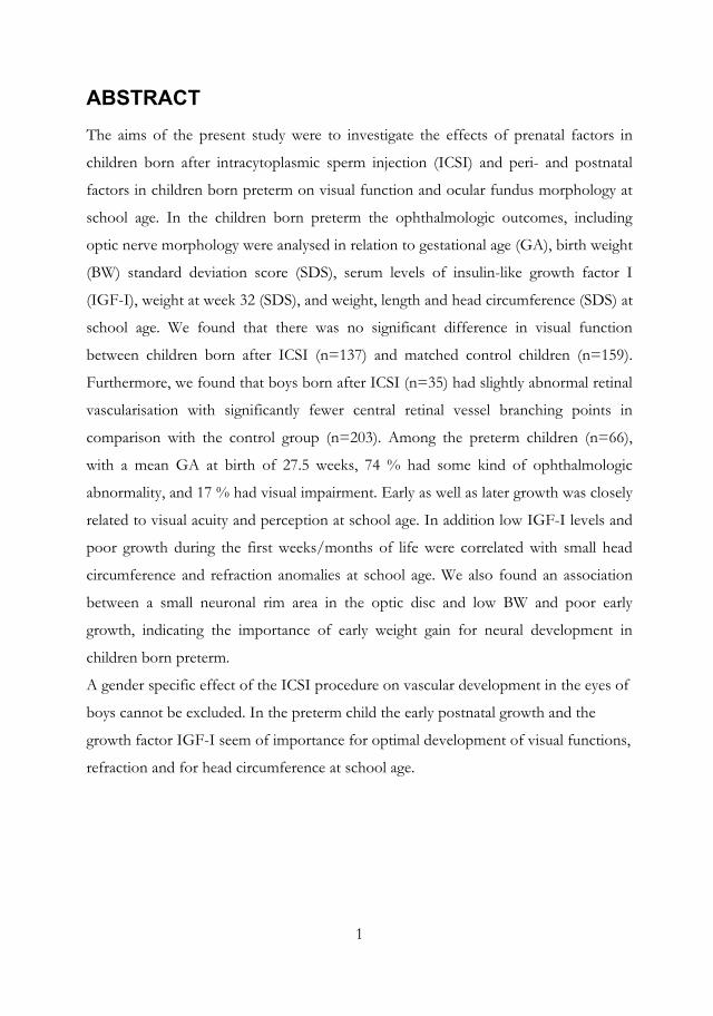
Figure 1. The retinal layers
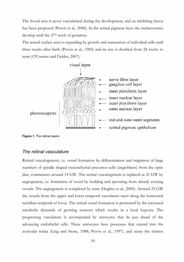
The retinal vasculature
Retinal vasculogenesis, i.e. vessel formation by differentiation and migration of large
numbers of spindle shaped mesenchymal precursor cells (angioblasts) from the optic
disc, commences around 14 GW. The retinal vasculogenesis is replaced at 21 GW by
angiogenesis, i.e. formation of vessel by budding and sprouting from already existing
vessels. The angiogenesis is completed by term (Hughes et al., 2000). Around 25 GW
the vessels from the upper and lower temporal vasculature meet along the horizontal
meridian temporal of fovea. The retinal vessel formation is promoted by the increased
metabolic demands of growing neurons which results in a local hypoxia. The
progressing vasculature is accompanied by astrocytes that lie just ahead of the
advancing endothelial cells. These astrocytes have processes that extend into the
avascular retina (Ling and Stone, 1988; Provis et al., 1997), and sense the relative
11
hypoxia which promotes production of vascular endothelial growth factor (VEGF)
within the astrocytes. VEGF stimulates endothelial cell proliferation at the vascular
front. It has been shown that insulin-like growth factor 1 (IGF-I) also influences
angiogenesis and acts as a critical permissive factor for normal vascularisation through
interaction with locally produced VEGF (Hellstrom et al., 2002; Hellstrom et al., 2001;
Smith et al., 1999). The proliferation of the endothelial cells causes capillaries to grow
into the previously hypoxic retina, resulting in a local decrease in hypoxia, and a down
regulation of the VEGF in nearby astrocytes. The process is iterated as the astrocytes
and vascular endothelial cells migrate towards the periphery.
Astrocytes also play a role in endothelial cell differentiation and blood barrier function
(Chan-Ling and Stone, 1992). In addition, astrocytes along with microglia contribute
to the perivascular glia limitans, important for vessel integrity (Provis, 2001).
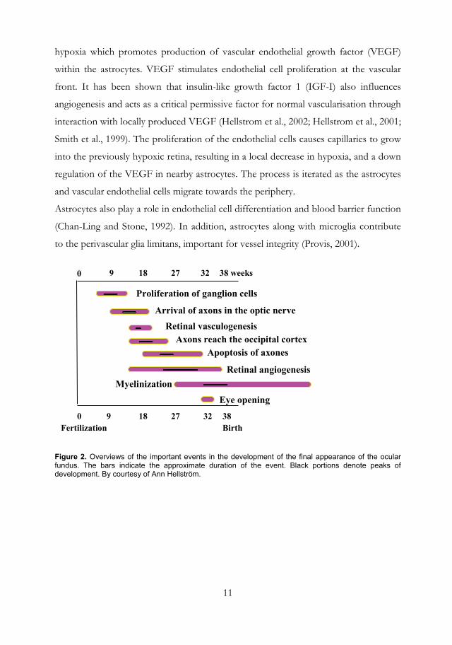
18 27 32
Fertilization9 18 27 32
9 38 weeks
Proliferation of ganglion cells
Arrival of axons in the optic nerve
Axons reach the occipital cortexApoptosis of axones
Retinal angiogenesisMyelinization
Eye opening
0
Birth380
Retinal vasculogenesis
Figure 2. Overviews of the important events in the development of the final appearance of the ocular fundus. The bars indicate the approximate duration of the event. Black portions denote peaks of development. By courtesy of Ann Hellström.
12
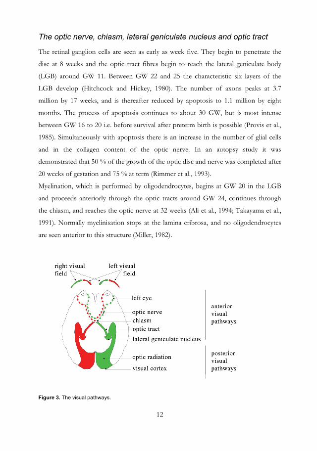
The optic nerve, chiasm, lateral geniculate nucleus and optic tract
The retinal ganglion cells are seen as early as week five. They begin to penetrate the
disc at 8 weeks and the optic tract fibres begin to reach the lateral geniculate body
(LGB) around GW 11. Between GW 22 and 25 the characteristic six layers of the
LGB develop (Hitchcock and Hickey, 1980). The number of axons peaks at 3.7
million by 17 weeks, and is thereafter reduced by apoptosis to 1.1 million by eight
months. The process of apoptosis continues to about 30 GW, but is most intense
between GW 16 to 20 i.e. before survival after preterm birth is possible (Provis et al.,
1985). Simultaneously with apoptosis there is an increase in the number of glial cells
and in the collagen content of the optic nerve. In an autopsy study it was
demonstrated that 50 % of the growth of the optic disc and nerve was completed after
20 weeks of gestation and 75 % at term (Rimmer et al., 1993).
Myelination, which is performed by oligodendrocytes, begins at GW 20 in the LGB
and proceeds anteriorly through the optic tracts around GW 24, continues through
the chiasm, and reaches the optic nerve at 32 weeks (Ali et al., 1994; Takayama et al.,
1991). Normally myelinisation stops at the lamina cribrosa, and no oligodendrocytes
are seen anterior to this structure (Miller, 1982).
Figure 3. The visual pathways.

13
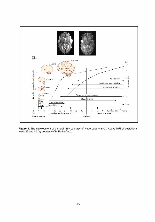
The brain
The cortex in the mature brain has six layers (I-VI). Layer I is the outermost layer next
to pia mater. In the foetus the forming layers I-VI are called the cortical plate, and the
layer below layer VI is a transient foetal structure called the cortical subplate.
Neurogenesis, proliferation of neurons, starts in GW five and is essentially completed
by weeks 20 to 24 (Bystron et al., 2008; Volpe, 2001b). Simultaneously, radial glial cells
produce systems of filaments which serve as guides for neurons in their migration
from their sites of origin in the ventricular and later subventricular place to their target
places in the cortical plate (Rakic, 1971; Watson, 1974). The radial glia cells also
facilitate the development of columnar organisation of the neurons in the cortex
(Rakic, 1988) which has a peak time from approximately the fifth month of gestation
to several years after birth. During the organisational period the subplate neurons are
established and differentiated, the dendrites and axons are ramified, synaptic contacts
occur, apoptosis, proliferation, and differentiation of glial cells take place (Volpe,
2001b). The subplate neurons are important for the formation of connections
between thalamus and cortex (Kanold, 2004; Kostovic and Judas, 2002; Volpe, 1996).
The transient subplate neurons, which are the major neuronal type in the cerebral
white matter, and the subplate region reaches its maximum thickness between 22 and
34 weeks of gestation (Ghosh and Shatz, 1992; Kostovic and Jovanov-Milosevic,
2006).
Apoptosis of the subplate begins late in the third trimester, and at about six months of
postnatal age approximately 90 % of the subplate neurons have disappeared. In
preterm babies the time course, when the subplate neurons are active in the
developing brain, corresponds closely to when periventricular haemorrhages and
ischemic lesions occur that may disrupt the subplate neurons, or their axonal
collaterals to the subcortical, or cortical sites (Volpe, 2001b). The neurite development
with ramifications is a very active process and a great number and variety of dendritic
spines appear (i.e. sites of synaptic contact) in the cortex during the third trimester
(Paldino and Purpura, 1979; Takashima et al., 1990).
14
The increase in cortical volume is particularly rapid between approximately
postconceptional weeks 28 and 40, which has been documented by quantitative MRI
measurements of cortical gray matter volumes in preterm infants during this period
(Huppi et al., 1998; Kapellou et al., 2006; Kostovic and Judas, 2002).
The glial cell proliferation and differentiation are important in the developing brain,
and there are many more glial cells than neurons in the CNS (Kinney and Back, 1998).
Radial glia produces astrocytes, which play an important role for nutrition and support
of neurons in reaction to metabolic and structural insults. Oligodendrocyte
proliferation and differentiation proceed in four stages, from oligodendroglial
progenitor, to preoligodendrocyte, to immature oligodendrocyte and finally to a
mature oligodendrocyte that can produce myelin. The myelin producing mature
oligodendrocytes are not abundant in the white matter until after term. During GW 24
to 32 there are mostly oligodendrocyte progenitors in the white matter with a peak in
number at GW 28, when 90 % are oligodendrocyte progenitors, while by term 50 %
are immature oligodendrocytes (Back et al., 2001). The immature oligodendrocytes are
especially vulnerable to ischemia and inflammation, which lead to excitotoxicity and
generation of free radicals that are produced by microglia. Microglia is involved during
brain development involving apoptosis, vascularisation, and axonal development and
the microglia reach a peak abundance in cerebral white matter in the third trimester
(Billiards et al., 2006). Myelin provides insulation and speed up nerve conduction.
The myelination process slowly starts in the second trimester and continues into
adulthood. Fifty percent of the oligodendrocytes are lost in apoptosis during their
development (Barres et al., 1992).
Cerebellum develops rapidly during the last half of gestation and a volumetric study of
premature infants has documented an approximately three-fold increase in volume
from 28 to 40 GW (Limperopoulos et al., 2005).
15
Figure 4. The development of the brain (by courtesy of Hugo Lagercrantz). Above MRI at gestational week 25 and 40 (by courtesy of M Rutherford).

16
IGF-I - neural and retinal vascular development
Insulin-like growth factor 1 (IGF-I) is a polypeptide, which resembles insulin in its
molecular structure. In humans IGF-I is primarily produced by hepatocytes in the
liver and the production is regulated by pituitary growth hormone (Holly and Perks,
2006). IGF-I exists extra-cellular and is bound to and controlled by six insulin-like
growth factor binding proteins (Holly and Perks, 2006). Seventy-five percent of the
IGF-I is bound to insulin-like growth factor binding protein 3 (IGFBP-3) together
with an acid labile subunit (Jones and Clemmons, 1995). The insulin-like growth
factor binding proteins can either inhibit, or potentiate cellular IGF-I responses, and
influence distribution and elimination of IGF-I. The cellular actions of IGF-I are
mediated through binding of IGF-I to the IGF-I receptor, which is located on the
surface of different cell types in all tissues. IGF-I can also bind to the insulin receptor,
but at a much lower affinity than insulin (Jones and Clemmons, 1995).
IGF-I is of major importance for foetal growth and is synthesized by all foetal tissues
early in gestation, and the placenta is actively involved in regulating circulatory foetal
levels of IGF-I (Gluckman and Pinal, 2003). Concentrations of foetal IGF-I are
closely related to placental transfer of nutrients. The disruption of placental nutrient
supply as well as amniotic supply (Han et al., 1996) at birth is followed by a rapid
decline in levels of IGF-I. During pregnancy thyroxine plays a more important role
than pituitary growth hormone in the regulation of foetal IGF-I (Deayton et al., 1993),
but after birth IGF-I is mainly regulated by pituitary growth hormone.
IGF-I is related to nutrition, BW (Giudice et al., 1995) and gestational age (Hellstrom
et al., 2003; Lineham et al., 1986; Smith et al., 1997b). At very preterm birth the IGF-I
levels of the newborn decrease abruptly, and do not reach normal intrauterine values
for several weeks/months (Engstrom et al., 2005; Lineham et al., 1986), in contrast to
in term infants, in whom serum levels of IGF-I are restored in a few days (Engstrom
et al., 2005; Kajantie et al., 2002; Lo et al., 2005). A recent study found a dramatic
decrease in the circulating serum levels of IGF-I and its major binding protein,
IGFBP-3 in very preterm infants, and that inflammation at birth with increased cord
levels of pro-inflammatory cytokines was associated with a decrease in IGF-I
17
(Hansen-Pupp et al., 2007). The important role of nutrition for the foetal IGF-I levels
was demonstrated in an animal study of foetuses of pregnant rats, who were fasted
during the last days of gestation, and the serum IGF-I levels were 30 % lower than in
the control foetuses (Davenport et al., 1990).
IGF-I acts directly on the brain and promotes differentiation, proliferation and
maturation of progenitors of neural stem cells, and has anti-apoptotic properties
(Hodge et al., 2007; McDonald et al., 2007; Ye and D'Ercole, 2006). Oligodendrocyte
maturation is crucial for myelination as mentioned above, and several studies on mice
and other rodents have shown an important role of IGF-I on differentiation of
oligodendrocyte progenitor cells (D'Ercole et al., 1996; Lin et al., 2005; Wilson et al.,
2003). In vitro, IGF-I has been found to promote remyelination (Mason et al., 2003),
and cerebellar Purkinje cell development (Fukudome et al., 2003). In addition, a
relationship has recently been shown in preterm infants between low cerebellar
volume and decreased serum IGF-I levels (Hansen-Pupp et al., 2009 ).
IGF-I may also play an important role in the stimulation of postnatal brain growth.
Over-expression of IGF-I in mice stimulated the brain growth and ameliorated the
brain growth even in the face of under-nutrition (Lee et al., 1999), and IGF-I
protected myelination in cases with under-nutritional insults (Ye et al., 2000). A
relationship between low circulating levels of IGF-I, the development of ROP, and
poor development of head circumference in preterm infants has also been
documented (Lofqvist et al., 2006b). IGF-I is essential for the development of normal
vascularisation of the human retina as mentioned above (Hellstrom et al., 2002;
Hellstrom et al., 2001; Smith et al., 1999), and promotes the angiogenesis in the brain
(Lofqvist et al., 2007; Lopez-Lopez et al., 2004). In the study by Lopez-Lopez and co-
workers systemic injections of IGF-I in adult mice increased the brain vessel density.
A gender difference in IGF-I levels, where boys had lower levels than girls, has been
shown in preterm (GA < 32 weeks) infants (Engstrom et al., 2005). In addition, in
singleton ICSI boys, serum IGF-I was found to be lower than that of normally
conceived boys (Kai et al., 2006).
IGF-I has also been shown to promote longitudinal postnatal growth (Fant and
Weisoly, 2001). In addition, ocular growth is influenced by IGF-I and treatment with
18
IGF-I increases the ocular axial eye length in patients with short axial lengths due to
growth hormone insensitivity (Laron syndrome) (Bourla et al., 2006).
Intracytoplasmic sperm injection technology
The ICSI procedure is recommended to couples who have failed to achieve
fertilisation following standard in vitro fertilisation (IVF) treatment, when the male
has abnormal sperm parameters (low count, poor motility, abnormal sperm forms and
high levels of antibodies in the semen), and when the male must have his sperm
surgically retrieved from the epididymis or testis because of lack of sperms in the
ejaculate (azoospermia) due to for example congenital absence of both vasa deferentia.
The treatment involves several stages. At first the woman is given a Gonadotrophin
Releasing Hormone analogue (nasal spray or subcutaneous injection) to “shut down”
the “normal” ovarian function. To ensure that the ovaries are inactive an ultrasound is
performed. She is then given Follicle Stimulating Hormone (FSH) on a daily basis,
which stimulates the ovaries to produce multiple follicles. The follicles will be assessed
in number and size by ultrasound scans. When at least 3 follicles at > 18 mm can be
observed, indicating that there may be a mature egg, the egg collection is scheduled.
Now another injection is administered, Human Chorionic Gonadotrophin (HCG).
This injection helps to mature and release the eggs in the follicles for the egg
collection. Approximately 36 hours after the HCG injection the oocyte retrieval takes
place under sedation or general anaesthesia. A vaginal probe with a needle guide is
under ultrasound guidance passed through the vaginal wall into each ovary. The
follicles are drained and the collected follicular fluid is searched for eggs. Once the
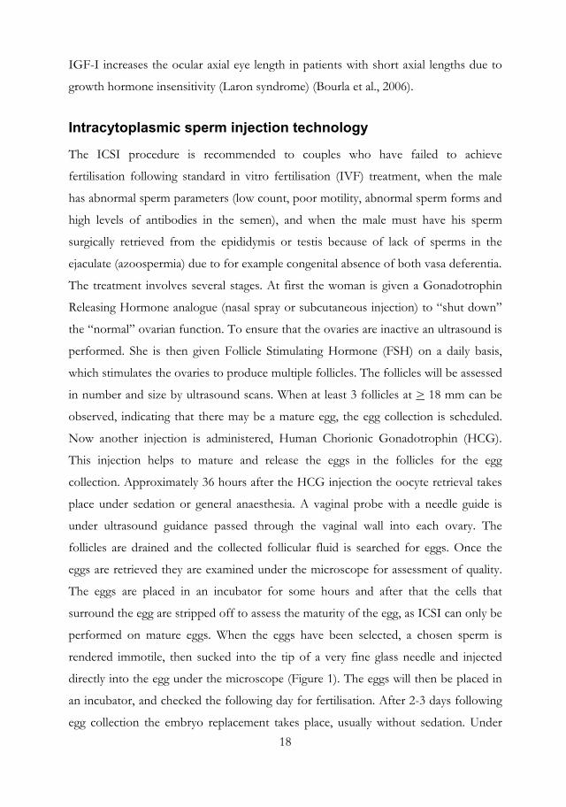
eggs are retrieved they are examined under the microscope for assessment of quality.
The eggs are placed in an incubator for some hours and after that the cells that
surround the egg are stripped off to assess the maturity of the egg, as ICSI can only be
performed on mature eggs. When the eggs have been selected, a chosen sperm is
rendered immotile, then sucked into the tip of a very fine glass needle and injected
directly into the egg under the microscope (Figure 1). The eggs will then be placed in
an incubator, and checked the following day for fertilisation. After 2-3 days following
egg collection the embryo replacement takes place, usually without sedation. Under
19
ultrasound guidance a fine catheter, loaded with the embryo, is passed through the
vagina, the cervix and into the uterus. Progesterone helps to maintain the thickness of
the lining of the uterus to aid implantation, and is given from the day of the embryo
transferral. A pregnancy test should be performed 14 days after the embryo transfer.
This procedure has raised concerns about risks for adverse outcome in children born
after ICSI as it includes artificial induction of ovulation with a possibility of changes in
follicle milieu and oocyte structure, the use of a sperm that cannot conceive naturally
and exposure of the egg, sperm and embryo to the artificial in vitro environment
including chemicals, freezing and mechanical manipulations.
Figure 5. Intracytoplasmic sperm injection, (by courtesy of Ulla-Britt Wennerholm).

20
21
ICSI AND PRETERM OUTCOME
Pre- and perinatal factors
Maternal
Women undergoing IVF/ICSI and women giving birth to a preterm child have
increased frequency of morbidity both before and during pregnancy. Diabetes mellitus
(Yeshaya et al., 1995), inflammatory intestinal disease (Bradley and Rosen, 2004) and
thyroid dysfunction (Poppe et al., 2007) have been associated with infertility. Maternal
medical disorders, such as thyroid disease, asthma, diabetes and hypertension are
associated with increased rates of preterm delivery (Goldenberg et al., 2008). Other
studies have also demonstrated that ICSI/IVF mothers had diabetes mellitus (pre-
existent) significantly more often than spontaneously conceiving mothers (Kapiteijn et
al., 2006; Katalinic et al., 2004). Diabetes is a known risk factor for preterm delivery
(Lepercq et al., 2004; Matsushita et al., 2008; Melamed et al., 2008).
Mothers undergoing ICSI/IVF more often use drugs as treatment for diseases such as
diabetes, hypo/hyperthyroidism, and inflammatory disease than other mothers, beside
the drug therapy associated with assisted reproduction. In addition, an increased use
of heparin, heparin-like substances, and thrombocyte aggregation inhibitors has been
related to conditions associated with subfertility (Kallen et al., 2005b).
All pregnant women in Sweden during a period of almost 10 years were studied, and
maternal use of thyroid hormones during pregnancy had an increased rate of pre-
eclampsia and diabetes (pre-existing or gestational), but the risk for preterm birth was
only marginal (Wikner et al., 2008). An increased risk of deep venous thrombosis after
single embryo transfer compared with spontaneously conceived pregnancies has been
reported (Poikkeus et al., 2007), and a history of deep vein thrombosis has also been
shown to be an independent risk factor for spontaneous preterm delivery (Ben-Joseph
et al., 2008). The aetiology of preterm birth is complex and involves environmental
and genetic factors, and the underlying mechanisms are not fully understood. In
comparison with all women giving birth in Sweden during 1982 - 2001, the women
who underwent IVF/ICSI during the same time period (n = 12 186) were older, more
22
often of first parity, smoked less, were more overweight and worked less outside
home. Their use of medication in early pregnancy was nearly three times higher than
among other pregnant women. In contrast, underweight body mass index in the pre-
pregnancy health status in mothers with spontaneous conception is a risk factor for
preterm delivery (Haas et al., 2005). Smoking is a risk factor for preterm delivery, but
women who gave birth after IVF/ICSI smoked less than other pregnant women
(Kallen et al., 2005b). Some studies have shown that older mothers have an increased
risk of preterm delivery (Cnattingius et al., 1992; Cnattingius and Haglund, 1992;
Prysak et al., 1995), while other authors found no increased risk of preterm delivery in
older mothers, but an increased risk of obstetric complications (Berkowitz et al., 1990;
Mbugua Gitau et al., 2009). Interestingly, it has been shown that mothers under the
age of 20 years also have an increased risk of preterm birth (Stevens-Simon and
McAnarney, 1991).
Many obstetric characteristics are similar in pregnancies of both women who are
pregnant after IVF/ICSI and women who deliver preterm. The pregnancies are
associated with an increased risk of gestational hypertension (Jackson et al., 2004;
Johnson et al., 2009; Kallen et al., 2005c). Women who have undergone IVF/ICSI
have increased risk of pre-eclampsia, placental abruption, placenta previa, preterm
premature rupture of membranes and gestational diabetes which are all risk factors for
preterm delivery (Ananth et al., 2001; Bonduelle et al., 2005; Covarrubias et al., 2008;
Goldenberg et al., 2008; Hossain et al., 2007; Jackson et al., 2004; Krupa et al., 2006;
Nygren et al., 2007; Poikkeus et al., 2007; Romundstad et al., 2006; Sutcliffe and
Ludwig, 2007). A study of all women known to have had IVF/ICSI in Sweden
between 1982 and 2001 showed, that the impact of the maternal obstetric
characteristics on preterm delivery was rather small (Kallen et al., 2005c). The authors
suggested that the reason for the increased risk of preterm delivery must be sought in
the infertility status of the women. Multiple births account for a substantial risk of
preterm delivery, and multiple births is a complication in pregnancies following
IVF/ICSI as a result of transferral of more than one embryo. The twinning rate was
about 23 % in Europe 2002, but lower in Sweden where the twinning rate after
IVF/ICSI was 18.5 % 2001. After a recommendation from the Swedish Medical
23
Association in 2003 only one embryo is transferred and the frequency of multiple
births had fallen to approximately 5 % in 2004 (Nygren et al., 2007).
Intrauterine infection is a frequent and important cause of preterm delivery, and
microbiological studies suggest that intrauterine infection might account for 25-40 %
of premature births (Goldenberg et al., 2000). The infection may activate the innate
immune system, and release chemokines and cytokines which stimulate inflammatory
mediators including prostaglandins which stimulate uterus contractility that can lead to
preterm premature ruptures of membranes (Romero et al., 2006). The earlier in
pregnancy a women presents with preterm labour, the higher is the frequency of
intrauterine infection (Mueller-Heubach et al., 1990). Intrauterine inflammation and
placental dysfunction are the most common causes of delivery before the 28th week of
gestation (McElrath et al., 2008).
Genetics
Paternal genetics in ICSI
The impact of sperm quality on the outcome after assisted reproduction, especially
after ICSI, which is the treatment for male factor infertility, has been discussed. It has
been found that infertile men with sperm abnormalities (numerical and structural)
have more chromosome aberrations (Aittomaki et al., 2005; Bonduelle et al., 2002;
Lundin et al., 1998; Van Assche et al., 1996).
Microdeletions in a region on the long arm of the Y chromosome were found in 7.3
% of infertile men, and the majority of these deletions were seen in the azoospermic
men. Once a deletion has occurred, it will be inherited by all male offspring if
infertility is treated with ICSI, and it has been found that 9 % of sons born after ICSI
had Y chromosome microdeletions (Kent-First et al., 1996). An increased frequency
of hypospadia, a condition that might be associated with male infertility, has been
reported in the ICSI male offspring compared with the general population (Bonduelle
et al., 2005; Ericson and Kallen, 2001; Fedder et al., 2007; Katalinic et al., 2004;
Wennerholm et al., 2000). An increased rate of chromosomal aberrations and sex
chromosome abnormalities, compared with an expected rate, (Jacobs et al., 1992;
24
Nielsen and Wohlert, 1991) has been recorded in pregnancies after ICSI (Bonduelle et
al., 2002; Wennerholm, 2004). These aberrations are most probably linked to maternal
age, or directly to the characteristics of the infertile men being treated, rather than to
the ICSI procedure itself (Wennerholm, 2004).
Genomic imprinting diseases
Genetic imprinting is a process, which occurs early in development and silences the
copy of a gene inherited from either the mother or the father. By abnormal
methylation pattern of an imprinted gene the gene expression may be altered.
Oligozoospermia (< 20 million/ml) itself increases the risk for genetic imprinting
disorders (Marques et al., 2004). Sutcliffe and co-workers found an association
between ICSI/IVF and Beckwith-Wiedemann syndrome. Children born after
IVF/ICSI with Beckwith-Wiedemann syndrome and Angelman syndrome showed
loss of maternal allele methylation at a critical imprinting control region (Sutcliffe et
al., 2006). An increased risk of imprinting diseases may be caused by the in vitro
embryo culture (Maher, 2005), or some factor associated with infertility per se. A
susceptibility to epigenetic imprinting diseases may be due to factors causing the
infertility and the ovarian stimulation as part of the infertility treatment may play a
role. Children with Angelman syndrome born after infertility treatment with induced
ovulation showed increased frequency of imprinting defects (Ludwig et al., 2005). In
the Swedish cohort of children born after ICSI one child had Silver Russel syndrome
and one had Prader Willi syndrome, and both syndromes are associated with
imprinting anomalies (Kallen et al., 2005a). In the Danish national IVF/ICSI cohort
study no child was found to have imprinting anomalies (Lidegaard et al., 2005).
Retinoblastoma (RB) is commonly caused by a mutation of one allele of the tumour
suppressor gene RB1, combined with chromosomal loss, or deletion of the other allele
(Thompson and Williams, 2005). Hypermethylation of the RB gene that inactivates
the tumour suppressor function may play a role in the development of retinoblastoma
(Ohtani-Fujita et al., 1997). Five children with retinoblastoma, associated with
IVF/ICSI have been reported from the Netherlands (Moll et al., 2003). Imprinting
25
disorders are rare conditions, and large prospective multi-centre cohort studies are
needed in order to conclude if there is a true increased prevalence in infants born after
IVF/ICSI (Manipalviratn et al., 2009).
Genetics and preterm delivery
There is a familial pattern of preterm delivery indicating a genetic predisposition.
Women who are born preterm are more likely to have a preterm delivery themselves
(Porter et al., 1997; Wilcox et al., 2008), as are sisters of women who have had a
preterm delivery (Winkvist et al., 1998). In a population-based prospective study
sisters of an affected individual had higher risk of preterm delivery than sisters of
unaffected individuals. The preterm deliveries were the result of premature rupture of
membranes, placental abruption, and pre-eclampsia and the results suggest that the
adverse pregnancy outcomes that aggregate in families may in part be explained by
genetics (Plunkett et al., 2008).
Studies for evaluating the genetic influence while attempting to avoid confounding
environmental influences have been performed on twins, and the genetics
contribution to preterm delivery has been estimated to approximately 30 % (Clausson
et al., 2000; Kistka et al., 2008; Lunde et al., 2007). In addition, racial disparities exist
which suggest a genetic contribution, where African-American mothers are more
prone to give birth prematurely (Palomar et al., 2007). Genetic studies have identified
markers, which more accurately predict preterm birth than currently known risk
factors e.g. proteins, and/or pathways involved in the disorder. Several genes have
been reported to be associated with preterm delivery, although inconsistency between
the studies has been problematic (Plunkett and Muglia, 2008).
Many of the genetic studies on preterm birth have focused on genes involved in
inflammation, like the gene for tumour necrosis factor-α, a pro-inflammatory cytokine.
However, studies of polymorphism in this gene in the mothers have been
inconclusive.

26
Short term consequences
Malformations
Many studies have addressed the risk of congenital malformations in children born
after IVF/ICSI. In large meta-analyses increased rates of up to 30-40 % risk of
congenital malformations after IVF/ICSI compared with children born after
spontaneous conception has been noted (Hansen et al., 2005; Kallen et al., 2005a;
McDonald et al., 2005; Rimm et al., 2004). The children born after ICSI in Papers I
and II were part of a larger multi-centre study, demonstrating malformations more
often in singleton children born after ICSI (6 %) than in the spontaneously conceived
group (3 %) at the age of 5 years (p = 0.037) (Bonduelle et al., 2004). In the large
Swedish registry study on ICSI/IVF children by Källén and co-workers (Kallen et al.,
2005a) the increase of malformations was 42 %, and was higher for singleton than for
multiple births in comparison with normally conceived singletons and twins,
respectively. After adjustments for year of birth, maternal age, parity, years of known
childlessness, and smoking, the risk increase disappeared, and the authors concluded
that the observed malformations mainly were due to maternal characteristics
associated with subfertility. A Danish registry-based study compared the malformation
rate in singletons born to fertile couples (time to pregnancy interval ≤ 12 months) and
singletons born to infertile couples (time to pregnancy interval of > 12 months) who
either conceived naturally, or with infertility treatment. Singletons born to infertile
couples had a higher rate of congenital malformations, independently of whether the
children were conceived spontaneously or after infertility treatment (Zhu et al., 2006).
The findings suggest that the increased risk of congenital malformations in children
born after IVF/ICSI is mostly a result of the underlying parental characteristics, rather
than the assisted reproduction techniques themselves.
The specific types of malformations observed are neural tube defects, cardiovascular
defects, choanal atresia and alimentary tract atresia (Kallen et al., 2005a). Similar
results with regard to malformations have been found after standard IVF and ICSI.
However, an increased risk of defects in the urogenital system in ICSI children e.g.
27
hypospadia has been found in several studies (Bonduelle et al., 2005; Ericson and
Kallen, 2001; Fedder et al., 2007; Kallen et al., 2005a; Wennerholm et al., 2000).
Major congenital malformations increase the risk for preterm birth. In a population-
based study, where chromosomal abnormalities were excluded, an increased risk for
malformations like cleft lip and palate, diaphragmatic hernia, urogenital anomalies,
heart defect, spina bifida, omphalocele/gastroschisis, tracheoesohageal anomalies and
renal agenesis was found in infants born preterm. The risk of preterm birth was higher
with multiple malformations, and the risk varied inversely with GA (Purisch et al.,
2008). Cardiovascular malformations have been found in twice as many preterm
children as in children born full term (Tanner et al., 2005). In addition, two other
population-based studies found that significant malformations were more common in
prematurely born babies than in infants born at term (Holmgren and Hogberg, 2001;
Rasmussen et al., 2001). A recent study showed a high risk of preterm birth in babies
with brain defects. Although the brains of preterm infants are particularly vulnerable
to injury, the brain defects in that study developed in utero, and not at birth, or after
birth. The authors speculated that it is either the brain defects themselves, or the
underlying cause of the defect that result in preterm birth. Another possible
mechanism may be that coagulopathy, which is associated with congenital brain
defects, is involved in the induction of the preterm delivery (Brown, 2009).
Neonatal morbidity
It is clear that preterm delivery and low BW, as well as perinatal morbidity, occur in a
higher frequency related to IVF/ICSI pregnancies than in naturally conceived
pregnancies. The increased risk of low BW associated with assisted reproduction has
been attributed largely to the higher rate of multiple gestations associated with the
technique, and a multiple pregnancy is a well-established risk factor for adverse
outcomes including preterm delivery, low BW and neonatal morbidity (Liu and Blair,
2002). There is no data suggesting that multiple births after IVF/ICSI have more
adverse perinatal outcomes than multiple births after spontaneous conception
(Helmerhorst et al., 2004).
28
However, several meta-analyses on singleton children born after IVF/ICSI have
found similar results, reporting an approximately two-fold risk of being born preterm,
and an increased risk of having a low, or very low BW, being born small for
gestational age, being admitted to neonatal intensive care unit, as well as having higher
perinatal mortality (Helmerhorst et al., 2004; Jackson et al., 2004; McDonald et al.,
2005). In a large Swedish study by Källen and co-workers the odds risk ratio of
adverse outcomes were reduced and not significant, when adjusting for year of birth,
maternal age, parity, smoking and number of years of involuntary childlessness. They
also reported a higher risk for cerebral haemorrhage, need for mechanical ventilation,
use of continuous positive airway pressure (CPAP), neonatal sepsis, neonatal
convulsions and respiratory problems after IVF/ICSI. The main reason for the
increased risk the authors concluded was the high rate of multiple births in the study
group (Kallen et al., 2005c). Significantly lower rates of premature delivery, low BW
and paediatric complications requiring neonatal intensive unit care following births
after transferral of only one embryo compared to dual-embryo transfer have been
reported (Kjellberg et al., 2006). Vanishing twins after multiple embryo replacement
has been considered to be one of the causes of the adverse neonatal outcome in
singletons born after IVF/ICSI (Pinborg et al., 2007; Pinborg et al., 2005; Schieve et
al., 2004), and that the infertility itself may play a causative role (Draper et al., 1999;
Pandian et al., 2001; Schieve et al., 2002).
The more immature the baby, the greater is the risk of morbidity. The major disorders
associated with preterm birth are respiratory distress syndrome (RDS),
bronchopulmonary dysplasia (BPD), necrotising enterocolitis (NEC), sepsis,
intraventricular haemorrhage (IVH), periventricular leucomalacia (PVL) and
retinopathy of prematurity (ROP). Increased survival of infants born before 25 GW,
during the last decade, has resulted in an increased prevalence of BPD as the tiniest
infants require longer time with mechanical ventilations and CPAP, and are prone to
develop sepsis (Lundqvist et al., 2009; Ronnestad et al., 2005). The disorders of the
brain associated with prematurity and ROP are discussed below.
29
Retinopathy of prematurity (ROP)
Although most cases of ROP are self limiting it may be a serious threat to vision.
In infants born prematurely the retina is not fully vascularised (see page 11). The more
premature the child, the larger is the avascular area. The sudden loss of nutrition and
growth factors necessary for normal growth at preterm birth causes the vascular
growth, that would normally occur in utero, to slow down or cease. In addition, the
relative hyperoxia of the extra-uterine milieu together with supplemental oxygen cause
a regression of already developed retinal vessel. As discussed earlier, IGF-I is
necessary for normal development of retinal blood vessels (Hellstrom et al., 2002).
Preterm birth is associated with a rapid fall in IGF-I, and the baby often suffers from
immaturity, poor nutrition, acidosis, hypothyroxemia and sepsis which all may further
reduce the IGF-I levels. When the neural elements of the retina mature and need
more oxygen, poor vascularisation leads to hypoxia and production of vascular
endothelial growth factor (VEGF). If sufficient IGF-I is not available VEGF is
accumulated, as a minimum level of IGF-I is required for VEGF to induce vessel
growth. When the IGF-I levels slowly increase when the infant matures, and IGF-I
reaches the minimum level for VEGF to promote vessel growth, an excessive and
uncontrolled neovascularisation may take place. However, in most cases vessel growth
retardation is not severe enough to cause the proliferative stages of ROP. The serum
levels of IGF-I during the first weeks of life in the babies are inversely correlated with
the severity of ROP (Hellstrom et al., 2003).
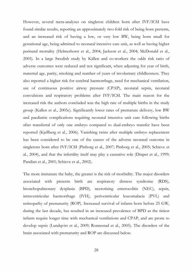
The international classification of the stages of ROP (Committee for the Classification
of Retinopathy of Prematurity Revisited, 2005) is presented in Figure 6 below. Stages
1 and 2 are considered as mild, while stages 3 to 5 are severe. The extraretinal
neovascularisation in stage 3 may lead to retinal detachment. If the detachment
involves the macula the child becomes blind. Dilatation and tortuosity of the central
vessels are called plus disease or pre-plus disease, and are signs of a progressive
disease. Aggressive posterior ROP is a virulent form of ROP seen in the tiniest babies
(Committee for the Classification of Retinopathy of Prematurity Revisited, 2005).
30
Stage 2
Stage 3
Stage 4
Stage 1
Stage 5
Figure 6: The stages of ROP. Stage 1: Demarcation line, Stage 2: Ridge, Stage 3: Ridge with extraretinal fibrovascular proliferation, Stage 4: Partial retinal detachment, Stage 5: Total retinal detachment (Drawings by Lisa Hård af Segerstad).
Risk factors for ROP are low GA/BW and oxygen supplementation (Lutty et al.,
2006; Smith, 2003; Tasman et al., 2006). Hyperglycemia has also been reported to be
associated with ROP (Ertl et al., 2006; Garg et al., 2003). An association between poor
postnatal growth and later development of ROP has been reported (Allegaert et al.,
2003; Wallace et al., 2000). A recent study has even shown that monitoring postnatal
weight development can predict proliferative ROP and may thus modify and reduce
traditional screening (Hellstrom et al., 2009).
The incidence in Sweden of ROP has been reported to be 36.4 % in infants with a
BW of 1500 g or below. Mild ROP occurred in 18.2 % of the children and 18.2 %
progressed to severe ROP. Twelve percent of the infants were treated with laser
therapy of the peripheral avascular retina (Larsson et al., 2002). Even though ablation
treatments have reduced the incidence of blindness by 25 % in the children with
severe ROP, some cases still progress to retinal detachment and blindness (Chen and
Smith, 2007). Advanced care is presently saving many critically ill, extremely
premature infants with high risk of developing severe ROP needing treatment. Two
recent studies report that the proportion of screened infants who needed ROP
treatment has doubled in the last ten years (Schiariti et al., 2008; Slidsborg et al., 2008).
31
Periventricular leucomalacia and neuronal/axonal disease -
”Encephalopathy of prematurity”
The encephalopathy of prematurity includes periventricular leucomalacia (PVL) and
accompanying neuronal and axonal deficits that involve the cerebral white matter,
thalamus, basal ganglia, cerebral cortex, brain stem and cerebellum (Volpe, 2009). The
initiating mechanisms in PVL, both focal and diffuse, are ischemia and inflammation
(Hagberg and Mallard, 2005). The focal PVL consists of microscopic areas of
necrosis, which give rise to cystic lesions. The diffuse PVL, which is much more
common, is characterised by marked astrogliosis and microgliosis, and a decrease in
premyelinating oligodendrocytes (Haynes et al., 2003; Robinson et al., 2006). Between
weeks 24 and 32 the periventricular white matter is in a watershed area between the
cortex and the subcortical areas as the end capillaries from the cortex have not
reached the periventricular area. Thus, there is a markedly low basal blood flow to the
cerebral white matter (Borch and Greisen, 1998; Khwaja and Volpe, 2008). In addition
to the low blood flow, the immature vessels lack smooth muscles, which interfere with
the ability to change diameter in response to changes in blood pressure (Soul et al.,
2007). Hypocarbia is a potent vasoconstrictor, and fluctuations in arterial carbon
dioxide tension are common in the preterm infant who needs mechanical ventilation
(Wiswell et al., 1996). Altogether, these mechanisms make cerebral white matter in the
periventricular area especially vulnerable to ischemic lesions. Inflammation is often
due to maternal intrauterine infection or postnatal sepsis, and results in excitotoxity
and free-radical attack, causing destruction and/or dysfunction of oligodendrocytes
and axons (Dammann et al., 2001). The white matter damage is accompanied by
cerebral cortex and deep matter gray abnormalities (Kostovic and Judas, 2006; Volpe,
2005), involving excess apoptosis (Robinson et al., 2006) without replacement
resulting in disturbances in the synaptogenesis and the connectivity (Ben-Ari, 2006;
Kesler et al., 2006). The neurons migrate from the germinal matrix
(ventricular/subventricular zones) through the white matter to the cortex when the
white matter is most vulnerable. Especially the pre-oligodendrocytes are prone to
ischemic-hypoxic insults and hypomyelination may occur (Segovia et al., 2008). The
32
vulnerability of pre-oligodendrocytes has been shown in humans in several studies
(Back et al., 2005; Haynes et al., 2003; Robinson et al., 2006).
MRI studies have documented white matter damages (Counsell et al., 2008; Dyet et
al., 2006; Inder et al., 2003), and diffusion-based MRI studies have shown
abnormalities consistent with axonal degeneration and impaired development in
children born preterm (Counsell et al., 2003; Counsell et al., 2007).
Axonal degeneration, studied by using apoptotic markers, has recently been found in
children with PVL (Haynes et al., 2008).
Several regions of the brain have been found to be affected in children born preterm
e.g. thalamus (Pierson et al., 2007), cerebellum (Limperopoulos et al., 2005) and the
brain stem (Pierson et al., 2007).
Long term consequences
Children born after IVF/ICSI
Regarding general health up to five to eight years of age including vision and hearing,
reassuring results have been found with no differences between singletons born after
IVF/ICSI and children born after spontaneous conception except for more medical
care in the IVF/ICSI group of children (Basatemur and Sutcliffe, 2008; Bonduelle et
al., 2004; Bonduelle et al., 2005; Knoester et al., 2008; Ludwig et al., 2009; Sutcliffe
and Ludwig, 2007). In the European study by Bonduelle and co-workers male infants
after ICSI needed more urogenital surgery (5 %) in comparison with 1 % among the
children born after spontaneous conception (Bonduelle et al., 2005). The systolic and
diastolic blood pressure were significantly higher in the eldest ICSI children being
followed up than in children born after spontaneous conception at the same age
(Belva et al., 2007). Low BW may be associated with later development of
cardiovascular disease (Barker et al., 1990; Lackland et al., 2003). IVF/ICSI singleton
children have an increased risk for cerebral palsy which has been associated with
preterm birth (Lidegaard et al., 2005; Stromberg et al., 2002). Several studies have
shown no difference regarding the neurodevelopmental and cognitive outcome,
including visual perception, at follow-up to five to ten years between children born
33
after IVF/ICSI and naturally conceived children (Belva et al., 2007; Leunens et al.,
2008; Ponjaert-Kristoffersen et al., 2005; Ponjaert-Kristoffersen et al., 2004; Steel and
Sutcliffe, 2009; Wagenaar et al., 2009).
Children born preterm
There are many studies that have documented neurodevelopmental disturbances in
preterm children that affect vision, cognition (Allen, 2008; Cheong et al., 2008; Hack
et al., 2002; Patra et al., 2006; Woodward et al., 2006), and motor functions (Allen,
2008; Inder et al., 2005; Larroque et al., 2008; Woodward et al., 2006), as well as
behaviour (Bhutta et al., 2002; Cheong et al., 2008; Stjernqvist and Svenningsen,
1999). These disturbances can mostly be related to the “encephalopathy of
prematurity” discussed above (Volpe, 2009). Other morbidities associated with
prematurity like necrotising enterocolites and bronchopulmonary dysplasia have been
found to be associated with adverse neurodevelopmental outcome (Anderson and
Doyle, 2006; Rees et al., 2007).
Visual outcome
Ocular morphology
In children born after IVF/ICSI there are only few reports on ocular morphology.
Retinoblastoma has been found in five children born after IVF/ICSI (Moll 2003).
One study of 47 children born after IVF/ICSI, who were referred to an eye clinic,
reported serious eye disorders, i.e. Coats disease, optic nerve hypoplasia/atrophy and
coloboma with microphthalmus (Anteby et al., 2001).
In prematurely born children both ROP and brain lesions may influence the
forthcoming visual outcome. In the macula an absent or reduced avascular zone has
been found in children born at GA of 30 weeks or less (Mintz-Hittner et al., 1999). In
children with a history of ROP increased central foveal thickness and preservation of
inner retinal layers within the fovea have been described (Recchia and Recchia, 2007),
indicating disturbed retinal development. In addition, it has been reported that
34
children born prematurely had a thicker central macula and thinner peripapillary nerve
fibre layer compared with children born at term. The lower the BW and the smaller
the head circumference the thicker central macula and the thinner the peripapillary
nerve fibre layer (Wang et al., 2006). Another study, of six year old children found that
the axial length of the eye and corneal radius were shorter in children born with low
BW and those with a small head circumference at birth. The effects on the size of the
eye lasted up to at least six years of age, however, no lasting effect on spherical
equivalent of refraction was seen (Ojaimi et al., 2005). These results emphasise the
importance of normal intrauterine growth for the eye development.
In the eye, the rod sensitivity as measured using electroretinography is reduced in
preterm infants and this reduction has been attributed to factors like insufficient
nutrition and retinal vascularisation and ROP (Fulton et al., 2008; Hamilton et al.,
2008). The retina is a controller of the refractive development (Troilo and Wallman,
1991), and an association between reduced rod sensitivity and early ametropia has
been found in children with ROP (Moskowitz et al., 2005).
Visual function
Children born after IVF/ICSI have only been tested for visual outcome in a few
studies and no difference in visual acuity, stereopsis and eye movements in
comparison with children born after spontaneous conception has been found (Belva
et al., 2007; Bonduelle et al., 2005; Wikstrand et al., 2006).
Several long-term population-based studies up to adolescence have shown that
children born preterm have reduced visual acuity, an increase in refractive errors,
strabismus, visual field defects and reduced contrast sensitivity (Cooke et al., 2004;
Fledelius, 1996; Hellgren et al., 2007; Holmstrom and Larsson, 2008; Holmstrom et
al., 2006; Holmstrom and Larsson, 2005; Larsson et al., 2006; Larsson and
Holmstrom, 2006; Larsson et al., 2003; Larsson et al., 2005; Lindqvist et al., 2007;
O'Connor et al., 2007). Following laser ablation for ROP the visual fields of the
prematurely born children may be peripherally constricted, and a decreased sensitivity
within the central visual field unrelated to the history of ROP, has been found
35
(Larsson et al., 2004). Inferior visual field defects have been documented due to
damage in the cerebral posterior superior white matter which is often seen in
prematurely born children (Dutton et al., 2004; Jacobson et al., 1996). Poor visual
perception has been found in several studies (Hard et al., 2000; Hellgren et al., 2009).
Two studies on the visuo-cognitive development at six to seven years in children born
before GW 32 compared with children born full term found that spatial function,
selective attention and executive control were most affected. These three areas of
functions have been linked to the dorsal visual cortical stream even though they
involve other areas of the brain, suggesting vulnerability for the dorsal steam, which
undergoes a longer developmental time course than the ventral stream (Atkinson and
Braddick, 2007; Santos et al., 2008).
The more immature the child is, the greater the risk for visual impairment which has
been shown in a recent Swedish follow-up study on children born before 25 GW. In
the study ROP was found in 97 %, 75 % developed proliferative ROP, and 63 %
needed laser ablation. A significant gender difference was found, where 32 % of the
boys were blind or visually impaired compared with 9 % of the girls (Jacobson et al.,
2009).
General outcome
Recent reports on long-term medical, psychiatric and social consequences in Swedish
and Norwegian national cohorts of adult people born preterm showed an increased
risk of having cerebral palsy, mental retardation and psychiatric disorders and the risks
increased with the degree of prematurity. Disability pension from the society was
several times higher in adults born preterm than in the group born at term. Preterm
birth was also associated with a lower chance of completing a university education,
and hence these individuals had a lower net salary (Lindstrom et al., 2009; Lindstrom
et al., 2007; Moster et al., 2008). However, the majority of adults who were born very
preterm lived an independent and self-supportive life and a large proportion
completed higher education and “seemed to live a satisfying life” (Lindstrom et al.,
2007; Moster et al., 2008).
36
Growth
The vast majority of studies which have examined growth in children born after
IVF/ICSI from the neonatal period up to eight years of age have not found any
differences regarding weight, stature and head circumference compared with
spontaneously conceived children (Belva et al., 2007; Bonduelle et al., 2004; Bonduelle
et al., 2005; Knoester et al., 2008; Ludwig et al., 2009). Few studies have reported
differences in growth parameter between ICSI/IVF children and naturally conceived
control children (Kai et al., 2006; Miles et al., 2007). The study of Danish children
born after ICSI found that they were significantly smaller than their target height
(SDS) compared with normally conceived children at three years of age, but at five
years of age there was no difference between the groups in stature. The study included
both twins and singletons born preterm and SGA after ICSI which may have
influenced the results (Kai et al., 2006). Another study of children born after
IVF/ICSI that excluded children born preterm and SGA found that the children born
after ICSI were taller, most evident in girls, than the normally conceived control
children after adjustment of parental height and age, and the IVF/ICSI children also
had higher serum levels of IGF-I. The authors proposed that the in vitro
manipulation had resulted in persistent metabolic alterations in the IVF/ICSI children
they studied (Miles et al., 2007).
Studies on growth in preterm children born before GW 29 have demonstrated an
initial decrease in weight during the first months of life, followed by an increase in
weight gain approximately weeks 36 to 40 PMA (first peak). After a period of slower
weight and length growth, a second increase in growth velocity, was demonstrated
from 6 months to 2 years of corrected age (second peak). The initial transient period
of slower weight gain may be ascribed to the discontinuation of parenteral nutrition
and the beginning of full enteral feeding. The infants born very preterm remained
growth retarded during the first 1-2 years, and they were growth retarded during a
postnatal period corresponding to the last trimester (Bertino et al., 2006; Niklasson et
al., 2003).

37
One study found that for children born very preterm it took up to seven years to
catch up in weight and stature (Niklasson et al., 2003), and another study by Cutfield
and co-workers found that five to ten year old children born with a very low BW
(<1500 g) did not achieve their genetic height potential, and that the combination of
SGA and prematurity was more likely to be associated with short stature than
prematurity alone (Cutfield et al., 2004). A study that followed children born very
preterm up to eight years of age showed that weight was lower at all ages, although
not significantly lower at the age of eight years. However, the head circumference was
smaller at birth and fell from birth to two years of age, and no catch up was found in
head circumference between two and eight years of age (Kan et al., 2008).
Studies have shown that infants with very low BW with major morbidities grow less
than infants with very low BW without morbidities (Bertino et al., 2006; Ehrenkranz
et al., 1999). The protein loss in infants with extremely low BW is two-fold higher
than in normal term infants, and protein and energy requirements are not being met in
most extremely low BW infants (Denne, 2001). It has been shown that an aggressive
enteral and parenteral nutritional approach reduced the growth retardation rate in
weight, length and head circumference (Wilson et al., 1997). In addition,
undernutrition may result in growth failure in infants with BPD, in which the
breathing requires extra energy (Biniwale and Ehrenkranz, 2006). Nutritional status
has been shown to regulate IGF-I (Fliesen et al., 1989; Isley et al., 1984). The
gastrointestinal tract of premature newborns is immature and the shorter the GA, the
less developed the gut function is. Premature infants often encounter problems with
enteral feeding after birth, and enteral feeding is a natural stimulus for intestinal
growth (Broussard, 1995; Kelly and Coutts, 2000; Teitelbaum and Allan Walker,
2005).
IGF-I is important for postnatal growth and serum levels IGF-I are positively
associated with rapid postnatal weight gain and growth (Chellakooty et al., 2006;
Fliesen et al., 1989; Ong et al., 2002). One study, which compared longitudinal
circulating levels of IGF-I in preterm babies (GA 26 -37 weeks) born AGA and SGA,
and full term babies born AGA found an immediate fall after birth from the mean
foetal IGF-I levels (cord values) to a basal postnatal circulating level of IGF-I. The

38
basal level of IGF-I was related only to GA, and increased slowly from 25 weeks of
gestation until four weeks after full term equivalent, and was independent of time of
birth. It was found that full term infants had a prominent surge and rapidly increased
the circulating IGF-I levels, and a less apparent increase was noted in the infants born
between 34-37 weeks, but in the infants born before GW 33 there was a slow increase
and they lacked the sharp postnatal increase of serum IGF-I levels seen in the more
mature infants (Lineham et al., 1986). Another study compared plasma levels of IGF-I
and its binding protein IGFBP-3 at the age of 5 to 10 years in children born SGA or
AGA, preterm (GA<32 weeks) and at term (>36 weeks). The preterm children
irrespective of whether they were born SGA or AGA had low plasma levels of IGF-I
and IGFBP-3 while children born SGA at term had elevevated levels of these
substances. The authors concluded that premature birth plays a more dominant role
than SGA for the IGF-I and its binding protein axis (Cutfield et al., 2004). In addition,
full term SGA children, who had lower levels of IGF-I at birth than full term AGA
children, more rapidly increased their levels of IGF-I after birth, and had similar mean
length and weight and higher levels of IGF-I at three years of age compared with the
full term AGA children, indicating a relative IGF-I resistance in the SGA children. A
gender difference was shown in both the SGA and AGA children, where the girls
raised their levels of IGF-I more rapidly than the boys (Iniguez et al., 2006).

39
RATIONALE Optimal growth is of critical importance for the developing foetus and infant. The
children born after ICSI have increased risk for low BW, preterm birth and a
moderate but significantly increased risk for being malformed.
Survival rates of preterm infants have improved with increased risks for peri- and
postnatal morbidities. “Encephalopathy of prematurity” gives rise to impairments in
vision, cognition, as well as in motor and psycho-neurological development and
behaviour with long-term consequences.
In order to understand the role of pre- peri- and postnatal factors on the development
of the visual system in children born after ICSI and in prematurely born children the
following studies were conducted.

40
41
AIMS The overall aim of the present study was to investigate the effects of prenatal factors
in children born after ICSI and peri- and postnatal factors (BW, GA, IGF-I and
growth) in children born preterm on visual function and ocular fundus morphology at
school age.
The central hypothesis was that ICSI, early and late postnatal growth as well as early
serum levels of IGF-I in preterm children influence the development of the visual
system and visual functions during childhood.
Paper I
To investigate visual function in children born after ICSI and compare the results with
those of a group of matched control children born after spontaneous conception.
Hypothesis: Visual function in children born after ICSI is different from visual function
in children born after spontaneous conception.
Paper II
To investigate the ocular fundus morphology in children born after ICSI, and
compare the results with those of a control group of children born after spontaneous
conception.
Hypothesis: Ocular fundus morphology in children born after ICSI is different from
ocular fundus morphology in children born after spontaneous conception.

42
Paper III
To evaluate the association between GA, BW, early (including IGF-I) and late
postnatal growth variables and ophthalmologic outcome at school age in children
born before 32 GW.
Hypothesis: Low GA, low BW and poor early and late postnatal growth variables have
an adverse influence on ophthalmologic outcome at school age in children born
before 32 GW.
Paper IV
To compare ocular fundus morphology in children born preterm with a control group
and to evaluate the influence of GA, BW, early (including IGF-I) and postnatal
growth variables on the ocular fundus morphology in the children born preterm.
Hypothesis: Ocular fundus morphology is different at school-age in children born
before 32 GW from ocular fundus morphology in a control group. Low GA, low BW,
poor early and late postnatal growth have an adverse influence on ocular fundus
morphology at school age in children born before 32 GW.
43
METHODOLOGICAL CONSIDERATIONS
MATERIAL
All children (Papers I-IV) were examined in the Eye Clinic at the Queen Silvia
Children’s Hospital, Gothenburg. The ICSI children born between 1994 and 1996,
and the preterm children born between December 1999 and April 2002 were
examined at a median age of 5.4 years as were the matched control children in the
ICSI studies. The examiners were blinded to the mode of conception in Papers I and
II. All investigations were performed by three experienced ophthalmologists and
experienced orthoptist from the same clinic have performed the orthoptic evaluations.
All fundus photographs have been analysed by the same ophthalmologist.
Ninety-nine of the ICSI children participated in an international, multidisciplinary,
multi-centre study (Bonduelle et al., 2004).
ICSI children
Paper I
Children consecutively born after ICSI from 1994 to 1996 in Gothenburg comprised
the study group. The ICSI was performed at the Department of Reproductive
Medicine at the Sahlgrenska University Hospital in Gothenburg and the Fertility
Centre Scandinavia at the Carlanderska Hospital, Gothenburg. A total of 160 ICSI
children were invited to participate in the study. The only exclusion criterion of the
study was inability to speak Swedish. The dropout rate because of refusals was 23/160
(14.4 %), 15 of 114 singletons and 8 out of 46 twins. Hence the participation rate was
85.6 %. There was no significant difference regarding gender, BW or GA between
participants and non-participants. Written consent was received from the children’s
parents.
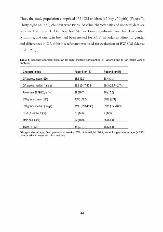
44
Thus, the study population comprised 137 ICSI children (67 boys, 70 girls) (Figure 7).
Thirty-eight (27.7 %) children were twins. Baseline characteristics of neonatal data are
presented in Table 1. One boy had Marcus Gunn syndrome, one had Goldenhar
syndrome, and one twin boy had been treated for ROP. In order to adjust for gender
and differences in GA at birth a reference was used for evaluation of BW SDS (Marsal
et al., 1996).
Table 1. Baseline characteristics for the ICSI children participating in Papers I and II (for retinal vessel analysis).
Characteristics Paper I (n=137) Paper II (n=57)
GA weeks, mean (SD) 38.6 (3.0) 38.4 (3.3)
GA weeks median (range) 39.6 (24.7-42.9) 39.3 (24.7-42.7)
Preterm (<37 GW)), n (%) 27 (19.7) 10 (17.5)
BW grams, mean (SD) 3096 (792) 3088 (874)
BW grams median (range) 3190 (650-4830) 3335 (650-4830)
SGA (≤ -22%), n (%) 20 (14.6) 7 (12.3)
Male sex, n (%) 67 (48.9) 35 (61.4)
Twins, n (%) 38 (27.7) 16 (28.1)
GA, gestational age; GW, gestational weeks; BW, birth weight; SGA, small for gestational age (≤ 22% compared with expected birth weight)

45
Paper II
At the examination of the 137 ICSI children fundus photographs through dilated
pupils were taken for digital image analysis. Only photographs of satisfactory quality
were analysed and 82 ICSI children (46 boys, 36 girls) fulfilled the criteria (Figure 7).
The remaining 55 children refused or cooperated poorly. Among the children with
fundus photographs 57 (35 boys, 22 girls) had photographs that allowed evaluation of
the retinal vessel morphology as well as of the optic nerve head. The photographs
must be focused with the optic disc centred (within half a disc off centre). Baseline
characteristics of neonatal available data in the children with images analysed for the
central retinal vessels, as well as for the optic nerve head, are presented in Table 1.
There were no differences in GA or BW between the ICSI children with fundus
photographs, and the ICSI children without fundus photographs.
n=160 ISCI children
n=137 Examined children
Paper I
n=82Digital image analysis
Paper II
n=55No digital image analysis
n=23 Parent refusals
n=160 ISCI children
n=137 Examined children
Paper I
n=82Digital image analysis
Paper II
n=55No digital image analysis
n=23 Parent refusals
Figure 7. Flow chart of the ICSI children participating in Papers I and II.
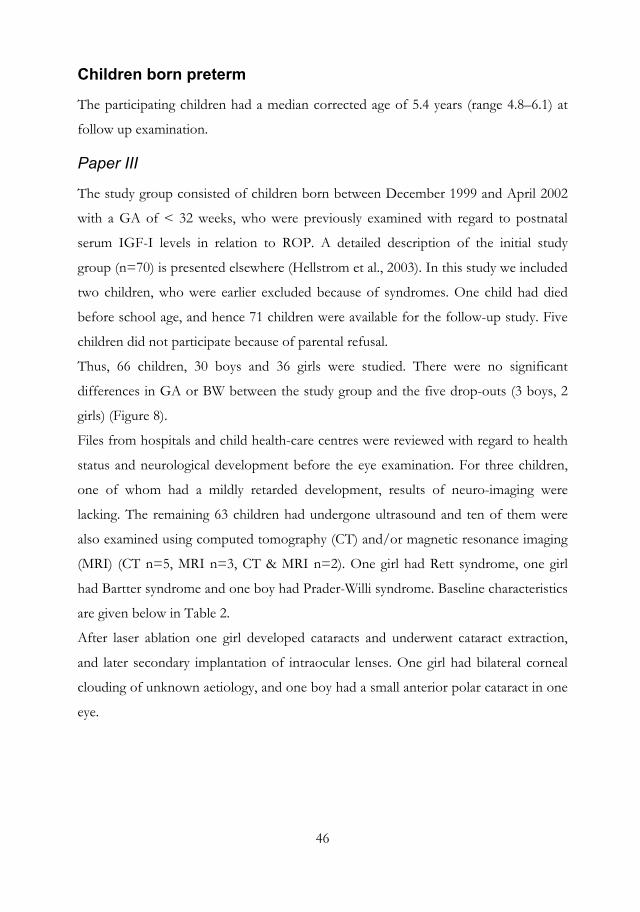
46
Children born preterm
The participating children had a median corrected age of 5.4 years (range 4.8–6.1) at
follow up examination.
Paper III
The study group consisted of children born between December 1999 and April 2002
with a GA of < 32 weeks, who were previously examined with regard to postnatal
serum IGF-I levels in relation to ROP. A detailed description of the initial study
group (n=70) is presented elsewhere (Hellstrom et al., 2003). In this study we included
two children, who were earlier excluded because of syndromes. One child had died
before school age, and hence 71 children were available for the follow-up study. Five
children did not participate because of parental refusal.
Thus, 66 children, 30 boys and 36 girls were studied. There were no significant
differences in GA or BW between the study group and the five drop-outs (3 boys, 2
girls) (Figure 8).
Files from hospitals and child health-care centres were reviewed with regard to health
status and neurological development before the eye examination. For three children,
one of whom had a mildly retarded development, results of neuro-imaging were
lacking. The remaining 63 children had undergone ultrasound and ten of them were
also examined using computed tomography (CT) and/or magnetic resonance imaging
(MRI) (CT n=5, MRI n=3, CT & MRI n=2). One girl had Rett syndrome, one girl
had Bartter syndrome and one boy had Prader-Willi syndrome. Baseline characteristics
are given below in Table 2.
After laser ablation one girl developed cataracts and underwent cataract extraction,
and later secondary implantation of intraocular lenses. One girl had bilateral corneal
clouding of unknown aetiology, and one boy had a small anterior polar cataract in one
eye.
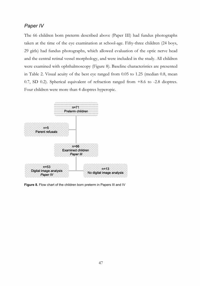
47
Paper IV
The 66 children born preterm described above (Paper III) had fundus photographs
taken at the time of the eye examination at school-age. Fifty-three children (24 boys,
29 girls) had fundus photographs, which allowed evaluation of the optic nerve head
and the central retinal vessel morphology, and were included in the study. All children
were examined with ophthalmoscopy (Figure 8). Baseline characteristics are presented
in Table 2. Visual acuity of the best eye ranged from 0.05 to 1.25 (median 0.8, mean
0.7, SD 0.2). Spherical equivalent of refraction ranged from +8.6 to -2.8 dioptres.
Four children were more than 4 dioptres hyperopic.
n=71 Preterm children
n=66 Examined children
Paper III
n=53Digital image analysis
Paper IV
n=13No digital image analysis
n=5 Parent refusals
n=71 Preterm children
n=66 Examined children
Paper III
n=53Digital image analysis
Paper IV
n=13No digital image analysis
n=5 Parent refusals
Figure 8. Flow chart of the children born preterm in Papers III and IV

48
Table 2. Baseline characteristics and morbidity in the preterm children participating in Papers III and IV.
Characteristics Paper III (n=66) Paper IV (n=53)
GA weeks, mean (SD) 27.5 (2.1) 27.6 (2.1)
GA median (range) 27.1 (23.4.4-31.1) 27.1 (23.4-31.1)
BW grams, mean (SD) 1025 (332) 1063 (342)
BW median (range) 948 (530-2015) 970 (530-2015)
SGA (≤ -2 SDS), n (%) 16 (24.2) 9 (17.0)
Male sex, n (%) 30 (45.5) 24 (45.2)
Singletons, n (%) 49 (74.2) 41 (77.4)
Other morbidity:
ROP stage <3, n (%) 30 (45.5) 25 (47.2)
ROP stage 3 total, n (%) 12 (18.2) 9 (17.0)
ROP treated, n (%) 7 (10.6) 4 (7.5)
BPD n (%) 13 (19.7) 10 (18.9)
NEC n (%) 4 (6.1) 2 (3.8)
Brain lesion, n (%) 10/63 (15.9) 6 (11.3)
Definition brain lesion: IVH ≥ grade III, Hydrocephalus, WMD/PVL, CP
GA, gestational age; BW, birth weight; SGA, small for gestational age; BPD, broncho-pulmonary dysplasia; NEC, necrotizing enterocolitis; IVH, intraventricular haemorrhage; WMD, white matter damage; PVL, periventricular leucomalacia; CP, cerebral palsy.

49
The children without fundus photographs
The 13 children (seven boys and six girls) who lacked fundus photographs of
sufficient quality had significantly lower serum IGF-I levels, and a lower weight week
32 (SDS) than the children included in the fundus evaluation (p=0.021 and p=0.0086,
respectively). There were no significant differences in GA and BWSDS between the
groups. Visual acuity of the best eye among the children without photographs was
significantly lower (median 0.4, range 0 to 0.9) than in the study group (median 0.8,
range 0.05 to 1.25), p<0.001. Their HCSDS (n=10) was significantly more negative
(median -0.6, range -3.1 to 0.3) in the comparison with HCSDS of the study group
(median 0.3, range -1.2 to 2.0), p<0.001. From ophthalmoscopy and evaluation of
photographs of poor quality only three children had a fundus considered without
clinical remarks. Of the remaining nine children three had large cupping, and/or
tortuous vessels (n=4), macula hypoplasia (n=3), or temporal dragging of retinal
vessels after laser ablation (n=1). One girl had drusen in the optic nerve diagnosed on
ultrasound. Four of the dropout children had brain lesions, three of them had WMD
with PVL, one boy was operated on for hydrocephalus with shunt, and two had
cerebral palsy. In addition, the group included the three children with syndomes, and
one girl with pseudohakia and one with corneal clouding.

50
Table 3. Baseline characteristics and morbidity in the children without photographs in Paper IV.
Characteristics Children without photographs Paper IV (n=13)
GA mean (SD) 27.1 (2.0)
GA median (range) 27.1 (24.3-30)
BW grams, mean (SD) 871 (227)
BW median (range) 790 (535-1335)
SGA (≤ -2SDS), n (%) 6 (46)
Male sex, n (%) 6 (46)
Singletons, n (%) 0 (0)
Other morbidity
ROP stage <3, n (%) 5 (38.5)
ROP stage 3 total, n (%) 3 (23.1)
ROP stage 3 treated, n (%) 3 (23.1)
BPD n (%) 3 (23.1)
NEC n (%) 2 (15.4)
Brain lesion, n (%) 4 (30.8)
Definition brain lesion: IVH ≥ grade III, Hydrocephalus, WMD/PVL, CP
GA, gestational age; BW, birth weight; SGA, small for gestational age; BPD broncho-pulmonary dysplasia; NEC, necrotizing enterocolitis; IVH, intraventricular haemorrhage; WMD, white matter damage; PVL, periventricular leucomalacia; CP, cerebral palsy.

51
Control groups
Paper I
The control children born after spontaneous conception were matched for gender, age
(+/3 months) and mother’s age (+/- 3 years). They were recruited from the Swedish
Medical Birth Registry. The exclusion criteria were inability to speak Swedish, and
residency outside the western region of Sweden at the time of the examination from
1999 to 2002. A total of 208 controls were invited to participate in the study. Among
the control children the refusals were 49 (23.6%) and the participation rate was 76.4
%.
Thus, the control group comprised 159 children (77 boys, 82 girls) born after
spontaneous conception (Figure 9). There were two twins in the control-group (1.3
%). Six out of 159 controls (3.8 %) had been born preterm (<37 GW), none before 32
GW. Eleven out of 159 controls (6.9 %) had been born SGA.
The matching variables did not differ statistically between the ICSI and control
groups, and their distribution was very similar. Therefore no adjustments for these
variables were needed in the analysis.
In the following this control group, used for comparison with the ICSI children in
Paper I, is referred to as the matched control group.
Papers II and IV
Among the matched control children, for the ICSI children in Paper I, 104 children
(41 boys, 63 girls) had photographs of their fundus taken. In addition, a group of 99
subjects (53 boys, 46 girls) born at term after spontaneous conception, with BW
appropriate for GA and no history of congenital, chronic or other serious disorder
were included to increase the control group. This group was described in a previous
study (Hellstrom and Svensson, 1998).
Thus, the control group comprised 203 children (94 boys, 109 girls) analysed for optic
nerve variables, and 181 of the controls (83 boys, 98 girls) had photographs of
satisfactory quality for retinal vessel analysis (Figure 9). In the following this group is
referred to as the control group.

52
n=208 Preterm children
n=159 Matched control group
Paper I
n=104Digital image analysis
Paper IV
n=55No digital image analysis
n=49 Parent refusals
n=208 Preterm children
n=159 Matched control group
Paper I
n=104Digital image analysis
Paper IV
n=55No digital image analysis
n=49 Parent refusals
n=99Digital image analysis
Older reference material
n=203 Matched control group
Paper II, IV
n=99Digital image analysis
Older reference material
n=203 Matched control group
Paper II, IV
Figure 9. Flow chart over matched control group (Paper I) and control group (Papers II and IV)
53
Methods
Ophthalmic evaluation
In the eye clinic, monocular visual acuity (VA) was tested at a distance of 3 metres
with the Lea Symbols Chart (Hered et al., 1997) in the ICSI children and the matched
control group (Papers I and II). The Lea Symbols Chart is linear with five optotypes
and in order to pass a line, four out of five optotypes have to be correctly recognized.
One ICSI child was unable to perform a visual acuity test, and one ICSI child only
allowed a binocular test. The child can name the symbols and do not need to hold a
chart in their hands and point. When the preterm children (Papers III and IV) were
examined we used the KM visual acuity chart, which by then had been evaluated and
considered to be more appropriate for children in the age group 5-7 years (Moutakis
et al., 2004). For seven children it was not possible to use the KM chart. Three
children born preterm were tested with the HVOT chart, and one with the Lea
Symbols Chart. Three children with a VA below 0.1 (logMAR 1.0), could only
perform binocular tests with less demanding charts (Cardiff cards n=2 and Kays
pictures n=1) and their binocular vision was valued as VA of the best eye in the
analysis. One boy born preterm had only light perception and was considered as blind
in accordance with the WHO definition (WHO, 2007). Four ICSI children, six
matched control children and sixteen children born preterm were tested with their
prescribed glasses. The orthoptic examination included cover testing at near and
distant fixation, stereoacuity with the Lang and/or TNO test, and evaluation of ocular
motility and convergence. The anterior segment of the eye was inspected using a slit-
lamp. The eyes were examined in cycloplegia, 45 minutes after administration of a
single drop (cyclopentholate 0.85 % and phenylephrine 1.5 %), with autorefraction,
and/or retinoscopy and ophthalmoscopy. Fundus photographs were taken for later
analysis (Papers II and IV). If the child/parents refused installation of cycloplegic
drops the refraction was not measured and the fundi were examined through
undilated pupils. This was the case in four ICSI children and three matched control
children. Refraction was not performed in the pseudophakic preterm girl, who was
operated on for bilateral cataract.

54
In Paper I significant refractive errors were defined as a spherical equivalent (SE) of
myopia >0.5 diopters (D), hyperopia >3.5 D, astigmatism >1.0 D and anisometropia
>1.0 D (De Becker et al., 1992). In Paper III we used another reference (Robaei et al.,
2005) for significant refractive error which had been used in another follow up study
of preterm children at the eye clinic (Hard et al., 2000). The only difference between
the two references for clinical significant refractive error was for hyperopia which in
Paper III was considered to be significant if SE was ≥3 D.
Visual perception
Visual perception (Paper III) was examined with the TVPS-R (Test of Visual
Perceptual Skills-Revised) (Gardner, 1996). This is a non-motor test for children aged
between 4 and 12 years and 11 months. It consists of seven subtests (1)
discrimination: identification of forms; (2) memory: ability to remember single forms
individually; (3) spatial relations: determination of correct direction of forms; (4) form
constancy: recognition of the same form when it varies in size from the stimulus; (5)
sequential memory: ability to remember a number of forms in a series; (6) figure
ground: finding a form when it is hidden among other forms; (7) closure:
determination of a whole form from parts of this form. Each subtest consists of 16
items and the number of correct responses yields a ‘raw score’ from 0 (all failed) to 16
(all correct). Based on this raw score, scaled scores are given for each subtest and age
(at 3-months intervals). The scaled scores are then added to give a scaled score for the
whole test which was used for analysis. A percentile rank (PR) based on the scaled
scores of an American reference group of children is also provided.

55
Digital image analysis of fundus photographs
In Papers II and IV the ocular fundus photographs were taken through dilated pupils
and analysed quantitatively, using a specially designed computer-assisted digital
mapping system (Stromland et al., 1995).
The mean value of the two eye measurements was calculated for each studied fundus
structure. If the fundus photograph of only one eye was of sufficient quality, that eye’s
photograph was used.
Digital mapping
The optic disc area was measured by marking the outlines with a cursor. The inner
border surrounding the nerve tissue defined the optic disc; care was taken not to
include the white peripapillary scleral ring. The cup was defined by contour, and its
definition was facilitated by the course of the vessels and its pallor. The neuronal rim
area was obtained by subtraction of the cup area from the disc area. The indices of
tortuosity for arteries and veins were defined as the path length of the vessel divided
by the linear distance from the vessel origin to a reference circle 3 mm from the centre
of the optic disc. The same person, who was masked to the mode of conception in
Paper II, performed all measurements, which eliminates inter-individual differences in
the measurements. As the optic disc size may vary systematically with the method of
measurement it is strength that all photographs in these studies are measured by the
same person using the same digital analysing program.
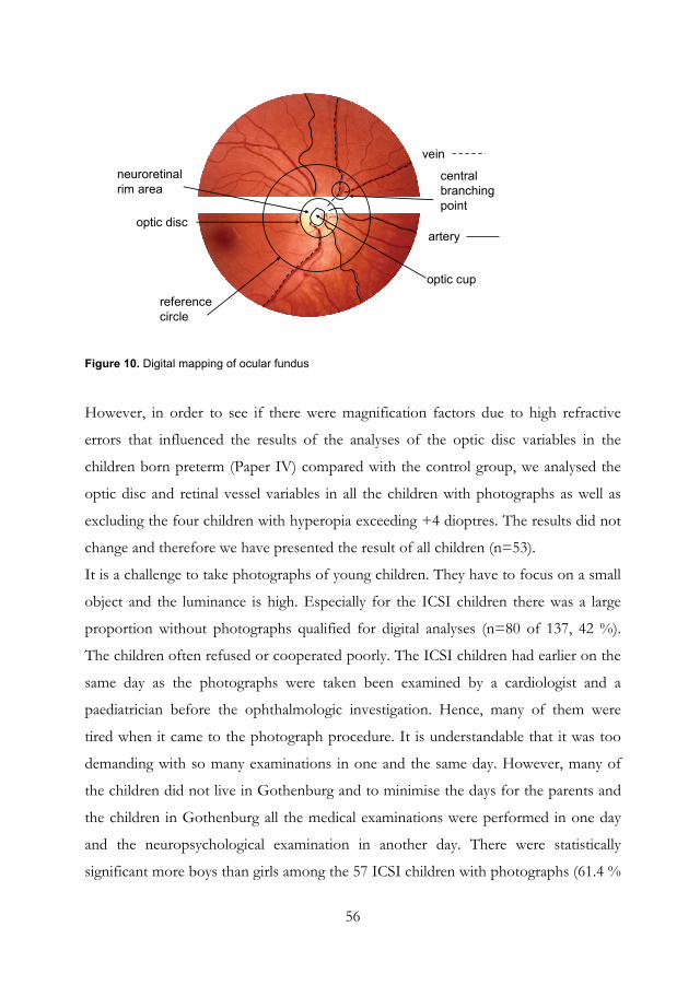
56
artery
vein
central branchingpoint
optic disc
neuroretinal rim area
optic cup
referencecircle
Figure 10. Digital mapping of ocular fundus
However, in order to see if there were magnification factors due to high refractive
errors that influenced the results of the analyses of the optic disc variables in the
children born preterm (Paper IV) compared with the control group, we analysed the
optic disc and retinal vessel variables in all the children with photographs as well as
excluding the four children with hyperopia exceeding +4 dioptres. The results did not
change and therefore we have presented the result of all children (n=53).
It is a challenge to take photographs of young children. They have to focus on a small
object and the luminance is high. Especially for the ICSI children there was a large
proportion without photographs qualified for digital analyses (n=80 of 137, 42 %).
The children often refused or cooperated poorly. The ICSI children had earlier on the
same day as the photographs were taken been examined by a cardiologist and a
paediatrician before the ophthalmologic investigation. Hence, many of them were
tired when it came to the photograph procedure. It is understandable that it was too
demanding with so many examinations in one and the same day. However, many of
the children did not live in Gothenburg and to minimise the days for the parents and
the children in Gothenburg all the medical examinations were performed in one day
and the neuropsychological examination in another day. There were statistically
significant more boys than girls among the 57 ICSI children with photographs (61.4 %
57
boys and 38.6 % girls) compared with the ICSI children without fundus photographs
(non-participants n=80, 40 % boys and 60 % girls) (p = 0.021). In addition, the
percentage of boys with photographs of quality to be analysed for retinal vessels was
higher in the ICSI group (61 %) than in the control group (46 %) that may be a bias
negative for the ICSI boys in our study. Among the preterm children we did not
receive fundus photographs from nine children, and the photographs were of a poor
quality in four children. Unfortunately, the sickest children born preterm were unable
to cooperate in the photography procedure and we investigated their fundi by indirect
ophthalmoscopy according to the study protocol. In addition, the sickest children
have regularly been checked at the eye clinic, and we scrutinized the files for
information of earlier or later fundus inspections. The photographs of the children
born preterm were taken with a different angle than those of the children born after
ICSI and the controls, which made the placement of the reference circle uncertain,
and thus, a comparison of number of branching points with the controls impossible.
While the area of the small structure of the optic nerve head could be transformed
using a magnification factor, the vessels subtend a larger and curved area making such
a transformation inappropriate, because of substantial risk for magnification errors. In
Paper IV we therefore chose to present only the optic nerve variables, but in the thesis
findings of the tortuosity index and central retinal branching points in relation to
growth variables will be presented in the results below.
Measurements of peri- and postnatal growth variables including IGF-I levels
In Papers III and IV we set out to evaluate the association between GA, BW SDS,
early (including IGF-1) and late postnatal growth variables and ophthalmologic
outcome at school age in children born before 32 GW.
Birth weight and weight at PMA 32 weeks were measured, and standard deviation
scores (BWSDS) and weight at 32 weeks (PMA) (SDS), were calculated for all children
(Marsal et al., 1996). At the examination at school age head circumference (Fredriks et
58
al., 2000), weight and length (Wikland et al., 2002) were measured according to clinical
praxis, and were expressed as standard deviation scores (SDS).
In the neonatal period venous blood samples (0.5 mL) had been taken weekly, and
analysed using a procedure described in the original paper (Hellstrom et al., 2003). The
mean values of serum IGF-I during postmenstrual age (PMA) 30 to 33 weeks (30.0 –
33.9) were used for analyses. We have chosen to analyse mean serum IGF-I levels
during PMA 30-33 weeks because earlier studies have shown that during this time
period the discrepancy in IGF-I levels is at its peak between children with and without
morbidity (Hellstrom et al., 2003). However, we do not know whether this is the most
optimal time period to measure serum IGF-I levels, and further analyses will be
performed.
Statistics
Descriptive statistics are given as mean, standard deviation, median and range. Fisher’s
exact test was used for comparison of proportions between groups, and Fisher’s non-
parametric permutation test was used for continuous and ordered variables (Good,
2000a).
Mann-Whitney U test was performed to test differences between groups for
continuous and ordered variables (Papers II and III). Spearman’s rank correlation
coefficient rs was used for correlation analysis in Paper II. In Paper IV correlation was
estimated with Pearson correlation coefficient and analysed with Pitman’s non-
parametric permutation test (Good, 2000b). All tests were two sided and conducted at
5 % significance level.
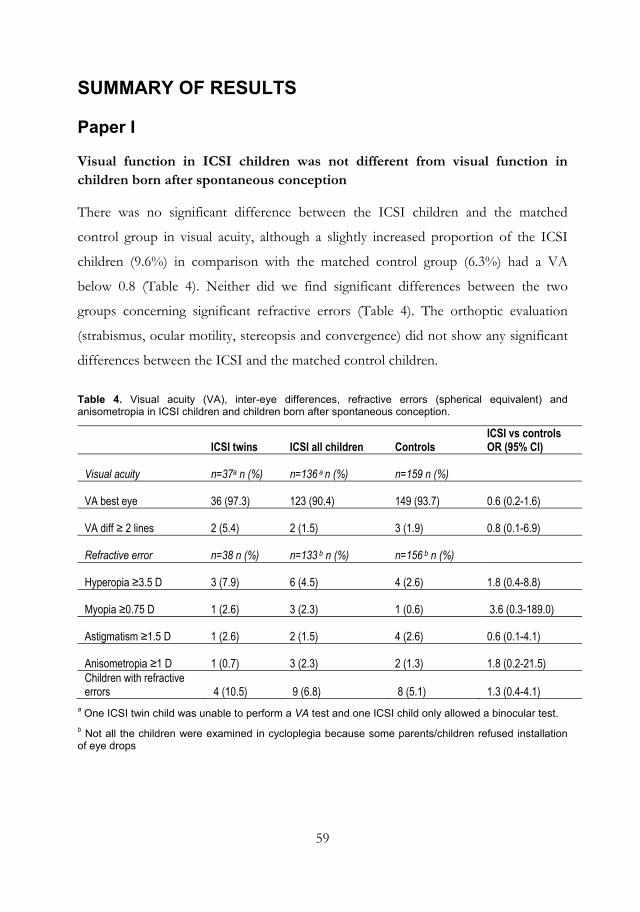
59
SUMMARY OF RESULTS
Paper I
Visual function in ICSI children was not different from visual function in children born after spontaneous conception
There was no significant difference between the ICSI children and the matched
control group in visual acuity, although a slightly increased proportion of the ICSI
children (9.6%) in comparison with the matched control group (6.3%) had a VA
below 0.8 (Table 4). Neither did we find significant differences between the two
groups concerning significant refractive errors (Table 4). The orthoptic evaluation
(strabismus, ocular motility, stereopsis and convergence) did not show any significant
differences between the ICSI and the matched control children.
Table 4. Visual acuity (VA), inter-eye differences, refractive errors (spherical equivalent) and anisometropia in ICSI children and children born after spontaneous conception.
ICSI twins ICSI all children Controls ICSI vs controls OR (95% CI)
Visual acuity n=37a n (%) n=136 a n (%) n=159 n (%)
VA best eye 36 (97.3) 123 (90.4) 149 (93.7) 0.6 (0.2-1.6)
VA diff ≥ 2 lines 2 (5.4) 2 (1.5) 3 (1.9) 0.8 (0.1-6.9)
Refractive error n=38 n (%) n=133 b n (%) n=156 b n (%)
Hyperopia ≥3.5 D 3 (7.9) 6 (4.5) 4 (2.6) 1.8 (0.4-8.8)
Myopia ≥0.75 D 1 (2.6) 3 (2.3) 1 (0.6) 3.6 (0.3-189.0)
Astigmatism ≥1.5 D 1 (2.6) 2 (1.5) 4 (2.6) 0.6 (0.1-4.1)
Anisometropia ≥1 D 1 (0.7) 3 (2.3) 2 (1.3) 1.8 (0.2-21.5) Children with refractive errors 4 (10.5) 9 (6.8) 8 (5.1) 1.3 (0.4-4.1)
a One ICSI twin child was unable to perform a VA test and one ICSI child only allowed a binocular test. b Not all the children were examined in cycloplegia because some parents/children refused installation of eye drops
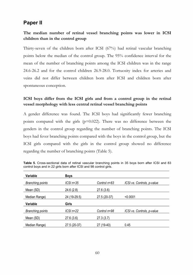
60
Paper II
The median number of retinal vessel branching points was lower in ICSI children than in the control group
Thirty-seven of the children born after ICSI (67%) had retinal vascular branching
points below the median of the control group. The 95% confidence interval for the
mean of the number of branching points among the ICSI children was in the range
24.6-26.2 and for the control children 26.9-28.0. Tortuosity index for arteries and
veins did not differ between children born after ICSI and children born after
spontaneous conception.
ICSI boys differ from the ICSI girls and from a control group in the retinal vessel morphology with less central retinal vessel branching points
A gender difference was found. The ICSI boys had significantly fewer branching
points compared with the girls (p=0.022). There was no difference between the
genders in the control group regarding the number of branching points. The ICSI
boys had fever branching points compared with the boys in the control group, but the
ICSI girls compared with the girls in the control group showed no difference
regarding the number of branching points (Table 5).
Table 5. Cross-sectional data of retinal vascular branching points in 35 boys born after ICSI and 83 control boys and in 22 girls born after ICSI and 98 control girls.
Variable Boys
Branching points ICSI n=35 Control n=83 ICSI vs. Controls, p-value
Mean (SD) 24.6 (2.8) 27.6 (3.6)
Median Range) 24 (19-29.5) 27.5 (20-37) <0.0001
Variable Girls
Branching points ICSI n=22 Control n=98 ICSI vs. Controls, p-value
Mean (SD) 27.6 (3.6) 27.3 (3.7)
Median Range) 27.5 (20-37) 27 (19-40) 0.45

61
There was a correlation between the number of branching points and tortuosity index
for arteries (rs=-0.27, p=0.043) and veins (rs=-0.26, p=0.049). The lower the number
of branching points, the higher the tortuosity index for arteries and veins.
We also found a relation between BW deviation and number of branching points
within the ICSI children. With a low BW deviation there was a tendency to have a
reduced number of branching points (p=0.058).
No difference in optic disc morphology between the ICSI children and the children born after spontaneous conception
Table 6. Cross-sectional data of optic disc variables and index of tortuosity for arteries and veins in ICSI children and in a control group.
Variable ICSI children n=82 Controls n=203 ICSI vs. Controls p-value Optic disc area, mm2 Mean (SD) Median (Range)
2.4 (0.4) 2.4 (1.5-3.7)
2.4 (0.5) 2.3 (1.6-5.3)
0.34
Cup area, mm2 Mean (SD) Median (Range)
0.4 (0.3) 0.3 (0-1.8)
0.3 (0.2) 0.3 (0-1.6)
0.34
Rim area, mm2 Mean (SD) Median (Range)
2.08 (0.3) 2.1 (1.3-2.8)
2.07 (0.4) 2.1(1.3-3.9)
0.36
Variable ICSI children n=57 Controls n=181 ICSI vs. Controls p-value ITA Mean (SD) Median (Range)
1.09 (0.04) 1.08 (1.03-1.2)
1.09 (0.03) 1.08 (1.04-1.2)
0.49
ITV Mean (SD) Median (Range)
1.06 (0.02) 1.06 (1.03-1.1)
1.06 (0.02) 1.06 (1.02-1.2)
0.98
ITA, index of tortuosity for arteries; ITV, index of tortuosity for veins
62
Paper III
Children born very preterm had affected visual capacity
Eleven of 66 (17 %) children had visual impairment (VA <0.3), one of whom was
blind (VA <0.05) and 28 children (42 %) had mildly reduced vision. Good VA was
found in 27 (41 %) children.
In all infants with ROP stage 3 (n=12), the retinopathy regressed either spontaneously
or after treatment, thus in no case was visual impairment due to retinal detachment.
Among the visually impaired children there was one girl with Rett syndrome, one
pseudophakic and one with cloudy corneas. Five of 11 visually impaired children had
been diagnosed with brain lesions. The other children with brain lesions had mildly
reduced or good vision.
A low visual acuity was associated with low gestational age, poor early and late growth
Low VA of the best eye was correlated with low GA, and low weight week 32 (SDS),
but not to BW (SDS). Visual acuity was also correlated with low weight, length and
head circumference (SDS) at follow-up (Table 7).
Refractive errors were associated with low gestational age, negative birth weight deviation and poor early as well as late growth
Significant hyperopia was associated with low neonatal serum levels of IGF-I, and to a
small head circumference (SDS) at follow-up. Significant myopia (only four children
were myopic) was related to low GA. Four of the hyperopic children were diagnosed
with brain lesions, and two of the myopic children were treated for ROP.
Significant astigmatism was associated with both low BW (SDS) and low weight at
week 32 PMA (SDS) (Table 7).
Anisometropia was found in 10/65 (15 %) of the children and nine (14 %) of the
children had strabismus. Two children with strabismus had brain lesions and one had
Rett syndrome. Anisometropia and strabismus showed no correlation to any of the
measured growth variables.

63
Poor visual perception was correlated with poor early postnatal growth and a small head circumference at school age
Of the 58 children, who were capable to perform the TVPS-R test, a majority 33/58
(57%) had results below the third percentile of the American reference group and 30
children had a percentile rank of 1. Six children with brain lesions performed the test
and three of them had a percentile rank of 1. Poor results were correlated with low
weight at week 32 (SDS) and small head circumference (SDS) at follow-up (Table 7).
Table 7. Results of statistical analysis regarding visual outcome and postnatal growth variables - early and at follow-up.
Early growth variables Visual acuity Hyperopiaa Myopiaab Astigmatisma TVPS-R SS
IGF-I mean n.s p=0.0096 n.s n.s n.s
Gestational age rs= 0.29* n.s p=0.027 n.s n.s
Birth weight SDS n.s n.s n.s p=0.047 n.s
Weight week 32 SDS rs =0.30* n.s n.s p=0.0041 rs =0.38**
Growth variables at follow-up
HC SDS at 5 years rs =0.55*** p=0.022 n.s n.s rs =0.39**
Weight SDS at 5 years rs =0.37** n.s n.s n.s n.s
Length SDS at 5 years rs =0.41*** n.s n.s n.s n.s a Clinically significant as defined in the Methods section, b n=4
TVPS-R SS, Test of Visual Perceptual Skills-Revised, sum of scaled scores; n.s., non significant, * p<0.05, ** p<0.01, *** p<0.001, Mann-Whitney U-test and Spearman’s correlation coefficient rs were used
Small head circumference at school age was correlated with low GA, poor early growth and low serum IGF-I levels
Head circumference (SDS) at school age was significantly correlated to GA (rs=0.40,
p=0.0012), neonatal serum levels of IGF-I (rs=0.37, p=0.0031) and to weight at week
32 (SDS) (rs=0.27, p=0.035). There was no correlation between early growth variables
and length and weight at follow-up.
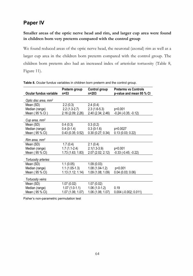
64
Paper IV
Smaller areas of the optic nerve head and rim, and larger cup area were found in children born very preterm compared with the control group
We found reduced areas of the optic nerve head, the neuronal (axonal) rim as well as a
larger cup area in the children born preterm compared with the control group. The
children born preterm also had an increased index of arteriolar tortuosity (Table 8,
Figure 11).
Table 8. Ocular fundus variables in children born preterm and the control group.
Ocular fundus variable Preterm group n=53
Control group n=203
Preterms vs Controls p-value and mean 95 % CI
Optic disc area, mm2 Mean (SD) Median (range) Mean ( 95 % CI )
2.2 (0.3) 2.2 (1.3-2.7) 2.16 (2.09; 2.26)
2.4 (0.4) 2.3 (1.6-5.3) 2.40 (2.34; 2.46)
p<0.001 -0.24 (-0.35; -0.12)
Cup area, mm2 Mean (SD) Median (range) Mean ( 95 % CI)
0.4 (0.3) 0.4 (0-1.4) 0.43 (0.35; 0.52)
0.3 (0.2) 0.3 (0-1.6) 0.30 (0.27; 0.34)
p=0.0027 0.13 (0.03; 0.22)
Rim area, mm2 Mean (SD) Median (range) Mean ( 95 % CI)
1.7 (0.4) 1.7 (1.1-2.4) 1.73 (1.63; 1.83)
2.1 (0.4) 2.1(1.3-3.9) 2.07 (2.02; 2.12)
p<0.001 -0.33 (-0.45; -0.22)
Tortuosity arteries Mean (SD) Median (range) Mean ( 95 % CI)
1.1 (0.05) 1.1 (1.05-1.3) 1.13 (1.12; 1.14)
1.09 (0.03) 1.08 (1.04-1.2) 1.09 (1.08; 1.09)
p<0.001 0.04 (0.03; 0.06)
Tortuosity veins Mean (SD) Median (range) Mean ( 95 % CI)
1.07 (0.02) 1.07 (1.0-1.1) 1.07 (1.06; 1.07)
1.07 (0.02) 1.06 (1.0-1.2) 1.06 (1.06; 1.07)
0.19 0.004 (-0.002; 0.011)
Fisher’s non-parametric permutation test

65
Low birth weight and poor early growth were related to large cupping of the optic nerve head and a small neuronal rim area
A low BW (SDS) and weight at 32 weeks PMA (SDS) were associated with a reduced
neuronal rim area and a large cupping (Table 9). There was no correlation between
either, sex, GA, neonatal serum IGF-I levels, length, weight, or head circumference at
follow-up and the ocular fundus structures at school age. Neither was there any
relationship between degree of arteriolar tortuosity, or number of retinal branching
points and any of the studied growth variables. At school age there was a tendency of
an association between visual acuity of the best eye and optic disc area (p=0.054).
Table 9. Correlations between optic nerve head variables and perinatal growth variables
Perinatal growth variables Optic disc area Cup area Rim area
IGF-I mean n.s n.s n.s
Gestational age n.s n.s n.s
Birth weight SDS n.s r=-0.37** r=0.01*
Weight week 32 SDS n.s r=-0.38** r=0.02*
Test of correlation with Pitmans’s non-parametric permutation test (r)
* p<0.05, ** p<0.01
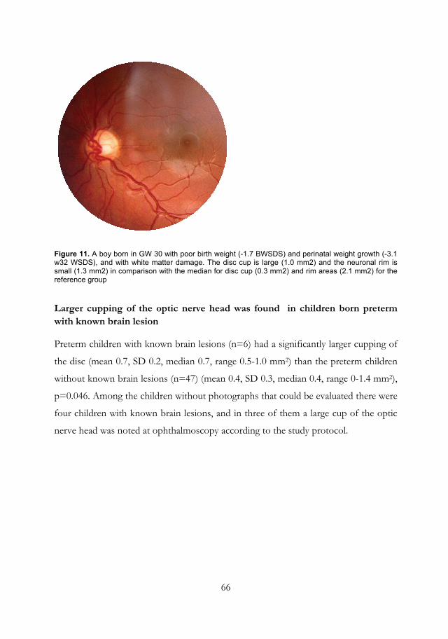
66
Figure 11. A boy born in GW 30 with poor birth weight (-1.7 BWSDS) and perinatal weight growth (-3.1 w32 WSDS), and with white matter damage. The disc cup is large (1.0 mm2) and the neuronal rim is small (1.3 mm2) in comparison with the median for disc cup (0.3 mm2) and rim areas (2.1 mm2) for the reference group
Larger cupping of the optic nerve head was found in children born preterm with known brain lesion
Preterm children with known brain lesions (n=6) had a significantly larger cupping of
the disc (mean 0.7, SD 0.2, median 0.7, range 0.5-1.0 mm2) than the preterm children
without known brain lesions (n=47) (mean 0.4, SD 0.3, median 0.4, range 0-1.4 mm2),
p=0.046. Among the children without photographs that could be evaluated there were
four children with known brain lesions, and in three of them a large cup of the optic
nerve head was noted at ophthalmoscopy according to the study protocol.

67
GENERAL DISCUSSION
Modern assisted reproduction techniques have given many infertile couples an
opportunity to become parents, and approximately one million children are born after
IVF/ICSI worldwide. The proportion of children born after assisted reproduction
now exceeds 5 % in the Scandinavian countries, and approximately half of them are
born after ICSI (Nyboe Andersen et al., 2008). Especially ICSI, which is an invasive
method, has raised worries about the outcome of the children. Infants born after ICSI
have an increased risk of being born prematurely, both singletons and those born after
multiple births, and account for a not negligible proportion of the increasing number
of babies born preterm.
Advanced neonatal care has increased the survival rate of the tiniest children born
prematurely, and there is also concern about the outcome of these small immature
babies. As many as up to 75 % of infant deaths can be attributed to preterm birth, as
can 42-47 % of children with cerebral palsy, and 23 to 37 % of the preterm population
has significant cognitive, visual and hearing impairments in USA (Behrman and Stith
Butler, 2007).
This thesis aims to evaluate effects of prenatal factors in children born after ICSI and
peri- and postnatal factors in children born very preterm on visual function and ocular
fundus morphology at school age. In the children born very preterm the
ophthalmologic outcomes, including optic nerve morphology, were analysed in
relation to GA, BW (SDS), serum levels of IGF-I, weight at week 32 (SDS), and
weight, length and head circumference (SDS) at school age.
In the first two Papers children born after ICSI were studied regarding visual function
and ocular morphology including the ocular fundus.
Regarding visual acuity in the children born after ICSI we found no difference
compared with spontaneously conceived children, and similar results have been
reported in other studies (Belva et al., 2007; Bonduelle et al., 2005; Knoester et al.,
2008). We found no significant difference in the rate of refractive error between the
ICSI children born at term, and the children born after spontaneous conception at
term, which also is in accordance with other studies of refractive status in children
68
born at term after ICSI (Knoester et al., 2008). There were 38 children born after
multiple births, and 27 of 137 children were born preterm (three born before GW 32),
and it is known that children born premature have more refractive errors than
children born at term (Larsson et al., 2003; O'Connor and Fielder, 2008). However,
the preterm children born after ICSI in our study did not differ regarding refractive
errors from the control group.
In our study one twin boy had Goldenhar syndrome, and one had Marcus Gunn
syndrome. In addition to our findings ocular anomalies have also been reported in
other studies (Anteby et al., 2001; Bonduelle et al., 2005; Knoester et al., 2008). In the
study by Anteby and co-workers optic nerve head hypoplasia and atrophy, coloboma
with microphthalmus, Coats disease, congenital glaucoma, congenital cataract and
retinoblastoma were found among 47 children referred to an eye clinic born after
IVF/ICSI. However, in the two later studies the anomalies were not specified
(Bonduelle et al., 2005; Knoester et al., 2008). Goldenhar syndrome has previously
been documented with an increased incidence among children, born after IVF/ICSI
compared with a group of naturally conceived children, and the authors speculate that
over-ripeness of the egg may affect epigenetic processes in the early zygote
(Wieczorek et al., 2007). It has been found that epigenetic dysregulation occurs in
some patients with Goldenhar syndrome (Fischer et al., 2006).
An increased risk for cerebral palsy has been reported in children born after IVF/ICSI
(Hvidtjorn et al., 2009; Lidegaard et al., 2005; Stromberg et al., 2002). In the present
study no participating child, but two dropouts born after ICSI had a diagnosis of
cerebral palsy. This may erroneously have led to too favourable results of the ICSI
children, since CP is associated with reduced vision, strabismus and refractive errors
(Kozeis et al., 2007).
The participation rate in our study, 85.6 % in ICSI children and 76.4% in naturally
conceived children, was not optimal, but comparable with several other studies
(Cederblad et al., 1996; Leslie et al., 2003; Morin et al., 1989). There was no difference
regarding variables such as gender, GA and BW between participants and non-
participants, making the study population reliable concerning these variables.
69
In Paper II we examined the ocular fundus in children born after ICSI and the results
were compared with those of a control group. This is, to our knowledge, the first
morphometric study of the ocular fundus in children born after ICSI. We found that
the boys, but not the girls, had less central retinal vessel density as demonstrated by
fewer retinal vessel branching points. Since the retina is part of the central nervous
system, and the vascularisation is adapted to the needs of the neural elements, one
might speculate that the reduced number of vessel branching points reflects
abnormalities in the neural retina as well as in the brain. Large registry-based studies
with long follow-up period show evidence of an increase in neurological problems
after IVF (Hvidtjorn et al., 2009; Lidegaard et al., 2005; Stromberg et al., 2002).
The reason for our finding we do not know. Unfortunately, we lack data of serum
IGF-I levels in our study group, but one may speculate that lower serum levels of
IGF-I in the boys, compared to the girls, may have contributed to the reduced
number of vascular branching points. IGF-I is important for normal retinal
vascularisation (Hellstrom et al., 2002), and persistent low serum levels of IGF-I after
premature birth are associated with later development of ROP (Hellstrom et al., 2003).
It has been found that children born preterm with low GA, and/or low BW have
abnormal retinal vascularisation with fewer central retinal branching points (Hellstrom
et al., 2000). Several studies on children born SGA have shown that these children
have lower concentrations of IGF-I both in intrauterine life and post-partum than do
children born AGA (Arosio et al., 1995; Ostlund et al., 1997). Even though seven
boys of 35 (20 %) born after ICSI were prematurely born, and five of 35 (14 %) were
born SGA, there was still a significant difference in number of vascular branching
points when the premature and SGA born boys were excluded. Interestingly, lower
serum levels of IGF-I in boys born after ICSI at three months of age in comparison
with boys born after spontaneous conception have been reported. The difference in
IGF-I levels had disappeared at the age of five years (Kai et al., 2006). In preterm
infants male gender has been associated with lower levels of IGF-I at birth and in the
neonatal period compared to girls (Engstrom et al., 2005). The reason for this gender
difference is not clear, but it has been suggested that differences in sex steroids could
influence the secretion of IGF-I in utero (Barrios et al., 1996). In addition, one study
70
in full term Caucasian babies has shown that the mean cord plasma concentrations of
IGF-I in males were significantly lower than in the female babies suggesting “a sexual
dimorphism in the GH-IGF axis” (Geary et al., 2003).
Remarkably, other studies have shown increased growth after in vitro fertilisation.
One study on IVF/ICSI at the age of 7 years, in which twins and children born
prematurely were excluded, found higher levels of IGF-I and IGF-II, and the children
born after IVF/ICSI were taller compared with a control group (Miles et al., 2007).
The authors speculated that the in vitro fertilisation process leads to epigenetic
alterations of imprinted genes expressed either from the allele from the mother or the
father. The authors also suggested that the children born after IVF/ICSI, with higher
serum IGF-I and II, may have mild phenotypic features of Beckwith-Wiedemann
syndrome with a normal BW and a taller stature in childhood (Miles 2007). Such
features have been found in relatives of children with Beckwith-Wiedemann syndrome
(Elliott and Maher, 1994). The overgrowth syndrome Beckwith-Wiedemann has been
reported to occur up to nine times more commonly after in vitro conception than in
the general population (DeBaun et al., 2003; Halliday et al., 2004; Maher, 2005). There
are over 50 known imprinted genes in the human, and many of these are involved in
cellular proliferation and growth. The IGF-II gene is imprinted and expressed only
from the paternal gene in the placenta and foetus (DeChiara et al., 1991). In animal
studies it has been shown that IGF-II plays a role for placental growth. Epigenetic
modification of imprinted genes (via changes in DNA methylation) may provide a
mechanism linking environmental cues to placental phenotype, with consequences for
development both before and after birth. Therefore, changes in expression of
imprinted genes have major implications for developmental programming (Fowden et
al., 2006).
One might speculate that the fertilisation procedure may have influenced imprinting
of genes in the children born after IVF/ICSI, and led to the alterations of retinal
vascular development found in the boys in this study. One case-control study of
children born after IVF/ICSI found an increased placenta/BW ratio in children born
after assisted reproduction (Koudstaal et al., 2000). Infants born small with a large
placenta has been associated with increased risk of hypertension and cardiovascular
71
disease in adulthood (Barker et al., 1990). One follow-up study of ICSI children at the
age of eight years (the eldest children born after ICSI being examined) found higher
diastolic and systolic blood pressure compared with a control group (Belva et al.,
2007). In the international multi-centre study, in which the ICSI children in our study
participated, blood pressure was similar in the children born after ICSI and in the
spontaneously conceived children (Bonduelle et al., 2004).
Another possible mechanism behind our finding of fewer retinal branching points
among the ICSI children could be maternal factors such as diabetes mellitus, and an
increased use of heparin-like substances and thrombocyte aggregation inhibitors
indicating a coagulopathy disorder in the mother (Kallen et al., 2005b). However, this
is not likely in our study as only boys were affected.
The retinal vasculature is adapted to meet the needs of the neural retina, and one
might anticipate a correlation between few vascular branching points, and reduced
neuronal rim area of the optic disc. However, we did not find a relation between
number of branching points and the neuronal rim area. Neither did we find any
gender difference in neuronal rim area. We have only measured the branching pattern
of the largest vessels in the most central part of the retina, and do not know whether a
reduced number of branching points may be compensated by increased branching in
the periphery, or by an increased density of the capillary bed. The development of the
central retinal vessels takes place between 14 and 21 GW and we do not know what
factors are involved in branching. Thus, neither the cause nor the clinical significance
can be clarified from the present study.
When we eliminated factors earlier shown to be associated with a low number of
central vascular branching points, i.e. low BW and preterm birth, the difference in
vascular pattern still remained. This suggests other mechanisms (e.g. infertility,
genetics of infertility, and/or the reproduction technology) responsible for the
somewhat reduced number of vascular retinal branching points demonstrated in boys
born after ICSI in the present study.
This is a limited group of children born after ICSI, and we cannot exclude a selection
bias due to the low number of children with photographs (42 %), and the higher
proportion of boys among the children with photographs, although there were no

72
differences in BW, or GA among the children with photographs and those without
photographs. Larger numbers of patients would be welcome, but adequate statistics
were obtained with this limited group.
The main limitation with Papers I and II is that our study groups are small, and when
large enough studies become available the results concerning visual defects might be
different. When studying relatively rare events, small studies can never demonstrate
absence or presence of a difference. To detect a genuine association between assisted
reproductive techniques and imprinting defects in children born after ICSI much
larger studies are needed since these conditions are rare (Basatemur and Sutcliffe,
2008).
Papers III and IV have evaluated the association between GA, BW, early (including
IGF-I) and late postnatal growth variables and ophthalmologic outcome including
optic nerve morphology at school age in children born very preterm.
We found that low GA and poor early postnatal growth as reflected in low weight
week 32 (SDS), but not BW (SDS), were correlated with reduced visual acuity of the
best eye, and that poor early weight gain was correlated to poor visual perceptual skills
at follow-up at five years of age. We also found that low weight (SDS), short stature
(SDS), and small head circumference (SDS) at follow-up were strongly correlated with
low visual acuity of the best eye. Interestingly, neither reduced visual acuity nor poor
perceptual skills were related to earlier ROP. Although ROP had been found in 64 %
of the children, none had retinal detachment.
In addition, we found that both low BW (SDS) and low weight at 32 weeks (SDS)
were associated with reduced neuronal tissue of the optic nerve head and a large cup.
The correlations may reflect the strong influence on the visual system of the catabolic
state, experienced by many very preterm infants during the neonatal period, with low
weight and small head circumference persisting later in childhood.
In this study, 90 % of the children born preterm had a BW below 1500 g, and 17 %
had a weight SDS below -2 at birth while a majority (64 %) had a weight SDS below -
2 at 32 weeks. It should also be noted that all children had a lower weight (SDS) at 32
73
weeks, compared with their BW, indicating a postnatal period of poor weight gain.
Children born very prematurely have problems with feeding in the neonatal period,
the gastrointestinal tract is immature, and the nutrient intakes of protein and energy
do not reach their required levels (Embleton et al., 2001). In addition, it is known that
IGF-1 serum levels fall rapidly after preterm birth (Lineham et al., 1986), and poor
nutrition and poor growth are associated with lower levels of IGF-I in tissues and
serum in preterm children (Engstrom et al., 2005; Smith et al., 1997a). In utero the
IGF-1 levels rise during the third trimester and children with poor BW and poor
weight gain have lower levels of IGF-1 (Chellakooty et al., 2006; Giudice et al., 1995;
Langford et al., 1998). IGF-1 is related to GA, BW and nutrition. As have been
mentioned earlier a recent study reported that a long duration of low levels of serum
IGF-1 was associated not only with ROP, but also with increased frequency of other
morbidities in premature infants, such as IVH, NEC and BPD (Lofqvist et al., 2006a).
Intra-uterine infection is a common cause of preterm delivery, and the immature
immune system of the preterm baby makes sepsis a common complication. In both
clinical and experimental studies inflammation has been shown to increase the risk of
neurological injury (Hagberg and Mallard, 2005). It has been proposed, in a recent
study on sheep foetuses, that prenatal endotoxin exposure through intrauterine
infections may be harmful to the developing retina and optic nerve, and may influence
the development of visual acuity (Loeliger et al., 2007).
Infections in very preterm infants with elevated levels of inflammatory cytokines in
cord and neonatal blood have been associated with white matter brain damage
(Hansen-Pupp et al., 2005; Yoon et al., 1996). The pre-myelinating oligodendrocytes
are vulnerable not only to ischemia, but also to inflammation (Khwaja and Volpe,
2008). Inflammation at birth in very preterm infants has been associated with a
decrease in IGF -I (Hansen-Pupp et al., 2007).
Reduced visual acuity has been reported in many follow-up studies of preterm
children (Cooke et al., 2004; Hard et al., 2000; O'Connor and Fielder, 2007), and 11 %
of visually impaired children in Sweden are born before GA 37 weeks (Blohme and
Tornqvist, 1997). Infants with a BW below 1000 g are more than three times more
74
likely to have a visual acuity below 0.1 (logMAR 1.0) at the age of eight years than
those born at term (Hack et al., 2005).
Decreased visual acuity may be the result of suboptimal development, or lesions to
different parts of the visual system, i.e. the eyes, the visual pathways and different
cerebral cortical areas involved in processing of the visual input. Thus, measuring
visual acuity is a simple test to assess the health of the eyes, pathways to the brain, and
the parts of the brain involved in processing the visual input. The strong correlation
we found between low visual acuity and both low GA and low weight at week 32
(SDS) may illustrate the influence of the prematurity on different parts of the visual
system, as both eyes and the brain were found to be affected. In our study eight of the
39 children with impaired or subnormal visual acuity had known brain lesions, which
may have contributed to the low visual acuity. Furthermore, 10 of 39 children with
visual acuity below 0.8 (logMAR 0.1) had findings on ultrasound with IVH grade 2 or
below or a minor or suspicious ventricular enlargement. These ultrasound
investigations were sometimes inconclusive and have therefore not been included in
the analyses.
One might speculate that different structures in the eye and brain may have suffered
form the lack of nutrients and growth factors experienced by most very preterm
infants (Denne, 2001; Embleton et al., 2001; Lineham et al., 1986), and this lack may
have contributed to the reduced visual function in the preterm children found in this
study. The rods are during the third trimester in a phase of rapid development, with
an increasing energy demand (Barishak, 1992). Parafoveal rods mature later and are
more affected by ROP than the peripheral ones (Barnaby et al., 2007). The retinal
function and vascularisation are closely related, and even though the photoreceptors
are supplied by the choriocapillaris and not by the retinal circulation, a mediating role
for the post-receptor retina has been proposed (Akula et al., 2007). In a mouse model
of ROP, IGF-I was expressed throughout the retina while IGF-II and the IGF-I
receptor were expressed almost exclusively in the photoreceptors and retinal vessels,
suggesting a communication between the photoreceptors and the vasculature
(Lofqvist et al., 2009b).
75
The optic disc is the only neuronal structure of the central nervous system that may be
viewed, photographed, and analysed without invasive methods. We found that the
children born very preterm at the age of five years had a smaller optic disc and smaller
neuronal rim area than those of the control group. This is in accordance with the
findings of a previous study of children born with GA <29 weeks (mean 27 weeks) in
which a smaller disc and neuronal rim were found at school age (Hellstrom et al.,
2000). In another study of preterm children born later (GA < 32 weeks, mean 29
weeks) there were no differences in the optic disc variables at the age of five years in
comparison with a full-term control group (Hellstrom et al., 1997). In an
epidemiologic study of Swedish visually impaired children, those who were born
preterm and those born small for gestational age had an increased risk for optic nerve
hypoplasia (Tornqvist et al., 2002). The examinations in our study were performed at
the age of five years, and histologic and clinical studies have shown that there is no
measurable growth of the optic nerve head after the age of three years (Hellstrom and
Svensson, 1998; Rimmer et al., 1993).
The finding of reduced neuronal rim area and small optic disc area indicate a
reduction of the axonal volume or a reduced number of axons. This may reflect a
more global effect on the neuronal growth of the brain.
It has been shown that IGF-I is important for neurogenesis, but also for angiogenesis
in the brain, and for repair after an insult (Lopez-Lopez et al., 2004; McDonald et al.,
2007). Injury to the periventricular white matter (Volpe, 2001a) and corpus callosum
(Johnston et al., 2001) has been linked to injury to oligodendrocyte precursors, which
are important for myelination (Khwaja and Volpe, 2008), and it has been shown that
IGF-I protects oligodendrocyte progenitor cells in the neonatal rat brain (Lin et al.,
2005).
We did not find a correlation between serum levels of IGF-I, measured during GW 30
to 33 and the optic nerve morphology. Instead, we found that a large negative weight
deviation at 32 weeks, that may reflect earlier growth retardation and growth factor
deficiency, was strongly correlated to poor vision and visual perception. However, we
did find an association between low early IGF-I serum levels and a small head at
school age. We chose to analyse mean serum IGF-I levels during PMA 30-33 weeks

76
because earlier studies have shown that during this time period the discrepancy in
IGF-I levels peaks between children with and without morbidity (Hellstrom et al.,
2003).
Unfortunately, 13 children (20 %) could not cooperate well enough to provide
satisfactory photographs. Fundus photography demands cooperation, concentration
and fixation, and it is not surprising that a majority of the 13 children who lacked
fundus photographs were more disabled than the children with fundus photographs.
In addition, the 13 children who lacked fundus photographs had significantly lower
serum IGF-I levels than those whose photographs were analysed. Four of these 13
children had known brain lesions, and three of them had large optic cups at
ophthalmoscopy. If we had received photographs of sufficient quality from all the
children, the results might have been different.
We found that low serum IGF-I levels during postmenstrual weeks 30-33 were
associated with hyperopia, which was found in 36 % of our children. Hyperopia is
normally associated with short axial lengths. Ocular growth is influenced by IGF-I,
and treatment with IGF-I increases the ocular axial length in patients with short axial
lengths due to growth hormone insensitivity (Laron syndrome) (Bourla et al., 2006).
Cook et al compared premature children born before 32 GW without ROP with full-
term infants, and at term the preterm group had shorter axial lengths, shallower
anterior chambers, and more highly curved corneas. There was less hyperopia than
expected in the premature group despite shorter axial lengths due to shallower
anterior chambers and more curved corneas (Cook et al., 2003). It has been reported
that infant hyperopia is associated with mild delays across many aspects of visual
cognitive and visual motor development (Atkinson et al., 2007). This was reflected in
our study where 25 % of the hyperopic children had known brain lesions.
Astigmatism was found in 35 % of the children and was associated with a low BW and
poor early growth (low weight at weeks 32). Astigmatism has previously been linked
to preterm birth and low BW (Hebbandi et al., 1997; Larsson and Holmstrom, 2006;
O'Connor et al., 2006). The reason for the excess rate of astigmatism in children born
preterm is not fully understood. The development of the anterior segment may be
arrested as a result of preterm birth, and therefore the growth of the eye may be
77
insufficient and the emmetropisation process of the eye disturbed (Cook et al., 2003;
Fledelius, 1990; O'Connor et al., 2006).
We did not find any gender differences in this study, although there are many studies
reporting on the disadvantage of the male sex both regarding risk of obstetric
complications (Demissie et al., 1999; Elsmen et al., 2004; Elsmen et al., 2006;
Ingemarsson, 2003), neonatal mortality (Ingemarsson, 2003; Stevenson et al., 2000),
and neonatal morbidity showing that boys needed more ventilatory and circulatory
support than girls (Elsmen et al., 2004; Stevenson et al., 2000). Both white matter
injury and IVH are more common in preterm boys than in preterm girls (Edwards,
2004). Significantly reduced white matter volumes have been found at the age of 8
years in preterm boys compared with boys born at term, but not in the girls born
preterm compared with girls born full term (Reiss et al., 2004). Preterm birth adversely
affects the development of the corpus callosum, especially in boys, and this may
impair verbal skills in boys (Nosarti et al., 2004). A reduction of cortical development
in prematurely born children, and more pronounced in boys than in girls, has been
found at the age of 2 years (Kapellou et al., 2006). In addition, cognitive deficits and
poor motor outcome are more common in prematurely born boys than in girls (Fily et
al., 2006; Hack et al., 2002; Hindmarsh et al., 2000; Johnson and Breslau, 2000;
Marlow et al., 2005; Ment et al., 2006; Wood et al., 2005). A recent study of children
born before GA 25 weeks showed that the frequency of visual impairment and
blindness among the boys was three-fold that of girls (Jacobson et al., 2009). A study
by Barrenäs and co-workers demonstrated that men born small for gestational age
without catch-up growth had an increased risk for sensorineural hearing loss, and
suggested that this could originate from low IGF-I concentrations during foetal life
(Barrenas et al., 2005). As discussed regarding the findings of lower number of retinal
vascular branching points in boys born after ICSI (Paper II), it has been reported in a
larger study group of very preterm children, in which the children in the present study
participated, that the circulating serum IGF-I levels in the neonatal period were lower
in the boys than in the girls (Engstrom et al., 2005). Our limited study group of
children born very preterm may be one factor explaining the lack of sex differences
found in this study.
78
Low weight (SDS) at week 32 and head circumference (SDS) at follow-up were also
related to poor results on the test of visual perception. Poor performance on the
visual perception test in prematurely born children in comparison with a Swedish
reference group of full term children has previously been reported (Hard et al., 2000).
Impaired visual perception indicates abnormal cerebral functions. Poor postnatal
growth, especially of the head, in children born preterm (GA <32 weeks), has
previously been found to be associated with cognitive impairment, including visual
spatial skills at the age of seven years (Cooke and Foulder-Hughes, 2003). The
children in that study were born with appropriate weight for age and there was a
normal distribution of head circumference, and the authors suggested that postnatal
growth restriction was a likely explanation to their findings (Cooke and Foulder-
Hughes, 2003). Small head circumference at two and eight years of age in very preterm
children has been reported to be strongly associated with motor and cognitive
impairments at the age of eight years, but head circumference at birth was not related
to school-age outcome. The authors also concluded that postnatal events soon after
birth, and certainly before 2 years of age, have an effect on brain growth and function
(Kan et al., 2008). A recent study following preterm infants up to two years of age
found that at birth 7.5 % of 227 infants born very preterm had microcephaly (head
circumference below -2 SDS) at term equivalent, and at the age of two years 28 %
were microcephalic. The poor postnatal head growth became more evident at two
years of age, and it was strongly associated with poor neurodevelopmental outcome
and cerebral palsy. They also found a strong correlation between head circumference
and brain volume at term on MRI (Cheong et al., 2008). In our study 15 % had been
diagnosed with brain lesions, which may have influenced the postnatal head growth.
However, in the study by Cheong and co-workers no significant association was found
between head size and head growth with qualitative white and gray matter
abnormalities on MRI, but the authors proposed a more global disturbance in brain
growth not reflected in the tissue-specific grading scales they used in analysing their
MRIs (Cheong et al., 2008).
During the time period of very preterm births, the cerebral blood flow to the white
matter is low in the premature infant. In addition the auto-regulation of cerebral blood
79
flow in response to changes in blood pressure is impaired which can cause ischemia
(Back et al., 2007; Khwaja and Volpe, 2008). The periventricular white matter is
especially vulnerable during the period between 24 and 32 postmenstrual weeks, i.e.
the time period when our study group was born, and the infants are prone to both
focal and diffuse lesions in the periventricular white matter (Leviton and Gressens,
2007; Volpe, 2009). Injury to this area is associated with an increased risk of visual
impairments, as the posterior visual pathways pass through this region of the brain.
IGF-I has been shown to promote angiogenesis of the brain by inducing growth of
endothelial cells, and protects neural tissue from ischemic lesions (Lopez-Lopez et al.,
2004).
Hansen Pupp et al recently presented data demonstrating a relationship between
cerebellar volume and serum IGF-I in very preterm infants, and suggested that
cerebellar growth may be influenced by low levels of IGF-I during early postnatal
catch-up, and that supplementation with IGF-I during early postnatal period may be
of benefit especially in growth restricted infant, and needs to be further studied
(Hansen-Pupp et al., 2009 ).

80

81
CONCLUDING REMARKS
Paper I
In this limited study group no significant difference in visual function was found
between children born after ICSI and matched control children at the age of 5 years.
Paper II
Our findings showed that ICSI boys have abnormal vessel morphology as
demonstrated by a reduced number of central retinal branching points. This difference
still remained after elimination of factors earlier shown to be associated with a low
number of vascular branching points, i.e. low BW and preterm birth, suggesting other
mechanisms (e.g. infertility, genetics of infertility, and/or the reproduction
technology) responsible for the low number of branching points.
Paper III
In very preterm children, independent of earlier development of ROP, poor early as
well as later growth was closely related to reduced visual acuity and poor perception at
school age. In addition, low IGF-I levels and poor growth during the first months of
life appeared to adversely influence head circumference and refraction at school age in
children born very preterm.
Paper IV
Preterm birth was associated with a reduced neuronal area of the optic disc. The
association found between both low BW and poor early growth and later reduced
optic nerve area indicate the importance of early weight gain for optimal neural
development in preterm children.

82
83
FUTURE PERSPECTIVES
In vitro fertilisation including ICSI are now well recognized techniques for
reproduction, helping many infertile couples to conceive, but the medical needs of
children born after IVF/ICSI are greater than for normally conceived children. The
number of children born after IVF/ICSI is increasing as the techniques are now
available in most countries (Adamson et al., 2006). Multiple pregnancy due to transfer
of more than one embryo is the major risk factor for adverse outcome after
IVF/ICSI, as it may lead to low BW and/or preterm birth. The frequency of high
order pregnancies differs between countries with approximately 5 % in Sweden and as
much as 40 % in e.g. Italy and Greece (Andersen et al., 2008). Recent studies have
shown a dramatic decline in the frequency of twin pregnancies as a result of single
embryo transfer without a substantial change of the overall pregnancy rate (Bergh,
2005; Gordts et al., 2005). World wide the future parents and children born after ICSI
would benefit from single embryo transfer.
Further studies are needed concerning the causes of infertility/subfertility of the
parents and the genetics. The field of epigenetics may provide tools to study the
consequences of expression of genes from both parents. The boys born after ICSI
whose fathers have Y-chromosome abnormalities will inherit the chromosome defect,
but the impact on the fertility status of the children born after IVF/ICSI is unknown.
Whether assisted reproductive techniques with ovarian stimulation, manipulation of
the oocyte and sperm and the exposure to culture media affect gene expression is not
known.
The increased rate of survival of children born very and extremely preterm has
resulted in an increased population of children with a high frequency of disabilities.
Many follow-up studies on children born preterm have been presented in this thesis
showing deficits that engage visual, cognitive, motor and behavioural abilities often in
combination. Besides the strain on the child and its family, it constitutes an economic
problem since many of these children need more resources from the public medical
and social welfare system than children born at term. Research in different disciplines
is needed.
84
Habilitation
There is a need to develop tools to identify the special visual problems of preterm
infants which may consist of reduced visual acuity and contrast sensitivity in
combination with visual perceptual deficits and poor visuo-constructive ability. There
is also a need for the development of habilitation strategies to ameliorate the child’s
ability to function in society and this has to be performed in a multidisciplinary way.
Obstetrics
Prevention of preterm birth would eliminate associated health problems and possible
preventative measures have to be explored. Implantation of only one embryo in IVF
is known to reduce multiple pregnancies and preterm birth but is not implemented
everywhere yet. Infection and/or inflammation are common causes of preterm
delivery which possibly could be reduced by early diagnosis and treatment. Other
factors responsible for preterm delivery have to be elucidated.
Paediatrics and ophthalmology
In neonatology much research is focused on creating an optimal environment for the
most immature babies. Although most very preterm infants receive supplemental
oxygen the optimal oxygen saturation levels is not known, and at present is the subject
of a large multicentre study (BOOST). Nutrition is a problem in these immature
individuals and poor growth is associated with increased morbidity.
We found a strong correlation of early and late growth on the visual function at
follow-up in children born very preterm. In addition, one recent study with the aim of
identifying the prematurely born infants at risk of needing treatment for ROP, showed
that by using an algorithm (Weight IGF-I, neonatal, ROP –WINROP™) factoring
postnatal development of serum IGF-I levels and/or development of weight early
prediction of ROP needing treatment was provided (Hellstrom et al., 2009; Lofqvist,
2009).
For decades, neonatal intensive care has focused on survival of the most immature
babies. Time has come to find methods to ameliorate the nutrition for the children

85
born very preterm in order to optimize normal growth and development of essential
structures such as vessels and neurons.
It has been shown that preterm infants who develop ROP and other severe postnatal
morbidities, i.e. bronchopulmonary dysplasia and necrotizing enterocolitis, have low
persisting levels of the growth factor IGF-I after birth and do not reach the
corresponding age-matched foetal levels (Hellstrom et al., 2003). In addition, it is
known, as discussed in this thesis, that IGF-I is essential for growth and development
of the immature brain, as well as for the vasculature of the eye and the brain.
Intervention with substitution of IGF-I to the very preterm babies to raise IGF-I up
to normal intrauterine levels may thus be beneficial. Studies in this field are ongoing
(Lofqvist et al., 2009a).

86

87
SAMMANFATTNING PÅ SVENSKA Prevalensen av för tidig födelse har ökat sedan senare delen av 1970-talet och
sjukvården räddar idag mycket och extremt för tidigt födda barn till livet i större
utsträckning än tidigare.
För tidig födelse orsakas av många, delvis okända, faktorer. Kända orsaker är bl.a.
infektion/inflammation och otillfredsställande funktion hos moderkakan.
Med modern teknik för assisterad befruktning, som IVF (provrörsbefruktning)
inklusive intracytoplasmatisk spermieinjektion (ICSI), föds barn som annars inte hade
kunnat bli till. Vid ICSI utsätts ägg, spermie och embryo för hantering som skiljer sig
från det fysiologiska skeendet vid normal befruktning. Barn födda efter ICSI föds ofta
för tidigt. Om flera embryon återförs till mamman leder det till flerbördsgraviditet
vilket ytterligare ökar risken för för tidig förlossning.
De mycket för tidigt födda barnen kräver neonatal intensivvård och utsätts för extern
påverkan under en period som de under normala betingelser skulle ha tillbringat i
livmodern. Dessa barn måste, under det som skulle ha varit sista tredjedelen av
graviditeten, anpassa sig till ett liv i en miljö som skiljer sig kraftigt från den i
livmodern.
För tidig födelse innebär förlust av den närande och tillväxtfrämjande intrauterina
miljön under en tid då det normalt sker en snabb mognad och tillväxt av vävnader. De
omogna kärl- och nervvävnaderna hos det mycket för tidigt födda barnet har en ökad
sårbarhet och skador kan därför uppstå i ögonen och i synbanorna i hjärnan.
Den aktuella undersökningen har gällt effekterna av prenatala faktorer på barn födda
efter ICSI samt peri- och postnatala faktorer på barn födda mycket för tidigt. Vår
studie avsåg att belysa hur dessa faktorer kan påverka barnens synfunktion och
ögonbottenmorfologi vid 5-6 års ålder.

88
Utfallet av ögonundersökningen inklusive ögonbottenfynden av de för tidigt födda
barnen analyserades i relation till gestationsålder, födelsevikt, nivåer i blodet av
tillväxtfaktorn IGF-I, vikt vecka 32 och vikt, längd och huvudomfång vid 5 års ålder.
Barn födda efter ICSI (n=137) och barn födda med gestationsålder <32 veckor
(n=66) genomgick en undersökning av ögon och synfunktion (synskärpa, och visuell
perception) vid 5 års ålder. Barnen födda efter ICSI jämfördes med en matchad
kontrollgrupp med barn födda efter normal befruktning (n=159).
Synnervens morfologi undersöktes med hjälp av digital bildanalys av
ögonbottenfotografier hos 82 barn födda efter ICSI och hos 53 för tidigt födda barn.
Dessutom undersöktes morfologin hos näthinnans centrala kärl hos 57 barn födda
efter ICSI. Resultaten av ögonbottenundersökningarna jämfördes med resultaten hos
en kontrollgrupp (n=203).
Vi fann ingen signifikant skillnad i synfunktionen mellan barn födda efter ICSI och
barn födda efter spontan befruktning när barnen var 5 år. Pojkarna (inte flickorna)
födda efter ICSI hade en avvikande kärlmorfologi med signifikant färre förgreningar
av blodkärlen centralt på näthinnan i jämförelse med kontrollgruppen. För pojkar
födda efter ICSI kvarstod skillnaden efter att vi uteslutit de pojkar som var för tidigt
födda och de med låg vikt för sin gestationsålder. Detta kan tala för att det reducerade
antalet kärlförgreningar, som tidigare setts hos prematurfödda barn och barn med låg
vikt relativt födelseålder, kan orsakas även av andra faktorer, t.ex. genetiska faktorer
eller faktorer relaterade till infertiliteten och befruktningsmetoden.
En klar majoritet (74 %) av de för tidigt födda barnen hade någon form av
synavvikelse och 17 % av dem var synskadade (synskärpa ≤0.3), jämfört med 0.03% i
normal befolkningen. Låg synskärpa och nedsatt visuell perceptionsförmåga vid 5 års
ålder korrelerade med låg vikt i vecka 32 och låg vikt, kortvuxenhet och litet
huvudomfång vid 5 års ålder. Refraktionsfel och litet huvudomfång vid 5 års ålder

89
hade ett samband med låg nivå av IGF-I i blodet under nyföddhetsperioden. De för
tidigt födda barnen hade en mindre synnervsarea och arean av den del av synnerven
som upptogs av nervvävnad var mindre än hos barnen i kontrollgruppen. Dessutom
konstaterades ett samband mellan en liten synnervsarea och en liten area av
nervvävnad med både låg födelsevikt i förhållande till gestationsålder och dålig tidig
viktökning. Dessa fynd talar för att god tidig tillväxt är viktig för utvecklingen av
nervvävnad hos för tidigt födda barn.
Vi har med detta arbete visat att synfunktion hos femåriga barn födda efter ICSI inte
skiljer sig från synfunktionen hos barn födda efter spontan befruktning. En annan
slutsats är att det kan finnas en könsspecifik påverkan på kärlutvecklingen i ögat hos
pojkar födda efter ICSI.
Vi har även visat att dålig tidig tillväxt, mätt som vikt i vecka 32, och låg nivå av IGF-I
i blodet har betydelse för den normala utvecklingen av synen och refraktionen liksom
för huvudomfånget vid 5 års ålder hos mycket för tidigt födda barn. Studierna belyser
betydelsen av den tidiga postnatala tillväxten och tillgången på tillväxtfaktorn IGF-I
för optimal ögon- och synutveckling.

90

91
ACKNOWLEDGEMENT I would like to express my sincere gratitude to all of those who have made this work
possible and have helped me during these years. In particular, I would like to thank:
Ann Hellström, “Knattan”, my supervisor and friend, for your generosity, patience,
reliable scientific competence, encouraging spirit, and concern! I would never have
managed this thesis without your inspiration, warm guidance and strong and genuine
support. I’m happy to know you and your family by now! Thanks to your husband
Mikael for his good comments on some of the articles.
Anna-Lena Hård, my dear clever “co-supervisor” and co-writer, colleague and
friend, for your enthusiastic commitment, competent advice, thorough eye and mind,
for opening your house for us to write, discuss, having fun and walk in the beautiful
nature of Bohuslän!
Aimon Niklasson, my co-supervisor for constructive criticism, scientific advice, nice
cooperation examining all the children on Saturdays and sending me gorgeous pictures
of flowers.
Birgitta Melander, our research coordinator and friend, for practical help with the
patients, for generously helping me through the years from small to big matters, being
a nice, service-minded, strong and reliable rock!
Chatarina Löfqvist, nice research collegue for analyses of neonatal data, but mostly
for being my very helpful with mathematical formula- and all the digital work, without
you and all your work I do not want to think of “the out-fit” of this thesis.
Ulla-Britt Wennerholm and Christina Bergh, co- authors and colleagues, working
in the field of infertility, for sharing your scientific knowledge with me, letting me see
the reproduction process ”live” at the Fertility laboratory.
The members of the “POF” group – Marita Andersson, Susann Andersson, Sonia
Grönqvist, and Eva Aring, for your support, friendship and encouragement. I am
grateful for all the laughter that made our struggles easier.
Kerstin Strömland, my former supervisor, for enthusiastically introducing me to
research in pediatric ophthalmology and showing me that it is “never too late”.

92
The staff at the Department of Pediatric Opththalmology, Eva Rudholm, Jonna
Johansson, Lena Kjellberg, Melanie Baumann and Jennifer Andersson for your
interest, support and patience, helpfulness and many good laughs!
Ulf Stenevi, professor in Ophthalmology and former chairman of the Department of
Ophthalmology, Bertil Lindblom professor in Ophthalmology, Gunnar Ladenvall,
former chairman of the Department of Ophthalmology, and Mats Tullberg, former
chairman of the Department of Ophthalmology, for providing working conditions
that made this work possible.
All my colleagues at the Department of Ophthalmology in Mölndal Hospital
for creating a nice and friendly working environment, especially Lena Sjödell, who
has had a hard workload, when I have been involved in research.
Nils-Gunnar Pehrson, Emma Jeansson, Gunnar Ekeroth, Mikael Holtenman
and Per-Erik Ekman, at the Statistiska Konsultgruppen i Göteborg, for helping me
with statistical analyses with patience, and special thanks to Nils- Gunnar who has
been with me all the way, explaining and teaching me a bit of your deep knowledge in
the mysteries of statistics.
Sara Flodin, orthoptist and co-author for many weekends, when we were examining
so many children and keeping me and the children in a good mood with “saft och
bullar”.
Eva Engström, collegue, and Anne Rosenkvist, study nurse at the Neonatology
Department for collection of neonatal data
All the children and their parents for participating in the study.
And above all, my dear husband Gerdt, thank you for standing by all the time,
supporting, taking good care of our household, treating me with good dinners and
wine now and then, putting up with me even when I have been in my “thesis world”.
Lisa, my ”bonus-daughter”, for skillful help me with figures and pictures.

93
Financial support This thesis has been supported by grants from Göteborgs Läkaresällskap, the Swedish
Research Council, the ”Föreningen De Blindas Vänner”, the ”Wilhelm och Martina
Lundgrens Vetenskapsfond”, the Bertarelli Foundation, the ”Margit Thyselius fond
för Blind Ungdom”, the ”Carmen och Bertil Regnérs Stiftelse for Forskning inom
området ögonsjukdomar”, and the ”Stiftelsen Handlanden Herman Svenssons fond
för blinda och synsvaga”.

94
95
REFERENCES Adamson GD, de Mouzon J, Lancaster P, Nygren KG, Sullivan E, Zegers-Hochschild
F. World collaborative report on in vitro fertilization, 2000. Fertil Steril 2006; 85: 1586-622.
Aittomaki K, Bergh C, Hazekamp J, Nygren KG, Selbing A, Soderstrom-Anttila V, et al. Genetics and assisted reproduction technology. Acta Obstet Gynecol Scand 2005; 84: 463-73.
Akula JD, Hansen RM, Martinez-Perez ME, Fulton AB. Rod photoreceptor function predicts blood vessel abnormality in retinopathy of prematurity. Invest Ophthalmol Vis Sci 2007; 48: 4351-9.
Ali BH, Logani S, Kozlov KL, Arnold AC, Bateman B. Progression of retinal nerve fiber myelination in childhood. Am J Ophthalmol 1994; 118: 515-7.
Allegaert K, Vanhole C, Casteels I, Naulaers G, Debeer A, Cossey V, et al. Perinatal growth characteristics and associated risk of developing threshold retinopathy of prematurity. J Aapos 2003; 7: 34-7.
Allen MC. Neurodevelopmental outcomes of preterm infants. Curr Opin Neurol 2008; 21: 123-8.
Ananth CV, Demissie K, Smulian JC, Vintzileos AM. Relationship among placenta previa, fetal growth restriction, and preterm delivery: a population-based study. Obstet Gynecol 2001; 98: 299-306.
Andersen AN, Goossens V, Ferraretti AP, Bhattacharya S, Felberbaum R, de Mouzon J, et al. Assisted reproductive technology in Europe, 2004: results generated from European registers by ESHRE. Hum Reprod 2008; 23: 756-71.
Anderson PJ, Doyle LW. Neurodevelopmental outcome of bronchopulmonary dysplasia. Semin Perinatol 2006; 30: 227-32.
Anteby I, Cohen E, Anteby E, BenEzra D. Ocular manifestations in children born after in vitro fertilization. Arch Ophthalmol 2001; 119: 1525-9.
Arosio M, Cortelazzi D, Persani L, Palmieri E, Casati G, Baggiani AM, et al. Circulating levels of growth hormone, insulin-like growth factor-I and prolactin in normal, growth retarded and anencephalic human fetuses. J Endocrinol Invest 1995; 18: 346-53.
Atkinson J, Braddick O. Visual and visuocognitive development in children born very prematurely. Prog Brain Res 2007; 164: 123-49.
Atkinson J, Braddick O, Nardini M, Anker S. Infant hyperopia: detection, distribution, changes and correlates-outcomes from the cambridge infant screening programs. Optom Vis Sci 2007; 84: 84-96.
Back SA, Luo NL, Borenstein NS, Levine JM, Volpe JJ, Kinney HC. Late oligodendrocyte progenitors coincide with the developmental window of vulnerability for human perinatal white matter injury. J Neurosci 2001; 21: 1302-12.
Back SA, Luo NL, Mallinson RA, O'Malley JP, Wallen LD, Frei B, et al. Selective vulnerability of preterm white matter to oxidative damage defined by F2-isoprostanes. Ann Neurol 2005; 58: 108-20.

96
Back SA, Riddle A, McClure MM. Maturation-dependent vulnerability of perinatal white matter in premature birth. Stroke 2007; 38: 724-30.
Barishak YR. Embryology of the eye and its adnexae. Dev Ophthalmol 1992; 24: 1-142.
Barker DJ, Bull AR, Osmond C, Simmonds SJ. Fetal and placental size and risk of hypertension in adult life. Bmj 1990; 301: 259-62.
Barnaby AM, Hansen RM, Moskowitz A, Fulton AB. Development of scotopic visual thresholds in retinopathy of prematurity. Invest Ophthalmol Vis Sci 2007; 48: 4854-60.
Barrenas ML, Jonsson B, Tuvemo T, Hellstrom PA, Lundgren M. High risk of sensorineural hearing loss in men born small for gestational age with and without obesity or height catch-up growth: a prospective longitudinal register study on birth size in 245,000 Swedish conscripts. J Clin Endocrinol Metab 2005; 90: 4452-6.
Barres BA, Hart IK, Coles HS, Burne JF, Voyvodic JT, Richardson WD, et al. Cell death in the oligodendrocyte lineage. J Neurobiol 1992; 23: 1221-30.
Barrios V, Argente J, Pozo J, Hervas F, Munoz MT, Sanchez JI, et al. Insulin-like growth factor I, insulin-like growth factor binding proteins, and growth hormone binding protein in Spanish premature and full-term newborns. Horm Res 1996; 46: 130-7.
Basatemur E, Sutcliffe A. Follow-up of children born after ART. Placenta 2008; 29 Suppl B: 135-40.
Behrman R, Stith Butler A. The National Academic Press, 2007. Belva F, Henriet S, Liebaers I, Van Steirteghem A, Celestin-Westreich S, Bonduelle M.
Medical outcome of 8-year-old singleton ICSI children (born >or=32 weeks' gestation) and a spontaneously conceived comparison group. Hum Reprod 2007; 22: 506-15.
Ben-Ari Y. Basic developmental rules and their implications for epilepsy in the immature brain. Epileptic Disord 2006; 8: 91-102.
Ben-Joseph R, Levy A, Wiznitzer A, Holcberg G, Mazor M, Sheiner E. Pregnancy outcome of patients following deep venous thrombosis. J Matern Fetal Neonatal Med 2008: 1-5.
Bergh C. Single embryo transfer: a mini-review. Hum Reprod 2005; 20: 323-7. Berkowitz GS, Skovron ML, Lapinski RH, Berkowitz RL. Delayed childbearing and
the outcome of pregnancy. N Engl J Med 1990; 322: 659-64. Bertino E, Coscia A, Mombro M, Boni L, Rossetti G, Fabris C, et al. Postnatal weight
increase and growth velocity of very low birthweight infants. Arch Dis Child Fetal Neonatal Ed 2006; 91: F349-56.
Bhutta AT, Cleves MA, Casey PH, Cradock MM, Anand KJ. Cognitive and behavioral outcomes of school-aged children who were born preterm: a meta-analysis. Jama 2002; 288: 728-37.
Billiards SS, Haynes RL, Folkerth RD, Trachtenberg FL, Liu LG, Volpe JJ, et al. Development of microglia in the cerebral white matter of the human fetus and infant. J Comp Neurol 2006; 497: 199-208.

97
Biniwale MA, Ehrenkranz RA. The role of nutrition in the prevention and management of bronchopulmonary dysplasia. Semin Perinatol 2006; 30: 200-8.
Birch EE, O'Connor AR. Preterm birth and visual development. Semin Neonatol 2001; 6: 487-97.
Blohme J, Tornqvist K. Visual impairment in Swedish children. III. Diagnoses. Acta Ophthalmol Scand 1997; 75: 681-7.
Bonduelle M, Bergh C, Niklasson A, Palermo GD, Wennerholm UB. Medical follow-up study of 5-year-old ICSI children. Reprod Biomed Online 2004; 9: 91-101.
Bonduelle M, Van Assche E, Joris H, Keymolen K, Devroey P, Van Steirteghem A, et al. Prenatal testing in ICSI pregnancies: incidence of chromosomal anomalies in 1586 karyotypes and relation to sperm parameters. Hum Reprod 2002; 17: 2600-14.
Bonduelle M, Wennerholm UB, Loft A, Tarlatzis BC, Peters C, Henriet S, et al. A multi-centre cohort study of the physical health of 5-year-old children conceived after intracytoplasmic sperm injection, in vitro fertilization and natural conception. Hum Reprod 2005; 20: 413-9.
BOOST. http://www.npeu.ox.ac.uk/boost/. Borch K, Greisen G. Blood flow distribution in the normal human preterm brain.
Pediatr Res 1998; 43: 28-33. Bourla DH, Laron Z, Snir M, Lilos P, Weinberger D, Axer-Siegel R. Insulinlike
growth factor I affects ocular development: a study of untreated and treated patients with Laron syndrome. Ophthalmology 2006; 113: 1197 e1-5.
Bradley RJ, Rosen MP. Subfertility and gastrointestinal disease: 'unexplained' is often undiagnosed. Obstet Gynecol Surv 2004; 59: 108-17.
Broussard DL. Gastrointestinal motility in the neonate. Clin Perinatol 1995; 22: 37-59. Brown WR. Association of preterm birth with brain malformations. Pediatr Res 2009. Bystron I, Blakemore C, Rakic P. Development of the human cerebral cortex: Boulder
Committee revisited. Nat Rev Neurosci 2008; 9: 110-22. Cederblad M, Friberg B, Ploman F, Sjoberg NO, Stjernqvist K, Zackrisson E.
Intelligence and behaviour in children born after in-vitro fertilization treatment. Hum Reprod 1996; 11: 2052-7.
Chan-Ling T, Stone J. Degeneration of astrocytes in feline retinopathy of prematurity causes failure of the blood-retinal barrier. Invest Ophthalmol Vis Sci 1992; 33: 2148-59.
Chellakooty M, Juul A, Boisen KA, Damgaard IN, Kai CM, Schmidt IM, et al. A prospective study of serum insulin-like growth factor I (IGF-I) and IGF-binding protein-3 in 942 healthy infants: associations with birth weight, gender, growth velocity, and breastfeeding. J Clin Endocrinol Metab 2006; 91: 820-6.
Chen J, Smith LE. Retinopathy of prematurity. Angiogenesis 2007; 10: 133-40. Cheong JL, Hunt RW, Anderson PJ, Howard K, Thompson DK, Wang HX, et al.
Head growth in preterm infants: correlation with magnetic resonance imaging and neurodevelopmental outcome. Pediatrics 2008; 121: e1534-40.

98
Clausson B, Lichtenstein P, Cnattingius S. Genetic influence on birthweight and gestational length determined by studies in offspring of twins. Bjog 2000; 107: 375-81.
Cnattingius S, Forman MR, Berendes HW, Isotalo L. Delayed childbearing and risk of adverse perinatal outcome. A population-based study. Jama 1992; 268: 886-90.
Cnattingius S, Haglund B. Socio-economic factors and feto-infant mortality. Scand J Soc Med 1992; 20: 11-3.
Committee for the Classification of Retinopathy of Prematurity Revisited. The International Classification of Retinopathy of Prematurity revisited. Arch Ophthalmol 2005; 123: 991-9.
Cook A, White S, Batterbury M, Clark D. Ocular growth and refractive error development in premature infants without retinopathy of prematurity. Invest Ophthalmol Vis Sci 2003; 44: 953-60.
Cooke RW, Foulder-Hughes L. Growth impairment in the very preterm and cognitive and motor performance at 7 years. Arch Dis Child 2003; 88: 482-7.
Cooke RW, Foulder-Hughes L, Newsham D, Clarke D. Ophthalmic impairment at 7 years of age in children born very preterm. Arch Dis Child Fetal Neonatal Ed 2004; 89: F249-53.
Counsell SJ, Allsop JM, Harrison MC, Larkman DJ, Kennea NL, Kapellou O, et al. Diffusion-weighted imaging of the brain in preterm infants with focal and diffuse white matter abnormality. Pediatrics 2003; 112: 1-7.
Counsell SJ, Dyet LE, Larkman DJ, Nunes RG, Boardman JP, Allsop JM, et al. Thalamo-cortical connectivity in children born preterm mapped using probabilistic magnetic resonance tractography. Neuroimage 2007; 34: 896-904.
Counsell SJ, Edwards AD, Chew AT, Anjari M, Dyet LE, Srinivasan L, et al. Specific relations between neurodevelopmental abilities and white matter microstructure in children born preterm. Brain 2008; 131: 3201-8.
Covarrubias LO, Aguirre GE, Chapuz JR, May AI, Velazquez JD, Eguiluz ME. [Maternal factors associated to prematurity]. Ginecol Obstet Mex 2008; 76: 526-36.
Cutfield WS, Regan FA, Jackson WE, Jefferies CA, Robinson EM, Harris M, et al. The endocrine consequences for very low birth weight premature infants. Growth Horm IGF Res 2004; 14 Suppl A: S130-5.
D'Ercole AJ, Ye P, Calikoglu AS, Gutierrez-Ospina G. The role of the insulin-like growth factors in the central nervous system. Mol Neurobiol 1996; 13: 227-55.
Dammann O, Hagberg H, Leviton A. Is periventricular leukomalacia an axonopathy as well as an oligopathy? Pediatr Res 2001; 49: 453-7.
Davenport ML, D'Ercole AJ, Underwood LE. Effect of maternal fasting on fetal growth, serum insulin-like growth factors (IGFs), and tissue IGF messenger ribonucleic acids. Endocrinology 1990; 126: 2062-7.
De Becker I, MacPherson HJ, LaRoche GR, Braunstein J, Cottle R, McIntyre LL, et al. Negative predictive value of a population-based preschool vision screening program. Ophthalmology 1992; 99: 998-1003.
99
Deayton JM, Young IR, Thorburn GD. Early hypophysectomy of sheep fetuses: effects on growth, placental steroidogenesis and prostaglandin production. J Reprod Fertil 1993; 97: 513-20.
DeBaun MR, Niemitz EL, Feinberg AP. Association of in vitro fertilization with Beckwith-Wiedemann syndrome and epigenetic alterations of LIT1 and H19. Am J Hum Genet 2003; 72: 156-60.
DeChiara TM, Robertson EJ, Efstratiadis A. Parental imprinting of the mouse insulin-like growth factor II gene. Cell 1991; 64: 849-59.
Demissie K, Breckenridge MB, Joseph L, Rhoads GG. Placenta previa: preponderance of male sex at birth. Am J Epidemiol 1999; 149: 824-30.
Denne SC. Protein and energy requirements in preterm infants. Semin Neonatol 2001; 6: 377-82.
Draper ES, Kurinczuk JJ, Abrams KR, Clarke M. Assessment of separate contributions to perinatal mortality of infertility history and treatment: a case-control analysis. Lancet 1999; 353: 1746-9.
Dutton GN, Saaed A, Fahad B, Fraser R, McDaid G, McDade J, et al. Association of binocular lower visual field impairment, impaired simultaneous perception, disordered visually guided motion and inaccurate saccades in children with cerebral visual dysfunction-a retrospective observational study. Eye 2004; 18: 27-34.
Dyet LE, Kennea N, Counsell SJ, Maalouf EF, Ajayi-Obe M, Duggan PJ, et al. Natural history of brain lesions in extremely preterm infants studied with serial magnetic resonance imaging from birth and neurodevelopmental assessment. Pediatrics 2006; 118: 536-48.
Edwards D. Brain protection for girls and boys. J Pediatr 2004; 145: 723-4. Ehrenkranz RA, Younes N, Lemons JA, Fanaroff AA, Donovan EF, Wright LL, et al.
Longitudinal growth of hospitalized very low birth weight infants. Pediatrics 1999; 104: 280-9.
Elliott M, Maher ER. Beckwith-Wiedemann syndrome. J Med Genet 1994; 31: 560-4. Elsmen E, Hansen Pupp I, Hellstrom-Westas L. Preterm male infants need more
initial respiratory and circulatory support than female infants. Acta Paediatr 2004; 93: 529-33.
Elsmen E, Kallen K, Marsal K, Hellstrom-Westas L. Fetal gender and gestational-age-related incidence of pre-eclampsia. Acta Obstet Gynecol Scand 2006; 85: 1285-91.
Embleton NE, Pang N, Cooke RJ. Postnatal malnutrition and growth retardation: an inevitable consequence of current recommendations in preterm infants? Pediatrics 2001; 107: 270-3.
Engstrom E, Niklasson A, Wikland KA, Ewald U, Hellstrom A. The role of maternal factors, postnatal nutrition, weight gain, and gender in regulation of serum IGF-I among preterm infants. Pediatr Res 2005; 57: 605-10.
Ericson A, Kallen B. Congenital malformations in infants born after IVF: a population-based study. Hum Reprod 2001; 16: 504-9.
Ertl T, Gyarmati J, Gaal V, Szabo I. Relationship between hyperglycemia and retinopathy of prematurity in very low birth weight infants. Biol Neonate 2006; 89: 56-9.

100
Fant ME, Weisoly D. Insulin and insulin-like growth factors in human development: implications for the perinatal period. Semin Perinatol 2001; 25: 426-35.
Fedder J, Gabrielsen A, Humaidan P, Erb K, Ernst E, Loft A. Malformation rate and sex ratio in 412 children conceived with epididymal or testicular sperm. Hum Reprod 2007; 22: 1080-5.
Fily A, Pierrat V, Delporte V, Breart G, Truffert P. Factors associated with neurodevelopmental outcome at 2 years after very preterm birth: the population-based Nord-Pas-de-Calais EPIPAGE cohort. Pediatrics 2006; 117: 357-66.
Fischer S, Ludecke HJ, Wieczorek D, Bohringer S, Gillessen-Kaesbach G, Horsthemke B. Histone acetylation dependent allelic expression imbalance of BAPX1 in patients with the oculo-auriculo-vertebral spectrum. Hum Mol Genet 2006; 15: 581-7.
Fledelius HC. Ophthalmic changes from age of 10 to 18 years. A longitudinal study of sequels to low birth weight. III. Ultrasound oculometry and keratometry of anterior eye segment. Acta Ophthalmol (Copenh) 1982; 60: 393-402.
Fledelius HC. Ocular features other than retinopathy of prematurity in the pre-term infant. Acta Ophthalmol (Copenh) 1990; 68: 214-7.
Fledelius HC. Pre-term delivery and subsequent ocular development. A 7-10 year follow-up of children screened 1982-84 for ROP. 3) Refraction. Myopia of prematurity. Acta Ophthalmol Scand 1996; 74: 297-300.
Fliesen T, Maiter D, Gerard G, Underwood LE, Maes M, Ketelslegers JM. Reduction of serum insulin-like growth factor-I by dietary protein restriction is age dependent. Pediatr Res 1989; 26: 415-9.
Fowden AL, Sibley C, Reik W, Constancia M. Imprinted genes, placental development and fetal growth. Horm Res 2006; 65 Suppl 3: 50-8.
Fredriks AM, van Buuren S, Burgmeijer RJ, Meulmeester JF, Beuker RJ, Brugman E, et al. Continuing positive secular growth change in The Netherlands 1955-1997. Pediatr Res 2000; 47: 316-23.
Fukudome Y, Tabata T, Miyoshi T, Haruki S, Araishi K, Sawada S, et al. Insulin-like growth factor-I as a promoting factor for cerebellar Purkinje cell development. Eur J Neurosci 2003; 17: 2006-16.
Fulton AB, Hansen RM, Moskowitz A. The cone electroretinogram in retinopathy of prematurity. Invest Ophthalmol Vis Sci 2008; 49: 814-9.
Gardner M. Test of Visual-Perceptual Skills (Non-Motor)-Revised, 1996. Garg R, Agthe AG, Donohue PK, Lehmann CU. Hyperglycemia and retinopathy of
prematurity in very low birth weight infants. J Perinatol 2003; 23: 186-94. Geary MP, Pringle PJ, Rodeck CH, Kingdom JC, Hindmarsh PC. Sexual dimorphism
in the growth hormone and insulin-like growth factor axis at birth. J Clin Endocrinol Metab 2003; 88: 3708-14.
Ghosh A, Shatz CJ. Involvement of subplate neurons in the formation of ocular dominance columns. Science 1992; 255: 1441-3.
Giudice LC, de Zegher F, Gargosky SE, Dsupin BA, de las Fuentes L, Crystal RA, et al. Insulin-like growth factors and their binding proteins in the term and preterm human fetus and neonate with normal and extremes of intrauterine growth. J Clin Endocrinol Metab 1995; 80: 1548-55.

101
Gluckman PD, Pinal CS. Regulation of fetal growth by the somatotrophic axis. J Nutr 2003; 133: 1741S-1746S.
Goldenberg RL, Culhane JF, Iams JD, Romero R. Epidemiology and causes of preterm birth. Lancet 2008; 371: 75-84.
Goldenberg RL, Hauth JC, Andrews WW. Intrauterine infection and preterm delivery. N Engl J Med 2000; 342: 1500-7.
Good P. Permutation Tests. A Practical Guide to Resampling Methods for Testing Hypotheses: Springer, Inc New York, 2000a: 36-37.
Good P. Permutation Tests. A Practical Guide to Resampling Methods for Testing Hypotheses: Springer, Inc New York, 2000b: 45-47.
Gordts S, Campo R, Puttemans P, Brosens I, Valkenburg M, Norre J, et al. Belgian legislation and the effect of elective single embryo transfer on IVF outcome. Reprod Biomed Online 2005; 10: 436-41.
Haas JS, Fuentes-Afflick E, Stewart AL, Jackson RA, Dean ML, Brawarsky P, et al. Prepregnancy health status and the risk of preterm delivery. Arch Pediatr Adolesc Med 2005; 159: 58-63.
Hack M, Flannery DJ, Schluchter M, Cartar L, Borawski E, Klein N. Outcomes in young adulthood for very-low-birth-weight infants. N Engl J Med 2002; 346: 149-57.
Hack M, Taylor HG, Drotar D, Schluchter M, Cartar L, Andreias L, et al. Chronic conditions, functional limitations, and special health care needs of school-aged children born with extremely low-birth-weight in the 1990s. Jama 2005; 294: 318-25.
Hagberg H, Mallard C. Effect of inflammation on central nervous system development and vulnerability. Curr Opin Neurol 2005; 18: 117-23.
Halliday J, Oke K, Breheny S, Algar E, D JA. Beckwith-Wiedemann syndrome and IVF: a case-control study. Am J Hum Genet 2004; 75: 526-8.
Hamilton R, Bradnam MS, Dudgeon J, Mactier H. Maturation of rod function in preterm infants with and without retinopathy of prematurity. J Pediatr 2008; 153: 605-11.
Han VK, Bassett N, Walton J, Challis JR. The expression of insulin-like growth factor (IGF) and IGF-binding protein (IGFBP) genes in the human placenta and membranes: evidence for IGF-IGFBP interactions at the feto-maternal interface. J Clin Endocrinol Metab 1996; 81: 2680-93.
Hansen-Pupp I, Harling S, Berg AC, Cilio C, Hellstrom-Westas L, Ley D. Circulating interferon-gamma and white matter brain damage in preterm infants. Pediatr Res 2005; 58: 946-52.
Hansen-Pupp I, Hellstrom-Westas L, Cilio CM, Andersson S, Fellman V, Ley D. Inflammation at birth and the insulin-like growth factor system in very preterm infants. Acta Paediatr 2007; 96: 830-6.
Hansen-Pupp I, Hovel H, Hellstrom A, Hellstrom-Westas L, Lofqvist C, Larsson E-M, et al. Decreased levels of Insulin-like growth factor I are associated with lower cerebelllar volume at term in extremely preterm infants. Pediatric Academic Societies' Annual Meeting 2009

102
Hansen M, Bower C, Milne E, de Klerk N, Kurinczuk JJ. Assisted reproductive technologies and the risk of birth defects--a systematic review. Hum Reprod 2005; 20: 328-38.
Harada T, Harada C, Nakayama N, Okuyama S, Yoshida K, Kohsaka S, et al. Modification of glial-neuronal cell interactions prevents photoreceptor apoptosis during light-induced retinal degeneration. Neuron 2000; 26: 533-41.
Harada T, Harada C, Parada LF. Molecular regulation of visual system development: more than meets the eye. Genes Dev 2007; 21: 367-78.
Hard AL, Niklasson A, Svensson E, Hellstrom A. Visual function in school-aged children born before 29 weeks of gestation: a population-based study. Dev Med Child Neurol 2000; 42: 100-5.
Hatakeyama J, Kageyama R. Retinal cell fate determination and bHLH factors. Semin Cell Dev Biol 2004; 15: 83-9.
Haynes RL, Billiards SS, Borenstein NS, Volpe JJ, Kinney HC. Diffuse axonal injury in periventricular leukomalacia as determined by apoptotic marker fractin. Pediatr Res 2008; 63: 656-61.
Haynes RL, Folkerth RD, Keefe RJ, Sung I, Swzeda LI, Rosenberg PA, et al. Nitrosative and oxidative injury to premyelinating oligodendrocytes in periventricular leukomalacia. J Neuropathol Exp Neurol 2003; 62: 441-50.
Hebbandi SB, Bowen JR, Hipwell GC, Ma PJ, Leslie GI, Arnold JD. Ocular sequelae in extremely premature infants at 5 years of age. J Paediatr Child Health 1997; 33: 339-42.
Hellgren K, Aring E, Jacobson L, Ygge J, Martin L. Visuospatial skills, ocular alignment, and magnetic resonance imaging findings in very low birth weight adolescents. J Aapos 2009.
Hellgren K, Hellstrom A, Jacobson L, Flodmark O, Wadsby M, Martin L. Visual and cerebral sequelae of very low birth weight in adolescents. Arch Dis Child Fetal Neonatal Ed 2007; 92: F259-64.
Hellstrom A, Carlsson B, Niklasson A, Segnestam K, Boguszewski M, de Lacerda L, et al. IGF-I is critical for normal vascularization of the human retina. J Clin Endocrinol Metab 2002; 87: 3413-6.
Hellstrom A, Engstrom E, Hard AL, Albertsson-Wikland K, Carlsson B, Niklasson A, et al. Postnatal serum insulin-like growth factor I deficiency is associated with retinopathy of prematurity and other complications of premature birth. Pediatrics 2003; 112: 1016-20.
Hellstrom A, Hard AL, Chen Y, Niklasson A, Albertsson-Wikland K. Ocular fundus morphology in preterm children. Influence of gestational age, birth size, perinatal morbidity, and postnatal growth. Invest Ophthalmol Vis Sci 1997; 38: 1184-92.
Hellstrom A, Hard AL, Engstrom E, Niklasson A, Andersson E, Smith L, et al. Early weight gain predicts retinopathy in preterm infants: new, simple, efficient approach to screening. Pediatrics 2009; 123: e638-45.
Hellstrom A, Hard AL, Svensson E, Niklasson A. Ocular fundus abnormalities in children born before 29 weeks of gestation: a population-based study. Eye 2000; 14 ( Pt 3A): 324-9.
103
Hellstrom A, Perruzzi C, Ju M, Engstrom E, Hard AL, Liu JL, et al. Low IGF-I suppresses VEGF-survival signaling in retinal endothelial cells: direct correlation with clinical retinopathy of prematurity. Proc Natl Acad Sci U S A 2001; 98: 5804-8.
Hellstrom A, Svensson E. Optic disc size and retinal vessel characteristics in healthy children. Acta Ophthalmol Scand 1998; 76: 260-7.
Helmerhorst FM, Perquin DA, Donker D, Keirse MJ. Perinatal outcome of singletons and twins after assisted conception: a systematic review of controlled studies. Bmj 2004; 328: 261.
Hendrickson AE. Primate foveal development: a microcosm of current questions in neurobiology. Invest Ophthalmol Vis Sci 1994; 35: 3129-33.
Hered RW, Murphy S, Clancy M. Comparison of the HOTV and Lea Symbols charts for preschool vision screening. J Ophthalmic Nurs Technol 1997; 16: 68-73.
Hindmarsh GJ, O'Callaghan MJ, Mohay HA, Rogers YM. Gender differences in cognitive abilities at 2 years in ELBW infants. Extremely low birth weight. Early Hum Dev 2000; 60: 115-22.
Hitchcock PF, Hickey TL. Prenatal development of the human lateral geniculate nucleus. J Comp Neurol 1980; 194: 395-411.
Hodge RD, D'Ercole AJ, O'Kusky JR. Insulin-like growth factor-I (IGF-I) inhibits neuronal apoptosis in the developing cerebral cortex in vivo. Int J Dev Neurosci 2007; 25: 233-41.
Hollenberg MJ, Spira AW. Early development of the human retina. Can J Ophthalmol 1972; 7: 472-91.
Holly J, Perks C. The role of insulin-like growth factor binding proteins. Neuroendocrinology 2006; 83: 154-60.
Holmgren PA, Hogberg U. The very preterm infant - a population-based study. Acta Obstet Gynecol Scand 2001; 80: 525-31.
Holmstrom G, Larsson E. Long-term follow-up of visual functions in prematurely born children--a prospective population-based study up to 10 years of age. J Aapos 2008; 12: 157-62.
Holmstrom G, Rydberg A, Larsson E. Prevalence and development of strabismus in 10-year-old premature children: a population-based study. J Pediatr Ophthalmol Strabismus 2006; 43: 346-52.
Holmstrom GE, Larsson EK. Development of spherical equivalent refraction in prematurely born children during the first 10 years of life: a population-based study. Arch Ophthalmol 2005; 123: 1404-11.
Holt CE, Bertsch TW, Ellis HM, Harris WA. Cellular determination in the Xenopus retina is independent of lineage and birth date. Neuron 1988; 1: 15-26.
Hossain R, Harris T, Lohsoonthorn V, Williams MA. Risk of preterm delivery in relation to vaginal bleeding in early pregnancy. Eur J Obstet Gynecol Reprod Biol 2007; 135: 158-63.
Hughes S, Yang H, Chan-Ling T. Vascularization of the human fetal retina: roles of vasculogenesis and angiogenesis. Invest Ophthalmol Vis Sci 2000; 41: 1217-28.
Huppi PS, Maier SE, Peled S, Zientara GP, Barnes PD, Jolesz FA, et al. Microstructural development of human newborn cerebral white matter

104
assessed in vivo by diffusion tensor magnetic resonance imaging. Pediatr Res 1998; 44: 584-90.
Hvidtjorn D, Schieve L, Schendel D, Jacobsson B, Svaerke C, Thorsen P. Cerebral Palsy, Autism Spectrum Disorders, and Developmental Delay in Children Born After Assisted Conception: A Systematic Review and Meta-analysis. Arch Pediatr Adolesc Med 2009; 163: 72-83.
Inder TE, Warfield SK, Wang H, Huppi PS, Volpe JJ. Abnormal cerebral structure is present at term in premature infants. Pediatrics 2005; 115: 286-94.
Inder TE, Wells SJ, Mogridge NB, Spencer C, Volpe JJ. Defining the nature of the cerebral abnormalities in the premature infant: a qualitative magnetic resonance imaging study. J Pediatr 2003; 143: 171-9.
Ingemarsson I. Gender aspects of preterm birth. Bjog 2003; 110 Suppl 20: 34-8. Iniguez G, Ong K, Bazaes R, Avila A, Salazar T, Dunger D, et al. Longitudinal
changes in insulin-like growth factor-I, insulin sensitivity, and secretion from birth to age three years in small-for-gestational-age children. J Clin Endocrinol Metab 2006; 91: 4645-9.
Isenberg SJ, Molarte A, Vazquez M. The fixed and dilated pupils of premature neonates. Am J Ophthalmol 1990; 110: 168-71.
Isley WL, Underwood LE, Clemmons DR. Changes in plasma somatomedin-C in response to ingestion of diets with variable protein and energy content. JPEN J Parenter Enteral Nutr 1984; 8: 407-11.
Jackson RA, Gibson KA, Wu YW, Croughan MS. Perinatal outcomes in singletons following in vitro fertilization: a meta-analysis. Obstet Gynecol 2004; 103: 551-63.
Jacobs PA, Browne C, Gregson N, Joyce C, White H. Estimates of the frequency of chromosome abnormalities detectable in unselected newborns using moderate levels of banding. J Med Genet 1992; 29: 103-8.
Jacobson L, Ek U, Fernell E, Flodmark O, Broberger U. Visual impairment in preterm children with periventricular leukomalacia--visual, cognitive and neuropaediatric characteristics related to cerebral imaging. Dev Med Child Neurol 1996; 38: 724-35.
Jacobson L, Hard AL, Horemuzova E, Hammaren H, Hellstrom A. Visual impairment is common in children born before 25 gestational weeks--boys are more vulnerable than girls. Acta Paediatr 2009; 98: 261-5.
Johnson AT, Kretzer FL, Hittner HM, Glazebrook PA, Bridges CD, Lam DM. Development of the subretinal space in the preterm human eye: ultrastructural and immunocytochemical studies. J Comp Neurol 1985; 233: 497-505.
Johnson EO, Breslau N. Increased risk of learning disabilities in low birth weight boys at age 11 years. Biol Psychiatry 2000; 47: 490-500.
Johnson TS, Rottier KJ, Luellwitz A, Kirby RS. Maternal prepregnancy body mass index and delivery of a preterm infant in missouri 1998-2000. Public Health Nurs 2009; 26: 3-13.
Johnston MV, Trescher WH, Ishida A, Nakajima W. Neurobiology of hypoxic-ischemic injury in the developing brain. Pediatr Res 2001; 49: 735-41.
105
Jones JI, Clemmons DR. Insulin-like growth factors and their binding proteins: biological actions. Endocr Rev 1995; 16: 3-34.
Kai CM, Main KM, Andersen AN, Loft A, Chellakooty M, Skakkebaek NE, et al. Serum insulin-like growth factor-I (IGF-I) and growth in children born after assisted reproduction. J Clin Endocrinol Metab 2006; 91: 4352-60.
Kajantie E, Dunkel L, Rutanen EM, Seppala M, Koistinen R, Sarnesto A, et al. IGF-I, IGF binding protein (IGFBP)-3, phosphoisoforms of IGFBP-1, and postnatal growth in very low birth weight infants. J Clin Endocrinol Metab 2002; 87: 2171-9.
Kallen B, Finnstrom O, Nygren KG, Olausson PO. In vitro fertilization (IVF) in Sweden: risk for congenital malformations after different IVF methods. Birth Defects Res A Clin Mol Teratol 2005a; 73: 162-9.
Kallen B, Finnstrom O, Nygren KG, Otterblad Olausson P. In vitro fertilization in Sweden: maternal characteristics. Acta Obstet Gynecol Scand 2005b; 84: 1185-91.
Kallen B, Finnstrom O, Nygren KG, Otterblad Olausson P, Wennerholm UB. In vitro fertilisation in Sweden: obstetric characteristics, maternal morbidity and mortality. Bjog 2005c; 112: 1529-35.
Kan E, Roberts G, Anderson PJ, Doyle LW. The association of growth impairment with neurodevelopmental outcome at eight years of age in very preterm children. Early Hum Dev 2008; 84: 409-16.
Kanold PO. Transient microcircuits formed by subplate neurons and their role in functional development of thalamocortical connections. Neuroreport 2004; 15: 2149-53.
Kapellou O, Counsell SJ, Kennea N, Dyet L, Saeed N, Stark J, et al. Abnormal cortical development after premature birth shown by altered allometric scaling of brain growth. PLoS Med 2006; 3: e265.
Kapiteijn K, de Bruijn CS, de Boer E, de Craen AJ, Burger CW, van Leeuwen FE, et al. Does subfertility explain the risk of poor perinatal outcome after IVF and ovarian hyperstimulation? Hum Reprod 2006; 21: 3228-34.
Katalinic A, Rosch C, Ludwig M. Pregnancy course and outcome after intracytoplasmic sperm injection: a controlled, prospective cohort study. Fertil Steril 2004; 81: 1604-16.
Kelly D, Coutts AG. Early nutrition and the development of immune function in the neonate. Proc Nutr Soc 2000; 59: 177-85.
Kent-First MG, Kol S, Muallem A, Ofir R, Manor D, Blazer S, et al. The incidence and possible relevance of Y-linked microdeletions in babies born after intracytoplasmic sperm injection and their infertile fathers. Mol Hum Reprod 1996; 2: 943-50.
Kesler SR, Vohr B, Schneider KC, Katz KH, Makuch RW, Reiss AL, et al. Increased temporal lobe gyrification in preterm children. Neuropsychologia 2006; 44: 445-53.
Khwaja O, Volpe JJ. Pathogenesis of cerebral white matter injury of prematurity. Arch Dis Child Fetal Neonatal Ed 2008; 93: F153-61.
Kinney HC, Back SA. Human oligodendroglial development: relationship to periventricular leukomalacia. Semin Pediatr Neurol 1998; 5: 180-9.
106
Kistka ZA, DeFranco EA, Ligthart L, Willemsen G, Plunkett J, Muglia LJ, et al. Heritability of parturition timing: an extended twin design analysis. Am J Obstet Gynecol 2008; 199: 43 e1-5.
Kjellberg AT, Carlsson P, Bergh C. Randomized single versus double embryo transfer: obstetric and paediatric outcome and a cost-effectiveness analysis. Hum Reprod 2006; 21: 210-6.
Knoester M, Helmerhorst FM, Vandenbroucke JP, van der Westerlaken LA, Walther FJ, Veen S. Perinatal outcome, health, growth, and medical care utilization of 5- to 8-year-old intracytoplasmic sperm injection singletons. Fertil Steril 2008; 89: 1133-46.
Kostovic I, Jovanov-Milosevic N. The development of cerebral connections during the first 20-45 weeks' gestation. Semin Fetal Neonatal Med 2006; 11: 415-22.
Kostovic I, Judas M. Correlation between the sequential ingrowth of afferents and transient patterns of cortical lamination in preterm infants. Anat Rec 2002; 267: 1-6.
Kostovic I, Judas M. Prolonged coexistence of transient and permanent circuitry elements in the developing cerebral cortex of fetuses and preterm infants. Dev Med Child Neurol 2006; 48: 388-93.
Koudstaal J, Braat DD, Bruinse HW, Naaktgeboren N, Vermeiden JP, Visser GH. Obstetric outcome of singleton pregnancies after IVF: a matched control study in four Dutch university hospitals. Hum Reprod 2000; 15: 1819-25.
Kozeis N, Anogeianaki A, Mitova DT, Anogianakis G, Mitov T, Klisarova A. Visual function and visual perception in cerebral palsied children. Ophthalmic Physiol Opt 2007; 27: 44-53.
Krupa FG, Faltin D, Cecatti JG, Surita FG, Souza JP. Predictors of preterm birth. Int J Gynaecol Obstet 2006; 94: 5-11.
Lackland DT, Egan BM, Ferguson PL. Low birth weight as a risk factor for hypertension. J Clin Hypertens (Greenwich) 2003; 5: 133-6.
Langford K, Nicolaides K, Miell JP. Maternal and fetal insulin-like growth factors and their binding proteins in the second and third trimesters of human pregnancy. Hum Reprod 1998; 13: 1389-93.
Larroque B, Ancel PY, Marret S, Marchand L, Andre M, Arnaud C, et al. Neurodevelopmental disabilities and special care of 5-year-old children born before 33 weeks of gestation (the EPIPAGE study): a longitudinal cohort study. Lancet 2008; 371: 813-20.
Larsson E, Carle-Petrelius B, Cernerud G, Ots L, Wallin A, Holmstrom G. Incidence of ROP in two consecutive Swedish population based studies. Br J Ophthalmol 2002; 86: 1122-6.
Larsson E, Martin L, Holmstrom G. Peripheral and central visual fields in 11-year-old children who had been born prematurely and at term. J Pediatr Ophthalmol Strabismus 2004; 41: 39-45.
Larsson E, Rydberg A, Holmstrom G. Contrast sensitivity in 10 year old preterm and full term children: a population based study. Br J Ophthalmol 2006; 90: 87-90.
Larsson EK, Holmstrom GE. Development of astigmatism and anisometropia in preterm children during the first 10 years of life: a population-based study. Arch Ophthalmol 2006; 124: 1608-14.
107
Larsson EK, Rydberg AC, Holmstrom GE. A population-based study of the refractive outcome in 10-year-old preterm and full-term children. Arch Ophthalmol 2003; 121: 1430-6.
Larsson EK, Rydberg AC, Holmstrom GE. A population-based study on the visual outcome in 10-year-old preterm and full-term children. Arch Ophthalmol 2005; 123: 825-32.
Lee KH, Calikoglu AS, Ye P, D'Ercole AJ. Insulin-like growth factor-I (IGF-I) ameliorates and IGF binding protein-1 (IGFBP-1) exacerbates the effects of undernutrition on brain growth during early postnatal life: studies in IGF-I and IGFBP-1 transgenic mice. Pediatr Res 1999; 45: 331-6.
Lepercq J, Coste J, Theau A, Dubois-Laforgue D, Timsit J. Factors associated with preterm delivery in women with type 1 diabetes: a cohort study. Diabetes Care 2004; 27: 2824-8.
Leslie GI, Gibson FL, McMahon C, Cohen J, Saunders DM, Tennant C. Children conceived using ICSI do not have an increased risk of delayed mental development at 5 years of age. Hum Reprod 2003; 18: 2067-72.
Leunens L, Celestin-Westreich S, Bonduelle M, Liebaers I, Ponjaert-Kristoffersen I. Follow-up of cognitive and motor development of 10-year-old singleton children born after ICSI compared with spontaneously conceived children. Hum Reprod 2008; 23: 105-11.
Leviton A, Gressens P. Neuronal damage accompanies perinatal white-matter damage. Trends Neurosci 2007; 30: 473-8.
Lidegaard O, Pinborg A, Andersen AN. Imprinting diseases and IVF: Danish National IVF cohort study. Hum Reprod 2005; 20: 950-4.
Limperopoulos C, Soul JS, Gauvreau K, Huppi PS, Warfield SK, Bassan H, et al. Late gestation cerebellar growth is rapid and impeded by premature birth. Pediatrics 2005; 115: 688-95.
Lin S, Fan LW, Pang Y, Rhodes PG, Mitchell HJ, Cai Z. IGF-1 protects oligodendrocyte progenitor cells and improves neurological functions following cerebral hypoxia-ischemia in the neonatal rat. Brain Res 2005; 1063: 15-26.
Lindqvist S, Vik T, Indredavik MS, Brubakk AM. Visual acuity, contrast sensitivity, peripheral vision and refraction in low birthweight teenagers. Acta Ophthalmol Scand 2007; 85: 157-64.
Lindstrom K, Lindblad F, Hjern A. Psychiatric morbidity in adolescents and young adults born preterm: a Swedish national cohort study. Pediatrics 2009; 123: e47-53.
Lindstrom K, Winbladh B, Haglund B, Hjern A. Preterm infants as young adults: a Swedish national cohort study. Pediatrics 2007; 120: 70-7.
Lineham JD, Smith RM, Dahlenburg GW, King RA, Haslam RR, Stuart MC, et al. Circulating insulin-like growth factor I levels in newborn premature and full-term infants followed longitudinally. Early Hum Dev 1986; 13: 37-46.
Ling TL, Stone J. The development of astrocytes in the cat retina: evidence of migration from the optic nerve. Brain Res Dev Brain Res 1988; 44: 73-85.
Liu YC, Blair EM. Predicted birthweight for singletons and twins. Twin Res 2002; 5: 529-37.
108
Lo HC, Tsao LY, Hsu WY, Chi CY, Tsai FA. Changes in serum insulin-like growth factors, not leptin, are associated with postnatal weight gain in preterm neonates. JPEN J Parenter Enteral Nutr 2005; 29: 87-92.
Loeliger M, Duncan J, Cock M, Harding R, Rees S. Vulnerability of dopaminergic amacrine cells and optic nerve myelination to prenatal endotoxin exposure. Invest Ophthalmol Vis Sci 2007; 48: 472-8.
Lofqvist C, Andersson E, Sigurdsson J, Engstrom E, Hard AL, Niklasson A, et al. Longitudinal postnatal weight and insulin-like growth factor I measurements in the prediction of retinopathy of prematurity. Arch Ophthalmol 2006a; 124: 1711-8.
Lofqvist C, Chen J, Connor KM, Smith AC, Aderman CM, Liu N, et al. IGFBP3 suppresses retinopathy through suppression of oxygen-induced vessel loss and promotion of vascular regrowth. Proc Natl Acad Sci U S A 2007; 104: 10589-94.
Lofqvist C, Engstrom E, Sigurdsson J, Hard AL, Niklasson A, Ewald U, et al. Postnatal head growth deficit among premature infants parallels retinopathy of prematurity and insulin-like growth factor-1 deficit. Pediatrics 2006b; 117: 1930-8.
Lofqvist C, Hansen-Pupp I, Andersson E, Holm K, Smith L, Ley D, Hellstrom A. Validation of a new ROP screening method monitoring longitudinal postnatal weight and IGF-I. Arch Ophthalmol 2009; Accepted.
Lofqvist C, Niklasson A, Engstrom E, Friberg LE, Camacho-Hubner C, Ley D, et al. A Pharmacokinetic and Dosing Study of Intravenous Insulin-Like Growth Factor-I and IGF-Binding Protein-3 Complex to Preterm Infants. Pediatr Res 2009a.
Lofqvist C, Willett KL, Aspegren O, Smith AC, Aderman CM, Connor KM, et al. Quantification and localization of the IGF/insulin system expression in retinal blood vessels and neurons during oxygen-induced retinopathy in mice. Invest Ophthalmol Vis Sci 2009b; 50: 1831-7.
Lopez-Lopez C, LeRoith D, Torres-Aleman I. Insulin-like growth factor I is required for vessel remodeling in the adult brain. Proc Natl Acad Sci U S A 2004; 101: 9833-8.
Ludwig AK, Katalinic A, Thyen U, Sutcliffe AG, Diedrich K, Ludwig M. Physical health at 5.5 years of age of term-born singletons after intracytoplasmic sperm injection: results of a prospective, controlled, single-blinded study. Fertil Steril 2009; 91: 115-24.
Ludwig M, Katalinic A, Gross S, Sutcliffe A, Varon R, Horsthemke B. Increased prevalence of imprinting defects in patients with Angelman syndrome born to subfertile couples. J Med Genet 2005; 42: 289-91.
Lunde A, Melve KK, Gjessing HK, Skjaerven R, Irgens LM. Genetic and environmental influences on birth weight, birth length, head circumference, and gestational age by use of population-based parent-offspring data. Am J Epidemiol 2007; 165: 734-41.
Lundin K, Hanson C, Hamberger L. Are the new microfertilization techniques associated with an increased genetic risk to the offspring? Acta Obstet Gynecol Scand 1998; 77: 792-8.
109
Lundqvist P, Kallen K, Hallstrom I, Westas LH. Trends in outcomes for very preterm infants in the southern region of Sweden over a 10-year period. Acta Paediatr 2009; 98: 648-53.
Lutty GA, Chan-Ling T, Phelps DL, Adamis AP, Berns KI, Chan CK, et al. Proceedings of the Third International Symposium on Retinopathy of Prematurity: an update on ROP from the lab to the nursery (November 2003, Anaheim, California). Mol Vis 2006; 12: 532-80.
Maher ER. Imprinting and assisted reproductive technology. Hum Mol Genet 2005; 14 Spec No 1: R133-8.
Manipalviratn S, DeCherney A, Segars J. Imprinting disorders and assisted reproductive technology. Fertil Steril 2009; 91: 305-15.
Marlow N, Wolke D, Bracewell MA, Samara M. Neurologic and developmental disability at six years of age after extremely preterm birth. N Engl J Med 2005; 352: 9-19.
Marquardt T, Gruss P. Generating neuronal diversity in the retina: one for nearly all. Trends Neurosci 2002; 25: 32-8.
Marques CJ, Carvalho F, Sousa M, Barros A. Genomic imprinting in disruptive spermatogenesis. Lancet 2004; 363: 1700-2.
Marsal K, Persson PH, Larsen T, Lilja H, Selbing A, Sultan B. Intrauterine growth curves based on ultrasonically estimated foetal weights. Acta Paediatr 1996; 85: 843-8.
Mason JL, Xuan S, Dragatsis I, Efstratiadis A, Goldman JE. Insulin-like growth factor (IGF) signaling through type 1 IGF receptor plays an important role in remyelination. J Neurosci 2003; 23: 7710-8.
Matsushita E, Matsuda Y, Makino Y, Sanaka M, Ohta H. Risk factors associated with preterm delivery in women with pregestational diabetes. J Obstet Gynaecol Res 2008; 34: 851-7.
Mbugua Gitau G, Liversedge H, Goffey D, Hawton A, Liversedge N, Taylor M. The influence of maternal age on the outcomes of pregnancies complicated by bleeding at less than 12 weeks. Acta Obstet Gynecol Scand 2009; 88: 116-8.
McDonald SD, Murphy K, Beyene J, Ohlsson A. Perinatel outcomes of singleton pregnancies achieved by in vitro fertilization: a systematic review and meta-analysis. J Obstet Gynaecol Can 2005; 27: 449-59.
McDonald TJ, Nijland MJ, Nathanielsz PW. The insulin-like growth factor system and the fetal brain: effects of poor maternal nutrition. Rev Endocr Metab Disord 2007; 8: 71-84.
McElrath TF, Hecht JL, Dammann O, Boggess K, Onderdonk A, Markenson G, et al. Pregnancy disorders that lead to delivery before the 28th week of gestation: an epidemiologic approach to classification. Am J Epidemiol 2008; 168: 980-9.
Melamed N, Chen R, Soiberman U, Ben-Haroush A, Hod M, Yogev Y. Spontaneous and indicated preterm delivery in pregestational diabetes mellitus: etiology and risk factors. Arch Gynecol Obstet 2008; 278: 129-34.
Ment LR, Peterson BS, Meltzer JA, Vohr B, Allan W, Katz KH, et al. A functional magnetic resonance imaging study of the long-term influences of early
110
indomethacin exposure on language processing in the brains of prematurely born children. Pediatrics 2006; 118: 961-70.
Miles HL, Hofman PL, Peek J, Harris M, Wilson D, Robinson EM, et al. In vitro fertilization improves childhood growth and metabolism. J Clin Endocrinol Metab 2007; 92: 3441-5.
Miller N. Embryology of the afferent visual system. . In Clinical Neuro-ophthalmology, 4th Ed. (eds Walsh and Hoyt W), WIlliams & Wilkins, Baltimore, 1982: 7-8.
Mintz-Hittner HA, Knight-Nanan DM, Satriano DR, Kretzer FL. A small foveal avascular zone may be an historic mark of prematurity. Ophthalmology 1999; 106: 1409-13.
Moll AC, Imhof SM, Cruysberg JR, Schouten-van Meeteren AY, Boers M, van Leeuwen FE. Incidence of retinoblastoma in children born after in-vitro fertilisation. Lancet 2003; 361: 309-10.
Morin NC, Wirth FH, Johnson DH, Frank LM, Presburg HJ, Van de Water VL, et al. Congenital malformations and psychosocial development in children conceived by in vitro fertilization. J Pediatr 1989; 115: 222-7.
Moskowitz A, Hansen R, Fulton A. Early ametropia and rod photoreceptor function in retinopathy of prematurity. Optom Vis Sci 2005; 82: 307-17.
Moster D, Lie RT, Markestad T. Long-term medical and social consequences of preterm birth. N Engl J Med 2008; 359: 262-73.
Moutakis K, Stigmar G, Hall-Lindberg J. Using the KM visual acuity chart for more reliable evaluation of amblyopia compared to the HVOT method. Acta Ophthalmol Scand 2004; 82: 547-51.
Mueller-Heubach E, Rubinstein DN, Schwarz SS. Histologic chorioamnionitis and preterm delivery in different patient populations. Obstet Gynecol 1990; 75: 622-6.
Nielsen J, Wohlert M. Chromosome abnormalities found among 34,910 newborn children: results from a 13-year incidence study in Arhus, Denmark. Hum Genet 1991; 87: 81-3.
Niklasson A, Engstrom E, Hard AL, Wikland KA, Hellstrom A. Growth in very preterm children: a longitudinal study. Pediatr Res 2003; 54: 899-905.
Nosarti C, Rushe TM, Woodruff PW, Stewart AL, Rifkin L, Murray RM. Corpus callosum size and very preterm birth: relationship to neuropsychological outcome. Brain 2004; 127: 2080-9.
Nyboe Andersen A, Carlsen E, Loft A. Trends in the use of intracytoplasmatic sperm injection marked variability between countries. Hum Reprod Update 2008; 14: 593-604.
Nygren KG, Finnstrom O, Kallen B, Olausson PO. Population-based Swedish studies of outcomes after in vitro fertilisation. Acta Obstet Gynecol Scand 2007; 86: 774-82.
O'Connor A, Fielder AR. Long term ophthalmic sequelae of prematurity. Early Hum Dev 2008.
O'Connor AR, Fielder AR. Visual outcomes and perinatal adversity. Semin Fetal Neonatal Med 2007; 12: 408-14.
111
O'Connor AR, Stephenson TJ, Johnson A, Tobin MJ, Ratib S, Fielder AR. Change of refractive state and eye size in children of birth weight less than 1701 g. Br J Ophthalmol 2006; 90: 456-60.
O'Connor AR, Wilson CM, Fielder AR. Ophthalmological problems associated with preterm birth. Eye 2007; 21: 1254-60.
Ohtani-Fujita N, Dryja TP, Rapaport JM, Fujita T, Matsumura S, Ozasa K, et al. Hypermethylation in the retinoblastoma gene is associated with unilateral, sporadic retinoblastoma. Cancer Genet Cytogenet 1997; 98: 43-9.
Ojaimi E, Robaei D, Rochtchina E, Rose KA, Morgan IG, Mitchell P. Impact of birth parameters on eye size in a population-based study of 6-year-old Australian children. Am J Ophthalmol 2005; 140: 535-7.
Ong K, Kratzsch J, Kiess W, Dunger D. Circulating IGF-I levels in childhood are related to both current body composition and early postnatal growth rate. J Clin Endocrinol Metab 2002; 87: 1041-4.
Ostlund E, Bang P, Hagenas L, Fried G. Insulin-like growth factor I in fetal serum obtained by cordocentesis is correlated with intrauterine growth retardation. Hum Reprod 1997; 12: 840-4.
Paldino AM, Purpura DP. Branching patterns of hippocampal neurons of human fetus during dendritic differentiation. Exp Neurol 1979; 64: 620-31.
Palomar L, DeFranco EA, Lee KA, Allsworth JE, Muglia LJ. Paternal race is a risk factor for preterm birth. Am J Obstet Gynecol 2007; 197: 152 e1-7.
Pandian Z, Bhattacharya S, Templeton A. Review of unexplained infertility and obstetric outcome: a 10 year review. Hum Reprod 2001; 16: 2593-7.
Patra K, Wilson-Costello D, Taylor HG, Mercuri-Minich N, Hack M. Grades I-II intraventricular hemorrhage in extremely low birth weight infants: effects on neurodevelopment. J Pediatr 2006; 149: 169-73.
Pierson CR, Folkerth RD, Billiards SS, Trachtenberg FL, Drinkwater ME, Volpe JJ, et al. Gray matter injury associated with periventricular leukomalacia in the premature infant. Acta Neuropathol 2007; 114: 619-31.
Pinborg A, Lidegaard O, Freiesleben NC, Andersen AN. Vanishing twins: a predictor of small-for-gestational age in IVF singletons. Hum Reprod 2007; 22: 2707-14.
Pinborg A, Lidegaard O, la Cour Freiesleben N, Andersen AN. Consequences of vanishing twins in IVF/ICSI pregnancies. Hum Reprod 2005; 20: 2821-9.
Plunkett J, Borecki I, Morgan T, Stamilio D, Muglia LJ. Population-based estimate of sibling risk for preterm birth, preterm premature rupture of membranes, placental abruption and pre-eclampsia. BMC Genet 2008; 9: 44.
Plunkett J, Muglia LJ. Genetic contributions to preterm birth: implications from epidemiological and genetic association studies. Ann Med 2008; 40: 167-95.
Poikkeus P, Gissler M, Unkila-Kallio L, Hyden-Granskog C, Tiitinen A. Obstetric and neonatal outcome after single embryo transfer. Hum Reprod 2007; 22: 1073-9.
Ponjaert-Kristoffersen I, Bonduelle M, Barnes J, Nekkebroeck J, Loft A, Wennerholm UB, et al. International collaborative study of intracytoplasmic sperm injection-conceived, in vitro fertilization-conceived, and naturally conceived

112
5-year-old child outcomes: cognitive and motor assessments. Pediatrics 2005; 115: e283-9.
Ponjaert-Kristoffersen I, Tjus T, Nekkebroeck J, Squires J, Verte D, Heimann M, et al. Psychological follow-up study of 5-year-old ICSI children. Hum Reprod 2004; 19: 2791-7.
Poppe K, Velkeniers B, Glinoer D. Thyroid disease and female reproduction. Clin Endocrinol (Oxf) 2007; 66: 309-21.
Porter TF, Fraser AM, Hunter CY, Ward RH, Varner MW. The risk of preterm birth across generations. Obstet Gynecol 1997; 90: 63-7.
Provis JM. Development of the primate retinal vasculature. Prog Retin Eye Res 2001; 20: 799-821.
Provis JM, Leech J, Diaz CM, Penfold PL, Stone J, Keshet E. Development of the human retinal vasculature: cellular relations and VEGF expression. Exp Eye Res 1997; 65: 555-68.
Provis JM, Sandercoe T, Hendrickson AE. Astrocytes and blood vessels define the foveal rim during primate retinal development. Invest Ophthalmol Vis Sci 2000; 41: 2827-36.
Provis JM, van Driel D, Billson FA, Russell P. Development of the human retina: patterns of cell distribution and redistribution in the ganglion cell layer. J Comp Neurol 1985; 233: 429-51.
Prysak M, Lorenz RP, Kisly A. Pregnancy outcome in nulliparous women 35 years and older. Obstet Gynecol 1995; 85: 65-70.
Purisch SE, DeFranco EA, Muglia LJ, Odibo AO, Stamilio DM. Preterm birth in pregnancies complicated by major congenital malformations: a population-based study. Am J Obstet Gynecol 2008; 199: 287 e1-8.
Rakic P. Guidance of neurons migrating to the fetal monkey neocortex. Brain Res 1971; 33: 471-6.
Rakic P. Specification of cerebral cortical areas. Science 1988; 241: 170-6. Rasmussen SA, Moore CA, Paulozzi LJ, Rhodenhiser EP. Risk for birth defects
among premature infants: a population-based study. J Pediatr 2001; 138: 668-73.
Recchia FM, Recchia CC. Foveal dysplasia evident by optical coherence tomography in patients with a history of retinopathy of prematurity. Retina 2007; 27: 1221-6.
Rees CM, Pierro A, Eaton S. Neurodevelopmental outcomes of neonates with medically and surgically treated necrotizing enterocolitis. Arch Dis Child Fetal Neonatal Ed 2007; 92: F193-8.
Reiss AL, Kesler SR, Vohr B, Duncan CC, Katz KH, Pajot S, et al. Sex differences in cerebral volumes of 8-year-olds born preterm. J Pediatr 2004; 145: 242-9.
Rimm AA, Katayama AC, Diaz M, Katayama KP. A meta-analysis of controlled studies comparing major malformation rates in IVF and ICSI infants with naturally conceived children. J Assist Reprod Genet 2004; 21: 437-43.
Rimmer S, Keating C, Chou T, Farb MD, Christenson PD, Foos RY, et al. Growth of the human optic disk and nerve during gestation, childhood, and early adulthood. Am J Ophthalmol 1993; 116: 748-53.
113
Robaei D, Rose K, Ojaimi E, Kifley A, Huynh S, Mitchell P. Visual acuity and the causes of visual loss in a population-based sample of 6-year-old Australian children. Ophthalmology 2005; 112: 1275-82.
Robinson J MM, Thompson JR, Fielder AR. Eyelid opening in preterm neonates. Arch Dis Child 1989; 64: 943-48.
Robinson S, Li Q, Dechant A, Cohen ML. Neonatal loss of gamma-aminobutyric acid pathway expression after human perinatal brain injury. J Neurosurg 2006; 104: 396-408.
Romero R, Espinoza J, Kusanovic JP, Gotsch F, Hassan S, Erez O, et al. The preterm parturition syndrome. Bjog 2006; 113 Suppl 3: 17-42.
Romundstad LB, Romundstad PR, Sunde A, von During V, Skjaerven R, Vatten LJ. Increased risk of placenta previa in pregnancies following IVF/ICSI; a comparison of ART and non-ART pregnancies in the same mother. Hum Reprod 2006; 21: 2353-8.
Ronnestad A, Abrahamsen TG, Medbo S, Reigstad H, Lossius K, Kaaresen PI, et al. Septicemia in the first week of life in a Norwegian national cohort of extremely premature infants. Pediatrics 2005; 115: e262-8.
Santos A, Duret M, Mancini J, Gire C, Deruelle C. Preterm birth affects dorsal-stream functioning even after age 6. Brain Cogn 2008.
Schiariti V, Matsuba C, Hoube JS, Synnes AR. Severe retinopathy of prematurity and visual outcomes in British Columbia: a 10-year analysis. J Perinatol 2008.
Schieve LA, Ferre C, Peterson HB, Macaluso M, Reynolds MA, Wright VC. Perinatal outcome among singleton infants conceived through assisted reproductive technology in the United States. Obstet Gynecol 2004; 103: 1144-53.
Schieve LA, Meikle SF, Ferre C, Peterson HB, Jeng G, Wilcox LS. Low and very low birth weight in infants conceived with use of assisted reproductive technology. N Engl J Med 2002; 346: 731-7.
Segovia KN, McClure M, Moravec M, Luo NL, Wan Y, Gong X, et al. Arrested oligodendrocyte lineage maturation in chronic perinatal white matter injury. Ann Neurol 2008; 63: 520-30.
Slidsborg C, Olesen HB, Jensen PK, Jensen H, Nissen KR, Greisen G, et al. Treatment for retinopathy of prematurity in Denmark in a ten-year period (1996 2005): is the incidence increasing? Pediatrics 2008; 121: 97-105.
Smith LE. Pathogenesis of retinopathy of prematurity. Semin Neonatol 2003; 8: 469-73.
Smith LE, Kopchick JJ, Chen W, Knapp J, Kinose F, Daley D, et al. Essential role of growth hormone in ischemia-induced retinal neovascularization. Science 1997a; 276: 1706-9.
Smith LE, Shen W, Perruzzi C, Soker S, Kinose F, Xu X, et al. Regulation of vascular endothelial growth factor-dependent retinal neovascularization by insulin-like growth factor-1 receptor. Nat Med 1999; 5: 1390-5.
Smith WJ, Underwood LE, Keyes L, Clemmons DR. Use of insulin-like growth factor I (IGF-I) and IGF-binding protein measurements to monitor feeding of premature infants. J Clin Endocrinol Metab 1997b; 82: 3982-8.
114
Soul JS, Hammer PE, Tsuji M, Saul JP, Bassan H, Limperopoulos C, et al. Fluctuating pressure-passivity is common in the cerebral circulation of sick premature infants. Pediatr Res 2007; 61: 467-73.
Steel AJ, Sutcliffe A. Long-term health implications for children conceived by IVF/ICSI. Hum Fertil (Camb) 2009; 12: 21-7.
Stevens-Simon C, McAnarney ER. Early vaginal bleeding, young maternal age, and preterm delivery. Pediatrics 1991; 87: 951-2.
Stevenson DK, Verter J, Fanaroff AA, Oh W, Ehrenkranz RA, Shankaran S, et al. Sex differences in outcomes of very low birthweight infants: the newborn male disadvantage. Arch Dis Child Fetal Neonatal Ed 2000; 83: F182-5.
Stjernqvist K, Svenningsen NW. Ten-year follow-up of children born before 29 gestational weeks: health, cognitive development, behaviour and school achievement. Acta Paediatr 1999; 88: 557-62.
Strachan T, Read AP. PAX genes. Curr Opin Genet Dev 1994; 4: 427-38. Stromberg B, Dahlquist G, Ericson A, Finnstrom O, Koster M, Stjernqvist K.
Neurological sequelae in children born after in-vitro fertilisation: a population-based study. Lancet 2002; 359: 461-5.
Stromland K, Hellstrom A, Gustavsson T. Morphometry of the optic nerve and retinal vessels in children by computer-assisted image analysis of fundus photographs. Graefes Arch Clin Exp Ophthalmol 1995; 233: 150-3.
Sutcliffe AG, Ludwig M. Outcome of assisted reproduction. Lancet 2007; 370: 351-9. Sutcliffe AG, Peters CJ, Bowdin S, Temple K, Reardon W, Wilson L, et al. Assisted
reproductive therapies and imprinting disorders--a preliminary British survey. Hum Reprod 2006; 21: 1009-11.
Takashima S, Mito T, Becker LE. Dendritic development of motor neurons in the cervical anterior horn and hypoglossal nucleus of normal infants and victims of sudden infant death syndrome. Neuropediatrics 1990; 21: 24-6.
Takayama S, Yamamoto M, Hashimoto K, Itoh H. Immunohistochemical study on the developing optic nerves in human embryos and fetuses. Brain Dev 1991; 13: 307-12.
Tanner K, Sabrine N, Wren C. Cardiovascular malformations among preterm infants. Pediatrics 2005; 116: e833-8.
Tasman W, Patz A, McNamara JA, Kaiser RS, Trese MT, Smith BT. Retinopathy of prematurity: the life of a lifetime disease. Am J Ophthalmol 2006; 141: 167-74.
Teitelbaum JE, Allan Walker W. The development of mucosal immunity. Eur J Gastroenterol Hepatol 2005; 17: 1273-8.
Thompson JR, Williams CJ. Genomic imprinting and assisted reproductive technology: connections and potential risks. Semin Reprod Med 2005; 23: 285-95.
Tornqvist K, Ericsson A, Kallen B. Optic nerve hypoplasia: Risk factors and epidemiology. Acta Ophthalmol Scand 2002; 80: 300-4.
Troilo D, Wallman J. The regulation of eye growth and refractive state: an experimental study of emmetropization. Vision Res 1991; 31: 1237-50.

115
Tucker SM, Enzenauer RW, Levin AV, Morin JD, Hellmann J. Corneal diameter, axial length, and intraocular pressure in premature infants. Ophthalmology 1992; 99: 1296-300.
Turner DL, Cepko CL. A common progenitor for neurons and glia persists in rat retina late in development. Nature 1987; 328: 131-6.
Wagenaar K, van Weissenbruch MM, Knol DL, Cohen-Kettenis PT, Delemarre-van de Waal HA, Huisman J. Information processing, attention and visual-motor function of adolescents born after in vitro fertilization compared with spontaneous conception. Hum Reprod 2009; 24: 913-21.
Wallace DK, Kylstra JA, Phillips SJ, Hall JG. Poor postnatal weight gain: a risk factor for severe retinopathy of prematurity. J AAPOS 2000; 4: 343-7.
Van Assche E, Bonduelle M, Tournaye H, Joris H, Verheyen G, Devroey P, et al. Cytogenetics of infertile men. Hum Reprod 1996; 11 Suppl 4: 1-24; discussion 25-6.
Wang XY, Huynh SC, Rochtchina E, Mitchell P. Influence of birth parameters on peripapillary nerve fiber layer and macular thickness in six-year-old children. Am J Ophthalmol 2006; 142: 505-7.
Watson WE. Physiology of neuroglia. Physiol Rev 1974; 54: 245-71. Wennerholm U, Bergh C. Outcome of IVF pregnancies. Fetal and Maternal Medicine
Review 2004; 15: 27-57. Wennerholm UB, Bergh C, Hamberger L, Lundin K, Nilsson L, Wikland M, et al.
Incidence of congenital malformations in children born after ICSI. Hum Reprod 2000; 15: 944-8.
Wetts R, Fraser SE. Multipotent precursors can give rise to all major cell types of the frog retina. Science 1988; 239: 1142-5.
WHO. International Statistical Classification of Diseases and Related Health Problems 10th Revision, 2007. Wieczorek D, Ludwig M, Boehringer S, Jongbloet PH, Gillessen-Kaesbach G,
Horsthemke B. Reproduction abnormalities and twin pregnancies in parents of sporadic patients with oculo-auriculo-vertebral spectrum/Goldenhar syndrome. Hum Genet 2007; 121: 369-76.
Wikland KA, Luo ZC, Niklasson A, Karlberg J. Swedish population-based longitudinal reference values from birth to 18 years of age for height, weight and head circumference. Acta Paediatr 2002; 91: 739-54.
Wikner BN, Sparre LS, Stiller CO, Kallen B, Asker C. Maternal use of thyroid hormones in pregnancy and neonatal outcome. Acta Obstet Gynecol Scand 2008; 87: 617-27.
Wikstrand MH, Stromland K, Flodin S, Bergh C, Wennerholm UB, Hellstrom A. Ophthalmological findings in children born after intracytoplasmic sperm injection. Acta Ophthalmol Scand 2006; 84: 177-81.
Wilcox AJ, Skjaerven R, Lie RT. Familial patterns of preterm delivery: maternal and fetal contributions. Am J Epidemiol 2008; 167: 474-9.
Wilson DC, Cairns P, Halliday HL, Reid M, McClure G, Dodge JA. Randomised controlled trial of an aggressive nutritional regimen in sick very low birthweight infants. Arch Dis Child Fetal Neonatal Ed 1997; 77: F4-11.

116
Wilson HC, Onischke C, Raine CS. Human oligodendrocyte precursor cells in vitro: phenotypic analysis and differential response to growth factors. Glia 2003; 44: 153-65.
Winkvist A, Mogren I, Hogberg U. Familial patterns in birth characteristics: impact on individual and population risks. Int J Epidemiol 1998; 27: 248-54.
Wiswell TE, Graziani LJ, Kornhauser MS, Stanley C, Merton DA, McKee L, et al. Effects of hypocarbia on the development of cystic periventricular leukomalacia in premature infants treated with high-frequency jet ventilation. Pediatrics 1996; 98: 918-24.
Volpe JJ. Subplate neurons--missing link in brain injury of the premature infant? Pediatrics 1996; 97: 112-3.
Volpe JJ. Neurobiology of periventricular leukomalacia in the premature infant. Pediatr Res 2001a; 50: 553-62.
Volpe JJ. The neurology of the newborn: 4th Ed. Philadelphia:Saunders 2001b. Volpe JJ. Encephalopathy of prematurity includes neuronal abnormalities. Pediatrics
2005; 116: 221-5. Volpe JJ. Brain injury in premature infants: a complex amalgam of destructive and
developmental disturbances. Lancet Neurol 2009; 8: 110-24. Wood NS, Costeloe K, Gibson AT, Hennessy EM, Marlow N, Wilkinson AR. The
EPICure study: associations and antecedents of neurological and developmental disability at 30 months of age following extremely preterm birth. Arch Dis Child Fetal Neonatal Ed 2005; 90: F134-40.
Woodward LJ, Anderson PJ, Austin NC, Howard K, Inder TE. Neonatal MRI to predict neurodevelopmental outcomes in preterm infants. N Engl J Med 2006; 355: 685-94.
Ye P, D'Ercole AJ. Insulin-like growth factor actions during development of neural stem cells and progenitors in the central nervous system. J Neurosci Res 2006; 83: 1-6.
Ye P, Lee KH, D'Ercole AJ. Insulin-like growth factor-I (IGF-I) protects myelination from undernutritional insult: studies of transgenic mice overexpressing IGF-I in brain. J Neurosci Res 2000; 62: 700-8.
Yeshaya A, Orvieto R, Dicker D, Karp M, Ben-Rafael Z. Menstrual characteristics of women suffering from insulin-dependent diabetes mellitus. Int J Fertil Menopausal Stud 1995; 40: 269-73.
Yoon BH, Romero R, Yang SH, Jun JK, Kim IO, Choi JH, et al. Interleukin-6 concentrations in umbilical cord plasma are elevated in neonates with white matter lesions associated with periventricular leukomalacia. Am J Obstet Gynecol 1996; 174: 1433-40.
Zhu JL, Basso O, Obel C, Bille C, Olsen J. Infertility, infertility treatment, and congenital malformations: Danish national birth cohort. Bmj 2006; 333: 679.