potential therapeutic benefits of strategies directed to mitochondria
TRANSCRIPT
COMPREHENSIVE INVITED REVIEW
Potential Therapeutic Benefitsof Strategies Directed to Mitochondria
Amadou K.S. Camara,1 Edward J. Lesnefsky,5,6 and David F. Stowe1–4
Abstract
The mitochondrion is the most important organelle in determining continued cell survival and cell death.Mitochondrial dysfunction leads to many human maladies, including cardiovascular diseases, neurodegenera-tive disease, and cancer. These mitochondria-related pathologies range from early infancy to senescence. Thecentral premise of this review is that if mitochondrial abnormalities contribute to the pathological state, alle-viating the mitochondrial dysfunction would contribute to attenuating the severity or progression of the disease.Therefore, this review will examine the role of mitochondria in the etiology and progression of several diseasesand explore potential therapeutic benefits of targeting mitochondria in mitigating the disease processes. Indeed,recent advances in mitochondrial biology have led to selective targeting of drugs designed to modulate andmanipulate mitochondrial function and genomics for therapeutic benefit. These approaches to treat mitochon-drial dysfunction rationally could lead to selective protection of cells in different tissues and various diseasestates. However, most of these approaches are in their infancy. Antioxid. Redox Signal. 13, 279–347.
I. Introduction and Topics Reviewed 280II. Anatomy and Function of Mitochondrial Membranes 282
A. Outer mitochondrial membrane and its potential role as therapeutic target 282B. Inner mitochondrial membrane and its potential role as therapeutic target 284C. Mitochondrial permeability transition pore 286
III. Electron Transport Chain and Oxidative Phosphorylation: Modulation by Mitochondrial Ion Channels andExchangers 289
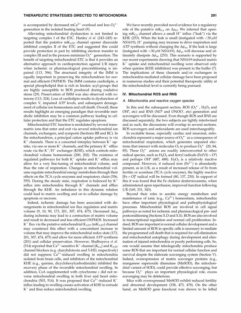
IV. Mitochondrial ROS and RNS 291A. Mitochondria and reactive oxygen species 291B. Mitochondria and reactive nitrogen species 294
V. Mitochondrial ROS Scavenging and Its Potential Therapeutic Value 295A. Manganese superoxide dismutase 296B. Glutathione thioredoxin, and peroxiredoxin systems 297C. Catalase and glutathione peroxidase 298D. Cytochrome c 298E. Mitochondria as scavengers of cytosolic O2
�� 299VI. Uncoupling Proteins in Modulation of Mitochondrial Function: Physiological and Pharmacologic Relevance 299
VII. Mitochondrial DNA-Related Pathologies and a Potential Therapeutic Target 300VIII. Mitochondrial Interaction with other Organelles: Therapeutic Implications 302
A. Mitochondrion–mitochondrion interaction 302B. Mitochondrion–nucleus interaction 303C. Mitochondria–endoplasmic=sarcoplasmic reticulum interaction 304
IX. Mitochondria-Related Diseases and Cell Injury 305A. Mitochondria and cardiac ischemia and reperfusion injury 305
Reviewing Editors: Enrique Cadenas, Andreas Daiber, Cherubino Di Lorenzo, Sanjeev Gupta, Sabzali Javadov, Jiri Neuzil, and
Michael Roth
Departments of 1Anesthesiology and 2Physiology, 3Cardiovascular Research Center, Medical College of Wisconsin, Milwaukee, Wisconsin.4Research Service, Veterans Affairs Medical Center, Milwaukee, Wisconsin.5Medical Service, Hunter Holmes McGuire Veterans Affairs Medical Center, Richmond, Virginia.6Departments of Medicine (Division of Cardiology) and Biochemistry, Virginia Commonwealth University, Richmond, Virginia.
ANTIOXIDANTS & REDOX SIGNALINGVolume 13, Number 3, 2010ª Mary Ann Liebert, Inc.DOI: 10.1089=ars.2009.2788
279
B. Mitochondria and the failing heart 306C. Mitochondria and diabetes 308D. Mitochondria and hypertension 309E. Mitochondria and neurodegenerative diseases 310
1. Alzheimer’s disease 3102. Parkinson’s disease 3103. Amyotrophic lateral sclerosis 3114. Friedreich’s ataxia 312
F. Neoplastic diseases 312G. Other mitochondria-related diseases 313
1. Mitochondria and psychiatric disorders 3132. Mitochondria and migraine headache 314
X. Mitochondrial Pharmacology and Therapeutic Potential 314A. Strategies for drug delivery to mitochondria 314B. Mitochondria-targeted drugs 317C. Approaches to improve mitochondrial function during ischemia and reperfusion 318D. Preconditioning 319E. Postconditioning 320
XI. Other Mitochondrial Therapeutic Approaches 321A. Lipid replacement therapy 321B. Transactivator of transcription proteins and mitochondrial therapy 322C. Molecular genetics approaches 322D. Mitochondria and caloric restriction 322E. Mitochondria and dietary supplements 323
XII. Mitochondria Age and Lifespan 323A. Mitochondria and age-associated diseases 323B. Mitochondrial p66shc and lifespan 324
XIII. Caveats and Potential Limitations in Mitochondrial Drug Targeting 325XIV. Conclusion and Perspectives 326
I. Introduction and Topics Reviewed
The recent research spotlight on mitochondria isattributed to observations that the organelle is involved
in a number of diseases, some of which are associated withmutations of mitochondrial DNA (mtDNA). The rekindledinterest in this organelle is coupled with its role in pro-grammed cell death, in which superoxide anions (O2
��) andits reactive oxygen species (ROS) products and dysfunctionin the energy production process are common underlyingfactors. This ‘‘new’’ role of mitochondria is crucial in under-standing their utility as potential targets against numeroushuman diseases. Overall cellular function is dependent on O2
consumption by functioning mitochondria to produce en-ergy with minimal electron leak to generate O2
��. Mitochon-dria are therefore vital for normal cellular function, includingintracellular metabolic activities and signal transduction ofvarious cellular pathways. They are involved in cellular ionhomeostasis, oxidative stress, and apoptotic and necrotic celldeath. Indeed, recent studies have identified a host of com-mon disorders with apparent ties to mitochondria, includingmetabolic (e.g., type 2 diabetes) and cardiovascular disorders,cancer, neurodegenerative diseases, psychiatric disorders,migraine headache, and the aging process.
A thorough understanding of mitochondrial function innormal and pathological states is critical in developing thefull therapeutic potential of the organelle in mitigating or pre-venting a given disease. Mitochondrial-related diseases arevastly different and much of the science linking mitochondria
to different disease states is still being intensively studied.The central premise of this review simply is that if mitochon-drial abnormalities contribute to a pathological state (directlyor indirectly), then alleviating the mitochondrial dysfunctionshould attenuate the severity or progression of the disease.Hence, the main objective of this review is to present theconcept that mitochondria of varying cell types can be poten-tially targeted for therapeutic intervention in mitochondria-associated diseases. That is, the review will focus on how themitochondrion has become a potential therapeutic target indisease management.
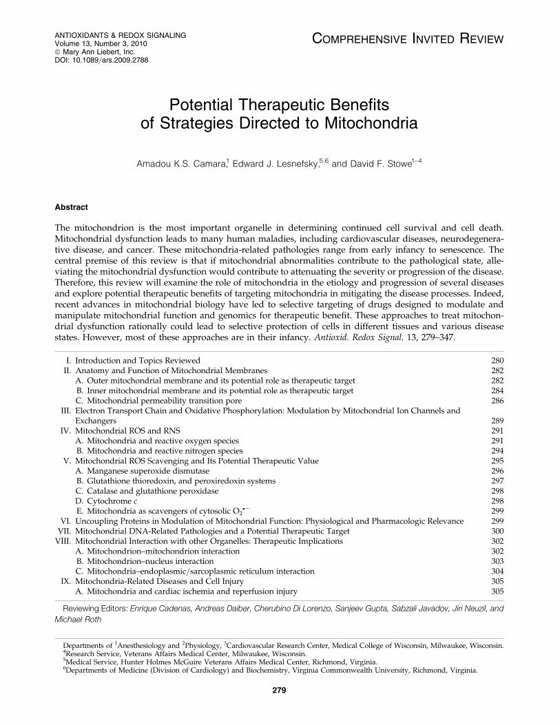
Sections II and III describe the primary function of mito-chondria, ATP synthesis through oxidative phosphorylation(OXPHOS), and how this energy production is carried out ona folded inner mitochondrial membrane (IMM) with proteincomplexes that transfer electrons from one protein complexto another at different redox potentials. Figure 1 providesa simple scheme of mitochondrial function. Figures 2 and 3illustrate the interactions of some IMM and outer mitochon-drial membrane (OMM) proteins important in the regula-tion of cell survival and cell death as a consequence ofoxidative stress, how regulation of transmembrane ionfluxes maintains cell homeostasis, and how perturbing thisanatomical and functional link can lead to pathological con-ditions.
Section III explains one of the side effects of electrontransport, generation of O2
�� and its products, and Section IVsummarizes the roles of ROS and reactive nitrogen species(RNS) as both regulators of cell function and cell death. Sec-
280 CAMARA ET AL.
tion V describes the elaborate free radical scavenging systemthat regulates ROS within physiologically acceptable valuesand the critical importance of balance in the production andscavenging of O2
�� in normal cellular physiology. These sec-tions summarize how mitochondria modulate bioenergetics
and serve as an effector of cell viability and how an imbalancein the rate of ROS production and the rate of ROS scavenginglead to oxidative stress, a marked contributor of mito-chondrial mediated pathology. Section VI discusses howuncoupling proteins and drugs enhance mitochondrial
FIG. 1. Basic mitochondrial structure and function. The figure shows the basic structural components of the five ETCcomplexes (I, II, III, IV, and V) as well as cytochrome c (Cyc), the flow of electrons through the complexes, and the generation ofATP. Fatty acid oxidation (FAO) and TCA cycle generate NADH and FADH2 needed to energize mitochondria and establishmitochondrial membrane potential (DCm; �180 to �200 mV). DCm is also modulated by uncoupling proteins (UCP). Phos-phate carriers, including the adenine nucleotide translocase (ANT), regulate mitochondrial matrix phosphate levels. Substrateuptake is mediated through inner mitochondrial membrane (IMM) proteins [e.g., carnitine palmitoyl transferase (CPT) andpyruvate dehydrogenase (PDH)]. Mitochondrial DNA (mtDNA) encodes mitochondrial-specific proteins and cytosolic pro-teins produced by nuclear DNA (n) are translocated to mitochondria through the translocator of the outer membrane (TOM)and inner mitochondrial membrane (TIM); Ca2þ is taken up through the calcium uniporter (CaU). The mitochondrialCa2þ level is dependent on the level of Ca2þwithin the microdomain with the endoplasmic reticulum (ER). This basic functionof mitochondria and its interaction with the nucleus and ER is the basis for understanding the role of the organelle in myriad ofmitochondria-related diseases. Reproduced and modified from Wall et al. (604).
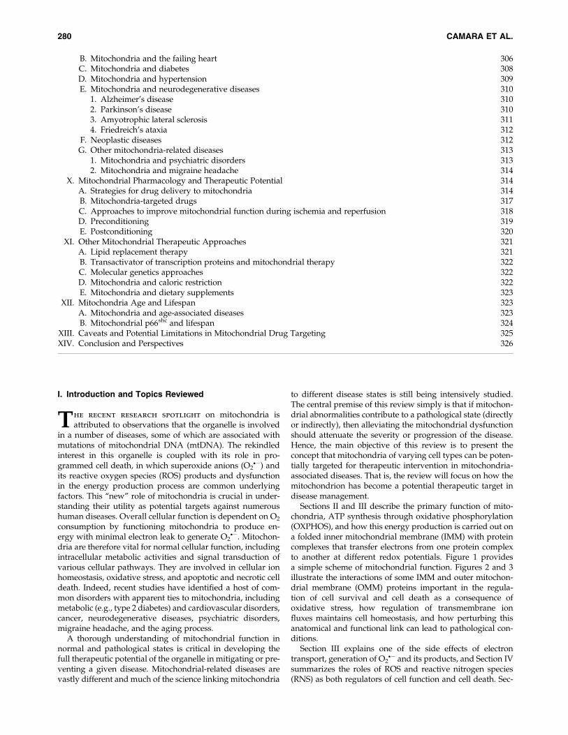
FIG. 2. Putative mitochondrialpermeability transition pore(mPTP) proteins (cylinders) in theouter (OMM) and inner (IMM)mitochondrial membrane in nor-mal physiological and patho-physiological conditions. Theconstituents within the OMM in-clude the VDAC, PBR, and othertranslocated proteins such as hex-okinase (HK) and the Bcl family ofproteins, Bax and Bak. The IMMproteins include ANT and CK;Cyclophilin D migrates to the IMMfrom the matrix. Initiators of apo-ptosis include cytochrome c andAIF. Small solutes include NADþ
and ADP. (For interpretation of thereferences to color in this figurelegend, the reader is referred to theweb version of this article atwww.liebertonline.com=ars).
THERAPEUTIC STRATEGIES DIRECTED TO MITOCHONDRIA 281
respiration, but not ATP synthesis, as a way to paradoxicallyprotect the organelle and cell.
Mitochondrial dysfunction and dysregulation may havetheir genesis in mtDNA mutations (e.g., colon and prostatecancer) and=or impairment or reversal of mitochondrial elec-tron transport chain (ETC). Section VII notes that manyhuman diseases are linked to mtDNA mutations and mito-chondrial dysfunction and describes how mitochondria haveemerged as central foci in the investigation of the etiologyof numerous cardiovascular, metabolic, neurological diseases,cancer, psychiatric disorders, and migraine. Mitochondrialmembrane integrity and mitochondrial functional and mor-phological connectivity with each other and with otherorganelles, for example, the nucleus or the endoplasmic re-ticulum (ER) are critical in maintaining cellular integrity andthey also provide continuity in cellular function. Most mito-chondrial proteins are encoded by the nuclear genome andcomplexes are encoded by both mitochondrial and nucleargenomes. Consequently, any defects in the production ofthese proteins could induce mitochondrial cytopathies thatunderlie a multitude of diseases or pathological conditions.Thus Section VIII explores the interaction of mitochondriawith themselves (the mitochondrial reticular network) andwith the nucleus and ER. In this section it is discussed howmutations in the genes for nuclear-encoded mitochondrialproteins, the so-called nuclear–mitochondrial crosstalk, areimplicated in a number of tissue degenerative disorders.Section IX summarizes how mitochondrial dysfunction un-derlies a number of diseases including cardiac ischemia andheart failure, diabetes, hypertension, as well as neurologic andneoplastic diseases and other lesser-known mitochondria-related diseases. For example, myocardial ischemia causesdamage to the mitochondrial distal ETC that could be an
important link between ischemia and the mitochondrial-induced myocyte damage that occurs on reperfusion. SectionX explores known strategies for delivering drugs to themitochondrion and discusses some mitochondria-targetedprocedures and drugs that appear useful in treating somedisease states, especially cardiac ischemia and reperfusion(I=R) injury. Other mitochondrial therapeutic approachesare presented in Section XI. Examples are lipid replace-ment therapy (LRT), transactivator of transcription (TAT)protein delivery, novel genetic approaches and the potentialbenefits of caloric restriction, and nutritional supplements.Section XII explores the role of mitochondria in the agingprocess and the role of the mitochondrial adapter protein,p66shc in lifespan determination. Finally, Sections XIII and XIVbring up the shortcomings and limitations of mitochondria-targeted drug delivery and the authors’ conclusions and per-spectives.
II. Anatomy and Function of Mitochondrial Membranes
A. Outer mitochondrial membrane and its potentialrole as a therapeutic target
The elaborate structure of a mitochondrion is important forthe normal functioning of the organelle and therefore asa potential therapeutic target. Two specialized membranesencircle each mitochondrion, dividing the organelle into anarrow intermembrane space (IMS) bordered by the OMMand the inner IMM (Figs. 1 and 2). The OMM contains manychannels formed by the protein porin that makes the mem-brane relatively permeable. One of the membrane proteins isthe peripheral benzodiazepine receptor (PBR). PBR is a smallevolutionarily conserved protein involved in cholesteroltransport and steroid synthesis; it is also a regulator of apo-
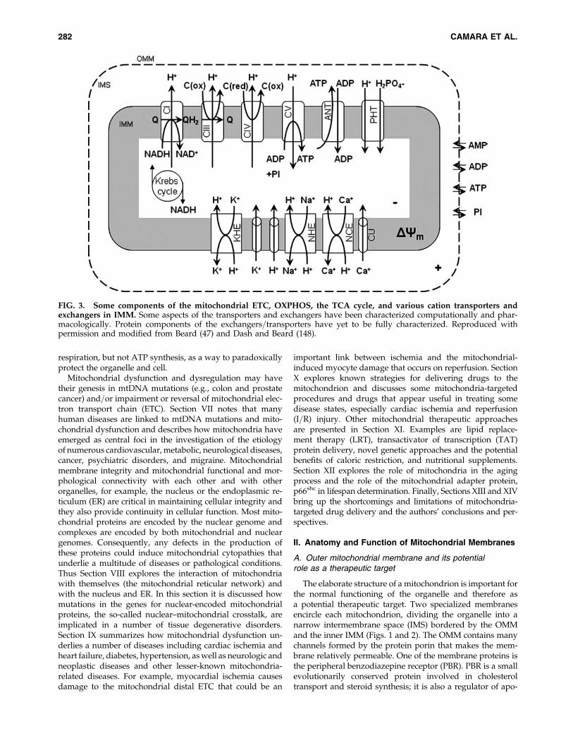
FIG. 3. Some components of the mitochondrial ETC, OXPHOS, the TCA cycle, and various cation transporters andexchangers in IMM. Some aspects of the transporters and exchangers have been characterized computationally and phar-macologically. Protein components of the exchangers=transporters have yet to be fully characterized. Reproduced withpermission and modified from Beard (47) and Dash and Beard (148).
282 CAMARA ET AL.
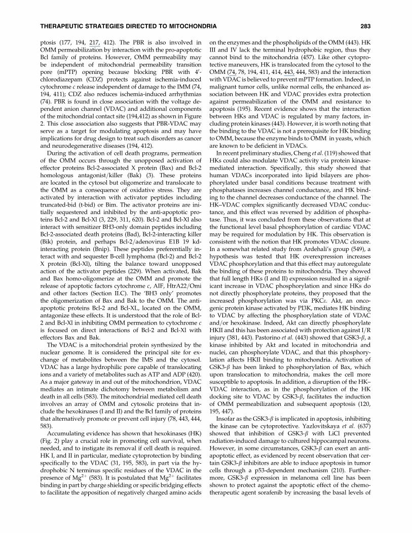
ptosis (177, 194, 217, 412). The PBR is also involved inOMM permeabilization by interaction with the pro-apoptoticBcl family of proteins. However, OMM permeability maybe independent of mitochondrial permeability transitionpore (mPTP) opening because blocking PBR with 40-chlorodiazepam (CDZ) protects against ischemia-inducedcytochrome c release independent of damage to the IMM (74,194, 411); CDZ also reduces ischemia-induced arrhythmias(74). PBR is found in close association with the voltage de-pendent anion channel (VDAC) and additional componentsof the mitochondrial contact site (194,412) as shown in Figure2. This close association also suggests that PBR-VDAC mayserve as a target for modulating apoptosis and may haveimplications for drug design to treat such disorders as cancerand neurodegenerative diseases (194, 412).
During the activation of cell death programs, permeationof the OMM occurs through the unopposed activation ofeffector proteins Bcl-2-associated X protein (Bax) and Bcl-2homologous antagonist=killer (Bak) (3). These proteinsare located in the cytosol but oligomerize and translocate tothe OMM as a consequence of oxidative stress. They areactivated by interaction with activator peptides includingtruncated-bid (t-bid) or Bim. The activator proteins are ini-tially sequestered and inhibited by the anti-apoptotic pro-teins Bcl-2 and Bcl-Xl (3, 229, 311, 620). Bcl-2 and Bcl-Xl alsointeract with sensitizer BH3-only domain peptides includingBcl-2-associated death proteins (Bad), Bcl-2-interacting killer(Bik) protein, and perhaps Bcl-2=adenovirus E1B 19 kd-interacting protein (Bnip). These peptides preferentially in-teract with and sequester B-cell lymphoma (Bcl-2) and Bcl-2X protein (Bcl-Xl), tilting the balance toward unopposedaction of the activator peptides (229). When activated, Bakand Bax homo-oligomerize at the OMM and promote therelease of apoptotic factors cytochrome c, AIF, HtrA22=Omiand other factors (Section II.C). The ‘BH3 only’ promotesthe oligomerization of Bax and Bak to the OMM. The anti-apoptotic proteins Bcl-2 and Bcl-XL, located on the OMM,antagonize these effects. It is understood that the role of Bcl-2 and Bcl-Xl in inhibiting OMM permeation to cytochrome cis focused on direct interactions of Bcl-2 and Bcl-Xl witheffectors Bax and Bak.
The VDAC is a mitochondrial protein synthesized by thenuclear genome. It is considered the principal site for ex-change of metabolites between the IMS and the cytosol.VDAC has a large hydrophilic pore capable of translocatingions and a variety of metabolites such as ATP and ADP (420).As a major gateway in and out of the mitochondrion, VDACmediates an intimate dichotomy between metabolism anddeath in all cells (583). The mitochondrial mediated cell deathinvolves an array of OMM and cytosolic proteins that in-clude the hexokinases (I and II) and the Bcl family of proteinsthat alternatively promote or prevent cell injury (78, 443, 444,583).
Accumulating evidence has shown that hexokinases (HK)(Fig. 2) play a crucial role in promoting cell survival, whenneeded, and to instigate its removal if cell death is required.HK I, and II in particular, mediate cytoprotection by bindingspecifically to the VDAC (31, 195, 583), in part via the hy-drophobic N terminus specific residues of the VDAC in thepresence of Mg2þ (583). It is postulated that Mg2þ facilitatesbinding in part by charge shielding or specific bridging effectsto facilitate the apposition of negatively charged amino acids
on the enzymes and the phospholipids of the OMM (443). HKIII and IV lack the terminal hydrophobic region, thus theycannot bind to the mitochondria (457). Like other cytopro-tective maneuvers, HK is translocated from the cytosol to theOMM (74, 78, 194, 411, 414, 443, 444, 583) and the interactionwith VDAC is believed to prevent mPTP formation. Indeed, inmalignant tumor cells, unlike normal cells, the enhanced as-sociation between HK and VDAC provides extra protectionagainst permeabilization of the OMM and resistance toapoptosis (195). Recent evidence shows that the interactionbetween HKs and VDAC is regulated by many factors, in-cluding protein kinases (443). However, it is worth noting thatthe binding to the VDAC is not a prerequisite for HK bindingto OMM, because the enzyme binds to OMM in yeasts, whichare known to be deficient in VDACs.
In recent preliminary studies, Cheng et al. (119) showed thatHKs could also modulate VDAC activity via protein kinase-mediated interaction. Specifically, this study showed thathuman VDACs incorporated into lipid bilayers are phos-phorylated under basal conditions because treatment withphosphatases increases channel conductance, and HK bind-ing to the channel decreases conductance of the channel. TheHK–VDAC complex significantly decreased VDAC conduc-tance, and this effect was reversed by addition of phospha-tase. Thus, it was concluded from these observations that atthe functional level basal phosphorylation of cardiac VDACmay be required for modulation by HK. This observation isconsistent with the notion that HK promotes VDAC closure.In a somewhat related study from Ardehali’s group (549), ahypothesis was tested that HK overexpression increasesVDAC phosphorylation and that this effect may autoregulatethe binding of these proteins to mitochondria. They showedthat full length HKs (I and II) expression resulted in a signif-icant increase in VDAC phosphorylation and since HKs donot directly phosphorylate proteins, they proposed that theincreased phosphorylation was via PKCe. Akt, an onco-genic protein kinase activated by PI3K, mediates HK bindingto VDAC by affecting the phosphorylation state of VDACand=or hexokinase. Indeed, Akt can directly phosphorylateHKII and this has been associated with protection against I=Rinjury (381, 443). Pastorino et al. (443) showed that GSK3-b, akinase inhibited by Akt and located in mitochondria andnuclei, can phosphorylate VDAC, and that this phosphory-lation affects HKII binding to mitochondria. Activation ofGSK3-b has been linked to phosphorylation of Bax, whichupon translocation to mitochondria, makes the cell moresusceptible to apoptosis. In addition, a disruption of the HK–VDAC interaction, as in the phosphorylation of the HKdocking site to VDAC by GSK3-b, facilitates the inductionof OMM permeabilization and subsequent apoptosis (120,195, 447).
Insofar as the GSK3-b is implicated in apoptosis, inhibitingthe kinase can be cytoprotective. Yazlovitskaya et al. (637)showed that inhibition of GSK3-b with LiCl preventedradiation-induced damage to cultured hippocampal neurons.However, in some circumstances, GSK3-b can exert an anti-apoptotic effect, as evidenced by recent observation that cer-tain GSK3-b inhibitors are able to induce apoptosis in tumorcells through a p53-dependent mechanism (210). Further-more, GSK3-b expression in melanoma cell line has beenshown to protect against the apoptotic effect of the chemo-therapeutic agent sorafenib by increasing the basal levels of
THERAPEUTIC STRATEGIES DIRECTED TO MITOCHONDRIA 283
the Bcl-2 proteins. Thus there is a strong rationale for the useof GSK3-b inhibitors (e.g., GSK3-IX) as adjuncts in the treat-ment of cancer (437).
HK binding promotes oligomerization of the VDAC (443)and impedes cytochrome c release (2, 523). Neoplastic cellsalso resist death in part by increasing the interaction betweenmitochondria and HK; this could be prevented, as a thera-peutic approach, by adding 3-bromopyruvate (3BrPA), aninhibitor of HK to VDAC, in order to kill a hepatoma cell linecharacterized by overexpression of HKI and HKII (447).Transfection of leukemia-derived U-937 cells with HK sig-nificantly reduced staurosporine-induced apoptosis whencompared to GFP transfected cells (31). Taken together, thesestudies suggest that interference of the binding of HK to mi-tochondria by VDAC-derived peptides (2) and peptide tar-geting of the N-terminal of the HK protein (120, 443) may offera novel strategy to potentiate the efficacy of other modes ofconventional chemotherapy (2). However, recent evidencealso suggests that HK-mediated protection against apoptoticsignals can occur independently of VDAC (443). For instance,HK is known to prevent apoptosis by interfering with Baxbinding to mitochondria to induce cytochrome c release (644).Thus, in normal cells, preservation of the OMM with HKs isaccompanied by increased retention of cytochrome c andimproved electron transfer and more effective OXPHOS.
These studies demonstrate that cellular injury could beascribed to increased permeability of the OMM and thatlimiting the permeability of the OMM will protect against celldamage and cell death due to oxidative stress. Indeed, HKIIdetachment from the OMM with clotrimazole, or with de-signed peptide fragments that target the N-terminal (aminoacid sequences) of the HKII domain for VDAC (120), leads tocell death. Therefore, a mitochondria-targeted therapy de-signed for the OMM as a potential therapeutic maneuver ishighly relevant and is the subject of intense research. A betterunderstanding of the interaction between the cytosolic pro-teins and OMM will greatly optimize therapies for treatingischemic heart disease, neurodegenerative diseases, and can-cer. These different mitochondrial related diseases and thepotential targeting of the organelle as a mitigating factor willbe discussed in much detail in the following sections.
B. Inner mitochondrial membrane and its potentialrole as therapeutic target
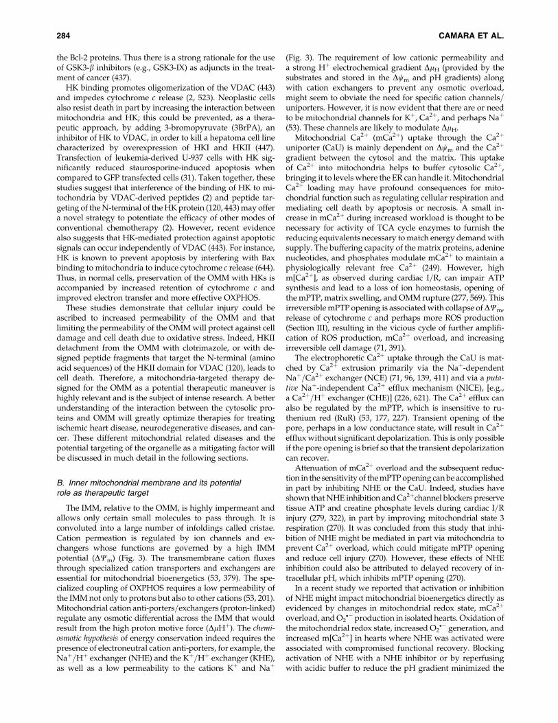
The IMM, relative to the OMM, is highly impermeant andallows only certain small molecules to pass through. It isconvoluted into a large number of infoldings called cristae.Cation permeation is regulated by ion channels and ex-changers whose functions are governed by a high IMMpotential (DCm) (Fig. 3). The transmembrane cation fluxesthrough specialized cation transporters and exchangers areessential for mitochondrial bioenergetics (53, 379). The spe-cialized coupling of OXPHOS requires a low permeability ofthe IMM not only to protons but also to other cations (53, 201).Mitochondrial cation anti-porters=exchangers (proton-linked)regulate any osmotic differential across the IMM that wouldresult from the high proton motive force (DmHþ). The chemi-osmotic hypothesis of energy conservation indeed requires thepresence of electroneutral cation anti-porters, for example, theNaþ=Hþ exchanger (NHE) and the Kþ=Hþ exchanger (KHE),as well as a low permeability to the cations Kþ and Naþ
(Fig. 3). The requirement of low cationic permeability anda strong Hþ electrochemical gradient DmH (provided by thesubstrates and stored in the Dcm and pH gradients) alongwith cation exchangers to prevent any osmotic overload,might seem to obviate the need for specific cation channels=uniporters. However, it is now evident that there are or needto be mitochondrial channels for Kþ, Ca2þ, and perhaps Naþ
(53). These channels are likely to modulate DmH.Mitochondrial Ca2þ (mCa2þ) uptake through the Ca2þ
uniporter (CaU) is mainly dependent on Dcm and the Ca2þ
gradient between the cytosol and the matrix. This uptakeof Ca2þ into mitochondria helps to buffer cytosolic Ca2þ,bringing it to levels where the ER can handle it. MitochondrialCa2þ loading may have profound consequences for mito-chondrial function such as regulating cellular respiration andmediating cell death by apoptosis or necrosis. A small in-crease in mCa2þ during increased workload is thought to benecessary for activity of TCA cycle enzymes to furnish thereducing equivalents necessary to match energy demand withsupply. The buffering capacity of the matrix proteins, adeninenucleotides, and phosphates modulate mCa2þ to maintain aphysiologically relevant free Ca2þ (249). However, highm[Ca2þ], as observed during cardiac I=R, can impair ATPsynthesis and lead to a loss of ion homeostasis, opening ofthe mPTP, matrix swelling, and OMM rupture (277, 569). Thisirreversible mPTP opening is associated with collapse ofDCm,release of cytochrome c and perhaps more ROS production(Section III), resulting in the vicious cycle of further amplifi-cation of ROS production, mCa2þ overload, and increasingirreversible cell damage (71, 391).
The electrophoretic Ca2þ uptake through the CaU is mat-ched by Ca2þ extrusion primarily via the Naþ-dependentNaþ=Ca2þ exchanger (NCE) (71, 96, 139, 411) and via a puta-tive Naþ-independent Ca2þ efflux mechanism (NICE), [e.g.,a Ca2þ=Hþ exchanger (CHE)] (226, 621). The Ca2þ efflux canalso be regulated by the mPTP, which is insensitive to ru-thenium red (RuR) (53, 177, 227). Transient opening of thepore, perhaps in a low conductance state, will result in Ca2þ
efflux without significant depolarization. This is only possibleif the pore opening is brief so that the transient depolarizationcan recover.
Attenuation of mCa2þ overload and the subsequent reduc-tion in the sensitivity of the mPTP opening can be accomplishedin part by inhibiting NHE or the CaU. Indeed, studies haveshown that NHE inhibition and Ca2þchannel blockers preservetissue ATP and creatine phosphate levels during cardiac I=Rinjury (279, 322), in part by improving mitochondrial state 3respiration (270). It was concluded from this study that inhi-bition of NHE might be mediated in part via mitochondria toprevent Ca2þ overload, which could mitigate mPTP openingand reduce cell injury (270). However, these effects of NHEinhibition could also be attributed to delayed recovery of in-tracellular pH, which inhibits mPTP opening (270).
In a recent study we reported that activation or inhibitionof NHE might impact mitochondrial bioenergetics directly asevidenced by changes in mitochondrial redox state, mCa2þ
overload, and O2�� production in isolated hearts. Oxidation of
the mitochondrial redox state, increased O2�� generation, and
increased m[Ca2þ] in hearts where NHE was activated wereassociated with compromised functional recovery. Blockingactivation of NHE with a NHE inhibitor or by reperfusingwith acidic buffer to reduce the pH gradient minimized the
284 CAMARA ET AL.
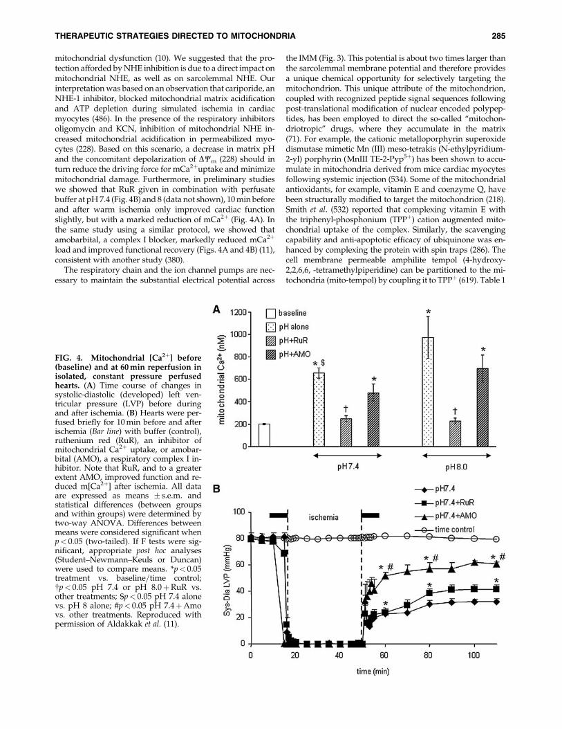
mitochondrial dysfunction (10). We suggested that the pro-tection afforded by NHE inhibition is due to a direct impact onmitochondrial NHE, as well as on sarcolemmal NHE. Ourinterpretation was based on an observation that cariporide, anNHE-1 inhibitor, blocked mitochondrial matrix acidificationand ATP depletion during simulated ischemia in cardiacmyocytes (486). In the presence of the respiratory inhibitorsoligomycin and KCN, inhibition of mitochondrial NHE in-creased mitochondrial acidification in permeabilized myo-cytes (228). Based on this scenario, a decrease in matrix pHand the concomitant depolarization of DCm (228) should inturn reduce the driving force for mCa2þuptake and minimizemitochondrial damage. Furthermore, in preliminary studieswe showed that RuR given in combination with perfusatebuffer at pH 7.4 (Fig. 4B) and 8 (data not shown), 10 min beforeand after warm ischemia only improved cardiac functionslightly, but with a marked reduction of mCa2þ (Fig. 4A). Inthe same study using a similar protocol, we showed thatamobarbital, a complex I blocker, markedly reduced mCa2þ
load and improved functional recovery (Figs. 4A and 4B) (11),consistent with another study (380).
The respiratory chain and the ion channel pumps are nec-essary to maintain the substantial electrical potential across
the IMM (Fig. 3). This potential is about two times larger thanthe sarcolemmal membrane potential and therefore providesa unique chemical opportunity for selectively targeting themitochondrion. This unique attribute of the mitochondrion,coupled with recognized peptide signal sequences followingpost-translational modification of nuclear encoded polypep-tides, has been employed to direct the so-called ‘‘mitochon-driotropic’’ drugs, where they accumulate in the matrix(71). For example, the cationic metalloporphyrin superoxidedismutase mimetic Mn (III) meso-tetrakis (N-ethylpyridium-2-yl) porphyrin (MnIII TE-2-Pyp5þ) has been shown to accu-mulate in mitochondria derived from mice cardiac myocytesfollowing systemic injection (534). Some of the mitochondrialantioxidants, for example, vitamin E and coenzyme Q, havebeen structurally modified to target the mitochondrion (218).Smith et al. (532) reported that complexing vitamin E withthe triphenyl-phosphonium (TPPþ) cation augmented mito-chondrial uptake of the complex. Similarly, the scavengingcapability and anti-apoptotic efficacy of ubiquinone was en-hanced by complexing the protein with spin traps (286). Thecell membrane permeable amphilite tempol (4-hydroxy-2,2,6,6, -tetramethylpiperidine) can be partitioned to the mi-tochondria (mito-tempol) by coupling it to TPPþ (619). Table 1
FIG. 4. Mitochondrial [Ca2þ] before(baseline) and at 60 min reperfusion inisolated, constant pressure perfusedhearts. (A) Time course of changes insystolic-diastolic (developed) left ven-tricular pressure (LVP) before duringand after ischemia. (B) Hearts were per-fused briefly for 10 min before and afterischemia (Bar line) with buffer (control),ruthenium red (RuR), an inhibitor ofmitochondrial Ca2þ uptake, or amobar-bital (AMO), a respiratory complex I in-hibitor. Note that RuR, and to a greaterextent AMO, improved function and re-duced m[Ca2þ] after ischemia. All dataare expressed as means � s.e.m. andstatistical differences (between groupsand within groups) were determined bytwo-way ANOVA. Differences betweenmeans were considered significant whenp< 0.05 (two-tailed). If F tests were sig-nificant, appropriate post hoc analyses(Student–Newmann–Keuls or Duncan)were used to compare means. *p< 0.05treatment vs. baseline=time control;{p< 0.05 pH 7.4 or pH 8.0þRuR vs.other treatments; $p< 0.05 pH 7.4 alonevs. pH 8 alone; #p< 0.05 pH 7.4þAmovs. other treatments. Reproduced withpermission of Aldakkak et al. (11).
THERAPEUTIC STRATEGIES DIRECTED TO MITOCHONDRIA 285
shows examples of mitochondria-targeted drugs or agentsthat are hitched to the carrier molecules that permeate themitochondrion.
Mitochondria-targeted peptides could also be recognizedby unique amino acid sequences that enable translocation of apeptide to a mitochondrion. However, other mitochondrialproteins translocate to the matrix without the targeting pep-tide sequences. These proteins interact with and bind to sitespresent on the OMM or IMM. For example, PKCe interactswith and phosphorylates its target proteins in the IMM bybinding to sites=adapter proteins on the IMM (150, 417). Thistranslocation of PKCe results in mitochondrial hyperpolar-ization and this may reduce depolarization of DCm duringischemia and increase ATP synthesis on reperfusion, which inturn may increase the energy-dependent processes that areinvolved in establishing the ion gradient across the sarco-lemma and mitochondrial membranes (150).
It is important that mitochondrial ion channels and ex-changers are controlled in order to provide the balance be-tween energy supply and demand that is crucial for normalcell function. Attempts to characterize the molecular struc-tures of these channels remain elusive, however. Achievingthis goal from a pharmacological standpoint could ‘‘spur thedevelopment of novel and specific therapeutic agents targetedto the mitochondria’’ (411).
C. Mitochondrial permeability transition pore
Crompton et al. (140, 141) were the first to demonstrate thatthe mPTP plays a crucial role in cardiac myocyte injury fol-lowing oxidative stress. The mPTP, a large, nonspecific chan-nel protein aggregate known to span the OMM and the IMM(Fig. 2), mediates the lethal permeability changes of the mi-tochondrial membranes leading to mitochondria-mediateddeath. It has been described as the rate-limiting step in themitochondria-mediated cell death pathway (391). However,recent studies have implicated the pore as a ‘‘physiologicalvalve’’, which alleviates mCa2þ overload as a consequence ofa brief surge of Ca2þ in a localized microdomain involving theER (Section VIII,C) (530). This transient brief opening of thepore (flickering) has also been implicated in providing pro-tection against cellular injury (301). This dual role of the mPTPin the survival and death of the cell is therefore critical inselective targeting of the pore for therapeutic interventions.Similarly, an understanding of the constituents of the poreand its molecular structure are paramount in this therapeuticgoal.
A model structure of the mPTP put forward by Halestrapet al. (236, 237) and Di Lisa and Bernadi (161) involves thecombination of the adenine nucleotide translocase (ANT) inthe IMM, the VDAC of the OMM, and a regulatory proteincyclophilin D (CypD) in the matrix (Fig. 2). Other variants ofthe ANT, ANT2, which are overexpressed in cancer cells, helpto stabilize mitochondrial membranes (195). Indeed, it wassuggested that in cancer cells, small interfering RNA (siRNA)that down regulate ANT2 may constitute a valid strategy forthe selective induction of tumor cell death (195, 269). Asso-ciated with the outer leaflet of the IMM, in the IMS, is themitochondrial creatine kinase (CK). CK, under physiologicalconditions, is crucial in catalyzing the transphosphorylationof creatine by ATP to phosphocreatine and ADP. In apoptotic-induced cell death, mitochondrial CK may facilitate contact
Ta
bl
e1.
St
ra
te
gie
sfo
rE
ffe
ct
iv
eD
ru
gD
el
iv
er
yin
to
th
eM
it
oc
ho
nd
ria
lM
at
rix
Low
mol
ecu
lar
wei
gh
tS
OD
mim
etic
sM
itoc
hon
dri
alm
embr
ane
pot
enti
al-d
epen
den
td
eliv
ery
syst
em(d
eloc
aliz
edca
tion
s)D
equ
alin
ium
lip
osom
e-ba
sed
del
iver
ysy
stem
Am
ino
acid
and
pep
tid
e-ba
sed
del
iver
ysy
stem
s
EU
K-8
and
EU
K-1
34(3
69)
Rh
od
amin
e12
3-co
nju
gat
edd
rug
s(2
8,29
,26
2)C
isp
lati
nan
dM
asto
par
an(2
8,13
6,26
2,27
6,33
5,35
8,56
3)
Pla
smid
DN
A(6
03,
631)
Am
ino
acid
-bas
edG
luta
thio
ne
cho
lin
ees
ter
(519
)N
AC
cho
lin
ees
ter
(28,
519)
Man
gan
ese
po
rph
yri
ns
(90,
99,
105,
534,
542)
:A
EO
L10
150
Mn
TB
AP
Mn
TP
yP
Mn
III
TE
-2-P
yP
5þ
Met
hy
ltri
ph
eny
lph
osp
ho
niu
m(T
PPþ
)co
nju
gat
edd
rug
s(2
8,29
,85
,39
3,45
3,49
8,52
7,58
4,61
9):
Mit
oQ
Mit
oV
itE
Ub
iqu
ino
lM
ito
tem
po
lM
ito
PB
N
Sm
all
mo
lecu
les
(an
ti-c
ance
rd
rug
s)(4
,28
,29
,72
,13
1,13
6,26
2,27
6,33
9,39
3,42
4,46
8,60
3,63
1):
CD
437
Pac
lita
xel
Ver
tep
orfi
n
Sm
all
pep
tid
esan
dp
rote
ins
(101
,12
3,13
6,21
2,26
2,39
3,46
5,50
7,55
1,55
3,55
4,57
4):
SS
tetr
apep
tid
es(a
nti
ox
idan
ts)
SS
31S
S01
TA
Tp
rote
ins
for
del
iver
yo
fp
rote
ins
tom
ito
cho
nd
ria
286 CAMARA ET AL.
between the VDAC and ANT to form the pore at the contactsite of the IMM and OMM (391) (Fig. 2). However, recentstudies have demonstrated that the VDAC and ANT act moreas regulatory proteins of the mPTP (37, 120).
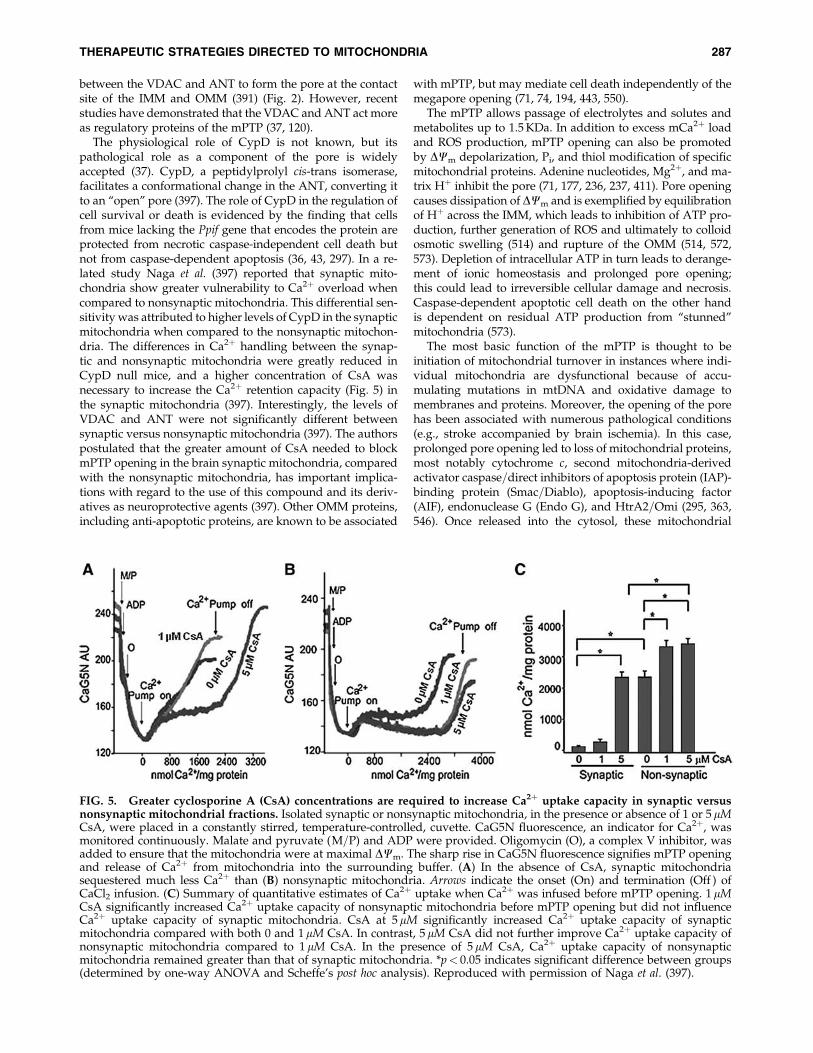
The physiological role of CypD is not known, but itspathological role as a component of the pore is widelyaccepted (37). CypD, a peptidylprolyl cis-trans isomerase,facilitates a conformational change in the ANT, converting itto an ‘‘open’’ pore (397). The role of CypD in the regulation ofcell survival or death is evidenced by the finding that cellsfrom mice lacking the Ppif gene that encodes the protein areprotected from necrotic caspase-independent cell death butnot from caspase-dependent apoptosis (36, 43, 297). In a re-lated study Naga et al. (397) reported that synaptic mito-chondria show greater vulnerability to Ca2þ overload whencompared to nonsynaptic mitochondria. This differential sen-sitivity was attributed to higher levels of CypD in the synapticmitochondria when compared to the nonsynaptic mitochon-dria. The differences in Ca2þ handling between the synap-tic and nonsynaptic mitochondria were greatly reduced inCypD null mice, and a higher concentration of CsA wasnecessary to increase the Ca2þ retention capacity (Fig. 5) inthe synaptic mitochondria (397). Interestingly, the levels ofVDAC and ANT were not significantly different betweensynaptic versus nonsynaptic mitochondria (397). The authorspostulated that the greater amount of CsA needed to blockmPTP opening in the brain synaptic mitochondria, comparedwith the nonsynaptic mitochondria, has important implica-tions with regard to the use of this compound and its deriv-atives as neuroprotective agents (397). Other OMM proteins,including anti-apoptotic proteins, are known to be associated
with mPTP, but may mediate cell death independently of themegapore opening (71, 74, 194, 443, 550).
The mPTP allows passage of electrolytes and solutes andmetabolites up to 1.5 KDa. In addition to excess mCa2þ loadand ROS production, mPTP opening can also be promotedby DCm depolarization, Pi, and thiol modification of specificmitochondrial proteins. Adenine nucleotides, Mg2þ, and ma-trix Hþ inhibit the pore (71, 177, 236, 237, 411). Pore openingcauses dissipation of DCm and is exemplified by equilibrationof Hþ across the IMM, which leads to inhibition of ATP pro-duction, further generation of ROS and ultimately to colloidosmotic swelling (514) and rupture of the OMM (514, 572,573). Depletion of intracellular ATP in turn leads to derange-ment of ionic homeostasis and prolonged pore opening;this could lead to irreversible cellular damage and necrosis.Caspase-dependent apoptotic cell death on the other handis dependent on residual ATP production from ‘‘stunned’’mitochondria (573).
The most basic function of the mPTP is thought to beinitiation of mitochondrial turnover in instances where indi-vidual mitochondria are dysfunctional because of accu-mulating mutations in mtDNA and oxidative damage tomembranes and proteins. Moreover, the opening of the porehas been associated with numerous pathological conditions(e.g., stroke accompanied by brain ischemia). In this case,prolonged pore opening led to loss of mitochondrial proteins,most notably cytochrome c, second mitochondria-derivedactivator caspase=direct inhibitors of apoptosis protein (IAP)-binding protein (Smac=Diablo), apoptosis-inducing factor(AIF), endonuclease G (Endo G), and HtrA2=Omi (295, 363,546). Once released into the cytosol, these mitochondrial
FIG. 5. Greater cyclosporine A (CsA) concentrations are required to increase Ca2þ uptake capacity in synaptic versusnonsynaptic mitochondrial fractions. Isolated synaptic or nonsynaptic mitochondria, in the presence or absence of 1 or 5 mMCsA, were placed in a constantly stirred, temperature-controlled, cuvette. CaG5N fluorescence, an indicator for Ca2þ, wasmonitored continuously. Malate and pyruvate (M=P) and ADP were provided. Oligomycin (O), a complex V inhibitor, wasadded to ensure that the mitochondria were at maximal DCm. The sharp rise in CaG5N fluorescence signifies mPTP openingand release of Ca2þ from mitochondria into the surrounding buffer. (A) In the absence of CsA, synaptic mitochondriasequestered much less Ca2þ than (B) nonsynaptic mitochondria. Arrows indicate the onset (On) and termination (Off ) ofCaCl2 infusion. (C) Summary of quantitative estimates of Ca2þ uptake when Ca2þ was infused before mPTP opening. 1 mMCsA significantly increased Ca2þ uptake capacity of nonsynaptic mitochondria before mPTP opening but did not influenceCa2þ uptake capacity of synaptic mitochondria. CsA at 5 mM significantly increased Ca2þ uptake capacity of synapticmitochondria compared with both 0 and 1mM CsA. In contrast, 5 mM CsA did not further improve Ca2þ uptake capacity ofnonsynaptic mitochondria compared to 1 mM CsA. In the presence of 5 mM CsA, Ca2þ uptake capacity of nonsynapticmitochondria remained greater than that of synaptic mitochondria. *p< 0.05 indicates significant difference between groups(determined by one-way ANOVA and Scheffe’s post hoc analysis). Reproduced with permission of Naga et al. (397).
THERAPEUTIC STRATEGIES DIRECTED TO MITOCHONDRIA 287
proteins trigger both caspase-dependent (by cytochrome c,Smac=DIABLO, or HtrA2=Omi), and caspase-independent(by AIF, Endo G, or HtrA2=Omi) apoptosis (110, 268).
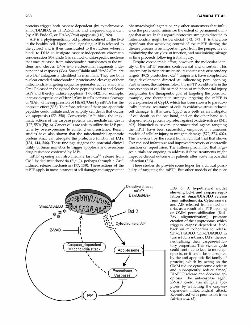
AIF is a phylogenetically old protein confined in the IMSin the healthy cell. Upon lethal signaling, AIF is released tothe cytosol and is then translocated to the nucleus where itbinds to DNA to instigate caspase-independent chromatincondensation (93). Endo G is a mitochondria-specific nucleasethat once released from mitochondria translocates to the nu-cleus and cleaves DNA into nucleosomal fragments inde-pendent of caspases (336). Smac=Diablo and HtrA2=Omi aretwo IAP antagonists identified in mammals. They are bothnuclear encoded mitochondrial proteins and cleavage of theirmitochondria-targeting sequence generates active Smac andOmi. Released in the cytosol these peptides bind to and cleaveIAPs and thereby induce apoptosis (177, 642). For example,increased expression of HtrA2=Omi in cells increases cleavageof XIAP, while suppression of HtrA2=Omi by siRNA has theopposite effect (535). Therefore, release of these pro-apoptoticpeptides could initiate and=or amplify cell death that occursvia apoptosis (177, 550). Conversely, IAPs block the enzy-matic actions of the caspase proteins that mediate cell death(177, 550) (Fig. 6). Cancer cells are able to utilize the IAP pro-teins by overexpression to confer chemoresistance. Recentstudies have also shown that the mitochondrial apoptoticprotein Smac can abrogate the protective function of IAPs(134, 144, 546). These findings suggest the potential clinicalutility of Smac mimetics to trigger apoptosis and overcomedrug resistance conferred by IAPs.
mPTP opening can also mediate fast Ca2þ release fromCa2þ loaded mitochondria (Fig. 2), perhaps through a Ca2þ
induced release mechanism (177, 550). These actions of themPTP apply in most instances of cell damage and suggest that
pharmacological agents or any other maneuvers that influ-ence the pore could minimize the extent of permanent dam-age that arises. In this regard, protective strategies directed tomitochondria might be beneficial (572, 573). It is thereforesignificant that achieving control of the mPTP during thedisease process is an important goal from the perspective ofminimizing the early loss of function, and maximizing salvageas time proceeds following initial injury.
Despite considerable effort, however, the molecular iden-tity of the mPTP remains controversial and uncertain. Thisuncertainty in the pore structure, its constituents or secondarytargets (ROS production, Ca2þ uniporter), have complicateddrug development directed at influencing pore opening.Furthermore, the dubious role of the mPTP constituents in thepreservation of cell life or mediation of mitochondrial injurycomplicates the therapeutic goal of targeting the pore. Forexample, one therapeutic strategy targeting the mPTP isoverexpression of CypD, which has been shown to paradox-ically increase resistance of cells to oxidative stress-inducedcell damage. In this case, CypD acts both as an instigatorof cell death on the one hand, and on the other hand as achaperone-like protein to protect against oxidative stress (341,602). Nonetheless, several pharmaceutical agents targetingthe mPTP have been successfully employed in numerousmodels of cellular injury to mitigate damage (572, 573, 602).This is evident by the recent human clinical trial that showsCsA reduced infarct size and improved recovery of contractilefunction on reperfusion. The authors proclaimed that large-scale trials are ongoing to address if these treatments mightimprove clinical outcome in patients after acute myocardialinfarction (213).
These studies do provide some hopes for a clinical possi-bility of targeting the mPTP. But other models of the pore
FIG. 6. A hypothetical modelshowing Bcl-2 and caspase regu-lation of Smac/DIABLO releasefrom mitochondria. Cytochrome cand AIF released from mitochon-dria, as a result of mPTP openingor OMM permeabilization (Bad–Bax oligomerization), promotecreation of the apoptosome, whichtriggers caspase-dependent feed-back on mitochondria to releaseSmac=DIABLO. Smac=DIABLO inturn inhibits intrinsic IAPs, therebyneutralizing their caspase-inhibi-tory properties. This vicious cyclecould continue to lead to more ap-optosis, or it could be interruptedby the anti-apoptotic Bcl family ofproteins, which by acting on theOMM reduce cytochrome c releaseand subsequently reduce Smac=DIABLO release and decrease ap-optosis. The anti-caspase agentZ-VAD could also mitigate apo-ptosis by inhibiting the caspase-dependent mitochondrial attack.Reproduced with permission fromAdrian et al. (5).
288 CAMARA ET AL.
maintain that activation of the pore with ROS would impairthe antioxidant effect of CypD. Overall, the major goal is de-velopment of novel, specific, and potent inhibitors that targetboth the primary constituents, which still remains elusive, andsecondary targets (e.g., CaU) whose activities directly or in-directly modulate pore activity. An example in this case is theuse of the antidepressant drug nortriptyline, which exerts itsneuroprotective effects against cerebral I=R injury in part viadelayed mPTP opening by resisting Ca2þ overload (646).Therefore, a complete molecular characterization of the CaUcould lead to better therapeutic targets that could minimizematrix Ca2þ uptake and indirectly mitigate mPTP-mediatedcellular damage.
Other therapeutic strategies involve the direct targeting ofconstituents of the pore. For example, ANT inhibitors havebeen used to block mPTP, but their use in the heart is oflimited values because the heart stops beating (74). CsA, andits nonimmunosuppresive derivative NIM811, prevent mPTPactivation in part by blocking CypD binding to ANT and thusprevent mitochondrial depolarization (573). Sanglifehrin, anovel immunosuppressive natural product that also bindsto CypD and inhibits its peptidyl-prolyl isomerase, is effectivein protecting against pore opening and minimizing I=R-mediated cellular injury. The translation of these agents fromexperimental studies to clinical trials is hampered, however,by their undesirable side effects. CsA is known to exert un-wanted side effects on the heart by inhibiting calcineurin (138,236). It is reported that CsA has a narrow window of activity;the optimal concentration is approximately 200 nM for opti-mal protection but it declines as a protective agent at higherconcentrations. Other mPTP inhibitors whose mode of actionsare not well known include trifluoperazine, which is onlyactive in energized mitochondria, and ubiquinone analogueswhich modulate pore opening by interacting with complex I(236).
Knowledge of the structural constituents of the mPTP andhow agents modulate the dynamic function and structure ofthe mPTP is essential to understand the role of mitochondriaas a therapeutic target for human diseases in which apoptoticand anti-apoptotic mechanisms are directly implicated in theetiology. The goal here would be to selectively manipulatemPTP protein function by therapeutic intervention, either toactivate it to induce apoptosis for cancer therapy, or to inhibitit to protect against cell death during cardiac or cerebral is-chemia.
III. Electron Transport Chain and OxidativePhosphorylation: Modulation by MitochondrialIon Channels and Exchangers
Mitochondria are the primary organelles for the generationof ATP under normal aerobic conditions. They contain theterminal oxidative pathway (TCA cycle) for carbohydrate andfat oxidations that produce the reducing equivalents NADHand FADH2 (Hþ and electron pairs). In OXPHOS, electronsare transferred from NADH and FADH2 to molecular O2
through the ETC complexes I–IV until two electrons and twoprotons combine with ½O2 to produce H2O at complex IV(respiration). Concomitantly, protons are pumped from themitochondrial matrix into the IMS. This generates a pH gra-dient and an electrostatic potential, DCm, across the IMM.Under normal physiological conditions, DCm contributes
most of the DmH, which drives the protons back into the mi-tochondrial matrix down their electrochemical gradientthrough the F1F0–ATPase (ATP synthase) to synthesize ATP(phosphorylation). Both DCm and DmH tend to decrease if thesupply of NADH and FADH2 through the TCA cycle does notmatch the increased flux through the ETC during mitochon-drial respiration. Together, the various compartments of mi-tochondria are able to work in harmony to generate ATP in acomplex multistep process.
ATP is involved in a myriad of cellular processes that areessential for cell survival such as maintaining ionic homeo-stasis, cell proliferation, and gene regulation. The dependenceof cells on mitochondrial ATP varies. For example, cancercells and astrocytes can survive well on ATP generated fromglycolysis and are much less dependent on mitochondrialOXPHOS to generate ATP. Other cells such as neurons andcardiomyocytes depend almost entirely on mitochondrialOXPHOS for their function. Preservation of the constituents ofthe mitochondrial ETC is paramount in maintaining the bio-energetics status of the mitochondrion and the cell homeo-stasis. Indeed, mitochondrial defects encompassing complexI–IV of the ETC characterize a large number of neurodegen-erative diseases (124, 125).
Mitochondrial ETC complexes are involved in cytoprotec-tion. Studies have shown that amobarbital and volatile anes-thetics block complex I, diazoxide blocks complex II, andhydrogen sulfide blocks complex IV. Although these drugshave additional effects, they emerge as potential means toprotect against cellular injury following I=R (9, 113, 244–246,324, 325). The targeting of mitochondrial complexes for atherapeutic purpose is in part ascribed to their vulnerabilityto oxidative stress. Therefore, a limitation of electron transferduring ischemia to complex III, a major site for electron leakand ROS production, is a new concept to limit mitochondrialdamage specifically during ischemia (9, 116, 117, 330). Mi-tochondria sustain progressive damage to the ETC duringthe course of myocardial ischemia; 10–20 min of ischemiadecreased complex I activity and caused damage to theOXPHOS apparatus, including complex V and the ANT (330).As ischemia time lengthens (30–45 min), damage to complexIII and IV becomes evident.
Hence, while a complex I defect occurs early in ischemia,damage continues to progress to involve complexes III and IV.Complex I activity will go down due to a decrease in theNADH dehydrogenase component, possibly the loss of theFMN coenzyme; complex I activity is also modulated bypost-translational modifications including S-nitrosylation andphosphorylation. These peptide alterations are amenable topharmacologic manipulation, as in the use of S-nitroso-2-mercaptopropionyl glycine (SNO-MPG) in providing pro-tection against ischemic damage (396). SNO-MPG inhibitscomplex I during the critical late ischemia and early reperfu-sion stage (59, 66) and in that way provides protection fromROS generated at complex III (117) (Section III).
The above observations are consistent with our recentstudies showing that blocking complex I with the reversibleinhibitor amobarbital protected the heart, and its mitochon-dria in particular, from I=R injury when the blocker waspresent only during ischemia. Amobarbital, a short-actingbarbiturate, inhibits complex I at the rotenone site at concen-trations of 1–3 mM. At higher concentrations (5 mM), amo-barbital also inhibits succinate respiration and complex V
THERAPEUTIC STRATEGIES DIRECTED TO MITOCHONDRIA 289
(113, 116). We have furnished recent additional data for thespecificity of amobarbital treatment in intact isolated heartsthat gives further insight into its mechanism of protection (9).This mechanism of protection was similar to that of ranola-zine, a late sodium channel blocker and anti-angina drugthat is also known to block complex I (82) and to inhibitb-oxidation enzymes (68, 538).
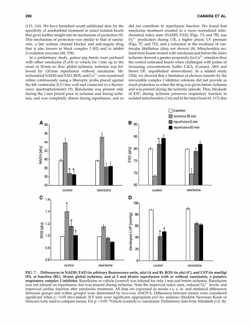
In a preliminary study, guinea pig hearts were perfusedwith either ranolazine (5mM) or vehicle for 1 min up to theonset of 30 min no flow global ischemia; ischemia was fol-lowed by 120 min reperfusion without ranolazine. Mi-tochondrial NADH and FAD, ROS, and Ca2þwere monitoredonline continuously using a fiberoptic probe placed againstthe left ventricular (LV) free wall and connected to a fluores-cence spectrophotometry (9). Ranolazine was present onlyduring the 1 min period prior to ischemia and during ische-mia, and was completely absent during reperfusion, and so
did not contribute to reperfusion function. We found thatranolazine treatment resulted in a more normalized mito-chondrial redox state (NADH, FAD) (Figs. 7A and 7B), lessO2�� production during I=R, a higher phasic LV pressure
(Figs. 7C and 7D), and a reduction in the incidence of ven-tricular fibrillation (data not shown) (8). Mitochondria iso-lated from hearts treated with ranolazine just before the indexischemia showed a greater propensity for Ca2þ retention thanthe control untreated hearts when challenged with pulses ofincreasing concentrations buffer CaCl2 (Camara AKS andStowe DF, unpublished observations). In a related study(324), we showed that a limitation of electron transfer by theirreversible complex I inhibitor rotenone did not provide asmuch protection as when the drug was given before ischemiaand was present during the ischemic episode. Thus, blockadeof ETC during ischemia preserves respiratory function inisolated mitochondria (116) and in the intact heart (9, 117); this
FIG. 7. Differences in NADH=FAD (in arbitrary fluorescence units, afu) (A and B), ROS (in afu) (C), and LVP (in mmHg)(D), at baseline (BL), 30 min global ischemia, and at 5 and 60 min reperfusion with or without ranolazine, a putativerespiratory complex I inhibitor. Ranolazine or vehicle (control) was infused for only 1 min just before ischemia. Ranolazinewas not infused on reperfusion, but was present during ischemia. Note the improved redox state, reduced O2
�� levels, andimproved cardiac function after ranolazine treatment. All data are expressed as means� s. e. m. and statistical differences(between groups and within groups) were determined by two-way ANOVA. Differences between means were consideredsignificant when p< 0.05 (two-tailed). If F tests were significant, appropriate post hoc analyses (Student–Newman–Keuls orDuncan) were used to compare means. For p< 0.05: *Vehicle (control) vs. ranolazine. Preliminary data from Aldakkak et al. (8).
290 CAMARA ET AL.
is accompanied by decreased mCa2þ overload and less O2��
generation in the isolated beating heart (9).Alleviating mitochondrial dysfunction is not limited to
targeting complex I of the ETC. Hanley et al. (243–245) re-ported that the putative mKATP channel opener diazoxideinhibited complex II of the ETC and suggested this couldprovide protection in part by inhibiting electron transfer tocomplex III and in the process minimize O2
�� generation. Thebenefit of targeting mitochondrial ETC is that it provides analternative approach to cardioprotection against I=R injurywhen ischemic or pharmacological preconditioning is im-paired (113, 396). The structural integrity of the IMM isequally important in preserving the mitochondrion for nor-mal and efficient OXPHOS. The IMM contains cardiolipin, aspecial phospholipid that is rich in linoleic acyl-groups thatare highly susceptible to ROS produced during oxidativestress (29). Preservation of IMM was also observed with ro-tenone (113, 324). Loss of cardiolipin results in dysfunction ofcomplex V, impaired ATP levels, and subsequent derange-ment of cellular ion homeostasis and cell death. Overall, theseresults highlight an emerging paradigm that reversible met-abolic inhibition may be a common pathway leading to cel-lular protection and that the ETC regulates apoptosis.
Mitochondrial ETC function is modulated by several trans-matrix ions that enter and exit via several mitochondrial ionchannels, exchangers, and symports (Sections IIB and IIC). Inthe mitochondrion, a principal cation uptake pathway is viaKþ channels. There is a concerted interplay between Kþ up-take, via one or more Kþ channels, and the primary Kþ effluxroute via the Kþ=Hþ exchanger (KHE), which controls mito-chondrial volume homeostasis (200, 202). The existence ofregulated pathways for both Kþ uptake and Kþ efflux mayallow for a very fine-tuning of mitochondrial volume, andthus the rate of respiration. Changes in mitochondrial vol-ume regulate mitochondrial energy metabolism through theireffects on the TCA cycle enzymes and respiratory chain (356,555). During the steady state, respiration is balanced by Kþ
influx into mitochondria through Kþ channels and effluxthrough the KHE. An imbalance in this dynamic relationcould lead to matrix swelling and on to cellular damage byapoptosis or necrosis.
Indeed, ischemic damage has been associated with de-rangements in mitochondrial ion flux regulation and matrixvolume (9, 10, 90, 173, 201, 307, 474, 475). Decreased Dcm
during ischemia may lead to a contraction of matrix volumeand result in decreased and less efficient OXPHOS. IncreasedKþ flux via the putative mitochondrial KATP (mKATP) channelmay counteract this effect with a concomitant increase involume that may improve the mitochondrial redox state (173,201, 307, 474, 475) and allow for more efficient ATP synthesis(201) and cellular preservation. However, Shalbuyeva et al.(514) reported that Ca2þ-sensitive Kþ channel (KCa) and KATP
channel blockers (e.g. charybdotoxin and 5-HD, respectively)did not suppress Ca2þ-induced swelling in mitochondriaisolated from brain cells, and inhibitors of the mitochondrialKHE (e.g., quinine, dicyclohexylcarbodimiide) inhibited therecovery phase of the reversible mitochondrial swelling. Inaddition, CsA supplemented with cytochrome c did not re-verse mitochondrial swelling in both liver and heart mito-chondria (503, 514). It was proposed that Ca2þ-induced Kþ
influx leading to swelling causes activation of KHE to extrudeKþ and thus reduce mitochondrial swelling.
We have recently provided novel evidence for a regulatoryrole of the putative mKCa on DmH. We inferred that open-ing mKCa channel allows a small Hþ influx (‘‘leak’’) via theKHE (253). When the leak is small (instigated with <30 mMNS1619), Hþ pumping may increase to drive respiration andATP synthesis without changing the Dcm. If the leak is large(instigated with >30 mM NS1619), Dcm will decrease and ul-timately dissipate Dcm (253). This scenario is supported byour recent experiments showing that NS1619-induced matrixKþ uptake and mitochondrial swelling were observed onlywhen quinine (KHE inhibition) was present in the buffer (7).The implications of these channels and=or exchangers inmitochondria-mediated cellular damage have been proposedin numerous studies and their potential therapeutic utility atthe mitochondrial level is currently being pursued.
IV. Mitochondrial ROS and RNS
A. Mitochondria and reactive oxygen species
In this and the subsequent section, ROS (O2��, H2O2 and
OH�, etc) and RNS (NO� and ONOO-, etc) generation andscavengers will be discussed. Even though ROS and RNS arediscussed separately, the two subjects are tightly intertwinedand as such, the discussions will overlap in several sections.ROS scavengers and antioxidants are used interchangeably.
In excitable tissue, especially cardiac and neuronal, mito-chondria represent a major source of O2
�� as a consequence ofmitochondrial respiration, which generates unpaired elec-trons that interact with molecular O2 to produce O2
�� (20, 84,85). These O2
�� anions are readily interconverted to otherradical species, such as H2O2 and ferryl radicals (Fe(VI)¼O�)and perhaps OH� (487, 488). H2O2 is a relatively inactivecompound. However, if reduced iron (Fe2þ) is abundantlypresent, as in I=R, as a result of increased release of Fe fromferritin or aconitase (TCA cycle enzyme), the highly reactive[Fe¼O]� radical will be formed (80, 137, 230). In support ofthis it was found that the Fe chelator desferrioxamine, whenadministered upon reperfusion, improved function followingI=R (169, 331, 343).
Beyond their roles in aerobic energy metabolism andmaintenance of ionic (e.g., Ca2þ) homeostasis, mitochondriahave other important physiological and pathophysiologicalprocesses. Mitochondrial ROS are involved in cell signalpathways as noted for ischemic and pharmacological pre- andpostconditioning (Sections X.D and X.E). ROS are also involvedin transcriptional regulation and normal cell proliferation. In-deed, ROS are important in normal cellular development and alimited amount of ROS in specific cells is necessary to mediatethe programmed cell death that is required for cell eliminationand mitochondrial autophagy during development and elim-ination of injured mitochondria or poorly performing cells. So,one would assume that teleologically mitochondria producesome ROS that are important for normal cellular function andsurvival despite the elaborate scavenging system (Section V).Indeed, overexpression of matrix scavenger proteins (e.g.,manganese superoxide dismutase (MnSOD), the mitochon-drial variant of SOD), could provide effective scavenging, butbecause O2
�� plays an important physiological role, excessscavenging may be deleterious.
Mice with overexpressed MnSOD exhibit reduced fertilityand abnormal development (338, 473, 478). On the otherhand, an MnSOD gene knockout was shown to be lethal
THERAPEUTIC STRATEGIES DIRECTED TO MITOCHONDRIA 291
(321, 340), whereas the CuZnSOD (extra-matrix SOD) geneknockout was not, though lifespan was shortened (183) andoxidative stress was elevated (389). These findings indicatethat the targeting of mitochondrial O2
�� as a therapeutic goalduring I=R would require careful balancing of the ‘‘good’’(physiological) and ‘‘bad’’ (pathological) O2
��. Deviation froma tight regulation of ROS is likely to contribute to mitochon-drial O2
�� damage leading to numerous degenerative diseasesand promotion of the aging process (Sections IX and XII).
Under physiological conditions, a net amount of O2�� is
produced (i.e., O2�� emission), as determined by the rate of
O2�� generated minus the rate of O2
�� scavenged. To maintainthis delicate balance, mitochondria are equipped with a va-riety of endogenous antioxidant defenses that regulate O2
��
within a physiological range. However, under pathologicalconditions, as in cardiac I=R and in the aging process, thedelicate balance (generation—scavenging) that keeps the levelof O2
�� to a minimum is altered so that the rate of O2�� gen-
eration exceeds the rate of scavenging. This can result infurther damage to mitochondria and may exacerbate ROS-induced ROS damage (66, 657).
Superoxide anions are generated from numerous sources inthe mitochondrion. These include monoamine oxidase andcytochrome b5 reductase in the OMM (85), in the IMM alongthe ETC (63, 64, 316, 347, 536, 580, 656), in the TCA cycle froma-ketoglutarate dehydrogenase and aconitase, and from non-TCA cycle enzymes, pyruvate dehydrogenase and glycerol-3-phosphate dehydrogenase (GPDH) (335). For example,isolated mitochondria supplemented with GPDH can produceO2�� from complex I to the matrix side and some from the
cytosolic side (20, 345) (Fig. 8). However, a majority of mito-chondrial O2
�� is generated within the IMM of the ETC, inparticular at complexes I and III (85, 113, 118, 581). The su-peroxide anions are generated by the ETC directed vectoriallyinto the IMS and the matrix. O2
�� generated in the OMM andfrom the TCA cycle will not be discussed further in this review.
In metabolically active cells such as cardiomyocytes andneurons, mitochondrial ETC is a key contributor in cellularO2�� production under normal and pathophysiological con-
ditions. In the absence of electron transport through the ETC,these cells cannot consume O2 or generate O2
�� from mito-chondria (235, 578). But the specific sites and mechanisms ofO2�� generation along the ETC remain controversial (14, 34,
118, 580). For example, the precise site for ROS productionfrom complex I is uncertain. It is believed that FMN in NADHdehydrogenase (220, 580), Fe–S cluster N2, and the two tightlybound ubiquinones located distal in the path of electrontransfer through complex I (206, 316), are all potential sites forO2�� generation.It is widely acknowledged that complex III is the dominant
site for the net production of ROS from intact mitochondria inthe baseline state (118, 240, 407, 536). Experiments show thatcomplex III generates O2
�� through the oxidation of ubise-miquinone, a radical intermediate formed through the Q-cycle of the complex, particularly at the Qo site which facesthe IMS, while complex I-mediated ROS is directed mostlytoward the matrix side (113, 117, 118). Inhibition of forwardelectron flow at upstream or downstream sites of complex I(NADH dehydrogenase) decreased and increased O2
�� gen-eration from complex I, respectively, whereas inhibition ofreverse electron flow at the upstream site of complex I en-hanced O2
��; this suggested that the site for O2�� generation in
complex I is distal to the FMN center at the FeS cluster N2(418, 512). However, many inhibitors of complex I may sharecommon binding domains (422).
In normal healthy aerobic cells, oxidation and the gen-eration of O2
�� occur at a controlled rate. But under highstress conditions or in disease states including cancer, ner-vous system disorders such as Parkinson’s disease (PD) andAlzheimer’s disease (AD), or cardiovascular disorders, ROSproduction is greatly increased, causing peroxidative changesof many proteins and lipids (421, 638, 639). MitochondrialDNA is also one of the main cellular targets of ROS-inducedoxidative damage due to their lack of histone protein protec-tion (334) (Section VII). Increased mitochondrial ROS pro-duction, for example during hyperglycemia, may be a majorfactor in the pathology of diabetes. Glucose-stimulated insulinsecretion by isolated islet cells can be used as an index foroxidative stress and=or impaired oxidative metabolism (51).
In cardiac I=R injury, impaired complex I (113, 327) can en-hance O2
�� formation as a result of increased electron leak aselectron transfer is impeded. When FADH2-related substratesare used and electrons enter the ETC at complex II, O2
��may begenerated by reverse electron transfer to the FMN site ofcomplex I (113, 543, 656). Although complex I is a site for O2
��
generation in cardiac cells under ischemic conditions, complexIII is also a major site for ROS production (117). Ischemiadamages complex III by a functional alteration in the Fe–Sprotein subunit (113). Regardless of the source of O2
�� pro-duction, the mechanism and quantity of O2
�� produced in vitrois dependent on the experimental substrate, the energeticconditions, and the trans-matrix pH gradient (316, 317).
An increase in O2�� production under pathological condi-
tions can also occur as a consequence of depletion or a defectin the mitochondrial antioxidant system. Increased ROS pro-duction under such conditions has been ascribed to a self-regenerating ROS production facilitated by ROS-inducedROS release (66, 543, 657). This increase in oxidative stressresults in further damage of OMM, IMM, and matrix proteinsthat are highly sensitive to oxidative stress. A point is reachedwhere the scavenging system almost completely collapsesand generation of ROS is perpetuated in a vicious cycle. Theassociation of ROS generation and various pathological con-ditions has made development of the ideal antioxidant ther-apy to target the mitochondrion a pre-eminent goal. Thetherapeutic strategy to limit mitochondrial O2
�� productionduring hyperglycemia, for example, counteracts their dam-aging effects and may be a useful complement to conventionaltherapies designed to normalize blood glucose (218).
However, targeting O2�� emission during I=R could be
problematic because recent evidence shows that O2�� pro-
duction occurs in heart cells not only during reperfusion butalso during ischemia with a surge during late ischemia (9, 10,90, 289, 290, 591, 592). Thus during ischemia, O2
�� generationsets the stage for an increase in O2
�� emission during re-perfusion as a mechanism of cellular injury (9, 113, 117).Clinically, this is a relevant area of research because patientswith active myocardial ischemia could theoretically receivepharmacologic therapies that target the spatial and temporalaspects of ROS generation. Pharmaceutical agents that pro-vide ROS scavenging systems are most effective if they ad-dress the problem at its source, in this case in the IMM (262).
Overall, a better understanding of the sources and direc-tion of O2
�� generation from the mitochondrion is crucial to
292 CAMARA ET AL.
FIG. 8. Mitochondrial O2�� generation (white stars) and antioxidant defense system (red stars). Mitochondria are primary
consumers of O2 and are endowed with redox enzymes capable of transferring a single electron to O2 to generate O2��. The
sources of O2�� in mitochondria are discussed in detail in Section IV and the scavenging systems are presented in Section V.
The sources of O2�� include MAO (monoamine oxidase) and cytochrome b5 reductase of the OMM; the ETC complexes and
glycerol-3-phosphate dehydrogenase (GPDH) and pyruvate dehydrogenase (PDH) of the IMM; the TCA cycle enzymes,aconitase (Aco) and a-ketogluterate dehydrogenase (aKGDH). The transfer of electrons to O2 to generate O2
�� is more likelywhen the redox carriers are fully reduced and DCm is high. To minimize the level of O2
�� within physiological range,mitochondria are replete with an elaborate antioxidant system to detoxify the O2
�� generated by the reactions shown. Instructurally intact mitochondria, a large scavenging capacity balances O2
�� generation, and consequently, there is little netROS production. The scavenging system consists of both nonenzymatic and enzymatic components. The nonenzymatic aspectincludes cytochrome c (C), coenzyme Q10 (Q), and glutathione (GSH), and the enzymatic components include manganesesuperoxide dismutase (MnSOD), the so-called SOD2, catalase (Cat), glutathione peroxidase (GPX), glutathione reductase (GR),peroxiredoxins (PRX3=5), glutaredoxin (GRX2), thioredoxin (TRX2), and thioredoxin reductase (TrxR2). The regeneration ofGSH (through GR) and reduced TRX2 (through TrxR2) depends on NADPH, which is derived from substrates or the mem-brane potential (through nicotinamide nucleotide transhydrogenase, TH). The antioxidant is also tied to the redox andenergetic state of the mitochondrion (GSSG, glutathione disulphide, o, oxidized state; r, reduced state). The interplay betweenthese redox systems (O2
�� generation and scavenging) is vital for normal cellular function. Reproduced and modified from Linand Beal (345). (For interpretation of the references to color in this figure legend, the reader is referred to the web version of thisarticle at www.liebertonline.com=ars).
THERAPEUTIC STRATEGIES DIRECTED TO MITOCHONDRIA 293
an understanding of the potential of particular antioxidantsused to mitigate oxidative stress and cellular damage. How-ever, effective delivery of these antioxidants into the cytosol ormatrix as a therapy is quite problematic. Attempts to boostantioxidants by dietary supplements do not help, probablybecause they cannot permeate the mitochondrial membraneinto the matrix where some free radicals are produced. Toaddress these physical limitations, therapeutic antioxidantshave been reformulated based on the strong negativity of thematrix membrane potential (Table 1). This will be addressedfurther in Sections X and XI.
B. Mitochondria and reactive nitrogen species
The free radical NO� is an endogenous mediator of nu-merous vital physiological processes, including cytoprotec-tion. NO� can also mediate cell injury (533). NO� has emergedas a crucial and potential player in the control of mitochon-drial function: it modulates mitochondrial activity at complexIV; it generates peroxynitrite (ONOO-) when it reacts withO2�� (Fig. 9; and it regulates mitochondrial biogenesis via
activation of guanylate cyclase. NO� is a major target for nu-
merous signaling pathways, and it in turn can trigger therelease of factors=proteins that initiate cellular events criticalin cell survival or death. These roles of NO� are determined bya delicate balance between physiologically relevant levels andpathological concentrations (298).
The source of mitochondrial NO� may be vascular endo-thelium, nerve terminals, or other cytosolic sources, as theactivity of mitochondrial NO� is low (533) or nonexistent.Some have proposed that mitochondria contain nitric oxidesynthase (NOS), which can be source of mitochondrial NO�
(44, 209). However, the notion of mitochondrial NOS remainscontroversial with major contentions surrounding the purityof mitochondria and several experimental artifacts in NO�-measuring systems (81). For example, in a recent study,Venkatakrishnan et al. (596), using HPLC-mass spectroscopy,found no evidence for NOS derived peptides, calmodulin(needed for NOS activity), or NOS activity measured as con-version of arginine to citrulline in highly purified liver mi-tochondria. The controversy over this subject will not bediscussed any further in this review.
NO� has multiple targets in mitochondria including he-moproteins such as cytochrome oxidase, proteins, and lipidthiols. The role of NO� and its products in the cell is akin to adouble-edged sword. NO� can act both as a scavenger and as afacilitator of cellular injury depending on the concentrationand the conditions of the lipid environment (81). NO� can actdirectly on mitochondria to protect tissue=organs, or it canprovide protection by way of a NO�-mediated signaling cas-cade. Recent data support strongly the role of NO� as a keymitochondrial regulator. Mitochondria can be considered acellular ‘‘hub’’ for NO� signaling, as evidenced by the presenceof many metal clusters and thiols; mitochondria also generatesecondary intermediates crucial for other NO� mediatedfunctions (81). One of the most important functions of NO� inmitochondria, and its most characterized effect, is the com-petitive reversible inhibition of O2 binding at the binuclearsite of complex IV, the terminal component of the ETC whereelectrons are transferred to O2. NO� inhibition of O2 binding isreversible as this depends on the concentration of the twogases in the mitochondrion. Thus the relative concentration ofNO� is crucial in the mechanism for controlling respiration,and if ONOO- is produced, in cell death (71, 298).
At a low concentration, NO� provides protection againstI=R injury, but at higher concentrations, or in the presence ofincreased matrix Ca2þ, NO� increases the apoptotogenic effectof O2
�� (391). Inhibition of complex IV by NO� is thought toelicit cardioprotection by preserving the limited supply of O2.By binding to complex IV, NO� may provide protection tomitochondria by indirectly reducing mCa2þ overload. Someof these effects are sensitive to the NO� inhibitor cPTIO. Astudy on the interaction of NO� with complex IV suggestedthat NO� interacts with either oxidized Fe2þ or Cu2þ so thatNO� is reduced to nitrite. Nitrites have been used to treatangina in patients for a very long time (576). In isolated ratheart experiments it was shown that reduction of nitrite toNO� during ischemia protects against myocardial damage(611). In addition, NO� is strongly implicated in the mecha-nisms underlying IPC, including nitroalkanes such as nitro-linoleate and S-nitrosation of complex I (395). NO� is alsoknown to preserve cardiolipin, which helps maintain IMMintegrity and complex IV activity, and to minimize the releaseof cytochrome c release and other apoptotic factors (215, 425,
FIG. 9. Mitochondrial ROS and RNS production andtargets of oxidative and nitrosative damage on mitochon-drial proteins. Mitochondrial NO� activity most likely arisesfrom extramitochondrial NOS. Mitochondria-derived NOS(mNOS) may also play a role, but this remains unresolved.The ONOO� (NO� þO2
��) arising from the extramitochon-drial sources or formed intramitochondrially undergoes re-actions in the different mitochondrial compartments andsmall amounts may escape to the cytosol. As shown, ONOO-
targets several mitochondrial proteins important for normalphysiological activity of the organelle. These include the ETCcomplexes, TCA cycle enzymes, and the scavenging system.The actions of RNS on these proteins could lead to mPTPopening and release of cytochrome c, or direct modulation ofVDAC (voltage-dependent anion channel) and ANT (ade-nine nucleotide translocase) to release AIF (apoptosis in-ducing factor) and cytochrome c. S-nitrosation (SNO) playsome regulatory role while nitration (NO2) reactions appearto be more permanent modifications and strictly linked tooxidative damage (Sox). Reproduced and modified fromRadi et al. (461). (For interpretation of the references to colorin this figure legend, the reader is referred to the web versionof this article at www.liebertonline.com=ars).
294 CAMARA ET AL.
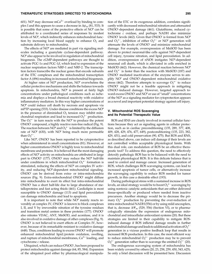
601). NO� may decrease mCa2þ overload by binding to com-plex I and this appears to cause a decrease in Dcm (81, 533). Itis possible that some of the protective effects of NO� can beattributed to a coordinated series of responses by modestlevels of NO�, which indirectly enhances mitochondrial func-tion by increasing local blood supply to enhance O2 andsubstrate delivery to mitochondria.
The effects of NO� are mediated in part via signaling mol-ecules including a guanylate cyclase-dependent pathway.NO� also appear to be involved in regulation of mitochondrialbiogenesis. The cGMP-dependent pathways are thought toactivate PGC-1a and PGC-1b, which lead to expression of thenuclear respiration factors NRF-1 and NRF-2. The transcrip-tion factors transcribe the nuclear genes that encode subunitsof the ETC complexes and the mitochondrial transcriptionfactor A (496) resulting in increased mitochondrial biogenesis.
At higher rates of NO� production, NO� overwhelms thecellular protective mechanisms and shifts the balance towardsapoptosis. In mitochondria, NO� is present at fairly highconcentrations under pathological conditions such as ische-mia and displays a broad chemical reactivity with oxidativeinflammatory mediators. In this way higher concentrations ofNO� could induce cell death by necrosis and apoptosis viamPTP opening (533). Under disease conditions the excess NO�
in the presence of diminished O2 tension may inhibit mito-chondrial respiration and lead to increased O2
�� production.The O2
�� in turn reacts with the NO� to produce the potentONOO- compound, a highly reactive non-free radical (Fig. 9).The reaction between NO� and O2
�� is limited by the diffusionrate of NO� (630), with NO� being much more permeablethan O2
��.Like NO�, ONOO- has been reported to be cytoprotective
when administered in small concentrations (81). However, athigher concentrations ONOO- is highly toxic to mitochondrialmembrane and proteins. It is believed that the most damagingeffects of NO� activity on mitochondria are attributed in largepart to ONOO- (177). ONOO- may reduce the NO� half-lifeunder conditions in which mitochondrial O2
�� formation isstimulated, reducing the effects of NO� on complex IV activ-ity, and reducing NO�-dependent mitochondrial signaling.ONOO- can be derived from extra- or intra-mitochondriasources (Fig. 9). Extra-mitochondrial ONOO- might diffuseinto mitochondria to exert its effect but intra-mitochondrialONOO- has a short half-life due to large abundance of me-talloproteins and fast acting thiols (461). Cardiolipin is mostsusceptible to ONOO- damage (113) and lipid peroxidationcan damage mitochondria and lead to apoptosis.
It is important to note that while NO� mainly reacts re-versibly at complex IV, ONOO- is known to block complexesI, II, and V by irreversible nitration of the tyrosine residuesand transition metal centers in these proteins (177). ONOO-
also nitrates VDAC, ANT, MnSOD, and aconitase, and it isalso involved in oxidative damage of other complexes (Fig. 9).ONOO- is not believed to exert effects at Complex IV, how-ever, because of its remarkable resistant to oxidative damage(648). Thus, conditions leading to excess ONOO- will promoteenhanced mitochondrial lipid=protein oxidation, swelling,and rupture of mitochondrial membranes, Ca2þ loading, andcytochrome c release.
Ubiquinol, which can reduce ONOO-, has been proposed toprotect mitochondria against damage (64, 85, 584). Expansionof the ubiquinol pool either by pharmacological manipula-
tion of the ETC or its exogenous addition, correlates signifi-cantly with decreased mitochondrial nitration and attenuatedONOO--dependent damage (461). Glutathione, reduced cy-tochrome c oxidase, and perhaps NADH also minimizeONOO- levels (462). Given that ONOO- is formed from NO�
and O2��, inhibition of either O2
�� or NO� generation willattenuate the levels of ONOO- and minimize mitochondrialdamage. For example, overexpression of MnSOD has beenshown to protect neuronal-like cells against NO�-dependentcell injury, tyrosine nitration, and lipid peroxidation. In ad-dition, overexpression of nNOS instigates NO�-dependentneuronal cell death, which is alleviated in cells enriched inMnSOD (462). However, the chemical reaction between NO�
and O2�� is faster than the reaction with MnSOD (187) and
ONOO- mediated inactivation of the enzyme serves to am-plify NO� and ONOO--dependent mitochondrial oxidativestress (462). Therefore attempts to scavenge O2
�� to reduceONOO- might not be a feasible approach in mitigatingONOO--induced damage. However, targeted approach to-ward excess ONOO- and NO� or use of ‘‘small’’ concentrationsof the NO� as a signaling mediator for cytoprotection appearsas a novel and important potential strategy against cell injury.
V. Mitochondrial ROS Scavengingand Its Potential Therapeutic Value
ROS and RNS are clearly involved in normal cellular func-tions because they act as signaling agents in cellular protec-tion, such as in cardiac preconditioning (135, 289, 377, 408,409, 428, 429, 476, 477, 649), postconditioning (135, 223, 382,429, 431), and cold preservation (90, 475). But ROS and RNS,as described above, can induce cell damage if their levels arenot controlled within acceptable physiological limits. Withthis dual role, can modulation of ROS be an effective thera-peutic tool? To address this question, the need to effectivelydetoxify pathologic ROS has to be balanced with the need tomaintain physiological ROS. It is this delicate balance that isused to control and manage cancer. Increased generation ofROS, which challenges ROS scavenging systems, can lead toincreased apoptosis of tumor cells, or alternatively increasethe scavenging capability to reduce ROS needed for tumorgrowth, in this case a desirable effect (193).
During pathological stress with a sustained increase in ROSlevels, an ideal strategy would be to boost O2
�� scavenging byusing nontoxic catalytic antioxidants that are either deliveredtissue-specifically or produced where needed from inactiveprecursors. Another strategy would be to decrease the pri-mary O2
�� production by preventing the over-reduction ofintra-mitochondrial NADH (556) or by using mild uncouplers,that is, decrease DCm (529, 556) (Section VI), or to pharma-cologically stimulate the expression of endogenous mito-chondrial and intracellular antioxidant systems (20). But thesestrategies are limited in their capability to mitigate ROS-induced damage if ROS inflicted damage results in furthermitochondrialdamageandleadstoadditionalactivationofO2
��
generation in a vicious positive feedback loop that results inincreased ROS production. In this case, the most efficient wayto reduce mitochondrial O2
�� production may be to preventO2�� generation rather than to scavenge the emitted O2
�� (20).The endogenous scavenging system of mitochondria has
been widely covered by others (20, 23, 258, 259, 450, 543, 625).So only a brief discussion will be presented here. Discussion
THERAPEUTIC STRATEGIES DIRECTED TO MITOCHONDRIA 295
on the use of antioxidants is only presented where they arerelevant in ameliorating mitochondrial related diseases. Theseantioxidants are mainly synthetic agents, for example MitoQ,a-tocopherol, vitamin-based antioxidants, and genetic ma-neuvers, such as overexpression of MnSOD.
Mitochondria possess an elaborate and well-definedmultileveled antioxidant defense system of enzymes andnonenzymes to scavenge mitochondrial O2
�� (345). Thescavenging system includes the matrix MnSOD and the glu-tathione (GSH) (Fig. 8) (20, 543) and thioredoxin (TRXSH2)(266, 571) systems, cytochrome c, peroxidase, and catalase(543). The counterpart to the matrix-bound MnSOD is the Cu-Zn SOD (SOD1) found predominantly in the cytosol, althoughrecent studies have reported the presence of SOD1 in the IMS(337); extracellular SOD is also found in interstitial fluid,plasma, lymph, and synovial fluid (285).
A. Manganese superoxide dismutase
MnSOD (SOD2) is a metallo-enzyme located primarily inthe mitochondrial matrix at levels of 10–20 mM (461) (Fig. 8)that does not require co-factors to detoxify O2
�� radicals.MnSOD plays an essential role in protecting against oxidativestress and the assembly of this tetrameric peptide into theactive manganese-based enzyme is key for survival. Its onlyknown function is to detoxify O2
�� to H2O2, thereby protect-ing mitochondrial Fe–S cluster containing enzymes from ox-idative damage (20, 199, 543). A defect in genes encodingSOD1 or SOD3 (extracellular Cu=Zn SOD) results in a mild,nonlethal phenotypic enzyme expression (98, 470). In markedcontrast, a SOD2 (MnSOD) knockout results in neonatal le-thality (263, 264) due to dilated ventricular cardiomyopathy,fibrosis, and other complications (20, 145, 262–264). Over-expression of the MnSOD gene on the other hand has beenassociated with protection against oxidative stress-mediatedcell death and cellular injury (145). For example, overex-pression of MnSOD has been shown to ameliorate the ex-
pression of proteins implicated in retinal impairment in amouse diabetic model (498).
A number of studies show that cells respond to oxidativestress, like lipopolysaccharide treatment, by increasing thelevel of MnSOD (142, 479). In contrast, in neoplastic cellswhere MnSOD is normally low, induction of the scavengerhas been implicated in the suppression of tumor growth. Thissuppressive effect in tumor cells is believed to be due tochanges in the activity and expression of transcription factorsincluding NF-kB (nuclear factor kappa of activated B cells)and NFAT (nuclear factor activated-T cells) (415).
Mitochondrial SOD mimetics (nonprotein) have been de-veloped to allow uptake into the mitochondrion to scavengeROS. The mitochondrial superoxide dismutase mimeticsMnTBAP, and Mn(III) meso-tetrakis (N-methylpryidinium-2-yl) porphyrin (MnTE-2-Py5þ) are permeable to the IMM.MnTE-2-Py5þ was shown to accumulate in heart mitochon-dria following intraperitoneal injection and MnTBAP wasreported to improve survival in MnSOD knockout mice (49).Since O2
�� is involved in signal transduction of IPC, weshowed that MnTBAP given alone abolished the cardiopro-tection afforded by preconditioning (289) (Fig. 10). MnTBAPwas also shown to scavenge ROS generated during coldperfusion (91), which confirms uptake into mitochondria.Genetically altered mice deficient in the sod2 gene die withheart failure in conjunction with other severe complications(264, 340, 369). Treating mice with MnTBAP ameliorated thecomplications and greatly increased lifespan (370).
Another cell permeable SOD2 mimetic, Mn(III)tetrakis[(1-methyl-4-pyridyl)-porphyrin] (MnTPyP), has both MnSODand catalase mimetic effects (607). We observed that MnTPyP,given before prolonged cold ischemia, protected the isolatedheart from cold-induced ROS damage during ischemia betterthan MnTBAP (Camara and Stowe, unpublished observa-tion). The protection afforded by MnTPyP was similar to thatprovided by an MnTBAP, catalase, glutathione cocktail (90).These actions of MnTPyP may be similar to the actions of a
FIG. 10. Developed (systolic–diastolic) left ventricularpressure (LVP) in two groupsof hearts exposed to eitherbrief ischemic pulses (IPC), orto IPC bracketed by MnTBAP,an SOD mimetic. DevelopedLVP returned to pre-ischemiavalues between the IPC pulsesand after the 2nd pulse (beforeindex 30 min ischemia). On re-perfusion after index ischemia,the IPC group had a better re-turn of developed LVP thanthe ischemia control group, theIPCþMnTBAP group, or theMnTBAP group. Thus, ROSscavenging blocked the pro-tection afforded by IPC. Statis-tical markings are shown inthe figure after ANOVA andStudent–Newman–Keuls testsof means from the differentgroups. Reproduced with per-mission of Kevin et al. (289).
296 CAMARA ET AL.
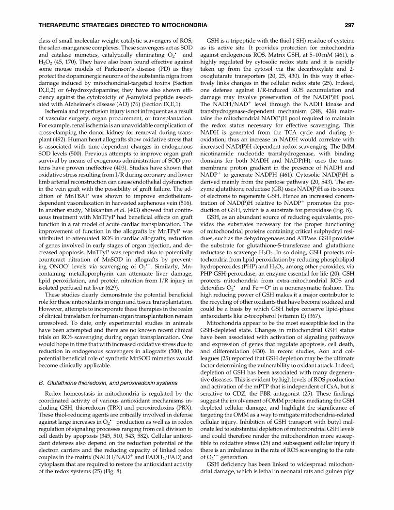
class of small molecular weight catalytic scavengers of ROS,the salen-manganese complexes. These scavengers act as SODand catalase mimetics, catalytically eliminating O2
�� andH2O2 (45, 170). They have also been found effective againstsome mouse models of Parkinson’s disease (PD) as theyprotect the dopaminergic neurons of the substantia nigra fromdamage induced by mitochondrial-targeted toxins (SectionIX,E,2) or 6-hydroxydopamine; they have also shown effi-ciency against the cytotoxicity of b-amyloid peptide associ-ated with Alzheimer’s disease (AD) (76) (Section IX,E,1).
Ischemia and reperfusion injury is not infrequent as a resultof vascular surgery, organ procurement, or transplantation.For example, renal ischemia is an unavoidable complication ofcross-clamping the donor kidney for removal during trans-plant (492). Human heart allografts show oxidative stress thatis associated with time-dependent changes in endogenousSOD levels (500). Previous attempts to improve organ graftsurvival by means of exogenous administration of SOD pro-teins have proven ineffective (403). Studies have shown thatoxidative stress resulting from I=R during coronary and lowerlimb arterial reconstruction can cause endothelial dysfunctionin the vein graft with the possibility of graft failure. The ad-dition of MnTBAP was shown to improve endothelium-dependent vasorelaxation in harvested saphenous vein (516).In another study, Nilakantan et al. (403) showed that contin-uous treatment with MnTPyP had beneficial effects on graftfunction in a rat model of acute cardiac transplantation. Theimprovement of function in the allografts by MnTPyP wasattributed to attenuated ROS in cardiac allografts, reductionof genes involved in early stages of organ rejection, and de-creased apoptosis. MnTPyP was reported also to potentiallycounteract nitration of MnSOD in allografts by prevent-ing ONOO- levels via scavenging of O2
��. Similarly, Mn-containing metalloporphyrin can attenuate liver damage,lipid peroxidation, and protein nitration from I=R injury inisolated perfused rat liver (629).
These studies clearly demonstrate the potential beneficialrole for these antioxidants in organ and tissue transplantation.However, attempts to incorporate these therapies in the realmof clinical translation for human organ transplantation remainunresolved. To date, only experimental studies in animalshave been attempted and there are no known recent clinicaltrials on ROS scavenging during organ transplantation. Onewould hope in time that with increased oxidative stress due toreduction in endogenous scavengers in allografts (500), thepotential beneficial role of synthetic MnSOD mimetics wouldbecome clinically applicable.
B. Glutathione thioredoxin, and peroxiredoxin systems
Redox homeostasis in mitochondria is regulated by thecoordinated activity of various antioxidant mechanisms in-cluding GSH, thioredoxin (TRX) and peroxiredoxins (PRX).These thiol-reducing agents are critically involved in defenseagainst large increases in O2
�� production as well as in redoxregulation of signaling processes ranging from cell division tocell death by apoptosis (345, 510, 543, 582). Cellular antioxi-dant defenses also depend on the reduction potential of theelectron carriers and the reducing capacity of linked redoxcouples in the matrix (NADH=NADþ and FADH2=FAD) andcytoplasm that are required to restore the antioxidant activityof the redox systems (25) (Fig. 8).
GSH is a tripeptide with the thiol (-SH) residue of cysteineas its active site. It provides protection for mitochondriaagainst endogenous ROS. Matrix GSH, at 5–10 mM (461), ishighly regulated by cytosolic redox state and it is rapidlytaken up from the cytosol via the decarboxylate and 2-oxoglutarate transporters (20, 25, 430). In this way it effec-tively links changes in the cellular redox state (25). Indeed,one defense against I=R-induced ROS accumulation anddamage may involve preservation of the NAD(P)H pool.The NADH=NADþ level through the NADH kinase andtranshydrogenase-dependent mechanism (248, 426) main-tains the mitochondrial NAD(P)H pool required to maintainthe redox status necessary for effective scavenging. ThisNADH is generated from the TCA cycle and during b-oxidation; thus an increase in NADH would correlate withincreased NAD(P)H dependent redox scavenging. The IMMnicotinamide nucleotide transhydrogenase, with bindingdomains for both NADH and NADP(H), uses the trans-membrane proton gradient in the presence of NADH andNADPþ to generate NADPH (461). Cytosolic NAD(P)H isderived mainly from the pentose pathway (20, 543). The en-zyme glutathione reductase (GR) uses NAD(P)H as its sourceof electrons to regenerate GSH. Hence an increased concen-tration of NAD(P)H relative to NADPþ promotes the pro-duction of GSH, which is a substrate for peroxidase (Fig. 8).
GSH, as an abundant source of reducing equivalents, pro-vides the substrates necessary for the proper functioningof mitochondrial proteins containing critical sulphydryl resi-dues, such as the dehydrogenases and ATPase. GSH providesthe substrate for glutathione-S-transferase and glutathionereductase to scavenge H2O2. In so doing, GSH protects mi-tochondria from lipid peroxidation by reducing phospholipidhydroperoxides (PHP) and H2O2, among other peroxides, viaPHP GSH-peroxidase, an enzyme essential for life (20). GSHprotects mitochondria from extra-mitochondrial ROS anddetoxifies O2
�� and Fe¼O� in a nonenzymatic fashion. Thehigh reducing power of GSH makes it a major contributor tothe recycling of other oxidants that have become oxidized andcould be a basis by which GSH helps conserve lipid-phaseantioxidants like a-tocopherol (vitamin E) (367).
Mitochondria appear to be the most susceptible foci in theGSH-depleted state. Changes in mitochondrial GSH statushave been associated with activation of signaling pathwaysand expression of genes that regulate apoptosis, cell death,and differentiation (430). In recent studies, Aon and col-leagues (25) reported that GSH depletion may be the ultimatefactor determining the vulnerability to oxidant attack. Indeed,depletion of GSH has been associated with many degenera-tive diseases. This is evident by high levels of ROS productionand activation of the mPTP that is independent of CsA, but issensitive to CDZ, the PBR antagonist (25). These findingssuggest the involvement of OMM proteins mediating the GSHdepleted cellular damage, and highlight the significance oftargeting the OMM as a way to mitigate mitochondria-relatedcellular injury. Inhibition of GSH transport with butyl mal-onate led to substantial depletion of mitochondrial GSH levelsand could therefore render the mitochondrion more suscep-tible to oxidative stress (25) and subsequent cellular injury ifthere is an imbalance in the rate of ROS scavenging to the rateof O2
�� generation.GSH deficiency has been linked to widespread mitochon-
drial damage, which is lethal in neonatal rats and guinea pigs
THERAPEUTIC STRATEGIES DIRECTED TO MITOCHONDRIA 297
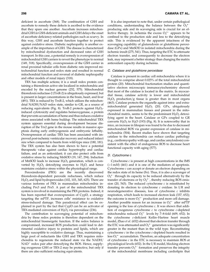
deficient in ascorbate (368). The combination of GSH andascorbate to remedy these defects is ascribed to the evidencethat they spare one another. Ascorbate increases mitochon-drial GSH in GSH-deficient animals and GSH delays the onsetof ascorbate deficiency-related pathologies such as scurvy. Inthis way, GSH and ascorbate function together to protectagainst oxidative stress. Diabetic nephropathy is another ex-ample of the importance of GSH. The disease is characterizedby mitochondrial dysfunction and decreased rates of GSHtransport. In this case, a potential remedy is overexpression ofmitochondrial GSH carriers to revert the phenotype to normal(149, 318). Specifically, overexpression of the GSH carrier inrenal proximal tubular cells from diabetic rats improved mi-tochondrial function and redox state and resulted in normalmitochondrial function and reversal of diabetic nephropathyand other models of renal injury (318).
TRX has multiple actions; it is a small redox protein con-taining a thioredoxin active site localized in mitochondria andencoded by the nuclear genome (252, 575). Mitochondrialthioredoxin reductase-2 (TrxR-2) is ubiquitously expressed, butis present in larger concentrations in the brain, heart, and liver(491). TRX is reduced by TrxR-2, which utilizes the mitochon-drial NADH=NAD redox state, similar to GR, as a source ofreducing equivalents (Fig. 8). TrxR-2 protects against H2O2-induced cytotoxicity and regulates hemeoxygenase, a proteinthat prevents accumulation of heme and thus reduces oxidativestress associated with heme buildup. The mitochondrial TRXsystem appears essential during development because dis-ruption of the Trx2 gene in the mouse results in massive apo-ptosis during early embryogenesis and embryonic lethality.Overexpression of cardiac TRX has been associated with im-proved post-ischemic ventricular recovery and reduced infarctsize when compared to hearts from wild-type mice (180, 181).The TRX system has also been shown to have a potentialtherapeutic value against cardiac hypertrophy and cardiacfailure, and as an antioxidant; it can also protect cells fromoxidative stress by inducing MnSOD (19, 147, 294). Inductionof MnSOD leads to increase H2O2 generation, which is con-verted by H2O2 detoxifying enzymes to H2O, and hencemaintains oxidative stress within the physiological range.
Peroxiredoxins (PRX) are the recently discoveredthioredoxin-dependent peroxide reductases, which reduceH2O2 and lipid hydroperoxides (102, 103, 345, 625). There arevarious isoforms of PRX in mammalian mitochondria in-cluding Prx3 and Prx5. A part of the mitochondrial TRXsystem is involved in maintaining the PRX proteins. Indeed, ithas been reported that overexpression of CypD, a strategytargeting the mPTP, increases cells’ resistance to oxidativestress-induced damage. This paradoxical effect can be ex-plained in part by the fact that CypD can be an activator ofTRX-mediated conversion of H2O2 via peroxidase (236).
The contribution to scavenging potential of mitochon-dria by these redox proteins is therefore dependent on themitochondrial bioenergetic function. If the redox balance iscompromised, increased ROS production could lead to det-rimental oxidative injury to proteins and lipids, which arehighly susceptible to oxidative damage. Thus, maintaining alarge pool of reductants like GSH and TRX requires mito-chondria to regenerate the reduced state via the NADH=NADþ redox pair after detoxifying the ROS. Hence, supply-ing exogenous GSH or TRX-2 may be protective, but only ifthere are also sufficient reducing equivalents.
It is also important to note that, under certain pathologicalconditions, understanding the balance between the O2
��
producing side and its scavenging side is crucial for an ef-fective therapy. In ischemia the excess O2
�� appears to beconfined to the production side and less to the detoxifyingside. This is evidenced by the apparent intactness of thescavenging capability of glutaredoxin or glutathione peroxi-dase (GPx) and MnSOD in isolated mitochondria during theischemic insult (275, 541). Thus, targeting the ETC to attenuateelectron transfer, and consequently to decrease the electronleak, may represent a better strategy than changing the matrixantioxidant capacity during ischemia.
C. Catalase and glutathione peroxidase
Catalase is present in cardiac cell mitochondria where it isthought to comprise about 0.025% of the total mitochondrialprotein (20). Mitochondrial fractionation studies and quanti-tative electron microscopic immunocytochemistry showedthat most of the catalase is located in the matrix. In myocar-dial tissue, catalase activity is lower and mitochondrialH2O2 production=g tissue is greater than in most organs(463). Catalase protects the organelle against intra- and extra-mitochondrial generated H2O2 (20). GPx, ubiquitouslyexpressed in mammalian tissues and present in the mito-chondrial matrix, seems to be the predominant H2O2 detoxi-fying agent in the heart. Catalase or GPx coupled to GRconverts H2O2 to H2O (193) (Fig. 8). It is noteworthy that inmice, an increase in lifespan was found to be due to decreasedmitochondrial ROS via greater expression of catalase in mi-tochondria (504). Recent studies have shown that targetingcatalase to the mitochondria can reduce cardiac pathology(e.g., cardiomyopathy with age, and cardiac amyloidosis) con-sistent with the effect of endogenous ROS to decrease heartfunctional capacity with aging (577).
D. Cytochrome c
Cytochrome c is present at high concentrations in the IMS(>1 mM) (461) and it is one of the mediators of apoptosis.Cytochrome c can accept or donate an electron depending onthe redox state of its heme (Fe). Thus, it is also a scavenger ofO2�� through its capacity to be reduced alternatively by the
transfer of electrons or by O2��, thereby reducing ROS emis-
sion (20, 543). The reduced cytochrome c is reinstituted bydonating its electron to cytochrome c oxidase. In I=R andneurodegenerative diseases, loss of cytochrome c inhibitsrespiration, which leads to increased electron leak (337, 652);the outcome is more O2
�� production and more cell damage.Another possible reason for an increase in O2
�� after mPTPopening is the loss of cytochrome c. It was shown that addi-tion of exogenous cytochrome c to cytochrome c-depletedmitochondria reduced O2
�� levels by 7–8-fold (609, 652). Inthe cytochrome c-deficient Keilin–Hartree heart musclemodel, Zhao et al. (652) showed that electron transfer throughthe ETC was attenuated and O2
�� generation was significantlygreater in the mutant than in the wild type. Reconstitutingcytochrome c in the cytochrome c-depleted hearts resulted inless O2
�� accumulation. Therefore, an adequate concentrationof cytochrome c in the ETC is necessary to maintain ROS atphysiological levels (652). In the I=R model, blocking electrontransfer prevents O2
�� formation and preserves the integrityof the mitochondrial membrane including cardiolipin that
298 CAMARA ET AL.

functions to retain cytochrome c content (114, 115, 262, 324).Hence, maintaining the integrity of cytochrome c could rep-resent a potential strategy for mitigating mitochondria-related cellular injury.
E. Mitochondria as scavengers of cytosolic O2��
Mitochondria are major scavengers of O2�� produced from
extra-mitochondrial sources. Mitochondria scavenge cyto-solic O2
�� radicals by maintaining a polarized IMM that ispositively charged during respiration. The [Hþ] in the IMSattracts the cellular O2
��. Here, O2�� radicals are protonated to
form hydroperoxyl radicals (224, 495), which can diffuse intothe matrix and become deprotonated so that the O2
�� is dis-mutated by matrix MnSOD. Net O2
�� consumption in mito-chondria creates a gradient for O2
��, which favors diffusionfrom the cytosol to the IMS. Therefore, increased MnSOD inmitochondria augments O2
�� removal from the cytosol as wellas from the mitochondria. In this regard, it may not be a co-incidence that mitochondria play a central role in cell deathand that a lack of MnSOD in the mitochondria leads to cardiacand neuronal lethality.
VI. Uncoupling Proteins in Modulationof Mitochondrial Function:Physiological and Pharmacologic Relevance
Mitochondrial uncoupling is an important physiologicalregulator of its function and redox potential and thus, is also aregulator of O2
�� production. One of the control systems thatregulate mitochondrial function and the flux of protons backacross the IMM to maintain a suitable Dcm (379) is a group ofuncoupling proteins (UCPs). The UCPs (1–4) and fatty acidsare believed to induce an inward Hþ ‘‘leak’’ in energized mi-tochondria (70, 274, 543, 544). A key role for UCPs in regu-lating mitochondrial metabolism is supported by the presenceof different isoforms in various mammalian tissues. UCPsprevent excessive ROS accumulation by maximizing respi-ration rate (401). The obscure roles of UCPs in normal phys-iology, and their emerging role in pathology, provide excitingpotential for further investigation. However, neither the exactphysiological nor biochemical roles of UCP homologues arewell understood (342, 626).
Natural uncouplers like fatty acids and proteins, and arti-ficial uncouplers like carbonyl cyanide m-chlorophenylhy-drazone (CCCP), inhibit ROS production by decreasing Dcm
(253, 536). UCPs are IMM proteins that dissipate the mito-chondrial Hþ gradient. Other biological effects of UCPs aretheir ability to attenuate mitochondrial ROS production (537)and to reduce the damaging effects of ROS during cardiac I=Ror hypoxia injury (42, 188). In IPC and PPC, activation ofputative mKATP channels may lead to mild uncoupling ofmitochondrial respiration via mild ‘‘proton leak’’ with a con-comitant increase in O2
�� generation that provides the signalfor protection against ischemic damage (261). This form of‘‘proton leak’’ is attenuated by GDP, a UCP inhibitor, sug-gesting that the Hþ leak is mediated via UCPs.
In a recent study (253), we showed that a Hþ leak instigatedby the putative mitochondrial big KCa (mBKCa) channel incardiomyocytes opening led to ROS production without dis-turbing Dcm. This is possible because the small Hþ leak andthe concomitant small increase in Hþ pumping by ETCcomplexes increases the respiration rate without decreasing
Dcm while increasing the rate of H2O2 production (253). Itis possible that this ‘‘mild’’ uncoupling may contribute to theprotection of the heart against I=R injury as shown by theBKCa channel agonist NS1619 (542). We showed that NS1619-induced protection was mediated by preservation of mito-chondrial redox state, reduced mCa2þ loading, and betterfunctional recovery; this protection was abolished whenhearts were treated with MnTBAP and paxilline, the BKCa
channel antagonist.It has also been reported that overexpression of UCP2
protein in mice protects brain from severe infarction in I=R(154). UCP2 is thought to mediate such protection, possibly bycausing mild mitochondrial depolarization that could limitmCa2þ uptake, reduce ROS production, and so protect cellsfrom damage (177). It is feasible that UCPs could alleviatedamage from I=R injury if they could be activated or stimu-lated before the insult to reduce Dcm, and before the redoxincreases because of inhibited electron transfer (543). More-over, it is possible that shifting from state 4 to state 3 respi-ration could be sufficient to lower Dcm enough to have thesame effects as uncoupling agents on ROS production. Thusmaintaining a depolarized Dcm state is crucial in mitigatingROS-mediated injury.
Recent studies have shown that UCPs play an importantrole in the pathogenesis of obesity, type-2 diabetes, aging, andtumor progression (626). It was reported that the expressionof UCPs increased in response to increased mitochondrialoxidative stress and that they serve as the link between dia-betes and mitochondrial ROS (449). In endothelial cells, highglucose levels increase mitochondrial ROS, and the normali-zation of mitochondrial ROS production by inhibitors of mi-tochondrial metabolism, or by the overexpression of UCP-1,prevent the glucose-induced formation of advanced glycationend products that are believed to underlie major moleculardiabetic complications (405). In contrast, in ob=ob mice thatlack a functional UCP-2 gene, or when UCP-2-deficient micewere fed high fatty diets, glucose stimulated insulin secretionwas enhanced compared to the wild type (342). These studiesimply that UCPs may be important in insulin-glucose ho-meostasis and may contribute to impaired glucose-stimulatedinsulin secretion in diabetes. In human islet cells, chronicglucose or free fatty acid concentrations increase UCP-2 ex-pression (342). These results are consistent with the findingthat UCP-2 overexpression impairs b-cell function (342). Thus,minimizing UCP-2 activity in pancreatic b-cells could repre-sent a valid and viable approach to improve b-cell functionand to treat diabetes. However, such a conclusion is consid-ered tenuous or ‘‘precocious’’ according to Anetor et al. (22),who reported an absence of significant oxidative stress inmitochondria, so that it was thought less likely that the UCP-2-superoxide pathway was involved in the inhibition ofglucose-stimulated insulin release.
Another possible alternative approach to ameliorate ROSproduction is to modulate mitochondrial bioenergetics withlow doses of artificial uncouplers. As discussed previously(543), ROS production is more likely when the Dcm is highlypolarized. Therefore, any slight decrease in Dcm, as has beenreported by most studies (309,317,543), or trans-matrix DpH(317), results in a marked reduction or cessation of ROS pro-duction. In this case, temporary partial uncoupling of respi-ration from phosphorylation by inducing a mild Hþ ‘‘leak’’has potential therapeutic benefit. For example, the use of
THERAPEUTIC STRATEGIES DIRECTED TO MITOCHONDRIA 299
dinitrophenol (DNP), which increases permeability of theIMM to protons, has been proposed and even attempted intreating obesity. However DNP has a low therapeutic index(narrow concentration difference between therapeutic po-tential and toxicity) that limits its utility as a viable therapeuticoption (Section VI) (58). An alternative approach to usingartificial uncouplers is the use of endogenous uncouplers thatinduce Hþ leak (449). However, the development of this ap-proach is dependent on unraveling the mechanisms involvedin activating the uncoupling proteins.
Some lipid peroxidation products, such as 4-hydroxy-trans-2-noneal (HNE) may induce partial uncoupling ofmitochondria through UCPs and are thought to initiate pro-tective mechanisms (60, 182). HNE can also induce un-coupling of OXPHOS by enhancing Hþ leak through othermembrane proteins such as ANT if Dcm is high (32). It hasbeen proposed recently by Brookes (69, 70) that ROS and Hþ
leak comprise a loop not requiring UCPs to operate, but ratheris dependent on Dcm alone. A high Dcm would generate ROSand the ROS would, in turn, cause a Hþ leak to reduce theDcm in a feedback manner. The ROS so generated could in-duce Hþ leak in part by lipid peroxidation or possibly byprotonation of O2
�� in the acidic IMS to HO2� (346, 543), which
is membrane permeable and is deprotonated by the alkalinepH in the matrix (153, 543). A decrease in OXPHOS as a resultof uncoupling and the less efficient metabolic rate associatedwith a Hþ leak may be linked to the aging process (70) (SectionXII). Taken together, these findings suggest a better under-standing of the physiological roles and molecular mecha-nisms of uncoupling proteins and the consequence of Hþ leakor Hþ ‘‘slip’’ (70) to better understand the role of this phe-nomenon in mitochondrial function and dysfunction (449). Abetter knowledge of the mechanism of Hþ leak by UCPs andsynthetic uncouplers could provide the basis for better designof drugs that target this aspect of mitochondrial biology in-volving compromised mitochondrial function in the diseasestate.
VII. Mitochondrial DNA-Related Pathologiesand a Potential Therapeutic Target
Mitochondria are inherited through the maternal lineage,though some recent evidence suggests that in rare instancesmitochondria may also be inherited via a paternal route (506).Mitochondrial DNA (mtDNA) exists in hundreds of identicalcopies=cell (homoplasmy) but can also exist in multiple non-identical copies within individual cells (heteroplasmy). Un-like nuclear DNA (nDNA), mitochondrial nonchromosomalDNA (mtDNA) does not get shuffled every generation, so it ispresumed to change at a much slower rate (75). It is believedthat mtDNA, organized in nucleoprotein complex (nucle-oids), are particularly sensitive to oxidative stress due to itsproximity to the ETC and the lack of histones (521, 634). Mi-tochondrial DNA also lacks introns, and as a result, mutationsin the genome occur primarily in the coding sequence, con-sistent with the notion that mtDNA has a higher mutation rate(10–20 times) than nDNA (50, 221). For these reasons, accu-mulation of mtDNA damage plays a causative role in variousdisorders that are associated with aging, cancer, neurode-generative diseases, and other diseases (634).
However, based on the organization of mtDNA into nu-cleoids and their proximity to the ETC, nucleoids may have an
integrated antioxidant system to protect mtDNA from oxi-dative stress (293). Indeed, Kienhofer et al. (293) recentlyconfirmed the presence of SOD2 in the nucleoid and notedthat it binds directly to mtDNA through ionic binding forces.This mtDNA–SOD2 interaction in the nucleoid protectedagainst oxidative damage as evidenced by the increasedvulnerability of free mtDNA to X-ray and H2O2 than mtDNAin a nucleoid complex. The mtDNA–SOD2 interaction mayalso explain in part the enhanced ROS levels, the increasedmtDNA damage, the reduced activities of the ETC complexesand TCA cycle enzymes, and endothelial dysfunction ob-served in MnSODþ=� mice (614).
Much of the mtDNA is used to code the manufactureof proteins that are key components of the energy produc-tion system. All 13 proteins encoded by mtDNA contributesubunit components to most of the respiratory complexes.mtDNA encodes 7 subunits of complex I, 1 subunit of com-plex III, 3 subunits of complex IV, and 2 subunits of complexV. Cells depleted of mtDNA, r0-cells, lack some of the criticalsubunits of the ETC complexes, which result in a defectiverespiratory system evidenced by a reduced Dcm. These cellsare resistant to anoxia-induced cell death (lack of ROS) andthey are dependent exclusively on anaerobic energy produc-tion via glycolysis (312, 524), much like tumor cells.
Most mitochondrial proteins that regulate replication andrepair are encoded by the nuclear genome (540). Complex I,the largest holoenzyme in the ETC, has 45 nuclear-encodedand 7 mitochondria-derived subunits (438). Complex II con-sists only of nuclear-encoded subunits, and complex IIIconsists of 10 nuclear-encoded and 1 mitochondria-derivedsubunits. Complex IV has 7–11 nuclear-encoded subunits,depending on the tissue type, and 3 subunits are mitochon-dria derived (323, 598) as noted above.
Currently, it is believed that various mutations are re-sponsible for more than 120 syndromes associated with mi-tochondrial proteins (357, 433) (Section IX). There are also anumber of mitochondrial diseases associated with specificmutations in mtDNA or in nDNA coding for mitochondrialproteins (165, 176, 515) (Table 2). In other diseases like PD,type 2 diabetes, and cancer, the disease process is morecomplex and involves multiple genetic and environmentalcauses (178, 423, 439, 458). In all these diseases, the geneticmutations lead to impaired mitochondrial energy-generatingmachinery. In this scenario, disabled mitochondria with im-paired ETC would cause electron leak and increased O2
��
production. Indeed, the most common source of somaticmutation of mtDNA is O2
�� generated from the ETC. TheO2�� produced in the mitochondrion can continue in a self-
perpetuating process leading to even more damage and moreO2�� generation (Section IV,A). It is likely that gene replace-
ment has potential to be used to correct a mutant mitochon-drial genome similar to classical gene transfer therapies thathave replaced defective nuclear genes (509). Indeed, geneticmaneuvers of different sorts have been employed to reversemitochondrial related diseases. These include, but are by nomeans limited to: a) DNA coupled covalently to mitochon-drial leader peptides (chimeras) that enter mitochondriathrough protein import pathways; b) manipulating mtDNAreplication by import into mitochondria endonucleases thatmight selectively destroy a specific mutant sequence; and c)suppression of mutant mtDNA expansion to alternativelysalvage OXPHOS (498, 655).
300 CAMARA ET AL.
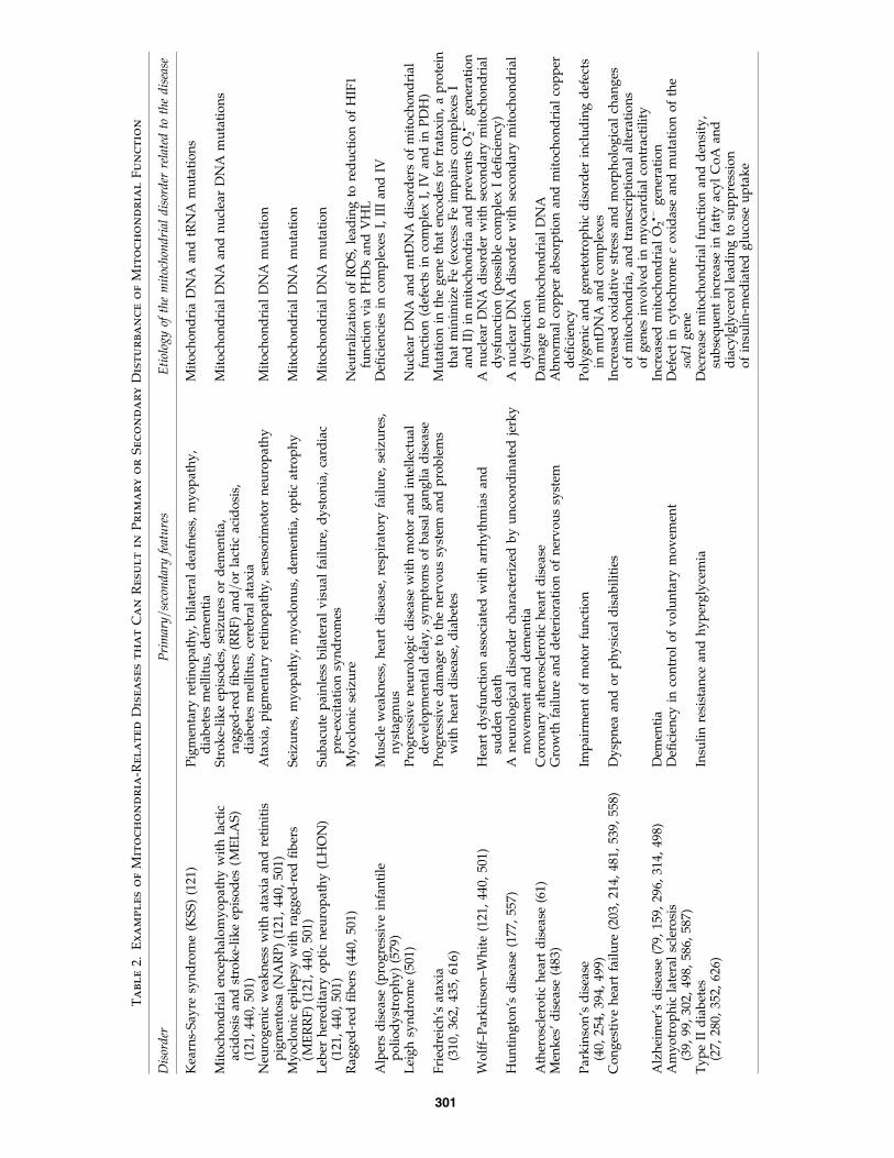
Ta
bl
e2.
Ex
am
pl
es
of
Mit
oc
ho
nd
ria
-R
el
at
ed
Dise
ase
st
ha
tC
an
Re
su
lt
in
Pr
im
ar
yo
rS
ec
on
da
ry
Dist
ur
ba
nc
eo
fM
it
oc
ho
nd
ria
lF
un
ct
io
n
Dis
ord
erP
rim
ary=
seco
nd
ary
feat
ure
sE
tiol
ogy
ofth
em
itoc
hon
dri
ald
isor
der
rela
ted
toth
ed
isea
se
Kea
rns-
Say
resy
nd
rom
e(K
SS
)(1
21)
Pig
men
tary
reti
no
pat
hy
,b
ilat
eral
dea
fnes
s,m
yo
pat
hy
,d
iab
etes
mel
litu
s,d
emen
tia
Mit
och
on
dri
aD
NA
and
tRN
Am
uta
tio
ns
Mit
och
on
dri
alen
cep
hal
om
yo
pat
hy
wit
hla
ctic
acid
osi
san
dst
rok
e-li
ke
epis
od
es(M
EL
AS
)(1
21,
440,
501)
Str
ok
e-li
ke
epis
od
es,
seiz
ure
so
rd
emen
tia,
rag
ged
-red
fib
ers
(RR
F)
and=
or
lact
icac
ido
sis,
dia
bet
esm
elli
tus,
cere
bra
lat
axia
Mit
och
on
dri
alD
NA
and
nu
clea
rD
NA
mu
tati
on
s
Neu
rog
enic
wea
kn
ess
wit
hat
axia
and
reti
nit
isp
igm
ento
sa(N
AR
P)
(121
,44
0,50
1)A
tax
ia,
pig
men
tary
reti
no
pat
hy
,se
nso
rim
oto
rn
euro
pat
hy
Mit
och
on
dri
alD
NA
mu
tati
on
My
ocl
on
icep
ilep
syw
ith
rag
ged
-red
fib
ers
(ME
RR
F)
(121
,44
0,50
1)S
eizu
res,
my
op
ath
y,
my
ocl
on
us,
dem
enti
a,o
pti
cat
rop
hy
Mit
och
on
dri
alD
NA
mu
tati
on
Leb
erh
ered
itar
yo
pti
cn
euro
pat
hy
(LH
ON
)(1
21,
440,
501)
Su
bac
ute
pai
nle
ssb
ilat
eral
vis
ual
fail
ure
,d
yst
on
ia,
card
iac
pre
-ex
cita
tio
nsy
nd
rom
esM
ito
cho
nd
rial
DN
Am
uta
tio
n
Rag
ged
-red
fib
ers
(440
,50
1)M
yo
clo
nic
seiz
ure
Neu
tral
izat
ion
of
RO
S,
lead
ing
tore
du
ctio
no
fH
IF1
fun
ctio
nv
iaP
HD
san
dV
HL
Alp
ers
dis
ease
(pro
gre
ssiv
ein
fan
tile
po
lio
dy
stro
ph
y)
(579
)M
usc
lew
eak
nes
s,h
eart
dis
ease
,re
spir
ato
ryfa
ilu
re,
seiz
ure
s,n
yst
agm
us
Defi
cien
cies
inco
mp
lex
esI,
III
and
IV
Lei
gh
syn
dro
me
(501
)P
rog
ress
ive
neu
rolo
gic
dis
ease
wit
hm
oto
ran
din
tell
ectu
ald
evel
op
men
tal
del
ay,
sym
pto
ms
of
bas
alg
ang
lia
dis
ease
Nu
clea
rD
NA
and
mtD
NA
dis
ord
ers
of
mit
och
on
dri
alfu
nct
ion
(def
ects
inco
mp
lex
I,IV
and
inP
DH
)F
ried
reic
h’s
atax
ia(3
10,
362,
435,
616)
Pro
gre
ssiv
ed
amag
eto
the
ner
vo
us
syst
eman
dp
rob
lem
sw
ith
hea
rtd
isea
se,
dia
bet
esM
uta
tio
nin
the
gen
eth
aten
cod
esfo
rfr
atax
in,
ap
rote
inth
atm
inim
ize
Fe
(ex
cess
Fe
imp
airs
com
ple
xes
Ian
dII
)in
mit
och
on
dri
aan
dp
rev
ents
O2� �
gen
erat
ion
Wo
lff–
Par
kin
son
–Wh
ite
(121
,44
0,50
1)H
eart
dy
sfu
nct
ion
asso
ciat
edw
ith
arrh
yth
mia
san
dsu
dd
end
eath
An
ucl
ear
DN
Ad
iso
rder
wit
hse
con
dar
ym
ito
cho
nd
rial
dy
sfu
nct
ion
(po
ssib
leco
mp
lex
Id
efici
ency
)H
un
tin
gto
n’s
dis
ease
(177
,55
7)A
neu
rolo
gic
ald
iso
rder
char
acte
rize
db
yu
nco
ord
inat
edje
rky
mo
vem
ent
and
dem
enti
aA
nu
clea
rD
NA
dis
ord
erw
ith
seco
nd
ary
mit
och
on
dri
ald
ysf
un
ctio
nA
ther
osc
lero
tic
hea
rtd
isea
se(6
1)C
oro
nar
yat
her
osc
lero
tic
hea
rtd
isea
seD
amag
eto
mit
och
on
dri
alD
NA
Men
kes
’d
isea
se(4
83)
Gro
wth
fail
ure
and
det
erio
rati
on
of
ner
vo
us
syst
emA
bn
orm
alco
pp
erab
sorp
tio
nan
dm
ito
cho
nd
rial
cop
per
defi
cien
cyP
ark
inso
n’s
dis
ease
(40,
254,
394,
499)
Imp
airm
ent
of
mo
tor
fun
ctio
nP
oly
gen
ican
dg
enet
otr
op
hic
dis
ord
erin
clu
din
gd
efec
tsin
mtD
NA
and
com
ple
xes
Co
ng
esti
ve
hea
rtfa
ilu
re(2
03,
214,
481,
539,
558)
Dy
spn
eaan
do
rp
hy
sica
ld
isab
ilit
ies
Incr
ease
do
xid
ativ
est
ress
and
mo
rph
olo
gic
alch
ang
eso
fm
ito
cho
nd
ria,
and
tran
scri
pti
on
alal
tera
tio
ns
of
gen
esin
vo
lved
inm
yo
card
ial
con
trac
tili
tyA
lzh
eim
er’s
dis
ease
(79,
159,
296,
314,
498)
Dem
enti
aIn
crea
sed
mit
och
on
dri
alO
2� �
gen
erat
ion
Am
yo
tro
ph
icla
tera
lsc
lero
sis
(39,
99,
302,
498,
586,
587)
Defi
cien
cyin
con
tro
lo
fv
olu
nta
rym
ov
emen
tD
efec
tin
cyto
chro
me
co
xid
ase
and
mu
tati
on
of
the
sod
1g
ene
Ty
pe
IId
iab
etes
(27,
280,
352,
626)
Insu
lin
resi
stan
cean
dh
yp
erg
lyce
mia
Dec
reas
em
ito
cho
nd
rial
fun
ctio
nan
dd
ensi
ty,
sub
seq
uen
tin
crea
sein
fatt
yac
yl
Co
Aan
dd
iacy
lgly
cero
lle
adin
gto
sup
pre
ssio
no
fin
suli
n-m
edia
ted
glu
cose
up
tak
e
301
Mitochondrial DNA has the capacity to form a mixture ofboth wild-type and mutant DNA genotypes within a cell(heteroplasmy). Cellular dysfunction usually occurs when theratio of mutated to wild-type mtDNA exceeds a threshold level(56). Mitochondrial disorders of the heteroplasmic type and theassociated disease could be remedied by selectively blockingthe replication of mutant DNA molecules to allow repopu-lation of the wild-type mtDNA (567). On the other hand, apotential therapeutic maneuver for destroying fast growingtumor cells involves the use of ethidium bromide to depletemtDNA and the antibiotic ciprofloxacin to block OXPHOSand deplete ATP needed for cell proliferation (28, 29, 100, 320).
Mitochondria have developed a complex system to importproteins. The import of nuclear encoded mitochondrial pro-teins is based on specific targeting sequences (498) that couldbe altered by mutation of the nucleotide base in the nucleargenome. The protein import pathway has been used to directthe import of chimeric proteins into mitochondria. It couldalso be used to direct the appropriate mitochondrial genesequences to mitochondria to correct for a defective protein(28, 29). In this case, the use of mitochondrial protein impor-tation machinery may lead to novel mtDNA delivery strate-gies (Section VIII). A number of mitochondria-related diseasescould be linked to faults in the transcription-translationalprocess, in the mitochondrial importation of proteins fol-lowing post-translational modification, or simply due tomutation of the mitochondrial genome. Defects in the importsystem are rare, but mutations of genes involved in the mi-tochondrial import machinery have been shown to be thecause of several maladies, including the Mohr–Tranebjaergsyndrome (sensorineural deafness, dystonia, dysphagia, cor-tical blindness, and paranoia) (498). Abnormalities in mito-chondrial protein import have also been implicated inneurological disorders such as AD (159). A detailed descrip-tion of mitochondrial and nuclear genomic related diseasesis provided in the literature and will not be described herebeyond cursory observations.
VIII. Mitochondrial Interaction with Other Organelles:Therapeutic Implications
Mitochondria often form a 3-D branching network insidecells along the cytoskeleton with tight connections to otherorganelles. This association with the cytoskeleton determinesmitochondrial shape and function (466) and ultimately propercellular function (21, 26). This anatomical connection providesfor a dynamic feature in mitochondrial biology. Mitochondriain the cell tend to have anatomical and functional connectivitywith each other and with other organelles such as the nucleusand the endoplasmic reticulum (ER). These dynamic func-tional interconnections are essential for normal function ofmitochondria and play a vital role in preserving cellularfunction and integrity. It is therefore anticipated that de-rangement of this link could be involved in pathologic statesassociated with mitochondrial dysfunction. Thus, exploita-tion of this dynamic anatomic relationship could represent anew target for potential therapy in altering mitochondrial-mediated cell death.
A. Mitochondrion–mitochondrion interaction
Mitochondrial shape, size, and number vary from organ toorgan, tissue-to-tissue, and cell-to-cell, and they vary under
physiological and pathological conditions. In vivo, mito-chondria often merge to form a reticulated network thatunder physiological conditions are governed by the dynamicequilibrium between fusion and fission. Indeed, the entiremitochondrial population is in constant flux and the identityof an individual mitochondrion is influenced by a nearbymitochondrion with its potentially different mitochondrialgenome (heteroplasmy) (105). Interestingly, proteins suchas GTPases, kinases, and phosphatases are involved in bi-directional communication between the mitochondrial retic-ular network and other organelles and the rest of the cell (365).
In recent years this new emerging concept has providedincreased molecular definition to the mitochondrial networkas a central platform in the execution of diverse cellularfunctions (i.e., from maintaining normal cell function to ini-tiating cell damage). With this comes a new concept focusedon the idea that mitochondrial dynamics (membrane fusion=fission) is a potential target in mitigating mitochondrial re-lated diseases. The DCm is essential in preserving and main-taining the dynamic process involved in establishing themitochondrial network, which in turn is important for uni-fying function and responses to intracellular signals (314).This dynamic relationship is maintained by specific proteinswhose presence is important in maintaining and regulatingthe integrity of the IMM and cytochrome c, both of which arenecessary to maintain mitochondrial respiration by genera-tion of the Hþ gradient necessary to establish DCm.
Mitochondrial fusion and fission genes have been de-scribed recently (105). The mitochondrial fusion proteins,encoded by mitofusin 1 (Mfn1) and mitofusin 2 (Mfn2), helpto regulate mitochondrial function and to maintain OMM andIMM fusion between mitochondria. To achieve fusion, theproton gradient of the IMM, but not the DCm, is required forthe connection of the OMM and to achieve IMM fusion; alarge DCm and GTP must be available (29, 105). The dynaminsmitofusin and optic atrophy 1 (OPA-1) are GTPase proteinsassociated with the OMM and IMM, respectively, and areessential for mitochondrial fusion (562). The morphologicalorientation of mitochondria is relevant in regulating cellularapoptosis with increased fragmentation of the network lead-ing to an increased tendency for cell death (365, 562). In nor-mal cells where DCm is maintained, mitochondrial fusion in atubular network is maintained. In cells derived from diseasestates that show compromise in the DCm as a result of a defectin the ETC complexes and reduced respiration, the cells showa degree of mitochondrial network disintegration with in-creased fragmentation leading to increased cell death (365,562). Loss of function of Mfn2 results in decreased substrateoxidation and DCm as a result of repression of nuclear en-coded proteins involved in OXPHOS. Thus, the machinerythat governs mitochondrial dynamics also participates in thetemporal regulation of metabolism (365).
Defects in mitochondrial fusion proteins and subsequentfragmentation of the mitochondrial tubular network are im-plicated in numerous mitochondrial myopathies, includingneurodegenerative diseases (314) and I=R injury (24, 65). Forexample, a set of mutations in Mfn2 leads to Charcot–Marie–Tooth type 2A, a peripheral neuropathy characterized byaxonal degeneration (372–374). In mouse embryonic fibro-blasts, a deficiency in Mfn1 or 2 leads to accumulation offragmented mitochondria and embryonic lethality, whereasoverexpression of either one of the mitofusins restores the
302 CAMARA ET AL.
mitochondrial tubular network (151, 152, 373). In anotherstudy it was shown that a muscle-specific knockdown of theDrosophila homologue of Mfn (Marf ) or OPA-1 results insignificant mitochondrial fragmentation and damaged cristae(155). In all these abnormalities, the common denominator isalteration of DCm because of reduced activity of the com-plexes. The process is reversed by OPA-1, which preventsrelease of cytochrome c and maintains IMM integrity.
Mitochondrial fission (fragmentation) depends on the mi-tochondrial fission proteins (Fis1) and another GTPase dy-namin family protein, DRP-1. These mitochondrial membraneproteins, when present, mark the spot where fragmentation isinitiated. Overexpression of Fis1 proteins in normal wild-typecells resulted in increased fragmentation and increased apo-ptosis as a result of increased Bax and Bak on the OMM andsubsequent cytochrome c release (105, 314, 498). Overexpres-sion of DRP-1 has been associated with significant fragmen-tation and disruption of IMM and matrix cristae (155). Theseproteins provide an intriguing link between pro- and anti-apoptotic proteins and permeabilization of the OMM. Mi-tochondrial fission also plays an important role in normalmitochondrial function. For example, it has been reported thatdisruption of fission proteins might lead to abnormal mito-chondrial function (105, 314, 498). Clearly, normal Dcm andelectron transfer have local effects on mitochondrial structure,networking design, and normal function. Thus, membranedynamic proteins of the IMM are also intimately involved inapoptotic regulation and represent potentially novel targetsfor therapeutic intervention.
In many diseases, in which a less polarized Dcm maycontribute to mitochondrial integrity, a strategy to maintainintegrity of the ETC with normal electron flux is a therapeuticoption. Moreover, the site within the ETC where the defect liesmay be critical in the design of drugs to target mitochondria.For example, in PD defects in complexes I and III are associ-ated with compromised Dcm and may contribute to subse-quent fragmentation of mitochondrial connectivity (40, 304,497, 557). On the other hand, a defect in complex V in thedisease NARP (neuropathy, ataxia, and retinitis pigmentosa)does not alter Dcm. Impaired levels of Bcl-2 anti-apoptoticproteins are also characteristic of NARP (314). A possibletherapeutic approach in the situation, where Dcm instability isa contributing factor in mitochondrial fission, is to providesubstrates like N,N,N0,N0-tetramethyl p-phenylenediamine torestore electron flux and thereby reestablish membranefunctional integrity and Dcm. A note of caution is that thistype of pharmacological maneuver may not be suitable fordiseases that are not linked to specific defects in the ETC. It istherefore essential to understand the specific underlyingcause of the defect to properly design the appropriate therapy.Another approach to reduce cytotoxicity involves overex-pression of the fusion genes and inhibition of the fission genesin a replicative cell line (606). However, this approach will notlikely work in mature post mitotic cells.
B. Mitochondrion–nucleus interaction
The existence of two spatially separated genomes, eachcontributing, albeit asymmetrically, to the biogenesis of mi-tochondria, has led to the suggestion that the two organellesinteract to provide a coordinated cellular response to intra-cellular changes. How the nucleus and mitochondria interact,
how mtDNA and nDNA gene expression is coordinated, orhow mtDNA is maintained within the cell (a process strictlydriven by nuclear factors), are key factors necessary for un-derstanding the molecular mechanisms underlying manydisease states. The nucleus regulates numerous mitochondrialfunctions because a majority of mitochondrial proteins areencoded by the nuclear genome. Thus, disruption in the line ofcommunication between the two organelles can lead to initi-ation of adaptive responses to reorganize cellular metabolism.The mitochondrial proteins encoded by the nuclear genomeare synthesized on cytoplasmic ribosomes and the peptidesare imported into the matrix after specific post-translationalmodifications. Import of nuclear encoded proteins or pre-cursor peptides relies on a proper function of the mitochon-drial protein uptake machinery, including chaperones andtransmembrane peptide import systems.
Mitochondrial oxidative stress that leads to the accumula-tion of unfolded proteins in the matrix subsequently leads tothe upregulation of nuclear-encoded chaperone genes that aretranscribed and imported into the mitochondria. Mitochon-drial chaperones, chaperonin 60 and chaperonin 10, are up-regulated via activation of the transcription factor CHOP, aCEBP-b analogue (650). The mitochondrial factor that leads toCHOP transcription remains unclear. The response is specificsince chaperonin 10 and 60 are induced, but HSP70, anothermitochondrial chaperone is not. Thus, mitochondria can sig-nal the nucleus in a specific fashion in order to mount a stressresponse that is specific to the mitochondrial compartment.
The transmembrane peptide import systems (i.e., the outerand inner membrane translocases (TOM and TIM, respec-tively)), import proteins determined by the amino terminalsequences of the pre-protein. The direction of proteins tospecific mitochondrial compartments contributes to regula-tion of mitochondrial metabolism (305, 330, 392). Thus, a co-ordinated protein import system is essential for mitochondrialmaintenance and biogenesis; a disturbance of this commu-nication between these organelles may underlie a diseaseprocess.
Mitochondria–nuclear communication has been implicatedin modulating mitochondrial biogenesis and intra-mCa2þ
distribution and swelling (550). A nuclear encoded pro-tein, the peroxisome proliferator-activated receptor-gammacoactivator-1a (PGC-1a), is a transcriptional co-activator thatmodulates mitochondrial biogenesis and in this way regulatescellular metabolism. One of many functions of PGC-1a isregulation of nuclear transcription factors, NRF-1 and -2 andmitochondrial transcription factor-1, a protein that transfersfrom the nucleus to the mitochondria where it promotes anincreased production of mitochondrially encoded proteinsand the replication of mtDNA (177, 589). PGC-1a also en-hances the activity of mitochondrial specific scavengers, in-cluding MnSOD and GPx1, during oxidative stress (537, 589).Indeed, increasing PGC-1a levels markedly enhances cellsurvival from oxidative stress. St-Pierre et al. (537) showedthat PGC-1a levels are regulated by mitochondrial ROS whichin turn activates the complex and multifaceted ROS scav-enging system. The implication of PGC-1a in regulating mi-tochondrial function, while at the same time minimizing ROSproduction, makes it an ideal protein to control or to limitmitochondrial damage. A deficiency in PGC-1a in the brainrenders it susceptible to neurodegeneration with apoptoticcell death and increased oxidative injury (537). On the other
THERAPEUTIC STRATEGIES DIRECTED TO MITOCHONDRIA 303
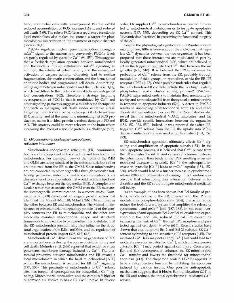
hand, endothelial cells with overexpressed PGC1-a exhibitreduced accumulation of ROS, increased Dcm, and reducedcell death (589). The role of PGC-1a as a regulatory function inlipid metabolism also makes the protein a target for phar-macological intervention in the treatment of type-2 diabetes(Section IX,C).
PGC-1a regulates nuclear gene transcription through amCa2þ signal to the nucleus and conversely, PGC-1a levelsare partly regulated by cytosolic Ca2þ (94, 550). This suggeststhat a feedback regulation operates between mitochondriaand the nucleus through cellular and mCa2þ signaling. Inaddition, the release of cytochrome c, and the subsequentactivation of caspase activity, ultimately lead to nuclearfragmentation, chromatin condensation, and the formation ofapoptotic bodies and programmed cell death. Another sig-naling agent between mitochondria and the nucleus is H2O2,which can diffuse to the nucleus where it acts as a mitogen atlow concentrations (602). Therefore a strategy involvingoverexpression of PGC-1a that is modulated by Ca2þ andother signaling pathways suggests a multifaceted therapeuticapproach in managing cell death under oxidative stress.Targeting the mitochondria by inducing PGC-1a to increaseETC activity, and at the same time minimizing net ROS pro-duction, makes it an ideal protein to reduce damage in PD andAD. This strategy could be more relevant to the brain whereincreasing the levels of a specific protein is a challenge (537).
C. Mitochondria–endoplasmic=sarcoplasmicreticulum interaction
Mitochondria–endoplasmic reticulum (ER) communica-tion is a vital component in the structure and function of themitochondria. For example, many of the lipids of the IMMand OMM are not synthesized in the mitochondria but ratherare imported from the ER to the OMM. Since mitochondriaare not connected to other organelles through vesicular traf-ficking pathways, mitochondria–ER communication is viadiscrete sites of close apposition that would facilitate lipid andCa2þ exchange between the organelles (308). A complex mo-lecular tether that associates the OMM with the ER mediatesthe interorganelle communication. In a recent study, Korn-mann et al. (308) developed an elegant genetic screen thatidentified the Mmm1=Mdm10=Mdm12=Mdm34 complex asthe tether between ER and mitochondria. The Mmm1 (main-tenance of mitochondrial morphology protein 1) of the com-plex connects the ER to mitochondria and the other coremolecules maintain mitochondrial shape and structuralframework to connect the two organelles. It is postulated thatthe ER–mitochondria junction may also influence the struc-tural organization of the IMM, mtDNA, and the regulation ofmitochondrial protein import (308, 617, 618).
Mitochondrial Ca2þ dynamics as well as generation of ROSare important events during the course of cellular injury andcell death. Mehrotra et al. (366) reported that oxidative stresspotentiates membrane damage induced by Ca2þ. The ana-tomical proximity between mitochondria and ER creates alocal microdomain in which the local mitochondrial [ATP]within the microdomain is required for ER Ca2þ signaling(177, 550). This proximity of mitochondria to Ca2þ releasesites has functional consequences for intracellular Ca2þ sig-naling. Mitochondrial uncouplers and the complex V blockeroligomycin are known to blunt ER Ca2þ uptake. In reverse
order, ER supplies Ca2þ to mitochondria as needed for con-trol of mitochondrial metabolism or to instigate apoptosis=necrosis (167, 550), depending on ER Ca2þ content. This‘‘dynamic duo’’ is critical in preserving the functional integrityof the cell.
Despite the physiological significance of ER-mitochondriamicrodomain, little is known about the molecules that regu-late Ca2þ dynamics between the two organelles. It has beenproposed that these interactions are modulated in part bylocally generated mitochondrial ROS, which are believed toact as the trigger to regulate the Ca2þ flux between the or-ganelles (605, 610). It is believed that ROS increases theprobability of Ca2þ release from the ER, probably throughmodulation of thiol groups on ryanodine, or via the ER IP3receptor (IP3R) (177). Other possible molecules that regulatethe mitochondria–ER contacts include the ‘‘sorting’’ protein,phosphofurin acidic cluster sorting protein-2 (PACS-2).PACS-2 helps mitochondria to maintain their membrane in-tegrity and to translocate Bid from the cytosol to mitochondriain response to apoptotic inducers (526). A defect in PACS-2results in uncoupling of mitochondria from ER and mito-chondrial fragmentation (Section VIII,B). Recent studies alsoreveal that the mitochondrial VDAC, mitofusins, and theIP3R, provide specific interactions between the organelles(151, 152, 373, 550). Indeed, it was reported that after IP3-triggered Ca2þ release from the ER, the uptake into Mfn2-deficient mitochondria was markedly diminished (151, 152,373).
ER–mitochondria apposition ultimately affects Ca2þ sig-naling and amplification of apoptotic signals (151). In theearly apoptotic process, it is believed that Ca2þ release fromthe ER activates the mPTP and causes cytochrome c release;the cytochrome c then binds to the IP3R resulting in an un-restrained increase in cytosolic [Ca2þ]; the subsequent in-crease in cytosolic [Ca2þ] leads to mCa2þ uptake (151, 152,550), which would lead to a further increase in cytochrome crelease (204) and ultimately cell damage. It is therefore con-ceivable that interrupting this interaction between mito-chondria and the ER could mitigate mitochondrial-mediatedcell injury.
As an example, it has been shown that Bcl family of pro-teins, which localize to the ER, interact with the IP3R tomodulate its phosphorylation state (204); this action couldreduce the feed-forward system that amplifies the release ofcytochrome c and mCa2þ load (167, 168). In this case, over-expression of anti-apoptotic Bcl-2 or Bcl-xl, or ablation of pro-apoptotic Bax and Bak, reduced ER calcium content byincreasing the leak of Ca2þ through IP3 receptors and pro-tected against cell death in vitro (615). Recent studies haveshown that anti-apoptotic Bcl-2 and Bcl-Xl reduced ER Ca2þ
content by binding to and sensitizing IP3 receptors (615). Theincreased Ca2þ leak may not alter m[Ca2þ] but could lead to amoderate elevation in cytosolic [Ca2þ], which unlike excessivecytosolic [Ca2þ] may protect against cell injury. Conversely,Bax and Bak overexpression enhances the ER-mitochondriaCa2þ transfer and lowers the threshold for mitochondrialapoptosis (413). The chaperone protein HSP 70 appears tohave a cytoprotective function by inhibiting the apoptosisinduced by various insults. One possible anti-apoptoticmechanism suggests that it blocks Bax translocation (204) tothe ER and reduces the initial cytochrome c -mediated Ca2þ
release.
304 CAMARA ET AL.
In a recent study, de Brito and Scorrano (151, 152) showedcomplementary mechanisms for the Bcl family proteinNIX-mediated cardiac cell death that involve direct ER–mitochondrial disruption. Since it is believed that an impor-tant mechanism of apoptotic cell death involves the mPTP(167, 168), a strategy of combined protection of mitochondriaand ER may represent an innovative therapeutic approachto enhance organ viability and functional integrity (176). Thisapproach will likely involve pharmacological modulationand or genetic manipulation of anti-apoptotic proteins.
IX. Mitochondria-Related Diseases and Cell Injury
Studies in the past decade have identified a host of commonmaladies with apparent links to mitochondria. These diseaseshave been linked to defects in nuclear genes, mitochondrialgenes, or potentially a combination of the two (121, 502). Themitochondrion is essential for ATP production; hence, whenthe production of ATP is impaired through mutation of a geneencoding a specific polypeptide involved in OXPHOS, tissuesthat rely heavily on high levels of ATP, such as the brain andheart, are most affected. Other organs beside the heart arehighly susceptible to mitochondria-related dysfunction; theseinclude liver, skeletal muscle, kidney, and the endocrine andrespiratory systems. In the brain, this can be phenotypicallyobserved as degeneration of motor neurons [e.g., amyo-trophic lateral sclerosis (ALS) and Friedreich’s ataxia (FA)],tremor [e.g., Parkinson’s disease (PD)], and progressive de-mentia [e.g., Alzheimer’s disease (AD)] (6, 39, 166, 225, 247,254, 384, 502, 634) (Section IX,E). In heart failure, defects havebeen purported to occur in ETC complexes or in componentsof the OXPHOS machinery. These alterations are usuallymanifested frequently as cardiomyopathy (481). Concomitantwith impaired OXPHOS is increased O2
�� generation andmCa2þ overload, which lead to the ‘‘vicious cycle’’ hypothesisof mitochondrial dysfunction (348) that may underlie themaladies associated with mitochondrial related disorders.
Mitochondrial metabolism and the OXPHOS cascade areemerging as key features in the generation of ROS associatedwith a large number of diseases. Thus, the basic role of mi-tochondria in sustaining the normal cellular function in everytissue and organ has made dysfunction of this organelle acentral feature of numerous diseases in any organ system atany stage of life (391). In fact the term ‘‘mitochondrial medi-cine’’ has emerged recently as an active field of research andclinical trials (531). Derangement of mitochondrial functionand loss of mitochondrial cell volume are now associated withseveral human disorders categorized as mitochondrial cyto-pathies (440, 501), of which mitochondrial encephalopathy,caused in part by a point mutation manifested as a defect incytochrome c oxidase function, is a notable example. Thephenotypic presentation of mitochondrial diseases includes awide range of clinical manifestations that can affect all bodytissues and the onset of these ailments varies from early in-fancy to senescence. However, the mystery remains as to howtissue or cell type specificity occurs, and how a systemic dis-order of one mitochondrial complex can cause a selectivedisease phenotype while leaving other tissues intact. For ex-ample, why do only voluntary motor neurons die in ALS, oronly basal ganglia neurons in PD?
It is believed that many of the mitochondria-related dis-eases might actually be an expression of progressive organ
system failure due to disruption of specific aspects of mito-chondrial function (391). Mitochondrial DNA is a particularlyvulnerable target because of its proximity to the ETC andROS. Thus, in most cases mitochondria-related diseases resultfrom mutations in mtDNA (Section VII) due to oxidativestress. Most mitochondrial diseases manifest themselves pri-marily in young children, but the impaired metabolic functionof mitochondria in adult onset diabetes has become muchmore commonly appreciated (232). In other mitochondrialdiseases, the underlying mitochondrial mutations accumulateover time and the dysfunction that becomes evident correlatewith the increasing fraction of mutated mtDNA (391). Thesediseases appear to be more prominent during later life inbrain, skeletal muscle, and heart where defects accumulatemore extensively than in rapidly dividing cells (391). In thesepostmitotic cells, mitochondria must remain in steady state,by either not replicating, or by at least maintaining a balancebetween degradation and replication.
Rare mutations in mitochondrial proteins can cause severemultisystem failure (540). A single mutation can lead to dif-ferent syndromes, whereas the same phenotype can be causedby different mutations, so many aspects of mitochondrialdiseases remain a mystery to date (540). Duchen (177) arguesthat the challenge facing experts engaged in the study of mi-tochondria-related diseases is to fully appreciate and under-stand the extent to which changes in mitochondrial functionrepresent primary vs. secondary components of the patho-logical process, and to fully understand the processes thatlead to manifestation of the disease. Each disease (e.g., cancer,type 2 diabetes, ischemic heart disease, and even aging) hasunique triggers and symptoms, and establishing the causallink with mitochondria is still in its infancy. However, at-tempts to unravel this link, even though tenuous, have led tosome drug designs that target the mitochondrion.
It suffices to say that the subject of mitochondrial diseaseshas been discussed extensively in the literature. In this review,we offer only a brief discussion of a few selected mitochon-dria-related diseases. Table 2 summarizes some of these dis-eases and their possible etiologies. Even though dysfunctionalmitochondria appear to be a common underlying problemfor all these diseases, they each exhibit unique triggers andsymptoms. Some of the diseases to be discussed further in thisreview include cardiac I=R injury, heart failure, hypertension,neurodegenerative diseases (PD, AD, and ALS), diabetes,psychiatric disorders, and migraine headache; potential ther-apeutic approaches targeting mitochondria will also be dis-cussed.
A. Mitochondria and cardiac ischemiaand reperfusion injury
Mitochondria are crucial regulators of life and death in avariety of cells and play a pivotal role in cardiomyocyte deathin response to cardiac I=R injury. During ischemia, ETCcomplex activity is depressed as a result of damage to cardi-olipin, ETC complexes, and increased Hþ leak in the IMM,thereby compromising its ability to maintain DCm and toprovide a sustained energy supply. This leads to impairedATP-dependent ion pumps required to maintain ion homeo-stasis. Intracellular acidosis produced during ischemiaquickly recovers on reperfusion and this leads to increases inintracellular [Naþ] and [Ca2þ] (Section II.B). The alteration of
THERAPEUTIC STRATEGIES DIRECTED TO MITOCHONDRIA 305
cytosolic Naþ and Ca2þ eventually predisposes mitochondriato dysregulation of ion homeostasis and increased mCa2þ
load. A large increase in matrix [Ca2þ] (Section II.B) and re-covery of matrix pH (less acidic) might alter both the functionand structure of mitochondria and contribute to opening ofthe mPTP (Section II.C) and subsequent loss of DCm. Otheralterations of mitochondrial function observed during I=Rinclude changes in mitochondrial ROS production and redoxstate.
As noted in Section IV.A, ROS are a normal byproduct ofmitochondrial respiration. Mitochondria generate cytotoxicamounts of ROS during cardiac ischemia mainly through theETC (Section III), as shown by damage to the respiratorycomplexes (117, 327, 332, 407) and the use of mitochondrialinhibitors (83, 91, 113, 118). Extra-mitochondrial sources ofROS including NAD(P)H oxidase (219, 299, 624) and xanthineoxidase in vasculature (46, 571) are other likely sources ofROS. However, our recent experiments using MnTBAP(SOD2 mimetic) in isolated hearts indicates that cardiomyo-cyte mitochondria are likely the main source of ROS duringcardiac ischemia (289). This subject is discussed in greaterdetail in our recent review article (543), and by others (14, 591,592), and will not be covered in any more detail here.
It is often stressed that excess ROS and mCa2þ overload arethe two major factors that are intertwined in the pathology ofI=R injury. But how they are interrelated or how they influ-ence each other is a subject of intense debate. It suffices to statehere that mCa2þ overload leads to inhibition of the majormatrix scavenging enzymes and in this way may increase netROS production. Mitochondrial Ca2þ overload could alsolead to mPTP opening and loss of GSH with dissipation ofDCm and NADH; these are all key factors involved in main-taining the redox balance in the GSH=GSSG system and effi-cient scavenging capacity (20, 391) (Fig. 8).
The association of O2�� generation with I=R injury has
made the development of antioxidants as therapeutic targets apre-eminent goal (Section X). For example, it has been pro-posed that an ideal strategy would be to boost ROS scav-enging by using nontoxic catalytic antioxidants that aredelivered tissue-specifically or produced from inactive pre-cursors (391) (Sections X and XI). Enhancing the endogenouslevels of the GSH pool is a viable strategy to protect againstmitochondria-related cellular injury. Indeed, we showedthat administering a cocktail of mitochondrial scavengers(MnTBAPþ glutathioneþ catalase) prior to 2 h cold cardiacischemia followed by 2 h warm reperfusion provided betterprotection against mCa2þ overload and ROS production andbetter preservation of cardiac function than did MnTBAPalone, which only dismutates O2
�� to H2O2 (90). Other po-tentially beneficial strategies may involve decreasing theprimary ROS production by preventing the overproduction ofNADH, which could help minimize ROS production by theuse of mild uncouplers (Section VI) (20); or, pharmacologi-cally directed attempts could be made to stimulate the ex-pression of endogenous mitochondrial and intracellularantioxidants.
The limited O2 availability during ischemia or hypoxialeads to a shift of glucose metabolism from OXPHOS to sub-strate level phosphorylation (glycolysis), and prolonged is-chemia leads to accumulation of lactic acid and depletion ofNADþ. An insufficient O2 supply also results in decreasedNADH dehydrogenase (160, 162), which appears to be asso-
ciated with inhibited electron transfer at the mitochondrialcytochrome a,a3 complex of the ETC. This could lead to anincrease in NADH and a decrease in FAD; that is, duringcardiac ischemia, mitochondria are in a relatively reducedstate. Consistent with this notion, we observed that duringischemia NADH increases (reduced mitochondria) but onreperfusion, we observed that NADH and FAD fluorescencesignals pointed to more mitochondrial oxidation (9, 10, 90,475, 542). The implications of our results are that a reducedredox state in the cardiac cell could be an important conditionfor efficient resumption of the Hþ gradient for ATP synthesisand hence for better functional return. Prevention of ATPhydrolysis to maintain DCm during ischemia is therefore avalid goal for preserving function.
B. Mitochondria and the failing heart
Substrate metabolism is dysfunctional in the failing heart.ATP synthesis in the healthy mammalian heart is primarilyoxidative with greater than 95% of ATP synthesized in themitochondria via OXPHOS (416, 539, 645). The major sourcesof mitochondrial fuel are illustrated in Figure 11; acetyl-CoAthat supplies the TCA cycle is derived from b-oxidation offatty acids and from dehydrogenation of pyruvate that issupplied by glycolysis, glycogenolysis, and lactate oxidation.Under normal conditions, between approximately 60% and90% of the acetyl-CoA consumed by the heart is derived fromfatty acids, with the remainder derived primarily from pyru-vate (208, 538, 539). Under different stressful conditions, in-cluding ischemia and hypoxia, the heart switches from adependence on fatty acid metabolism toward utilization ofglycolytic metabolism (Fig. 11). This substrate switch makesadaptive sense because under stressful conditions, the limitedO2 supply makes oxidation of glucose yielding glycolytic ATPmore efficient than fat oxidation. The ability to switch sub-strate utilization by a number of regulatory mechanisms is ahallmark of a healthy heart (557).
In contrast, remodeling in failing hearts results in impairedability to oxidize both fatty acids and glucose (539). This is dueto a downregulation of enzymes involved in b-oxidation (469,493, 539), along with an impaired ability to utilize glucose dueto suppression of glycolytic activity, and to decreased abilityof the cardiomyocytes to take up glucose (559). In the extreme,the work capacity of the heart is limited not by the availabilityof metabolic substrates or O2, but by the impaired ability toconsume the available substrates (267, 539). As Taegtmeyernoted it, ‘‘. . . the heart fails in the midst of plenty’’ (558). Theacute and chronic maladaptation of certain metabolic en-zymes in the disease state occurs on a background of normalphysiological metabolic regulation.
The mitochondrial content and hence oxidative capacity ofboth skeletal and cardiac muscle cells are diminished in heartfailure (203). This results in limited work in the failing heartdue to limited free mitochondrial energy (214, 416). Indeedmitochondria isolated from explanted human hearts exhibit-ing severe cardiomyopathies show significant depression ofstate 3 respiration and lower respiratory control indices (517).Thus, the altered metabolic pattern observed in heart failurehas a clear impact on the energetic state of the heart. Thepotential consequences of a diminished energetic state includean impaired ability of the heart to work and respond to acuteand chronic stresses. Metabolic dysfunction can precede and
306 CAMARA ET AL.
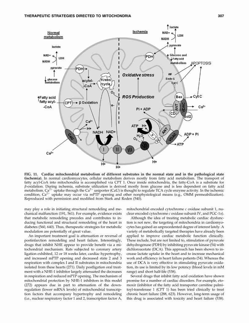
may play a role in initiating structural remodeling and me-chanical malfunction (191, 561). For example, evidence existsthat metabolic remodeling precedes and contributes to in-ducing functional and structural remodeling of the heart indiabetes (560, 640). Thus, therapeutic strategies for metabolicmodulation are potentially of great value.
An important treatment goal is attenuation or reversal ofpostinfarction remodeling and heart failure. Interestingly,drugs that inhibit NHE appear to provide benefit via a mi-tochondrial mechanism. Rats subjected to coronary arteryligation exhibited, 12 or 18 weeks later, cardiac hypertrophy,and increased mPTP opening and decreased state 2 and 3respiration with complex I and II substrates in mitochondriaisolated from these hearts (271). Daily postligation oral treat-ment with a NHE-1 inhibitor largely attenuated the decreasesin respiration and reduced mPTP opening. The mechanism ofmitochondrial protection by NHE-1 inhibitors in this model(272) appears due in part to attenuation of the down-regulation (lower mRNA levels) of mitochondrial transcrip-tion factors that accompany hypertrophy and remodeling(i.e., nuclear respiratory factor 1 and 2, transcription factor A,
mitochondrial encoded cytochrome c oxidase subunit 1, nu-clear encoded cytochrome c oxidase subunit IV, and PGC-1a).
Although the idea of treating metabolic cardiac dysfunc-tion is not new, the targeting of mitochondria in cardiomyo-cytes has gained an unprecedented degree of interest lately. Avariety of metabolically targeted therapies have already beenapplied to improve cardiac metabolic function clinically.These include, but are not limited to, stimulation of pyruvatedehydrogenase (PDH) by inhibiting pyruvate kinase (54) withdichloroacetate (DCA). This approach has been shown to in-crease lactate uptake in the heart and to increase mechanicalwork and efficiency in heart failure patients (54). Whereas theuse of DCA is very effective in stimulating pyruvate oxida-tion, its use is limited by its low potency (blood levels in mMrange) and short half-life (538).
Several drugs that inhibit fatty acid oxidation have shownpromise for a number of cardiac disorders. For example, eto-moxir (inhibitor of the fatty acid transporter carnitine palmi-toyl-transferase 1 (CPT 1) has been tried clinically to treatchronic heart failure (288, 623). However, long-term usage ofthis drug is associated with toxicity and heart failure (538).
FIG. 11. Cardiac mitochondrial metabolism of different substrates in the normal state and in the pathological state(ischemia). In normal cardiomyocytes, cellular metabolism derives mostly from fatty acid metabolism. The transport offatty acyl-CoA into mitochondria is accomplished via CPT 1. Once inside mitochondria, the fatty-CoA is a substrate forb-oxidation. During ischemia, substrate utilization is derived mostly from glucose and is less dependent on fatty acidmetabolism. Ca2þ uptake through the Ca2þ uniporter (CaU) is thought to regulate TCA cycle enzyme activity. In the ischemiccondition, Ca2þ uptake may occur via mPTP opening and other nonphysiological means (e.g., OMM permeabilization).Reproduced with permission and modified from Stark and Roden (540).
THERAPEUTIC STRATEGIES DIRECTED TO MITOCHONDRIA 307
Other drugs, such as trimetazidine, commonly prescribed asan anti-angina drug (68, 171), and ranolazine (currently beingtested for its effect on heart failure) directly inhibit b-oxidationenzymes and potentially other metabolic enzymes (68, 538).Irrespective of their individual mechanism of action, thesedrugs have two fundamental effects in common. They all in-duce a substrate switch from fatty acid to glucose and theyimprove the efficiency of ATP production (171).
In the larger context, metabolic dysfunction of mitochon-dria plays a role in the initiation and progression of a numberof health concerns. Therefore, targeting metabolic dysfunctionby enhancing glucose utilization and pyruvate oxidation atthe expense of fatty acid oxidation, as in cardiac ischemia,appears to be a valid therapeutic approach (Section IX,C).Some of the therapeutic approaches may also be applicable inother organs or tissues. The intense research into developingmore sophisticated techniques for assessing and treating mi-tochondrial disorders in a wide range of human diseasesshould be based on rational approaches in order to under-stand the underlying mechanisms of mitochondrial dysfunc-tion by which to develop new pharmacological therapies.
C. Mitochondria and diabetes
Peripheral insulin resistance characterizes diabetes mellituswith a defect in insulin secretion by pancreatic b-cells. Alle-viating the metabolic disorder can reduce or prevent the de-velopment and progression of diabetes (27). Intense studiesare currently underway to identify novel therapies for a dis-ease that is responsible for the deaths of millions worldwideand has caused crippling disabilities in millions more. Theetiology of diabetes has both genetic and environmentalcomponents (27). Disruption of mitochondrial function is alsoimplicated in the etiology of the disease. For example, mito-chondria of type 2 diabetic patients have reduced ETC capa-cities (401). The ability of pancreatic b-cells to regulate bloodglucose levels is dependent on mitochondrial ATP production.Indeed, the impairment in glucose homeostasis is associatedwith a severe ETC defect caused by a deficiency in the mtDNA-encoded cytochrome c oxidase. Therefore, defective mtDNAcan result in mitochondrial dysfunction that could lead to thedevelopment of diabetes as a result of impaired insulin release.Patients with ‘‘mitochondrial diabetes’’ (352) and mitochon-drial dysfunction have reduced glucose-stimulated insulinsecretion, a finding that underscores the importance of normalmitochondrial function in b-cells (50,51).
Whether mitochondrial dysfunction is at the center inthe etiology of insulin resistance in type 2 diabetes, or theunderlying cause of impaired b-cells function in type 1 dia-betes, remains unresolved. Nonetheless, insulin action resultsfrom a cascade of events from insulin-induced conformationalchange of insulin receptor subunits on the sarcolemma toautophosphorylation of tyrosine residues. The insulin recep-tor then acquires tyrosine kinase activity to phosphorylate theintracellular insulin receptor substrate (IRS) family of mole-cules (528). These events lead to activation of downstreamPI3K and activation of Akt, which mediates most of themetabolic actions of insulin. Phosphorylation of the IRS byGSK3-b inhibits the tyrosine phosphorylation of IRS catalyzedby the insulin receptor.
Akt induces translocation of GLUT4 to the cell membraneresulting in increased glucose uptake. Mitochondrial dys-
function or reduced mitochondrial number could impair theinsulin signaling cascade. For example, impaired mitochon-drial function and subsequent impediment of fatty acid oxi-dation could lead to increased intracellular fatty acyl-coA anddiaylglycerol content and consequently to activation of PKC.PKC is thought to trigger a serine kinase cascade that phos-phorylates serine residues of the IRS, consequently blockingthe insulin signaling pathway (528). Activation of insulin re-ceptors also leads to translocation of activated Akt to mito-chondria in cardiac muscle (635). Akt translocation tomitochondria is thought to represent a link between insulinreceptor signaling and mitochondrial dysfunction in diabeticmyocardium. Insulin action likely plays a major role in theregulation of myocardial OXPHOS because insulin receptorKO mice showed decreased OXPHOS and exacerbated ven-tricular dysfunction (511). Indeed, in a recent study, Yang et al.(635) reported that insulin modulates myocardial OXPHOSby the PI3K-Akt pathway in diabetic myocardium. Further-more, insulin was shown to increase complex V activity incontrol mice; this was blunted in mice with a high fat=highfructose diet, but was increased in the streptocozin diabeticmodel (528).
Increased mitochondrial ROS production during hyper-glycemia may be another way mitochondrial dysfunctioncontributes in the pathology of diabetes. Undeniably, mito-chondrial ROS production and the concomitant oxidativedamage may contribute to the onset and progression of bothtypes 1 and 2 diabetes (280, 627). This is because elevatedglucose and=or fatty acids may lead to greater O2 consump-tion, and coupled with the higher DCm, to more ROS pro-duction (528). The mechanisms by which ROS may contributeto the pathophysiology of diabetes have been discussed ex-tensively (265, 281, 405). Excess mitochondrial ROS in b-cellsinhibits OXPHOS leading to a decrease in ATP for glucoki-nase (expressed in b-cells) and the low ATP=ADP ratio willresult in inactivated KATP channels and impaired insulinsecretion. Chronic oxidative stress on peripheral tissues ulti-mately leads to organ damage. Indeed, evidence for a con-nection between elevated blood glucose and oxidative stresshas been obtained from experiments on endothelial cells,wherein increasing the glucose level was found to increase netcytosolic ROS levels (184, 651). The ROS generated in re-sponse to hyperglycemia may represent the proximal defectthat eventually leads to other pathological consequences ofthe disease. This suggests that therapeutic strategies to limitmitochondrial ROS production or to increase the rate of ROSscavenging may be useful adjuvants to conventional therapiesdesigned to normalize blood glucose (643).
When administered to mice, mitochondrial-targeted ROSscavengers, such as ubiquinone and vitamin E, have showngreat efficacy against cell damage associated with high glu-cose (494). However, clinical trials using a-tocopherol (vita-min E), ascorbate (vitamin C), coenzyme Q, and a-lipoic acidhave yielded ambiguous results. Artificial antioxidants suchas SOD mimetics may be more potent than their naturalcounterparts, but their usefulness in clinical trials has notbeen determined (391, 498). In addition to detoxifying ROSwith mitochondria-designed antioxidants, another possibleapproach is to target the DCm. In this case, one could lower thestate of depolarization (mild) by overexpressing UCPs or bytitrating the dose of DNP, which has been shown to improveserum glucose, triglycerides and insulin levels in mice (88).
308 CAMARA ET AL.
Type 2 diabetes results in an elevation of plasma free fattyacid levels, an increase in b-oxidation in the heart, inhibitionof PDH activity, and impaired glucose oxidation (353). Thesuggestion that diabetics would benefit from therapies thatsuppress fatty acid uptake and oxidation and increase pyru-vate oxidation has been entertained in recent attempts tomitigate the disease. Indeed, PGC-1a, a member of a family oftranscription co-activators that modulates cellular energy,mitochondrial biogenesis (344), and regulates ROS (242, 589),is intimately involved in disorders such as obesity, diabetes,and cardiomyopathy. In particular, its regulatory function inlipid metabolism makes it an inviting target for pharmaco-logical intervention for treating obesity and type 2 diabetes.Overexpression of PGC-1a in mouse skeletal muscle increasedglucose as well as the expression of proteins that are involvedin fat oxidation and glucose transport (52). Thiazolidine-diones (e.g., rosiglitazone) are a class of antidiabetic drugsthat increase myocardial glucose utilization while loweringserum triglycerides (319). In skeletal muscle biopsy studies, itwas shown that PGC-1a is reduced in patients with type 2diabetes (446). Moreover, thiazolidinediones mediate theireffect in part through the ability of PGC-1a to activate mito-chondrial biogenesis and increase mitochondrial function(344). Consistent with this notion is the observation that micedeficient in PGC-1a were found to be defective in contractileprotein function in skeletal and heart muscle. Further evi-dence show impaired ATP levels because the protein levels ofATP synthase and creatine kinase B were reduced in the di-abetic patients (260). The PGC-1a signaling cascade may alsoalleviate diabetes in part by the induction of MnSOD (27).
D. Mitochondria and hypertension
The contribution of mitochondria in kidney disease andhypertension has gained attention recently. Loss of redoxhomeostasis and generation of ROS appear to play a criticalrole in the etiology of renal diseases and hypertension (406).Mitochondrial ROS may contribute to this pathogenesis andtherefore mitochondria may be a target in the disease process.The kidney is intimately involved in the disease process ofhypertension and the effects of ROS ultimately depend on thepro- and antioxidant pathways (406). In the kidney, the renin–angiotensin–aldosterone-system (RAAS) is key in the controlof arterial blood pressure (ABP) and the pathogenesis of hy-pertension. Angiotensin II (AII), an oligopeptide, is a potenthypertensive hormone that causes peripheral vasoconstric-tion and also stimulates aldosterone release. Aldosterone, inturn, increases renal salt retention by acting on the distal tu-bule. All these actions can lead to increased ABP.
Recent landmark studies concluded that augmented O2��
production underlies the pathogenesis of hypertension, andthis was attributed primarily to AII (406, 410). For example,AII infusion in rats led to increased ABP and this effect wasreversed by SOD treatment. In an in vivo model, Nozoe et al.(410) reported that mitochondria-derived ROS induced byAII mediated sympathoexcitation in the rostral ventrolateralmedulla (RVLM), a brainstem site that maintains sympatheticvasomotor tone, resulted in a pressor response. Overexpres-sion of MnSOD and administration of rotenone inhibitedthe AII-induced ROS production and attenuated the pressorresponse (410). In addition, the authors reported that deple-tion of extracellular Ca2þ with EGTA and blocking mCa2þ
uptake with carbonyl cyanide p-(trifluoromethoxy) phe-nylhydrazone (FCCP), an uncoupler, also blocked AII-elicitedmitochondrial ROS production (410). It was concluded thatAII increases mCa2þ uptake, which leads to mitochondrialROS production. The effect of rotenone to reduce ROS and toreduce ABP may be attributed to its effect on the ETC. Thissuggests that rotenone prevents forward electron transferfrom complex I to complex III, a source of O2
�� generation.This is analogous to the effects of rotenone observed by others(18, 324) and to our recent study showing that amobarbitalattenuated O2
�� generation during cardiac I=R injury (9).Spontaneously hypertensive rats (SHR) exhibit ETC defects
in complex I and II activities compared to normotensiveWistar–Kyoto rats (107). In a recent study, Kung et al. (313)demonstrated the importance of complex I in maintaining thehigh ABP in the SHR model. In their study, it was reportedthat in the SHR, microinjection of the adenovirus vector tooverexpress eNOS in the RVLM reduced complex I activ-ity and increased O2
�� and ONOO-, which were reversedwith MnSOD transfection or decomposition of ONOO-. Co-transfection of MnSOD with eNOS prevented the reboundin ABP induced by eNOS overexpression in the SHR. Otherstudies reported an alteration in mCa2þ handling in brainmitochondria during hypertension (89). These studies dem-onstrate the contribution of RNS, ROS, and mCa2þ in regu-lating ABP through their actions on ETC complexes. A betterunderstanding of the role of mitochondria in the etiology orprogression of hypertension may lead to better design ofdrugs that target the root cause of the disease. One possiblestrategy would be the use of gene transfer (186) that wouldtarget mitochondria in the kidney, vascular endothelium, andin the sympathoexcitatory neurons of the RVLM. In supportof this strategy, overexpression of SOD or catalase in the SHRby gene transfer reversed mitochondrial impairment, bluntedROS in RVLM, and mitigated sympathetic vasomotor tone(107).
AII also mediates cardiovascular dysfunction via ROS-induced ROS generation that culminates in cardiovascularpathology including hypertension. Ricci et al. (472) showedthat increased AII initiates a signaling cascade involving PKCto generate NADPH oxidase (Nox) dependent ROS and RNSin the cytosol. Studies show that hypoxia-triggered mito-chondrial ROS activate Nox-dependent ROS formation inpulmonary artery smooth muscle cells, resulting in moreROS=RNS production (467). Indeed cytosolic ROS=RNS donot directly mediate cell damage, rather they trigger mito-chondrial ROS=RNS, which in turn leads to cell damage ordeath (174, 472). Apocynin and chelerythrine dramaticallyattenuate mitochondrial O2
�� generation in response to AII(174). Furthermore, AII stimulated Nox-dependent O2
�� actson mitoKATP channels, or with NO� to form ONOO-, whichdamages ETC, leading to a feed-forward loop of moreROS=RNS generation and to further activation of Nox andmore intracellular ROS production (174), thereby progressingto cardiovascular disease.
Other studies have reported similar crosstalk between mi-tochondria and Nox in AII-induced endothelial dysfunctionand hypertension (174, 613). Blocking complex I, or prevent-ing mPTP opening (613), or blocking the mitoKATP channel,could attenuate vascular dysfunction and ameliorate patho-logical conditions (174, 613). The actions of 5HD stronglysupport the role of mitoKATP channels in the dynamic duo
THERAPEUTIC STRATEGIES DIRECTED TO MITOCHONDRIA 309
between these two distinct sources of oxidative stress withdistinct mechanisms (613). Interestingly, chronic nitroglyc-erin treatment results in the development of nitrate toleranceassociated with vascular dysfunction; this might involvesimilar crosstalk, but in reverse order, between mitochondrialROS and cytosolic Nox-dependent O2
�� generation. Specifi-cally, mitochondrial ROS=RNS exit into the cytosol wherethey may activate vascular Nox in a PKC-dependent process(613).
Although it is generally understood that vascular Nox is amain source of ROS in cardiovascular diseases, the concept ofmitochondrial-triggered activation of Nox is novel and isbecoming attractive in other research areas. Mitochondrialcontrol of Nox1 redox is implicated in breast and ovariancancer (158). Therefore, strategies to target mitochondria aswell as Nox-dependent oxidative stress or interfere with thecrosstalk of ROS between mitochondria and Nox (613) rep-resents a potential approach for use of targeted antioxidantsin mitigating mitochondrial-related diseases that includehypertension.
E. Mitochondria and neurodegenerative diseases
The neurodegenerative diseases are a key health issue be-cause they are profoundly debilitating (177). Mitochondrialdysfunction and the mechanisms that favor apoptosis andnecrosis are believed to play a significant role in many neu-rodegenerative diseases (391). The central nervous system isparticularly vulnerable to oxidative stress due to its high O2
demand and energy expenditure. Mitochondrial dysfunctionleads to oxidative damage that is well documented in manyof these diseases. Oxidative damage to mitochondrial mem-branes, enzymes, and the ETC components, culminate in im-paired mitochondrial ATP production and facilitated mPTPopening (602). Recent studies have also implicated mito-chondrial fission and fusion proteins (Section VIII.A) duringthe onset and progression of neurodegeneration. Specifically,Knott et al. (304) proposed that an imbalance in mitochondrialfission and fusion proteins might be the underlying common-thread that links most of these diseases to mitochondria. Forexample, hereditary mutations in the mitochondrial OPA-1and mitofusin-2 proteins have been implicated in neurode-generative diseases (304).
The neurodegenerative diseases include, but are by nomeans limited to, Alzheimer’s disease (AD), Parkinson’s dis-ease (PD), Friedrich ataxia (FA), multiple sclerosis (MS),amyotrophic lateral sclerosis (ALS), and the rare Huntington’sdisease (HD). Interested readers are referred to the followingcitations for more information (28, 398, 399). In addition, Ta-ble 2 summarizes some of the neurological diseases related tomitochondria disorders. Only a brief discussion of some se-lected diseases will be presented in this review. One salientobservation in all these diseases is that even though the fun-damental pathological mechanisms remain unclear, it is sug-gested from the evidence that mitochondrial dysfunction is acontributing factor at some level of the pathogenic process(177). These diseases are commonly associated with muta-tions in mtDNA, impaired bioenergetics, increased ROSproduction, and abnormal protein dynamics, including themitochondrial accumulation of disease specific proteins (e.g.,amyloid-b for AD, parkin in PD, mutant SOD1 for ALS, andfrataxin for FD) (28, 133, 310, 362).
1. Alzheimer’s disease. Alzheimer’s disease (AD), themost common neurodegenerative disease, is characterized byaccumulation of plaques in brain tissue and progressive ce-rebral neurodegeneration with advanced age (177). It is themost common form of dementia and is a complex neurolog-ical affliction that is characterized clinically by loss of memoryand progressive deficits in cognitive ability (482). The un-derpinnings of the disease are mutations in the genes pro-ducing presenilin, the so-called amyloid precursor proteins(APP) (262, 296, 498). A mutation in the genes for the APPs isassociated with expression of amyloid-b (A-b) (177). A-b istaken up into mitochondria with its mitochondrial targetingsequences via the TIM and TOM import machinery (247).After uptake, it becomes localized in the cristae of the matrix(247). Accumulation of A-b occurs in transgenic mice over-expressing mutant A-b peptide precursor protein, and ispresent in autopsied brains from AD patients (112). It isnoteworthy that at an early stage of AD there is already areduced number of mitochondria (247, 257), brain glucosemetabolism is decreased (247, 287), and the activities of TCAcycle enzymes (79, 520) and cytochrome c oxidase are reduced(97, 247, 441) while there is enhanced cytochrome c release(177, 296), mPTP opening (384), and inhibition of OXPHOS(498).
AD and its association with mitochondria have been re-ported extensively in the literature (79, 112, 247, 257, 520). Themitochondrial dysfunction in AD is thought to be secondaryto an increase in oxidative stress (498, 548) and or mCa2þ
overload (384). The ROS generation increases the amyloido-genic process and sets up conditions conducive for further celldamage so that a destructive cycle of oxidative stress andmitochondrial damage ensues (177). Using brain mitochon-dria from 20-month-old diabetic Goto–Kakizaki rats, Moreiraet al. (384) reported that treatment with CoQ10, a natural an-tioxidant and a highly mobile electron carrier between com-plexes I and III or complexes II and III, counteracts brainmitochondrial dysfunction induced by A-b neurotoxic aminoacid sequences. Cyclosporine A or EGTA also reversed theadverse effects of A-b neurotoxicity on brain mitochondria(385). The use of chloroquinol, an antimalarial drug, hasshown some effectiveness in ameliorating symptoms of AD inpart by reducing Ca2þ accumulation and oxidative stress(177); mitochondrial A-b amyloidosis is also known to pro-mote mitochondrial fission which may contribute to pro-gression of the disease.
A detailed description of the etiology of AD and the con-troversy over the different proposals to reduce its incidenceand severity is well discussed by Duchen (177). Our reviewaddresses only the targeting of mitochondria as a possibletherapeutic option in the treatment of AD. In AD as well as inPD the disease is propagated by its spreading to neighboringcells. It is therefore crucial that the root cause of the diseases bepreemptively targeted if control of either of these diseases isto be achieved. A growing body of evidence suggests thata greater understanding of mitochondrial dynamics (fission=fusion), and the regulatory factors involved, may lead tonovel therapeutic strategies for improving treatments andcontainment of the disease (155, 304).
2. Parkinson’s disease. Parkinson’s disease (PD) is thesecond most frequent neurodegenerative disorder after AD inthe elderly (638). The etiology of the disease is not clearly
310 CAMARA ET AL.
defined. Present evidence suggests that PD is a multifactorialdisorder probably caused by a combination of age, genetics,and environmental factors. The environmental link betweenmitochondria and PD was suggested nearly 2 decades agowhen it was reported that a toxic byproduct of meperi-dine, 1-methyl-4-phenyl-1,2, 3,6-tetra-hydropyridine (MPTH),blocked mitochondrial ETC and produced symptoms similarto those seen in late stage PD (638). MPTH effects on mito-chondria were energy depletion and increased ROS produc-tion (638). Chronic exposure to rotenone also reproduced aPD- like syndrome in rats that was associated with selectiveloss of dopaminergic neurons (254, 304). Studies have shownthat mitochondrial genetic alteration, as in a missense muta-tion in the ND4 subunit of complex I, can lead to an atypicalPD like disorder and loss of nigrostriatal neurons (254) as aconsequence of oxidative stress.
Selective damage to mitochondrial complex I, with a con-comitant increase in ROS within the dopaminergic neurons inthe substantia nigra and an increase in cell death, is believedto be a major causative event in PD (254, 394, 497, 498). Somepatients with PD have a high frequency of single nucleotidepolymorphisms in the gene for MnSOD (216, 522). This al-teration may contribute to an increase in ROS that can bereversed by treatment with tempol, a O2
�� scavenger that wasreported to diminish the severity of PD syndrome (337). Inpatients with PD, administration of CoQ10 both enhanced theETC function and reduced ROS. A large phase III clinical trialis underway to examine if high-dose oral CoQ10 will slow theprogression of the disease (254).
Several recent lines of evidence have also implicated reac-tive nitrogen species (RNS) in neurodegeneration (638) lead-ing to PD. There is growing evidence indicating that RNS are amajor contributor to the pathogenesis and progression of PD.The association of NO� with PD is strengthened by studiesthat show induction of iNOS in glial cells contributes to thedegeneration of dopaminergic neurons in a mice model of PD(638). NO� may contribute to the deterioration of dopami-nergic neurons in part via ONOO-. ONOO- can block complexI function (Section IV,B), mainly by forming 3-nitrotyrosineand nitrosothiol (394, 638), and by increasing oxidation ofcardiolipin (453). Other prevailing conditions in PD are thedepletion of GSH (122, 453) and the susceptibility of complexIV to RNS and ROS (254, 453, 498). This may lead to a furtherincrease in ROS levels and possibly to a vicious cycle offunctional and anatomical deterioration.
The use of antioxidants to treat PD suggests that the pri-mary mechanism of injury is via oxidative stress; metabolictherapies may not be effective since metabolic insufficiencyappears to play no role in the disease (177). Recent data seemto support this contention. Trolox and GSH are both capableof slowing down the ONOO--mediated damage to these do-paminergic neurons (453). In this experimental setting, theNO� scavenger cPTIO only partially attenuated inhibition ofcomplex I, whereas FeTPPS, a selective blocker of ONOO- andMnTBAP, completely blocked the inhibitory affects on com-plex I. Other approaches are to use molecules such as MitoQand MitoVit E (ubiquinone and vitE homologues, respec-tively), which target and accumulate in the mitochondriaand could both enhance ETC function and scavenging ofROS and prevent oxidation of cardiolipin, all of which mayresult in better functioning of the mitochondrial complexes(453, 498).
Recent evidence implicates a derangement of themitochondrial-reticular network (Section VIII.A) in theetiology and progression of PD. Rotenone also initiates mi-tochondrial fission (304). Deng et al. (155) have proposed adirect genetic link of PD to mitochondrial dynamics. Theyshowed that the pink1 (PTEN-induced kinase 1) gene of D.melanogaster interacts with the mitochondrial fusion=fissionmachinery. The PINK-1 protein is a serine-threonine kinaselocalized to the mitochondrial membrane via an 18-KDaamino-terminal in the mitochondrial targeting sequence (254).A pink1 deficiency in D. melanogaster resulted in a disorga-nized morphology similar to the parkin mutation and loss ofdopaminergic neurons (254). The morphological changescould result from a dependency on OXPHOS for maintainingDCm and an intact mitochondrial network (254). A knock-down of opa1, or overexpression of drp1, rescued the phe-notype of muscle disintegration, mitochondrial abnormalities,and cell death in pink1 mutants. These studies demonstratethat pink1 promotes mitochondrial fission and=or inhibitsfusion by negatively regulating opa1 function, and=or posi-tively regulating drp1 (155). Rasagiline, an MAO inhibitor,has multiple effects on mitochondrial function, includingstabilization of DCm, which is necessary for maintaining themitochondrial network, and has a PD-modifying effect (254).Thus, maintenance of the mitochondrial DCm, a prerequisitefor organizing the mitochondrial reticular network, is an es-sential feature in the strategy to alleviate PD.
3. Amyotrophic lateral sclerosis. Amyotrophic lateralsclerosis (ALS; Lou Gehrig’s disease) is the most commonadult-onset motor neuron disorder. It is characterized by se-lective neuronal (lower motor neurons in the spinal cord,brainstem, and motor cortex) degeneration (39, 586) and celldeath. ALS can be fatal due to respiratory muscle weaknessand complications of paralysis. The final stage of the diseaseends with the patient’s inability to initiate and control anyvoluntary movement. In the vast majority of affected people,the disease (90%) has no known cause and is termed ‘‘spo-radic’’ ALS. In only a few victims is it inherited (familial ALS)(586, 634). The most common defects are in cytochrome coxidase activity and in a mutation of sod1 (498, 634). Someinvestigators believe that mitochondrial dysfunction may bedownstream of a primary disease process such as accumula-tion of a pathogenic protein. However, other recent studieshave suggested that mtDNA damage plays a crucial role inthe etiology of neurodegenerative diseases (634).
One potential consequence of mitochondrial dysfunction inALS, as in other neurodegenerative diseases, is impaired en-ergy metabolism and an increase in ROS emission. Indeed,impaired ETC function has been detected in muscle and spi-nal cord cells of ALS patients, and there is a significantlyhigher level of point mutations in spinal cord neuronalmtDNA. For example, there was observed an increase in 8-oxoguanine, a common marker of DNA lesion resulting fromROS, in the motor cortex, spinal cord, and plasma of thesepatients (222, 634). Of the small number of familial ALS pa-tients, about 20% of the defects are mapped to the sod1 gene(586). The autosomal dominant nature of SOD1-associatedALS suggests a toxic gain of function for mutant SOD1 (586).
It is now firmly established that in the fraction of familialALS linked to the sod1 gene, the expression of ALS-mutantSOD1 proteins is the ultimate cause of motor neuron death
THERAPEUTIC STRATEGIES DIRECTED TO MITOCHONDRIA 311
(586). It is reported that the mutant sod1 gene in mice leads to ahigh molecular weight, oligomerized species of SOD1 protein,which is toxic and accumulates in motor neurons (586) andcould be involved in ROS formation (587). Studies showedthat expression of mutant sod1 in cultured neuroblastoma cellsresulted in increased levels of mitochondrial O2
�� productionthat was counteracted by MnSOD scavenging (189, 654).Other studies show a disturbance in mCa2þ homeostasis (99,634). Overexpression of the mutant human sod1 gene in micecaused abnormal mitochondrial morphology [i.e., damage toIMM cristae in spinal cord motor neurons (302), disruptedcytochrome c, and decreased respiration (99)]. The alteredSOD1 protein may interact with the pro-survival protein Bcl-2which compromises its cell survival-promoting function.Indeed, increased levels of the pro-apoptotic proteins Baxand Bid, and decreased anti-apoptotic proteins Bcl-2 andBcl-Xl, were found in spinal cord tissue of ALS patients(256, 442).
Other proposed mechanisms for oxidative damage in ALSinclude increased NO� production and decreased GSH levels.Increased NO� and O2
�� facilitates ONOO- production (Sec-tion IV,B), which can damage key mitochondrial enzymes (81,99). Treating transgenic mice with L-NAME or preincubationwith Mito-Q better preserved ETC function and improvedrespiration in transgenic, but not in nontransgenic mice. Inaddition, the antioxidant MnTE-2-Pyp (contains oxidizedMn3þ) restored mitochondrial respiration, in part by the re-duction of Mn3þ by complexes I and II to Mn2þ which de-grades ONOO- (81, 99). Further studies are needed tounderstand why mutant expression of SOD1 and ‘‘sporadic’’incidents cause motor neuron disease and until this mecha-nism is elucidated, the therapeutic approach targeting ALSvia motor neuron mitochondria is very limited.
4. Friedreich’s ataxia. Friedreich’s ataxia (FA) is a clearexample of a disease in which there are hallmarks of mito-chondrial oxidative stress (362, 435). FA is an autosomal re-cessive disease that is the most common of the hereditaryataxias and is characterized by progressive damage with rel-atively early onset (435). The disease is characterized by highlevels of iron (Fe) (362, 616) due to decrease frataxin, a mito-chondrial Fe chaperone protein involved in regulating Fewithin mitochondria (310, 362). Mutation of the genes en-coding frataxin frequently leads to loss of activity by theprotein (435), impairment of ETC complexes, and OXPHOS,and reduced levels of aconitase and other TCA cycle enzymes.These defects contribute to ROS production and a higher ox-idative load in the cell (17, 484).
Therapeutic remedies for FA include idebenone, a CoQ10
analog and inhibitor of lipoperoxide, or the combination ofidebenone and CoQ10, which improved neurological coordi-nation (489, 490). The accumulation of Fe in the mitochondriasuggests the potential for chelation therapy. Indeed, Fe che-lators have been used to mitigate mitochondrial Fe load and toimprove mitochondrial and cellular conditions. For example,recent studies show that Fe accumulates in the mitochondriaof a mouse FA model, and that development and use of themitochondrial permeable Fe chelator, desferrioxamine, re-duced Fe load and prevents generation of OH� driven by theFenton reaction or direct scavenging of O2
��. In a D. melano-gaster FA model, ectopic overexpression of scavenging en-zymes restored the activity of aconitase (17); this is indicative
and supportive of the theory that ROS are a salient feature inthe pathogenesis of FA.
F. Neoplastic diseases
Mitochondrial involvement in the etiology of neoplasticdiseases (ND) has been hypothesized since the 1930s, whenthe Nobel prize-winning German physician and scientist,Otto Heinrich Warburg, discovered that mitochondria incancer cells do not efficiently generate energy (464). Unlike theneurodegenerative and ischemia-induced diseases discussedabove, mitochondrial dysfunction in tumor cells tends to leadto cell survival and resistance to chemotherapy; hence, theterm the ‘‘Warburg effect’’ (212, 485). In marked contrast to thetherapeutic strategy for neurodegenerative and ischemicdiseases, which is prevention of cell death, in ND the maingoal of targeting the mitochondria is to kill malignant cells byinducing apoptosis (212). A growing body of evidence sug-gests that tumor cells exhibit increased intrinsic ROS stressdue in part to oncogenic stimulation, increased metabolicactivity, and mitochondrial dysfunction (56). Despite theirvariability, almost all neoplastic cells demonstrate enhanceduptake and utilization of glucose for glycolysis to generateATP (212). The most glycolytic tumor cells were found to bemost resistant to therapy and most aggressive in metastasis(212, 364).
A pivotal player in the switch from OXPHOS to glycolysismay be the HKII binding at the VDAC (Section II,A). As in-dicated previously, tumor cells adopt this phenotype for theirsurvival. This includes most neoplastic cells that metastasize(364). HKII binding to VDAC prevents the pro-apoptoticproteins Bad and Bax from binding to the OMM, and so helpimmortalize these cells by gaining preferential access to newlysynthesized ATP for phosphorylating glucose (136, 212, 364).Tumor cells are also better able to withstand an adverse mi-croenvironment (hypoxia, acidosis, and shortage of growthfactors) by virtue of their metabolic adaptation. Consideringthe key role of mitochondria in cell death, it appears that therelative mitochondrial ‘‘silencing’’ in neoplastic cells can, atleast in part, explain resistance of most tumors to treatment.
Emerging insights from the Korsmeyer laboratory (101,156) group identified a novel key intermediate phenotype thatappears to be the lynchpin in the dynamic role of mitochon-drial OMM permeability in apoptosis, and provided an im-portant insight into the dysregulation of apoptosis in theneoplastic cell. Cancer cell (e.g., Hela and non-small cell lung)mitochondria are more resistant to OMM permeability be-cause of overexpression of the Bcl protein family (195, 197,386). Generation of sensitizer and activator pro-apoptoticpeptides is robust in many neoplastic cells as disorganizedmetabolism and intracellular signaling generate robust stim-uli for the activation of apoptosis. However, many of thesecells also dramatically express Bcl-2 that in turn sequesters thepro-apoptotic peptides and blocks activation of apoptosis.Vigorous apoptosis ensues when Bcl-2 is inhibited by smallmolecule antagonists, such as ABT-737 (156, 212) or by shortpeptide antagonists (101), resulting in OMM permeabiliza-tion. This led Korsmeyer and Letai to propose the "primed todie" mitochondrial phenotype (333). The response of isolatedmitochondria from neoplastic cells can functionally identifythe anti-apoptotic peptide that is in fact preserving cell sur-vival, based upon the small molecules or peptides that elicit
312 CAMARA ET AL.
cytochrome c release from their isolated mitochondria, socalled ‘‘BH3 profiling’’ (156, 333). These findings have en-hanced the understanding of the regulation of blockade ofapoptosis, and have substantially increased an understandingbeyond the relatively simple "dead or alive" Bax=Bcl-2 ratioconcept that previously dominated the field. Identifying thesetumor-specific changes responsible for the resistance to celldeath is crucial for developing targeted chemotherapeuticagents (195, 197) based on a strategic approach to ensurenormal cell survival.
The actual etiologies of ND are highly sought after. It isbelieved that apoptosis-regulatory genes (e.g., those encodingp53, PTEN and Bcl-2) are involved in the pathogenesis ofmany human cancers (136). PTEN is a phosphatase that in-hibits Akt activation by reducing PIP3 levels (448). The mPTPwhose constituents are different between normal and malig-nant cells play a role in this pathological process (136). Also itis postulated that this difference may explain resistance toapoptosis and the cancer-specific metabolic alterations (136)in these cells. For instance, HKII-VDAC interaction preventsbinding of pro-apoptotic proteins binding to VDAC andthereby the induction of apoptosis (212,364). Consequently,a variety of compounds including avicins, which are pro-apoptotic, anti-inflammatory molecules with antioxidantproperties, perturb mitochondrial function and initiate apo-ptosis in tumor cells (212, 364). Colon cancer cells HCT 116express a small amount of Bak (383). Cisplatin, an anti-neoplastic drug kills colon cancer cells in part by activatingBak (276, 359, 563).
Another strategy used by neoplastic cells to confer protec-tion is the overexpression of PBR (Section II), and Bcl-2 on theOMM (136, 194). Synthetic PBR ligands (e.g., melphalan)display increased cytotoxicity in a variety of rat and humanbrain tumor cell lines (194). Melphalan is considered a Smacmimetic and is used in the treatment of multiple myeloma (57,110, 434); it induces apoptosis even in the presence of over-expressed Bcl-2 anti-apoptotic proteins in some of these tumorcells (205). Recent evidence indicates that the tumor sup-pressor protein p53 has extra-nuclear effects that contribute toits cell cycle-arresting and pro-apoptotic functions (195, 197).Smac=DIABLO can abrogate the protective function of IAPs(175, 597, 628), which confers chemoresistance in various celltypes (109, 196, 231, 335). Cytoplasmic p53 can induce OMMpermeabilization in part by direct interaction with the Bclfamily proteins located on the OMM. Thus, since OMM per-meabilization is impaired in tumor cells, its pharmacologicalinduction constitutes a therapeutic strategy. In this case, theultimate strategy would be to minimize the sequestration oflarge amounts of BH3-only proteins such as Bim in complexeswith anti-apoptotic Bcl-2 proteins. In the absence of Bcl-2, Bimis not sequestered, apoptosis is triggered (315), and cell deathis initiated.
Other strategies applied in mitochondria-directed thera-pies include increasing p53 and Bax, decreasing Bcl-Xl, andenhancing the activation of caspase 3. It was reported that thechemotherapeutic agent CD437 exerts its therapeutic effect bythese mechanisms (136). It has been postulated that VDACsand ANT (ANT2 variant) are highly expressed in neoplasticcells when compared to normal cells because of the highglycolytic phenotype in tumor cells (525). The VDAC–ANT–HKII complex was shown to be a requirement for ATPtransport in neoplastic cells (67). These studies support the
notion that VDAC and ANT could be possible pharmacologictargets, or at least could be subjected to metabolic alterationin neutralizing neoplastic cells. Indeed, the upregulation ofVDAC in tumor cells makes them susceptible to the anti-tumor effects of furanonapthoquinones, whose apoptotic ac-tivity is via NADH-dependent O2
�� generation (525). Anotherstrategy involves the use of 3-BPR (Section II,A) that selec-tively enters and destroys tumor cells by targeting the HKIIand the mitochondrial synthasome. This leads to rapid de-pletion of ATP and tumor cell destruction without harm tonormal cells (364).
As an adaptation to an increase in size with limited nutri-ents and diminished O2, tumor cells are known to adapt tohypoxia by inducing the transcription of multiple genes viaactivation of the transcription factor hypoxia-inducible factor(HIF-1a). The proteins induced by HIF-1a are involved inregulation of glycolytic metabolism as well as tumor growthand angiogenesis. Therefore, preventing HIF activation mayact to suppress tumor growth and cell proliferation. An O2
sensor may be central in this dynamic process that also clearlyinvolves mitochondrial ROS (48). It is proposed that ROS,specifically H2O2, produced from complex III is a likely sensorfor initiating and stabilization of HIF-1a during hypoxia (108).Therefore, a better understanding of how mitochondria func-tion to initiate a hypoxic response will lead to development oftherapies that target O2 consumption, ROS production, oralter key metabolite concentrations in mitochondria (48).
In recent studies, PGC-1a levels were downregulatedin hepatic, breast, colon, and epithelial ovarian tumor cells(33, 647), while overexpression induced apoptosis occurredin epithelial ovarian tumor cells (Ho-8910), but not in CHO(Chinese Hamster Ovary) cells. It was suggested that the in-creased expression of PGC-1a results in upregulation ofthe pro-apoptotic gene Bax and downregulation of the anti-apoptotic gene Bcl-2 (33, 647). Moreover, PGC-1a-inducedapoptosis was proposed to be mediated by a decreasing ratioof Bcl-2=Bax, which can lead to destabilization of mitochon-dria and release of cytochrome c (647). The finding that PGC-1a expression decreased in ovarian tumor cells and thatincreased expression promoted apoptosis suggests that PGC-1a might be involved in the pathogenesis of some cancers. Thestrategy of increased expression of the PGC-1a gene maybe useful for cancer therapy; this again belies mitochondria asthe epicenter of this strategy.
G. Other mitochondria-related diseases
1. Mitochondria and psychiatric disorders. Several stud-ies have reported a role for mitochondria in the pathophysi-ology of bipolar disease (BD), major depressive disorders(MDD), including post-traumatic stress disorder (PTSD) andschizophrenia (SZ) (278, 480, 545). The features of mitochon-drial abnormalities include deficiencies in OXPHOS andmtDNA deletion in the brain, and associations with mtDNAmutations=polymorphisms or nuclear-encoded mitochon-drial genes (278, 545). It is suggested that the high rates ofmaternal offsprings with SZ and BD compared to paternalrates support the notion that increased risk for these diseasesmight be related to mitochondrial impairment (480). In SZ itis reported that there is an increased number of synony-mous base substitution in mtDNA in the dorsolateral pre-frontal cortex (DLPFC; Brodmann area 9=46) (480). In PTSD,
THERAPEUTIC STRATEGIES DIRECTED TO MITOCHONDRIA 313
decreased activity in the DLPFC, a brain area involved in theregulation of memory and preparation and selection of fearresponses (545), was reported and it was proposed that mi-tochondrial dysfunction is a factor in the etiology of the dis-order. Su et al. (545) studied dysregulation of mitochondrialgenes in these brain areas (Brodmann) from postmortem pa-tients with and without PTSD using human mitochondria-focused cDNA microarrays. The study revealed that amajority of the dysregulated genes were associated with mi-tochondrial dysfunction and neurological diseases. Some ofthe dysregulated genes appear confined in areas involved inneuronal function–survival and thus may be targets for neu-ropsychiatric drugs. The authors concluded that mitochon-drial dysfunction is involved in PTSD ‘‘and provide theexpression fingerprints that may ultimately serve as a bio-marker for PTSD diagnosis and the drugs and moleculartargets that may prove useful for the development of reme-dies for prevention and treatment of PTSD’’ (545).
Preliminary studies have shown that lower levels of brainenergy metabolism are associated with MDD. Impaired en-ergy metabolism in the brain detected by magnetic resonancespectroscopy suggests that mitochondrial dysfunction is animportant component in MDD. A salient feature is that glu-cose is metabolized very slowly. It has also been proposedthat altered mitochondrial OXPHOS malfunction is involvedin psychiatric disorders and that brain mitochondria of SZpatients exhibit reduced complex IV activity in the frontalcortex; in other studies, low complex IV activity was highlyassociated with increased emotional and intellectual impair-ment, but not motor impairment. Other studies have reportedreduced complexes II and III activities in frontal and temporalcortex of SZ patients (278, 480). A reduction in the activity ofthe complexes affects electron transfer and therefore maypotentially interfere with mitochondrial metabolism and ATPproduction. In the brain this could lead to cell death as aconsequence of disturbance in the regulation of intracellularhomeostasis. Gerich et al. (207) showed a correlation betweenthe spread of hypoxia-induced depression in rat CA1 regionof hippocampal slices and DCm depolarization. The authorpresented two basic questions to these findings: could it bemitochondrial depolarization that triggers the depressionspread? If it is the mitochondria, how can the depolariza-tion be transmitted to the cell membrane? A likely scenarioaccording to the authors is that mitochondrial depolariza-tion may reduce sequestration of Ca2þ by mitochondria andCa2þ release from malfunctioning mitochondria; subsequentchanges in intracellular ionic conditions and pH would beconsidered as putative signaling mechanisms.
However, a note of caution is that it remains to be resolvedwhether the occurrence of psychiatric symptoms in patients isjust a coincidence or is more directly related to mitochondrialdysfunction, itself. The answer to this question requires fur-ther investigation. To date, dietary supplements, have beenused to ‘‘fuel’’ mitochondrial function and in so doing mitigatethe symptoms of psychiatric disorders. Some of these nutri-tional maneuvers are discussed below (Section XI,E).
2. Mitochondria and migraine headache. Initial studieshave also raise the question that mitochondrial dysfunctionmay contribute to migraine headaches, at least in selectedpatients. Migraine headache is a neurological disorder be-lieved to be a manifested in individuals with mtDNA
sequence-related mitochondrial dysfunction (163). Indeed,epidemiologic evidence for frequent maternal transmissionhas implicated a role for a mitochondrial genetic background.The link of mitochondria and migraine headache is supportedby the observation that it is frequently associated with adeficit in energy metabolism and in MELAS, the classic mi-tochondriopathies (163, 306). This is consistent with the ob-servation that OXPHOS is impaired in migraineurs andincreased numbers of sequence variants were detected in thenoncoding control regions of mtDNA in migraineurs withoccipital stroke (163). It is also conceivable that migraineheadache may also be associated with mutation of nucleargenes that encode the ETC complex because it has been notedthat other studies failed to show the connection between theheadaches and mtDNA abnormalities (306). Regardless ofthe source of mitochondrial protein defect, the consequenceof mitochondrial dysfunction when coupled with loss of in-tracellular Mg2þ in the brain, an occurrence in migraineurs,culminates in a metabolic shift that cause instability of neu-ronal function, which then enhances the development of amigraine attack (612). Disruption in mitochondrial metabo-lism may thus provide future pharmacologic targets for noveltherapies against migraine headaches. Therapeutic strategiesare briefly discussed in Section XI,E below.
X. Mitochondrial Pharmacologyand Therapeutic Potential
The primary cause of most of the mitochondria-relateddiseases discussed in our review have multifaceted etiologies;hence efforts to develop effective drugs should be devoted tothe design of individual new compounds that work in syn-ergy to protect the mitochondrion. Current mitochondria-targeted drugs cover a wide range of pharmacological agents(556) (Table 3; Fig. 12). Some of these drugs target mito-chondria directly, whereas others affect mitochondria as asecondary or side effect. Nonetheless, identification of themitochondrion as a target of a drug may assist in better un-derstanding of a drug’s mechanisms of action and allow newperspectives for its application (556). Only selected aspects oftargeting mitochondria for therapeutic benefit have beencovered in all the sections discussed above. This is inevitableconsidering the broad nature of the subject and how mito-chondrial function lies at the center of cell viability and celldeath. This section will discuss briefly a) strategies for mito-chondrial drug delivery, b) mitochondria-targeted drugs, c)maneuvers to protect against I=R injury, and d) other mito-chondrial therapeutic approaches
A. Strategies for drug delivery to mitochondria
As a first step in designing a mitochondria-specific therapy,it is foremost important to develop a drug that can access themitochondrial matrix. In recent years the search for newprotective remedies against excess mitochondrial ROS hastaken on a new sense of urgency. Some well-known scaven-gers like MnSOD have proved ineffective at preventing oxi-dative damage in animal disease models, presumably becausethey are unable to permeate the cell membranes. One generalsolution is to utilize the large DCm and to attach a molecule,for example, one with antioxidant properties, onto a lipoidvehicle that can penetrate membranes (262). With DCm ap-proaching �180 mV in the nonphosphorylating state, for
314 CAMARA ET AL.
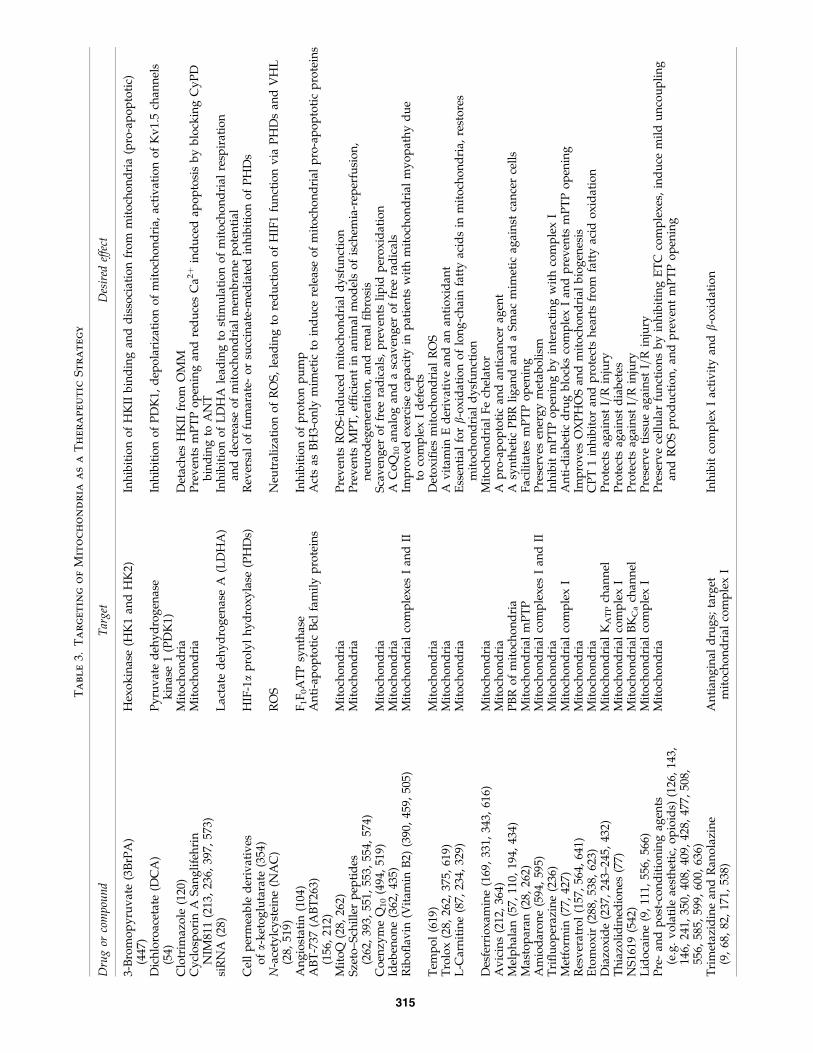
Ta
bl
e3.
Ta
rg
et
in
go
fM
it
oc
ho
nd
ria
as
aT
he
ra
pe
ut
ic
St
ra
te
gy
Dru
gor
com
pou
nd
Tar
get
Des
ired
effe
ct
3-B
rom
op
yru
vat
e(3
BrP
A)
(447
)H
exo
kin
ase
(HK
1an
dH
K2)
Inh
ibit
ion
of
HK
IIb
ind
ing
and
dis
soci
atio
nfr
om
mit
och
on
dri
a(p
ro-a
po
pto
tic)
Dic
hlo
roac
etat
e(D
CA
)(5
4)P
yru
vat
ed
ehy
dro
gen
ase
kin
ase
1(P
DK
1)In
hib
itio
no
fP
DK
1,d
epo
lari
zati
on
of
mit
och
on
dri
a,ac
tiv
atio
no
fK
v1.
5ch
ann
els
Clo
trim
azo
le(1
20)
Mit
och
on
dri
aD
etac
hes
HK
IIfr
om
OM
MC
ycl
osp
ori
nA
San
gli
feh
rin
NIM
811
(213
,23
6,39
7,57
3)M
ito
cho
nd
ria
Pre
ven
tsm
PT
Po
pen
ing
and
red
uce
sC
a2þ
ind
uce
dap
op
tosi
sb
yb
lock
ing
Cy
PD
bin
din
gto
AN
Tsi
RN
A(2
8)L
acta
ted
ehy
dro
gen
ase
A(L
DH
A)
Inh
ibit
ion
of
LD
HA
lead
ing
tost
imu
lati
on
of
mit
och
on
dri
alre
spir
atio
nan
dd
ecre
ase
of
mit
och
on
dri
alm
emb
ran
ep
ote
nti
alC
ell
per
mea
ble
der
ivat
ives
ofa-
ket
og
luta
rate
(354
)H
IF-1a
pro
lyl
hy
dro
xy
lase
(PH
Ds)
Rev
ersa
lo
ffu
mar
ate-
or
succ
inat
e-m
edia
ted
inh
ibit
ion
of
PH
Ds
N-a
cety
lcy
stei
ne
(NA
C)
(28,
519)
RO
SN
eutr
aliz
atio
no
fR
OS
,le
adin
gto
red
uct
ion
of
HIF
1fu
nct
ion
via
PH
Ds
and
VH
L
An
gio
stat
in(1
04)
F1F
0A
TP
syn
thas
eIn
hib
itio
no
fp
roto
np
um
pA
BT
-737
(AB
T26
3)(1
56,
212)
An
ti-a
po
pto
tic
Bcl
fam
ily
pro
tein
sA
cts
asB
H3-
on
lym
imet
icto
ind
uce
rele
ase
of
mit
och
on
dri
alp
ro-a
po
pto
tic
pro
tein
s
Mit
oQ
(28,
262)
Mit
och
on
dri
aP
rev
ents
RO
S-i
nd
uce
dm
ito
cho
nd
rial
dy
sfu
nct
ion
Sze
to–S
chil
ler
pep
tid
es(2
62,
393,
551,
553,
554,
574)
Mit
och
on
dri
aP
rev
ents
MP
T,
effi
cien
tin
anim
alm
od
els
of
isch
emia
-rep
erfu
sio
n,
neu
rod
egen
erat
ion
,an
dre
nal
fib
rosi
sC
oen
zym
eQ
10
(494
,51
9)M
ito
cho
nd
ria
Sca
ven
ger
of
free
rad
ical
s,p
rev
ents
lip
idp
ero
xid
atio
nId
eben
on
e(3
62,
435)
Mit
och
on
dri
aA
Co
Q1
0an
alo
gan
da
scav
eng
ero
ffr
eera
dic
als
Rib
ofl
avin
(Vit
amin
B2)
(390
,45
9,50
5)M
ito
cho
nd
rial
com
ple
xes
Ian
dII
Imp
rov
edex
erci
seca
pac
ity
inp
atie
nts
wit
hm
ito
cho
nd
rial
my
op
ath
yd
ue
toco
mp
lex
Id
efec
tsT
emp
ol
(619
)M
ito
cho
nd
ria
Det
ox
ifies
mit
och
on
dri
alR
OS
Tro
lox
(28,
262,
375,
619)
Mit
och
on
dri
aA
vit
amin
Ed
eriv
ativ
ean
dan
anti
ox
idan
tL
-Car
nit
ine
(87,
234,
329)
Mit
och
on
dri
aE
ssen
tial
forb-
ox
idat
ion
of
lon
g-c
hai
nfa
tty
acid
sin
mit
och
on
dri
a,re
sto
res
mit
och
on
dri
ald
ysf
un
ctio
nD
esfe
rrio
xam
ine
(169
,33
1,34
3,61
6)M
ito
cho
nd
ria
Mit
och
on
dri
alF
ech
elat
or
Av
icin
s(2
12,
364)
Mit
och
on
dri
aA
pro
-ap
op
toti
can
dan
tica
nce
rag
ent
Mel
ph
alan
(57,
110,
194,
434)
PB
Ro
fm
ito
cho
nd
ria
Asy
nth
etic
PB
Rli
gan
dan
da
Sm
acm
imet
icag
ain
stca
nce
rce
lls
Mas
top
aran
(28,
262)
Mit
och
on
dri
alm
PT
PF
acil
itat
esm
PT
Po
pen
ing
Am
iod
aro
ne
(594
,59
5)M
ito
cho
nd
rial
com
ple
xes
Ian
dII
Pre
serv
esen
erg
ym
etab
oli
smT
rifl
uo
per
azin
e(2
36)
Mit
och
on
dri
aIn
hib
itm
PT
Po
pen
ing
by
inte
ract
ing
wit
hco
mp
lex
IM
etfo
rmin
(77,
427)
Mit
och
on
dri
alco
mp
lex
IA
nti
-dia
bet
icd
rug
blo
cks
com
ple
xI
and
pre
ven
tsm
PT
Po
pen
ing
Res
ver
atro
l(1
57,
564,
641)
Mit
och
on
dri
aIm
pro
ves
OX
PH
OS
and
mit
och
on
dri
alb
iog
enes
isE
tom
ox
ir(2
88,
538,
623)
Mit
och
on
dri
aC
PT
1in
hib
ito
ran
dp
rote
cts
hea
rts
fro
mfa
tty
acid
ox
idat
ion
Dia
zox
ide
(237
,24
3–24
5,43
2)M
ito
cho
nd
rial
KA
TP
chan
nel
Pro
tect
sag
ain
stI=
Rin
jury
Th
iazo
lid
ined
ion
es(7
7)M
ito
cho
nd
rial
com
ple
xI
Pro
tect
sag
ain
std
iab
etes
NS
1619
(542
)M
ito
cho
nd
rial
BK
Ca
chan
nel
Pro
tect
sag
ain
stI=
Rin
jury
Lid
oca
ine
(9,
111,
556,
566)
Mit
och
on
dri
alco
mp
lex
IP
rese
rve
tiss
ue
agai
nst
I=R
inju
ryP
re-
and
po
st-c
on
dit
ion
ing
agen
ts(e
.g.
vo
lati
leae
sth
etic
,o
pio
ids)
(126
,14
3,14
6,24
1,35
0,40
8,40
9,42
8,47
7,50
8,55
6,58
5,59
9,60
0,63
6)
Mit
och
on
dri
aP
rese
rve
cell
ula
rfu
nct
ion
sb
yin
hib
itin
gE
TC
com
ple
xes
,in
du
cem
ild
un
cou
pli
ng
and
RO
Sp
rod
uct
ion
,an
dp
rev
ent
mP
TP
op
enin
g
Tri
met
azid
ine
and
Ran
ola
zin
e(9
,68
,82
,17
1,53
8)A
nti
ang
inal
dru
gs;
targ
etm
ito
cho
nd
rial
com
ple
xI
Inh
ibit
com
ple
xI
acti
vit
yan
db-
ox
idat
ion
315
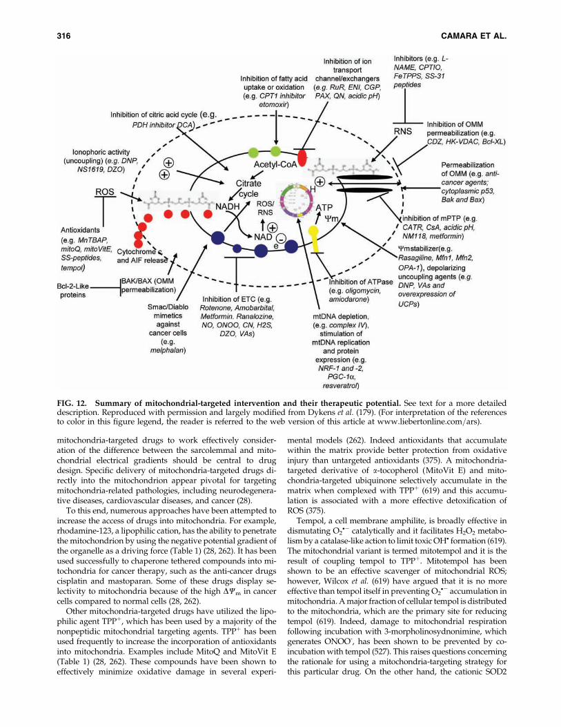
mitochondria-targeted drugs to work effectively consider-ation of the difference between the sarcolemmal and mito-chondrial electrical gradients should be central to drugdesign. Specific delivery of mitochondria-targeted drugs di-rectly into the mitochondrion appear pivotal for targetingmitochondria-related pathologies, including neurodegenera-tive diseases, cardiovascular diseases, and cancer (28).
To this end, numerous approaches have been attempted toincrease the access of drugs into mitochondria. For example,rhodamine-123, a lipophilic cation, has the ability to penetratethe mitochondrion by using the negative potential gradient ofthe organelle as a driving force (Table 1) (28, 262). It has beenused successfully to chaperone tethered compounds into mi-tochondria for cancer therapy, such as the anti-cancer drugscisplatin and mastoparan. Some of these drugs display se-lectivity to mitochondria because of the high DCm in cancercells compared to normal cells (28, 262).
Other mitochondria-targeted drugs have utilized the lipo-philic agent TPPþ, which has been used by a majority of thenonpeptidic mitochondrial targeting agents. TPPþ has beenused frequently to increase the incorporation of antioxidantsinto mitochondria. Examples include MitoQ and MitoVit E(Table 1) (28, 262). These compounds have been shown toeffectively minimize oxidative damage in several experi-
mental models (262). Indeed antioxidants that accumulatewithin the matrix provide better protection from oxidativeinjury than untargeted antioxidants (375). A mitochondria-targeted derivative of a-tocopherol (MitoVit E) and mito-chondria-targeted ubiquinone selectively accumulate in thematrix when complexed with TPPþ (619) and this accumu-lation is associated with a more effective detoxification ofROS (375).
Tempol, a cell membrane amphilite, is broadly effective indismutating O2
�� catalytically and it facilitates H2O2 metabo-lism by a catalase-like action to limit toxic OH� formation (619).The mitochondrial variant is termed mitotempol and it is theresult of coupling tempol to TPPþ. Mitotempol has beenshown to be an effective scavenger of mitochondrial ROS;however, Wilcox et al. (619) have argued that it is no moreeffective than tempol itself in preventing O2
�� accumulation inmitochondria. A major fraction of cellular tempol is distributedto the mitochondria, which are the primary site for reducingtempol (619). Indeed, damage to mitochondrial respirationfollowing incubation with 3-morpholinosydnonimine, whichgenerates ONOO-, has been shown to be prevented by co-incubation with tempol (527). This raises questions concerningthe rationale for using a mitochondria-targeting strategy forthis particular drug. On the other hand, the cationic SOD2
FIG. 12. Summary of mitochondrial-targeted intervention and their therapeutic potential. See text for a more detaileddescription. Reproduced with permission and largely modified from Dykens et al. (179). (For interpretation of the referencesto color in this figure legend, the reader is referred to the web version of this article at www.liebertonline.com=ars).
316 CAMARA ET AL.
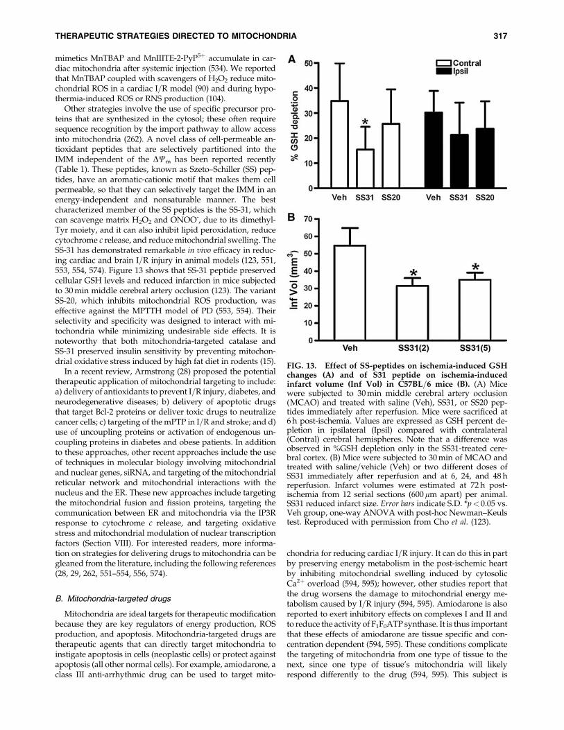
mimetics MnTBAP and MnIIITE-2-PyP5þ accumulate in car-diac mitochondria after systemic injection (534). We reportedthat MnTBAP coupled with scavengers of H2O2 reduce mito-chondrial ROS in a cardiac I=R model (90) and during hypo-thermia-induced ROS or RNS production (104).
Other strategies involve the use of specific precursor pro-teins that are synthesized in the cytosol; these often requiresequence recognition by the import pathway to allow accessinto mitochondria (262). A novel class of cell-permeable an-tioxidant peptides that are selectively partitioned into theIMM independent of the DCm has been reported recently(Table 1). These peptides, known as Szeto–Schiller (SS) pep-tides, have an aromatic-cationic motif that makes them cellpermeable, so that they can selectively target the IMM in anenergy-independent and nonsaturable manner. The bestcharacterized member of the SS peptides is the SS-31, whichcan scavenge matrix H2O2 and ONOO-, due to its dimethyl-Tyr moiety, and it can also inhibit lipid peroxidation, reducecytochrome c release, and reduce mitochondrial swelling. TheSS-31 has demonstrated remarkable in vivo efficacy in reduc-ing cardiac and brain I=R injury in animal models (123, 551,553, 554, 574). Figure 13 shows that SS-31 peptide preservedcellular GSH levels and reduced infarction in mice subjectedto 30 min middle cerebral artery occlusion (123). The variantSS-20, which inhibits mitochondrial ROS production, waseffective against the MPTTH model of PD (553, 554). Theirselectivity and specificity was designed to interact with mi-tochondria while minimizing undesirable side effects. It isnoteworthy that both mitochondria-targeted catalase andSS-31 preserved insulin sensitivity by preventing mitochon-drial oxidative stress induced by high fat diet in rodents (15).
In a recent review, Armstrong (28) proposed the potentialtherapeutic application of mitochondrial targeting to include:a) delivery of antioxidants to prevent I=R injury, diabetes, andneurodegenerative diseases; b) delivery of apoptotic drugsthat target Bcl-2 proteins or deliver toxic drugs to neutralizecancer cells; c) targeting of the mPTP in I=R and stroke; and d)use of uncoupling proteins or activation of endogenous un-coupling proteins in diabetes and obese patients. In additionto these approaches, other recent approaches include the useof techniques in molecular biology involving mitochondrialand nuclear genes, siRNA, and targeting of the mitochondrialreticular network and mitochondrial interactions with thenucleus and the ER. These new approaches include targetingthe mitochondrial fusion and fission proteins, targeting thecommunication between ER and mitochondria via the IP3Rresponse to cytochrome c release, and targeting oxidativestress and mitochondrial modulation of nuclear transcriptionfactors (Section VIII). For interested readers, more informa-tion on strategies for delivering drugs to mitochondria can begleaned from the literature, including the following references(28, 29, 262, 551–554, 556, 574).
B. Mitochondria-targeted drugs
Mitochondria are ideal targets for therapeutic modificationbecause they are key regulators of energy production, ROSproduction, and apoptosis. Mitochondria-targeted drugs aretherapeutic agents that can directly target mitochondria toinstigate apoptosis in cells (neoplastic cells) or protect againstapoptosis (all other normal cells). For example, amiodarone, aclass III anti-arrhythmic drug can be used to target mito-
chondria for reducing cardiac I=R injury. It can do this in partby preserving energy metabolism in the post-ischemic heartby inhibiting mitochondrial swelling induced by cytosolicCa2þ overload (594, 595); however, other studies report thatthe drug worsens the damage to mitochondrial energy me-tabolism caused by I=R injury (594, 595). Amiodarone is alsoreported to exert inhibitory effects on complexes I and II andto reduce the activity of F1F0ATP synthase. It is thus importantthat these effects of amiodarone are tissue specific and con-centration dependent (594, 595). These conditions complicatethe targeting of mitochondria from one type of tissue to thenext, since one type of tissue’s mitochondria will likelyrespond differently to the drug (594, 595). This subject is
FIG. 13. Effect of SS-peptides on ischemia-induced GSHchanges (A) and of S31 peptide on ischemia-inducedinfarct volume (Inf Vol) in C57BL=6 mice (B). (A) Micewere subjected to 30 min middle cerebral artery occlusion(MCAO) and treated with saline (Veh), SS31, or SS20 pep-tides immediately after reperfusion. Mice were sacrificed at6 h post-ischemia. Values are expressed as GSH percent de-pletion in ipsilateral (Ipsil) compared with contralateral(Contral) cerebral hemispheres. Note that a difference wasobserved in %GSH depletion only in the SS31-treated cere-bral cortex. (B) Mice were subjected to 30 min of MCAO andtreated with saline=vehicle (Veh) or two different doses ofSS31 immediately after reperfusion and at 6, 24, and 48 hreperfusion. Infarct volumes were estimated at 72 h post-ischemia from 12 serial sections (600 mm apart) per animal.SS31 reduced infarct size. Error bars indicate S.D. *p< 0.05 vs.Veh group, one-way ANOVA with post-hoc Newman–Keulstest. Reproduced with permission from Cho et al. (123).
THERAPEUTIC STRATEGIES DIRECTED TO MITOCHONDRIA 317
discussed further in the Section XIII, Caveats and Limitationsto Targeting Mitochondria.
Some antidiabetic drugs can accumulate in mitochondria asa result of the highly negative DCm that favors preferentialuptake into mitochondria. For example, metformin (N1,N1-dimethylbiguanide), an antidiabetic drug, blocks complex Iof the ETC because it accumulates in the matrix (427). Thesedrugs are more effective in intact mitochondria than in sub-mitochondrial particles. The positive charges on these drugscoupled with the DCm in energized mitochondria makesit favorable for their accumulation in the matrix (427). Met-formin has a broad spectrum of effects, which include fatbreakdown, reduction of circulating free fatty acids with amodest inhibition of fatty acid oxidation, and an increase inglucose transport and glucose utilization (655). In otherstudies, metformin was shown to block mPTP opening as apossible mechanism of action to preserve cellular function.In the nondiabetic state, metformin alleviated some patho-logical conditions (e.g., inflammatory conditions in lung in-jury), in part by inhibiting complex I activity (77, 655). It isdebatable if these actions of metformin also alter mitochon-drial ATP production which would impair mitochondrialenergy conservation as a mechanism of action. However,Zmijewski et al. (655) reported that the actions of metforminare not ATP dependent, whereas Brunmair et al. (77) reportedthat metformin not only blocks complex I, but also uncouplesOXPHOS.
There are recent advances in understanding the complexinteraction between mitochondria within a reticular networkof mitochondria and mitochondrial interaction with otherorganelles (Section VIII.BC). This has opened a new potentialtherapeutic avenue for assessing and treating mitochondria-related disorders. For example, resveratrol, a polyphenoliccompound abundant in grape skins, induces PGC-1a toexpress genes involved in mitochondrial biogenesis andOXPHOS (564). Increased expression of PGC-1a has also beenshown to mitigate MPTH-induced cell injury in a PD model(254). How resveratrol treats PD is unclear. Resveratrol alsoreduces ischemic damage in brain (641) and cardiac cells (157).The effects observed experimentally suggest quite a promis-ing therapeutic approach.
Other interventions to reduce cellular damage during aperiod of oxidative stress, as in post-ischemic injury, includeenhancement of endogenous scavenging enzymes and none-nzymes. Pyruvate has been shown to be protective in variousin vivo and in vitro models of oxidative stress (608). Wanget al. (608) showed that pyruvate can remarkably abrogateperoxide-induced toxicity in SK-N-SH neuroblastoma cellsby a direct antioxidant protective effect on the mitochondrion,as evidenced by its dampening of mitochondrial O2
�� pro-duction and its preservation of DCm. As a readily oxidizablesubstrate, pyruvate can bolster the cytosolic energy state andthereby provide the energy needed to maintain cellularfunction in the face of a metabolic challenge (608).
Permeabilization of the OMM and the IMM via mPTPopening (Section II) as a consequence of oxidative stress is animportant underlying factor in most of the neurodegenera-tive diseases discussed above (Section IX,E). CsA treatmenthas been shown to prolong the survival of ALS transgenicmice compared to control (vehicle) treated mice (284). In PDmodels, daily doses of CsA showed partial preservation ofstriatal dopaminergic neurons and altered the progression
of the disease in the ALS model (602). In the CNS, the effectsof CsA is limited because it does not cross the blood brainbarrier (284, 602). This restriction can be circumvented byvarious methods including direct administration of the drugsinto the lateral ventricles of the CNS (284, 602).
Mitochondrial antioxidant systems are important in cancerchemotherapy. Tamoxifen, a synthetic nonsteroidal anties-trogen widely used to treat breast cancer, is known to haveantioxidant and cardioprotective effects in part by inductionof MnSOD (145). However, normal tissue injury is a majorproblem that limits the success of cancer therapy in protocolsinvolving chemotherapeutic drugs (e.g., adriamycin). Gen-eration of ROS is implicated in the toxicity of a large numberof these agents and the injury is manifested at the mitochon-drial level (145). These potential side effects will be discussedin Section XIV.
It is clear that the subject of mitochondrial pharmacology isvast and the above discussion only represents some examples.It is also clear that mitochondria are key factor in the etiologyof numerous human mitochondria-related and nonrelateddiseases, which can be mitigated by targeting the mitochon-drion with specific drugs designed for uptake by the organ-elle. Interested readers are urged to examine the reviewliterature for more information.
C. Approaches to improve mitochondrial functionduring ischemia and reperfusion
Mitochondria play a central role in I=R injury and hence arepromising targets for novel anti-ischemic therapies. The im-portance of mitochondria as both targets and mediators of I=Ris becoming increasingly recognized (113). Cardiac I=R resultin mitochondrial dysfunction as shown by a decrease in oxi-dative capacity, loss of cytochrome c, and generation of ROS.Recent studies show that increased ROS is especially evidentduring late ischemia (9, 10, 90, 289, 475, 542, 591). Therefore,protection of mitochondrial respiration during ischemia couldrepresent a new therapeutic approach in mitigating the del-eterious effects of I=R injury.
A pioneering study by Ganote and colleagues (198) showedthat inhibition of mitochondrial respiration could decreasecontraction band formation and attenuate enzyme releaseduring reoxygenation; they suggested that resumption ofmitochondrial metabolism during reoxygenation can initiallylead to deleterious consequences. We discussed earlier howa reperfusion-induced decline in cardiac function and accu-mulation of oxidatively damaged lipids were diminishedwhen complex I was reversibly inhibited during early re-perfusion with amobarbital (14), thus underlining the physi-ologic significance of mitochondrial ROS production tocardiac injury during reperfusion (113). We also discussedhow amobarbital, when administered briefly before ischemia,preserved mitochondrial bioenergetics and improved cardiacfunction upon reperfusion. In addition, we noted suggestionson how rotenone preserved mitochondrial structural integrityand improved function (324).
Thus, there is support for the concept that mitochondrialfunction can be protected during global ischemia as well ason reperfusion as shown by reduced levels of mCa2þ uptakeand O2
�� generation, and improved redox state after pre-conditioning (ischemic or pharmacological) (Section X,D–E)(476, 477), inhibition of NHE (10), hypothermia (475), and
318 CAMARA ET AL.
ROS scavengers (90). In each of these mitochondrial protectivestrategies, contractile function was improved and infarct sizewas reduced. In a recent study (12), we reported novel find-ings that hyperkalemic depolarizing cardioplegia was pro-tective by means other than its effect on sparing high-energyphosphates; in the same article we also showed that lidocaine,a hyperpolarizing cardioplegic agent, also protected hearts inpart by a direct action on the mitochondria, a concept sup-ported by other studies (111, 566). Lipid-soluble local anes-thetics could exert their cytoprotection in part by direct effectsto attenuate complex I activity and to uncouple respiration(556). We found that lidocaine blocked complex I as suggestedby an increased NADH without a change in FAD during li-docaine perfusion just before ischemia. This situation wassimilar to what we observed with the complex I inhibitoramobarbital (9).
It has long been thought that most of the cellular injuryoccurred during reperfusion because mPTP opening, mCa2þ
uptake, and O2�� generation were more likely to occur after
ischemia. However, recent studies have reported that ische-mic injury can lead to a persistent defect in OXPHOS duringearly reperfusion (113, 115, 117). In an ex vivo reperfusionstudy of guinea pig hearts, SS-peptide (Szeto–Schiller) wasreported to prevent myocardial stunning and to significantlyimprove contractility (551, 554). Restriction of oxidative me-tabolism during early reperfusion using a hypoxic reperfusateattenuated mitochondrial and cardiac damage (282, 513).Pharmacologic inhibition of mitochondrial respiration on re-perfusion with amobarbital also decreased mitochondria-driven myocardial injury (14). Thus, there are a variety ofstrategies that can target mitochondria to interrupt the linkbetween ischemic damage to mitochondria and mitochon-dria-mediated cellular damage during reperfusion.
D. Preconditioning
Preconditioning is a mechanism for reducing organ I=Rinjury on return of blood flow to the tissue. It is mediated afterthe removal of a protective stimulus (brief ischemia or a drug)some time before the onset of index (longer, damaging) is-chemia. The stimulus does not directly induce cytoprotectionbut rather some downstream signaling factors are evoked toprovide a lasting protection (memory effect). Ischemic pre-conditioning (IPC) and pharmacologic preconditioning (PPC)of the heart decrease mitochondrial damage from subsequentindex ischemia (113, 350). IPC was identified as an endoge-nous cytoprotective phenomenon, whereas PPC has the ad-vantage of not requiring brief episodes of ischemia to elicitcellular protection. The cellular and mitochondrial protectionelicited from IPC or PPC involves a coordinated interplay oftrigger and effector mechanisms (113). There is much cir-cumstantial evidence that mitochondria-derived ROS play animportant role to initiate IPC and PPC (126, 143), which areeffected by intracellular protein kinase cascades, especiallyPKCe (150, 283) and PKG (135, 241). The key effector signalingpathways converge on mitochondria to modulate oxidativemetabolism before prolonged ischemia (113) or to preventmPTP opening on reperfusion (251, 261, 273). For example,IPC, pre-ischemic diazoxide treatment, and post ischemicCsA treatment were each found to reduce infarct size after I=Rinjury in rat isolated hearts and this protective effect wasblocked by the mPTP opener atractyloside when given on
reperfusion (319). However, protection by IPC against mPTPopening after reperfusion injury appears to be better achievedby blocking the factors that induce mPTP opening rather thanby inhibiting mPTP directly with drugs such as CsA (273). Inother examples, inhibition of apoptosis by PKCe involves themPTP, which might be a principal target of this kinase (38,150). The protective targets in mitochondria also involve theinteraction of PKCe with complex IV of the ETC, resulting inincreased activity and increased mitochondrial ETC effi-ciency, decreased loss of cytochrome c, preservation of DCm,and inhibition of mPTP opening due to decreased ROS pro-duction (150). IPC also diminishes mitochondrial dysfunctionafter ischemia and confers protection in the brain (150).
The mechanisms of cytoprotection afforded by PPC, forexample, anesthetic preconditioning (APC), has been in-tensely investigated and discussed (290, 408, 409, 428, 429,476, 477) and among other signaling factors common toIPC, they also involve sarcolemmal and the putative mKATP
channels (55, 585). IPC and PPC are proposed to activatemKATP channels, which may lead to mild uncoupling of mi-tochondrial respiration. The mild uncoupling is believed toresult in partial depolarization of the mitochondria, either asa result of Kþ entry or activation of KHE in response to Kþ
entry (Section II,B). It is suggested that this depolarizationmay reduce mCa2þ uptake, thereby reducing mCa2þ overloadand minimizing cellular damage. A mild uncoupling effectwithout changing DCm could also increase mitochondrialROS production (253), which can then lead to activation ofdownstream end-effectors. The putative mKATP channel ag-onist diazoxide was proposed to mediate cytoprotection inpart on the Kþ channel as well as by exerting a direct effect onthe mPTP (236). But diazoxide also attenuates complex II ac-tivity and TCA cycle supported respiration, which appears toresult in O2
�� production and hence may contribute to theO2�� generation during PPC (236, 243–245). Interestingly, the
mitochondrial O2�� scavenger N-mercapto-propionyl-glycine
was shown to block protective effects of diazoxide (432). Thisand other studies suggest that an indirect effect of diazoxideon complex II is to induce O2
�� generation (243–245).Recently, we reported that the putative mCa2þ-dependent
Kþ channel (KCa) may also play a role in modulating mito-chondrial bioenergetics and provide preconditioning cardio-protection against I=R injury. Mitochondrial KCa agonists,such as KATP agonists, appear to mediate their cytoprotectionthrough ROS-dependent mechanisms as the mitochondrialROS scavenger MnTBAP blocked the protection (542). In thisstudy we reported that preconditioning with NS 1619, a well-known KCa channel agonist, protected the heart from ischemicinjury (542). NS 1619 markedly decreased the deleterious in-creases in ROS and m[Ca2þ] with I=R injury, better preservedredox state (NADH and FAD), and improved cardiac func-tion. Paxilline, an NS 1619 antagonist, and MnTBAP, bothabolished these effects (542).
It was also reported that preconditioning-mediated pro-tection through mPTP opening with atractyloside abolishedthe beneficial effects of IPC or NS 1619 in isolated rat heartsand myocytes (95). Sedlic et al. (508) reported recently thatisoflurane, like DNP, protects cardiomyocytes in part via milddecrease in DCm, which attenuates ROS production understress and leads to a delay in mPTP opening. Other pre-conditioning drugs have resulted in improved function alongwith improved tissue redox state (NADH and FAD),
THERAPEUTIC STRATEGIES DIRECTED TO MITOCHONDRIA 319
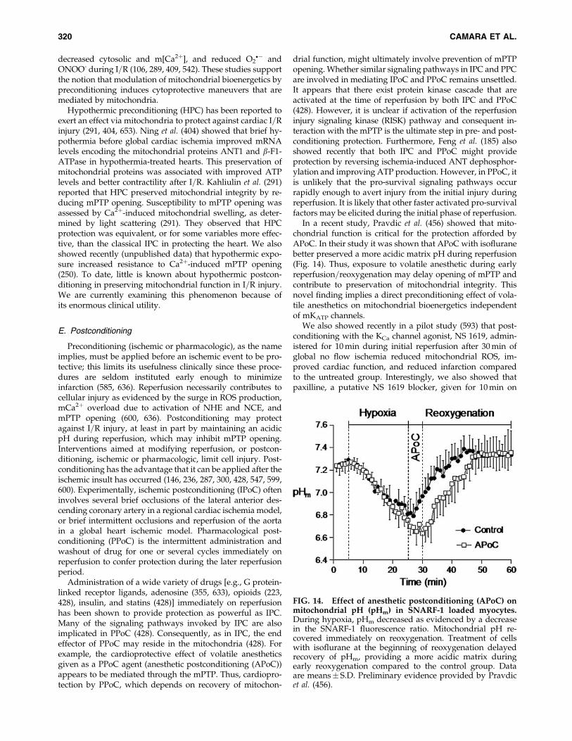
decreased cytosolic and m[Ca2þ], and reduced O2�� and
ONOO- during I=R (106, 289, 409, 542). These studies supportthe notion that modulation of mitochondrial bioenergetics bypreconditioning induces cytoprotective maneuvers that aremediated by mitochondria.
Hypothermic preconditioning (HPC) has been reported toexert an effect via mitochondria to protect against cardiac I=Rinjury (291, 404, 653). Ning et al. (404) showed that brief hy-pothermia before global cardiac ischemia improved mRNAlevels encoding the mitochondrial proteins ANT1 and b-F1-ATPase in hypothermia-treated hearts. This preservation ofmitochondrial proteins was associated with improved ATPlevels and better contractility after I=R. Kahliulin et al. (291)reported that HPC preserved mitochondrial integrity by re-ducing mPTP opening. Susceptibility to mPTP opening wasassessed by Ca2þ-induced mitochondrial swelling, as deter-mined by light scattering (291). They observed that HPCprotection was equivalent, or for some variables more effec-tive, than the classical IPC in protecting the heart. We alsoshowed recently (unpublished data) that hypothermic expo-sure increased resistance to Ca2þ-induced mPTP opening(250). To date, little is known about hypothermic postcon-ditioning in preserving mitochondrial function in I=R injury.We are currently examining this phenomenon because ofits enormous clinical utility.
E. Postconditioning
Preconditioning (ischemic or pharmacologic), as the nameimplies, must be applied before an ischemic event to be pro-tective; this limits its usefulness clinically since these proce-dures are seldom instituted early enough to minimizeinfarction (585, 636). Reperfusion necessarily contributes tocellular injury as evidenced by the surge in ROS production,mCa2þ overload due to activation of NHE and NCE, andmPTP opening (600, 636). Postconditioning may protectagainst I=R injury, at least in part by maintaining an acidicpH during reperfusion, which may inhibit mPTP opening.Interventions aimed at modifying reperfusion, or postcon-ditioning, ischemic or pharmacologic, limit cell injury. Post-conditioning has the advantage that it can be applied after theischemic insult has occurred (146, 236, 287, 300, 428, 547, 599,600). Experimentally, ischemic postconditioning (IPoC) ofteninvolves several brief occlusions of the lateral anterior des-cending coronary artery in a regional cardiac ischemia model,or brief intermittent occlusions and reperfusion of the aortain a global heart ischemic model. Pharmacological post-conditioning (PPoC) is the intermittent administration andwashout of drug for one or several cycles immediately onreperfusion to confer protection during the later reperfusionperiod.
Administration of a wide variety of drugs [e.g., G protein-linked receptor ligands, adenosine (355, 633), opioids (223,428), insulin, and statins (428)] immediately on reperfusionhas been shown to provide protection as powerful as IPC.Many of the signaling pathways invoked by IPC are alsoimplicated in PPoC (428). Consequently, as in IPC, the endeffector of PPoC may reside in the mitochondria (428). Forexample, the cardioprotective effect of volatile anestheticsgiven as a PPoC agent (anesthetic postconditioning (APoC))appears to be mediated through the mPTP. Thus, cardiopro-tection by PPoC, which depends on recovery of mitochon-
drial function, might ultimately involve prevention of mPTPopening. Whether similar signaling pathways in IPC and PPCare involved in mediating IPoC and PPoC remains unsettled.It appears that there exist protein kinase cascade that areactivated at the time of reperfusion by both IPC and PPoC(428). However, it is unclear if activation of the reperfusioninjury signaling kinase (RISK) pathway and consequent in-teraction with the mPTP is the ultimate step in pre- and post-conditioning protection. Furthermore, Feng et al. (185) alsoshowed recently that both IPC and PPoC might provideprotection by reversing ischemia-induced ANT dephosphor-ylation and improving ATP production. However, in PPoC, itis unlikely that the pro-survival signaling pathways occurrapidly enough to avert injury from the initial injury duringreperfusion. It is likely that other faster activated pro-survivalfactors may be elicited during the initial phase of reperfusion.
In a recent study, Pravdic et al. (456) showed that mito-chondrial function is critical for the protection afforded byAPoC. In their study it was shown that APoC with isofluranebetter preserved a more acidic matrix pH during reperfusion(Fig. 14). Thus, exposure to volatile anesthetic during earlyreperfusion=reoxygenation may delay opening of mPTP andcontribute to preservation of mitochondrial integrity. Thisnovel finding implies a direct preconditioning effect of vola-tile anesthetics on mitochondrial bioenergetics independentof mKATP channels.
We also showed recently in a pilot study (593) that post-conditioning with the KCa channel agonist, NS 1619, admin-istered for 10 min during initial reperfusion after 30 min ofglobal no flow ischemia reduced mitochondrial ROS, im-proved cardiac function, and reduced infarction comparedto the untreated group. Interestingly, we also showed thatpaxilline, a putative NS 1619 blocker, given for 10 min on
FIG. 14. Effect of anesthetic postconditioning (APoC) onmitochondrial pH (pHm) in SNARF-1 loaded myocytes.During hypoxia, pHm decreased as evidenced by a decreasein the SNARF-1 fluorescence ratio. Mitochondrial pH re-covered immediately on reoxygenation. Treatment of cellswith isoflurane at the beginning of reoxygenation delayedrecovery of pHm, providing a more acidic matrix duringearly reoxygenation compared to the control group. Dataare means� S.D. Preliminary evidence provided by Pravdicet al. (456).
320 CAMARA ET AL.
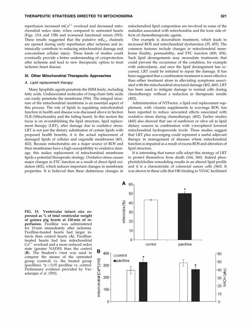
reperfusion increased mCa2þ overload and decreased mito-chondrial redox state, when compared to untreated hearts(Figs. 15A and 15B) and worsened functional return (593).These results suggested that the putative mKCa channelsare opened during early reperfusion after ischemia and in-trinsically contribute to reducing mitochondrial damage andconcomitant cellular injury. These kinds of studies couldeventually provide a better understanding of cytoprotectionafter ischemia and lead to new therapeutic option to treatischemic heart disease.
XI. Other Mitochondrial Therapeutic Approaches
A. Lipid replacement therapy
Many lipophilic agents penetrate the IMM freely, includingfatty acids. Undissociated molecules of long-chain fatty acidscan easily penetrate the membrane (556). The integral struc-ture of the mitochondrial membrane is an essential aspect ofthis process. The role of lipids in regulating mitochondrialfunction in health and disease was discussed above in SectionIX,B (Mitochondria and the failing heart). In this section thefocus is on re-establishing the lipid structure, lipid replace-ment therapy (LRT), after damage due to oxidative stress.LRT is not just the dietary substitution of certain lipids withproposed health benefits, it is the actual replacement ofdamaged lipids of cellular and organelle membranes (401,402). Because mitochondria are a major source of ROS andtheir membranes have a high susceptibility to oxidative dam-age, this makes replacement of mitochondrial membranelipids a potential therapeutic strategy. Oxidative stress causesmajor changes in ETC function as a result of direct lipid oxi-dation (402), which induces important changes in membraneproperties. It is believed that these deleterious changes in
mitochondrial lipid composition are involved in some of themaladies associated with mitochondria and the toxic side ef-fects of chemotherapeutic agents.
One example is doxorubicin treatment, which leads toincreased ROS and mitochondrial dysfunction (35, 455). Thecommon features include changes in mitochondrial mem-brane fluidity, permeability, and ETC function (400, 454).Such lipid derangements may necessitate treatments thatcould prevent the occurrence of the condition, for examplewith antioxidants, and once the lipid derangement has oc-curred, LRT could be initiated to repair the damage. It hasbeen suggested that a combination treatment is more effectivethan either treatment alone in alleviating symptoms associ-ated with the mitochondrial structural damage (402, 460). LRThas been used to mitigate damage to normal cells duringchemotherapy without a reduction in therapeutic results(402).
Administration of NTFactor, a lipid oral replacement sup-plement, with vitamin supplements to scavenge ROS, hasbeen reported to reduce unwanted effects associated withoxidative stress during chemotherapy (402). Earlier studies(460) also showed that use of sunflower or olive oil as lipiddietary sources in combination with a-tocopherol loweredmitochondrial hydroperoxide levels. These studies suggestthat LRT plus scavenging could represent a useful adjuvanttherapy in management of diseases where mitochondrialfunction is impaired as a result of excess ROS and alteration oflipid structure.
It is interesting that tumor cells adopt this strategy of LRTto protect themselves from death (164, 360). Indeed phos-photidylcholine remodeling results in an altered lipid profileand it is a characteristic of colorectal cancer cells (360). Itwas shown in these cells that HK binding to VDAC facilitated
FIG. 15. Ventricular infarct size ex-pressed as % of total ventricular weightof guinea pig hearts at 120 min of re-perfusion. Paxilline was administeredfor 10 min immediately after ischemia.Paxilline-treated hearts had larger in-farcts than control hearts (A). Paxilline-treated hearts had less mitochondrialCa2þ overload and a more reduced redoxstate (greater NADH) than the control(B). The Student’s t-test was used tocompare the means of the untreatedgroup (control) vs. the treated group(paxilline). *p< 0.05 paxilline vs. control.Preliminary evidence provided by Var-adarajan et al. (593).
THERAPEUTIC STRATEGIES DIRECTED TO MITOCHONDRIA 321
uptake of cholesterol and the elevated cholesterol contentamplified the binding of HK to VDAC. This adaptive re-sponse, along with the observation that increased mito-chondrial cholesterol blunts the ability of Bax to initiatedysfunction in cancer cells, makes them refractory to injury(443). It is evident that the therapeutic strategies adopted byneoplastic cells for survival involve mitochondria and this maypose yet another dilemma in the selective treatment of a spe-cific disease by exclusively targeting cell specific mitochondria.
B. Transactivator of transcription proteinsand mitochondrial therapy
The mitochondrial protein import pathways, specificallythe mitochondria signal peptide-tagged cargo, have beenused to deliver DNA molecules to the matrix. Thus, the tar-geting of covalently linked genetic information to mitochon-drial peptide import machinery could potentially be used torectify a mutant mitochondrial genome similar to classicalgene replacement therapies that attempt to replace a cor-rected copy of defective nuclear genome (28). However, it issometimes technically challenging to insert small DNAs tointroduce small peptides in cells. The transactivator of tran-scription (TAT) protein-penetrating transduction system isbeing used to introduce small peptides into living cells (86,507), as these peptides are often unstable and susceptible tocellular degradation. TAT-mediated protein transduction oc-curs in a rapid fashion that is independent of receptors andtransporters (86, 238, 465, 507). Disorders of some mitochon-drial proteins can be corrected by this system of uptake. Thetechnique has been utilized in the isolated cell (86, 465, 507)and in the whole organ model where it has been shown tofacilitate peptide uptake in the isolated heart (238).
TAT proteins with their ‘‘cargo’’ rapidly cross the cellmembrane and enter mitochondria where they can replacemutated endogenous peptides (465). Rapoport et al. (465)showed recently that the TAT-mediated replacement ap-proach could be used to correct for a mutated component ofa protein complex, in this case the E3 subunit shared by thea-ketoacid dehydrogenases like pyruvate dehydrogenase(PDH). That is, this approach was used to increase the activityof the PDH complex, which before treatment showed lowactivity due to mutations in E3. TAT approaches could pro-vide a new treatment for enzyme deficiencies as well as forother mitochondrial and metabolic disorders (465).
TAT-mediated transduction of the mutant form of Bnip3(TAT-Bnip3DTM), a pro-apoptotic member of the Bcl family ofmitochondrial proteins expressed in the adult myocardium,was also reported to reduce I=R-induced injury. The wild-typeBnip3 induced apoptosis in part by increasing the fragmenta-tion of mitochondrial connectivity and causing mitochondrialdysfunction (i.e., loss of DCm, increased cytochrome c and AIFrelease, and disruption of energy production) (238). An in-crease in our understanding of the role of these proteins and thestrategies to manipulate them in mitochondria could be im-portant in the development of effective therapies.
C. Molecular genetic approaches
Genetic maneuvers of various types have been attemptedto reverse mtDNA-related diseases. These include suppres-sion of mutant mtDNA expansion and manipulation ofmtDNA replication by import into the mitochondrion of en-
donucleases that might selectively destroy a specific mutantsequence. In a recent study, Alexander et al. (13) showed thatin cells heteroplasmic for the T8993G mutation that causesNARP (Section VIII,B), infection with an adenovirus, whichencodes the mitochondria-targeted restriction endonuclease,led to selective destruction of mutant mtDNA. This led toa significantly increased rate of O2 consumption and ATPproduction and concurrently, decreased rate of lactic acidproduction in these cells, which is a marker of mitochondrialdysfunction. The specificity of this mitochondrial approachwas demonstrated by the absence of nDNA damage (13).
Other potential therapeutic approaches involve overex-pression of targeted proteins, for example CypD in cancertherapy, or its selective inhibition using a siRNA approachduring I=R-related cellular injury (28). Other approaches aredesigned to impede the action of Bcl-2 by use of antisensetechnology to inhibit Bcl-Xl expression, or alternatively, totarget pro-apoptotic Bcl-2 peptides to mitochondria (28).Other investigators have used similar approaches. For ex-ample, gene therapy using adenoviral Bax-delivering vectorshas been successful in activating apoptosis (631) and a similarapproach has been used to induce apoptosis using stablegenerated BH3 peptidomimetics designed to block Bcl-2 andto activate Bax and Bak (603). In another study, Li et al. (339)used a similar approach to develop Smac=DIABLO mimeticsto inhibit the action of IAPs.
The inter- and intra-organelle communication betweenmitochondria as a group and mitochondria with ER and thenucleus are important factors in maintaining normal cellularfunction; disturbances in any of these complex systems ortheir coordinated activity will generate disease. The Bclfamily of proteins is also believed to influence mitochondrialfission=fusion. For example, Bax and Bak promote fragmen-tation of the mitochondrial network, possibly by activatingfission machinery; on the other hand, targeting these fissionproteins through expression of Bcl-Xl prevented cytochrome crelease (518). These findings indicate that Bcl family proteinscan influence mitochondrial fission and fusion. However,another study reported that Bak blocks mitochondrial fusionto induce fragmentation. By this function, Bak may collabo-rate with Bax to permeabilize the OMM leading to apoptosis(72, 73). Thus these Bcl family proteins appear to be attractivetherapeutic targets in fighting neoplastic cells (4, 131).
The disruption of the mitochondria to ER communication,required for normal cellular function, could be targeted bymolecular approaches that would up- or downregulate theproteins involved in the link between these organelles. Inthe mitochondria–nuclear communication, overexpression ofPGC1-a and or HSP70 and 60, which are involved in theimportation of mitochondrial proteins, could also serve astargets for therapy to improve mitochondrial function. Othergenetically targeted approaches have been used for IMMproteins and they have also furnished some effectiveness as aremedy against mitochondria-related pathologies (28). Yet,which, or if any of these mitochondrial genetic approacheswill become clearly applicable in treating human maladiesremains to be determined.
D. Mitochondria and caloric restriction
Morbid obesity is associated with numerous diseases fromthe metabolic syndrome to cancer. Indeed, obesity as a result
322 CAMARA ET AL.
of high-energy intake has been shown to increase the risk ofage-related cognitive decline (622). Caloric restriction (CR;either a decrease in food intake or intermittent fasting) delaysthe rate of aging in many species and increases resistance todiseases (41, 129). In animal models of aging and neurode-generative diseases, CR protected hippocampal, striatal, andcortical neurons, and ameliorated functional decline (622). CRhas been shown to minimize age-related loss of mitochondrialfunction and biogenesis in several tissues, including heart,liver, skeletal muscle, and brain (190, 588) and to mitigate theseverity of dysfunction. For example, studies have shown thatthe complete maintenance of skeletal aerobic contractile func-tion with aging by CR is associated with improved mito-chondrial function and a reduction of the age-related declinein mitochondrial capacity (255). The decline in mitochondrialbiogenesis in obese animal has been associated with a de-crease in PGC-1a (128). It was reported that the rate of declineof PGC-1a was lower in CR animals than in non-CR animals(255). This increase in mitochondrial biogenesis, coupled withincreased oxidative capacity and lower ROS levels (62), mighttranslate to less accumulation of oxidatively damaged mito-chondria, thereby accounting for the protection of mitochon-drial function with CR (255).
E. Mitochondria and dietary supplements
The potential contribution of impaired energy metabolismto behavioral disorders and migraine headache has beenconsidered over the years. Initial studies correlated defi-ciencies in riboflavin or folate to the prevalence of depression.Higher dietary intake in folate and riboflavin in recent clini-cal trials in Japan was associated with lower prevalence ofdepressive symptoms in some patients (390). Riboflavin’s(vitamin B2) effect is required for the formation of FADþ,which is critical for the TCA and as an electron donor tocomplex II. FADþ is a key cofactor in folate-dependent meth-ylation pathways that would lead to improved neuronalmetabolism and improved function. Riboflavin in combi-nation with CoQ10 has been shown to reduce neurologicalepisodes associated with migraine headaches (505). Thecombined approach augments complex I activity and hasbeen used in clinical trials to demonstrate their combined ef-fectiveness for preventing migraine attacks (459). Niacin hasalso been used as a supplement to boost mitochondrial energymetabolism by increasing substrate availability to complex I;in this way it might act to deter=reduce migraine headache(459). Clearly, the efficacies of these approaches remain to beresolved, their side effects remain under scrutiny, and they arelimited by the fact that some studies have failed to make anyassociation of migraine headache or dietary supplements withspecific mitochondria deficits.
XII. Mitochondria Age and Lifespan
A. Mitochondria and age-associated diseases
The term aging generally refers to organismal senescence,a process whereby cells lose their ability to divide after anumber of cell divisions, and by a decline in the capacity of anorganism to respond to stress. Although aging is not a disease,the frequency of many disease processes increases with age.Mitochondria are proposed to be critical components in theprocess of aging, in the regulation of the cell cycle, and in
limiting cellular lifespan (401, 632). Mitochondria are in turn aprimary target of the aging process, as evidenced by a declinein mitochondrial oxidative capacity in both skeletal and heartmuscle with age (255, 292, 328). Aging has been associatedwith excessive oxidative stress and over production of RNS(401). Indeed, oxidative=nitrosative stress is thought to bean important contributor to the degeneration of long-livedpostmitotic cells such as cardiomyocytes and neurons. Thisexplains in part the relation between acquired cardiac andneurodegenerative diseases and aging (391).
In aged mitochondria, a defect in mtDNA is coincidentwith a decrease in complex IV activity and in some casesreduced activity of complex I (130). Although we were unableto show age-related changes in complex I activity in mito-chondria, we did show a decrease in complex III activity, butonly in interfibrillar mitochondria (328). These defects areconducive for increasing oxidative stress that further predis-poses mitochondria to injury (Fig. 16). The exact mechanismfor the relationship between impaired mitochondria andaging remains unclear. However, Melov et al. (371) recentlyreported that in older animals a characteristic mitochondrial‘‘signature’’ of declining mitochondrial function could be re-versed in part through exercise and suggested that a sufficientreserve capacity exists in the genome to reverse certain age-dependent expression profiles back to more youthful geno-types.
The current view is that mtDNA mutations accumulate at arate that is several folds higher than nDNA mutations (292).This increase in mutation rate occurs primarily in postmitoticorgans such as brain, skeletal muscle, and heart, compared tocells that undergo frequent mitosis (568). Thus the evidence,albeit tenuous, supports the notion that accumulation of ROSover time is wholly or partially responsible for aging. Cer-tainly, increased oxidative stress has been associated withincreased mtDNA strand breaks during aging and in the agedMnSOD-deficient mice (614). However, this belief has beenquestioned for vertebrate and invertebrate animal models ofaging (172, 388). Furthermore, targeting of mitochondria to‘‘reverse’’ the aging process and therefore mitigate age-relateddiseases has remained elusive. Indeed, overexpression of sod2and catalase genes in the fruit fly D. melanogaster has yieldedthe opposite outcome, that is, lifespan was decreased insteadof increased (16). In another study in the nematode C. elegans,Doonan et al. (172) showed that overexpression of sod genesdid not alter lifespan, whereas other studies reported thatROS might in fact be responsible for extending the lifespan ofC. elegans (590).
Some studies have consistently reported a role for mito-chondrial ROS in aging-related maladies. This includes someof the neurodegenerative diseases discussed above (SectionIX,E); others include presbyacusis, the hearing loss associatedwith aging as a consequence of the progressive deteriorationof the cochlear mtDNA (391, 451). Antioxidants targetingmitochondria were reported to reduce this age-related hear-ing loss (127). Toxic aldehydes, including HNE, an end prod-uct of lipid peroxides, are known to accumulate in the brainin neurodegenerative disease. Mitochondrial aldehyde dehy-drogenase 2 (ALDH2) detoxifies HNE by oxidizing the alde-hyde group. ALDH2-deficient neuronal cells show increasedvulnerability to HNE. The increased oxidative stress may inpart explain some age-dependent neurodegeneration, such asloss of pyramidal cells and marked deficits in cognition. These
THERAPEUTIC STRATEGIES DIRECTED TO MITOCHONDRIA 323

neurological deficits could be mitigated by expression ofALDH2 (419). The implication is that patients deficient in theenzyme may benefit if treated with activators.
In the aged heart, ischemic damage to mitochondria is su-perimposed upon aging-induced defects in mitochondrialoxidative metabolism (327, 328, 330). The aged heart generallysustains increased damage during I=R (30, 192, 326). Isolated,buffer-perfused hearts from 24 mo-old Fischer 344 rats sus-tained greater myocardial injury after I=R than hearts from6 mo-old adult controls (326, 349). Unfortunately, IPC andAPC were found to provide minimal cardioprotection in theaged rat (1, 565) and in human hearts (376). Older patientswith ischemic heart disease generally have impaired recoveryof myocardial function after cardiac surgery or other cardiacinterventions when compared to younger patients (471).There is evidence also that older patients are more sensitive toischemic damage and have a poorer prognosis and highermortality due to acute myocardial infarction (361). Thus,the potentially useful approach of conditioning to protectaged myocardium appears not to be very effective. Thishighlights the importance of considering other approachesto modulate mitochondrial oxidative metabolism to protectthe aged heart.
In the aging heart, enhancing basal glucose uptake andlowering fatty acid oxidation by overexpression of GLUT1 inthe heart protects against I=R injury (351). Also the observeddecrease in OXPHOS in the older heart may be attributed toan aging-induced defect of cytochrome c oxidase (113). Stu-dies have shown that acetylcarnitine reversed an aging-induced decrease in cytochrome c oxidase activity, as well asenhanced the maximal rate of OXPHOS in mitochondria to
rates observed in the younger hearts (87, 329). Acetylcarnitinehad no effect on the extent of myocardial or contractile re-covery in the adult heart. In an earlier study, Hagen et al. (234)demonstrated that acetyl-L-carnitine restored mitochondrialcardiolipin levels in hearts of old animals compared to heartsof young animals and improved OXPHOS. As in the adultheart, the ETC contributes to mitochondrial damage duringischemia in the aged heart (329). Amobarbital treatment im-mediately before ischemia protected OXPHOS in the agedheart on reperfusion (Tanaka–Esposito et al. unpublished ob-servations). These observations provide strong experimentalsupport for the association of aging-related defects in mito-chondrial metabolism to the enhanced cardiac damage inolder hearts observed following I=R.
The increased susceptibility in the aging heart to I=R injurycould also be attributed in part to an increased sensitivity tocell stress, possibly due to a reduction of antioxidants andchaperone proteins, altered mitochondrial respiration, anda shift from the survival pathway to the cell death signalingpathway. Thus, a better understanding of molecular and cel-lular mechanisms of aging could improve not only medicalcare of the elderly, but also may offer some hope in findingfeasible solutions to slow down the aging process as well.
B. Mitochondrial p66shc and lifespan
The 66 KDa isoform of the growth factor adapter Shc(p66shc) is located in IMS and may play a role as a redoxenzyme involved in ROS generation via electron transfer fromreduced cytochrome c (92, 211). The protein is a signaling linkbetween cellular stress and mitochondrial pro-apoptotic ac-
FIG. 16. ROS as a byproduct of oxidative stress and an essential component of some cellular functions and cell death.ROS are an integral component of the aging process, in which oxidative stress within mitochondria slowly degradesmitochondrial proteins, including the ETC complexes and matrix scavenging proteins. These alterations result in a self-perpetuating cycle of damage that eventually can lead to a decline in bioenergetic capacity and ultimately a compromise inorgan system functional reserve. These factors predispose to increased probability and susceptibility damage. Reproducedwith permission and modified from Muravchick et al. (391).
324 CAMARA ET AL.
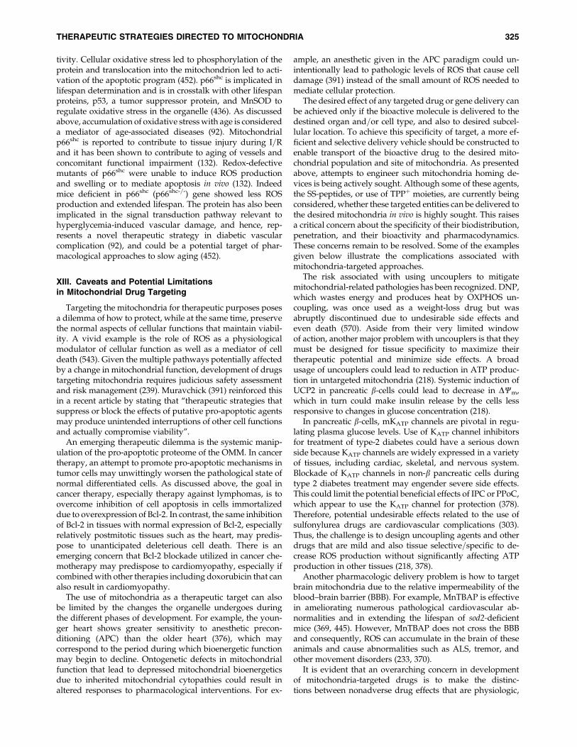
tivity. Cellular oxidative stress led to phosphorylation of theprotein and translocation into the mitochondrion led to acti-vation of the apoptotic program (452). p66shc is implicated inlifespan determination and is in crosstalk with other lifespanproteins, p53, a tumor suppressor protein, and MnSOD toregulate oxidative stress in the organelle (436). As discussedabove, accumulation of oxidative stress with age is considereda mediator of age-associated diseases (92). Mitochondrialp66shc is reported to contribute to tissue injury during I=Rand it has been shown to contribute to aging of vessels andconcomitant functional impairment (132). Redox-defectivemutants of p66shc were unable to induce ROS productionand swelling or to mediate apoptosis in vivo (132). Indeedmice deficient in p66shc (p66shc-=-) gene showed less ROSproduction and extended lifespan. The protein has also beenimplicated in the signal transduction pathway relevant tohyperglycemia-induced vascular damage, and hence, rep-resents a novel therapeutic strategy in diabetic vascularcomplication (92), and could be a potential target of phar-macological approaches to slow aging (452).
XIII. Caveats and Potential Limitationsin Mitochondrial Drug Targeting
Targeting the mitochondria for therapeutic purposes posesa dilemma of how to protect, while at the same time, preservethe normal aspects of cellular functions that maintain viabil-ity. A vivid example is the role of ROS as a physiologicalmodulator of cellular function as well as a mediator of celldeath (543). Given the multiple pathways potentially affectedby a change in mitochondrial function, development of drugstargeting mitochondria requires judicious safety assessmentand risk management (239). Muravchick (391) reinforced thisin a recent article by stating that ‘‘therapeutic strategies thatsuppress or block the effects of putative pro-apoptotic agentsmay produce unintended interruptions of other cell functionsand actually compromise viability’’.
An emerging therapeutic dilemma is the systemic manip-ulation of the pro-apoptotic proteome of the OMM. In cancertherapy, an attempt to promote pro-apoptotic mechanisms intumor cells may unwittingly worsen the pathological state ofnormal differentiated cells. As discussed above, the goal incancer therapy, especially therapy against lymphomas, is toovercome inhibition of cell apoptosis in cells immortalizeddue to overexpression of Bcl-2. In contrast, the same inhibitionof Bcl-2 in tissues with normal expression of Bcl-2, especiallyrelatively postmitotic tissues such as the heart, may predis-pose to unanticipated deleterious cell death. There is anemerging concern that Bcl-2 blockade utilized in cancer che-motherapy may predispose to cardiomyopathy, especially ifcombined with other therapies including doxorubicin that canalso result in cardiomyopathy.
The use of mitochondria as a therapeutic target can alsobe limited by the changes the organelle undergoes duringthe different phases of development. For example, the youn-ger heart shows greater sensitivity to anesthetic precon-ditioning (APC) than the older heart (376), which maycorrespond to the period during which bioenergetic functionmay begin to decline. Ontogenetic defects in mitochondrialfunction that lead to depressed mitochondrial bioenergeticsdue to inherited mitochondrial cytopathies could result inaltered responses to pharmacological interventions. For ex-
ample, an anesthetic given in the APC paradigm could un-intentionally lead to pathologic levels of ROS that cause celldamage (391) instead of the small amount of ROS needed tomediate cellular protection.
The desired effect of any targeted drug or gene delivery canbe achieved only if the bioactive molecule is delivered to thedestined organ and=or cell type, and also to desired subcel-lular location. To achieve this specificity of target, a more ef-ficient and selective delivery vehicle should be constructed toenable transport of the bioactive drug to the desired mito-chondrial population and site of mitochondria. As presentedabove, attempts to engineer such mitochondria homing de-vices is being actively sought. Although some of these agents,the SS-peptides, or use of TPPþ moieties, are currently beingconsidered, whether these targeted entities can be delivered tothe desired mitochondria in vivo is highly sought. This raisesa critical concern about the specificity of their biodistribution,penetration, and their bioactivity and pharmacodynamics.These concerns remain to be resolved. Some of the examplesgiven below illustrate the complications associated withmitochondria-targeted approaches.
The risk associated with using uncouplers to mitigatemitochondrial-related pathologies has been recognized. DNP,which wastes energy and produces heat by OXPHOS un-coupling, was once used as a weight-loss drug but wasabruptly discontinued due to undesirable side effects andeven death (570). Aside from their very limited windowof action, another major problem with uncouplers is that theymust be designed for tissue specificity to maximize theirtherapeutic potential and minimize side effects. A broadusage of uncouplers could lead to reduction in ATP produc-tion in untargeted mitochondria (218). Systemic induction ofUCP2 in pancreatic b-cells could lead to decrease in DCm,which in turn could make insulin release by the cells lessresponsive to changes in glucose concentration (218).
In pancreatic b-cells, mKATP channels are pivotal in regu-lating plasma glucose levels. Use of KATP channel inhibitorsfor treatment of type-2 diabetes could have a serious downside because KATP channels are widely expressed in a varietyof tissues, including cardiac, skeletal, and nervous system.Blockade of KATP channels in non-b pancreatic cells duringtype 2 diabetes treatment may engender severe side effects.This could limit the potential beneficial effects of IPC or PPoC,which appear to use the KATP channel for protection (378).Therefore, potential undesirable effects related to the use ofsulfonylurea drugs are cardiovascular complications (303).Thus, the challenge is to design uncoupling agents and otherdrugs that are mild and also tissue selective=specific to de-crease ROS production without significantly affecting ATPproduction in other tissues (218, 378).
Another pharmacologic delivery problem is how to targetbrain mitochondria due to the relative impermeability of theblood–brain barrier (BBB). For example, MnTBAP is effectivein ameliorating numerous pathological cardiovascular ab-normalities and in extending the lifespan of sod2-deficientmice (369, 445). However, MnTBAP does not cross the BBBand consequently, ROS can accumulate in the brain of theseanimals and cause abnormalities such as ALS, tremor, andother movement disorders (233, 370).
It is evident that an overarching concern in developmentof mitochondria-targeted drugs is to make the distinc-tions between nonadverse drug effects that are physiologic,
THERAPEUTIC STRATEGIES DIRECTED TO MITOCHONDRIA 325
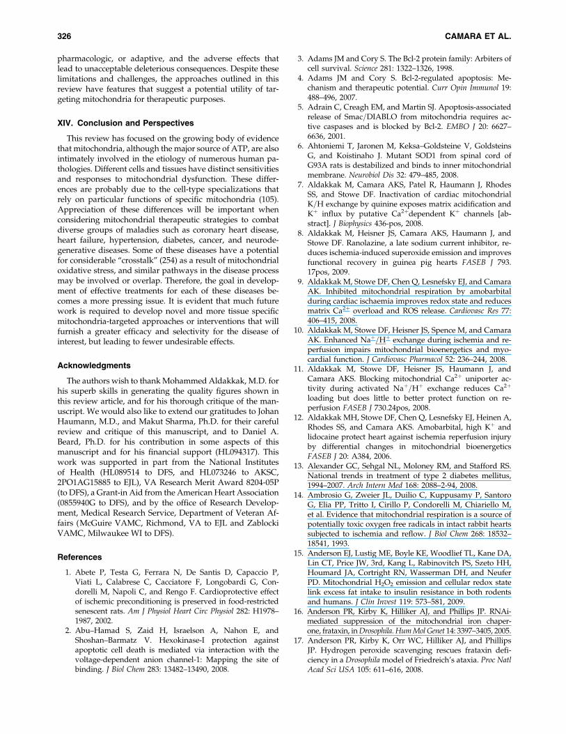
pharmacologic, or adaptive, and the adverse effects thatlead to unacceptable deleterious consequences. Despite theselimitations and challenges, the approaches outlined in thisreview have features that suggest a potential utility of tar-geting mitochondria for therapeutic purposes.
XIV. Conclusion and Perspectives
This review has focused on the growing body of evidencethat mitochondria, although the major source of ATP, are alsointimately involved in the etiology of numerous human pa-thologies. Different cells and tissues have distinct sensitivitiesand responses to mitochondrial dysfunction. These differ-ences are probably due to the cell-type specializations thatrely on particular functions of specific mitochondria (105).Appreciation of these differences will be important whenconsidering mitochondrial therapeutic strategies to combatdiverse groups of maladies such as coronary heart disease,heart failure, hypertension, diabetes, cancer, and neurode-generative diseases. Some of these diseases have a potentialfor considerable ‘‘crosstalk’’ (254) as a result of mitochondrialoxidative stress, and similar pathways in the disease processmay be involved or overlap. Therefore, the goal in develop-ment of effective treatments for each of these diseases be-comes a more pressing issue. It is evident that much futurework is required to develop novel and more tissue specificmitochondria-targeted approaches or interventions that willfurnish a greater efficacy and selectivity for the disease ofinterest, but leading to fewer undesirable effects.
Acknowledgments
The authors wish to thank Mohammed Aldakkak, M.D. forhis superb skills in generating the quality figures shown inthis review article, and for his thorough critique of the man-uscript. We would also like to extend our gratitudes to JohanHaumann, M.D., and Makut Sharma, Ph.D. for their carefulreview and critique of this manuscript, and to Daniel A.Beard, Ph.D. for his contribution in some aspects of thismanuscript and for his financial support (HL094317). Thiswork was supported in part from the National Institutesof Health (HL089514 to DFS, and HL073246 to AKSC,2PO1AG15885 to EJL), VA Research Merit Award 8204-05P(to DFS), a Grant-in Aid from the American Heart Association(0855940G to DFS), and by the office of Research Develop-ment, Medical Research Service, Department of Veteran Af-fairs (McGuire VAMC, Richmond, VA to EJL and ZablockiVAMC, Milwaukee WI to DFS).
References
1. Abete P, Testa G, Ferrara N, De Santis D, Capaccio P,Viati L, Calabrese C, Cacciatore F, Longobardi G, Con-dorelli M, Napoli C, and Rengo F. Cardioprotective effectof ischemic preconditioning is preserved in food-restrictedsenescent rats. Am J Physiol Heart Circ Physiol 282: H1978–1987, 2002.
2. Abu–Hamad S, Zaid H, Israelson A, Nahon E, andShoshan–Barmatz V. Hexokinase-I protection againstapoptotic cell death is mediated via interaction with thevoltage-dependent anion channel-1: Mapping the site ofbinding. J Biol Chem 283: 13482–13490, 2008.
3. Adams JM and Cory S. The Bcl-2 protein family: Arbiters ofcell survival. Science 281: 1322–1326, 1998.
4. Adams JM and Cory S. Bcl-2-regulated apoptosis: Me-chanism and therapeutic potential. Curr Opin Immunol 19:488–496, 2007.
5. Adrain C, Creagh EM, and Martin SJ. Apoptosis-associatedrelease of Smac=DIABLO from mitochondria requires ac-tive caspases and is blocked by Bcl-2. EMBO J 20: 6627–6636, 2001.
6. Ahtoniemi T, Jaronen M, Keksa–Goldsteine V, GoldsteinsG, and Koistinaho J. Mutant SOD1 from spinal cord ofG93A rats is destabilized and binds to inner mitochondrialmembrane. Neurobiol Dis 32: 479–485, 2008.
7. Aldakkak M, Camara AKS, Patel R, Haumann J, RhodesSS, and Stowe DF. Inactivation of cardiac mitochondrialK=H exchange by quinine exposes matrix acidification andKþ influx by putative Ca2þdependent Kþ channels [ab-stract]. J Biophysics 436-pos, 2008.
8. Aldakkak M, Heisner JS, Camara AKS, Haumann J, andStowe DF. Ranolazine, a late sodium current inhibitor, re-duces ischemia-induced superoxide emission and improvesfunctional recovery in guinea pig hearts FASEB J 793.17pos, 2009.
9. Aldakkak M, Stowe DF, Chen Q, Lesnefsky EJ, and CamaraAK. Inhibited mitochondrial respiration by amobarbitalduring cardiac ischaemia improves redox state and reducesmatrix Ca2þ overload and ROS release. Cardiovasc Res 77:406–415, 2008.
10. Aldakkak M, Stowe DF, Heisner JS, Spence M, and CamaraAK. Enhanced Naþ=Hþ exchange during ischemia and re-perfusion impairs mitochondrial bioenergetics and myo-cardial function. J Cardiovasc Pharmacol 52: 236–244, 2008.
11. Aldakkak M, Stowe DF, Heisner JS, Haumann J, andCamara AKS. Blocking mitochondrial Ca2þ uniporter ac-tivity during activated Naþ=Hþ exchange reduces Ca2þ
loading but does little to better protect function on re-perfusion FASEB J 730.24pos, 2008.
12. Aldakkak MH, Stowe DF, Chen Q, Lesnefsky EJ, Heinen A,Rhodes SS, and Camara AKS. Amobarbital, high Kþ andlidocaine protect heart against ischemia reperfusion injuryby differential changes in mitochondrial bioenergeticsFASEB J 20: A384, 2006.
13. Alexander GC, Sehgal NL, Moloney RM, and Stafford RS.National trends in treatment of type 2 diabetes mellitus,1994–2007. Arch Intern Med 168: 2088–2-94, 2008.
14. Ambrosio G, Zweier JL, Duilio C, Kuppusamy P, SantoroG, Elia PP, Tritto I, Cirillo P, Condorelli M, Chiariello M,et al. Evidence that mitochondrial respiration is a source ofpotentially toxic oxygen free radicals in intact rabbit heartssubjected to ischemia and reflow. J Biol Chem 268: 18532–18541, 1993.
15. Anderson EJ, Lustig ME, Boyle KE, Woodlief TL, Kane DA,Lin CT, Price JW, 3rd, Kang L, Rabinovitch PS, Szeto HH,Houmard JA, Cortright RN, Wasserman DH, and NeuferPD. Mitochondrial H2O2 emission and cellular redox statelink excess fat intake to insulin resistance in both rodentsand humans. J Clin Invest 119: 573–581, 2009.
16. Anderson PR, Kirby K, Hilliker AJ, and Phillips JP. RNAi-mediated suppression of the mitochondrial iron chaper-one, frataxin, in Drosophila. Hum Mol Genet 14: 3397–3405, 2005.
17. Anderson PR, Kirby K, Orr WC, Hilliker AJ, and PhillipsJP. Hydrogen peroxide scavenging rescues frataxin defi-ciency in a Drosophila model of Friedreich’s ataxia. Proc NatlAcad Sci USA 105: 611–616, 2008.
326 CAMARA ET AL.
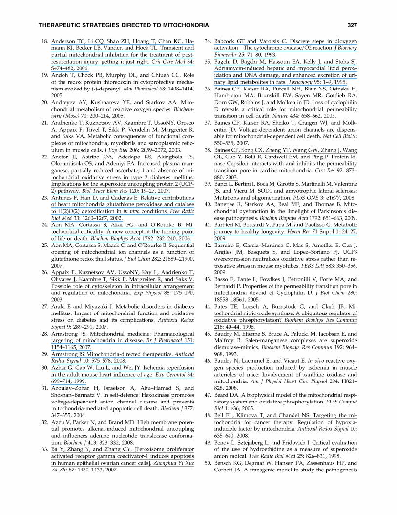
18. Anderson TC, Li CQ, Shao ZH, Hoang T, Chan KC, Ha-mann KJ, Becker LB, Vanden and Hoek TL. Transient andpartial mitochondrial inhibition for the treatment of post-resuscitation injury: getting it just right. Crit Care Med 34:S474–482, 2006.
19. Andoh T, Chock PB, Murphy DL, and Chiueh CC. Roleof the redox protein thioredoxin in cytoprotective mecha-nism evoked by (-)-deprenyl. Mol Pharmacol 68: 1408–1414,2005.
20. Andreyev AY, Kushnareva YE, and Starkov AA. Mito-chondrial metabolism of reactive oxygen species. Biochem-istry (Mosc) 70: 200–214, 2005.
21. Andrienko T, Kuznetsov AV, Kaambre T, UssoNY, OroscoA, Appaix F, Tiivel T, Sikk P, Vendelin M, Margreiter R,and Saks VA. Metabolic consequences of functional com-plexes of mitochondria, myofibrils and sarcoplasmic retic-ulum in muscle cells. J Exp Biol 206: 2059–2072, 2003.
22. Anetor JI, Asiribo OA, Adedapo KS, Akingbola TS,Olorunnisola OS, and Adeniyi FA. Increased plasma man-ganese, partially reduced ascorbate, 1 and absence of mi-tochondrial oxidative stress in type 2 diabetes mellitus:Implications for the superoxide uncoupling protein 2 (UCP-2) pathway. Biol Trace Elem Res 120: 19–27, 2007.
23. Antunes F, Han D, and Cadenas E. Relative contributionsof heart mitochondria glutathione peroxidase and catalaseto H(2)O(2) detoxification in in vivo conditions. Free RadicBiol Med 33: 1260–1267, 2002.
24. Aon MA, Cortassa S, Akar FG, and O’Rourke B. Mi-tochondrial criticality: A new concept at the turning pointof life or death. Biochim Biophys Acta 1762: 232–240, 2006.
25. Aon MA, Cortassa S, Maack C, and O’Rourke B. Sequentialopening of mitochondrial ion channels as a function ofglutathione redox thiol status. J Biol Chem 282: 21889–21900,2007.
26. Appaix F, Kuznetsov AV, UssoNY, Kay L, Andrienko T,Olivares J, Kaambre T, Sikk P, Margreiter R, and Saks V.Possible role of cytoskeleton in intracellular arrangementand regulation of mitochondria. Exp Physiol 88: 175–190,2003.
27. Araki E and Miyazaki J. Metabolic disorders in diabetesmellitus: Impact of mitochondrial function and oxidativestress on diabetes and its complications. Antioxid RedoxSignal 9: 289–291, 2007.
28. Armstrong JS. Mitochondrial medicine: Pharmacologicaltargeting of mitochondria in disease. Br J Pharmacol 151:1154–1165, 2007.
29. Armstrong JS. Mitochondria-directed therapeutics. AntioxidRedox Signal 10: 575–578, 2008.
30. Azhar G, Gao W, Liu L, and Wei JY. Ischemia-reperfusionin the adult mouse heart influence of age. Exp Gerontol 34:699–714, 1999.
31. Azoulay–Zohar H, Israelson A, Abu–Hamad S, andShoshan–Barmatz V. In self-defence: Hexokinase promotesvoltage-dependent anion channel closure and preventsmitochondria-mediated apoptotic cell death. Biochem J 377:347–355, 2004.
32. Azzu V, Parker N, and Brand MD. High membrane poten-tial promotes alkenal-induced mitochondrial uncouplingand influences adenine nucleotide translocase conforma-tion. Biochem J 413: 323–332, 2008.
33. Ba Y, Zhang Y, and Zhang CY. [Peroxisome proliferatoractivated receptor gamma coactivator-1 induces apoptosisin human epithelial ovarian cancer cells]. Zhonghua Yi XueZa Zhi 87: 1430–1433, 2007.
34. Babcock GT and Varotsis C. Discrete steps in dioxygenactivation—The cytochrome oxidase=O2 reaction. J BioenergBiomembr 25: 71–80, 1993.
35. Bagchi D, Bagchi M, Hassoun EA, Kelly J, and Stohs SJ.Adriamycin-induced hepatic and myocardial lipid perox-idation and DNA damage, and enhanced excretion of uri-nary lipid metabolites in rats. Toxicology 95: 1–9, 1995.
36. Baines CP, Kaiser RA, Purcell NH, Blair NS, Osinska H,Hambleton MA, Brunskill EW, Sayen MR, Gottlieb RA,Dorn GW, Robbins J, and Molkentin JD. Loss of cyclophilinD reveals a critical role for mitochondrial permeabilitytransition in cell death. Nature 434: 658–662, 2005.
37. Baines CP, Kaiser RA, Sheiko T, Craigen WJ, and Molk-entin JD. Voltage-dependent anion channels are dispens-able for mitochondrial-dependent cell death. Nat Cell Biol 9:550–555, 2007.
38. Baines CP, Song CX, Zheng YT, Wang GW, Zhang J, WangOL, Guo Y, Bolli R, Cardwell EM, and Ping P. Protein ki-nase Cepsilon interacts with and inhibits the permeabilitytransition pore in cardiac mitochondria. Circ Res 92: 873–880, 2003.
39. Banci L, Bertini I, Boca M, Girotto S, Martinelli M, ValentineJS, and Vieru M. SOD1 and amyotrophic lateral sclerosis:Mutations and oligomerization. PLoS ONE 3: e1677, 2008.
40. Banerjee R, Starkov AA, Beal MF, and Thomas B. Mito-chondrial dysfunction in the limelight of Parkinson’s dis-ease pathogenesis. Biochim Biophys Acta 1792: 651–663, 2009.
41. Barbieri M, Boccardi V, Papa M, and Paolisso G. Metabolicjourney to healthy longevity. Horm Res 71 Suppl 1: 24–27,2009.
42. Barreiro E, Garcia–Martinez C, Mas S, Ametller E, Gea J,Argiles JM, Busquets S, and Lopez–Soriano FJ. UCP3overexpression neutralizes oxidative stress rather than ni-trosative stress in mouse myotubes. FEBS Lett 583: 350–356,2009.
43. Basso E, Fante L, Fowlkes J, Petronilli V, Forte MA, andBernardi P. Properties of the permeability transition pore inmitochondria devoid of Cyclophilin D. J Biol Chem 280:18558–18561, 2005.
44. Bates TE, Loesch A, Burnstock G, and Clark JB. Mi-tochondrial nitric oxide synthase: A ubiquitous regulator ofoxidative phosphorylation? Biochem Biophys Res Commun218: 40–44, 1996.
45. Baudry M, Etienne S, Bruce A, Palucki M, Jacobsen E, andMalfroy B. Salen-manganese complexes are superoxidedismutase-mimics. Biochem Biophys Res Commun 192: 964–968, 1993.
46. Baudry N, Laemmel E, and Vicaut E. In vivo reactive oxy-gen species production induced by ischemia in musclearterioles of mice: Involvement of xanthine oxidase andmitochondria. Am J Physiol Heart Circ Physiol 294: H821–828, 2008.
47. Beard DA. A biophysical model of the mitochondrial respi-ratory system and oxidative phosphorylation. PLoS ComputBiol 1: e36, 2005.
48. Bell EL, Klimova T, and Chandel NS. Targeting the mi-tochondria for cancer therapy: Regulation of hypoxia-inducible factor by mitochondria. Antioxid Redox Signal 10:635–640, 2008.
49. Benov L, Sztejnberg L, and Fridovich I. Critical evaluationof the use of hydroethidine as a measure of superoxideanion radical. Free Radic Biol Med 25: 826–831, 1998.
50. Bensch KG, Degraaf W, Hansen PA, Zassenhaus HP, andCorbett JA. A transgenic model to study the pathogenesis
THERAPEUTIC STRATEGIES DIRECTED TO MITOCHONDRIA 327
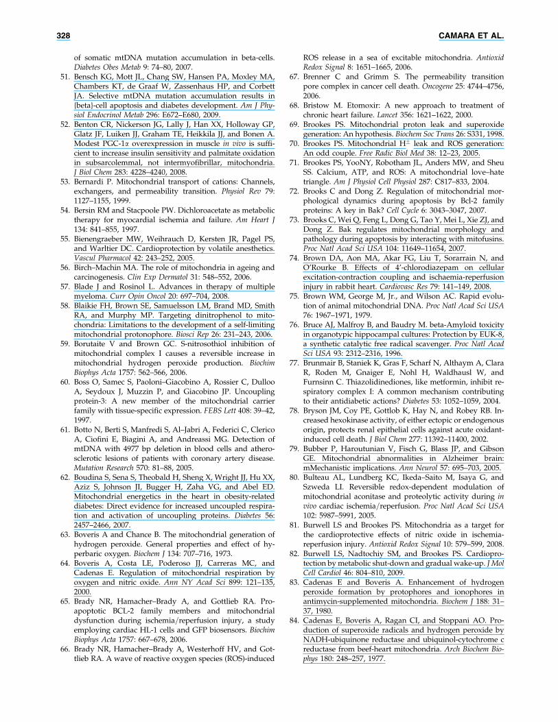
of somatic mtDNA mutation accumulation in beta-cells.Diabetes Obes Metab 9: 74–80, 2007.
51. Bensch KG, Mott JL, Chang SW, Hansen PA, Moxley MA,Chambers KT, de Graaf W, Zassenhaus HP, and CorbettJA. Selective mtDNA mutation accumulation results in{beta}-cell apoptosis and diabetes development. Am J Phy-siol Endocrinol Metab 296: E672–E680, 2009.
52. Benton CR, Nickerson JG, Lally J, Han XX, Holloway GP,Glatz JF, Luiken JJ, Graham TE, Heikkila JJ, and Bonen A.Modest PGC-1a overexpression in muscle in vivo is suffi-cient to increase insulin sensitivity and palmitate oxidationin subsarcolemmal, not intermyofibrillar, mitochondria.J Biol Chem 283: 4228–4240, 2008.
53. Bernardi P. Mitochondrial transport of cations: Channels,exchangers, and permeability transition. Physiol Rev 79:1127–1155, 1999.
54. Bersin RM and Stacpoole PW. Dichloroacetate as metabolictherapy for myocardial ischemia and failure. Am Heart J134: 841–855, 1997.
55. Bienengraeber MW, Weihrauch D, Kersten JR, Pagel PS,and Warltier DC. Cardioprotection by volatile anesthetics.Vascul Pharmacol 42: 243–252, 2005.
56. Birch–Machin MA. The role of mitochondria in ageing andcarcinogenesis. Clin Exp Dermatol 31: 548–552, 2006.
57. Blade J and Rosinol L. Advances in therapy of multiplemyeloma. Curr Opin Oncol 20: 697–704, 2008.
58. Blaikie FH, Brown SE, Samuelsson LM, Brand MD, SmithRA, and Murphy MP. Targeting dinitrophenol to mito-chondria: Limitations to the development of a self-limitingmitochondrial protonophore. Biosci Rep 26: 231–243, 2006.
59. Borutaite V and Brown GC. S-nitrosothiol inhibition ofmitochondrial complex I causes a reversible increase inmitochondrial hydrogen peroxide production. BiochimBiophys Acta 1757: 562–566, 2006.
60. Boss O, Samec S, Paoloni–Giacobino A, Rossier C, DullooA, Seydoux J, Muzzin P, and Giacobino JP. Uncouplingprotein-3: A new member of the mitochondrial carrierfamily with tissue-specific expression. FEBS Lett 408: 39–42,1997.
61. Botto N, Berti S, Manfredi S, Al–Jabri A, Federici C, ClericoA, Ciofini E, Biagini A, and Andreassi MG. Detection ofmtDNA with 4977 bp deletion in blood cells and athero-sclerotic lesions of patients with coronary artery disease.Mutation Research 570: 81–88, 2005.
62. Boudina S, Sena S, Theobald H, Sheng X, Wright JJ, Hu XX,Aziz S, Johnson JI, Bugger H, Zaha VG, and Abel ED.Mitochondrial energetics in the heart in obesity-relateddiabetes: Direct evidence for increased uncoupled respira-tion and activation of uncoupling proteins. Diabetes 56:2457–2466, 2007.
63. Boveris A and Chance B. The mitochondrial generation ofhydrogen peroxide. General properties and effect of hy-perbaric oxygen. Biochem J 134: 707–716, 1973.
64. Boveris A, Costa LE, Poderoso JJ, Carreras MC, andCadenas E. Regulation of mitochondrial respiration byoxygen and nitric oxide. Ann NY Acad Sci 899: 121–135,2000.
65. Brady NR, Hamacher–Brady A, and Gottlieb RA. Pro-apoptotic BCL-2 family members and mitochondrialdysfunction during ischemia=reperfusion injury, a studyemploying cardiac HL-1 cells and GFP biosensors. BiochimBiophys Acta 1757: 667–678, 2006.
66. Brady NR, Hamacher–Brady A, Westerhoff HV, and Got-tlieb RA. A wave of reactive oxygen species (ROS)-induced
ROS release in a sea of excitable mitochondria. AntioxidRedox Signal 8: 1651–1665, 2006.
67. Brenner C and Grimm S. The permeability transitionpore complex in cancer cell death. Oncogene 25: 4744–4756,2006.
68. Bristow M. Etomoxir: A new approach to treatment ofchronic heart failure. Lancet 356: 1621–1622, 2000.
69. Brookes PS. Mitochondrial proton leak and superoxidegeneration: An hypothesis. Biochem Soc Trans 26: S331, 1998.
70. Brookes PS. Mitochondrial Hþ leak and ROS generation:An odd couple. Free Radic Biol Med 38: 12–23, 2005.
71. Brookes PS, YooNY, Robotham JL, Anders MW, and SheuSS. Calcium, ATP, and ROS: A mitochondrial love–hatetriangle. Am J Physiol Cell Physiol 287: C817–833, 2004.
72. Brooks C and Dong Z. Regulation of mitochondrial mor-phological dynamics during apoptosis by Bcl-2 familyproteins: A key in Bak? Cell Cycle 6: 3043–3047, 2007.
73. Brooks C, Wei Q, Feng L, Dong G, Tao Y, Mei L, Xie ZJ, andDong Z. Bak regulates mitochondrial morphology andpathology during apoptosis by interacting with mitofusins.Proc Natl Acad Sci USA 104: 11649–11654, 2007.
74. Brown DA, Aon MA, Akar FG, Liu T, Sorarrain N, andO’Rourke B. Effects of 4’-chlorodiazepam on cellularexcitation-contraction coupling and ischaemia-reperfusioninjury in rabbit heart. Cardiovasc Res 79: 141–149, 2008.
75. Brown WM, George M, Jr., and Wilson AC. Rapid evolu-tion of animal mitochondrial DNA. Proc Natl Acad Sci USA76: 1967–1971, 1979.
76. Bruce AJ, Malfroy B, and Baudry M. beta-Amyloid toxicityin organotypic hippocampal cultures: Protection by EUK-8,a synthetic catalytic free radical scavenger. Proc Natl AcadSci USA 93: 2312–2316, 1996.
77. Brunmair B, Staniek K, Gras F, Scharf N, Althaym A, ClaraR, Roden M, Gnaiger E, Nohl H, Waldhausl W, andFurnsinn C. Thiazolidinediones, like metformin, inhibit re-spiratory complex I: A common mechanism contributingto their antidiabetic actions? Diabetes 53: 1052–1059, 2004.
78. Bryson JM, Coy PE, Gottlob K, Hay N, and Robey RB. In-creased hexokinase activity, of either ectopic or endogenousorigin, protects renal epithelial cells against acute oxidant-induced cell death. J Biol Chem 277: 11392–11400, 2002.
79. Bubber P, Haroutunian V, Fisch G, Blass JP, and GibsonGE. Mitochondrial abnormalities in Alzheimer brain:mMechanistic implications. Ann Neurol 57: 695–703, 2005.
80. Bulteau AL, Lundberg KC, Ikeda–Saito M, Isaya G, andSzweda LI. Reversible redox-dependent modulation ofmitochondrial aconitase and proteolytic activity during invivo cardiac ischemia=reperfusion. Proc Natl Acad Sci USA102: 5987–5991, 2005.
81. Burwell LS and Brookes PS. Mitochondria as a target forthe cardioprotective effects of nitric oxide in ischemia-reperfusion injury. Antioxid Redox Signal 10: 579–599, 2008.
82. Burwell LS, Nadtochiy SM, and Brookes PS. Cardiopro-tection by metabolic shut-down and gradual wake-up. J MolCell Cardiol 46: 804–810, 2009.
83. Cadenas E and Boveris A. Enhancement of hydrogenperoxide formation by protophores and ionophores inantimycin-supplemented mitochondria. Biochem J 188: 31–37, 1980.
84. Cadenas E, Boveris A, Ragan CI, and Stoppani AO. Pro-duction of superoxide radicals and hydrogen peroxide byNADH-ubiquinone reductase and ubiquinol-cytochrome creductase from beef-heart mitochondria. Arch Biochem Bio-phys 180: 248–257, 1977.
328 CAMARA ET AL.
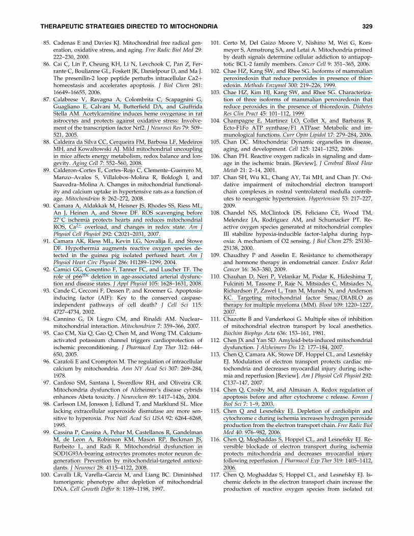
85. Cadenas E and Davies KJ. Mitochondrial free radical gen-eration, oxidative stress, and aging. Free Radic Biol Med 29:222–230, 2000.
86. Cai C, Lin P, Cheung KH, Li N, Levchook C, Pan Z, Fer-rante C, Boulianne GL, Foskett JK, Danielpour D, and Ma J.The presenilin-2 loop peptide perturbs intracellular Ca2þhomeostasis and accelerates apoptosis. J Biol Chem 281:16649–16655, 2006.
87. Calabrese V, Ravagna A, Colombrita C, Scapagnini G,Guagliano E, Calvani M, Butterfield DA, and GiuffridaStella AM. Acetylcarnitine induces heme oxygenase in ratastrocytes and protects against oxidative stress: Involve-ment of the transcription factor Nrf2. J Neurosci Res 79: 509–521, 2005.
88. Caldeira da Silva CC, Cerqueira FM, Barbosa LF, MedeirosMH, and Kowaltowski AJ. Mild mitochondrial uncouplingin mice affects energy metabolism, redox balance and lon-gevity. Aging Cell 7: 552–560, 2008.
89. Calderon–Cortes E, Cortes–Rojo C, Clemente–Guerrero M,Manzo–Avalos S, Villalobos–Molina R, Boldogh I, andSaavedra–Molina A. Changes in mitochondrial functional-ity and calcium uptake in hypertensive rats as a function ofage. Mitochondrion 8: 262–272, 2008.
90. Camara A, Aldakkak M, Heisner JS, Rhodes SS, Riess ML,An J, Heinen A, and Stowe DF. ROS scavenging before278C ischemia protects hearts and reduces mitochondrialROS, Ca2þ overload, and changes in redox state. Am JPhysiol Cell Physiol 292: C2021–2031, 2007.
91. Camara AK, Riess ML, Kevin LG, Novalija E, and StoweDF. Hypothermia augments reactive oxygen species de-tected in the guinea pig isolated perfused heart. Am JPhysiol Heart Circ Physiol 286: H1289–1299, 2004.
92. Camici GG, Cosentino F, Tanner FC, and Luscher TF. Therole of p66Shc deletion in age-associated arterial dysfunc-tion and disease states. J Appl Physiol 105: 1628–1631, 2008.
93. Cande C, Cecconi F, Dessen P, and Kroemer G. Apoptosis-inducing factor (AIF): Key to the conserved caspase-independent pathways of cell death? J Cell Sci 115:4727–4734, 2002.
94. Cannino G, Di Liegro CM, and Rinaldi AM. Nuclear–mitochondrial interaction. Mitochondrion 7: 359–366, 2007.
95. Cao CM, Xia Q, Gao Q, Chen M, and Wong TM. Calcium-activated potassium channel triggers cardioprotection ofischemic preconditioning. J Pharmacol Exp Ther 312: 644–650, 2005.
96. Carafoli E and Crompton M. The regulation of intracellularcalcium by mitochondria. Ann NY Acad Sci 307: 269–284,1978.
97. Cardoso SM, Santana I, Swerdlow RH, and Oliveira CR.Mitochondria dysfunction of Alzheimer’s disease cybridsenhances Abeta toxicity. J Neurochem 89: 1417–1426, 2004.
98. Carlsson LM, Jonsson J, Edlund T, and Marklund SL. Micelacking extracellular superoxide dismutase are more sen-sitive to hyperoxia. Proc Natl Acad Sci USA 92: 6264–6268,1995.
99. Cassina P, Cassina A, Pehar M, Castellanos R, GandelmanM, de Leon A, Robinson KM, Mason RP, Beckman JS,Barbeito L, and Radi R. Mitochondrial dysfunction inSOD1G93A-bearing astrocytes promotes motor neuron de-generation: Prevention by mitochondrial-targeted antioxi-dants. J Neurosci 28: 4115–4122, 2008.
100. Cavalli LR, Varella–Garcia M, and Liang BC. Diminishedtumorigenic phenotype after depletion of mitochondrialDNA. Cell Growth Differ 8: 1189–1198, 1997.
101. Certo M, Del Gaizo Moore V, Nishino M, Wei G, Kors-meyer S, Armstrong SA, and Letai A. Mitochondria primedby death signals determine cellular addiction to antiapop-totic BCL-2 family members. Cancer Cell 9: 351–365, 2006.
102. Chae HZ, Kang SW, and Rhee SG. Isoforms of mammalianperoxiredoxin that reduce peroxides in presence of thior-edoxin. Methods Enzymol 300: 219–226, 1999.
103. Chae HZ, Kim HJ, Kang SW, and Rhee SG. Characteriza-tion of three isoforms of mammalian peroxiredoxin thatreduce peroxides in the presence of thioredoxin. DiabetesRes Clin Pract 45: 101–112, 1999.
104. Champagne E, Martinez LO, Collet X, and Barbaras R.Ecto-F1Fo ATP synthase=F1 ATPase: Metabolic and im-munological functions. Curr Opin Lipidol 17: 279–284, 2006.
105. Chan DC. Mitochondria: Dynamic organelles in disease,aging, and development. Cell 125: 1241–1252, 2006.
106. Chan PH. Reactive oxygen radicals in signaling and dam-age in the ischemic brain. [Review]. J Cerebral Blood FlowMetab 21: 2–14, 2001.
107. Chan SH, Wu KL, Chang AY, Tai MH, and Chan JY. Oxi-dative impairment of mitochondrial electron transportchain complexes in rostral ventrolateral medulla contrib-utes to neurogenic hypertension. Hypertension 53: 217–227,2009.
108. Chandel NS, McClintock DS, Feliciano CE, Wood TM,Melendez JA, Rodriguez AM, and Schumacker PT. Re-active oxygen species generated at mitochondrial complexIII stabilize hypoxia-inducible factor-1alpha during hyp-oxia: A mechanism of O2 sensing. J Biol Chem 275: 25130–25138, 2000.
109. Chaudhry P and Asselin E. Resistance to chemotherapyand hormone therapy in endometrial cancer. Endocr RelatCancer 16: 363–380, 2009.
110. Chauhan D, Neri P, Velankar M, Podar K, Hideshima T,Fulciniti M, Tassone P, Raje N, Mitsiades C, Mitsiades N,Richardson P, Zawel L, Tran M, Munshi N, and AndersonKC. Targeting mitochondrial factor Smac=DIABLO astherapy for multiple myeloma (MM). Blood 109: 1220–1227,2007.
111. Chazotte B and Vanderkooi G. Multiple sites of inhibitionof mitochondrial electron transport by local anesthetics.Biochim Biophys Acta 636: 153–161, 1981.
112. Chen JX and Yan SD. Amyloid-beta-induced mitochondrialdysfunction. J Alzheimers Dis 12: 177–184, 2007.
113. Chen Q, Camara AK, Stowe DF, Hoppel CL, and LesnefskyEJ. Modulation of electron transport protects cardiac mi-tochondria and decreases myocardial injury during ische-mia and reperfusion [Review]. Am J Physiol Cell Physiol 292:C137–147, 2007.
114. Chen Q, Crosby M, and Almasan A. Redox regulation ofapoptosis before and after cytochrome c release. Korean JBiol Sci 7: 1–9, 2003.
115. Chen Q and Lesnefsky EJ. Depletion of cardiolipin andcytochrome c during ischemia increases hydrogen peroxideproduction from the electron transport chain. Free Radic BiolMed 40: 976–982, 2006.
116. Chen Q, Moghaddas S, Hoppel CL, and Lesnefsky EJ. Re-versible blockade of electron transport during ischemiaprotects mitochondria and decreases myocardial injuryfollowing reperfusion. J Pharmacol Exp Ther 319: 1405–1412,2006.
117. Chen Q, Moghaddas S, Hoppel CL, and Lesnefsky EJ. Is-chemic defects in the electron transport chain increase theproduction of reactive oxygen species from isolated rat
THERAPEUTIC STRATEGIES DIRECTED TO MITOCHONDRIA 329
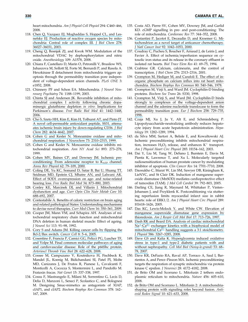
heart mitochondria. Am J Physiol Cell Physiol 294: C460–466,2008.
118. Chen Q, Vazquez EJ, Moghaddas S, Hoppel CL, and Les-nefsky EJ. Production of reactive oxygen species by mito-chondria: Central role of complex III. J Biol Chem 278:36027–36031, 2003.
119. Cheng Q, Bosnjak ZJ, and Kwok WM. Modulation of themitochondrial VDAC by dephosphorylation and nitricoxide. Anesthesiology 109: A1578, 2008.
120. Chiara F, Castellaro D, Marin O, Petronilli V, Brusilow WS,Juhaszova M, Sollott SJ, Forte M, Bernardi P, and Rasola A.Hexokinase II detachment from mitochondria triggers ap-optosis through the permeability transition pore indepen-dent of voltage-dependent anion channels. PLoS ONE 3:e1852, 2008.
121. Chinnery PF and Schon EA. Mitochondria. J Neurol Neu-rosurg Psychiatry 74: 1188–1199, 2003.
122. Chinta SJ and Andersen JK. Reversible inhibition of mito-chondrial complex I activity following chronic dopa-minergic glutathione depletion in vitro: Implications forParkinson’s disease. Free Radic Biol Med 41: 1442–1448,2006.
123. Cho S, Szeto HH, Kim E, Kim H, Tolhurst AT, and Pinto JT.A novel cell-permeable antioxidant peptide, SS31, attenu-ates ischemic brain injury by down-regulating CD36. J BiolChem 282: 4634–4642, 2007.
124. Cohen G and Kesler N. Monoamine oxidase and mito-chondrial respiration. J Neurochem 73: 2310–2315, 1999.
125. Cohen G and Kesler N. Monoamine oxidase inhibits mi-tochondrial respiration. Ann NY Acad Sci 893: 273–278,1999.
126. Cohen MV, Baines CP, and Downey JM. Ischemic pre-conditioning: From adenosine receptor to KATP channel.Annu Rev Physiol 62: 79–109, 2000.
127. Coling DE, Yu KC, Somand D, Satar B, Bai U, Huang TT,Seidman MD, Epstein CJ, Mhatre AN, and Lalwani AK.Effect of SOD1 overexpression on age- and noise-relatedhearing loss. Free Radic Biol Med 34: 873–880, 2003.
128. Conley KE, Marcinek DJ, and Villarin J. Mitochondrialdysfunction and age. Curr Opin Clin Nutr Metab Care 10:688–692, 2007.
129. Contestabile A. Benefits of caloric restriction on brain agingand related pathological States: Understanding mechanismsto devise novel therapies. Curr Med Chem 16: 350–361, 2009.
130. Cooper JM, Mann VM, and Schapira AH. Analyses of mi-tochondrial respiratory chain function and mitochondrialDNA deletion in human skeletal muscle: Effect of ageing.J Neurol Sci 113: 91–98, 1992.
131. Cory S and Adams JM. Killing cancer cells by flipping theBcl-2=Bax switch. Cancer Cell 8: 5–6, 2005.
132. Cosentino F, Francia P, Camici GG, Pelicci PG, Luscher TF,and Volpe M. Final common molecular pathways of agingand cardiovascular disease: Role of the p66Shc protein.Arterioscl Thromb Vasc Biol 28: 622–628, 2008.
133. Cossee M, Campuzano V, Koutnikova H, Fischbeck K,Mandel JL, Koenig M, Bidichandani SI, Patel PI, MolteMD, Canizares J, De Frutos R, Pianese L, Cavalcanti F,Monticelli A, Cocozza S, Montermini L, and Pandolfo M.Frataxin fracas. Nat Genet 15: 337–338, 1997.
134. Cossu F, Mastrangelo E, Milani M, Sorrentino G, Lecis D,Delia D, Manzoni L, Seneci P, Scolastico C, and BolognesiM. Designing Smac-mimetics as antagonists of XIAP,cIAP1, and cIAP2. Biochem Biophys Res Commun 378: 162–167, 2009.
135. Costa AD, Pierre SV, Cohen MV, Downey JM, and GarlidKD. cGMP signalling in pre- and post-conditioning: Therole of mitochondria. Cardiovasc Res 77: 344–352, 2008.
136. Costantini P, Jacotot E, Decaudin D, and Kroemer G. Mi-tochondrion as a novel target of anticancer chemotherapy.J Natl Cancer Inst 92: 1042–1053, 2000.
137. Coudray C, Pucheu S, Boucher F, Arnaud J, de Leiris J, andFavier A. Effect of ischemia=reperfusion sequence on cy-tosolic iron status and its release in the coronary effluent inisolated rat hearts. Biol Trace Elem Res 41: 69–75, 1994.
138. Crabtree GR. Calcium, calcineurin, and the control oftranscription. J Biol Chem 276: 2313–2316, 2001.
139. Crompton M, Hediger M, and Carafoli E. The effect of in-organic phosphate on calcium influx into rat heart mito-chondria. Biochem Biophys Res Commun 80: 540–546, 1978.
140. Crompton M, Virji S, and Ward JM. Cyclophilin-D bindingproteins. Biochem Soc Trans 26: S330, 1998.
141. Crompton M, Virji S, and Ward JM. Cyclophilin-D bindsstrongly to complexes of the voltage-dependent anionchannel and the adenine nucleotide translocase to form thepermeability transition pore. Eur J Biochem 258: 729–735,1998.
142. Czaja MJ, Xu J, Ju Y, Alt E, and Schmiedeberg P.Lipopolysaccharide-neutralizing antibody reduces hepato-cyte injury from acute hepatotoxin administration. Hepa-tology 19: 1282–1289, 1994.
143. da Silva MM, Sartori A, Belisle E, and Kowaltowski AJ.Ischemic preconditioning inhibits mitochondrial respira-tion, increases H2O2 release, and enhances Kþ transport.Am J Physiol Heart Circ Physiol 285: H154–162, 2003.
144. Dai Y, Liu M, Tang W, DeSano J, Burstein E, Davis M,Pienta K, Lawrence T, and Xu L. Molecularly targetedradiosensitization of human prostate cancer by modulatinginhibitor of apoptosis. Clin Cancer Res 14: 7701–7710, 2008.
145. Daosukho C, Ittarat W, Lin SM, Sawyer DB, Kiningham K,LieNYC, and St Clair DK. Induction of manganese super-oxide dismutase (MnSOD) mediates cardioprotective effectof tamoxifen (TAM). J Mol Cell Cardiol 39: 792–803, 2005.
146. Darling CE, Jiang R, Maynard M, Whittaker P, Vinten–Johansen J, and Przyklenk K. Postconditioning via stutter-ing reperfusion limits myocardial infarct size in rabbithearts: role of ERK1=2. Am J Physiol Heart Circ Physiol 289:H1618–1626, 2005.
147. Das KC, Lewis-Molock Y, and White CW. Elevation ofmanganese superoxide dismutase gene expression bythioredoxin. Am J Respir Cell Mol Biol 17: 713–726, 1997.
148. Dash RK and Beard DA. Analysis of cardiac mitochondrialNaþ-Ca2þ exchanger kinetics with a biophysical model ofmitochondrial Ca2þ handling suggests a 3:1 stoichiometry.J Physiol 586: 3267–3285, 2008.
149. Dave GS and Kalia K. Hyperglycemia induced oxidativestress in type-1 and type-2 diabetic patients with andwithout nephropathy. Cell Mol Biol (Noisy-le-grand) 53: 68–78, 2007.
150. Dave KR, DeFazio RA, Raval AP, Torraco A, Saul I, Bar-rientos A, and Perez–Pinzon MA. Ischemic preconditioningtargets the respiration of synaptic mitochondria via proteinkinase C epsilon. J Neurosci 28: 4172–4182, 2008.
151. de Brito OM and Scorrano L. Mitofusin 2 tethers endo-plasmic reticulum to mitochondria. Nature 456: 605–610,2008.
152. de Brito OM and Scorrano L. Mitofusin 2: A mitochondria-shaping protein with signaling roles beyond fusion. Anti-oxid Redox Signal 10: 621–633, 2008.
330 CAMARA ET AL.
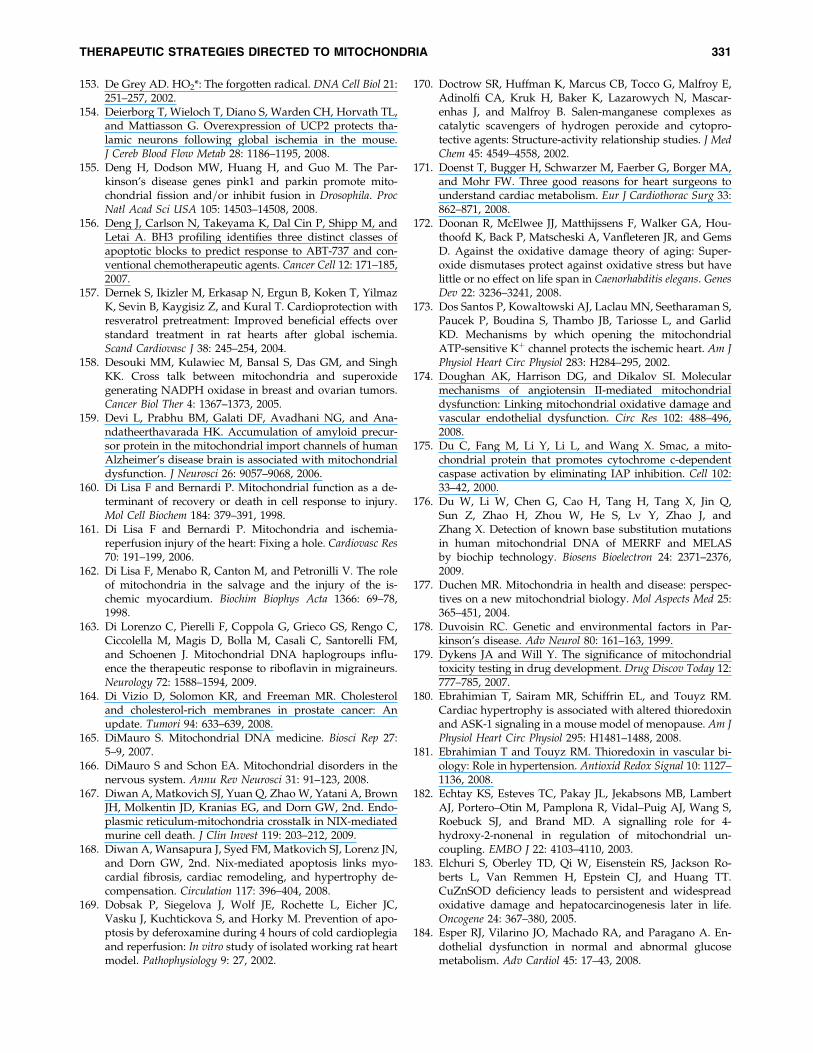
153. De Grey AD. HO2*: The forgotten radical. DNA Cell Biol 21:251–257, 2002.
154. Deierborg T, Wieloch T, Diano S, Warden CH, Horvath TL,and Mattiasson G. Overexpression of UCP2 protects tha-lamic neurons following global ischemia in the mouse.J Cereb Blood Flow Metab 28: 1186–1195, 2008.
155. Deng H, Dodson MW, Huang H, and Guo M. The Par-kinson’s disease genes pink1 and parkin promote mito-chondrial fission and=or inhibit fusion in Drosophila. ProcNatl Acad Sci USA 105: 14503–14508, 2008.
156. Deng J, Carlson N, Takeyama K, Dal Cin P, Shipp M, andLetai A. BH3 profiling identifies three distinct classes ofapoptotic blocks to predict response to ABT-737 and con-ventional chemotherapeutic agents. Cancer Cell 12: 171–185,2007.
157. Dernek S, Ikizler M, Erkasap N, Ergun B, Koken T, YilmazK, Sevin B, Kaygisiz Z, and Kural T. Cardioprotection withresveratrol pretreatment: Improved beneficial effects overstandard treatment in rat hearts after global ischemia.Scand Cardiovasc J 38: 245–254, 2004.
158. Desouki MM, Kulawiec M, Bansal S, Das GM, and SinghKK. Cross talk between mitochondria and superoxidegenerating NADPH oxidase in breast and ovarian tumors.Cancer Biol Ther 4: 1367–1373, 2005.
159. Devi L, Prabhu BM, Galati DF, Avadhani NG, and Ana-ndatheerthavarada HK. Accumulation of amyloid precur-sor protein in the mitochondrial import channels of humanAlzheimer’s disease brain is associated with mitochondrialdysfunction. J Neurosci 26: 9057–9068, 2006.
160. Di Lisa F and Bernardi P. Mitochondrial function as a de-terminant of recovery or death in cell response to injury.Mol Cell Biochem 184: 379–391, 1998.
161. Di Lisa F and Bernardi P. Mitochondria and ischemia-reperfusion injury of the heart: Fixing a hole. Cardiovasc Res70: 191–199, 2006.
162. Di Lisa F, Menabo R, Canton M, and Petronilli V. The roleof mitochondria in the salvage and the injury of the is-chemic myocardium. Biochim Biophys Acta 1366: 69–78,1998.
163. Di Lorenzo C, Pierelli F, Coppola G, Grieco GS, Rengo C,Ciccolella M, Magis D, Bolla M, Casali C, Santorelli FM,and Schoenen J. Mitochondrial DNA haplogroups influ-ence the therapeutic response to riboflavin in migraineurs.Neurology 72: 1588–1594, 2009.
164. Di Vizio D, Solomon KR, and Freeman MR. Cholesteroland cholesterol-rich membranes in prostate cancer: Anupdate. Tumori 94: 633–639, 2008.
165. DiMauro S. Mitochondrial DNA medicine. Biosci Rep 27:5–9, 2007.
166. DiMauro S and Schon EA. Mitochondrial disorders in thenervous system. Annu Rev Neurosci 31: 91–123, 2008.
167. Diwan A, Matkovich SJ, Yuan Q, Zhao W, Yatani A, BrownJH, Molkentin JD, Kranias EG, and Dorn GW, 2nd. Endo-plasmic reticulum-mitochondria crosstalk in NIX-mediatedmurine cell death. J Clin Invest 119: 203–212, 2009.
168. Diwan A, Wansapura J, Syed FM, Matkovich SJ, Lorenz JN,and Dorn GW, 2nd. Nix-mediated apoptosis links myo-cardial fibrosis, cardiac remodeling, and hypertrophy de-compensation. Circulation 117: 396–404, 2008.
169. Dobsak P, Siegelova J, Wolf JE, Rochette L, Eicher JC,Vasku J, Kuchtickova S, and Horky M. Prevention of apo-ptosis by deferoxamine during 4 hours of cold cardioplegiaand reperfusion: In vitro study of isolated working rat heartmodel. Pathophysiology 9: 27, 2002.
170. Doctrow SR, Huffman K, Marcus CB, Tocco G, Malfroy E,Adinolfi CA, Kruk H, Baker K, Lazarowych N, Mascar-enhas J, and Malfroy B. Salen-manganese complexes ascatalytic scavengers of hydrogen peroxide and cytopro-tective agents: Structure-activity relationship studies. J MedChem 45: 4549–4558, 2002.
171. Doenst T, Bugger H, Schwarzer M, Faerber G, Borger MA,and Mohr FW. Three good reasons for heart surgeons tounderstand cardiac metabolism. Eur J Cardiothorac Surg 33:862–871, 2008.
172. Doonan R, McElwee JJ, Matthijssens F, Walker GA, Hou-thoofd K, Back P, Matscheski A, Vanfleteren JR, and GemsD. Against the oxidative damage theory of aging: Super-oxide dismutases protect against oxidative stress but havelittle or no effect on life span in Caenorhabditis elegans. GenesDev 22: 3236–3241, 2008.
173. Dos Santos P, Kowaltowski AJ, Laclau MN, Seetharaman S,Paucek P, Boudina S, Thambo JB, Tariosse L, and GarlidKD. Mechanisms by which opening the mitochondrialATP-sensitive Kþ channel protects the ischemic heart. Am JPhysiol Heart Circ Physiol 283: H284–295, 2002.
174. Doughan AK, Harrison DG, and Dikalov SI. Molecularmechanisms of angiotensin II-mediated mitochondrialdysfunction: Linking mitochondrial oxidative damage andvascular endothelial dysfunction. Circ Res 102: 488–496,2008.
175. Du C, Fang M, Li Y, Li L, and Wang X. Smac, a mito-chondrial protein that promotes cytochrome c-dependentcaspase activation by eliminating IAP inhibition. Cell 102:33–42, 2000.
176. Du W, Li W, Chen G, Cao H, Tang H, Tang X, Jin Q,Sun Z, Zhao H, Zhou W, He S, Lv Y, Zhao J, andZhang X. Detection of known base substitution mutationsin human mitochondrial DNA of MERRF and MELASby biochip technology. Biosens Bioelectron 24: 2371–2376,2009.
177. Duchen MR. Mitochondria in health and disease: perspec-tives on a new mitochondrial biology. Mol Aspects Med 25:365–451, 2004.
178. Duvoisin RC. Genetic and environmental factors in Par-kinson’s disease. Adv Neurol 80: 161–163, 1999.
179. Dykens JA and Will Y. The significance of mitochondrialtoxicity testing in drug development. Drug Discov Today 12:777–785, 2007.
180. Ebrahimian T, Sairam MR, Schiffrin EL, and Touyz RM.Cardiac hypertrophy is associated with altered thioredoxinand ASK-1 signaling in a mouse model of menopause. Am JPhysiol Heart Circ Physiol 295: H1481–1488, 2008.
181. Ebrahimian T and Touyz RM. Thioredoxin in vascular bi-ology: Role in hypertension. Antioxid Redox Signal 10: 1127–1136, 2008.
182. Echtay KS, Esteves TC, Pakay JL, Jekabsons MB, LambertAJ, Portero–Otin M, Pamplona R, Vidal–Puig AJ, Wang S,Roebuck SJ, and Brand MD. A signalling role for 4-hydroxy-2-nonenal in regulation of mitochondrial un-coupling. EMBO J 22: 4103–4110, 2003.
183. Elchuri S, Oberley TD, Qi W, Eisenstein RS, Jackson Ro-berts L, Van Remmen H, Epstein CJ, and Huang TT.CuZnSOD deficiency leads to persistent and widespreadoxidative damage and hepatocarcinogenesis later in life.Oncogene 24: 367–380, 2005.
184. Esper RJ, Vilarino JO, Machado RA, and Paragano A. En-dothelial dysfunction in normal and abnormal glucosemetabolism. Adv Cardiol 45: 17–43, 2008.
THERAPEUTIC STRATEGIES DIRECTED TO MITOCHONDRIA 331
185. Feng J, Zhu M, Schaub MC, Gehrig P, Roschitzki B, Luc-chinetti E, and Zaugg M. Phosphoproteome analysis ofisoflurane-protected heart mitochondria: phosphorylationof adenine nucleotide translocator-1 on Tyr194 regulatesmitochondrial function. Cardiovasc Res 80: 20–29, 2008.
186. Fennell JP, Brosnan MJ, Frater AJ, Hamilton CA, AlexanderMY, Nicklin SA, Heistad DD, Baker AH, and DominiczakAF. Adenovirus-mediated overexpression of extracellularsuperoxide dismutase improves endothelial dysfunction ina rat model of hypertension. Gene Ther 9: 110–117, 2002.
187. Ferdinandy P and Schulz R. Nitric oxide, superoxide, andperoxynitrite in myocardial ischaemia-reperfusion injuryand preconditioning. Br J Pharmacol 138: 532–543, 2003.
188. Fiskum G, Rosenthal RE, Vereczki V, Martin E, HoffmanGE, Chinopoulos C, and Kowaltowski A. Protectionagainst ischemic brain injury by inhibition of mitochondrialoxidative stress. J Bioenerg Biomembr 36: 347–352, 2004.
189. Flanagan SW, Anderson RD, Ross MA, and Oberley LW.Overexpression of manganese superoxide dismutase at-tenuates neuronal death in human cells expressing mutant(G37R) Cu=Zn-superoxide dismutase. J Neurochem 81: 170–177, 2002.
190. Fontan–Lozano A, Lopez–Lluch G, Delgado–Garcia JM,Navas P, and Carrion AM. Molecular bases of caloric re-striction regulation of neuronal synaptic plasticity. MolNeurobiol 38: 167–177, 2008.
191. Friehs I, Moran AM, Stamm C, Colan SD, Takeuchi K, Cao–Danh H, Rader CM, McGowan FX, and del Nido PJ. Im-paired glucose transporter activity in pressure-overloadhypertrophy is an early indicator of progression to failure.Circulation 100: II187–II193, 1999.
192. Frolkis VV, Frolkis RA, Mkhitarian LS, and Fraifeld VE.Age-dependent effects of ischemia and reperfusion oncardiac function and Ca2þ transport in myocardium. Ger-ontology 37: 233–239, 1991.
193. Fruehauf JP and Meyskens FL, Jr. Reactive oxygen species:A breath of life or death? Clin Cancer Res 13: 789–794, 2007.
194. Galiegue S, Tinel N, and Casellas P. The peripheral ben-zodiazepine receptor: a promising therapeutic drug target.Curr Med Chem 10: 1563–1572, 2003.
195. Galluzzi L, Kepp O, Tajeddine N, and Kroemer G. Dis-ruption of the hexokinase-VDAC complex for tumor ther-apy. Oncogene 27: 4633–4635, 2008.
196. Galluzzi L, Larochette N, Zamzami N, and Kroemer G.Mitochondria as therapeutic targets for cancer chemother-apy. Oncogene 25: 4812–4830, 2006.
197. Galluzzi L, Morselli E, Kepp O, and Kroemer G. Targetingpost-mitochondrial effectors of apoptosis for neuroprotec-tion. Biochim Biophys Acta 1787: 402–413, 2009.
198. Ganote CE, Worstell J, and Kaltenbach JP. Oxygen-inducedenzyme release after irreversible myocardial injury. Effectsof cyanide in perfused rat hearts. Am J Pathol 84: 327–350,1976.
199. Gardner PR, Raineri I, Epstein LB, and White CW. Super-oxide radical and iron modulate aconitase activity in mam-malian cells. J Biol Chem 270: 13399–13405, 1995.
200. Garlid KD. Opening mitochondrial KATP in the heart—What happens, and what does not happen. Basic Res Cardiol95: 275–279, 2000.
201. Garlid KD, Dos Santos P, Xie ZJ, Costa AD, and Paucek P.Mitochondrial potassium transport: The role of the mito-chondrial ATP-sensitive Kþ channel in cardiac function andcardioprotection. Biochim Biophys Acta 1606: 1–21, 2003.
202. Garlid KD and Paucek P. Mitochondrial potassium trans-port: The Kþ cycle. Biochim Biophys Acta 1606: 23–41, 2003.
203. Garnier A, Fortin D, Delomenie C, Momken I, Veksler V,and Ventura–Clapier R. Depressed mitochondrial tran-scription factors and oxidative capacity in rat failing car-diac and skeletal muscles. J Physiol 551: 491–501, 2003.
204. Garrido C, Galluzzi L, Brunet M, Puig PE, Didelot C, andKroemer G. Mechanisms of cytochrome c release frommitochondria. Cell Death Differ 13: 1423–1433, 2006.
205. Gazitt Y, Fey V, Thomas C, and Alvarez R. Bcl-2 over-expression is associated with resistance to dexamethasone,but not melphalan, in multiple myeloma cells. Int J Oncol13: 397–405, 1998.
206. Genova ML, Ventura B, Giuliano G, Bovina C, FormigginiG, Parenti Castelli G, and Lenaz G. The site of productionof superoxide radical in mitochondrial Complex I is not abound ubisemiquinone but presumably iron-sulfur clusterN2. FEBS Lett 505: 364–368, 2001.
207. Gerich FJ, Hepp S, Probst I, and Muller M. Mitochondrialinhibition prior to oxygen-withdrawal facilitates the oc-currence of hypoxia-induced spreading depression in rathippocampal slices. J Neurophysiol 96: 492–504, 2006.
208. Gertz EW, Wisneski JA, Stanley WC, and Neese RA.Myocardial substrate utilization during exercise in humans.Dual carbon-labeled carbohydrate isotope experiments.J Clin Invest 82: 2017–2025, 1988.
209. Ghafourifar P and Cadenas E. Mitochondrial nitric oxidesynthase. Trends Pharmacol Sci 26: 190–195, 2005.
210. Ghosh JC and Altieri DC. Activation of p53-dependentapoptosis by acute ablation of glycogen synthase kinase-3beta in colorectal cancer cells. Clin Cancer Res 11: 4580–4588, 2005.
211. Giorgio M, Migliaccio E, Orsini F, Paolucci D, Moroni M,Contursi C, Pelliccia G, Luzi L, Minucci S, Marcaccio M,Pinton P, Rizzuto R, Bernardi P, Paolucci F, and Pelicci PG.Electron transfer between cytochrome c and p66Shc gen-erates reactive oxygen species that trigger mitochondrialapoptosis. Cell 122: 221–233, 2005.
212. Gogvadze V, Orrenius S, and Zhivotovsky B. Mitochondriaas targets for cancer chemotherapy. Semin Cancer Biol 19:57–66, 2009.
213. Gomez L, Li B, Mewton N, Sanchez I, Piot C, Elbaz M, andOvize M. Inhibition of mitochondrial permeability transi-tion pore opening: Translation to patients. Cardiovasc Res83: 226–233, 2009.
214. Gong G, Liu J, Liang P, Guo T, Hu Q, Ochiai K, Hou M, YeY, Wu X, Mansoor A, From AH, Ugurbil K, Bache RJ, andZhang J. Oxidative capacity in failing hearts. Am J PhysiolHeart Circ Physiol 285: H541–548, 2003.
215. Gorbunov NV, Yalowich JC, Gaddam A, Thampatty P,Ritov VB, Kisin ER, Elsayed NM, and Kagan VE. Nitricoxide prevents oxidative damage produced by tert-butylhydroperoxide in erythroleukemia cells via nitrosylation ofheme and non-heme iron. Electron paramagnetic resonanceevidence. J Biol Chem 272: 12328–12341, 1997.
216. Grasbon–Frodl EM, Kosel S, Riess O, Muller U, Mehraein P,and Graeber MB. Analysis of mitochondrial targeting se-quence and coding region polymorphisms of the manga-nese superoxide dismutase gene in German Parkinsondisease patients. Biochem Biophys Res Commun 255: 749–752,1999.
217. Green DR. Apoptotic pathways: The roads to ruin. Cell 94:695–698, 1998.
332 CAMARA ET AL.
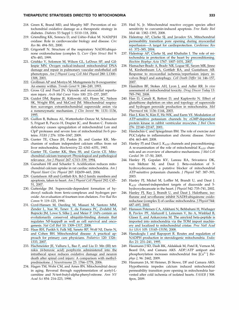
218. Green K, Brand MD, and Murphy MP. Prevention of mi-tochondrial oxidative damage as a therapeutic strategy indiabetes. Diabetes 53 Suppl 1: S110–118, 2004.
219. Griendling KK, Sorescu D, and Ushio–Fukai M. NAD(P)Hoxidase: Role in cardiovascular biology and disease. CircRes 86: 494–501, 2000.
220. Grigorieff N. Structure of the respiratory NADH:ubiqui-none oxidoreductase (complex I). Curr Opin Struct Biol 9:476–483, 1999.
221. Grishko V, Solomon M, Wilson GL, LeDoux SP, and Gil-lespie MN. Oxygen radical-induced mitochondrial DNAdamage and repair in pulmonary vascular endothelial cellphenotypes. Am J Physiol Lung Cell Mol Physiol 280: L1300–1308, 2001.
222. Grollman AP and Moriya M. Mutagenesis by 8-oxoguanine:An enemy within. Trends Genet 9: 246–249, 1993.
223. Gross GJ and Peart JN. Opioids and myocardial reperfu-sion injury. Arch Mal Coeur Vaiss 100: 231–237, 2007.
224. Guidot DM, Repine JE, Kitlowski AD, Flores SC, NelsonSK, Wright RM, and McCord JM. Mitochondrial respira-tion scavenges extramitochondrial superoxide anion viaa nonenzymatic mechanism. J Clin Invest 96: 1131–1136,1995.
225. Guillon B, Bulteau AL, Wattenhofer–Donze M, SchmuckerS, Friguet B, Puccio H, Drapier JC, and Bouton C. Frataxindeficiency causes upregulation of mitochondrial Lon andClpP proteases and severe loss of mitochondrial Fe-S pro-teins. FEBS J 276: 1036–1047, 2009.
226. Gunter TE, Chace JH, Puskin JS, and Gunter KK. Me-chanism of sodium independent calcium efflux from ratliver mitochondria. Biochemistry 22: 6341–6351, 1983.
227. Gunter TE, Gunter KK, Sheu SS, and Gavin CE. Mito-chondrial calcium transport: Physiological and pathologicalrelevance. Am J Physiol 267: C313–339, 1994.
228. Gursahani HI and Schaefer S. Acidification reduces mito-chondrial calcium uptake in rat cardiac mitochondria. Am JPhysiol Heart Circ Physiol 287: H2659–665, 2004.
229. Gustafsson AB and Gottlieb RA. Bcl-2 family members andapoptosis, taken to heart. Am J Physiol Cell Physiol 292: C45–51, 2007.
230. Gutteridge JM. Superoxide-dependent formation of hy-droxyl radicals from ferric-complexes and hydrogen per-oxide: An evaluation of fourteen iron chelators. Free Rad ResComm 9: 119–125, 1990.
231. Gyrd-Hansen M, Darding M, Miasari M, Santoro MM,Zender L, Xue W, Tenev T, da Fonseca PC, Zvelebil M,Bujnicki JM, Lowe S, Silke J, and Meier P. IAPs contain anevolutionarily conserved ubiquitin-binding domain thatregulates NF-kappaB as well as cell survival and onco-genesis. Nat Cell Biol 10: 1309–1317, 2008.
232. Haas RH, Parikh S, Falk MJ, Saneto RP, Wolf NI, Darin N,and Cohen BH. Mitochondrial disease: A practical ap-proach for primary care physicians. Pediatrics 120: 1326–1333, 2007.
233. Hachmeister JE, Valluru L, Bao F, and Liu D. Mn (III) tet-rakis (4-benzoic acid) porphyrin administered into theintrathecal space reduces oxidative damage and neurondeath after spinal cord injury: A comparison with methyl-prednisolone. J Neurotrauma 23: 1766–1778, 2006.
234. Hagen TM, Wehr CM, and Ames BN. Mitochondrial decayin aging. Reversal through supplementation of acetyl-L-carnitine and N-tert-butyl-alpha-phenyl-nitrone. Ann NYAcad Sci 854: 214–223, 1998.
235. Hail N, Jr. Mitochondrial reactive oxygen species affectsensitivity to curcumin-induced apoptosis. Free Radic BiolMed 44: 1382–1393, 2008.
236. Halestrap AP, Clarke SJ, and Javadov SA. Mitochondrialpermeability transition pore opening during myocardialreperfusion—A target for cardioprotection. Cardiovasc Res61: 372–385, 2004.
237. Halestrap AP, Clarke SJ, and Khaliulin I. The role of mi-tochondria in protection of the heart by preconditioning.Biochim Biophys Acta 1767: 1007–1031, 2007.
238. Hamacher-Brady A, Brady NR, Logue SE, Sayen MR, JinnoM, Kirshenbaum LA, Gottlieb RA, and Gustafsson AB.Response to myocardial ischemia=reperfusion injury in-volves Bnip3 and autophagy. Cell Death Differ 14: 146–157,2007.
239. Hamilton BF, Stokes AH, Lyon J, and Adler RR. In vivoassessment of mitochondrial toxicity. Drug Discov Today 13:785–790, 2008.
240. Han D, Canali R, Rettori D, and Kaplowitz N. Effect ofglutathione depletion on sites and topology of superoxideand hydrogen peroxide production in mitochondria. MolPharmacol 64: 1136–1144, 2003.
241. Han J, Kim N, Kim E, Ho WK, and Earm YE. Modulation ofATP-sensitive potassium channels by cGMP-dependentprotein kinase in rabbit ventricular myocytes. J Biol Chem276: 22140–22147, 2001.
242. Handschin C and Spiegelman BM. The role of exercise andPGC1alpha in inflammation and chronic disease. Nature454: 463–469, 2008.
243. Hanley PJ and Daut J. KATP channels and preconditioning:A re-examination of the role of mitochondrial KATP chan-nels and an overview of alternative mechanisms. J Mol CellCardiol 39: 17–50, 2005.
244. Hanley PJ, Gopalan KV, Lareau RA, Srivastava DK,von Meltzer M, and Daut J. Beta-oxidation of 5-hydroxydecanoate, a putative blocker of mitochondrialATP-sensitive potassium channels. J Physiol 547: 387–393,2003.
245. Hanley PJ, Mickel M, Loffler M, Brandt U, and Daut J.KATP channel-independent targets of diazoxide and 5-hydroxydecanoate in the heart. J Physiol 542: 735–741, 2002.
246. Hanley PJ, Ray J, Brandt U, and Daut J. Halothane, iso-flurane and sevoflurane inhibit NADH:ubiquinone oxido-reductase (complex I) of cardiac mitochondria. J Physiol 544:687–693, 2002.
247. Hansson Petersen CA, Alikhani N, Behbahani H, WiehagerB, Pavlov PF, Alafuzoff I, Leinonen V, Ito A, Winblad B,Glaser E, and Ankarcrona M. The amyloid beta-peptide isimported into mitochondria via the TOM import machin-ery and localized to mitochondrial cristae. Proc Natl AcadSci USA 105: 13145–13150, 2008.
248. Hanukoglu I and Rapoport R. Routes and regulation ofNADPH production in steroidogenic mitochondria. EndocRes 21: 231–241, 1995.
249. Haumann J SD, Dash RK, Aldakkak M, Patel R, Vernon M,Beard DA, and Camara AKS. ADP=ATP antiport andphosphorylation increases mitochondrial free [Ca2þ] Bio-phys J, 96: 2442, 2009.
250. Haumann JA, M Heisner, JS Stowe, DF and Camara AKS.Hypothermia impedes calcium induced mitochondrialpermeability transition pore opening in mitochondria har-vested after cold ischemia of isolated hearts. FASEB J 508.4pos, 2009.
THERAPEUTIC STRATEGIES DIRECTED TO MITOCHONDRIA 333
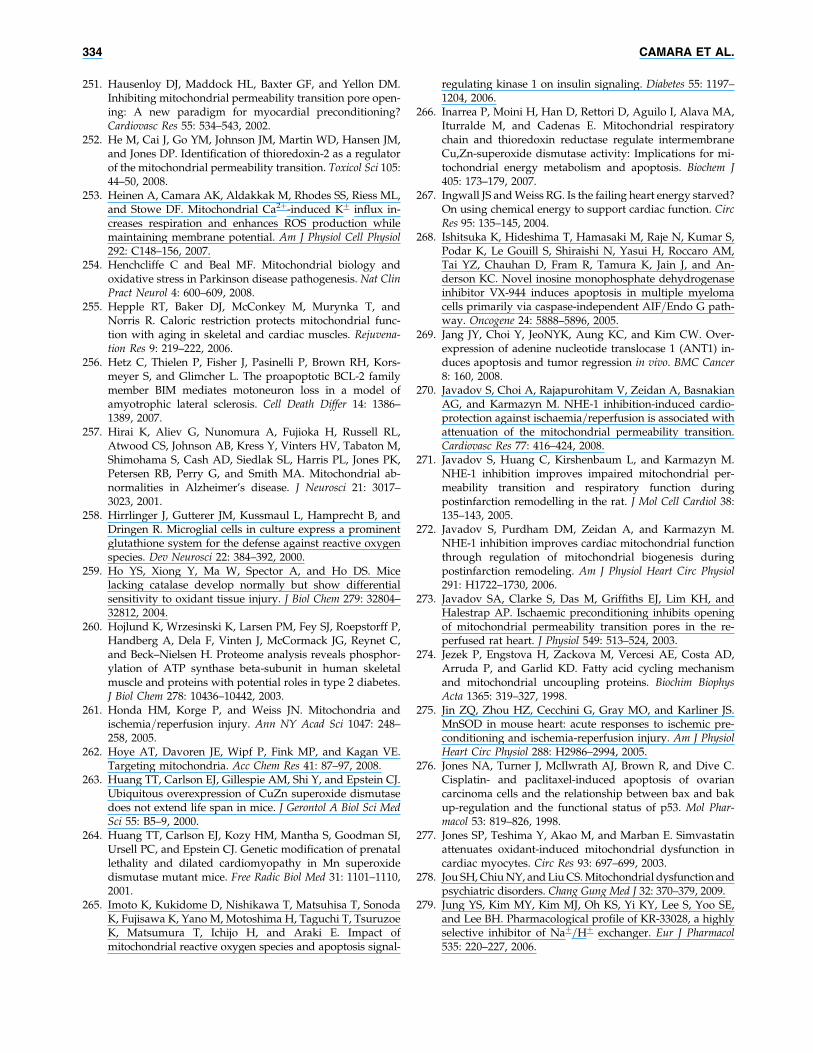
251. Hausenloy DJ, Maddock HL, Baxter GF, and Yellon DM.Inhibiting mitochondrial permeability transition pore open-ing: A new paradigm for myocardial preconditioning?Cardiovasc Res 55: 534–543, 2002.
252. He M, Cai J, Go YM, Johnson JM, Martin WD, Hansen JM,and Jones DP. Identification of thioredoxin-2 as a regulatorof the mitochondrial permeability transition. Toxicol Sci 105:44–50, 2008.
253. Heinen A, Camara AK, Aldakkak M, Rhodes SS, Riess ML,and Stowe DF. Mitochondrial Ca2þ-induced Kþ influx in-creases respiration and enhances ROS production whilemaintaining membrane potential. Am J Physiol Cell Physiol292: C148–156, 2007.
254. Henchcliffe C and Beal MF. Mitochondrial biology andoxidative stress in Parkinson disease pathogenesis. Nat ClinPract Neurol 4: 600–609, 2008.
255. Hepple RT, Baker DJ, McConkey M, Murynka T, andNorris R. Caloric restriction protects mitochondrial func-tion with aging in skeletal and cardiac muscles. Rejuvena-tion Res 9: 219–222, 2006.
256. Hetz C, Thielen P, Fisher J, Pasinelli P, Brown RH, Kors-meyer S, and Glimcher L. The proapoptotic BCL-2 familymember BIM mediates motoneuron loss in a model ofamyotrophic lateral sclerosis. Cell Death Differ 14: 1386–1389, 2007.
257. Hirai K, Aliev G, Nunomura A, Fujioka H, Russell RL,Atwood CS, Johnson AB, Kress Y, Vinters HV, Tabaton M,Shimohama S, Cash AD, Siedlak SL, Harris PL, Jones PK,Petersen RB, Perry G, and Smith MA. Mitochondrial ab-normalities in Alzheimer’s disease. J Neurosci 21: 3017–3023, 2001.
258. Hirrlinger J, Gutterer JM, Kussmaul L, Hamprecht B, andDringen R. Microglial cells in culture express a prominentglutathione system for the defense against reactive oxygenspecies. Dev Neurosci 22: 384–392, 2000.
259. Ho YS, Xiong Y, Ma W, Spector A, and Ho DS. Micelacking catalase develop normally but show differentialsensitivity to oxidant tissue injury. J Biol Chem 279: 32804–32812, 2004.
260. Hojlund K, Wrzesinski K, Larsen PM, Fey SJ, Roepstorff P,Handberg A, Dela F, Vinten J, McCormack JG, Reynet C,and Beck–Nielsen H. Proteome analysis reveals phosphor-ylation of ATP synthase beta-subunit in human skeletalmuscle and proteins with potential roles in type 2 diabetes.J Biol Chem 278: 10436–10442, 2003.
261. Honda HM, Korge P, and Weiss JN. Mitochondria andischemia=reperfusion injury. Ann NY Acad Sci 1047: 248–258, 2005.
262. Hoye AT, Davoren JE, Wipf P, Fink MP, and Kagan VE.Targeting mitochondria. Acc Chem Res 41: 87–97, 2008.
263. Huang TT, Carlson EJ, Gillespie AM, Shi Y, and Epstein CJ.Ubiquitous overexpression of CuZn superoxide dismutasedoes not extend life span in mice. J Gerontol A Biol Sci MedSci 55: B5–9, 2000.
264. Huang TT, Carlson EJ, Kozy HM, Mantha S, Goodman SI,Ursell PC, and Epstein CJ. Genetic modification of prenatallethality and dilated cardiomyopathy in Mn superoxidedismutase mutant mice. Free Radic Biol Med 31: 1101–1110,2001.
265. Imoto K, Kukidome D, Nishikawa T, Matsuhisa T, SonodaK, Fujisawa K, Yano M, Motoshima H, Taguchi T, TsuruzoeK, Matsumura T, Ichijo H, and Araki E. Impact ofmitochondrial reactive oxygen species and apoptosis signal-
regulating kinase 1 on insulin signaling. Diabetes 55: 1197–1204, 2006.
266. Inarrea P, Moini H, Han D, Rettori D, Aguilo I, Alava MA,Iturralde M, and Cadenas E. Mitochondrial respiratorychain and thioredoxin reductase regulate intermembraneCu,Zn-superoxide dismutase activity: Implications for mi-tochondrial energy metabolism and apoptosis. Biochem J405: 173–179, 2007.
267. Ingwall JS and Weiss RG. Is the failing heart energy starved?On using chemical energy to support cardiac function. CircRes 95: 135–145, 2004.
268. Ishitsuka K, Hideshima T, Hamasaki M, Raje N, Kumar S,Podar K, Le Gouill S, Shiraishi N, Yasui H, Roccaro AM,Tai YZ, Chauhan D, Fram R, Tamura K, Jain J, and An-derson KC. Novel inosine monophosphate dehydrogenaseinhibitor VX-944 induces apoptosis in multiple myelomacells primarily via caspase-independent AIF=Endo G path-way. Oncogene 24: 5888–5896, 2005.
269. Jang JY, Choi Y, JeoNYK, Aung KC, and Kim CW. Over-expression of adenine nucleotide translocase 1 (ANT1) in-duces apoptosis and tumor regression in vivo. BMC Cancer8: 160, 2008.
270. Javadov S, Choi A, Rajapurohitam V, Zeidan A, BasnakianAG, and Karmazyn M. NHE-1 inhibition-induced cardio-protection against ischaemia=reperfusion is associated withattenuation of the mitochondrial permeability transition.Cardiovasc Res 77: 416–424, 2008.
271. Javadov S, Huang C, Kirshenbaum L, and Karmazyn M.NHE-1 inhibition improves impaired mitochondrial per-meability transition and respiratory function duringpostinfarction remodelling in the rat. J Mol Cell Cardiol 38:135–143, 2005.
272. Javadov S, Purdham DM, Zeidan A, and Karmazyn M.NHE-1 inhibition improves cardiac mitochondrial functionthrough regulation of mitochondrial biogenesis duringpostinfarction remodeling. Am J Physiol Heart Circ Physiol291: H1722–1730, 2006.
273. Javadov SA, Clarke S, Das M, Griffiths EJ, Lim KH, andHalestrap AP. Ischaemic preconditioning inhibits openingof mitochondrial permeability transition pores in the re-perfused rat heart. J Physiol 549: 513–524, 2003.
274. Jezek P, Engstova H, Zackova M, Vercesi AE, Costa AD,Arruda P, and Garlid KD. Fatty acid cycling mechanismand mitochondrial uncoupling proteins. Biochim BiophysActa 1365: 319–327, 1998.
275. Jin ZQ, Zhou HZ, Cecchini G, Gray MO, and Karliner JS.MnSOD in mouse heart: acute responses to ischemic pre-conditioning and ischemia-reperfusion injury. Am J PhysiolHeart Circ Physiol 288: H2986–2994, 2005.
276. Jones NA, Turner J, McIlwrath AJ, Brown R, and Dive C.Cisplatin- and paclitaxel-induced apoptosis of ovariancarcinoma cells and the relationship between bax and bakup-regulation and the functional status of p53. Mol Phar-macol 53: 819–826, 1998.
277. Jones SP, Teshima Y, Akao M, and Marban E. Simvastatinattenuates oxidant-induced mitochondrial dysfunction incardiac myocytes. Circ Res 93: 697–699, 2003.
278. Jou SH, Chiu NY, and Liu CS. Mitochondrial dysfunction andpsychiatric disorders. Chang Gung Med J 32: 370–379, 2009.
279. Jung YS, Kim MY, Kim MJ, Oh KS, Yi KY, Lee S, Yoo SE,and Lee BH. Pharmacological profile of KR-33028, a highlyselective inhibitor of Naþ=Hþ exchanger. Eur J Pharmacol535: 220–227, 2006.
334 CAMARA ET AL.
280. Kaneto H, Katakami N, Kawamori D, Miyatsuka T, Saka-moto K, Matsuoka TA, Matsuhisa M, and Yamasaki Y.Involvement of oxidative stress in the pathogenesis of di-abetes. Antioxid Redox Signal 9: 355–366, 2007.
281. Kanwar YS, Wada J, Sun L, Xie P, Wallner EI, Chen S,Chugh S, and Danesh FR. Diabetic nephropathy: Mechan-isms of renal disease progression. Exp Biol Med (Maywood)233: 4–11, 2008.
282. Kasamaki Y, Guo AC, and McDonald TF. Protection byhypoxic preconditioning against hypoxia-reoxygenationinjury in guinea-pig papillary muscles. Cardiovasc Res 34:313–322, 1997.
283. Kawata H, Yoshida K, Kawamoto A, Kurioka H, Takase E,Sasaki Y, Hatanaka K, Kobayashi M, Ueyama T, Ha-shimoto T, and Dohi K. Ischemic preconditioning upregu-lates vascular endothelial growth factor mRNA expressionand neovascularization via nuclear translocation of proteinkinase C epsilon in the rat ischemic myocardium. Circ Res88: 696–704, 2001.
284. Keep M, Elmer E, Fong KS, and Csiszar K. Intrathecal cy-closporin prolongs survival of late-stage ALS mice. BrainRes 894: 327–331, 2001.
285. Kelly M, Lacour B, and Nguyen–Khoa T. Dysregulation ofsuperoxide dismutase in chronic kidney disease. NephronClin Pract 100: c103–104, 2005.
286. Kelso GF, Porteous CM, Coulter CV, Hughes G, PorteousWK, Ledgerwood EC, Smith RA, and Murphy MP. Selec-tive targeting of a redox-active ubiquinone to mitochondriawithin cells: Antioxidant and antiapoptotic properties. J BiolChem 276: 4588–4596, 2001.
287. Kerendi F, Kin H, Halkos ME, Jiang R, Zatta AJ, Zhao ZQ,Guyton RA, and Vinten–Johansen J. Remote postcondi-tioning. Brief renal ischemia and reperfusion applied beforecoronary artery reperfusion reduces myocardial infarct sizevia endogenous activation of adenosine receptors. Basic ResCardiol 100: 404–412, 2005.
288. Kerner J, Zaluzec E, Gage D, and Bieber LL. Characteriza-tion of the malonyl-CoA-sensitive carnitine palmitoyltrans-ferase (CPTo) of a rat heart mitochondrial particle. Evidencethat the catalytic unit is CPTi. J Biol Chem 269: 8209–8219,1994.
289. Kevin LG, Camara AK, Riess ML, Novalija E, and StoweDF. Ischemic preconditioning alters real-time measure ofO2 radicals in intact hearts with ischemia and reperfusion.Am J Physiol Heart Circ Physiol 284: H566–574, 2003.
290. Kevin LG, Novalija E, Riess ML, Camara AK, Rhodes SS,and Stowe DF. Sevoflurane exposure generates superoxidebut leads to decreased superoxide during ischemia andreperfusion in isolated hearts. Anesth Analg 96: 949–955,2003.
291. Khaliulin I, Clarke SJ, Lin H, Parker J, Suleiman MS, andHalestrap AP. Temperature preconditioning of isolated rathearts—A potent cardioprotective mechanism involving areduction in oxidative stress and inhibition of the mito-chondrial permeability transition pore. J Physiol 581: 1147–1161, 2007.
292. Khrapko K and Vijg J. Mitochondrial DNA mutations andaging: Devils in the details? Trends Genet 25: 91–98, 2009.
293. Kienhofer J, Haussler DJ, Ruckelshausen F, Muessig E,Weber K, Pimentel D, Ullrich V, Burkle A, and BachschmidMM. Association of mitochondrial antioxidant enzymeswith mitochondrial DNA as integral nucleoid constituents.FASEB J 23: 2034–2044, 2009.
294. Kim A, Murphy MP, and Oberley TD. Mitochondrial redoxstate regulates transcription of the nuclear-encoded mito-chondrial protein manganese superoxide dismutase: Aproposed adaptive response to mitochondrial redox im-balance. Free Radic Biol Med 38: 644–654, 2005.
295. Kim GT, ChuNYS, Park JW, and Kim MS. Role ofapoptosis-inducing factor in myocardial cell death byischemia-reperfusion. Biochem Biophys Res Commun 309:619–624, 2003.
296. Kim HS, Lee JH, Lee JP, Kim EM, Chang KA, Park CH,Jeong SJ, Wittendorp MC, Seo JH, Choi SH, and Suh YH.Amyloid beta peptide induces cytochrome C release fromisolated mitochondria. Neuroreport 13: 1989–1993, 2002.
297. Kim JS, He L, and Lemasters JJ. Mitochondrial permeabilitytransition: A common pathway to necrosis and apoptosis.Biochem Biophys Res Commun 304: 463–470, 2003.
298. Kim YM, Bombeck CA, and Billiar TR. Nitric oxide as abifunctional regulator of apoptosis. Circ Res 84: 253–256,1999.
299. Kimura S, Zhang GX, Nishiyama A, Shokoji T, Yao L, FaNYY, Rahman M, Suzuki T, Maeta H, and Abe Y. Role ofNAD(P)H oxidase- and mitochondria-derived reactive ox-ygen species in cardioprotection of ischemic reperfusioninjury by angiotensin II. Hypertension 45: 860–866, 2005.
300. Kin H, Zatta AJ, Lofye MT, Amerson BS, Halkos ME,Kerendi F, Zhao ZQ, Guyton RA, Headrick JP, and Vinten–Johansen J. Postconditioning reduces infarct size viaadenosine receptor activation by endogenous adenosine.Cardiovasc Res 67: 124–133, 2005.
301. Kindler DD, Thiffault C, Solenski NJ, Dennis J, Kostecki V,Jenkins R, Keeney PM, and Bennett JP, Jr. Neurotoxic nitricoxide rapidly depolarizes and permeabilizes mitochondriaby dynamically opening the mitochondrial transition pore.Mol Cell Neurosci 23: 559–573, 2003.
302. Kirkinezos IG, Bacman SR, Hernandez D, Oca–Cossio J,Arias LJ, Perez–Pinzon MA, Bradley WG, and Moraes CT.Cytochrome c association with the inner mitochondrialmembrane is impaired in the CNS of G93A-SOD1 mice.J Neurosci 25: 164–172, 2005.
303. Klamann A, Sarfert P, Launhardt V, Schulte G, SchmiegelWH, and Nauck MA. Myocardial infarction in diabetic vsnon-diabetic subjects. Survival and infarct size followingtherapy with sulfonylureas (glibenclamide). Eur Heart J 21:220–229, 2000.
304. Knott AB, Perkins G, Schwarzenbacher R, and Bossy–Wetzel E. Mitochondrial fragmentation in neurodegenera-tion. Nat Rev Neurosci 9: 505–518, 2008.
305. Koehler CM. Protein translocation pathways of the mito-chondrion. FEBS Lett 476: 27–31, 2000.
306. Koga Y and Nataliya P. [Migraine headache and mito-chondrial DNA abnormality]. Nippon Rinsho. Jpn J Clin Med63: 1720–1726, 2005.
307. Korge P, Honda HM, and Weiss JN. Kþ-dependent regu-lation of matrix volume improves mitochondrial functionunder conditions mimicking ischemia-reperfusion. Am JPhysiol Heart Circ Physiol 289: H66–77, 2005.
308. Kornmann B, Currie E, Collins SR, Schuldiner M, NunnariJ, Weissman JS, and Walter P. An ER-mitochondria teth-ering complex revealed by a synthetic biology screen. Sci-ence 325: 477–481, 2009.
309. Korshunov SS, Skulachev VP, and Starkov AA. High protonicpotential actuates a mechanism of production of reactiveoxygen species in mitochondria. FEBS Lett 416: 15–18, 1997.
THERAPEUTIC STRATEGIES DIRECTED TO MITOCHONDRIA 335
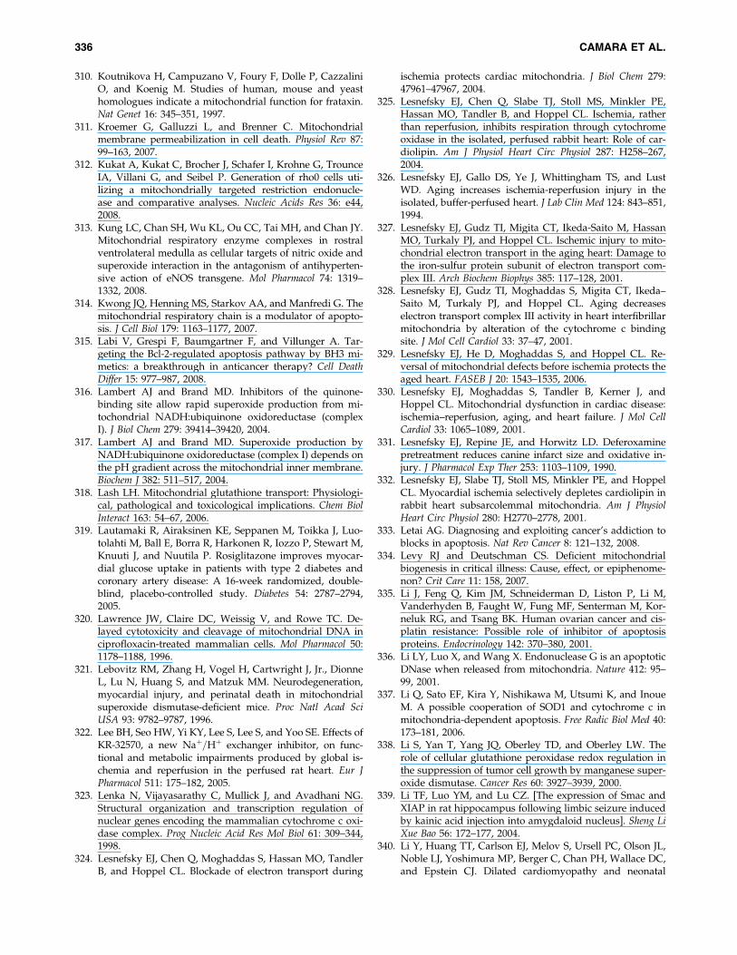
310. Koutnikova H, Campuzano V, Foury F, Dolle P, CazzaliniO, and Koenig M. Studies of human, mouse and yeasthomologues indicate a mitochondrial function for frataxin.Nat Genet 16: 345–351, 1997.
311. Kroemer G, Galluzzi L, and Brenner C. Mitochondrialmembrane permeabilization in cell death. Physiol Rev 87:99–163, 2007.
312. Kukat A, Kukat C, Brocher J, Schafer I, Krohne G, TrounceIA, Villani G, and Seibel P. Generation of rho0 cells uti-lizing a mitochondrially targeted restriction endonucle-ase and comparative analyses. Nucleic Acids Res 36: e44,2008.
313. Kung LC, Chan SH, Wu KL, Ou CC, Tai MH, and Chan JY.Mitochondrial respiratory enzyme complexes in rostralventrolateral medulla as cellular targets of nitric oxide andsuperoxide interaction in the antagonism of antihyperten-sive action of eNOS transgene. Mol Pharmacol 74: 1319–1332, 2008.
314. Kwong JQ, Henning MS, Starkov AA, and Manfredi G. Themitochondrial respiratory chain is a modulator of apopto-sis. J Cell Biol 179: 1163–1177, 2007.
315. Labi V, Grespi F, Baumgartner F, and Villunger A. Tar-geting the Bcl-2-regulated apoptosis pathway by BH3 mi-metics: a breakthrough in anticancer therapy? Cell DeathDiffer 15: 977–987, 2008.
316. Lambert AJ and Brand MD. Inhibitors of the quinone-binding site allow rapid superoxide production from mi-tochondrial NADH:ubiquinone oxidoreductase (complexI). J Biol Chem 279: 39414–39420, 2004.
317. Lambert AJ and Brand MD. Superoxide production byNADH:ubiquinone oxidoreductase (complex I) depends onthe pH gradient across the mitochondrial inner membrane.Biochem J 382: 511–517, 2004.
318. Lash LH. Mitochondrial glutathione transport: Physiologi-cal, pathological and toxicological implications. Chem BiolInteract 163: 54–67, 2006.
319. Lautamaki R, Airaksinen KE, Seppanen M, Toikka J, Luo-tolahti M, Ball E, Borra R, Harkonen R, Iozzo P, Stewart M,Knuuti J, and Nuutila P. Rosiglitazone improves myocar-dial glucose uptake in patients with type 2 diabetes andcoronary artery disease: A 16-week randomized, double-blind, placebo-controlled study. Diabetes 54: 2787–2794,2005.
320. Lawrence JW, Claire DC, Weissig V, and Rowe TC. De-layed cytotoxicity and cleavage of mitochondrial DNA inciprofloxacin-treated mammalian cells. Mol Pharmacol 50:1178–1188, 1996.
321. Lebovitz RM, Zhang H, Vogel H, Cartwright J, Jr., DionneL, Lu N, Huang S, and Matzuk MM. Neurodegeneration,myocardial injury, and perinatal death in mitochondrialsuperoxide dismutase-deficient mice. Proc Natl Acad SciUSA 93: 9782–9787, 1996.
322. Lee BH, Seo HW, Yi KY, Lee S, Lee S, and Yoo SE. Effects ofKR-32570, a new Naþ=Hþ exchanger inhibitor, on func-tional and metabolic impairments produced by global is-chemia and reperfusion in the perfused rat heart. Eur JPharmacol 511: 175–182, 2005.
323. Lenka N, Vijayasarathy C, Mullick J, and Avadhani NG.Structural organization and transcription regulation ofnuclear genes encoding the mammalian cytochrome c oxi-dase complex. Prog Nucleic Acid Res Mol Biol 61: 309–344,1998.
324. Lesnefsky EJ, Chen Q, Moghaddas S, Hassan MO, TandlerB, and Hoppel CL. Blockade of electron transport during
ischemia protects cardiac mitochondria. J Biol Chem 279:47961–47967, 2004.
325. Lesnefsky EJ, Chen Q, Slabe TJ, Stoll MS, Minkler PE,Hassan MO, Tandler B, and Hoppel CL. Ischemia, ratherthan reperfusion, inhibits respiration through cytochromeoxidase in the isolated, perfused rabbit heart: Role of car-diolipin. Am J Physiol Heart Circ Physiol 287: H258–267,2004.
326. Lesnefsky EJ, Gallo DS, Ye J, Whittingham TS, and LustWD. Aging increases ischemia-reperfusion injury in theisolated, buffer-perfused heart. J Lab Clin Med 124: 843–851,1994.
327. Lesnefsky EJ, Gudz TI, Migita CT, Ikeda-Saito M, HassanMO, Turkaly PJ, and Hoppel CL. Ischemic injury to mito-chondrial electron transport in the aging heart: Damage tothe iron-sulfur protein subunit of electron transport com-plex III. Arch Biochem Biophys 385: 117–128, 2001.
328. Lesnefsky EJ, Gudz TI, Moghaddas S, Migita CT, Ikeda–Saito M, Turkaly PJ, and Hoppel CL. Aging decreaseselectron transport complex III activity in heart interfibrillarmitochondria by alteration of the cytochrome c bindingsite. J Mol Cell Cardiol 33: 37–47, 2001.
329. Lesnefsky EJ, He D, Moghaddas S, and Hoppel CL. Re-versal of mitochondrial defects before ischemia protects theaged heart. FASEB J 20: 1543–1535, 2006.
330. Lesnefsky EJ, Moghaddas S, Tandler B, Kerner J, andHoppel CL. Mitochondrial dysfunction in cardiac disease:ischemia–reperfusion, aging, and heart failure. J Mol CellCardiol 33: 1065–1089, 2001.
331. Lesnefsky EJ, Repine JE, and Horwitz LD. Deferoxaminepretreatment reduces canine infarct size and oxidative in-jury. J Pharmacol Exp Ther 253: 1103–1109, 1990.
332. Lesnefsky EJ, Slabe TJ, Stoll MS, Minkler PE, and HoppelCL. Myocardial ischemia selectively depletes cardiolipin inrabbit heart subsarcolemmal mitochondria. Am J PhysiolHeart Circ Physiol 280: H2770–2778, 2001.
333. Letai AG. Diagnosing and exploiting cancer’s addiction toblocks in apoptosis. Nat Rev Cancer 8: 121–132, 2008.
334. Levy RJ and Deutschman CS. Deficient mitochondrialbiogenesis in critical illness: Cause, effect, or epiphenome-non? Crit Care 11: 158, 2007.
335. Li J, Feng Q, Kim JM, Schneiderman D, Liston P, Li M,Vanderhyden B, Faught W, Fung MF, Senterman M, Kor-neluk RG, and Tsang BK. Human ovarian cancer and cis-platin resistance: Possible role of inhibitor of apoptosisproteins. Endocrinology 142: 370–380, 2001.
336. Li LY, Luo X, and Wang X. Endonuclease G is an apoptoticDNase when released from mitochondria. Nature 412: 95–99, 2001.
337. Li Q, Sato EF, Kira Y, Nishikawa M, Utsumi K, and InoueM. A possible cooperation of SOD1 and cytochrome c inmitochondria-dependent apoptosis. Free Radic Biol Med 40:173–181, 2006.
338. Li S, Yan T, Yang JQ, Oberley TD, and Oberley LW. Therole of cellular glutathione peroxidase redox regulation inthe suppression of tumor cell growth by manganese super-oxide dismutase. Cancer Res 60: 3927–3939, 2000.
339. Li TF, Luo YM, and Lu CZ. [The expression of Smac andXIAP in rat hippocampus following limbic seizure inducedby kainic acid injection into amygdaloid nucleus]. Sheng LiXue Bao 56: 172–177, 2004.
340. Li Y, Huang TT, Carlson EJ, Melov S, Ursell PC, Olson JL,Noble LJ, Yoshimura MP, Berger C, Chan PH, Wallace DC,and Epstein CJ. Dilated cardiomyopathy and neonatal
336 CAMARA ET AL.
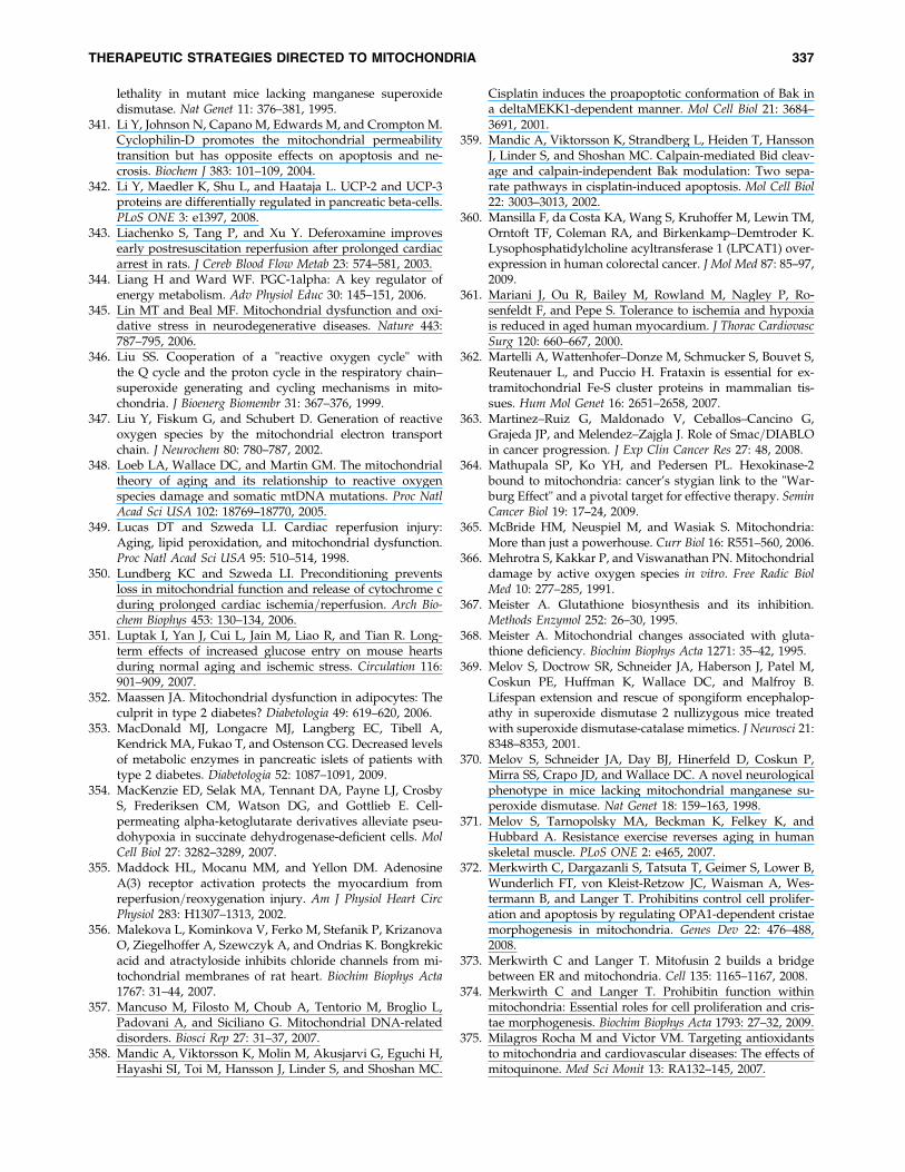
lethality in mutant mice lacking manganese superoxidedismutase. Nat Genet 11: 376–381, 1995.
341. Li Y, Johnson N, Capano M, Edwards M, and Crompton M.Cyclophilin-D promotes the mitochondrial permeabilitytransition but has opposite effects on apoptosis and ne-crosis. Biochem J 383: 101–109, 2004.
342. Li Y, Maedler K, Shu L, and Haataja L. UCP-2 and UCP-3proteins are differentially regulated in pancreatic beta-cells.PLoS ONE 3: e1397, 2008.
343. Liachenko S, Tang P, and Xu Y. Deferoxamine improvesearly postresuscitation reperfusion after prolonged cardiacarrest in rats. J Cereb Blood Flow Metab 23: 574–581, 2003.
344. Liang H and Ward WF. PGC-1alpha: A key regulator ofenergy metabolism. Adv Physiol Educ 30: 145–151, 2006.
345. Lin MT and Beal MF. Mitochondrial dysfunction and oxi-dative stress in neurodegenerative diseases. Nature 443:787–795, 2006.
346. Liu SS. Cooperation of a "reactive oxygen cycle" withthe Q cycle and the proton cycle in the respiratory chain–superoxide generating and cycling mechanisms in mito-chondria. J Bioenerg Biomembr 31: 367–376, 1999.
347. Liu Y, Fiskum G, and Schubert D. Generation of reactiveoxygen species by the mitochondrial electron transportchain. J Neurochem 80: 780–787, 2002.
348. Loeb LA, Wallace DC, and Martin GM. The mitochondrialtheory of aging and its relationship to reactive oxygenspecies damage and somatic mtDNA mutations. Proc NatlAcad Sci USA 102: 18769–18770, 2005.
349. Lucas DT and Szweda LI. Cardiac reperfusion injury:Aging, lipid peroxidation, and mitochondrial dysfunction.Proc Natl Acad Sci USA 95: 510–514, 1998.
350. Lundberg KC and Szweda LI. Preconditioning preventsloss in mitochondrial function and release of cytochrome cduring prolonged cardiac ischemia=reperfusion. Arch Bio-chem Biophys 453: 130–134, 2006.
351. Luptak I, Yan J, Cui L, Jain M, Liao R, and Tian R. Long-term effects of increased glucose entry on mouse heartsduring normal aging and ischemic stress. Circulation 116:901–909, 2007.
352. Maassen JA. Mitochondrial dysfunction in adipocytes: Theculprit in type 2 diabetes? Diabetologia 49: 619–620, 2006.
353. MacDonald MJ, Longacre MJ, Langberg EC, Tibell A,Kendrick MA, Fukao T, and Ostenson CG. Decreased levelsof metabolic enzymes in pancreatic islets of patients withtype 2 diabetes. Diabetologia 52: 1087–1091, 2009.
354. MacKenzie ED, Selak MA, Tennant DA, Payne LJ, CrosbyS, Frederiksen CM, Watson DG, and Gottlieb E. Cell-permeating alpha-ketoglutarate derivatives alleviate pseu-dohypoxia in succinate dehydrogenase-deficient cells. MolCell Biol 27: 3282–3289, 2007.
355. Maddock HL, Mocanu MM, and Yellon DM. AdenosineA(3) receptor activation protects the myocardium fromreperfusion=reoxygenation injury. Am J Physiol Heart CircPhysiol 283: H1307–1313, 2002.
356. Malekova L, Kominkova V, Ferko M, Stefanik P, KrizanovaO, Ziegelhoffer A, Szewczyk A, and Ondrias K. Bongkrekicacid and atractyloside inhibits chloride channels from mi-tochondrial membranes of rat heart. Biochim Biophys Acta1767: 31–44, 2007.
357. Mancuso M, Filosto M, Choub A, Tentorio M, Broglio L,Padovani A, and Siciliano G. Mitochondrial DNA-relateddisorders. Biosci Rep 27: 31–37, 2007.
358. Mandic A, Viktorsson K, Molin M, Akusjarvi G, Eguchi H,Hayashi SI, Toi M, Hansson J, Linder S, and Shoshan MC.
Cisplatin induces the proapoptotic conformation of Bak ina deltaMEKK1-dependent manner. Mol Cell Biol 21: 3684–3691, 2001.
359. Mandic A, Viktorsson K, Strandberg L, Heiden T, HanssonJ, Linder S, and Shoshan MC. Calpain-mediated Bid cleav-age and calpain-independent Bak modulation: Two sepa-rate pathways in cisplatin-induced apoptosis. Mol Cell Biol22: 3003–3013, 2002.
360. Mansilla F, da Costa KA, Wang S, Kruhoffer M, Lewin TM,Orntoft TF, Coleman RA, and Birkenkamp–Demtroder K.Lysophosphatidylcholine acyltransferase 1 (LPCAT1) over-expression in human colorectal cancer. J Mol Med 87: 85–97,2009.
361. Mariani J, Ou R, Bailey M, Rowland M, Nagley P, Ro-senfeldt F, and Pepe S. Tolerance to ischemia and hypoxiais reduced in aged human myocardium. J Thorac CardiovascSurg 120: 660–667, 2000.
362. Martelli A, Wattenhofer–Donze M, Schmucker S, Bouvet S,Reutenauer L, and Puccio H. Frataxin is essential for ex-tramitochondrial Fe-S cluster proteins in mammalian tis-sues. Hum Mol Genet 16: 2651–2658, 2007.
363. Martinez–Ruiz G, Maldonado V, Ceballos–Cancino G,Grajeda JP, and Melendez–Zajgla J. Role of Smac=DIABLOin cancer progression. J Exp Clin Cancer Res 27: 48, 2008.
364. Mathupala SP, Ko YH, and Pedersen PL. Hexokinase-2bound to mitochondria: cancer’s stygian link to the "War-burg Effect" and a pivotal target for effective therapy. SeminCancer Biol 19: 17–24, 2009.
365. McBride HM, Neuspiel M, and Wasiak S. Mitochondria:More than just a powerhouse. Curr Biol 16: R551–560, 2006.
366. Mehrotra S, Kakkar P, and Viswanathan PN. Mitochondrialdamage by active oxygen species in vitro. Free Radic BiolMed 10: 277–285, 1991.
367. Meister A. Glutathione biosynthesis and its inhibition.Methods Enzymol 252: 26–30, 1995.
368. Meister A. Mitochondrial changes associated with gluta-thione deficiency. Biochim Biophys Acta 1271: 35–42, 1995.
369. Melov S, Doctrow SR, Schneider JA, Haberson J, Patel M,Coskun PE, Huffman K, Wallace DC, and Malfroy B.Lifespan extension and rescue of spongiform encephalop-athy in superoxide dismutase 2 nullizygous mice treatedwith superoxide dismutase-catalase mimetics. J Neurosci 21:8348–8353, 2001.
370. Melov S, Schneider JA, Day BJ, Hinerfeld D, Coskun P,Mirra SS, Crapo JD, and Wallace DC. A novel neurologicalphenotype in mice lacking mitochondrial manganese su-peroxide dismutase. Nat Genet 18: 159–163, 1998.
371. Melov S, Tarnopolsky MA, Beckman K, Felkey K, andHubbard A. Resistance exercise reverses aging in humanskeletal muscle. PLoS ONE 2: e465, 2007.
372. Merkwirth C, Dargazanli S, Tatsuta T, Geimer S, Lower B,Wunderlich FT, von Kleist-Retzow JC, Waisman A, Wes-termann B, and Langer T. Prohibitins control cell prolifer-ation and apoptosis by regulating OPA1-dependent cristaemorphogenesis in mitochondria. Genes Dev 22: 476–488,2008.
373. Merkwirth C and Langer T. Mitofusin 2 builds a bridgebetween ER and mitochondria. Cell 135: 1165–1167, 2008.
374. Merkwirth C and Langer T. Prohibitin function withinmitochondria: Essential roles for cell proliferation and cris-tae morphogenesis. Biochim Biophys Acta 1793: 27–32, 2009.
375. Milagros Rocha M and Victor VM. Targeting antioxidantsto mitochondria and cardiovascular diseases: The effects ofmitoquinone. Med Sci Monit 13: RA132–145, 2007.
THERAPEUTIC STRATEGIES DIRECTED TO MITOCHONDRIA 337
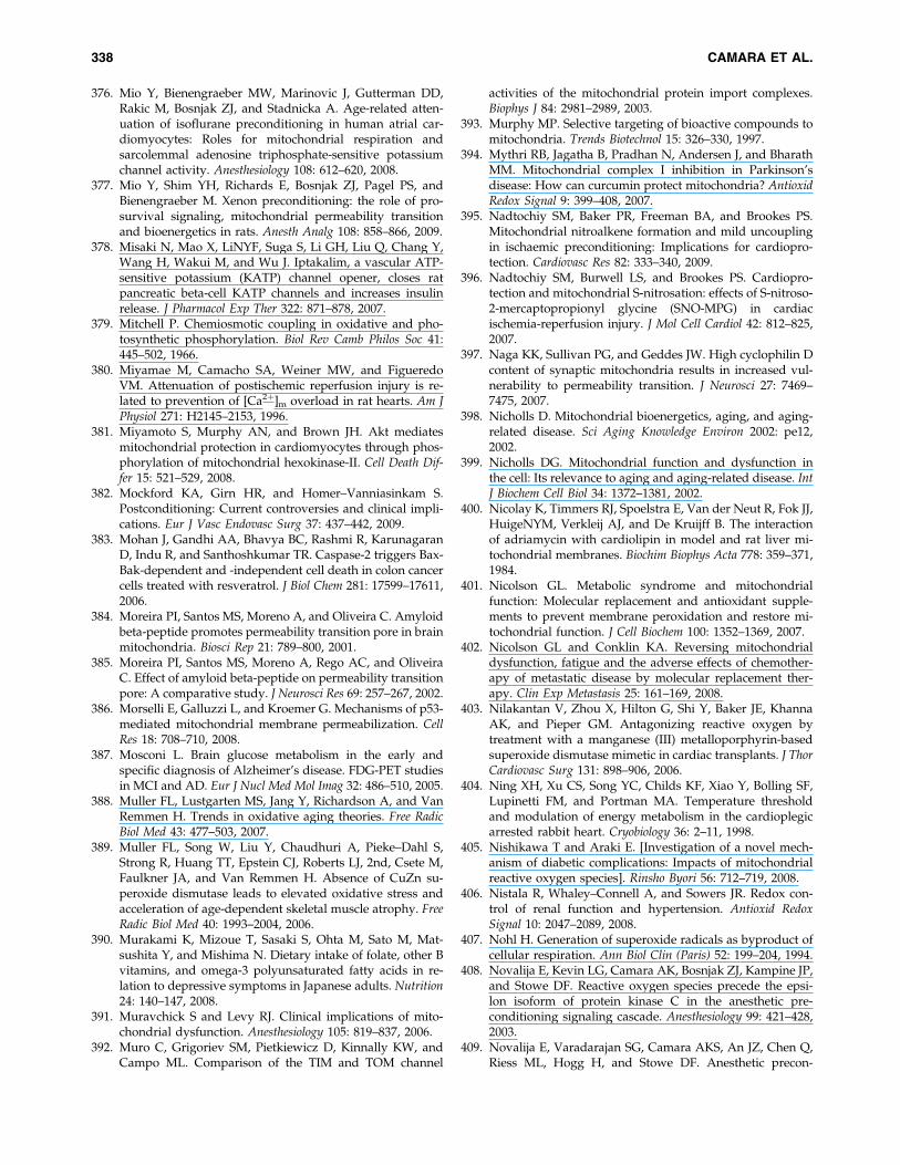
376. Mio Y, Bienengraeber MW, Marinovic J, Gutterman DD,Rakic M, Bosnjak ZJ, and Stadnicka A. Age-related atten-uation of isoflurane preconditioning in human atrial car-diomyocytes: Roles for mitochondrial respiration andsarcolemmal adenosine triphosphate-sensitive potassiumchannel activity. Anesthesiology 108: 612–620, 2008.
377. Mio Y, Shim YH, Richards E, Bosnjak ZJ, Pagel PS, andBienengraeber M. Xenon preconditioning: the role of pro-survival signaling, mitochondrial permeability transitionand bioenergetics in rats. Anesth Analg 108: 858–866, 2009.
378. Misaki N, Mao X, LiNYF, Suga S, Li GH, Liu Q, Chang Y,Wang H, Wakui M, and Wu J. Iptakalim, a vascular ATP-sensitive potassium (KATP) channel opener, closes ratpancreatic beta-cell KATP channels and increases insulinrelease. J Pharmacol Exp Ther 322: 871–878, 2007.
379. Mitchell P. Chemiosmotic coupling in oxidative and pho-tosynthetic phosphorylation. Biol Rev Camb Philos Soc 41:445–502, 1966.
380. Miyamae M, Camacho SA, Weiner MW, and FigueredoVM. Attenuation of postischemic reperfusion injury is re-lated to prevention of [Ca2þ]m overload in rat hearts. Am JPhysiol 271: H2145–2153, 1996.
381. Miyamoto S, Murphy AN, and Brown JH. Akt mediatesmitochondrial protection in cardiomyocytes through phos-phorylation of mitochondrial hexokinase-II. Cell Death Dif-fer 15: 521–529, 2008.
382. Mockford KA, Girn HR, and Homer–Vanniasinkam S.Postconditioning: Current controversies and clinical impli-cations. Eur J Vasc Endovasc Surg 37: 437–442, 2009.
383. Mohan J, Gandhi AA, Bhavya BC, Rashmi R, KarunagaranD, Indu R, and Santhoshkumar TR. Caspase-2 triggers Bax-Bak-dependent and -independent cell death in colon cancercells treated with resveratrol. J Biol Chem 281: 17599–17611,2006.
384. Moreira PI, Santos MS, Moreno A, and Oliveira C. Amyloidbeta-peptide promotes permeability transition pore in brainmitochondria. Biosci Rep 21: 789–800, 2001.
385. Moreira PI, Santos MS, Moreno A, Rego AC, and OliveiraC. Effect of amyloid beta-peptide on permeability transitionpore: A comparative study. J Neurosci Res 69: 257–267, 2002.
386. Morselli E, Galluzzi L, and Kroemer G. Mechanisms of p53-mediated mitochondrial membrane permeabilization. CellRes 18: 708–710, 2008.
387. Mosconi L. Brain glucose metabolism in the early andspecific diagnosis of Alzheimer’s disease. FDG-PET studiesin MCI and AD. Eur J Nucl Med Mol Imag 32: 486–510, 2005.
388. Muller FL, Lustgarten MS, Jang Y, Richardson A, and VanRemmen H. Trends in oxidative aging theories. Free RadicBiol Med 43: 477–503, 2007.
389. Muller FL, Song W, Liu Y, Chaudhuri A, Pieke–Dahl S,Strong R, Huang TT, Epstein CJ, Roberts LJ, 2nd, Csete M,Faulkner JA, and Van Remmen H. Absence of CuZn su-peroxide dismutase leads to elevated oxidative stress andacceleration of age-dependent skeletal muscle atrophy. FreeRadic Biol Med 40: 1993–2004, 2006.
390. Murakami K, Mizoue T, Sasaki S, Ohta M, Sato M, Mat-sushita Y, and Mishima N. Dietary intake of folate, other Bvitamins, and omega-3 polyunsaturated fatty acids in re-lation to depressive symptoms in Japanese adults. Nutrition24: 140–147, 2008.
391. Muravchick S and Levy RJ. Clinical implications of mito-chondrial dysfunction. Anesthesiology 105: 819–837, 2006.
392. Muro C, Grigoriev SM, Pietkiewicz D, Kinnally KW, andCampo ML. Comparison of the TIM and TOM channel
activities of the mitochondrial protein import complexes.Biophys J 84: 2981–2989, 2003.
393. Murphy MP. Selective targeting of bioactive compounds tomitochondria. Trends Biotechnol 15: 326–330, 1997.
394. Mythri RB, Jagatha B, Pradhan N, Andersen J, and BharathMM. Mitochondrial complex I inhibition in Parkinson’sdisease: How can curcumin protect mitochondria? AntioxidRedox Signal 9: 399–408, 2007.
395. Nadtochiy SM, Baker PR, Freeman BA, and Brookes PS.Mitochondrial nitroalkene formation and mild uncouplingin ischaemic preconditioning: Implications for cardiopro-tection. Cardiovasc Res 82: 333–340, 2009.
396. Nadtochiy SM, Burwell LS, and Brookes PS. Cardiopro-tection and mitochondrial S-nitrosation: effects of S-nitroso-2-mercaptopropionyl glycine (SNO-MPG) in cardiacischemia-reperfusion injury. J Mol Cell Cardiol 42: 812–825,2007.
397. Naga KK, Sullivan PG, and Geddes JW. High cyclophilin Dcontent of synaptic mitochondria results in increased vul-nerability to permeability transition. J Neurosci 27: 7469–7475, 2007.
398. Nicholls D. Mitochondrial bioenergetics, aging, and aging-related disease. Sci Aging Knowledge Environ 2002: pe12,2002.
399. Nicholls DG. Mitochondrial function and dysfunction inthe cell: Its relevance to aging and aging-related disease. IntJ Biochem Cell Biol 34: 1372–1381, 2002.
400. Nicolay K, Timmers RJ, Spoelstra E, Van der Neut R, Fok JJ,HuigeNYM, Verkleij AJ, and De Kruijff B. The interactionof adriamycin with cardiolipin in model and rat liver mi-tochondrial membranes. Biochim Biophys Acta 778: 359–371,1984.
401. Nicolson GL. Metabolic syndrome and mitochondrialfunction: Molecular replacement and antioxidant supple-ments to prevent membrane peroxidation and restore mi-tochondrial function. J Cell Biochem 100: 1352–1369, 2007.
402. Nicolson GL and Conklin KA. Reversing mitochondrialdysfunction, fatigue and the adverse effects of chemother-apy of metastatic disease by molecular replacement ther-apy. Clin Exp Metastasis 25: 161–169, 2008.
403. Nilakantan V, Zhou X, Hilton G, Shi Y, Baker JE, KhannaAK, and Pieper GM. Antagonizing reactive oxygen bytreatment with a manganese (III) metalloporphyrin-basedsuperoxide dismutase mimetic in cardiac transplants. J ThorCardiovasc Surg 131: 898–906, 2006.
404. Ning XH, Xu CS, Song YC, Childs KF, Xiao Y, Bolling SF,Lupinetti FM, and Portman MA. Temperature thresholdand modulation of energy metabolism in the cardioplegicarrested rabbit heart. Cryobiology 36: 2–11, 1998.
405. Nishikawa T and Araki E. [Investigation of a novel mech-anism of diabetic complications: Impacts of mitochondrialreactive oxygen species]. Rinsho Byori 56: 712–719, 2008.
406. Nistala R, Whaley–Connell A, and Sowers JR. Redox con-trol of renal function and hypertension. Antioxid RedoxSignal 10: 2047–2089, 2008.
407. Nohl H. Generation of superoxide radicals as byproduct ofcellular respiration. Ann Biol Clin (Paris) 52: 199–204, 1994.
408. Novalija E, Kevin LG, Camara AK, Bosnjak ZJ, Kampine JP,and Stowe DF. Reactive oxygen species precede the epsi-lon isoform of protein kinase C in the anesthetic pre-conditioning signaling cascade. Anesthesiology 99: 421–428,2003.
409. Novalija E, Varadarajan SG, Camara AKS, An JZ, Chen Q,Riess ML, Hogg H, and Stowe DF. Anesthetic precon-
338 CAMARA ET AL.
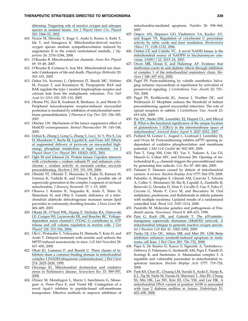
ditioning: Triggering role of reactive oxygen and nitrogenspecies in isolated hearts. Am J Physiol Heart Circ Physiol283: H44–52, 2002.
410. Nozoe M, Hirooka Y, Koga Y, Araki S, Konno S, Kishi T,Ide T, and Sunagawa K. Mitochondria-derived reactiveoxygen species mediate sympathoexcitation induced byangiotensin II in the rostral ventrolateral medulla. J Hy-pertens 26: 2176–2184, 2008.
411. O’Rourke B. Mitochondrial ion channels. Annu Rev Physiol69: 19–49, 2007.
412. O’Rourke B, Cortassa S, Aon MA. Mitochondrial ion chan-nels: Gatekeepers of life and death. Physiology (Bethesda) 20:303–315, 2005.
413. Oakes SA, Scorrano L, Opferman JT, Bassik MC, NishinoM, Pozzan T, and Korsmeyer SJ. Proapoptotic BAX andBAK regulate the type 1 inositol trisphosphate receptor andcalcium leak from the endoplasmic reticulum. Proc NatlAcad Sci USA 102: 105–110, 2005.
414. Obame FN, Zini R, Souktani R, Berdeaux A, and Morin D.Peripheral benzodiazepine receptor-induced myocardialprotection is mediated by inhibition of mitochondrial mem-brane permeabilization. J Pharmacol Exp Ther 323: 336–345,2007.
415. Oberley LW. Mechanism of the tumor suppressive effect ofMnSOD overexpression. Biomed Pharmacother 59: 143–148,2005.
416. Ochiai K, Zhang J, Gong G, Zhang Y, Liu J, Ye Y, Wu X, LiuH, Murakami Y, Bache RJ, Ugurbil K, and From AH. Effectsof augmented delivery of pyruvate on myocardial high-energy phosphate metabolism at high workstate. Am JPhysiol Heart Circ Physiol 281: H1823–1832, 2001.
417. Ogbi M and Johnson JA. Protein kinase Cepsilon interactswith cytochrome c oxidase subunit IV and enhances cyto-chrome c oxidase activity in neonatal cardiac myocytepreconditioning. Biochem J 393: 191–199, 2006.
418. Ohnishi ST, Ohnishi T, Muranaka S, Fujita H, Kimura H,Uemura K, Yoshida K, and Utsumi K. A possible site ofsuperoxide generation in the complex I segment of rat heartmitochondria. J Bioenerg Biomembr 37: 1–15, 2005.
419. Ohsawa I, Kamino K, Nagasaka K, Ando F, Niino N,Shimokata H, and Ohta S. Genetic deficiency of a mito-chondrial aldehyde dehydrogenase increases serum lipidperoxides in community-dwelling females. J Hum Genet 48:404–409, 2003.
420. Okada SF, O’Neal WK, Huang P, Nicholas RA, OstrowskiLE, Craigen WJ, Lazarowski ER, and Boucher RC. Voltage-dependent anion channel-1 (VDAC-1) contributes to ATPrelease and cell volume regulation in murine cells. J GenPhysiol 124: 513–526, 2004.
421. Oki C, Watanabe Y, Yokoyama H, Shimoda T, Kato H, andAraki T. Delayed treatment with arundic acid reduces theMPTP-induced neurotoxicity in mice. Cell Mol Neurobiol 28:417–430, 2008.
422. Okun JG, Lummen P, and Brandt U. Three classes of in-hibitors share a common binding domain in mitochondrialcomplex I (NADH:ubiquinone oxidoreductase). J Biol Chem274: 2625–2630, 1999.
423. Onyango IG. Mitochondrial dysfunction and oxidativestress in Parkinson’s disease. Neurochem Res 33: 589–597,2008.
424. Orzaez M, Mondragon L, Marzo I, Sanclimens G, Messe-guer A, Perez–Paya E, and Vicent MJ. Conjugation of anovel Apaf-1 inhibitor to peptide-based cell-membranetransporters: Effective methods to improve inhibition of
mitochondria-mediated apoptosis. Peptides 28: 958–968,2007.
425. Osipov AN, Stepanov GO, Vladimirov YA, Kozlov AV,and Kagan VE. Regulation of cytochrome C peroxidaseactivity by nitric oxide and laser irradiation. Biochemistry(Mosc) 71: 1128–1132, 2006.
426. Outten CE and Culotta VC. A novel NADH kinase is themitochondrial source of NADPH in Saccharomyces cere-visiae. EMBO J 22: 2015–24, 2003.
427. Owen MR, Doran E, and Halestrap AP. Evidence thatmetformin exerts its anti-diabetic effects through inhibitionof complex 1 of the mitochondrial respiratory chain. Bio-chem J 348: 607–614, 2000.
428. Pagel PS. Postconditioning by volatile anesthetics: Salva-ging ischemic myocardium at reperfusion by activation ofprosurvival signaling. J Cardiothorac Vasc Anesth 22: 753–765, 2008.
429. Pagel PS, Krolikowski JG, Amour J, Warltier DC, andWeihrauch D. Morphine reduces the threshold of heliumpreconditioning against myocardial infarction: The role ofopioid receptors in rabbits. J Cardiothorac Vasc Anesth 23:619–624, 2009.
430. Pai HV, Starke DW, Lesnefsky EJ, Hoppel CL, and MieyalJJ. What is the functional significance of the unique locationof glutaredoxin 1 (GRx1) in the intermembrane space ofmitochondria? Antioxid Redox Signal 9: 2027–2033, 2007.
431. Paillard M, Gomez L, Augeul L, Loufouat J, Lesnefsky EJ,and Ovize M. Postconditioning inhibits mPTP opening in-dependent of oxidative phosphorylation and membranepotential. J Mol Cell Cardiol 46: 902–909, 2009.
432. Pain T, Yang XM, Critz SD, Yue Y, Nakano A, Liu GS,Heusch G, Cohen MV, and Downey JM. Opening of mi-tochondrial KATP channels triggers the preconditioned stateby generating free radicals. Circ Res 87: 460–466, 2000.
433. Palmieri F. Diseases caused by defects of mitochondrialcarriers: A review. Biochim Biophys Acta 1777: 564–578, 2008.
434. Palumbo A, Bringhen S, Liberati AM, Caravita T, FalconeA, Callea V, Montanaro M, Ria R, Capaldi A, Zambello R,Benevolo G, Derudas D, Dore F, Cavallo F, Gay F, Falco P,Ciccone G, Musto P, Cavo M, and Boccadoro M. Oralmelphalan, prednisone, and thalidomide in elderly patientswith multiple myeloma: Updated results of a randomizedcontrolled trial. Blood 112: 3107–3114, 2008.
435. Pandolfo M. Molecular genetics and pathogenesis of Frie-dreich ataxia. Neuromusc Disord 8: 409–415, 1998.
436. Pani G, Koch OR, and Galeotti T. The p53-p66shc-manganese superoxide dismutase (MnSOD) network: Amitochondrial intrigue to generate reactive oxygen species.Int J Biochem Cell Biol 41: 1002–1005, 2009.
437. Panka DJ, Cho DC, Atkins MB, and Mier JW. GSK-3betainhibition enhances sorafenib-induced apoptosis in mela-noma cell lines. J Biol Chem 283: 726–732, 2008.
438. Papa S, De Rasmo D, Scacco S, Signorile A, Technikova–Dobrova Z, Palmisano G, Sardanelli AM, Papa F, Panelli D,Scaringi R, and Santeramo A. Mammalian complex I: Aregulable and vulnerable pacemaker in mitochondrial re-spiratory function. Biochim Biophys Acta 1777: 719–728,2008.
439. Park KS, Chan JC, Chuang LM, Suzuki S, Araki E, Nanjo K,Ji L, Ng M, Nishi M, Furuta H, Shirotani T, Ahn BY, ChungSS, Min HK, Lee SW, Kim JH, Cho YM, and Lee HK. Amitochondrial DNA variant at position 16189 is associatedwith type 2 diabetes mellitus in Asians. Diabetologia 51:602–608, 2008.
THERAPEUTIC STRATEGIES DIRECTED TO MITOCHONDRIA 339
440. Parker JC, Jr. Commentary: Human mitochondrial cyto-pathies. Ann Clin Lab Sci 30: 159–162, 2000.
441. Parker WD, Jr., Filley CM, and Parks JK. Cytochrome oxi-dase deficiency in Alzheimer’s disease. Neurology 40: 1302–1303, 1990.
442. Pasinelli P, Belford ME, Lennon N, Bacskai BJ, Hyman BT,Trotti D, and Brown RH, Jr. Amyotrophic lateral sclerosis-associated SOD1 mutant proteins bind and aggregatewith Bcl-2 in spinal cord mitochondria. Neuron 43: 19–30,2004.
443. Pastorino JG and Hoek JB. Regulation of hexokinasebinding to VDAC. J Bioenerg Biomembr 40: 171–182, 2008.
444. Pastorino JG, Hoek JB, and Shulga N. Activation ofglycogen synthase kinase 3beta disrupts the binding ofhexokinase II to mitochondria by phosphorylating voltage-dependent anion channel and potentiates chemotherapy-induced cytotoxicity. Cancer Res 65: 10545–10554, 2005.
445. Patel MN. Metalloporphyrins improve the survival ofSod2-deficient neurons. Aging Cell 2: 219–222, 2003.
446. Patti ME, Butte AJ, Crunkhorn S, Cusi K, Berria R, KashyapS, Miyazaki Y, Kohane I, Costello M, Saccone R, LandakerEJ, Goldfine AB, Mun E, DeFronzo R, Finlayson J, KahnCR, and Mandarino LJ. Coordinated reduction of genes ofoxidative metabolism in humans with insulin resistanceand diabetes: Potential role of PGC1 and NRF1. Proc NatlAcad Sci USA 100: 8466–8471, 2003.
447. Pedersen PL, Mathupala S, Rempel A, Geschwind JF, andKo YH. Mitochondrial bound type II hexokinase: A keyplayer in the growth and survival of many cancers and anideal prospect for therapeutic intervention. Biochim BiophysActa 1555: 14–20, 2002.
448. Pelicano H, Xu RH, Du M, Feng L, Sasaki R, Carew JS, HuY, Ramdas L, Hu L, Keating MJ, Zhang W, Plunkett W,Huang P. Mitochondrial respiration defects in cancer cellscause activation of Akt survival pathway through a redox-mediated mechanism. J Cell Biol 175: 913–923, 2006.
449. Pepe S. Mitochondrial function in ischaemia and reperfu-sion of the ageing heart. Clin Exp Pharmacol Physiol 27: 745–750, 2000.
450. Pereverzev MO, Vygodina TV, Konstantinov AA, andSkulachev VP. Cytochrome c, an ideal antioxidant. BiochemSoc Trans 31: 1312–1315, 2003.
451. Pickles JO. Mutation in mitochondrial DNA as a cause ofpresbyacusis. Audiol Neurootol 9: 23–33, 2004.
452. Pinton P, Rimessi A, Marchi S, Orsini F, Migliaccio E,Giorgio M, Contursi C, Minucci S, Mantovani F, Wieck-owski MR, Del Sal G, Pelicci PG, and Rizzuto R. Proteinkinase C beta and prolyl isomerase 1 regulate mitochon-drial effects of the life-span determinant p66Shc. Science315: 659–663, 2007.
453. Pope S, Land JM, and Heales SJ. Oxidative stress and mi-tochondrial dysfunction in neurodegeneration; Cardiolipina critical target? Biochim Biophys Acta 1777: 794–799, 2008.
454. Praet M, Laghmiche M, Pollakis G, Goormaghtigh E, andRuysschaert JM. In vivo and in vitro modifications of themitochondrial membrane induced by 4’ Epi-adriamycin.Biochem Pharmacol 35: 2923–2928, 1986.
455. Praet M and Ruysschaert JM. In vivo and in vitro mito-chondrial membrane damages induced in mice by adria-mycin and derivatives. Biochim Biophys Acta 1149: 79–85,1993.
456. Pravdic DB, M Sedlic, F, Stadnicka A, and Bosnjak Z. In-tracellular Ca2þ and pH modulation of in cardiomyocytesduring postconditioning. Anesthesiology 109: A-859, 2008.
457. Preller A and Wilson JE. Localization of the type III iso-zyme of hexokinase at the nuclear periphery. Arch BiochemBiophys 294: 482–492, 1992.
458. Prior SL, Griffiths AP, Baxter JM, Baxter PW, Hodder SC,Silvester KC, and Lewis PD. Mitochondrial DNA mutationsin oral squamous cell carcinoma. Carcinogenesis 27: 945–950,2006.
459. Prousky J and Seely D. The treatment of migraines andtension-type headaches with intravenous and oral niacin(nicotinic acid): Systematic review of the literature. Nutr J 4:3, 2005.
460. Quiles JL, Ramirez–Tortosa MC, Huertas JR, Ibanez S,Gomez JA, Battino M, and Mataix J. Olive oil supplementedwith vitamin E affects mitochondrial coenzyme Q levels inliver of rats after an oxidative stress induced by adriamy-cin. Biofactors 9: 331–336, 1999.
461. Radi R, Cassina A, Hodara R, Quijano C, and Castro L.Peroxynitrite reactions and formation in mitochondria. FreeRadical Biol Med 33: 1451–1464, 2002.
462. Radi R, Peluffo G, Alvarez MN, Naviliat M, and Cayota A.Unraveling peroxynitrite formation in biological systems.Free Radical Biol and Med 30: 463–488, 2001.
463. Radi R, Turrens JF, Chang LY, Bush KM, Crapo JD, andFreeman BA. Detection of catalase in rat heart mitochon-dria. J Biol Chem 266: 22028–22034, 1991.
464. Raju TN. The Nobel chronicles. 1931: Otto Heinrich War-burg (1883–1970). Lancet 352: 2028, 1998.
465. Rapoport M, Saada A, and Elpeleg O, Lorberboum–GalskiH. TAT-mediated delivery of LAD restores pyruvate de-hydrogenase complex activity in the mitochondria of pa-tients with LAD deficiency. Mol Ther 16: 691–697, 2008.
466. Rappaport L, Oliviero P, and Samuel JL. Cytoskeleton andmitochondrial morphology and function. Mol Cell Biochem184: 101–105, 1998.
467. Rathore R, Zheng YM, Niu CF, Liu QH, Korde A, Ho YS,and Wang YX. Hypoxia activates NADPH oxidase to in-crease [ROS]i and [Ca2þ]i through the mitochondrial ROS-PKCepsilon signaling axis in pulmonary artery smoothmuscle cells. Free Radic Biol Med 45: 1223–1231, 2008.
468. Ravagnan L, Marzo I, Costantini P, Susin SA, Zamzami N,Petit PX, Hirsch F, Goulbern M, Poupon MF, Miccoli L, XieZ, Reed JC, and Kroemer G. Lonidamine triggers apoptosisvia a direct, Bcl-2-inhibited effect on the mitochondrialpermeability transition pore. Oncogene 18: 2537–2546, 1999.
469. Razeghi P, Young ME, Cockrill TC, Frazier OH, andTaegtmeyer H. Downregulation of myocardial myocyte en-hancer factor 2C and myocyte enhancer factor 2C-regulatedgene expression in diabetic patients with nonischemic heartfailure. Circulation 106: 407–411, 2002.
470. Reaume AG, Elliott JL, Hoffman EK, Kowall NW, FerranteRJ, Siwek DF, Wilcox HM, Flood DG, Beal MF, Brown RH,Jr., Scott RW, and Snider WD. Motor neurons in Cu=Znsuperoxide dismutase-deficient mice develop normally butexhibit enhanced cell death after axonal injury. Nat Genet13: 43–47, 1996.
471. Reynen K and Bachmann K. Coronary arteriography inelderly patients: Risk, therapeutic consequences and long-term follow-up. Coron Artery Dis 8: 657–666, 1997.
472. Ricci C, Pastukh V, Leonard J, Turrens J, Wilson G, SchafferD, and Schaffer SW. Mitochondrial DNA damage triggersmitochondrial-superoxide generation and apoptosis. Am JPhysiol Cell Physiol 294: C413–422, 2008.
473. Ridnour LA, Oberley TD, and Oberley LW. Tumor sup-pressive effects of MnSOD overexpression may involve
340 CAMARA ET AL.
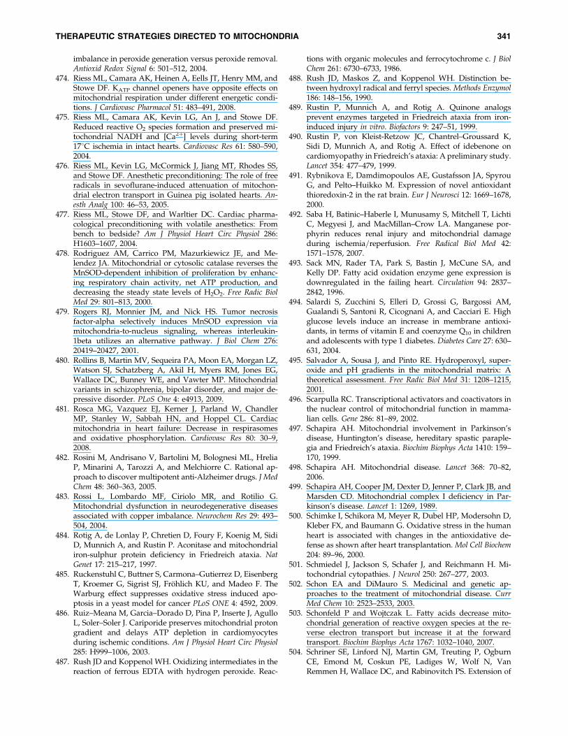
imbalance in peroxide generation versus peroxide removal.Antioxid Redox Signal 6: 501–512, 2004.
474. Riess ML, Camara AK, Heinen A, Eells JT, Henry MM, andStowe DF. KATP channel openers have opposite effects onmitochondrial respiration under different energetic condi-tions. J Cardiovasc Pharmacol 51: 483–491, 2008.
475. Riess ML, Camara AK, Kevin LG, An J, and Stowe DF.Reduced reactive O2 species formation and preserved mi-tochondrial NADH and [Ca2þ] levels during short-term178C ischemia in intact hearts. Cardiovasc Res 61: 580–590,2004.
476. Riess ML, Kevin LG, McCormick J, Jiang MT, Rhodes SS,and Stowe DF. Anesthetic preconditioning: The role of freeradicals in sevoflurane-induced attenuation of mitochon-drial electron transport in Guinea pig isolated hearts. An-esth Analg 100: 46–53, 2005.
477. Riess ML, Stowe DF, and Warltier DC. Cardiac pharma-cological preconditioning with volatile anesthetics: Frombench to bedside? Am J Physiol Heart Circ Physiol 286:H1603–1607, 2004.
478. Rodriguez AM, Carrico PM, Mazurkiewicz JE, and Me-lendez JA. Mitochondrial or cytosolic catalase reverses theMnSOD-dependent inhibition of proliferation by enhanc-ing respiratory chain activity, net ATP production, anddecreasing the steady state levels of H2O2. Free Radic BiolMed 29: 801–813, 2000.
479. Rogers RJ, Monnier JM, and Nick HS. Tumor necrosisfactor-alpha selectively induces MnSOD expression viamitochondria-to-nucleus signaling, whereas interleukin-1beta utilizes an alternative pathway. J Biol Chem 276:20419–20427, 2001.
480. Rollins B, Martin MV, Sequeira PA, Moon EA, Morgan LZ,Watson SJ, Schatzberg A, Akil H, Myers RM, Jones EG,Wallace DC, Bunney WE, and Vawter MP. Mitochondrialvariants in schizophrenia, bipolar disorder, and major de-pressive disorder. PLoS One 4: e4913, 2009.
481. Rosca MG, Vazquez EJ, Kerner J, Parland W, ChandlerMP, Stanley W, Sabbah HN, and Hoppel CL. Cardiacmitochondria in heart failure: Decrease in respirasomesand oxidative phosphorylation. Cardiovasc Res 80: 30–9,2008.
482. Rosini M, Andrisano V, Bartolini M, Bolognesi ML, HreliaP, Minarini A, Tarozzi A, and Melchiorre C. Rational ap-proach to discover multipotent anti-Alzheimer drugs. J MedChem 48: 360–363, 2005.
483. Rossi L, Lombardo MF, Ciriolo MR, and Rotilio G.Mitochondrial dysfunction in neurodegenerative diseasesassociated with copper imbalance. Neurochem Res 29: 493–504, 2004.
484. Rotig A, de Lonlay P, Chretien D, Foury F, Koenig M, SidiD, Munnich A, and Rustin P. Aconitase and mitochondrialiron-sulphur protein deficiency in Friedreich ataxia. NatGenet 17: 215–217, 1997.
485. Ruckenstuhl C, Buttner S, Carmona–Gutierrez D, EisenbergT, Kroemer G, Sigrist SJ, Frohlich KU, and Madeo F. TheWarburg effect suppresses oxidative stress induced apo-ptosis in a yeast model for cancer PLoS ONE 4: 4592, 2009.
486. Ruiz–Meana M, Garcia–Dorado D, Pina P, Inserte J, AgulloL, Soler–Soler J. Cariporide preserves mitochondrial protongradient and delays ATP depletion in cardiomyocytesduring ischemic conditions. Am J Physiol Heart Circ Physiol285: H999–1006, 2003.
487. Rush JD and Koppenol WH. Oxidizing intermediates in thereaction of ferrous EDTA with hydrogen peroxide. Reac-
tions with organic molecules and ferrocytochrome c. J BiolChem 261: 6730–6733, 1986.
488. Rush JD, Maskos Z, and Koppenol WH. Distinction be-tween hydroxyl radical and ferryl species. Methods Enzymol186: 148–156, 1990.
489. Rustin P, Munnich A, and Rotig A. Quinone analogsprevent enzymes targeted in Friedreich ataxia from iron-induced injury in vitro. Biofactors 9: 247–51, 1999.
490. Rustin P, von Kleist-Retzow JC, Chantrel–Groussard K,Sidi D, Munnich A, and Rotig A. Effect of idebenone oncardiomyopathy in Friedreich’s ataxia: A preliminary study.Lancet 354: 477–479, 1999.
491. Rybnikova E, Damdimopoulos AE, Gustafsson JA, SpyrouG, and Pelto–Huikko M. Expression of novel antioxidantthioredoxin-2 in the rat brain. Eur J Neurosci 12: 1669–1678,2000.
492. Saba H, Batinic–Haberle I, Munusamy S, Mitchell T, LichtiC, Megyesi J, and MacMillan–Crow LA. Manganese por-phyrin reduces renal injury and mitochondrial damageduring ischemia=reperfusion. Free Radical Biol Med 42:1571–1578, 2007.
493. Sack MN, Rader TA, Park S, Bastin J, McCune SA, andKelly DP. Fatty acid oxidation enzyme gene expression isdownregulated in the failing heart. Circulation 94: 2837–2842, 1996.
494. Salardi S, Zucchini S, Elleri D, Grossi G, Bargossi AM,Gualandi S, Santoni R, Cicognani A, and Cacciari E. Highglucose levels induce an increase in membrane antioxi-dants, in terms of vitamin E and coenzyme Q10 in childrenand adolescents with type 1 diabetes. Diabetes Care 27: 630–631, 2004.
495. Salvador A, Sousa J, and Pinto RE. Hydroperoxyl, super-oxide and pH gradients in the mitochondrial matrix: Atheoretical assessment. Free Radic Biol Med 31: 1208–1215,2001.
496. Scarpulla RC. Transcriptional activators and coactivators inthe nuclear control of mitochondrial function in mamma-lian cells. Gene 286: 81–89, 2002.
497. Schapira AH. Mitochondrial involvement in Parkinson’sdisease, Huntington’s disease, hereditary spastic paraple-gia and Friedreich’s ataxia. Biochim Biophys Acta 1410: 159–170, 1999.
498. Schapira AH. Mitochondrial disease. Lancet 368: 70–82,2006.
499. Schapira AH, Cooper JM, Dexter D, Jenner P, Clark JB, andMarsden CD. Mitochondrial complex I deficiency in Par-kinson’s disease. Lancet 1: 1269, 1989.
500. Schimke I, Schikora M, Meyer R, Dubel HP, Modersohn D,Kleber FX, and Baumann G. Oxidative stress in the humanheart is associated with changes in the antioxidative de-fense as shown after heart transplantation. Mol Cell Biochem204: 89–96, 2000.
501. Schmiedel J, Jackson S, Schafer J, and Reichmann H. Mi-tochondrial cytopathies. J Neurol 250: 267–277, 2003.
502. Schon EA and DiMauro S. Medicinal and genetic ap-proaches to the treatment of mitochondrial disease. CurrMed Chem 10: 2523–2533, 2003.
503. Schonfeld P and Wojtczak L. Fatty acids decrease mito-chondrial generation of reactive oxygen species at the re-verse electron transport but increase it at the forwardtransport. Biochim Biophys Acta 1767: 1032–1040, 2007.
504. Schriner SE, Linford NJ, Martin GM, Treuting P, OgburnCE, Emond M, Coskun PE, Ladiges W, Wolf N, VanRemmen H, Wallace DC, and Rabinovitch PS. Extension of
THERAPEUTIC STRATEGIES DIRECTED TO MITOCHONDRIA 341
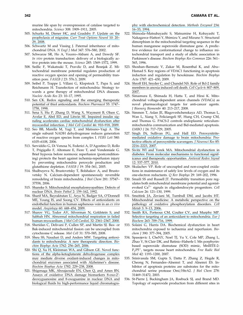
murine life span by overexpression of catalase targeted tomitochondria. Science 308: 1909–1911, 2005.
505. Schurks M, Diener HC, and Goadsby P. Update on theprophylaxis of migraine. Curr Treat Options Neurol 10: 20–29, 2008.
506. Schwartz M and Vissing J. Paternal inheritance of mito-chondrial DNA. N Engl J Med 347: 576–580, 2002.
507. Schwarze SR, Ho A, Vocero–Akbani A, and Dowdy SF.In vivo protein transduction: delivery of a biologically ac-tive protein into the mouse. Science 285: 1569–1572, 1999.
508. Sedlic F, Wakatsuki T, Pravdic D, and Bosnjak Z. Mi-tochondrial membrane potential regulates production ofreactive oxygen species and opening of permeability tran-sition pore. FASEB J 23: 576.3, 2009.
509. Seibel P, Trappe J, Villani G, Klopstock T, Papa S, andReichmann H. Transfection of mitochondria: Strategy to-wards a gene therapy of mitochondrial DNA diseases.Nucleic Acids Res 23: 10–17, 1995.
510. Sen CK. Redox signaling and the emerging therapeuticpotential of thiol antioxidants. Biochem Pharmacol 55: 1747–1758, 1998.
511. Sena S, Hu P, Zhang D, Wang X, Wayment B, Olsen C,Avelar E, Abel ED, and Litwin SE. Impaired insulin sig-naling accelerates cardiac mitochondrial dysfunction aftermyocardial infarction. J Mol Cell Cardiol 46: 910–918, 2009.
512. Seo BB, Marella M, Yagi T, and Matsuno–Yagi A. Thesingle subunit NADH dehydrogenase reduces generationof reactive oxygen species from complex I. FEBS Lett 580:6105–6108, 2006.
513. Serviddio G, Di Venosa N, Federici A, D’Agostino D, RolloT, Prigigallo F, Altomare E, Fiore T, and Vendemiale G.Brief hypoxia before normoxic reperfusion (postcondition-ing) protects the heart against ischemia-reperfusion injuryby preventing mitochondria peroxyde production andglutathione depletion. FASEB J 19: 354–361, 2005.
514. Shalbuyeva N, Brustovetsky T, Bolshakov A, and Brusto-vetsky N. Calcium-dependent spontaneously reversibleremodeling of brain mitochondria. J Biol Chem 281: 37547–37558, 2006.
515. Shanske S. Mitochondrial encephalomyopathies: Defects ofnuclear DNA. Brain Pathol 2: 159–162, 1992.
516. Sharif MA, Bayraktutan U, Arya N, Badger SA, O’DonnellME, Young IS, and Soong CV. Effects of antioxidants onendothelial function in human saphenous vein in an ex vivomodel. Angiology 60: 448–454, 2009.
517. Sharov VG, Todor AV, Silverman N, Goldstein S, andSabbah HN. Abnormal mitochondrial respiration in failedhuman myocardium. J Mol Cell CardioL 32: 2361–2367, 2000.
518. Sheridan C, Delivani P, Cullen SP, and Martin SJ. Bax- orBak-induced mitochondrial fission can be uncoupled fromcytochrome C release. Mol Cell 31: 570–585, 2008.
519. Sheu SS, Nauduri D, and Anders MW. Targeting antioxi-dants to mitochondria: A new therapeutic direction. Bio-chim Biophys Acta 1762: 256–265, 2006.
520. Shi Q, Xu H, Kleinman WA, and Gibson GE. Novel func-tions of the alpha-ketoglutarate dehydrogenase complexmay mediate diverse oxidant-induced changes in mito-chondrial enzymes associated with Alzheimer’s disease.Biochim Biophys Acta 1782: 229–238, 2008.
521. Shigenaga MK, Aboujaoude EN, Chen Q, and Ames BN.Assays of oxidative DNA damage biomarkers 8-oxo-20-deoxyguanosine and 8-oxoguanine in nuclear DNA andbiological fluids by high-performance liquid chromatogra-
phy with electrochemical detection. Methods Enzymol 234:16–33, 1994.
522. Shimoda–Matsubayashi S, Matsumine H, Kobayashi T,Nakagawa–Hattori Y, Shimizu Y, and Mizuno Y. Structuraldimorphism in the mitochondrial targeting sequence in thehuman manganese superoxide dismutase gene. A predic-tive evidence for conformational change to influence mi-tochondrial transport and a study of allelic association inParkinson’s disease. Biochem Biophys Res Commun 226: 561–565, 1996.
523. Shoshan–Barmatz V, Zakar M, Rosenthal K, and Abu–Hamad S. Key regions of VDAC1 functioning in apoptosisinduction and regulation by hexokinase. Biochim BiophysActa 1787: 421–430, 2009.
524. Shroff EH, Snyder C, and Chandel NS. Role of Bcl-2 familymembers in anoxia induced cell death. Cell Cycle 6: 807–809,2007.
525. Simamura E, Shimada H, Hatta T, and Hirai K. Mito-chondrial voltage-dependent anion channels (VDACs) asnovel pharmacological targets for anti-cancer agents.J Bioenerg Biomembr 40: 213–217, 2008.
526. Simmen T, Aslan JE, Blagoveshchenskaya AD, Thomas L,Wan L, Xiang Y, Feliciangeli SF, Hung CH, Crump CM,and Thomas G. PACS-2 controls endoplasmic reticulum-mitochondria communication and Bid-mediated apoptosis.EMBO J 24: 717–729, 2005.
527. Singh IN, Sullivan PG, and Hall ED. Peroxynitrite-mediated oxidative damage to brain mitochondria: Pro-tective effects of peroxynitrite scavengers. J Neurosci Res 85:2216–2223, 2007.
528. Sivitz WI and Yorek MA. Mitochondrial dysfunction indiabetes: From molecular mechanisms to functional signif-icance and therapeutic opportunities. Antioxid Redox Signal12: 537–577, 2010.
529. Skulachev VP. Role of uncoupled and non-coupled oxida-tions in maintenance of safely low levels of oxygen and itsone-electron reductants. Q Rev Biophys 29: 169–202, 1996.
530. Smaili SS and Russell JT. Permeability transition pore reg-ulates both mitochondrial membrane potential and agonist-evoked Ca2þ signals in oligodendrocyte progenitors. CellCalcium 26: 121–130, 1999.
531. Smeitink JA, Zeviani M, Turnbull DM, and Jacobs HT.Mitochondrial medicine: A metabolic perspective on thepathology of oxidative phosphorylation disorders. CellMetab 3: 9–13, 2006.
532. Smith RA, Porteous CM, Coulter CV, and Murphy MP.Selective targeting of an antioxidant to mitochondria. Eur JBiochem 263: 709–716, 1999.
533. Solaini G, Harris DA. Biochemical dysfunction in heartmitochondria exposed to ischaemia and reperfusion. Bio-chem J 390: 377–394, 2005.
534. Spasojevic I, CheNY, Noel TJ, Yu Y, Cole MP, Zhang L,Zhao Y, St Clair DK, and Batinic–Haberle I. Mn porphyrin-based superoxide dismutase (SOD) mimic, MnIIITE-2-PyP5þ, targets mouse heart mitochondria. Free Radic BiolMed 42: 1193–1200, 2007.
535. Srinivasula SM, Gupta S, Datta P, Zhang Z, Hegde R,Cheong N, Fernandes–Alnemri T, and Alnemri ES. In-hibitor of apoptosis proteins are substrates for the mito-chondrial serine protease Omi=HtrA2. J Biol Chem 278:31469–31472, 2003.
536. St-Pierre J, Buckingham JA, Roebuck SJ, and Brand MD.Topology of superoxide production from different sites in
342 CAMARA ET AL.
the mitochondrial electron transport chain. J Biol Chem 277:44784–44790, 2002.
537. St-Pierre J, Drori S, Uldry M, Silvaggi JM, Rhee J, Jager S,Handschin C, Zheng K, Lin J, Yang W, Simon DK, BachooR, and Spiegelman BM. Suppression of reactive oxygenspecies and neurodegeneration by the PGC-1 transcrip-tional coactivators. Cell 127: 397–408, 2006.
538. Stanley WC, Lopaschuk GD, and McCormack JG. Regula-tion of energy substrate metabolism in the diabetic heart.Cardiovasc Res 34: 25–33, 1997.
539. Stanley WC, Recchia FA, and Lopaschuk GD. Myocardialsubstrate metabolism in the normal and failing heart.Physiol Rev 85: 1093–1129, 2005.
540. Stark R and Roden M. ESCI Award 2006. Mitochondrialfunction and endocrine diseases. Eur J Clin Invest 37: 236–248, 2007.
541. Starke DW, CheNY, Bapna CP, Lesnefsky EJ, and Mieyal JJ.Sensitivity of protein sulfhydryl repair enzymes to oxida-tive stress. Free Radic Biol Med 23: 373–384, 1997.
542. Stowe DF, Aldakkak M, Camara AKS, Riess ML, Heinen A,Varadarajan SG, and Jiang MT. Cardiac mitochondrialpreconditioning by big Ca2þ-sensitive Kþ channel openingrequires superoxide radical generation. Am J Physiol HeartCirc Physiol 290: H434–H440, 2006.
543. Stowe DF and Camara AKS. Mitochondrial reactive oxygenspecies production in excitable cells: Modulators of mito-chondrial and cell function Antioxid Redox Signal 11: 1373–1396, 2009.
544. Stuart JA, Cadenas S, Jekabsons MB, Roussel D, and BrandMD. Mitochondrial proton leak and the uncoupling protein1 homologues. Biochim Biophys Acta 1504: 144–158, 2001.
545. Su YA, Wu J, Zhang L, Zhang Q, Su DM, He P, Wang BD,Li H, Webster MJ, Rennert OM, and Ursano RJ. Dysregu-lated mitochondrial genes and networks with drug targetsin postmortem brain of patients with posttraumatic stressdisorder (PTSD) revealed by human mitochondria-focusedcDNA microarrays. Int J Biol Sci 4: 223–235, 2008.
546. Sun H, Nikolovska–Coleska Z, Yang CY, Qian D, Lu J, QiuS, Bai L, Peng Y, Cai Q, and Wang S. Design of small-molecule peptidic and nonpeptidic Smac mimetics. AccChem Res 41: 1264–1277, 2008.
547. Sun HY, Wang NP, Kerendi F, Halkos M, Kin H, GuytonRA, Vinten–Johansen J, and Zhao ZQ. Hypoxic post-conditioning reduces cardiomyocyte loss by inhibiting ROSgeneration and intracellular Ca2þ overload. Am J PhysiolHeart Circ Physiol 288: H1900–1908, 2005.
548. Sun KH, de Pablo Y, Vincent F, and Shah K. DeregulatedCdk5 promotes oxidative stress and mitochondrial dys-function. J Neurochem 107: 265–278, 2008.
549. Sun L, Shukair S, Naik TJ, Moazed F, and Ardehali H.Glucose phosphorylation and mitochondrial binding arerequired for the protective effects of hexokinases I and II.Mol Cell Biol 28: 1007–1017, 2008.
550. Szabadkai G and Duchen MR. Mitochondria: The hubof cellular Ca2þ signaling. Physiology (Bethesda) 23: 84–94,2008.
551. Szeto HH. Cell-permeable, mitochondrial-targeted, peptideantioxidants. AAPS J 8: E277–283, 2006.
552. Szeto HH. Mitochondria-targeted peptide antioxidants:Novel neuroprotective agents. AAPS J 8: E521–531, 2006.
553. Szeto HH. Development of mitochondria-targeted aromatic-cationic peptides for neurodegenerative diseases. Ann NYAcad Sci 1147: 112–121, 2008.
554. Szeto HH. Mitochondria-targeted cytoprotective peptidesfor ischemia-reperfusion injury. Antioxid Redox Signal 10:601–619, 2008.
555. Szewczyk A, Skalska J, Glab M, Kulawiak B, Malinska D,Koszela–Piotrowska I, and Kunz WS. Mitochondrial po-tassium channels: From pharmacology to function. BiochimBiophys Acta 1757: 715–720, 2006.
556. Szewczyk A and Wojtczak L. Mitochondria as a pharma-cological target. Pharmacol Rev 54: 101–127, 2002.
557. Tabrizi SJ, Cleeter MW, Xuereb J, Taanman JW, Cooper JM,and Schapira AH. Biochemical abnormalities and ex-citotoxicity in Huntington’s disease brain. Ann Neurol 45:25–32, 1999.
558. Taegtmeyer H. Switching metabolic genes to build a betterheart. Circulation 106: 2043–2045, 2002.
559. Taegtmeyer H. Cardiac metabolism as a target for thetreatment of heart failure. Circulation 110: 894–896, 2004.
560. Taegtmeyer H, McNulty P, and Young ME. Adaptationand maladaptation of the heart in diabetes: Part I: Generalconcepts. Circulation 105: 1727–1733, 2002.
561. Taegtmeyer H and Overturf ML. Effects of moderate hy-pertension on cardiac function and metabolism in the rab-bit. Hypertension 11: 416–426, 1988.
562. Taguchi N, Ishihara N, Jofuku A, Oka T, and Mihara K.Mitotic phosphorylation of dynamin-related GTPase Drp1participates in mitochondrial fission. J Biol Chem 282:11521–11529, 2007.
563. Takaoka S, Iwase M, Uchida M, Yoshiba S, Kondo G,Watanabe H, Ohashi M, Nagumo M, and Shintani S. Effectof combining epidermal growth factor receptor inhibitorsand cisplatin on proliferation and apoptosis of oral squa-mous cell carcinoma cells. Int J Oncol 30: 1469–1476, 2007.
564. Tan L, Yu JT, and Guan HS. Resveratrol exerts pharmaco-logical preconditioning by activating PGC-1alpha. MedHypotheses 71: 664–667, 2008.
565. Tani M, Honma Y, Hasegawa H, and Tamaki K. Directactivation of mitochondrial KATP channels mimics pre-conditioning but protein kinase C activation is less effectivein middle-aged rat hearts. Cardiovasc Res 49: 56–68, 2001.
566. Tarba C and Cracium C. A comparative study of the effectsof procaine, lidocaine, tetracaine and dibucaine on thefunctions and ultrastructure of isolated rat liver mitochon-dria. Biochim Biophys Acta 1019: 19–28, 1990.
567. Taylor RW, Chinnery PF, Turnbull DM, and LightowlersRN. Selective inhibition of mutant human mitochondrialDNA replication in vitro by peptide nucleic acids. Nat Genet15: 212–215, 1997.
568. Terman A and Brunk UT. Myocyte aging and mitochon-drial turnover. Exp Gerontol 39: 701–705, 2004.
569. Teshima Y, Akao M, Jones SP, and Marban E. Uncouplingprotein-2 overexpression inhibits mitochondrial death path-way in cardiomyocytes. Circ Res 93: 192–200, 2003.
570. Tewari A, Ali A, O’Donnell A, and Butt MS. Weight loss and2,4-dinitrophenol poisoning. Br J Anaesth 102: 566–567, 2009.
571. Thannickal VJ and Fanburg BL. Reactive oxygen speciesin cell signaling. Am J Physiol Lung Cell Mol Physiol 279:L1005–1028, 2000.
572. Theruvath TP, Snoddy MC, Zhong Z, and Lemasters JJ.Mitochondrial permeability transition in liver ischemia andreperfusion: role of c-Jun N-terminal kinase 2. Transplan-tation 85: 1500–1504, 2008.
573. Theruvath TP, Zhong Z, Pediaditakis P, Ramshesh VK,Currin RT, Tikunov A, Holmuhamedov E, and Lemasters
THERAPEUTIC STRATEGIES DIRECTED TO MITOCHONDRIA 343
JJ. Minocycline and N-methyl-4-isoleucine cyclosporin(NIM811) mitigate storage=reperfusion injury after rat livertransplantation through suppression of the mitochondrialpermeability transition. Hepatology 47: 236–246, 2008.
574. Thomas DA, Stauffer C, Zhao K, Yang H, Sharma VK,Szeto HH, and Suthanthiran M. Mitochondrial targetingwith antioxidant peptide SS-31 prevents mitochondrialdepolarization, reduces islet cell apoptosis, increases isletcell yield, and improves posttransplantation function. J AmSoc Nephrol 18: 213–222, 2007.
575. Tonissen KF and Di Trapani G. Thioredoxin system in-hibitors as mediators of apoptosis for cancer therapy. MolNutr Food Res 53: 87–103, 2009.
576. Torres J, Sharpe MA, Rosquist A, Cooper CE, and WilsonMT. Cytochrome c oxidase rapidly metabolises nitric oxideto nitrite. FEBS Letters 475: 263–266, 2000.
577. Treuting PM, Linford NJ, Knoblaugh SE, Emond MJ,Morton JF, Martin GM, Rabinovitch PS, and Ladiges WC.Reduction of age-associated pathology in old mice byoverexpression of catalase in mitochondria. J Gerontol A BiolSci Med Sci 63: 813–822, 2008.
578. Trifunovic A and Larsson NG. Mitochondrial dysfunctionas a cause of ageing. J Intern Med 263: 167–178, 2008.
579. Tulinius MH, Holme E, Kristiansson B, Larsson NG, andOldfors A. Mitochondrial encephalomyopathies in child-hood. II. Clinical manifestations and syndromes. J Pediat119: 251–259, 1991.
580. Turrens JF, Alexandre A, and Lehninger AL. Ubisemiqui-none is the electron donor for superoxide formation bycomplex III of heart mitochondria. Arch Biochem Biophys237: 408–414., 1985.
581. Turrens JF and Boveris A. Generation of superoxide anionby the NADH dehydrogenase of bovine heart mitochon-dria. Biochem J 191: 421–427, 1980.
582. Ueda S, Nakamura H, Masutani H, Sasada T, Yonehara S,Takabayashi A, Yamaoka Y, and Yodoi J. Redox regulationof caspase-3(-like) protease activity: Regulatory roles ofthioredoxin and cytochrome c. J Immunol 161: 6689–6695,1998.
583. Ujwal R, Cascio D, Colletier JP, Faham S, Zhang J, Toro L,Ping P, and Abramson J. The crystal structure of mouseVDAC1 at 2.3 A resolution reveals mechanistic insights intometabolite gating. Proc Natl Acad Sci USA 105: 17742–17747, 2008.
584. Valdez LB, Alvarez S, Arnaiz SL, Schopfer F, Carreras MC,Poderoso JJ, and Boveris A. Reactions of peroxynitrite inthe mitochondrial matrix. Free Radic Biol Med 29: 349–356,2000.
585. Valen G and Vaage J. Pre- and postconditioning duringcardiac surgery. Basic Res Cardiol 100: 179–186, 2005.
586. Valentine JS, Doucette PA, and Zittin Potter S. Copper-zincsuperoxide dismutase and amyotrophic lateral sclerosis.Annu Rev Biochem 74: 563–593, 2005.
587. Valentine JS and Hart PJ. Misfolded CuZnSOD andamyotrophic lateral sclerosis. Proc Natl Acad Sci USA 100:3617–3622, 2003.
588. Valle A, Guevara R, Garcia-Palmer FJ, Roca P, and Oliver J.Caloric restriction retards the age-related decline in mito-chondrial function of brown adipose tissue. RejuvenationRes 11: 597–604, 2008.
589. Valle I, Alvarez–Barrientos A, Arza E, Lamas S, andMonsalve M. PGC-1alpha regulates the mitochondrial an-tioxidant defense system in vascular endothelial cells.Cardiovasc Res 66: 562–573, 2005.
590. Van Raamsdonk JM and Hekimi S. Deletion of the mito-chondrial superoxide dismutase sod-2 extends lifespan inCaenorhabditis elegans. PLoS Genet 5: e1000361, 2009.
591. Vanden Hoek TL, Li C, Shao Z, Schumacker PT, and BeckerLB. Significant levels of oxidants are generated by isolatedcardiomyocytes during ischemia prior to reperfusion. J MolCell Cardiol 29: 2571–2583, 1997.
592. Vanden Hoek TL, Shao Z, Li C, Schumacker PT, and BeckerLB. Mitochondrial electron transport can become a signifi-cant source of oxidative injury in cardiomyocytes. J Mol CellCardiol 29: 2441–2450, 1997.
593. Varadarajan SC, Rhodes SS, Aldakkak M, Heisner JS, andStowe DF. Cardiac mitochondrial Ca2 -dependent big Kþ
channels are open during reperfusion. FASEB J 21: A1224,2007.
594. Varbiro G, Toth A, Tapodi A, Bognar Z, Veres B, Sumegi B,and Gallyas F, Jr. Protective effect of amiodarone but notN-desethylamiodarone on postischemic hearts through theinhibition of mitochondrial permeability transition. J Phar-macol Exp Ther 307: 615–625, 2003.
595. Varbiro G, Toth A, Tapodi A, Veres B, Sumegi B, andGallyas F, Jr. Concentration dependent mitochondrial effectof amiodarone. Biochem Pharmacol 65: 1115–1128, 2003.
596. Venkatakrishnan P, Nakayasu ES, Almeida IC, and MillerRT. Absence of nitric-oxide synthase in sequentially puri-fied rat liver mitochondria. J Biol Chem 284: 19843–19855,2009.
597. Verhagen AM, Ekert PG, Pakusch M, Silke J, Connolly LM,Reid GE, Moritz RL, Simpson RJ, and Vaux DL. Identifi-cation of DIABLO, a mammalian protein that promotesapoptosis by binding to and antagonizing IAP proteins.Cell 102: 43–53, 2000.
598. Vijayasarathy C, Biunno I, Lenka N, Yang M, Basu A, HallIP, and Avadhani NG. Variations in the subunit contentand catalytic activity of the cytochrome c oxidase complexfrom different tissues and different cardiac compartments.Biochim Biophys Acta 1371: 71–82, 1998.
599. Vinten–Johansen J, Yellon DM, and Opie LH. Postcondi-tioning: A simple, clinically applicable procedure to im-prove revascularization in acute myocardial infarction.Circulation 112: 2085–2088, 2005.
600. Vinten–Johansen J, Zhao ZQ, Zatta AJ, Kin H, Halkos ME,and Kerendi F. Postconditioning–A new link in nature’sarmor against myocardial ischemia-reperfusion injury.Basic Res Cardiol 100: 295–310, 2005.
601. Vlasova, II, Tyurin VA, Kapralov AA, Kurnikov IV, OsipovAN, Potapovich MV, Stoyanovsky DA, and Kagan VE.Nitric oxide inhibits peroxidase activity of cytochromec.cardiolipin complex and blocks cardiolipin oxidation.J Biol Chem 281: 14554–14562, 2006.
602. Waldmeier PC, Zimmermann K, Qian T, Tintelnot–BlomleyM, and Lemasters JJ. Cyclophilin D as a drug target. CurrMed Chem 10: 1485–1506, 2003.
603. Walensky LD, Kung AL, Escher I, Malia TJ, Barbuto S,Wright RD, Wagner G, Verdine GL, and Korsmeyer SJ.Activation of apoptosis in vivo by a hydrocarbon-stapledBH3 helix. Science 305: 1466–1470, 2004.
604. Wall JA, Wei J, Ly M, Belmont P, Martindale JJ, Tran D, SunJ, Chen WJ, Yu W, Oeller P, Briggs S, Gustafsson AB, SayenMR, Gottlieb RA, and Glembotski CC. Alterations in oxi-dative phosphorylation complex proteins in the hearts oftransgenic mice that overexpress the p38 MAP kinase ac-tivator, MAP kinase kinase 6. Am J Physiol Heart Circ Physiol291: H2462–2472, 2006.
344 CAMARA ET AL.
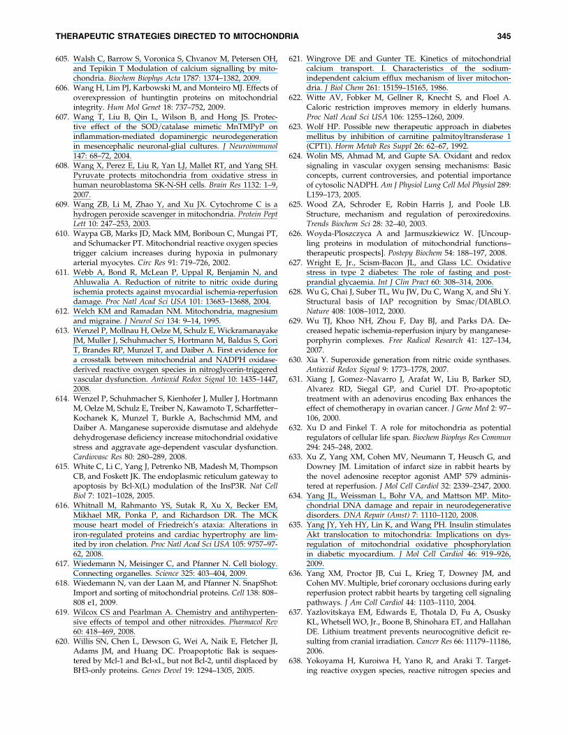
605. Walsh C, Barrow S, Voronica S, Chvanov M, Petersen OH,and Tepikin T Modulation of calcium signalling by mito-chondria. Biochem Biophys Acta 1787: 1374–1382, 2009.
606. Wang H, Lim PJ, Karbowski M, and Monteiro MJ. Effects ofoverexpression of huntingtin proteins on mitochondrialintegrity. Hum Mol Genet 18: 737–752, 2009.
607. Wang T, Liu B, Qin L, Wilson B, and Hong JS. Protec-tive effect of the SOD=catalase mimetic MnTMPyP oninflammation-mediated dopaminergic neurodegenerationin mesencephalic neuronal-glial cultures. J Neuroimmunol147: 68–72, 2004.
608. Wang X, Perez E, Liu R, Yan LJ, Mallet RT, and Yang SH.Pyruvate protects mitochondria from oxidative stress inhuman neuroblastoma SK-N-SH cells. Brain Res 1132: 1–9,2007.
609. Wang ZB, Li M, Zhao Y, and Xu JX. Cytochrome C is ahydrogen peroxide scavenger in mitochondria. Protein PeptLett 10: 247–253, 2003.
610. Waypa GB, Marks JD, Mack MM, Boriboun C, Mungai PT,and Schumacker PT. Mitochondrial reactive oxygen speciestrigger calcium increases during hypoxia in pulmonaryarterial myocytes. Circ Res 91: 719–726, 2002.
611. Webb A, Bond R, McLean P, Uppal R, Benjamin N, andAhluwalia A. Reduction of nitrite to nitric oxide duringischemia protects against myocardial ischemia-reperfusiondamage. Proc Natl Acad Sci USA 101: 13683–13688, 2004.
612. Welch KM and Ramadan NM. Mitochondria, magnesiumand migraine. J Neurol Sci 134: 9–14, 1995.
613. Wenzel P, Mollnau H, Oelze M, Schulz E, WickramanayakeJM, Muller J, Schuhmacher S, Hortmann M, Baldus S, GoriT, Brandes RP, Munzel T, and Daiber A. First evidence fora crosstalk between mitochondrial and NADPH oxidase-derived reactive oxygen species in nitroglycerin-triggeredvascular dysfunction. Antioxid Redox Signal 10: 1435–1447,2008.
614. Wenzel P, Schuhmacher S, Kienhofer J, Muller J, HortmannM, Oelze M, Schulz E, Treiber N, Kawamoto T, Scharffetter–Kochanek K, Munzel T, Burkle A, Bachschmid MM, andDaiber A. Manganese superoxide dismutase and aldehydedehydrogenase deficiency increase mitochondrial oxidativestress and aggravate age-dependent vascular dysfunction.Cardiovasc Res 80: 280–289, 2008.
615. White C, Li C, Yang J, Petrenko NB, Madesh M, ThompsonCB, and Foskett JK. The endoplasmic reticulum gateway toapoptosis by Bcl-X(L) modulation of the InsP3R. Nat CellBiol 7: 1021–1028, 2005.
616. Whitnall M, Rahmanto YS, Sutak R, Xu X, Becker EM,Mikhael MR, Ponka P, and Richardson DR. The MCKmouse heart model of Friedreich’s ataxia: Alterations iniron-regulated proteins and cardiac hypertrophy are lim-ited by iron chelation. Proc Natl Acad Sci USA 105: 9757–97-62, 2008.
617. Wiedemann N, Meisinger C, and Pfanner N. Cell biology.Connecting organelles. Science 325: 403–404, 2009.
618. Wiedemann N, van der Laan M, and Pfanner N. SnapShot:Import and sorting of mitochondrial proteins. Cell 138: 808–808 e1, 2009.
619. Wilcox CS and Pearlman A. Chemistry and antihyperten-sive effects of tempol and other nitroxides. Pharmacol Rev60: 418–469, 2008.
620. Willis SN, Chen L, Dewson G, Wei A, Naik E, Fletcher JI,Adams JM, and Huang DC. Proapoptotic Bak is seques-tered by Mcl-1 and Bcl-xL, but not Bcl-2, until displaced byBH3-only proteins. Genes Devel 19: 1294–1305, 2005.
621. Wingrove DE and Gunter TE. Kinetics of mitochondrialcalcium transport. I. Characteristics of the sodium-independent calcium efflux mechanism of liver mitochon-dria. J Biol Chem 261: 15159–15165, 1986.
622. Witte AV, Fobker M, Gellner R, Knecht S, and Floel A.Caloric restriction improves memory in elderly humans.Proc Natl Acad Sci USA 106: 1255–1260, 2009.
623. Wolf HP. Possible new therapeutic approach in diabetesmellitus by inhibition of carnitine palmitoyltransferase 1(CPT1). Horm Metab Res Suppl 26: 62–67, 1992.
624. Wolin MS, Ahmad M, and Gupte SA. Oxidant and redoxsignaling in vascular oxygen sensing mechanisms: Basicconcepts, current controversies, and potential importanceof cytosolic NADPH. Am J Physiol Lung Cell Mol Physiol 289:L159–173, 2005.
625. Wood ZA, Schroder E, Robin Harris J, and Poole LB.Structure, mechanism and regulation of peroxiredoxins.Trends Biochem Sci 28: 32–40, 2003.
626. Woyda-Ploszczyca A and Jarmuszkiewicz W. [Uncoup-ling proteins in modulation of mitochondrial functions–therapeutic prospects]. Postepy Biochem 54: 188–197, 2008.
627. Wright E, Jr., Scism-Bacon JL, and Glass LC. Oxidativestress in type 2 diabetes: The role of fasting and post-prandial glycaemia. Int J Clin Pract 60: 308–314, 2006.
628. Wu G, Chai J, Suber TL, Wu JW, Du C, Wang X, and Shi Y.Structural basis of IAP recognition by Smac=DIABLO.Nature 408: 1008–1012, 2000.
629. Wu TJ, Khoo NH, Zhou F, Day BJ, and Parks DA. De-creased hepatic ischemia-reperfusion injury by manganese-porphyrin complexes. Free Radical Research 41: 127–134,2007.
630. Xia Y. Superoxide generation from nitric oxide synthases.Antioxid Redox Signal 9: 1773–1778, 2007.
631. Xiang J, Gomez–Navarro J, Arafat W, Liu B, Barker SD,Alvarez RD, Siegal GP, and Curiel DT. Pro-apoptotictreatment with an adenovirus encoding Bax enhances theeffect of chemotherapy in ovarian cancer. J Gene Med 2: 97–106, 2000.
632. Xu D and Finkel T. A role for mitochondria as potentialregulators of cellular life span. Biochem Biophys Res Commun294: 245–248, 2002.
633. Xu Z, Yang XM, Cohen MV, Neumann T, Heusch G, andDowney JM. Limitation of infarct size in rabbit hearts bythe novel adenosine receptor agonist AMP 579 adminis-tered at reperfusion. J Mol Cell Cardiol 32: 2339–2347, 2000.
634. Yang JL, Weissman L, Bohr VA, and Mattson MP. Mito-chondrial DNA damage and repair in neurodegenerativedisorders. DNA Repair (Amst) 7: 1110–1120, 2008.
635. Yang JY, Yeh HY, Lin K, and Wang PH. Insulin stimulatesAkt translocation to mitochondria: Implications on dys-regulation of mitochondrial oxidative phosphorylationin diabetic myocardium. J Mol Cell Cardiol 46: 919–926,2009.
636. Yang XM, Proctor JB, Cui L, Krieg T, Downey JM, andCohen MV. Multiple, brief coronary occlusions during earlyreperfusion protect rabbit hearts by targeting cell signalingpathways. J Am Coll Cardiol 44: 1103–1110, 2004.
637. Yazlovitskaya EM, Edwards E, Thotala D, Fu A, OsuskyKL, Whetsell WO, Jr., Boone B, Shinohara ET, and HallahanDE. Lithium treatment prevents neurocognitive deficit re-sulting from cranial irradiation. Cancer Res 66: 11179–11186,2006.
638. Yokoyama H, Kuroiwa H, Yano R, and Araki T. Target-ing reactive oxygen species, reactive nitrogen species and
THERAPEUTIC STRATEGIES DIRECTED TO MITOCHONDRIA 345
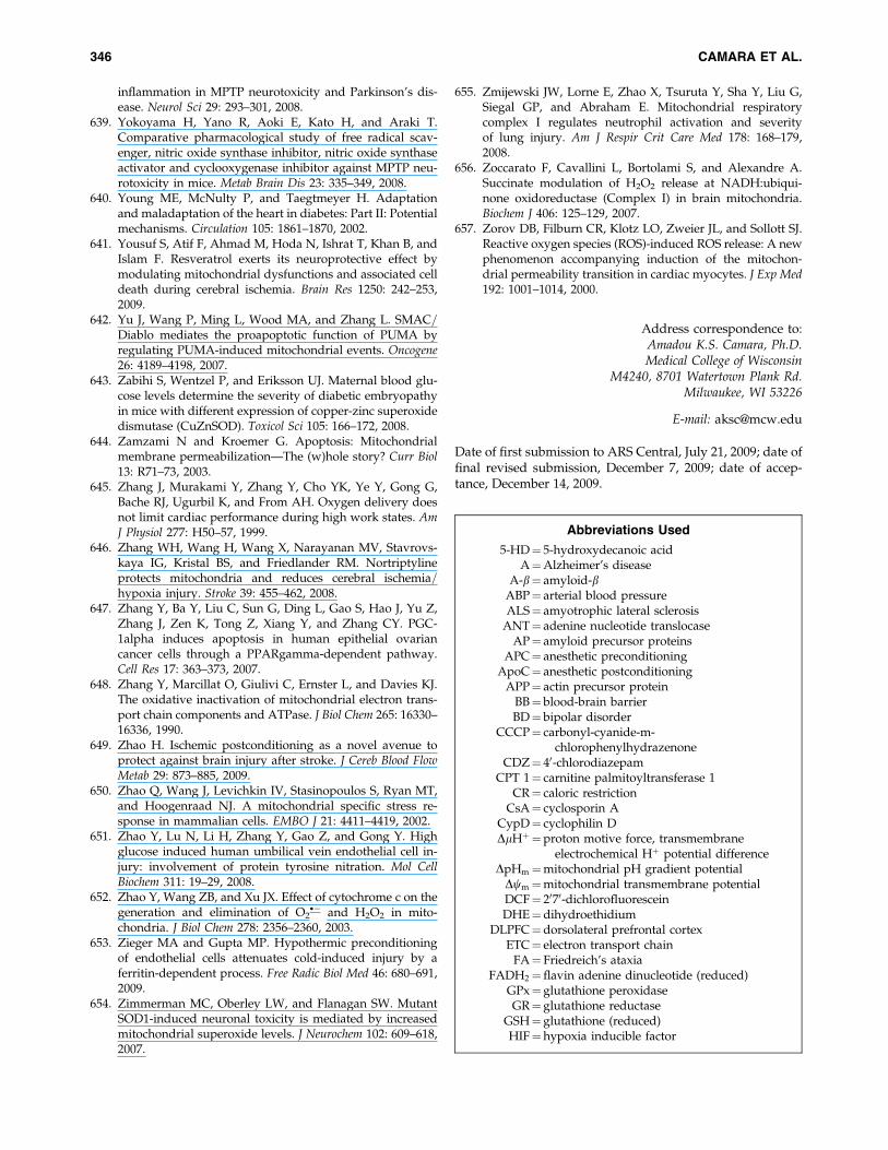
inflammation in MPTP neurotoxicity and Parkinson’s dis-ease. Neurol Sci 29: 293–301, 2008.
639. Yokoyama H, Yano R, Aoki E, Kato H, and Araki T.Comparative pharmacological study of free radical scav-enger, nitric oxide synthase inhibitor, nitric oxide synthaseactivator and cyclooxygenase inhibitor against MPTP neu-rotoxicity in mice. Metab Brain Dis 23: 335–349, 2008.
640. Young ME, McNulty P, and Taegtmeyer H. Adaptationand maladaptation of the heart in diabetes: Part II: Potentialmechanisms. Circulation 105: 1861–1870, 2002.
641. Yousuf S, Atif F, Ahmad M, Hoda N, Ishrat T, Khan B, andIslam F. Resveratrol exerts its neuroprotective effect bymodulating mitochondrial dysfunctions and associated celldeath during cerebral ischemia. Brain Res 1250: 242–253,2009.
642. Yu J, Wang P, Ming L, Wood MA, and Zhang L. SMAC=Diablo mediates the proapoptotic function of PUMA byregulating PUMA-induced mitochondrial events. Oncogene26: 4189–4198, 2007.
643. Zabihi S, Wentzel P, and Eriksson UJ. Maternal blood glu-cose levels determine the severity of diabetic embryopathyin mice with different expression of copper-zinc superoxidedismutase (CuZnSOD). Toxicol Sci 105: 166–172, 2008.
644. Zamzami N and Kroemer G. Apoptosis: Mitochondrialmembrane permeabilization—The (w)hole story? Curr Biol13: R71–73, 2003.
645. Zhang J, Murakami Y, Zhang Y, Cho YK, Ye Y, Gong G,Bache RJ, Ugurbil K, and From AH. Oxygen delivery doesnot limit cardiac performance during high work states. AmJ Physiol 277: H50–57, 1999.
646. Zhang WH, Wang H, Wang X, Narayanan MV, Stavrovs-kaya IG, Kristal BS, and Friedlander RM. Nortriptylineprotects mitochondria and reduces cerebral ischemia=hypoxia injury. Stroke 39: 455–462, 2008.
647. Zhang Y, Ba Y, Liu C, Sun G, Ding L, Gao S, Hao J, Yu Z,Zhang J, Zen K, Tong Z, Xiang Y, and Zhang CY. PGC-1alpha induces apoptosis in human epithelial ovariancancer cells through a PPARgamma-dependent pathway.Cell Res 17: 363–373, 2007.
648. Zhang Y, Marcillat O, Giulivi C, Ernster L, and Davies KJ.The oxidative inactivation of mitochondrial electron trans-port chain components and ATPase. J Biol Chem 265: 16330–16336, 1990.
649. Zhao H. Ischemic postconditioning as a novel avenue toprotect against brain injury after stroke. J Cereb Blood FlowMetab 29: 873–885, 2009.
650. Zhao Q, Wang J, Levichkin IV, Stasinopoulos S, Ryan MT,and Hoogenraad NJ. A mitochondrial specific stress re-sponse in mammalian cells. EMBO J 21: 4411–4419, 2002.
651. Zhao Y, Lu N, Li H, Zhang Y, Gao Z, and Gong Y. Highglucose induced human umbilical vein endothelial cell in-jury: involvement of protein tyrosine nitration. Mol CellBiochem 311: 19–29, 2008.
652. Zhao Y, Wang ZB, and Xu JX. Effect of cytochrome c on thegeneration and elimination of O2
�� and H2O2 in mito-chondria. J Biol Chem 278: 2356–2360, 2003.
653. Zieger MA and Gupta MP. Hypothermic preconditioningof endothelial cells attenuates cold-induced injury by aferritin-dependent process. Free Radic Biol Med 46: 680–691,2009.
654. Zimmerman MC, Oberley LW, and Flanagan SW. MutantSOD1-induced neuronal toxicity is mediated by increasedmitochondrial superoxide levels. J Neurochem 102: 609–618,2007.
655. Zmijewski JW, Lorne E, Zhao X, Tsuruta Y, Sha Y, Liu G,Siegal GP, and Abraham E. Mitochondrial respiratorycomplex I regulates neutrophil activation and severityof lung injury. Am J Respir Crit Care Med 178: 168–179,2008.
656. Zoccarato F, Cavallini L, Bortolami S, and Alexandre A.Succinate modulation of H2O2 release at NADH:ubiqui-none oxidoreductase (Complex I) in brain mitochondria.Biochem J 406: 125–129, 2007.
657. Zorov DB, Filburn CR, Klotz LO, Zweier JL, and Sollott SJ.Reactive oxygen species (ROS)-induced ROS release: A newphenomenon accompanying induction of the mitochon-drial permeability transition in cardiac myocytes. J Exp Med192: 1001–1014, 2000.
Address correspondence to:Amadou K.S. Camara, Ph.D.Medical College of Wisconsin
M4240, 8701 Watertown Plank Rd.Milwaukee, WI 53226
E-mail: [email protected]
Date of first submission to ARS Central, July 21, 2009; date offinal revised submission, December 7, 2009; date of accep-tance, December 14, 2009.
Abbreviations Used
5-HD¼ 5-hydroxydecanoic acidA¼Alzheimer’s disease
A-b¼ amyloid-bABP¼ arterial blood pressureALS¼ amyotrophic lateral sclerosis
ANT¼ adenine nucleotide translocaseAP¼ amyloid precursor proteins
APC¼ anesthetic preconditioningApoC¼ anesthetic postconditioning
APP¼ actin precursor proteinBB¼ blood-brain barrierBD¼ bipolar disorder
CCCP¼ carbonyl-cyanide-m-chlorophenylhydrazenone
CDZ¼ 40-chlorodiazepamCPT 1¼ carnitine palmitoyltransferase 1
CR¼ caloric restrictionCsA¼ cyclosporin A
CypD¼ cyclophilin DDmHþ ¼proton motive force, transmembrane
electrochemical Hþ potential differenceDpHm ¼mitochondrial pH gradient potentialDcm ¼mitochondrial transmembrane potentialDCF¼ 2070-dichlorofluoresceinDHE¼dihydroethidium
DLPFC¼dorsolateral prefrontal cortexETC¼ electron transport chain
FA¼ Friedreich’s ataxiaFADH2¼flavin adenine dinucleotide (reduced)
GPx¼ glutathione peroxidaseGR¼ glutathione reductase
GSH¼ glutathione (reduced)HIF¼hypoxia inducible factor
346 CAMARA ET AL.
Abbreviations Used (cont.)
HKI and HKII ¼ hexokinase I and IIHNE ¼ 4-hydroxy-trans-2-nonealH2O2 ¼ hydrogen peroxideHSP ¼ heat shock proteins
IMM ¼ inner mitochondrial membraneIMS ¼ intermembrane spaceIPC ¼ ischemic preconditioning
IPoC ¼ ischemic postconditioningIRS ¼ insulin receptor substrate
KATP ¼ ATP-sensitive Kþ channelKCa ¼ Ca2þ sensitive Kþ channel
KHE ¼ Kþ=Hþ exchangeL-NAME ¼ NG-nitro-L-arginine methyl ester
LRT ¼ lipid replacement therapyMDD ¼ major depressive disorder
MnIII TE-2-Pyp5þ ¼ Mn (III) meso-tetrakis(N-ethylpyridium-2-yl) porphyrin
MnSOD ¼ manganese superoxide dismutaseMnTBAP ¼ Mn(II)tetrakis(4-benzoate) porphyrin
chlorineMnTPyP ¼ Mn(III)tetrakis [(1-methyl-4-pyridyl)-
porphyrin]MPTH ¼ 1-methyl-4-phenyl-1,2,3,6-tetra-
hydropyridinemPTP ¼ mitochondrial permeability transition
poremtDNA ¼ mitochondrial DNANADH ¼ nicotinamide adenine dinucleotide
(reduced)NARP ¼ neuropathy, ataxia, and retinitis
pigmentosaND ¼ neoplastic disease
NHE ¼ Naþ=Hþ exchangeNIM811 ¼ N-methyl-4-isoleucine-cyclosporin
NO� ¼ nitric oxide radicalNox ¼ NADPH oxidaseO2�� ¼ superoxide anion radical
OMM ¼ outer mitochondrial membraneONOO� ¼ peroxynitrite
OPA-1 ¼ optic atrophy 1OXPHOS ¼ oxidative phosphorylation
PBR ¼ peripheral benzodiazepine receptorPD ¼ Parkinson’s disease
PDH ¼ pyruvate dehydrogenasePGC-1a ¼ peroxisome proliferator-activated
receptor-gamma coactivator-1aPH ¼ prolyl-4-hydroxylase
PHP ¼ phospholipid hydroperoxidesPINK-1 ¼ PTEN-induced kinase 1
PPC ¼ pharmacologic preconditioningPPoC ¼ pharmacological postconditioning
PRX ¼ peroxiredoxinsPTSD ¼ post-traumatic stress disorder
Q ¼ coenzyme Q10, ubiquinone, quinoneRISK ¼ reperfusion injury signaling kinaseRNS ¼ reactive nitrogen speciesROS ¼ reactive oxygen speciesRuR ¼ ruthenium red
RVLM ¼ rostral ventrolateral medullaSHR ¼ spontaneous hypertensive rat
Smac=Diablo ¼ second mitochondria-derivedactivator caspase=direct inhibitorsof apoptosis protein (IAP), inhibitorsof apoptosis protein
SNO-MPG ¼ S-nitroso-2 mercaptopropionyl glycineSOD ¼ superoxide dismutase
SR ¼ sarcoplasmic reticulumSS ¼ Sezto–Schiller tetrapeptidesSZ ¼ schizophrenia
TAT ¼ transactivator of transcriptionTCA ¼ tricarboxylic acid
TOM and TIM ¼ outer and inner membrane translocases,respectively
TPPþ ¼ triphenylphosphoniumTRX ¼ thioredoxin
TRXSH2 ¼ thioredoxin (reduced)UCP ¼ uncoupling proteins
VDAC ¼ voltage dependent anion channel
THERAPEUTIC STRATEGIES DIRECTED TO MITOCHONDRIA 347
