polymer-assisted sol–gel synthesis and characterization of zn< sub> 2 sio< sub> 4:...
TRANSCRIPT

This article appeared in a journal published by Elsevier. The attachedcopy is furnished to the author for internal non-commercial researchand education use, including for instruction at the authors institution
and sharing with colleagues.
Other uses, including reproduction and distribution, or selling orlicensing copies, or posting to personal, institutional or third party
websites are prohibited.
In most cases authors are permitted to post their version of thearticle (e.g. in Word or Tex form) to their personal website orinstitutional repository. Authors requiring further information
regarding Elsevier’s archiving and manuscript policies areencouraged to visit:
http://www.elsevier.com/copyright

Author's personal copy
Journal of Alloys and Compounds 480 (2009) 494–498
Contents lists available at ScienceDirect
Journal of Alloys and Compounds
journa l homepage: www.e lsev ier .com/ locate / ja l l com
Polymer-assisted sol–gel synthesis and characterization of Zn2SiO4:Eu3+ powders
Radenka Krsmanovic, Zeljka Antic, Ivana Zekovic, Miroslav D. Dramicanin ∗
Institute of Nuclear Sciences “Vinca”, University of Belgrade, P.O. Box 522, 11001 Belgrade, Serbia
a r t i c l e i n f o
Article history:Received 7 November 2008Received in revised form 17 January 2009Accepted 22 January 2009Available online 6 February 2009
Keywords:PhosphorsSol–gel processesMicrostructureOptical propertiesLuminescence
a b s t r a c t
The synthesis of nanophosphors for various applications and the improvement of their working param-eters constitute one of the most topical problems of nowadays technology. Zinc-silicate has beenextensively used as host material for phosphors in cathode ray tubes, and nowadays for phosphors in elec-troluminescent devices. In this work we present novel synthesis procedure for obtaining Zn2SiO4:Eu3+
powder, based on the combination of standard sol–gel and modified combustion method. Influence ofpreparation conditions on the structure and morphology are analyzed using X-ray diffraction and scan-ning electron microscopy. Luminescence properties of Eu3+ are investigated with fluorescence emissionand lifetime measurements.
© 2009 Elsevier B.V. All rights reserved.
1. Introduction
Among inorganic phosphors zinc-silicate presents very suitablehost for both rare earths (RE) and transition metals activators. Whendoped with transition metal ions of cobalt this material is usedas blue pigments in ceramic industry [1] while doping with man-ganese ions provides long persistent green phosphor with mainapplications in lamps and CRTs [2–4]. However, the utility of thisphosphor as green component for plasma display panels is lim-ited due to long decay time of Mn2+ emission [5–7]. In our previouswork we investigated, in more details, optical properties of Zn2SiO4powders doped with transition metal ions of Ni, Co and Mn [8].More recently rare earth doped Zn2SiO4 phosphors, in powder andthin film forms, have been attracted great attention for their lumi-nescence properties and possible applications in flat panel displaydevices [9,10]. Therefore we performed a research in this area withan idea to create a simple and low-cost synthesis method whichcould be easily scaled up for industrial needs.
Many synthesis approaches for nanocrystalline phosphors havebeen developed such as sol–gel, precipitation, co-precipitation,spray and flame pyrolysis, combustion, etc. Among them the sol–geltechnology offers several processing advantages as the startingmaterials are mixed at the nanoscale level. That ensures a com-plete and controlled mixing of components in the preliminary stage,increasing the reaction rate and decreasing the reaction tempera-
∗ Corresponding author.E-mail address: [email protected] (M.D. Dramicanin).
ture. For variety of materials good results were also gained withcombustion synthesis using polymeric precursors. The utility ofthis polymeric approach comes from the chemical bonding of thecations onto the polymer chains during the gelation process result-ing in very low cation mobility. The polymer precursor works bothas chelating agent and as an organic fuel to provide combustion heatfor the calcination process [11]. Up to now, the combustion processis not used at a large scale because of security problems. However,with a better process control this processing technique has a greatperspective in the future.
We present here the results of a novel synthesis route basedon the standard sol–gel method and non-conventional combustionmethod. This synthesis route enables fast and energetically effi-cient production of nanocrystalline Zn2SiO4 powder activated witheuropium ions, at much lower temperature than needed for theclassical, solid state methods. The synthesis started from TEOS as analkoxide precursor while polyethylene glycol (PEG) with differentaverage molecular weights (200, 4000, 20,000) was used as a fuel.The resulting powders were fired in two ways: in a conventionalfurnace and in a microwave oven. We investigated the structure,morphology and luminescence emission and decay characteristicsof obtained powders and how different ignition conditions, in amicrowave oven and conventional furnace, affect these character-istics.
2. Materials and methods
Zn2SiO4 powder samples doped with 1 at.% of Eu3+ were prepared using poly-mer modified salted sol–gel method. Tetraethyl orthosilicate (TEOS) (Aldrich, 98%),zinc(II)-oxide (Alfa Aesar, 99%) and europium(III)-oxide (Alfa Aesar, 99.9%) were used
0925-8388/$ – see front matter © 2009 Elsevier B.V. All rights reserved.doi:10.1016/j.jallcom.2009.01.107

Author's personal copy
R. Krsmanovic et al. / Journal of Alloys and Compounds 480 (2009) 494–498 495
Fig. 1. From top to bottom are shown X-ray diffraction patterns of: crystallographicdata according to JCPDS No. 37-1485 card, sol–gel obtained Zn2SiO4:Eu3+ powder(without PEG) and all PEG-obtained Zn2SiO4:Eu3+ powders combusted in microwaveoven and in conventional furnace. With � is denoted the strongest, 1 0 1 reflectionof ZnO (JCPDS No. 36-1451).
as starting materials, while as solvent ethanol was used. Polyethylene glycols withaverage molecular weight 200, 4000 and 20,000 (Alfa Aesar; latter be denoted asPEG 200, PEG 4000 and PEG 20000) were used as fuel to provide the combus-tion reaction. Solutions containing appropriate concentrations of zinc and europiumnitrates were prepared by dissolving zinc-oxide and adequate quantity of europium-oxide in 6 M nitric acid. Ethanol solution of equimolar TEOS was added to mixture.In the resulting sol we added PEG in 1:1 mass ratio to the expected mass of thefinal product, and stirred obtained mixture for 60 min at room temperature. Theacidity was adjusted by slowly adding ammonia solution and stirring until gel wasobtained. Drying is performed at 100 ◦C for 5 days. One half of resulted dry gel isfired in a conventional furnace, in air, at 1180 ◦C and left at the same temperaturefor 1 h. Other half of the sample is first combusted in a microwave oven, within5 min, and after this thermally treated at 1180 ◦C for 1 h in a conventional furnace,in air.
Samples were characterized with X-ray diffraction technique for phase iden-tification and line broadening analysis. XRD measurements are obtained on thePhilips PW 1050 instrument, using Ni filtered Cu K�1,2 radiation. Diffraction datawere recorded in a 2� range from 10◦ to 80◦ , counting for 10 s in 0.05◦ steps.
The microstructure of the calcinated samples was observed in a scanningelectron microscope (JEOL JSM 6460 LV). The composition of the sample wasdetermined by energy dispersive X-ray spectrometry (EDX) using an Oxford Instru-ments X-ray microanalysis unit, acquiring a spectrum for 120 s (live time) at anaccelerating voltage of 25 kV. The homogeneous distribution of the dopant through-out the material was verified with the EDX mapping technique, acquiring mapsfor 600 s (live time). For EDX analyses powders were cold pressed into pelletsof 13 mm diameter under a load of about 3 tonnes and then coated with car-bon.
Optical properties of synthesized powders were investigated using fluorescenceemission and lifetime measurements. Luminescence emission measurements areperformed at room temperature on the Fluorolog-3 Model FL3-221 spectroflurome-ter system (Horiba Jobin-Yvon), utilizing 450-W Xenon lamp as excitation source andR928P photomultiplier tube as detector. Lifetime measurements are done with TBX-04-D PMT detector, compact and integrated picosecond photon detection module.Samples were prepared as pellets under a load of 5 tonnes/cm2.
Fig. 2. SEM images of Zn2SiO4:Eu3+ powder combusted in the conventional furnace: (A and B) PEG 200, (C and D) PEG 4,000× and (E and F) PEG 20,000×. Images (A, C andE) are taken at low magnification (5,000×) and (B, D and F) at higher magnification (30,000×) of the corresponding samples.

Author's personal copy
496 R. Krsmanovic et al. / Journal of Alloys and Compounds 480 (2009) 494–498
Fig. 3. SEM images of Zn2SiO4:Eu3+ powder combusted in the microwave oven: (A and B) PEG 200, (C and D) PEG 4,000× and (E and F) PEG 20,000×. Images (A, C and E) aretaken at low magnification (5,000×) and (B, D and F) at higher magnification (30,000×) of the corresponding samples.
Fig. 4. EDX maps of a representative region of PEG 200 sample fired in a conventional oven.
Author's personal copy
R. Krsmanovic et al. / Journal of Alloys and Compounds 480 (2009) 494–498 497
3. Results and discussion
Under ordinary conditions Zn2SiO4 crystallizes in phenacitestructure and belongs to the rhombohedral structure, space groupR3, with both Zn2+ and Si4+ ions coordinated tetrahedrally to fouroxygen atoms. It is very hard to obtain pure Zn2SiO4 phase usingtraditional, solid state procedure. Even under high temperaturesand long thermal treatments Zn2SiO4 crystalline phase is accom-panied by residual zinc-oxide and quartz phases [12,13]. Before thisexperiment we study classical sol–gel preparation as a synthesisapproach. With this method we did not succeed in eliminating ZnOphase even though we tried several high temperature treatments(950 ◦C for 8 h, 1000 ◦C for 1 h, 1050 ◦C for 2 h, 1050 ◦C for 4 h and1180 ◦C for 1 h for which XRD spectrum is shown in Fig. 1 undersol–gel label). Knowing that PEG is a strong organic fuel, able toprovide high temperature for fast combustion reaction we decidedto use it in our experiment in order to achieve direct forming of apure zinc-silicate phase.
X-ray diffraction patterns of Zn2SiO4 powders obtained withPEG-assisted sol–gel method are presented in Fig. 1. One can noticecomplete matching of diffraction peaks and their agreement withthe JCPDS No. 37-1485 data of Zn2SiO4. However, small quantityof ZnO (its strongest peak is marked with �, on the basis of JCPDSNo. 36-1451) is observed in the samples obtained with PEG 200and PEG 20000. For PEG 200 samples ZnO fraction is smaller inthe microwave-treated sample then in the conventionally com-busted one, while opposite happened for PEG 20000 samples. ForPEG 4000 samples we obtained pure zinc-silicate phase for bothcases, without any trace of ZnO. The average crystallite size of pow-ders, according to the Scherrer’s equation, is calculated using thestrongest peak of (1 1 3) reflection, to be around 40–45 nm for PEG4000 samples, and 60–65 nm for other four samples.
A final microstructure characterization of fired samples wascarried out by SEM. The micrographs of samples treated in theconventional furnace are presented in Fig. 2, and those treated inthe microwave oven in Fig. 3. SEM observations show that mor-phology of Zn2SiO4 powders slightly differ with the combustiontreatment. At low magnification agglomerates of several tens ofmicrons in size are visible in both kind of samples (see A, C andE in Figs. 2 and 3), which is common feature for the materials pro-duced with combustion synthesis. The presence of pores and voidsis a result of large quantity of escaping gases during combustionreaction. Observations at higher magnifications show a network offused-like particles in microwave-treated samples, while in com-busted samples the agglomerates are made up of a small, aroundhalf micron big, chunks. This difference is most clearly visible forPEG 4000 samples. The appearance of network and strong neckingbetween particles in the microwaves-treated samples suggestedthat the higher temperature is achieved in the microwave ovenduring the combustion than in the conventional furnace [14].
The spectra obtained from the EDX analysis (see Fig. 4) qual-itatively confirmed the purity of the material. Quantitative EDXanalysis verified successful doping: an elemental ratio Zn/Eu ≈ 5.1is obtained which is close to value expected for a solid solution(5.37) of the theoretical composition Zn1.93Eu0.07SiO4. The chemi-cal homogeneity of the samples is checked using an EDX mappingtechnique. The elemental maps of Zn, Si, Eu and O show a uniformdensity distribution, confirming that the distribution of europiumis homogeneous throughout the material. As an example EDX mapsobtained for PEG 200 sample are presented in Fig. 4.
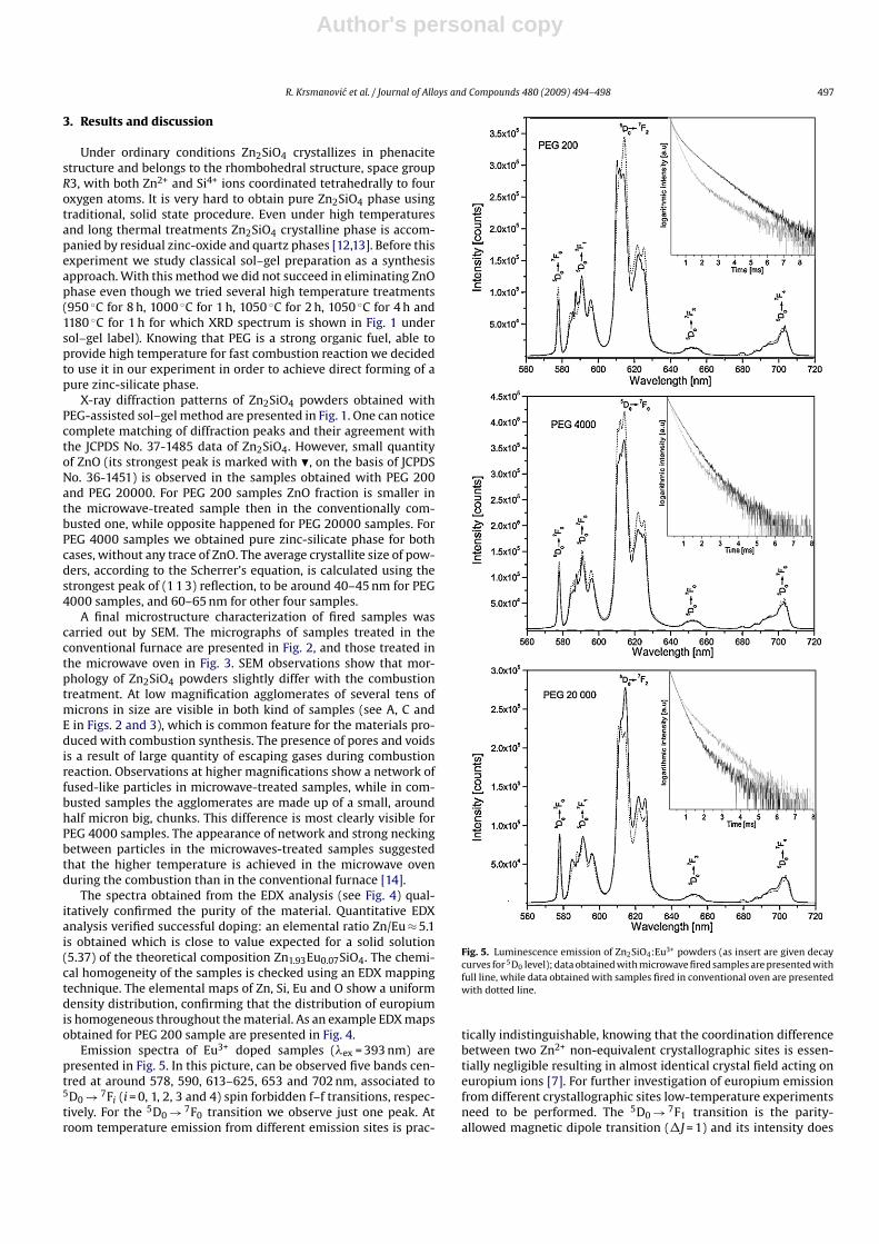
Emission spectra of Eu3+ doped samples (�ex = 393 nm) arepresented in Fig. 5. In this picture, can be observed five bands cen-tred at around 578, 590, 613–625, 653 and 702 nm, associated to5D0 → 7Fi (i = 0, 1, 2, 3 and 4) spin forbidden f–f transitions, respec-tively. For the 5D0 → 7F0 transition we observe just one peak. Atroom temperature emission from different emission sites is prac-
Fig. 5. Luminescence emission of Zn2SiO4:Eu3+ powders (as insert are given decaycurves for 5D0 level); data obtained with microwave fired samples are presented withfull line, while data obtained with samples fired in conventional oven are presentedwith dotted line.
tically indistinguishable, knowing that the coordination differencebetween two Zn2+ non-equivalent crystallographic sites is essen-tially negligible resulting in almost identical crystal field acting oneuropium ions [7]. For further investigation of europium emissionfrom different crystallographic sites low-temperature experimentsneed to be performed. The 5D0 → 7F1 transition is the parity-allowed magnetic dipole transition (�J = 1) and its intensity does
Author's personal copy
498 R. Krsmanovic et al. / Journal of Alloys and Compounds 480 (2009) 494–498
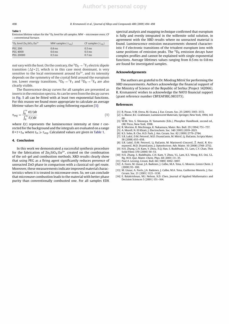
Table 1Emission lifetime values for the 5D0 level for all samples. MW – microwave oven; CF– conventional furnace.
5D0 level Zn2SiO4:Eu3+ MW samples (�avg) CF samples (�avg)
PEG 200 0.8 ms 0.5 msPEG 4000 0.6 ms 0.5 msPEG 20000 0.5 ms 0.7 ms
not vary with the host. On the contrary, the 5D0 → 7F2 electric dipoletransition (�J = 2), which is in this case most dominant, is verysensitive to the local environment around Eu3+, and its intensitydepends on the symmetry of the crystal field around the europiumion. Lower energy transitions, 5D0 → 7F3 and 5D0 → 7F4 are alsoclearly visible.
The fluorescence decay curves for all samples are presented asinserts in the emission spectra. As can be seen from the decay curvesin Fig. 5 all can be fitted with at least two exponential functions.For this reason we found more appropriate to calculate an averagelifetime values for all samples using following equation [3]:
�avg =∫ ∞
0tI(t)dt
∫ ∞0
I(t)dt(1)
where I(t) represents the luminescence intensity at time t cor-rected for the background and the integrals are evaluated on a range0 < t < tm where tm � �avg. Calculated values are given in Table 1.
4. Conclusion
In this work we demonstrated a successful synthesis procedurefor the fabrication of Zn2SiO4:Eu3+, created on the combinationof the sol–gel and combustion methods. XRD results clearly showthat using PEG as a firing agent significantly reduces presence ofunreacted ZnO phase in comparison with a classical sol–gel route.Moreover, these measurements indicate improved material charac-teristics when it is treated in microwave oven. So, we can concludethat microwave combustion leads to the material with better phasepurity than conventionally combusted one. For all samples EDX
spectral analysis and mapping technique confirmed that europiumis fully and evenly integrated in the willemite solid solution, inagreement with the XRD results where no unreacted material isfound. Luminescence emission measurements showed character-istic f–f electronic transitions of the trivalent europium ions withsame positions of emission peaks. The 5D0 emission decays havecomplex profiles and cannot be explained with single exponentialfunctions. Average lifetimes values ranging from 0.5 ms to 0.8 msare found for investigated samples.
Acknowledgements
The authors are grateful to Dr. Miodrag Mitric for performing theXRD measurements. Authors acknowledge the financial support ofthe Ministry of Science of the Republic of Serbia (Project 142066).R. Krsmanovic wishes to acknowledge the NATO financial support(grant reference number CBP.EAP.RIG.983373).
References
[1] R. Pozas, V.M. Orera, M. Ocana, J. Eur. Ceram. Soc. 25 (2005) 3165–3172.[2] G. Blasse, B.C. Grabmaier, Luminescent Materials, Springer, New York, 1994, 143
pp.[3] W.M. Yen, S. Shionoya, H. Yamamoto (Eds.), Phosphor Handbook, second ed.,
CRC Press, New York, 1998.[4] R. Morimo, R. Mochinaga, K. Nakamura, Mater. Res. Bull. 29 (1994) 751–757.[5] A. Morell, N. El Khiati, J. Electrochem. Soc. 140 (1993) 2019–2021.[6] K.S. Sohn, B. Cho, H.D. Park, J. Am. Ceram. Soc. 82 (1999) 2779–2784.[7] S.R. Lukic, D.M. Petrovic, M.D. Dramicanin, M. Mitric, Lj. Ðacanin, Scripta Mater.
58 (2008) 655–658.[8] S.R. Lukic, D.M. Petrovic, Lj. Ðacanin, M. Marinovic-Cincovic, Z. Antic, R. Krs-
manovic, M.D. Dramicanin, J. Optoelectron. Adv. Mater. 10 (2008) 2748–2752.[9] H.X. Zhang, C.H. Kam, Y. Zhou, X.Q. Han, S. Buddhudu, Y.L. Lam, C.Y. Chan, Thin
Solid Films 370 (2000) 50–53.[10] H.X. Zhang, S. Buddhudu, C.H. Kam, Y. Zhou, Y.L. Lam, K.S. Wong, B.S. Ooi, S.L.
Ng, W.X. Que, Mater. Chem. Phys. 68 (2001) 31–35.[11] Paul A. Lessing, Ceram. Bull. 68 (1989) 1002–1007.[12] A. Fores, M. Llusar, J.A. Badenes, J. Calbo, M.A. Tena, G. Monros, Green Chem. 2
(2000) 93–100.[13] M. Llusar, A. Forés, J.A. Badenes, J. Calbo, M.A. Tena, Guillermo Monrós, J. Eur.
Ceram. Soc. 21 (2001) 1121–1130.[14] E. Balakrishnan, M.I. Nelson, X.D. Chen, Journal of Applied Mathematics and
Decision Sciences 5 (2001) 151–164.