plasticity of the mapk signaling network in response to mechanical stress
TRANSCRIPT

Seediscussions,stats,andauthorprofilesforthispublicationat:https://www.researchgate.net/publication/263933719
PlasticityoftheMAPKSignalingNetworkinResponsetoMechanicalStress
ARTICLEinPLOSONE·JULY2014
ImpactFactor:3.23·DOI:10.1371/journal.pone.0101963·Source:PubMed
CITATION
1
READS
32
7AUTHORS,INCLUDING:
CiceroneTudor
CarlZeissAG
16PUBLICATIONS337CITATIONS
SEEPROFILE
ShashankShekhar
I2BC,CNRS,France
16PUBLICATIONS55CITATIONS
SEEPROFILE
VinodSubramaniam
VUUniversityAmsterdam
271PUBLICATIONS6,022CITATIONS
SEEPROFILE
EnriqueMartin-Blanco
SpanishNationalResearchCouncil
44PUBLICATIONS3,395CITATIONS
SEEPROFILE
Availablefrom:EnriqueMartin-Blanco
Retrievedon:08February2016

Plasticity of the MAPK Signaling Network in Response toMechanical StressAndrea M. Pereira1.¤a, Cicerone Tudor2.¤b, Philippe-Alexandre Pouille1., Shashank Shekhar2,
Johannes S. Kanger2, Vinod Subramaniam2*, Enrique Martın-Blanco1*
1 Instituto de Biologıa Molecular de Barcelona (CSIC), Parc Cientific de Barcelona, Baldiri Reixac 10–12, Barcelona, Spain, 2 Nanobiophysics, MESA+ Institute for
Nanotechnology & MIRA Institute for Biomedical Technology and Technical Medicine, University of Twente, Enschede, The Netherlands
Abstract
Cells display versatile responses to mechanical inputs and recent studies have identified the mitogen-activated proteinkinase (MAPK) cascades mediating the biological effects observed upon mechanical stimulation. Although, MAPK pathwayscan act insulated from each other, several mechanisms facilitate the crosstalk between the components of these cascades.Yet, the combinatorial complexity of potential molecular interactions between these elements have prevented theunderstanding of their concerted functions. To analyze the plasticity of the MAPK signaling network in response tomechanical stress we performed a non-saturating epistatic screen in resting and stretched conditions employing as readouta JNK responsive dJun-FRET biosensor. By knocking down MAPKs, and JNK pathway regulators, singly or in pairs inDrosophila S2R+ cells, we have uncovered unexpected regulatory links between JNK cascade kinases, Rho GTPases, MAPKsand the JNK phosphatase Puc. These relationships have been integrated in a system network model at equilibriumaccounting for all experimentally validated interactions. This model allows predicting the global reaction of the network toits modulation in response to mechanical stress. It also highlights its context-dependent sensitivity.
Citation: Pereira AM, Tudor C, Pouille P-A, Shekhar S, Kanger JS, et al. (2014) Plasticity of the MAPK Signaling Network in Response to Mechanical Stress. PLoSONE 9(7): e101963. doi:10.1371/journal.pone.0101963
Editor: Jordi Garcia-Ojalvo, Universitat Pompeu Fabra, Spain
Received March 22, 2014; Accepted June 12, 2014; Published July 15, 2014
Copyright: � 2014 Pereira et al. This is an open-access article distributed under the terms of the Creative Commons Attribution License, which permitsunrestricted use, distribution, and reproduction in any medium, provided the original author and source are credited.
Data Availability: The authors confirm that all data underlying the findings are fully available without restriction. All relevant data are within the paper and itsSupporting Information files.
Funding: A.M.P held a Spanish FPU PhD studentship, and C.T. was supported by the Marie Curie Research Training Network IMMUNANOMAP. Research in theE.M.-B. laboratory is funded by grants of the Ministry of Economy and Competitivity (DGI and Consolider). Research in the V.S. laboratory is supported by‘‘Stitching voor Fundamenteel Onderzoek der Materie’’ (FOM), the Nederlandse Organisatie voor Wetenschappelik Onderzoek (N.W.O.), and the European Union.The funders had no role in study design, data collection and analysis, decision to publish, or preparation of the manuscript.
Competing Interests: The authors have declared that no competing interests exist.
* Email: [email protected] (VS); [email protected] (EMB)
. These authors contributed equally to this work.
¤a Current address: Laboratory for Molecular Cell Biology, Faculty of Life Sciences, University College London, United Kingdom¤b Current address: MPI of Immunobiology and Epigenetics, Freiburg, Germany
Introduction
Cells display versatile responses to mechanical inputs triggering
signals leading to increased gene expression, protein synthesis, or
mitogenesis [1,2]. Altering the mechanical properties of the
environment or subjecting cells to mechanical insults directs
alternative differentiation pathways, promoting cytoskeleton rear-
rangements or altering the composition of the extracellular matrix
[3]. For example, stem cells specification has been shown to be
strongly influenced by the mechanical properties of the surround-
ing matrix [4].
Recent studies have identified some intracellular pathways
mediating the biological effects observed upon mechanical
stimulation. These include the mitogen-activated protein kinase
(MAPK) cascades [5]. Three main MAPK pathways have been
identified: the extracellular signal-regulated kinase (ERK), the c-
Jun N-terminal kinase (JNK) and the p38 cascades. MAPK
cascades are organized as modular pathways in which activation of
upstream kinases leads to sequential activation of a MAPK module
(MAPKKK – MAPKK - MAPK) [6]. Within these cascades,
specificity is maintained primarily through structural mechanisms
that limit protein interactions [7]. Further, all tiers of MAPK
signaling can be regulated by protein phosphatases [8] emphasiz-
ing the importance of the balance between phosphorylation and
dephosphorylation in regulating their functions. Finally, members
of the Ras family of GTPases, including Ras itself and the Rho
subfamily members, Rho, Rac1 and cdc42 trigger the activation of
MAPK signaling. While Ras mainly targets the ERK pathway,
Rho, Rac1 and cdc42 are mainly involved in the activation of JNK
and p38 cascades [9,10].
Conventionally, MAPK cascades have been depicted as linear
signaling pathways insulated from each other. However, undefined
mechanisms facilitating their crosstalk are also known to exist [11].
Indeed, recently published work indicates that ERK activity can
be suppressed by JNK/p38 kinases through the activation of
inhibitory phosphatases (PP2A, MKPs) [12,13]. Yet, despite these
extensive evidences, how different integrative cellular responses
mediated by these cascades are regulated remains unknown. This
is at least in part due to the combinatorial complexity of molecular
interactions and a variety of feedback and feed-forward loops [14].
PLOS ONE | www.plosone.org 1 July 2014 | Volume 9 | Issue 7 | e101963

To analyze the plasticity of the MAPK network in response to
mechanical stress we performed a non-saturating epistatic screen
in resting and stretched conditions. To simplify the analysis we
focused on the response of Drosophila S2R+ cells to stretch
employing as readout the JNK responsive dJun-FRET biosensor
previously shown to be a sensitive reporter of the activation of the
pathway by mechanical stretch [15].
The genome of Drosophila possesses a single ERK encoded by
the gene rolled (rl), a single JNK [basket (bsk)] and two p38 kinases
[mpk2 (p38a) and p38b] [16,17]. It also contains a JNKK
[hemipterous (hep)], a JN3K [slipper (slpr)], a JN4K [misshapen (msn)]
and a JNK dual-specificity phosphatase [puckered (puc)] [18,19]].
Several Rho family members have also been identified in Drosophila
[20]. Further, several other MAPKK, MAP3K, MAP4K, MAPK
phosphatases and three Ras homologues are also present in flies
[21].
By knocking down MAPKs, and JNK pathway regulators, singly
or in pairs by RNA interference (RNAi) in S2R+ cells, we have
uncovered unexpected regulatory links between JNK cascade
kinases, Rho GTPases, MAPKs and the JNK phosphatase Puc.
These relationships are highlighted in a system network model at
equilibrium accounting for the experimentally identified interac-
tions. This model allows predicting the global reaction of the
network to disparate inputs and, in particular, its modulation in
response to mechanical stress. It also highlights its context-
dependent sensitivity.
Results
Different roles for upstream dJun kinases in themechanical stretch activation of dJun
We assessed in real-time the activity of the MAPK network in
S2R+ cells by Fluorescence Lifetime Imaging Microscopy (FLIM)
using an intramolecular phosphorylation-dependent dJun-FRET
(Fluorescence Resonance Energy Transfer) biosensor. This
biosensor has been shown in Drosophila BG2 cells to respond
specifically to JNK activity [22]. Further, known chemical
activators of the JNK pathway yield to a positive response of the
dJun-FRET biosensor in Drosophila S2R+ cells that correlates with
increased P-Jun levels [15]. These cells subjected to static
mechanical stretch also show a significant increase in dJun-FRET
biosensor response, reaching a stable steady state in approximately
20 minutes, which lasts for at least 3 hours [15].
To elucidate the contribution of individual JNK cascade
elements in modulating the response of the dJun-FRET biosensor
at rest or in the presence of mechanical stretch, we targeted bsk,
hep, slpr and msn co-transfecting individual dsRNAs with the
biosensor into S2R+ cells (see Methods).
At rest, the S2R+ cells treated with single dsRNAs against bsk,
hep, slpr and msn show average FL values of 2.0860.14 ns,
2.2760.18 ns, 2.3060.18 ns and 2.2760.10 ns respectively
(Figure 1A to D and Table S1). These lifetimes were significantly
lower than the donor mCFP lifetime at rest (2.4360.15 ns) for
untreated cells (WTR) or cells treated with dsRNA against GFP.
Summarizing, Bsk and its upstream kinases in S2R+ cells,
unexpectedly and unlike in BG2 cells [22], behave as inhibitors
of the biosensor in resting conditions, inhibition being the highest
for Bsk.
As previously reported [15], static mechanical stretch of S2R+cells for 2 hours resulted in a strong activation of the dJun-FRET
biosensor, decreasing its FL to 2.0060.15 ns (WTS), an average
decrease of 0.43 ns versus WTR. Knocking down the JNK cascade
kinases also altered the response of S2R+ cells to stretch. bsk2
stretched cells showed average FL values of 1.9160.14 ns, while
hep2, slpr2 and msn2 displayed FLs of 2.1460.17 ns, 2.1960.20 ns
and 2.2260.10 ns respectively. In all cases, stretching cells led to a
variable additive activation [0.17 ns (bsk2), 0.13 ns (hep2), 0.11 ns
(slpr2), and 0.07 ns (msn2), highest for bsk2] of the dJun-FRET
biosensor when compared to resting dsRNA treated cells
(Figure 1A to D and Tables S2 and S3). Comparing FL values
for dsRNA treated cells under stretch to FL values of WT cells
under stretch (WTS), we observed that while the absence of Bsk
results in a further response from the biosensor, hep2, slpr2 and
msn2 cells showed less activity than the WTS cells. Therefore, the
presence of Hep, Slpr or Msn is necessary for the full activation of
the biosensor in response to stretch, while Bsk seems to act
independently in parallel restraining the activity/phosphorylation
of the biosensor both in resting and stretch conditions. This
confirmed a net negative input of Bsk on dJun phosphorylation in
S2R+ cells, not only in resting conditions but also under stretch.
Rl (ERK) but not P38s is an activator of dJunphosphorylation
ERKs and p38s have been implicated in the activation of AP1,
and particularly Jun, in many processes, including stress responses.
S2R+ cells were co-transfected with the dJun-FRET biosensor and
dsRNAs for rl, mpk2 (p38a) and p38b individually and FLIM values
were measured at rest and in stretched conditions (2 hours).
Cells knocked down for p38a and p38b barely activated the
dJun-FRET biosensor at rest (FL 2.3860.16 ns for both p38a2
and p38b2 conditions) (Figure 1E and 1F). Under stretch, p38a2
and p38b2 cells display FL values of 2.2360.12 ns and
2.3160.17 ns respectively (Figure 1E and 1F). Similar to the
JNK cascade elements loss of function conditions, these cells show
an additive activation [0.15 ns (p38a2) and 0.07 ns (p38b2)] in the
presence of mechanical stretch. If, as above, we compare FL values
for p38a2 and p38b2 cells under stretch to FL values of WT cells
under stretch (WTS), they showed lower activity (Figure 1E and
1F). Hence, P38a and P38b are necessary for the full activation of
the dJun-FRET biosensor in response to stretch.
In resting conditions, cells knocked down for rl, in contrast to
JNK cascade elements and p38s, showed an increase in average FL
value from 2.4360.15 ns in WTR cells to 2.5260.15 ns
(Figure 1G). Consequently, Rl, unlike Bsk, P38a or P38b, behaves
as an activator of dJun phosphorylation at rest in S2R+ cells. In
the presence of mechanical stretch rl2 cells maintain a FL value of
2.5260.15 ns (Figure 1G), indicating that in cells devoid of Rl, the
dJun-FRET biosensor completely loses its ability to respond to the
stretch activation. Thus, Rl is essential both for the activation of
the dJun-FRET biosensor and for its response to stretch.
Puc is an inhibitor of dJun phosphorylationpuc encodes for a well-characterized dual phosphatase that acts
as a negative regulator of the JNK signaling pathway in many
developmental processes in Drosophila [18]. S2R+ cells co-
transfected with the dJun-FRET biosensor and dsRNA for puc
showed an average FL value of 2.1060.16 ns in resting conditions
(Figure 1H). This FL value is lower than for WTR cells
(2.4360.15 ns), indicating that Puc, as expected, inhibits the
dJun-FRET biosensor phosphorylation. When puc2 S2R+ cells
were subjected to mechanical stretch they activated the dJun-
FRET biosensor to the same extent as the WTS cells with a FL
value of 2.0260.17 ns (Figure 1H). Thus, Puc is apparently not
required for the dJun-FRET biosensor activation by mechanical
stretch, even though it acts as an inhibitor of the phosphorylation
of dJun in resting conditions.
MAPKs and Mechanics
PLOS ONE | www.plosone.org 2 July 2014 | Volume 9 | Issue 7 | e101963

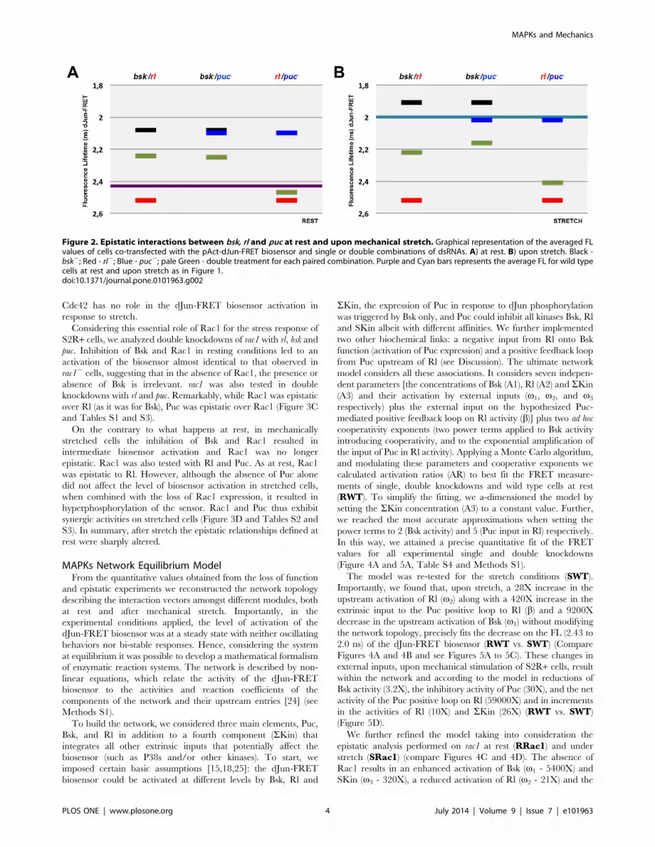
Double knockdowns at rest and in stretched conditionsindicate complex epistatic interactions
To explore the epistatic relationships between Bsk, Rl and Puc
in the response of the dJun-FRET biosensor, we interfered with
their expression in pairs. The response of the dJun-FRET
biosensor by FRET/FLIM was first analyzed in resting conditions
(Table S1). P38s, which did not affect dJun-FRET biosensor
activation were not included in this analysis.
When comparing to WTR control cells, single knockdowns of
bsk and puc resulted in different increments in the phosphorylation
of the dJun-FRET biosensor at rest, while reduction in the levels of
Rl decreased/inhibited its phosphorylation. Double knockdown
of these regulators gave rise to a complex set of results. Inhibition
of Bsk and Rl resulted in a moderate intermediate activation of the
biosensor. On the contrary, the double knockdown of bsk and puc
resulted in a moderate level of biosensor activation, much lower
than that reached in single knockdowns for any of them. Finally,
puc loss of function was tested in double knockdowns with rl. puc
and rl double mutant cells showed a level of activation of the
biosensor close to that observed in rl2 cells (Figure 2A). In
summary, at rest, Rl is epistatic over Puc and Bsk and Puc cancel
each other; furthermore, the opposite activities of Bsk and Rl
appear to be independent.
Under stretch, the activity of the dJun-FRET biosensor (WTS)
was much higher than at rest (WTR), bsk knockdown induced a
further activation of the biosensor, puc did not affect the dJun–
FRET (WTS) biosensor activity and a single knockdown for rl
blocked the stimulation of the biosensor by stretch. This scenario is
remarkably different than that observed at rest anticipating specific
changes in epistatic relationships. Double knockdowns of bsk and rl
resulted, as at rest, in a moderate intermediate activation of the
biosensor when compared to WTS. The double inhibition of Bsk
and Puc resulted in a moderate level of biosensor activation
compared to WTS, much lower than that reached in response to
single knockdowns for any of them. Finally, under stretch, in puc
double knockdown with rl, the activity of the biosensor was
brought back to WTR levels. The absence of Rl (an activator of the
biosensor in single knockdowns) is epistatic to the stretch response
preventing dJun phosphorylation (Figure 2B). In summary, after
stretch some epistatic relationships defined at rest are conserved,
Bsk and Puc cancel each other and the opposite activities of Bsk
and Rl are independent. However, a new interaction was
observed, where Rl and Puc activities became independent,
although they are in some manner coordinated, being both
necessary for the stretch response.
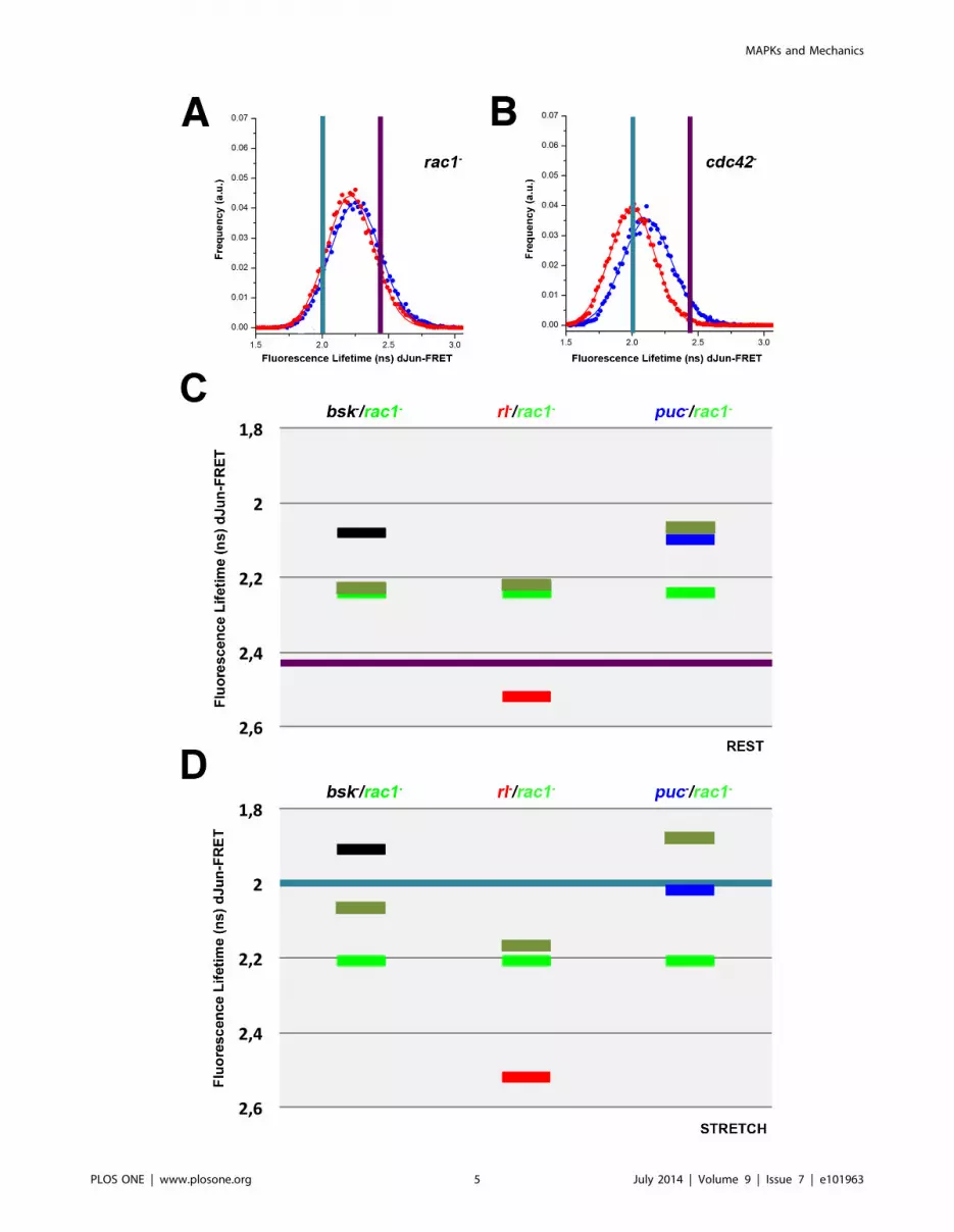
Rho GTPases have distinct roles in the mechanical stretchactivation of dJun
Rac1 and cdc42 are genes coding for Rho GTPases known to
regulate the activity of the JNK pathway [23]. S2R+ cells at rest,
treated with dsRNA for rac1 and cdc42 show a reduction
(compared to WTR cells) in the FL of the dJun-FRET biosensor
(2.2460.19 ns and 2.1160.18 ns respectively) (Figure 3A and 3B
and Tables S1 to S3). Rac1 and Cdc42, as is the case for the JNK
cascade elements, inhibit the phosphorylation of dJun. In the
presence of stretch, we observed as well an additive activation of
the biosensor, 0.11 ns, for cdc422 cells (2.0060.15 ns) but no
differences for rac12 cells (0.03 ns) (2.2160.18 ns) (Figure 3A and
3B and Tables S1 to S3). Thus, Rac1 activity is apparently
epistatic over mechanical stretch and in the absence of Rac1, the
FL values of resting and mechanically stretched cells are essentially
the same. When compared to WTS, cdc422 stretched cells showed
the same level of dJun-FRET biosensor activation suggesting that
Figure 1. Distinct roles for kinases and phosphatases during mechanical stretch activation. Drosophila S2R+ cells were co-transfectedwith the pAct-dJun-FRET and different dsRNAs. A) bsk2. B) hep2. C) slpr2. D) msn2. E) p38a2. F) p38b2. G) rl2. H) puc2. Fluorescence lifetimes (FL)for the donor mCFP were collected and curves representing data recorded from ,75 cells for cells at rest. Blue data points denote the measurementsobtained at rest while red data points show the measurements obtained after 3 hours of continuous static stretch. In each panel, the purple barrepresents the average FL determined for control wild type cells at rest, while the cyan bar represents the average FL of control wild type cellsstretched for 3 hours.doi:10.1371/journal.pone.0101963.g001
MAPKs and Mechanics
PLOS ONE | www.plosone.org 3 July 2014 | Volume 9 | Issue 7 | e101963
Cdc42 has no role in the dJun-FRET biosensor activation in
response to stretch.
Considering this essential role of Rac1 for the stress response of
S2R+ cells, we analyzed double knockdowns of rac1 with rl, bsk and
puc. Inhibition of Bsk and Rac1 in resting conditions led to an
activation of the biosensor almost identical to that observed in
rac12 cells, suggesting that in the absence of Rac1, the presence or
absence of Bsk is irrelevant. rac1 was also tested in double
knockdowns with rl and puc. Remarkably, while Rac1 was epistatic
over Rl (as it was for Bsk), Puc was epistatic over Rac1 (Figure 3C
and Tables S1 and S3).
On the contrary to what happens at rest, in mechanically
stretched cells the inhibition of Bsk and Rac1 resulted in
intermediate biosensor activation and Rac1 was no longer
epistatic. Rac1 was also tested with Rl and Puc. As at rest, Rac1
was epistatic to Rl. However, although the absence of Puc alone
did not affect the level of biosensor activation in stretched cells,
when combined with the loss of Rac1 expression, it resulted in
hyperphosphorylation of the sensor. Rac1 and Puc thus exhibit
synergic activities on stretched cells (Figure 3D and Tables S2 and
S3). In summary, after stretch the epistatic relationships defined at
rest were sharply altered.
MAPKs Network Equilibrium ModelFrom the quantitative values obtained from the loss of function
and epistatic experiments we reconstructed the network topology
describing the interaction vectors amongst different modules, both
at rest and after mechanical stretch. Importantly, in the
experimental conditions applied, the level of activation of the
dJun-FRET biosensor was at a steady state with neither oscillating
behaviors nor bi-stable responses. Hence, considering the system
at equilibrium it was possible to develop a mathematical formalism
of enzymatic reaction systems. The network is described by non-
linear equations, which relate the activity of the dJun-FRET
biosensor to the activities and reaction coefficients of the
components of the network and their upstream entries [24] (see
Methods S1).
To build the network, we considered three main elements, Puc,
Bsk, and Rl in addition to a fourth component (SKin) that
integrates all other extrinsic inputs that potentially affect the
biosensor (such as P38s and/or other kinases). To start, we
imposed certain basic assumptions [15,18,25]: the dJun-FRET
biosensor could be activated at different levels by Bsk, Rl and
SKin, the expression of Puc in response to dJun phosphorylation
was triggered by Bsk only, and Puc could inhibit all kinases Bsk, Rl
and SKin albeit with different affinities. We further implemented
two other biochemical links: a negative input from Rl onto Bsk
function (activation of Puc expression) and a positive feedback loop
from Puc upstream of Rl (see Discussion). The ultimate network
model considers all these associations. It considers seven indepen-
dent parameters [the concentrations of Bsk (A1), Rl (A2) and SKin
(A3) and their activation by external inputs (v1, v2, and v3
respectively) plus the external input on the hypothesized Puc-
mediated positive feedback loop on Rl activity (b)] plus two ad hoc
cooperativity exponents (two power terms applied to Bsk activity
introducing cooperativity, and to the exponential amplification of
the input of Puc in Rl activity). Applying a Monte Carlo algorithm,
and modulating these parameters and cooperative exponents we
calculated activation ratios (AR) to best fit the FRET measure-
ments of single, double knockdowns and wild type cells at rest
(RWT). To simplify the fitting, we a-dimensioned the model by
setting the SKin concentration (A3) to a constant value. Further,
we reached the most accurate approximations when setting the
power terms to 2 (Bsk activity) and 5 (Puc input in Rl) respectively.
In this way, we attained a precise quantitative fit of the FRET
values for all experimental single and double knockdowns
(Figure 4A and 5A, Table S4 and Methods S1).
The model was re-tested for the stretch conditions (SWT).
Importantly, we found that, upon stretch, a 28X increase in the
upstream activation of Rl (v2) along with a 420X increase in the
extrinsic input to the Puc positive loop to Rl (b) and a 9200X
decrease in the upstream activation of Bsk (v1) without modifying
the network topology, precisely fits the decrease on the FL (2.43 to
2.0 ns) of the dJun-FRET biosensor (RWT vs. SWT) (Compare
Figures 4A and 4B and see Figures 5A to 5C). These changes in
external inputs, upon mechanical stimulation of S2R+ cells, result
within the network and according to the model in reductions of
Bsk activity (3.2X), the inhibitory activity of Puc (30X), and the net
activity of the Puc positive loop on Rl (59000X) and in increments
in the activities of Rl (10X) and SKin (26X) (RWT vs. SWT)
(Figure 5D).
We further refined the model taking into consideration the
epistatic analysis performed on rac1 at rest (RRac1) and under
stretch (SRac1) (compare Figures 4C and 4D). The absence of
Rac1 results in an enhanced activation of Bsk (v1 - 5400X) and
SKin (v3 - 320X), a reduced activation of Rl (v2 - 21X) and the
Figure 2. Epistatic interactions between bsk, rl and puc at rest and upon mechanical stretch. Graphical representation of the averaged FLvalues of cells co-transfected with the pAct-dJun-FRET biosensor and single or double combinations of dsRNAs. A) at rest. B) upon stretch. Black -bsk2; Red - rl2; Blue - puc2; pale Green - double treatment for each paired combination. Purple and Cyan bars represents the average FL for wild typecells at rest and upon stretch as in Figure 1.doi:10.1371/journal.pone.0101963.g002
MAPKs and Mechanics
PLOS ONE | www.plosone.org 4 July 2014 | Volume 9 | Issue 7 | e101963
MAPKs and Mechanics
PLOS ONE | www.plosone.org 5 July 2014 | Volume 9 | Issue 7 | e101963

extrinsic activation of the Puc to Rl positive loop (b - 790X) when
comparing to the WT condition. Importantly, these changes take
place irrespective of the cells’ biomechanical condition (RWT vs.
RRac1 and SWT vs. SRac1) (Figure 5C) indicating that Rac1
does not influence the impact of mechanical stress in the system
(all network inputs). On the other hand, within the core of the
network, Rac1 depletion at rest (RWT vs. RRac1) results in an
increment of SKin activity (95X) and reductions of Rl (30X) and
the net Puc positive loop on Rl (140X) activities, while the
activities of Bsk and Puc are unaffected (Figure 5D). Upon stretch,
the absence of Rac1 (SWT vs. SRac1) results in the increment in
activities of Bsk (3,2X), SKin (4X), Puc (29X) and the net Puc
positive loop on Rl (24000X) while Rl activity decreases (7X)
(Figure 5D). Remarkably, in the absence of Rac1 (RRac1 vs.
SRac1), cell stretching results in an increase of Rl activity (30X)
and the net Puc positive loop on Rl (140X), barely affecting Bsk,
SKin and Puc activities (Table S4 and Figure 5D).
Gain of Function AnalysisThe overexpression of Puc resulted in a mild inhibition of the
biosensor phosphorylation at rest (2.4960.14 ns) (RPuc+). Upon
stretch, this overexpression resulted again in a partial inhibition of
Figure 3. Rho GTPases have distinct roles during mechanical stretch activation; Epistatic relationships of rac1 over bsk, rl and puc.Drosophila S2R+ cells were co-transfected with the pAct-dJun-FRET biosensor and different dsRNAs. A) rac12. B) cdc422. Fluorescence lifetimes (FL)for the donor mCFP were collected and curves representing data recorded from ,75 cells for each condition are displayed. Blue and red data pointsdenote the measurements obtained at rest or upon stretch as in Figure 1. Graphical representation of the averaged FL values of cells co-transfectedwith the pAct-dJun-FRET biosensor and single or double combinations of dsRNAs. C) at rest. D) upon stretch. Black - bsk2; Red - rl2; Blue - puc2;bright Green - rac12; pale Green - double treatment for each pairwise combination. Purple and Cyan bars represents the average FL for wild type cellsat rest and upon stretch as in Figure 1.doi:10.1371/journal.pone.0101963.g003
Figure 4. MAPKs Network Dynamic Model. To build the network, we took into account three elements, Puc, Bsk, and Rl plus an additionalcomponent (SKin) integrating all other potentially involved kinases (such as P38s). We considered that the dJun-FRET biosensor could be activated atdifferent levels by Bsk, Rl and SKin, that the expression of Puc in response to dJun phosphorylation was only triggered by Bsk, and that Puc inhibit allkinases Bsk, Rl and SKin with different affinities. We further established two other biochemical links: a negative input from Rl onto Bsk function(activation of Puc expression) and a positive feedback loop from Puc upstream of Rl. We then determined a set of parameters allowing calculatedactivation ratios to best fit the FRET measurements of single, and double knockdowns and the control condition at rest (A) or upon stretch (B). Wefurther evaluated the model taking into consideration the epistatic analysis performed on rac1 at rest (C) and upon stretch (D). Componentsconcentrations (font size) and levels of activation or repression (rainbow look up table) are displayed according to logarithmic scales following thevalues defined in Table S4.doi:10.1371/journal.pone.0101963.g004
MAPKs and Mechanics
PLOS ONE | www.plosone.org 6 July 2014 | Volume 9 | Issue 7 | e101963

Figure 5. Mechanical stretch and the absence of Rac1 alter the magnitude of extrinsic inputs and intrinsic interactions within theMAPK network. The different experimental conditions and control experiments yielded specific FL values from the dJun-FRET biosensor FLIMmeasurements. Fitted AR measurements applying the network model very precisely reproduced the experimental data (AR) for all conditions at restand upon stretch. A) Dark grey – Experimental AR at rest; Dark blue – Fitted AR at rest. B) Pale grey – Experimental AR upon stretch; Dark red – FittedAR upon stretch. C) The extrinsic inputs into the network ( Bsk, Rl; SKin; Puc loop) show different activation levels at rest (dark blue) and uponstretch (dark red) except for SKin. When rac1 expression is abolished, these values are altered but their ratio is sustained (pale blue at rest; pale redupon stretch). See also Figure 4. D) The intrinsic positive and negative interactions (activity levels) between the network different nodes (Bsk , Rl ;SKin ; Puc ; Puc loop ) are distinctively modified both upon stretch and, synergically, in the absence of Rac1. Components concentrations andlevels of activation are displayed as in Figure 4.doi:10.1371/journal.pone.0101963.g005
MAPKs and Mechanics
PLOS ONE | www.plosone.org 7 July 2014 | Volume 9 | Issue 7 | e101963

the biosensor activity (2.3060.14 ns) (SPuc+) (Table S4). In both
cases, as expected, the biosensor response was opposite to that
observed in puc loss of function conditions.
Our model predicts that an increase of Puc levels should not
affect the extrinsic inputs to the network (v1, v2, v3 and b) and
indeed this is the case. On the other hand, the modeled Puc
overexpression points to an increase in Puc levels and increments
of different magnitudes in the Puc inhibitory capacity and the net
Puc positive loop on Rl activity both at rest and after stretching
[4,5X and 1800X (RWT vs. RPuc+) vs. 100X and 1,161010X
(SWT vs. SPuc+) respectively]. Further, Puc overexpression, when
compared to the WT condition, does not affect Bsk activity at rest,
but promotes its decrease 70X upon stretch. Meanwhile, Rl and
Skin activities are both reduced 4X at rest and decreased upon
stretch 2X and 85X respectively (Figure 6).
Summarizing, none of the interactions between the different
elements or the network topology are modified in response to Puc
overexpression. A simple quantitative modulation of the intrinsic
interactions between the network nodes fit the model parameters
to the experimental FL values both before and after mechanical
stretch.
Discussion
An extensive literature supports that MAPK pathways activities
are linked by undefined mechanisms facilitating their crosstalk
[21]. By resorting to the activity of a dJun-FRET biosensor in
Drosophila S2R+ cells in culture [15] we propose a functional
network model linking individual MAPK cascades at rest or in the
presence of mechanical stretch.
Surprisingly, we found that knocking down different elements of
the JNK cascade resulted in an increase in the phosphorylation of
the dJun-FRET biosensor in either condition, while inactivating
the inhibitory dual-specificity MAPK phosphatase Puc also led to
its activation. This drew a distinction with the observed biosensor
inhibition consequence of knocking down Rl, an ERK homologue.
The apparent contradiction between the known direct activation
of dJun by Bsk and the activation of the biosensor after knocking
down bsk and other members of the JNK cascade was solved by
generating a network model taking into account cross-regulatory
links between the JNK and ERK pathways.
To generate a MAPK network model by non-linear equations
we considered a set of different literature supported evidences.
First, the AP1 complex, mediating the transcription of puc [18], is
Figure 6. puc gain of function does not affect the MAPK network topology but influences intrinsic network interactions. Wecalculated activation ratios to best fit the FRET measurements upon Puc overexpression at rest (A) or upon stretch (B). The extrinsic inputs into thenetwork ( Bsk, Rl; SKin; Puc loop) (C) and the intrinsic positive and negative interactions (activity levels) between the network’s different nodes(Bsk , Rl ; SKin ; Puc ; Puc loop ) (D) were determined by fitting. Components concentrations and levels of activation or repression are displayedas in Figure 4.doi:10.1371/journal.pone.0101963.g006
MAPKs and Mechanics
PLOS ONE | www.plosone.org 8 July 2014 | Volume 9 | Issue 7 | e101963

composed of Jun and Fos, both of them being phosphorylated by
Bsk. However, mammalian ERK can also phosphorylate Fos,
albeit on distinct residues, resulting in the transcriptional
regulation of different target genes by the AP1 complex [26].
This suggests that in S2R+ cells Rl may act as a repressor of the
JNK mediated expression of puc. Second, the Puc dual-specificity
phosphatase, which mainly operates on the phosphorylated form
of Bsk can also impinge on ERK (Rl) signaling [25] and,
potentially, on other kinases. Finally, as stated above, bsk and puc
knockdowns increase the FRET signal/activation of the dJun-
FRET biosensor, suggesting that both proteins behave as effective
inhibitors. However, previous work has shown that Bsk is a direct
activator of dJun driving the expression of Puc, which feeds back
negatively to the activity of JNK. Considering the results of their
single knockdowns one would assume that the double knockdown
of these genes should activate the biosensor even more. However,
this is not the case, implying the existence of a positive feedback
loop from Puc upstream of the MAPKs. Indeed, it has been shown
that SEK1, a kinase upstream of MAPKs is negatively regulated
by phosphorylation [27] and it has been further reported that JNK
is indirectly activated by JKAP, a dual-specificity phosphatase, and
by its human orthologue JSP1 [28]. Thus, a positive loop from Puc
impacting on Rl activity might be potentially feasible.
The model we developed indicates that the effective inhibition
of the dJun-FRET biosensor by Bsk does not imply different
affinities of the proteins involved or a specific change of the
network topology in S2R+ cells. A negative regulation of MAPK
activities by Puc, a negative input of Rl on Bsk activity, and an
activation of the ERK pathway by Puc are sufficient to account for
all the experimental measurements of the biosensor activity. This
holds good both at rest and upon stretch. The model also denotes
that in S2R+ cells the concentration of Rl is 4X higher than Bsk
(and these are not altered upon stretch). Further, Puc concentra-
tion is 1000X that of Bsk at rest, and drops 30X upon stretch thus
reducing its influence on the activities of Bsk, Rl and Skin.
The implication of Rac1 in the response to mechanical stress in
multiple cell lines has been thoroughly sustained [29,30].
However, we found that the level of activation of the biosensor
is not affected by the cells biomechanical condition (at rest or upon
stretch) when Rac1 is inhibited in S2R+ cells (Figure 3A).
Importantly, our model indicates that the extrinsic inputs to the
network (v1, v2, v3 and b) are, once Rac1 expression is inhibited,
much bigger for v1 and v3 and smaller for v2 and b) than for WT
cells. However, they are modulated in the same proportions
between cells at rest and under stretch irrespectively of the
presence of Rac1. Still, without Rac1, the intrinsic interactions
between the different nodes display disparate responses than WT
cells. In particular, the activities of Bsk, Skin and Puc, which are
different between cells at rest and upon stretch in the WT
condition, are essentially locked at a particular level in the absence
of Rac1. In contrast, Rl activity is different in cells at rest and
under strech when Rac1 is not present and decreases differently at
rest (22X) and upon stretch (7X) when compared to WT cells. This
emphasizes the key role of Rl modulating the level of activation of
the biosensor (Figure 1G).
In the proposed model, the activation of the dJun-FRET
biosensor varies within a specific dynamic range in response to the
concerted actions of multiple negative and positive loops. It is
intriguing to find that in comparing the different experimental
conditions assesed some kinases duplicate or triplicate their
activity, while others change their levels of activity up to 5 orders
of magnitude. Although at the origin of these differences we could
place the disparity between the fine-tuning of activity levels vs the
activation from a negligible ground state, systems-level precise
behavioral regulation may also be very influencial. Thus, global
effects such as competition for substrates, multisite phosphoryla-
tion and kinetic proofreading regulating specificity by phospha-
tases in complex mixtures of proteins [31] can account for
dramatic differences in individual network-elements activities.
Signaling cascades can transduce information in different ways
[32]. Cascades may behave gradually when the activity of the
terminal kinase quantitatively reflect the concentration of the
extracellular stimulus. Alternatively, the cascade may act as an
ultrasensitive switch that responds in a all-or-none manner:
amplification and cellular commitment only occur once a
threshold stimulus is reached. Theoretical studies revealed that
minimal models of multi-step protein kinase cascades show
gradual dose-response behavior at steady state [33]. Indeed, the
intrinsic hierarchical nature of MAPK pathways cascades prompts
to major signal amplification outcomes. A classical example is the
neurite outgrowth induced by NGF vs the proliferative signal
without neurite formation promoted by EGF in PC12 cells via the
same signal transduction MAPK cascade. These differential
responses are thought to be determined by the duration of MAPK
activation; NGF induces sustained MAPK activation for several
hours and translocation to the nuclei, but EGF leads to short-lived
activation [34].
The model also explains how crosstalk within pathways can
integrate responses differing markedly between cells at rest and
under mechanical stress. Thus, may be useful in the understanding
of how mechanical (or eventually chemical or hormonal) inputs
may disturb signal processing. This is particularly important in the
context of cancer and tumor related conditions such as hypoxia, as
many cancer cells and cells exposed to low oxygen levels display
increased expression of dual specificity phosphatases [35]. This
network model may provide possible explanations for the complex
behavior of MAPK systems in different oncogenic paradigms
resorting to MAPKs hyperactivity and it may help clarify the
regulatory mechanisms linked to the transitions from a normal
apoptotic cell to uncontrolled proliferation [14,36].
This canonical model forms a basis for experimental design and
can be tailored to different experimental systems on two levels, by
parameter estimation and by extending the model to incorporate
different MAPK isoforms and upstream, downstream and
structural elements. Such refined models possess quantitative
predictive power and cannot only be used for identifying gaps in
knowledge, but also for elucidating the effect of drugs, thus
building the theoretical basis for identifying optimal treatment
strategies.
Materials and Methods
Cell cultureDrosophila S2R+ cells were grown in Schneider’s Drosophila
medium (GIBCO, Invitrogen) supplemented with 10% heat-
inactivated fetal bovine serum (GIBCO, Invitrogen) at 25uC.
Penicillin and streptomycin were included at 100 units/ml and
100 mg/ml, respectively.
RNAi and overexpression treatments200,000 cells were seeded in a 24 well plate and incubated at
25uC overnight. Cells were co-transfected with different dsRNAs
(,5 ug of RNAi in each reaction) or ,5 mg of a puc overexpres-
sion construct (pAct 5C-Puc) and 2 mg/ml pAct-dJun-FRET
biosensor simultaneously, at ,80% confluence using Effectene
(Qiagen) following the manufacturer’s instructions. Transfected
cells were incubated for 4 days and then re-plated on collagen-
coated silicone membranes in medium deprived of serum, one day
MAPKs and Mechanics
PLOS ONE | www.plosone.org 9 July 2014 | Volume 9 | Issue 7 | e101963

prior to vacuum-assisted stretch FLIM analysis. The dsRNAs and
information about potential off-targets were obtained from the
DRSC (http://flyrnai.org). Cells transfected with dsRNAs were
re-plated on collagen-coated silicone membranes, in medium
deprived of serum, one day prior to vacuum-assisted stretch FLIM
analysis.
ModelingTo model the interaction network leading to the activation of
the dJun-FRET biosensor in resting and stretch conditions we
applied a system of non-linear equations. Details are presented in
the Methods S1.
Supporting Information
Table S1 Fluorescence Lifetimes (FL) of S2R+ cellssubjected to distinct single and double knockdowns atrest. FL measurements not significantly differing of the wild type
(WT) values are displayed in blue. FL values significantly smaller
than WT ones are displayed in red. FL values significantly bigger
than WT ones are displayed in green.
(PDF)
Table S2 Fluorescence Lifetimes (FL) of S2R+ cellssubjected to distinct single and double knockdownsupon stretch. FL measurements not significantly differing of the
wild type (WT) values are displayed in blue. FL values significantly
smaller than WT ones are displayed in red. FL values significantly
bigger than WT ones are displayed in green.
(PDF)
Table S3 Differences of Fluorescence Lifetimes (FL) atrest vs stretch conditions of S2R+ cells subjected todistinct single and double knockdowns. FL measurements
not significantly differing between both conditions are displayed in
blue. FL values significantly smaller upon stretch versus resting
conditions are displayed in red.
(PDF)
Table S4 Consolidated parametric fitted values for theMAPK Network for the distinct single and doubleknockdowns and the overexpression of Puc at rest andupon stretch. Experimental AR (FL), fitted AR, A1 ([Bsk]), A2
([Rl]), A3 ([Skin]), Puc ([Puc] and ,Puc), Omega1 (.BskExt), Omega2 (.Rl Ext), Omega3 (.Skin Ext), Beta (.Puc LExt), K1 (,Bsk), K2 (,Rl), K3 (,Skin), K1‘2.[(1-K2)/(1+K2)]
[.Puc ,(Bsk.Rl)], Puc‘5 (,Puc L) and Beta.Puc‘5 (,PucL.Puc L Ext) values for each experimentally analyzed condition
at rest and upon stretch. Shadowed in green are the values
represented in Figures 4A to 4D, Figures 5C and 5D and
Figures 6A to 6D. Shadowed in Orange are the values presented
in Figure 5A and 5B.
(PDF)
Methods S1 Supporting Methods.
(PDF)
Acknowledgments
We wish to thank all members of our laboratories for encouragements and
constructive criticisms.
Author Contributions
Conceived and designed the experiments: VS EMB. Performed the
experiments: AP CT SS JSK PAP. Analyzed the data: PAP VS EMB.
Contributed to the writing of the manuscript: VS EMB. Designed the
software used in analysis: PAP.
References
1. Liu M, Skinner SJ, Xu J, Han RN, Tanswell AK, et al. (1992) Stimulation of
fetal rat lung cell proliferation in vitro by mechanical stretch. Am J Physiol 263:
L376–383.
2. Wilson E, Mai Q, Sudhir K, Weiss RH, Ives HE (1993) Mechanical strain
induces growth of vascular smooth muscle cells via autocrine action of PDGF.
J Cell Biol 123: 741–747.
3. Girard PR, Nerem RM (1995) Shear stress modulates endothelial cell
morphology and F-actin organization through the regulation of focal
adhesion-associated proteins. J Cell Physiol 163: 179–193.
4. Engler AJ, Sen S, Sweeney HL, Discher DE (2006) Matrix elasticity directs stem
cell lineage specification. Cell 126: 677–689.
5. Komuro I, Kudo S, Yamazaki T, Zou Y, Shiojima I, et al. (1996) Mechanical
stretch activates the stress-activated protein kinases in cardiac myocytes. FASEB J
10: 631–636.
6. Dhillon AS, Hagan S, Rath O, Kolch W (2007) MAP kinase signalling pathways
in cancer. Oncogene 26: 3279–3290.
7. Kolch W (2005) Coordinating ERK/MAPK signalling through scaffolds and
inhibitors. Nat Rev Mol Cell Biol 6: 827–837.
8. Junttila MR, Li SP, Westermarck J (2008) Phosphatase-mediated crosstalk
between MAPK signaling pathways in the regulation of cell survival. FASEB J
22: 954–965.
9. Renshaw MW, Toksoz D, Schwartz MA (1996) Involvement of the small
GTPase rho in integrin-mediated activation of mitogen-activated protein kinase.
J Biol Chem 271: 21691–21694.
10. Minden A, Lin A, Claret FX, Abo A, Karin M (1995) Selective activation of the
JNK signaling cascade and c-Jun transcriptional activity by the small GTPases
Rac and Cdc42Hs. Cell 81: 1147–1157.
11. Xiao YQ, Malcolm K, Worthen GS, Gardai S, Schiemann WP, et al. (2002)
Cross-talk between ERK and p38 MAPK mediates selective suppression of pro-
inflammatory cytokines by transforming growth factor-beta. J Biol Chem 277:
14884–14893.
12. Li SP, Junttila MR, Han J, Kahari VM, Westermarck J (2003) p38 Mitogen-
activated protein kinase pathway suppresses cell survival by inducing
dephosphorylation of mitogen-activated protein/extracellular signal-regulated
kinase kinase1,2. Cancer Res 63: 3473–3477.
13. Shen YH, Godlewski J, Zhu J, Sathyanarayana P, Leaner V, et al. (2003) Cross-
talk between JNK/SAPK and ERK/MAPK pathways: sustained activation of
JNK blocks ERK activation by mitogenic factors. J Biol Chem 278: 26715–
26721.
14. Fey D, Croucher DR, Kolch W, Kholodenko BN (2012) Crosstalk and signaling
switches in mitogen-activated protein kinase cascades. Front Physiol 3: 355.
15. Pereira AM, Tudor C, Kanger JS, Subramaniam V, Martin-Blanco E (2011)
Integrin-dependent activation of the JNK signaling pathway by mechanical
stress. PLoS One 6: e26182.
16. Riesgo-Escovar JR, Jenni M, Fritz A, Hafen E (1996) The Drosophila Jun-N-
terminal kinase is required for cell morphogenesis but not for DJun-dependent
cell fate specification in the eye. Genes Dev 10: 2759–2768.
17. Han ZS, Enslen H, Hu X, Meng X, Wu IH, et al. (1998) A conserved p38
mitogen-activated protein kinase pathway regulates Drosophila immunity gene
expression. Mol Cell Biol 18: 3527–3539.
18. Martin-Blanco E, Gampel A, Ring J, Virdee K, Kirov N, et al. (1998) puckered
encodes a phosphatase that mediates a feedback loop regulating JNK activity
during dorsal closure in Drosophila. Genes Dev 12: 557–570.
19. Stronach B, Perrimon N (2002) Activation of the JNK pathway during dorsal
closure in Drosophila requires the mixed lineage kinase, slipper. Genes Dev 16:
377–387.
20. Hakeda-Suzuki S, Ng J, Tzu J, Dietzl G, Sun Y, et al. (2002) Rac function and
regulation during Drosophila development. Nature 416: 438–442.
21. Shvartsman SY, Coppey M, Berezhkovskii AM (2009) MAPK signaling in
equations and embryos. Fly (Austin) 3: 62–67.
22. Bakal C, Linding R, Llense F, Heffern E, Martin-Blanco E, et al. (2008)
Phosphorylation networks regulating JNK activity in diverse genetic back-
grounds. Science 322: 453–456.
23. Hall A (2005) Rho GTPases and the control of cell behaviour. Biochem Soc
Trans 33: 891–895.
24. Canela-Xandri O, Sagues F, Reigada R, Buceta J (2008) A spatial toggle switch
drives boundary formation in development. Biophys J 95: 5111–5120.
25. Martin-Blanco E (1998) Regulatory control of signal transduction during
morphogenesis in Drosophila. Int J Dev Biol 42: 363–368.
26. Monje P, Marinissen MJ, Gutkind JS (2003) Phosphorylation of the carboxyl-
terminal transactivation domain of c-Fos by extracellular signal-regulated kinase
mediates the transcriptional activation of AP-1 and cellular transformation
induced by platelet-derived growth factor. Mol Cell Biol 23: 7030–7043.
MAPKs and Mechanics
PLOS ONE | www.plosone.org 10 July 2014 | Volume 9 | Issue 7 | e101963
27. Park HS, Kim MS, Huh SH, Park J, Chung J, et al. (2002) Akt (protein kinase B)
negatively regulates SEK1 by means of protein phosphorylation. J Biol Chem277: 2573–2578.
28. Chen AJ, Zhou G, Juan T, Colicos SM, Cannon JP, et al. (2002) The dual
specificity JKAP specifically activates the c-Jun N-terminal kinase pathway. J BiolChem 277: 36592–36601.
29. Ren K, Liu F, Huang Y, Liang W, Cui W, et al. (2012) Periodic mechanicalstress activates integrinbeta1-dependent Src-dependent PLCgamma1-indepen-
dent Rac1 mitogenic signal in rat chondrocytes through ERK1/2. Cell Physiol
Biochem 30: 827–842.30. Poh YC, Na S, Chowdhury F, Ouyang M, Wang Y, et al. (2009) Rapid
activation of Rac GTPase in living cells by force is independent of Src. PLoSOne 4: e7886.
31. Ubersax JA, Ferrell JE Jr (2007) Mechanisms of specificity in proteinphosphorylation. Nature reviews Molecular cell biology 8: 530–541.
32. Jeschke M, Baumgartner S, Legewie S (2013) Determinants of Cell-to-Cell
Variability in Protein Kinase Signaling. PLoS computational biology 9:e1003357.
33. Heinrich R, Neel BG, Rapoport TA (2002) Mathematical models of protein
kinase signal transduction. Molecular cell 9: 957–970.34. Marshall C (1995) Specificity of receptor tyrosine kinase signaling: transient
versus sustained extracellular signal-regulated kinase activation. Cell 80: 179–185.
35. Sonna LA, Cullivan ML, Sheldon HK, Pratt RE, Lilly CM (2003) Effect of
hypoxia on gene expression by human hepatocytes (HepG2). Physiol Genomics12: 195–207.
36. Kreeger PK, Mandhana R, Alford SK, Haigis KM, Lauffenburger DA (2009)RAS mutations affect tumor necrosis factor-induced apoptosis in colon
carcinoma cells via ERK-modulatory negative and positive feedback circuitsalong with non-ERK pathway effects. Cancer Res 69: 8191–8199.
MAPKs and Mechanics
PLOS ONE | www.plosone.org 11 July 2014 | Volume 9 | Issue 7 | e101963